Embed Size (px)
Citation preview

Impact of Breathing Phases on Social Stimuli Processing
by
Sharmi D. Purkayestha
A thesis submitted in partial fulfillment of the requirements for the degree of
Master of Science (Psychology)
in the University of Michigan - Dearborn 2020
Master’s Thesis Committee: Assistant Professor Zhong Xu Liu, Chair Professor Nancy Wrobel, Co-Chair

i
Acknowledgments
I would like to acknowledge and thank all those who have supported me throughout this
endeavor. First, I would like to offer my deepest gratitude to Dr. Zhong Xu Liu for guiding me,
teaching me, and having patience with me through this thesis during a very tumultuous year.
Before this, I had little knowledge in this field of research, so I am very grateful for you for
allowing me to learn and grow in this field of research. I would like to thank Dr. Wrobel for
coming on board with this thesis and offering me meaningful advice that helped me reign in my
thesis objectives. I want to thank all the professors I have had the privilege of learning from
during my time in this master’s program. They have all provided me with a wonderful learning
experience and perspectives that I will take with me. I also want to thank the lab members who
have helped me collect the data, because without them this thesis would not have been feasible. I
would like to thank my family and friends for being understanding and cheering me on
throughout this program and thesis. Finally, I want to thank my parents, because without you
both this would have been impossible. This year has been especially hard with everything you’ve
both had to deal with, but your unwavering support for me has urged me to go on further!

ii
Table of Contents
Acknowledgements ............................................................................................................ i
List of Figures .................................................................................................................... v
List of Tables ...................................................................................................................... v
List of Appendices ........................................................................................................... vii
Abstract ............................................................................................................................ vii
Chapter I Introduction ..................................................................................................... 1
Neural mechanisms of breathing .................................................................................... 2
Breathing links to emotional and cognitive systems ....................................................... 4
Breathing modulation of neural oscillations, cognition, and mood ................................ 6
Gaps in research .............................................................................................................. 8
The current study ............................................................................................................ 9
Electrophysiology of face processing using EEG components .................................... 10
Hypotheses .................................................................................................................... 12
Behavioral Investigations.......................................................................................... 12
Neural Component Investigations............................................................................. 13
Exploring Individual Characteristics. ....................................................................... 13

iii
Chapter II Methods ........................................................................................................ 15
Participants .................................................................................................................... 15
General Procedures ....................................................................................................... 15
Face Processing Task .................................................................................................... 16
Stimuli. ...................................................................................................................... 16
Counterbalancing. ..................................................................................................... 17
Encoding/Rating. ...................................................................................................... 18
Retrieval. ................................................................................................................... 18
Data Acquisition............................................................................................................ 19
EEG. .......................................................................................................................... 19
Phase Locking. .......................................................................................................... 19
Physiological Data. ................................................................................................... 21
Survey Measures ........................................................................................................... 21
Screener Survey. ....................................................................................................... 21
Depression Anxiety and Stress Scale 21 Items (DASS-21). ..................................... 21
Mindful Acceptance and Awareness Scale (MAAS). ............................................... 22
Emotion Regulation Questionnaire (ERQ). .............................................................. 22
Data Analyses................................................................................................................ 23
EEG Preprocessing. .................................................................................................. 23
ERP Analyses. ........................................................................................................... 25

iv
Behavioral Data: Negative rating during encoding. ................................................. 26
Behavioral Data: Recognition memory during encoding. ........................................ 26
Additionally, we used the .......................................................................................... 26
Impact of Individual Characteristic. ......................................................................... 27
Chapter III Results ......................................................................................................... 28
Behavioral Results ........................................................................................................ 28
ERP Results ................................................................................................................... 30
Exploring Individual Characteristics ............................................................................ 32
Chapter IV Discussion .................................................................................................... 33
Breathing Impact on Behavior ...................................................................................... 34
Breathing effects on electro-neurophysiology .............................................................. 37
Impact of Individual Characteristics ............................................................................. 39
Limitations .................................................................................................................... 39
Conclusions ................................................................................................................... 40
References ........................................................................................................................ 66

v
List of Figures
Figure 1a ....................................................................................................................................... 42
Figure 1b ....................................................................................................................................... 43
Figure 2 ......................................................................................................................................... 45
Figure 3 ......................................................................................................................................... 46
Figure 4 ......................................................................................................................................... 47
Figure 5 ......................................................................................................................................... 48
Figure 6 ......................................................................................................................................... 49
Figure 7 ......................................................................................................................................... 50
Figure 8 ......................................................................................................................................... 51
Figure 9 ......................................................................................................................................... 52
Figure 10 ....................................................................................................................................... 53
Figure 11 ....................................................................................................................................... 53

vi
List of Tables
Table 1 ........................................................................................................................................... 55
Table 2 ........................................................................................................................................... 56
Table 3 ........................................................................................................................................... 57
Table 4 ........................................................................................................................................... 58

vii
List of Appendices
Appendix A: Screener and Demographics Questionnaire ............................................................ 59
Appendix B: Depression Anxiety and Stress - 21 ......................................................................... 61
Appendix C: Mindful Attention Awareness Scale ........................................................................ 63
Appendix D: Emotion Regulation Questionnaire ......................................................................... 65

viii
Abstract
Recent studies have demonstrated the respiratory entrainment of brain cycles, leading to
implications for cognitive and emotional processes. Notably, the preBötzinger complex, an
important breathing area, sends out inhalation-modulated projections to the locus coeruleus,
amygdala, and hippocampus, essential for arousal, emotion, and memory. Using a breathing
phase-locked face processing task (with neutral face pictures), this study investigated breathing
phases’ effect on emotion and memory processing at behavioral and neural levels, using
negativity ratings, memory performance, and ERP (event-related-potential) measures of early
(N170) and later (P300) processing. Participants provided negativity ratings to faces that were
presented either at the inhalation or exhalation phase of breathing cycles, while their neural
activity was being recorded using EEG (electroencephalograph). Their memory for the faces was
later tested and their trait anxiety and depression were measured using questionnaires. It was
hypothesized that the negativity ratings, memory accuracy, N170 and P300 amplitude will be
greater for inhalation versus exhalation phases. Results indicated no differences in negativity
ratings and overall face recognition memory between the two breathing phase conditions.
However, we found evidence that recognition memory was enhanced for faces encoded at
inhalation and retrieved at exhalation. Accuracy of correct rejection was enhanced during the
inhalation versus exhalation phase. There were no breathing phase differences between a priori
selected electrodes P9/P10 for ERP N170 and Pz/POz for P300. However, N170 at other
electrodes in the parietal regions showed a greater negative amplitude for the inhalation versus
exhalation phase. A significant correlation was found between high levels of depression and

ix
negativity rating differences between the phases. Taken together, our results support the idea that
the limbic system, and related cognitive/affective processes, can be modulated by different
phases of breathing cycles, which justifies further mechanistic investigations on how breathing
rhythms and techniques affect human emotion and cognition.
Keywords: respiration, breathing, inhalation, exhalation, emotion, memory, N170, P300
IMPACT OF BREATHING PHASES
1
Chapter I Introduction
The breathing cycle is essential for survival. Aside from being a necessary process of life,
studies have long linked breathing activity and psychological symptoms. Specifically, there is a
high prevalence of anxiety and depression in those who have chronic breathing disorders (Kunik
et al., 2005; Maurer et al., 2008; Yohannes et al., 2010). Further, persistent respiratory anomalies
are present in patients with panic disorders (Abelson et al., 2001). Breathing activities’ impact on
psychological functioning is highlighted by the use of breathing techniques in treating anxiety,
trauma, and stress-related disorders (Brown et al., 2013), as they reduce physiological arousal
and moderate emotional responses to anxiety and stress (Varvogli & Darviri, 2011).
Beyond the breathing activity’s association with psychological processes, recent literature
has highlighted the breathing cycle’s entrainment of neural oscillations, or moderation of brain
rhythms, which impacts emotional and cognitive processes (Del Negro et al., 2018; Molle &
Benoit, 2019; Zelano et al., 2016). Thus, it is important to investigate stimuli processing with
respect to breathing phases, as it will allow us to understand how components of a critical
survival rhythm may impact higher-level functioning. Moreover, understanding specific
breathing related impacts may lead to further advancement in understanding the role of
breathing-related techniques in treatment for psychological disorders. In order to provide a
rationale for the current study, this chapter provides a review of neural mechanisms associated
with the breathing rhythm generation and the connections between breathing centers and
structures related to emotion, memory, and arousal. Further, existing evidence supporting the
breathing entrainment of key brain processes, including emotion and memory is discussed.

IMPACT OF BREATHING PHASES
2
Finally, research questions in relation to the current study are derived and hypotheses are
specified.
Neural mechanisms of breathing
Although we can alter our breathing rhythm consciously by making it slower or faster
(Feldman et al., 2009), breathing is an autonomic cycle. The brainstem contains breathing
centers which generate the breathing rhythm and produce the innate drive to breathe.
Importantly, the preBötzinger complex (preBötC) is crucial for forming and managing the
breathing rhythm and eupnea, or normal breathing (Garcia et al., 2011; Rekling & Feldman,
1998; Yang & Feldman, 2018). In mice, when genes were manipulated such that the preBötC
was mutated to an unrecognizable degree, it resulted in the death of mice at birth because they
did not breathe despite having all the necessary systems to breath (Bouvier et al., 2010). In
humans, the preBötC forms and manages the breathing rhythm by projecting onto other
breathing areas of the brainstem (Yang & Feldman, 2018). The preBötC has excitatory
(glutamatergic) and inhibitory (GABAergic and glycinergic) interneurons which work together
to form a pattern to produce the drive for inhalation (Muñoz-Ortiz et al., 2019). The excitatory
interneurons are linked to the timing of inhalations, such that the pharmacologically silencing
them induces life-threatening apnea in rats (Cui et al., 2016; Gray et al., 2001; Tan et al., 2008).
Silencing inhibitory glycinergic interneurons resulted in the termination of inhalation and delay
in the onset of the next inhalation (Sherman et al., 2015). The preBötC uses both excitatory and
inhibitory interneurons to coordinate between other breathing centers including the bötzinger
complex (BötC), ventral respiratory group (VRG), periaqueductal gray (PAG), parabrachial
nuclei (PBN), and nucleus tractus solitarius (NTS; Yang & Feldman, 2018). The bötC controls
breathing and responds to hypoxia (Hirooka et al., 1997; Nitsos & Walker, 1999). The VRG is

IMPACT OF BREATHING PHASES
3
active in forceful breathing and inactive during quiet breathing (Bautista et al., 2014). PAG
facilitates the behavioral modulation of breathing by changing the breathing pattern in response
to fight or flight or other strenuous activity (Subramanian et al., 2008). The PBN acts as a
respiratory pacemaker, while the NTS coordinates respiratory activity in response to sympathetic
signals, by monitoring oxygen levels, carbon dioxide, blood pH, and hormonal changes (Yang &
Feldman, 2018). The preBötC also projects on to the post-inspiratory complex (PiCo) and
parafacial respiratory group/retrotrapezoid nucleus (pFRG/RTN), which are important for timing
the pause between the breathing phases and the exhalation phase, respectively (Anderson &
Ramirez, 2017). Through these projections the preBötC ultimately sets the overall breathing
rhythm.
While the preBötC and other breathing centers in the brainstem are important for
breathing rhythm generation, breathing is primarily done nasally. Thus, nasal breathing is an
important factor by which breathing activity can affect limbic and cortical brain structures. Nasal
breathing in breath control yoga (i.e., Pranayama) has been linked to the activation of olfactory
sensory neurons, which does not occur during oral breathing (Wu et al., 2017; Zaccaro et al.,
2018). Nasal breathing coincides with olfactory information processing in the olfactory bulb
(OB; Adrian, 1950; Cenier et al., 2009; Lockmann et al., 2018). Additionally, studies have
shown the electrical activity in the OB is synchronized with the breathing rhythm of rodents
(Adrian, 1950; Buonviso et al., 2006). Nasal respiration’s connection with the OB alludes to
more pathways through which breathing entrainment of neural oscillations is possible, as the OB
has anatomical projections to the locus coeruleus (LC), piriform cortex, amygdala, hippocampus,
insula, orbitofrontal cortex, and anterior cingulate cortex (Biancardi et al., 2020; Maric et al.,
2020; Yang & Feldman, 2018), which are important areas for stress, emotion, and memory. This

IMPACT OF BREATHING PHASES
4
suggests that the modality of breathing, in addition to the neural mechanism of the breathing
rhythm generation, allows for breathing entrainment of emotional and cognitive systems.
Breathing links to emotional and cognitive systems
Although there are neural mechanisms associated with breathing, the breathing rhythm is
not fixed. Breathing rate and depth is affected by both extrinsic and intrinsic factors. Extrinsic
factors include air quality and the environment. Intrinsic factors include both physical and neural
systems, as well as emotional and cognitive states, including stress, fear, affect, and anxiety (Del
Negro et al., 2018). Moreover, regions of the breathing centers are susceptible to emotion and
arousal related chemicals. Norepinephrine, a stress hormone, has been shown to directly
modulate the breathing rate by facilitating the synchronization of the breathing circuitry (Zanella
et al., 2014). Additionally, the breathing centers have close associations with structures that are
responsible for arousal, such as LC, hypothalamus, thalamus, and parts of the limbic system,
such as the basal ganglia, amygdala, and hippocampus (Yang & Feldman, 2018). Therefore,
breathing activity corresponding to the different phases may affect the activity of other brain
systems, including emotional processing and higher-level cognition, such as decision making and
memory performance.
Breathing techniques are used to combat stressful states. Specifically, deep breathing was
associated with decreased stress in subjective responses and lowering heart rate and salivary
cortisol, which served as objective measures of stress (Perciavalle et al., 2017). Breathwork is an
effective form of treatment for individuals with PTSD, anxiety, and victims of mass disasters
( Brown & Gerbarg, 2009). However, studies focused on different breathing phases’ effects on
stress are sparse. One study by de Couck and colleagues (2019) investigated the impact of
different breathing patterns (i.e., an equal ratio of inhaling/exhalation timing versus longer

IMPACT OF BREATHING PHASES
5
exhalation than inhalation) on heart rate variability, stress, and decision making. They found the
group taking longer exhalations (compared to inhalations) reported lower stress, increased heart
rate variability, and a higher number of correct responses during the decision-making stress.
These findings indicate the existence of a collateral pathway through which breathing phases
impact stress, in addition to higher-order functioning, such as decision making or planning, as
well as emotional processing.
Emotions play a significant role in our perception of the world around us, and emotional
states are often associated with various breathing types. Boiten and colleagues (1994) found that
an increased respiratory rate and deeper breathing were associated with the evocation of negative
emotions. A study by Suess and associates (1980) found an increased respiratory rate following
threats of electric shock (however, no shocks were administered). Irregularity in breathing was
found in individuals with anxiety disorders (Drakatos et al., 2017; Jerath et al., 2015; Stein et al.,
1994). Additionally, the anatomy of prominent emotional structures, such as the amygdala and
hypothalamus, overlapping with the olfactory system, allows for a close link between nasal
breathing and emotion (Carmichael et al., 1994; LeDoux, 2000; Eichenbaum et al., 2007). This
connection alludes to a viable pathway that allows nasal breathing to influence limbic activity,
regulating emotion processing, and other cognitive functions.
Memory processing can also be affected by arousal/stress, emotions, cognitive
processing, and attention, facilitated by the hippocampus and the prefrontal cortex (Preston &
Eichenbaum, 2013). In a study by Heck and colleagues (2019), the two critical memory
structures, hippocampus, and prefrontal cortex were observed to be influenced by nasal
respiration cycle-by-cycle, in addition to the olfactory system, in mice. This is another avenue
through which memory performance may be impacted by breathing.

IMPACT OF BREATHING PHASES
6
Breathing modulation of neural oscillations, cognition, and mood
The breathing rhythm’s modulation of central nervous system components may come as a
surprise at first glance, despite the breathing being one of the constant rhythms of life. Breathing
behavior is embedded in many motor functions (Del Negro et al., 2018; Moore et al., 2013;
Yadav & Mutha, 2016), including orofacial motor behaviors and emotional expression (laughing,
crying, sighing, and groaning). Additionally, intentional breathing during pranayama, meditation,
or psychotherapy can modulate emotion, arousal states, and stress. Attention to breathing (ATB)
was associated with regulating aversive emotions by activating the left dorsomedial prefrontal
cortex (necessary for decision-making) and frontoparietal (cognitive control) network (Doll et
al., 2016). ATB down-regulated amygdala activation and increased amygdala-prefrontal
connectivity.
Aside from the more complex processes of emotion, memory, and arousal, respiration has
been shown to have effects on lower-level functioning. Respiration-locked olfactory bulb activity
(sense of smell) in mice corresponded with delta-frequency neural oscillations in the
somatosensory cortex (Heck et al., 2017; Ito et al., 2014). In humans, a study found nasal
inhalation increased task-related brain activity, resulting in improved performance in a
visuospatial task (Perl et al., 2019). The breathing phase likely played a role in the improved
performance. Breathing-entrained brain rhythms are global and found in the brain's frontal
regions (Tort et al., 2018), and these waves may be used in long-range communication in the
brain. These findings suggest that the breathing rhythm may be an overarching brain cycle which
synchronizes neural activity.
Psychophysiological mechanisms underlying slow breathing techniques have been shown
to promote autonomic changes by increasing heart rate variability and respiratory sinus
IMPACT OF BREATHING PHASES
7
arrhythmia (Zaccaro et al., 2018). This study further showed that these increases were associated
with central nervous system activity modification, which is supported by a subsequent
electroencephalogram (EEG) study that found an increase in alpha and a decrease in theta power.
Zaccaro and colleagues (2018) also demonstrated that slow breathing was associated with
reduced arousal and anxiety, along with increased alertness, vigor, and relaxation.
Recent literature demonstrates the breathing system’s influence on the amygdala and the
hippocampus (Fontanini & Bower, 2006; Jung et al., 2006; Molle & Benoit, 2019), which play
critical roles in emotion, memory processing, and arousal. Arshamian and colleagues (2018)
investigated the effects of nasal breathing compared to oral breathing on olfactory memory recall
(i.e., memory of odors). They found nasal breathing had improved recognition of odors
compared to mouth breathing while controlling for potential attentional bias. These studies
suggest that breathing modality and phases may have profound effects on memory and emotion
processing.
The breathing system’s role in memory consolidation was further investigated by Karalis
and Sirota (2018). They used a fear conditioning paradigm with mice, which involved pairing a
neutral tone with aversive stimuli numerous times, resulting in fearful responses to the neutral
tone after a while. They found despite pharmacologically removing olfactory mechanoreceptors
(i.e., the mice’s ability to smell) the breathing system modulated sharp-wave-ripples (SWRs),
located in the hippocampus. SWRs play a role in memory consolidation and retrieval during
sleep and awake rest periods (Liu et al., 2017). This continued effect of the breathing cycle on
SWRs further strengthens the argument for the breathing cycle’s modulation of memory from the
olfactory system, distinguishing nasal breathing from the sense of smell. Notably, a subset of
preBötC neurons in the brainstem projecting excitatory signals directly to the LC during

IMPACT OF BREATHING PHASES
8
inhalation (Yackle et al., 2017). As in mice, in humans the LC’s norepinephrine system
heightens the processing of emotional or traumatic memories in the amygdala (Mather et al.,
2016; Tully & Bolshakov, 2010). Hence, this connection between the breathing-related preBötC
and stress-related LC regions may also be a collateral pathway through which the respiratory
rhythm modulates emotions and memory processing.
When exploring the respiratory system’s effects on brain structures associated with
memory and emotions, Zelano and colleagues (2016) discovered different neural activity patterns
in the amygdala and hippocampus during inhalation compared to exhalation in mice. In humans
they found considerably different neural activity patterns in the amygdala and hippocampus
during inhalation compared with exhalation. They followed up by investigating the breathing
phases’ modulation of processing emotional stimuli and found that fearful faces were identified
quicker during inhalation than exhalation when participants were breathing nasally. This was not
the case when participants breathed through their mouths, indicating that nasal breathing may
entrain these brain regions and affect neural activity. Further, in a subsequent recognition task,
participant performance was enhanced during inhalation compared to exhalation. Notably, this
study used clear, fearful, and surprised expressions as stimuli. It is unclear whether presenting
more neutral stimuli will have a similar effect. However, if the breathing cycle indeed entrains
and affects our emotion and memory processing of social stimuli, the presentation of more
neutral faces should have similar effects.
Gaps in research
Despite preliminary evidence, the breathing rhythm’s role in emotions and memory has
been primarily studied in animals. Human studies have been scarce. Additionally, past human
studies rarely implemented real-time phase detection and stimuli presentation precisely during

IMPACT OF BREATHING PHASES
9
breathing phases in study paradigms. Without the equivalent presentation of stimuli during both
inhalation and exhalation phases, data gathered may be less comparable. To rigorously test
effects of the breathing phases, real-time detection of the breathing signal is necessary to present
stimuli at specific breathing phases. This will ensure that the observed behavioral and neural
responses are likely due to the breathing phase effects rather than extraneous variables. Further,
this is a relatively new area of research, as such there are many questions that need to be
investigated, including breathing phase effects on ambiguous social stimuli processing, as social
stimuli serve as important cues for daily living. If the breathing cycle indeed entrains and affects
emotion and memory processing of social stimuli, such effects should be present when
presenting more ambiguous stimuli, such as faces with more neutral expressions.
The current study
Therefore, in this study we investigated the role of the breathing phases in human
awareness and the neural activity associated with breathing phase related excitation, using social
stimuli. This was done by presenting social stimuli (i.e., face pictures) during the peak (end or
near the end of the inhalation phase) and trough (end or near the end of the exhalation phase)
during a computer task. Note that from this point on peak and inhalation will be used
interchangeably, likewise trough and exhalation will be used interchangeably. Following stimuli
presentation during specific phases, participants were asked to process face images (i.e., give a
negativity rating). This face rating also served as an incidental encoding task and participants’
memory on the face stimuli was tested in a memory recognition task. While participants
completed the face processing task, EEG recording was used to monitor their neural activity.
In this study, facial stimuli were used to understand the breathing cycle's effects on
emotions and memory on both a behavioral and neural level. Faces are social stimuli by nature

IMPACT OF BREATHING PHASES
10
and can automatically trigger social/emotional processing (Eimer & Holmes, 2007). Faces offer
essential clues about moods, gender, age, ethnicity, and race, which are then used to judge people
and situations (Simion et al., 2011). Moreover, faces are suitable stimuli for memory because
humans have expertise in memorizing faces (Gliga & Csibra, 2007). Previous research explored
breathing phases’ impact on fearful expressions (Zelano et al., 2016), but it is unclear whether
breathing phases have an effect on ambiguous social stimuli, such as neutral or slightly negative
expressions. To investigate the effects of the breathing phase on more ambiguous facial
expressions, the stimuli in the current study consisted of slightly negative facial expressions.
The brain response to facial stimuli has been localized to the fusiform face area
(Kanwisher & Yovel, 2006). Moreover, face processing elicits robust electrical activity or event-
related potentials (ERP) in the brain EEG studies, including N170 (Bentin et al., 1996). Thus,
using face stimuli and ERP methods allows us to investigate a hallmark face processing signal’s
activity during a particular respiratory phase.
Electrophysiology of face processing using EEG components
EEG/ERP studies have found that a characteristic brain response is triggered about 170
milliseconds after a face stimulus onset (Bentin et al., 1996; Liu et al., 2002; Nguyen &
Cunnington, 2014). This response has been termed N170 and can serve as a neural marker for
face processing. N170 activity is evoked during the presentation of faces, but not for objects
(Itier & Taylor, 2004) or scenes (Rousselet et al., 2004). Activity during N170 is largely due to
“real” faces (Hadjikhani et al., 2009), such that even face-like objects do not result in such
activity. N170 activity is localized to the fusiform gyrus (Deffke et al., 2007), which has
bidirectional communication with the amygdala (Herrington et al., 2011). This suggests the
emotional region of the brain influences the N170 activity. Moreover, there is some evidence for
IMPACT OF BREATHING PHASES
11
olfactory activity affecting the N170 signal, such that disgusted faces elicited greater negative
N170 activity for pleasant odor compared with an unpleasant odor (Syrjänen et al., 2018). This
olfactory connection with N170 may indicate an indirect pathway through which the breathing
cycle can affect N170 activity, therefore social stimuli processing. Based on these studies we
speculate that the respiratory entrainment of the amygdala and limbic system may lead to
differences in the N170 amplitude between the peak and trough conditions. Examining ERP
N170 differences between inhalation and exhalation phases will help us better understand the
impact of the breathing phases on early stages of social/emotional stimuli processing.
In addition to the early perceptual processing, the breathing phase’s effect on later
cognitive processing of faces will be investigated with a later ERP component (i.e., P300) that
indices higher ordered cognition, related to later processing, including decision making,
evaluations, and memory registration. Notably, ERP P300 origins can be found in tasks requiring
working memory and conscious awareness (Polich, 2007). Trait and state levels of arousal,
which are mediated by the amygdala and prefrontal cortex, affect the availability of attentional
processes that modulate P300 (Rozenkrants & Polich, 2008). For example, researchers primed
participants with emotional faces (happy, angry, or neutral), then participants judged whether the
target face (self, friend, and stranger) was familiar or unfamiliar (Guan et al., 2015). They found
that priming with happy or neutral faces elicited greater P300 amplitude for self-faces. P300
amplitude was enhanced for friend and stranger faces after priming with angry faces (Guan et al.,
2017). This suggests that not only is P300 a viable source for face processing, but it can also be
modulated by emotions. Hence, examining differences in the P300 activity between the
inhalation and exhalation conditions may help us explore how the breathing phases impact later,
higher ordered cognitive processing, such as evaluation, memory registration, and decision

IMPACT OF BREATHING PHASES
12
making.
Hypotheses
Behavioral Investigations. To test subjective responses to face stimuli associated with
the different phases of breathing, we presented stimuli to participants during specific breathing
phases and asked participants to respond how negative they feel about it. Previous studies have
found greater activity in regions such as the amygdala during inhalation, suggesting more
significant emotional processing during the inhalation phase (Zelano et al., 2016). Further,
previous studies have found greater activity in the amygdala corresponds to greater fear
responses (Barrett et al., 2007; Ressler, 2010). As the activity in the amygdala is linked to
increased perceptual sensitivity to negative stimuli, the connection between the inhalation phase
and increased activity in the amygdala may lead to a greater a negative response to stimuli.
• Thus, we predicted that participants would feel more negative during the
inhalation versus exhalation phase (Hypothesis 1).
We used a recognition memory task to test how memory performance is affected by the
different phases of breathing during encoding. Previous studies found greater activity in the
hippocampus and amygdala during inhalation which may be associated with better memory
performance during inhalation. Additionally, Zelano and colleagues (2016) found enhanced
memory performance for stimuli retrieved during inhalation, as well as for those both encoded
and retrieved during inhalation.
• Thus, we predicted that that memory performance will be enhanced for pictures
encoded during the inhalation versus exhalation phase (Hypothesis 2).
• Based on previous research (Zelano et al., 2016), we predicted that memory
performance for stimuli both encoded and retrieved during inhalation will be

IMPACT OF BREATHING PHASES
13
enhanced (Hypothesis 3).
Neural Component Investigations. Past research illustrated greater activity in the
amygdalar region during inhalation (Zelano et al., 2016). To extend this research we will
investigate the breathing phases’ effects on two different neural components, N170 and P300.
Studying the N170 component with respect to the breathing phases will allow us to see whether
the breathing phase has an effect on social stimuli processing. Face stimuli processing is
impacted by the amygdala due to its connections to the fusiform gyrus (Geissberger et al., 2020;
Herrington et al., 2011). Importantly activity in the amygdala is also impacted by breathing
phases (Zelano et al., 2016).
• We predicted that the N170 activity will be affected by the breathing phase, such
that stimuli presented during inhalation will elicit a greater negative amplitude
(Hypothesis 4).
Higher level cognitive processing, such as cognitive appraisal and attention, is dictated by
how we feel towards something or someone. Moreover, feelings generated are typically
facilitated by the amygdala and hippocampus, in addition to the prefrontal cortex.
• Due to higher levels of activity observed in the amygdala and hippocampus region
in Zelano et al.’s (2016) study during inhalation, it was predicted that the P300
activity will be greater than during inhalation versus exhalation (Hypothesis 5).
Exploring Individual Characteristics. Different emotional and arousal states are known
to moderate breathing rate and depth (Perciavalle et al., 2017). Emotional states and
intrapersonal qualities also affect cognitive and emotional functioning. Specifically, higher
depression levels are associated with increased memory problems (Hill et al., 2020). Higher
anxiety levels are associated with greater threat-sensitivity and fear (Britton et al., 2011;

IMPACT OF BREATHING PHASES
14
sThompson et al., 2014). Increased stress levels lead to a negative emotional state (Hoscheidt et
al., 2014) and adversely impacted memory performance (Luethi et al., 2009). High emotional
regulation is associated with lower threat-sensitivity (Dennis & Chen, 2007; Gole et al., 2012).
Thus, individual characteristics (i.e., levels of depression, anxiety, stress, emotion regulation, and
mindfulness) may influence breathing phase effects on behavior and neural activity. Therefore,
this study explored the relationship among individual characteristics and the differences between
the breathing phases for subjective ratings, memory performance, ERP N170, and P300.
• It was predicted that individual characteristics, including levels of depression,
anxiety, stress, emotion regulation, and mindfulness, will be associated with the
breathing phase related differences in subjective rating responses, memory
performance, and ERP activity (Hypothesis 6).

IMPACT OF BREATHING PHASES
15
Chapter II Methods Participants
A total of 25 participants were recruited through the University of Michigan-Dearborn’s
undergraduate participant pool. The participants were screened for eligibility using the following
inclusion criteria: age between 18-30 years, no current or previous (within one year) diagnosis of
any major medical (e.g., diabetes, stroke, seizures) or psychiatric conditions (e.g., schizophrenia,
bipolar disorder). Of the 25, one participant voluntarily withdrew from the study, and another
participant’s data was not analyzed due to insufficient completion of the task. All participants
received credits towards their class requirements. Sixty-one percent were female, and 39% were
male. Their ages ranged from 18 to 25 years, with a mean age of 19.4. 57% of participants were
freshmen, 30% were sophomores, 9% were juniors, and 4% were seniors. Thirteen percent of the
participants were White, 48% were African/African American, 17% identified as Arab
American/Middle Eastern, 13% were Asian/Pacific Islander, 4% Hispanic or Latino/a, and 4%
were Native American. See Table 1 for more information.
General Procedures
Upon arrival, each participant received a consent form and was provided an opportunity
to ask questions about the study. Next, participants completed a screener to ensure they meet the
inclusion criteria. Once they completed the screener, the participants completed a demographics
measure that included questions regarding age, sex, ethnicity, current school standing, and
mindfulness experiences. Afterward, each participant was connected with the EEG and eye-
tracking system. They performed a face-processing task consisting of encoding/rating and

IMPACT OF BREATHING PHASES
16
retrieval blocks (see Face Processing Task section). While completing the face processing task,
brain activity, breathing rhythm, heart rate, and skin conductance were measured using the
Biosemi EEG system. Adjacently, the eye tracker was utilized to monitor pupil dilation and eye
movement. Following this, participants completed questionnaires, which included the Mindful
Attention Awareness Scale (MAAS), Emotion Regulation Questionnaire (ERQ), and the
Depression, Anxiety, and Stress Scales-21 (DASS-21). These questionnaires assessed levels of
mindfulness, emotional control, and affective states, respectively. At the completion of the study
visit participants were debriefed.
Face Processing Task
Event IDE (OkazoLab) was used to present the face processing task (Figure 1a), control
data acquisition equipment, and collect behavioral data. Additionally, EEG data (see EEG
section) were continuously recorded throughout the Face Processing Task. The complete task
consisted of eight alternating encoding and retrieval blocks consisting of 800 trials in total.
The task was counterbalanced (see Counterbalance section) to limit the effects of the
picture presentation sequence and individual pictures on conditions. For every trial in the task,
pictures were presented only after participants’ fixation was detected on the fixation dot.
Fixation refers to the participant’s eyes being located (fixated) on a particular screen area. In this
case, the fixation was located in the middle of the screen, where the stimuli were presented.
Fixation helped to ensure participants were looking at the stimuli. The task took approximately
60 minutes to 80 minutes to complete. Task time was contingent on the length of participant
breaks and their breathing signal.
Stimuli. The face processing task consisted of 480 pictures of face pictures. The majority
of these were images of incarcerated individuals, and the rest were model headshots. The

IMPACT OF BREATHING PHASES
17
pictures were found through different government websites and stock image sites. Google search
keywords included “male headshots,” “female headshots,” “male mugshots,” “female
mugshots,” “prison headshots,” and “public record mugshots.” Only pictures containing slightly
negative expressions were used as stimuli. Through several searches, 480 pictures were chosen
as stimuli (240 male and 240 female pictures). Steps were taken to limit the effects of various
factors that may impact subjective responses, including screening out celebrities or faces with
distinct features, such as tattoos or injuries. Each picture was processed and made grayscale to
ensure the effects of saturation, colors, brightness, and quality were limited. They were resized to
300x300 pixels. Three research assistants evaluated the quality and features of the finalized
images. Specifically, 80 images were shown in each encoding block, and 120 images were
presented in each retrieval block. The retrieval block consisted of 80 “old” pictures shown in the
preceding encoding block, along with 40 lures (20 female and 20 male). The lures are pictures
that the participants have not seen before.
Counterbalancing. As mentioned earlier, the face processing task consists of two types
of blocks, specifically, four encoding and four retrieval blocks. For each encoding block pictures
were presented specifically during the inhalation and exhalation phases (see Figure 1b). Different
combinations of the images were created such that each picture was present in each condition for
the same number of times across the different participants. For the first encoding/rating and
retrieval block, stimuli encoded at inhalation were tested during the inhalation and exhalation
phase of the first retrieval block. Likewise, stimuli encoded during exhalation were tested during
both inhalation and exhalation phases. The counterbalanced procedure ensured that particular
pictures played no roles in breathing phase effects.

IMPACT OF BREATHING PHASES
18
Encoding/Rating. Each encoding block (Blocks 1, 3, 5, and 7) consisted of a total of 80
trials. Each trial consisted of a picture presentation (for 1000 ms) and a rating response
associated with the picture (Figure 1a). Each picture was presented during either the trough
(exhalation) or peak (inhalation) of the breathing rhythm, using a phase-locked algorithm in
Event IDE (see Phase-Locking Procedure section for more information). The phase-locked
algorithm predicted peaks and troughs based on the participants’ real-time breathing rhythm. The
algorithm attempted to present 40 pictures during the peaks and 40 pictures during troughs.
Following each picture presentation, a rating bar appeared on the screen. Participants used the
mouse to click how negative they felt towards the picture on a scale of 0 to 100, where 0
represented feeling no negativity and 100 represented feeling extremely negative. Participants
were instructed to respond as quickly as possible and told that rating does not involve any deep
thought.
Retrieval. As mentioned before, each retrieval block (Blocks 2, 4, 6, and 8) consisted of
120 trials. Each trial consisted of a picture presentation (for 1000 ms) and memory recognition
response for the picture. Following the picture presentation, participants responded using the
number pad. They were instructed to press 1 if they believed the picture was presented
previously or press 2 if they believed it was a lure. Participants were instructed to respond as
quickly and accurately as possible.
To explore how the different breathing conditions affect retrieval processes, Block 2 (i.e.,
the first retrieval block) trials were phase-locked (see Phase Locking Procedure section).
Specifically, out of the 40 pictures presented at exhalation in the first rating block (FRaB), 20
were tested at exhalation, and 20 tested at inhalation. Out of the forty pictures shown in the
inhalation of the FRaB, 20 were tested at exhalation, and 20 at inhalation. Further, 40 lures were

IMPACT OF BREATHING PHASES
19
presented in the first retrieval block, where 20 were presented at inhalation, and 20 were
presented at exhalation. Blocks 4, 6, and 8 were not phase-locked (to make sure the task can be
completed in an appropriate amount of time). For the retrieval trials in those blocks, pictures
were directly presented on the screen and participants responded to whether the picture had been
learned.
Data Acquisition
EEG. The EEG data were acquired using Ag/AgCl active electrodes EEG mounted on
BiosemiActive 2 (Biosemi) headset, which includes 64 channels filled with electrolyte gel. The
64 electrode channels were distributed according to the 10-10 reference placement system.
Additionally, 4 channels of EOG (electrooculogram) on the side and bottom of the right and left
eyes and ocular artifacts were monitored. The data was digitized at 512 Hz. Skin conductance
and heart rate data were also collected but not analyzed for this project. Skin conductance
electrodes supported by Biosemi EEG recording system were placed on the left index and middle
fingers’ distal phalanx using Parker Signagel Electrode gel. Heart rate was monitored by placing
an extra Biosemi active electrode on the pulse in the left hand.
The breathing rhythm was recorded and monitored through BioSemi ActiView 2,
utilizing a respiratory belt, which was placed above the participant’s chest. The breathing rhythm
was measured by the tension in the belt, such that as the participants were inhaling, the tension in
the belt increased, and as they were exhaling, the tension in the belt decreased. To avoid
muscular artifacts in the EEG recordings, participants were asked to concentrate on the computer
screen before them and refrain from moving or clenching their teeth.
Phase Locking. Phase-Locking refers to the presentation of stimulus locked to a selected
phase of the EEG signal. Specifically, Event IDE (OkazoLab), contains an extension which

IMPACT OF BREATHING PHASES
20
allows for stimulus presentation during a particular point of the breathing cycle. We chose to
present stimulus at either peak (inhalation) or trough (exhalation) of the breathing cycle. To
accomplish this real-time breathing data recorded by BioSemi EEG system was transferred to
EventIDE via TCP/IP from the EEG acquisition computer to the stimulus presentation computer.
Event IDE's phase prediction module used an algorithm to continuously estimate a time interval
to predict the next phase in which a stimulus may be presented. The estimated time interval is
then used by EventIDE to initiate a stimulus presentation. The algorithm used the real-time
breathing signal to create a continuous sine wave model. As the breathing signal is a dynamic
wave and continuously changes, a set period of time is used to continuously shape the sine wave
model by taking into account the last 1.5 second of real time breathing. Using the model of the
breathing rhythm, the algorithm predicts the next target phase (e.g., trough or peak).
The breathing rhythm was varied between individuals, and as it is necessary for
predictions to be as accurate as possible, feasible parameters were adjusted for each participant
to achieve the best phase locking through visually examining the plot on which the predicted
phases and the breathing waveform were simultaneously depicted. This was to balance phase-
locking quality and time spent on completing the task. One parameter included prediction time,
i.e., the amount of time in the future, for which the algorithm would predict the breathing phase.
The range for this prediction value was set anywhere between 300 ms to 1000 ms. For the
prediction time, a shorter time period for predicting the phase would be more accurate, due to the
changes in the breathing rhythm. However, when the prediction time is too short, the algorithm
more likely misses the peak/trough for the upcoming breathing cycles, and it takes a longer time
to present the next stimulus. The error term parameter was set at anywhere between 10-40%.
This refers to the fit of the sine wave model to the actual breathing signal, which determined how

IMPACT OF BREATHING PHASES
21
much error was allowed for a deemed successful fitting of the model sine wave to the actual
breathing signal. For a more thorough explanation of the algorithm specifics, please refer to Cox
and colleagues (2014).
Physiological Data. Skin conductance and heart rate data were collected simultaneously
with EEG data, using electrodes. Eye movement was monitored and recorded at 500 Hz using an
infrared video eye-tracking system, LiveTracker Lightning (Cambridge Systems). Additionally,
the eye tracker monitored and recorded the pupil dilation. Physiological data, including skin
conductance, heart rate, pupil dilation and eye movement were not analyzed in this study, despite
being collected from participants.
Survey Measures
Screener Survey. Participants were asked to provide general information regarding their
age, gender, marital status, class standing, and race. They were also asked if they practice
mindfulness. These individuals were further asked the duration of their experience practicing
mindfulness in terms of years and months. Additionally, participants were asked if they practice
diaphragmatic breathing or any other type of breathing exercise. See Appendix A.
Depression Anxiety and Stress Scale 21 Items (DASS-21). The DASS-21 consists of
three subscales (Lovibond & Lovibond, 1995), consisting of depression (i.e., loss of self-esteem
and depressed mood), anxiety (e.g., fear and anticipation of negative events), and stress (e.g.,
persistent state of hyperarousal and low frustration tolerance; Appendix B). It is a self-report
questionnaire with 21 items (seven items per each subscale) based on a four-point rating scale.
The DASS-21 exhibits construct validity for the dimensions of depression, anxiety, and
stress (Henry & Crawford, 2005). Additionally, the items load on to the general dimension of
psychological distress. To be comparable to the full DASS (consisting of 42 items), each 7-item

IMPACT OF BREATHING PHASES
22
scale was multiplied by two. Items included, “I felt that I had nothing to look forward to”, “I felt
scared without any good reason” and “I found it difficult to relax”. Participants were asked to
rate how many of each of the items (in the form of statements) applied to them over the past
week, with “0 = did not apply to me at all” to “3 = applied to me very much, or most of the
time”. Higher scores indicate more severe emotional distress.
Mindful Acceptance and Awareness Scale (MAAS). The MAAS measures the
frequency of mindful states in day-to-day life (MacKillop & Anderson, 2007; Appendix C). The
scale consists of 15 items which include general and situation specific. The scale is brief and
does not require participants to be familiar with meditation. Further, the items in the MAAS
share a single-factor structure and the measure has construct validity (MacKillop & Anderson,
2007). Participants were given statements reflecting everyday experiences and were asked to
respond on a rating scale ranging from “1 = almost always” to “6 = almost never”. Statements
included “I find it difficult to stay focused on what’s happening in the present” and “I could be
experiencing some emotion and not be conscious of it until sometime later”. The responses of the
15 items were totaled and averaged. Higher scores indicate greater mindfulness.
Emotion Regulation Questionnaire (ERQ). The ERQ measures the individuals tend to
regulate their emotions with two facets, which consist of cognitive reappraisal and expressive
suppression (Gross & John, 2003; Appendix D). The ERQ demonstrates construct validity
(Melka et al., 2011). Participants were given 10 statements regarding emotional experience (what
the feel on the inside) and emotional expression (how they show emotions via their behaviors),
and they were asked to respond on a rating scale ranging from “1 = strongly disagree” to “7 =
strongly agree”. Items included “I control my emotions by changing the way I think about the
situation I’m in” and “When I am feeling negative emotions, I make sure not to express them”.

IMPACT OF BREATHING PHASES
23
Items 1, 3, 5, 7, 8, 10 measure the cognitive reappraisal facet, and items 2, 4, 6, and 9 represent
the expression suppression facet. Higher scores on the cognitive reappraisal facet indicates
higher levels of greater levels of reappraisal, while high scores in the thought suppression scale
indicate higher levels of thought suppression.
Data Analyses
For all analyses, statistical significance was determined to be p-value of 5% or lower.
Note, out of the twenty-three participants used for data analyses, EEG data for two participants
were excluded due to a low number of trials. Survey measures from four participants were not
used in analyses due to being incomplete. Negativity rating data from one participant and
memory performance data from three participants were excluded due to corrupted data files.
EEG Preprocessing. EEG data analysis was performed offline using EEGLAB
(Delorme & Makeig, 2004). The raw data was imported into EEGLAB, and upon import, the
EEG data was referenced to Cz. Following this, the real breathing phase was calculated.
Afterward, eye electrode labels and channel information were imported using standardized
electrode data of 20/20 64 electrodes. The data was down sampled from 512 Hz to 250 Hz. The
data were filtered with a high pass filter of .1 and a low pass filter of 30 Hz. Bad channels were
detected using the function in Matlab that searches for anomalies in electrode channels (Kothe &
Makeig, 2013; Mullen et al., 2015). The parameters include the detection of any channels flat for
more than five seconds, maximum high-frequency noise’s standard deviation of 4 Hz and
channels a .8 minimum correlation with nearby channels. Bad channels detected by this function
were further reviewed by research assistants to ensure that they were indeed bad before removing
these channels. Upon further investigation, no bad channels were detected.

IMPACT OF BREATHING PHASES
24
The data was then epoched or separated into different segments which contained one
stimuli presentation. Each epoch included 500 ms before the onset of the face stimuli, and 1500
following the onset, thus each epoch was 2000 ms long. Following this, independent component
analysis (ICA) was performed in EEGlab (Delorme & Makeig, 2004), to decompose the signal
into different components. By doing such, it allowed for the identification and removal of
artifacts (e.g., eye movement, blinks, muscle movement, etc). Components with significant eye
artifacts were identified by the aid of ICLabels, which labeled each component by the course it
likely originated from (Pion-Tonachini et al., 2019). Additionally, research assistants used the
various elements associated with the ICA component, including the component time series,
active power spectrum, and ERP plot to evaluate the components. Only components with clear
eye movement and blinking were deleted. It then rearranged the signals back to their original
form without the artifact components.
Following this, the epochs were detrended to remove any artifacts causing data distortion.
Afterward, an epoch-by-epoch bad trial detection was conducted to account for any remaining
artifacts not remedied by ICA, via the eeglab plugin toolbox (Delorme & Makeig, 2004).
Specifically, this step removed any extremely bad trials (or epochs) and any bad channels in
specific epochs. Parameters included a lower threshold of -75 uV and upper threshold of 75 uV.
Any epochs with more than 10 bad channels were deleted. Additionally, it interpolated those bad
channels that were removed from the specific epochs. Then, the datasets were manually checked
for any remaining epochs, which were subsequently removed. All data were re-referenced to the
common average of 64 electrodes. Baseline correction was then performed on the data. The ERP
time course was obtained from averaging all trials.

IMPACT OF BREATHING PHASES
25
Phase-locking quality of each participant was assessed. First, breathing signal was
detrended, demeaned (i.e., mean subtracted), and smoothed using a 60 s moving window using
Breathmetrics toolbox (Noto et al., 2018). The signal was then transformed to Z scores and the
instantaneous phase was calculated using Hilbert transformation in Matlab using Breathmetrics
toolbox. The phase value when face stimuli were presented were obtained for each trial. Any
trials for the exhalation condition falling outside the range of 160 to 200 degrees when the
stimulus was presented were excluded. Further, any trials falling outside -20 to 20 degrees were
excluded for the inhalation condition. Additionally, any participants with less than 20 trials were
excluded from the analyses. Two participants were excluded from the analyses due to having less
than 20 trials.
ERP Analyses. The analysis time window for both N170 and P3 were selected based on
both previous research and the grand ERP average (i.e., average of all participants and breathing
conditions) from this study. Additionally, a priori analyses for N170 were determined by the
largest highest negative post-stimulus deflection between 150 and 190 ms, measured from pre-
stimulus baseline to peak. Electrodes for a priori data analyses were also selected through
reviewing previous research which indicated that N170 elicits greater occipitotemporal
electrodes (Bentin et al., 1996; Cai et al., 2013; Song et al., 2017). Electrodes P9 and P10 were
chosen for analyses as they had the strongest ERP N170 activity (see Figure 2). For each
participant, the average amplitude of N170 electrodes P9 and P10 were averaged for the
inhalation and exhalation conditions. Paired t-tests were conducted to compare the average
amplitude between the inhalation and exhalation conditions for each N170 electrode (i.e., P9 and
P10).

IMPACT OF BREATHING PHASES
26
A priori P300 electrodes were determined by viewing the highest positive post-stimulus
deflection between 350 and 550 ms, measured from pre-stimulus baseline to peak (see Figure 2)
and reviewing the literature. After visually inspecting the ERP plot during the P300 time frame
(see Figure 2), electrodes Pz and POz were chosen as they depicted the greatest activity during
P300. For each participant, the average amplitude of P300 electrodes POz and Pz were averaged
for the inhalation and exhalation conditions. Paired t-tests were conducted to compare the
activity between the inhalation and exhalation conditions for each P300 electrode (i.e., Pz and
POz).
Behavioral Data: Negative rating during encoding. For each participant, the average
negativity rating and reaction time for negativity rating responses were obtained for inhalation
and exhalation condition. Paired sample t-tests were used to compare the average negativity
ratings to faces and reaction time between the inhalation and exhalation conditions.
Behavioral Data: Recognition memory during encoding. Breathing phase effects on
correct recognition (hit rate) for stimuli encoding phases was tested. Specifically, a paired t-test
was used to compare the hit rate between the inhalation and exhalation phases.
Additionally, we used the first retrieval block to test whether memory performance was
selectively enhanced when the same pictures were presented in same (or different) breathing
phases across encoding and retrieval sessions. This block was phase locked such that pictures
were presented in four categories: (1) pictures encoded in the inhalation which were retrieved in
inhalation; (2) pictures encoded in exhalation which were retrieved in inhalation; (3) pictures
encoded in inhalation which were retrieved in exhalation; and (4) pictures encoded in exhalation
which were retrieved in exhalation. This arrangement conformed to a factorial design, with
factors of task phases (encoding vs. retrieval) and breathing phases (inhalation vs. exhalation). A

IMPACT OF BREATHING PHASES
27
two-way repeated-measures ANOVA (encoding/retrieval by inhalation/exhalation) was used for
this analysis.
Impact of Individual Characteristic. To explore the relationships among individual
characteristics (i.e., levels of depression, anxiety, stress, emotion regulation, and mindfulness)
and breathing phase effect, we used Pearson’s correlations. Specifically, we explored the
relationships among survey measures for depression, anxiety, and stress (i.e., DASS-21),
emotion regulation (i.e., ERQ), and mindfulness (i.e., MAAS) and the mean differences of
breathing conditions (calculated by subtracting exhalation from inhalation averages) for the
negativity rating to faces, memory performance, N170, and P300 amplitude.
IMPACT OF BREATHING PHASES
28
Chapter III Results Behavioral Results
We investigated behavioral output, consisting of negativity ratings and memory
responses, with respect to breathing phases. A paired t-test revealed no differences on the
average face negativity ratings between the inhalation (M=42.8, SD=16.8) and exhalation phases
(M=42.5, SD=17.8), t(21)=-.491, p =.629; d =.11. To evaluate whether differences in reaction
time existed, we also examined participants' reaction time for negativity rating between the
inhalation and exhalation phases. Results showed no significant differences, t(21)=1.38, p = .18;
d =.29.
The face processing task was long and consisted of eight blocks (four encoding and four
retrieval), thus there is a possibility of block effects on stimuli perception and memory, leading
to differences between earlier and later blocks. To explore this possibility, we conducted a
repeated measures ANOVA, in which the block number was included as another independent
variable, in addition to the breathing condition, i.e., 2x4 (breath condition by block number), and
the negativity rating was the dependent variable. A main effect was found for encoding block
number on negativity ratings, F(1,42)= 3.67, η2=.024, p=.02. Tukey’s post hoc comparisons
revealed differences between the first and last encoding blocks, t(42)= -3.15, p=0.015.
Specifically, the negativity ratings for the inhalation (M=40.3, SD=13.3) and exhalation
(M=39.8, SD=14.2) phases in the first encoding block were lower than the ratings for the
inhalation (M=47, SD=21.8) and exhalation (M=47.6, SD=21.0) phases for the last encoding

IMPACT OF BREATHING PHASES
29
block. However, there were no differences in the negativity ratings with respect to breathing
phases for any blocks.
Next, we investigated whether participants' memory of the faces was affected by the
encoding breathing phase. To this end, we gathered the hit rate (i.e., rate of correct recognition of
“old” faces) for the encoding inhalation and exhalation phases and conducted a paired t-test. Our
results indicated no significant differences between the hit rate for the pictures presented
between the inhalation (M=.48, SD=.09) and exhalation (M=.44, SD=.13) phases during
encoding, t(19)= -1.05, p =0.31; d=-.24. We further used a paired sample t-test to evaluate
differences between retrieval reaction time, and found no differences between the inhalation
(M=490, SD=213) and exhalation (M=491, SD=210) phases; t(19)=0.05, p =.36; d=.01.
To explore whether memory performance was selectively enhanced when the same
pictures were presented in same (or different) breathing phases across encoding and retrieval
sessions, we tested the memory performance on the first retrieval block. A two-way repeated-
measures ANOVA (encoding phase by retrieval phase) revealed a main effect for the encoding
phase (F(1,19) = 5.87, η2=.04, p = 0.026) such that recognition was better for pictures encoding
during inhalation (M=.531, SD=.142) than exhalation (M=.461, SD=.084). Another main effect
was observed for the retrieval phase, F(1,19)= 4.45, η2=.03, p=.048, such that recognition was
better for stimuli retrieved during the exhalation phase (M=.528, SD=.144) versus inhalation
(M=.464, SD=.086) phase. Finally, there was an interaction between the encoding and retrieval
phases, F(1,19)=6.17 η2=.09, p=0.022. Subsequent paired t-test analyses revealed memory recall
was better for encoding inhalation/retrieval exhalation (M=.616, SD=.144) compared to encoding
exhalation/retrieval exhalation (M=.440, SD=.168), t(19)=3.31, p = .004; d =.74, encoding

IMPACT OF BREATHING PHASES
30
exhalation/retrieval inhalation (M=.483, SD=.106) , t(19)=2.74, p = .013; d =.61, and encoding
inhalation/retrieval inhalation (M=.446, SD=.169), t(19)=3.129, p = .006; d =.70 (Figure 3).
Finally, we investigated how correct rejection of lures is affected by the breathing
condition. There was a significant difference between percentage of correctly identified lures
presented at inhalation (M=.622, SD=.222) versus exhalation (M=.516, SD=.102) conditions,
such that lures were more correctly rejected when presented during inhalation compared to the
exhalation phase of the retrieval block; t(19)=2.110, p =.048; d = .472.
ERP Results
Prior to testing our hypotheses regarding brain activity, specifically that N170 and P300
activity will be greater in the peak (inhalation), than in the trough (exhalation), we calculated the
real breathing phase at the time face stimuli were presented to confirm the effectiveness of our
experimental and data pre-processing manipulation (i.e., eliminating trials with inaccurate
phases). As can be seen in Figure 4, the mean phase for peak is 177 degrees, for trough is 358
degrees, which confirmed the effectiveness of experimental and data pre-processing
manipulations, as 0 degrees represented the peak, and 180 degrees represented the trough. ERP
time course of all electrodes is also shown in Figure 4.
To test whether breathing phases affected neural activity related to facial processing, we
compared ERP N170 and P300 magnitude between the two breathing phases during encoding.
As mentioned in the method section, we tested the breath phase effect on N170 at P9 and P10
separately (Figure 5). We found no differences in the average P9 activity between the inhalation
(M=-2.56, SD=1.05) and exhalation (M=-2.48, SD=2.99) phases; t(21)=.275, p =.786; d =.06.
Likewise, there was not a significant difference in the average P10 activity between the
inhalation (M=-1.33, SD=-1.49) and exhalation (M=-0.99, SD=-1.45) phases; t(21)=1.134, p

IMPACT OF BREATHING PHASES
31
=.27; d =.25. See Figures 5 for plots of the differences between the encoding phases for
electrodes P9 and P10.
When we visually examined the topographic differences between the inhalation and
exhalation phases in Figure 6, we observed largest differences between the two phases in other
electrode regions. To explore the possibility of breathing phase effects, we chose electrodes from
these regions, consisting of P3, CP5, P8, and PO8, and averaged the activity for the inhalation
and exhalation phases. Then we conducted a paired sample t-test of the average N170 amplitude
between the breathing breathing. There was a significant difference between the peak (M=-0.046,
SD=-0.27) and trough (M=0.61, SD=0.14) conditions; t(20)=3.20, p =.004; d =0.698, such that
there was greater negative amplitude for the inhalation phase, compared to exhalation phase
(Figure 7). Detailed statistics for all N170 analyses are located in Table 2.
Following our second hypothesis for neural activity, we compared ERP P300 magnitude
between the two conditions to explore whether the breathing phase affected later higher-level
cognitive processing. Specifically, we tested the breath phase effect on P300 with electrodes POz
and Pz separately. We found no difference in the average POz activity between the inhalation
(M=4.02, SD=4.35) and exhalation (M=4.26, SD=3.89) phases; t(21)= 0.691, p =.498; d =.151.
Likewise, there was not a significant difference in the average Pz activity between the inhalation
(M=5.66, SD=5.16) and exhalation (M=5.73, SD=5.17) phases; t(21)=0.217, p =.831; d =0.047
(Figure 8).
Similarly, to N170 exploration, when we looked at topographic maps of the inhalation
and exhalation phases for P300 in Figure 9, we observed differences in other regions. To explore
the possibility of breathing phase effects elsewhere, we chose electrodes CP3, CP5, P3, P5, PO3,
and PO7 from one region with visually large differences, and conducted a paired sample t-test of
IMPACT OF BREATHING PHASES
32
the average P300 amplitude between the breathing phases. There was a not a significant
difference between the inhalation (M=3.49, SD=2.12) and exhalation (M=3.02, SD=2.17) phases;
t(20)=1.72, p =.102; d =0.37 (Figure 10). Detailed statistics for all P300 analyses are in Table 3.
Exploring Individual Characteristics
For completeness, we explored effects of participants' individual characteristics, such as
levels of stress, anxiety, depression, mindfulness, and emotion regulation, on N170/P300, rating,
and memory performance differences between the two breathing phases using Pearson’s
correlation. The results showed that the level of depression and the differences in negativity
ratings were positively correlated, r(19) = .5, p = .029 (Figure 11). There were no other
significant results, Table 4 consists of detailed statistics from the Pearson’s correlations. Despite
this significant result, it should be noted that many correlations were calculated, and no multiple
testing corrections were applied.

IMPACT OF BREATHING PHASES
33
Chapter IV Discussion
Past studies have found that humans alter their breathing patterns in response to
emotional stimuli (Boiten, 1998; Boiten et al., 1994), as well as cognitive load (Huijbers et al.,
2014; Vlemincx et al., 2011). It is implied that the breathing rhythm promotes and optimizes
information processing during heightened arousal (Zelano et al., 2016). Preliminary evidence
purported emotional and memory processes are entrained by the breathing cycle (Liu et al., 2017;
Tort et al., 2018; Yanovsky et al., 2014). Indeed, there is evidence for excitatory projections in
the brainstem’s breathing centers to neural regions for emotion and memory s, such as the
amygdala and hippocampus (Yang & Feldman, 2018).
Building upon previous research, this study’s purpose was to investigate the effects of the
breathing phases on multiple levels of perception, including both behavioral and neural levels,
using ambiguous face images as stimuli. Specifically, we used a face processing task in which
we presented stimuli during specific parts of the breathing signal, the peak (end of inhalation)
and trough (end of exhalation) in all the encoding blocks and first retrieval block. To investigate
the effects of the breathing phases, we compared subjective negativity ratings (to the face
pictures) between the peak and trough conditions on the behavioral level. Additionally, we used
the memory component of the face processing task, to also evaluate behavioral differences
between the breathing conditions. On a neural level, we tested the ERP differences between the
two breathing phases on two levels, early and later face processing, using ERP N170 and P300,
respectively. Additionally, we explored how individual characteristics, such as levels of stress,

IMPACT OF BREATHING PHASES
34
anxiety, depression, mindfulness, and emotion regulation, effect behavioral and neural output in
relation to the different breathing phases.
The results revealed no differences in negativity rating between face pictures presented
during inhalation or exhalation, however there were memory differences associated with the
breathing phases. Despite no overall differences in the memory performance between the
breathing phases, there was a difference in the interaction between the encoding and retrieval
phases, such that recognition memory was enhanced for pictures encoded during inhalation and
retrieved during exhalation, compared to other conditions. This study also found an enhanced
correct rejection for lures during inhalation versus exhalation. Further, neural activity results
included a greater negative N170 amplitude for the inhalation versus exhalation phase. However,
there were no differences for P300 amplitudes between the inhalation versus exhalation phases.
Exploratory correlational analyses looking at negative affect, stress, mindfulness, and emotion
regulation associated with differences in the breathing conditions for the subjective and neural
measures, revealed a positive correlation between higher depression levels and greater
differences in the negativity ratings between exhalation and inhalation conditions.
Breathing Impact on Behavior
Due to anatomical relationships between breathing systems and emotion and arousal
systems and excitatory projections from the preBötC to the amygdala (Yang & Feldman, 2018),
we hypothesized greater negativity ratings during inhalation versus exhalation condition.
However, we did not find differences between the two conditions. Our finding suggests that
subjective perceptions may not be affected by individual breathing conditions. However, it
should be noted that the task was quite lengthy, and any effects that may have existed, might
have been overshadowed by the tired state of the participants. Additionally, rather than the

IMPACT OF BREATHING PHASES
35
specific phase, the whole respiratory cycle may have a larger effect on this negativity rating task.
Specifically, previous studies have found that the breathing rate and frequency moderate
intraindividual factors, such that individuals with a slower breathing rate and lower
inhalation/exhalation ratio reported increased relaxation, stress reduction, mindfulness (Van
Diest et al., 2014).
Further, the peak and trough points of the breathing cycle may not affect the perception,
as opposed to other segments of the breathing cycle. The inhalation segments of the breathing
cycle include the areas that rise up and have a positive slope. To explore the effects of inhalation,
we chose to study the peak, which is ideally the end of inhalation, when the lungs is filled at
capacity. However, choosing a different segment of the inhalation may yield differing results.
For example, an inspiratory breathing signal serves as an internal body signal which results in an
increased fight/flight response that may lead to potential withdrawal or avoidance behaviors
(Paulus & Stein, 2006), thus presenting stimuli at the start of inhalation, middle of inhalation,
and end of inhalation may have differing effects on subjective evaluation, if any. The type of
stimuli used in this study may also have had an impact. Specifically, we chose face images with
slightly negative expressions, in order to study the effects of breathing phases on more neutral
stimuli. Neutral faces are perceived to be negative or threatening for individuals with social
anxiety (Yoon & Zinbarg, 2008) or a history of childhood maltreatment (Pfaltz et al., 2019).
Thus, it is possible that intraindividual factors moderate the effect of breathing entrainment.
Due to extensive findings previously discussed (Arshamian et al., 2018; Liu et al., 2017;
Tort et al., 2018; Yanovsky et al., 2014), we predicted better memory performance for stimuli
presented during the encoding inhalation versus exhalation. However, we did not find any
significant results with respect to the overall encoding breathing phases. Notably, the stimuli

IMPACT OF BREATHING PHASES
36
used in the current study is different from those used in Zelano and associates’ (2016) study, as
they used objects. Thus, the results may not be directly comparable.
However, we did find that memory performance for pictures encoded in inhalation and
retrieved during exhalation was enhanced compared to pictures encoded at exhalation and
retrieved at exhalation, encoded at exhalation and retrieved retrieval inhalation, and encoded at
inhalation and retrieved retrieval inhalation. This result is different from Zelano and colleagues
(2016), who found that memory accuracy was better for stimuli which were both encoded and
retrieved during inhalation. Although it is unclear what the exact reason for this difference is, it
is important to note the differences in the study stimuli, as we used the same category of face.
Stimuli from the same category may cause more interference. It could also be that the differing
effects of the breathing phases effect on retrieving old stimuli and processing new stimuli has led
to such a result. However, the finding that the breathing phase can affect memory processing is
in line with Zelano. Future studies should focus whether inhalation and exhalation conditions
will have a different effect on retrieval of old memory and detect new stimuli.
Finally, performance was enhanced for lures presented during the inhalation phase
compared to the exhalation phase during the phase-locked retrieval block. This result yielded a
medium effect size, suggesting that the effect of breathing phase when presented with new
stimuli likely exists. This is in line with past research which found better memory performance
during inhalation (Zelano et al., 2016), although the previous study tested this with memory
retrieval, and the current result is for correct rejection. The breathing phase appears to affect
processing of new stimuli. This suggests that the respiratory rhythm has an effect on decision
processes, in addition to entraining memory processes, as identifying novel items differs between
inhalation and exhalation phases. This difference may be related to previous research which has

IMPACT OF BREATHING PHASES
37
found that during nasal breathing there is a distinct sub-theta oscillation which are phase-locked
hippocampal theta activity (Biskamp et al., 2017). As such, they termed it prefrontal respiration
rhythm which they found entrained GABAergic interneurons and pyramidal cells, found in the
cerebral cortex, hippocampus, and amygdala. These sub-theta oscillations likely aid in decision-
making processes, which are a part of memory related tasks involving making correct rejections.
Despite the differences in some results, these results serve as evidence for the respiratory
rhythm’s modulation of memory performance which is consistent with Zelano and associates’
(2016) results.
Breathing effects on electro-neurophysiology
Previous face processing studies posited N170 to be a neural marker for face processing
(Bentin et al., 1996), with its roots in the fusiform gyrus (Deffke et al., 2007), a brain region
known for processing faces. There is also bidirectional communication between the fusiform
gyrus and amygdala during face processing (Herrington et al., 2011). These connections between
the N170 and emotional regions linked to respiration modulation led us to hypothesize that the
inhalation phase will yield greater negative N170 amplitude compared to the exhalation phase.
Our analysis using a priori selected electrodes (i.e., P9/P10) yielded no significant differences
between the inhalation and exhalation phases. However, our data showed a difference between
the breathing conditions in certain areas of the brain. Through the exploratory analysis, using
averaged results of the selected electrodes (i.e., CP5/ P3/P8/PO8), we found significant
differences between the inhalation and exhalation phases, such that a greater negative amplitude
was found during inhalation versus exhalation, suggesting that early processing is affected by
breathing phases. It should be noted that most of these electrodes used in analyses are not
typically used for N170, thus the differences between the peak and trough conditions cannot

IMPACT OF BREATHING PHASES
38
directly be attributed to facial processing. However, the finding that N170 was different for the
different phase of breathing cycles is in line with previous research which found greater neural
activity in the amygdala and hippocampus during inhalation (Zelano et al., 2016). This
significant difference suggests that the breathing phase indeed affects how facial stimuli is
processed at the brain level. Thus, the connection between the amygdala and fusiform cortex
(Herrington et al., 2011) could be a medium through which respiration effects activity during
ERP N170 as this signal is also known to be modulated by emotional facial expressions (Blau et
al., 2007), which are linked to brain activity in the amygdala.
Another neural signal, ERP P300, was used to assess how breathing phases impacted
later processing such as decision-making and memory registration. Results yielded no significant
differences between the two breathing phases in either the apriori analyses (using electrodes
POz/Pz) or exploratory analyses (using electrodes CP3/CP5/P3/P5/PO3/PO7). This may indicate
that breathing phases during stimuli onset do not affect processes related to later higher ordered
cognitive processing, such as evaluation, memory registration, and decision making (Linden,
2006), which seems to be consistent with our null findings on the subjective negativity ratings.
However, more thorough analyses, e.g., multivariate analyses using multiple electrode data at
different time points, are needed to reach a firm conclusion from the current dataset. This is
necessary considering that previous studies indicated that breathing phases may affect later
activity involved in evaluations and decisions making. Specifically, decision-making improved
significantly in individuals in individuals whose exhalation lasted longer than inhalation (De
Couck et al., 2019).

IMPACT OF BREATHING PHASES
39
Impact of Individual Characteristics
Our exploration of individual characteristics with differences in the exhalation and
inhalation conditions for the subjective responses and neural activity, yielded one significant
positive correlation between the difference in negativity ratings and depression levels. This
suggests that negative affect may moderate effects of breathing phases on subjective appraisal.
Specifically, this result suggests that individuals with greater levels of depression may be more
sensitive to breathing phase effects in their emotion related processes, thus breathing phases may
be more consequential for such individuals. Indeed, stress and affect can be mediated by
breathwork (Zautra et al., 2010).
Despite this result, we understand multiple correlations were conducted, and these results
should be interpreted with caution. Although a relationship between the breathing phase related
differences in negativity rating and levels of depression appear to be promising, a more rigorous
research design is needed to ascertain the role of these individual characteristics in relation to the
respiratory phases and subjective and neural responses. However, it has long been established
that breathing and emotions are affected by one another (Del Negro et al., 2018), thus, it is still
feasible that the effects related to the breathing phases are moderated by individual
characteristics.
Limitations
Although phase-locking stimuli presentation ideally had benefits of enabling comparable
results between the peak and trough condition, the study design also had limitations. Due to the
conditions set forth by phase-locking algorithm, the study may have been lengthy. Specifically,
the presentation of stimuli during the encoding blocks and first retrieval blocks impinged on
participants breathing signal quality, which was affected by their breathing rate, movement, and

IMPACT OF BREATHING PHASES
40
other noise. As such, stimuli presentation was slow, which may have led to participants feeling
frustrated at the task. So, results may have been impacted by participant feelings elicited by the
testing conditions.
This study only looked at the breathing phase manipulation effect at the moment when
stimuli were initially presented. However, the breathing phase is dynamic, thus by the time the
participants were required to respond (either for negativity rating or memory), their breathing
phase already changed. There is a possibility that the phase closer to when they responded had a
stronger effect on their response. Studies focused on breathing phase when responding to stimuli
are warranted to investigate this. Finally, due to uncontrollable factors1, data collection was
halted, which limited our sample size. Consequently, we could not explore moderation effects of
individual characteristics on the breathing phases’ relationship to negativity ratings, memory
performance, N170 amplitude, or P300 amplitude.
Conclusions
Previous research demonstrated the breathing rhythm’s entrainment of global neural
oscillations, and the current study used a face processing task to extend this research and to
investigate impacts of the breathing phases. Key findings of the current study strengthened
support for breathing phase effects on neural processing as the study found differences in early
neural processing, indexed by N170, between the inhalation and exhalation phases. Moreover,
specific breathing phases also have perennial effects on cognitive processes, facilitating memory
recognition and lure rejection in memory retrieval. Results from this study, in addition to past
research, affirms that breathing is not just a passive target of cognitions and emotions, as even its
1 Due to the Coronavirus outbreak in March 2020, data collection was halted, leading us to make changes
in the planned analyses.

IMPACT OF BREATHING PHASES
41
components, the breathing phases, moderate cognitions and emotions through entrainment of
neural activities that affect information processing and related behaviors.

IMPACT OF BREATHING PHASES
42
Figures
Figure 1a
Face processing task
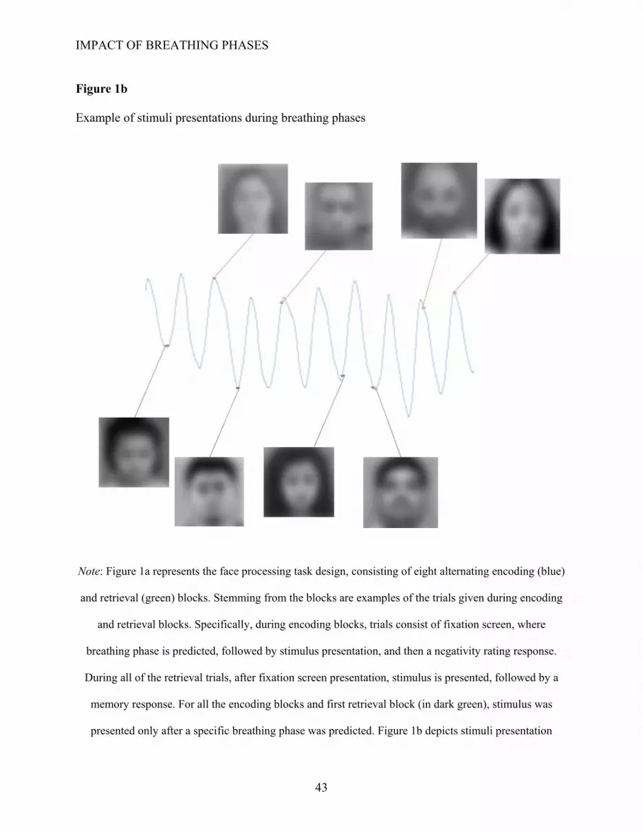
IMPACT OF BREATHING PHASES
43
Figure 1b
Example of stimuli presentations during breathing phases
Note: Figure 1a represents the face processing task design, consisting of eight alternating encoding (blue)
and retrieval (green) blocks. Stemming from the blocks are examples of the trials given during encoding
and retrieval blocks. Specifically, during encoding blocks, trials consist of fixation screen, where
breathing phase is predicted, followed by stimulus presentation, and then a negativity rating response.
During all of the retrieval trials, after fixation screen presentation, stimulus is presented, followed by a
memory response. For all the encoding blocks and first retrieval block (in dark green), stimulus was
presented only after a specific breathing phase was predicted. Figure 1b depicts stimuli presentation

IMPACT OF BREATHING PHASES
44
during specific phases of breathing cycle, where peak represents inhalation and trough represents
exhalation. Stimuli consisted of incarcerated individuals and are blurred for privacy. However, they were
not blurred during the face processing task.

IMPACT OF BREATHING PHASES
45
Figure 2
ERP time courses averaged across inhalation and exhalation conditions for all electrodes
Note: This figure consists of the grand means (mean of all trials from all breathing conditions from all
participants), from the time segment 500 ms before stimulus onset, and 1500 ms after stimulus onset. P9
and P10 appear to have the largest negative N170 amplitude, while Pz and POz appear to have the largest
P300 amplitude.

IMPACT OF BREATHING PHASES
46
Figure 3
“Hit-rate” from first retrieval block of the ANOVA analysis of encoding phase by retrieval phase
Note: Hit rate or correct recognition of “old” faces were enhanced for pictures encoded during inhalation
and retrieved during exhalation, compared to other conditions. Error bars represent +/- standard errors.

IMPACT OF BREATHING PHASES
47
Figure 4
Phase locking quality for breathing phases across all trials
Note: This figure displays the phase locking quality at different time points for the inhalation (first row)
and exhalation (second row) trials. 0 represents end of inhalation, while 180 represents end of exhalation.
The red line represents the average phase, while the blue represents the breathing phase distribution for
the trials. The first circle, in both rows, represents the phase distribution at stimulus onset, the second
circle represents the phase distribution 500 ms after stimulus onset, and the third circle indicates the phase
distribution 1000 ms after stimulus onset.

IMPACT OF BREATHING PHASES
48
Figure 5
ERP N170 at electrodes P9 and P10
Note: This figure consists of the ERP for P9 and P10 during the inhalation (red) and exhalation (blue)
phase. There is no significant difference during N170, which is depicted by the green line.

IMPACT OF BREATHING PHASES
49
Figure 6
Topographic scalp activity of and difference between the breathing phases during N170
Note: The final figure consists of P300 electrode activity during the inhalation subtracted from activity
during the exhalation. Warm colors indicate a positive difference, while blue colors represent a negative
difference. There appears to be a bilateral activity in two regions, consisting of electrodes CP5, P3, PO8,
and P8.

IMPACT OF BREATHING PHASES
50
Figure 7
ERP N170 average for P3, CP5, P8, and PO8
Note: This figure consists of the average ERP for electrode P3, CP5, P8, and PO8during the inhalation
(red) and exhalation (blue) phase. There appears to be a significant difference during N170, which is
depicted by the green line.

IMPACT OF BREATHING PHASES
51
Figure 8
ERP P300 at electrodes POZ and Pz
Note: This figure consists of the ERP for POz and Pz during the inhalation (red) and exhalation (blue)
phase. There is no significant difference during P300, which is depicted by the green line.

IMPACT OF BREATHING PHASES
52
Figure 9
Topographic scalp activity of and difference between the breathing phases during P300
Note: The final figure consists of P300 electrode activity during the inhalation subtracted from activity
during the exhalation. Warm colors indicate a positive difference, while blue colors represent a negative
difference. The region consisting of electrodes CP3, CP5, P3, P5, PO3, and PO7 appears to have a
positive difference between the two conditions.

IMPACT OF BREATHING PHASES
53
Figure 10
ERP P300 average for CP3, CP5, P3, P5, PO3, and PO7
Note: This figure consists of the average ERP for electrodes picked out from the analysis of the
topographic map during the inhalation (red) and exhalation (blue) phase. There appears to be a difference
during P300, which is depicted by the green line.

IMPACT OF BREATHING PHASES
54
Figure 11
Correlation between DASS-21 Depression Subscale and Negativity Rating Differences
Note: This scatterplot depicts the correlation between depressive levels, represented by the Depression
Anxiety Stress Survey-21 items (DASS-21) score for the depression subscale, and difference in the
negativity ratings for the face stimuli (calculated by subtracting exhalation rating from inhalation rating).

IMPACT OF BREATHING PHASES
55
Tables
Table 1
Descriptive statistics of sample demographic data (N = 23)
Variable Average Range Age 19.4 18-25 Class Standing
1.62 1-4
Males Females Gender 14 9
Caucasian Asian Black Hispanic or
Latino
Arab American/Middle Eastern
Native American
Race 3 11 4 3 1 1
Note. Class standing consisted of 1=freshmen; 2=sophomore; 3=junior; 4=senior

IMPACT OF BREATHING PHASES
56
Table 2
Results of t-tests for N170 amplitude by breathing phase
Paired Variables Mean SD
Statistic: Student's
t p Mean
difference SE
difference Cohen's
d
N170_P9_E -2.4803 -2.993 0.275 0.786 0.081 0.294 0.06 N170_P9_I -2.5613 -1.053
N170_P10_E -0.9863 -1.449 1.134 0.27 0.3431 0.303 0.2475 N170_P10_I -1.3295 -1.487
N170_exh_Avg 0.607 0.136 3.2 0.004 0.653 0.204 0.6983 N170_Inh_Avg -0.0459 -0.266
Note. The ending _E denotes trough (exhalation condition), while _I denotes peak (inhalation condition),
N170AvgInh represents the average of electrodes for the inhalation encoding trials, while N170AvgExh
represents the average of electrodes for the exhalation encoding trials. For N170 averages, electrodes
P3/CP5/P8/PO8 were used.

IMPACT OF BREATHING PHASES
57
Table 3
Results of t-tests for P300 amplitudes by breathing phase
Paired Variables Mean SD Statistic:
Student's t p Mean difference
SE difference
Cohen's d
P300_Pz_E 4.256 1.95 0.691 0.498 0.2357 0.341 0.1507 P300_Pz_I 4.02 1.99
P300_POZ_E 5.731 2.97 0.217 0.831 0.073 0.337 0.0473 P300_POZ_I 5.658 2.84
P3_exh_Avg 3.49 2.12 1.72 0.102 0.468 0.273 0.374 P3_inh_Avg 3.02 2.17
Note. The ending _E denotes trough (exhalation condition), while _I denotes peak (inhalation condition),
P300_inh_Avg represents the average of electrodes for the inhalation encoding trials, while P300_exh_Avg
represents the average of electrodes for the exhalation encoding trials. For P300 averages, electrodes
CP3/CP5/P3/P5/PO3/PO7 were used.

IMPACT OF BREATHING PHASES
58
Table 4
Pearson’s R correlation for Individual Characteristics and Differences in Negativity Rating, Hit-Rate, N170, and P300
DASS-21S
DASS-21A
DASS-21D MAAS ERQ
Suppression ERQ
Reappraisal RatingDiff 0.322 0.379† 0.500* 0.131 0.088 0.174
(0.178) (0.109) (0.029) (0.592) (0.72) (0.476) N=19 N=19 N=19 N=19 N=19 N=19
MemDiff 0.048 -0.11 -0.108 -0.233 0.045 -0.035 (0.846) (0.655) (0.659) (0.337) (0.854) (0.887) N=19 N=19 N=19 N=19 N=19 N=19
N170Diff 0.229 0.169 0.354 0.054 0.332 0.163 (0.376) (0.517) (0.163) (0.837) (0.193) (0.533) N=17 N=17 N=17 N=17 N=17 N=17
P300DIff 0.111 0.038 0.017 0.025 0.117 0.045 (0.671) (0.886) (0.949) (0.923) (0.656) (0.864)
N=17 N=17 N=17 N=17 N=17 N=17
Note. RatingDiff refers to face negativity rating differences between the breathing conditions (inhalation
and exhalation). MemDiff refers to the hit-rate difference between the breathing conditions. N170Diff
refers to the average N170 amplitude difference (of electrodes P3/CP5/P8/PO8) between the breathing
conditions. P300Diff refers to the average amplitude difference (of electrodes CP3/CP5/P3/P5/PO3/PO7).
The difference was calculated by subtracting the exhalation value from inhalation value for each variable.
DASS-21 denotes Depression Anxiety and Stress Scale 21, where S represents stress, A represents
anxiety, and D represents depression. MAAS denotes Mindful Attention and Awareness scale. ERQ
denotes Emotion Regulation Questionnaire.
† = marginal significance
* Correlation is significant at the 0.05 level (2-tailed)

IMPACT OF BREATHING PHASES
59
Appendices
Appendix A: Screener and Demographics Questionnaire
ID NUMBER:________________ Screening and Demographics
SCREENER 1. How old are you? _________________
a. For this research project, we only recruit healthy young adults with an age range between 18 – 30 years
2. Also, the participants for this study should have no current or recent history (within one year) of major medical conditions, including psychological, psychiatric, or neurological conditions. Do you have any of the following conditions or any other major medical conditions you think it is necessary for us to know?
a. Stroke b. brain injury, c. heart disease, d. drug addictions, e. schizophrenia, f. anxiety and depression, g. migraine, h. seizure
YES NO
DEMOGRAPHICS 1. Gender: Female Male Non-binary
2. Age: __________
3. Marital Status: Single Married
Divorced Separated
Widowed Partnered 4. Class Standing: Freshmen Junior
Sophomore Senior
5. Race: Caucasian Asian/Pacific Islander
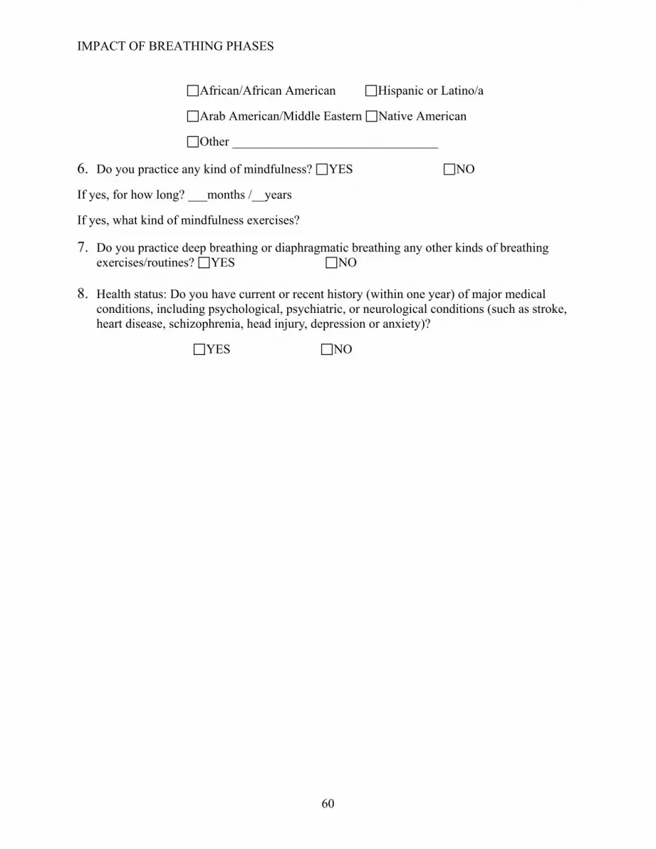
IMPACT OF BREATHING PHASES
60
African/African American Hispanic or Latino/a
Arab American/Middle Eastern Native American
Other ________________________________
6. Do you practice any kind of mindfulness? YES NO
If yes, for how long? ___months /__years
If yes, what kind of mindfulness exercises?
7. Do you practice deep breathing or diaphragmatic breathing any other kinds of breathing exercises/routines? YES NO
8. Health status: Do you have current or recent history (within one year) of major medical conditions, including psychological, psychiatric, or neurological conditions (such as stroke, heart disease, schizophrenia, head injury, depression or anxiety)?
YES NO

IMPACT OF BREATHING PHASES
61
Appendix B: Depression Anxiety and Stress - 21

IMPACT OF BREATHING PHASES
62

IMPACT OF BREATHING PHASES
63
Appendix C: Mindful Attention Awareness Scale

IMPACT OF BREATHING PHASES
64

IMPACT OF BREATHING PHASES
65
Appendix D: Emotion Regulation Questionnaire
IMPACT OF BREATHING PHASES
66
References
Abelson, J. L., Weg, J. G., Nesse, R. M., & Curtis, G. C. (2001). Persistent Respiratory
Irregularity in Patients with Panic Disorder. BIOL PSYCHIATRY, 8.
Adrian, E. D. (1950). The electrical activity of the mammalian olfactory bulb.
Electroencephalography and Clinical Neurophysiology, 2(1), 377–388.
https://doi.org/10.1016/0013-4694(50)90075-7
Anderson, T. M., & Ramirez, J.-M. (2017). Respiratory rhythm generation: Triple oscillator
hypothesis. F1000Research, 6. https://doi.org/10.12688/f1000research.10193.1
Arshamian, A., Iravani, B., Majid, A., & Lundström, J. N. (2018). Respiration Modulates
Olfactory Memory Consolidation in Humans. The Journal of Neuroscience, 38(48),
10286–10294. https://doi.org/10.1523/JNEUROSCI.3360-17.2018
Barrett, L. F., Bliss-Moreau, E., Duncan, S. L., Rauch, S. L., & Wright, C. I. (2007). The
amygdala and the experience of affect. Social Cognitive and Affective Neuroscience, 2(2),
73–83. https://doi.org/10.1093/scan/nsl042
Bautista, T. G., Pitts, T. E., Pilowsky, P. M., & Morris, K. F. (2014). Chapter 18—The Brainstem
Respiratory Network. In C. L. Faingold & H. Blumenfeld (Eds.), Neuronal Networks in
Brain Function, CNS Disorders, and Therapeutics (pp. 235–245). Academic Press.
https://doi.org/10.1016/B978-0-12-415804-7.00018-6
Bentin, S., Allison, T., Puce, A., Perez, E., & McCarthy, G. (1996). Electrophysiological Studies
of Face Perception in Humans. Journal of Cognitive Neuroscience, 8(6), 551–565.
https://doi.org/10.1162/jocn.1996.8.6.551

IMPACT OF BREATHING PHASES
67
Biancardi, V., Saini, J., Pageni, A., M, H. P., Funk, G. D., & Pagliardini, S. (n.d.). Mapping of the
excitatory, inhibitory, and modulatory afferent projections to the anatomically defined
active expiratory oscillator in adult male rats. Journal of Comparative Neurology,
n/a(n/a). https://doi.org/10.1002/cne.24984
Biskamp, J., Bartos, M., & Sauer, J.-F. (2017). Organization of prefrontal network activity by
respiration-related oscillations. Scientific Reports, 7(1), 45508.
https://doi.org/10.1038/srep45508
Boiten, F. A. (1998). The effects of emotional behaviour on components of the respiratory cycle.
Biological Psychology, 49(1), 29–51. https://doi.org/10.1016/S0301-0511(98)00025-8
Boiten, F. A., Frijda, N. H., & Wientjes, C. J. E. (1994). Emotions and respiratory patterns:
Review and critical analysis. International Journal of Psychophysiology, 17(2), 103–128.
https://doi.org/10.1016/0167-8760(94)90027-2
Bouvier, J., Thoby-Brisson, M., Renier, N., Dubreuil, V., Ericson, J., Champagnat, J., Pierani, A.,
Chédotal, A., & Fortin, G. (2010). Hindbrain interneurons and axon guidance signaling
critical for breathing. Nature Neuroscience, 13(9), 1066–1074.
https://doi.org/10.1038/nn.2622
Britton, J. C., Lissek, S., Grillon, C., Norcross, M. A., & Pine, D. S. (2011). Development of
anxiety: The role of threat appraisal and fear learning. Depression and Anxiety, 28(1), 5–
17. https://doi.org/10.1002/da.20733
Brown, R., & Gerbarg, P. (2005). Sudarshan Kriya Yogic Breathing in the Treatment of Stress,
Anxiety, and Depression: Part I—Neurophysiologic Model. Journal of Alternative and
Complementary Medicine (New York, N.Y.), 11, 189–201.
https://doi.org/10.1089/acm.2005.11.189

IMPACT OF BREATHING PHASES
68
Brown, R. P., Gerbarg, P. L., & Muench, F. (2013). Breathing practices for treatment of
psychiatric and stress-related medical conditions. Psychiatric Clinics of North America,
36(1), 121–140. https://doi.org/10.1016/j.psc.2013.01.001
Carmichael, S. T., Clugnet, M.-C., & Price, J. L. (1994). Central olfactory connections in the
macaque monkey. Journal of Comparative Neurology, 346(3), 403–434.
https://doi.org/10.1002/cne.903460306
Cenier, T., David, F., Litaudon, P., Garcia, S., Amat, C., & Buonviso, N. (2009). Respiration-
gated formation of gamma and beta neural assemblies in the mammalian olfactory bulb.
European Journal of Neuroscience, 29(5), 921–930. https://doi.org/10.1111/j.1460-
9568.2009.06651.x
Cox, R., Korjoukov, I., Boer, M. de, & Talamini, L. M. (2014). Sound Asleep: Processing and
Retention of Slow Oscillation Phase-Targeted Stimuli. PLOS ONE, 9(7), e101567.
https://doi.org/10.1371/journal.pone.0101567
Cui, Y., Kam, K., Sherman, D., Janczewski, W. A., Zheng, Y., & Feldman, J. L. (2016). Defining
preBötzinger Complex Rhythm- and Pattern-Generating Neural Microcircuits In Vivo.
Neuron, 91(3), 602–614. https://doi.org/10.1016/j.neuron.2016.07.003
De Couck, M., Caers, R., Musch, L., Fliegauf, J., Giangreco, A., & Gidron, Y. (2019). How
breathing can help you make better decisions: Two studies on the effects of breathing
patterns on heart rate variability and decision-making in business cases. International
Journal of Psychophysiology, 139, 1–9. https://doi.org/10.1016/j.ijpsycho.2019.02.011
Deffke, I., Sander, T., Heidenreich, J., Sommer, W., Curio, G., Trahms, L., & Lueschow, A.
(2007). MEG/EEG sources of the 170-ms response to faces are co-localized in the

IMPACT OF BREATHING PHASES
69
fusiform gyrus. NeuroImage, 35(4), 1495–1501.
https://doi.org/10.1016/j.neuroimage.2007.01.034
Del Negro, C. A., Funk, G. D., & Feldman, J. L. (2018). Breathing matters. Nature Reviews.
Neuroscience, 19(6), 351–367. https://doi.org/10.1038/s41583-018-0003-6
Dennis, T. A., & Chen, C.-C. (2007). Neurophysiological mechanisms in the emotional
modulation of attention: The interplay between threat sensitivity and attentional control.
Biological Psychology, 76(1–2), 1–10. https://doi.org/10.1016/j.biopsycho.2007.05.001
Drakatos, P., Kent, B. D., Birdseye, A., Dastagir, S., Higgins, S., Williams, A. J., & Leschziner,
G. (2017). Phasic activity and the importance of irregular breathing. Sleep Medicine, 38,
21–23. https://doi.org/10.1016/j.sleep.2017.06.031
Eichenbaum, H., Yonelinas, A. R., & Ranganath, C. (2007). The Medial Temporal Lobe and
Recognition Memory. Annual Review of Neuroscience, 30, 123–152.
https://doi.org/10.1146/annurev.neuro.30.051606.094328
Eimer, M., & Holmes, A. (2007). Event-related brain potential correlates of emotional face
processing. Neuropsychologia, 45(1), 15–31.
https://doi.org/10.1016/j.neuropsychologia.2006.04.022
Feldman, J. L., McCrimmon, D. R., Mitchell, G. S., & Negro, C. A. D. (2009). Brainstem
Respiratory Circuits. Encyclopedia of Neuroscience, 463–470.
https://doi.org/10.1016/B978-008045046-9.00169-8
Fontanini, A., & Bower, J. M. (2006). Slow-waves in the olfactory system: An olfactory
perspective on cortical rhythms. Trends in Neurosciences, 29(8), 429–437.
https://doi.org/10.1016/j.tins.2006.06.013
IMPACT OF BREATHING PHASES
70
Garcia, A. J., Zanella, S., Koch, H., Doi, A., & Ramirez, J.-M. (2011). Networks within
networks: The neuronal control of breathing. Progress in Brain Research, 188, 31–50.
https://doi.org/10.1016/B978-0-444-53825-3.00008-5
Geissberger, N., Tik, M., Sladky, R., Woletz, M., Schuler, A.-L., Willinger, D., &
Windischberger, C. (2020). Reproducibility of amygdala activation in facial emotion
processing at 7T. NeuroImage, 211, 116585.
https://doi.org/10.1016/j.neuroimage.2020.116585
Gliga, T., & Csibra, G. (2007). Seeing the face through the eyes: A developmental perspective on
face expertise. In Progress in Brain Research (Vol. 164, pp. 323–339). Elsevier.
https://doi.org/10.1016/S0079-6123(07)64018-7
Gole, M., Köchel, A., Schäfer, A., & Schienle, A. (2012). Threat engagement, disengagement,
and sensitivity bias in worry-prone individuals as measured by an emotional go/no-go
task. Journal of Behavior Therapy and Experimental Psychiatry, 43(1), 532–539.
https://doi.org/10.1016/j.jbtep.2011.07.002
Gray, P. A., Janczewski, W. A., Mellen, N., McCrimmon, D. R., & Feldman, J. L. (2001). Normal
breathing requires preBötzinger complex neurokinin-1 receptor-expressing neurons.
Nature Neuroscience, 4(9), 927–930. https://doi.org/10.1038/nn0901-927
Gross, J. J., & John, O. P. (2003). Individual differences in two emotion regulation processes:
Implications for affect, relationships, and well-being.
Guan, L., Qi, M., Li, H., Hitchman, G., Yang, J., & Liu, Y. (2015). Priming with threatening
faces modulates the self-face advantage by enhancing the other-face processing rather
than suppressing the self-face processing. Brain Research, 1608, 97–107.
https://doi.org/10.1016/j.brainres.2015.03.002

IMPACT OF BREATHING PHASES
71
Guan, L., Zhao, Y., Wang, Y., Chen, Y., & Yang, J. (2017). Self-esteem Modulates the P3
Component in Response to the Self-face Processing after Priming with Emotional Faces.
Frontiers in Psychology, 8. https://doi.org/10.3389/fpsyg.2017.01399
Hadjikhani, N., Kveraga, K., Naik, P., & Ahlfors, S. P. (2009). Early (N170) activation of face-
specific cortex by face-like objects. Neuroreport, 20(4), 403–407.
https://doi.org/10.1097/WNR.0b013e328325a8e1
Heck, D. H., McAfee, S. S., Liu, Y., Babajani-Feremi, A., Rezaie, R., Freeman, W. J., Wheless, J.
W., Papanicolaou, A. C., Ruszinkó, M., Sokolov, Y., & Kozma, R. (2017). Breathing as a
Fundamental Rhythm of Brain Function. Frontiers in Neural Circuits, 10.
https://doi.org/10.3389/fncir.2016.00115
Henry, J. D., & Crawford, J. R. (2005). The short-form version of the Depression Anxiety Stress
Scales (DASS-21): Construct validity and normative data in a large non-clinical sample.
British Journal of Clinical Psychology, 44(2), 227–239.
https://doi.org/10.1348/014466505X29657
Herrington, J. D., Taylor, J. M., Grupe, D. W., Curby, K. M., & Schultz, R. T. (2011).
Bidirectional communication between amygdala and fusiform gyrus during facial
recognition. NeuroImage, 56(4), 2348–2355.
https://doi.org/10.1016/j.neuroimage.2011.03.072
Hill, N. L., Mogle, J., Bhargava, S., Bell, T. R., Bhang, I., Katz, M., & Sliwinski, M. J. (2020).
Longitudinal relationships among depressive symptoms and three types of memory self-
report in cognitively intact older adults. International Psychogeriatrics, 32(6), 719–732.
https://doi.org/10.1017/S104161021900084X

IMPACT OF BREATHING PHASES
72
Hirooka, Y., Polson, J. W., Potts, P. D., & Dampney, R. A. L. (1997). Hypoxia-induced Fos
expression in neurons projecting to the pressor region in the rostral ventrolateral medulla.
Neuroscience, 80(4), 1209–1224. https://doi.org/10.1016/S0306-4522(97)00111-5
Hoscheidt, S. M., LaBar, K. S., Ryan, L., Jacobs, W. J., & Nadel, L. (2014). Encoding negative
events under stress: High subjective arousal is related to accurate emotional memory
despite misinformation exposure. Neurobiology of Learning and Memory, 112, 237–247.
https://doi.org/10.1016/j.nlm.2013.09.008
Huijbers, W., Pennartz, C. M. A., Beldzik, E., Domagalik, A., Vinck, M., Hofman, W. F., Cabeza,
R., & Daselaar, S. M. (2014). Respiration phase-locks to fast stimulus presentations:
Implications for the interpretation of posterior midline “deactivations”: The Interplay
Between Cognition, Respiration, and fMRI Signal. Human Brain Mapping, 35(9), 4932–
4943. https://doi.org/10.1002/hbm.22523
Ito, J., Roy, S., Liu, Y., Cao, Y., Fletcher, M., Lu, L., Boughter, J. D., Grün, S., & Heck, D. H.
(2014). Whisker barrel cortex delta oscillations and gamma power in the awake mouse
are linked to respiration. Nature Communications, 5(1), 1–10.
https://doi.org/10.1038/ncomms4572
Jerath, R., Edry, J. W., Barnes, V. A., & Jerath, V. (2006). Physiology of long pranayamic
breathing: Neural respiratory elements may provide a mechanism that explains how slow
deep breathing shifts the autonomic nervous system. Medical Hypotheses, 67(3), 566–
571. https://doi.org/10.1016/j.mehy.2006.02.042
Jung, J., Hudry, J., Ryvlin, P., Royet, J.-P., Bertrand, O., & Lachaux, J.-P. (2006). Functional
Significance of Olfactory-induced Oscillations in the Human Amygdala. Cerebral
Cortex, 16(1), 1–8. https://doi.org/10.1093/cercor/bhi090
IMPACT OF BREATHING PHASES
73
Kanwisher, N., & Yovel, G. (2006). The fusiform face area: A cortical region specialized for the
perception of faces. Philosophical Transactions of the Royal Society B: Biological
Sciences, 361(1476), 2109–2128. https://doi.org/10.1098/rstb.2006.1934
Karalis, N., & Sirota, A. (2018). Breathing coordinates limbic network dynamics underlying
memory consolidation [Preprint]. Neuroscience. https://doi.org/10.1101/392530
Kunik, M. E., Roundy, K., Veazey, C., Souchek, J., Richardson, P., Wray, N. P., & Stanley, M. A.
(2005). Surprisingly High Prevalence of Anxiety and Depression in Chronic Breathing
Disorders. Chest, 127(4), 1205–1211. https://doi.org/10.1016/S0012-3692(15)34468-8
LeDoux, J. E. (2000). Emotion Circuits in the Brain. 31.
Lee, J. S., Park, G., Song, M. J., Choi, K.-H., & Lee, S.-H. (2015, December 22). Early visual
processing for low spatial frequency fearful face is correlated with cortical volume in
patients with schizophrenia. Neuropsychiatric Disease and Treatment; Dove Press.
https://doi.org/10.2147/NDT.S97089
Linden, D. (2006). The P300: Where in the Brain Is It Produced and What Does It Tell Us? The
Neuroscientist : A Review Journal Bringing Neurobiology, Neurology and Psychiatry, 11,
563–576. https://doi.org/10.1177/1073858405280524
Liu, J., Harris, A., & Kanwisher, N. (2002). Stages of processing in face perception: An MEG
study. Nature Neuroscience, 5(9), 910–916. https://doi.org/10.1038/nn909
Liu, Y., McAfee, S. S., & Heck, D. H. (2017). Hippocampal sharp-wave ripples in awake mice
are entrained by respiration. Scientific Reports, 7(1), 8950.
https://doi.org/10.1038/s41598-017-09511-8

IMPACT OF BREATHING PHASES
74
Lockmann, A. L. V., Laplagne, D. A., & Tort, A. B. L. (2018). Olfactory bulb drives respiration-
coupled beta oscillations in the rat hippocampus. European Journal of Neuroscience,
48(8), 2663–2673. https://doi.org/10.1111/ejn.13665
Lovibond, S. H., & Lovibond, P. F. (1995). Depression Anxiety Stress Scales. PsycTESTS.
https://doi.org/10.1037/t01004-000
Luethi, M., Meier, B., & Sandi, C. (2009). Stress effects on working memory, explicit memory,
and implicit memory for neutral and emotional stimuli in healthy men. Frontiers in
Behavioral Neuroscience, 2. https://doi.org/10.3389/neuro.08.005.2008
MacKillop, J., & Anderson, E. J. (2007). Further Psychometric Validation of the Mindful
Attention Awareness Scale (MAAS). Journal of Psychopathology and Behavioral
Assessment, 29(4), 289–293. https://doi.org/10.1007/s10862-007-9045-1
Maric, V., Ramanathan, D., & Mishra Ramanathan, J. (2020). Respiratory Regulation &
Interactions with Neuro-Cognitive Circuitry. Neuroscience & Biobehavioral Reviews,
112. https://doi.org/10.1016/j.neubiorev.2020.02.001
Mather, M., Clewett, D., Sakaki, M., & Harley, C. W. (2016). Norepinephrine ignites local hot
spots of neuronal excitation: How arousal amplifies selectivity in perception and memory.
The Behavioral and Brain Sciences, 39, e200.
https://doi.org/10.1017/S0140525X15000667
Maurer, J., Rebbapragada, V., Borson, S., Goldstein, R., Kunik, M. E., Yohannes, A. M., &
Hanania, N. A. (2008). Anxiety and Depression in COPD. Chest, 134(4 Suppl), 43S-56S.
https://doi.org/10.1378/chest.08-0342
Molle, L., & Benoit, C.-É. (2019). The Respiratory Modulation of Memory. The Journal of
Neuroscience, 39(30), 5836–5838. https://doi.org/10.1523/JNEUROSCI.0224-19.2019

IMPACT OF BREATHING PHASES
75
Moore, J. D., Deschênes, M., Furuta, T., Huber, D., Smear, M. C., Demers, M., & Kleinfeld, D.
(2013). Hierarchy of orofacial rhythms revealed through whisking and breathing. Nature,
497(7448), 205–210. https://doi.org/10.1038/nature12076
Muñoz-Ortiz, J., Muñoz-Ortiz, E., López-Meraz, L., Beltran-Parrazal, L., & Morgado-Valle, C.
(2019). The pre-Bötzinger complex: Generation and modulation of respiratory rhythm.
Neurología (English Edition), 34(7), 461–468.
https://doi.org/10.1016/j.nrleng.2018.05.006
Nguyen, V. T., & Cunnington, R. (2014). The superior temporal sulcus and the N170 during face
processing: Single trial analysis of concurrent EEG–fMRI. NeuroImage, 86, 492–502.
https://doi.org/10.1016/j.neuroimage.2013.10.047
Nitsos, I., & Walker, D. W. (1999). The distribution of FOS-immunoreactive neurons in the
brainstem, midbrain and diencephalon of fetal sheep in response to acute hypoxia in mid
and late gestation. Developmental Brain Research, 114(1), 9–26.
https://doi.org/10.1016/S0165-3806(99)00010-3
Perciavalle, V., Blandini, M., Fecarotta, P., Buscemi, A., Di Corrado, D., Bertolo, L., Fichera, F.,
& Coco, M. (2017). The role of deep breathing on stress. Neurological Sciences, 38, 451–
458. https://doi.org/10.1007/s10072-016-2790-8
Perl, O., Ravia, A., Rubinson, M., Eisen, A., Soroka, T., Mor, N., Secundo, L., & Sobel, N.
(2019). Human non-olfactory cognition phase-locked with inhalation. Nature Human
Behaviour, 3(5), 501–512. https://doi.org/10.1038/s41562-019-0556-z
Pfaltz, M. C., Passardi, S., Auschra, B., Fares-Otero, N. E., Schnyder, U., & Peyk, P. (2019). Are
you angry at me? Negative interpretations of neutral facial expressions are linked to child

IMPACT OF BREATHING PHASES
76
maltreatment but not to posttraumatic stress disorder. European Journal of
Psychotraumatology, 10(1). https://doi.org/10.1080/20008198.2019.1682929
Polich, J. (2007). Updating P300: An Integrative Theory of P3a and P3b. Clinical
Neurophysiology : Official Journal of the International Federation of Clinical
Neurophysiology, 118(10), 2128–2148. https://doi.org/10.1016/j.clinph.2007.04.019
Preston, A. R., & Eichenbaum, H. (2013). Interplay of Hippocampus and Prefrontal Cortex in
Memory. Current Biology, 23(17), R764–R773.
https://doi.org/10.1016/j.cub.2013.05.041
Rekling, J. C., & Feldman, J. L. (1998). PREBÖTZINGER COMPLEX AND PACEMAKER
NEURONS: Hypothesized Site and Kernel for Respiratory Rhythm Generation. Annual
Review of Physiology, 60(1), 385–405. https://doi.org/10.1146/annurev.physiol.60.1.385
Ressler, K. J. (2010). Amygdala Activity, Fear, and Anxiety: Modulation by Stress. Biological
Psychiatry, 67(12), 1117–1119. https://doi.org/10.1016/j.biopsych.2010.04.027
Rousselet, G. A., Macé, M. J.-M., & Fabre-Thorpe, M. (2004). Animal and human faces in
natural scenes: How specific to human faces is the N170 ERP component? Journal of
Vision, 4(1), 2–2. https://doi.org/10.1167/4.1.2
Rozenkrants, B., & Polich, J. (2008). Affective ERP Processing in a Visual Oddball Task:
Arousal, Valence, and Gender. Clinical Neurophysiology : Official Journal of the
International Federation of Clinical Neurophysiology, 119(10), 2260–2265.
https://doi.org/10.1016/j.clinph.2008.07.213
Sherman, D., Worrell, J. W., Cui, Y., & Feldman, J. L. (2015). Optogenetic perturbation of
preBötzinger complex inhibitory neurons modulates respiratory pattern. Nature
Neuroscience, 18(3), 408–414. https://doi.org/10.1038/nn.3938

IMPACT OF BREATHING PHASES
77
Simion, F., Di Giorgio, E., Leo, I., & Bardi, L. (2011). The processing of social stimuli in early
infancy: From faces to biological motion perception. Progress in Brain Research, 189,
173–193. https://doi.org/10.1016/B978-0-444-53884-0.00024-5
Stein, M. B., Millar, T. W., Sc, M., Larsen, D. K., & Kryger, M. H. (1994). Irregular Breathing
During Sleep in Patients With Panic Disorder.
Subramanian, H. H., Balnave, R. J., & Holstege, G. (2008). The Midbrain Periaqueductal Gray
Control of Respiration. Journal of Neuroscience, 28(47), 12274–12283.
https://doi.org/10.1523/JNEUROSCI.4168-08.2008
Suess, W. M., Alexander, A. B., Smith, D. D., Sweeney, H. W., & Marion, R. J. (1980). The
Effects of Psychological Stress on Respiration: A Preliminary Study of Anxiety and
Hyperventilation. Psychophysiology, 17(6), 535–540. https://doi.org/10.1111/j.1469-
8986.1980.tb02293.x
Syrjänen, E., Wiens, S., Fischer, H., Zakrzewska, M., Wartel, A., Larsson, M., & Olofsson, J. K.
(2018). Background Odors Modulate N170 ERP Component and Perception of Emotional
Facial Stimuli. Frontiers in Psychology, 9. https://doi.org/10.3389/fpsyg.2018.01000
Tan, W., Janczewski, W. A., Yang, P., Shao, X. M., Callaway, E. M., & Feldman, J. L. (2008).
Silencing preBötzinger Complex somatostatin-expressing neurons induces persistent
apnea in awake rat. Nature Neuroscience, 11(5), 538–540.
https://doi.org/10.1038/nn.2104
Thompson, K. L., Hannan, S. M., & Miron, L. R. (2014). Fight, flight, and freeze: Threat
sensitivity and emotion dysregulation in survivors of chronic childhood maltreatment.
Personality and Individual Differences, 69, 28–32.
https://doi.org/10.1016/j.paid.2014.05.005

IMPACT OF BREATHING PHASES
78
Tort, A. B. L., Brankačk, J., & Draguhn, A. (2018). Respiration-Entrained Brain Rhythms Are
Global but Often Overlooked. Trends in Neurosciences, 41(4), 186–197.
https://doi.org/10.1016/j.tins.2018.01.007
Tully, K., & Bolshakov, V. Y. (2010). REemviewotional enhancement of memory: How
norepinephrine enables synaptic plasticity. 9.
Van Diest, I., Verstappen, K., Aubert, A. E., Widjaja, D., Vansteenwegen, D., & Vlemincx, E.
(2014). Inhalation/Exhalation ratio modulates the effect of slow breathing on heart rate
variability and relaxation. Applied Psychophysiology and Biofeedback, 39(3–4), 171–180.
https://doi.org/10.1007/s10484-014-9253-x
Varvogli, L., & Darviri, C. (2011). Stress Management Techniques: Evidence-based procedures
that reduce stress and promote health. Health Science Journal.
Vlemincx, E., Taelman, J., Peuter, S. D., Diest, I. V., & Bergh, O. V. D. (2011). Sigh rate and
respiratory variability during mental load and sustained attention. Psychophysiology,
48(1), 117–120. https://doi.org/10.1111/j.1469-8986.2010.01043.x
Wu, X. R., Liu, Y., Wang, L., Li, B., & Xu, F. (n.d.). Activity Patterns Elicited by Airflow in the
Olfactory Bulb and Their Possible Functions. 12.
Yackle, K., Schwarz, L. A., Kam, K., Sorokin, J. M., Huguenard, J. R., Feldman, J. L., Luo, L.,
& Krasnow, M. A. (2017). Breathing Control Center Neurons that Promote Arousal in
Mice. Science (New York, N.Y.), 355(6332), 1411–1415.
https://doi.org/10.1126/science.aai7984
Yadav, G., & Mutha, P. K. (2016). Deep Breathing Practice Facilitates Retention of Newly
Learned Motor Skills. Scientific Reports, 6(1), 1–8. https://doi.org/10.1038/srep37069

IMPACT OF BREATHING PHASES
79
Yang, C. F., & Feldman, J. L. (2018). Efferent projections of excitatory and inhibitory
preBötzinger Complex neurons. The Journal of Comparative Neurology, 526(8), 1389–
1402. https://doi.org/10.1002/cne.24415
Yanovsky, Y., Ciatipis, M., Draguhn, A., Tort, A. B. L., & Branka k, J. (2014). Slow Oscillations
in the Mouse Hippocampus Entrained by Nasal Respiration. Journal of Neuroscience,
34(17), 5949–5964. https://doi.org/10.1523/JNEUROSCI.5287-13.2014
Yohannes, A. M., Willgoss, T. G., Baldwin, R. C., & Connolly, M. J. (2010). Depression and
anxiety in chronic heart failure and chronic obstructive pulmonary disease: Prevalence,
relevance, clinical implications and management principles. International Journal of
Geriatric Psychiatry, 25(12), 1209–1221. https://doi.org/10.1002/gps.2463
Yoon, K. L., & Zinbarg, R. E. (2008). Interpreting neutral faces as threatening is a default mode
for socially anxious individuals. Journal of Abnormal Psychology, 117(3), 680–685.
https://doi.org/10.1037/0021-843X.117.3.680
Zaccaro, A., Piarulli, A., Laurino, M., Garbella, E., Menicucci, D., Neri, B., & Gemignani, A.
(2018). How Breath-Control Can Change Your Life: A Systematic Review on Psycho-
Physiological Correlates of Slow Breathing. Frontiers in Human Neuroscience, 12.
https://doi.org/10.3389/fnhum.2018.00353
Zanella, S., Doi, A., Garcia, A. J., Elsen, F., Kirsch, S., Wei, A. D., & Ramirez, J.-M. (2014).
When Norepinephrine Becomes a Driver of Breathing Irregularities: How Intermittent
Hypoxia Fundamentally Alters the Modulatory Response of the Respiratory Network.
The Journal of Neuroscience, 34(1), 36–50. https://doi.org/10.1523/JNEUROSCI.3644-
12.2014

IMPACT OF BREATHING PHASES
80
Zautra, A. J., Fasman, R., Davis, M. C., & Craig, A. D. (Bud). (2010). The effects of slow
breathing on affective responses to pain stimuli: An experimental study: Pain, 149(1),
12–18. https://doi.org/10.1016/j.pain.2009.10.001
Zelano, C., Jiang, H., Zhou, G., Arora, N., Schuele, S., Rosenow, J., & Gottfried, J. A. (2016).
Nasal Respiration Entrains Human Limbic Oscillations and Modulates Cognitive
Function. The Journal of Neuroscience: The Official Journal of the Society for
Neuroscience, 36(49), 12448–12467. https://doi.org/10.1523/JNEUROSCI.2586-16.2016


