Embed Size (px)
Citation preview
In vitro gastrointestinal digestion ofpomegranate peel (Punica granatum) flourobtained from co-products: Changes in theantioxidant potential and bioactive compoundsstability
Beatriz Gullon a, Manuela E. Pintado a, Juana Fernández-López b,José A. Pérez-Álvarez b, Manuel Viuda-Martos b,*a CBQF – Centro de Biotecnologia e Química Fina – Laboratório Associado, Escola Superior de Biotecnologia,Universidade Católica Portuguesa/Porto, Rua Dr. António Bernardino Almeida, 4200-072 Porto, Portugalb IPOA Research Group, AgroFood Technology Department. Escuela Politécnica Superior de Orihuela. MiguelHernández University. Crta. Beniel km. 3,2. E-03312 Orihuela, Alicante, Spain
A R T I C L E I N F O
Article history:
Received 16 June 2015
Received in revised form 21
September 2015
Accepted 28 September 2015
Available online 26 October 2015
A B S T R A C T
The effect of in vitro gastrointestinal digestion (GID) on the recovery, bioaccessibility and
stability of polyphenolic compounds, the changes in antioxidant activity and the short chain
fatty acids (SCFAs) production of pomegranate peel flour (PPF) were evaluated. The ex-
tracts obtained in each step of GID were used to determine the stability of polyphenolic profile
using HPLC whilst the antioxidant properties were determined using five methodologies.
The SCFAs production from PPF fermentation was also determined. At the end of GID process,
the bioaccessibility of phenolic and flavonoid compounds was 35.90 and 64.02%, respec-
tively. The polyphenolic compounds decreased after GID except that for ellagic acid which
increased. GID increased the chelating activity and reducing power. However, the scaveng-
ing properties were reduced. Fermentation of PPF by colonic bacteria generated acetic, propionic
and butyric acids. PPF could be used in the food industry as a potential ingredient to develop
functional foods that promote health benefits.
© 2015 Elsevier Ltd. All rights reserved.
Keywords:
In vitro digestion
Antioxidant
Polyphenolic compounds
Bioaccessibility index
Short chain fatty acids
1. Introduction
Pomegranate (Punica granatum) is an important fruit due to itsmany functional components. This fruit is, mainly, con-sumed in fresh; the arils contain proteins, crude fibres, vitamins,minerals, pectin, sugars and polyphenols (Viuda-Martos,Fernández-López, & Pérez-Alvarez, 2010).The arils are also usedto make popular pomegranate products such as fresh juice.
Once the juice has been extracted, the co-products that remainare composed of two fractions: bagasses and peel. Uses for theseco-products are limited. However, due to their composition,these co-products have the potential to be used for other endsfor example to obtain bioactive compounds or antioxidantdietary fibre (Viuda-Martos et al., 2011).
In the last years, special attention has been made to pome-granate non-edible parts. Nevertheless, pomegranate peel isan important source of bioactive compounds such as phenolic
* Corresponding author. Miguel Hernández University, Alicante,Spain. Tel.: +34 966749661; fax: +34 966749677.E-mail address: [email protected] (M. Viuda-Martos).
http://dx.doi.org/10.1016/j.jff.2015.09.0561756-4646/© 2015 Elsevier Ltd. All rights reserved.
J o u rna l o f Func t i ona l F ood s 1 9 ( 2 0 1 5 ) 6 1 7 – 6 2 8
Available online at www.sciencedirect.com
journal homepage: www.elsevier.com/ locate / j ff
ScienceDirect
acids, flavonoids, proanthocyanidins and ellagitannins as wellas ellagic acid and ellagic acid glycosides (Çam, Içyer, & Erdogan,2014; Devatkal, Narsaiah, & Borah, 2010) or antioxidant dietaryfibre which is defined as dietary fibre rich in associated poly-phenolic compounds.This product combines in a single materialthe physiological effects of both dietary fibre and antioxi-dants (Saura-Calixto, 1998). Evidence from epidemiologicalstudies suggests that diets rich in antioxidant dietary fibre areprotective against several degenerative diseases such as cancer,cardiovascular diseases, diabetes and metabolic syndrome,among others (Grooms, Ommerborn, Pham, Djoussé, & Clark,2013; Kaczmarczyk, Miller, & Freund, 2012). In addition, anti-oxidant dietary fibre has shown techno-functional propertiesthat play an important role in the digestion process and is es-pecially beneficial from a physiological point of view (Hasnaoui,Wathelet, & Jimenez-Araujo, 2014). In this way, studies aboutthe bioaccessibility of bioactive compounds present in foodsare necessary, because a link between the differences inbioactive compounds digestibility, bioaccessibility and the foodmatrix composition has been suggested. The bioaccessibilitycan be defined as the amount of compound solubilised inthe small intestine and available for subsequent absorption.This process comprises the release of compounds from foodmatrices and their stability under the gastrointestinal condi-tion (Gawlik-Dziki, 2012; Tagliazucchi, Verzelloni, Bertolini, &Conte, 2010). Definitive studies regarding the bioaccessibilityof bioactive compounds require in vivo experiments withhumans. However, in vitro methods have also proven to beuseful in determining their stability under gastrointestinal con-ditions (Toydemir et al., 2013). The in vitro methods are appliedto a system of simulated gastrointestinal digestion usingα-amylase in mouth phase, pepsin in the gastric phase anda mixture of pancreatin and bile salts during the intestinaltract. The element diffused through a semi-permeable mem-brane in the intestinal phase is used as measure of thecompounds bioaccessibility (Kulkarni, Acharya, Rajurkar, &Reddy, 2007). Indeed, despite limitations that constitute a staticmodel of digestion, the evaluation of bioaccessibility by in vitromodels can be well correlated with results from humanstudies and animal models (Bouayed, Hoffman, & Bohn,2011).
To the best of our knowledge, there are no studies, in thescientific literature, regarding the stability of phenolic and fla-vonoid compounds and the antioxidant activity changes duringin vitro gastrointestinal digestion of pomegranate peel flour (PPF).Thus, the effect of in vitro gastrointestinal digestion on (i) therecovery and bioaccessibility indexes, (ii) the stability of phe-nolic and flavonoid compounds, (iii) the changes in antioxidantactivity and (iv) the short chain fatty acids (SCFAs) produc-tion of pomegranate peel flour (PPF) obtained from co-productsof juice industry were evaluated.
2. Material and methods
2.1. Pomegranate peel flour (PPF)
Pomegranate (cv. “Mollar de Elche”) peels, obtained as a co-product during pomegranate juice extraction, were supplied
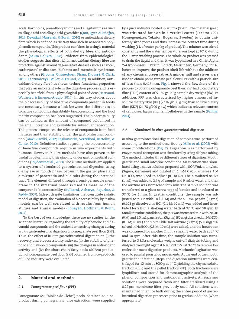
by a juice industry located in Murcia (Spain).The material (peel)was triturated for 40 s in a vertical cutter (Tecator 1094Homogeneizer, Tekator, Hoganas, Sweden) to obtain uni-formly sized pieces and thus increase the contact time duringwashing (1 L of water per kg of product).The mixture was stirredconstantly and the water temperature was kept at 40° C duringthe 10 min washing process.The whole co-product was pressedto drain the liquid and then it was lyophilised in a Christ Alpha2-4 lyophiliser (B. Braun Biotech, Melsungen, Germany) for 48hours to improve the product shelf life without the additionof any chemical preservative. A grinder mill and sieves wereused to obtain pomegranate peel flour (PPF) with a particle sizeof less than 0.417 mm. Fig. 1 showed the flowchart of theprocess to obtain pomegranate peel flour. PPF had total dietaryfibre (TDF) content of 51.80 g/100 g sample dry weight (dw). Inaddition, PPF was characterised by higher amount of in-soluble dietary fibre (IDF) (27.02 g/100 g dw) than soluble dietaryfibre (SDF) (24.78 g/100 g dw) which indicates relevant contentof celluloses, lignin and hemicelluloses in the sample (Bailina,2014).
2.2. Simulated in vitro gastrointestinal digestion
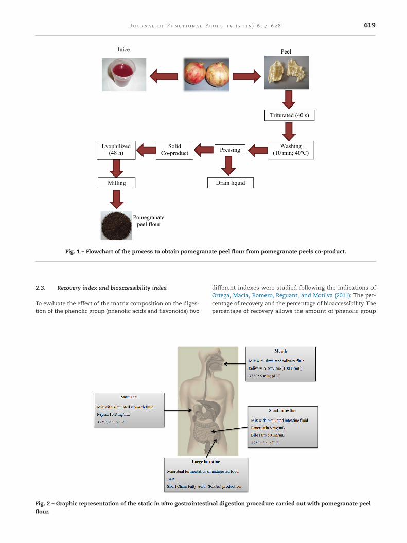
In vitro gastrointestinal digestion of samples was performedaccording to the method described by Mills et al. (2008) withsome modifications (Fig. 2). Digestion was performed byenzymes and absorption was simulated by using dialysis tubing.The method includes three different stages of digestion: Mouth,gastric and small intestine conditions. Mastication was simu-lated using a saliva solution prepared with 100 U/mL α-amylase(Sigma, Germany) and diluted in 1 mM CaCl2, whereas 1 MNaHCO3 was used to adjust pH to 6.9. The simulated saliva(1 mL) was added to 2.4 g of sample and 9 mL of water and thenthe mixture was stomached for 2 min.The sample solution wastransferred to a glass screw topped bottles and incubated at37 °C for 5 min. In gastric conditions, the sample was ad-justed to pH 2 with HCl (6 M) and then 1 mL pepsin (Sigma)(0.108 g) dissolved in HCl (0.1 M; 10 mL) was added and incu-bated for 2 h in a shaking water bath at 37 °C and 50 rpm. InSmall intestine conditions, the pH was increased to 7 with NaOH(6 M) and 2.5 mL pancreatin (Sigma) (80 mg) dissolved in NaHCO3
(0.5 M; 10 mL) and 2.5 mL bile salt mixture (Sigma) (500 mg) dis-solved in NaHCO3 (0.5 M; 10 mL) were added, and the incubationwas continued for another 2 h in a shaking water bath at 37 °Cand 50 rpm. After this time, the sample solution was trans-ferred to 3 kDa molecular weight cut-off dialysis tubing anddialysed overnight against NaCl (10 mM) at 37 °C to remove lowmolecular mass digestion products. Mechanical agitation wasused to parallel peristaltic movements. At the end of the mouth,gastric and intestinal steps, the digestion mixtures were cen-trifuged for 12 min at 8000 g at 4 °C, yielding the chyme solublefraction (CSF) and the pellet fraction (PF). Both fractions werelyophilised and stored for chromatographic analysis of thephenol composition and antioxidant activity. All enzymessolutions were prepared fresh and filter-sterilised using a0.22 µm-membrane filter previously used. All solutions weremaintained in an ice bath during the entire period of gastro-intestinal digestion processes prior to gradual addition (whenappropriate).
618 J o u rna l o f Func t i ona l F ood s 1 9 ( 2 0 1 5 ) 6 1 7 – 6 2 8
2.3. Recovery index and bioaccessibility index
To evaluate the effect of the matrix composition on the diges-tion of the phenolic group (phenolic acids and flavonoids) two
different indexes were studied following the indications ofOrtega, Macia, Romero, Reguant, and Motilva (2011): The per-centage of recovery and the percentage of bioaccessibility. Thepercentage of recovery allows the amount of phenolic group
PeelJuice
Triturated (40 s)
Washing(10 min; 40ºC)PressingLyophilized
(48 h)
Milling Drain liquid
Solid Co-product
Pomegranate peel flour
Fig. 1 – Flowchart of the process to obtain pomegranate peel flour from pomegranate peels co-product.
Fig. 2 – Graphic representation of the static in vitro gastrointestinal digestion procedure carried out with pomegranate peelflour.
619J o u rna l o f Func t i ona l F ood s 1 9 ( 2 0 1 5 ) 6 1 7 – 6 2 8
present in the complete digestion (CSF and PF) after mouth,gastric and intestinal digestion of test food to be measured ac-cording to:
Recovery index PC PCDF TF%( ) ( )= × 100
Where PCDF is the total phenol content (mg) in the di-gested (CSF + PF) and PCTF is the total phenol content (mg)quantified in test matrix.
For each phenol group, the bioaccessibility is defined as thepercentage of polyphenolic compounds that is solubilised inCSF after intestinal dialysis step. Thus, this index defines theproportion of the polyphenolic compounds that could becomeavailable for absorption into the systematic circulation:
Bioaccessibility index PC PCS DF%( ) ( )= × 100
where: PCS is the total phenol content (mg) in the CSF afterthe intestinal dialysis step and PCDF is the total phenol content(mg) in the digested sample (CSF + PF) after the intestinal step.
2.4. Total phenol content
The total phenol content (TPC) of each extract was per-formed using the Folin–Ciocalteu’s reagent (Singleton & Rossi,1965). Lyophilised samples were dissolved in methanol to obtaina concentration comprise between 20 and 40 mg/mL. Gallic acid(GA) was the reference standard and the results were ex-pressed as mg GA equivalents/g sample. Each assay was carriedout in triplicate.
2.5. Total flavonoid content
For the total flavonoid content (TFC), the method based on Blasaet al. (2005) was used. Methanolic solutions of lyophilisedsamples were used for the analysis. Different concentrationsof rutin (8.5–170 µg/mL) were used for calibration. The resultswere expressed in mg rutin equivalents (RE)/g of sample asmean of three replicates.
2.6. Determination of polyphenolic compounds
Samples (20 µL) were injected into a Hewlett-Packard HPLC series1100 instrument (Woldbronn, Germany) equipped with a diodearray detector. Separations were realised on a C18 Teknokromacolumn (Mediterranea Sea18, 25 × 0.4 cm, 5 µm particle size.Teknokroma, Barcelona, Spain) and the chromatograms were re-corded at 280, 320, 360 or 520 nm. Phenolic compounds wereanalysed, in standard and sample solutions, using a gradientelution at 1 mL/min with the following gradient programme,starting with 95% A, 75% A at 20 min, 50% A at 40 min, 20% Aat 50 min and 20% A at 60 min. The mobile phases were com-posed by formic acid in water (4.5:95.5, v/v) as solvent A andacetonitrile as solvent B. The quantitative analysis of the com-ponents was achieved with reference to authentic standards.The standards used were catechin and epicatechin, as well ascaffeic, ferulic, sinapic, p-coumaric, ellagic, gallic, and chloro-genic acids along with ellagitannin, punicalagin, rutin, quercetin
and apigenin (Extrasynthese, Genay, France). Compound iden-tification was carried out by comparing UV absorption spectraand retention times of each compound with those of pure stan-dards injected in the same conditions. The compounds werequantified through calibration curves of standard compounds.Ellagic acid derivatives were tentatively quantified using the cali-bration curves of ellagic acid. All determinations were made intriplicate.
2.7. Antioxidant activity
2.7.1. Ferric reducing antioxidant powerThe ferric reducing antioxidant power (FRAP) of lyophilisedsamples digested was determined using the potassiumferricyanide-ferric chloride method (Oyaizu, 1986). The FRAPvalue was estimated in mg Trolox equivalents (TE)/g of sample.Each assay was carried out in triplicate.
2.7.2. Ferrous ion-chelating ability assayFerrous ions (Fe2+) chelating activity, of lyophilised samples di-gested, was measured by inhibiting the formation of Fe2+-ferrozine complex after treatment of test material with Fe2+,following the method of Carter (1971). Results were ex-pressed in mg EDTA equivalents/g of sample as mean of threereplicates.
2.7.3. DPPH radical scavenging ability assayThe antioxidant activity of different lyophilised samples di-gested was measured in terms of radical scavenging ability,using the stable radical DPPH (Brand-Williams, Cuvelier, &Berset, 1995). Results were expressed in mg Trolox equiva-lents (TE)/g of sample as mean of three replicates.
2.7.4. Oxygen radical absorbance capacity (ORAC assay)The oxygen radical absorbance capacity (ORAC) of differentlyophilised samples was determined at different concentra-tions by the method of Ou, Hampsch-Woodill, and Prior (2001)using fluorescein as the “fluorescent probe” with some modi-fications. Briefly, 20 µL of phosphate buffer (75 mM, pH 7),Troloxor lyophilised samples at different concentrations were incu-bated with 120 µL of fluorescein (20 nM) at 40 °C for 10 min.The reaction was started by thermal decomposition of AAPH(19 mM in 75 mM phosphate buffer, pH 7.0) and performed at37 °C in 96-well black plates. Fluorescence was measured im-mediately after the addition of AAPH at excitation of 485 nmand an emission wavelength of 535 nm for 105 min at 40 °C.The automated ORAC assay was carried out on a FLUOstarOptima fluorescence microplate reader (BMG LABTECH GmbH).The ORAC values, expressed as µg trolox equivalents per mgsample, were calculated by applying the following formula.
Relative ORAC value C AUC AUC kAUC
Trolox Sample Blank
Tro
= × −( ) ×( )llox BlankAUC−( )
where CTrolox is the concentration of Trolox, k is the sample di-lution factor, and AUC is the area under the fluorescence decaycurve of the sample, blank, and trolox, respectively.
620 J o u rna l o f Func t i ona l F ood s 1 9 ( 2 0 1 5 ) 6 1 7 – 6 2 8
2.7.5. ABTS radical cation (ABTS•+) scavenging activityassayThe ABTS•+ scavenging activity assay was determined as de-scribed by Leite et al. (2011) with some modifications.The ABTS•+
solution was produced by reacting aqueous ABTS solution(7 mM) with potassium persulfate (2.45 mM). Diluted ABTS•+ so-lution with an absorbance of 0.70 ± 0.02 at 734 nm was employedin the analysis. The reactions were performed by adding 990 µLof ABTS•+ solution to 10 µL of each extract solution. After 6 minof incubation at room temperature, absorbance values weremeasured on a spectrophotometer at 734 nm. The results werecalculated based on a calibration curve of Trolox, and resultswere expressed as mg Trolox equivalents (TE)/g of sample.
2.8. In vitro microbial fermentation
2.8.1. Faecal inoculaThe faecal samples were obtained fresh at the premises of thedepartment from three healthy human donors, who were freeof any known metabolic and gastrointestinal diseases; nottaking probiotic or prebiotic supplements and had not takenany form of antibiotics 3 months prior to faecal sample do-nation. Faeces were collected into sterile vials, kept in ananaerobic cabinet and used within a maximum of 2 h after col-lection. Faecal inocula (FI) were prepared by dilution in a reducedphysiological salt solution (RPS; cysteine-HCl (Merck, Darm-stadt, Germany) 0.5 g/L and NaCl (Panreac, Madrid, Spain) 8.5 g/L)at a 100 g faeces/L RPS and pH 6.8, following the methodol-ogy described by Gullón et al. (2014).
2.8.2. Fermentation mediaThe nutrient base medium used in fermentations comprisedof 5.0 g/L trypticase soya broth (TSB) without dextrose (BBL,Lockeysville, USA), 5.0 g/L bactopeptone (Amersham, Bucking-hamshire, UK), 0.5 g/L cysteine-HCl (Merck, Darmstadt,Germany), 1.0 % (v/v) of salt solution A (100.0 g/L NH4Cl, 10.0 g/LMgCl2·6H2O, 10.0 g/L CaCl2·2H2O) and trace minerals solution,0.2% (v/v) of salt solution B (200.0 g/L K2HPO4·3H2O) and 0.2%(v/v) of 0.5 g/L resazurin solution, prepared in distilled water.The final pH of the medium was adjusted to 6.8. Aliquots weredispensed into airtight anaerobic serum bottles, which weresealed with aluminium caps before sterilisation by auto-clave. Stock solution of Yeast Nitrogen Base (YNB) (Sigma-Aldrich Co., St. Louis, USA) was sterilised under syringe filtersof 0.22 µm (Chromafils, Macherey-Nagel, Düren, Germany) intosterile airtight serum bottles.
2.9. In vitro colonic fermentation
YNB solution and pomegranate lyophilised residue obtainedafter enzymatic digestion were aseptically added to the an-aerobic serum bottles with nutrient base medium beforeinoculation to achieve a final concentration of 5.0 g of YNB/Land 10.0 g of extract/L. The serum bottles were inoculated withfaecal slurry dilution to a final concentration of 2% (v/v) andincubated at 37 °C for 24 h without shaking. All additions andinoculations were carried out inside an anaerobic cabinet (5%H2, 10% CO2 and 85% N2). Samples were collected 24 h of fer-mentation, cultures were centrifuged at 16,000 g for 10 min, and
supernatants were collected for analysis of short chain fattyacids (SCFA) using high performance liquid chromatography(HPLC).
2.9.1. Determination of fermentation products in cell-freesupernatantsSupernatants from the anaerobic culture tubes inoculated withFI were filtered through 0.20 µm cellulose acetate mem-branes. Aliquots of the filtered samples were assayed for organicacids (acetic, propionic and butyric acids) using an Agilent 1200series HPLC instrument with a UV-Vis Diode Array Detector(Agilent, Germany). Twenty microlitres of sample were in-jected in an Aminex HPX-87H column (from BioRad, Hercules,CA) using sulphuric acid (0.003 M) as mobile phase, operatingflow rate of 0.6 mL/min. Samples were run at 50 °C. Stan-dards of organic acids (acetic, butyric, propionic, fumaric andsuccinic acids) were obtained from Sigma (Poole, Dorset, UK).Peaks were identified by comparison with retention time of thestandards and were quantified by regression formula ob-tained with the standards.
2.10. Statistical assay
Statistical analysis and comparisons among means were carriedout using the Statistical Package SPSS 19.0 (SPSS Inc., Chicago,IL, USA). All experiments were carried out in triplicate and datawere reported as mean ± standard deviation. The differencesof mean values among concentration of bioactive compoundsor antioxidant activity and that obtained in the different stepsof the in vitro gastrointestinal digestion were analysed by one-way analysis of variance (ANOVA). The Tukey’s post hoc testwas applied for comparisons of means; differences were con-sidered significant at p < 0.05. Correlation analysis wasperformed between phenolic compounds (phenolic acids andflavonoids) contents and antioxidant activities of extracts usingPearson correlation analysis.
3. Results and discussion
3.1. Recovery index and bioaccessibility index
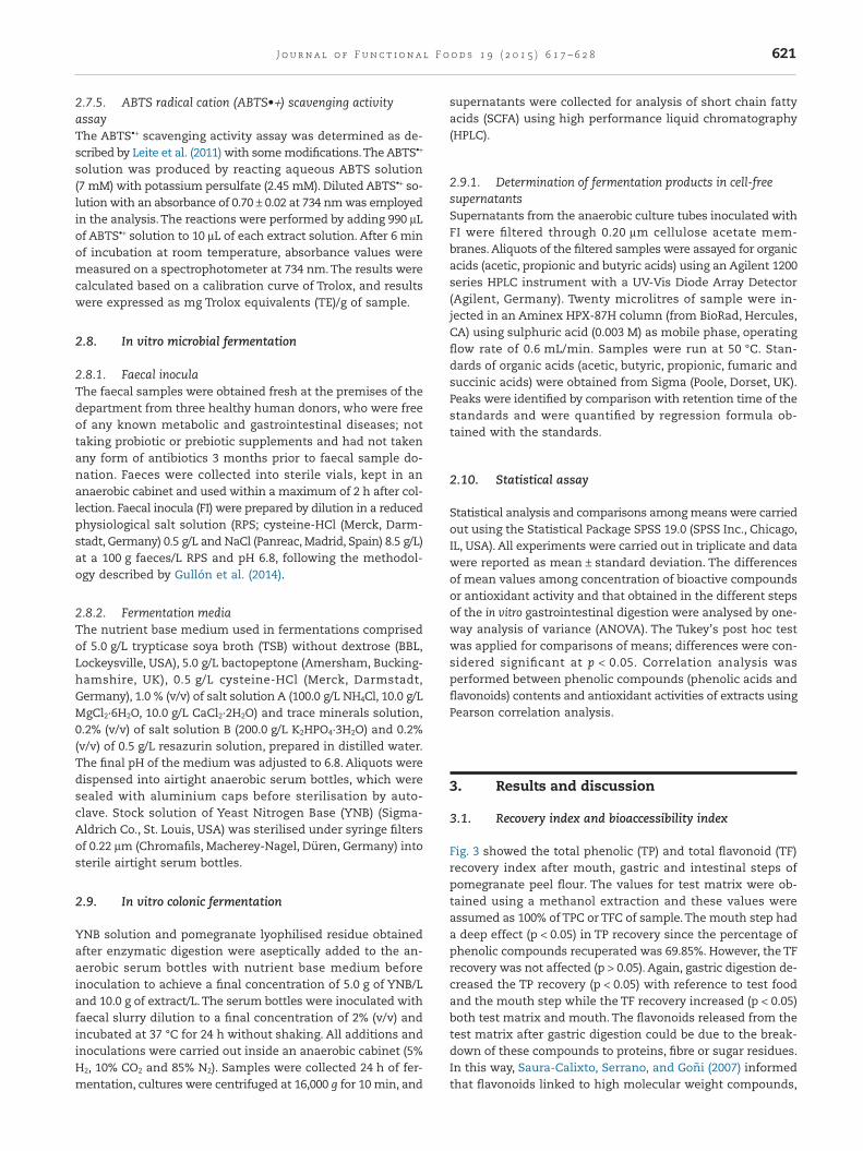
Fig. 3 showed the total phenolic (TP) and total flavonoid (TF)recovery index after mouth, gastric and intestinal steps ofpomegranate peel flour. The values for test matrix were ob-tained using a methanol extraction and these values wereassumed as 100% of TPC or TFC of sample. The mouth step hada deep effect (p < 0.05) in TP recovery since the percentage ofphenolic compounds recuperated was 69.85%. However, the TFrecovery was not affected (p > 0.05). Again, gastric digestion de-creased the TP recovery (p < 0.05) with reference to test foodand the mouth step while the TF recovery increased (p < 0.05)both test matrix and mouth. The flavonoids released from thetest matrix after gastric digestion could be due to the break-down of these compounds to proteins, fibre or sugar residues.In this way, Saura-Calixto, Serrano, and Goñi (2007) informedthat flavonoids linked to high molecular weight compounds,
621J o u rna l o f Func t i ona l F ood s 1 9 ( 2 0 1 5 ) 6 1 7 – 6 2 8
such as proteins and carbohydrates, may be released by di-gestive enzyme action, leading to a significant increase in theirconcentrations after gastric digestion. At the end of intesti-nal step, both TP and TF recovery were profoundly affected(p < 0.05) by the recovery index of 43.00 and 45.26% respec-tively. This observation is in agreement with the work carriedout by Ortega et al. (2011) in which TP and TF recovery indexof washed carob flour decreased after intestinal step. Li, Deng,Liu, Loewen, and Tsao (2014) also detected that the TP and TFrecovery index decreased after intestinal digestion of purpletomato. Argyri, Komaitis, and Kapsokefalou (2006) observed thatsolubility and availability of phenolic compounds (phenolic acidsand flavonoids) are influenced by pH conditions and interac-tion with dietary constituents, such as iron, fibre or proteins.
To exert bioactivity, phenolic acids and flavonoids first mustbe bioaccessible, i.e. released from the food matrix andsolubilised as mentioned in Bouayed et al. (2011). Thebioaccessibility of phenolic and flavonoid compounds in pome-granate peel flour at the end of intestinal digestion was 35.90and 64.02% respectively. These values suggest that severalchanges in phenolic and flavonoid compounds like modifica-tion of chemical structure, increased or reduced solubility orinteraction with other compounds might happen during thegastrointestinal digestion of pomegranate peel flour, which in-fluence the bioaccessibility.Thus, Helal, Tagliazucchi, Verzelloni,and Conte (2014) obtained a bioaccessibility of 79.8% of totalphenolic compounds in cinnamon beverage. Ortega et al. (2011)informed that the bioaccessibility, after intestinal digestion, ofphenolic and flavonoids compounds were 81 and 65% respec-tively. Rodríguez-Roque, Rojas-Graü, Elez-Martínez, andMartín-Belloso (2013) reported that the total flavonoids presentin soymilk showed a bioaccessibility of 16%, whereas total phe-nolic acids were not bioaccessible after gastrointestinaldigestion.
3.2. Stability of polyphenolic compounds present inpomegranate peel flour during simulated in vitrogastrointestinal digestion
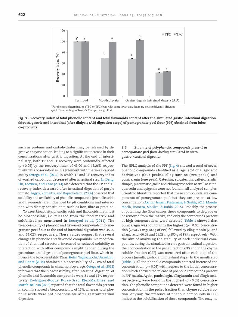
The HPLC analysis of the PPF (Fig. 4) showed a total of sevenphenolic compounds identified as ellagic acid or ellagic acidderivatives (four peaks), ellagitannins (two peaks) andpunicalagin (one peak). Catechin, epicatechin, caffeic, ferulic,sinapic, p-coumaric, gallic and chlorogenic acids as well as rutin,quercetin and apigenin were not found in all analysed samples.Scientific literature reported that these compounds are com-ponents of pomegranate peel but they are present at lowconcentration (Akhtar, Ismail, Fraternale, & Sestili, 2015; Mosele,Macià, Romero, Motilva, & Rubió, 2015). Probably, the processof obtaining the flour causes these compounds to degrade orbe removed from the matrix, and only the compounds presentat high concentrations were detected. Table 1 showed thatpunicalagin was found with the highest (p < 0.05) concentra-tion (2850.21 mg/100 g of PPF) followed by ellagitannin (2) andellagic acid (66.05 and 65.28 mg/100 g of PPF, respectively). Withthe aim of analysing the stability of each individual com-pounds, during the simulated in vitro gastrointestinal digestion,their concentration in the pellet fraction (PF) and in the chymesoluble fraction (CSF) was measured after each step of theprocess (mouth, gastric and intestinal steps). In the mouth step(Table 1), all the phenolic compounds detected increased theconcentration (p < 0.05) with respect to the initial concentra-tion which showed the release of phenolic compounds presentin PPF matrix. Again, punicalagin, ellagitannin and ellagic acid,respectively, were found in the highest (p > 0.05) concentra-tion. The phenolic compounds detected were found in higherconcentration in the pellet fraction than chyme soluble frac-tion. Anyway, the presence of phenolic compounds in CSFindicates the solubilisation of these compounds. The enzyme
¥For the same determination (TPC or TFC) bars with same lower case letter are not significantly different (p>0.05) according to Tukey’s Multiple Range Test.
0
20
40
60
80
100
120
Test food Mouth digesta Gastric digesta Intestinal digesta (AD)
Rec
over
y in
dex
(%)
TPC TFCa¥
b c
d
a a b
c
Fig. 3 – Recovery index of total phenolic content and total flavonoids content after the simulated gastro-intestinal digestion(Mouth, gastric and intestinal (after dialysis (AD) digestion steps) of pomegranate peel flour (PPF) obtained from juiceco-products.
622 J o u rna l o f Func t i ona l F ood s 1 9 ( 2 0 1 5 ) 6 1 7 – 6 2 8
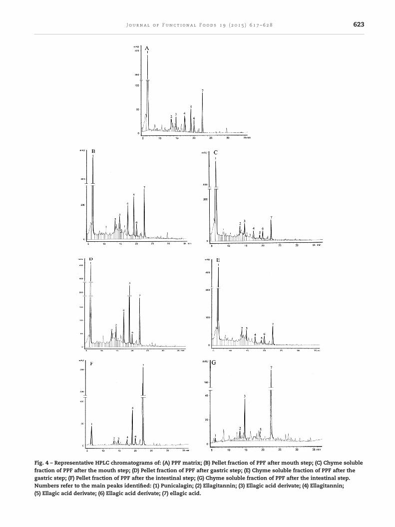
Fig. 4 – Representative HPLC chromatograms of: (A) PPF matrix; (B) Pellet fraction of PPF after mouth step; (C) Chyme solublefraction of PPF after the mouth step; (D) Pellet fraction of PPF after gastric step; (E) Chyme soluble fraction of PPF after thegastric step; (F) Pellet fraction of PPF after the intestinal step; (G) Chyme soluble fraction of PPF after the intestinal step.Numbers refer to the main peaks identified: (1) Punicalagin; (2) Ellagitannin; (3) Ellagic acid derivate; (4) Ellagitannin;(5) Ellagic acid derivate; (6) Ellagic acid derívate; (7) ellagic acid.
623J o u rna l o f Func t i ona l F ood s 1 9 ( 2 0 1 5 ) 6 1 7 – 6 2 8
activity or agitation conditions could facilitate the breakage oflarge molecules as high molecular weight phenols which ini-tially may be insoluble. In gastric step (Table 1), punicalagin,ellagitannin and ellagic acid were slightly (p < 0.05) reduced (2.59,4.68 and 2.86%, respectively) with respect to initial values. Onthe other hand, other phenolic compounds detected had anincrement (p < 0.05) of their concentration in reference to initialvalues. The behaviour of phenolic compounds under gastricconditions was contradictory. Therefore, the phenolic acidsshowed low stability in agreement with the results reportedby Kamiloglu and Capanoglu (2013) or Mosele et al. (2015) whilstflavonoids and tannins were more stable under the same con-ditions (Bouayed, Deußer, Hoffmann, & Bohn, 2012). Except forpunicalagin, the phenolic compounds concentration was higher(p < 0.05) in PF than CSF. It should be noted that, as men-tioned in Chandrasekara and Shahidi (2012), the phenoliccompounds released in the gastric step may be absorbed andthey may have some local antioxidant effects in the small in-testine as well due to their solubility in the digest.
At the end of the intestinal step (Table 1), a drastic reduc-tion of polyphenolic compounds was found. The resultsobtained are similar to those reported by Ortega et al. (2011)who showed that after the intestinal digestion step, impor-tant losses of phenolic compounds were found. As mentionedabove, all the compounds detected were reduced (p < 0.05) withpercentage of reduction between 58.32 and 96.3% by refer-ence to initial concentration, except the ellagic acid, whichincreased (p < 0.05) by 112.69% with reference to initial values.The increased concentration of ellagic acid after intestinal di-gestion might be due to this compound being bound to proteinsor fibre in the original matrix and as a result of enzymatic di-gestion was then released from these structures. In this sense,Mosele et al. (2015) reported that ellagic acid concentration in-creased after gastrointestinal incubation due to liberation fromthe complex phenols present in the food matrix. It is impor-tant to notice that in this step, the phenolic compoundsdetected were found in higher concentration in the pellet frac-tion than in chyme soluble fraction.
3.3. Antioxidant activities of gastrointestinal digest
The antioxidant activity of fruit products are linked to the phe-nolic acids and flavonoids content. However, the antioxidantproperties of these compounds might change due to the chemi-cal transformations during the gastrointestinal digestion. Inaddition, since the antioxidant activity of foodstuff is deter-mined by multiple reaction characteristics and differentmechanisms, it is necessary to combine more than one methodin order to determine the in vitro antioxidant capacity of foods.In addition, each method only provides an estimate of anti-oxidant capacity that is subjective to its conditions and reagents(Ma et al., 2011; Viuda-Martos, Ruiz-Navajas, Sánchez-Zapata,Fernández-López, & Pérez-Álvarez, 2010).Therefore, in this workto evaluate the changes produced by the gastrointestinal di-gestion of PPF extracts, the antioxidant activity was determinedusing five different methodologies.
Table 2 showed the ferrous ion chelating activity (FIC) of di-gested PPF. The mouth step increased (p < 0.05) the FIC valuesby 57.14% with reference to initial values.The PF showed a slightdecrease (p > 0.05) with respect to PPF values whilst that of the
Tabl
e1
–Po
lyp
hen
olic
con
cen
trat
ion
obta
ined
from
the
two
frac
tion
s(p
elle
tfr
acti
on(P
F)an
dch
yme
solu
ble
frac
tion
(CS
F))a
fter
the
sim
ula
ted
gast
roin
test
inal
dig
esti
on(M
outh
,gas
tric
and
inte
stin
alst
eps)
ofp
omeg
ran
ate
pee
lflou
r(P
PF)o
btai
ned
from
juic
eco
-pro
du
cts.
Phen
olic
com
pou
nd
PPF
(100
g)
Mou
thSt
epG
astr
icst
epIn
test
inal
step
PFC
SFTo
tal
PFC
SFTo
tal
PFC
SFTo
tal
Pun
ical
agin
2850
.21
±3.
1cA17
90.2
1±
4.1
1180
.20
±3.
129
70.4
1±
3.2aA
1340
.49
±3.
114
35.8
2±
2.2
2776
.31
±3.
1bA12
4.63
±1.
12.
39±
0.1
127.
02±
1.1d
B
Ella
gita
nn
in57
.83
±0.
1bC39
.83
±0.
119
.50
±0.
159
.33
±0.
1aD32
.60
±0.
122
.52
±0.
155
.12
±0.
1cE4.
62±
0.1
3.25
±0.
17.
87±
0.1d
D
Ella
gic
acid
der
ivat
ive
29.5
9±
0.1cE
22.8
1±
0.1
7.73
±0.
130
.54
±0.
1aF21
.58
±0.
110
.98
±0.
132
.56
±0.
1bF3.
00±
0.1
1.99
±0.
14.
99±
0.1d
E
Ella
gita
nn
in66
.05
±0.
1bB63
.05
±0.
17.
94±
0.1
70.9
9±
0.4aB
61.5
4±
0.1
9.01
±0.
170
.55
±0.
4aC2.
18±
0.1
0.00
±0.
12.
18±
0.1cF
Ella
gic
acid
der
ivat
ive
52.3
6±
0.1cD
49.1
1±
0.1
5.57
±0.
154
.68
±0.
1bE77
.61
±0.
16.
21±
0.1
83.8
2±
0.1aB
19.5
5±
0.1
2.27
±0.
121
.82
±0.
1dC
Ella
gic
acid
der
ivat
ive
29.3
2±
0.1bE
23.7
1±
0.1
7.53
±0.
131
.24
±0.
2aG22
.14
±0.
19.
04±
0.1
31.1
8±
0.1aG
4.96
±0.
10.
00±
0.1
4.96
±0.
1cE
ella
gic
acid
65.2
8±
0.9cB
57.2
7±
0.1
11.4
2±
0.1
68.6
8±
0.2bC
50.2
1±
0.1
13.2
0±
0.1
63.4
1±
0.1d
D10
2.22
±0.
136
.63
±0.
113
8.85
±0.
1aA
Val
ues
exp
ress
edas
mg
ofea
chco
mp
oun
dp
er10
0g
ofp
rod
uct
.Fo
rth
esa
me
ph
enol
icco
mp
oun
d,v
alu
esin
the
sam
ero
wfo
llow
edw
ith
sam
elo
wer
case
lett
erar
en
otsi
gnifi
can
tly
dif
fere
nt
(p>
0.05
)ac
cord
ing
toTu
key’
sM
ult
iple
Ran
geTe
st.
For
the
sam
ed
iges
tion
step
,val
ues
inth
esa
me
colu
mn
foll
owed
wit
hsa
me
up
per
case
lett
erar
en
otsi
gnifi
can
tly
dif
fere
nt
(p>
0.05
)ac
cord
ing
toTu
key’
sM
ult
iple
Ran
geTe
st.
624 J o u rna l o f Func t i ona l F ood s 1 9 ( 2 0 1 5 ) 6 1 7 – 6 2 8
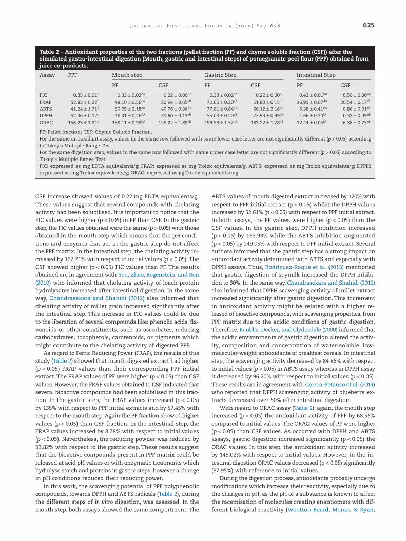
CSF increase showed values of 0.22 mg EDTA equivalents/g.These values suggest that several compounds with chelatingactivity had been solubilised. It is important to notice that theFIC values were higher (p < 0.05) in PF than CSF. In the gastricstep, the FIC values obtained were the same (p > 0.05) with thoseobtained in the mouth step which means that the pH condi-tions and enzymes that act in the gastric step do not affectthe PPF matrix. In the intestinal step, the chelating activity in-creased by 167.71% with respect to initial values (p < 0.05). TheCSF showed higher (p < 0.05) FIC values than PF. The resultsobtained are in agreement with You, Zhao, Regenstein, and Ren(2010) who informed that chelating activity of loach proteinhydrolysates increased after intestinal digestion. In the sameway, Chandrasekara and Shahidi (2012) also informed thatchelating activity of millet grain increased significantly afterthe intestinal step. This increase in FIC values could be dueto the liberation of several compounds like: phenolic acids, fla-vonoids or other constituents, such as ascorbates, reducingcarbohydrates, tocopherols, carotenoids, or pigments whichmight contribute to the chelating activity of digested PPF.
As regard to Ferric Reducing Power (FRAP), the results of thisstudy (Table 2) showed that mouth digested extract had higher(p < 0.05) FRAP values than their corresponding PPF initialextract. The FRAP values of PF were higher (p < 0.05) than CSFvalues. However, the FRAP values obtained to CSF indicated thatseveral bioactive compounds had been solubilised in this frac-tion. In the gastric step, the FRAP values increased (p < 0.05)by 135% with respect to PPF initial extracts and by 57.45% withrespect to the mouth step. Again the PF fraction showed highervalues (p < 0.05) than CSF fraction. In the intestinal step, theFRAP values increased by 8.78% with respect to initial values(p < 0.05). Nevertheless, the reducing powder was reduced by53.82% with respect to the gastric step. These results suggestthat the bioactive compounds present in PPF matrix could bereleased at acid pH values or with enzymatic treatments whichhydrolyse starch and proteins in gastric steps; however a changein pH conditions reduced their reducing power.
In this work, the scavenging potential of PPF polyphenoliccompounds, towards DPPH and ABTS radicals (Table 2), duringthe different steps of in vitro digestion, was assessed. In themouth step, both assays showed the same comportment. The
ABTS values of mouth digested extract increased by 120% withrespect to PPF initial extract (p < 0.05) whilst the DPPH valuesincreased by 52.61% (p < 0.05) with respect to PPF initial extract.In both assays, the PF values were higher (p < 0.05) than theCSF values. In the gastric step, DPPH inhibition increased(p < 0.05) by 153.93% while the ABTS inhibition augmented(p < 0.05) by 249.05% with respect to PPF initial extract. Severalauthors informed that the gastric step has a strong impact onantioxidant activity determined with ABTS and especially withDPPH assays. Thus, Rodríguez-Roque et al. (2013) mentionedthat gastric digestion of soymilk increased the DPPH inhibi-tion to 30%. In the same way, Chandrasekara and Shahidi (2012)also informed that DPPH scavenging activity of millet extractincreased significantly after gastric digestion. This incrementin antioxidant activity might be related with a higher re-leased of bioactive compounds, with scavenging properties, fromPPF matrix due to the acidic conditions of gastric digestion.Therefore, Baublis, Decker, and Clydesdale (2000) informed thatthe acidic environments of gastric digestion altered the activ-ity, composition and concentration of water-soluble, low-molecular-weight antioxidants of breakfast cereals. In intestinalstep, the scavenging activity decreased by 84.86% with respectto initial values (p < 0.05) in ABTS assay whereas in DPPH assayit decreased by 96.20% with respect to initial values (p < 0.05).These results are in agreement with Correa-Betanzo et al. (2014)who reported that DPPH scavenging activity of blueberry ex-tracts decreased over 50% after intestinal digestion.
With regard to ORAC assay (Table 2), again, the mouth stepincreased (p < 0.05) the antioxidant activity of PPF by 68.55%compared to initial values. The ORAC values of PF were higher(p < 0.05) than CSF values. As occurred with DPPH and ABTSassays, gastric digestion increased significantly (p < 0.05) theORAC values. In this step, the antioxidant activity increasedby 145.02% with respect to initial values. However, in the in-testinal digestion ORAC values decreased (p < 0.05) significantly(87.95%) with reference to initial values.
During the digestion process, antioxidants probably undergomodifications which increase their reactivity, especially due tothe changes in pH, as the pH of a substance is known to affectthe racemisation of molecules creating enantiomers with dif-ferent biological reactivity (Wootton-Beard, Moran, & Ryan,
Table 2 – Antioxidant properties of the two fractions (pellet fraction (PF) and chyme soluble fraction (CSF)) after thesimulated gastro-intestinal digestion (Mouth, gastric and intestinal steps) of pomegranate peel flour (PPF) obtained fromjuice co-products.
Assay PPF Mouth step Gastric Step Intestinal Step
PF CSF PF CSF PF CSF
FIC 0.35 ± 0.01c 0.33 ± 0.02cA 0.22 ± 0.00dB 0.33 ± 0.02cA 0.22 ± 0.00dB 0.43 ± 0.01bB 0.50 ± 0.00aA
FRAP 52.83 ± 0.22b 48.10 ± 0.56cA 30.94 ± 0.65eB 72.65 ± 0.20aA 51.80 ± 0.15bB 36.93 ± 0.07dA 20.54 ± 0.17fB
ABTS 41.24 ± 1.71d 50.05 ± 2.18cA 40.76 ± 0.36dB 77.81 ± 0.84aA 66.12 ± 2.16bB 5.38 ± 0.41eA 0.86 ± 0.01fB
DPPH 52.36 ± 0.12c 48.31 ± 0.26dA 31.60 ± 0.13eB 55.03 ± 0.20bB 77.93 ± 0.99aA 1.66 ± 0.30fA 0.33 ± 0.09gB
ORAC 156.23 ± 1.24c 138.11 ± 0.99dA 125.22 ± 1.89eB 199.58 ± 1.57aA 183.22 ± 1.78bB 12.44 ± 0.04fA 6.38 ± 0.75gB
PF: Pellet fraction; CSF: Chyme Soluble Fraction.For the same antioxidant assay, values in the same row followed with same lower case letter are not significantly different (p > 0.05) accordingto Tukey’s Multiple Range Test.For the same digestion step, values in the same row followed with same upper case letter are not significantly different (p > 0.05) according toTukey’s Multiple Range Test.FIC: expressed as mg EDTA equivalents/g; FRAP: expressed as mg Trolox equivalents/g; ABTS: expressed as mg Trolox equivalents/g; DPPH:expressed as mg Trolox equivalents/g; ORAC: expressed as µg Trolox equivalents/mg.
625J o u rna l o f Func t i ona l F ood s 1 9 ( 2 0 1 5 ) 6 1 7 – 6 2 8
2011). Thus, antioxidant compounds would be more reactiveparticularly at acidic pH (as occurs in gastric digestion) and lessreactive at pH close to neutrality (as occurs in intestinal di-gestion) (Wootton-Beard et al., 2011).
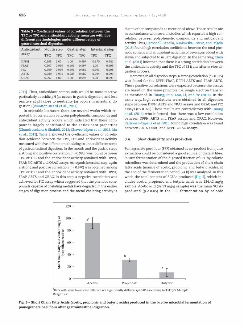
In scientific literature there are several works which re-ported that correlation between polyphenolic compounds andantioxidant activity occurs which indicated that these com-pounds largely contributed to the antioxidant properties(Chandrasekara & Shahidi, 2012; Chaves-López et al., 2015; Maet al., 2011). Table 3 showed the coefficient values of correla-tion achieved between the TPC, TFC and antioxidant activitymeasured with five different methodologies under different stepsof gastrointestinal digestion. In the mouth and the gastric stepsa strong and positive correlation (r > 0.980) was found betweenTPC or TFC and the antioxidant activity obtained with DPPH,FRAP, FIC,ABTS and ORAC assays.As regards intestinal step, againa strong and positive correlation (r > 0.970) was obtained amongTPC or TFC and the antioxidant activity obtained with DPPH,FRAP, ABTS and ORAC. In this step, a negative correlation wasachieved for FIC assay which suggested that the phenolic com-pounds capable of chelating metals have degraded in the earlierstages of digestion process and the metal chelating activity is
due to other compounds as mentioned above.These results arein concordance with several studies which reported a high cor-relation between polyphenolic compounds and antioxidantactivity.Thus, Carbonell-Capella, Buniowska, Esteve, and Frígola(2015) found high correlation coefficients between the total phe-nolic content and antioxidant activities of beverages added withstevia and subjected to in vitro digestion. In the same way, Chenet al. (2014) informed that there is a strong correlation betweenthe antioxidant activity and the TPC of 33 fruits after in vitro di-gestion process.
Moreover, in all digestion steps, a strong correlation (r > 0.975)was found for the DPPH-FRAP, DPPH-ABTS and FRAP-ABTS.These positive correlations were expected because the assaysare based on the same principle, i.e. single electron transferas mentioned in Huang, Sun, Lou, Li, and Ye (2014). In thesame way, high correlations were obtained in all digestionsteps between DPPH, ABTS and FRAP assays and ORAC and FICassays (r > 0.970). These results are contradictory with Huanget al. (2014) who informed that there was a low correlationbetween DPPH, ABTS and FRAP assays and ORAC. However,Carbonell-Capella et al. (2015) found high correlation was foundbetween ABTS-ORAC and DPPH-ORAC assays.
3.4. Short chain fatty acids production
Pomegranate peel flour (PPF) obtained as co-product from juiceextraction could be considered a good source of dietary fibre.In vitro fermentation of the digested fraction of PPF by colonicmicroflora was determined and the production of short chainfatty acids (mainly of acetic, propionic and butyric acids), atthe end of the fermentation period (24 h) was analysed. In thiswork, the total content of SCFAs produced (Fig. 5), which in-cludes acetic, propionic and butyric acids was 134.42 mg/gsample. Acetic acid (92.53 mg/g sample) was the main SCFAsproduced (p < 0.05) in the PPF fermentation by colonic
Table 3 – Coefficient values of correlation between theTPC or TFC and antioxidant activity measure with fivedifferent methodologies under different steps ofgastrointestinal digestion.
Antioxidantassay
Mouth step Gastric step Intestinal step
TPC TFC TPC TFC TPC TFC
DPPH 0.995 1.00 1.00 0.997 0.970 0.981FRAP 0.997 0.999 0.999 0.997 1.00 0.999FIC 0.999 0.999 0.991 0.982 −0.993 −0.998ABTS 0.989 0.975 0.985 0.989 0.994 0.999ORAC 0.997 1.00 1.00 0.997 1.00 0.999
¥Bars with same lower case letter are not significantly different (p>0.05) according to Tukey’s Multiple Range Test.
0
20
40
60
80
100
120
Acetate Propionate Butyrate
Shor
t cha
in fa
tty
acid
con
tent
mg/
g of
ly
ophi
lized
res
idue
a¥
b
c
Fig. 5 – Short Chain Fatty Acids (acetic, propionic and butyric acids) produced in the in vitro microbial fermentation ofpomegranate peel flour after gastrointestinal digestion.
626 J o u rna l o f Func t i ona l F ood s 1 9 ( 2 0 1 5 ) 6 1 7 – 6 2 8
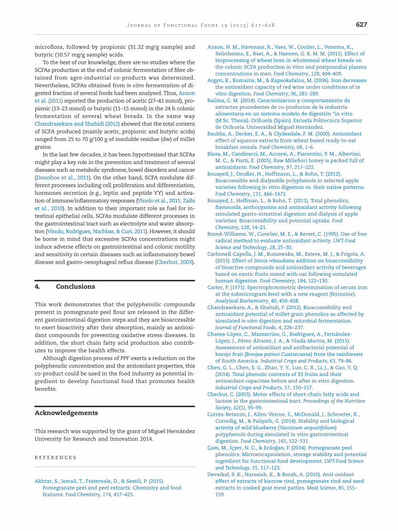
microflora, followed by propionic (31.32 mg/g sample) andbutyric (10.57 mg/g sample) acids.
To the best of our knowledge, there are no studies where theSCFAs production at the end of colonic fermentation of fibre ob-tained from agro-industrial co-products was determined.Nevertheless, SCFAs obtained from in vitro fermentation of di-gested fraction of several foods had been analysed.Thus, Ansonet al. (2011) reported the production of acetic (27–41 mmol), pro-pionic (13–23 mmol) or butyric (11–15 mmol) in the 24 h colonicfermentation of several wheat breads. In the same wayChandrasekara and Shahidi (2012) showed that the total contentof SCFA produced (mainly acetic, propionic and butyric acids)ranged from 25 to 70 g/100 g of insoluble residue (dw) of milletgrains.
In the last few decades, it has been hypothesised that SCFAsmight play a key role in the prevention and treatment of severaldiseases such as metabolic syndrome, bowel disorders and cancer(Donohoe et al., 2011). On the other hand, SCFA modulate dif-ferent processes including cell proliferation and differentiation,hormones secretion (e.g., leptin and peptide YY) and activa-tion of immune/inflammatory responses (Vinolo et al., 2011; Zaibiet al., 2010). In addition to their important role as fuel for in-testinal epithelial cells, SCFAs modulate different processes inthe gastrointestinal tract such as electrolyte and water absorp-tion (Vinolo, Rodrigues, Nachbar, & Curi, 2011). However, it shouldbe borne in mind that excessive SCFAs concentrations mightinduce adverse effects on gastrointestinal and colonic motilityand sensitivity in certain diseases such as inflammatory boweldisease and gastro-oesophageal reflux disease (Cherbut, 2003).
4. Conclusions
This work demonstrates that the polyphenolic compoundspresent in pomegranate peel flour are released in the differ-ent gastrointestinal digestion steps and they are bioaccessibleto exert bioactivity after their absorption, mainly as antioxi-dant compounds for preventing oxidative stress diseases. Inaddition, the short chain fatty acid production also contrib-utes to improve the health effects.
Although digestion process of PPF exerts a reduction on thepolyphenolic concentration and the antioxidant properties, thisco-product could be used in the food industry as potential in-gredient to develop functional food that promotes healthbenefits.
Acknowledgements
This research was supported by the grant of Miguel HernándezUniversity for Research and Innovation 2014.
R E F E R E N C E S
Akhtar, S., Ismail, T., Fraternale, D., & Sestili, P. (2015).Pomegranate peel and peel extracts: Chemistry and foodfeatures. Food Chemistry, 174, 417–425.
Anson, N. M., Havenaar, R., Vaes, W., Coulier, L., Venema, K.,Selinheimo, E., Bast, A., & Haenen, G. R. M. M. (2011). Effect ofbioprocessing of wheat bran in wholemeal wheat breads onthe colonic SCFA production in vitro and postprandial plasmaconcentrations in men. Food Chemistry, 128, 404–409.
Argyri, K., Komaitis, M., & Kapsokefalou, M. (2006). Iron decreasesthe antioxidant capacity of red wine under conditions of invitro digestion. Food Chemistry, 96, 281–289.
Bailina, C. M. (2014). Caracterizacion y comportamiento deextractos procedentes de co-productos de la industriaalimentaria en un sistema modelo de digestión “in vitro(M.Sc. Thesis). Orihuela (Spain). Escuela Politecnica Superiorde Orihuela. Universidad Miguel Hernández.
Baublis, A., Decker, E. A., & Clydesdale, F. M. (2000). Antioxidanteffect of aqueous extracts from wheat based ready-to-eatbreakfast cereals. Food Chemistry, 68, 1–6.
Blasa, M., Candiracci, M., Accorsi, A., Piacentini, P. M., Albertini,M. C., & Piatti, E. (2005). Raw Millefiori honey is packed full ofantioxidants. Food Chemistry, 97, 217–222.
Bouayed, J., Deußer, H., Hoffmann, L., & Bohn, T. (2012).Bioaccessible and dialysable polyphenols in selected applevarieties following in vitro digestion vs. their native patterns.Food Chemistry, 131, 466–1472.
Bouayed, J., Hoffman, L., & Bohn, T. (2011). Total phenolics,flavonoids, anthocyanins and antioxidant activity followingsimulated gastro-intestinal digestion and dialysis of applevarieties: Bioaccessibility and potential uptake. FoodChemistry, 128, 14–21.
Brand-Williams, W., Cuvelier, M. E., & Berset, C. (1995). Use of freeradical method to evaluate antioxidant activity. LWT-FoodScience and Technology, 28, 25–30.
Carbonell-Capella, J. M., Buniowska, M., Esteve, M. J., & Frígola, A.(2015). Effect of Stevia rebaudiana addition on bioaccessibilityof bioactive compounds and antioxidant activity of beveragesbased on exotic fruits mixed with oat following simulatedhuman digestion. Food Chemistry, 184, 122–130.
Carter, P. (1971). Spectrophotometric determination of serum ironat the submicrogram level with a new reagent (ferrozine).Analytical Biochemistry, 40, 450–458.
Chandrasekara, A., & Shahidi, F. (2012). Bioaccessibility andantioxidant potential of millet grain phenolics as affected bysimulated in vitro digestion and microbial fermentation.Journal of Functional Foods, 4, 226–237.
Chaves-López, C., Mazzarrino, G., Rodríguez, A., Fernández-López, J., Pérez-Álvarez, J. A., & Viuda-Martos, M. (2015).Assessment of antioxidant and antibacterial potential ofborojo fruit (Borojoa patinoi Cuatrecasas) from the rainforestsof South America. Industrial Crops and Products, 63, 79–86.
Chen, G. L., Chen, S. G., Zhao, Y. Y., Luo, C. X., Li, J., & Gao, Y. Q.(2014). Total phenolic contents of 33 fruits and theirantioxidant capacities before and after in vitro digestion.Industrial Crops and Products, 57, 150–157.
Cherbut, C. (2003). Motor effects of short-chain fatty acids andlactate in the gastrointestinal tract. Proceedings of the NutritionSociety, 62(1), 95–99.
Correa-Betanzo, J., Allen-Vercoe, E., McDonald, J., Schroeter, K.,Corredig, M., & Paliyath, G. (2014). Stability and biologicalactivity of wild blueberry (Vaccinium angustifolium)polyphenols during simulated in vitro gastrointestinaldigestion. Food Chemistry, 165, 522–531.
Çam, M., Içyer, N. C., & Erdogan, F. (2014). Pomegranate peelphenolics: Microencapsulation, storage stability and potentialingredient for functional food development. LWT-Food Scienceand Technology, 55, 117–123.
Devatkal, S. K., Narsaiah, K., & Borah, A. (2010). Anti-oxidanteffect of extracts of kinnow rind, pomegranate rind and seedextracts in cooked goat meat patties. Meat Science, 85, 155–159.
627J o u rna l o f Func t i ona l F ood s 1 9 ( 2 0 1 5 ) 6 1 7 – 6 2 8
Donohoe, D. R., Garge, N., Zhang, X., Sun, W., O’Connell, T. M.,Bunger, M. K., & Bultman, S. J. (2011). The microbiome andbutyrate regulate energy metabolism and autophagy in themammalian colon. Cell Metabolism, 13, 517–526.
Gawlik-Dziki, U. (2012). Changes in the antioxidant activities ovegetables as a consequence of interactions between activecompounds. Journal of Functional Foods, 4, 872–882.
Grooms, K. N., Ommerborn, M. J., Pham, D. Q., Djoussé, L., &Clark, C. R. (2013). Dietary fiber intake and cardiometabolicrisks among US Adults, NHANES 1999–2010. The AmericanJournal of Medicine, 126, 1059–1067.
Gullón, B., Gullón, P., Tavaria, F., Pintado, M., Gomes, A. M.,Alonso, J. L., & Parajó, J. C. (2014). Structural features andassessment of prebiotic activity of refinedarabinoxylooligosaccharides from wheat bran. Journal ofFunctional Foods, 6, 438–449.
Hasnaoui, N., Wathelet, B., & Jimenez-Araujo, A. (2014).Valorization of pomegranate peel from 12 cultivars: Dietaryfibre composition, antioxidant capacity and functionalproperties. Food Chemistry, 160, 196–203.
Helal, A., Tagliazucchi, D., Verzelloni, E., & Conte, A. (2014).Bioaccessibility of polyphenols and cinnamaldehyde incinnamon beverages subjected to in vitro gastro-pancreaticdigestion. Journal of Functional Foods, 7, 506–516.
Huang, H., Sun, Y., Lou, S., Li, H., & Ye, X. (2014). In vitro digestioncombined with cellular assay to determine the antioxidantactivity in Chines bayberry (Myrica rubra Sieb. Et Zucc.) fruits:A comparison with traditional methods. Food Chemistry, 146,363–370.
Kaczmarczyk, M. M., Miller, M. J., & Freund, G. G. (2012). Thehealth benefits of dietary fiber: Beyond the usual suspects oftype 2 diabetes mellitus, cardiovascular disease and coloncancer. Metabolism: Clinical and Experimental, 61, 1058–1066.
Kamiloglu, S., & Capanoglu, E. (2013). Investigating the in vitrobioaccessibility of polyphenols in fresh and sun-dried figs(Ficus carica L). International Journal of Food Science andTechnology, 48, 2621–2629.
Kulkarni, S. D., Acharya, R., Rajurkar, N. S., & Reddy, A. V. R.(2007). Evaluation of bioaccessibility of some essentialelements from wheatgrass (Triticum aestivum L.) by in vitrodigestion method. Food Chemistry, 103, 681–688.
Leite, A., Malta, L. G., Riccio, M. F., Eberlin, M. N., Pastore, G. M., &Marostica Junior, M. R. (2011). Antioxidant potential of ratplasma by administration of freeze-dried jaboticaba peel(Myrciaria jaboticaba Vell Berg). Journal of Agricultural and FoodChemistry, 59, 2277–2283.
Li, H., Deng, Z., Liu, R., Loewen, S., & Tsao, R. (2014).Bioacccessibility, In vitro antioxidant activities and in vitroanti-inflamatory activities of a purple tomato (Solanumlycopersicum L). Food Chemistry, 159, 353–360.
Ma, X., Wu, H., Liu, L., Yao, Q., Wang, S., Zhan, R., Xing, S., & Zhou,Y. (2011). Polyphenolic compounds and antioxidant propertiesin mango fruits. Scientia Horticulturae, 129, 102–107.
Mills, D. J. S., Tuohy, K. M., Booth, J., Buck, M., Crabbe, M. J.,Gibson, C. G. R., & Ames, J. M. (2008). Dietary glycated proteinmodulates the colonic microbiota towards a moredetrimental composition in ulcerative colitis patients andnon-ulcerative colitis subjects. Journal of Applied Microbiology,105, 706–714.
Mosele, J. I., Macià, A., Romero, M. P., Motilva, M. J., & Rubió, L.(2015). Application of in vitro gastrointestinal digestion andcolonic fermentation models to pomegranate products (juice,pulp and peel extract) to study the stability and catabolism ofphenolic compounds. Journal of Functional Foods, 14, 529–540.
Ortega, N., Macia, A., Romero, M. P., Reguant, J., & Motilva, M. J.(2011). Matrix composition effect on the digestibility of carob
flour phenols by an in-vitro digestion model. Food Chemistry,124, 65–71.
Ou, B., Hampsch-Woodill, M., & Prior, R. L. (2001). Developmentand validation of an improved oxygen radical absorbancecapacity assay using fluorescein as the fluorescent probe.Journal of Agricultural and Food Chemistry, 49(10), 4619–4626.
Oyaizu, M. (1986). Studies on products of browning reaction:Antioxidative activity of products of browning reactionprepared from glucosamine. Japan Journal of Nutrition, 44, 307–315.
Rodríguez-Roque, M. J., Rojas-Graü, M. A., Elez-Martínez, P., &Martín-Belloso, O. (2013). Soymilk phenolic compounds,isoflavones and antioxidant activity as affected by in vitrogastrointestinal digestion. Food Chemistry, 136, 206–212.
Saura-Calixto, F. (1998). Antioxidant dietary fiber product: A newconcept and a potential food ingredient. Journal of Agriculturaland Food Chemistry, 46(10), 4303–4306.
Saura-Calixto, F. J., Serrano, J., & Goñi, I. (2007). Intake andbioaccessibility of total polyphenols in a whole diet. FoodChemistry, 101, 492–501.
Singleton, V. L., & Rossi, J. A. (1965). Colorimetry of total phenolicswith phosphomolybdic phosphotungstic acid reagents.American Journal of Enology and Viticulture, 16, 144–158.
Tagliazucchi, D., Verzelloni, E., Bertolini, E., & Conte, A. (2010). Invitro bioaccessibility and antioxidant activity of grape berriespolyphenols. Food Chemistry, 120, 599–606.
Toydemir, G., Capanoglu, E., Kamiloglu, S., Boyacioglu, D., de Vos,R. C. H., Hall, R. D., & Beekwilder, J. (2013). Changes in sourcherry (Prunus cerasus L.) antioxidants during nectarprocessing and in vitro gastrointestinal digestion. Journal ofFunctional Foods, 5, 1402–1413.
Vinolo, M. A. R., Rodrigues, H. G., Nachbar, R. T., & Curi, R. (2011).Regulation of inflammation by short chain fatty acids.Nutrients, 3, 858–876.
Vinolo, M. A., Rodrigues, H. G., Hatanaka, E., Sato, F. T., Sampaio,S. C., & Curi, R. (2011). Suppressive effect of short-chain fattyacids on production of pro-inflammatory mediators byneutrophils. Journal of Nutrition Biochemistry, 22, 849–855.
Viuda-Martos, M., Fernández-López, J., & Pérez-Alvarez, J. A.(2010). Pomegranate and its many functional components asrelated to human health: A review. Comprehensive Reviews inFood Science and Food Safety, 9, 635–654.
Viuda-Martos, M., Ruiz-Navajas, Y., Fernández-López, J., Sendra,E., Sayas-Barberá, E., & Pérez-Álvarez, J. A. (2011). Antioxidantproperties of pomegranate (Punica granatum L.) bagassesobtained as co-product in the juice extraction. Food ResearchInternational, 44, 1217–1223.
Viuda-Martos, M., Ruiz-Navajas, Y., Sánchez-Zapata, E.,Fernández-López, E., & Pérez-Álvarez, J. A. (2010). Antioxidantactivity of essential oils of five spice plantswidely used in aMediterranean diet. Flavour Fragrance Journal, 25, 13–19.
Wootton-Beard, P. C., Moran, A., & Ryan, L. (2011). Stability of thetotal antioxidant capacity and total polyphenol content of 23commercially available vegetable juices before and after invitro digestion measured by FRAP, DPPH, ABTS and Folin–Ciocalteau methods. Food Research International, 44, 217–224.
You, L., Zhao, M., Regenstein, J. M., & Ren, J. (2010). Changes in theaintioxidant activity of loach (Misgurnus anguillicaudatus)protein hydrolysates during a simulated gastrointestinaldigestion. Food Chemistry, 120, 810–816.
Zaibi, M. S., Stocker, C. J., O’Dowd, J., Davies, A., Bellahcene, M.,Cawthorne, M. A., Brown, A. J., Smith, D. M., & Arch, J. R.(2010). Roles of GPR41 and GPR43 in leptin secretoryresponses of murine adipocytes to short chain fatty acids.FEBS Letters, 584, 2381–2386.
628 J o u rna l o f Func t i ona l F ood s 1 9 ( 2 0 1 5 ) 6 1 7 – 6 2 8


