Embed Size (px)
Citation preview

IAJPS 2016, 3 (3), 252-264 P. Selva Kumar et al ISSN 2349-7750
w w w . i a j p s . c o m
Page 252
CODEN (USA): IAJPBB ISSN: 2349-7750
IINNDDOO AAMMEERRIICCAANN JJOOUURRNNAALL OOFF
PPHHAARRMMAACCEEUUTTIICCAALL SSCCIIEENNCCEESS
Available online at: http://www.iajps.com Research Article
IN VITRO PHYTOCHEMICAL, ANTIMICROBIAL AND
ANTIOXIDANT ACTIVITY STUDIES ON ALOCASIA
SANDERIANA W.BULL P.Selvakumar1*, Devi Kaniakumari2 and V.Loganathan3
* 1,3Departement of Chemistry, Periyar University, Salem, Tamilnadu, India. 2Departement of chemistry, Quaid-E-Millath Government College for women, Chennai, India.
Abstract:
Objective: This research is to investigate the phytochemical screening, antimicrobial and antioxidant activity of
ethanolic leaf, stem and root tubers extracts of Alocasia sanderiana W.Bull.
Methods: Antimicrobial activity of ethanolic extracts of leaf, stem and root tubers of Alocasia sanderiana
W.Bull was evaluated using agar well diffusion method against bacterial and fungal strains. Gentamicin
10μg/mL and Ketoconazole 10μg/mL were used as standards for antibacterial and antifungal assay respectively.
Phytochemical screenings were done using standard methods. Antioxidant activity of ethanolic extracts of leaf,
stem and root tubers of Alocasia sanderiana was evaluated by free radical scavenging activity on DPPH●,
ABTS●+. The reducing power assay was evaluated against standard ascorbic acid.
Results: In the phytochemical study leaf, stem and roots tubers of plant parts were compared. The values show
that leaf is more potential other then the stem and root. Leaf, stem and root extract showed significant in vitro
antibacterial and antifungal activities. Antioxidant activity on comparing the phytochemical properties shows
more potential other then the stem and root. Ethanolic extracts of leaf, stem and root tubers of Alocasia
sanderiana showed significant in vitro antioxidant activity.
Conclusion: In phytochemical study, leaf shows more potential values compared to stem and root tubers. In
antibacterial study, stem shows more inhibition than leaf and root. In antifungal study, stem and root show more
inhibition than leaf. Antioxidant activity studies of leaf shows more potential compared to stem and root tubers
of A.Sanderiana due to presence of various phytoconstituents and it could be a source of new compounds.
Keywords: Phytochemical study, antimicrobial activity, Araceae, Antioxidant activity.
Corresponding author:
P.Selvakumar,
Departement of Chemistry,
Periyar University, Salem,
Tamilnadu, India.
Email: [email protected]
Please cite this article in press as P. Selva Kumar et al, In Vitro Phytochemical, Antimicrobial and
Antioxidant Activity Studies on Alocasia Sanderiana W.Bull, Indo Am. J. Pharm. Sci, 2016; 3(3).
QR
code

IAJPS 2016, 3 (3), 252-264 P. Selva Kumar et al ISSN 2349-7750
w w w . i a j p s . c o m
Page 253
INTRODUCTION: Alocasia sanderiana W.Bull is a plant in the
Araceae family. Alocasia Sanderiana W.Bull is also
known as the kris plant because of the resemblance
of its leaf edges to the wavy blade of the kalis
dagger (also known as kris plant). It is a tropical
perennial with upright shiny, V-shaped and deeply
lobed leaves. The plant can be up to 6 ft (2m) tall
and large in its native habitat. However, cultivated
specimens are smaller. It possesses leaves that are
evergreen, pelted, V-shaped, deeply lobed, and a
glossy deep-green with large silvery white veins.
They are about 12-16 in (30-40cm) long and 6-8 in
(15-20cm) wide, with red-green undersides. The
leaf stem is about 2 ft (60cm) long. The rhizome of
A.Sanderiana is vertically placed and is known as
root stock. Female flowers are grouped at the lower
part of the inflorescence, whereas the male flowers
are at the top. According to literature report,
alocasia is a kris plant native to tropical and
subtropical Asia to Eastern Australia. Alocasia
genus consists of about 79 species of which 28 are
cultivated variety. Alocasia sanderiana W.Bull
plant extract used in nanosilver particles to fight
and prevent bacteria in vitro [1] and Alocasia
sanderiana W.Bull endemic plant available in
Tamilnadu[2,3,4], India [5]. The various species of
alocasia plats are used in the treatment of
dysentery, leucorrhoea and they have anti-
inflammatory, wound healing [6], cytotoxic [7,8,9],
antimicrobial [10,11,12,13,14], Antioxidant
[15,16,17,18,19], anti-diabetic [20,21], anticancer
[22] and antitumor properties [23,24]. Selection of
this plant is study of phytochemical, antioxidant,
antimicrobial, anti-inflammatory and antidiabetic
activity of different parts like leaf, stem, and root
tubers of alocasia sanderiana W.Bull plant. The
aim of the present study is to evaluate the
phytochemical analysis, antimicrobial and
antioxidant activity of different parts like leaf,
stem, and root tubers of alocasia sanderiana plant.
MATERIALS AND METHODS:
Materials
All chemicals and solvents are of analytical reagent
grade and procured from HI MEDIA and SD FINE
chemicals. The healthy and disease free plant parts
leaf, stem and root tubers of alocasia sanderiana
were collected from southern region of Coimbatore
Tamilnadu, India, in the month of January 2012.
The botanical identification was authenticated by a
botanist. The fresh plant parts of each leaf stem and
root tubers were washed with tap water and then
rinsed with distilled water. Washed plant material
was air dried in the laboratory at room temperature
for 5-8 days or until they were easily broken by
hand. Once completely dried, plant parts were
grounded to a fine powder using an electronic
blender. Plants were stored in a closed container at
room temperature until required.
Preparation of Ethanolic Crude Extract
The powdered plant parts leaf, stem and root of
each material were mixed with sufficient quantity
of ethanol solvent. It was kept in rotary shaker at
100rpm for 48 hrs. At the end of 48 hrs, each
extract was filtered through Whatman No.1 filter
paper and the filtrates were concentrated at room
temperature in order to reduce the volume. The
paste like extracts were stored in pre-weighed
screw capped bottles and the yield of extracts were
weighed. These screw scrapped bottles were kept in
refrigerator at 4 °C for future usage. Each extract
was individually reconstituted using minimal
amounts of the extracting solvent prior to use.
Qualitative Phytochemical Screening
The phytochemical screening of the sample was
carried out as described by Nweze et al., (2004)[25]
and Senthilkumar and Reetha (2009)[26]. The
samples were screened for carbohydrates,
alkaloids, flavonoids, steroids, phenols, tannins,
saponin, glycosides, proteins, terpenes and amino
acids. Phytochemical study results are tabulated in
table 1.
Antimicrobial Activity
Preparation of Inocula for Antibacterial
Activity
The test organisms Staphylococcus aureus
(MTCC3381), Bacillus cereus (MTCC430),
Pseudomonas aeruginosa (MTCC424), Klebsiella
pneumonia (MTCC432) and Escherichia coli
(MTCC739) were subcultured by streaking them on
nutrient agar [28], followed by incubation for 24 h
at 37 °C. Several colonies of each bacterial species
were transferred to sterile nutrient broth. The
suspensions were mixed for 15 sec and incubated
for 24 hrs at 37 °C on an orbital incubator shaker.
Working concentration of the microbial suspension
was prepared in 3 mL of sterile saline to turbidity
equivalent to 0.5 McFarland scale (i.e., adjusting
the optical density to 0.1 at 600 nm), yielding a cell
density of 1-2 × 10⁵ CFU/mL.
Antibacterial Activity
Nutrient Agar (NA) plates were seeded with broth
culture of different bacteria. In each of these plates,
wells were cut out using sterile cork borer. Using
sterilized dropping pipettes, different
concentrations (1000, 2000 and 3000 μg/well) of
leaf, root plant extract and (250, 500 and 750
μg/well) of stem extract was carefully added into
the wells and allowed to diffuse at room
temperature for 2 hrs. The plates were then
incubated at 37 °C for 18–24 hrs. Gentamicin (10
μg) was used as positive control and DMSO as
negative control. The antibacterial activity was
evaluated by measuring the diameter of inhibition
zone. Results are tabulated in table 2.
Preparation of Inocula for Antifungal Activity
The fungal pathogens Candida albicans
(MTCC227), Fusarium solani (MTCC2935),
Aspergillus fumigates (MTCC343), Rhizopus

IAJPS 2016, 3 (3), 252-264 P. Selva Kumar et al ISSN 2349-7750
w w w . i a j p s . c o m
Page 254
oryzae (MTCC262) and Aspergillus terreus
(MTCC1281) were cultured in Sabouraud dextrose
agar for 72 hrs at 27 °C and the spores/cells were
harvested in sterile saline using a sterile squirrel
brush [27]. Working concentration of spore
suspension was prepared with sterile saline to
turbidity equivalent to 0.5 McFarland scale (i.e.,
adjusting the optical density to 0.1 at 530 nm),
yielding a cell density of 1-5 × 10⁶ CFU/mL.
Antifungal Activity
Sabouraud dextrose agar plates were seeded with
one hundred microliter of spore/cell suspension of
different test organisms. In each of these plates,
wells were cut out using sterile cork borer. Using
sterilized dropping pipettes, different
concentrations (1000, 2000 and 3000 μg/well) of
plant extract was carefully added into the wells and
allowed to diffuse at room temperature for 2 hrs.
The plates were then incubated at 27 °C for 48 hrs.
Ketoconazole (10 μg) was used as positive control
and DMSO as negative control. The antimicrobial
activity was evaluated by measuring the diameter
of inhibition zone. Results are tabulated in table 3.
Antioxidant Activity
Free Radical Scavenging Activity on DPPH●
Principle:
The scavenging reaction between (DPPH.) and an
antioxidant (H-A) can be written as:
(DPPH) + (H-A) DPPH-H + (A)
(Purple) (Yellow)
Antioxidants react with (1,1-diphenyl-2-
picrylhydrazyl), which is a stable free radical and is
reduced to the DPPHH and as consequence the
absorbance of DPPH radical decreased. The degree
of discoloration indicates the scavenging potential
of the antioxidant compounds or extracts in terms
of hydrogen donating ability.
Chemicals Required:
1.0.1 Mm solution of DPPH in methanol
was prepared and used in the study.
2. Ascorbic acid (1%)
Procedure
The antioxidant activity [28] of the sample leaf,
stem and root tubers was determined in terms of
hydrogen donating or radical scavenging ability,
using the stable radical DPPH, according to the
method of Blois (1958). The sample extracts at
various concentrations (100 - 500μg/mL) were
taken and the volume was adjusted to 100 μl with
methanol. Methanolic solution (5 ml of 0.1 mM)
of DPPH• was added and allowed to stand for 20
min at 27°C. The absorbance of the sample was
measured at 517 nm. Ascorbic acid at various
concentrations (10 to 50μg/mL) was used as
standard. Lower the absorbance of the reaction
mixture indicates higher free radical scavenging
activity. Percentage radical scavenging activity of
the sample was calculated as follows:
% DPPH radical scavenging activity = (Control
OD-sample OD / Control OD) × 100
The analysis was performed in triplicate. The
sample concentration providing 50% inhibition
(IC50) under the assay condition was calculated
from the graph of inhibition percentage against
sample concentration. Results are presented in
Table 4.
Free Radical Scavenging Activity on ABTS●+
The antioxidant activity [29] of the samples leaf,
stem and root tubers was measured by ABTS
radical cation decolorization assay according to the
method of Re et al. (1999). ABTS●+ was produced
by reacting 7 mM ABTS aqueous solution with 2.4
mM potassium persulfate in the dark for 12-16 h at
room temperature. Prior to assay, this solution was
diluted in ethanol (about 1:89 v/v) and equilibrated
at 30 oC to give an absorbance at 734 nm of 0.700 ±
0.02. The stock solution of the sample extracts
were diluted such that after introduction of 10 μl
aliquots into the assay, they produced between 20%
and 80% inhibition of the blank absorbance. After
the addition of 1 ml of diluted ABTS solution to 10
μl of sample (100-500 μg/mL), absorbance was
measured at 734 nm at exactly 30 min after the
initial mixing. Samples were analyzed in triplicate.
Percentage radical scavenging activity of the
sample was calculated as follows:
% ABTS Radical Scavenging Activity =
(Control OD-sample OD / Control OD) × 100
The analysis was performed in triplicate. The
sample concentration providing 50% inhibition
(IC50) under the assay condition was calculated
from the graph of inhibition percentage against
sample concentration. Results are presented in
Table 5.
Reducing Power Assay
Principle:
The reducing power of ethanolic extract of
alocasia was determined by the slight
modification of the method of Oyaizu, [30].
Substances, which have reduction potential, react
with potassium ferricyanide (Fe3+) to form
potassium ferrocyanide (Fe2+), which then reacts
with ferric chloride to form ferric ferrous complex
that has an absorption maximum at 700 nm.
Potassium ferricyanide + Ferric chloride
Potassium ferricyanide + ferrous chloride
Chemicals Required
Potassium ferricyanide (1% w/v), phosphate buffer
(0.2 M, pH 6.6), trichloro acetic acid (10%), ferric
chloride (0.1%) and ascorbic acid (1%).

IAJPS 2016, 3 (3), 252-264 P. Selva Kumar et al ISSN 2349-7750
w w w . i a j p s . c o m
Page 255
Phosphate Buffer Preparation
Dibasic sodium phosphate (37.50 ml of 0.2M) is
mixed with 62.5 ml monobasic sodium phosphate
and diluted to 100 ml with water.
Protocol for Reducing Power
Various concentrations of the plant leaf, stem and
root tubers extracts (100,200,300,400, 500μg/mL)
in corresponding solvents were mixed with
phosphate buffer (2.5 ml) and potassium
ferricyanide (2.5 ml). This mixture was kept at
50ºC in water bath for 20 minutes. After cooling,
2.5 ml of 10% trichloro acetic acid was added and
centrifuged at 3000 rpm for 10 min whenever
necessary. The upper layer of solution (2.5 ml) was
mixed with distilled water (2.5 ml) and a freshly
prepared ferric chloride solution (0.5 ml). The
absorbance was measured at 700 nm. Control was
prepared in similar manner excluding samples.
Ascorbic acid at various concentrations
(100,200,300,400, 500μg/mL) was used as
standard. Increased absorbance of the reaction
mixture indicates increase in reducing power.
Reducing power was measured by varying the
concentration of the extract and the contact time.
Results are presented in Table 6.
RESULTS AND DISCUSSION:
In consideration of the importance of
phytochemical and antimicrobial investigation on
endemic plants, A. Sanderiana was chosen for the
present work. The result of the study and the
discussion pertaining to it is presented below.
Phytochemical Screening
The phytoconstituents of leaf stem and root tubers
of A.Sanderiana were investigated using ethanolic
extracts and the results are summarized in Table 1.
Phytochemical Screening of Leaf Extract
The preliminary phytochemical screening of the
ethanolic leaf extracts of A. Sanderiana revealed
strong presence of various chemical substances
such as tannins, flavonoids, phenols and steroids,
moderate presence of glycosides. Trace amount of
saponins, terpenes, carbohydrates and proteins are
present. Alkaloids and amino acids are not present.
Phytochemical Screening of Stem Extract
The preliminary phytochemical screening of the
ethanolic stem extracts of A. Sanderiana revealed
strong presence of secondary metabolites such as
tannins, moderate presence of flavonoids and
phenols. Trace amounts of saponins, terpenes,
carbohydrates, proteins, glycosides and steroids are
present. Alkaloids and amino acids are not present.
Phytochemical screening of root extract
The preliminary phytochemical screening of the
ethanolic root tubers extracts of A. Sanderiana
revealed moderate presence of tannins, trace
amounts of glycosides, steroids, saponins, terpenes,
alkaloids, flavonoids, carbohydrates, proteins and
phenols. Amino acids are not present.
Table 1: Phytochemical Screening of Leaf, Stem and Root Ethanolic Extracts
S.No Secondary Metabolites Leaf Stem Root
1 Tannins +++ +++ ++
2 Glycosides ++ + +
3 Saponins + + +
4 Terpenes + + +
5 Flavonoids +++ ++ +
6 Alkaloids - - +
7 Carbohydrates + + +
8 Steroids +++ + +
9 Proteins + + +
10 Phenols +++ ++ +
11 Amino acids - - -
+++ - Strongly positive ++ - Moderately positive
+ - Trace – - Not detected
‘

IAJPS 2016, 3 (3), 252-264 P. Selva Kumar et al ISSN 2349-7750
w w w . i a j p s . c o m
Page 256
Antimicrobial Activity [Antibacterial Activity]
Antimicrobial activity of ethanolic extracts of leaf,
stem and roots tubers of A. Sanderiana were
analyzed against ten clinically significant
organisms using disc diffusion method (Table 2, 3).
All the extracts tested showed a measurable zone of
inhibition.
Antibacterial Activity of Leaf Extracts
Antibacterial activities of ethanolic extracts of leaf
were analyzed against five clinically significant
organisms using disc diffusion method (Table 2).
The leaf extracts of different concentrations (1000,
2000 and 3000 μg/well) test showed a measurable
zone of inhibition. The standard positive control
showed inhibition diameter ranging from 22-23
mm (Gentamicin 10 μg) against the tested
organisms. Staphylococcus aureus, escherichia
coli, klebsiella pneumonia, pseudomonas
aeruginosa, and bacillus cereus were tested with
plant leaf extracts. Pseudomonas aeruginosa shows
higher zone of inhibition compared with other test
organisms (concentration 1000, 2000 and 3000
μg/well, zone of inhibition 31.00±0.00, 33.33±1.55,
34.67±0.58%) other four test organisms showed in
the rage of 10-13 mm zone of inhibition with
different concentrations. Increase in the plant
extract concentration (1000, 2000 and 3000
μg/well) increases zone of inhibition. This indicates
that plant leaf contains active bio marker
compounds.
Antibacterial Activity of Stem Extracts
Antibacterial activities of ethanolic extracts of stem
were analyzed against five clinically significant
organisms using disc diffusion method (Table 2).
The stem extracts of different concentrations (250,
500 and 750 μg/well) showed a measurable zone of
inhibition. The standard positive control showed
inhibition diameter ranging from 22-23 mm
(Gentamicin 10 μg) against the tested organisms.
Staphylococcus aureus, escherichia coli, klebsiella
pneumonia, pseudomonas aeruginosa, and bacillus
cereus were tested with plant stem extracts.
Staphylococcus aureus shows higher zone of
inhibition compared with other test organisms
(concentration 250, 500 and 750 μg/well, zone of
inhibition 32.33±1.15, 35.67±0.58, 38.33±0.58%) .
Other test organisms showed in the rage of 10-16
mm zone of inhibition with different
concentrations. 250 μg/well plant extracts did not
show any zone of inhibition against escherichia
coli, klebsiella pneumonia, and bacillus cereus.
When plant extract concentration (250, 500 and
750μg/well) is increased, zone of inhibition also
increases. This indicates that the plant stem
contains active bio marker compounds.
Antibacterial Activity of Root Extracts
Antibacterial activities of ethanolic extracts of the
root were analyzed against clinically significant
organisms viz. staphylococcus aureus, escherichia
coli, klebsiella pneumonia, pseudomonas
aeruginosa, and bacillus cereus using disc
diffusion method (Table 2). The standard positive
control showed inhibition diameter ranging from
26-28mm (Gentamicin 10μg) against the tested
organisms. Staphylococcus aureus, escherichia
coli, klebsiella pneumonia, pseudomonas
aeruginosa, and bacillus cereus tested with plant
root extracts of different concentrations (1000,
2000 and 3000μg/well) showed only the inhibition
of pseudomonas aeruginosa (3000 μg/well,
10.00±0.00), other four test organs did not showi
zone of inhibition against plant root extract. It
indicates that plant root does not contain
antibacterial active bio marker compounds.
Table 2: Antibacterial Activity of Alocasia Sanderiana Ethanolic Extracts
Plant
parts
Concentration
( μg)
Zone of inhibition (mm)*
Staphylococcu
s aureus
Escherichia
coli
Klebsiella
pneumonia
Pseudomonas
aeruginosa
Bacillus
cereus
Leaf
extract
1000 10.67±0.58 10.00±0.00 11.33±0.58 31.00±0.00 10.00±0.00
2000 12.33±0.58 11.33±0.58 12.67±0.58 33.33±1.55 11.33±0.58
3000 13.33±0.58 12.33±0.58 13.67±0.58 34.67±0.58 12.67±0.58
Gentamici 10 μg 23.00±0.00 22.00±0.00 22.67±0.58 22.00±0.00 22.33±0.58
Stem
extract
250 32.33±1.15 - - 10.00±0.00 -
500 35.67±0.58 10.00±0.00 10.00±0.00 13.00±0.00 12.00±0.00
750 38.33±0.58 14.00±0.00 13.00±0.00 16.00±0.00 14.00±0.00
Gentamicin10 μg 22.67±0.58 22.33±0.58 23.33±0.58 24.00±0.00 23.33±0.58
Root
extract
1000 - - - - -
2000 - - - - -
3000 - - - 10.00±0.00 -
Gentamicin 10 μg 28.00±0.00 26.00±0.00 28.00±0.00 28.00±0.00 26.00±0.00
*Values are means of three independent analysis ± Standard Deviation (n=3)
IAJPS 2016, 3 (3), 252-264 P. Selva Kumar et al ISSN 2349-7750
w w w . i a j p s . c o m
Page 257
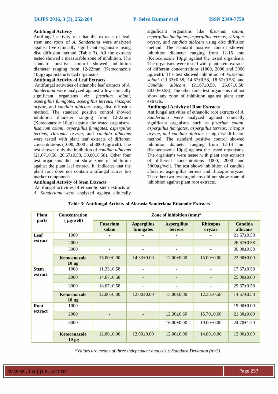
Antifungal Activity
Antifungal activity of ethanolic extracts of leaf,
stem and roots of A. Sanderiana were analyzed
against five clinically significant organisms using
disc diffusion method (Table 3). All the extracts
tested showed a measurable zone of inhibition. The
standard positive control showed inhibition
diameter ranging from 12-22mm (Ketoconazole
10μg) against the tested organisms.
Antifungal Activity of Leaf Extracts
Antifungal activities of ethanolic leaf extracts of A.
Sanderiana were analyzed against a few clinically
significant organisms viz. fusarium solani,
aspergillus fumigates, aspergillus terreus, rhizopus
oryzae, and candida albicans using disc diffusion
method. The standard positive control showed
inhibition diameter ranging from 12-22mm
(Ketoconazole 10μg) against the tested organisms.
fusarium solani, aspergillus fumigates, aspergillus
terreus, rhizopus oryzae, and candida albicans
were tested with plant leaf extracts of different
concentrations (1000, 2000 and 3000 μg/well). The
test showed only the inhibition of candida albicans
(21.67±0.58, 26.67±0.58, 30.00±0.58). Other four
test organisms did not show zone of inhibition
against the plant leaf extract. It indicates that the
plant root does not contain antifungal active bio
marker compounds. Antifungal Activity of Stem Extracts
Antifungal activities of ethanolic stem extracts of
A. Sanderiana were analyzed against clinically
significant organisms like fusarium solani,
aspergillus fumigates, aspergillus terreus, rhizopus
oryzae, and candida albicans using disc diffusion
method. The standard positive control showed
inhibition diameter ranging from 12-15 mm
(Ketoconazole 10μg) against the tested organisms.
The organisms were tested with plant stem extracts
of different concentrations (1000, 2000 and 3000
μg/well). The test showed inhibition of Fusarium
solani (11.33±0.58, 14.67±0.58, 18.67±0.58) and
Candida albicans (21.67±0.58, 26.67±0.58,
30.00±0.58). The other three test organisms did not
show any zone of inhibition against plant stem
extracts.
Antifungal Activity of Root Extracts
Antifungal activities of ethanolic root extracts of A.
Sanderiana were analyzed against clinically
significant organisms such as fusarium solani,
aspergillus fumigates, aspergillus terreus, rhizopus
oryzae, and candida albicans using disc diffusion
method. The standard positive control showed
inhibition diameter ranging from 12-14 mm
(Ketoconazole 10μg) against the tested organisms.
The organisms were tested with plant root extracts
of different concentrations 1000, 2000 and
3000μg/well. The test shows inhibition of candida
albicans, aspergillus terreus and rhizopus oryzae.
The other two test organisms did not show zone of
inhibition against plant root extracts.
Table 3: Antifungal Activity of Alocasia Sanderiana Ethanolic Extracts
Plant
parts
Concentration
( μg/well)
Zone of inhibition (mm)*
Fusarium
solani
Aspergillus
fumigates
Aspergillus
terreus
Rhizopus
oryzae
Candida
albicans
Leaf
extract
1000 - - - - 21.67±0.58
2000 - - - - 26.67±0.58
3000 - - - - 30.00±0.58
Ketoconazole
10 μg
15.00±0.00 14.33±0.00 12.00±0.00 15.00±0.00 22.00±0.00
Stem
extract
1000 11.33±0.58 - - - 17.67±0.58
2000 14.67±0.58 - - - 25.00±0.00
3000 18.67±0.58 - - - 29.67±0.58
Ketoconazole
10 μg
12.00±0.00 12.00±0.00 13.00±0.00 12.33±0.58 14.67±0.58
Root
extract
1000 - - - - 19.00±0.00
2000 - - 12.30±0.60 15.70±0.60 21.30±0.60
3000 - - 16.00±0.00 19.00±0.00 24.70±1.20
Ketoconazole
10 μg
12.00±0.00 12.00±0.00 12.00±0.00 14.00±0.00 12.00±0.00
*Values are means of three independent analysis ± Standard Deviation (n=3)

IAJPS 2016, 3 (3), 252-264 P. Selva Kumar et al ISSN 2349-7750
w w w . i a j p s . c o m
Page 258
In consideration of the importance of antioxidant
activity, investigation on A. Sanderiana was carried
out in the present work. The result of the study and
the discussion pertaining to it is presented below.
Antioxidant Activity
The DPPH radical scavenging potentials of leaf,
stem and root tubers extracts of A.Sanderiana are
presented in Table 4. The ethanolic leaf, stem and
root extract DPPH radical scavenging capacity was
found to be comparable to ascorbic acid.
DPPH Radical Scavenging Activity of Leaf
Extracts
The DPPH radical scavenging capacity of leaf
extracts of A.Sanderiana leaves are presented in
Table 4 and Figure 1. The ethanolic leaf extract at
100, 200, 300, 400, 500 μg/ml exhibited DPPH
radical scavenging activity comparable to ascorbic
acid (Figure 2). The DPPH scavenging activities of
the extracts are expressed as IC50 value. The
ethanolic leaf extracts (IC50= 84.75±0.47 μg/mL)
showed the strongest radical scavenging activity
(Figure 3).
DPPH scavenging is widely used to test the free
radical scavenging activity of several natural
products [31]. This radical scavenging activity of
extracts could be related to the nature of phenolic
compounds, thus contributing to their electron
transfer/hydrogen-donating ability. The result
indicates that medicinal plants have significant
effects on scavenging free radicals. The better
DPPH scavenging activity may be related to the
higher phenolic contents [32]. The reduction
mechanism of the DPPH radical correlates with the
hydroxyl groups on the antioxidant molecule[33],
so the mechanism might involve the delocalization
of an electron onto the p-substituted OH-
group on the molecule prior to the donation of a
second hydrogen to reduce DPPH, which also
depends on the stability and reaction potential of
the molecular structure [34].
DPPH Radical Scavenging Activity Of Stem
Extracts
The DPPH radical scavenging capacity of stem
extracts of A.Sanderiana is presented in Table 4
and Figure 1. The ethanolic stem extract exhibited
DPPH radical scavenging activity was found to be
comparable to ascorbic acid (Figure 2). The DPPH
scavenging activities of the extracts are expressed
as IC50 values. The ethanolic stem extracts (IC50=
129.31±0.15 μg/mL) showed the strongest radical
scavenging activity (Figure 3).
Table 4: DPPH Radical Scavenging Assay of A. Sanderiana Ethanolic Extracts
S.No
Sample Extract
concentration
(μg)
Percentage of inhibition activity (%) Ascorbic acid
standard
concentration
(μg)
Standard
ascorbic acid
% inhibition Leaf extract Stem extract Root extract
1 100 42.10±0.45 30.00±0.22 20.88±0.23 10 47.56±0.81
2 200 53.84±0.15 42.22±0.33 31.22±0.29 20 59.98±0.71
3 300 56.56±0.25 47.67±0.15 36.36±0.27 30 68.83±0.54
4 400 67.29±0.57 58.01±0.55 39.41±0.34 40 79.09±0.53
5 500 70.00±0.15 62.55±0.25 48.44±0.38 50 88.58±0.67
6 IC50 (μg/mL) 84.75±0.47 129.31±0.15 193.11±11.75 IC50 (μg/mL) 9.70±0.03
*Values are means of three independent analysis ± Standard Deviation (n=3)

IAJPS 2016, 3 (3), 252-264 P. Selva Kumar et al ISSN 2349-7750
w w w . i a j p s . c o m
Page 259
Fig1: Comparison of DPPH Radical Scavenging Assay
Fig 2: Standard Ascorbic acid DPPH Radical Scavenging Activity
DPPH Radical Scavenging Activity of Root
Extracts
The DPPH radical scavenging capacity of root
extracts of A.Sanderiana are presented in Table 4
and Figure 1. The ethanolic root extract exhibited
DPPH radical scavenging activity comparable to
ascorbic acid (Figure 2). The DPPH scavenging
activities of the extracts are expressed as IC50
values. The ethanolic root extracts (IC50=
193.11±11.75 μg/mL) strongest radical scavenging
activity (Figure 3).
Plant extracts having less IC50 value indicate that
the extracts have more antioxidant activity. Among
the three samples, leaf extract showed more
antioxidant activity than stem and root extracts.
Fig 3: Comparison of DPPH Radical Scavenging Activity IC50 Values with Plant Extracts

IAJPS 2016, 3 (3), 252-264 P. Selva Kumar et al ISSN 2349-7750
w w w . i a j p s . c o m
Page 260
ABTS Free Radical Scavenging Activity
The ABTS Free radical scavenging activity of leaf,
stem and root extracts of A.Sanderiana are
presented in Table 5. The ethanolic leaf, stem and
root extract ABTS Free radical scavenging
potential was found to be comparable to ascorbic
acid.
ABTS Free Radical Scavenging Activity of Leaf
Extracts
The ABTS free radical scavenging activity of leaf
extracts of A.Sanderiana leaves are presented in
Table 5 and Figure 4. The ethanolic leaf extract
exhibited ABTS radical scavenging activity
comparable to ascorbic acid (Figure 5). The ABTS
scavenging activities of the extracts are expressed
as IC50 values. The ethanolic leaf extracts (IC50=
98.62±0.25 μg/mL) showed strongest radical
scavenging activity (Figure 6).
ABTS Free Radical Scavenging Activity of Stem
Extracts
The ABTS Free radical scavenging capacity of
stem extracts of A.Sanderiana are presented in
Table 5 and Figure 4. The ethanolic stem extract
exhibited ABTS radical scavenging activity
comparable to ascorbic acid comparable to ascorbic
acid (Figure 5). The ABTS scavenging activities of
the extracts are expressed as IC50 values. The
ethanolic stem extracts (IC50= 159.11±0.72 μg/mL)
showed strongest radical scavenging activity
(Figure 6).
Table 5: ABTS Free Radical Scavenging Activity of A.Sanderiana Ethanolic Extracts
*Values are means of three independent analysis ± Standard Deviation (n=3)
Fig4: Comparison of ABTS Free
Radical Scavenging Activity
Fig 5: Standard Ascorbic acid ABTS
Free Radical Scavenging Activity
S.No Sample Extract
concentration
(μg/mL)
Percentage of inhibition activity (%) Ascorbic acid
standard
concentration
(μg/mL)
Standard
Ascorbic acid %
inhibition Leaf extract Stem extract Root extract
1 100 35.57±0.95 22.70±0.11 10.06±0.67 10 32.76±3.76
2 200 65.86±0.86 39.21±0.17 18.39±0.10 20 51.21±3.85
3 300 86.21±1.07 51.22±0.55 31.00±2.60 30 67.66±4.07
4 400 89.97±0.08 64.17±1.02 39.22±1.68 40 84.26±0.84
5 500 90.97±0.22 73.62±0.81 50.89±1.64 50 99.33±0.08
6 IC50 (μg/mL) 98.62±0.25 159.11±0.72 223.00±3.43 IC50 (μg/mL) 9.39±0.15

IAJPS 2016, 3 (3), 252-264 P. Selva Kumar et al ISSN 2349-7750
w w w . i a j p s . c o m
Page 261
Fig 6: Comparison of ABTS Free radical scavenging activity IC50 values with plant extracts
ABTS Free Radical Scavenging Activity of Root
Extracts
The ABTS Free radical scavenging activity of root
extracts of A.Sanderiana are presented in Table 5
and Figure 4. The ethanolic root extract exhibited
ABTS radical scavenging activity comparable to
ascorbic acid (Figure 5). The ABTS scavenging
activities of the extracts are expressed as IC50
values. The ethanolic root extracts (IC50=
223.00±3.43 μg/mL) showed strongest radical
scavenging activity (Figure 6).
Plant extracts having less IC50 value it’s indicating
that extracts having more antioxidant activity.
Among the three sample leaf extract showing more
antioxidant activity than stem and root extracts.
Reducing Power Activity
In this assay, the yellow colour of the test solution
changes to various shades of green and blue
depending upon the reducing power of each
compound. The presence of radicals (i.e
antioxidant) causes the conversion of the Fe3+/
ferricyanide complex used in this method to the
ferrous form. Therefore by measuring the
formation of pearls of Prussian blue at 700 nm [5]
we can monitor the Fe2+ concentration; a higher
absorbance at 700 nm indicates a higher reducing
power. The results of the reducing power activity
of A.Sanderiana ethanolic extracts assay are given
in Table 6.
Table 6: Reducing Power Activity of A.Sanderiana Ethanolic Extracts
S.No Sample Extract
concentration
(μg/mL)
Absorbance at 700nm
Leaf extract Stem extract Root extract Ascorbic acid
standard
1 100 0.140±0.021 0.101±0.007 0.083±0.041 0.211±0.081
2 200 0.231±0.007 0.192±0.025 0.152±0.017 0.410±0.090
3 300 0.397±0.027 0.294±0.009 0.202±0.008 0.610±0.011
4 400 0.540±0.013 0.405±0.016 0.310±0.044 0.797±0.081
5 500 0.664±0.023 0.487±0.022 0.394±0.081 0.971±0.075
*Values are means of three independent analysis ± Standard Deviation (n=3)

IAJPS 2016, 3 (3), 252-264 P. Selva Kumar et al ISSN 2349-7750
w w w . i a j p s . c o m
Page 262
Reducing Power Activity of Leaf Extracts
The reducing power capacity of leaf extracts of
A.Sanderiana are presented in Table 6 Figure 7.
The ethanolic leaf extract showed better reducing
power comparable to standard ascorbic acid (Figure
7). Reducing Power Activity of Stem Extracts
The reducing power capacities of stem extracts of
A.Sanderiana are presented in Table 6 Figure 7.
The ethanolic leaf extract showed better reducing
power comparable to standard ascorbic acid (Figure
7) comparable to ascorbic acid. Reducing Power Activity of Root Extracts
The reducing power capacities of root extracts of
A.Sanderiana are presented in Table 6 and Figure
7. The ethanolic root extract showed better
reducing power comparable to standard ascorbic
acid (Figure 7).
Fig 7: Comparison of Reducing Power Activity
The higher absorbance of leaf extracts may be due
to its strong reducing power potential. Comparing
leaf, stem and root tubers extracts, the leaf extracts
showed better reducing power than stem and root
extracts. The reducing power of the extracts may be
due to the biologically active compounds in the
extract which possess potent donating abilities.
This assay further confirmed the antioxidant
properties of the extracts.
CONCLUSION
In the present study, the preliminary phytochemical
screening of the ethanolic leaf extracts of A.
Sanderiana revealed strong presence of tannins,
flavonoids, phenols and steroids, moderate
presence of glycosides. Trace amount of saponins,
terpenes, carbohydrates and proteins are also
present. Stem extracts revealed strong presence of
tannins, moderate presence of flavonoids and
phenols. Trace amount of saponins, terpenes,
carbohydrates, proteins, glycosides and steroids are
also present. Alkaloids and amino acids are not
present in leaf and stem. Root extracts revealed
moderate presence of tannins, trace amounts of
glycosides, steroids, saponins, terpenes, alkaloids,
flavonoids, carbohydrates, proteins and phenols.
Amino acids are not present. When all the three
plant parts are compared, leaf shows more potential
than the stem and root. These activities may be due
to the strong occurrence of polyphenolic
compounds such as flavonoids, tannins, steroids,
and phenols.
Antibacterial activities of ethanolic extracts of leaf,
stem and root were analyzed against five clinically
significant organisms viz. staphylococcus aureus,
escherichia coli, klebsiella pneumonia,
pseudomonas aeruginosa and bacillus cereus using
disc diffusion method. The standard positive
control showed inhibition diameter ranging from
26-28 mm (Gentamicin 10 μg) against the tested
organisms. Leaf extracts (1000, 2000, 3000 μg/well
concentration) were found to be effective in
inhibiting the growth of pseudomonas aeruginosa
compared with other bacteria. Stem extracts (250,
500, 750 μg/well concentration) were found to be
effective in inhibiting the growth of staphylococcus
aureus compared with other bacteria. Root extracts
(1000, 2000, 3000 μg/well concentration) were not
found to be effective in inhibiting the growth of
any bacteria. Compared to leaf and root, stem
shows more inhibition against bacterial growth.
Antifungal activities of ethanolic extracts of leaf,
stem and root tubers were analyzed against five
clinically significant organisms viz. fusarium
solani, aspergillus fumigates, aspergillus terreus,
rhizopus oryzae, candida albicans using disc
diffusion method. The standard positive control
showed inhibition diameter ranging from 12-22
mm (Ketoconazole 10 μg) against the tested
organisms. The test with different concentrations
(1000, 2000 and 3000μg/well) of leaf extracts
showed only the inhibition of candida albicans
(21.67±0.58, 26.67±0.58, 30.00±0.58%) The test
with stem extracts of different concentrations
(1000, 2000 and 3000 μg/well) showed inhibition
of fusarium solani and candida albicans. The test
with different concentrations (1000, 2000 and
3000μg/well) of root extracts test shows inhibition
of candida albicans, aspergillus terreus and
rhizopus oryzae. Compared to leaf, stem and root
tubers show more inhibition of fungal growth.
Among the three samples, leaf extract shows more
antioxidant activity (DPPH and ABTS) than stem
and root extracts. Comparing leaf, stem and root
tubers extracts, the leaf extracts showed better
reducing power than stem and root extracts. The
reducing power of the extracts may be due to the
biologically active compounds in the extract which
possess potent donating abilities. From the present
investigations, we can conclude that A.sanderiana
possesses significant antimicrobial activity due to
the presence of various phytoconstituents and it
could be a source of new compounds. Ethanolic
extracts of leaf, stem and root tubers of
A.Sanderiana showed in vitro anti-inflammatory

IAJPS 2016, 3 (3), 252-264 P. Selva Kumar et al ISSN 2349-7750
w w w . i a j p s . c o m
Page 263
and antidiabetic activities. From the ethanolic
extracts of A.sanderiana, two new compounds have
been isolated. The characterization and structural
conformation of the new compounds are in
progress.
REFERENCES:
1.Elżbieta Zenkteler, Piotr Sobiczewski, Teresa
Orlikowska, Krzysztof Langer, Jerzy Langer.
Application of nanomaterials to fight and prevent
bacteria in vitro. ACTA BIOLOGICA
CRACOVIENSIA Series Botanica.2009; 51 suppl.
1:28.
2.Thankappan Sarasabai, Shynin Brintha, James
Edwin James and Solomon Jeeva. Vascular Plants,
Scott Christian College, Nagercoil, Tamilnadu,
India. Science Research Reporter, 2015: 5(1):36-
66. ISSN: 2249-7846.
3.https://commons.wikimedia.org/wiki/Category:C
ultivated_plants_of_Yercaud.
4.Thangavelu Muthukumar, Eswaranpillai Uma,
Perumalsamy Priyadharsini. Occurrence of
foliicolous parasitic alga Cephaleuros virescens on
cultivated ornamental plants in southern India,
BOTANICA LITHUANICA, 2014, 20(2): 87–98.
ISSN 1392-1665.
5.Arvind Singh, Observations on the Flora of
Varanasi District in Uttar Pradesh State of India,
Global Journal of Environmental Science and
Technology: 2015; 3(10): 368-389.
6.Rahman M, Hossain A, Siddique SA, Biplab KP,
Uddin H. Antihyperglycemic, antioxidant, and
cytotoxic activities of Alocasia macrorrhizos (L.)
rhizome extract. Turk J Biol 36 (2012) 574-57.
7.Omale James, Okafor Oolycarp Nnacheta and
Omede Ameh. Polyphenol contents, cytotoxicity
and antioxidant activities of some selected nigerian
vegetable foods. Int.J.Chem. Sci.: 2008;6(4):1714-
1725.
8.Haque M, Jahan T, Rashid MA. Antibacterial and
Cytotoxic Activities of Alocasia fornicata (Roxb.).
Int J Nutr Pharmacol Neurol Dis. 2014; 4, S1:29-
33.
9.Wang H X., Ng T.B. Alocasin, an antifungal
protein from rhizomes of Alocasia macrorrhiza.
Protein expression & purification;2003; 28(1):9-14.
10.Mulla WA, Sargade PB, Pawar AM, Tarkasband
HA, Sayyad FJ, (2010). Evaluation of antimicrobial
activity of leaves of Alocasia indica Linn.
International Journal of Pharm Tech
Research;2(1):327-333.
11.Hoque T, Sikder MA, Kaisar MA, Asad A,
Chowdhury, Rashid MA, Biological Screenings of
Two Araceous Plants Growing in Bangladesh.
Dhaka Univ. J. Pharm. Sci; 2011;10(2):131-35.
12.Haque M, Jahan T, Rashid MA. Antibacterial
and Cytotoxic Activities of Alocasia fornicata
(Roxb.). Int J Nutr Pharmacol Neurol Dis. 2014; 4,
S1:29-33.
13.Judee N.Nogodula, Jessa Marie D. Draug,
Maryjane S. Jamero, Charmaine Lei E. Suyom.
Phytochemical and Antibacterial Action of Taro
(Colocasia esculenta, Araceaea) Aqueous-Ethanolic
Leaf Extract against Selected Bacterial Strains.UIC
Research Journal.2012;18(1):221-236.
14.Mulla WA, Kuchekar SB, Thorat VS, Chopde
AR, Kuchekar BS. Antioxidant, Antinociceptive
and anti-inflammatory activities of ethanolic
extract of leaves of Alocasia indica Schott. Journal
of young pharmacist; 2010;2(2):137-143.
15.Rahman M, Hossain A, Siddique SA, Biplab
KP, Uddin H. Antihyperglycemic, antioxidant, and
cytotoxic activities of Alocasia macrorrhizos
(L.)rhizome extract. Turk J Biol 2012;36 ,574-57.
16.Suman Khowala, Swagata Pal, Ankita
Bhattacharjee, Sandip Mukherjee, Koushik
Bhattacharya. Antioxidant and Hepatoprotective
Activity of Ethanolic Extract of Alocasia indica
Tuber. American Journal of Phytomedicine and
Clinical Therapeutics -AJPCT[2][2][2014]191-208.
17.Gouriprosad data, Subhashree basu, anurupa
sen, moumita das and pranabes nath.
phytochemical evaluation and in vitro study of
antioxidant potency of amorphophallus
campanulatus, alocasia indica and colocasia
esculenta: a comparative analysis. Int J Pharm Bio
Sci ,2012 ; 3(3): 170-180.
18.Saswati Roy, M. Dutta Choudhury, S.B. Paul.
Antioxidant potential of rhizome of alocasia
decipiens schott. Asian J Pharm Clin Res,2013;
6(2): 120-122.
19.Md. Rabiul Karim, Nasrin Ferdous, Narayan
Roy, Subed Chandra Dev Sharma, M. G.Sarowar
Jahan, Mohammad Shariar Shovon. A study on
antidiabetic activity of the leaf and stem of
Alocasia indica L. in steptozotocin induced diabetic
rats. International Journal of Biosciences .2014; 5,
(6), 195-202.
20.S. A. Sreenivas, P. V. Deshmukh, M. Srikanth,
Avijit Choudhury, A. E. Wagh. Antidiabetic &
Hypolipidemic Potential of Alocasia indica Schott.
Leaves in Streptozotocin Induced Diabetic Rats.
Int. J. Drug Dev. & Res., 2012, 4 (4): 368-374.
21.Shengtao Fang, Caiyu Lin, Quanbo Zhang, Li
Wang, Ping Lin, Jie Zhang, Xiujie Wang.
Anticancer potential of aqueous extract of alocasia
macrorrhiza against hepatic cancer in vitro and in
vivo. Journal of Ethnopharmacology, 2012;141( 3):
947-956.
22.Lei Xiao, Feng Yi, Liang Shuang, Zheng Xiang-
Wei. Chemical Components of the Tuber of
Alocasia cucullata. Chemistry of Natural
Compounds. 2014;50(1): 133-134.
23.Wei P, Zhiyu C, Xu T, Xiangwei Z. Antitumor
effect and apoptosis induction of Alocasia cucullata
(Lour.) G. Don in human gastric cancer cells in
vitro and in vivo. BMC Complement Altern Med.
2015 ;15:33.
24.Peng Q, Cai H, Sun X, Li X, Mo Z, et al. (2013)
Alocasia cucullata Exhibits Strong Antitumor
Effect In Vivo by Activating Antitumor Immunity.
PLoS ONE 8(9): e75328, 2013.

IAJPS 2016, 3 (3), 252-264 P. Selva Kumar et al ISSN 2349-7750
w w w . i a j p s . c o m
Page 264
25.Nweze ET, Okafor JI and Njoku O.
Antimicrobial activities of methanolic extract of
Trumeguineesis (Schumm and Thorn) and Morinda
lucinda Benth used in Nigerian herb medicinal
practice. J. Bio. Res. Biotechnol.,2004; 2(1): 34-46.
26.Senthilkumar PK and Reetha D. Screening of
antimicrobial properties of certain Indian medicinal
plants. J. Phytol.,2009; 1(3): 193-198.
27.Perez, C., Pauli, M. and Bazerque, P. An
antibiotic assay by agar-well diffusion method.
Acta Biologiae et Medecine Experimentaalis,1990;
15:113-115.
28.Blois MS. Antioxidant determinations by the
use of a stable free radical. Nature. 1958; 26: 1199-
1200.
29.Re, R., Pellegrini, N., Proteggente, A., Pannala,
A., Yang, M. and Rice-Evans, C. Antioxidant
activity applying an improved ABTS radical cation
decolorization assay. Free Radical Biology and
Medicine, 1999;26: 1231–1237.
30.Oyaizu M. J Pharm Nutr 1986; 44:307-15.
31.Ahn, R., Kumazawa, S., Usui, Y., Nakamura, J.,
Matsuka, M., Zhu, F. and Nakayama, J.,
“Antioxidant Activity and Constituents of Propoils
Collected in Various Areas of China”, Food Chem,
2007;10, 1383-1392.
32.Frankel, S., Robinson, G.E. and Berenbaum,
M.R., “Antioxidant Capacity and Correlated
Characteristics of 14 Unifloral Honeys”, Journal of
Agricultural Research, 1998; 37,7-31.
33.Cotelle, N., Bernier, J.L., Catteau, J.P.,
Pommery, J., Wallet, J.C. and Gaydou, E.M.,
“Antioxidant Properties of Hydroxyl Flavones”,
Free Rad. Biol. Med. 1996;21, 35-43.
34.Brand-Williams, W., Cuvelier, M.E. and Berset,
C., “Use of a Free Radical Method to Evaluate
Antioxidant Activity”, Lebensm. Wiss. Technol.,
28, 1995, 25-30.


