Embed Size (px)
Citation preview
HAL Id: tel-02547641https://hal.inrae.fr/tel-02547641
Submitted on 20 Apr 2020
HAL is a multi-disciplinary open accessarchive for the deposit and dissemination of sci-entific research documents, whether they are pub-lished or not. The documents may come fromteaching and research institutions in France orabroad, or from public or private research centers.
L’archive ouverte pluridisciplinaire HAL, estdestinée au dépôt et à la diffusion de documentsscientifiques de niveau recherche, publiés ou non,émanant des établissements d’enseignement et derecherche français ou étrangers, des laboratoirespublics ou privés.
Increased intake of fermentable carbohydrates inducesIBS-like symptoms; a complementary understanding of
mechanisms involvedJasper Kamphuis
To cite this version:Jasper Kamphuis. Increased intake of fermentable carbohydrates induces IBS-like symptoms; a com-plementary understanding of mechanisms involved. Life Sciences [q-bio]. INPT, 2019. English.�tel-02547641�
En vue de l'obtention du
DOCTORAT DE L'UNIVERSITÉ DE TOULOUSEDélivré par :
Institut National Polytechnique de Toulouse (Toulouse INP)Discipline ou spécialité :
Pathologie, Toxicologie, Génétique et Nutrition
Présentée et soutenue par :M. JASPER KAMPHUIS
le vendredi 4 octobre 2019
Titre :
Unité de recherche :
Ecole doctorale :
Increased intake of fermentable carbohydrates induces IBS-like symptoms;a complementary understanding of mechanisms involved
Sciences Ecologiques, Vétérinaires, Agronomiques et Bioingénieries (SEVAB)
Toxicologie Alimentaire (ToxAlim)Directeur(s) de Thèse :
MME HÉLÈNE EUTAMENEMME VASSILIA THEODOROU
Rapporteurs :M. BRUNO BONAZ, CHU GRENOBLE
M. JAVIER SANTOS VICENTE, VALL D'HEBRON UNIVERSITY HOSPITAL
Membre(s) du jury :Mme CATHERINE MULLER, UNIVERSITE TOULOUSE 3, Président
Mme HÉLÈNE EUTAMENE, EI PURPAN, MembreMme INGRID RENES, NUTRICIA RESEARCH UTRECHT, Membre
Mme VASSILIA THEODOROU, EI PURPAN, Membre


Increased intake of fermentable
carbohydrates induces IBS-like symptoms;
a complementary understanding of
mechanisms involved
Visceral sensitivity, intestinal barrier function,
bacterial metabolites, and organisation of mucus secretions
Jasper Kamphuis, 2019

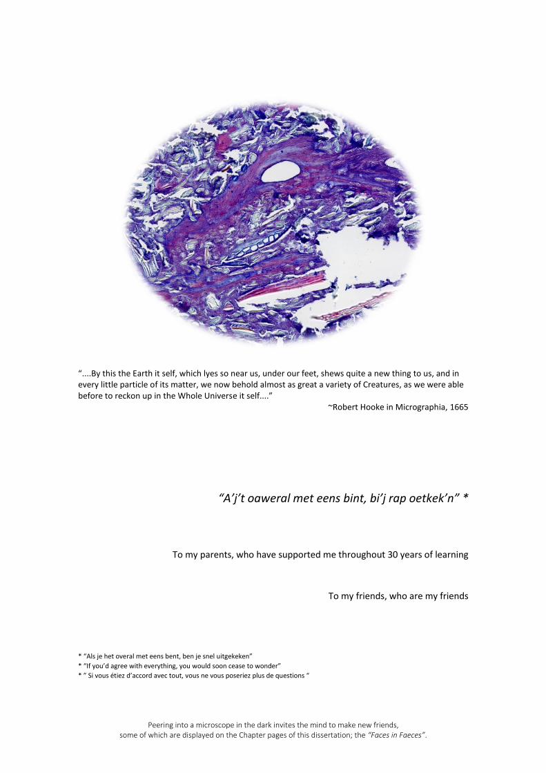
Peering into a microscope in the dark invites the mind to make new friends, some of which are displayed on the Chapter pages of this dissertation; the “Faces in Faeces”.
“....By this the Earth it self, which lyes so near us, under our feet, shews quite a new thing to us, and in every little particle of its matter, we now behold almost as great a variety of Creatures, as we were able before to reckon up in the Whole Universe it self....” ~Robert Hooke in Micrographia, 1665
“A’j’t oaweral met eens bint, bi’j rap oetkek’n” *
To my parents, who have supported me throughout 30 years of learning
To my friends, who are my friends
* “Als je het overal met eens bent, ben je snel uitgekeken” * “If you’d agree with everything, you would soon cease to wonder”
* ” Si vous étiez d’accord avec tout, vous ne vous poseriez plus de questions “
Table of Contents Summary ................................................................................................................................... 1
Résumé ...................................................................................................................................... 2
Abbreviations ............................................................................................................................ 3
1 General Introduction ............................................................................................................. 9
1.1 A fresh look at IBS – opportunities for systems medicine approaches ...................... 10
1.2 IBS, FODMAPs .............................................................................................................. 15
1.2.1 Functional gastrointestinal disorders (FGIDs) ..................................................... 15
1.2.2 Aetiology IBS and physiopathology ..................................................................... 21
1.2.3 Clinical practice ................................................................................................... 38
1.2.4 FODMAP diet ....................................................................................................... 43
1.3 Intestinal microbiota ................................................................................................... 49
1.3.1 Function ............................................................................................................... 49
1.3.2 Toxic metabolite hypothesis ............................................................................... 56
1.4 Bacterial Metabolites; aldehydes/ methylglyoxal ....................................................... 57
1.4.1 Chemical properties of methylglyoxal ................................................................. 57
1.4.2 Biological properties of methylglyoxal ................................................................ 58
1.4.3 Deleterious effects of dicarbonyl stress .............................................................. 60
1.4.4 Detoxification – glyoxalase system ..................................................................... 62
1.5 Intestinal barrier function ........................................................................................... 67
1.5.1 Epithelial barrier .................................................................................................. 67
1.5.2 Permeability ........................................................................................................ 71
1.5.3 Mucus barrier ...................................................................................................... 73
1.6 Aims and Outline of Thesis .......................................................................................... 83
1.6.1 Hypotheses .......................................................................................................... 83
1.6.2 Approach ............................................................................................................. 85
1.6.3 Experimental procedures .................................................................................... 86
2 Project Results ..................................................................................................................... 97
2.1 Mucus organisation is shaped by colonic content; a new view .................................. 97
2.2 FODMAPs increase visceral sensitivity in mice through glycation processes, increasing
mast cell counts in colonic mucosae ..................................................................................... 111
2.3 Increased FODMAP intake alters colonic mucus barrier function through glycation
processes and increased mastocyte counts .......................................................................... 151
3 General Discussion ............................................................................................................ 171
3.1 Fermentable carbohydrates and IBS symptoms ....................................................... 172
3.2 Immune activation .................................................................................................... 176
3.3 Permeability .............................................................................................................. 177
3.4 Role of glycation in efficacy of low-FODMAP diet .................................................... 178
3.5 Mucus barrier ............................................................................................................ 181
3.6 Concluding remarks ................................................................................................... 184
4 Acknowledgments ............................................................................................................. 189
5 Publications, Dissemination and Training Activities ......................................................... 195
5.1 Publications part of this thesis .................................................................................. 195
5.2 Other Publications ..................................................................................................... 195
5.3 Oral presentations ..................................................................................................... 196
5.4 Poster presentations ................................................................................................. 197
5.5 Other training and dissemination activities .............................................................. 199
6 References .............................................................................................................................. I

P a g e | 1
Summary
Irritable bowel syndrome (IBS) is a functional gastrointestinal disorder (FGID) characterized by
abdominal pain, bloating, and erratic bowel habits. It is an affliction with a high prevalence of
around 11% worldwide. It carries a significant economic cost in lost productivity and work
absence, and more importantly, it has a strong negative impact on quality of life. Because it is a
functional disorder of which the causes are not well understood, treatment is difficult. In recent
years, a low-FODMAP diet (low in Fermentable Oligo-, Di-, Mono-saccharides And Polyols) has
been successfully used to reduce symptoms of IBS. The efficacity of this approach is not
completely understood, but a reduction in enteric distension by reduced gas production and
small intestinal water bulk by osmotic effects are most often cited. The bacterial metabolic toxin
hypothesis, proposed by Campbell et al. poses that anaerobic fermentation of unabsorbed
carbohydrates by the colonic gut microbiota, producing such metabolites as alcohols, ketones,
and aldehydes, are responsible for food intolerances such as lactose intolerance. We
hypothesized that this same mechanism could be extended to FODMAPs to explain the efficacity
of the low-FODMAP diet.
In this thesis, we looked for complementary mechanisms on how FODMAPs could influence IBS
symptoms, besides distension related complaints. Our studies in a healthy mouse model show
a complex role for FODMAPs in IBS physiopathology; FODMAP treatments cause a visceral and
abdominal hypersensitivity, and a mucus barrier dysregulation, characterized using an
innovative approach.
We hypothesized that this is due to generation of glycating agents by the intestinal microbiota,
and the prevention of these effects by co-treatment with pyridoxamine indicates that this
hypothesis is correct. Mucosal mast cell counts were increased in FODMAP treated animals, but
not in animals co-treated with pyridoxamine. Mast cells are implicated in visceral
hypersensitivity, as well as in mucus barrier dysregulation, and increased mucosal mast cell
numbers or activity are often linked to IBS.
This work thus offers a link between the efficacity of the low-FODMAP diet and the involvement
of intestinal mast cells in IBS.
2 | P a g e
Résumé
Le syndrome de l’intestin irritable (SII) est un trouble gastro-intestinal fonctionnel caractérisé
par des douleurs abdominales, des ballonnements et des troubles du transit intestinal. Cette
pathologie digestive a une prévalence mondiale importante d'environ 11%. Elle entraîne un coût
économique important : perte de productivité et absentéisme au travail. De plus, elle entraine
une forte dégradation de la qualité de la vie des patients. Les causes de ce trouble fonctionnel
ne sont pas bien comprises rendant le traitement thérapeutique difficile. Au cours des dernières
années, un régime alimentaire à faible teneur en FODMAPs (Fermentable Oligo-, Di-, Mono-
saccharides And Polyols) s’est révélé efficace dans la réduction des symptômes du SII. Sur le plan
mécanistique ces effets positifs restent à élucider. Toutefois, on cite le plus souvent une
réduction de la distension entérique due à une réduction de la production de gaz et du volume
d’eau intestinale par des effets osmotiques. Campbell et al. pose l’hypothèse qu’une
fermentation anaérobie de carbohydrates non-absorbés par le microbiote intestinal provoque
la formation endoluminale de métabolites tels que les alcools, les cétones et les aldéhydes,
responsables d'intolérances alimentaires comme l'intolérance au lactose. Nous avons émis
l’hypothèse que ce mécanisme pourrait être étendu aux FODMAPs pour expliquer l’efficacité du
régime alimentaire pauvre en FODMAPs chez les patients SII.
Nos études montrent un rôle complexe des FODMAPs sur des modèles murins qui reflètent la
physiopathologie du SII. Les traitements par FODMAPs (lactose et fructo-oligosaccharides)
provoquent une hypersensibilité viscérale et abdominale et une dysfonction de la barrière de
mucus au niveau de la muqueuse intestinale.
Nous avons démontré que ces effets étaient dûs à la production d’agents de glycation par le
microbiote intestinal. En effet, ces effets étaient prévenus par un co-traitement à la
pyridoxamine. Le nombre de mastocytes muqueux était également augmenté chez les animaux
traités par FODMAPs et significativement réduit par un co-traitement à la pyridoxamine. Les
mastocytes sont connus pour être impliqués dans l’hypersensibilité viscérale et dans la
dysrégulation de la barrière de mucus de l’intestin. Par ailleurs, une augmentation du nombre
et/ou de l'activité des mastocytes est retrouvée sur des biopsie de patients SII.
Ce travail de thèse original permet donc de faire un lien entre l'efficacité du régime alimentaire
à faible teneur en FODMAPs, la symptomatologie et l'implication des mastocytes intestinaux
chez le patient SII.
P a g e | 3
Abbreviations
4-HNE 4-hydroxynonenal
ACTH Adrenocorticotropic hormone
AGEs Advanced glycation end products
ANS Autonomous nervous system
APC Antigen-presenting cell
CFAP Chronic Functional Abdominal Pain
cfu colony forming units
cGMP Cyclic guanosine monophosphate
CGRP receptor Calcitonin gene-related peptide
CNS Central nervous system
CRF Corticotropin releasing factor
DAMP Damage-associated molecular pattern
DC Dendritic cell
DSCG Disodium cromoglycate
DSS Dextran sodium sulphate
EMG Electromyographic
ENS Enteric nervous system
FAE Follicle-associated epithelium
FGIDs Functional gastrointestinal disorders
FISH Fluorescent in situ hybridisation
FITC Fluorescein isothiocyanate
FODMAP Fermentable Oligo-, Di-, Mono-saccharides and Polyols
FOS Fructo-oligosaccharides
GABA γ-aminobutyric acid
GALT Gut-associated lymphoid tissue
GAP Goblet cell-associated antigen passages
GC Goblet cell
GI Gastrointestinal
Glo Glyoxalase
GOS Galacto-oligosaccharides
GR Glucocorticoid receptors
GSH Glutathione
4 | P a g e
HPA-axis Hypothalamic-pituitary-adrenal axis
IBD Inflammatory Bowel Disease
IBS Irritable Bowel Syndrome
IEC Intestinal epithelial cells
Ig Immunoglobulin
IL Interleukin
LAB Lactic acid bacteria
LFD Low-FODMAP diet
LPS Lipopolysaccharide
M cell Microfold cell
MAMP Microbe-associated molecular pattern
MAPK Mitogen-activated protein kinase
MDA Malonaldehyde
MG Methylglyoxal
MGS Methylglyoxal synthase
MLCK Myosin Light Chain Kinase
MR Mineralocorticoid receptors
NF-κB Nuclear factor kappa-light-chain-enhancer of activated B cells
NGF Nerve growth factor
NLRP6 Nod-like receptor family pyrin domain containing 6
OR Odds ratio
PAR-2 Protease-activated receptor 2
PI-IBS Post-infectious Irritable Bowel Syndrome
PM Pyridoxamine
PRR Pattern recognition receptor
PRS Partial restraint stress
QOL Quality of Life
RAGE Receptor for advanced glycation end products
RCT Randomized controlled trial
ROS Reactive oxygen species
SAM Sympathetic adrenomedullary system
SCFA Short-chain fatty acid
senGC Sentinel goblet cell
P a g e | 5
SIBO Small intestinal bacterial overgrowth
SP Substance P
TGFβ Transforming growth factor beta
TJ Tight junction
TLR Toll-like receptor
TNBS 2,4,6-trinitrobenzene sulfonic acid
TNF-α Tumour Necrosis Factor α
Tregs Regulatory T-cells
UC Ulcerative Colitis
UWL Unstirred water layer
WAS Water avoidance stress
WHO World Health Organization
ZO1 Zona occludens-1 / tight junction protein 1


1 General Introduction
Background Information, Thesis Context

P a g e | 9
1 General Introduction
This thesis project was part of the Initial Training Network (ITN) NeuroGUT, financed by the
People Programme of the European Union’s Seventh Framework Programme. This training
network aimed to offer training in neurogastroenterology and complementary skills to young
researchers, to help form the next generation of scientists dedicated to the rapidly developing
field of neurogastroenterology. The projects financed in this way all pertain to functional
gastrointestinal disorders (FGIDs) and investigate such topics as the involvement of gut-brain
interaction, nutritional challenges of the immune system, low-grade inflammation, and post-
infectious changes to the enteric nervous system as possible pathophysiological mechanisms.
The network contains both clinical and basic science partners, and fellows with both medical
and research science backgrounds were recruited. During the project, fellows met several times
per year for training activities and annual meetings, as well as for scientific conferences. One
such training activities was a Summer School organised around the Bologna IBS Days 2016, and
the results of training activities during this summer school were published as a Position Paper in
the Neurogastroenterology and Motility journal (Albusoda, Barki et al., 2017), which is included
as the first section after this general introduction.
The goal of this thesis project was to investigate and describe mechanisms by which FODMAPs
(Fermentable Oligo-, Di-, Mono-saccharides and Polyols) can induce symptoms of Irritable Bowel
Syndrome (IBS), based on the facts that a low-FODMAP diet can successfully reduce symptoms
in IBS patients (Gibson and Shepherd, 2005; Halmos, Power et al., 2014; Böhn, Störsrud et al.,
2015; Marum, Moreira et al., 2016a; Prince, Myers et al., 2016; McIntosh, Reed et al., 2017),
and that fermentation of certain non-absorbable carbohydrates can lead to the formation of
methylglyoxal, with negative local and systemic effects (Campbell, Waud et al., 2005; Campbell,
Matthews et al., 2010). We hypothesized that FODMAP fermentation could have sensitizing and
immuno-modulatory effects in the colon by production of toxic microbial metabolites, which
supplements the prevailing idea that the efficacity of the low-FODMAP diet is related to the
increased intestinal distension via osmotic effects and gas production (Barrett, Gearry et al.,
2010; Ong, Mitchell et al., 2010) induced by FODMAP intake, in an already hypersensitive
system. To get more insight into this matter and test our hypothesis, we used an animal model
to measure relevant markers in colonic tissues and digesta, such as mastocyte counts, mucus
barrier function, and aldehyde contents of faeces, as well as more systemic parameters, such as
visceral and abdominal sensitivity.
10 | P a g e
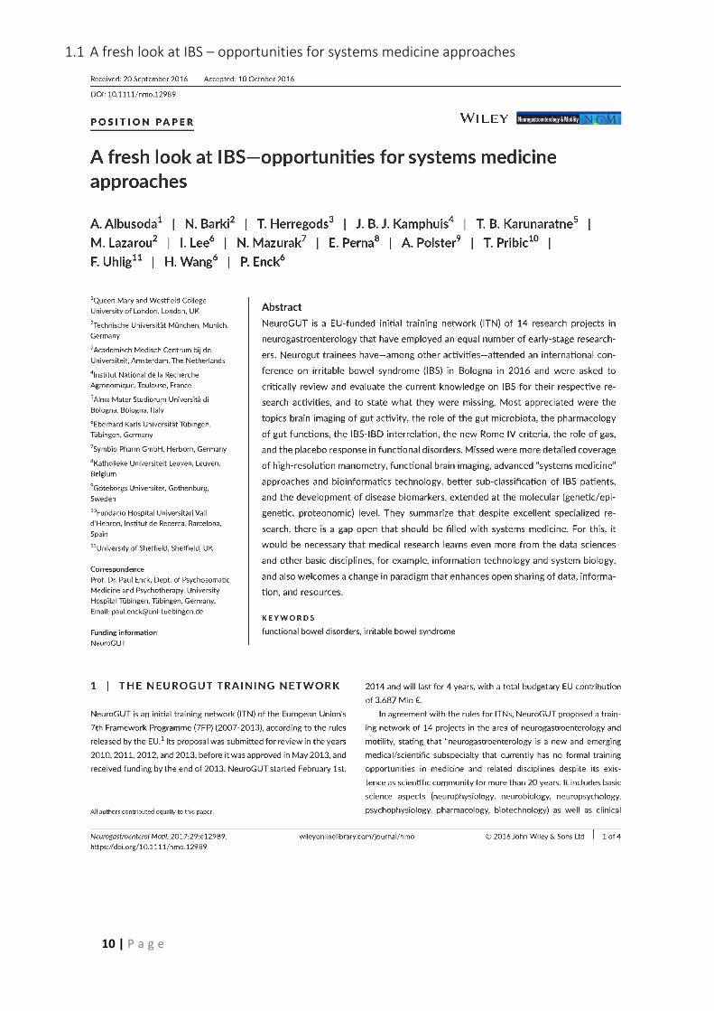
1.1 A fresh look at IBS – opportunities for systems medicine approaches
Received: 20 September 2016 Accepted: 10 October 2016
DOi: 10.1111/nmo.12989
POSITION PAPER WILEY f~eurogaslloenterology&Mobbtyr ~~ rf
A fresh look at 185-opportunities for systems medicine approaches
A. Albusoda1
M. Lazarou2
F. Uhlig11
1 N. Barki2 T. Herregods3 J. B. J. Kamphuis4 T. B. Karunaratne5
T. Pribic10 1 1. Lee6
1 N. Mazurak7 E. Perna8 1 A. Polster9
H. Wang6 1 P. Enck6
1Queen Mary and Westfield College
University of London, London, UK
2Technische Universitat München, Munich,
Germany
3Academisch Medisch Centrum bij de
Universiteit, Amsterdam, The Netherlands
4Institut National de la Recherche
Agronomique, Toulouse, France
5Alma Mater Studiorum Università di Bologna, Bologna, ltaly
6Eberhard Karls Universitat Tûbingen,
TLibingen, Germany
7Symbio Pharm GmbH, Herborn, Germany
8Katholieke Universiteit Leuven, Leuven,
Belgium
9Gëteborgs Universitet, Gothenburg,
Sweden
1°Fundacio Hospital Universitari Vall d'Hebron, Institut de Recerca, Barcelona,
Spain
11University of Sheffield, Sheffield, UK
Correspondence
Prof. Dr. Paul Enck, Dept. of Psychosomatic
Medicine and Psychotherapy, University
Hospital Tûbingen, Tûbingen, Germany.
Email: [email protected]
Funding information
NeuroGUT
Abstract
NeuroGUT is a EU-funded initial training network (ITN) of 14 research projects in
neurogastroenterology that have employed an equal number of early-stage research
ers. Neurogut trainees have-among other activities-attended an international con
ference on irritable bowel syndrome (185) in Balogna in 2016 and were asked to
critically review and evaluate the current knowledge on 185 for their respective re
search activities, and to state what they were missing. Most appreciated were the
tapies brain imaging of gut activity, the role of the gut microbiota, the pharmacology
of gut functions, the 185-18D interrelation, the new Rome IV criteria, the role of gas,
and the placebo response in functional disorders. Missed were more detailed coverage
of high-resolution manometry, functional brain imaging, advanced "systems medicine"
approaches and bioinformatics technology, better sub-classification of 185 patients,
and the development of disease biomarkers, extended at the molecular (genetic/ epi
genetic, proteonomic) level. They summarize that despite excellent specialized re
search, there is a gap open that should be filled with systems medicine. For t his, it
would be necessary that medical research learns even more from the data sciences
and other basic disciplines, for example, information technology and system biology,
and also welcomes a change in paradigm that enhances open sharing of data, informa
tion, and resources.
KEYWORDS
functional bowel disorders, irritable bowel syndrome
1 THE NEUROGUT TRAINING NETWORK 2014 and will last for 4 years, with a total budgetary EU contribution
of 3.687 Mio€.
NeuroGUT is an initial training network (ITN) of the European Union's
7th Framework Programme (7FP) (2007-2013), according to the rules
released by the EU.1 lts proposai was submitted for review in the years
2010, 2011, 2012, and 2013, before it was approved in May 2013, and
received funding by the end of 2013. NeuroGUT started February 1st,
Ali authors contributed equally to this paper.
ln agreement with the ru les for ITNs, NeuroGUT proposed a t rain
ing network of 14 projects in the area of neurogastroenterology and
motility, stating that "neurogastroenterology is a new and emerging
medical/ scientific subspecialty that currently has no formai training
opportunities in medicine and related disciplines despite its exis
tence as scientific community for more than 20 years. lt includes basic
science aspects (neurophysiology, neurobiology, neuropsychology,
psychophysiology, pharmacology, biotechnology) as well as clinical
Neurogastroenterol Mon/. 2017;29:e12989.
https://doi.org/ 10.1111/nmo.12989
wileyonlinelibrary.com/ journal/nmo © 2016 John W iley & Sons Ltd 1 1 of 4
P a g e | 11
~ WI LEY- Pi@&iii@:i%A Al aspects (gastroenterology, neurology, internai medicine, surgery, psy
chology, psychosomatic medicine) of the neural control of intestinal
functions (motility, secretion, absorption, immunity, sensitivity, food
intake) in health and disease."2
Initial training networks are thought as training networks, notas
much as research projects: this implies that the focus is-beside sci
entific excellence-on training of a future generation of scientists and
clinicians, to qualify them for academic, clinical, or industrial work in
the related areas. ln consequence, the funding received is to allow
completion of 3-year PhD training for early stage researchers (ESRs)
at each participating partner laboratory, with supplementary money
for the participating centres. Among others, the requirement to work
as ESR in one of the projects is mobility of the researcher: he/she
cannot have resided in the country of his/ her host institution for
more than 1 year in the 3 years prier to his/her recruitment; in most
cases he/she has to change the country of residency. ITNs within
FP7 also included experienced (post-doctoral) researchers (ERs) es
pecially to work with industrial partners (small and medium-size en
terprises, SME) for 2 years. NeuroGUT involves 11 ESRs and 3 ERs
(see Table 1).
Ali ESRs had to enroll in local PhD programs in addition to their
NeuroGUT training activities; these included seminars in paper and
grant writing, didactic courses in oral presentation skills, a mini-MBA
course to foster business activities, summer, and winter schools on spe
cific topics of general interest, self-organized researcher camps, and sec
ondments (short-term stays in other laboratories, etc.). One such activity
was the participation in the international IBS Bologna Days 2016.3
The principle investigators and supervisors of the ESRs/ERs of the
NeuroGUT network consist of internationally leading experts in the
respective research fields, coming from academia and from the private
Project title Pl
Projects for early state researchers (ESR), 36 months
Enck
ALBUSODA ET AL.
KEY POINTS
• NeuroGUT trainees attended an international conference on
irritable bowel syndrome (IBS) in Bologna in 2016.
• They critically evaluated the current knowledge on IB5 pre
sented for t heir respective research activities.
• They summarize that there is a gap open that should be fil led
with systems medicine.
sector, that have designed 14 state-of the-art research subprojects to
explore the neuronal and immunological control of gut functions in
health and in major functional gastrointestinal disorders such as the
irritable bowel syndrome (IBS).
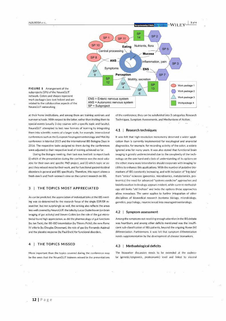
These 14 projects were distributed to three work packages (basic
science, translational science, clinical science), each supplemented
with one industry-based project. Ali projects were pre-arranged in a
kind of cycle (called the "digestive cycle") starting and ending with the
process of food ingestion, as organizing principle (see Figure 1).
The research project for each ESR covered a period of 3 years,
while projects for ERs were planned for 2 years. For details of the indi
vidual projects, we refer to the NeuroGUTwebsite.2
2 THE TASK
Both ESRs and ERs of NeuroGUT are exposed to a number of learn
ing events beyond the individual PhD programs they are involved in
TABLE 1 Short titles of the 14 ESR/ER
subprojects (SP) of the ITN NeuroGUT with principle investigator (Pl) and ESR/ ER
IL (initiais); see www.neurogut.eu for further
details SP1: Central representation of food intake in health & disease
SP2: Epithelial barrier function and micro-inflammation Theodorou JBJK
SP3: Mucosa-ENS-signaling in chronic bowel diseases
SP4: Dysfunction of esophagus & esophagogastric junction
SPS: Luminal bacteria, immune system and enteric nerves
SP6: Neuro-immune mechanisms in visceral pain perception
SP7: Inflammation and Pain
SPS: Pathophysiological alterations/ symptoms in FBD
SP9: Gastrointestinal motility/ sensitivity as a key to FBD
SP10: Autonomie nervous system in visceral hypersensitivity
SP11: Processing of visceral sensation & the gui microbiome
Projects for experienced researcher (ER), 24 months
SP12: Intestinal motility by endoluminal image analysis
SP13: SHT and other receptors in visceral hypersensitivity
SP14: Bacterial flora in health and functional bowel disorder
'Position empty alter 11 months.
Schemann
Smout
Stanghellini
Boeckxstaens
Grundy
Simren
Azpiroz
Aziz
Enck
Rabinovitz/ Horn (Given)
Schemann/Grundy
Zimmermann (Symbio)
ML
TH
TBK
EP
FU
AP
TP
AA
HW
NN'
NB
NM
12 | P a g e
ALBUSODA ET AL.
0 Work package 1
Work package 2 FIGURE 1 Arrangement of the
subprojects (SPs) of the NeuroGUT network. Colors and shapes represent work packages (see text below) and are
related to the collaborative aspects of the NeuroGUT networking
ENS = Enteric nervous system ANS = Autonomie nervous system SP = Subproject
6 Work package 3
• Workpackage 4
at their home institutions, and among them are training seminars and
summer schools. W ith respect to the latter, rather than inviting them to
special events (usually 3-day courses with a specific tapie and faculty),
NeuroGUT attempted to test new formats of learning by integrating
them into scientific events of a larger scale, for example, international
conferences such as the European Neurogastroenterology and Motility
conference in Istanbul 2015 and the international IBS Balogna Days in
2016. The respective tasks assigned to them during the conferences
were adjusted to their respective level of training achieved so far.
During the Bologna meeting, their task was twofold: to report back
(i) which of t he presentation du ring the conference was the most valu
able for their own and specific PhD project, and (ii) which tapie or as
pect they missed most for their work, and for functional gastrointestinal
disorders in general and IBS specifically. Therefore, t his report allows a
fresh-men's and fresh-women's view on the current research on IBS.
3 THE TOPICS MOST APPRECIATED
As can be predicted, t he appreciation of individual talks at t he IBS meet
ing was co-determined by the research focus of the single ESR/ ER re
searcher, but not surprisingly as well, the ranking also reflects the areas
less well covered by NeuroGUT: the talks by Lucas Oudenhoven (on brain
imaging of gut activity) and Steven Collins (on the raie of the gut micro
biota) found high appreciation, as did the pharmacology of gut functions
(by Jan Tack), the IBS-IBD interrelation (byThierry Piché), the new Rome
IV criteria (by Douglas Drossman), the raie of gas (by Fernando Azpiroz)
and the placebo response (by Paul Enck) for functional disorders.
4 THE TOPICS MISSED
More important t han the tapies covered during the conference may
be the ones that t he NeuroGUT trainees missed in the presentations
of the conference; they can be subdivided into 3 categories: Research
Techniques, Symptom Assessments, and Mechanisms of Action:
4.1 1 Research techniques
lt was felt that high-resolution manometry deserved a wider appli
cation than is currently implemented for esophageal and anorectal
diagnostics, for example, for recording activity of t he colon, a w idely
ignored area for many years. lt was also stated that functional brain
imaging is grossly underestimated due to the complexity of the tech
nology on the one hand and a lack of understanding of its options on
the other; many more laboratories should cooperate with imaging fa
cilities to enhance this applications. With the number of putative bio
markers of IBS constantly increasing, and with inclusion of "big data"
from "omics"-sciences (genomics, microbiomics, metabolomics, pro
teomics) the need for advanced "systems medicine" approaches and
bioinformation technology appears evident , whi le current methodol
ogy still looks "old fashion" and lacks the options these approaches
allow nowadays. The same applies to further integration of other
disciplines of biomedical research (systems biology, microbiology,
genetics, psychology, neuroscience) into neurogastroenterology.
4.2 1 Symptom assessment
Among the symptoms not receiving enough attention in the IBS debate
was heartburn, and among other deficits mentioned was the insuffi
cient sub-classification of IBS patients, beyond the ongoing Rome (IV)
differentiation. Furthermore, it was felt that symptom differentiation
needs supplementation by the development of disease biomarkers.
4.3 1 Methodological deficits
The biomarker discussion needs to be extended at the molecu
lar (genetic/epigenetic, proteonomic) level and linked to visceral
P a g e | 13

P a g e | 15
1.2 IBS, FODMAPs
The main subject of this thesis concerns the mechanism of effect by which a low-FODMAP diet
reduces symptoms in irritable bowel syndrome (IBS). In this chapter, the current state of
knowledge concerning IBS and some other functional gastrointestinal disorders (FGIDs) will be
discussed.
1.2.1 Functional gastrointestinal disorders (FGIDs)
Functional gastrointestinal disorders (FGIDs) are a group of distinct disorders of different
sections of the gastrointestinal tract with the commonality that they have no clear
pathophysiological cause (hence ‘functional’). FGIDs are common among the population, with
Irritable Bowel Syndrome (IBS) and Functional Dyspepsia the most frequent. On a whole, FGIDs
are the most commonly diagnosed disorders by gastroenterologists. Because they are difficult
to define and, for the moment, have no clear pathological cause, their treatment is complicated,
consisting of a variety of pharmacological, psychological, dietary, and complementary medical
treatments (Whitfield and Shulman, 2009). In an ongoing effort to classify and define the FGIDs
for diagnosis, the international “Rome process” has yielded the 4th edition of the Rome criteria
for FGIDs in 2016 (Drossman, 2016).
1.2.1.1 Rome Process and criteria
The Rome Foundation first issued criteria for the diagnosis of Irritable Bowel Syndrome in 1989
(Thompson et al. 1989), following with the Rome Classification System for FGIDs in
1990 (Drossman, 2007). Since then, 4 classifications have been published; Rome I (1994), Rome
II (1999-2000), Rome III (2006), and Rome IV (2016) (Drossman, 2016). It has grown from being
mostly a tool for researchers of FGIDs to better classify experimental subjects, to a robust
diagnostic tool for clinical practitioners. The current Rome IV criteria have been updated to
specifically take ‘pain’ into account, changing from ‘discomfort’ which was often difficult for
patients to respond to, and doesn’t translate well between cultures (Drossman, 2016). A full
overview of Functional Gastrointestinal Disorders specified by Rome IV can be found in Table 1.
There are some limitations to using the Rome criteria in clinical practice. For example, they may
exclude some treatable patients who do not strictly fall within the definitions set for each
disorder, and patients with multiple FGIDs might not be recognized as such (Oświęcimska,
Szymlak et al., 2017). Additionally, changing diagnosis criteria from one iteration to the next
might cause some patients to be ‘cured’ of their disease on rediagnosis, without any change in
their health situation.

16 | P a g e
Although the Rome criteria are increasingly useful and accepted for clinical practice, its
background as a tool for patient selection for trial purposes means its first goal was mostly to
select for those patients who were the most clearly includable and thus likely to respond to
treatment, preventing false positives.
Table 1 – Overview of FGIDs specified by Rome IV (Drossman, 2016)
P a g e | 17
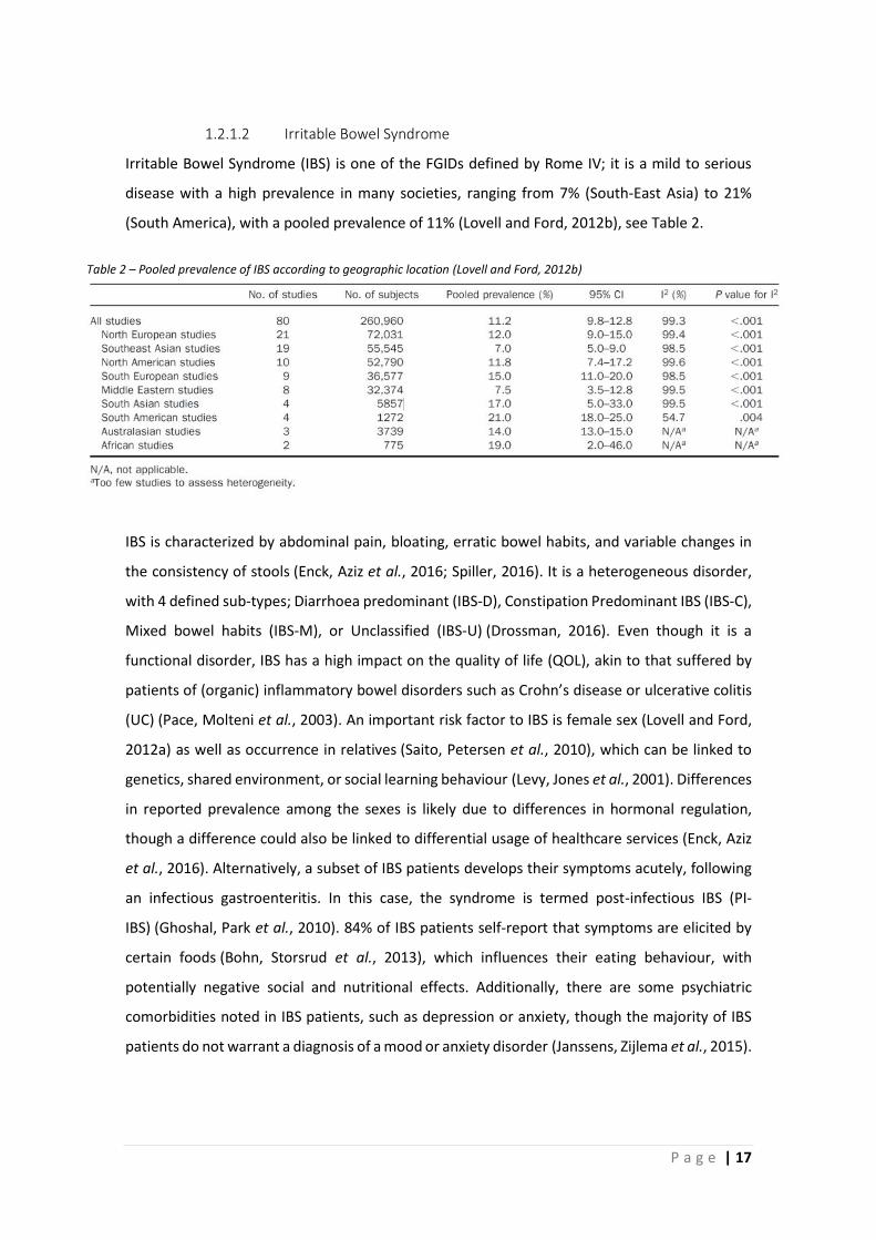
1.2.1.2 Irritable Bowel Syndrome
Irritable Bowel Syndrome (IBS) is one of the FGIDs defined by Rome IV; it is a mild to serious
disease with a high prevalence in many societies, ranging from 7% (South-East Asia) to 21%
(South America), with a pooled prevalence of 11% (Lovell and Ford, 2012b), see Table 2.
IBS is characterized by abdominal pain, bloating, erratic bowel habits, and variable changes in
the consistency of stools (Enck, Aziz et al., 2016; Spiller, 2016). It is a heterogeneous disorder,
with 4 defined sub-types; Diarrhoea predominant (IBS-D), Constipation Predominant IBS (IBS-C),
Mixed bowel habits (IBS-M), or Unclassified (IBS-U) (Drossman, 2016). Even though it is a
functional disorder, IBS has a high impact on the quality of life (QOL), akin to that suffered by
patients of (organic) inflammatory bowel disorders such as Crohn’s disease or ulcerative colitis
(UC) (Pace, Molteni et al., 2003). An important risk factor to IBS is female sex (Lovell and Ford,
2012a) as well as occurrence in relatives (Saito, Petersen et al., 2010), which can be linked to
genetics, shared environment, or social learning behaviour (Levy, Jones et al., 2001). Differences
in reported prevalence among the sexes is likely due to differences in hormonal regulation,
though a difference could also be linked to differential usage of healthcare services (Enck, Aziz
et al., 2016). Alternatively, a subset of IBS patients develops their symptoms acutely, following
an infectious gastroenteritis. In this case, the syndrome is termed post-infectious IBS (PI-
IBS) (Ghoshal, Park et al., 2010). 84% of IBS patients self-report that symptoms are elicited by
certain foods (Bohn, Storsrud et al., 2013), which influences their eating behaviour, with
potentially negative social and nutritional effects. Additionally, there are some psychiatric
comorbidities noted in IBS patients, such as depression or anxiety, though the majority of IBS
patients do not warrant a diagnosis of a mood or anxiety disorder (Janssens, Zijlema et al., 2015).
Table 2 – Pooled prevalence of IBS according to geographic location (Lovell and Ford, 2012b)
18 | P a g e
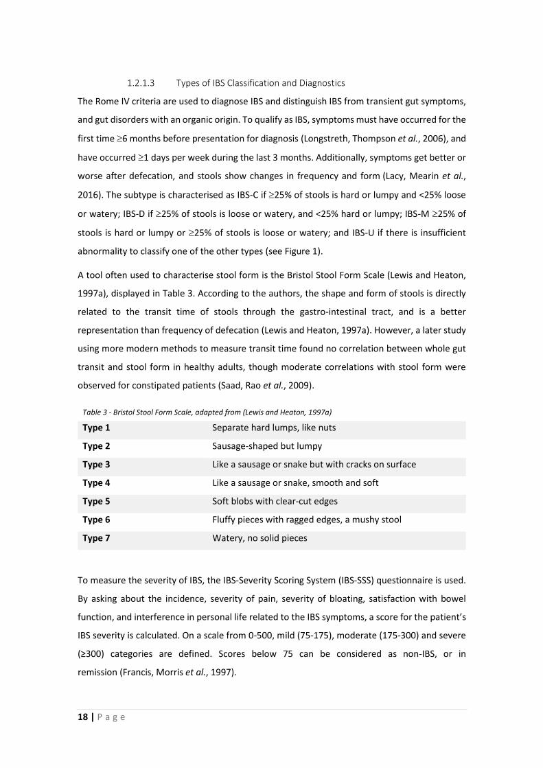
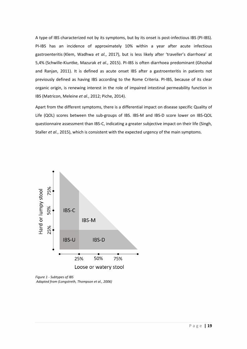
1.2.1.3 Types of IBS Classification and Diagnostics
The Rome IV criteria are used to diagnose IBS and distinguish IBS from transient gut symptoms,
and gut disorders with an organic origin. To qualify as IBS, symptoms must have occurred for the
first time 6 months before presentation for diagnosis (Longstreth, Thompson et al., 2006), and
have occurred 1 days per week during the last 3 months. Additionally, symptoms get better or
worse after defecation, and stools show changes in frequency and form (Lacy, Mearin et al.,
2016). The subtype is characterised as IBS-C if 25% of stools is hard or lumpy and <25% loose
or watery; IBS-D if 25% of stools is loose or watery, and <25% hard or lumpy; IBS-M 25% of
stools is hard or lumpy or 25% of stools is loose or watery; and IBS-U if there is insufficient
abnormality to classify one of the other types (see Figure 1).
A tool often used to characterise stool form is the Bristol Stool Form Scale (Lewis and Heaton,
1997a), displayed in Table 3. According to the authors, the shape and form of stools is directly
related to the transit time of stools through the gastro-intestinal tract, and is a better
representation than frequency of defecation (Lewis and Heaton, 1997a). However, a later study
using more modern methods to measure transit time found no correlation between whole gut
transit and stool form in healthy adults, though moderate correlations with stool form were
observed for constipated patients (Saad, Rao et al., 2009).
Table 3 - Bristol Stool Form Scale, adapted from (Lewis and Heaton, 1997a)
Type 1 Separate hard lumps, like nuts
Type 2 Sausage-shaped but lumpy
Type 3 Like a sausage or snake but with cracks on surface
Type 4 Like a sausage or snake, smooth and soft
Type 5 Soft blobs with clear-cut edges
Type 6 Fluffy pieces with ragged edges, a mushy stool
Type 7 Watery, no solid pieces
To measure the severity of IBS, the IBS-Severity Scoring System (IBS-SSS) questionnaire is used.
By asking about the incidence, severity of pain, severity of bloating, satisfaction with bowel
function, and interference in personal life related to the IBS symptoms, a score for the patient’s
IBS severity is calculated. On a scale from 0-500, mild (75-175), moderate (175-300) and severe
(≥300) categories are defined. Scores below 75 can be considered as non-IBS, or in
remission (Francis, Morris et al., 1997).
P a g e | 19
A type of IBS characterized not by its symptoms, but by its onset is post-infectious IBS (PI-IBS).
PI-IBS has an incidence of approximately 10% within a year after acute infectious
gastroenteritis (Klem, Wadhwa et al., 2017), but is less likely after ‘traveller’s diarrhoea’ at
5,4% (Schwille‐Kiuntke, Mazurak et al., 2015). PI-IBS is often diarrhoea predominant (Ghoshal
and Ranjan, 2011). It is defined as acute onset IBS after a gastroenteritis in patients not
previously defined as having IBS according to the Rome Criteria. PI-IBS, because of its clear
organic origin, is renewing interest in the role of impaired intestinal permeability function in
IBS (Matricon, Meleine et al., 2012; Piche, 2014).
Apart from the different symptoms, there is a differential impact on disease specific Quality of
Life (QOL) scores between the sub-groups of IBS. IBS-M and IBS-D score lower on IBS-QOL
questionnaire assessment than IBS-C, indicating a greater subjective impact on their life (Singh,
Staller et al., 2015), which is consistent with the expected urgency of the main symptoms.
Figure 1 - Subtypes of IBS Adapted from (Longstreth, Thompson et al., 2006)
20 | P a g e
1.2.1.4 Chronic Functional Abdominal Pain
Chronic Functional Abdominal Pain (CFAP) is another FGID characterized by abdominal pain. The
difference with IBS is that CFAP is not related to changes in bowel habit or stool form (Drossman,
2016). The absence of changes in bowel habit in CFAP can be taken as indication that the pain
component in CFAP, but likely also in subgroups of IBS patients, is not related to motility
disorders or other defects likely to influence stool transit. An altered visceral sensitivity and
changes to the function of the brain-gut axis are therefore more likely causes.
P a g e | 21
1.2.2 Aetiology IBS and physiopathology
As mentioned before, IBS has an unclear aetiology and pathophysiology. However, a growing
number of publications point to several factors implied in causing and/or perpetuating it. A
combination of visceral hypersensitivity, genetics, psychological stress, dysfunctional intestinal
permeability, and involvement of the microbiota are all possibly implicated in IBS.
1.2.2.1 Visceral sensitivity
A key component of IBS is abdominal pain. A logical and probable cause for this abdominal pain
is an increased visceral sensitivity to different stimuli of the gastrointestinal tract. The first to
describe the increased sensitivity to distension of IBS patients was James Ritchie, in
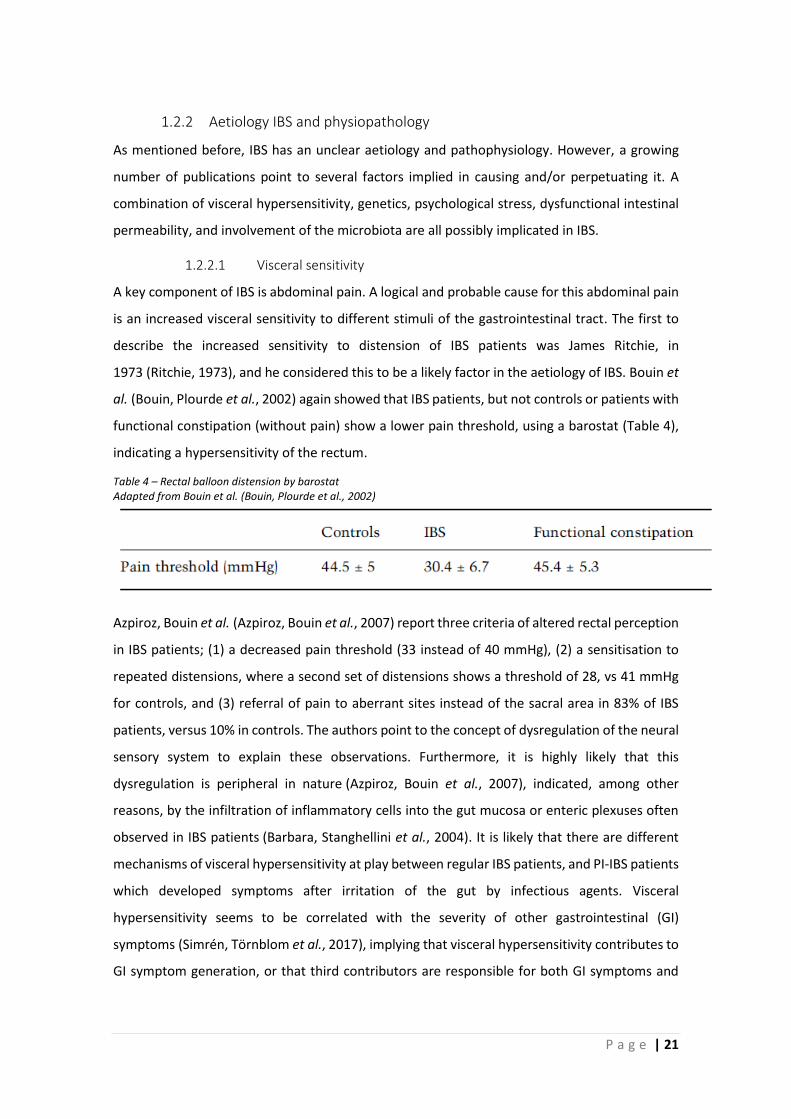
1973 (Ritchie, 1973), and he considered this to be a likely factor in the aetiology of IBS. Bouin et
al. (Bouin, Plourde et al., 2002) again showed that IBS patients, but not controls or patients with
functional constipation (without pain) show a lower pain threshold, using a barostat (Table 4),
indicating a hypersensitivity of the rectum.
Azpiroz, Bouin et al. (Azpiroz, Bouin et al., 2007) report three criteria of altered rectal perception
in IBS patients; (1) a decreased pain threshold (33 instead of 40 mmHg), (2) a sensitisation to
repeated distensions, where a second set of distensions shows a threshold of 28, vs 41 mmHg
for controls, and (3) referral of pain to aberrant sites instead of the sacral area in 83% of IBS
patients, versus 10% in controls. The authors point to the concept of dysregulation of the neural
sensory system to explain these observations. Furthermore, it is highly likely that this
dysregulation is peripheral in nature (Azpiroz, Bouin et al., 2007), indicated, among other
reasons, by the infiltration of inflammatory cells into the gut mucosa or enteric plexuses often
observed in IBS patients (Barbara, Stanghellini et al., 2004). It is likely that there are different
mechanisms of visceral hypersensitivity at play between regular IBS patients, and PI-IBS patients
which developed symptoms after irritation of the gut by infectious agents. Visceral
hypersensitivity seems to be correlated with the severity of other gastrointestinal (GI)
symptoms (Simrén, Törnblom et al., 2017), implying that visceral hypersensitivity contributes to
GI symptom generation, or that third contributors are responsible for both GI symptoms and
Table 4 – Rectal balloon distension by barostat Adapted from Bouin et al. (Bouin, Plourde et al., 2002)
22 | P a g e
visceral hypersensitivity. Illustrating the possible common cause of visceral sensitivity and GI
symptoms, a reduction of visceral sensitivity by blocking the histamine receptor HRH1 is
accompanied by a reduction in GI symptoms (Wouters, Balemans et al., 2016).
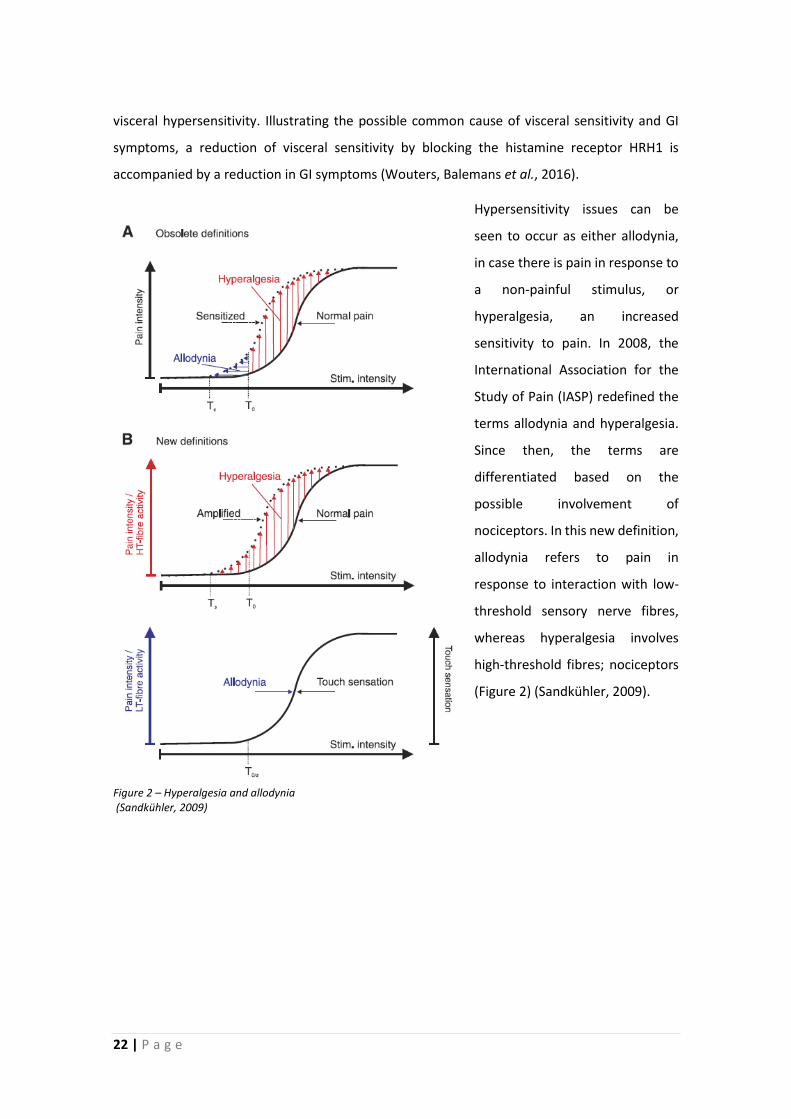
Hypersensitivity issues can be
seen to occur as either allodynia,
in case there is pain in response to
a non-painful stimulus, or
hyperalgesia, an increased
sensitivity to pain. In 2008, the
International Association for the
Study of Pain (IASP) redefined the
terms allodynia and hyperalgesia.
Since then, the terms are
differentiated based on the
possible involvement of
nociceptors. In this new definition,
allodynia refers to pain in
response to interaction with low-
threshold sensory nerve fibres,
whereas hyperalgesia involves
high-threshold fibres; nociceptors
(Figure 2) (Sandkühler, 2009).
Figure 2 – Hyperalgesia and allodynia (Sandkühler, 2009)
P a g e | 23
There is a clear link between mental stress and an increase of visceral sensitivity, and this effect
is mainly mediated by the release of corticotropin-releasing factor (CRF) by the hypothalamus.
This activates release of adrenocorticotropic hormone (ACTH) from the pituitary gland into the
bloodstream, which then leads to cortisol release from the adrenal glands. Cortisol has many
effects in the body, for example, it is involved in increasing blood-glucose concentration, and
suppression of the immune system. We will however focus on the effects of Hypothalamic-
Pituitary-Adrenal (HPA)-axis activation on visceral sensitivity.
In rats, visceral hyperalgesia can be induced by repeated water-avoidance stress treatments, a
model of psychological stress. This effect can be prevented by injection of CRF antagonists,
showing that CRF-receptors play a main role in the generation of hyperalgesia by psychological
stress (Larauche, Bradesi et al., 2008). Similarly, it has been shown that mast cells play a role in
the generation of visceral hypersensitivity due to stressful conditions, through central pathways
involving CRF (Gue, Del Rio-Lacheze et al., 1997). The nervous system can directly convey signals
resulting from psychological stress to mast cells by CRF and/or Substance P (SP) release, which
can trigger them to release mediators via piecemeal degranulation, in response to crowding
stress (Vicario, Guilarte et al., 2010).
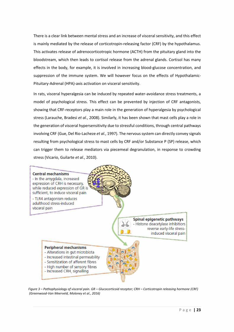
Figure 3 – Pathophysiology of visceral pain. GR – Glucocorticoid receptor; CRH – Corticotropin releasing hormone (CRF) (Greenwood-Van Meerveld, Moloney et al., 2016)
24 | P a g e
1.2.2.2 Genetics/ epidemiology
Regardless of the largely unclear aetiology and pathophysiology of IBS, epidemiology can shed
light on correlated and possibly causative factors. Research into this topic has focused on
relation to family connections, comorbidity with psychological afflictions, and the local
occurrence of infectious diseases.
Firstly, looking at social connections, IBS aggregates heavily to families. It has a high odds ratio
(OR) between siblings (3.12), children (2.12), and parents (1.91). Interestingly, there was no
correlation between spouses (OR 0.89), indicating that genetics and not shared environment
might be responsible for the observed effect (Saito, Petersen et al., 2010). This effect could then
be explained by genes directly responsible for IBS, or genes increasing the likelihood of a related
trait, such as lactose intolerance, depression or anxiety, somatization, or an immune system that
increases risk of infection (Saito, Petersen et al., 2010). In a recent study done on twins, familial
and intra-uterine factors were shown to affect the co-occurrence of IBS and psychological
factors such as depression and anxiety. They were able to show that the comorbidity of IBS with
mental disorders (depression and anxiety) were only present for those twins in a lower weight
group of smaller than 2,5kg (Bengtson, Aamodt et al., 2015). This has led to the hypothesis that
the HPA-axis links these two afflictions in intrauterine growth restriction and might explain the
often-observed comorbidity, because an intrauterine physical stress could pre-program the
HPA-axis to be more responsive (Bengtson, Aamodt et al., 2015).
Looking further into the genetic aspects of this family aggregation it is important to realize that
the susceptibility of the majority of IBS patients is caused by a complex interaction of many
genes and the environment of the patient (Henström and D’Amato, 2016). However, in some
cases, single genes may be responsible for symptom generation. A loss-of-function mutation of
the SCN5A gene, encoding for the NaV1.5 ion channel present on interstitial cells of Cajal, seems
to be heavily related to IBS-C (Beyder, Mazzone et al., 2014). Because of the more well-known
function of the NaV1.5 ion channel, these patients were initially identified because of cardiac
arrhythmia. Another gene of interest is TNFSF15, which codes for TNF-like ligand 1A, which is a
clear susceptibility related gene for IBS (Zucchelli, Camilleri et al., 2011). This gene is also
involved in the development of colitis in mice by DSS treatment, by supporting TH1 and TH17
effector functions (Takedatsu, Michelsen et al., 2008), furthermore, it is an important
susceptibility locus in for example Crohn’s Disease (CD) (Barrett, Hansoul et al., 2008) and
arthritis of the spine (Zinovieva, Bourgain et al., 2009). That this gene is related to IBS can be
P a g e | 25
seen as an indication that IBS is related to an altered immune response in patients with positively
correlated alleles.
1.2.2.3 Stress and anxiety
Because IBS and certain psychological
profiles seem to be related, it is
interesting to investigate the effects
of mental stress and anxiety on IBS
symptom generation and perception.
Early life trauma, such as childhood
abuse, has a significantly higher
prevalence in IBS patients than in
healthy controls, particularly in
women. A history of emotional abuse
specifically has a high predicting
power; feeling ignored, and sexual
abuse increase the odds of
developing IBS with 2.08 and 3.05
times, respectively (Bradford, Shih et
al., 2012). IBS patients meeting
criteria for any psychiatric disorder
range from 40% to 94% in some
studies, though this is most likely an
overestimation due to tertiary care
patient selection. It is clear however
that IBS patients have a higher frequency of depressive, panic, or generalized anxiety disorders
than other patients in the same clinics (Surdea-Blaga, Băban et al., 2012).
The mechanisms for this observed association are likely to involve both true psychological
factors, as well as the gut-brain and HPA axes (see Figure 4). In rats, psychological stress can
increase visceral hypersensitivity by corticotropin releasing factor (CRF) release, which involves
mast cell activation (Gue, Del Rio-Lacheze et al., 1997). Similarly, stress can increase the
paracellular permeability through corticotropin releasing factor (CRF), stimulating the release of
nerve growth factor (NGF) by mast cells (Barreau, Cartier et al., 2007) and the increase of mucus
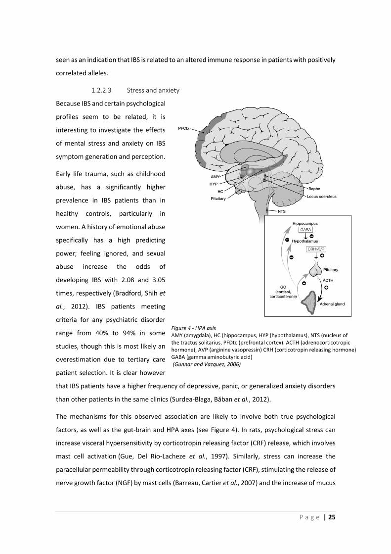
Figure 4 - HPA axis AMY (amygdala), HC (hippocampus, HYP (hypothalamus), NTS (nucleus of the tractus solitarius, PFDtc (prefrontal cortex). ACTH (adrenocorticotropic hormone), AVP (arginine vasopressin) CRH (corticotropin releasing hormone) GABA (gamma aminobutyric acid) (Gunnar and Vazquez, 2006)
26 | P a g e
production in response to stress is mast cell dependent as well (Castagliuolo, Lamont et al.,
1996). Alternatively, somatization is considered an important factor in IBS, correlating with
visceral hypersensitivity (Grinsvall, Törnblom et al., 2017). Because of this, cognitive behavioural
therapy can improve GI symptoms through pathways improving anxiety levels (Jones, Koloski et
al., 2011). Logically, improving stress levels in this way would improve both HPA-axis and
psychological mediators.
While an understanding of the proximate causes of the link between stress and visceral
sensitivity is crucial in research, appreciating a more ultimate cause of this link is important too.
Stress, either physical or psychological, induces adaptive responses by activation of both the
HPA axis, as already discussed, and the sympathetic adrenomedullary system (SAM). Activation
of SAM is very rapid, directly responsible for adaptations related to the flight-or-fight response
and characterized by the release of adrenaline and noradrenaline by chromaffin cells innervated
by the sympathetic preganglionic neurons, leading to acute adaptations such as increased
cardiac output and blood glucose levels. Interestingly the noradrenaline:adrenaline release ratio
seems to be dependent on neural integration in response to the nature and magnitude of
stimulus (Vollmer, 1996). In contrast, activation of the HPA axis and subsequent increased
cortisol levels have more chronic effects, and work mainly through regulating gene
transcription (Sapolsky, Romero et al., 2000). Proper HPA axis function permits effective flight-
or-flight by optimising the response to adrenaline (the SAM response). Glucocorticoids released
in HPA axis activation interact with 2 types of receptors in the brain, glucocorticoid receptors
(GR) and mineralocorticoid receptors (MR), activation of which often have opposite effects. The
balance of activation is determined by the concentration of glucocorticoids; at basal
concentrations, only the MRs are activated, supporting maintenance processes such as ensuring
responsiveness of neurons to neurotransmitters and regulating the circadian rhythm (Sapolsky,
Romero et al., 2000), these processes permit effective responses to stress by optimising
responses to adrenaline/noradrenaline (Gunnar and Vazquez, 2006). At peak concentrations
GRs are engaged as well, which leads to stress responses, but with deleterious effects on brain
function, such as inhibiting glucose utilisation by neurons, and impairing neural plasticity,
learning and memory (Gunnar and Quevedo, 2007). These negative effects are likely meant to
reverse the acute response to stressors, and support a return to homeostasis (Sapolsky, Romero
et al., 2000). The balancing act between the activity of the HPA axis and SAM function to
maintain homeostasis is called ‘allostasis’ (McEwen and Seeman, 1999), where the intended
equilibrium is not at a fixed level, but dependent on context.
P a g e | 27
In case of prolonged or repeated stress, this allostatic system begins to become deleterious, due
to negative health effects of otherwise adaptive responses (Gunnar and Quevedo, 2007;
Greenwood-Van Meerveld, Moloney et al., 2016), with the gastrointestinal tract particularly
vulnerable to negative effects (Mayer, 2000).
1.2.2.4 Gut permeability in IBS
An increase in gut permeability has been seen in IBS patients, particularly in PI-IBS, and can be
associated to inflammatory processes and increased visceral sensitivity. In this section, we will
investigate the role of increased permeability in IBS. For more information on intestinal
permeability in general, please refer to section 1.5.2.
It has been shown that the increased visceral sensitivity in response to acute (restraint) stress in
rats is mediated by an increase in intestinal permeability, and prevention of this increase in
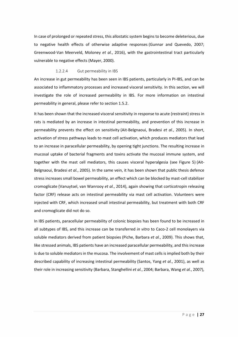
permeability prevents the effect on sensitivity (Ait-Belgnaoui, Bradesi et al., 2005). In short,
activation of stress pathways leads to mast cell activation, which produces mediators that lead
to an increase in paracellular permeability, by opening tight junctions. The resulting increase in
mucosal uptake of bacterial fragments and toxins activate the mucosal immune system, and
together with the mast cell mediators, this causes visceral hyperalgesia (see Figure 5) (Ait-
Belgnaoui, Bradesi et al., 2005). In the same vein, it has been shown that public thesis defence
stress increases small bowel permeability, an effect which can be blocked by mast-cell stabilizer
cromoglicate (Vanuytsel, van Wanrooy et al., 2014), again showing that corticotropin releasing
factor (CRF) release acts on intestinal permeability via mast cell activation. Volunteers were
injected with CRF, which increased small intestinal permeability, but treatment with both CRF
and cromoglicate did not do so.
In IBS patients, paracellular permeability of colonic biopsies has been found to be increased in
all subtypes of IBS, and this increase can be transferred in vitro to Caco-2 cell monolayers via
soluble mediators derived from patient biopsies (Piche, Barbara et al., 2009). This shows that,
like stressed animals, IBS patients have an increased paracellular permeability, and this increase
is due to soluble mediators in the mucosa. The involvement of mast cells is implied both by their
described capability of increasing intestinal permeability (Santos, Yang et al., 2001), as well as
their role in increasing sensitivity (Barbara, Stanghellini et al., 2004; Barbara, Wang et al., 2007),
28 | P a g e
and the observation that IBS patients have increased mucosal mast cell counts (Matricon,
Meleine et al., 2012), which holds up in a recent meta-analysis (Bashashati, Moossavi et al.,
2017). Another recent study claims that the colonic barrier function is uncompromised in female
IBS-C patients (Peters, Edogawa et al., 2017), in contradiction to earlier findings claiming the
opposite (Piche, Barbara et al., 2009). Additionally, they found no differences in expression of
the important tight junction proteins Occludin, ZO-1, ZO-2, or ZO-3 (Peters, Edogawa et al.,
2017).
Figure 5 – Possible mechanism by which stress can lead to increased visceral sensitivity through increased intestinal permeability. (Ait-Belgnaoui, Bradesi et al., 2005)
P a g e | 29
A possible explanation for these conflicting reports might be the use of different fluorescent
markers in the ex vivo biopsy study; fluorescein–5.6 sulfonic acid (478 Da) for Piche, versus 4kDa
FITC–dextran for Peters, and that the earlier study conducted by Piche, Barbara et al. used a
cohort of IBS patients of all subtypes, diagnosed using Rome 2 criteria including slightly less IBS-
C patients, whereas the study by Peters, Edogawa et al. only looked at IBS-C patients diagnosed
using Rome 3 criteria. This underlines the importance of distinguishing subgroups of IBS not only
on their symptoms, but possibly also on the underlying causes for their affliction, although the
study by Piche, Barbara et al. found no differences between subgroups.
There are other possible mechanisms that could be responsible for the observed increase in
permeability in IBS; a variety of proteases, whether derived from the host or the microbiota,
have been described as involved in increasing the paracellular permeability of the host (Van
Spaendonk, Ceuleers et al., 2017). Especially mast-cell derived trypsin and tryptase can be
responsible for an increase in permeability due to PAR-2 activation, at the same time causing an
increase in visceral sensitivity (Annahazi, Ferrier et al., 2013). Likewise, in the context of PI-IBS,
an increased intestinal permeability plays an important role. Following Campylobacter infection,
approximately 25% of patients develop ‘post-dysenteric irritable bowel syndrome’ (later called
post-infectious IBS, or PI-IBS), characterized by increased intestinal permeability (Spiller, Jenkins
et al., 2000). Furthermore, a study investigating possible genetic risk factors in the development
of PI-IBS shows that certain alleles of IL-6, TLR9, and CDH1 rendered the patient more
susceptible (Villani, Lemire et al., 2010). CDH1 (cadherin-1/E-cadherin) is a trans-membrane
glycoprotein involved in cell-cell adhesion, and tight-junction formation by facilitating Adherens
junctions (Hartsock and Nelson, 2008). Disruption of epithelial tight-junctions by enteric
pathogens such as Campylobacter or E.coli causes an increase in intestinal permeability during
infection (Berkes, Viswanathan et al., 2003; Wu, Rhee et al., 2007); so it is interesting that certain
variants of the E-cadherin gene could be linked to development of PI-IBS. Either an impaired
ability to adequately restore tight-junctions after infection, or a pre-existing higher permeability
caused by this genetic variation could increase the likelihood of PI-IBS.
30 | P a g e
1.2.2.5 Immune System
As mentioned before, the immune system is often seen as a key player in the physiopathology
of IBS, and not only in the context of post-infectious IBS. Here, some possible mechanisms of
involvement of the immune system in IBS will be discussed.
Firstly, the clearest link between IBS and the immune system is represented by post-infectious
IBS. After an infectious gastroenteritis, the risk of developing IBS is increased six times, which
lasts for up to three years after the infection is resolved (Thabane, Kottachchi et al., 2007).
Apparently, the activation of the immune system during the infection has long lasting effects on
gut function, even after elimination of the infectious agent. In a similar fashion, IBD patients in
remission often have IBS-like complaints (Barbara, Cremon et al., 2014), which indicates that
even after the inflammation is abated, the recent immune activation has leftover effects
resembling IBS. The reason for this link between inflammation and IBS symptoms might be
related to the increased permeability of the tissue induced by it, as discussed in the previous
section.
A marker of the innate immune system, β-defensin 2, produced by neutrophils, monocytes and
lymphocytes is increased in IBS patients, like in Ulcerative Colitis patients (Langhorst, Junge et
al., 2009). Anti-microbial β-defensin 2 production is induced by pro-inflammatory cytokines in
response to microbial invasion, after activation of toll-like receptors (TLRs) (Selsted and
Ouellette, 2005), possibly indicating a partially shared origin of these maladies, or merely
indicating an increase in permeability permitting more bacterial components to trigger TLRs in
both cases. IBS symptoms in quiescent IBD seem to be related to undetectable inflammation
involving increased numbers of intraepithelial lymphocytes and TNF-α, which were absent in IBS
patients and quiescent IBD patients without IBS symptoms (Vivinus-Nebot, Frin-Mathy et al.,
2014).
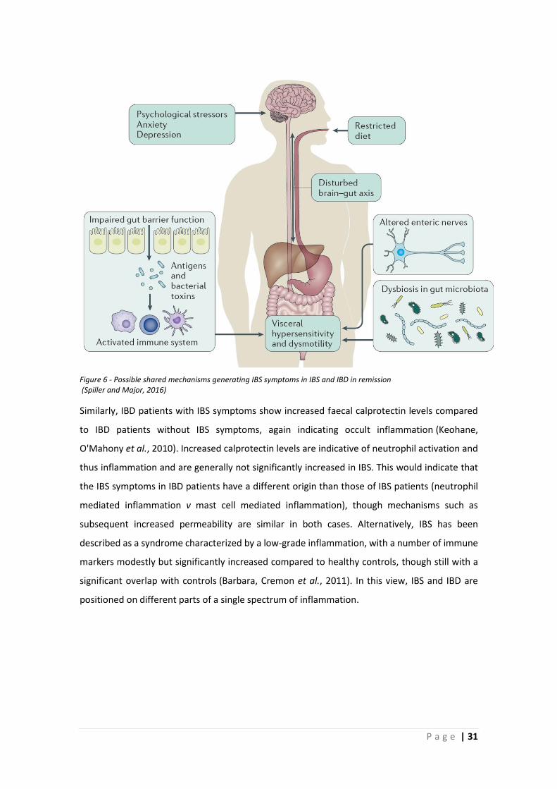
P a g e | 31
Similarly, IBD patients with IBS symptoms show increased faecal calprotectin levels compared
to IBD patients without IBS symptoms, again indicating occult inflammation (Keohane,
O'Mahony et al., 2010). Increased calprotectin levels are indicative of neutrophil activation and
thus inflammation and are generally not significantly increased in IBS. This would indicate that
the IBS symptoms in IBD patients have a different origin than those of IBS patients (neutrophil
mediated inflammation v mast cell mediated inflammation), though mechanisms such as
subsequent increased permeability are similar in both cases. Alternatively, IBS has been
described as a syndrome characterized by a low-grade inflammation, with a number of immune
markers modestly but significantly increased compared to healthy controls, though still with a
significant overlap with controls (Barbara, Cremon et al., 2011). In this view, IBS and IBD are
positioned on different parts of a single spectrum of inflammation.
Figure 6 - Possible shared mechanisms generating IBS symptoms in IBS and IBD in remission (Spiller and Major, 2016)
32 | P a g e
IBD patients in remission that do not show occult inflammation, i.e., without increased faecal
calprotectin levels but showing IBS symptoms, make up 31% of total IBD patients in
remission (Berrill, Green et al., 2013), which is a higher proportion than observed in the general
population. Figure 6 shows the possible mechanisms that could explain these patients, with
mechanisms that largely correspond to PI-IBS, which is not surprising, considering the shared
history of inflammation in PI-IBS and IBD patients. A subtle but interesting difference exists
between the sanguine cytokine levels of healthy subjects and IBS patients. While no
distinguishing profiles could be characterized, subgroups of IBS patients showed an increased
immune activity, and cytokine profiles were more variable in the IBS group (Bennet, Polster et
al., 2016). This further indicates the involvement of the immune system, at least in subsets of
IBS patients.
Inflammatory processes can lead to increased sensitivity by disconnecting nerves with their
targets, which then causes reconnection and remodelling of these nerves (Byers, Suzuki et al.,
2003; Spiller and Major, 2016). For example, following recovery from TNBS induced colitis,
substance P levels remained significantly increased in mucosal nerves in the medium term, and
mucosal nerve galanin and muscular nerve substance P remained elevated on the long term
together with a generally increased nerve innervation throughout the affected tissues, indicative
of an increased potential for nociception (Simpson, Sundler et al., 2008). Relatedly, visceral
sensitivity in IBS-D patients has been linked to mast-cell derived nerve growth factor (NGF) (Xu,
Zhang et al., 2017), which could play a role in similar ways; promoting nerve growth and
differentiation. However, the increased mucosal mast cell counts in IBS-D patients found in the
same work might also be related to the increased visceral sensitivity independent of concurring
increased levels of NGF. A recent meta-analysis indicates that mucosal mast cell numbers in the
descending and rectosigmoid colon are indeed significantly increased in IBS patients, but no
significant increase is observed in the ascending colon, additionally, CD3+ T cells show a
significant increase in the rectosigmoid region, in IBS patients (Bashashati, Moossavi et al.,
2017).
P a g e | 33
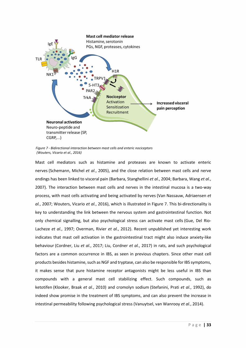
Mast cell mediators such as histamine and proteases are known to activate enteric
nerves (Schemann, Michel et al., 2005), and the close relation between mast cells and nerve
endings has been linked to visceral pain (Barbara, Stanghellini et al., 2004; Barbara, Wang et al.,
2007). The interaction between mast cells and nerves in the intestinal mucosa is a two-way
process, with mast cells activating and being activated by nerves (Van Nassauw, Adriaensen et
al., 2007; Wouters, Vicario et al., 2016), which is illustrated in Figure 7. This bi-directionality is
key to understanding the link between the nervous system and gastrointestinal function. Not
only chemical signalling, but also psychological stress can activate mast cells (Gue, Del Rio-
Lacheze et al., 1997; Overman, Rivier et al., 2012). Recent unpublished yet interesting work
indicates that mast cell activation in the gastrointestinal tract might also induce anxiety-like
behaviour (Cordner, Liu et al., 2017; Liu, Cordner et al., 2017) in rats, and such psychological
factors are a common occurrence in IBS, as seen in previous chapters. Since other mast cell
products besides histamine, such as NGF and tryptase, can also be responsible for IBS symptoms,
it makes sense that pure histamine receptor antagonists might be less useful in IBS than
compounds with a general mast cell stabilizing effect. Such compounds, such as
ketotifen (Klooker, Braak et al., 2010) and cromolyn sodium (Stefanini, Prati et al., 1992), do
indeed show promise in the treatment of IBS symptoms, and can also prevent the increase in
intestinal permeability following psychological stress (Vanuytsel, van Wanrooy et al., 2014).
Figure 7 - Bidirectional interaction between mast cells and enteric nociceptors (Wouters, Vicario et al., 2016)
34 | P a g e
1.2.2.6 Microbiota particularities/ dysbiosis
Starting with the recognition of the association of gastroenteritis, as discussed before, and the
use of antibiotics as a risk factor for developing IBS (Villarreal, Aberger et al., 2012), the
microbiota is increasingly recognized as a possible factor in IBS. The microbiota profiles of IBS
patients tend to differ from those of healthy patients (Rajilic-Stojanovic, Biagi et al., 2011), and
they can be used to predict the responsiveness of IBS patients to dietary intervention with a low-
FODMAP diet (Bennet, Böhn et al., 2017; Valeur, Smastuen et al., 2018). However, because IBS
is characterized by changes in bowel habits, which are very likely to influence the microbiota
composition, specific differences are difficult to interpret. Additionally, it is not yet clear whether
a specific microbiota pattern exists for IBS, but in general, a reduction of bacterial diversity and
an increased instability have been consistently found (Collins, 2014).
It is very likely that the two-way signalling between microbiota and epithelium which can
regulate secretion of mucus and other molecules involved in host-microbe interactions is
involved in IBS, because IBS patients show a dysregulation of the mucus layer and β-defensin-2
peptides (Swidsinski, Loening-Baucke et al., 2008; Langhorst, Junge et al., 2009; Simren, Barbara
et al., 2013). Illustrating a role for microbiota in IBS, patients show a modulated expression of
certain Toll-like receptors (TLRs), which perceive pathogen-associated molecular patterns
(PAMPs) such as lipopolysaccharides (LPS) for the innate immune system, indicating
involvement of microbiota-immune interactions in IBS. Both IBD and IBS patients show
alterations in the composition of the gut microbiota (Spiller and Lam, 2011; Casen, Vebo et al.,
2015), also known as ‘dysbiosis’, though it is not immediately clear whether this dysbiosis is
cause, effect, or both.
Transit time and stool consistency directly influence microbiota composition (Vandeputte,
Falony et al., 2015), and both these factors are altered in IBS and IBD, making it difficult to relate
microbiota differences as a cause. Additionally, microbiota composition is affected by dietary
patterns (Rajilic-Stojanovic, Jonkers et al., 2015), and patients often change their dietary habits
in an effort to mitigate symptoms. Perhaps unsurprisingly, the measure of dysbiosis found in IBS
patients is correlated to the gravity of their symptoms (Tap, Derrien et al., 2017), and
interestingly, these differences in microbiota composition were not correlated to diet.
P a g e | 35
It is not straightforward to clearly link certain phyla or species of bacteria to IBS, but certain
trends have been consistently observed; such as a depletion of Bifidobacteria in both faecal and
mucosal microbiota (Malinen, Rinttila et al., 2005; Kerckhoffs, Samsom et al., 2009). Duplicate
HITChip microarray analyses of faecal samples of IBS patients and healthy controls in Finland
showed that IBS patients had 1.5-fold decreased Bifidobacteria counts, and a 2-fold increased
ratio of Firmicutes/Bacteroidetes (Rajilic-Stojanovic, Biagi et al., 2011). Additionally, a Japanese
study showed that a pooled group of IBS-subtypes had significantly higher counts of Veillonella
and Lactobacillus than healthy controls (Tana, Umesaki et al., 2010).
That it is possible for the microbiota to have a causative effect in generating IBS symptoms has
been shown in animal experiments. Inoculating germ-free rats with a microbiota from IBS
patients renders them hypersensitive to colorectal distension, compared to conventional rats,
but being inoculated with healthy human microbiota does not have this effect (Crouzet, Gaultier
et al., 2013), and similarly, a decreased transit time, impaired intestinal permeability, and
anxiety-like behaviour can be transferred to germ-free mice by inoculating them with IBS-D
patient microbiota (De Palma, Lynch et al., 2017), results which indicate a possible causative
effect of a disorder-associated microbiota profile.
36 | P a g e
1.2.2.7 Small intestinal bacterial overgrowth
Small intestinal bacterial overgrowth (SIBO) is a syndrome often linked to IBS. It consists of an
increased number of bacteria in the small bowel, defined as numbers over 1*103cfu/ml proximal
jejunal contents (Khoshini, Dai et al., 2008), while the small bowel should normally contain small
bacterial populations (Bouhnik, Alain et al., 1999; Bures, Cyrany et al., 2010). The expansion of
bacteria from the large into the small intestine is held responsible for such symptoms as
bloating, abdominal discomfort, and changes in stool form (Pimentel and Lezcano, 2008), which
is reminiscent of IBS, but with an organic cause. However, a higher proportion of diagnosed IBS
patients than healthy controls are reported to present with SIBO (Lupascu, Gabrielli et al., 2005),
which makes it questionable whether they actually have true IBS, or it might indeed indicate a
causative role for SIBO in IBS, depending on interpretation.
This possible causal relationship between SIBO and (all cases of) IBS has been hotly contested,
among other reasons, because there are issues with the methods used to characterize SIBO; the
lactulose hydrogen breath test (LHBT) to determine abnormally early carbohydrate
fermentation is flawed, and it has recently been shown that in most (88%) of the IBS patients
for who the test is positive, the bolus was already in the caecum, as measured by oro-caecal
scintigraphy (Yu, Cheeseman et al., 2010). Additionally, bacterial numbers in the jejunum used
to determine severity of SIBO do not seem to correlate to IBS symptom severity (Grover,
Kanazawa et al., 2008). In short, it is unlikely that SIBO has a clear causal role to play in
IBS (Spiegel, 2011).
It is often beneficial for patients to exclude lactose from the diet and to reduce other simple
sugars (Bures, Cyrany et al., 2010), which resembles the low-FODMAP diet in form and function,
i.e., lower the amount of available fermentable carbohydrates to the gut microbiota. SIBO is
often mis- or underdiagnosed due to the wide range of clinical manifestations, and
understanding its mechanisms better will hopefully lead to future improvements to patient
care (Sachdev and Pimentel, 2013).
We will talk more about the functions of the microbiota in a later chapter (1.3) devoted to the
matter.
P a g e | 37
1.2.2.8 Biomarkers IBS
To better characterize patients, and to facilitate diagnosis, a lot of effort has been targeted to
identify biochemical markers (biomarkers) for IBS and its subgroups, with varying success.
Though IBS is increasingly seen as having organic characteristics, its history as a functional
disorder has not yielded much in the way of biomarkers. However, there are some promising
developments to help determine appropriate treatment of patients; microbiota profiles can
apparently serve to predict responsiveness to dietary intervention (Bennet, Böhn et al., 2017),
and genetic markers can be used to predict responsiveness to certain drugs (Enck, Aziz et al.,
2016). Additionally, technical advancements such as the pressure-, temperature-, and
pH‑sensitive SmartPill (Given Imaging Ltd, Yoqneam, Israel) can be used to get detailed insights
into gastrointestinal motility of patients (Rao, Kuo et al., 2009), which can be used for patient
characterisation and help to identify targets for antispasmodics (Enck, Aziz et al., 2016). Another
possible approach is the use of cytokine profiles (Bennet, Polster et al., 2016), though as of yet,
there are no biomarkers that can be used to reliably diagnose IBS.
38 | P a g e
1.2.3 Clinical practice
Treatment of IBS is complicated, with many treatments not sufficiently effective. Because the
underlying causes for IBS are not well understood, it has proven difficult to design evidence-
based therapies with a clear mechanism of effect. Additionally, scientific underpinning for
treatment of functional disorders in general suffers from a high placebo effect to treatment.
Diagnosis of IBS depends mostly on excluding any other cause for the symptoms. Diagnostic
algorithms for diagnosis of IBS must therefore make sure that there are no more serious origins
of complaints (Enck, Aziz et al., 2016).
What follows now is an oversight of current clinical practice in IBS patients.
1.2.3.1 IBS – Medication
Several pharmaceutical treatments of IBS have been developed over the years, mostly aimed at
tackling one or the other symptom, in accordance with the functional nature of the syndrome.
To treat the constipation in a subset of IBS patients, laxatives can be effective, and the laxative
and analgesic linaclotide has proven useful in treating constipation in IBS (Chey, Lembo et al.,
2012), while at the same time reducing pain by activating epithelial guanylate cyclase-C receptor
(GC-C), which increases extracellular cGMP, thereby reducing nociceptor sensitivity (Castro,
Harrington et al., 2013). To reduce painful smooth muscle spasms, common in IBS,
antispasmodic drugs are used. However, these drugs can cause constipation, and should not be
tried in already constipated patients (Enck, Aziz et al., 2016). One such antispasmodics is
peppermint oil, which blocks calcium channels, thereby inhibiting contractions (Hawthorn,
Ferrante et al., 1988). It is specifically effective when administered in capsules which release
throughout the small intestine (Cash, Epstein et al., 2016). It follows from what we know about
the involvement of mast cells in IBS, that mastocyte stabilizers can be expected to have
beneficial effects in IBS. An animal IBS model inducing visceral sensitivity in rats by partial
restraint stress (PRS), shows that mast cell stabilizer doxantrazole suppresses stress-induced
visceral hypersensitivity (Gue, Del Rio-Lacheze et al., 1997). Similarly, an animal IBS model using
chemical induction of visceral hypersensitivity by acetic acid-induced colitis shows that the same
mast cell stabilizer can significantly attenuate the hypersensitivity (La, Kim et al., 2004). In
humans, trials with anti-inflammatory drugs have proven more ambiguous, with mast-cell
stabiliser ketotifen showing promise in reducing visceral hypersensitivity and improving
QOL (Klooker, Braak et al., 2010), but mesalazine, another mast cell stabilizer, no better than
P a g e | 39
placebo, after a pilot study initially yielded hopeful results (Corinaldesi, Stanghellini et al., 2009;
Barbara, Cremon et al., 2016).
1.2.3.2 IBS – Probiotics
Because of the often-cited link to the intestinal microbiota, there is a significant interest in
treating IBS with probiotics. Oral probiotics are defined as ‘live micro-organisms, which upon
ingestion in certain numbers, exert health benefits beyond inherent basic nutrition’ (Guarner
and Schaafsma, 1998), and later by the WHO, very similarly, as ‘Live microorganisms which when
administered in adequate amounts confer a health benefit on the host’ (Joint FAO/WHO
Working Group, 2002) although it has to be considered that non-live microbial preparations and
subcellular components are often sufficient to induce beneficial effects (Collins, 2014).
In a maternal deprivation early life stress rat model of IBS, and similarly in the more acute partial
restraint stress (PRS) model, it has been clearly shown that certain probiotics are superior to
others in preventing the increase in gut permeability and sensitivity induced by these stressors.
Lactobacillus paracasei NCC2461, but not Bifidobacterium lactis NCC362 or Lactobacillus
johnsonii NCC533, was effective in ameliorating gut health impacted by stress, showing both
that probiotics can be effective in these cases, and that this efficacity is strain-
specific (Eutamene, Lamine et al., 2007). Similarly, a recent in vitro study has shown that
potential probiotic strains of lactic acid bacteria (LAB) can modulate goblet cell gene expression
in a treatment time- and strain-specific manner (Ren, Dokter‐Fokkens et al., 2018). Extrapolation
of these results to in vivo situations would indicate that these LAB can exert an effect on the gut
barrier function by modulating mucus-related genes. Interpretation is complicated due to the
strong fluctuations, both positive and negative, of gene expression profiles on short-scale
treatment time differences reported in this paper. This makes it entirely unclear how to translate
such findings to an in vivo setting, in which exact exposure time cannot be controlled, but
progresses naturally.
A meta-analysis of randomised controlled trials (RCTs) of probiotic treatment of IBS patients
showed a general positive effect, in contrast to prebiotics and synbiotics, which were not shown
to be effective (Ford, Quigley et al., 2014). However, these kinds of meta-analyses often cite a
lack of high-quality data, whilst claiming a positive effect for probiotics, according to a meta-
analysis from a year later, identifying reasons for difficulties to reach a strong overall conclusion
on the efficacity of probiotics in treating IBS (Mazurak, Broelz et al., 2015). They found that the
grouping of different probiotic species and strains as ‘probiotics’ is a major hurdle to a strong
40 | P a g e
conclusion (Mazurak, Broelz et al., 2015), which supports the idea of strong variability between
strains and species.
Additionally, to better treat IBS patients with probiotics, it would be helpful to first analyse their
microbiota and thereby characterise their possible dysbiosis. In a patient for which the bacterial
function to be supplemented is not lacking, doing so should not be expected to bring about
significant improvement, and no normalisation of microbiota function should be expected by
this approach.
It is not clear that the efficacy of probiotics indicates an important role for the microbiota in IBS,
because oral administration of probiotics can be viewed as simply delivering substances of
therapeutic value (of microbial origin), with little effect on the host microbiota. Upon reaching
the colon, where the majority of the intestinal microbiota resides, the concentration of
probiotics alive after passing through the preceding gastrointestinal tract is orders of magnitude
less abundant than the endogenous bacteria, and after stopping treatment with probiotics, the
comparatively small effect on microbiota composition is soon lost (Collins, 2014).
P a g e | 41
1.2.3.3 IBS – Diet
Around 60% (Brandt, Chey et al., 2009) to 82% (Bohn, Storsrud et al., 2013) of IBS patients say
that their symptoms worsen with the intake of certain foods, which, together with the
inadequacy of pharmaceutical treatments, has led to the development of dietary interventions.
A common dietary intervention aims to increase the dietary fibre intake, often through
supplementation, to improve bowel movements (Enck, Aziz et al., 2016). However, these
interventions have often not shown a convincing increased effect over placebo diet treatments,
and a meta-analysis of 14 randomised controlled trials (RCTs) yielded a weak recommendation
of these kinds of treatments in IBS patients (Ford, Moayyedi et al., 2014). Additionally, it is noted
that fibres can lead to increased fermentation in the intestines, leading to production of gases,
a variety of other metabolites, as well as increasing the microbial population (Flamm, Glinsmann
et al., 2001; Eswaran, Muir et al., 2013). These effects of increased dietary fibre intake can be
problematic in IBS patients, the corresponding increase in distension can be painful in patients
with increased visceral sensitivity. One systematic review found that they can be beneficial in
some patients, but ineffective or even worsen symptoms in other patients, with especially
insoluble fibres proven problematic (Bijkerk, Muris et al., 2004) shown also by a randomised
placebo controlled trial, where (poorly fermentable) soluble psyllium proved useful, while
insoluble bran proved problematic enough for patients to drop-out of the trial at an increased
rate (Bijkerk, De Wit et al., 2009). On the other hand, fermentable soluble fibres have also been
linked to gastrointestinal troubles, such as bloating, flatulence, and painful cramps. Fructo-
oligosaccharides (FOS) and galacto-oligosaccharides (GOS) for example, are rapidly fermented
in the terminal ileum and early colon under production of a lot of gas which can induce
gastrointestinal symptoms in IBS patients (Eswaran, Muir et al., 2013). It is too broad to state
the efficacy of fibres in general, because their properties are not uniform; fermentable or non-
fermentable, soluble or insoluble.
Currently, dietary advice most often dispensed are the ‘traditional IBS diet’, and the ‘low-
FODMAP diet’. A traditional IBS dietary approach generally calls for increased attention to when
and how to eat rather than focusing on specific dietary choices (Böhn, Störsrud et al., 2015).
Nicely summarised in this paper: “the participants were instructed to regularly eat 3 meals and
3 snacks a day, never too much or too little each time, never to be hungry or too full; to eat in
peace and quiet and to chew thoroughly; reduce intake of fatty or spicy foods, coffee, alcohol,
onions, cabbage, and beans; avoid soft drinks and carbonated beverages, chewing gums, and
sweeteners that end with -ol, and to eat fibres but distribute the intake evenly during the
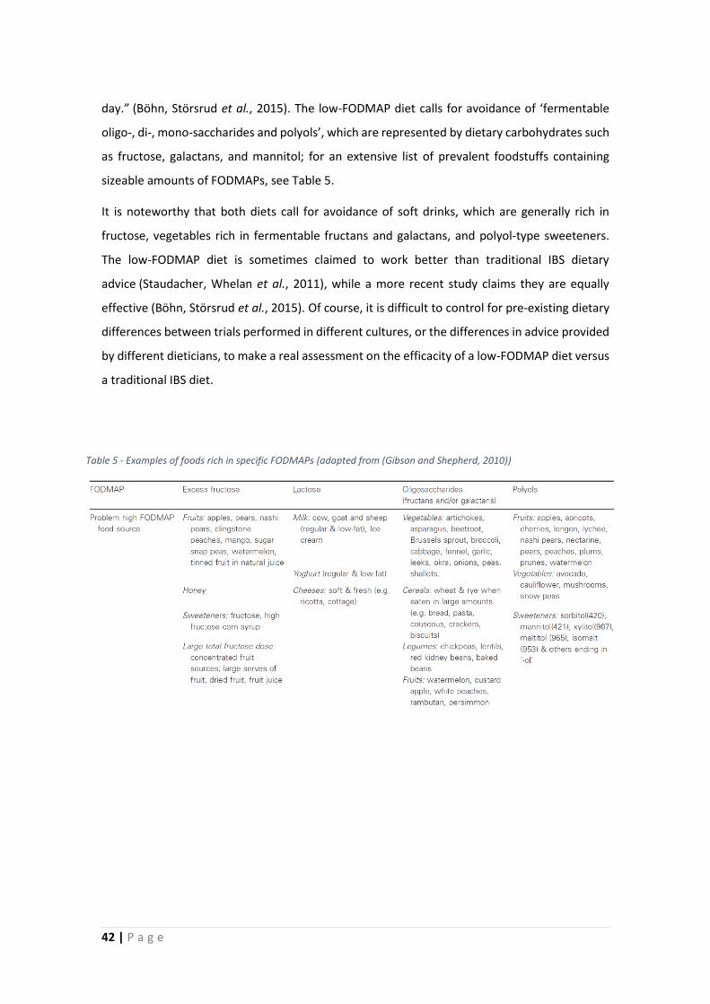
42 | P a g e
day.” (Böhn, Störsrud et al., 2015). The low-FODMAP diet calls for avoidance of ‘fermentable
oligo-, di-, mono-saccharides and polyols’, which are represented by dietary carbohydrates such
as fructose, galactans, and mannitol; for an extensive list of prevalent foodstuffs containing
sizeable amounts of FODMAPs, see Table 5.
It is noteworthy that both diets call for avoidance of soft drinks, which are generally rich in
fructose, vegetables rich in fermentable fructans and galactans, and polyol-type sweeteners.
The low-FODMAP diet is sometimes claimed to work better than traditional IBS dietary
advice (Staudacher, Whelan et al., 2011), while a more recent study claims they are equally
effective (Böhn, Störsrud et al., 2015). Of course, it is difficult to control for pre-existing dietary
differences between trials performed in different cultures, or the differences in advice provided
by different dieticians, to make a real assessment on the efficacity of a low-FODMAP diet versus
a traditional IBS diet.
Table 5 - Examples of foods rich in specific FODMAPs (adapted from (Gibson and Shepherd, 2010))
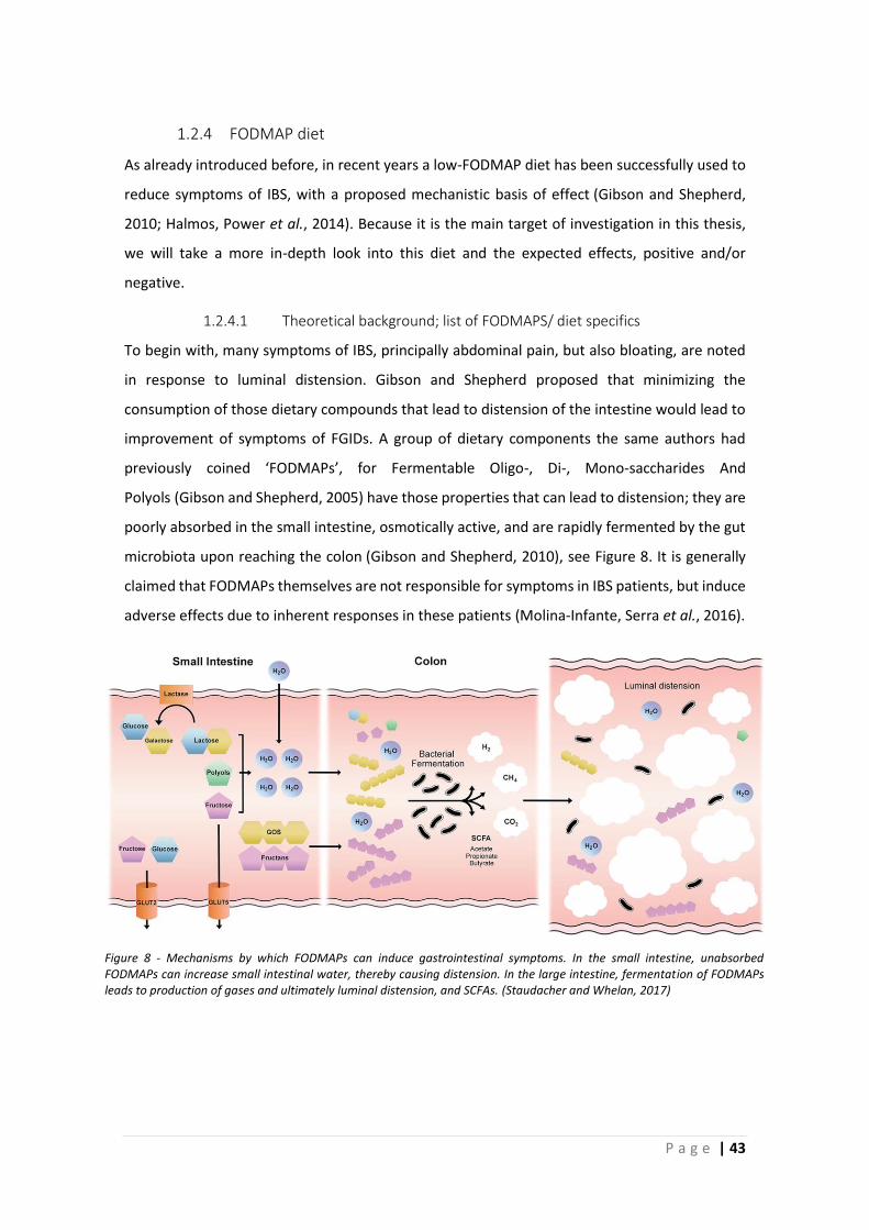
P a g e | 43
1.2.4 FODMAP diet
As already introduced before, in recent years a low-FODMAP diet has been successfully used to
reduce symptoms of IBS, with a proposed mechanistic basis of effect (Gibson and Shepherd,
2010; Halmos, Power et al., 2014). Because it is the main target of investigation in this thesis,
we will take a more in-depth look into this diet and the expected effects, positive and/or
negative.
1.2.4.1 Theoretical background; list of FODMAPS/ diet specifics
To begin with, many symptoms of IBS, principally abdominal pain, but also bloating, are noted
in response to luminal distension. Gibson and Shepherd proposed that minimizing the
consumption of those dietary compounds that lead to distension of the intestine would lead to
improvement of symptoms of FGIDs. A group of dietary components the same authors had
previously coined ‘FODMAPs’, for Fermentable Oligo-, Di-, Mono-saccharides And
Polyols (Gibson and Shepherd, 2005) have those properties that can lead to distension; they are
poorly absorbed in the small intestine, osmotically active, and are rapidly fermented by the gut
microbiota upon reaching the colon (Gibson and Shepherd, 2010), see Figure 8. It is generally
claimed that FODMAPs themselves are not responsible for symptoms in IBS patients, but induce
adverse effects due to inherent responses in these patients (Molina-Infante, Serra et al., 2016).
Figure 8 - Mechanisms by which FODMAPs can induce gastrointestinal symptoms. In the small intestine, unabsorbed FODMAPs can increase small intestinal water, thereby causing distension. In the large intestine, fermentation of FODMAPs leads to production of gases and ultimately luminal distension, and SCFAs. (Staudacher and Whelan, 2017)
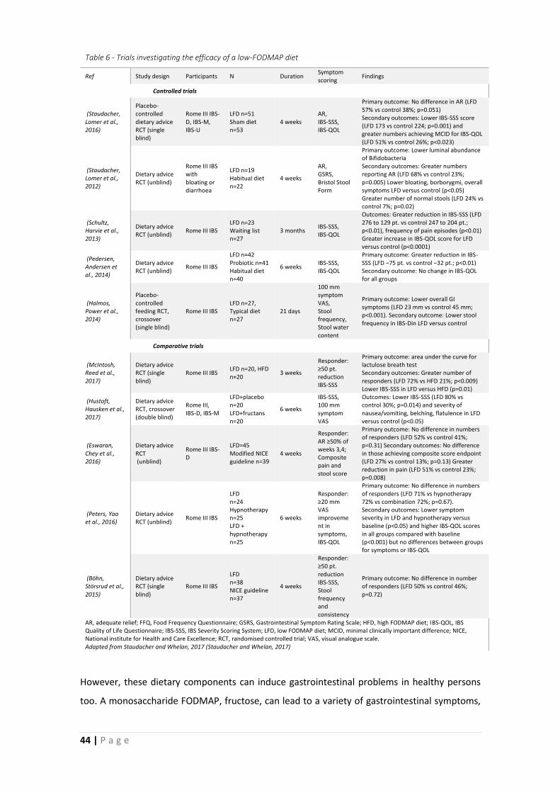
44 | P a g e
However, these dietary components can induce gastrointestinal problems in healthy persons
too. A monosaccharide FODMAP, fructose, can lead to a variety of gastrointestinal symptoms,
Table 6 - Trials investigating the efficacy of a low-FODMAP diet
Ref Study design Participants N Duration Symptom scoring
Findings
Controlled trials
(Staudacher, Lomer et al., 2016)
Placebo-controlled dietary advice RCT (single blind)
Rome III IBS-D, IBS-M, IBS-U
LFD n=51 Sham diet n=53
4 weeks AR, IBS-SSS, IBS-QOL
Primary outcome: No difference in AR (LFD 57% vs control 38%; p=0.051) Secondary outcomes: Lower IBS-SSS score (LFD 173 vs control 224; p=0.001) and greater numbers achieving MCID for IBS-QOL (LFD 51% vs control 26%; p<0.023)
(Staudacher, Lomer et al., 2012)
Dietary advice RCT (unblind)
Rome III IBS with bloating or diarrhoea
LFD n=19 Habitual diet n=22
4 weeks
AR, GSRS, Bristol Stool Form
Primary outcome: Lower luminal abundance of Bifidobacteria Secondary outcomes: Greater numbers reporting AR (LFD 68% vs control 23%; p=0.005) Lower bloating, borborygmi, overall symptoms LFD versus control (p<0.05) Greater number of normal stools (LFD 24% vs control 7%; p=0.02)
(Schultz, Harvie et al., 2013)
Dietary advice RCT (unblind)
Rome III IBS LFD n=23 Waiting list n=27
3 months IBS-SSS, IBS-QOL
Outcomes: Greater reduction in IBS-SSS (LFD 276 to 129 pt. vs control 247 to 204 pt.; p<0.01), frequency of pain episodes (p<0.01) Greater increase in IBS-QOL score for LFD versus control (p<0.0001)
(Pedersen, Andersen et al., 2014)
Dietary advice RCT (unblind)
Rome III IBS
LFD n=42 Probiotic n=41 Habitual diet n=40
6 weeks IBS-SSS, IBS-QOL
Primary outcome: Greater reduction in IBS-SSS (LFD −75 pt. vs control −32 pt.; p<0.01) Secondary outcome: No change in IBS-QOL for all groups
(Halmos, Power et al., 2014)
Placebo-controlled feeding RCT, crossover (single blind)
Rome III IBS LFD n=27, Typical diet n=27
21 days
100 mm symptom VAS, Stool frequency, Stool water content
Primary outcome: Lower overall GI symptoms (LFD 23 mm vs control 45 mm; p<0.001). Secondary outcome: Lower stool frequency in IBS-Din LFD versus control
Comparative trials
(McIntosh, Reed et al., 2017)
Dietary advice RCT (single blind)
Rome III IBS LFD n=20, HFD n=20
3 weeks
Responder: ≥50 pt. reduction IBS-SSS
Primary outcome: area under the curve for lactulose breath test Secondary outcomes: Greater number of responders (LFD 72% vs HFD 21%; p<0.009) Lower IBS-SSS in LFD versus HFD (p=0.01)
(Hustoft, Hausken et al., 2017)
Dietary advice RCT, crossover (double blind)
Rome III, IBS-D, IBS-M
LFD+placebo n=20 LFD+fructans n=20
6 weeks
IBS-SSS, 100 mm symptom VAS
Outcomes: Lower IBS-SSS (LFD 80% vs control 30%; p=0.014) and severity of nausea/vomiting, belching, flatulence in LFD versus control (p<0.05)
(Eswaran, Chey et al., 2016)
Dietary advice RCT (unblind)
Rome III IBS-D
LFD=45 Modified NICE guideline n=39
4 weeks
Responder: AR ≥50% of weeks 3,4; Composite pain and stool score
Primary outcome: No difference in numbers of responders (LFD 52% vs control 41%; p=0.31) Secondary outcomes: No difference in those achieving composite score endpoint (LFD 27% vs control 13%; p=0.13) Greater reduction in pain (LFD 51% vs control 23%; p=0.008)
(Peters, Yao et al., 2016)
Dietary advice RCT (unblind)
Rome III IBS
LFD n=24 Hypnotherapy n=25 LFD + hypnotherapy n=25
6 weeks
Responder: ≥20 mm VAS improvement in symptoms, IBS-QOL
Primary outcome: No difference in numbers of responders (LFD 71% vs hypnotherapy 72% vs combination 72%; p=0.67). Secondary outcomes: Lower symptom severity in LFD and hypnotherapy versus baseline (p<0.05) and higher IBS-QOL scores in all groups compared with baseline (p<0.001) but no differences between groups for symptoms or IBS-QOL
(Böhn, Störsrud et al., 2015)
Dietary advice RCT (single blind)
Rome III IBS
LFD n=38 NICE guideline n=37
4 weeks
Responder: ≥50 pt. reduction IBS-SSS, Stool frequency and consistency
Primary outcome: No difference in number of responders (LFD 50% vs control 46%; p=0.72)
AR, adequate relief; FFQ, Food Frequency Questionnaire; GSRS, Gastrointestinal Symptom Rating Scale; HFD, high FODMAP diet; IBS-QOL, IBS Quality of Life Questionnaire; IBS-SSS, IBS Severity Scoring System; LFD, low FODMAP diet; MCID, minimal clinically important difference; NICE, National institute for Health and Care Excellence; RCT, randomised controlled trial; VAS, visual analogue scale. Adapted from Staudacher and Whelan, 2017 (Staudacher and Whelan, 2017)
P a g e | 45
such as diarrhoea, cramps and flatulence (Ravich, Bayless et al., 1983; Major, Pritchard et al.,
2017), but fructose malabsorption is ameliorated when fructose is taken together with glucose,
due to the facilitating effect to small intestinal absorption of these two compounds together
which prevents symptom generation (Truswell, Seach et al., 1988). These days, fructose is
consumed at increasing doses as a result of the growing usage of high-fructose corn syrup in
many products, likely increasing the concentration reaching the colon for microbial processing.
In lactase non-persistent persons, the disaccharide FODMAP lactose reduces the oro-caecal
transit time, independently from the increased luminal distension of the small intestine induced
by the osmotically active lactose (He, Venema et al., 2008). Instead, it is claimed that colonic
fermentation of lactose in maldigesters could be partly responsible for symptoms of lactose
intolerance (He, Priebe et al., 2006). In relatively high doses, inulin, a fructo-oligosaccharide, can
lead to gastro-intestinal symptoms such as flatulence (Pedersen, Sandström et al., 1997; Major,
Pritchard et al., 2017). Sorbitol, a polyol FODMAP, too is known to induce gastrointestinal
symptoms at higher doses (>10 g) (Hyams, 1983). However, simultaneous malabsorption of
modest doses of multiple of these compounds is relatively rare (Ladas, Grammenos et al., 2000),
as measured by the occurrence of gastrointestinal symptoms, showing that generally,
reasonable doses are tolerated in healthy subjects. As mentioned before, the low-FODMAP diet
intends to reduce the intake of these compounds (see Table 5), to lessen intestinal distension,
and thus, lessen the provocation of symptoms in IBS patients. The diet has shown promising
results since it has been introduced, see Table 6.
Generally, these studies show that the low-FODMAP is effective in reducing IBS symptoms,
though it is not superior to NICE guidelines (traditional IBS diet) (Böhn, Störsrud et al., 2015), or
hypnotherapy (Peters, Yao et al., 2016). Although another study shows an increased effect on
abdominal pain, bloating, stool consistency, stool frequency, and urgency in IBS-D patients,
compared to modified NICE guidelines, but not on general adequate relief (AR) (Eswaran, Chey
et al., 2016). A meta-analysis from 2015 covering 6 RCT and 16 non-randomized interventions
supports the efficacy of low-FODMAP diet compared to non-treatment, with pooled odds ratios
for significant improvement of IBS-SSS questionnaire scores of 0.44 for RCTs and 0.03 for the
non-randomized trials (Marsh, Eslick et al., 2016). Generally, we can conclude that the low-
FODMAP diet delivers a significant improvement to the situation of IBS patients.
46 | P a g e
1.2.4.2 Possible adverse effects low-FODMAP diet
In most cases, the low-FODMAP approach calls for drastic changes to the day-to-day diet. Here,
we will look at the possible adverse effects of following a low-FODMAP diet.
First, since FODMAPs are fermentable, they have a prebiotic function, meaning that they provide
the intestinal microbiota with nutrients, and therefore support the bacterial populations in the
gut. Drastically reducing these compounds logically influences the composition and density of
the intestinal microbiota. Because of this, the main concern with the low-FODMAP approach are
unintended long-term consequences to the intestinal microbiota (Halmos, Christophersen et al.,
2015), as well as possible nutritional deficiencies due to the restrictive diet (Staudacher and
Whelan, 2017), although in a comparative study, only calcium intake was slightly impacted, due
to the reduced intake of lactose-containing dairy products (Staudacher, Lomer et al., 2012).
However, since a low-FODMAP diet is supported by a professional dietician, we can expect
nutritional sufficiency to be better safeguarded than in patients who try to adapt their diet by
themselves to prevent adverse events; which is common among IBS patients (Monsbakken,
Vandvik et al., 2006).
Concerning microbiota composition, several studies have been undertaken, showing a low-
FODMAP diet can be responsible for a reduction in Bifidobacteria (Staudacher, Lomer et al.,
2012; Halmos, Christophersen et al., 2015; McIntosh, Reed et al., 2017; Staudacher, Lomer et
al., 2017), F.prausnitzii, Clostridium cluster IV, and total bacterial numbers (Halmos,
Christophersen et al., 2015). However, it is still unclear whether there are health effects to these
changes and what these would be, though Bifidobacteria are generally considered to be
beneficial, and have been shown to already be reduced in untreated IBS patients (Malinen,
Rinttila et al., 2005; Kerckhoffs, Samsom et al., 2009; Rajilic-Stojanovic, Biagi et al., 2011). The
patients in these studies were not on specified diets, so it is possible that they have lower
Bifidobacteria counts due to evasion of prebiotic FODMAPs on their own initiative.
Apart from effects on the composition of the microbiota, a reduction in supposedly beneficial
bacterial metabolites, such as short-chain fatty acids (SCFA) is an additional possible concern.
P a g e | 47
Technical difficulties and conflicting results have left this consideration to be clarified for the
time being. Faecal SCFA concentrations have been shown to be actually higher in IBS patients
than in control patients, with total SCFA, propionic acid, and acetic acid significantly increased,
which was correlated with IBS symptoms (Tana, Umesaki et al., 2010), although the direction of
the possible causation is not clear. In animal experiments however, butyrate enemas can induce
a visceral hypersensitivity in rats mediated by CGRP receptor, offering a non-inflammatory
hypersensitivity model of IBS, according to the authors (Bourdu, Dapoigny et al., 2005).
Conversely, SCFA produced by bacteria play an important role in host energy
provision (Bergman, 1990), enterocyte energy provision in particular (Basson, Turowski et al.,
1996), and colonic Treg cell homeostasis (Smith, Howitt et al., 2013) among other important
functions. Consistent with the decreased intake of prebiotic material, faecal SCFA
concentrations are reduced in IBS-M and IBS-D patients on a low-FODMAP diet (Hustoft,
Hausken et al., 2017).
However, it has to be noted that faecal SCFA analysis is not a reliable method, because it reflects
colonic absorption better than production (Cook and Sellin, 1998; Sakata, 2018), and is heavily
influenced by transit time (Ringel-Kulka, Choi et al., 2015), which we know can be highly variable,
in particular between IBS patients with either diarrhoea or constipation, and healthy patients.
In short, without measuring SCFA concentrations where they are produced, mostly in the
ascending colon, it will not be possible to clearly identify the link between SCFA concentrations,
IBS symptoms, and possible effects of the low-FODMAP diet (Staudacher and Whelan, 2017), so
most of these results can be interpreted as a mix of artefacts of sampling method and less than
useful correlations.

P a g e | 49
1.3 Intestinal microbiota
The intestinal microbiota plays a vital role in vertebrate health. In this chapter we will look into
the many functions the gut microbiota has, but also how it can be co-responsible for some health
issues.
1.3.1 Function
Our gastrointestinal tract is populated by microbes whose numbers, complex population
composition, and ecology boggle the mind. They do not limit themselves to being impressive
though; they are important in digestion, immunity, and resistance to pathogens as well.
The gut microbiota is needed for the correct development of innate and adaptive immunity, the
integrity of the intestinal epithelium, provision of the host with both energy sources and
vitamins, prevention of colonization of our gastrointestinal tract by pathogenic microbes as
well (Dave, Higgins et al., 2012). Research into the microbiome has become more and more
translational in recent years, now driving investments coming from big pharma and other
private institutions to benefit from the many expected applications of knowledge to treat health
conditions with probiotics, as well as of identification of useful microbiota diagnostic
biomarkers (Gilbert, Blaser et al., 2018).
50 | P a g e
1.3.1.1 Symbiosis; never alone
The human gut microbiota has evolved together with us, and due to this long-shared history,
there is a high level of symbiosis. Vertebrates benefit greatly from their microbiota, although, as
shown in the possibility of rearing germ-free animals, it is not essential in the strict sense.
Normally, we are inoculated by gut microbes during natural birth (Mandar and Mikelsaar, 1996),
a process which does not take place during Caesarean section, with possible ramifications to
later health (Di Mauro, Neu et al., 2013; Jakobsson, Abrahamsson et al., 2014). A healthy
microbiota contains many different phylogenetic types; molecular analysis of stool samples of
124 subjects shows that there are over 1000 bacterial species associated to the human
microbiota, with at least 160 species per individual (Qin, Li et al., 2010). This large microbial
diversity contains many unique genes, which, by modest estimation, contains at least 9 million
unique genes (Yang, Xie et al., 2009) though this relates to the totality of known gut microbiota
species, which are not all present in every individual. Still, seeing as the human genome contains
a maximum of approximately 39 thousand genes (including hypothetical and unannotated
genes) (Venter, Adams et al., 2001), the bacterial genes associated to our gut microbiota are at
least 200 times more abundant than eukaryotic host genes. We have to keep in mind that most
of these genes regulate normal functioning of bacterial lifecycles and metabolism and that many
are homologous and/or analogous between different species and strains, indicating a
substantial redundancy. It is often claimed that there are 10 times more bacterial cells in the
intestines than there are animal cells in our bodies (Savage, 1977), a claim that is eagerly
repeated in many article introductions. However, this number is based on two very rough
estimates for host and microbial cell numbers. In 2016, using more informed calculations, it has
been estimated that the ratio is closer to 1:1,3, wherein bacteria still outnumber host
cells (Sender, Fuchs et al., 2016). They have not recalculated the number of host cells, which
might still change this ratio.
What drives the relation between host and microbe is a common interest in metabolism as a
requisite for life. The host provides the microbiota with nutrients by its animal mobility and
motivations, as well as a highly beneficial anaerobic, warm, and humid environment represented
by the gastrointestinal tract. Our gut and its microbial ecology are so suitable for the guest
organisms that we can cultivate the majority of gut microbiota exclusively inside of ourselves.
However, considerable advancements have recently been made on the cultivability of gut
bacteria, based on the extensive sporulation taking place. This sporulation and the subsequent
P a g e | 51
reactivation under suitable conditions is key to the colonisation of and transit through
inhospitable environments (Browne, Forster et al., 2016).
A good host always makes sure that their guests are well-fed, even at a possible cost to
themselves. A very nice example of this is reported in a study from 2014, where it is shown that
animals suffering from an infection and going through a temporary anorexia to deal with this
infection will increase fucosylation of the intestinal epithelium to support the commensal
bacteria, which would otherwise have no energy provision (Pickard, Maurice et al., 2014). This
underlines that the host is a pro-active actor in their relationship with the gut microbiota.
Genetics and co-evolution play a role in host-microbe interactions, already visible in the
exclusivity of bacterial capabilities to colonize mammals, indicating the necessity of a certain
suitable set of traits. Within our species, genetical differences between people can influence
which microbes feel at home, though this effect is limited; there is a small but significant increase
in similarity in microbiota composition between homozygotic twins compared to dizygotic
twins (Goodrich, Waters et al., 2014).
52 | P a g e
1.3.1.2 Metabolism; collecting the rent
Of course, a host does not always accept guests without expecting something in return. The
microbiota pays us back by indispensable metabolic processes, digesting components which are
indigestible to us, producing simpler molecules that we can use. Additionally, the gut microbiota
is an important source of vitamins, such as riboflavin (vitamin B2), biotin (vitamin B7), folic acid
(vitamin B9), cobalamin (vitamin B12), and vitamin K (Hill, 1997). In herbivore animals, cellulose
and other cell wall components are processed by microbes to provide otherwise unavailable
energy, particularly important in ruminants (Flint, 1997), but also in omnivores such as humans,
the microbiota plays an important role in breaking down dietary carbohydrates (Cockburn and
Koropatkin, 2016). Interestingly, the mucus produced by the host supports a metabolic network
based on mucolytic activities of bacteria such as Akkermansia muciniphila that produces
butyrate and vitamin B12 as well (Belzer, Chia et al., 2017). Butyrate produced by the microbiota
is an important source of energy especially for colonocytes, who forego glucose from the blood
in favour of butyrate produced by the microbiota (Roediger, 1980), other short-chain fatty acids
produced are also absorbed and transported to be metabolised by rest of the body (den Besten,
van Eunen et al., 2013). It is estimated that in humans, 6 to 10 per cent of energy requirements
are provided by microbiota-derived SCFAs, based on the intake of a standard British
diet (McNeil, 1984; Bergman, 1990), the authors point out however, that fermentable
carbohydrate consumption differs greatly around the world, and therefore these energy
contributions must vary substantially as well (Bergman, 1990).
The metabolites produced during microbial processing of intestinal contents are not only utilised
by the host, but a complex ecology exists between different microbes as well. Many species are
dependent on other species with complementary activities, and in this way, a complex metabolic
network forms with mutualistic, amensalistic, or parasitic relationships between different
species, where mutualism in the gut is particularly increased under anoxic conditions (Heinken
and Thiele, 2015). Depending on the variety of resources they can utilize, species can be
classified as either generalists or specialists. While we would normally expect generalists to be
widely distributed and specialists to occur mostly in smaller niches (Cockburn and Koropatkin,
2016), in the gut, this does not necessarily hold true (Carbonero, Oakley et al., 2014), with
specialists often being more widely distributed than generalist species.
P a g e | 53
1.3.1.3 House rules; reciprocal control
In the relation between the host and the intestinal microbiome, both actors influence each other
and exert a modicum of control. Under healthy conditions, the host enforces sterility where it is
necessary, and supports the microbiota where desired. The microbiota in its turn, communicates
with the host to support immune tolerance and optimal conditions to its prosperity.
The microbiota, through their gene expression and products, but also metabolites such as SCFAs,
influences the host immune cells and cytokine production (Geuking, Koller et al., 2014), while it
itself is subject to change by host-produced antimicrobial peptides and IgA secretions, which
have regulatory functions, which can be, but are not always, microbicidal (Neish, 2009). The
innate and adaptive immunity both play a significant role in monitoring and regulating the
microbiota. For the innate immune system, some of these are produced independently from
circumstances, such as α- and β-defensins (Pütsep, Axelsson et al., 2000), and Crohn’s disease
patients exhibit a defect in α-defensin production by Paneth cells, underlining its important
function (Wehkamp, Salzman et al., 2005),the abundance of other secretions, such as C-type-
lectin Reg3γ, is regulated by the activity of pattern recognition receptors (PRRs) (Gong, Xu et al.,
2010), important regulators of innate immunity. Reg3γ is poorly expressed in germ-free mice,
and introduction of a mixed mouse microbiota increases this expression 20-fold, related to the
ability of the bacteria to reach the epithelium (Cash, Whitham et al., 2006), indicating the
involvement of epithelial sensing, most likely through PRRs. Many of these PRRs belong to the
toll-like receptor family (TLRs), but Nod1 and Nod2, as well as several other types of receptors
are important PRRs too. Many PRRs are activated by microbe-associated molecular patterns
(MAMPs), which are represented among others by lipopeptides, lipoproteins, flagellins, and
lipopolysaccharides; i.e., molecules that indicate the presence of microbes (Medzhitov and
Janeway, 2002). Upon recognition of such a molecule, the PRR is activated and, depending on
which PRR it is, activates regulatory pathways such as the mitogen-activated protein kinase
(MAPK), nuclear factor B (NF-B)/Rel pathways, or the inflammasome (Neish, 2009). PRRs can
be present on the cell membrane, on endosomes, or in the cytoplasm (Neish, 2009). In most
tissues, PRRs activate inflammatory pathways, but because of the high microbial presence in the
gut, PRRs there are also used to retain tolerance. For example, while activation of basolaterally
positioned TLR9 leads to NF-B activation by degradation of IBα, apical activation leads to
stabilisation of IBα and p105, preventing activation of the NF-B pathway (Lee, Mo et al.,
2006). TLR9-/- mice have a lower NF-B activation threshold, and consequently are more
susceptible to experimental colitis induced by DSS (Lee, Mo et al., 2006), further indicating the
54 | P a g e
vital role of apical PRRs in maintaining immune tolerance in the gut. Similarly, flagellin-sensitive
TLR5 is located only basolaterally to be activated when bacteria are present on the ‘wrong’ side
of the epithelium, inducing inflammatory processes upon activation by bacteria penetrating the
epithelium (Gewirtz, Navas et al., 2001).
On the side of the adaptive immune system, there is an impressive production of IgA-type
antibodies, which are secreted into the lumen. Production of bacterial antigen specific IgAs is
induced upon colonisation of the gut, and the variety of specificities of these antibodies is
related to the composition of the microbiota (Hapfelmeier, Lawson et al., 2010). In mice, a
deleterious mutation in AID, leading to defective somatic hypermutation and thus loss of
affinity-maturated antibodies, the composition of the microbiota is altered, indicating the
capacity of specific antibody production to shape microbiota composition (Wei, Shinkura et al.,
2011). In animals lacking both immunoglobulin isotype switching and somatic hypermutation,
leading to the lack of specific antibody production and the absence of IgA isotypes, overgrowth
of anaerobes in the small intestine takes place (Fagarasan, Muramatsu et al., 2002), indicating a
role for this IgA production in maintaining a favourable symbiotic relation with the gut
microbiota. To regulate the response to the intestinal microbiota, the immune system uses a
variety of cell types, informed by the intestinal epithelial cells, such as dendritic cells (DCs), intra-
epithelial lymphocytes (IELs), and macrophages. Based on epithelial cell signals in the form of
retinoic acid, thymic stromal lymphopoietin (TLSP), and TGFβ, tolerogenic dendritic cells (DCs)
and macrophages are generated. Migrating CD103+ DCs present microbial antigens to the
adaptive immune system in a context of tolerance, while intestine-resident macrophages stay
in close contact with epithelial cells to rapidly clear any microbes that pass the intestinal
epithelial barrier (Peterson and Artis, 2014) (see Figure 9).
Microbial metabolites, for their part, regulate host immune responses as well. For example,
microbiota-produced SCFAs play a crucial role in regulation and proper resolution of the
inflammatory response through their interaction with G-protein-coupled receptor 43 (GPR43),
evidenced by an exacerbated DSS-induced colitis in GPR43-deficient mice, and in germ-free
mice (Maslowski, Vieira et al., 2009). Similarly, the increase in SCFAs (e.g., butyrate) connected
to increased dietary fibre intake can help reduce inflammation in ulcerative colitis (UC) (Hallert,
Bjorck et al., 2003), based on the notion that application of butyrate can reduce inflammation
in UC patients (Scheppach, Sommer et al., 1992).
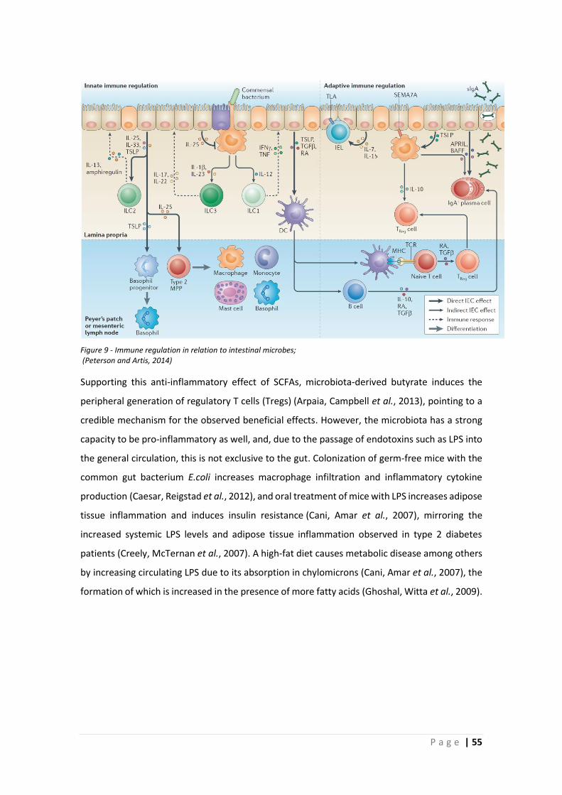
P a g e | 55
Supporting this anti-inflammatory effect of SCFAs, microbiota-derived butyrate induces the
peripheral generation of regulatory T cells (Tregs) (Arpaia, Campbell et al., 2013), pointing to a
credible mechanism for the observed beneficial effects. However, the microbiota has a strong
capacity to be pro-inflammatory as well, and, due to the passage of endotoxins such as LPS into
the general circulation, this is not exclusive to the gut. Colonization of germ-free mice with the
common gut bacterium E.coli increases macrophage infiltration and inflammatory cytokine
production (Caesar, Reigstad et al., 2012), and oral treatment of mice with LPS increases adipose
tissue inflammation and induces insulin resistance (Cani, Amar et al., 2007), mirroring the
increased systemic LPS levels and adipose tissue inflammation observed in type 2 diabetes
patients (Creely, McTernan et al., 2007). A high-fat diet causes metabolic disease among others
by increasing circulating LPS due to its absorption in chylomicrons (Cani, Amar et al., 2007), the
formation of which is increased in the presence of more fatty acids (Ghoshal, Witta et al., 2009).
Figure 9 - Immune regulation in relation to intestinal microbes; (Peterson and Artis, 2014)
56 | P a g e
1.3.2 Toxic metabolite hypothesis
As written in previous chapters, the gut microbiota produces many useful metabolites. However,
it also has the capacity to make products with negative effects on gut function and general
health. For example, bacteria-produced proteases can induce inflammation and break down the
mucus barrier (Steck, Mueller et al., 2012), and production of methane by bacteria such as
Methanobrevibacter smithii increases gut transit time, which can lead to constipation (Bennet,
Öhman et al., 2015).
Some particularly toxic products can be generated during anaerobic fermentation of
unabsorbed carbohydrates; methylglyoxal and other reactive aldehydes and ketones (Campbell,
Matthews et al., 2010). Campbell et al. hold these metabolites responsible for a wide range of
local and systemic effects observed in lactose malabsorption, such as headache, fatigue, heart
palpitations, and depression. The mechanisms by which methylglyoxal can induce health
complications are elucidated more deeply, which will be further discussed in the coming section
1.4. Methylglyoxal, or pyruvaldehyde, affects both prokaryote cells, e.g., impacting growth rates
and thus changing the microbiota, and eukaryotic cells, e.g., changing cell proliferation, gene
expression patterns and cell signalling processes (Campbell, Matthews et al., 2010).
Fermentation products of non-absorbed carbohydrates such as lactose can induce symptoms
reminiscent of IBS, both in terms of gut symptoms, and more systemic symptoms, but other
metabolites, such as those produced by putrefaction of proteins and amino acids are often toxic
as well (Campbell, Waud et al., 2005). Another way by which bacterial fermentation can impact
gut health is by changes in pH. In IBS patients, colonic intraluminal pH is decreased compared to
healthy controls, as measured using a pH-measuring ingested SmartPill system, which indicates
an increased fermentation (Ringel-Kulka, Choi et al., 2015).
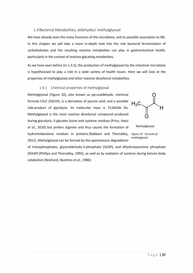
P a g e | 57
1.4 Bacterial Metabolites; aldehydes/ methylglyoxal
We have already seen the many functions of the microbiota, and its possible association to IBS.
In this chapter we will take a more in-depth look into the role bacterial fermentation of
carbohydrates and the resulting reactive metabolites can play in gastrointestinal health,
particularly in the context of reactive glycating metabolites.
As we have seen before (in 1.3.2), the production of methylglyoxal by the intestinal microbiota
is hypothesised to play a role in a wide variety of health issues. Here we will look at the
properties of methylglyoxal and other reactive dicarbonyl metabolites.
1.4.1 Chemical properties of methylglyoxal
Methylglyoxal (Figure 10), also known as pyruvaldehyde, chemical
formula CH3C (O)CHO, is a derivative of pyruvic acid, and a possible
side-product of glycolysis. Its molecular mass is 72,06266 Da.
Methylglyoxal is the most reactive dicarbonyl compound produced
during glycolysis, it glycates lysine and cysteine residues (Price, Hassi
et al., 2010) but prefers arginine and thus causes the formation of
hydroimidazolone residues in proteins (Rabbani and Thornalley,
2012). Methylglyoxal can be formed by the spontaneous degradation
of triosephosphates, glyceraldehyde-3-phosphate (GA3P), and dihydroxyacetone phosphate
(DHAP) (Phillips and Thornalley, 1993), as well as by oxidation of acetone during ketone body
catabolism (Reichard, Skutches et al., 1986).
Methylglyoxal
Figure 10 - Formula of methylglyoxal
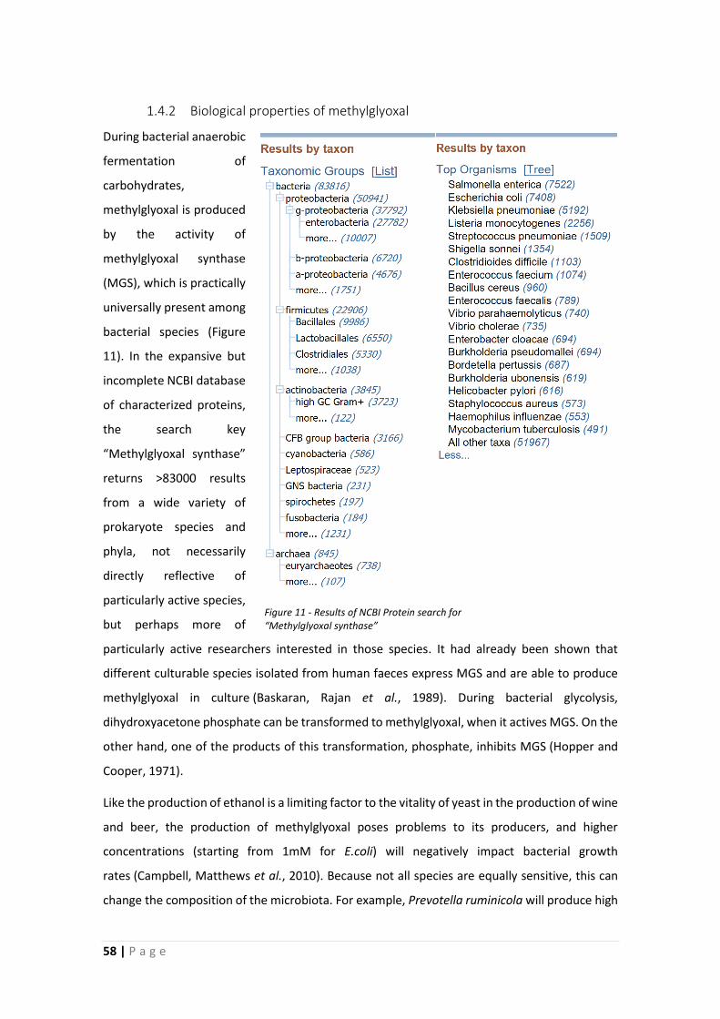
58 | P a g e
1.4.2 Biological properties of methylglyoxal
During bacterial anaerobic
fermentation of
carbohydrates,
methylglyoxal is produced
by the activity of
methylglyoxal synthase
(MGS), which is practically
universally present among
bacterial species (Figure
11). In the expansive but
incomplete NCBI database
of characterized proteins,
the search key
“Methylglyoxal synthase”
returns >83000 results
from a wide variety of
prokaryote species and
phyla, not necessarily
directly reflective of
particularly active species,
but perhaps more of
particularly active researchers interested in those species. It had already been shown that
different culturable species isolated from human faeces express MGS and are able to produce
methylglyoxal in culture (Baskaran, Rajan et al., 1989). During bacterial glycolysis,
dihydroxyacetone phosphate can be transformed to methylglyoxal, when it actives MGS. On the
other hand, one of the products of this transformation, phosphate, inhibits MGS (Hopper and
Cooper, 1971).
Like the production of ethanol is a limiting factor to the vitality of yeast in the production of wine
and beer, the production of methylglyoxal poses problems to its producers, and higher
concentrations (starting from 1mM for E.coli) will negatively impact bacterial growth
rates (Campbell, Matthews et al., 2010). Because not all species are equally sensitive, this can
change the composition of the microbiota. For example, Prevotella ruminicola will produce high
Figure 11 - Results of NCBI Protein search for “Methylglyoxal synthase”
P a g e | 59
concentrations (3-4 mM) of methylglyoxal when offered an excess of glucose, which will kill the
culture, but different strains of this species were not equal producers (Russell, 1993).
Additionally, the activity of the glyoxalase detoxification system in bacteria is predictive of their
ability to cope with methylglyoxal, and glyoxalase knockout mutants become very sensitive to
even modest concentrations (Ferguson, Totemeyer et al., 1998). It seems that for bacteria, the
production of methylglyoxal serves to balance phosphate metabolism, and a MGS knockout
mutant E.coli reaches growth inhibition more rapidly due to accumulation of toxic
phosphorylated metabolites (Totemeyer, Booth et al., 1998).
Similarly, growing E.coli in phosphate-limited medium elevates methylglyoxal levels, which can
be increased by overexpressing MGS (Booth, Ferguson et al., 2003), again indicating an intended
increase in phosphate turnover. An important observation is that abundant methylglyoxal
production is expected in case of a sudden influx of carbon sources, causing a loss of control
over carbon entry in the bacterial cells (Totemeyer, Booth et al., 1998) which is indeed the
situation caused by the ingestion of significant amounts of unabsorbable carbohydrates,
prevented by a low-FODMAP diet.
In diabetic humans, the methylglyoxal levels in the blood are increased, which is unlikely to be
directly related to activity of the microbiota. The generally accepted hypothesis explaining the
increased MG levels in diabetic patients points to hyperglycaemia, leading to an increased
production of methylglyoxal in blood cells (Kalapos, 2013). Even brief periods of hyperglycaemia
can significantly increase MG levels (Beisswenger, Howell et al., 2001), leading to accumulation
of dicarbonyl metabolites and the modification of DNA and proteins (Rabbani, Xue et al., 2016a),
which will be elaborated upon in the next section.
Apart from methylglyoxal, glyoxal and 3-deoxyglucosone (3-DG) are important reactive
dicarbonyl compounds as well, with similar effects and detoxification processes (Hellwig,
Gensberger-Reigl et al., 2018).
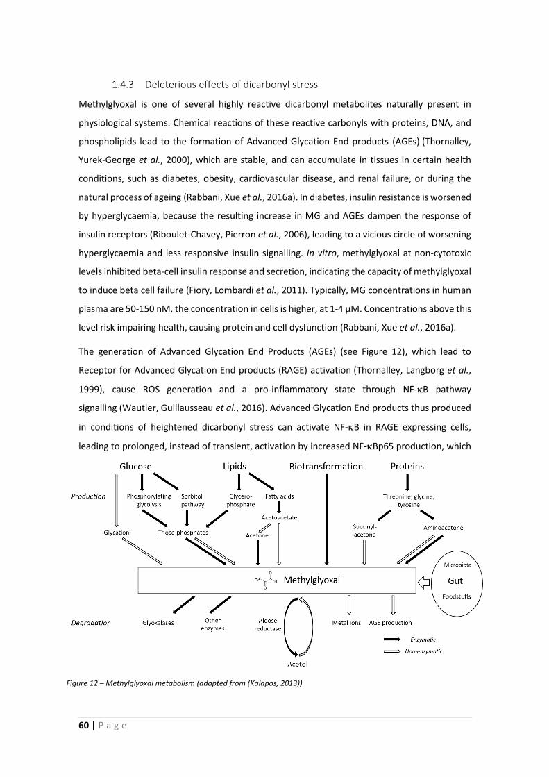
60 | P a g e
1.4.3 Deleterious effects of dicarbonyl stress
Methylglyoxal is one of several highly reactive dicarbonyl metabolites naturally present in
physiological systems. Chemical reactions of these reactive carbonyls with proteins, DNA, and
phospholipids lead to the formation of Advanced Glycation End products (AGEs) (Thornalley,
Yurek-George et al., 2000), which are stable, and can accumulate in tissues in certain health
conditions, such as diabetes, obesity, cardiovascular disease, and renal failure, or during the
natural process of ageing (Rabbani, Xue et al., 2016a). In diabetes, insulin resistance is worsened
by hyperglycaemia, because the resulting increase in MG and AGEs dampen the response of
insulin receptors (Riboulet-Chavey, Pierron et al., 2006), leading to a vicious circle of worsening
hyperglycaemia and less responsive insulin signalling. In vitro, methylglyoxal at non-cytotoxic
levels inhibited beta-cell insulin response and secretion, indicating the capacity of methylglyoxal
to induce beta cell failure (Fiory, Lombardi et al., 2011). Typically, MG concentrations in human
plasma are 50-150 nM, the concentration in cells is higher, at 1-4 µM. Concentrations above this
level risk impairing health, causing protein and cell dysfunction (Rabbani, Xue et al., 2016a).
The generation of Advanced Glycation End Products (AGEs) (see Figure 12), which lead to
Receptor for Advanced Glycation End products (RAGE) activation (Thornalley, Langborg et al.,
1999), cause ROS generation and a pro-inflammatory state through NF-B pathway
signalling (Wautier, Guillausseau et al., 2016). Advanced Glycation End products thus produced
in conditions of heightened dicarbonyl stress can activate NF-B in RAGE expressing cells,
leading to prolonged, instead of transient, activation by increased NF-Bp65 production, which
Figure 12 – Methylglyoxal metabolism (adapted from (Kalapos, 2013))
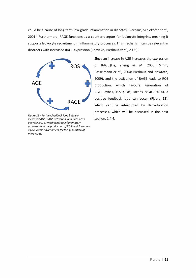
P a g e | 61
could be a cause of long-term low-grade inflammation in diabetes (Bierhaus, Schiekofer et al.,
2001). Furthermore, RAGE functions as a counterreceptor for leukocyte integrins, meaning it
supports leukocyte recruitment in inflammatory processes. This mechanism can be relevant in
disorders with increased RAGE expression (Chavakis, Bierhaus et al., 2003).
Since an increase in AGE increases the expression
of RAGE (He, Zheng et al., 2000; Simm,
Casselmann et al., 2004; Bierhaus and Nawroth,
2009), and the activation of RAGE leads to ROS
production, which favours generation of
AGE (Baynes, 1991; Ott, Jacobs et al., 2014), a
positive feedback loop can occur (Figure 13),
which can be interrupted by detoxification
processes, which will be discussed in the next
section, 1.4.4.
Figure 13 - Positive feedback loop between increased AGE, RAGE activation, and ROS. AGEs activate RAGE, which leads to inflammatory processes and the production of ROS, which creates a favourable environment for the generation of more AGEs.
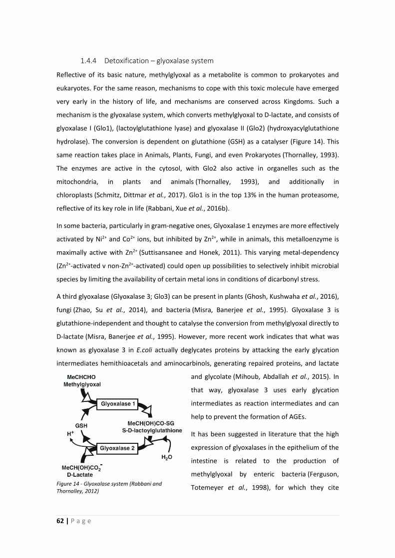
62 | P a g e
1.4.4 Detoxification – glyoxalase system
Reflective of its basic nature, methylglyoxal as a metabolite is common to prokaryotes and
eukaryotes. For the same reason, mechanisms to cope with this toxic molecule have emerged
very early in the history of life, and mechanisms are conserved across Kingdoms. Such a
mechanism is the glyoxalase system, which converts methylglyoxal to D-lactate, and consists of
glyoxalase I (Glo1), (lactoylglutathione lyase) and glyoxalase II (Glo2) (hydroxyacylglutathione
hydrolase). The conversion is dependent on glutathione (GSH) as a catalyser (Figure 14). This
same reaction takes place in Animals, Plants, Fungi, and even Prokaryotes (Thornalley, 1993).
The enzymes are active in the cytosol, with Glo2 also active in organelles such as the
mitochondria, in plants and animals (Thornalley, 1993), and additionally in
chloroplasts (Schmitz, Dittmar et al., 2017). Glo1 is in the top 13% in the human proteasome,
reflective of its key role in life (Rabbani, Xue et al., 2016b).
In some bacteria, particularly in gram-negative ones, Glyoxalase 1 enzymes are more effectively
activated by Ni2+ and Co2+ ions, but inhibited by Zn2+, while in animals, this metalloenzyme is
maximally active with Zn2+ (Suttisansanee and Honek, 2011). This varying metal-dependency
(Zn2+-activated v non-Zn2+-activated) could open up possibilities to selectively inhibit microbial
species by limiting the availability of certain metal ions in conditions of dicarbonyl stress.
A third glyoxalase (Glyoxalase 3; Glo3) can be present in plants (Ghosh, Kushwaha et al., 2016),
fungi (Zhao, Su et al., 2014), and bacteria (Misra, Banerjee et al., 1995). Glyoxalase 3 is
glutathione-independent and thought to catalyse the conversion from methylglyoxal directly to
D-lactate (Misra, Banerjee et al., 1995). However, more recent work indicates that what was
known as glyoxalase 3 in E.coli actually deglycates proteins by attacking the early glycation
intermediates hemithioacetals and aminocarbinols, generating repaired proteins, and lactate
and glycolate (Mihoub, Abdallah et al., 2015). In
that way, glyoxalase 3 uses early glycation
intermediates as reaction intermediates and can
help to prevent the formation of AGEs.
It has been suggested in literature that the high
expression of glyoxalases in the epithelium of the
intestine is related to the production of
methylglyoxal by enteric bacteria (Ferguson,
Totemeyer et al., 1998), for which they cite Figure 14 - Glyoxalase system (Rabbani and Thornalley, 2012)
P a g e | 63
Baskaran et al. (Baskaran, Rajan et al., 1989) who demonstrated the presence of Glyoxalase 1 in
simian intestine (Baskaran and Balasubramanian, 1987). Their claim that this expression may
protect against methylglyoxal produced by enterobacteria is based on an article that shows
glyoxalase expression in erythrocytes and liver cells (Aronsson and Mannervik, 1977).
While the hypothesis has merit, this train of inferences skips several logical steps, as none of the
sources show a particularly high glyoxalase expression in the intestine which would support it,
and findings have been mischaracterised from one publication to the next. Comparative
expression levels of glyoxalase throughout animal tissues are hard to come by, but one study on
bovines show the highest level of glyoxalase 1 expressed in the liver, though regrettably they
did not analyse intestinal tissues (Hayes, Milner et al., 1989). However, based on the general
understanding that glyoxalase is ubiquitously expressed, and the microbiota is capable of
producing methylglyoxal, intestinal detoxification of this chemical would indeed be necessary to
prevent health complications.
In the brain, where glucose is the major energy substrate, and detoxification of by-products of
glycolysis is important, it can equally be expected that the glyoxalases are highly expressed.
Indeed, the glyoxalase system seems to be important in protecting the brain, with expression in
the brain increasing until 55 years of age as levels of dicarbonyl stress rise with age, after which
expression levels drop and neurodegenerative processes associated with ageing show a
significant increase (Kuhla, Boeck et al., 2006), and AGEs accumulate (Luth, Ogunlade et al.,
2005). There is a significantly higher expression of Glo1 in astrocytes compared to neurons (9.8
times higher) (Belanger, Yang et al., 2011), which corresponds to their higher rate of glycolysis,
after which they supply derived energy substrates to the neurons, which have a higher energy
expenditure (Allaman, Belanger et al., 2015). This mechanism might be effective in shielding
neurons, which are less replaceable, from glycation damage.
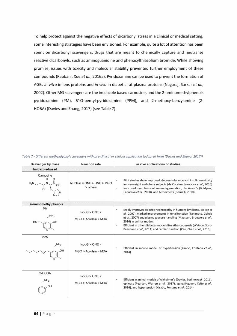
64 | P a g e
To help protect against the negative effects of dicarbonyl stress in a clinical or medical setting,
some interesting strategies have been envisioned. For example, quite a lot of attention has been
spent on dicarbonyl scavengers, drugs that are meant to chemically capture and neutralise
reactive dicarbonyls, such as aminoguanidine and phenacylthiazolium bromide. While showing
promise, issues with toxicity and molecular stability prevented further employment of these
compounds (Rabbani, Xue et al., 2016a). Pyridoxamine can be used to prevent the formation of
AGEs in vitro in lens proteins and in vivo in diabetic rat plasma proteins (Nagaraj, Sarkar et al.,
2002). Other MG scavengers are the imidazole based carnosine, and the 2-aminomethylphenols
pyridoxamine (PM), 5′-O-pentyl-pyridoxamine (PPM), and 2-methoxy-benzylamine (2-
HOBA) (Davies and Zhang, 2017) (see Table 7).
• Pilot studies show improved glucose tolerance and insulin sensitivity
in overweight and obese subjects (de Courten, Jakubova et al., 2016) • Improved symptoms of neurodegeneration; Parkinson’s (Boldyrev,
Fedorova et al., 2008), and Alzheimer’s (Cornelli, 2010)
• Mildly improves diabetic nephropathy in humans (Williams, Bolton et al., 2007), marked improvements in renal function (Tanimoto, Gohda et al., 2007) and plasma glucose handling (Maessen, Brouwers et al., 2016) in animal models
• Efficient in other diabetes models like atherosclerosis (Watson, Soro-Paavonen et al., 2011) and cardiac function (Cao, Chen et al., 2015)
• Efficient in mouse model of hypertension (Kirabo, Fontana et al., 2014)
• Efficient in animal models of Alzheimer’s (Davies, Bodine et al., 2011),
epilepsy (Pearson, Warren et al., 2017), aging (Nguyen, Caito et al., 2016), and hypertension (Kirabo, Fontana et al., 2014)
Table 7 - Different methylglyoxal scavengers with pre-clinical or clinical application (adapted from (Davies and Zhang, 2017))
P a g e | 65
Another suitable strategy to alleviate dicarbonyl stress in patients is by increasing Glo1
expression, and a synergetic effect of 2 clinically applicable substances (trans-resveratrol (tRES)
and hesperetin (HESP)) have been shown to induce this effect. A treatment with these 2
compounds could increase expression of Glo1 by 27%, thus reducing total body methylglyoxal-
protein glycation by 14%, in highly overweight subjects (Xue, Weickert et al., 2016). In metabolic
syndrome and diabetes, such a treatment could help prevent complications, but it is possibly
less optimal for situations which present more topical dicarbonyl stress.
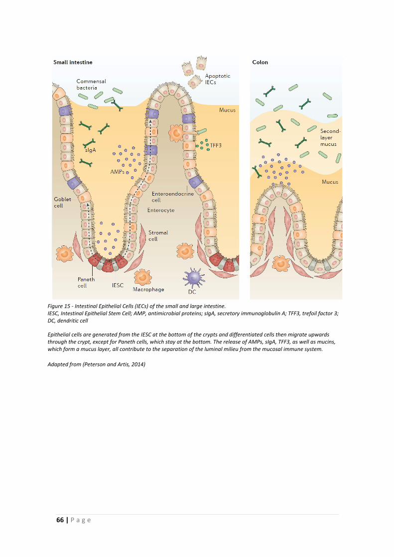
66 | P a g e
Figure 15 - Intestinal Epithelial Cells (IECs) of the small and large intestine. IESC, Intestinal Epithelial Stem Cell; AMP, antimicrobial proteins; sIgA, secretory immunoglobulin A; TFF3, trefoil factor 3; DC, dendritic cell
Epithelial cells are generated from the IESC at the bottom of the crypts and differentiated cells then migrate upwards through the crypt, except for Paneth cells, which stay at the bottom. The release of AMPs, sIgA, TFF3, as well as mucins, which form a mucus layer, all contribute to the separation of the luminal milieu from the mucosal immune system.
Adapted from (Peterson and Artis, 2014)
P a g e | 67
1.5 Intestinal barrier function
We have seen that diet together with the intestinal microbiota can generate problems, which
can impact the crucial barrier that separates the self from the environment. In this chapter, we
will look at the functioning of the intestinal barrier, and some pathophysiologically relevant
complications of its disturbance.
1.5.1 Epithelial barrier
When we think of our intestines, we usually picture it as part of our inside. However, the
gastrointestinal lumen is part of the external environment, with our bodies forming a continuous
tube. To keep external and internal separated, the mucosal lining of our gastrointestinal tract
has an important barrier function. The first cellular components in contact with the exterior are
the epithelial cells, which form the luminal part of the mucosal lining of the gastrointestinal tract.
Underneath the epithelium are located the lamina propria, and the muscularis mucosa. The
epithelial cells are tightly connected by so-called tight and adherens junctions which regulate
passage in between cells. The cells themselves are impermeable to most hydrophilic
components by virtue of their cell plasma membranes, unless there is a specific transporter
protein (-complex) present. Epithelial cells in the small intestine consist of absorptive
enterocytes, goblet cells, enteroendocrine cells, and Paneth cells. In the large intestine, there
are enterocytes, enteroendocrine cells, and goblet cells (see Figure 15). These components
together form a dynamic barrier that protects the organism from harmful inflammatory stimuli,
while at the same time allowing digestion and absorption of nutrients.
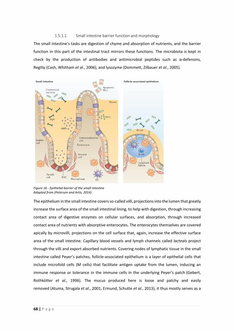
68 | P a g e
1.5.1.1 Small intestine barrier function and morphology
The small intestine’s tasks are digestion of chyme and absorption of nutrients, and the barrier
function in this part of the intestinal tract mirrors these functions. The microbiota is kept in
check by the production of antibodies and antimicrobial peptides such as α-defensins,
RegIIIγ (Cash, Whitham et al., 2006), and lysozyme (Dommett, Zilbauer et al., 2005).
The epithelium in the small intestine covers so-called villi, projections into the lumen that greatly
increase the surface area of the small intestinal lining, to help with digestion, through increasing
contact area of digestive enzymes on cellular surfaces, and absorption, through increased
contact area of nutrients with absorptive enterocytes. The enterocytes themselves are covered
apically by microvilli, projections on the cell surface that, again, increase the effective surface
area of the small intestine. Capillary blood vessels and lymph channels called lacteals project
through the villi and export absorbed nutrients. Covering nodes of lymphatic tissue in the small
intestine called Peyer’s patches, follicle-associated epithelium is a layer of epithelial cells that
include microfold cells (M cells) that facilitate antigen uptake from the lumen, inducing an
immune response or tolerance in the immune cells in the underlying Peyer’s patch (Gebert,
Rothkötter et al., 1996). The mucus produced here is loose and patchy and easily
removed (Atuma, Strugala et al., 2001; Ermund, Schutte et al., 2013), it thus mostly serves as a
Figure 16 - Epithelial barrier of the small intestine Adapted from (Peterson and Artis, 2014)
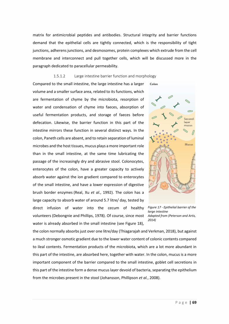
P a g e | 69
matrix for antimicrobial peptides and antibodies. Structural integrity and barrier functions
demand that the epithelial cells are tightly connected, which is the responsibility of tight
junctions, adherens junctions, and desmosomes, protein complexes which extrude from the cell
membrane and interconnect and pull together cells, which will be discussed more in the
paragraph dedicated to paracellular permeability.
1.5.1.2 Large intestine barrier function and morphology
Compared to the small intestine, the large intestine has a larger
volume and a smaller surface area, related to its functions, which
are fermentation of chyme by the microbiota, resorption of
water and condensation of chyme into faeces, absorption of
useful fermentation products, and storage of faeces before
defecation. Likewise, the barrier function in this part of the
intestine mirrors these function in several distinct ways. In the
colon, Paneth cells are absent, and to retain separation of luminal
microbes and the host tissues, mucus plays a more important role
than in the small intestine, at the same time lubricating the
passage of the increasingly dry and abrasive stool. Colonocytes,
enterocytes of the colon, have a greater capacity to actively
absorb water against the ion gradient compared to enterocytes
of the small intestine, and have a lower expression of digestive
brush border enzymes (Real, Xu et al., 1992). The colon has a
large capacity to absorb water of around 5.7 litre/ day, tested by
direct infusion of water into the cecum of healthy
volunteers (Debongnie and Phillips, 1978). Of course, since most
water is already absorbed in the small intestine (see Figure 18),
the colon normally absorbs just over one litre/day (Thiagarajah and Verkman, 2018), but against
a much stronger osmotic gradient due to the lower water content of colonic contents compared
to ileal contents. Fermentation products of the microbiota, which are a lot more abundant in
this part of the intestine, are absorbed here, together with water. In the colon, mucus is a more
important component of the barrier compared to the small intestine, goblet cell secretions in
this part of the intestine form a dense mucus layer devoid of bacteria, separating the epithelium
from the microbes present in the stool (Johansson, Phillipson et al., 2008).
Figure 17 - Epithelial barrier of the large intestine Adapted from (Peterson and Artis, 2014)
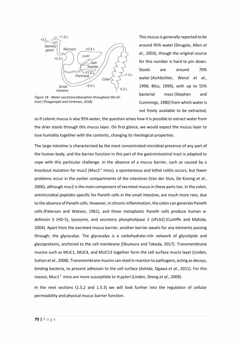
70 | P a g e
This mucus is generally reported to be
around 95% water (Strugala, Allen et
al., 2003), though the original source
for this number is hard to pin down.
Stools are around 70%
water (Aichbichler, Wenzl et al.,
1998; Bliss, 1999), with up to 55%
bacterial mass (Stephen and
Cummings, 1980) from which water is
not freely available to be extracted,
so if colonic mucus is also 95% water, the question arises how it is possible to extract water from
the drier stools through this mucus layer. On first glance, we would expect the mucus layer to
lose humidity together with the contents, changing its rheological properties.
The large intestine is characterized by the most concentrated microbial presence of any part of
the human body, and the barrier function in this part of the gastrointestinal tract is adapted to
cope with this particular challenge. In the absence of a mucus barrier, such as caused by a
knockout mutation for muc2 (Muc2-/- mice), a spontaneous and lethal colitis occurs, but fewer
problems occur in the earlier compartments of the intestines (Van der Sluis, De Koning et al.,
2006), although muc2 is the main component of excreted mucus in these parts too. In the colon,
antimicrobial peptides specific for Paneth cells in the small intestine, are much more rare, due
to the absence of Paneth cells. However, in chronic inflammation, the colon can generate Paneth
cells (Paterson and Watson, 1961), and these metaplastic Paneth cells produce human α-
defensin 5 (HD-5), lysozyme, and secretory phospholipase 2 (sPLA2) (Cunliffe and Mahida,
2004). Apart from the excreted mucus barrier, another barrier awaits for any elements passing
through; the glycocalyx. The glycocalyx is a carbohydrate-rich network of glycolipids and
glycoproteins, anchored to the cell membrane (Okumura and Takeda, 2017). Transmembrane
mucins such as MUC1, MUC4, and MUC13 together form the cell surface mucin layer (Linden,
Sutton et al., 2008). Transmembrane mucins can shed in reaction to pathogens, acting as decoys,
binding bacteria, to prevent adhesion to the cell surface (Ashida, Ogawa et al., 2011). For this
reason, Muc1-/- mice are more susceptible to H.pylori (Linden, Sheng et al., 2009).
In the next sections (1.5.2 and 1.5.3) we will look further into the regulation of cellular
permeability and physical mucus barrier function.
Figure 18 - Water excretion/absorption throughout the GI-tract (Thiagarajah and Verkman, 2018)
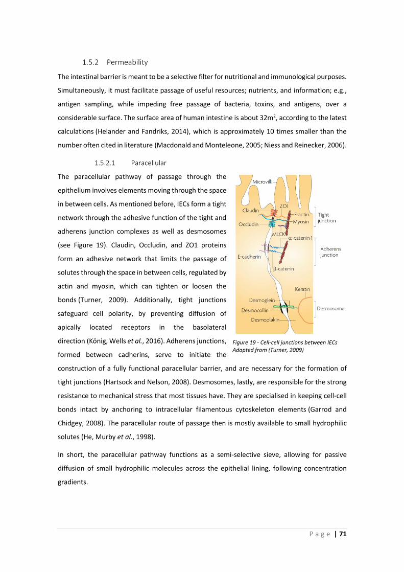
P a g e | 71
1.5.2 Permeability
The intestinal barrier is meant to be a selective filter for nutritional and immunological purposes.
Simultaneously, it must facilitate passage of useful resources; nutrients, and information; e.g.,
antigen sampling, while impeding free passage of bacteria, toxins, and antigens, over a
considerable surface. The surface area of human intestine is about 32m2, according to the latest
calculations (Helander and Fandriks, 2014), which is approximately 10 times smaller than the
number often cited in literature (Macdonald and Monteleone, 2005; Niess and Reinecker, 2006).
1.5.2.1 Paracellular
The paracellular pathway of passage through the
epithelium involves elements moving through the space
in between cells. As mentioned before, IECs form a tight
network through the adhesive function of the tight and
adherens junction complexes as well as desmosomes
(see Figure 19). Claudin, Occludin, and ZO1 proteins
form an adhesive network that limits the passage of
solutes through the space in between cells, regulated by
actin and myosin, which can tighten or loosen the
bonds (Turner, 2009). Additionally, tight junctions
safeguard cell polarity, by preventing diffusion of
apically located receptors in the basolateral
direction (König, Wells et al., 2016). Adherens junctions,
formed between cadherins, serve to initiate the
construction of a fully functional paracellular barrier, and are necessary for the formation of
tight junctions (Hartsock and Nelson, 2008). Desmosomes, lastly, are responsible for the strong
resistance to mechanical stress that most tissues have. They are specialised in keeping cell-cell
bonds intact by anchoring to intracellular filamentous cytoskeleton elements (Garrod and
Chidgey, 2008). The paracellular route of passage then is mostly available to small hydrophilic
solutes (He, Murby et al., 1998).
In short, the paracellular pathway functions as a semi-selective sieve, allowing for passive
diffusion of small hydrophilic molecules across the epithelial lining, following concentration
gradients.
Figure 19 - Cell-cell junctions between IECs Adapted from (Turner, 2009)
72 | P a g e
1.5.2.2 Transcellular
“Transcellular pathway” refers to the passage of molecules through cells. It involves passive
transport by hydrophobic molecules partitioning in and out of lipid bilayers, active transport by
specific transporter proteins and channels, or endocytosis by the cell membrane (Keita and
Söderholm, 2010). The transcellular pathway is mostly inaccessible to hydrophilic elements, due
to the lipid bilayer of the cell plasma membranes. Instead, membrane-bound receptors and
pores on the one hand, and endocytosis on the other hand can provide access to components,
to take this route through the epithelium. Transcellular transport is partially dependent on the
integrity of the paracellular barrier, because the established concentration gradient would cause
passive diffusion to re-establish equilibrium otherwise (Turner, 2009).
Transcellular transport is performed by a set of highly regulated and complex processes, and
apart from the uptake of nutrients, serves to transport antigens to be processed by the immune
system.
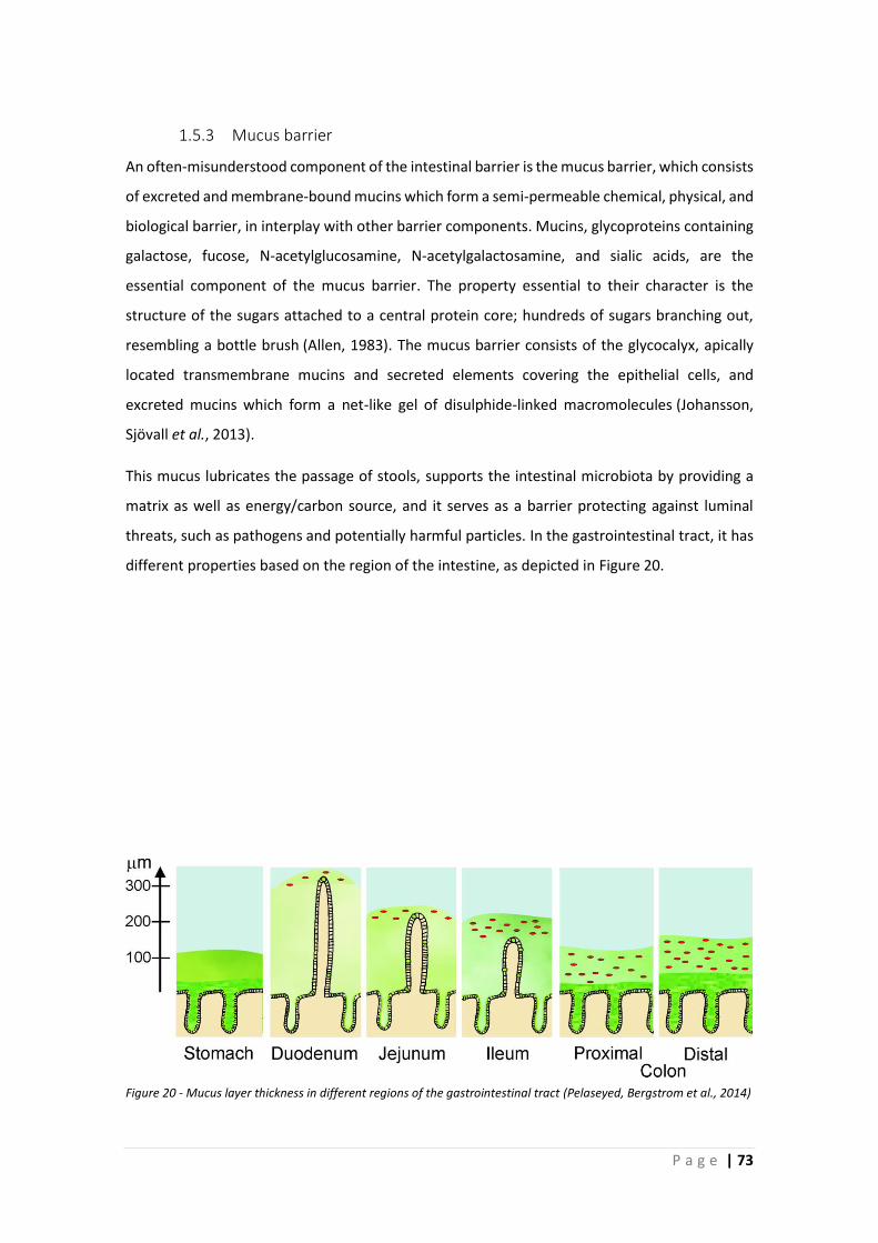
P a g e | 73
1.5.3 Mucus barrier
An often-misunderstood component of the intestinal barrier is the mucus barrier, which consists
of excreted and membrane-bound mucins which form a semi-permeable chemical, physical, and
biological barrier, in interplay with other barrier components. Mucins, glycoproteins containing
galactose, fucose, N-acetylglucosamine, N-acetylgalactosamine, and sialic acids, are the
essential component of the mucus barrier. The property essential to their character is the
structure of the sugars attached to a central protein core; hundreds of sugars branching out,
resembling a bottle brush (Allen, 1983). The mucus barrier consists of the glycocalyx, apically
located transmembrane mucins and secreted elements covering the epithelial cells, and
excreted mucins which form a net-like gel of disulphide-linked macromolecules (Johansson,
Sjövall et al., 2013).
This mucus lubricates the passage of stools, supports the intestinal microbiota by providing a
matrix as well as energy/carbon source, and it serves as a barrier protecting against luminal
threats, such as pathogens and potentially harmful particles. In the gastrointestinal tract, it has
different properties based on the region of the intestine, as depicted in Figure 20.
Figure 20 - Mucus layer thickness in different regions of the gastrointestinal tract (Pelaseyed, Bergstrom et al., 2014)
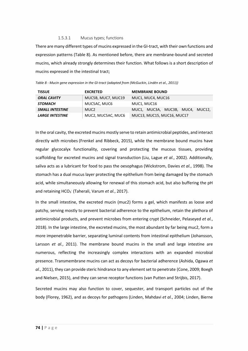
74 | P a g e
1.5.3.1 Mucus types; functions
There are many different types of mucins expressed in the GI-tract, with their own functions and
expression patterns (Table 8). As mentioned before, there are membrane-bound and secreted
mucins, which already strongly determines their function. What follows is a short description of
mucins expressed in the intestinal tract;
Table 8 - Mucin gene expression in the GI-tract (adapted from (McGuckin, Lindén et al., 2011))
TISSUE EXCRETED MEMBRANE BOUND
ORAL CAVITY MUC5B, MUC7, MUC19 MUC1, MUC4, MUC16
STOMACH MUC5AC, MUC6 MUC1, MUC16
SMALL INTESTINE MUC2 MUC1, MUC3A, MUC3B, MUC4, MUC12,
MUC13, MUC15, MUC16, MUC17 LARGE INTESTINE MUC2, MUC5AC, MUC6
In the oral cavity, the excreted mucins mostly serve to retain antimicrobial peptides, and interact
directly with microbes (Frenkel and Ribbeck, 2015), while the membrane bound mucins have
regular glycocalyx functionality, covering and protecting the mucous tissues, providing
scaffolding for excreted mucins and signal transduction (Liu, Lague et al., 2002). Additionally,
saliva acts as a lubricant for food to pass the oesophagus (Wickstrom, Davies et al., 1998). The
stomach has a dual mucus layer protecting the epithelium from being damaged by the stomach
acid, while simultaneously allowing for renewal of this stomach acid, but also buffering the pH
and retaining HCO3- (Taherali, Varum et al., 2017).
In the small intestine, the excreted mucin (muc2) forms a gel, which manifests as loose and
patchy, serving mostly to prevent bacterial adherence to the epithelium, retain the plethora of
antimicrobial products, and prevent microbes from entering crypt (Schneider, Pelaseyed et al.,
2018). In the large intestine, the excreted mucins, the most abundant by far being muc2, form a
more impenetrable barrier, separating luminal contents from intestinal epithelium (Johansson,
Larsson et al., 2011). The membrane bound mucins in the small and large intestine are
numerous, reflecting the increasingly complex interactions with an expanded microbial
presence. Transmembrane mucins can act as decoys for bacterial adherence (Ashida, Ogawa et
al., 2011), they can provide steric hindrance to any element set to penetrate (Cone, 2009; Boegh
and Nielsen, 2015), and they can serve receptor functions (van Putten and Strijbis, 2017).
Secreted mucins may also function to cover, sequester, and transport particles out of the
body (Florey, 1962), and as decoys for pathogens (Linden, Mahdavi et al., 2004; Linden, Bierne
P a g e | 75
et al., 2008), in a way that reminds somewhat of airway mucins, which capture and remove
environmental pollutants and pathogens (Knowles and Boucher, 2002).
Excreted mucus plays an important role in modulating infectious disease severity. For example,
Muc2, the main mucin of intestinal excreted mucus, is key in controlling Salmonella infection, as
evidenced by use of a Muc2-deficient mouse model (Zarepour, Bhullar et al., 2013). The
researchers showed that O-glycosylation properties of muc2 is essential for this functionality, as
mutants with aberrant glycosylation show increased susceptibility (Zarepour, Bhullar et al.,
2013). The mechanism behind protection by mucus is more complex than just physical
obstruction; secreted antibodies like IgM are slightly mucophilic, but diffuse through the mucus,
accumulating on antigen-covered particles, such as pathogens. Due to this accumulation, even
normally motile mucus-penetrating bacteria become immobile in the mucus, with antigens
forming polyvalent low affinity bonds to the gel (Cone, 2009).
It is often said that contact between bacteria and the epithelium leads to inflammation, and that
the excreted mucus barrier prevents this contact (Johansson, Gustafsson et al., 2014). While it
is clear that muc2-/- mice without an excreted mucus barrier develop severe colitis (Van der Sluis,
De Koning et al., 2006), it has not been clearly shown that this is solely due to epithelium-
bacterial contact. Since it is known that in the small intestine the mucus layer is patchy and easily
removed (Atuma, Strugala et al., 2001; Ermund, Schutte et al., 2013), the microbial presence
there should lead to inflammation too if contact alone would be enough, even when taking into
account that the small intestine has its own array of anti-microbial defences, as discussed in
1.5.1.1. Still, small intestinal bacterial overgrowth (SIBO), should cause extensive contact
between epithelium and bacteria, it is however not specifically characterized by ileitis.
Apart from protecting against microbial threats, the excreted mucus barrier is also involved in
protecting against chemical agents, such as food-borne environmental pollutants and possibly
harmful food additives, although this has not been sufficiently studied and documented until
today (Gillois, Leveque et al., 2018).
76 | P a g e
Besides protective functions, in the intestine, mucus also serves a supportive purpose for the
resident microbes; mucus produced by the host supports a microbial network of mucolytic
bacteria, such as Akkermansia muciniphila and associates (Derrien, Vaughan et al., 2004; Belzer,
Chia et al., 2017). Intestinal mucus thus provides a suitable niche to the commensal microbiota,
supported by beneficial fucosylation of mucins to support the microbiota (Pickard, Maurice et
al., 2014), as well as differing glycosylation patterns throughout the intestine, which seem to
have role in selecting microbial population composition, while at the same time being influenced
by the microbiota (Sekirov, Russell et al., 2010; Johansson, Jakobsson et al., 2015). For example,
fucosylation of the glycocalyx is increased by signalling from B. thetaiotamicron, and this fucose
is then used by this bacterium (Hooper and Gordon, 2001). This example nicely demonstrates
that host and microbe communicate to shape a suitable niche for the microbiota through mucin
modifications.
The goblet cell to total cell ratio in mice progresses upwards throughout the intestine,
approximately 2:50 in duodenum, 3:50 in jejunum, 6:50 in ileum (Cheng, 1974), and 8:50 in the
distal colon (Chang and Leblond, 1971), which correlates to the density of microbes in these
intestinal compartments, which increases from proximal to distal regions. This holds well with
the general idea that mucus is a main mediator in host-microbe interactions, as part of the
variety of adaptations of the metaorganism to exist in eubiosis, which are studied in the rapidly
upcoming field of metaorganism or holobiont research (Esser, Lange et al., 2018).
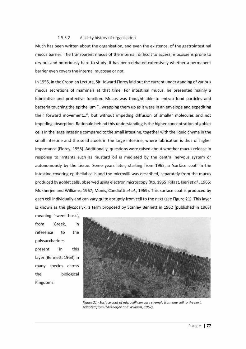
P a g e | 77
1.5.3.2 A sticky history of organisation
Much has been written about the organisation, and even the existence, of the gastrointestinal
mucus barrier. The transparent mucus of the internal, difficult to access, mucosae is prone to
dry out and notoriously hard to study. It has been debated extensively whether a permanent
barrier even covers the internal mucosae or not.
In 1955, in the Croonian Lecture, Sir Howard Florey laid out the current understanding of various
mucus secretions of mammals at that time. For intestinal mucus, he presented mainly a
lubricative and protective function. Mucus was thought able to entrap food particles and
bacteria touching the epithelium “…wrapping them up as it were in an envelope and expediting
their forward movement…”, but without impeding diffusion of smaller molecules and not
impeding absorption. Rationale behind this understanding is the higher concentration of goblet
cells in the large intestine compared to the small intestine, together with the liquid chyme in the
small intestine and the solid stools in the large intestine, where lubrication is thus of higher
importance (Florey, 1955). Additionally, questions were raised about whether mucus release in
response to irritants such as mustard oil is mediated by the central nervous system or
autonomously by the tissue. Some years later, starting from 1965, a ‘surface coat’ in the
intestine covering epithelial cells and the microvilli was described, separately from the mucus
produced by goblet cells, observed using electron microscopy (Ito, 1965; Rifaat, Iseri et al., 1965;
Mukherjee and Williams, 1967; Monis, Candiotti et al., 1969). This surface coat is produced by
each cell individually and can vary quite abruptly from cell to the next (see Figure 21). This layer
is known as the glycocalyx, a term proposed by Stanley Bennett in 1962 (published in 1963)
meaning ‘sweet husk’,
from Greek, in
reference to the
polysaccharides
present in this
layer (Bennett, 1963) in
many species across
the biological
Kingdoms.
Figure 21 - Surface coat of microvilli can vary strongly from one cell to the next. Adapted from (Mukherjee and Williams, 1967)
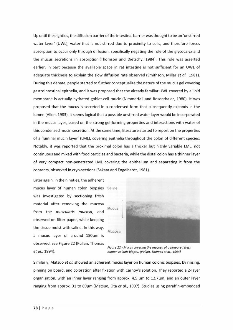
78 | P a g e
Up until the eighties, the diffusion barrier of the intestinal barrier was thought to be an ‘unstirred
water layer’ (UWL), water that is not stirred due to proximity to cells, and therefore forces
absorption to occur only through diffusion, specifically negating the role of the glycocalyx and
the mucus secretions in absorption (Thomson and Dietschy, 1984). This role was asserted
earlier, in part because the available space in rat intestine is not sufficient for an UWL of
adequate thickness to explain the slow diffusion rate observed (Smithson, Millar et al., 1981).
During this debate, people started to further conceptualize the nature of the mucus gel covering
gastrointestinal epithelia, and it was proposed that the already familiar UWL covered by a lipid
membrane is actually hydrated goblet-cell mucin (Nimmerfall and Rosenthaler, 1980). It was
proposed that the mucus is secreted in a condensed form that subsequently expands in the
lumen (Allen, 1983). It seems logical that a possible unstirred water layer would be incorporated
in the mucus layer, based on the strong gel-forming properties and interactions with water of
this condensed mucin secretion. At the same time, literature started to report on the properties
of a ‘luminal mucin layer’ (LML), covering epithelia throughout the colon of different species.
Notably, it was reported that the proximal colon has a thicker but highly variable LML, not
continuous and mixed with food particles and bacteria, while the distal colon has a thinner layer
of very compact non-penetrated LML covering the epithelium and separating it from the
contents, observed in cryo-sections (Sakata and Engelhardt, 1981).
Later again, in the nineties, the adherent
mucus layer of human colon biopsies
was investigated by sectioning fresh
material after removing the mucosa
from the muscularis mucosa, and
observed on filter paper, while keeping
the tissue moist with saline. In this way,
a mucus layer of around 150µm is
observed, see Figure 22 (Pullan, Thomas
et al., 1994).
Similarly, Matsuo et al. showed an adherent mucus layer on human colonic biopsies, by rinsing,
pinning on board, and coloration after fixation with Carnoy’s solution. They reported a 2-layer
organisation, with an inner layer ranging from approx. 4,5 µm to 12,7µm, and an outer layer
ranging from approx. 31 to 89µm (Matsuo, Ota et al., 1997). Studies using paraffin-embedded
Figure 22 - Mucus covering the mucosa of a prepared fresh human colonic biopsy. (Pullan, Thomas et al., 1994)
P a g e | 79
rodent intestine fixed in Carnoy’s solution in combination with ex vivo measurements using
explants mounted in specially designed mucus chambers would then clarify more about the
function of this mucus covering the epithelial lining on the intestines. In 2008, it was reported
that ex vivo, the outer layer covering mouse colon is about 100µm, and is easily removed, and
the inner around 50µm, which is firmly attached to the epithelium. In the same study, in
histological material, it is shown that the outer layer contains bacteria, while the inner layer does
not (Johansson, Phillipson et al., 2008). This notion, together with the severe and often lethal
colitis observed in muc2-/- mice (Van der Sluis, De Koning et al., 2006), which lack a mucus
barrier, led to the understanding that the mucus barrier is necessary to keep bacteria away from
the epithelium, and thus essential in preventing inflammation.
A proper understanding of the nature and functions of the intestinal mucus barrier is important,
because it impacts the design and veracity of theoretical, practical and mathematical models of
interactions in the gut. For example, results obtained using promising systems such as gut-on-a-
chip (Kim, Huh et al., 2012) have to be compared to a precise understanding of physiological
conditions in vivo, to allow realistic interpretation. Additionally, pharmacological approaches
take interactions of medicinal compounds with the mucosal barrier into account (Netsomboon
and Bernkop-Schnurch, 2016), and to effectively do this, correct estimations of mucus barrier
organisation and regulation are essential.
One thing is sure, mucosae produce mucus…
80 | P a g e
1.5.3.3 Regulation
Even though the general description of the mucus layer is fairly static, there are several known
important mechanisms that regulate mucus secretion, and finetune the production in relation
to different situations and challenges. Since presence of a pathogen is one of the most acute
naturally occurring challenges to the intestinal epithelium, and protection against pathogens of
the main functions of the mucus barrier, it stands to reason that it influences mucus barrier
regulation. NLRP6 inflammasome activity is strongly implied in proper barrier function, as
NLRP6-deficient mice have a defective mucus barrier, which renders them more susceptible to
Citrobacter rodentium (Wlodarska, Thaiss et al., 2014). Inflammasomes are multiprotein
complexes that regulate innate immune responses of cells, integrating information from
cytokine signalling, microbial products, and damage-associated molecular patterns
(DAMPs) (Rathinam, Vanaja et al., 2012). Signalling from TLR4 in response to bacterial
lipopolysaccharides (LPS) causes mucin granule exocytosis of goblet cells, thereby causing an
increase in its thickness, and flushing away the bacteria from the epithelial surface (Dharmani,
Srivastava et al., 2009). An insufficient mucus barrier, such as observed in germ-free animals,
due to lack of signals from microbial products, renders mice exceptionally sensitive to chemically
induced colitis by treatment with dextran sodium sulphate (DSS) (Kitajima, Morimoto et al.,
2001), indicating both that the microbiota and microbial signalling is necessary for optimal
mucus barrier regulation, and that the mucus barrier protects against harmful toxic chemicals.
Colonic crypts are ‘guarded’ by sentinel goblet cells (senGC), which reacts strongly to the
presence of bacteria by NLRP6 activation by PRR signalling, in response to LPS and flagellin. This
causes compound exocytosis of muc2 mucus, and triggers mucus release in neighbouring goblet
cells as well, through an intercellular gap-junction signal (Birchenough, Nyström et al., 2016). In
the ileum, other PRRs are expressed, and crypts there are not responsive to LPS or
flagellin (Birchenough, Nyström et al., 2016). This could reflect the different situations in these
compartments; whereas bacteria reaching the epithelium in the colon is a danger that warrants
a reaction, in the ileum, where no true separating mucus barrier exists, this is a normal situation.
Apart from signalling from microbes or luminal chemicals, mucus release is also promoted by
cholinergic signalling (Specian and Neutra, 1980) and histamine (Castagliuolo, Lamont et al.,
1996). Though both small and large intestine mucus production is activated by acetylcholine,
the small intestine is by far more sensitive to this (Birchenough, Johansson et al., 2015).
P a g e | 81
Together with the observation that the large intestine is more responsive to TLR activation than
the small intestine, this reinforces the idea that the mucus release is regulated differently in
these two different organs; the small intestine mucus barrier more responsive to
parasympathetic activity, the large intestine more to luminal signalling.
82 | P a g e
1.5.3.4 Dietary effects
The mucus barrier can be impacted by the diet in several ways. What we eat can have effects on
the expression of mucins and the regulation of mucus release, but the physical properties of the
bolus and faeces also have an impact on the properties of the mucus layer, and the rheology of
mucus excretions. Here, we will look at some examples of known effects of the diet on the mucus
barrier.
A major influence on the mucus barrier is dietary fibre; an in vivo rat model testing different
dietary fibre contents showed that mucus turnover as well as production was reduced by fibre
deficiency, and different fibres have different effects on these factors (Brownlee, Havler et al.,
2003). In the same vein, fibre deficiency in a mouse model causes the expansion and increased
activity of mucus-degrading bacterial species, leading to a reduced mucus barrier, and a reduced
resistance to C. rodentium (Desai, Seekatz et al., 2016). In a way, it speaks for itself that nutrients
that lead to the production of SCFAs by the microbiota, such as these dietary fibres, have an
effect on the mucus barrier, because SCFAs have effects on mucus release and
production (Sakata and von Engelhardt, 1981; Shimotoyodome, Meguro et al., 2000;
Hedemann, Theil et al., 2009), as well as possible modulation of mucin gene expression (Paturi,
Butts et al., 2012).
Dietary lipid intake has an effect on the mucus barrier as well, changing the permeability of this
barrier, decreasing the mobility of particles (Yildiz, Speciner et al., 2015). Contrary to the effects
of SCFAs, these effects of lipids play a direct role in interaction with the mucus itself.
Apart from these effects, because we have already described microbiota interactions with the
host and its effects on mucus barrier regulation, diets with specific impacts on microbiota
composition or activity, logically also have an effect on the mucus barrier.
P a g e | 83
1.6 Aims and Outline of Thesis
1.6.1 Hypotheses
Based on the available literature and the theoretical framework, we propose that FODMAP
fermentation by the intestinal microbiota can have harmful effects which increase the tendency
to display gastrointestinal symptoms. This is complementary to the known mechanisms by which
FODMAP ingestion provokes these symptoms; an increased distension of the intestine by an
increase of the osmotic load and water volume in the small intestine, and of gas volume in the
large intestine, generated during fermentation. As functional parameters to these harmful
effects, we selected to investigate the visceral sensitivity, intestinal barrier function, and
microbial metabolite profiles of the colonic contents in relation to treatment with selected
representatives of FODMAPs. We formulate the following hypotheses:
1. FODMAP ingestion can induce gastrointestinal symptoms of IBS by itself, in an otherwise
healthy system
2. This effect relies on the production of harmful bacterial fermentation metabolites, among
which methylglyoxal
3. Methylglyoxal and other strong glycation agents lead to an increase in AGEs and a pro-
inflammatory state through activation of RAGE
A recurring theme and major field of interest in research of gastrointestinal disease and function
is the relation of the host with their microbiota. In this context, the contact between intestinal
microbes and host surfaces are considered important, and an understanding of the properties
of the excreted mucus barrier, which separates microbes from the epithelium in the colon where
the concentration of microbes is highest, is key to understanding host-microbe relations in the
intestine. For this reason, to better understand the mucus barrier function, we have identified
some situations in which information regarding this interesting topic was missing and designed
experimental approaches to fill in these gaps of knowledge.
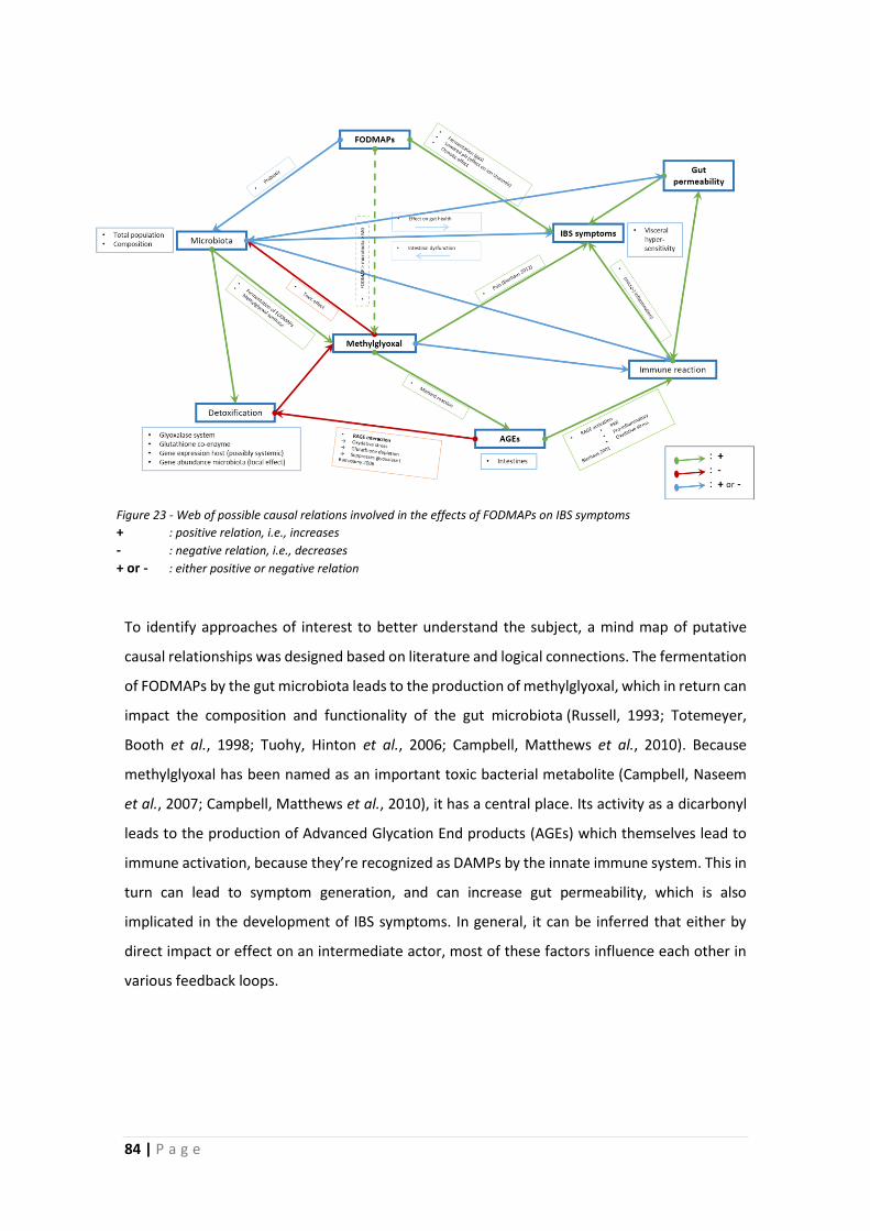
84 | P a g e
(Bierhaus, Schiekofer et al., 2001; Ramasamy, Yan et al., 2006; Bierhaus, Fleming et al., 2012)
To identify approaches of interest to better understand the subject, a mind map of putative
causal relationships was designed based on literature and logical connections. The fermentation
of FODMAPs by the gut microbiota leads to the production of methylglyoxal, which in return can
impact the composition and functionality of the gut microbiota (Russell, 1993; Totemeyer,
Booth et al., 1998; Tuohy, Hinton et al., 2006; Campbell, Matthews et al., 2010). Because
methylglyoxal has been named as an important toxic bacterial metabolite (Campbell, Naseem
et al., 2007; Campbell, Matthews et al., 2010), it has a central place. Its activity as a dicarbonyl
leads to the production of Advanced Glycation End products (AGEs) which themselves lead to
immune activation, because they’re recognized as DAMPs by the innate immune system. This in
turn can lead to symptom generation, and can increase gut permeability, which is also
implicated in the development of IBS symptoms. In general, it can be inferred that either by
direct impact or effect on an intermediate actor, most of these factors influence each other in
various feedback loops.
Figure 23 - Web of possible causal relations involved in the effects of FODMAPs on IBS symptoms
+ : positive relation, i.e., increases
- : negative relation, i.e., decreases
+ or - : either positive or negative relation
P a g e | 85
1.6.2 Approach
To investigate the relations between FODMAP intake, IBS symptoms, immune activation, and
bacterial fermentation products, we have designed and performed animal experiments related
to each hypothesis; male C57Bl/6 mice have been given FODMAP representatives (lactose and
fructo-oligosaccharides) for 3 weeks, to induce FODMAP related changes to their physiology.
Additionally, an anti-glycation agent (pyridoxamine) was administered intended to counteract
the effects of glycating elements produced during bacterial fermentation of said FODMAPs.
Next, analyses were performed to answer our questions;
Hypothesis 1: To test whether FODMAPs can induce IBS symptoms we have measured the
visceral sensitivity, gut permeability, mast cell counts in the colon, and colonic mucus barrier
dysregulation in the treated animals.
Hypothesis 2: To test whether these effects were caused by glycation processes, we have
analysed whether administration of anti-glycation agent pyridoxamine protects against these
consequences, additionally, generation of reactive aldehydes and ketones in the colonic
contents was analysed by HPLC/MS after derivatisation with BBHA (1-((aminooxy)ethyl)-2-
bromobenzene hydrochloride).
Hypothesis 3: To identify the effect of these glycation processes, we have measured the
occurrence of advanced glycation end-products (AGEs), and the expression of Receptor for AGEs
(RAGE) in the colon of treated animals.
86 | P a g e
1.6.3 Experimental procedures
1.6.3.1 Treatment setup
C57Bl/6 mice are housed in groups and treated with daily gavages of lactose and/or
pyridoxamine, or put on a custom FOS diet (AIN-93M +/-10% FOS, starch partly replaced).
Animals are weighed 2 times per week. Animals for permeability assays and mechanical
behavioural testing are also used for histological purposes, animals for visceral sensitivity assays
are not.
1.6.3.2 Visceral sensitivity: EMG, von Frey
Since a central IBS symptom relieved with the low-FODMAP diet is abdominal pain/discomfort,
we set out to measure the possible induction of increased sensitivity by FODMAP ingestion. The
reference method of recording of electromyographic response to colorectal balloon distension,
as well as an alternative method of mechanical behavioural testing using von Frey filaments
were performed. This alternative was necessary as FOS-treatment lead to increased amounts of
intestinal content that caused the colorectal cavity to remain full even after fasting and
habituation periods, which would impede reliable data gathering.
Visceral sensitivity (Electromyography (EMG))
Electrodes are implanted into the abdominal muscles and colorectal distensions are used as
noxious stimuli to evaluate visceral hyperalgesia by electromyographic (EMG) recording.
Striated muscle’s EMG activity was recorded and analysed according to (Larsson, Arvidsson et
al., 2003). Basal EMG activity was subtracted from the EMG activity registered during the periods
of distension.
This procedure is considered the gold standard in visceral sensitivity analysis, measuring the
abdominal contractions in response to a painful stimulus in a quantifiable way. Visceral
hypersensitivity is observable through an increased response to the colorectal distension
stimulus, both by a response to stimuli that do not provoke a reaction in normosensitive subjects
(allodynia), as well as an increased response to painful stimuli (hyperalgesia).

P a g e | 87
Mechanical behavioural testing (von Frey)
Three von Frey filaments with bending force 0.16, 0.6 and 1.4 g are applied to the mid-plantar
area of each hind paw and nociceptive specific behaviours noted.
This procedure allows us to compare the central sensitivity of animals, by valuing the
responsiveness to mechanical stimuli on the hind paws, as well as the abdominal region. Animals
that have an increased sensitivity exhibit a higher responsiveness. Animals with an increased
visceral sensitivity will have a higher central sensitivity through sensitization processes mediated
through the spinal cord (Lamb, Zhong et al., 2006), probably through convergence of primary
afferents on the same neurons in the spinal cord.
1.6.3.3 Intestinal permeability
An important parameter of intestinal health is its permeability, which is known to play a role in
IBS as well (Dunlop, Hebden et al., 2006; Bischoff, Barbara et al., 2014; Enck, Aziz et al., 2016;
König, Wells et al., 2016). To investigate the effects of our FODMAP interventions on this
parameter, we have performed in vivo measurements of intestinal permeability.
In vivo measurement of permeability using FITC
Fasted mice are gavaged with 250µl of fluorescein isothiocyanate (FITC) solution (1mg/ml). After
4 hours, when the entirety of the gastrointestinal has been passed by the solution, 100µl of
blood is taken and kept in the dark on ice. Serum is recovered and analysed in a
spectrophotometer. FITC concentration of blood samples is calculated using the standard curve.
This in vivo procedure is designed to measure the passage of a fluorescent marker to the blood,
reflecting the paracellular or transcellular permeability of the gastrointestinal tract, depending
on the size and nature of the marker used. We used fluorescein isothiocyanate (FITC), a small
(~390Da) molecule, meant to measure predominantly the paracellular permeability.
88 | P a g e
1.6.3.4 Immunofluorescence/ histology
Histological sample preparation
Collected tissues were fixated for 1 day in Carnoy’s fixative and included in paraffin, and
subsequently microsectioned and attached to glass microscopical slides.
Immunofluorescence
To visualize marker proteins and measure their expression, microscopic immunofluorescence
assays were used.
AGE: Slides were deparaffinated and incubated overnight at 4°C with primary antibodies
(Polyclonal Rabbit-anti-AGE (ab23722), followed by incubation with secondary antibody (Alexa
Fluor 488 Donkey-anti-Rabbit (A21206).
Following our hypothesis that FODMAP fermentation leads to the production of reactive
carbonyl species, we sought to quantify the occurrence of the retained, and cumulative, marker
of glycation processes, advanced glycation end products (AGEs).
RAGE: Slides were deparaffinated and incubated overnight at 4°C primary antibodies (Polyclonal
Goat-anti-RAGE (ab7764), followed by incubation with secondary antibody (Alexa Fluor 488
Donkey-anti-Goat (A-11055).
An increased occurrence of AGEs in the tissue can be expected to activate Receptor for AGEs
(RAGE), which consecutively leads to the increased expression of this receptor (Bierhaus and
Nawroth, 2009).
MMCP: Slides were deparaffinated and incubated overnight at 4°C with primary antibodies
(Sheep anti-mMCP1 (MS-RM8), followed by incubation with secondary antibody (Alexa Fluor
594 Donkey-anti-Sheep (A-11016).
Mouse Mast Cell Protease-1 was chosen as a selective marker of (mucosal) mast cells (Dai and
Korthuis, 2011), to visualize and quantify the mucosal mast cells in histological slides. Because it
is easy to trigger mast cell degranulation and exocytosis of this marker, particular care was taken
not to disturb the tissue before fixation, i.e., no touching of the tissue, no flushing of contents,
no cutting to size, until after fixation.
P a g e | 89
Muc2: Slides were deparaffinated and incubated overnight at 4°C with primary antibodies
(Rabbit anti-mucin 2 (sc-15334), followed by incubation with secondary antibody (Alexa Fluor
488 Donkey-anti-Rabbit (A21206).
Muc2 is the predominant mucin in small intestinal and colonic mucus, and this antibody staining
is used to very clearly visualize mucus in immunofluorescent slides. This can be combined with
FISH.
AB/H&E: Slides were deparaffinated and stained by 5 min in Hematoxylin, 10 min in running
water, 30 min in Alcian Blue solution (pH 3.0) followed by 5 min in running water, 3 min in Eosin.
The haematoxylin and eosin stain (H&E) is a standard staining to visualize general morphology
and recognize immune cell infiltrates in tissues, while also staining bacterial masses, the
combination with Alcian blue (AB) staining visualizes goblet cells and mucus. The combination
AB/H&E thus allows to analyse a diverse set of parameters.
Fluorescent in situ Hybridization (FisH): FISH staining for all bacteria was performed using probes
EUB338I (5′ CGTGCCTCCCGTAGGAGT 3′), EUB338II (5′ GCAGCCACCCGTAGGTGT 3′), and
EUB338III (5′ GCTGCCACCCGTAGGTGT 3′) conjugated to DY-590. Slides were deparaffinated and
incubated with the FISH probes at 48°C overnight.
These 3 probes (EUB338I, EUB338II, EUB338III) together target virtually all true bacteria (Daims,
Brühl et al., 1999), so this mix can be used to reliably stain all bacteria found in the intestine, to
clearly visualize the presence and location of bacteria.
90 | P a g e
1.6.3.5 Markers of immune activation
Lipocalin-2
Faecal supernatants were analysed for lipocalin-2 content by ELISA.
Lipocalin-2 (LCN-2) is a very sensitive marker for intestinal inflammation, even subclinical low-
grade inflammation, where histopathology is not clear (Chassaing, Srinivasan et al., 2012). LCN2
is produced by neutrophils and macrophages.
Microscopic score
Composite micrographs were scored according to protocol on a scale from 0-4 (Neurath, Fuss et
al., 1995); 0: no signs of inflammation, 1: very low level of leukocytic infiltration, 2: low level of
leukocytic infiltration, 3: high level of leukocytic infiltration, high vascular density, thickening of
the colon wall, 4: transmural infiltrations, loss of goblet cells, high vascular density, thickening
of the colon wall.
This scoring system for the mouse colon evaluates signs of overt inflammation, and thus grades
severity of colitis.
1.6.3.6 Physiological markers of transit
Transit time
Mice were temporarily housed in individual cages, carmine red solution was administered by
intragastric gavage, and starting from 2 hours after administration, every 15 minutes or when
the researcher noted defecation, the cages were checked for red-coloured droppings, the time
of first appearance of a red dropping was used to determine the transit time.
This test tells us how long it takes for material to pass through the gastrointestinal tract, starting
at the stomach.
Faecal water content
Faecal droppings were weighed, dried for 2 days at 80°C, and weighed again. Faecal humidity is
the percentage of weight lost between wet and dry weights.
Faecal water content is a reasonable marker of colonic transit time, because a faster transit leads
to higher percentages of water in faeces (Degen and Phillips, 1996; Lewis and Heaton, 1997b).
We were interested in transit time and faecal water content because FODMAPs can be expected
to influence these markers, which is important to consider in relation to further effects of
treatment.
P a g e | 91
1.6.3.7 Analysis of microbiota composition
Faecal DNA extraction, 16S rRNA gene sequencing, and bacterial community analysis
Genomic DNA was obtained from frozen faeces, 16S rRNA was amplified by PCR. Pooled
amplicon libraries were sequenced employing an Illumina MiSeq. For further data on analysis of
microbiota, please refer to section 2.2.
We analysed microbiota profiles to see the effect of lactose treatment on gut microbiota
composition, as well as the effect of the anti-glycation agent pyridoxamine, because FODMAPs
have a known prebiotic function, and could therefore be expected to impact microbiota profile
composition, even at 5 mg daily.
1.6.3.8 Analysis of carbonyl compounds in colonic content
Colonic content extracts were incubated with 1-((ammoniooxy)methyl)-2-bromobenzene
chloride (BBHA) to generate brominated aldehyde derivates which were analysed by high-
performance liquid chromatography coupled to high-resolution mass spectrometry (HPLC-
HRMS). Resulting semi-quantitative mass spectra were analysed statistically to characterize
differential ions and construct heat-map profiles for the different treatment groups. For a
complete description, please refer to section 2.2.
This analysis allows us to follow the effects of our dietary interventions on carbonyl metabolite
profiles.
1.6.3.9 Mucus barrier function analysis
Imaging; Manual-ultra high-resolution Composite Image Overview (MUCIO)
Manual Ultra-high-resolution Composite Image Overview (MUCIO) approach: datasets of
overlapping microscope images covering entire slides were generated by manual microscope
photography and stitched together using dedicated software. The generated composite images
contain all the information of the data set in a single ultra-high-resolution file. No selection for
‘representative regions’ is necessary, only obvious artefacts (e.g., possibly folded double/
heavily damaged sections) are evaded. Subsequent downscaling is necessary to facilitate
interaction with images for further applications, but close-up views in full quality can be
extracted from the original composite.
92 | P a g e
Mucus layer thickness measurements
The faecal mucus layer thickness was measured every 100 micrometres along the entire imaged
faecal surface generated with the MUCIO approach. Data interpretation: The hundreds of
measurements generated were interpreted by calculating several statistics. First of all, of course,
the average mucus layer thickness is calculated, but this does not tell us anything about the
variation, and the standard deviation or confidence interval is calculated between individuals,
not on multiple measurements per individual. To mitigate this, the Coefficient of Variation (CoV)
is calculated for the dataset of each individual, and these values are compared for treatment
groups.
Active goblet cell numbers
One ratio discharging goblet cell:crypt per mouse based on analysis of 25-90 crypts in transversal
sections of empty colon, dependent on availability of suitable visual material
By calculating the average mucus layer, the CoV for this mucus layer, as well as the active goblet
cells in empty colon, we can characterize the mucus barrier function, considering the variable
situations that naturally occur in the distal colon.


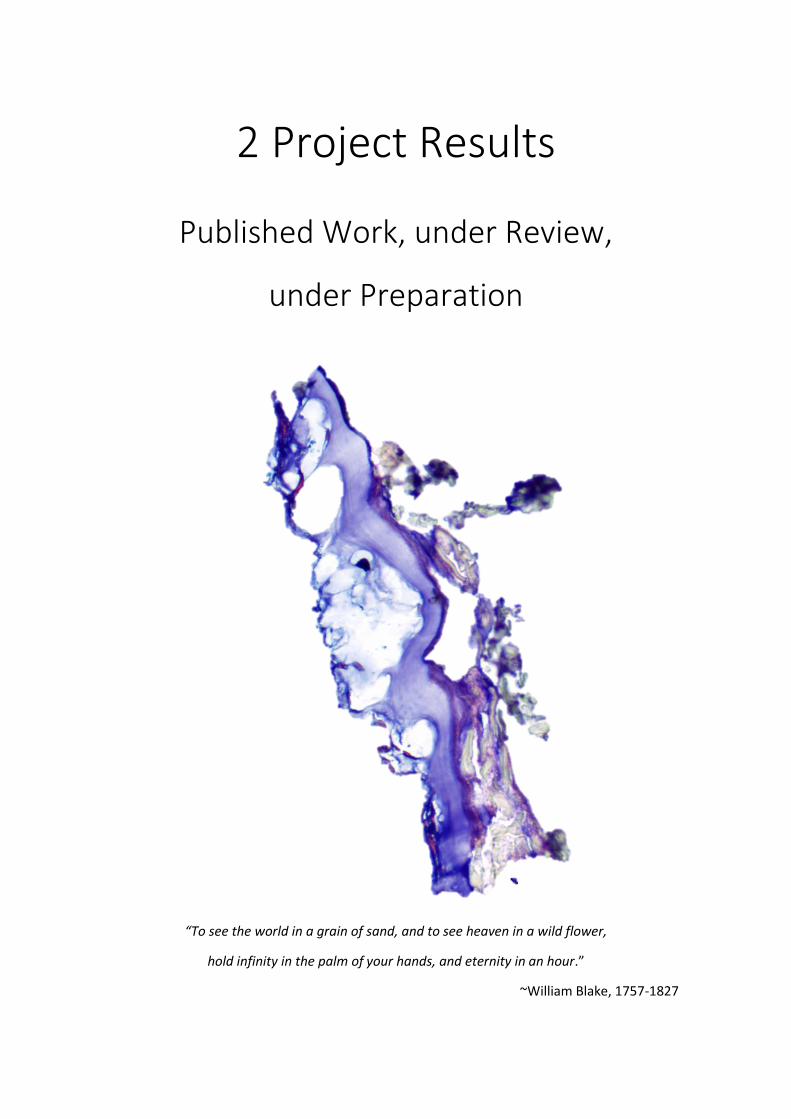
2 Project Results
Published Work, under Review,
under Preparation
“To see the world in a grain of sand, and to see heaven in a wild flower,
hold infinity in the palm of your hands, and eternity in an hour.”
~William Blake, 1757-1827

P a g e | 97
2 Project Results
2.1 Mucus organisation is shaped by colonic content; a new view
While analysing the possible effects of FODMAPs on mucus barrier function, analysing
histological sections in both regions with and without faeces, some observations were made
that warranted further investigation. Notably, in sections lacking a faecal pellet, there is a
marked absence of the classical inner-outer mucus layer organisation. This absence has been
described before, and explained by the supposed protection from washing off of the mucus layer
by the faecal pellet (Johansson, Larsson et al., 2011). However, since we routinely acquire and
fix several centimetres of colon, containing regions with and without faeces, and there is no
flushing of this intact intestinal tube either before or after fixation, we wanted to test whether
this absence is due to washing off of mucus, as postulated in literature.
Therefore, we decided to make longitudinal sections, and image the entire section, to see what
is going on in the transition from empty to full sections of the colon, and vice versa. Additionally,
we reasoned that if a possible collapse of the mucus was responsible for the absence of the
described two-layer system, the bacteria should still be present, but now probably in closer
contact to the epithelium. To visualize these bacteria, we performed fluorescence in situ
hybridisation (FisH) of longitudinal and transversal sections of empty and full sections of
proximal and distal colon.
The results of this investigation were published in Scientific Reports (Springer Nature).
98 | P a g e
-SCIENTIFIC REPO RTS
0
Received: IOApril 2017
Accepted: 17 July 2017
OPEN
Published onl ine: 17 August 2017
Mucus organisation is shaped by colonie content; a new view J. B. J. Kamphuis, M. Mercier-Bonin, H. Eutamène & V. Theodorou
The colonie mucus barrier is commonly described as a continuous double layer covering the epithelium, separating the microbiota from the intestinal tissue. This model is currently considered valid throughout the colon. The colon is characterised by regional anatomo-functional specificities such as presence and consistency of contents and location. ln this study, we characterised the organisation of the colonie mucus barrier in proximal and distal colon of rodents by histological and FISH staining, ta king into account aforementioned specificities. By using longitudinal sections and imaging extensive areas of tissue with and without colonie contents, we have obtained a spatiotemporal overview of mucus organisation in the colon. We describe for the first time that the colonie mucus layer covers the faeces instead of the epithelium in the distal colon. This faecal mucus layer confines the microbiota to t he faeces and prevents it from remaining in empty distal colon. ln the proximal colon, the mucus did not forma separating layer between bacteria and epithelium. We conclude that the organisation of colonie mucus is reliant on the presence of the colonie content, and the location within the colon. Our findings reopen the discussion on the nature of the colonie mucus barrier.
The intestinal mucus layer has a critical role in gut health. lt facilitates the passage of faeces through the intestine, reducing the risk of damage to the gut epithelium 1• The critical importance of intest inal mucus to gut health is underlined by the spontaneous development of colitis in Muc2 knockout (Muc2- 1-J mice2, and their increased susceptibility to pathogens3• Bacteria of the intestinal microbiota profit the host with their metabolic activities', and modulation of the immune system5
. Limiting the contact between intestinal microbes and the colonie epithelium, a thick mucus layer is described that separates the two6• Johansson et a/.7 reported that the distal colonie mucus layer is organised in 2 parts, a firm component and a loose one, both built around Muc2 mucin protein. This layer has been described to be organised as a loose layer inhabited by bacteria, and a layer firmly attached to the epithelium, devoid ofbacteria7
• Primarily, this understanding is based on histological observations of transversal sections of colon, imaging the mucus layers separating microbes from the epithelium. lt became evident that it was impossible to image the mucus barrier in histological sections lacking a faecal pellet, so the method of reference became to section samples containing a faecal pellet to investigate the properties of the mucus layer. A given expia nation for this necessity was that the faecal pellet protects and conserves the mucus barrier during histological processing7
. Additionally, to further investigate mucus barrier properties, ex vivo experiments involving expiant tissue8, and in vivo experiments have been designed and performed6•9. The general organisation is described to be constantly sustained7, though specifics are variable over lime, and influenced by harmful fac tors such as in flammation 10 or even the lime frame of microbiota colonisation 11, emphasising the in timate relationship between intestinal mucus and gut microbiota. Furthermore, microscopie analysis of the biostructure ofhuman faeces shows mucus irregularly intersecting into the faecal mass12
, and a mucus layer is observed on expelled faeces from rodents as well as humans12• 13• These results obtained from faecal material illustrate the organisation of the contents of distal colon, which does not necessarily reflect the situation in the proximal colon. Apart from the morphological differences between the proximal and distal colon, the contents too are dissimilar in their consistency and humidity; the proximal colon content is liquid whereas the contents of distal colon normally form pellets with a firmer consistency. Changes in the physical and biochemical properties of colonie mucus have been documented in pathophysiological conditions. For instance, colonie mucus from ulcerative colitis (UC) patients in the active phase of the disease is thinner and more penetrable to fluorescent beads compared to healthy subjects14 and glycosylation of colonie mucins is found to be correlated with the severity of inflammation in UC patients 15
. Likewise, O-glycosylation of mucins is strongly affected by chronic psychological stress in rats, associated with a flattening and a Joss of cohesive properties of the mucus layer16. Despite data indicating that the
Neuro-Gastroente rology and Nutrition Team, UMR 1331, INRA Toxalim, INP-EI-Purpan, Université de Toulouse, Toulouse, France. Correspondence and requests fo r materials should be addressed to H.E. (email: he lene. [email protected])
SCIENTIFIC REPORTS 17: 85271 DO l:10.1038/s41598-017-08938-3 1
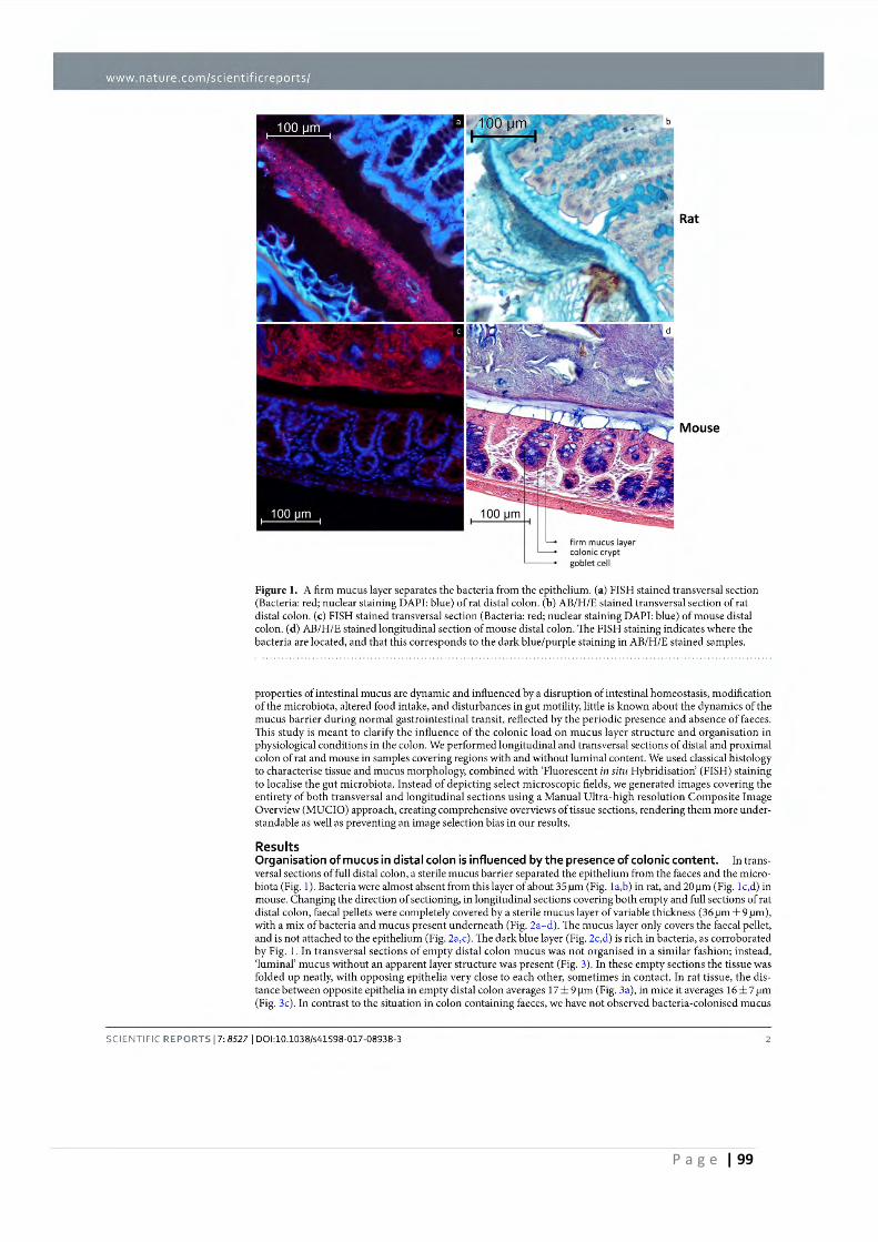
P a g e | 99
www. nature. corn/se i e ntifi cre ports/
firm mucus layer colonie crypt gobie! cell
Rat
Mouse
Figure l. A firm mucus layer separates the bacteria from the epithelium. (a) FISH stained transversal section (Bacteria: red; nuclear staining DAPI: blue) of rat distal colon. (b) AB/H/E stained transversal section of rat distal colon. (c) FISH stained transversal section (Bacteria: red; nuclear staining DAPI: blue) of mouse distal colon. (d) AB/H/E stained longitudinal section of mouse distal colon. The FISH staining indicates where the bacteria are located, and that this corresponds to the dark blue/purple staining in AB/H/E stained samples.
properties of intestinal mucus are dynamic and influenced by a disruption of intestinal homeostasis, modification of the microbiota, altered food in take, and disturbances in gut motility, little is known about the dynamics of the mucus barrier during normal gastrointestinal transit, reflected by the periodic presence and absence of faeces. This study is meant to clarify the influence of the colonie Joad on mucus layer structure and organisation in physiological conditions in the colon. We performed longitudinal and transversal sections of distal and proximal colon of rat and mouse in samples covering regions with and without luminaJ content. We used classical histology to characterise tissue and mucus morphology, combined with 'Fluorescent in situ Hybridisation' (FISH) staining to localise the gut microbiota. Instead of depicting select microscopie fields, we generated images covering the entirety ofboth transversal and longitudinal sections using a Manual Ultra-high resolution Composite Image Overview (MUCIO) approach, creating comprehensive overviews of tissue sections, rendering them more understandable as well as preventing an image selection bias in our results.
Results Organisation of mucus in distal colon is influenced by the presence of colonie content. In transversal sections of full distal colon, a sterile mucus barrier separated the epithelium from the faeces and the microbiota (Fig. ! ). Bacteria were aJmost absent from this layer of about 35µm (Fig. la,b) in rat, and 20 µm (Fig. lc,d) in mouse. Changing the direction of sectioning, in longitudinal sections covering both empty and full sections of rat distal colon, faecal pellets were completely covered by a sterile mucus layer of variable thickness (36 µm ± 9 µm), with a mix ofbacteria and mucus presen t underneath (Fig. 2a-d). The mucus layer only covers the faecal pellet, and is not attached to the epithelium (Fig. 2a,c). The dark blue layer (Fig. 2c,d) is rich in bacteria, as corroborated by Fig. 1. In t ransversal sections of empty distal colon mucus was not organised in a similar fashion; instead, 'luminal' mucus without an apparent layer structure was present (Fig. 3). ln these empty sections the tissue was folded up neatly, with opposing epithelia very close to each other, sometimes in contact. ln rat tissue, the distance between opposite epithelia in empty distal colon averages 17 ± 9 µm (Fig. 3a), in mice it averages 16 ± 7 µm (Fig. 3c). In contras! to the situation in colon containing faeces, we have not observed bacteria-colonised mucus
SCIENTIFIC REPORTS 17: 85271 DOl:10.1038/s41598-017-08938-3 2
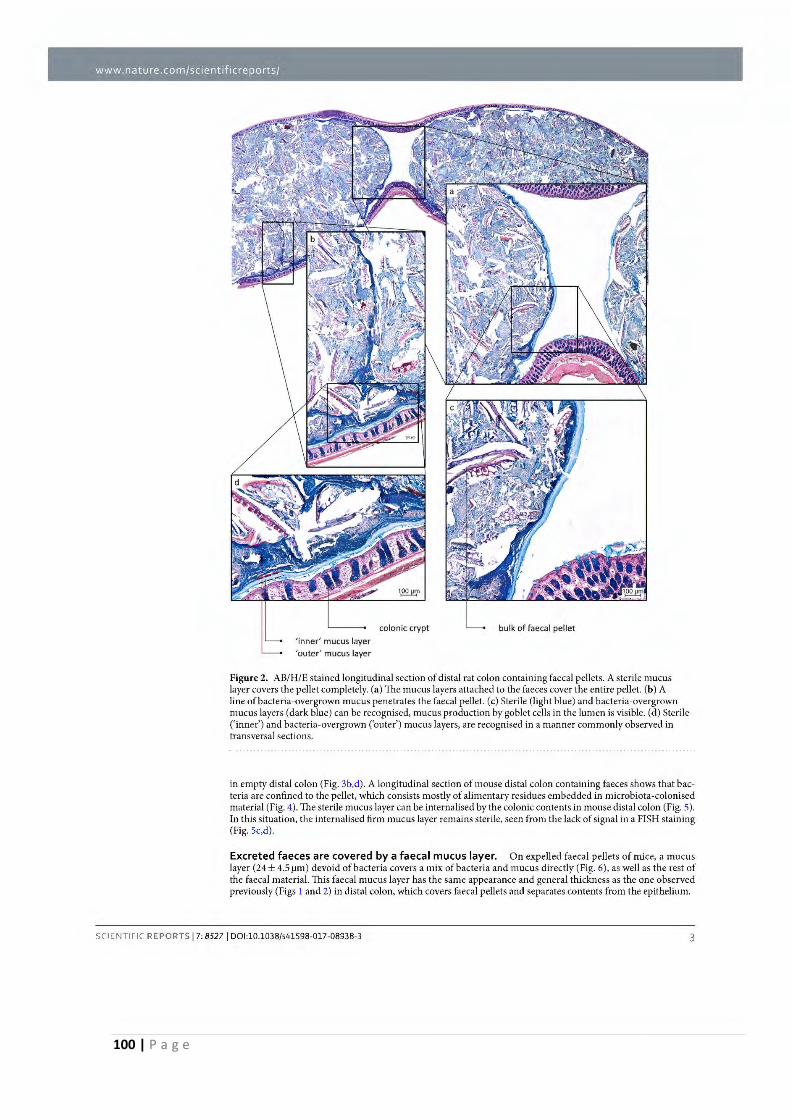
100 | P a g e
www. nature. com/sci entifi cre ports/
'inner' mucus layer
'outer' mucus layer
colonie crypt bulk of faecal pellet
Figure 2. AB/H/E stained longitudinal section of distal rat colon containing faecal pellets. A sterile mucus layer covers the pellet completely. (a) The mucus layers attached to the faeces cover the entire pellet. (b) A line ofbacteria-overgrown mucus penetrates the faecal pellet. (c) Sterile (light blue) and bacteria-overgrown mucus layers (dark blue) can be recognised, mucus production by goblet cells in the lumen is visible. (d) Sterile ('inner') and bacteria-overgrown ('outer') mucus layers, are recognised in a manner commonly observed in transversal sections.
in empty distal colon (Fig. 3b,d). A longitudinal section of mouse distal colon containing faeces shows that bacteria are confined to the pellet, which consists mostly of alimentary residues embedded in microbiota-colonised material (Fig. 4). The sterile mucus layer can be internalised by the colonie contents in mouse distal colon (Fig. 5). In this situation, the internalised firm mucus layer remains sterile, seen from the lack of signal in a FISH staining (Fig. Sc,d).
Excreted faeces are covered by a faecal mucus layer. On expelled faecal pellets of mice, a mucus layer (24 ± 4.5 µm ) devoid of bacteria covers a mix of bacteria and mucus directly (Fig. 6), as well as the rest of the faecal material. This faecal mucus layer has the same appearance and general thickness as the one observed previously (Figs I and 2) in distal colon, which covers faecal pellets and separates contents from the epithelium.
SCIENTIFIC REPORTS 17: 85271 DOl:10.1038/s41598-017-08938-3 3
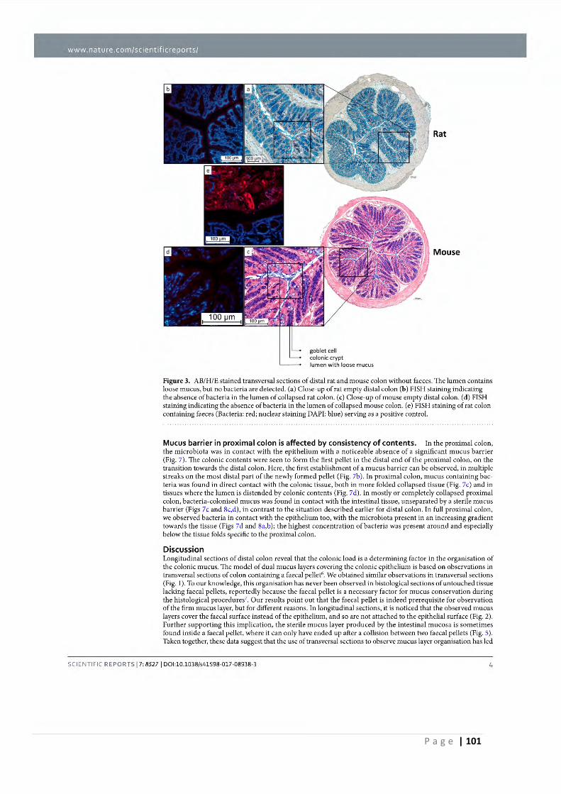
P a g e | 101
www. nature. corn/se i e ntifi cre ports/
goblet cell colonie crypt lumen with loose mucus
Rat
Mouse
Figure 3. AB/H/E stained transversal sections of distal rat and mouse colon without faeces. The lumen con tains loose mucus, but no bacteria are detected. (a) Close-up of rat empty distal colon (b) FISH staining indicating the absence ofbacteria in the lumen of collapsed rat colon. (c) Close-up of mouse empty distal colon. (d) FISH staining indicating the absence ofbacteria in the lumen of collapsed mouse colon. (e) FISH staining of rat colon containing faeces (Bacteria: red; nuclear stain ing DAPI: blue) serving as a positive control.
Mucus barrier in proximal colon is affected by consistency of contents. In the proximal colon, the microbiota was in contact with the epithelium with a noticeable absence of a significant mucus barrier (Fig. 7). The colonie contents were seen to form the first pellet in the distal end of the proximal colon, on the transition towards the distal colon. Here, the first establishment of a mucus barrier can be observed, in multiple streaks on the most distal part of the newly formed pellet (Fig. 7b). In proximal colon, mucus containing bacteria was found in direct contact with the colonie tissue, both in more folded collapsed tissue (Fig. 7c) and in tissues where the lumen is distended by colonie contents (Fig. 7d). In mostly or completely collapsed proximal colon, bacteria-colonised mucus was found in contact with the intestinal tissue, unseparated by a sterile mucus barrier (Figs 7c and 8c,d), in contras! to the situation described earlier for distal colon. In full proximal colon, we observed bacteria in contact with the epithelium too, with the microbiota present in an increasing gradient towards the tissue (Figs 7d and 8a,b); the highest concentration of bacteria was present around and especially below the tissue folds specific to the proximal colon.
Discussion Longitudinal sections of distal colon reveal that the colonie Joad is a determining factor in the organisation of the colonie mucus. The mode! of dual mucus layers covering the colonie epithelium is based on observations in transversal sections of colon containing a faecal pellet6• We obtained similar observations in transversal sections (Fig. 1). To our knowledge, this organisation has never been observed in histological sections ofuntouched tissue lacking faecal pellets, reportedly because the faecal pellet is a necessary fac tor fo r mucus conservation during the histological procedures7
• Our results point out that the faecal pellet is indeed prerequisite for observation of the firm mucus layer, but for different reasons. In longitudinal sections, it is noticed that the observed mucus layers cover the faecal surface instead of the epithelium, and so are not attached to the epithelial surface (Fig. 2). Further supporting this implication, the sterile mucus layer produced by the intestinal mucosa is sometimes found inside a faecal pellet, where it can only have ended up after a collision between two faecal pellets (Fig. 5). Taken together, these data suggest that the use of transversal sections to observe mucus layer organisation has led
SCIENTIFIC REPORTS 17: 85271 DOl:10.1038/s41598-017-08938-3 4
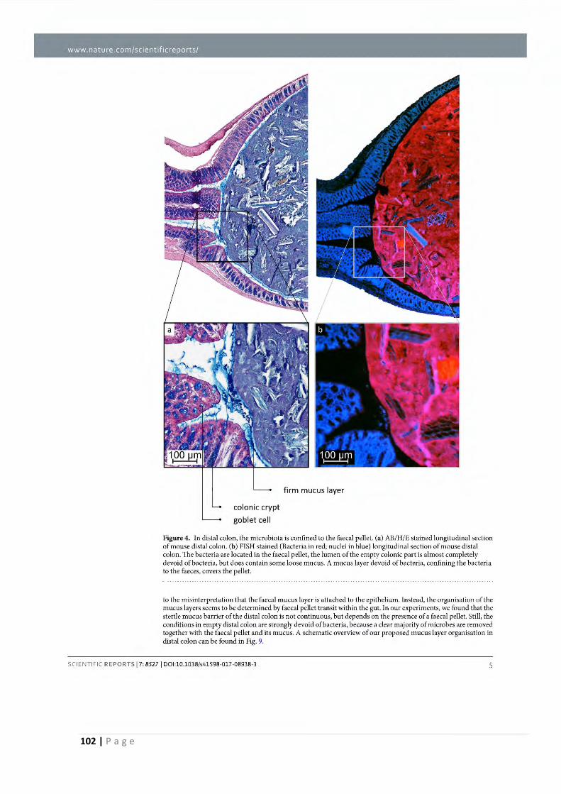
102 | P a g e
www. nature. corn/se i e ntifi cre ports/
colonie crypt
goblet cell
1'
100 µm ~
firm mucus layer
1 1
111!
> \
., . . •,,.. t. . ..
i..~ . ' ~,
Figure 4. ln distal colon, the microbiota is confined to the faecal pellet. (a) AB/H/E stained longitudinal section of mou se distal colon. (b) FISH stained (Bacteria in red; nuclei in blue) longitudinal section of mouse distal colon. The bacteria are located in the faecal pellet, the lumen of the empty colonie part is almost completely devoid ofbacteria, but does contain some loose mucus. A mucus layer devoid ofbacteria, confining the bacteria to the faeces, covers the pellet.
to the misinterpretation that the faecal mucus layer is attached to the epithelium. lnstead, the organisation of the mucus layers seems to be determined by faecal pellet transit within the gut. In our experiments, we found that the sterile mucus barrier of the distal colon is not continuous, but depends on the presence of a faecal pellet. Still, the conditions in empty distal colon are strongly devoid ofbacteria, because a clear majority of microbes are removed together with the faecal pellet and ils mucus. A schematic overview of our proposed mucus layer organisation in distal colon can be found in Fig. 9.
SCIENTIFIC REPORTS 17: 85271 DOl:10.1038/s41598-017-08938-3 5
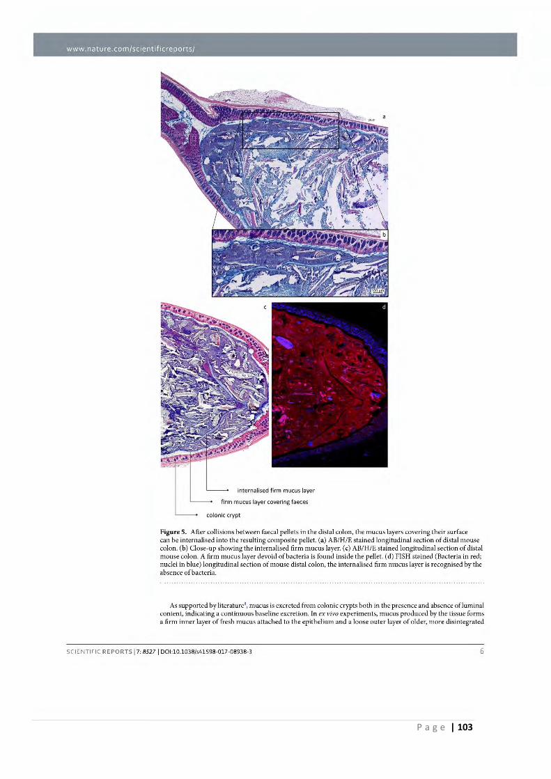
P a g e | 103
www. nature. com/sci entifi cre ports/
internalised firm mucus layer
firm mucus layer covering faeces
colonie crypt
Figure 5. After collisions between faecal pellets in the distal colon, the mucus layers covering their surface can be internalised into the resulting composite pellet. (a) AB/HIE stained longitudinal section of distal mouse colon. (b) Close-up showing the interna lised firm mucus layer. (c) AB/ H/E stained longitudinal section of distal mouse colon. A fi rm mucus layer devoid ofbacteria is found inside the pellet. (d) FISH stained (Bacteria in red; nuclei in blue} longitudinal section of mouse distal colon, the internalised firm mucus layer is recognised by the absence of bacteria.
As supported by literature8, mucus is excreted from colonie crypts both in the presence and absence ofluminal content, indicating a continuous baseline excretion. In ex vivo experiments, mucus produced by the t issue forms a firm inner layer of fresh mucus attached to the epithelium and a loose outer layer of older, more disintegrated
SCIENTIFIC REPORTS 17: 85271 DO l:10.1038/s41598-017-08938-3 6
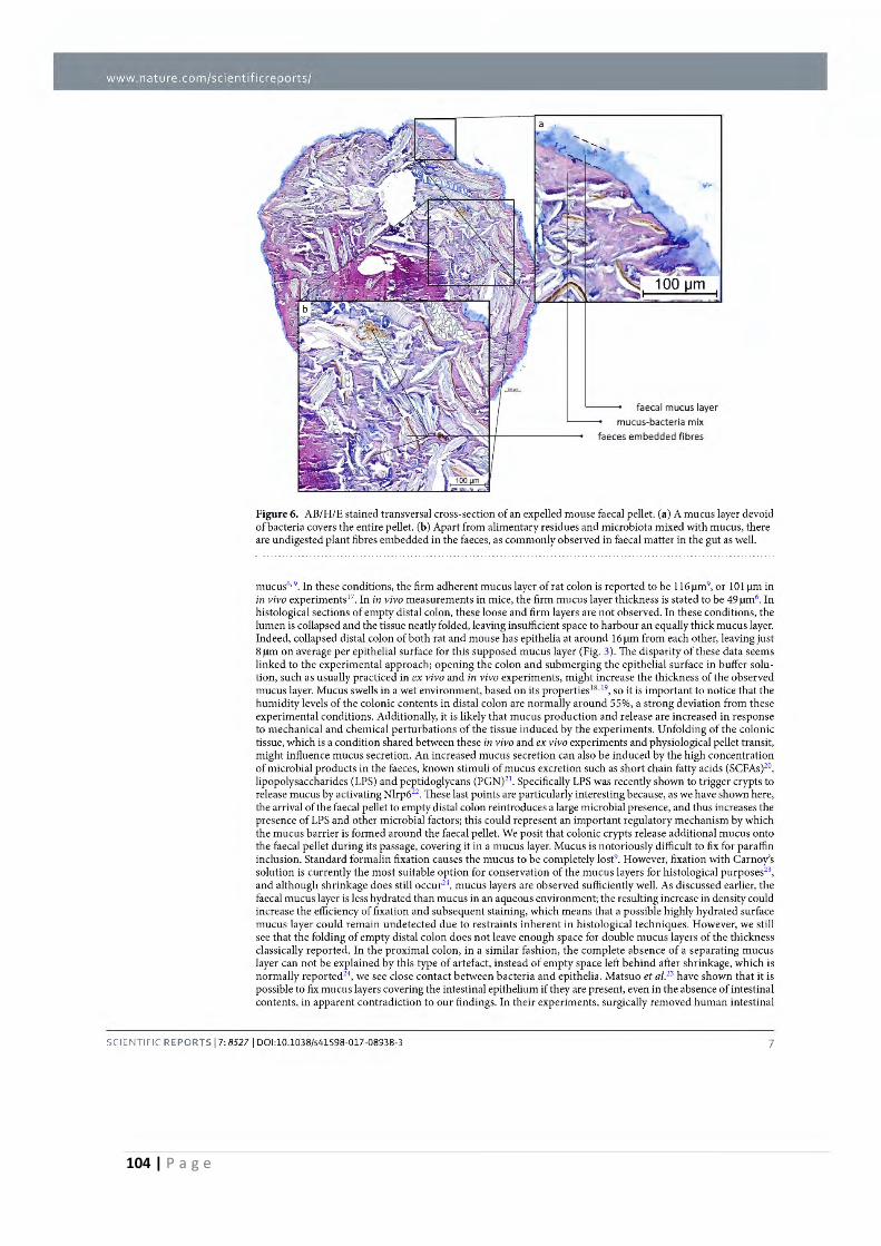
104 | P a g e
Bookmark 1
www. nature. corn/se i e ntifi cre ports/
faecal mucus layer mucus-bacteria mix
faeces embedded fibres
Figure 6. AB/H/E stained transversal cross-section of an expelled mouse faecal pellet. (a) A mucus layer devoid ofbacteria covers the entire pellet. (b) Apart from alimentary residues and microbiota mixed with mucus, there are undigested plant fibres embedded in the faeces, as commonly observed in faecal malter in the gut as well.
mucus6•9
• In these conditions, the firm adherent mucus layer of rat colon is reported to be l 16µm9, or 101 µmin
in vivo experiments17• In in vivo measurements in mice, the firm mucus layer thickness is stated to be 49 µm•. In histological sections of empty distal colon, these loose and firm layers are not observed. In these conditions, the lumen is collapsed and the tissue neatly folded, leaving insufficient space to harbour an equally thick mucus layer. Indeed, collapsed distal colon of both rat and mouse has epithelia at around 16 µm from each other, leaving just 8 µmon average per epithelial surface for this supposed mucus layer (Fig. 3). The disparity of these data seems linked to the experimental approach; opening the colon and submerging the epithelial surface in buffer solution, such as usually pract iced in ex vivo and in vivo experiments, might increase the thickness of the observed mucus layer. Mucus swells in a wet environment, based on its properties 18• 19, soit is important to notice that the humidity levels of the colonie contents in distal colon are normally around 55%, a strong deviation from these experimental conditions. Additionally, it is likely that mucus production and release are increased in response to mechanical and chemical perturbations of the tissue induced by the experiments. Unfolding of the colonie tissue, which is a condition shared between these in vivo and ex vivo experiments and physiological pellet transit, might influence mucus secretion. An increased mucus secretion can also be induced by the high concentration of microbial products in the faeces, known stimuli of mucus excretion such as short chain fatty acids (SCFAs)20,
lipopolysaccharides (LPS) and peptidoglycans (PGN)21• Specifically LPS was recently shown to trigger crypts to
release mucus by activating Nlrp622• These last points are particularly interesting because, as we have shown here, the arrivai of the faecal pellet to empty distal colon reintroduces a large microbial presence, and thus increases the presence of LPS and other microbial factors; this could represent an important regulatory mechanism by which the mucus barrier is formed around the faecal pellet. We posit that colonie crypts release additional mucus onto the faecal pellet during ils passage, covering it in a mucus layer. Mucus is notoriously difficult to fix for paraffin inclusion. Standard formalin fixation causes the mucus to be completely lost9• However, fixation with Carnoy's solution is currently the most sui table option for conservation of the mucus layers for histological purposes23,
and although shrinkage does still occur24, mucus layers are observed sufficiently well. As discussed earlier, the faecal mucus layer is Jess hydrated than mucus in an aqueous environ ment; the resulting increase in density could increase the efficiency of fixation and subsequent staining, which means that a possible highly hydrated surface mucus layer could remain undetected due to restraints inherent in histological techniques. However, we still see that the fo l ding of empty distal colon does not leave enough space for double mucus layers of the thickness classically reported. In the proximal colon, in a similar fashion, the complete absence of a separating mucus layer can not be explained by this type of artefact, instead of empty space left behind after shrinkage, which is normally reported24, we see close contact between bacteria and epithelia. Matsuo et al.23 have shown that it is possible to fix mucus layers covering the intestinal epithelium if they are present, even in the absence of intestinal contents, in apparent contradiction to our findings. In their experiments, surgically removed human intestinal
SCIENTIFIC REPORTS 17: 85271 DOl:10.1038/s41598-017-08938-3 7
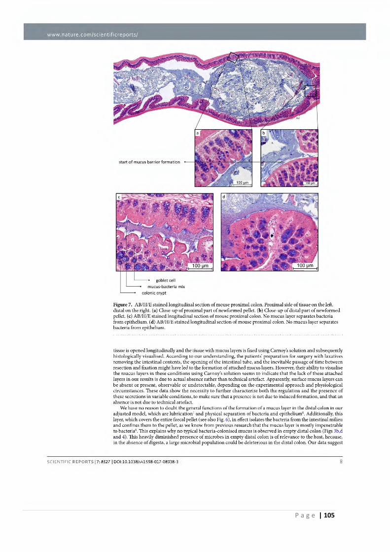
P a g e | 105
www. nature. corn/se i entifi cre ports/
start of mucus barrier formation
goblet cell
mucus-bacteria mix
colonie crypt
Figure 7. AB/H/E stained longitudinal section of mouse proximal colon. Proximal sicle of tissue on the left, distal on the right. (a) Close-up of proximal part of newformed pellet. (b) Close-up of distal part of newformed pellet. (c) AB/H/E stained longitudinal section of mouse proximal colon. No mucus layer separates bacteria from epithelium. (d) AB/H/E stained longitudinal section of mouse proximal colon. No mucus layer separates bacteria from epithelium.
tissue is opened longitudinally and the tissue with mucus layers is fixed using Carnoy's solution and subsequently histologically visualised. According to our understanding, the patients' preparation for surgery with laxatives removing the intestinal contents, the opening of the intestinal tube, and the inevitable passage of time between resection and fixation might have led to the formation of attached mucus layers. However, their ability to visualise the mucus layers in these conditions using Carnoy's solution seems to indicate that the lack of these attached layers in our results is due to actual absence rather than technical artefact. Apparently, surface mucus layers can be absent or present, observable or undetectable, depending on the exper imental approach and physiological circumstances. These data show the necessity to further characterise both the regulat ion and the presence of these secretions in variable conditions, to make sure that a presence is not due to induced formation, and that an absence is not due to technical artefact.
We have no reason to doubt the general fonctions of the formation of a mucus layer in the distal colon in our adjusted mode!, which are lubrication 1 and physical separation of bacteria and epithelium6 • Addit ionally, th is layer, which covers the entire faecal pellet (see also Fig. 6), in effect isolates the bacteria from the intestinal milieu and confines them to the pellet, as we know from previous research that the mucus layer is mostly impenetrable to bacteria6. This explains why no typical bacteria-colonised mucus is observed in empty distal colon (Figs 3b,d and 4). This heavily diminished presence of microbes in empty distal colon is of relevance to the host, because, in the absence of digesta, a large microbial population could be deleterious in the distal colon. Our data suggest
SCIENTIFIC REPORTS 17: 85271 DO l:10.1038/s41598-017-08938-3 8
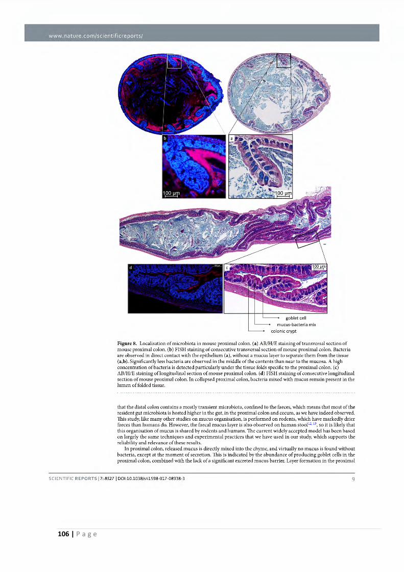
106 | P a g e
www. nature. corn/se i e ntifi cre ports/
mucus-bacteria mix
colonie crypt
Figure 8. Localisation of microbiota in mouse proximal colon. (a) AB/H/E staining of transversal section of mouse proximal colon. (b) FISH staining of consecutive transversal section of mouse proximal colon. Bacteria are observed in direct contact with the epithelium (a), without a mucus layer to separate them from the tissue (a,b). Significantly Jess bacteria are observed in the middle of the contents than near to the mucosa. A high concentration ofbacteria is detected particularly under the tissue folds specific to the proximal colon. (c) AB/H/E staining of longitudinal section of mouse proximal colon. (d) FISH staining of consecutive longitudinal section of mouse proximal colon. In collapsed proximal colon, bacteria mixed with mucus remain present in the lumen of folded tissue.
that the distal colon contains a mostly transient microbiota, confined to the faeces, which means that most of the resident gut microbiota is hosted higher in the gut, in the proximal colon and cecum, as we have indeed observed. This study, like many other stud ies on mucus organisation, is performed on rodents, which have markedly drier faeces than humans do. However, the faecal mucus layer is also observed on human stool 12·
13, so it is likely that
this organisation of mucus is shared by rodents and humans. The current widely accepted mode! has been based on largely the same techn iques and experimental practices that we have used in our study, which supports the reliability and relevance of these results.
In proximal colon, released mucus is directly mixed into the chyme, and virtually no mucus is found without bacteria, except at the moment of secretion. This is indicated by the abundance of producing goblet cells in the proximal colon, combined with the lack of a significant excreted mucus barrier. Layer formation in the proximal
SCIENTIFIC REPORTS 17: 85271 DOl:10.1038/s41598-017-08938-3 9
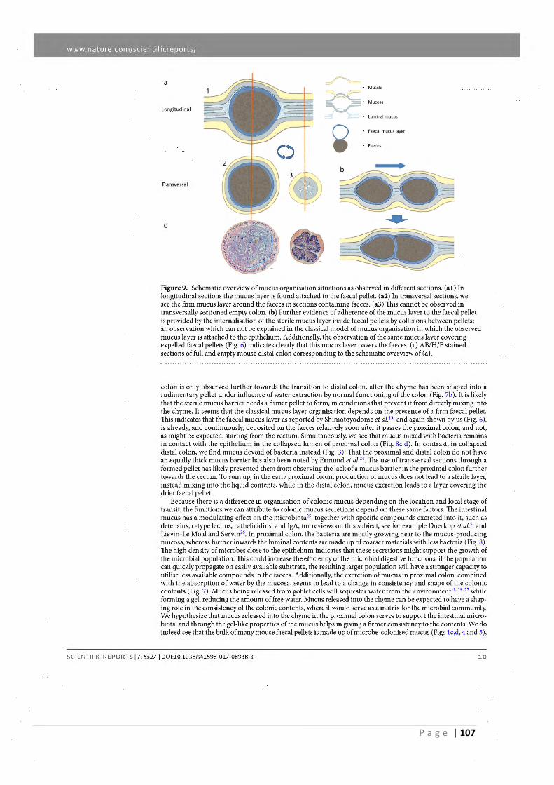
P a g e | 107
www. nature. corn/se i e ntifi cre ports/
a r-.. Muscle
~ -Mucosa
longitudinal
Luminal mucus
~ Faecal mucus layer
Faeces
Transversal
C
Figure 9. Schematic overview of mucus organisation situations as observed in different sect ions. (al ) ln longitudinal sections the mucus layer is found attached to the faecal pellet. (a2) In transversal sections, we see the firm mucus layer around the faeces in sections containing faeces. (a3) This cannot be observed in transversally sectioned empty colon. (b) Further evidence of adherence of the mucus layer to the faecal pellet is provided by the internalisation of the sterile mucus layer inside faecal pellets by collisions between pellets; an observation which can not be explained in the classical mode! of mucus organisation in which the observed mucus layer is attached to the epithelium. Additionally, the observation of the same mucus layer covering expelled faecal pellets (Fig. 6) indicates clearly that this mucus layer covers the faeces. (c) AB/H/E stained sections offull and empty mouse distal colon corresponding to the schematic overview of (a) .
colon is only observed forther towards the transition to distal colon, after the chyme has been shaped into a rudimentary pellet under influence of water extraction by normal fonctioning of the colon (Fig. 7b). It is likely that the sterile mucus barrier needs a firmer pellet to form, in conditions that prevent it from directly mixing into the chyme. lt seems that the classical mucus layer organisation depends on the presence of a firm faecal pellet. This indicates that the faecal mucus layer as reported by Shimotoyodome et al. 13, and again shown by us (Fig. 6), is already, and continuously, deposited on the faeces relatively soon after it passes the proximal colon, and not, as might be expected, starting from the rectum. Simultaneously, we see that mucus mixed with bacteria remains in contact with the epithelium in the collapsed lumen of proximal colon (Fig. Sc,d). In contras!, in collapsed distal colon, we find mucus devoid ofbacteria instead (Fig. 3). Thal the proximal and distal colon do not have an equally thick mucus barrier has also been noted by Ermund et al.24. The use of transversal sections through a formed pellet has likely prevented them from observing the lack of a mucus barrier in the proximal colon forther towards the cecum. To sum up, in the early proximal colon, production of mucus does not lead to a sterile layer, instead mixing into the liquid contents, while in the distal colon, mucus excretion leads to a layer covering the drier faecal pellet.
Because there is a difference in organisation of colonie mucus depending on the location and local stage of transit, the fonctions we can attribute to colonie mucus secretions depend on these same factors. The intestinal mucus has a modulating effect on the microbiota25
, together with specific compounds excreted into it, such as defensins, c-type lectins, cathelicidins, and IgA; for reviews on this subject, see for example Duerkop et al.', and Liévin-Le Moal and Servin26. In proximal colon, the bacteria are mostly growing near to the mucus-producing mucosa, whereas forther inwards the luminal contents are made up of coarser materials with less bacteria (Fig. 8). The high density of microbes close to the epithelium indicates that these secretions might support the growth of the microbial population. This could increase the efficiency of the microbial digestive fonctions; if the population can quick:ly propagate on easily available substrate, the resulting larger population will have a stronger capacity to utilise less available compounds in the faeces. Additionally, the excretion of mucus in proximal colon, combined with the absorption of water by the mucosa, seems to lead to a change in consistency and shape of the colonie contents (Fig. 7). Mucus being released from goblet cells will sequester water from the environment18
• 19
•27 while
forming a gel, reducing the amount of free water. Mucus released into the chyme can be expected to have a shaping role in the consistency of the colonie contents, where it would serve as a matrix for the microbial commun ity. We hypothesize that mucus released into the chyme in the proximal colon serves to support the intestinal microbiota, and through the gel-like properties of the mucus helps in giving a firmer consistency to the contents. We do indeed see that the bulk of many rnouse faecal pellets is made up of microbe-colonised mucus (Figs lc,d, 4 and 5),
SCIENTIFIC REPORTS 17: 85271 DOl:10.1038/s41598-017-08938-3 10
108 | P a g e
www. nature. corn/se i e ntifi cre ports/
indistinguishable from the described6 loose mucus layer containing bacteria. Regarding resource and energy efficiency, it seems indeed more expedient to cover the faecal pellet in a thick mucus layer than to cover the entire colonie epithelium, to separate these two components. Clearly, a lubricating mucus layer becomes more crucial as the pellet gels drier and more compact as il progresses through the colon, to prevent damaging abrasion of the tissue. In the proximal colon, when the contents are still liquid, a mucus barrier does not form, even though there is significant production. Direct exposure of the colonie epithelium to the microbiota and digesta is generally expected to cause a strong immune reaction and tissue damage, the prevention of which is often cited as an important function of the mucus barrier. The apparent lack of these effects in these physiological conditions in the proximal colon warrants further investigation.
Our MUCIO approach to histology, in combination with multiple orientation sectioning, has allowed for a comprehensive overview of the evolution of the mucus barrier throughout the colon. Until now, h istological results are commonly depicted in single-image microscopie views, which can complicate a thorough understanding of the full results, might underrepresent possible variations throughout the section, and can give rise to a selection bias for 'sui table regions' on a slide.
In conclusion, we have shown that a mucus layer is attached to and covers faecal pellets in the distal colon, isolating the faecal bacteria from the intestinal lumen. Mucus is mixed into the chyme and faeces in the proximal colon and in collision events between faeces segments in the distal colon. In the proximal colon, in contras! to the distal colon, no firm mucus barrier is formed until the chyme starts to gain a pellet structure. Before the mucus layer is established, bacteria are in contact with the epithelial surface, and in these conditions, we should reconsider the nature of the barrier function in this region of the colon.
Materials and Methods Animais and sample collection. 8 adult male Wistar rats (Janvier, Le Genest St Isle, France) were individually housed in polypropylene cages and offered unlimited access to standard rodent food (Mucedola Global Diet 2018, Harlan, Italy) and water. 8 adult male C57BL/6 mice (Janvier, Le Genest St Isle, France) were housed in polypropylene cages in groups of 4 and offered unlimited access to standard rodent food (Mucedola Global Diet 2018, Harlan, Italy) and water. Rats were euthanized by decapitation, after which 3 cm of distal colon containing faeces was removed and stored in Carnoy's fixat ive (60% ethanol, 30% chloroform, 10% glacial acetic acid) overnight. Mice were euthanized by cervica l dislocation, after which both 1.5 to 2 cm of distal colon and of proximal colon covering regions with and without contents were removed and stored in Carnoy's fixative overnight. Mouse faecal pellets were collected d irectly from the anus, and fixated immediately in Carnoy's fixative overnight. Ali animal experiments were performed in accordance with EU directive 20 10/63/EU and approved by the local Animal Care and Use Committee of Toulouse Midi-Pyrénées (agreement CEEA-86).
Tissue processing. The mice and rat tissues were automatically processed by a Shandon™ Excelsior™ ES Tissue Processor using the following program: 2 x 60 min anhydrous ethanol, 2 x 60 min butanol, 480 min butanol, 3 x 80 min paraffin al 60 °C. Tissue samples were included in paraffin blocks using a Thermo Scientific ™ HistoStar™ Embedding Workstation. Tissues were oriented for longitudinal or transversal sectioning. 6µm tissue sections, and 4 µm faecal sections were made using a Microm TM HM 340 E microtome and attached to Thermo Scientific Menzel-Glaser Superfrost® Plus slides.
Histological staining (AB/H/E staining) . Paraffin embedded sections were deparaffinised using American Master tech Clearify™ and subsequent passage through an increasingly diluted ethanol-water series, starting with anhydrous ethanol. Staining was performed by 5 min in Hematoxylin, 10 min in running water, 30 min in Alcian Blue solution (pH 3.0) followed by 5 min in running water, 3 min in Eosin, 10 min in 95% ethanol, followed by dehyd ration in an ethanol series of increasing purity, finishing with dry ethanol, ending with 3 baths of American Mastertech Clearify™, followed by mounting with Diamount mountant.
Fluorescent in situ Hybridisation. FISH staining for ail bacteria was performed using probes EUB3381 (5' CGTGCCTCCCGTAGGAGT 31
) , EUB338Il (5' GCAGCCACCCGTAGGTGT 3'), and EUB338III (51 GCTGCCACCCGTAGGTGT 3'). After deparaffin ising as described for 'Histological staining' (see above), the slides were incubated with FISH hybridisation solution (0.9 M NaCI, 20 mM Tris/HCI, 0.01 % (v/v) SDS, with 5 ng/µL of EUB338(1/II/III) probes at 48 °C overnight, followed by a washjng step with the hybridisation solution without probe at 49 °C for 25 min. Slides were rinsed with demineralised water and briefly air-dried, followed by mounting using ProLong Gold® antifade reagent with DAPI (Thermo Fisher Scientific, USA).
lmaging; Manual Ultra-high resolution Composite Image Overview (MUCIO). Manual Ultra-high resolution Composite Image Overview (MUCIO) approach: datasets of 50 to 500 overlapping microscope views covering entire slides were generated by manual microscope photography (single photo resolution: 1280 x 1024 pixels) and stitched together using Microsoft Image Composite Editor (MICE), specifying 'planar motion' in the program interface, to best fit the movement of the microscope camera relative to the microscopie slide. The generated composite images contain ail the information of the data set in a single ultra-high resolution file. No selection for 'representative regions' is necessary, only obvious artefacts (e.g. possibly folded double/heavily damaged sections) are evaded. Sam pies were imaged using a Nikon Eclipse 90i microscope fitted with a DXM 1200 F Digital Camera. Image sets were taken with 1 00x or 200x magnification, overlapping and covering complete sections. Resulting images were saved uncompressed in TIFF format. To prevent scaling issues, image resolutions were only downscaled after the appropriate scale bar was integrated. Subsequent downscaling is necessary to
SCIENTIFIC REPORTS 17: 85271 DO l:10.1038/s41598-017-08938-3 11
P a g e | 109
www. nature. corn/se i e ntifi cre ports/
fac ilitate interaction with images for further applications, but close-up views in full quality can be extracted from the original composite.
Data availability. The data generated and analysed during the current study are available from the corresponding author upon reasonable request.
References 1. Florey, H. The Croonian Lecture: Mucin and the protection of the body. Proceedings of the Royal Society of London. Series B.
Biological Sciences, 147- 158 (1955). 2. Van der Sluis, M. et al, Muc2-deficient mice spontaneously develop colitis, indicating that MUC2 is critical for colonie protection.
Gastroenterology 131, 117-129 (2006). 3. Bergstrom, K. S. et al. Muc2 protects against lethal infectious colitis by disassociating pathogenic and commensal bacteria from the
colonie mucosa. PLoS Pathog 6, el 000902, doi: 10.1371/journal.ppat. 1000902 (2010). 4. Backhed, F., Ley, R. E., Sonnenburg, J. L., Pe terson, O. A. & Gordon, J. 1. Host-bacterial mutualism in the human intestine. Science
307, 1915-1920(2005). S. Duerkop, B. A., Vaishnava, S. & Hooper, L. V. Immune responses to the microbiota at the intestinal mucosal surface. Immunity 31,
368-376 (2009). 6. Johansson, M. E. et al. The inner of the two Muc2 mucin-dependent mucus layers in colon is devoid ofbacteria. Proceedings of the
national academy of sciences 105, 15064-15069 (2008). 7. Johansson, M. E., Larsson, J. M. H. & Hansson, G. C. The two mucus layers of colon are organized by the MUC2 mucin, whereas the
outer layer is a legislator of host-microbial interactions. Proceedings of the national academy of sciences 108, 4659-4665 (2011). 8. Gustafsson, J. K. et al. An ex vivo method for studying mucus formation, properties, and thickness in human colonie biopsies and
mouse small and large intestinal explants. Arnerican Journal of Physiology-Gastrointestinal and Liver Physiology 302, G430- G438 (2012).
9. Atuma, C., Strugala, V., Allen, A. & Holrn, L. The adherent gastrointestinal mucus gel layer: th ickness and physical state in vivo. American Journal of Physiology-Gastrointestinal and Liver Physiology 280, G922- G929(2001 ).
10. McGuckin, M. A., Lindén, S. K., Sutton, P. & Florin, T. H. Mucin dynamics and enteric pathogens. Nature Reviews Microbio/ogy 9, 265- 278 (2011).
11. Johansson, M. E. V. et al. Normalization of Host Intestinal Mucus Layers Requires Long-Tenu Microbial Colonization. Cell Host & Microbe 18, 582-592, doi:I0. 1016/j.chom.2015. I0.007 (2015)
12. Swidsinski, A., Loening- Baucke, V., Verstraelen, H., Osowska, S. & Doerffel, Y. Biostructure of fecal microbiota in healthy subjects and patients with chronic idiopathie diarrhea. Gastroenterology 135, 568-579.e562 (2008).
13. Shimotoyodome, A., Meguo, S., Tokimitsu, 1. & Sakata, T. Histochemical structure of the mucus gel layer coating the fecal surface of rodents, rabbits and humans. Journal of nutritional science and vitaminology 51, 287- 291 (2005).
14. Johansson, M. E. et al. Bacteria penetrate the normally impenetrable inner colon mucus layer in bath murine colitis models and patients with ulcerative colitis. Gut, gutjnl-2012-303207 (2013).
15. McMahon, R. F. T., Jones, C. J. P., Dutt, S. & Stoddart, R. W Altered oligosaccharide expression in ukerative colitis with increasing grades of inflammation. Glycosylation & Disease 1, 235-245, <loi: 10.1007 /bf00919331 ( 1994).
16. Da Silva, S. et al. Stress disrupts intestinal mucus barrier in rats via mucin O-glycosylation shift: prevention by a probiotic treatment. American Journal of Physiology-Gastrointestinal and Liver Physiology 307, G420-G429 (201 4).
17. Strugala, V., Allen, A., Dettmar, P. W. & Pearson, J. P. Colonie mucin: methods ofmeasuring mucus thickness. Proceedings of the Nutrition Society 62, 237-243 (2003).
18. Forstner, G. Signal transduction packaging and secretion of mucins. Ammal Review of Physiology 57, 585-605 (1995). 19. Verdugo, P. Go blet cells secret ion and mucogenesis. Annua/ review of physiology 52, 157- 176 (1990). 20. Shimotoyodome, A., Meguro, S., Hase, T., Tok.imitsu, L & Sakata, T. Short chain fatty acids but not lactate or succinate stimulate
mucus release in the rat colon. Comparative Biochemistry and Physiology Part A: Molecular & Integrative Physiology 125, 525-531, <loi: I0.1016/SI095-6433(00)00183-5 (2000)
21. Petersson, J. et al. Importance and regulation of the colonie mucus barrier in a mouse model of colitis. Am J Physiol Gastrointest Liver Physiol 300, G327-333, <loi: IO. ll52/ajpgi.00422.2010 (201 1 ).
22. Birchenough, G. M., Nystrom, E. E., Johansson, M. E. & Hansson, G. C. A sentine! goblet cell guards the colonie crypt by triggering Nlrp6-dependent Muc2 secretion. Science 352, 1535-1542 (2016).
23. Matsuo, K., Ota, H., Akamatsu, T., Sugiyama, A. & Katsuyama, T. Histochemistry of the surface mucous gel layer of the human colon. Gut 40, 782-789 (1997).
24. Ermund, A., Schutte, A., Johansson, M. E., Gustafsson, J. K. & Hansson, G. C. Studies of mucus in mouse stomach, small intestine, and colon. l. Gastroin testinal mucus layers have different properties depending on location as well as over the Peyer's patches. Am J Physiol Gastrointest Liver Physiol 30S, G34 l -347, <loi: IO. l l 52/ajpgi.00046.2013 (2013).
25. Deplancke, B. & Gaskins, H . R. Microbial modulation ofinnate defense: goblet cells and the intestinal mucus layer. Tiu A,nerican journal of clinical nutrition 73, 1131S-1 141S (2001).
26. Liévin-Le Moal, V. & Servin, A. L. The front line of enteric hast defense against unwelcome intrusion ofharmful microorgan isms: mucins, antimicrobial peptides, and microbiota. Clinicat microbiology reviews 19, 315-337 (2006).
27. Ambort, D. et al. Calcium and pH-dependent packing and release of the gel-forming MUC2 mucin. Proceedings of the National Academy of Sciences 109, 5645-5650 (2012).
Acknowledgements The research leading to these results has received funding from the People Programme of the EU's 7th Framework Programme under REA grant agreement no. 607652 (ITN NeuroGut).
Author Contributions J.B.J.K., H.E., and V.T. designed the study; J.B.J.K. developed the methodology and performed the research; J.B.J.K., M.M.B., H.E., and V:T. analysed and interpreted the data, and wrote the paper.
Additional Information Competing Interests: The authors declare that they have no competing interests.
Publisher's note: Springer Nature remains neutral with regard to jurisdictional daims in published maps and institutional affiliations.
SCIENTIFIC REPORTS 17: 85271 DO l:10.1038/s41598-017-08938-3 12
110 | P a g e
`` P a g e | 111
2.2 FODMAPs increase visceral sensitivity in mice through glycation processes,
increasing mast cell counts in colonic mucosae
This work regroups the main findings of the thesis work, i.e., the effects of FODMAP
administration on visceral sensitivity, the mucosal mast cell population, the expression of
Receptor for AGEs (RAGE), and the prevention of these effects by anti-glycation agent
pyridoxamine, indicating the involvement of glycating processes. Analysis of the gut microbiota
composition showed no significant effects of our lactose treatment on population composition,
and no effect for pyridoxamine either. Carbonyl metabolite analysis showed differences
between control and FODMAP treated groups, but no additional effect by co-treatment with
pyridoxamine.
These results and this manuscript are currently under a second round of reviewing in the
Gastroenterology journal (Elsevier) as an original article.
112 | P a g e
P a g e | 113
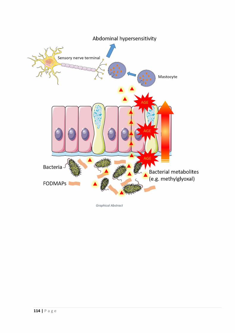
114 | P a g e
Graphical Abstract
P a g e | 115
116 | P a g e
P a g e | 117
118 | P a g e
P a g e | 119
120 | P a g e
P a g e | 121
122 | P a g e
P a g e | 123
124 | P a g e
P a g e | 125
126 | P a g e
P a g e | 127
128 | P a g e
P a g e | 129
130 | P a g e
P a g e | 131
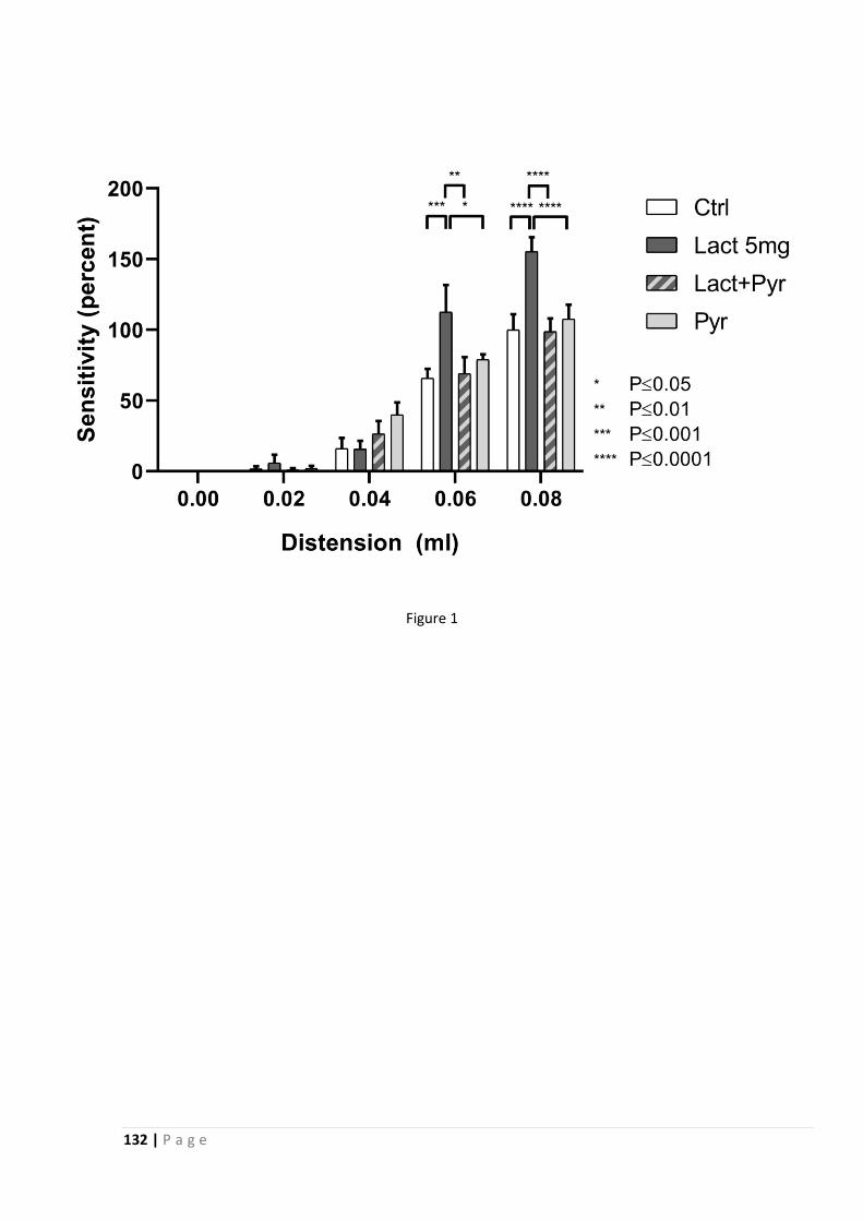
132 | P a g e
Figure 1
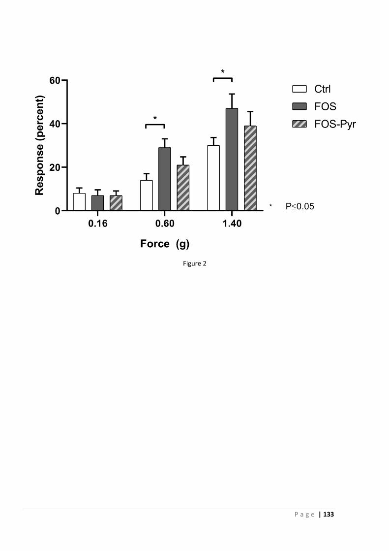
P a g e | 133
Figure 2
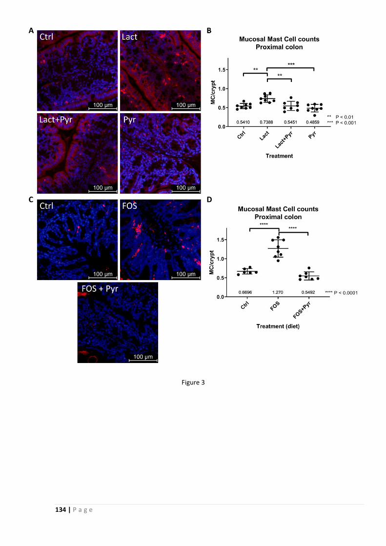
134 | P a g e
Figure 3
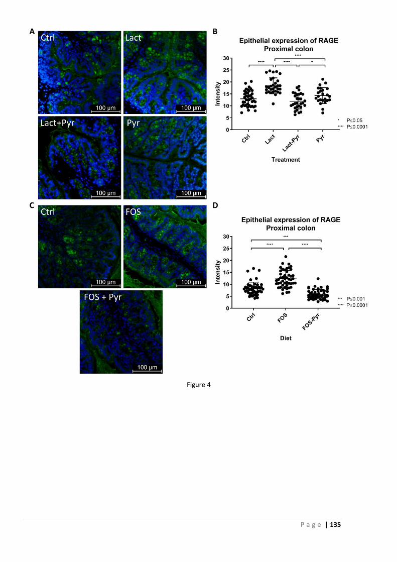
P a g e | 135
Figure 4
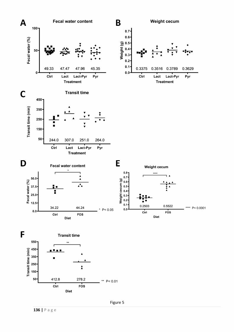
136 | P a g e
Figure 5
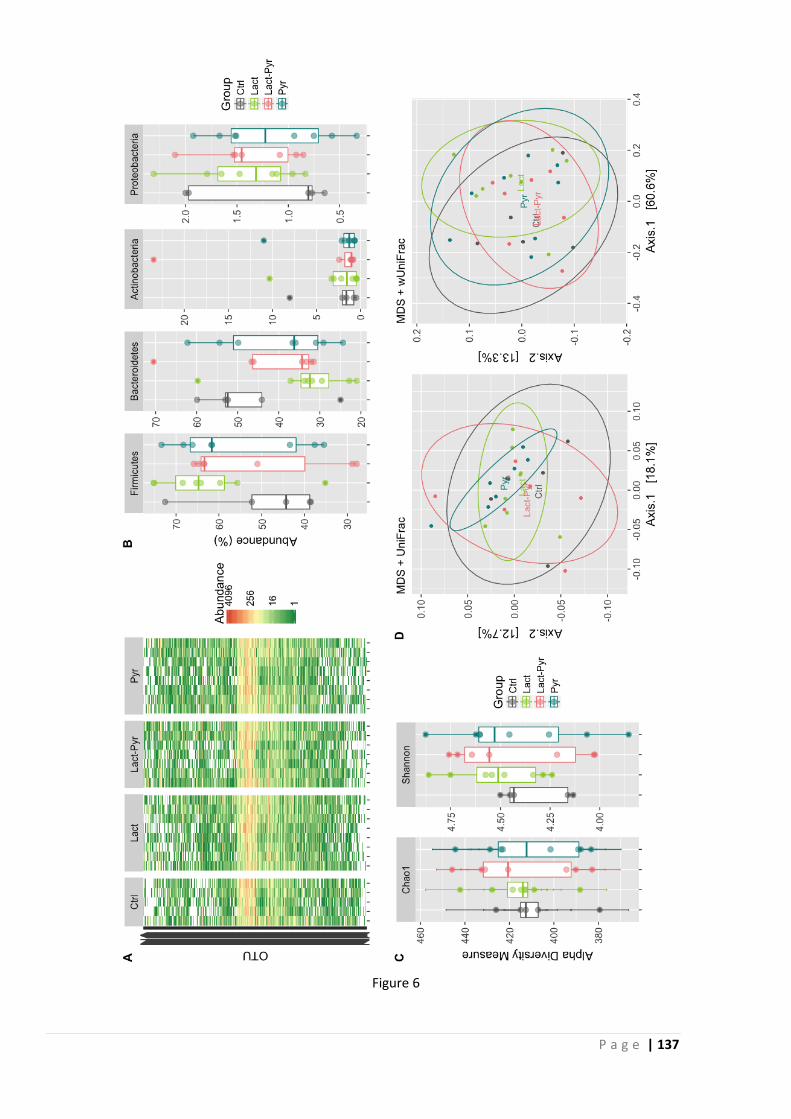
P a g e | 137
Figure 6
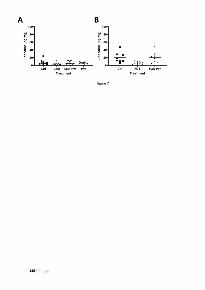
138 | P a g e
Figure 7
P a g e | 139
140 | P a g e
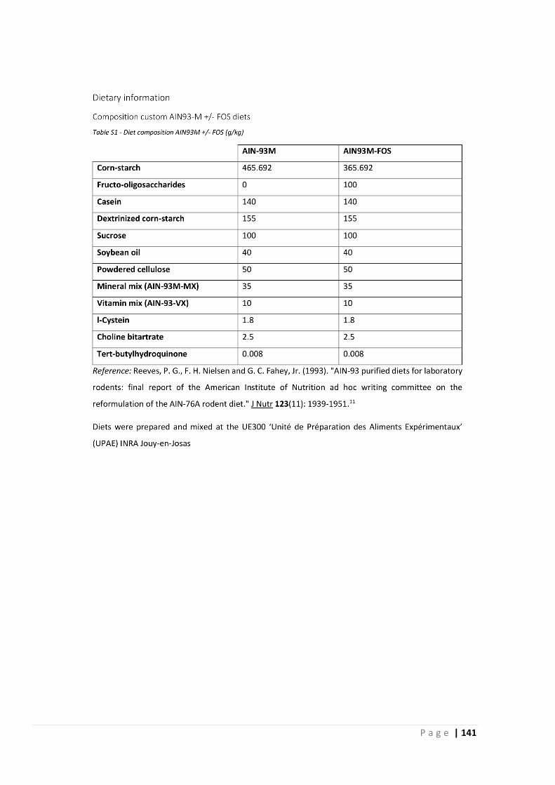
P a g e | 141
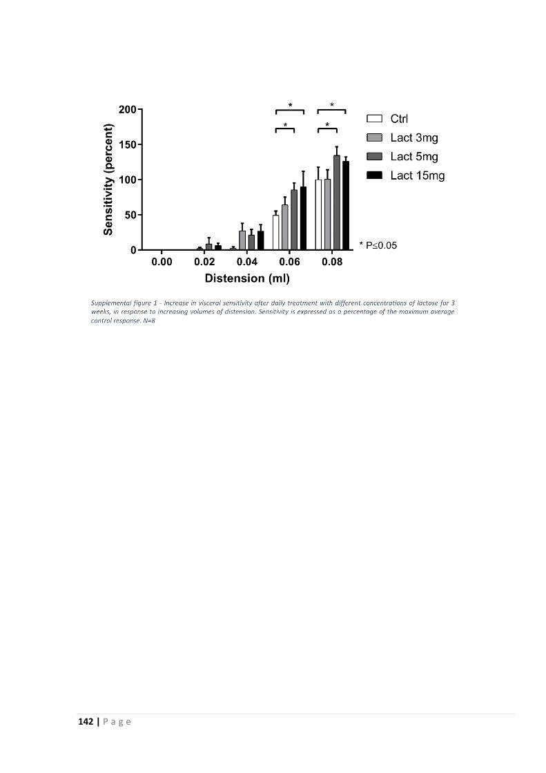
142 | P a g e
P a g e | 143
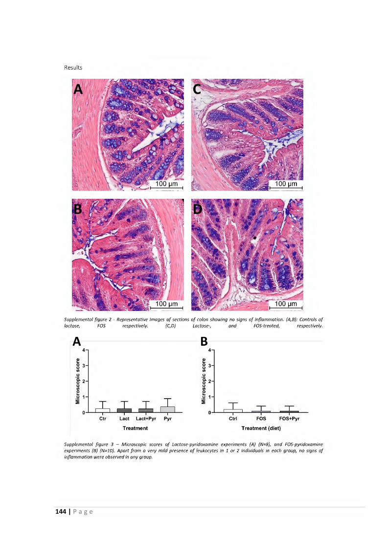
144 | P a g e
Results
Supplemental figure 2 - Representative images af sections af colon showing no signs of inf lammation. {A,B}: Contrais of lactose, FOS respective/y. {C,D) Lactose-, and FOS-treated, respective/y.
A
Ctr Lact Lact+Pyr Pyr
Treatment
B 4
f 8 J 1/)
(,)
·g- 2 (,) 1/) 0 ... (,)
i
Ctrl FOS FOS+Pyr
Treatment (diet)
Supplemental figure 3 - Microscopie scores of Lactose-pyridoxamine experiments (A) (N=B), and FOS-pyridoxamine experiments (B) (N=lO}. Apart /rom a very mild presence of leukocytes in 1 or 2 individua/s in each group, no signs of inflammation were observed in any group.
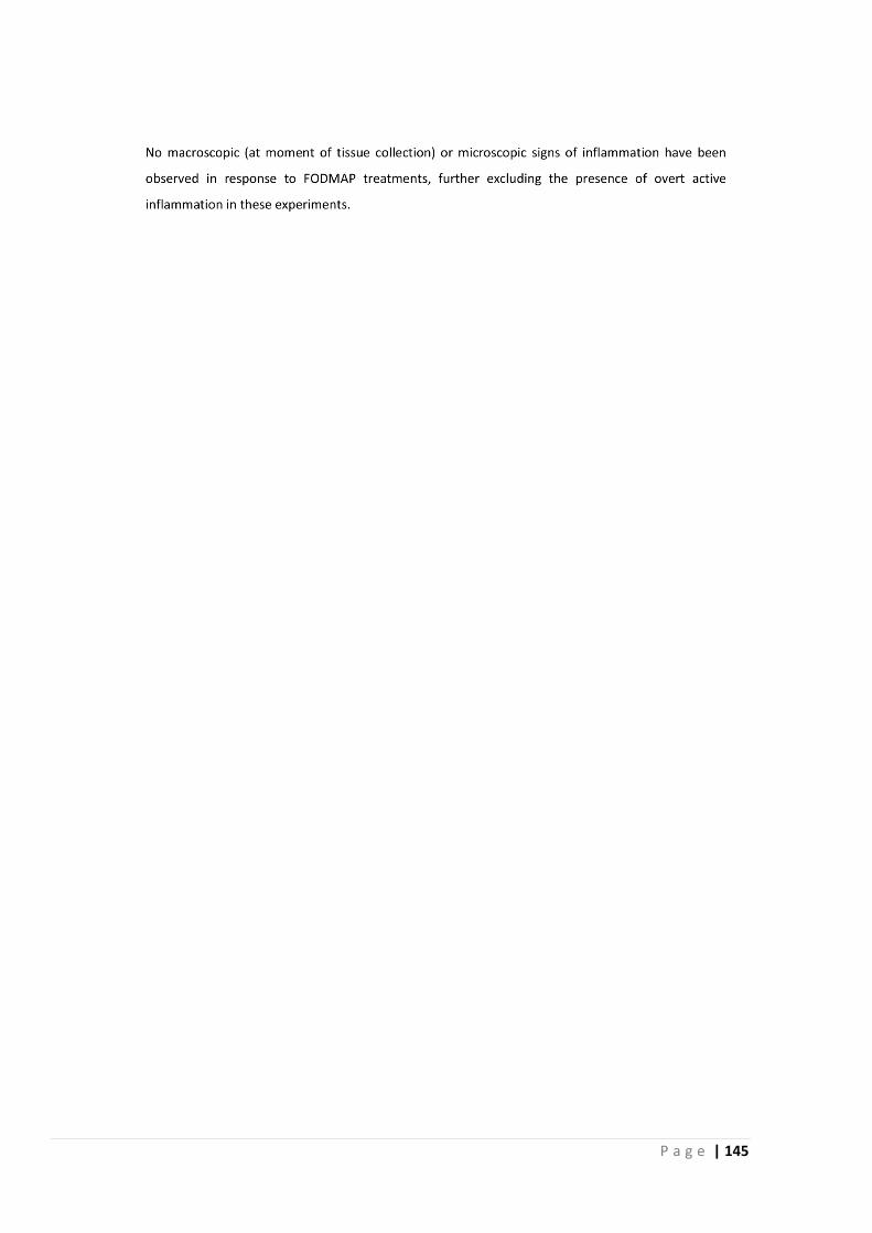
P a g e | 145
146 | P a g e

P a g e | 147
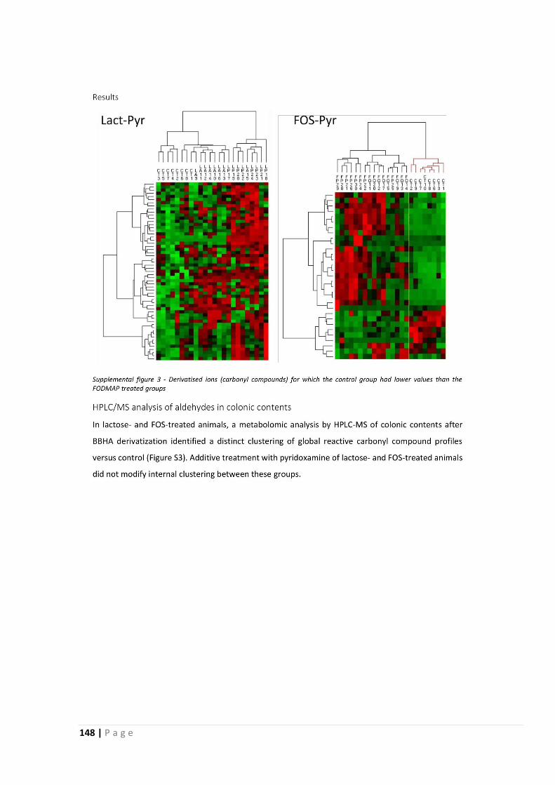
148 | P a g e
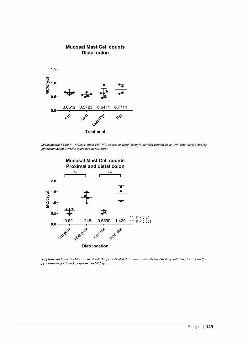
P a g e | 149
150 | P a g e
P a g e | 151
2.3 Increased FODMAP intake alters colonic mucus barrier function through
glycation processes and increased mastocyte counts
This work reports the results of our investigation of the effects on the colonic mucus barrier of
FODMAP treatment, as a reflection on intestinal barrier function. We used our MUCIO approach
and considered our new model of mucus organisation developed in the earlier work presented
in section 2.1. Apart from finding that FODMAPs can dysregulate the colonic mucus barrier
function through effects on mucosal mast cells and production of glycating agents described in
section 2.2 we show that the increased production of mucus in the absence of faecal matter
goes together with an increased variability and decreased coverage of the faecal mucus layer,
indicative of a less functional mucus barrier.
These results and this manuscript are ready to be submitted to the American Journal of
Physiology – Gastrointestinal and Liver Physiology, as a Rapid Report.
152 | P a g e
P a g e | 153
154 | P a g e
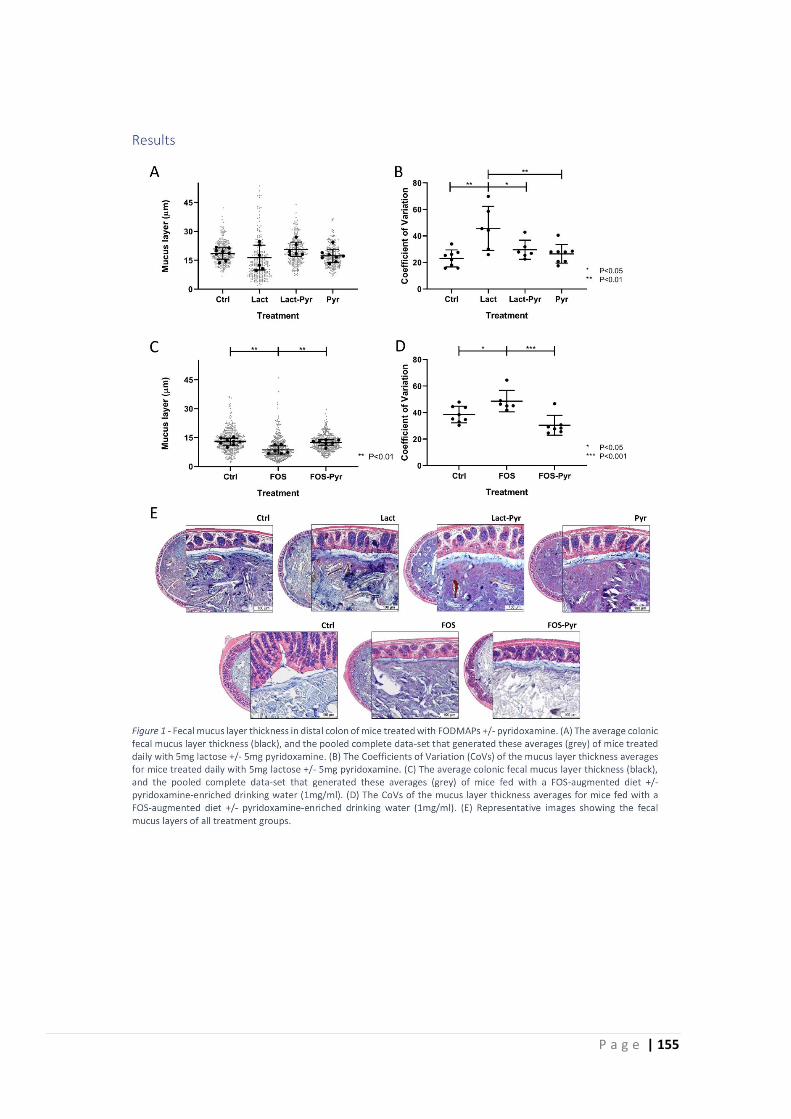
P a g e | 155
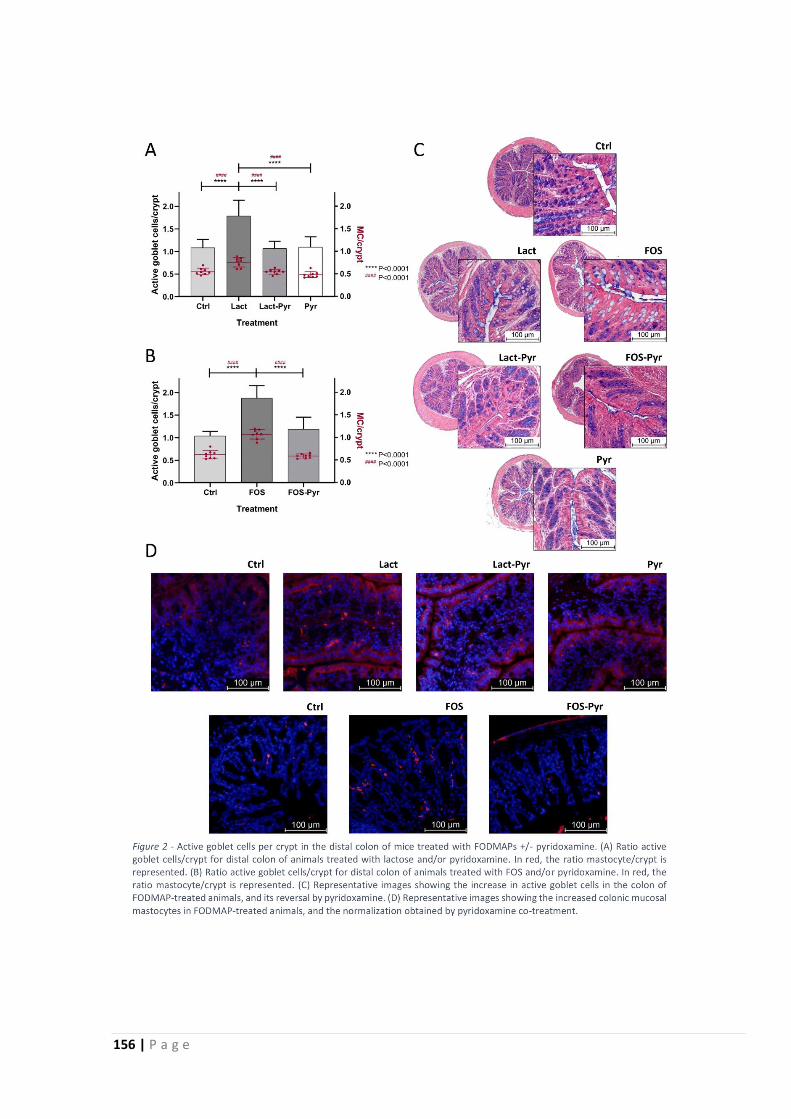
156 | P a g e
P a g e | 157
158 | P a g e
P a g e | 159
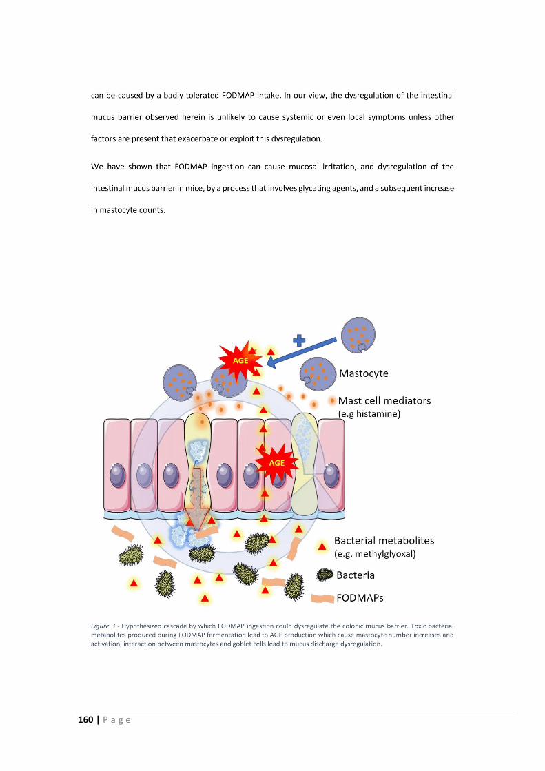
160 | P a g e
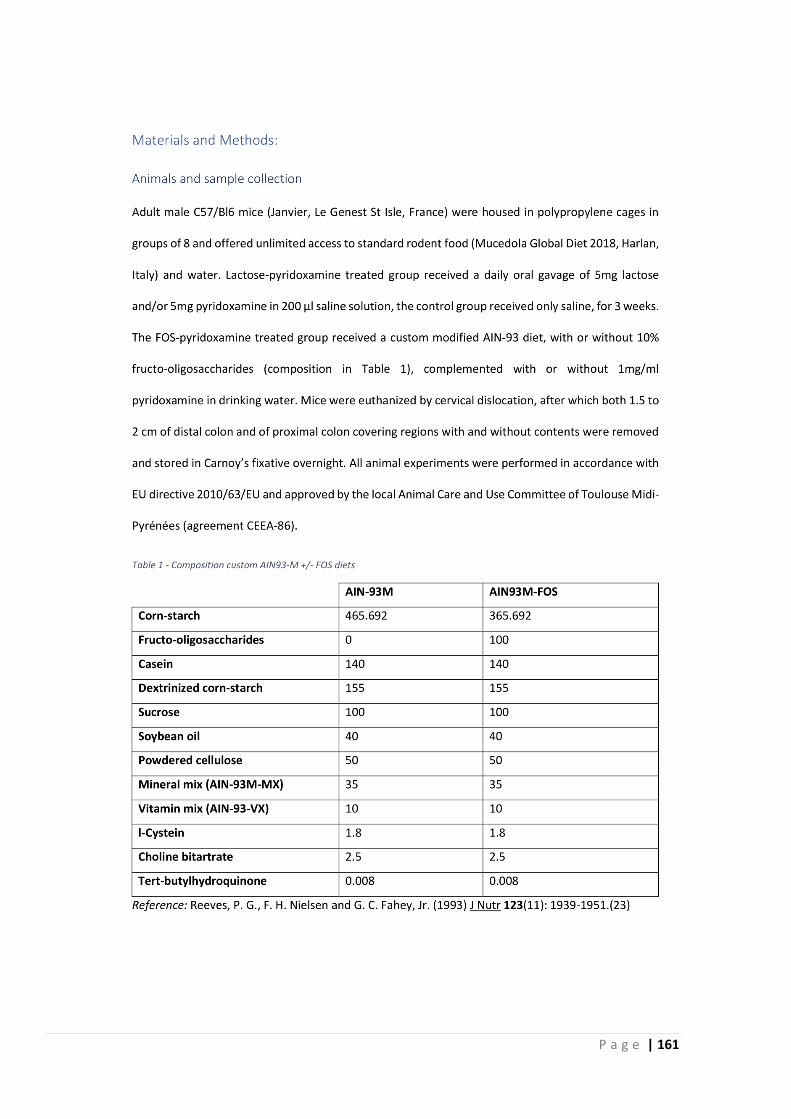
P a g e | 161
162 | P a g e
P a g e | 163
164 | P a g e
P a g e | 165
166 | P a g e


3 General Discussion
Discussion, Perspectives, and Conclusion

P a g e | 171
3 General Discussion
As mentioned at the start in the introduction, this thesis is part of the NeuroGut project, which
ran from 2014 to 2018. This Initial Training Network, financed by the European Commission, has
led to interesting findings, many of which have been published in renowned international
scientific journals, such as Neurogastroenterology and Motility, Scientific Reports, Alimentary
Pharmacology and Therapeutics, Neurotherapeutics, and The American Journal of Clinical
Nutrition.
More importantly, it has allowed a selection of young researchers to develop their professional
and personal skills, and international network. For our part in this project, we investigated the
effects of certain alimentary compounds on intestinal health, in relation to functional
gastrointestinal disorders, particularly irritable bowel syndrome (IBS), which represents an
important negative factor on quality of life (QOL) of patients, as well as costing society billions
of euros in lost work-hours and productivity. IBS patients are increasingly following a low-
FODMAP diet, which is quite effective in improving QOL and appearance of symptoms, but it
remains somewhat difficult to explain the reason for this efficacity.
In this thesis dissertation, complementary mechanisms of effect behind the efficacity of the low-
FODMAP diet are explored, by using a mouse model exposed to an increased FODMAP intake.
We have shown that FODMAP ingestion by itself can induce certain physiological effects
reminiscent of IBS, such as an increased visceral and central sensitivity (section 2.2), and
intestinal barrier function dysregulation (section 2.3), and these effects seem related to an
increase in glycating agents caused by microbial fermentation of these FODMAPs by the gut
microbiota, indicated by the increased expression of Receptor for AGEs (RAGE), and the
prevention of observed effects by administration of anti-glycation agent pyridoxamine, a form
of vitamin B6. The negative effects on visceral sensitivity and barrier function seem to be
mediated by an increased mucosal mast cell population (sections 2.2, 2.3).
172 | P a g e
3.1 Fermentable carbohydrates and IBS symptoms
Since FODMAPs are defined by their ‘unabsorbed’ status, individual characteristics, such as
lactase deficiency or fructose malabsorption, can influence which carbohydrates should be
considered FODMAPs in different individuals. Some FODMAPs, such as many polyols, fructo-
oligosaccharide (FOS) or fructans, and galacto-oligosaccharide (GOS) are
undigestible/unabsorbable for all humans, while for others this depends on factors such as dose,
intestinal health status, transporter complexes, and (brush border) enzyme expression, such as
for lactose and fructose (Gerbault, Liebert et al., 2011; Brown-Esters, Mc Namara et al., 2012).
We have used lactose as a representative of FODMAPs, because mice, like all mammals except
a sub-population of humans, lose lactase activity after weaning, and should be considered
lactose-malabsorbers.
The insight that FODMAPs can induce an increased abdominal sensitivity without further
challenge is interesting, because it indicates that it might render patients even more sensitive
to the distension that it causes. Because stress is an important factor in IBS symptom
generation (Qin, 2014; Greenwood-Van Meerveld, Moloney et al., 2016), and mast cells are an
important mediator in stress-induced visceral hypersensitivity (Gue, Del Rio-Lacheze et al.,
1997) and permeability (Santos, Yang et al., 2001; Barreau, Cartier et al., 2007; Vanuytsel, van
Wanrooy et al., 2014), it can be expected that the increased mast cell population observed in
FODMAP-treated animals renders them more susceptible to mast cell mediated effects of
psychological stress.
Our finding that the performed FODMAP treatments can increase visceral but also central
sensitivity fits with the frequent association of FGIDs with non-GI symptoms, such as for
fibromyalgia and chronic fatigue syndrome (Mathieu, 2009; Wilder-Smith, Olesen et al., 2018),
all of which are often exacerbated by stress (Mathieu, 2009). The increase in mastocyte counts
could provide a link between these GI and non-GI symptoms in combination with an increased
vulnerability to psychological stress, because mast cells can be activated by mental stress
through nerve system signalling (Castagliuolo, Lamont et al., 1996; Barbara, Stanghellini et al.,
2004; Barbara, Wang et al., 2007; Moon, Befus et al., 2014; Carabotti, Scirocco et al., 2015;
Zhang, Song et al., 2016).
Related to the psychological aspects of IBS and fermentable carbohydrates, lactose and fructose
malabsorption has been linked to a decreased L-tryptophan metabolism, precursor to
serotonin (Ledochowski, Sperner-Unterweger et al., 1998; Ledochowski, Widner et al., 2000),
P a g e | 173
suggesting a reduced serotonin producing capacity. Indeed, the tryptophan plasma level of
fructose malabsorbers is reduced (Ledochowski, Widner et al., 2001). Moreover, in depressed
patients, reduced levels of plasma tryptophan are found (Ogawa, Fujii et al., 2014). In contrast,
a recent study has found that fructose malabsorption is associated to depression, without a
significant relation between circulating tryptophan levels and depression (Enko, Wagner et al.,
2018). Conversely, Burokas et al. report that prebiotic unabsorbed carbohydrates FOS and GOS
exert anti-depressant and anxiolytic effects in mice (Burokas, Arboleya et al., 2017). However,
in this study plasma tryptophan levels were reduced for the prebiotic groups GOS and FOS+GOS.
These data indicate a link between disturbed tryptophan-metabolism and non-absorbed
carbohydrates. While none of these studies deeply explore the mechanism behind these effects,
Ledochowski et al. propose that non-absorbed carbohydrates react with peptides and amino-
acids, forming complexes that interfere with normal metabolism (Ledochowski, Widner et al.,
2001).
It is not yet clear whether the effects of FODMAPs on sensitivity and the mucus barrier function
reported in sections 2.2 and 2.3 are linked, i.e. the mucus barrier dysfunction aggravates the
increase in sensitivity, or whether both are downstream effects of the increase in mast cells,
without significantly influencing each other. It is known that barrier defects are correlated to an
increased sensitivity (Annahazi, Ferrier et al., 2013), in this case increased paracellular
permeability. A mucus barrier dysregulation could thus also have similar effects on gut
sensitivity, but the effects observed on this barrier in our model were relatively mild and are not
likely to have a significant impact on visceral sensitivity without further challenge.
Reactive microbial fermentation products of unabsorbed carbohydrates could have the same
effects on tryptophan levels as FOS/GOS, but are generally more prone to strong and fast
glycation reactions (Campbell, Matthews et al., 2010), and it would be interesting to
characterize tryptophan levels in FODMAP-treated animals, as well as ethology related to
depressive and anxious behaviour. We have not observed any obvious differences in mouse
behaviour between treatment groups, but a dedicated study might be interesting. It would
therefore also be interesting to see how increased FODMAP intake impacts the chronic stress
response of mice, for example by using a water avoidance stress (WAS), or maternal separation
model. It has been shown that the increase in visceral sensitivity and induction of intestinal
inflammation by WAS is aggravated by FOS in mice (Chen, Du et al., 2017), though in this work,
174 | P a g e
contrary to ours, FOS alone did not have effects, and was administered at lower doses (8g/kg
b.w., i.e. +/- 0.2g per day, versus our 10% of food intake, i.e. +/- 0.4g per day).
In IBS patients too, it would be interesting to investigate tryptophan levels on normal and low-
FODMAP diets and relate this to the (improvement in) psychological well-being.
To definitely prove that glycating carbonyls that cause the physiological effects in our
experiments are produced in fermentation and not in spontaneous breakdown of
carbohydrates, a germ-free or microbiota depleted animal model should be used to compare
metabolite profiles and effects of the treatments an sich. However, the physiology of germ-free
animals is significantly different from conventional mice. For example, the transit time of germ
free Swiss-Webster mice is about 60% longer than for conventional Swiss-Webster
mice (Kashyap, Marcobal et al., 2013). At the same time no beneficial metabolites of microbial
processing will be produced, which causes an underdeveloped immune system (Parker, Lawson
et al., 2017).
An experiment using non-absorbed antibiotics to deplete the microbiota would alter aspects of
normal digestion and furthermore induce a dysbiosis, which of course has its own effects on gut
health which could confound the findings. It is important to realize that we have not found
microbiota profile changes in our experiments involving lactose, and that a reduced or absent
microbiota by itself could induce visceral hypersensitivity (Verdu, 2006; Luczynski, Tramullas et
al., 2017), which complicates the reliability of an experiment into these features.
Confusingly, contrary to what is observed in entirely germ-free animals, some review literature
report a decrease in intestinal transit time after antibiotic use (Gaskins, Collier et al., 2002). In
fact, the source reference states “… and the subsequent changes in the intestine, including a
slower intestinal passage rate,…” (emphasis mine), while the table in the same report does
indeed imply a reduced transit time (Sweden Ministry of Agriculture, 1997). Other primary
research do however report an increased whole gut transit (Anitha, Vijay-Kumar et al., 2012; Ge,
Ding et al., 2017), caused by activation of TLR4 receptors by microbial products (Anitha, Vijay-
Kumar et al., 2012). Apart from supporting the general idea that microbial depletion does indeed
bring the physiology closer to that of germ-free animals, this illustrates that it can be important
to check the sources of claims presented in review articles.
P a g e | 175
Concerning visceral sensitivity and the importance of the microbiota, it has proven impossible
to find information showing the basal sensitivity of germ-free animals, while antibiotic
treatment (not necessarily microbial depletion) induces an increased visceral sensitivity that can
be prevented by co-treatment with the probiotic L. paracasei, or its spent medium without live
bacteria (Verdu, 2006). On the other hand, using intracellular recordings of myenteric plexus
nerve cells showed that the absence of the microbiome in germ-free mice makes these nerves
hypo-excitable, and the microbiome is thus necessary for normal excitability (McVey Neufeld,
Mao et al., 2013), pointing to a less sensitive system in these animals. Until comparable
techniques are used to compare the visceral sensitivity of germ-free and conventional animals,
it is not entirely clear whether the microbiome generally increases or decreases this sensitivity.
In relation to the increased visceral and central sensitivity observed in IBS patients, it would be
interesting to see if these factors ameliorate after following a low-FODMAP diet for a longer
period, for example by barostat testing such as used to show visceral hypersensitivity (Bouin,
Plourde et al., 2002), and a Cold Water Tolerance Test, such as performed in FGID
patients (Bouin, Meunier et al., 2001) and in fibromyalgia patients (Reyes del Paso, Garrido et
al., 2011). Interestingly, there is a comorbidity between IBS and fibromyalgia (FM) (Martinez-
Martinez, Mora et al., 2014), where some authors even propose it’s the same disease but
through the lens of different medical fields (Kindler, Bennett et al., 2011; Clauw, 2015), FM
patients seem to benefit from a low-FODMAP diet as IBS patients (Marum, Moreira et al.,
2016b), and this affliction too has been linked to mast cell activation (Tsilioni, Russell et al.,
2016).
176 | P a g e
3.2 Immune activation
We have not observed clear immune activation by our FODMAP treatments, apart from
mastocyte population increases; faecal lipocalin-2 levels were not significantly different
between any groups but instead tended to be slightly decreased through FOS-treatment. In IBS
patients, 3 weeks of low-FODMAP treatment however, lowered pro-inflammatory cytokines IL-
6 and IL-8, but not TNF-α, from levels that never were significantly different from baseline for
the healthy population, and 10-day FOS supplementation following this low-FODMAP
intervention did not impact these cytokines in these patients (Hustoft, Hausken et al., 2017).
This indicates that FODMAPs, but not FOS in particular, can influence cytokine profiles and
inflammatory processes. Indeed, it is known that FOS as a prebiotic can improve rather than
worsen inflammation in the intestine (Capitan-Canadas, Ocon et al., 2016), even though it has
not proven to be effective in improving true inflammatory disorders such as Crohn’s
disease (Marteau, 2011). Additionally, the low-FODMAP diet has effects on gut microbiota
profile composition (Staudacher, Lomer et al., 2012; Halmos, Christophersen et al., 2015;
Hustoft, Hausken et al., 2017; McIntosh, Reed et al., 2017; Staudacher, Lomer et al., 2017), and
FOS supplementation can help to normalise the microbiota after an increased dysbiosis index
induced by a low FODMAP intake, although this did worsen the IBS symptom severity (Hustoft,
Hausken et al., 2017). This indicates that normalising the microbiota profile in this case should
not be of primary concern, and a ‘dysbiosis’ mainly indicates a change from baseline, without
necessarily representing a dysfunction.
To see if the improved inflammatory profile of IBS-patients relies on the same mechanisms as
described in this thesis, it would be interesting to test whether anti-glycation agents such as
described in section 1.4.4 can mimic the positive effects observed for the low-FODMAP diet.
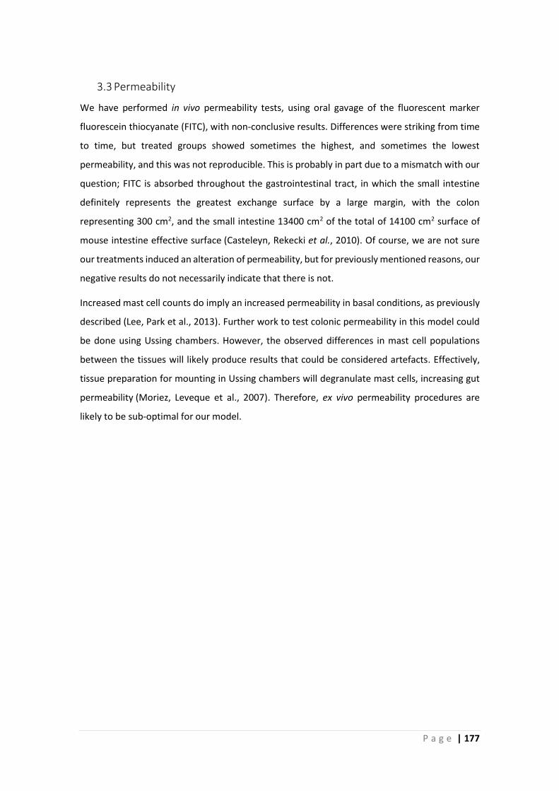
P a g e | 177
3.3 Permeability
We have performed in vivo permeability tests, using oral gavage of the fluorescent marker
fluorescein thiocyanate (FITC), with non-conclusive results. Differences were striking from time
to time, but treated groups showed sometimes the highest, and sometimes the lowest
permeability, and this was not reproducible. This is probably in part due to a mismatch with our
question; FITC is absorbed throughout the gastrointestinal tract, in which the small intestine
definitely represents the greatest exchange surface by a large margin, with the colon
representing 300 cm2, and the small intestine 13400 cm2 of the total of 14100 cm2 surface of
mouse intestine effective surface (Casteleyn, Rekecki et al., 2010). Of course, we are not sure
our treatments induced an alteration of permeability, but for previously mentioned reasons, our
negative results do not necessarily indicate that there is not.
Increased mast cell counts do imply an increased permeability in basal conditions, as previously
described (Lee, Park et al., 2013). Further work to test colonic permeability in this model could
be done using Ussing chambers. However, the observed differences in mast cell populations
between the tissues will likely produce results that could be considered artefacts. Effectively,
tissue preparation for mounting in Ussing chambers will degranulate mast cells, increasing gut
permeability (Moriez, Leveque et al., 2007). Therefore, ex vivo permeability procedures are
likely to be sub-optimal for our model.
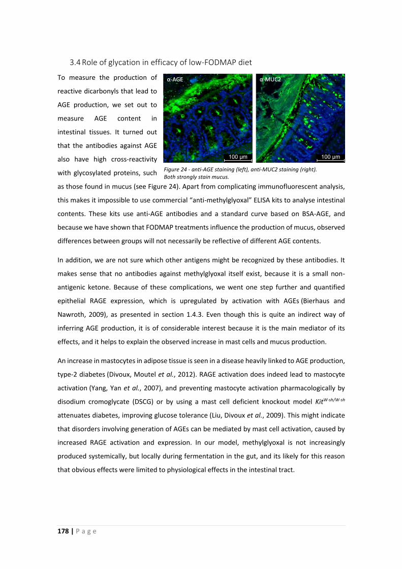
178 | P a g e
3.4 Role of glycation in efficacy of low-FODMAP diet
To measure the production of
reactive dicarbonyls that lead to
AGE production, we set out to
measure AGE content in
intestinal tissues. It turned out
that the antibodies against AGE
also have high cross-reactivity
with glycosylated proteins, such
as those found in mucus (see Figure 24). Apart from complicating immunofluorescent analysis,
this makes it impossible to use commercial “anti-methylglyoxal” ELISA kits to analyse intestinal
contents. These kits use anti-AGE antibodies and a standard curve based on BSA-AGE, and
because we have shown that FODMAP treatments influence the production of mucus, observed
differences between groups will not necessarily be reflective of different AGE contents.
In addition, we are not sure which other antigens might be recognized by these antibodies. It
makes sense that no antibodies against methylglyoxal itself exist, because it is a small non-
antigenic ketone. Because of these complications, we went one step further and quantified
epithelial RAGE expression, which is upregulated by activation with AGEs (Bierhaus and
Nawroth, 2009), as presented in section 1.4.3. Even though this is quite an indirect way of
inferring AGE production, it is of considerable interest because it is the main mediator of its
effects, and it helps to explain the observed increase in mast cells and mucus production.
An increase in mastocytes in adipose tissue is seen in a disease heavily linked to AGE production,
type-2 diabetes (Divoux, Moutel et al., 2012). RAGE activation does indeed lead to mastocyte
activation (Yang, Yan et al., 2007), and preventing mastocyte activation pharmacologically by
disodium cromoglycate (DSCG) or by using a mast cell deficient knockout model KitW-sh/W-sh
attenuates diabetes, improving glucose tolerance (Liu, Divoux et al., 2009). This might indicate
that disorders involving generation of AGEs can be mediated by mast cell activation, caused by
increased RAGE activation and expression. In our model, methylglyoxal is not increasingly
produced systemically, but locally during fermentation in the gut, and its likely for this reason
that obvious effects were limited to physiological effects in the intestinal tract.
Figure 24 - anti-AGE staining (left), anti-MUC2 staining (right). Both strongly stain mucus.
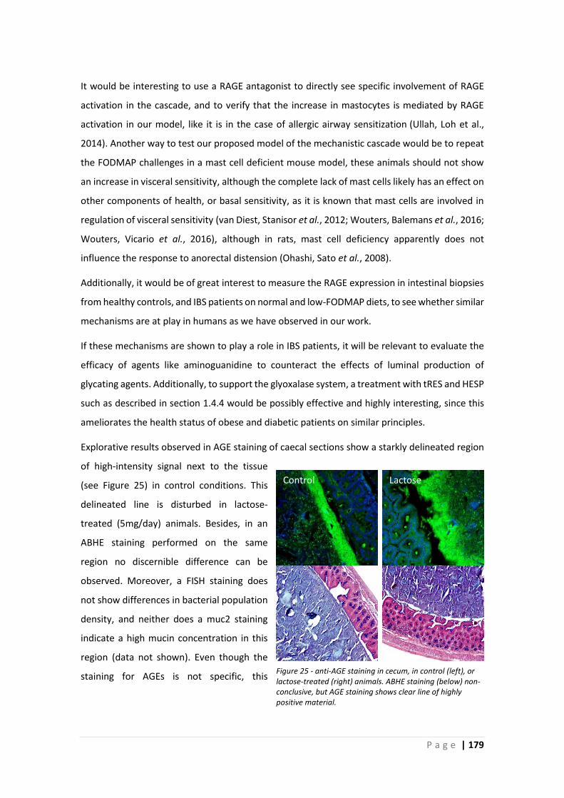
P a g e | 179
It would be interesting to use a RAGE antagonist to directly see specific involvement of RAGE
activation in the cascade, and to verify that the increase in mastocytes is mediated by RAGE
activation in our model, like it is in the case of allergic airway sensitization (Ullah, Loh et al.,
2014). Another way to test our proposed model of the mechanistic cascade would be to repeat
the FODMAP challenges in a mast cell deficient mouse model, these animals should not show
an increase in visceral sensitivity, although the complete lack of mast cells likely has an effect on
other components of health, or basal sensitivity, as it is known that mast cells are involved in
regulation of visceral sensitivity (van Diest, Stanisor et al., 2012; Wouters, Balemans et al., 2016;
Wouters, Vicario et al., 2016), although in rats, mast cell deficiency apparently does not
influence the response to anorectal distension (Ohashi, Sato et al., 2008).
Additionally, it would be of great interest to measure the RAGE expression in intestinal biopsies
from healthy controls, and IBS patients on normal and low-FODMAP diets, to see whether similar
mechanisms are at play in humans as we have observed in our work.
If these mechanisms are shown to play a role in IBS patients, it will be relevant to evaluate the
efficacy of agents like aminoguanidine to counteract the effects of luminal production of
glycating agents. Additionally, to support the glyoxalase system, a treatment with tRES and HESP
such as described in section 1.4.4 would be possibly effective and highly interesting, since this
ameliorates the health status of obese and diabetic patients on similar principles.
Explorative results observed in AGE staining of caecal sections show a starkly delineated region
of high-intensity signal next to the tissue
(see Figure 25) in control conditions. This
delineated line is disturbed in lactose-
treated (5mg/day) animals. Besides, in an
ABHE staining performed on the same
region no discernible difference can be
observed. Moreover, a FISH staining does
not show differences in bacterial population
density, and neither does a muc2 staining
indicate a high mucin concentration in this
region (data not shown). Even though the
staining for AGEs is not specific, this
Control Lactose
Figure 25 - anti-AGE staining in cecum, in control (left), or lactose-treated (right) animals. ABHE staining (below) non-conclusive, but AGE staining shows clear line of highly positive material.
180 | P a g e
interesting region highly suggests a bacterial organisation, resembling a biofilm.
Currently, we propose 3 hypotheses, based on these results;
1) Effectively, the AGE staining could have stained a glycosylated extracellular protein belonging
to the biofilm matrix.
2) Otherwise, maybe the host tissue produces chemicals that support AGE formation, such as
ROS.
3) It is also possible that (partial) bacterial breakdown of mucus makes the muc2 staining lose
specificity, while the less specific AGE staining still stains these glycosylated protein(s)
(fragments).
It would be immensely interesting to find/develop a way to determine whether this is a biofilm
or not, to get more insights in host-microbe interactions in the caecum. Additionally, this might
help to understand the implications of its possible disruption in response to FODMAP
treatments.
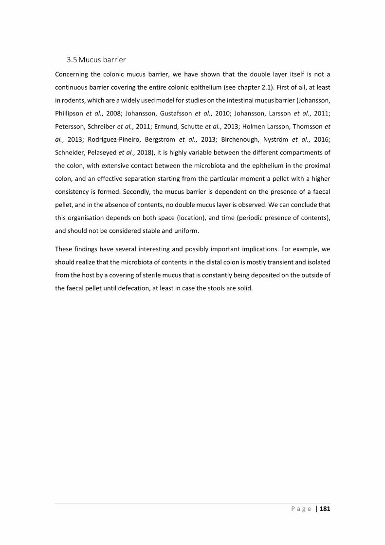
P a g e | 181
3.5 Mucus barrier
Concerning the colonic mucus barrier, we have shown that the double layer itself is not a
continuous barrier covering the entire colonic epithelium (see chapter 2.1). First of all, at least
in rodents, which are a widely used model for studies on the intestinal mucus barrier (Johansson,
Phillipson et al., 2008; Johansson, Gustafsson et al., 2010; Johansson, Larsson et al., 2011;
Petersson, Schreiber et al., 2011; Ermund, Schutte et al., 2013; Holmen Larsson, Thomsson et
al., 2013; Rodriguez-Pineiro, Bergstrom et al., 2013; Birchenough, Nyström et al., 2016;
Schneider, Pelaseyed et al., 2018), it is highly variable between the different compartments of
the colon, with extensive contact between the microbiota and the epithelium in the proximal
colon, and an effective separation starting from the particular moment a pellet with a higher
consistency is formed. Secondly, the mucus barrier is dependent on the presence of a faecal
pellet, and in the absence of contents, no double mucus layer is observed. We can conclude that
this organisation depends on both space (location), and time (periodic presence of contents),
and should not be considered stable and uniform.
These findings have several interesting and possibly important implications. For example, we
should realize that the microbiota of contents in the distal colon is mostly transient and isolated
from the host by a covering of sterile mucus that is constantly being deposited on the outside of
the faecal pellet until defecation, at least in case the stools are solid.
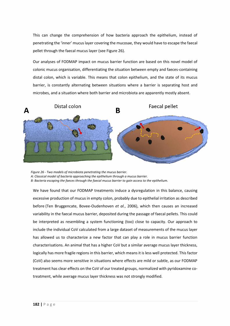
182 | P a g e
This can change the comprehension of how bacteria approach the epithelium, instead of
penetrating the ‘inner’ mucus layer covering the mucosae, they would have to escape the faecal
pellet through the faecal mucus layer (see Figure 26).
Our analyses of FODMAP impact on mucus barrier function are based on this novel model of
colonic mucus organisation, differentiating the situation between empty and faeces-containing
distal colon, which is variable. This means that colon epithelium, and the state of its mucus
barrier, is constantly alternating between situations where a barrier is separating host and
microbes, and a situation where both barrier and microbiota are apparently mostly absent.
We have found that our FODMAP treatments induce a dysregulation in this balance, causing
excessive production of mucus in empty colon, probably due to epithelial irritation as described
before (Ten Bruggencate, Bovee-Oudenhoven et al., 2006), which then causes an increased
variability in the faecal mucus barrier, deposited during the passage of faecal pellets. This could
be interpreted as resembling a system functioning (too) close to capacity. Our approach to
include the individual CoV calculated from a large dataset of measurements of the mucus layer
has allowed us to characterize a new factor that can play a role in mucus barrier function
characterisations. An animal that has a higher CoV but a similar average mucus layer thickness,
logically has more fragile regions in this barrier, which means it is less well protected. This factor
(CoV) also seems more sensitive in situations where effects are mild or subtle, as our FODMAP
treatment has clear effects on the CoV of our treated groups, normalized with pyridoxamine co-
treatment, while average mucus layer thickness was not strongly modified.
Figure 26 - Two models of microbiota penetrating the mucus barrier. A: Classical model of bacteria approaching the epithelium through a mucus barrier. B: Bacteria escaping the faeces through the faecal mucus barrier to gain access to the epithelium.
P a g e | 183
It is unlikely that these effects on the intestinal mucus barrier have serious consequences in
otherwise healthy systems, but it could imply an increased vulnerability to challenges.
For the proximal colon, where the chyme does not form high-consistency pellets, future
research to investigate the barrier function and immune tolerance evident from the lack of
inflammation despite extensive contact between bacteria and host in this compartment of the
colon will be of great interest.
In light of a possible increased vulnerability, it would be interesting to challenge FODMAP-
treated mice with pathogens, such as Citrobacter rodentium, a model that shares pathogenic
mechanisms with putative human pathogens such as E. coli (Collins, Keeney et al., 2014).
Considering that in the absence of a faecal pellet, the microbiota is strongly diminished, the
periodic presence of contents and microbial signalling could cause the activity of immune and
goblet cells, and possibly tissue regeneration, to display waveform functions instead of a
continuous or stochastic expression, which in itself is an interesting topic for future research.
We have shown that the mucus layer classically measured in histology is attached to the faecal
pellets, which means that it should be feasible to measure the mucus layer on Carnoy-fixated
paraffin-embedded droppings, instead of intestinal sections. The faecal mucus layer on
droppings is well-conserved and has the same general characteristics as when measured in
colonic sections (see figure 6 of section 2.1, page 104). This has several important benefits; it is
non-invasive, non-terminal, and technically easier to perform. Additionally, the same
individual’s droppings can be analysed throughout an experiment, collected at different
timepoints, allowing for characterisation of the progressive effects of treatment. In the context
of the 3 Rs (Replacement, Reduction, Refinement) of animal research, this approach would allow
us to both Reduce and Refine mucus barrier measurements, obtaining more information from
each individual, while causing them less harm.
In patients too, it represents an important improvement, because it can be performed on stools,
and does not necessitate invasive procedures. It should be verified however that the mucus
barrier in humans is organised similarly to that of rodents. In any case, a mucus layer covers
human stools, and it can be analysed, as shown in literature (Shimotoyodome, Meguro et al.,
2005; Swidsinski, Loening-Baucke et al., 2008; Swidsinski, Loening‐Baucke et al., 2008). As it
becomes more widely accepted that the distal colon mucus layer observed in histology is
identical to the faecal mucus layer, it will become more attractive to use this approach.
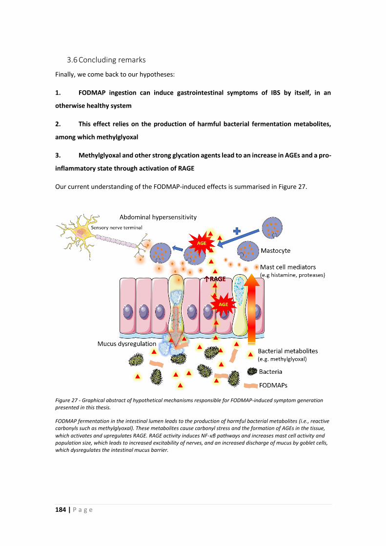
184 | P a g e
3.6 Concluding remarks
Finally, we come back to our hypotheses:
1. FODMAP ingestion can induce gastrointestinal symptoms of IBS by itself, in an
otherwise healthy system
2. This effect relies on the production of harmful bacterial fermentation metabolites,
among which methylglyoxal
3. Methylglyoxal and other strong glycation agents lead to an increase in AGEs and a pro-
inflammatory state through activation of RAGE
Our current understanding of the FODMAP-induced effects is summarised in Figure 27.
Figure 27 - Graphical abstract of hypothetical mechanisms responsible for FODMAP-induced symptom generation presented in this thesis.
FODMAP fermentation in the intestinal lumen leads to the production of harmful bacterial metabolites (i.e., reactive carbonyls such as methylglyoxal). These metabolites cause carbonyl stress and the formation of AGEs in the tissue,
which activates and upregulates RAGE. RAGE activity induces NF-β pathways and increases mast cell activity and population size, which leads to increased excitability of nerves, and an increased discharge of mucus by goblet cells, which dysregulates the intestinal mucus barrier.
P a g e | 185
We have shown that FODMAPs can indeed induce GI symptoms of IBS by itself, in a healthy
mouse model. Visceral and central sensitivity were increased, and the colonic mucus barrier
disrupted. That this effect relies on the generation of reactive dicarbonyls was shown indirectly
by a preventive effect obtained by co-treatment with pyridoxamine. Due to technical
constraints, we have not shown directly that AGEs were increased, but the expression of their
receptor (RAGE) increased in response to treatment and normalized with pyridoxamine co-
treatment. A plausible explanation for both the increased sensitivity and mucus barrier
dysregulation is the observed increase in mastocyte counts, probably mediated by RAGE,
interactions that have been reported in literature for other situations.
Knowing that FODMAPs can influence these factors and a better understanding of the
mechanisms responsible for the efficacity of the low-FODMAP diet will hopefully lead to
development of novel approaches to benefit FGID patients, for example by improving dicarbonyl
stress status in the intestine using pharmacologicals, or by using probiotic bacteria that have an
increased capacity to detoxify methylglyoxal in situ.
The work presented in this thesis describes a novel supplementary mechanism by which
FODMAPs are involved in GI-symptom generation and opens up new approaches to research
and possibly treat IBS and other FGIDs.

4 Acknowledgments

P a g e | 189 *SSS; snakeskin stress
4 Acknowledgments
First of all, I would like to thank my Thesis supervisors Hélène Eutamène and Vassilia Theodorou
for allowing me this great opportunity to work with them, and their allowing me the freedom to
pursue my interests and spur-of-the-moment ghost-chasing, as well as for the fruitful and
entertaining (but professional!) project meetings. I would also like to thank Paul Enck, and Sigrid
Diether for their kindness and support, and their great organisation of the NeuroGut network.
A special thanks to the European Commission, without which this thesis and the NeuroGut ITN
would not have been possible. Of course, I’m also thankful to the NeuroGut fellows who’ve
made it a real pleasure to attend our many meeting opportunities, I feel we’ve built a good
dynamic and great friendships.
Many thanks to Javier Santos and Bruno Bonaz for having accepted to be thesis reviewers and
jury members, and Ingrid Renes, and Catherine Muller equally for being part of the jury.
Vassilia, thank you for the supportive and engaging conversations on topics professional,
political, and personal, and for your comforting words and positive attitude that always helped
to recuperate the motivation to continue. Also, thanks for the many laughs we had!
Hélène, thank you for your trust and support to pursue my interests. Thanks for the freedom to
develop myself, and your kindness and support on the moments when it was needed.
Corinne, Coco ! Honneur exceptionnel ; en français, même si tu es forte en anglais aussi… Merci
pour ta présence maternelle ce qui m’a aidé beaucoup pour survivre ma thèse. Merci pour tous
les moments conviviaux en partageant de la bouffe, tes gâteaux merveilleux, du thé, du café,
même des cocktails ou de la sangria de temps en temps… Merci pour tout !
Muriel, thanks for the many interesting discussions on our common passion, slime! Thanks for
the supportive words and the interest in my thesis, as well as for the kind use of your impressive
network skills to get me into great professional opportunities! It has been a pleasure to work
with you, and it will undoubtably be a pleasure to work with you in the future. But before that,
we’ll meet again on the dance floor!
Hervé, thanks for the many good conversations and discussions on our mutual interests, and
thanks for balancing the ratio XX:XY a bit! Thanks for your supportive interest in my thesis and
brainstorming on new approaches to solve old mysteries. It’s high time someone (us) develops
the SSS* protocol! It’s a pleasure working with you, and thanks for your wisdom and valuable
career and scientific advice.
190 | P a g e
Bruno, thanks for your indispensable help with the von Frey filament experiments and
introducing me to this nice technique with a lot of patience. Maïwenn, thanks for your smiling
presence and humour, it has been exciting to try to uncover the enigmas of the caecum
microbiota, though there is still (practically all the) work to be done! Mathilde, thanks for your
kindness and patience, trying to understand my French even when it was quite rudimentary,
and thank you for laughing at most of my jokes! Valérie Ba, thank you for guarding my back for
years, it has been nice to work with you at the ice-chip factory. Valerie Be, a big thanks for the
friendliness and laughs, if every sacrifice could be as efficient… Valérie T, thank you for your
inspiring curiosity and boundless energy, never stop learning! Laurent, thanks for the
stimulating conversations and interesting exchange of ideas on the interaction of aldehydes and
mast cells, and the good times in the City by the Bay! Christine, thanks for your humour and
jokes, and your appreciation of what life as a foreigner entails. Christel, thanks for your kindness,
patience and limitless helpfulness. A warm thanks to Cathy, Sandrine, et Laurence too.
Les ‘collègues’ dit ‘vrais amis’;
Sophie, a.k.a. Popy, a.k.a. Sooophieee, thanks for all the great times we’ve had, the many jokes
and beers we shared, and reassuring me every time leaving the office that you wouldn’t be gone
forever (~“je reviens!”). Let’s continue rocking, swinging, dancing, drinking, and having a good
time! Yann, thanks for the good times, and the many laughs, even if I’m either ‘bête’,
‘insortable’, or you haven’t read what I’m talking about. It’s always been a pleasure to take a
break around a good cup of coffee (for me) and whatever you drink (…that stuff is definitely not
coffee…). Hanna, thanks for your kind presence and optimistic attitude, and the support when
Germanic and Gallic cultures had to be compared... Kévin, you arrived later than Sophie and
Yann (or did I leave late perhaps?), perfect! Thanks for the laughs at work, and the laughs
elsewhere. Thanks for the good conversations and keep an eye out for any #niceviews wherever
you go. Fanny, real job, real world, English, deal with it. Thanks for coming to Toxalim and
bringing even more warmth and smiles to the team, your arrival improved the atmosphere for
me, and surely for many others. Thanks for all the fun, and your youthful presence. Still, you
could’ve introduced me to some more of your ‘vraies amies’… Anaïs, thanks for your sunny
presence and inspiring tenaciousness. You’re a welcome addition to the team, and it’s an honour
sharing an office with you. Marie, the best friends take a while to find, thanks for always being
up for a drink and a dance, many more mojitos still to take! Adèle, voisine soon to be, thanks for
making me feel like less of a freak when dancing like nobody’s watching! The Golden Girls, Laura,
Chloé, and Manon, thanks for the smiles, the laughter, the cocktails, and the apéros de rêve, it
was always nice to meet for a little break at work, or a drink somewhere else!
P a g e | 191
Everyone, named and unnamed, thanks for the great experience and all the memories gained
during my years at Toxalim, merci beaucoup!

5 Publications,
dissemination, and training

P a g e | 195
5 Publications, Dissemination and Training Activities
5.1 Publications part of this thesis
• Kamphuis, J.B.J., M. Mercier-Bonin, H. Eutamene and V. Theodorou (2017). "Mucus
organisation is shaped by colonic content; a new view."
Scientific Reports 7 (1): 8527.
• Albusoda, A., N. Barki, T. Herregods, J.B.J. Kamphuis, T. B. Karunaratne, M. Lazarou, I.
Lee, N. Mazurak, E. Perna, A. Polster, T. Pribic, F. Uhlig, H. Wang and P. Enck (2017). "A
fresh look at IBS-opportunities for systems medicine approaches."
Neurogastroenterology and Motility 29 (3).
• Kamphuis, J.B.J., B. Guiard, M. Leveque, M. Olier, I. Jouanin, S. Yvon, V. Tondereau, P.
Rivière, F. Guéraud, S. Chevolleau, M.-H. Noguer-Meireles, J.-F. Martin, L. Debrauwer,
H. Eutamène, V. Theodorou (2019). “FODMAPs increase visceral sensitivity in mice
through glycation processes, increasing mast cell counts in colonic mucosae.”
Under review in Gastroenterology
• Kamphuis, J.B.J., H. Eutamène, V. Theodorou (2019) “Increased FODMAP intake alters
colonic mucus barrier function through glycation processes and increased mastocyte
counts”
Manuscript under preparation
5.2 Other Publications
• Radziwill-Bienkowska, J.M., P. Talbot, J.B.J. Kamphuis, V. Robert, C. Cartier, I. Fourquaux,
E. Lentzen, J. N. Audinot, F. Jamme, M. Refregiers, J. K. Bardowski, P. Langella, M.
Kowalczyk, E. Houdeau, M. Thomas and M. Mercier-Bonin (2018). "Toxicity of Food-
Grade TiO2 to Commensal Intestinal and Transient Food-Borne Bacteria: New Insights
Using Nano-SIMS and Synchrotron UV Fluorescence Imaging."
Frontiers in Microbiology 9: 794.
• Talbot, P., J.M. Radziwill-Bienkowska, J.B.J. Kamphuis, K. Steenkeste, S. Bettini, V.
Robert, M. L. Noordine, C. Mayeur, E. Gaultier, P. Langella, C. Robbe-Masselot, E.
Houdeau, M. Thomas and M. Mercier-Bonin (2018). "Food-grade TiO2 is trapped by
intestinal mucus in vitro but does not impair mucin O-glycosylation and short-chain
fatty acid synthesis in vivo: implications for gut barrier protection."
Journal of Nanobiotechnology 16(1): 53.
196 | P a g e
5.3 Oral presentations
• JFHOD 2018, Paris, France
22 March 2018
“L’administration de FODMAPS de type Fructo-oligosaccharides (FOS) augmente la
sensibilité abdominale et le nombre de mastocytes muqueux via la production
d’agents de glycation par le microbiote intestinal.”
• Mucins in Health and Disease (14th International Workshop), Cambridge, UK,
24-28 July 2017
“Mucus organisation is shaped by colonic content; a new view”
• CECED, JFHOD Joint Conferences 2017, Paris, France
22-26 March 2017
“Colonic load differentiates organization of mucus in colon of mouse and rat; a
remodelling”
• PA3S conference, Toulouse, France
20 January 2017
“Un régime enrichi en FODMAPs induit la production de métabolites toxiques par le
microbiote intestinal responsables d’une hypersensibilité viscérale.”
• GFNG 2016 conference, Rouen, France
23 June 2016
“Effects of a high FODMAPs diet on visceral sensitivity: involvement of advanced
glycation end products and colonic mast cells”
P a g e | 197
5.4 Poster presentations
• UEGW 2018, Vienna, Austria,
20-24 October 2018
1) “Dietary FODMAPs such as fructo-oligosaccharides and lactose can increase
abdominal sensitivity and the number of mucosal mastocytes in mice; a study on
underlying mechanisms.” J.B.J. Kamphuis, B. Guiard, P. Rivière, S. Yvon, V. Tondereau,
H. Eutamène, V. Theodorou
2) “Dietary FODMAPs can lead to microbial production of glycation agents, increasing
mucosal mastocytes and impacting colonic mucus barrier function.”
J.B.J. Kamphuis, S. Yvon, V. Tondereau, H. Eutamène, V. Theodorou
Abstract was awarded €750 Travel Grant
• FNM 2018, Amsterdam, the Netherlands,
29 August – 1 September 2018
“The effects of a diet rich in fructo-oligosaccharides on abdominal sensitivity; a study
on underlying mechanisms of action.” J.B.J. Kamphuis, B. Guiard, P. Rivière, S. Yvon, V.
Tondereau, H. Eutamène, V. Theodorou
• DDW 2018, Washington, USA,
2-5 June 2018
in absentia
“Dietary FODMAPs such as fructo-oligosaccharides can increase abdominal sensitivity
and the number of mucosal mastocytes in mice through production of glycating agents
by the intestinal microbiota.” J.B.J. Kamphuis, B. Guiard, P. Rivière, S. Yvon, V.
Tondereau, H. Eutamène, V. Theodorou
Abstract selected for Oral ePoster presentation
• DDW 2017, Chicago, USA
6-9 May
in absentia
“Colonic load differentiates organization of mucus in colon of mouse and rat; a
remodelling”
J.B.J. Kamphuis, H. Eutamène, V. Theodorou
198 | P a g e
• FNM 2016, San Francisco, USA
24-28 August 2016
“Effects of a high FODMAPs diet on visceral sensitivity: involvement of advanced
glycation end products and colonic mast cells”
J.B.J. Kamphuis, P. Rivière, S. Yvon, V. Tondereau, H. Eutamène, V. Theodorou
• JFHOD 2016, Paris, France,
17-20 March 2016
in absentia
“Effets et mécanismes des régimes contenant des ‘oligosaccharides, disaccharides,
monosaccharides fermentescibles et polyols’ (FODMAPs) sur la sensibilité viscérale”
J.B.J. Kamphuis, P. Rivière, S. Yvon, V. Tondereau, H. Eutamène, V. Theodorou
P a g e | 199
5.5 Other training and dissemination activities
2014
Utrecht, the Netherlands,
8-19 December
Cursus Proefdierkunde, Universiteit Utrecht
(Animal Experimentation Course)
2015
London, UK,
4-6 March
First Annual NeuroGut Meeting and
Progress Report
Project update presentation and discussion
with network partners
Tübingen, Germany,
13-17 April
Entrepreneurship Academy NeuroGut
Module 1
Istanbul, Turkey,
4-6 June
NeuroGASTRO 2015
Summer School NeuroGut
Toulouse, France,
17 September
Journée des Doctorants INP- EI-Purpan
Oral + poster presentation
Tübingen, Germany,
21-25 September
1st NeuroGut Complementary Training
Course, University of Tübingen
Intellectual Property, Scientific Writing
Skills, Scientific Presentation Skill
Tübingen, Germany,
22-26 November
Entrepreneurship Academy NeuroGut
Module 2
200 | P a g e
2016
Brussels, Belgium,
27-28 February
Mid-term Review Meeting
Progress Report with Officer of European
Commission
Project update oral + poster presentation;
Discussion with network partners and
European Commission Officer
Heidelberg, Germany,
11-13 March
COST Genieur Training School,
+ NeuroGut Spring School
“Molecular Methods in IBS Research:
genetics, epigenetics and microbiota
research”
Tübingen, Germany,
17-21 April
Entrepreneurship Academy NeuroGut
Module 3
Toulouse, France,
2 May
CODIR EI-Purpan
Project update, oral presentation and
discussion
Bologna, Italy,
8-10 June
Bologna IBS Days
NeuroGut Summer School
Toulouse, France,
30 September
La Nuit Européenne des Chercheurs
Prepared microscopy and educational
material & assisted at the Toxalim
educational stand
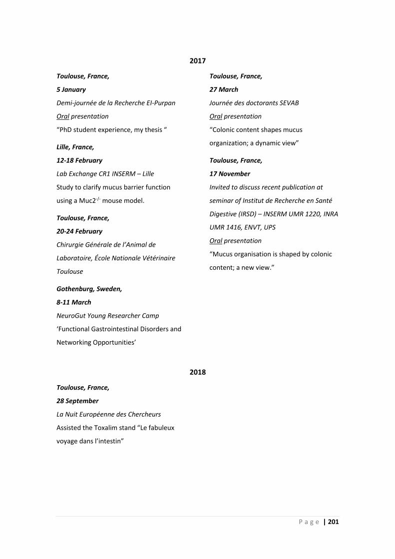
P a g e | 201
2017
Toulouse, France,
5 January
Demi-journée de la Recherche EI-Purpan
Oral presentation
“PhD student experience, my thesis “
Lille, France,
12-18 February
Lab Exchange CR1 INSERM – Lille
Study to clarify mucus barrier function
using a Muc2-/- mouse model.
Toulouse, France,
20-24 February
Chirurgie Générale de l’Animal de
Laboratoire, École Nationale Vétérinaire
Toulouse
Gothenburg, Sweden,
8-11 March
NeuroGut Young Researcher Camp
‘Functional Gastrointestinal Disorders and
Networking Opportunities’
Toulouse, France,
27 March
Journée des doctorants SEVAB
Oral presentation
“Colonic content shapes mucus
organization; a dynamic view”
Toulouse, France,
17 November
Invited to discuss recent publication at
seminar of Institut de Recherche en Santé
Digestive (IRSD) – INSERM UMR 1220, INRA
UMR 1416, ENVT, UPS
Oral presentation
“Mucus organisation is shaped by colonic
content; a new view.”
2018
Toulouse, France,
28 September
La Nuit Européenne des Chercheurs
Assisted the Toxalim stand “Le fabuleux
voyage dans l’intestin”


6 References

I | P a g e
6 References
Aichbichler, B.W., H.H. Wenzl, C.A. Santa Ana, J.L. Porter, L.R. Schiller and J.S. Fordtran (1998). "A comparison of stool characteristics from normal and constipated people." Dig Dis Sci 43(11): 2353-2362.
Ait-Belgnaoui, A., S. Bradesi, J. Fioramonti, V. Theodorou and L. Bueno (2005). "Acute stress-induced hypersensitivity to colonic distension depends upon increase in paracellular permeability: role of myosin light chain kinase." Pain 113(1): 141-147.
Albusoda, A., N. Barki, T. Herregods, J.B. Kamphuis, T.B. Karunaratne, M. Lazarou, . . . P. Enck (2017). "A fresh look at IBS-opportunities for systems medicine approaches." Neurogastroenterol Motil 29(3).
Allaman, I., M. Belanger and P.J. Magistretti (2015). "Methylglyoxal, the dark side of glycolysis." Front Neurosci 9(23): 23.
Allen, A. (1983). "Mucus—a protective secretion of complexity." Trends in Biochemical Sciences 8(5): 169-173.
Anitha, M., M. Vijay-Kumar, S.V. Sitaraman, A.T. Gewirtz and S. Srinivasan (2012). "Gut microbial products regulate murine gastrointestinal motility via Toll-like receptor 4 signaling." Gastroenterology 143(4): 1006-1016 e1004.
Annahazi, A., L. Ferrier, V. Bezirard, M. Leveque, H. Eutamene, A. Ait-Belgnaoui, . . . L. Bueno (2013). "Luminal cysteine-proteases degrade colonic tight junction structure and are responsible for abdominal pain in constipation-predominant IBS." Am J Gastroenterol 108(8): 1322-1331.
Aronsson, A.C. and B. Mannervik (1977). "Characterization of glyoxalase I purified from pig erythrocytes by affinity chromatography." Biochem J 165(3): 503-509.
Arpaia, N., C. Campbell, X. Fan, S. Dikiy, J. van der Veeken, P. deRoos, . . . A.Y. Rudensky (2013). "Metabolites produced by commensal bacteria promote peripheral regulatory T-cell generation." Nature 504(7480): 451-455.
Ashida, H., M. Ogawa, M. Kim, H. Mimuro and C. Sasakawa (2011). "Bacteria and host interactions in the gut epithelial barrier." Nat Chem Biol 8(1): 36-45.
Atuma, C., V. Strugala, A. Allen and L. Holm (2001). "The adherent gastrointestinal mucus gel layer: thickness and physical state in vivo." American Journal of Physiology-Gastrointestinal and Liver Physiology 280(5): G922-G929.
Azpiroz, F., M. Bouin, M. Camilleri, E. Mayer, P. Poitras, J. Serra and R. Spiller (2007). "Mechanisms of hypersensitivity in IBS and functional disorders." Neurogastroenterology & Motility 19(s1): 62-88.
Barbara, G., C. Cremon, V. Annese, G. Basilisco, F. Bazzoli, M. Bellini, . . . P. Buldrini (2016). "Randomised controlled trial of mesalazine in IBS." Gut 65(1): 82-90.
Barbara, G., C. Cremon, G. Carini, L. Bellacosa, L. Zecchi, R. De Giorgio, . . . V. Stanghellini (2011). "The immune system in irritable bowel syndrome." J Neurogastroenterol Motil 17(4): 349-359.
Barbara, G., C. Cremon and V. Stanghellini (2014). "Inflammatory bowel disease and irritable bowel syndrome: similarities and differences." Curr Opin Gastroenterol 30(4): 352-358.
Barbara, G., V. Stanghellini, R. De Giorgio, C. Cremon, G.S. Cottrell, D. Santini, . . . R. Corinaldesi (2004). "Activated mast cells in proximity to colonic nerves correlate with abdominal pain in irritable bowel syndrome." Gastroenterology 126(3): 693-702.
Barbara, G., B. Wang, V. Stanghellini, R. de Giorgio, C. Cremon, G. Di Nardo, . . . R. Corinaldesi (2007). "Mast cell-dependent excitation of visceral-nociceptive sensory neurons in irritable bowel syndrome." Gastroenterology 132(1): 26-37.
Barreau, F., C. Cartier, M. Leveque, L. Ferrier, R. Moriez, V. Laroute, . . . L. Bueno (2007). "Pathways involved in gut mucosal barrier dysfunction induced in adult rats by maternal deprivation:
II | P a g e
corticotrophin‐releasing factor and nerve growth factor interplay." The Journal of physiology 580(1): 347-356.
Barrett, J.C., S. Hansoul, D.L. Nicolae, J.H. Cho, R.H. Duerr, J.D. Rioux, . . . M.M. Barmada (2008). "Genome-wide association defines more than 30 distinct susceptibility loci for Crohn's disease." Nature genetics 40(8): 955-962.
Barrett, J.S., R.B. Gearry, J.G. Muir, P.M. Irving, R. Rose, O. Rosella, . . . P.R. Gibson (2010). "Dietary poorly absorbed, short-chain carbohydrates increase delivery of water and fermentable substrates to the proximal colon." Aliment Pharmacol Ther 31(8): 874-882.
Bashashati, M., S. Moossavi, C. Cremon, M.R. Barbaro, S. Moraveji, G. Talmon, . . . G. Barbara (2017). "Colonic immune cells in irritable bowel syndrome: A systematic review and meta-analysis." Neurogastroenterol Motil.
Baskaran, S. and K.A. Balasubramanian (1987). "Purification and active site modification studies on glyoxalase I from monkey intestinal mucosa." Biochimica et Biophysica Acta (BBA) - Protein Structure and Molecular Enzymology 913(3): 377-385.
Baskaran, S., D.P. Rajan and K.A. Balasubramanian (1989). "Formation of methylglyoxal by bacteria isolated from human faeces." J Med Microbiol 28(3): 211-215.
Basson, M.D., G.A. Turowski, Z. Rashid, F. Hong and J.A. Madri (1996). "Regulation of human colonic cell line proliferation and phenotype by sodium butyrate." Digestive Diseases and Sciences 41(10): 1986-1993.
Baynes, J.W. (1991). "Role of Oxidative Stress in Development of Complications in Diabetes." Diabetes 40(4): 405-412.
Beisswenger, P.J., S.K. Howell, R.M. O'Dell, M.E. Wood, A.D. Touchette and B.S. Szwergold (2001). "alpha-Dicarbonyls increase in the postprandial period and reflect the degree of hyperglycemia." Diabetes Care 24(4): 726-732.
Belanger, M., J. Yang, J.M. Petit, T. Laroche, P.J. Magistretti and I. Allaman (2011). "Role of the glyoxalase system in astrocyte-mediated neuroprotection." J Neurosci 31(50): 18338-18352.
Belzer, C., L.W. Chia, S. Aalvink, B. Chamlagain, V. Piironen, J. Knol and W.M. de Vos (2017). "Microbial Metabolic Networks at the Mucus Layer Lead to Diet-Independent Butyrate and Vitamin B12 Production by Intestinal Symbionts." mBio 8(5).
Bengtson, M.-B., G. Aamodt, M.H. Vatn and J.R. Harris (2015). "Co-occurrence of IBS and symptoms of anxiety or depression, among Norwegian twins, is influenced by both heredity and intrauterine growth." BMC gastroenterology 15(1): 9.
Bennet, S.M., L. Böhn, S. Störsrud, T. Liljebo, L. Collin, P. Lindfors, . . . M. Simrén (2017). "Multivariate modelling of faecal bacterial profiles of patients with IBS predicts responsiveness to a diet low in FODMAPs." Gut: gutjnl-2016-313128.
Bennet, S.M., L. Öhman and M. Simrén (2015). "Gut microbiota as potential orchestrators of irritable bowel syndrome." Gut and liver 9(3): 318.
Bennet, S.M., A. Polster, H. Tornblom, S. Isaksson, S. Capronnier, A. Tessier, . . . L. Ohman (2016). "Global Cytokine Profiles and Association With Clinical Characteristics in Patients With Irritable Bowel Syndrome." Am J Gastroenterol 111(8): 1165-1176.
Bennett, H.S. (1963). "Morphological aspects of extracellular polysaccharides." Journal of histochemistry & cytochemistry 11(1): 14-23.
Bergman, E.N. (1990). "Energy contributions of volatile fatty acids from the gastrointestinal tract in various species." Physiol Rev 70(2): 567-590.
Berkes, J., V. Viswanathan, S. Savkovic and G. Hecht (2003). "Intestinal epithelial responses to enteric pathogens: effects on the tight junction barrier, ion transport, and inflammation." Gut 52(3): 439-451.
Berrill, J.W., J.T. Green, K. Hood and A.K. Campbell (2013). "Symptoms of irritable bowel syndrome in patients with inflammatory bowel disease: examining the role of sub-clinical inflammation and the impact on clinical assessment of disease activity." Aliment Pharmacol Ther 38(1): 44-51.
III | P a g e
Beyder, A., A. Mazzone, P.R. Strege, D.J. Tester, Y.A. Saito, C.E. Bernard, . . . A. Dlugosz (2014). "Loss-of-function of the voltage-gated sodium channel Na V 1.5 (channelopathies) in patients with irritable bowel syndrome." Gastroenterology 146(7): 1659-1668.
Bierhaus, A., T. Fleming, S. Stoyanov, A. Leffler, A. Babes, C. Neacsu, . . . P.P. Nawroth (2012). "Methylglyoxal modification of Nav1.8 facilitates nociceptive neuron firing and causes hyperalgesia in diabetic neuropathy." Nat Med 18(6): 926-933.
Bierhaus, A. and P.P. Nawroth (2009). "Multiple levels of regulation determine the role of the receptor for AGE (RAGE) as common soil in inflammation, immune responses and diabetes mellitus and its complications." Diabetologia 52(11): 2251-2263.
Bierhaus, A., S. Schiekofer, M. Schwaninger, M. Andrassy, P.M. Humpert, J. Chen, . . . I. Klöting (2001). "Diabetes-associated sustained activation of the transcription factor nuclear factor-κB." Diabetes 50(12): 2792-2808.
Bijkerk, C., N. De Wit, J. Muris, P. Whorwell, J. Knottnerus and A. Hoes (2009). "Soluble or insoluble fibre in irritable bowel syndrome in primary care? Randomised placebo controlled trial." Bmj 339: b3154.
Bijkerk, C., J. Muris, J. Knottnerus, A. Hoes and N. De Wit (2004). "Systematic review: the role of different types of fibre in the treatment of irritable bowel syndrome." Alimentary pharmacology & therapeutics 19(3): 245-251.
Birchenough, G.M., M.E. Johansson, J.K. Gustafsson, J.H. Bergstrom and G.C. Hansson (2015). "New developments in goblet cell mucus secretion and function." Mucosal Immunol 8(4): 712-719.
Birchenough, G.M., E.E. Nyström, M.E. Johansson and G.C. Hansson (2016). "A sentinel goblet cell guards the colonic crypt by triggering Nlrp6-dependent Muc2 secretion." Science 352(6293): 1535-1542.
Bischoff, S.C., G. Barbara, W. Buurman, T. Ockhuizen, J.D. Schulzke, M. Serino, . . . J.M. Wells (2014). "Intestinal permeability--a new target for disease prevention and therapy." BMC Gastroenterol 14(1): 189.
Bliss, D. (1999). "Comparison of subjective classification of stool consistency and stool water content*1." Journal of WOCN 26(3): 137-141.
Boegh, M. and H.M. Nielsen (2015). "Mucus as a barrier to drug delivery - understanding and mimicking the barrier properties." Basic Clin Pharmacol Toxicol 116(3): 179-186.
Böhn, L., S. Störsrud, T. Liljebo, L. Collin, P. Lindfors, H. Törnblom and M. Simrén (2015). "Diet Low in FODMAPs Reduces Symptoms of Irritable Bowel Syndrome as Well as Traditional Dietary Advice: A Randomized Controlled Trial." Gastroenterology 149(6): 1399-1407.e1392.
Bohn, L., S. Storsrud, H. Tornblom, U. Bengtsson and M. Simren (2013). "Self-reported food-related gastrointestinal symptoms in IBS are common and associated with more severe symptoms and reduced quality of life." Am J Gastroenterol 108(5): 634-641.
Boldyrev, A., T. Fedorova, M. Stepanova, I. Dobrotvorskaya, E. Kozlova, N. Boldanova, . . . S. Illarioshkin (2008). "Carnosine [corrected] increases efficiency of DOPA therapy of Parkinson's disease: a pilot study." Rejuvenation Res 11(4): 821-827.
Booth, I.R., G.P. Ferguson, S. Miller, C. Li, B. Gunasekera and S. Kinghorn (2003). "Bacterial production of methylglyoxal: a survival strategy or death by misadventure?" Biochem Soc Trans 31(Pt 6): 1406-1408.
Bouhnik, Y., S. Alain, A. Attar, B. Flourie, L. Raskine, M.J. Sanson-Le Pors and J.C. Rambaud (1999). "Bacterial populations contaminating the upper gut in patients with small intestinal bacterial overgrowth syndrome." Am J Gastroenterol 94(5): 1327-1331.
Bouin, M., P. Meunier, M. Riberdy-Poitras and P. Poitras (2001). "Pain hypersensitivity in patients with functional gastrointestinal disorders: a gastrointestinal-specific defect or a general systemic condition?" Digestive diseases and sciences 46(11): 2542-2548.
IV | P a g e
Bouin, M., V. Plourde, M. Boivin, M. Riberdy, F. Lupien, M. Laganière, . . . P. Poitras (2002). "Rectal distention testing in patients with irritable bowel syndrome: sensitivity, specificity, and predictive values of pain sensory thresholds." Gastroenterology 122(7): 1771-1777.
Bourdu, S., M. Dapoigny, E. Chapuy, F. Artigue, M.P. Vasson, P. Dechelotte, . . . D. Ardid (2005). "Rectal instillation of butyrate provides a novel clinically relevant model of noninflammatory colonic hypersensitivity in rats." Gastroenterology 128(7): 1996-2008.
Bradford, K., W. Shih, E.J. Videlock, A.P. Presson, B.D. Naliboff, E.A. Mayer and L. Chang (2012). "Association between early adverse life events and irritable bowel syndrome." Clinical gastroenterology and hepatology 10(4): 385-390. e383.
Brandt, L.J., W.D. Chey, A.E. Foxx-Orenstein, L.R. Schiller, P.S. Schoenfeld, B.M. Spiegel, . . . E.M. Quigley (2009). "An evidence-based position statement on the management of irritable bowel syndrome." Am J Gastroenterol 104 Suppl 1: S1-35.
Brown-Esters, O., P. Mc Namara and D. Savaiano (2012). "Dietary and biological factors influencing lactose intolerance." International Dairy Journal 22(2): 98-103.
Browne, H.P., S.C. Forster, B.O. Anonye, N. Kumar, B.A. Neville, M.D. Stares, . . . T.D. Lawley (2016). "Culturing of 'unculturable' human microbiota reveals novel taxa and extensive sporulation." Nature 533(7604): 543-546.
Brownlee, I.A., M.E. Havler, P.W. Dettmar, A. Allen and J.P. Pearson (2003). "Colonic mucus: secretion and turnover in relation to dietary fibre intake." Proceedings of the Nutrition Society 62(1): 245-249.
Bures, J., J. Cyrany, D. Kohoutova, M. Forstl, S. Rejchrt, J. Kvetina, . . . M. Kopacova (2010). "Small intestinal bacterial overgrowth syndrome." World J Gastroenterol 16(24): 2978-2990.
Burokas, A., S. Arboleya, R.D. Moloney, V.L. Peterson, K. Murphy, G. Clarke, . . . J.F. Cryan (2017). "Targeting the Microbiota-Gut-Brain Axis: Prebiotics Have Anxiolytic and Antidepressant-like Effects and Reverse the Impact of Chronic Stress in Mice." Biol Psychiatry 82(7): 472-487.
Byers, M.R., H. Suzuki and T. Maeda (2003). "Dental neuroplasticity, neuro-pulpal interactions, and nerve regeneration." Microsc Res Tech 60(5): 503-515.
Caesar, R., C.S. Reigstad, H.K. Backhed, C. Reinhardt, M. Ketonen, G.O. Lunden, . . . F. Backhed (2012). "Gut-derived lipopolysaccharide augments adipose macrophage accumulation but is not essential for impaired glucose or insulin tolerance in mice." Gut 61(12): 1701-1707.
Campbell, A.K., S.B. Matthews, N. Vassel, C.D. Cox, R. Naseem, J. Chaichi, . . . K.T. Wann (2010). "Bacterial metabolic 'toxins': a new mechanism for lactose and food intolerance, and irritable bowel syndrome." Toxicology 278(3): 268-276.
Campbell, A.K., R. Naseem, I.B. Holland, S.B. Matthews and K.T. Wann (2007). "Methylglyoxal and other carbohydrate metabolites induce lanthanum-sensitive Ca2+ transients and inhibit growth in E. coli." Arch Biochem Biophys 468(1): 107-113.
Campbell, A.K., J.P. Waud and S.B. Matthews (2005). "The molecular basis of lactose intolerance." Sci Prog 88(Pt 3): 157-202.
Cani, P.D., J. Amar, M.A. Iglesias, M. Poggi, C. Knauf, D. Bastelica, . . . R. Burcelin (2007). "Metabolic endotoxemia initiates obesity and insulin resistance." Diabetes 56(7): 1761-1772.
Cao, W., J. Chen, Y. Chen, S. Chen, X. Chen, H. Huang and P. Liu (2015). "Advanced glycation end products induced immune maturation of dendritic cells controls heart failure through NF-kappaB signaling pathway." Arch Biochem Biophys 580: 112-120.
Capitan-Canadas, F., B. Ocon, C.J. Aranda, A. Anzola, M.D. Suarez, A. Zarzuelo, . . . O. Martinez-Augustin (2016). "Fructooligosaccharides exert intestinal anti-inflammatory activity in the CD4+ CD62L+ T cell transfer model of colitis in C57BL/6J mice." Eur J Nutr 55(4): 1445-1454.
Carabotti, M., A. Scirocco, M.A. Maselli and C. Severi (2015). "The gut-brain axis: interactions between enteric microbiota, central and enteric nervous systems." Annals of gastroenterology: quarterly publication of the Hellenic Society of Gastroenterology 28(2): 203.
V | P a g e
Carbonero, F., B.B. Oakley and K.J. Purdy (2014). "Metabolic flexibility as a major predictor of spatial distribution in microbial communities." PLoS One 9(1): e85105.
Casen, C., H.C. Vebo, M. Sekelja, F.T. Hegge, M.K. Karlsson, E. Ciemniejewska, . . . K. Rudi (2015). "Deviations in human gut microbiota: a novel diagnostic test for determining dysbiosis in patients with IBS or IBD." Aliment Pharmacol Ther 42(1): 71-83.
Cash, B.D., M.S. Epstein and S.M. Shah (2016). "A novel delivery system of peppermint oil is an effective therapy for irritable bowel syndrome symptoms." Digestive diseases and sciences 61(2): 560-571.
Cash, H.L., C.V. Whitham, C.L. Behrendt and L.V. Hooper (2006). "Symbiotic bacteria direct expression of an intestinal bactericidal lectin." Science 313(5790): 1126-1130.
Castagliuolo, I., J.T. Lamont, B. Qiu, S.M. Fleming, K.R. Bhaskar, S.T. Nikulasson, . . . C. Pothoulakis (1996). "Acute stress causes mucin release from rat colon: role of corticotropin releasing factor and mast cells." Am J Physiol 271(5 Pt 1): G884-892.
Casteleyn, C., A. Rekecki, A. Van der Aa, P. Simoens and W. Van den Broeck (2010). "Surface area assessment of the murine intestinal tract as a prerequisite for oral dose translation from mouse to man." Lab Anim 44(3): 176-183.
Castro, J., A.M. Harrington, P.A. Hughes, C.M. Martin, P. Ge, C.M. Shea, . . . S.M. Brierley (2013). "Linaclotide inhibits colonic nociceptors and relieves abdominal pain via guanylate cyclase-C and extracellular cyclic guanosine 3',5'-monophosphate." Gastroenterology 145(6): 1334-1346 e1331-1311.
Chang, W.W. and C.P. Leblond (1971). "Renewal of the epithelium in the descending colon of the mouse. I. Presence of three cell populations: vacuolated-columnar, mucous and argentaffin." Am J Anat 131(1): 73-99.
Chassaing, B., G. Srinivasan, M.A. Delgado, A.N. Young, A.T. Gewirtz and M. Vijay-Kumar (2012). "Fecal lipocalin 2, a sensitive and broadly dynamic non-invasive biomarker for intestinal inflammation." PLoS One 7(9): e44328.
Chavakis, T., A. Bierhaus, N. Al-Fakhri, D. Schneider, S. Witte, T. Linn, . . . P.P. Nawroth (2003). "The pattern recognition receptor (RAGE) is a counterreceptor for leukocyte integrins: a novel pathway for inflammatory cell recruitment." J Exp Med 198(10): 1507-1515.
Chen, B.-R., L.-J. Du, H.-Q. He, J.J. Kim, Y. Zhao, Y.-W. Zhang, . . . N. Dai (2017). "Fructo-oligosaccharide intensifies visceral hypersensitivity and intestinal inflammation in a stress-induced irritable bowel syndrome mouse model." World journal of gastroenterology 23(47): 8321-8333.
Cheng, H. (1974). "Origin, differentiation and renewal of the four main epithelial cell types in the mouse small intestine. II. Mucous cells." Am J Anat 141(4): 481-501.
Chey, W.D., A.J. Lembo, B.J. Lavins, S.J. Shiff, C.B. Kurtz, M.G. Currie, . . . J.M. Johnston (2012). "Linaclotide for irritable bowel syndrome with constipation: a 26-week, randomized, double-blind, placebo-controlled trial to evaluate efficacy and safety." Am J Gastroenterol 107(11): 1702-1712.
Clauw, D.J. (2015). "Fibromyalgia and related conditions." Mayo Clin Proc 90(5): 680-692. Cockburn, D.W. and N.M. Koropatkin (2016). "Polysaccharide Degradation by the Intestinal Microbiota
and Its Influence on Human Health and Disease." Journal of Molecular Biology 428(16): 3230-3252.
Collins, J.W., K.M. Keeney, V.F. Crepin, V.A. Rathinam, K.A. Fitzgerald, B.B. Finlay and G. Frankel (2014). "Citrobacter rodentium: infection, inflammation and the microbiota." Nat Rev Microbiol 12(9): 612-623.
Collins, S.M. (2014). "A role for the gut microbiota in IBS." Nat Rev Gastroenterol Hepatol 11(8): 497-505.
Cone, R.A. (2009). "Barrier properties of mucus." Adv Drug Deliv Rev 61(2): 75-85. Cook, S.I. and J.H. Sellin (1998). "Review article: short chain fatty acids in health and disease." Aliment
Pharmacol Ther 12(6): 499-507.
VI | P a g e
Cordner, Z., L. Liu, T. Moran, Q. Li, A. Bhargave, K. Tamashiro and P. Pasricha (2017). "789. A Novel Mast Cell-Vagal Network Regulates Chronic Depression and Anxiety-Like Behaviors after Transient Gastric Irritation." Biological Psychiatry 81(10): S320-S321.
Corinaldesi, R., V. Stanghellini, C. Cremon, L. Gargano, R.F. Cogliandro, R. De Giorgio, . . . G. Barbara (2009). "Effect of mesalazine on mucosal immune biomarkers in irritable bowel syndrome: a randomized controlled proof-of-concept study." Aliment Pharmacol Ther 30(3): 245-252.
Cornelli, U. (2010). "Treatment of Alzheimer's disease with a cholinesterase inhibitor combined with antioxidants." Neurodegener Dis 7(1-3): 193-202.
Creely, S.J., P.G. McTernan, C.M. Kusminski, M. Fisher f, N.F. Da Silva, M. Khanolkar, . . . S. Kumar (2007). "Lipopolysaccharide activates an innate immune system response in human adipose tissue in obesity and type 2 diabetes." Am J Physiol Endocrinol Metab 292(3): E740-747.
Crouzet, L., E. Gaultier, C. Del'Homme, C. Cartier, E. Delmas, M. Dapoigny, . . . A. Bernalier‐Donadille (2013). "The hypersensitivity to colonic distension of IBS patients can be transferred to rats through their fecal microbiota." Neurogastroenterology & Motility 25(4).
Cunliffe, R.N. and Y.R. Mahida (2004). "Expression and regulation of antimicrobial peptides in the gastrointestinal tract." J Leukoc Biol 75(1): 49-58.
Dai, H. and R.J. Korthuis (2011). "Mast Cell Proteases and Inflammation." Drug Discov Today Dis Models 8(1): 47-55.
Daims, H., A. Brühl, R. Amann, K.-H. Schleifer and M. Wagner (1999). "The Domain-specific Probe EUB338 is Insufficient for the Detection of all Bacteria: Development and Evaluation of a more Comprehensive Probe Set." Systematic and Applied Microbiology 22(3): 434-444.
Dave, M., P.D. Higgins, S. Middha and K.P. Rioux (2012). "The human gut microbiome: current knowledge, challenges, and future directions." Transl Res 160(4): 246-257.
Davies, S.S., C. Bodine, E. Matafonova, B.G. Pantazides, N. Bernoud-Hubac, F.E. Harrison, . . . L.J. Roberts, 2nd (2011). "Treatment with a gamma-ketoaldehyde scavenger prevents working memory deficits in hApoE4 mice." J Alzheimers Dis 27(1): 49-59.
Davies, S.S. and L.S. Zhang (2017). "Reactive Carbonyl Species Scavengers—Novel Therapeutic Approaches for Chronic Diseases." Current pharmacology reports 3(2): 51-67.
de Courten, B., M. Jakubova, M.P. de Courten, I.J. Kukurova, S. Vallova, P. Krumpolec, . . . B. Ukropcova (2016). "Effects of carnosine supplementation on glucose metabolism: Pilot clinical trial." Obesity (Silver Spring) 24(5): 1027-1034.
De Palma, G., M.D. Lynch, J. Lu, V.T. Dang, Y. Deng, J. Jury, . . . P. Bercik (2017). "Transplantation of fecal microbiota from patients with irritable bowel syndrome alters gut function and behavior in recipient mice." Sci Transl Med 9(379): eaaf6397.
Debongnie, J.C. and S.F. Phillips (1978). "Capacity of the human colon to absorb fluid." Gastroenterology 74(4): 698-703.
Degen, L.P. and S.F. Phillips (1996). "How well does stool form reflect colonic transit?" Gut 39(1): 109-113.
den Besten, G., K. van Eunen, A.K. Groen, K. Venema, D.J. Reijngoud and B.M. Bakker (2013). "The role of short-chain fatty acids in the interplay between diet, gut microbiota, and host energy metabolism." J Lipid Res 54(9): 2325-2340.
Derrien, M., E.E. Vaughan, C.M. Plugge and W.M. de Vos (2004). "Akkermansia muciniphila gen. nov., sp. nov., a human intestinal mucin-degrading bacterium." Int J Syst Evol Microbiol 54(Pt 5): 1469-1476.
Desai, M.S., A.M. Seekatz, N.M. Koropatkin, N. Kamada, C.A. Hickey, M. Wolter, . . . A. Muller (2016). "A Dietary Fiber-Deprived Gut Microbiota Degrades the Colonic Mucus Barrier and Enhances Pathogen Susceptibility." Cell 167(5): 1339-1353. e1321.
Dharmani, P., V. Srivastava, V. Kissoon-Singh and K. Chadee (2009). "Role of intestinal mucins in innate host defense mechanisms against pathogens." J Innate Immun 1(2): 123-135.
VII | P a g e
Di Mauro, A., J. Neu, G. Riezzo, F. Raimondi, D. Martinelli, R. Francavilla and F. Indrio (2013). "Gastrointestinal function development and microbiota." Ital J Pediatr 39(1): 15.
Divoux, A., S. Moutel, C. Poitou, D. Lacasa, N. Veyrie, A. Aissat, . . . K. Clement (2012). "Mast cells in human adipose tissue: link with morbid obesity, inflammatory status, and diabetes." J Clin Endocrinol Metab 97(9): E1677-1685.
Dommett, R., M. Zilbauer, J.T. George and M. Bajaj-Elliott (2005). "Innate immune defence in the human gastrointestinal tract." Mol Immunol 42(8): 903-912.
Drossman, D.A. (2007). "Introduction. The Rome Foundation and Rome III." Neurogastroenterol Motil 19(10): 783-786.
Drossman, D.A. (2016). "Functional Gastrointestinal Disorders: History, Pathophysiology, Clinical Features and Rome IV." Gastroenterology.
Dunlop, S.P., J. Hebden, E. Campbell, J. Naesdal, L. Olbe, A.C. Perkins and R.C. Spiller (2006). "Abnormal intestinal permeability in subgroups of diarrhea-predominant irritable bowel syndromes." The American journal of gastroenterology 101(6): 1288.
Enck, P., Q. Aziz, G. Barbara, A.D. Farmer, S. Fukudo, E.A. Mayer, . . . R.C. Spiller (2016). "Irritable bowel syndrome." Nat Rev Dis Primers 2: 16014.
Enko, D., H. Wagner, G. Kriegshäuser, W. Brandmayr, G. Halwachs-Baumann, W.J. Schnedl, . . . A. Meinitzer (2018). "Assessment of tryptophan metabolism and signs of depression in individuals with carbohydrate malabsorption." Psychiatry Research 262: 595-599.
Ermund, A., A. Schutte, M.E. Johansson, J.K. Gustafsson and G.C. Hansson (2013). "Studies of mucus in mouse stomach, small intestine, and colon. I. Gastrointestinal mucus layers have different properties depending on location as well as over the Peyer's patches." Am J Physiol Gastrointest Liver Physiol 305(5): G341-347.
Esser, D., J. Lange, G. Marinos, M. Sieber, L. Best, D. Prasse, . . . F. Sommer (2018). "Functions of the Microbiota for the Physiology of Animal Metaorganisms." J Innate Immun: 1-12.
Eswaran, S., J. Muir and W.D. Chey (2013). "Fiber and functional gastrointestinal disorders." Am J Gastroenterol 108(5): 718-727.
Eswaran, S.L., W.D. Chey, T. Han-Markey, S. Ball and K. Jackson (2016). "A Randomized Controlled Trial Comparing the Low FODMAP Diet vs. Modified NICE Guidelines in US Adults with IBS-D." Am J Gastroenterol 111(12): 1824-1832.
Eutamene, H., F. Lamine, C. Chabo, V. Theodorou, F. Rochat, G.E. Bergonzelli, . . . L. Bueno (2007). "Synergy between Lactobacillus paracasei and its bacterial products to counteract stress-induced gut permeability and sensitivity increase in rats." The Journal of nutrition 137(8): 1901-1907.
Fagarasan, S., M. Muramatsu, K. Suzuki, H. Nagaoka, H. Hiai and T. Honjo (2002). "Critical roles of activation-induced cytidine deaminase in the homeostasis of gut flora." Science 298(5597): 1424-1427.
Ferguson, G.P., S. Totemeyer, M.J. MacLean and I.R. Booth (1998). "Methylglyoxal production in bacteria: suicide or survival?" Arch Microbiol 170(4): 209-218.
Fiory, F., A. Lombardi, C. Miele, J. Giudicelli, F. Beguinot and E. Van Obberghen (2011). "Methylglyoxal impairs insulin signalling and insulin action on glucose-induced insulin secretion in the pancreatic beta cell line INS-1E." Diabetologia 54(11): 2941-2952.
Flamm, G., W. Glinsmann, D. Kritchevsky, L. Prosky and M. Roberfroid (2001). "Inulin and oligofructose as dietary fiber: a review of the evidence." Critical reviews in food science and nutrition 41(5): 353-362.
Flint, H.J. (1997). "The rumen microbial ecosystem--some recent developments." Trends Microbiol 5(12): 483-488.
Florey, H.W. (1955). "The Croonian Lecture: Mucin and the protection of the body." Proceedings of the Royal Society of London. Series B - Biological Sciences 143(911): 147-158.
Florey, H.W. (1962). "The secretion and function of intestinal mucus." Gastroenterology 43: 326-329.
VIII | P a g e
Ford, A.C., P. Moayyedi, B.E. Lacy, A.J. Lembo, Y.A. Saito, L.R. Schiller, . . . D. Task Force on the Management of Functional Bowel (2014). "American College of Gastroenterology monograph on the management of irritable bowel syndrome and chronic idiopathic constipation." Am J Gastroenterol 109 Suppl 1: S2-26; quiz S27.
Ford, A.C., E.M.M. Quigley, B.E. Lacy, A.J. Lembo, Y.A. Saito, L.R. Schiller, . . . P. Moayyedi (2014). "Efficacy of Prebiotics, Probiotics, and Synbiotics in Irritable Bowel Syndrome and Chronic Idiopathic Constipation: Systematic Review and Meta-analysis." The American Journal Of Gastroenterology 109: 1547.
Francis, C.Y., J. Morris and P.J. Whorwell (1997). "The irritable bowel severity scoring system: a simple method of monitoring irritable bowel syndrome and its progress." Alimentary pharmacology & therapeutics 11(2): 395-402.
Frenkel, E.S. and K. Ribbeck (2015). "Salivary mucins in host defense and disease prevention." J Oral Microbiol 7(1): 29759.
Garrod, D. and M. Chidgey (2008). "Desmosome structure, composition and function." Biochim Biophys Acta 1778(3): 572-587.
Gaskins, H.R., C.T. Collier and D.B. Anderson (2002). "Antibiotics as growth promotants: mode of action." Anim Biotechnol 13(1): 29-42.
Ge, X., C. Ding, W. Zhao, L. Xu, H. Tian, J. Gong, . . . N. Li (2017). "Antibiotics-induced depletion of mice microbiota induces changes in host serotonin biosynthesis and intestinal motility." J Transl Med 15(1): 13.
Gebert, A., H.-J. Rothkötter and R. Pabst (1996). M cells in Peyer's patches of the intestine. International review of cytology, Elsevier. 167: 91-159.
Gerbault, P., A. Liebert, Y. Itan, A. Powell, M. Currat, J. Burger, . . . M.G. Thomas (2011). "Evolution of lactase persistence: an example of human niche construction." Philos Trans R Soc Lond B Biol Sci 366(1566): 863-877.
Geuking, M.B., Y. Koller, S. Rupp and K.D. McCoy (2014). "The interplay between the gut microbiota and the immune system." Gut Microbes 5(3): 411-418.
Gewirtz, A.T., T.A. Navas, S. Lyons, P.J. Godowski and J.L. Madara (2001). "Cutting edge: bacterial flagellin activates basolaterally expressed TLR5 to induce epithelial proinflammatory gene expression." J Immunol 167(4): 1882-1885.
Ghosh, A., H.R. Kushwaha, M.R. Hasan, A. Pareek, S.K. Sopory and S.L. Singla-Pareek (2016). "Presence of unique glyoxalase III proteins in plants indicates the existence of shorter route for methylglyoxal detoxification." Sci Rep 6: 18358.
Ghoshal, S., J. Witta, J. Zhong, W. de Villiers and E. Eckhardt (2009). "Chylomicrons promote intestinal absorption of lipopolysaccharides." J Lipid Res 50(1): 90-97.
Ghoshal, U.C., H. Park and K.A. Gwee (2010). "Bugs and irritable bowel syndrome: The good, the bad and the ugly." Journal of gastroenterology and hepatology 25(2): 244-251.
Ghoshal, U.C. and P. Ranjan (2011). "Post-infectious irritable bowel syndrome: the past, the present and the future." J Gastroenterol Hepatol 26 Suppl 3: 94-101.
Gibson, P. and S. Shepherd (2005). "Personal view: food for thought–western lifestyle and susceptibility to Crohn's disease. The FODMAP hypothesis." Alimentary pharmacology & therapeutics 21(12): 1399-1409.
Gibson, P.R. and S.J. Shepherd (2010). "Evidence-based dietary management of functional gastrointestinal symptoms: The FODMAP approach." J Gastroenterol Hepatol 25(2): 252-258.
Gilbert, J.A., M.J. Blaser, J.G. Caporaso, J.K. Jansson, S.V. Lynch and R. Knight (2018). "Current understanding of the human microbiome." Nat Med 24(4): 392-400.
Gillois, K., M. Leveque, V. Theodorou, H. Robert and M. Mercier-Bonin (2018). "Mucus: An Underestimated Gut Target for Environmental Pollutants and Food Additives." Microorganisms 6(2).
IX | P a g e
Gong, J., J. Xu, W. Zhu, X. Gao, N. Li and J. Li (2010). "Epithelial-specific blockade of MyD88-dependent pathway causes spontaneous small intestinal inflammation." Clin Immunol 136(2): 245-256.
Goodrich, J.K., J.L. Waters, A.C. Poole, J.L. Sutter, O. Koren, R. Blekhman, . . . R.E. Ley (2014). "Human genetics shape the gut microbiome." Cell 159(4): 789-799.
Greenwood-Van Meerveld, B., R.D. Moloney, A.C. Johnson and M. Vicario (2016). "Mechanisms of Stress-Induced Visceral Pain: Implications in Irritable Bowel Syndrome." J Neuroendocrinol 28(8).
Grinsvall, C., H. Törnblom, J. Tack, L. Van Oudenhove and M. Simrén (2017). "Relationships between psychological state, abuse, somatization and visceral pain sensitivity in irritable bowel syndrome." United European Gastroenterology Journal: 2050640617715851.
Group, J.F.W.W. (2002). Guidelines for the evaluation of probiotics in food: report of a Joint FAO/WHO Working Group on Drafting Guidelines for the Evaluation of Probiotics in Food, London, Ontario, Canada. 2002.
Grover, M., M. Kanazawa, O.S. Palsson, D.K. Chitkara, L.M. Gangarosa, D.A. Drossman and W.E. Whitehead (2008). "Small intestinal bacterial overgrowth in irritable bowel syndrome: association with colon motility, bowel symptoms, and psychological distress." Neurogastroenterol Motil 20(9): 998-1008.
Guarner, F. and G.J. Schaafsma (1998). "Probiotics." Int J Food Microbiol 39(3): 237-238. Gue, M., C. Del Rio-Lacheze, H. Eutamene, V. Theodorou, J. Fioramonti and L. Bueno (1997). "Stress-
induced visceral hypersensitivity to rectal distension in rats: role of CRF and mast cells." Neurogastroenterol Motil 9(4): 271-279.
Gunnar, M. and K. Quevedo (2007). "The neurobiology of stress and development." Annu Rev Psychol 58: 145-173.
Gunnar, M.R. and D. Vazquez (2006). "Stress neurobiology and developmental psychopathology." Hallert, C., I. Bjorck, M. Nyman, A. Pousette, C. Granno and H. Svensson (2003). "Increasing fecal
butyrate in ulcerative colitis patients by diet: controlled pilot study." Inflamm Bowel Dis 9(2): 116-121.
Halmos, E.P., C.T. Christophersen, A.R. Bird, S.J. Shepherd, P.R. Gibson and J.G. Muir (2015). "Diets that differ in their FODMAP content alter the colonic luminal microenvironment." Gut 64(1): 93-100.
Halmos, E.P., V.A. Power, S.J. Shepherd, P.R. Gibson and J.G. Muir (2014). "A diet low in FODMAPs reduces symptoms of irritable bowel syndrome." Gastroenterology 146(1): 67-75 e65.
Hapfelmeier, S., M.A. Lawson, E. Slack, J.K. Kirundi, M. Stoel, M. Heikenwalder, . . . A.J. Macpherson (2010). "Reversible microbial colonization of germ-free mice reveals the dynamics of IgA immune responses." Science 328(5986): 1705-1709.
Hartsock, A. and W.J. Nelson (2008). "Adherens and tight junctions: structure, function and connections to the actin cytoskeleton." Biochim Biophys Acta 1778(3): 660-669.
Hawthorn, M., J. Ferrante, E. Luchowski, A. Rutledge, X.Y. Wei and D.J. Triggle (1988). "The actions of peppermint oil and menthol on calcium channel dependent processes in intestinal, neuronal and cardiac preparations." Aliment Pharmacol Ther 2(2): 101-118.
Hayes, J.D., S.W. Milner and S.W. Walker (1989). "Expression of glyoxalase, glutathione peroxidase and glutathione S-transferase isoenzymes in different bovine tissues." Biochim Biophys Acta 994(1): 21-29.
He, C.J., F. Zheng, A. Stitt, L. Striker, M. Hattori and H. Vlassara (2000). "Differential expression of renal AGE-receptor genes in NOD mice: possible role in nonobese diabetic renal disease." Kidney Int 58(5): 1931-1940.
He, T., M.G. Priebe, H.J. Harmsen, F. Stellaard, X. Sun, G.W. Welling and R.J. Vonk (2006). "Colonic fermentation may play a role in lactose intolerance in humans." J Nutr 136(1): 58-63.
He, T., K. Venema, M.G. Priebe, G.W. Welling, R.J. Brummer and R.J. Vonk (2008). "The role of colonic metabolism in lactose intolerance." Eur J Clin Invest 38(8): 541-547.
X | P a g e
He, Y.L., S. Murby, G. Warhurst, L. Gifford, D. Walker, J. Ayrton, . . . M. Rowland (1998). "Species differences in size discrimination in the paracellular pathway reflected by oral bioavailability of poly(ethylene glycol) and D-peptides." J Pharm Sci 87(5): 626-633.
Hedemann, M.S., P.K. Theil and K.E. Bach Knudsen (2009). "The thickness of the intestinal mucous layer in the colon of rats fed various sources of non-digestible carbohydrates is positively correlated with the pool of SCFA but negatively correlated with the proportion of butyric acid in digesta." Br J Nutr 102(1): 117-125.
Heinken, A. and I. Thiele (2015). "Anoxic Conditions Promote Species-Specific Mutualism between Gut Microbes In Silico." Appl Environ Microbiol 81(12): 4049-4061.
Helander, H.F. and L. Fandriks (2014). "Surface area of the digestive tract - revisited." Scand J Gastroenterol 49(6): 681-689.
Hellwig, M., S. Gensberger-Reigl, T. Henle and M. Pischetsrieder (2018). "Food-derived 1,2-dicarbonyl compounds and their role in diseases." Semin Cancer Biol 49: 1-8.
Henström, M. and M. D’Amato (2016). "Genetics of irritable bowel syndrome." Molecular and cellular pediatrics 3(1): 7.
Hill, M.J. (1997). "Intestinal flora and endogenous vitamin synthesis." Eur J Cancer Prev 6 Suppl 1: S43-45.
Holmen Larsson, J.M., K.A. Thomsson, A.M. Rodriguez-Pineiro, H. Karlsson and G.C. Hansson (2013). "Studies of mucus in mouse stomach, small intestine, and colon. III. Gastrointestinal Muc5ac and Muc2 mucin O-glycan patterns reveal a regiospecific distribution." Am J Physiol Gastrointest Liver Physiol 305(5): G357-363.
Hooper, L.V. and J.I. Gordon (2001). "Commensal host-bacterial relationships in the gut." Science 292(5519): 1115-1118.
Hopper, D.J. and R.A. Cooper (1971). "The regulation of Escherichia coli methylglyoxal synthase; a new control site in glycolysis?" FEBS Lett 13(4): 213-216.
Hustoft, T.N., T. Hausken, S.O. Ystad, J. Valeur, K. Brokstad, J.G. Hatlebakk and G.A. Lied (2017). "Effects of varying dietary content of fermentable short-chain carbohydrates on symptoms, fecal microenvironment, and cytokine profiles in patients with irritable bowel syndrome." Neurogastroenterol Motil 29(4).
Hyams, J. (1983). "Sorbitol intolerance: An unappreciated cause of functional gastrointestinal complaints." Plastic and Reconstructive Surgery 72(5): 746.
Ito, S. (1965). "The enteric surface coat on cat intestinal microvilli." J Cell Biol 27(3): 475-491. Jakobsson, H.E., T.R. Abrahamsson, M.C. Jenmalm, K. Harris, C. Quince, C. Jernberg, . . . A.F. Andersson
(2014). "Decreased gut microbiota diversity, delayed Bacteroidetes colonisation and reduced Th1 responses in infants delivered by caesarean section." Gut 63(4): 559-566.
Janssens, K.A., W.L. Zijlema, M.L. Joustra and J.G. Rosmalen (2015). "Mood and anxiety disorders in chronic fatigue syndrome, fibromyalgia, and irritable bowel syndrome: results from the LifeLines cohort study." Psychosomatic medicine 77(4): 449-457.
Johansson, M., J.K. Gustafsson, K.E. Sjoberg, J. Petersson, L. Holm, H. Sjovall and G.C. Hansson (2010). "Bacteria penetrate the inner mucus layer before inflammation in the dextran sulfate colitis model." PloS One 5(8): e12238.
Johansson, M.E., J.M.H. Larsson and G.C. Hansson (2011). "The two mucus layers of colon are organized by the MUC2 mucin, whereas the outer layer is a legislator of host–microbial interactions." Proceedings of the national academy of sciences 108(Supplement 1): 4659-4665.
Johansson, M.E., M. Phillipson, J. Petersson, A. Velcich, L. Holm and G.C. Hansson (2008). "The inner of the two Muc2 mucin-dependent mucus layers in colon is devoid of bacteria." Proceedings of the national academy of sciences 105(39): 15064-15069.
Johansson, M.E., H. Sjövall and G.C. Hansson (2013). "The gastrointestinal mucus system in health and disease." Nature Reviews Gastroenterology and Hepatology 10(6): 352-361.
XI | P a g e
Johansson, M.E.V., J.K. Gustafsson, J. Holmén-Larsson, K.S. Jabbar, L. Xia, H. Xu, . . . G.C. Hansson (2014). "Bacteria penetrate the normally impenetrable inner colon mucus layer in both murine colitis models and patients with ulcerative colitis." Gut 63(2): 281-291.
Johansson, Malin E.V., Hedvig E. Jakobsson, J. Holmén-Larsson, A. Schütte, A. Ermund, Ana M. Rodríguez-Piñeiro, . . . Gunnar C. Hansson (2015). "Normalization of Host Intestinal Mucus Layers Requires Long-Term Microbial Colonization." Cell Host & Microbe 18(5): 582-592.
Jones, M., N. Koloski, P. Boyce and N.J. Talley (2011). "Pathways connecting cognitive behavioral therapy and change in bowel symptoms of IBS." Journal of psychosomatic research 70(3): 278-285.
Kalapos, M.P. (2013). "Where does plasma methylglyoxal originate from?" Diabetes Res Clin Pract 99(3): 260-271.
Kashyap, P.C., A. Marcobal, L.K. Ursell, M. Larauche, H. Duboc, K.A. Earle, . . . M. Million (2013). "Complex interactions among diet, gastrointestinal transit, and gut microbiota in humanized mice." Gastroenterology 144(5): 967-977.
Keita, A. and J. Söderholm (2010). "The intestinal barrier and its regulation by neuroimmune factors." Neurogastroenterology and motility: the official journal of the European Gastrointestinal Motility Society 22(7): 718-733.
Keohane, J., C. O'Mahony, L. O'Mahony, S. O'Mahony, E.M. Quigley and F. Shanahan (2010). "Irritable bowel syndrome-type symptoms in patients with inflammatory bowel disease: a real association or reflection of occult inflammation?" Am J Gastroenterol 105(8): 1788, 1789-1794; quiz 1795.
Kerckhoffs, A.P., M. Samsom, M.E. van der Rest, J. de Vogel, J. Knol, K. Ben-Amor and L.M. Akkermans (2009). "Lower Bifidobacteria counts in both duodenal mucosa-associated and fecal microbiota in irritable bowel syndrome patients." World J Gastroenterol 15(23): 2887-2892.
Khoshini, R., S.C. Dai, S. Lezcano and M. Pimentel (2008). "A systematic review of diagnostic tests for small intestinal bacterial overgrowth." Dig Dis Sci 53(6): 1443-1454.
Kim, H.J., D. Huh, G. Hamilton and D.E. Ingber (2012). "Human gut-on-a-chip inhabited by microbial flora that experiences intestinal peristalsis-like motions and flow." Lab Chip 12(12): 2165-2174.
Kindler, L.L., R.M. Bennett and K.D. Jones (2011). "Central sensitivity syndromes: mounting pathophysiologic evidence to link fibromyalgia with other common chronic pain disorders." Pain Manag Nurs 12(1): 15-24.
Kirabo, A., V. Fontana, A.P. de Faria, R. Loperena, C.L. Galindo, J. Wu, . . . D.G. Harrison (2014). "DC isoketal-modified proteins activate T cells and promote hypertension." J Clin Invest 124(10): 4642-4656.
Kitajima, S., M. Morimoto, E. Sagara, C. Shimizu and Y. Ikeda (2001). "Dextran sodium sulfate-induced colitis in germ-free IQI/Jic mice." Exp Anim 50(5): 387-395.
Klem, F., A. Wadhwa, L.J. Prokop, W.J. Sundt, G. Farrugia, M. Camilleri, . . . M. Grover (2017). "Prevalence, risk factors, and outcomes of irritable bowel syndrome after infectious enteritis: a systematic review and meta-analysis." Gastroenterology 152(5): 1042-1054. e1041.
Klooker, T.K., B. Braak, K.E. Koopman, O. Welting, M.M. Wouters, S. van der Heide, . . . G.E. Boeckxstaens (2010). "The mast cell stabiliser ketotifen decreases visceral hypersensitivity and improves intestinal symptoms in patients with irritable bowel syndrome." Gut 59(9): 1213-1221.
Knowles, M.R. and R.C. Boucher (2002). "Mucus clearance as a primary innate defense mechanism for mammalian airways." J Clin Invest 109(5): 571-577.
König, J., J. Wells, P.D. Cani, C.L. García-Ródenas, T. MacDonald, A. Mercenier, . . . R.-J. Brummer (2016). "Human intestinal barrier function in health and disease." Clinical and translational gastroenterology 7(10): e196.
Kuhla, B., K. Boeck, H.J. Luth, A. Schmidt, B. Weigle, M. Schmitz, . . . T. Arendt (2006). "Age-dependent changes of glyoxalase I expression in human brain." Neurobiol Aging 27(6): 815-822.
La, J.H., T.W. Kim, T.S. Sung, H.J. Kim, J.Y. Kim and I.S. Yang (2004). "Role of mucosal mast cells in visceral hypersensitivity in a rat model of irritable bowel syndrome." J Vet Sci 5(4): 319-324.
XII | P a g e
Lacy, B.E., F. Mearin, L. Chang, W.D. Chey, A.J. Lembo, M. Simren and R. Spiller (2016). "Bowel disorders." Gastroenterology 150(6): 1393-1407. e1395.
Ladas, S.D., I. Grammenos, P.S. Tassios and S.A. Raptis (2000). "Coincidental malabsorption of lactose, fructose, and sorbitol ingested at low doses is not common in normal adults." Digestive diseases and sciences 45(12): 2357-2362.
Lamb, K., F. Zhong, G.F. Gebhart and K. Bielefeldt (2006). "Experimental colitis in mice and sensitization of converging visceral and somatic afferent pathways." Am J Physiol Gastrointest Liver Physiol 290(3): G451-457.
Langhorst, J., A. Junge, A. Rueffer, J. Wehkamp, D. Foell, A. Michalsen, . . . G.J. Dobos (2009). "Elevated human beta-defensin-2 levels indicate an activation of the innate immune system in patients with irritable bowel syndrome." Am J Gastroenterol 104(2): 404-410.
Larauche, M., S. Bradesi, M. Million, P. McLean, Y. Taché, E.A. Mayer and J.A. McRoberts (2008). "Corticotropin-releasing factor type 1 receptors mediate the visceral hyperalgesia induced by repeated psychological stress in rats." American Journal of Physiology-Gastrointestinal and Liver Physiology 294(4): G1033-G1040.
Larsson, M., S. Arvidsson, C. Ekman and A. Bayati (2003). "A model for chronic quantitative studies of colorectal sensitivity using balloon distension in conscious mice–effects of opioid receptor agonists." Neurogastroenterology & Motility 15(4): 371-381.
Ledochowski, M., B. Sperner-Unterweger, B. Widner and D. Fuchs (1998). "Fructose malabsorption is associated with early signs of mental depression." European journal of medical research 3: 295-298.
Ledochowski, M., B. Widner, C. Murr, B. Sperner-Unterweger and D. Fuchs (2001). "Fructose malabsorption is associated with decreased plasma tryptophan." Scandinavian journal of gastroenterology 36(4): 367-371.
Ledochowski, M., B. Widner, B. Sperner-Unterweger, T. Propst, W. Vogel and D. Fuchs (2000). "Carbohydrate malabsorption syndromes and early signs of mental depression in females." Dig Dis Sci 45(7): 1255-1259.
Lee, H., J.H. Park, D.I. Park, H.J. Kim, Y.K. Cho, C.I. Sohn, . . . S.W. Chae (2013). "Mucosal mast cell count is associated with intestinal permeability in patients with diarrhea predominant irritable bowel syndrome." J Neurogastroenterol Motil 19(2): 244-250.
Lee, J., J.H. Mo, K. Katakura, I. Alkalay, A.N. Rucker, Y.T. Liu, . . . E. Raz (2006). "Maintenance of colonic homeostasis by distinctive apical TLR9 signalling in intestinal epithelial cells." Nat Cell Biol 8(12): 1327-1336.
Levy, R.L., K.R. Jones, W.E. Whitehead, S.I. Feld, N.J. Talley and L.A. Corey (2001). "Irritable bowel syndrome in twins: heredity and social learning both contribute to etiology." Gastroenterology 121(4): 799-804.
Lewis, S. and K. Heaton (1997a). "Stool form scale as a useful guide to intestinal transit time." Scandinavian journal of gastroenterology 32(9): 920-924.
Lewis, S.J. and K.W. Heaton (1997b). "Stool form scale as a useful guide to intestinal transit time." Scand J Gastroenterol 32(9): 920-924.
Linden, S., J. Mahdavi, J. Hedenbro, T. Boren and I. Carlstedt (2004). "Effects of pH on Helicobacter pylori binding to human gastric mucins: identification of binding to non-MUC5AC mucins." Biochem J 384(Pt 2): 263-270.
Linden, S.K., H. Bierne, C. Sabet, C.W. Png, T.H. Florin, M.A. McGuckin and P. Cossart (2008). "Listeria monocytogenes internalins bind to the human intestinal mucin MUC2." Arch Microbiol 190(1): 101-104.
Linden, S.K., Y.H. Sheng, A.L. Every, K.M. Miles, E.C. Skoog, T.H. Florin, . . . M.A. McGuckin (2009). "MUC1 limits Helicobacter pylori infection both by steric hindrance and by acting as a releasable decoy." PLoS Pathog 5(10): e1000617.
XIII | P a g e
Linden, S.K., P. Sutton, N.G. Karlsson, V. Korolik and M.A. McGuckin (2008). "Mucins in the mucosal barrier to infection." Mucosal Immunol 1(3): 183-197.
Liu, B., J.R. Lague, D.P. Nunes, P. Toselli, F.G. Oppenheim, R.V. Soares, . . . G.D. Offner (2002). "Expression of membrane-associated mucins MUC1 and MUC4 in major human salivary glands." J Histochem Cytochem 50(6): 811-820.
Liu, J., A. Divoux, J. Sun, J. Zhang, K. Clement, J.N. Glickman, . . . G.P. Shi (2009). "Genetic deficiency and pharmacological stabilization of mast cells reduce diet-induced obesity and diabetes in mice." Nat Med 15(8): 940-945.
Liu, L., Z.A. Cordner, K.L. Tamashiro, A. Bhargava, T.H. Moran, Q. Li and P.J. Pasricha (2017). "The Mast Cell Stabilizer Ketotifen Attenuates Both Visceral Pain and Psychological Behavior in a Rat Model of Functional Dyspepsia (FD)." Gastroenterology 152(5): S928.
Longstreth, G.F., W.G. Thompson, W.D. Chey, L.A. Houghton, F. Mearin and R.C. Spiller (2006). "Functional bowel disorders." Gastroenterology 130(5): 1480-1491.
Lovell, R.M. and A.C. Ford (2012a). "Effect of gender on prevalence of irritable bowel syndrome in the community: systematic review and meta-analysis." Am J Gastroenterol 107(7): 991-1000.
Lovell, R.M. and A.C. Ford (2012b). "Global prevalence of and risk factors for irritable bowel syndrome: a meta-analysis." Clinical Gastroenterology and Hepatology 10(7): 712-721. e714.
Luczynski, P., M. Tramullas, M. Viola, F. Shanahan, G. Clarke, S. O'Mahony, . . . J.F. Cryan (2017). "Microbiota regulates visceral pain in the mouse." Elife 6.
Lupascu, A., M. Gabrielli, E.C. Lauritano, E. Scarpellini, A. Santoliquido, G. Cammarota, . . . A. Gasbarrini (2005). "Hydrogen glucose breath test to detect small intestinal bacterial overgrowth: a prevalence case-control study in irritable bowel syndrome." Aliment Pharmacol Ther 22(11-12): 1157-1160.
Luth, H.J., V. Ogunlade, B. Kuhla, R. Kientsch-Engel, P. Stahl, J. Webster, . . . G. Munch (2005). "Age- and stage-dependent accumulation of advanced glycation end products in intracellular deposits in normal and Alzheimer's disease brains." Cereb Cortex 15(2): 211-220.
Macdonald, T.T. and G. Monteleone (2005). "Immunity, inflammation, and allergy in the gut." Science 307(5717): 1920-1925.
Maessen, D.E., O. Brouwers, K.H. Gaens, K. Wouters, J.P. Cleutjens, B.J. Janssen, . . . C.G. Schalkwijk (2016). "Delayed Intervention With Pyridoxamine Improves Metabolic Function and Prevents Adipose Tissue Inflammation and Insulin Resistance in High-Fat Diet-Induced Obese Mice." Diabetes 65(4): 956-966.
Major, G., S. Pritchard, K. Murray, J.P. Alappadan, C.L. Hoad, L. Marciani, . . . R. Spiller (2017). "Colon Hypersensitivity to Distension, Rather Than Excessive Gas Production, Produces Carbohydrate-Related Symptoms in Individuals With Irritable Bowel Syndrome." Gastroenterology 152(1): 124-133 e122.
Malinen, E., T. Rinttila, K. Kajander, J. Matto, A. Kassinen, L. Krogius, . . . A. Palva (2005). "Analysis of the fecal microbiota of irritable bowel syndrome patients and healthy controls with real-time PCR." Am J Gastroenterol 100(2): 373-382.
Mandar, R. and M. Mikelsaar (1996). "Transmission of mother's microflora to the newborn at birth." Biol Neonate 69(1): 30-35.
Marsh, A., E.M. Eslick and G.D. Eslick (2016). "Does a diet low in FODMAPs reduce symptoms associated with functional gastrointestinal disorders? A comprehensive systematic review and meta-analysis." Eur J Nutr 55(3): 897-906.
Marteau, P. (2011). "Prebiotic carbohydrates: not sweet yet for Crohn's disease?" Gut 60(7): 882-883. Martinez-Martinez, L.A., T. Mora, A. Vargas, M. Fuentes-Iniestra and M. Martinez-Lavin (2014).
"Sympathetic nervous system dysfunction in fibromyalgia, chronic fatigue syndrome, irritable bowel syndrome, and interstitial cystitis: a review of case-control studies." J Clin Rheumatol 20(3): 146-150.
XIV | P a g e
Marum, A.P., C. Moreira, F. Saraiva, P. Tomas-Carus and C. Sousa-Guerreiro (2016a). "A low fermentable oligo-di-mono saccharides and polyols (FODMAP) diet reduced pain and improved daily life in fibromyalgia patients." Scandinavian Journal of Pain.
Marum, A.P., C. Moreira, F. Saraiva, P. Tomas-Carus and C. Sousa-Guerreiro (2016b). "A low fermentable oligo-di-mono saccharides and polyols (FODMAP) diet reduced pain and improved daily life in fibromyalgia patients." Scand J Pain 13(1): 166-172.
Maslowski, K.M., A.T. Vieira, A. Ng, J. Kranich, F. Sierro, D. Yu, . . . C.R. Mackay (2009). "Regulation of inflammatory responses by gut microbiota and chemoattractant receptor GPR43." Nature 461(7268): 1282-1286.
Mathieu, N. (2009). "Comorbidités somatiques dans le Syndrome de l’Intestin Irritable : fibromyalgie, syndrome de fatigue chronique et cystite interstitielle/syndrome de la vessie douloureuse." Gastroentérologie Clinique et Biologique 33: S17-S25.
Matricon, J., M. Meleine, A. Gelot, T. Piche, M. Dapoigny, E. Muller and D. Ardid (2012). "Associations between immune activation, intestinal permeability and the irritable bowel syndrome." Alimentary pharmacology & therapeutics 36(11-12): 1009-1031.
Matsuo, K., H. Ota, T. Akamatsu, A. Sugiyama and T. Katsuyama (1997). "Histochemistry of the surface mucous gel layer of the human colon." Gut 40(6): 782-789.
Mayer, E.A. (2000). "The neurobiology of stress and gastrointestinal disease." Gut 47(6): 861-869. Mazurak, N., E. Broelz, M. Storr and P. Enck (2015). "Probiotic Therapy of the Irritable Bowel Syndrome:
Why Is the Evidence Still Poor and What Can Be Done About It?" Journal of Neurogastroenterology and Motility 21(4): 471-485.
McEwen, B.S. and T. Seeman (1999). "Protective and damaging effects of mediators of stress. Elaborating and testing the concepts of allostasis and allostatic load." Ann N Y Acad Sci 896(1): 30-47.
McGuckin, M.A., S.K. Lindén, P. Sutton and T.H. Florin (2011). "Mucin dynamics and enteric pathogens." Nature Reviews Microbiology 9(4): 265-278.
McIntosh, K., D.E. Reed, T. Schneider, F. Dang, A.H. Keshteli, G. De Palma, . . . S. Vanner (2017). "FODMAPs alter symptoms and the metabolome of patients with IBS: a randomised controlled trial." Gut 66(7): 1241-1251.
McNeil, N.I. (1984). "The contribution of the large intestine to energy supplies in man." Am J Clin Nutr 39(2): 338-342.
McVey Neufeld, K.A., Y.K. Mao, J. Bienenstock, J.A. Foster and W.A. Kunze (2013). "The microbiome is essential for normal gut intrinsic primary afferent neuron excitability in the mouse." Neurogastroenterol Motil 25(2): 183-e188.
Medzhitov, R. and C.A. Janeway, Jr. (2002). "Decoding the patterns of self and nonself by the innate immune system." Science 296(5566): 298-300.
Mihoub, M., J. Abdallah, B. Gontero, J. Dairou and G. Richarme (2015). "The DJ-1 superfamily member Hsp31 repairs proteins from glycation by methylglyoxal and glyoxal." Biochem Biophys Res Commun 463(4): 1305-1310.
Misra, K., A.B. Banerjee, S. Ray and M. Ray (1995). "Glyoxalase III from Escherichia coli: a single novel enzyme for the conversion of methylglyoxal into d-lactate without reduced glutathione." Biochemical Journal 305(3): 999-1003.
Molina-Infante, J., J. Serra, F. Fernandez-Banares and F. Mearin (2016). "The low-FODMAP diet for irritable bowel syndrome: Lights and shadows." Gastroenterol Hepatol 39(2): 55-65.
Monis, B., A. Candiotti and J.E. Fabro (1969). "On the glycocalyx, the external coat of the plasma membrane, of some secretory cells." Zeitschrift fúr Zellforschung und Mikroskopische Anatomie 99(1): 64-73.
Monsbakken, K., P. Vandvik and P. Farup (2006). "Perceived food intolerance in subjects with irritable bowel syndrome–etiology, prevalence and consequences." European journal of clinical nutrition 60(5): 667.
XV | P a g e
Moon, T.C., A.D. Befus and M. Kulka (2014). "Mast cell mediators: their differential release and the secretory pathways involved." Frontiers in immunology 5.
Moriez, R., M. Leveque, C. Salvador-Cartier, F. Barreau, V. Theodorou, J. Fioramonti, . . . H. Eutamene (2007). "Mucosal mast cell proteases are involved in colonic permeability alterations and subsequent bacterial translocation in endotoxemic rats." Shock 28(1): 118-124.
Mukherjee, T.M. and A.W. Williams (1967). "A comparative study of the ultrastructure of microvilli in the epithelium of small and large intestine of mice." J Cell Biol 34(2): 447-461.
Nagaraj, R.H., P. Sarkar, A. Mally, K.M. Biemel, M.O. Lederer and P.S. Padayatti (2002). "Effect of pyridoxamine on chemical modification of proteins by carbonyls in diabetic rats: characterization of a major product from the reaction of pyridoxamine and methylglyoxal." Arch Biochem Biophys 402(1): 110-119.
Neish, A.S. (2009). "Microbes in gastrointestinal health and disease." Gastroenterology 136(1): 65-80. Netsomboon, K. and A. Bernkop-Schnurch (2016). "Mucoadhesive vs. mucopenetrating particulate drug
delivery." Eur J Pharm Biopharm 98: 76-89. Neurath, M.F., I. Fuss, B.L. Kelsall, E. Stuber and W. Strober (1995). "Antibodies to interleukin 12
abrogate established experimental colitis in mice." J Exp Med 182(5): 1281-1290. Nguyen, T.T., S.W. Caito, W.E. Zackert, J.D. West, S. Zhu, M. Aschner, . . . L.J. Roberts, 2nd (2016).
"Scavengers of reactive gamma-ketoaldehydes extend Caenorhabditis elegans lifespan and healthspan through protein-level interactions with SIR-2.1 and ETS-7." Aging (Albany NY) 8(8): 1759-1780.
Niess, J.H. and H.C. Reinecker (2006). "Dendritic cells in the recognition of intestinal microbiota." Cell Microbiol 8(4): 558-564.
Nimmerfall, F. and J. Rosenthaler (1980). "Significance of the goblet-cell mucin layer, the outermost luminal barrier to passage through the gut wall." Biochemical and Biophysical Research Communications 94(3): 960-966.
Ogawa, S., T. Fujii, N. Koga, H. Hori, T. Teraishi, K. Hattori, . . . H. Kunugi (2014). "Plasma L-tryptophan concentration in major depressive disorder: new data and meta-analysis." J Clin Psychiatry 75(9): e906-915.
Ohashi, K., Y. Sato, M. Kawai and Y. Kurebayashi (2008). "Abolishment of TNBS-induced visceral hypersensitivity in mast cell deficient rats." Life Sci 82(7-8): 419-423.
Okumura, R. and K. Takeda (2017). "Roles of intestinal epithelial cells in the maintenance of gut homeostasis." Experimental & Molecular Medicine 49(5): e338.
Ong, D.K., S.B. Mitchell, J.S. Barrett, S.J. Shepherd, P.M. Irving, J.R. Biesiekierski, . . . J.G. Muir (2010). "Manipulation of dietary short chain carbohydrates alters the pattern of gas production and genesis of symptoms in irritable bowel syndrome." Journal of Gastroenterology and Hepatology 25(8): 1366-1373.
Oświęcimska, J., A. Szymlak, W. Roczniak, K. Girczys-Połedniok and J. Kwiecień (2017). "New insights into the pathogenesis and treatment of irritable bowel syndrome." Advances in Medical Sciences 62(1): 17-30.
Ott, C., K. Jacobs, E. Haucke, A. Navarrete Santos, T. Grune and A. Simm (2014). "Role of advanced glycation end products in cellular signaling." Redox Biol 2: 411-429.
Overman, E.L., J.E. Rivier and A.J. Moeser (2012). "CRF induces intestinal epithelial barrier injury via the release of mast cell proteases and TNF-α." PloS one 7(6): e39935.
Pace, F., P. Molteni, S. Bollani, P. Sarzi-Puttini, R. Stockbrugger, G. Bianchi Porro and D.A. Drossman (2003). "Inflammatory bowel disease versus irritable bowel syndrome: a hospital-based, case-control study of disease impact on quality of life." Scand J Gastroenterol 38(10): 1031-1038.
Parker, A., M.A.E. Lawson, L. Vaux and C. Pin (2017). "Host-microbe interaction in the gastrointestinal tract." Environ Microbiol.
Paterson, J.C. and S.H. Watson (1961). "Paneth cell metaplasia in ulcerative colitis." Am J Pathol 38: 243-249.
XVI | P a g e
Paturi, G., C.A. Butts, H. Stoklosinski and J. Ansell (2012). "Effects of early dietary intervention with a fermentable fibre on colonic microbiota activity and mucin gene expression in newly weaned rats." Journal of Functional Foods 4(2): 520-530.
Pearson, J.N., E. Warren, L.P. Liang, L.J. Roberts, 2nd and M. Patel (2017). "Scavenging of highly reactive gamma-ketoaldehydes attenuates cognitive dysfunction associated with epileptogenesis." Neurobiol Dis 98: 88-99.
Pedersen, A., B. Sandström and J.M. Van Amelsvoort (1997). "The effect of ingestion of inulin on blood lipids and gastrointestinal symptoms in healthy females." British Journal of Nutrition 78(02): 215-222.
Pedersen, N., N.N. Andersen, Z. Vegh, L. Jensen, D.V. Ankersen, M. Felding, . . . P. Munkholm (2014). "Ehealth: low FODMAP diet vs Lactobacillus rhamnosus GG in irritable bowel syndrome." World J Gastroenterol 20(43): 16215-16226.
Pelaseyed, T., J.H. Bergstrom, J.K. Gustafsson, A. Ermund, G.M. Birchenough, A. Schutte, . . . G.C. Hansson (2014). "The mucus and mucins of the goblet cells and enterocytes provide the first defense line of the gastrointestinal tract and interact with the immune system." Immunol Rev 260(1): 8-20.
Peters, S., C. Yao, H. Philpott, G. Yelland, J. Muir and P. Gibson (2016). "Randomised clinical trial: the efficacy of gut‐directed hypnotherapy is similar to that of the low Fodmap diet for the treatment of irritable bowel syndrome." Alimentary pharmacology & therapeutics 44(5): 447-459.
Peters, S.A., S. Edogawa, W.J. Sundt, R.B. Dyer, D.A. Dalenberg, A. Mazzone, . . . M. Grover (2017). "Constipation-Predominant Irritable Bowel Syndrome Females Have Normal Colonic Barrier and Secretory Function." Am J Gastroenterol 112(6): 913-923.
Peterson, L.W. and D. Artis (2014). "Intestinal epithelial cells: regulators of barrier function and immune homeostasis." Nat Rev Immunol 14(3): 141-153.
Petersson, J., O. Schreiber, G.C. Hansson, S.J. Gendler, A. Velcich, J.O. Lundberg, . . . M. Phillipson (2011). "Importance and regulation of the colonic mucus barrier in a mouse model of colitis." Am J Physiol Gastrointest Liver Physiol 300(2): G327-333.
Phillips, S.A. and P.J. Thornalley (1993). "The formation of methylglyoxal from triose phosphates. Investigation using a specific assay for methylglyoxal." European Journal of Biochemistry 212(1): 101-105.
Piche, T. (2014). "Tight junctions and IBS‐the link between epithelial permeability, low‐grade inflammation, and symptom generation?" Neurogastroenterology & Motility 26(3): 296-302.
Piche, T., G. Barbara, P. Aubert, S.B. Des Varannes, R. Dainese, J.-L. Nano, . . . J.P. Galmiche (2009). "Impaired intestinal barrier integrity in the colon of patients with irritable bowel syndrome: involvement of soluble mediators." Gut 58(2): 196-201.
Pickard, J.M., C.F. Maurice, M.A. Kinnebrew, M.C. Abt, D. Schenten, T.V. Golovkina, . . . P.J. Turnbaugh (2014). "Rapid fucosylation of intestinal epithelium sustains host-commensal symbiosis in sickness." Nature.
Pimentel, M. and S. Lezcano (2008). "Irritable bowel syndrome: Bacterial overgrowth—What’s known and what to do." Current Treatment Options in Gastroenterology 10(4): 328-337.
Price, C.L., H.O. Hassi, N.R. English, A.I. Blakemore, A.J. Stagg and S.C. Knight (2010). "Methylglyoxal modulates immune responses: relevance to diabetes." J Cell Mol Med 14(6B): 1806-1815.
Prince, A.C., C.E. Myers, T. Joyce, P. Irving, M. Lomer and K. Whelan (2016). "Fermentable carbohydrate restriction (low FODMAP diet) in clinical practice improves functional gastrointestinal symptoms in patients with inflammatory bowel disease." Inflammatory bowel diseases 22(5): 1129-1136.
Pullan, R.D., G.A. Thomas, M. Rhodes, R.G. Newcombe, G.T. Williams, A. Allen and J. Rhodes (1994). "Thickness of adherent mucus gel on colonic mucosa in humans and its relevance to colitis." Gut 35(3): 353-359.
XVII | P a g e
Pütsep, K., L.-G. Axelsson, A. Boman, T. Midtvedt, S. Normark, H.G. Boman and M. Andersson (2000). "Germ-free and Colonized Mice Generate the Same Products from Enteric Prodefensins." Journal of Biological Chemistry 275(51): 40478-40482.
Qin, H.-Y. (2014). "Impact of psychological stress on irritable bowel syndrome." World Journal of Gastroenterology 20(39): 14126.
Qin, J., R. Li, J. Raes, M. Arumugam, K.S. Burgdorf, C. Manichanh, . . . J. Wang (2010). "A human gut microbial gene catalogue established by metagenomic sequencing." Nature 464(7285): 59-65.
Rabbani, N. and P.J. Thornalley (2012). "Methylglyoxal, glyoxalase 1 and the dicarbonyl proteome." Amino Acids 42(4): 1133-1142.
Rabbani, N., M. Xue and P.J. Thornalley (2016a). "Dicarbonyls and glyoxalase in disease mechanisms and clinical therapeutics." Glycoconj J 33(4): 513-525.
Rabbani, N., M. Xue and P.J. Thornalley (2016b). "Methylglyoxal-induced dicarbonyl stress in aging and disease: first steps towards glyoxalase 1-based treatments." Clin Sci (Lond) 130(19): 1677-1696.
Rajilic-Stojanovic, M., E. Biagi, H.G. Heilig, K. Kajander, R.A. Kekkonen, S. Tims and W.M. de Vos (2011). "Global and deep molecular analysis of microbiota signatures in fecal samples from patients with irritable bowel syndrome." Gastroenterology 141(5): 1792-1801.
Rajilic-Stojanovic, M., D.M. Jonkers, A. Salonen, K. Hanevik, J. Raes, J. Jalanka, . . . J. Penders (2015). "Intestinal microbiota and diet in IBS: causes, consequences, or epiphenomena?" Am J Gastroenterol 110(2): 278-287.
Ramasamy, R., S.F. Yan and A.M. Schmidt (2006). "Methylglyoxal comes of AGE." Cell 124(2): 258-260. Rao, S.S., B. Kuo, R.W. McCallum, W.D. Chey, J.K. DiBaise, W.L. Hasler, . . . R. Saad (2009). "Investigation
of colonic and whole-gut transit with wireless motility capsule and radiopaque markers in constipation." Clinical gastroenterology and hepatology 7(5): 537-544.
Rathinam, V.A., S.K. Vanaja and K.A. Fitzgerald (2012). "Regulation of inflammasome signaling." Nat Immunol 13(4): 333-342.
Ravich, W.J., T.M. Bayless and M. Thomas (1983). "Fructose: incomplete intestinal absorption in humans." Gastroenterology 84(1): 26-29.
Real, F.X., M. Xu, M.R. Vila and C. de Bolos (1992). "Intestinal brush-border-associated enzymes: co-ordinated expression in colorectal cancer." Int J Cancer 51(2): 173-181.
Reichard, G.A., C.L. Skutches, R.D. Hoeldtke and O.E. Owen (1986). "Acetone Metabolism in Humans During Diabetic Ketoacidosis." Diabetes 35(6): 668-674.
Ren, C., J. Dokter‐Fokkens, S. Figueroa Lozano, Q. Zhang, B.J. Haan, H. Zhang, . . . P. Vos (2018). "Lactic Acid Bacteria may Impact Intestinal Barrier Function by Modulating Goblet Cells." Molecular nutrition & food research.
Reyes del Paso, G.A., S. Garrido, A. Pulgar and S. Duschek (2011). "Autonomic cardiovascular control and responses to experimental pain stimulation in fibromyalgia syndrome." J Psychosom Res 70(2): 125-134.
Riboulet-Chavey, A., A. Pierron, I. Durand, J. Murdaca, J. Giudicelli and E. Van Obberghen (2006). "Methylglyoxal Impairs the Insulin Signaling Pathways Independently of the Formation of Intracellular Reactive Oxygen Species." Diabetes 55(5): 1289-1299.
Rifaat, M.K., O.A. Iseri and L.S. Gottlieb (1965). "An Ultrastructural Study of the “Extraneous Coat” of Human Colonic Mucosa." Gastroenterology 48(5): 593-601.
Ringel-Kulka, T., C.H. Choi, D. Temas, A. Kim, D.M. Maier, K. Scott, . . . Y. Ringel (2015). "Altered Colonic Bacterial Fermentation as a Potential Pathophysiological Factor in Irritable Bowel Syndrome." Am J Gastroenterol 110(9): 1339-1346.
Ritchie, J. (1973). "Pain from distension of the pelvic colon by inflating a balloon in the irritable colon syndrome." Gut 14(2): 125-132.
Rodriguez-Pineiro, A.M., J.H. Bergstrom, A. Ermund, J.K. Gustafsson, A. Schutte, M.E. Johansson and G.C. Hansson (2013). "Studies of mucus in mouse stomach, small intestine, and colon. II.
XVIII | P a g e
Gastrointestinal mucus proteome reveals Muc2 and Muc5ac accompanied by a set of core proteins." Am J Physiol Gastrointest Liver Physiol 305(5): G348-356.
Roediger, W.E. (1980). "Role of anaerobic bacteria in the metabolic welfare of the colonic mucosa in man." Gut 21(9): 793-798.
Russell, J. (1993). "Glucose toxicity in Prevotella ruminicola: methylglyoxal accumulation and its effect on membrane physiology." Applied and environmental microbiology 59(9): 2844-2850.
Saad, R.J., S.S.C. Rao, K.L. Koch, B. Kuo, H.P. Parkman, R.W. McCallum, . . . W.D. Chey (2009). "Do Stool Form and Frequency Correlate With Whole-Gut and Colonic Transit? Results From a Multicenter Study in Constipated Individuals and Healthy Controls." The American Journal Of Gastroenterology 105: 403.
Sachdev, A.H. and M. Pimentel (2013). "Gastrointestinal bacterial overgrowth: pathogenesis and clinical significance." Ther Adv Chronic Dis 4(5): 223-231.
Saito, Y.A., G.M. Petersen, J.J. Larson, E.J. Atkinson, B.L. Fridley, M. de Andrade, . . . N.J. Talley (2010). "Familial aggregation of irritable bowel syndrome: a family case-control study." Am J Gastroenterol 105(4): 833-841.
Sakata, T. (2018). "Pitfalls in short-chain fatty acid research: A methodological review." Anim Sci J. Sakata, T. and W.v. Engelhardt (1981). "Luminal mucin in the large intestine of mice, rats and guinea
pigs." Cell and tissue research 219(3): 629-635. Sakata, T. and W. von Engelhardt (1981). "Influence of short-chain fatty acids and osmolality on mucin
release in the rat colon." Cell Tissue Res 219(2): 371-377. Sandkühler, J. (2009). "Models and Mechanisms of Hyperalgesia and Allodynia." Physiological Reviews
89(2): 707-758. Santos, J., P. Yang, J.D. Söderholm, M. Benjamin and M. Perdue (2001). "Role of mast cells in chronic
stress induced colonic epithelial barrier dysfunction in the rat." Gut 48(5): 630-636. Sapolsky, R.M., L.M. Romero and A.U. Munck (2000). "How do glucocorticoids influence stress
responses? Integrating permissive, suppressive, stimulatory, and preparative actions." Endocrine reviews 21(1): 55-89.
Savage, D.C. (1977). "Microbial ecology of the gastrointestinal tract." Annu Rev Microbiol 31(1): 107-133.
Schemann, M., K. Michel, M. Ceregrzyn, F. Zeller, S. Seidl and S.C. Bischoff (2005). "Human mast cell mediator cocktail excites neurons in human and guinea-pig enteric nervous system." Neurogastroenterol Motil 17(2): 281-289.
Scheppach, W., H. Sommer, T. Kirchner, G.M. Paganelli, P. Bartram, S. Christl, . . . H. Kasper (1992). "Effect of butyrate enemas on the colonic mucosa in distal ulcerative colitis." Gastroenterology 103(1): 51-56.
Schmitz, J., I.C. Dittmar, J.D. Brockmann, M. Schmidt, M. Hudig, A.W. Rossoni and V.G. Maurino (2017). "Defense against Reactive Carbonyl Species Involves at Least Three Subcellular Compartments Where Individual Components of the System Respond to Cellular Sugar Status." Plant Cell 29(12): 3234-3254.
Schneider, H., T. Pelaseyed, F. Svensson and M.E.V. Johansson (2018). "Study of mucin turnover in the small intestine by in vivo labeling." Sci Rep 8(1): 5760.
Schultz, M., R. Harvie and A. Chisholm (2013). "A reduction in Fodmap intake correlates strongly with a reduction in Ibs symptoms–The Fibs study." Journal of Gastroenterology and Hepatology 28: 352-353.
Schwille‐Kiuntke, J., N. Mazurak and P. Enck (2015). "Systematic review with meta‐analysis: post‐infectious irritable bowel syndrome after travellers' diarrhoea." Alimentary pharmacology & therapeutics 41(11): 1029-1037.
Sekirov, I., S.L. Russell, L.C. Antunes and B.B. Finlay (2010). "Gut microbiota in health and disease." Physiol Rev 90(3): 859-904.
XIX | P a g e
Selsted, M.E. and A.J. Ouellette (2005). "Mammalian defensins in the antimicrobial immune response." Nat Immunol 6(6): 551-557.
Sender, R., S. Fuchs and R. Milo (2016). "Are we really vastly outnumbered? Revisiting the ratio of bacterial to host cells in humans." Cell 164(3): 337-340.
Shimotoyodome, A., S. Meguro, T. Hase, I. Tokimitsu and T. Sakata (2000). "Short chain fatty acids but not lactate or succinate stimulate mucus release in the rat colon." Comparative Biochemistry and Physiology Part A: Molecular & Integrative Physiology 125(4): 525-531.
Shimotoyodome, A., S. Meguro, I. Tokimitsu and T. Sakata (2005). "Histochemical structure of the mucus gel layer coating the fecal surface of rodents, rabbits and humans." J Nutr Sci Vitaminol (Tokyo) 51(4): 287-291.
Simm, A., C. Casselmann, A. Schubert, S. Hofmann, A. Reimann and R.E. Silber (2004). "Age associated changes of AGE-receptor expression: RAGE upregulation is associated with human heart dysfunction." Exp Gerontol 39(3): 407-413.
Simpson, J., F. Sundler, D.J. Humes, D. Jenkins, D. Wakelin, J.H. Scholefield and R.C. Spiller (2008). "Prolonged elevation of galanin and tachykinin expression in mucosal and myenteric enteric nerves in trinitrobenzene sulphonic acid colitis." Neurogastroenterol Motil 20(4): 392-406.
Simren, M., G. Barbara, H.J. Flint, B.M. Spiegel, R.C. Spiller, S. Vanner, . . . C. Rome Foundation (2013). "Intestinal microbiota in functional bowel disorders: a Rome foundation report." Gut 62(1): 159-176.
Simrén, M., H. Törnblom, O.S. Palsson, M.A. van Tilburg, L. Van Oudenhove, J. Tack and W.E. Whitehead (2017). "Visceral hypersensitivity is associated with GI symptom severity in functional GI disorders: consistent findings from five different patient cohorts." Gut: gutjnl-2016-312361.
Singh, P., K. Staller, K. Barshop, E. Dai, J. Newman, S. Yoon, . . . B. Kuo (2015). "Patients with irritable bowel syndrome-diarrhea have lower disease-specific quality of life than irritable bowel syndrome-constipation." World Journal of Gastroenterology: WJG 21(26): 8103.
Smith, P.M., M.R. Howitt, N. Panikov, M. Michaud, C.A. Gallini, Y.M. Bohlooly, . . . W.S. Garrett (2013). "The microbial metabolites, short-chain fatty acids, regulate colonic Treg cell homeostasis." Science 341(6145): 569-573.
Smithson, K.W., D.B. Millar, L.R. Jacobs and G.M. Gray (1981). "Intestinal diffusion barrier: unstirred water layer or membrane surface mucous coat?" Science 214(4526): 1241-1244.
Specian, R.D. and M.R. Neutra (1980). "Mechanism of rapid mucus secretion in goblet cells stimulated by acetylcholine." J Cell Biol 85(3): 626-640.
Spiegel, B.M. (2011). "Questioning the bacterial overgrowth hypothesis of irritable bowel syndrome: an epidemiologic and evolutionary perspective." Clin Gastroenterol Hepatol 9(6): 461-469; quiz e459.
Spiller, R. (2016). "Irritable bowel syndrome: new insights into symptom mechanisms and advances in treatment." F1000Res 5.
Spiller, R. and C. Lam (2011). "The shifting interface between IBS and IBD." Curr Opin Pharmacol 11(6): 586-592.
Spiller, R. and G. Major (2016). "IBS and IBD—separate entities or on a spectrum?" Nature Reviews Gastroenterology and Hepatology 13(10): 613.
Spiller, R.C., D. Jenkins, J.P. Thornley, J.M. Hebden, T. Wright, M. Skinner and K.R. Neal (2000). "Increased rectal mucosal enteroendocrine cells, T lymphocytes, and increased gut permeability following acute Campylobacter enteritis and in post-dysenteric irritable bowel syndrome." Gut 47(6): 804-811.
Staudacher, H.M., M. Lomer, P. Louis, F. Farquharson, J.O. Lindsay, P.M. Irving and K. Whelan (2016). The Low FODMAP Diet Reduces Symptoms in Irritable Bowel Syndrome Compared With Placebo Diet and the Microbiota Alterations May Be Prevented by Probiotic Co-Administration: A 2x2
XX | P a g e
Factorial Randomized Controlled Trial. Digestive Disease Week (DDW), WB SAUNDERS CO-ELSEVIER INC.
Staudacher, H.M., M.C. Lomer, J.L. Anderson, J.S. Barrett, J.G. Muir, P.M. Irving and K. Whelan (2012). "Fermentable carbohydrate restriction reduces luminal bifidobacteria and gastrointestinal symptoms in patients with irritable bowel syndrome." J Nutr 142(8): 1510-1518.
Staudacher, H.M., M.C.E. Lomer, F.M. Farquharson, P. Louis, F. Fava, E. Franciosi, . . . K. Whelan (2017). "A Diet Low in FODMAPs Reduces Symptoms in Patients With Irritable Bowel Syndrome and A Probiotic Restores Bifidobacterium Species: A Randomized Controlled Trial." Gastroenterology 153(4): 936-947.
Staudacher, H.M. and K. Whelan (2017). "The low FODMAP diet: recent advances in understanding its mechanisms and efficacy in IBS." Gut 66(8): 1517-1527.
Staudacher, H.M., K. Whelan, P.M. Irving and M.C. Lomer (2011). "Comparison of symptom response following advice for a diet low in fermentable carbohydrates (FODMAPs) versus standard dietary advice in patients with irritable bowel syndrome." J Hum Nutr Diet 24(5): 487-495.
Steck, N., K. Mueller, M. Schemann and D. Haller (2012). "Bacterial proteases in IBD and IBS." Gut 61(11): 1610-1618.
Stefanini, G.F., E. Prati, M.C. Albini, G. Piccinini, S. Capelli, E. Castelli, . . . G. Gasbarrini (1992). "Oral disodium cromoglycate treatment on irritable bowel syndrome: an open study on 101 subjects with diarrheic type." Am J Gastroenterol 87(1): 55-57.
Stephen, A.M. and J.H. Cummings (1980). "The Microbial Contribution to Human Faecal Mass." Journal of Medical Microbiology 13(1): 45-56.
Strugala, V., A. Allen, P.W. Dettmar and J.P. Pearson (2003). "Colonic mucin: methods of measuring mucus thickness." Proceedings of the Nutrition Society 62(01): 237-243.
Surdea-Blaga, T., A. Băban and D.L. Dumitrascu (2012). "Psychosocial determinants of irritable bowel syndrome." World journal of gastroenterology: WJG 18(7): 616.
Suttisansanee, U. and J.F. Honek (2011). "Bacterial glyoxalase enzymes." Semin Cell Dev Biol 22(3): 285-292.
Sweden Ministry of Agriculture (1997). Antimicrobial Feed Additives. Report from the Commission on Antimicrobial Feed Additives. C. o. A. F. Additives. Stockholm, SOU. 1997:132.
Swidsinski, A., V. Loening-Baucke, H. Verstraelen, S. Osowska and Y. Doerffel (2008). "Biostructure of fecal microbiota in healthy subjects and patients with chronic idiopathic diarrhea." Gastroenterology 135(2): 568-579.
Swidsinski, A., V. Loening‐Baucke, M. Vaneechoutte and Y. Doerffel (2008). "Active Crohn's disease and ulcerative colitis can be specifically diagnosed and monitored based on the biostructure of the fecal flora." Inflammatory bowel diseases 14(2): 147-161.
Taherali, F., F. Varum and A.W. Basit (2017). "A slippery slope: On the origin, role and physiology of mucus." Advanced Drug Delivery Reviews.
Takedatsu, H., K.S. Michelsen, B. Wei, C.J. Landers, L.S. Thomas, D. Dhall, . . . S.R. Targan (2008). "TL1A (TNFSF15) regulates the development of chronic colitis by modulating both T-helper 1 and T-helper 17 activation." Gastroenterology 135(2): 552-567. e552.
Tana, C., Y. Umesaki, A. Imaoka, T. Handa, M. Kanazawa and S. Fukudo (2010). "Altered profiles of intestinal microbiota and organic acids may be the origin of symptoms in irritable bowel syndrome." Neurogastroenterology & Motility 22(5): 512-e115.
Tanimoto, M., T. Gohda, S. Kaneko, S. Hagiwara, M. Murakoshi, T. Aoki, . . . Y. Tomino (2007). "Effect of pyridoxamine (K-163), an inhibitor of advanced glycation end products, on type 2 diabetic nephropathy in KK-A(y)/Ta mice." Metabolism 56(2): 160-167.
Tap, J., M. Derrien, H. Tornblom, R. Brazeilles, S. Cools-Portier, J. Dore, . . . M. Simren (2017). "Identification of an Intestinal Microbiota Signature Associated With Severity of Irritable Bowel Syndrome." Gastroenterology 152(1): 111-123 e118.
XXI | P a g e
Ten Bruggencate, S.J., I.M. Bovee-Oudenhoven, M.L. Lettink-Wissink, M.B. Katan and R. van der Meer (2006). "Dietary fructooligosaccharides affect intestinal barrier function in healthy men." The Journal of nutrition 136(1): 70-74.
Thabane, M., D. Kottachchi and J. Marshall (2007). "Systematic review and meta‐analysis: the incidence and prognosis of post‐infectious irritable bowel syndrome." Alimentary pharmacology & therapeutics 26(4): 535-544.
Thiagarajah, J.R. and A.S. Verkman (2018). "Water Transport in the Gastrointestinal Tract." 1249-1272. Thomson, A.B.R. and J.M. Dietschy (1984). "The Role of the Unstirred Water Layer in Intestinal
Permeation." 70 / 2: 165-269. Thornalley, P.J. (1993). "The glyoxalase system in health and disease." Mol Aspects Med 14(4): 287-371. Thornalley, P.J., A. Langborg and H.S. Minhas (1999). "Formation of glyoxal, methylglyoxal and 3-
deoxyglucosone in the glycation of proteins by glucose." Biochem J 344 Pt 1: 109-116. Thornalley, P.J., A. Yurek-George and O.K. Argirov (2000). "Kinetics and mechanism of the reaction of
aminoguanidine with the alpha-oxoaldehydes glyoxal, methylglyoxal, and 3-deoxyglucosone under physiological conditions." Biochem Pharmacol 60(1): 55-65.
Totemeyer, S., N.A. Booth, W.W. Nichols, B. Dunbar and I.R. Booth (1998). "From famine to feast: the role of methylglyoxal production in Escherichia coli." Mol Microbiol 27(3): 553-562.
Truswell, A.S., J.M. Seach and A.W. Thorburn (1988). "Incomplete absorption of pure fructose in healthy subjects and the facilitating effect of glucose." Am J Clin Nutr 48(6): 1424-1430.
Tsilioni, I., I.J. Russell, J.M. Stewart, R.M. Gleason and T.C. Theoharides (2016). "Neuropeptides CRH, SP, HK-1, and Inflammatory Cytokines IL-6 and TNF Are Increased in Serum of Patients with Fibromyalgia Syndrome, Implicating Mast Cells." J Pharmacol Exp Ther 356(3): 664-672.
Tuohy, K.M., D.J. Hinton, S.J. Davies, M.J. Crabbe, G.R. Gibson and J.M. Ames (2006). "Metabolism of Maillard reaction products by the human gut microbiota--implications for health." Mol Nutr Food Res 50(9): 847-857.
Turner, J.R. (2009). "Intestinal mucosal barrier function in health and disease." Nat Rev Immunol 9(11): 799-809.
Ullah, M.A., Z. Loh, W.J. Gan, V. Zhang, H. Yang, J.H. Li, . . . M.B. Sukkar (2014). "Receptor for advanced glycation end products and its ligand high-mobility group box-1 mediate allergic airway sensitization and airway inflammation." J Allergy Clin Immunol 134(2): 440-450.
Valeur, J., M.C. Smastuen, T. Knudsen, G.A. Lied and A.G. Roseth (2018). "Exploring Gut Microbiota Composition as an Indicator of Clinical Response to Dietary FODMAP Restriction in Patients with Irritable Bowel Syndrome." Dig Dis Sci 63(2): 429-436.
Van der Sluis, M., B.A. De Koning, A.C. De Bruijn, A. Velcich, J.P. Meijerink, J.B. Van Goudoever, . . . I.B. Renes (2006). "Muc2-deficient mice spontaneously develop colitis, indicating that MUC2 is critical for colonic protection." Gastroenterology 131(1): 117-129.
van Diest, S.A., O.I. Stanisor, G.E. Boeckxstaens, W.J. de Jonge and R.M. van den Wijngaard (2012). "Relevance of mast cell-nerve interactions in intestinal nociception." Biochim Biophys Acta 1822(1): 74-84.
Van Nassauw, L., D. Adriaensen and J.P. Timmermans (2007). "The bidirectional communication between neurons and mast cells within the gastrointestinal tract." Auton Neurosci 133(1): 91-103.
van Putten, J.P.M. and K. Strijbis (2017). "Transmembrane Mucins: Signaling Receptors at the Intersection of Inflammation and Cancer." J Innate Immun 9(3): 281-299.
Van Spaendonk, H., H. Ceuleers, L. Witters, E. Patteet, J. Joossens, K. Augustyns, . . . B.Y. De Winter (2017). "Regulation of intestinal permeability: The role of proteases." World J Gastroenterol 23(12): 2106-2123.
Vandeputte, D., G. Falony, S. Vieira-Silva, R.Y. Tito, M. Joossens and J. Raes (2015). "Stool consistency is strongly associated with gut microbiota richness and composition, enterotypes and bacterial growth rates." Gut: gutjnl-2015-309618.
XXII | P a g e
Vanuytsel, T., S. van Wanrooy, H. Vanheel, C. Vanormelingen, S. Verschueren, E. Houben, . . . J. Tack (2014). "Psychological stress and corticotropin-releasing hormone increase intestinal permeability in humans by a mast cell-dependent mechanism." Gut 63(8): 1293-1299.
Venter, J.C., M.D. Adams, E.W. Myers, P.W. Li, R.J. Mural, G.G. Sutton, . . . X. Zhu (2001). "The sequence of the human genome." Science 291(5507): 1304-1351.
Verdu, E.F. (2006). "Specific probiotic therapy attenuates antibiotic induced visceral hypersensitivity in mice." Gut 55(2): 182-190.
Vicario, M., M. Guilarte, C. Alonso, P. Yang, C. Martinez, L. Ramos, . . . J. Santos (2010). "Chronological assessment of mast cell-mediated gut dysfunction and mucosal inflammation in a rat model of chronic psychosocial stress." Brain Behav Immun 24(7): 1166-1175.
Villani, A.C., M. Lemire, M. Thabane, A. Belisle, G. Geneau, A.X. Garg, . . . J.K. Marshall (2010). "Genetic risk factors for post-infectious irritable bowel syndrome following a waterborne outbreak of gastroenteritis." Gastroenterology 138(4): 1502-1513.
Villarreal, A.A., F.J. Aberger, R. Benrud and J.D. Gundrum (2012). "Use of broad-spectrum antibiotics and the development of irritable bowel syndrome." WMJ 111(1): 17-20.
Vivinus-Nebot, M., G. Frin-Mathy, H. Bzioueche, R. Dainese, G. Bernard, R. Anty, . . . T. Piche (2014). "Functional bowel symptoms in quiescent inflammatory bowel diseases: role of epithelial barrier disruption and low-grade inflammation." Gut 63(5): 744-752.
Vollmer, R.R. (1996). "Selective neural regulation of epinephrine and norepinephrine cells in the adrenal medulla-cardiovascular implications." Clinical and experimental hypertension 18(6): 731-751.
Watson, A.M., A. Soro-Paavonen, K. Sheehy, J. Li, A.C. Calkin, A. Koitka, . . . K.J. Jandeleit-Dahm (2011). "Delayed intervention with AGE inhibitors attenuates the progression of diabetes-accelerated atherosclerosis in diabetic apolipoprotein E knockout mice." Diabetologia 54(3): 681-689.
Wautier, M.-P., P.-J. Guillausseau and J.-L. Wautier (2016). "Activation of the receptor for advanced glycation end products and consequences on health." Diabetes & Metabolic Syndrome: Clinical Research & Reviews.
Wehkamp, J., N.H. Salzman, E. Porter, S. Nuding, M. Weichenthal, R.E. Petras, . . . C.L. Bevins (2005). "Reduced Paneth cell alpha-defensins in ileal Crohn's disease." Proc Natl Acad Sci U S A 102(50): 18129-18134.
Wei, M., R. Shinkura, Y. Doi, M. Maruya, S. Fagarasan and T. Honjo (2011). "Mice carrying a knock-in mutation of Aicda resulting in a defect in somatic hypermutation have impaired gut homeostasis and compromised mucosal defense." Nat Immunol 12(3): 264-270.
Whitfield, K.L. and R.J. Shulman (2009). "Treatment Options for Functional Gastrointestinal DisordersFrom Empiric to Complementary Approaches." Pediatric annals 38(5).
Wickstrom, C., J.R. Davies, G.V. Eriksen, E.C. Veerman and I. Carlstedt (1998). "MUC5B is a major gel-forming, oligomeric mucin from human salivary gland, respiratory tract and endocervix: identification of glycoforms and C-terminal cleavage." Biochem J 334 ( Pt 3)(3): 685-693.
Wilder-Smith, C.H., S.S. Olesen, A. Materna and A.M. Drewes (2018). "Fermentable Sugar Ingestion, Gas Production, and Gastrointestinal and Central Nervous System Symptoms in Patients With Functional Disorders." Gastroenterology 155(4): 1034-1044 e1036.
Williams, M.E., W.K. Bolton, R.G. Khalifah, T.P. Degenhardt, R.J. Schotzinger and J.B. McGill (2007). "Effects of pyridoxamine in combined phase 2 studies of patients with type 1 and type 2 diabetes and overt nephropathy." Am J Nephrol 27(6): 605-614.
Wlodarska, M., C.A. Thaiss, R. Nowarski, J. Henao-Mejia, J.P. Zhang, E.M. Brown, . . . R.A. Flavell (2014). "NLRP6 inflammasome orchestrates the colonic host-microbial interface by regulating goblet cell mucus secretion." Cell 156(5): 1045-1059.
Wouters, M.M., D. Balemans, S. Van Wanrooy, J. Dooley, V. Cibert-Goton, Y.A. Alpizar, . . . G.E. Boeckxstaens (2016). "Histamine Receptor H1-Mediated Sensitization of TRPV1 Mediates Visceral Hypersensitivity and Symptoms in Patients With Irritable Bowel Syndrome." Gastroenterology 150(4): 875-887 e879.
XXIII | P a g e
Wouters, M.M., M. Vicario and J. Santos (2016). "The role of mast cells in functional GI disorders." Gut 65(1): 155-168.
Wu, S., K.J. Rhee, M. Zhang, A. Franco and C.L. Sears (2007). "Bacteroides fragilis toxin stimulates intestinal epithelial cell shedding and gamma-secretase-dependent E-cadherin cleavage." J Cell Sci 120(Pt 11): 1944-1952.
Xu, X.J., Y.L. Zhang, L. Liu, L. Pan and S.K. Yao (2017). "Increased expression of nerve growth factor correlates with visceral hypersensitivity and impaired gut barrier function in diarrhoea-predominant irritable bowel syndrome: a preliminary explorative study." Aliment Pharmacol Ther 45(1): 100-114.
Xue, M., M.O. Weickert, S. Qureshi, N.B. Kandala, A. Anwar, M. Waldron, . . . P.J. Thornalley (2016). "Improved Glycemic Control and Vascular Function in Overweight and Obese Subjects by Glyoxalase 1 Inducer Formulation." Diabetes 65(8): 2282-2294.
Yang, X., L. Xie, Y. Li and C. Wei (2009). "More than 9,000,000 unique genes in human gut bacterial community: estimating gene numbers inside a human body." PLoS One 4(6): e6074.
Yang, Z., W.X. Yan, H. Cai, N. Tedla, C. Armishaw, N. Di Girolamo, . . . C.L. Geczy (2007). "S100A12 provokes mast cell activation: a potential amplification pathway in asthma and innate immunity." J Allergy Clin Immunol 119(1): 106-114.
Yildiz, H.M., L. Speciner, C. Ozdemir, D.E. Cohen and R.L. Carrier (2015). "Food-associated stimuli enhance barrier properties of gastrointestinal mucus." Biomaterials 54: 1-8.
Yu, D., F. Cheeseman and S. Vanner (2010). "Combined oro-caecal scintigraphy and lactulose hydrogen breath testing demonstrate that breath testing detects oro-caecal transit, not small intestinal bacterial overgrowth in patients with IBS." Gut 60(3): 334-340.
Zarepour, M., K. Bhullar, M. Montero, C. Ma, T. Huang, A. Velcich, . . . B.A. Vallance (2013). "The mucin Muc2 limits pathogen burdens and epithelial barrier dysfunction during Salmonella enterica serovar Typhimurium colitis." Infect Immun 81(10): 3672-3683.
Zhang, L., J. Song and X. Hou (2016). "Mast Cells and Irritable Bowel Syndrome: From the Bench to the Bedside." J Neurogastroenterol Motil 22(2): 181-192.
Zhao, Q., Y. Su, Z. Wang, C. Chen, T. Wu and Y. Huang (2014). "Identification of glutathione (GSH)-independent glyoxalase III from Schizosaccharomyces pombe." BMC Evol Biol 14(1): 86.
Zinovieva, E., C. Bourgain, A. Kadi, F. Letourneur, B. Izac, R. Said-Nahal, . . . S. Jacques (2009). "Comprehensive linkage and association analyses identify haplotype, near to the TNFSF15 gene, significantly associated with spondyloarthritis." PLoS genetics 5(6): e1000528.
Zucchelli, M., M. Camilleri, A.N. Andreasson, F. Bresso, A. Dlugosz, J. Halfvarson, . . . B. Ohlsson (2011). "Association of TNFSF15 polymorphism with irritable bowel syndrome." Gut: gut. 2011.241877.