Embed Size (px)
Citation preview

Increased local dopamine secretion has growth promotingeffects in cholangiocarcinoma
Monique Coufal1, Pietro Invernizzi3, Eugenio Gaudio4, Francesca Bernuzzi3,5, Gabriel A.Frampton1, Paolo Onori6, Antonio Franchitto4, Guido Carpino7, Jonathan C. Ramirez1,Domenico Alvaro8, Marco Marzioni9, Guido Battisti10, Antonio Benedetti9, and SharonDeMorrow1,2,*
1Department of Medicine, Texas A&M Health Science Center, College of Medicine, Scott & WhiteHospital, Temple Texas2Digestive Disease Research Center, Scott & White Hospital3Division of Internal Medicine and Hepatobiliary Immunopathology Unit, IRCCS Instituto ClinicoHumanitas, Rozzano, Italy4Division of Anatomy, University “La Sapienza”, Rome, Italy5University of Milan, Milan, Italy6Dept of Experimental Medicine, University of L’Aquila, Italy7Department of Health Science, University of Rome “Foro Italico”, Rome, Italy8Dept. of Clinical Medicine, Division of Gastroenterology, University of Rome “La Sapienza”, PoloPontino, Italy9Department of Gastroenterology, Università Politecnica delle Marche, Ancona, Italy10Department of Experimental Medicine, Division of General Surgery, University of Rome “LaSapienza”, Polo Pontino, Italy
AbstractCholangiocarcinoma is a devastating cancer of biliary origin with limited treatment options.Symptoms are usually evident after blockage of the bile duct by the tumor, and at this late stage,they are relatively resistant to chemotherapy and radiation therapy. Therefore, it is imperative thatalternative treatment options are explored. We have previously shown that serotonin metabolism isdysregulated in cholangiocarcinoma leading to an increased secretion of serotonin, which hasgrowth-promoting effects. Because serotonin and dopamine share the degradation machinery, weevaluated the secretion of dopamine from cholangiocarcinoma and its effects on cell proliferation.Using 4 cholangiocarcinoma cell lines and human biopsy samples, we demonstrated that there wasan increase in mRNA and protein expression of the dopamine synthesis enzymes tyrosinehydroxylase and dopa decarboxylase in cholangiocarcinoma. There was increased dopaminesecretion from cholangiocarcinoma cell lines compared to H69 and HIBEC cholangiocytes andincreased dopamine immunoreactivity in human biopsy samples. Furthermore, administration ofdopamine to all cholangiocarcinoma cell lines studied increased proliferation by up to 30% whichcould be blocked by the pretreatment of the D2 and D4 dopamine receptor antagonists, whereasblocking dopamine production by α-methyldopa administration suppressed growth by up to 25%.Administration of α-methyldopa to nude mice also suppressed cholangiocarcinoma tumor growth.
*Address correspondence to: Sharon DeMorrow, Ph.D., Department of Internal Medicine, Scott and White Hospital and TexasA&M Health Science Center, College of Medicine. Medical Research Building 702 SW H.K. Dodgen Loop, Temple, TX, 76504Phone: 254-724-6240, Fax: 254-724-8070, [email protected]
NIH Public AccessAuthor ManuscriptInt J Cancer. Author manuscript; available in PMC 2011 May 1.
Published in final edited form as:Int J Cancer. 2010 May 1; 126(9): 2112–2122. doi:10.1002/ijc.24909.
NIH
-PA Author Manuscript
NIH
-PA Author Manuscript
NIH
-PA Author Manuscript
The data presented here represent the first evidence that dopamine metabolism is dysregulated incholangiocarcinoma and that modulation of dopamine synthesis may represent an alternativetarget for the development of therapeutic strategies.
KeywordsTyrosine hydroxylase; biliary cancer; Dopa decarboxylase; Monoamine oxidase A; biogenicamines
INTRODUCTIONCholangiocarcinoma arises from the neoplastic transformation of the epithelial cells orcholangiocytes that line the bile ducts. It accounts for about 3% of all gastrointestinalcancers and represent the second most common primary liver tumor after hepatocellularcarcinoma 1. Biliary tumors are extremely aggressive and display a poor prognosis 1-4. Thelack of therapeutic tools for such a devastating disease is due, at least in part, to the lack ofknowledge regarding the mechanisms regulating cholangiocarcinoma growth 2-4. However,increasing evidence has shown that neuropeptides and neuroendocrine hormones areamongst those factors that are able to affect cholangiocarcinoma biology, either promotingor inhibiting its growth 1, 4-7.
We recently demonstrated that cholangiocarcinoma over-produces and secretes serotonin,which has growth promoting effects in an autocrine manner 6. Specifically, compared to anon-malignant human cholangiocyte cell line, cholangiocarcinoma cells express higherlevels of tryptophan hydroxylase 1, the rate-limiting enzyme in serotonin biosynthesis,whereas there is a marked decrease in expression of monoamine oxidase A (MAO A), theenzyme responsible for the degradation of various biogenic amines, including serotonin 6.This pattern of expression was also confirmed in human biopsy samples. Furthermore,treatment of cholangiocarcinoma cells with serotonin increased proliferation, whereasinhibiting serotonin synthesis decreased proliferation in vitro and reduced tumor volume invivo. Because MAO A is also responsible for the degradation of dopamine 8, we wished todetermine the relative levels of dopamine in cholangiocarcinoma compared to non-malignant tissue and to evaluate the consequences of these changes on cell growth.
Dopamine is synthesized mainly by nervous tissue and adrenal glands, first by the enzymaticconversion of tyrosine to DOPA ( 3 , 4-dihydroxyphenylalanine) by tyrosine hydroxylase(TH) and then by the decarboxylation of DOPA by Dopa decarboxylase (DDC) 8. Thedopamine receptors (DR) are a class of metabotropic G protein-coupled receptors and, todate, there are 5 types; D1-D5 9. Activation of these receptors has differing effects on signaltransduction pathways. For example, the D1DR interacts with the Gs complex to activateadenylyl cyclase, whereas the D2DR interacts with Gi to inhibit cAMP production9. As withserotonin, dopamine is rapidly cleared from the extracellular space by a dopamine specificre-uptake transporter where it is then degraded, predominantly by MAO A8.
Several opposing effects of dopamine on tumor growth have been reported. Lung carcinomapatients have 5-fold more circulating dopamine levels, which, it is speculated, may inhibitthe proliferation, and cytotoxicity of specific T-cells via a D1DR-mediated mechanism,thereby assisting in the malignant process 10, 11. Conversely, dopamine levels are depletedin malignant colon tissue 12 and gastric cancer tissue 13. In addition, modulation ofdopamine receptors is being proposed as a possible treatment of pituitary tumors due to thesuppressive effects of dopamine on prolactin secretion 14. It may also have a role in thetreatment of neuroblastoma cells, where D1DR agonists have a toxic effect on cell
Coufal et al. Page 2
Int J Cancer. Author manuscript; available in PMC 2011 May 1.
NIH
-PA Author Manuscript
NIH
-PA Author Manuscript
NIH
-PA Author Manuscript

proliferation which appears to be neuronal specific 15. To date, nothing is known about theinvolvement of dopamine in the neoplastic transformation and growth ofcholangiocarcinoma.
In the present study, we show a dysregulation of the cellular machinery responsible for themetabolism of dopamine in cholangiocarcinoma cell lines and human samples, which resultsin an increased production and secretion of dopamine from cholangiocarcinoma.Furthermore, we show that the increased secretion of dopamine has growth-promotingeffects on cholangiocarcinoma cells and that inhibiting dopamine synthesis significantlyblocks cholangiocarcinoma cell proliferation in vitro and in vivo.
METHODSIn vitro
Cell Lines—We used four human cholangiocarcinoma cell lines (Mz-ChA-1, HuCC-T1,CCLP1, and SG231) with different origins. Mz-ChA-1 cells, from human gallbladder 16were a gift from Dr. G. Fitz (University of Texas Southwestern Medical Center, Dallas, TX).CCLP-1 17, HuCC-T1 18 and SG231 19 all from intrahepatic bile ducts were a kind giftfrom Dr AJ Demetris (University of Pittsburg, PA) and were cultured as described 17-19.The human immortalized, nonmalignant cholangiocyte cell line, H69 (from Dr. G.J Gores,Mayo Clinic, Rochester, MN), was cultured as described 20. In addition, the primary humanintrahepatic cholangiocyte cell line (HIBEC) was purchased from Sciencell (Carlsbad, CA)and cultured according to the manufacturer’s instructions.
Real time PCR—RNA was extracted from all cell lines using the RNeasy Mini Kit(Qiagen Inc, Valencia, CA) according to the instructions provided by the vendor and reversetranscribed using the Reaction Ready™ First Strand cDNA synthesis kit (SA Bioscience,Frederick, MD). These reactions were used as templates for the PCR assays using a SYBRGreen PCR master mix (SA Bioscience, Frederick, MD) in the real-time thermal cycler(ABI Prism 7900HT sequence detection system) using commercially available primersdesigned against human TH, DDC and the specific dopamine receptor subtypes (SABioscience, Frederick MD). A ΔΔCT analysis was performed using the normalcholangiocytes as the control sample. Data are expressed as relative mRNA levels ± SEM(n=3).
Immunoblotting—Following trypsinization, all cell lines (1×10⁶ cells) were resuspendedin lysis buffer 21 and sonicated. Immunoblots to detect TH, DDC and β-actin wereperformed as previously described22 using specific antibodies against each protein (SantaCruz Biotechnology, Santa Cruz, CA). Data are expressed as fold change (mean ± SEM) ofthe relative expression after normalization with β-actin.
Dopamine secretion—All cell lines were trypsinized and the resulting cell pellet wasresuspended in Hank’s-buffered saline buffer (1 × 10⁷ cells/mL). Cells were then incubatedfor 6 hr at 37°C and the amount of dopamine released into the media was assayed using acommercially available dopamine ELISA kit (Invitrogen, Carlsbad, CA) according to themanufacturer’s instructions.
MTS cell proliferation assays—Cell lines were seeded into 96 well plates (10,000 cells/well) in a final volume of 200 μl of growth medium and allowed to adhere to the plateovernight. Cells were serum-starved for 24 hr prior to stimulation with dopamine (10-9 M to10-5 M) or L-(-)-α-methyldopa (a specific DDC inhibitor; 10-6 M to 10-8 M; Ki = 39.3 μM;23) for 48 hours. In parallel experiments, cells were pretreated with commercially available
Coufal et al. Page 3
Int J Cancer. Author manuscript; available in PMC 2011 May 1.
NIH
-PA Author Manuscript
NIH
-PA Author Manuscript
NIH
-PA Author Manuscript

specific dopamine receptor antagonists all at 10 nM (D1DR antagonist, LE 300 24; D2DRantagonist, L-741,626 25; D3DR antagonist, NGB 2904 26; D4DR antagonist L-745,870trihydrochloride 27, all purchased from Tocris Bioscience., (Ellisville, MI), for 1 hr prior tothe addition of dopamine (10-7 M). Cell proliferation was assessed using a colorimetric cellproliferation assay (CellTiter 96 AQueous; Promega Corp., Madison, WI) and absorbancewas measured at 490 nm by a microplate spectrophotometer (Versamax; Molecular Devices,Sunnyvale, CA). In all cases, data were expressed as the fold change of treated cellscompared to vehicle-treated controls.
Bromodeoxyuridine (BrdU) incorporation assays—BrdU assays were performed asdescribed previously 22 using Mz-ChA-1 cells stimulated with dopamine (100 nM) and α-Methyldopa (10-6 M) for 48 hr. The number of BrdU-positive nuclei was counted andexpressed as a percentage of total cells in 5 random fields for each treatment group. Data isaverage ± SEM of 5 fields in 3 independent experiments.
Animal ModelNude mice treatment—In vivo experiments were performed as described previously 28.Male balb/c 8-week-old nude (nu/nu) mice were kept in a temperature-controlledenvironment (20-22°C) with a 12-hour light-dark cycle with free access to drinking waterand to standard mouse chow. Mz-ChA-1 cells (5 × 10⁶) were suspended in 0.25 mL ofextracellular matrix gel and injected subcutaneously in the left back flank of these animals.After the establishment of the tumors, mice received α-Methyl dopa (100 mg/kg/day ip 29,30) injected 3 times per week. Tumor parameters were measured twice a week by anelectronic calliper and volume determined as: tumor volume (mm3) = 0.5 × [length (mm) ×width (mm) × height (mm)]. After approximately 2 months, mice were anesthetized withsodium pentobarbital (50 mg/kg ip) and sacrificed according to institutional guidelines.Serum was collected and AST and ALT levels were measured using a Dimension® RxLMax Integrated Chemistry system (Dade Behring Inc., Deerfield IL) by the Scott & WhiteHospital’s, Chemistry Department.
Tumor tissues were excised from the flank of these mice, fixed in formalin, and embeddedin paraffin. Tumor protein selectively expressed by cholangiocytes was evaluated afterCK-19 immunohistochemical staining. The local levels of dopamine were assessed byimmunohistochemical staining using a dopamine-specific antibody. Proliferating cellnuclear antigen (PCNA) immunoreactivity in tumor sections was also evaluated and inparallel, PCNA mRNA expression was evaluated in tumor tissue by real time PCR.
In each case, sections were counterstained with hematoxylin prior to analysis. Lightmicroscopy and immunohistochemistry observation were performed on the BX-40 lightmicroscope (Olympus, Tokyo, Japan) with a videocam (Model No U-PMTVC; Olympus,Tokyo, Japan) and processed with Image Capturing Software (DP2-BSW Olympus, Tokyo,Japan). Three pathologists independently performed the analysis in a blind manner.
Human studiesCholangiocarcinoma tissue analysis—Immunoreactivity for dopamine, TH, and DDCwas assessed in commercially available Accumax tissue arrays (Isu Abxis Co, LTD, Seoul,Korea) and in a limited number (n=6) of cholangiocarcinoma tissues compared to thesurrounding non-cancerous liver tissue containing normal cholangiocytes byimmunohistochemistry as described 6 using specific antibodies. The tissue arrays contain 48well-characterized cholangiocarcinoma biopsy samples from a variety of tumordifferentiation grades as well as 4 control liver biopsy samples. Semi-quantitative analysiswas performed by three independent observers, in a blind fashion, using the following
Coufal et al. Page 4
Int J Cancer. Author manuscript; available in PMC 2011 May 1.
NIH
-PA Author Manuscript
NIH
-PA Author Manuscript
NIH
-PA Author Manuscript

parameters. Staining intensity was assessed on a scale from 1-4 (1=no staining, 4= intensestaining) and the abundance of positively stained cells was given a score from 1 to 5 (1= nocells stained, 5 = 100% stained). The staining index was then calculated by the stainingintensity multiplied by the staining abundance that gave a range from 1 to 20.
Dopamine secretion—Serum samples were obtained from cholangiocarcinoma patientsand age-matched controls and were stored at -80°C until further analysis. Hepatic bile alsowas collected aseptically from T-tube drainage during post-operative day 1-3 from patientswith intrahepatic stones or gallstones in association with common bile duct stones (n=25)and from cholangiocarcinoma patients (n=22). Bile samples were immediately frozen at-80°C until analysis for dopamine content via EIA kits as outlined above.
Statistical Analysis—All data are expressed as mean ± SEM. Differences betweengroups were analyzed by the Student unpaired t-test when two groups were analyzed andANOVA when more than two groups were analyzed, followed by an appropriate post hoctest. A p value of less than 0.05 was used to indicate statistical significance. For the humanstudies, power analysis was performed using the G*Power3 software31.
RESULTSIn vitro studies
Expression of metabolic enzymes for dopamine is dysregulated incholangiocarcinoma—The de novo synthesis of dopamine is via the sequential reactionscatalyzed by TH and DDC. The expression of TH mRNA was significantly upregulated(from 2 to 7 fold) in 3 out of 4 cholangiocarcinoma cell lines when compared to the non-malignant H69 and HIBEC cell lines (Figure 1A). This trend was confirmed by TH proteinexpression as demonstrated by immunoblotting (Figure 1A). Similarly, the mRNAexpression of DDC was significantly increased (from 4 to 20 fold) in all cholangiocarcinomacell lines studied when compared to non-malignant H69 and HIBEC cell lines (Figure 1B),which was paralleled in DDC protein expression as demonstrated by immunoblotting(Figure 1B). Considering the increase in expression of dopamine synthesis enzymes anddecrease in the degradation enzyme MAO A shown previously 6, it would be reasonable toexpect an overall increase in dopamine production and secretion from cholangiocarcinomacells. Indeed, the secretion of dopamine was increased in all cholangiocarcinoma cell lines(Figure 1C).
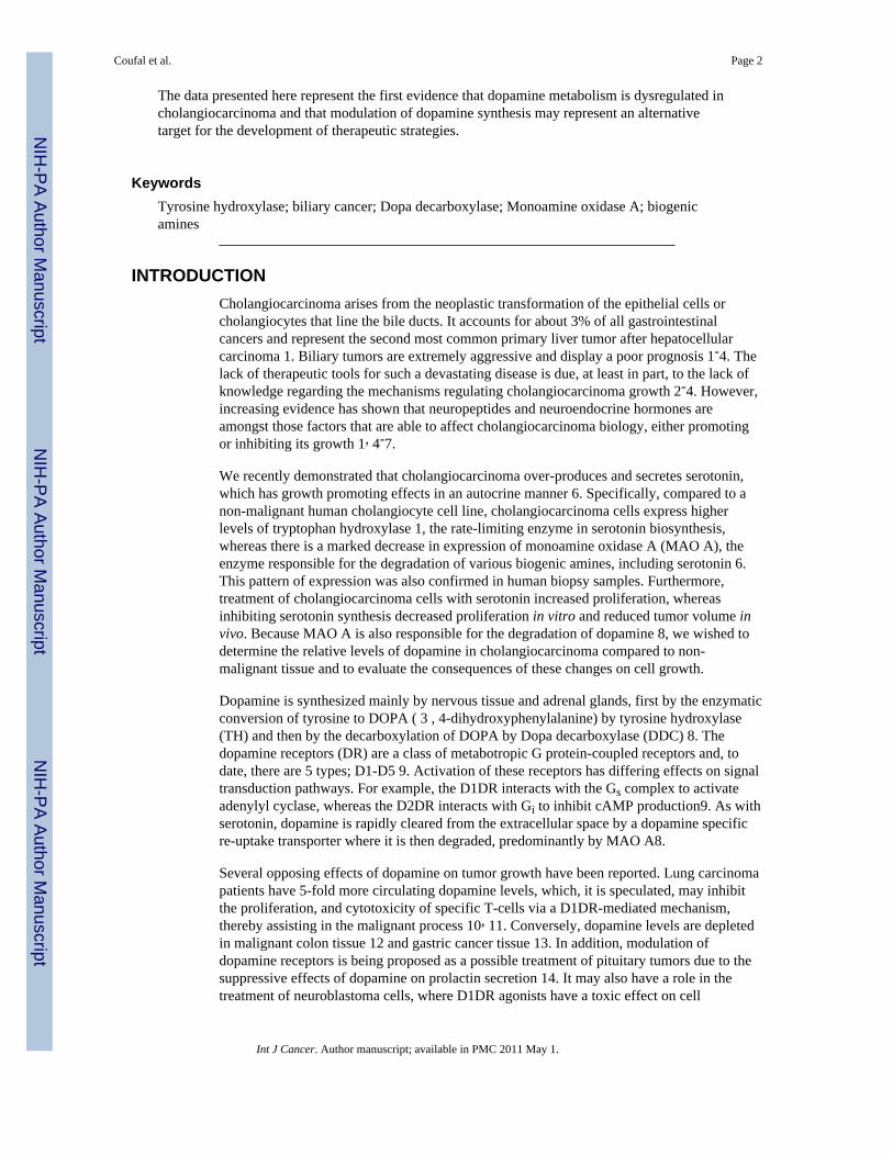
Increased local dopamine secretion has implications on cholangiocarcinomacell growth in vitro—The potential implications of the increased local dopaminesecretion were explored in vitro using the cholangiocarcinoma cell lines. Treatment ofhuman cholangiocyte cell lines, H69 and HIBEC, with various concentrations of dopaminehad no significant impact on cell proliferation (Figure 2), whereas treatment of all of thecholangiocarcinoma cell lines with dopamine (10-5 to 10-9 M) caused a significant increasein cell proliferation after 48 hr as demonstrated by MTS cell proliferation assays (Figure 2).Every dopamine receptor subtype is expressed in both cholangiocytes andcholangiocarcinoma cells, the relative levels of which are summarized in Table 1. Theproliferative effects of dopamine could be blocked by pretreatment with D2 and D4dopamine receptor inhibitors (Figure 3). In contrast, blocking dopamine synthesis using α-Methyl dopa, a specific DDC inhibitor, markedly inhibited cholangiocarcinoma cellproliferation, at concentrations from 10-6 M to 10-8 M (Figure 4). Once again, no effect wasobserved in H69 or HIBEC cells. These data were confirmed using a BrdU incorporationassay as an indication of cell cycle progression. Further, dopamine treatment increased BrdUincorporation, while α-Methyl dopa decreased BrdU incorporation (Figure 5). Taken
Coufal et al. Page 5
Int J Cancer. Author manuscript; available in PMC 2011 May 1.
NIH
-PA Author Manuscript
NIH
-PA Author Manuscript
NIH
-PA Author Manuscript

together, these data indicate that the increased dopamine secretion from cholangiocarcinomacells may have a growth promoting effect on tumor progression.
Animal model of cholangiocarcinomaInhibition of dopamine synthesis inhibits cholangiocarcinoma tumor growthin vivo—By treating an in vivo xenograft model of cholangiocarcinoma tumors with theDDC inhibitor, we significantly suppressed tumor growth (Figure 6A). In addition, thelatency of tumor growth (i.e., time taken for tumor volume to increase to 150% of theoriginal size) was increased after α-Methyl dopa treatment compared to vehicle treatment(Figure 6B). Analysis of liver enzymes in the serum revealed that there was no significantdifference in AST (Vehicle, 93.3 ± 13.5 vs α-Methyl dopa, 102.0 ± 12.6) and ALT levels(Vehicle, 27.0 ± 12.6 vs α-Methyl dopa, 31.3 ± 4.6) between α-Methyl dopa-treated andvehicle-treated animals, both of which fell within normal range, suggesting that the α-Methyl dopa treatment was well tolerated and did not cause liver damage.
Histological analysis of the excised tumors revealed that all cells within tumors from α-Methyl dopa-treated and vehicle-treated animals were CK-19 positive, indicatingcholangiocyte phenotype (Figure 7). As expected, α-Methyl dopa treatment significantlydecreased the dopamine immunoreactivity (Figure 7). Using PCNA immunoreactivity as amarker of proliferative capacity, α-Methyl dopa treatment decreased the number of PCNApositive nuclei compared to vehicle treatment (Figure 7). This decrease in PCNA expressionwas confirmed by real time PCR analysis of RNA extracted from the excised tumors (Figure7).
Human studiesDopamine secretion is increased in cholangiocarcinoma—Immunohistochemicalanalysis of human liver biopsy samples (tissue arrays) indicated an increased THimmunoreactivity in cholangiocarcinoma samples compared to controls as assessed by threeindependent observers (Figure 8A; data not shown). We acknowledge that thesecommercially available tissue arrays are limited in the number and appropriateness of thecontrol samples; therefore we also performed a parallel pilot study in a limited number ofcholangiocarcinoma sections that also contained surrounding non-cancerous liver cells asthe control. Once again, TH immunoreactivity was increased in cholangiocarcinoma tissuecompared to that in normal cholangiocytes in the surrounding non-cancerous liver (Figure8A). Similarly, immunohistochemical analysis of human liver biopsy samples from both thetissue arrays and the cholangiocarcinoma sections indicated that there is also increased DDCimmunoreactivity in cholangiocarcinoma samples compared to control as assessed by threeindependent observers (Figure 8B; data not shown).
Analysis of serum samples from cholangiocarcinoma patients versus age-matched controlsrevealed no significant difference in dopamine levels (data not shown), which is notsurprising given that the normal tissue surrounding the tumor presumably has normaldegradation machinery (i.e., MAO A expression) and would possibly take up the excessdopamine and metabolize it. We then performed a pilot study with a limited number of bilesamples taken from cholangiocarcinoma patients. As controls, we used bile samplescollected from patients affected by intrahepatic cholelithiasis (e.g. nonmalignant disease).Analysis of these treatment groups revealed an increase in dopamine levels incholangiocarcinoma patients compared to “controls” (p<0.05, Figure 9A). In addition,dopamine immunoreactivity was also increased in the cholangiocarcinoma tissue comparedto the control sections from both the commercially available tissue arrays as well as thecholangiocarcinoma sections as assessed by 3 independent observers (Figure 9B and datanot shown).
Coufal et al. Page 6
Int J Cancer. Author manuscript; available in PMC 2011 May 1.
NIH
-PA Author Manuscript
NIH
-PA Author Manuscript
NIH
-PA Author Manuscript

DISCUSSIONThe major findings presented here relate to the dysregulation of dopamine metabolism incholangiocarcinoma. We demonstrated that the expression of the dopamine synthesisenzymes TH and DDC are markedly increased in cholangiocarcinoma, which together withthe suppression of the degradation machinery shown previously 6, results in an increaseddopamine production in cholangiocarcinoma cell lines and tumor tissue, and increaseddopamine in the bile of cholangiocarcinoma patients. Treatment of cholangiocarcinoma celllines with dopamine increased cell proliferation in vitro, and inhibition of dopaminesynthesis decreased cell proliferation in vitro and in an in vivo xenograft model ofcholangiocarcinoma. These data suggest that the dysregulation of dopamine metabolismmay be a key feature associated with the progression of cholangiocarcinoma, and that themodulation of this metabolic pathway may be targeted for the development of effectiveadjunct therapies to treat this deadly disease.
In support of the observations that dopamine metabolism is dysregulated incholangiocarcinoma, increased dopamine production and secretion can be found in a numberof other tumors. There is an increased circulating level of dopamine in lung tumors, whichseems to play a protective role by inhibiting cytotoxic t-cell proliferation thereby preventingtheir ability to mount an adequate attack on the tumor cells 10, 11. Furthermore, dopaminesecretion is increased in some cases of the rare malignancy, pheochromocytoma 32-34 andin carcinoid tumors 35, although the consequences of this secretion on tumor growth orprogression is unclear. However, agents such as dexamethasone that increasepheochromocytoma dopamine content also increase cell proliferation36, suggesting thatthere may be a causal link between increased dopamine content and cell proliferation inthese tumor cells. Conversely, dopamine levels are depleted in malignant colon tissue 12 andgastric cancer tissues 13, whereas dopamine treatement slows tumor growth, presumably bydecreasing the expression of vascular epithelial growth factor and subsequent angiogenesis13, 37. We have previously shown that cholangiocarcinoma, while not strictly classified as acarcinoid tumor, displays many features of a neuroendocrine phenotype, such asChromogranin A, neuron specific enolase expression, and enhanced serotonin secretion 6.Because some carcinoid tumors also produce higher levels of dopamine 35, our observationsof increased dopamine production and secretion are conceivable. Serotonin and dopaminehave been shown to interact in both an antagonistic38, 39 and synergistic40 manner. Wehave clearly demonstrated both serotonin and dopamine are overproduced incholangiocarcinoma and subsequently exert growth-promoting effects, however, whetherboth dopamine and serotonin interact in a synergistic manner in this tumor type is largelyunknown and will be a topic of future research in our laboratory.
The expression of TH and DDC mRNA in blood or bone marrow has been shown to behigher in patients with neuroblastoma, which can be correlated with poor prognosis of thisdisease 41. Furthermore, increased TH expression can also be demonstrated inpheochromocytomas and midgut carcinoid tumors 42. The data presented here support thenotion that the expression of dopamine synthesizing enzymes may be involved in theprocess of tumor progression, and perhaps in controlling cell proliferation.
In our study, we demonstrated an antiproliferative effect of α-Methyl dopa oncholangiocarcinoma cell growth. Chronic treatment of mice bearing cholangiocarcinomatumors decreased the amount of dopamine produced by the tumor cells and slowed tumorgrowth over the period of time studied. The notion of targeting dopamine synthesis as ananticancer treatment is not without precedent. Treatment of small cell lung carcinoma cellsand pulmonary carcinoid cells with another DDC inhibitor caused a selective cytotoxicity43.
Coufal et al. Page 7
Int J Cancer. Author manuscript; available in PMC 2011 May 1.
NIH
-PA Author Manuscript
NIH
-PA Author Manuscript
NIH
-PA Author Manuscript

In conclusion, the data presented here indicate a dysregulated dopamine metabolic pathwayin cholangiocarcinoma compared with nonmalignant cholangiocytes. Specifically, there isan increase in the expression of TH and DDC, the enzymes responsible for dopaminesynthesis. This leads to an increased accumulation and secretion of dopamine fromcholangiocarcinoma and an increase in dopamine in the bile from cholangiocarcinomapatients. Specific inhibition of dopamine production leads to a suppression of tumor growthin a xenograft model of cholangiocarcinoma, which suggests that agents that modulate themetabolism of dopamine may be useful therapeutic tools for the treatment of this devastatingcancer.
AcknowledgmentsWe sincerely thank Dr. Gianfranco Alpini for his assistance and expert advice in the development of thismanuscript. Also, we thank Ms. Julie Venter and Ms. Shelley Kopriva for their assistance with the animal studies.This work was supported by an NIH K01 grant award (DK078532) to Dr. DeMorrow.
Abbreviations
CK-19 Cytokeratin19
DDC Dopa decarboxylase
DR Dopamine receptor
HIBEC human intrahepatic biliary epithelial cells
MAO A monoamine oxidase A
PCNA proliferating cell nuclear antigen
TH tyrosine hydroxylase
REFERENCES1. Gores GJ. Cholangiocarcinoma: current concepts and insights. Hepatology. 2003; 37:961–9.
[PubMed: 12717374]2. Blechacz B, Gores GJ. Tumor-specific marker genes for intrahepatic cholangiocarcinoma: utility
and mechanistic insight. J Hepatol. 2008; 49:160–2. [PubMed: 18538440]3. Blechacz BR, Gores GJ. Cholangiocarcinoma. Clin Liver Dis. 2008; 12:131–50. ix. [PubMed:
18242501]4. Sirica AE. Cholangiocarcinoma: molecular targeting strategies for chemoprevention and therapy.
Hepatology. 2005; 41:5–15. [PubMed: 15690474]5. Fava G, Alpini G, Rychlicki C, Saccomanno S, DeMorrow S, Trozzi L, Candelaresi C, Venter J, Di
Sario A, Marzioni M, Bearzi I, Glaser S, et al. Leptin enhances cholangiocarcinoma cell growth.Cancer Res. 2008; 68:6752–61. [PubMed: 18701500]
6. Alpini G, Invernizzi P, Gaudio E, Venter J, Kopriva S, Bernuzzi F, Onori P, Franchitto A, CoufalM, Frampton G, Alvaro D, Lee SP, et al. Serotonin metabolism is dysregulated incholangiocarcinoma, which has implications for tumor growth. Cancer Res. 2008; 68:9184–93.[PubMed: 19010890]
7. DeMorrow S, Francis H, Gaudio E, Venter J, Franchitto A, Kopriva S, Onori P, Mancinelli R,Frampton G, Coufal M, Mitchell B, Vaculin B, et al. The endocannabinoid anandamide inhibitscholangiocarcinoma growth via activation of the noncanonical Wnt signaling pathway. Am JPhysiol Gastrointest Liver Physiol. 2008; 295:G1150–8. [PubMed: 18832445]
8. Shih JC, Chen K, Ridd MJ. Role of MAO A and B in neurotransmitter metabolism and behavior.Pol J Pharmacol. 1999; 51:25–9. [PubMed: 10389141]
9. Callier S, Snapyan M, Le Crom S, Prou D, Vincent JD, Vernier P. Evolution and cell biology ofdopamine receptors in vertebrates. Biol Cell. 2003; 95:489–502. [PubMed: 14597267]
Coufal et al. Page 8
Int J Cancer. Author manuscript; available in PMC 2011 May 1.
NIH
-PA Author Manuscript
NIH
-PA Author Manuscript
NIH
-PA Author Manuscript

10. Saha B, Mondal AC, Basu S, Dasgupta PS. Circulating dopamine level, in lung carcinoma patients,inhibits proliferation and cytotoxicity of CD4+ and CD8+ T cells by D1 dopamine receptors: an invitro analysis. Int Immunopharmacol. 2001; 1:1363–74. [PubMed: 11460316]
11. Saha B, Mondal AC, Majumder J, Basu S, Dasgupta PS. Physiological concentrations of dopamineinhibit the proliferation and cytotoxicity of human CD4+ and CD8+ T cells in vitro: a receptor-mediated mechanism. Neuroimmunomodulation. 2001; 9:23–33. [PubMed: 11435749]
12. Basu S, Dasgupta PS. Decreased dopamine receptor expression and its second-messenger cAMP inmalignant human colon tissue. Dig Dis Sci. 1999; 44:916–21. [PubMed: 10235597]
13. Chakroborty D, Sarkar C, Mitra RB, Banerjee S, Dasgupta PS, Basu S. Depleted dopamine ingastric cancer tissues: dopamine treatment retards growth of gastric cancer by inhibitingangiogenesis. Clin Cancer Res. 2004; 10:4349–56. [PubMed: 15240521]
14. Ivan G, Szigeti-Csucs N, Olah M, Nagy GM, Goth MI. Treatment of pituitary tumors: dopamineagonists. Endocrine. 2005; 28:101–10. [PubMed: 16311416]
15. Chan AS, Ng LW, Poon LS, Chan WW, Wong YH. Dopaminergic and adrenergic toxicities onSK-N-MC human neuroblastoma cells are mediated through G protein signaling and oxidativestress. Apoptosis. 2007; 12:167–79. [PubMed: 17136323]
16. Knuth A, Gabbert H, Dippold W, Klein O, Sachsse W, Bitter-Suermann D, Prellwitz W, Meyerzum Buschenfelde KH. Biliary adenocarcinoma. Characterisation of three new human tumor celllines. J Hepatol. 1985; 1:579–96. [PubMed: 4056357]
17. Shimizu Y, Demetris AJ, Gollin SM, Storto PD, Bedford HM, Altarac S, Iwatsuki S, HerbermanRB, Whiteside TL. Two new human cholangiocarcinoma cell lines and their cytogenetics andresponses to growth factors, hormones, cytokines or immunologic effector cells. Int J Cancer.1992; 52:252–60. [PubMed: 1355757]
18. Miyagiwa M, Ichida T, Tokiwa T, Sato J, Sasaki H. A new human cholangiocellular carcinomacell line (HuCC-T1) producing carbohydrate antigen 19/9 in serum-free medium. In Vitro CellDev Biol. 1989; 25:503–10. [PubMed: 2544546]
19. Storto PD, Saidman SL, Demetris AJ, Letessier E, Whiteside TL, Gollin SM. Chromosomalbreakpoints in cholangiocarcinoma cell lines. Genes Chromosomes Cancer. 1990; 2:300–10.[PubMed: 2176543]
20. Grubman SA, Perrone RD, Lee DW, Murray SL, Rogers LC, Wolkoff LI, Mulberg AE,Cherington V, Jefferson DM. Regulation of intracellular pH by immortalized human intrahepaticbiliary epithelial cell lines. Am J Physiol. 1994; 266:G1060–70. [PubMed: 8023938]
21. Kanno N, Glaser S, Chowdhury U, Phinizy JL, Baiocchi L, Francis H, LeSage G, Alpini G. Gastrininhibits cholangiocarcinoma growth through increased apoptosis by activation of Ca2+-dependentprotein kinase C-alpha. J Hepatol. 2001; 34:284–91. [PubMed: 11281558]
22. DeMorrow S, Glaser S, Francis H, Venter J, Vaculin B, Vaculin S, Alpini G. Opposing actions ofendocannabinoids on cholangiocarcinoma growth: Recruitment of fas and fas ligand to lipid rafts.J Biol Chem. 2007; 282:13098–113. [PubMed: 17329257]
23. Bertoldi M, Dominici P, Moore PS, Maras B, Voltattorni CB. Reaction of dopa decarboxylase withalpha-methyldopa leads to an oxidative deamination producing 3,4-dihydroxyphenylacetone, anactive site directed affinity label. Biochemistry. 1998; 37:6552–61. [PubMed: 9572873]
24. Kassack MU, Hofgen B, Decker M, Eckstein N, Lehmann J. Pharmacological characterization ofthe benz[d]indolo[2,3-g]azecine LE300, a novel type of a nanomolar dopamine receptorantagonist. Naunyn Schmiedebergs Arch Pharmacol. 2002; 366:543–50. [PubMed: 12444495]
25. Bowery BJ, Razzaque Z, Emms F, Patel S, Freedman S, Bristow L, Kulagowski J, Seabrook GR.Antagonism of the effects of (+)-PD 128907 on midbrain dopamine neurones in rat brain slices bya selective D2 receptor antagonist L-741,626. Br J Pharmacol. 1996; 119:1491–7. [PubMed:8968560]
26. Yuan J, Chen X, Brodbeck R, Primus R, Braun J, Wasley JW, Thurkauf A. NGB 2904 and NGB2849: two highly selective dopamine D3 receptor antagonists. Bioorg Med Chem Lett. 1998;8:2715–8. [PubMed: 9873609]
27. Bristow LJ, Kramer MS, Kulagowski J, Patel S, Ragan CI, Seabrook GR. Schizophrenia andL-745,870, a novel dopamine D4 receptor antagonist. Trends Pharmacol Sci. 1997; 18:186–8.[PubMed: 9226994]
Coufal et al. Page 9
Int J Cancer. Author manuscript; available in PMC 2011 May 1.
NIH
-PA Author Manuscript
NIH
-PA Author Manuscript
NIH
-PA Author Manuscript

28. Fava G, Marucci L, Glaser S, Francis H, De Morrow S, Benedetti A, Alvaro D, Venter J,Meininger C, Patel T, Taffetani S, Marzioni M, et al. gamma-Aminobutyric acid inhibitscholangiocarcinoma growth by cyclic AMP-dependent regulation of the protein kinase A/extracellular signal-regulated kinase 1/2 pathway. Cancer Res. 2005; 65:11437–46. [PubMed:16357152]
29. Kapoor V, Chalmers J. Correlation between fall in blood pressure and in vivo amine release afteralpha-methylDOPA. Eur J Pharmacol. 1989; 164:531–8. [PubMed: 2767124]
30. Sivagnanam G, Adithan C, Thakur LC, Raveendran R, Bapna JS. Alphamethyldopa analgesia: itspossible mechanism of action. Arch Int Pharmacodyn Ther. 1985; 277:168–76. [PubMed:4062430]
31. Faul F, Erdfelder E, Lang AG, Buchner A. G*Power 3: a flexible statistical power analysisprogram for the social, behavioral, and biomedical sciences. Behav Res Methods. 2007; 39:175–91. [PubMed: 17695343]
32. Awada SH, Grisham A, Woods SE. Large dopamine-secreting pheochromocytoma: case report.South Med J. 2003; 96:914–7. [PubMed: 14513991]
33. Tam V, Ng KF, Fung LM, Wong YY, Chan MH, Lam CW, Tam S. The importance of theinterpretation of urine catecholamines is essential for the diagnosis and management of patientwith dopamine-secreting paraganglioma. Ann Clin Biochem. 2005; 42:73–7. [PubMed: 15802039]
34. Yasunari K, Kohno M, Minami M, Kano H, Ohhira M, Nakamura K, Yoshikawa J. A dopamine-secreting pheochromocytoma. J Cardiovasc Pharmacol. 2000; 36(Suppl 2):S75–7. [PubMed:11206726]
35. Kema IP, de Vries EG, Slooff MJ, Biesma B, Muskiet FA. Serotonin, catecholamines, histamine,and their metabolites in urine, platelets, and tumor tissue of patients with carcinoid tumors. ClinChem. 1994; 40:86–95. [PubMed: 7507008]
36. Yang TT, Tsao CW, Li JS, Wu HT, Hsu CT, Cheng JT. Changes of dopamine content and cellproliferation by dexamethsone via pituitary adenylate cyclase-activating polypeptide in PC12 cell.Neurosci Lett. 2007; 426:45–8. [PubMed: 17884294]
37. Sarkar C, Chakroborty D, Mitra RB, Banerjee S, Dasgupta PS, Basu S. Dopamine in vivo inhibitsVEGF-induced phosphorylation of VEGFR-2, MAPK, and focal adhesion kinase in endothelialcells. Am J Physiol Heart Circ Physiol. 2004; 287:H1554–60. [PubMed: 15371263]
38. Olvera-Cortes ME, Anguiano-Rodriguez P, Lopez-Vazquez MA, Alfaro JM. Serotonin/dopamineinteraction in learning. Prog Brain Res. 2008; 172:567–602. [PubMed: 18772051]
39. Di Giovanni G, Di Matteo V, Pierucci M, Esposito E. Serotonin-dopamine interaction:electrophysiological evidence. Prog Brain Res. 2008; 172:45–71. [PubMed: 18772027]
40. Gonzalez-Burgos I, Feria-Velasco A. Serotonin/dopamine interaction in memory formation. ProgBrain Res. 2008; 172:603–23. [PubMed: 18772052]
41. Trager C, Vernby A, Kullman A, Ora I, Kogner P, Kagedal B. mRNAs of tyrosine hydroxylase anddopa decarboxylase but not of GD2 synthase are specific for neuroblastoma minimal disease andpredicts outcome for children with high-risk disease when measured at diagnosis. Int J Cancer.2008; 123:2849–55. [PubMed: 18814238]
42. Meijer WG, Copray SC, Hollema H, Kema IP, Zwart N, Mantingh-Otter I, Links TP, WillemsePH, de Vries EG. Catecholamine-synthesizing enzymes in carcinoid tumors andpheochromocytomas. Clin Chem. 2003; 49:586–93. [PubMed: 12651811]
43. Gilbert JA, Frederick LM, Ames MM. The aromatic-L-amino acid decarboxylase inhibitorcarbidopa is selectively cytotoxic to human pulmonary carcinoid and small cell lung carcinomacells. Clin Cancer Res. 2000; 6:4365–72. [PubMed: 11106255]
Coufal et al. Page 10
Int J Cancer. Author manuscript; available in PMC 2011 May 1.
NIH
-PA Author Manuscript
NIH
-PA Author Manuscript
NIH
-PA Author Manuscript

Figure 1.Enzymes responsible for dopamine synthesis are upregulated in cholangiocarcinoma cells invitro. TH (A) and DDC (B) expression was assessed by real time PCR and immunoblottingin four cholangiocarcinoma cell lines as well as the non-malignant cholangiocyte cell linesH69 and HIBEC. In each case, data are expressed as average ± SEM (n=3). Asterisk denotessignificance (p<0.05) compared with expression in H69 cells. Dopamine levels were alsoassessed in the supernatant of cell suspensions of cholangiocarcinoma cell lines and the non-malignant cholangiocyte cell lines H69 and HIBEC by EIA after 6 hr (C). Data areexpressed as average dopamine concentration (ng/mL left axis and nMol, right axis) ± SEM(n=3). Asterisk denotes significance (p<0.05) compared with dopamine levels secreted fromH69 cells.
Coufal et al. Page 11
Int J Cancer. Author manuscript; available in PMC 2011 May 1.
NIH
-PA Author Manuscript
NIH
-PA Author Manuscript
NIH
-PA Author Manuscript

Figure 2.Dopamine increases cholangiocarcinoma cell proliferation in vitro. Cholangiocarcinomacells and the non-malignant cholangiocyte cell lines, H69 and HIBEC, were treated withvarious concentrations of dopamine (10-9 M to 10-5 M) for 48 hr. Cell proliferation wasassessed using an MTS cell proliferation assay. Data are expressed as fold change inproliferation (average ± SEM, n=7) and the asterisk denotes p<0.05 compared to basaltreatment within each cell line.
Coufal et al. Page 12
Int J Cancer. Author manuscript; available in PMC 2011 May 1.
NIH
-PA Author Manuscript
NIH
-PA Author Manuscript
NIH
-PA Author Manuscript

Figure 3.Specific dopamine receptor antagonists inhibit the growth-promoting effects of dopamine.Mz-ChA-1 cells were pretreated with specific antagonists of the dopamine receptorsindicated (all at 10 nM), prior to the addition of dopamine (100 nM) for 48 hr. Cellproliferation was assessed using an MTS cell proliferation assay. Data are expressed as foldchange in proliferation (average ± SEM, n=7) and the asterisk denotes p<0.05 compared toantagonist alone.
Coufal et al. Page 13
Int J Cancer. Author manuscript; available in PMC 2011 May 1.
NIH
-PA Author Manuscript
NIH
-PA Author Manuscript
NIH
-PA Author Manuscript
Figure 4.Inhibition of dopamine synthesis decreases cholangiocarcinoma cell proliferation in vitro.Cholangiocarcinoma cells and the non-malignant cholangiocyte cell lines, H69 and HIBEC,were treated with various concentrations of α-methyl dopa (10-6 M to 10-8 M) for 48 hr. Cellproliferation was assessed using an MTS cell proliferation assay. Data are expressed as foldchange in proliferation (average ± SEM, n=7) and the asterisk denotes p<0.05 compared tobasal treatment within each cell line.
Coufal et al. Page 14
Int J Cancer. Author manuscript; available in PMC 2011 May 1.
NIH
-PA Author Manuscript
NIH
-PA Author Manuscript
NIH
-PA Author Manuscript

Figure 5.BrdU labeling of cholangiocarcinoma cells indicates changes in cell cycle progression aftertreatment with dopamine and α-methyl dopa. Mz-ChA-1 cells were treated with dopamine(10-7M) and CPA (10-6M) for 48 hr and BrdU uptake was determined. The number ofBrdU-positive cells was expressed as a percentage of total cells. Data was expressed as theaverage ± SEM from 5 random fields from 3 independent experiments. Asterisk denotessignificance (p<0.05) when compared to basal treatment.
Coufal et al. Page 15
Int J Cancer. Author manuscript; available in PMC 2011 May 1.
NIH
-PA Author Manuscript
NIH
-PA Author Manuscript
NIH
-PA Author Manuscript

Figure 6.Inhibition of dopamine synthesis decreases tumor growth in an in vivo xenograft model ofcholangiocarcinoma. Mz-ChA-1 cells were injected into the flank of athymic mice. Aftertumors were established, mice were treated with 100 mg/kg/day (ip) α-methyl dopa, threedays per week for 70 days and tumor volume assessed (A). Tumor latency was assessed asthe time taken for the tumor to grow to 150% of the original size (B). Data are expressed asaverage latency (days ± SEM) and the asterisk denotes significance (p<0.05) from vehicle-treated tumors.
Coufal et al. Page 16
Int J Cancer. Author manuscript; available in PMC 2011 May 1.
NIH
-PA Author Manuscript
NIH
-PA Author Manuscript
NIH
-PA Author Manuscript

Figure 7.Immunohistological analysis of tumors. Immunohistochemistry on tumors from vehicle- (A,C, E) and α-methyl dopa-treated mice was performed using specific antibodies againstCK-19 (A, B), dopamine (C, D) and PCNA (E, F). Representative photomicrographs of theimmunoreactivity are shown (magnification X40). Semi-quantitative analysis of PCNAimmunoreactivity was performed and data was expressed as average (± SEM) PCNApositive nuclei per field (G) and the asterisk denotes significance (p<0.05) compared tovehicle-treated tumors. PCNA expression in the tumors was also assessed by real time PCR(H). Data are expressed as average ± SEM (n=3). Asterisk denotes significance (p<0.05)compared to vehicle-treated tumors.
Coufal et al. Page 17
Int J Cancer. Author manuscript; available in PMC 2011 May 1.
NIH
-PA Author Manuscript
NIH
-PA Author Manuscript
NIH
-PA Author Manuscript

Figure 8.Enzymes responsible for dopamine synthesis are upregulated in cholangiocarcinoma tumorbiopsy samples. TH (A) and DDC (B) immunoreactivity was assessed in biopsy samplesfrom 48 cholangiocarcinoma patients and 4 healthy controls by immunohistochemistry(upper panel) and in paraffin-embedded sections containing cholangiocarcinoma and thesurrounding non-malignant liver tissue (n=6; lower panel). Representative photomicrographsof the TH (A) and DDC (B) immunoreactivity are shown (magnification X40). Stainingintensity was assessed as described in the methods. P values and the power analysis valuesare shown in parentheses.
Coufal et al. Page 18
Int J Cancer. Author manuscript; available in PMC 2011 May 1.
NIH
-PA Author Manuscript
NIH
-PA Author Manuscript
NIH
-PA Author Manuscript

Figure 9.
Coufal et al. Page 19
Int J Cancer. Author manuscript; available in PMC 2011 May 1.
NIH
-PA Author Manuscript
NIH
-PA Author Manuscript
NIH
-PA Author Manuscript

Dopamine secretion in cholangiocarcinoma. Dopamine levels in bile samples fromcholangiocarcinoma patients and patients with intrahepatic cholelithiasis were determinedby EIA (A). Data are expressed in a scatter plot of dopamine concentration (ng/mL left axisand nMol right axis). Dopamine immunoreactivity was assessed in biopsy samples from 48cholangiocarcinoma patients and 4 healthy controls by immunohistochemistry (upper panel)and in paraffin-embedded sections containing cholangiocarcinoma and the surrounding non-malignant liver tissue (n=6; lower panel). Representative photomicrographs of the dopamineimmunoreactivity are shown (B; magnification X40). Staining intensity was assessed asdescribed in the methods. P values and the power analysis values are shown in parentheses.
Coufal et al. Page 20
Int J Cancer. Author manuscript; available in PMC 2011 May 1.
NIH
-PA Author Manuscript
NIH
-PA Author Manuscript
NIH
-PA Author Manuscript

NIH
-PA Author Manuscript
NIH
-PA Author Manuscript
NIH
-PA Author Manuscript
Coufal et al. Page 21
Table 1
Fold change in dopamine receptor subunit expression in cholangiocarcinoma compared to H69 cholangiocytecells
Receptor Mz-ChA-1 HuCCT-1 SG231 CCLP-1
D1DR 5.43 ± 1.25* 0.66 ± 0.11* 0.85 ± 0.08 0.48 ± 0.05*
D2DR 3.92 ± 0.72* 2.14 ± 0.21* 0.16 ± 0.03* 3.13 ± 0.21*
D3DR 2.87 ± 0.76* 1.01 ± 0.03 0.79 ± 0.03 0.49 ± 0.03*
D4DR 0.38 ± 0.06* 0.49 ± 0.02* 0.27 ± 0.02* 0.76 ± 0.04*
D5DR 2.45 ± 0.71* 0.27 ± 0.03* 0.49 ± 0.05* 0.18 ± 0.02*
● p<0.05 compared to non -malignant cholangiocytes (H69)
● significant increases is shaded in purple, significant decrease are shaded in gray, no shading indicates no significance.
Int J Cancer. Author manuscript; available in PMC 2011 May 1.


