Embed Size (px)
Citation preview

Increased Self-Focus in Major Depressive DisorderIs Related to Neural Abnormalities inSubcortical-Cortical Midline Structures
Simone Grimm,1 Jutta Ernst,1 Peter Boesiger,2 Daniel Schuepbach,1
Daniel Hell,1 Heinz Boeker,1 and Georg Northoff1,3*
1Department of Psychiatry, University of Zurich, Zurich, Switzerland2Institute of Biomedical Engineering, ETH and University of Zurich, Zurich, Switzerland
3Department of Psychiatry, University of Magdeburg, Magdeburg, Germany
Abstract: Patients with major depressive disorder (MDD) often show a tendency to strongly introspectand reflect upon their self, which has been described as increased self-focus. Although subcortical-corti-cal midline structures have been associated with reflection and introspection of oneself in healthy sub-jects, the neural correlates of the abnormally increased attribution of negative emotions to oneself, i.e.negative self-attribution, as hallmark of the increased self-focus in MDD remain unclear. The aim ofthe study was, therefore, to investigate the neural correlates during judgment of self-relatedness of pos-itive and negative emotional stimuli thereby testing for emotional self-attribution. Using fMRI, weinvestigated 27 acute MDD patients and compared them with 25 healthy subjects employing a para-digm that focused on judgment of self-relatedness when compared with mere perception of the verysame emotional stimuli. Behaviourally, patients with MDD showed significantly higher degrees of self-relatedness of specifically negative emotional stimuli when compared with healthy subjects. Neurally,patients with MDD showed significantly lower signal intensities in various subcortical and corticalmidline regions like the dorsomedial prefrontal cortex (DMPFC), supragenual anterior cingulate cortex,precuneus, ventral striatum (VS), and the dorsomedial thalamus (DMT). Signal changes in the DMPFCcorrelated with depression severity and hopelessness whereas those in the VS and the DMT wererelated to judgment of self-relatedness of negative emotional stimuli. In conclusion, we present firstevidence that the abnormally increased negative self-attribution as hallmark of the increased self-focusin MDD might be mediated by altered neural activity in subcortical-cortical midline structures. HumBrain Mapp 00:000–000, 2009. VVC 2008 Wiley-Liss, Inc.
Key words: major depressive disorder; fMRI; neuroimaging; self-focus; self-relatedness
Contract grant sponsor: German Research Foundation; Contractgrant number: DFG, SFB 779/A6 (to G.N.); Contract grant spon-sors: Salus Foundation, Hope of Depression Research Foundation(to G.N.); Contract grant sponsor: Swiss National Research Foun-dation; Contract grant number: 3100A0-100830 (to G.N. H.B.);Contract grant sponsors: Research Foundation at the University ofZurich (to H.B.), Salus Foundation Sachsen Anhalt/Germany (toG.N.), ETH Zurich (SEP) and Philips Medical Systems, Best, NL(to P.B.), Hartmann- Muller-Foundation, the Stanley-Thomas-John-son Foundation and the Gebert-Ruf-Foundation (to H.B.).
*Correspondence to: Georg Northoff, Department of Psychiatry,University of Magdeburg, Leipziger Strasse 44, 39120 Magdeburg,Germany. E-mail: [email protected] for publication 26 June 2008; Revised 1 October 2008;Accepted 14 October 2008
DOI: 10.1002/hbm.20693Published online in Wiley InterScience (www.interscience.wiley.com).
VVC 2008 Wiley-Liss, Inc.
r Human Brain Mapping 00:000–000 (2009) r

INTRODUCTION
Patients with major depressive disorder (MDD) can becharacterized by multiple self-abnormalities includingruminations, self-blame, and increased association of theirself with negative emotions (Gruenbaum et al., 2005;Ingram, 1990; Northoff, 2007; Rimes and Watkins, 2005;Treynor, 2003). Clinically, these different abnormalities ofthe self may be subsumed under the concept of increasedself-focus (Ingram, 1990; Northoff, 2007). One hallmark ofsuch increased self-focus is the abnormal attribution ofnegative emotions to patients’ self which may conse-quently be described as increased self-attribution of nega-tive emotions, i.e., emotional or negative self-attribution.Despite these well-known clinical phenomena, the
underlying neural networks and the pathophysiologicalmechanisms remain unclear. The topic of self has recentlybecome a focus in functional neuroimaging. Imaging stud-ies in healthy subjects demonstrated recruitment of thedorsomedial prefrontal cortex (DMPFC) and other corticaland subcortical midline structures like the supragenual an-terior cingulate cortex (SACC), the precuneus, the dorso-medial thalamus (DMT), and the ventral striatum (VS)during self-related stimuli (Northoff and Bermpohl, 2004;Northoff et al., 2006; Schmitz and Johnson, 2007). Thesesubcortical-cortical midline regions are supposed to medi-ate introspective processes like reflection, evaluation, andrecollection upon oneself (Damasio, 1999; deGreck et al.,2008; Keenan et al., 2001; Kelley et al., 2002; Mitchell et al.,2006; Northoff et al., 2006; Northoff, 2007; Northoff, 2008;Ochsner et al., 2005; Schmitz and Johnson, 2007; Uddinet al., 2007). These introspective processes and hence theself have specifically been associated with high restingstate neural activity in the subcortical-cortical midlineregions (Northoff et al., 2006, Schneider et al., in press,Mason et al., 2007, D’Argembeau et al., 2005). Lateral corti-cal regions have been associated with other aspects of theself. The insula, for instance, is supposed to be implicatedin the bodily-vegetative aspects of the self (Craig, 2002,2003, 2004) and the ventro- and dorsolateral prefrontal cor-tex (DLPFC) in processing recognition of oneself (Keenanet al., 2001; Northoff et al., 2006).Interestingly, the very same regions have also been
observed to be abnormal in MDD. For instance, severalstudies demonstrated altered neural activity in subcortical-cortical midline regions like the DMPFC, the SACC, theprecuneus, the DMT, and the VS in MDD during variouskinds of emotional-cognitive stimulation (Fitzgerald et al.,2008; Grimm et al., 2008; Mayberg, 2002, 2003; Phillipset al., 2003; Siegle et al., 2006; Steele et al., 2007). Further-more, there is strong evidence that these subcortical-corti-cal midline regions might be crucial in the so-calleddefault-mode network (Raichle et al., 2001, 2005, Buckneret al., 2008) that show increased neural activity during theresting state in MDD (Greicius et al., 2007; Grimm et al.,2008; Mayberg, 2002, 2003; Philips et al., 2003 for review).Such increased resting state neural activity should then
lead to decreased activity as induced by external stimuli,i.e., stimulus-induced activity. This has indeed been dem-onstrated in a recent study of ours with regard to emo-tional stimuli though unfortunately not in association withself-relatedness (see Grimm et al., 2008).In contrast to these studies, so far no studies reported
neural abnormalities of the self during emotional-cognitivestimulation in MDD. The aim of our study was to investi-gate the neural mechanisms underlying the abnormalitiesof the self in MDD, e.g., the ability to reflect upon andintrospect the relation of positive and negative emotions tooneself as it is for instance required when attributing emo-tions to oneself. We hypothesized that the apparentlyabnormally high resting state neural activity in thesepatients should lead to lower stimulus-induced signalchanges by self-relatedness in subcortical-cortical midlineregions. One would consequently expect that the tendencyof patients with MDD to strongly reflect upon oneself, i.e.,the increased self-focus, and particularly the increased self-attribution of negative emotions may be directly modu-lated by decreased signal changes in subcortical-corticalmidline regions. To test these hypotheses, we investigated27 patients with acute MDD during the judgment of self-relatedness of positive and negative emotional stimuli thusinvestigating self-attribution of emotions as hallmark ofwhat can be clinically described as increased self-focus.
METHODS AND MATERIALS
Subjects
Subjects with an acute MDD episode (DSM-IV, Ameri-can Psychiatric Association, 1994) were recruited from theinpatient department of Psychiatry at the University ofZurich. Eligibility screening procedures included the 21-item Hamilton Depression Rating Scale (HDRS) (Hamilton,1960), the 21-item Beck Depression Inventory (BDI) (Becket al., 1961), the 20-item Beck Hopelessness Scale (BHS)(Beck et al., 1974), which includes many items about one-self, and clinical laboratory tests. Diagnoses of depressionwere made by the participants’ treating psychiatrists.Inclusion criteria were a score of at least 18 on the HDRSand the BDI. Exclusion criteria were major medical ill-nesses, histories of seizures, head trauma with loss of con-sciousness, abnormal clinical laboratory tests, and preg-nancy. In addition, patients who were actively suicidal,met criteria for any psychiatric disorder other than MDD,had a history of substance abuse or electroconvulsive ther-apy in the previous 6 mo, or had a history of substance de-pendence were excluded from the study. Healthy subjectswithout any psychiatric, neurologic, or medical illnesswere self-referred from online study advertisements. Thestudy was approved by the University of Zurichs’ Institu-tional Review Board, and all subjects gave writteninformed consent before screening. All subjects were righthanded as assessed with the Edingburgh Handedness
r Grimm et al. r
r 2 r

Inventory (Oldfield et al., 1970). After applying the exclu-sion criteria above, fMRI scans from 27 depressed subjectsand 25 healthy control subjects were processed. Of thesescans, two could not be included in the analysis owing tostructural abnormalities in the 3D T1-weighted anatomicalscan (two depressed subjects). This resulted in usablefMRI data on 25 subjects with depression and 25 healthycontrol subjects.
Pictorial Stimuli
Subjects viewed full-color pictures selected from theInternational Affective Picture System (IAPS) (Lang et al.,1999) with positive (IAPS norm ratings: 7.32 6 2.06) andnegative (IAPS norm ratings: 2.24 6 2.67) valence. The pic-ture sets were counterbalanced across all subjects as wellas within each subject according to the two categories ofvalence as well as according to dominance, intensity,human faces, and human figures. We used IAPS stimulithat were successfully applied in previous studies (Grimmet al., 2006, 2008) and also validated with regard to self-relatedness in two separate groups of healthy subjects(Northoff et al., in press). The pictures were generated byPresentation1 (Neurobehavioral Systems, Albany, CA) andrear projected onto a projection screen positioned at thehead end of the MRI scanner bore. Subjects viewed thescreen through a mirror mounted on the head coil andresponded by pushing a fiber-optic light sensitive key-press.
Experimental Design
The fMRI design was ‘‘event related’’’’ with positive andnegative stimuli alternating with a fixation control condi-tion. The IAPS pictures were presented for 4 s. A total of150 pictures was presented twice: once for judgment andonce for passive viewing (PV). In case of the judgmentcondition, subjects had to judge the pictures with regardto their self-relatedness [‘‘picture judgment’’ (‘‘PJ’’)]; thiswas indicated by the letter ‘‘B’’ (i.e., German term forjudgment or evaluation) in one corner of the picture. Pic-tures had to be judged as either self-related or not (yes-nooption).During the viewing condition, subjects had to passively
view the picture (‘‘PV’’), which was indicated by the letter‘‘E’’ in one corner of the picture. Here, subjects had toarbitrarily press a button without making any judgment.Responses and reaction times were recorded. After thepresentation of each picture, a resting period followed,where a fixation cross was presented for 6–8 s (6.0, 6.5, 7.0,7.5, 8.0 s). This allowed the subjects to recover from emo-tional stimulation and, in addition, served as a baselinecondition to distinguish between positive and negativeBOLD responses (Stark and Squire, 2001). A total of 300trials was presented in six runs; 150 trials were presentedfor PJ and 150 trials for PV. The different task conditionswere pseudorandomized within and across the six runs
and their order counterbalanced across all subjects. Imme-diately after the fMRI session pictures were presented fora second time. Each of the 160 pictures (including 10 newones for distraction) was followed by a task period whichconsisted of a concern rating (subjects had to rate whetherthey felt affected by the picture), dominance rating, inten-sity rating, valence rating, and self-relatedness rating. Allresponses were given using a scale ranging from 1 to 9.The 10 new emotional pictures were matched in valence,intensity, and dominance with those presented in fMRI.The mean of each of the five ratings was calculated foreach subject. Analysis of postscanning ratings was con-ducted separately for positive and negative pictures. Post-scanning ratings were conducted with 16 healthy controlsand 15 depressed subjects, because some of the subjectswere too exhausted after the fMRI-scan to continue theinvestigation.
Functional Imaging
Measurements were performed on a Philips Intera 3Twhole-body MR unit equipped with an eight-channel Phi-lips SENSE head coil. Functional time series were acquiredwith a sensitivity encoded (Pruessmann et al., 1999) single-shot echo-planar sequence (SENSE-sshEPI). The followingacquisition parameters were used in the fMRI protocol:echo time 5 35 ms, field of view 5 22 cm, acquisitionmatrix 5 80 3 80, interpolated to 128 3 128, voxel size:2.75 3 2.75 3 4 mm3, SENSE acceleration factor R 5 2.0.Using a midsaggital scout image, 32 contiguous axial sliceswere placed along the anterior-posterior commissure planecovering the entire brain with a TR 5 3000 ms (y 5 828).The first three acquisitions were discarded due to T1 satu-ration effects.
Statistical Analysis
Behavioral data
Reaction times and judgments (self-relatedness rating)were analyzed in a multivariate analysis of variance(ANOVA) with the factors group (healthy subjects/patients with MDD), valence (positive/negative pictures),and task (PJ/PV). Postscanning ratings of concern, domi-nance, valence, intensity, and self-relatedness were ana-lyzed in a group 3 valence ANOVA. We performed multi-variate ANOVAS where we included the differentsubstance—classes of psychotropic medication (SSRIs, tri-cyclica, benzodiazepines, etc.) as well as age as covariateswhereas the dosage of the medication was not consideredin the analysis.
fMRI data
fMRI data were analyzed using MATLAB 6.5.1 (TheMathworks, Natick, MA) and SPM2 (Statistical parametricmapping software, SPM; Wellcome Department of Imaging
r Self-Focus and Neural Abnormalities in Depression r
r 3 r

Neuroscience, London, UK; http://www.fil.ion.ucl.ac.uk).For each subject, a design matrix was defined modelingjudgment of self-relatedness (PJ) and perception (PV) asseparate events. In addition to these two events the base-line condition was included in the design matrix and mod-eled separately, independent of the other events. In addi-tion, for each experimental run, the six parametersobtained in the realignment procedure were included asregressors in the design matrix.For the fMRI data group analyses the contrast images
from the analysis of the individual subjects were analyzedusing two-sample t tests to compare signal changes in theabove mentioned conditions between healthy and MDDsubjects. Activations are reported at a level of significanceP < 0.001, uncorrected and a cluster threshold of greaterthan 5. All group comparisons included age and psycho-tropic medication as co-variates.For the regions of interest (ROI) analyses of peak voxels,
coordinates that were obtained in contrasts of the between-group analyses (Table I) were selected. ROI were function-ally defined by centering spheres on the respective peakvoxels with a radius of 3 mm. Analyses were carried out forthe DMPFC (28, 36, 12), SACC (12, 30, 28), precuneus (24,272, 38), VS (10, 10, 6), bilateral DMT (210, 214, 10; 12,214, 14), left DLPFC (254, 6, 42), and insula (242, 16, 4).For the ROI analyses, % signal changes for the different con-ditions were extracted for each subject separately usingMarsbar (http://marsbar.sourceforge.net/). For each event% signal changes were calculated relative to the mean signalintensity of this ROI across the whole experiment. Time-course analyses were performed applying a finite impulseresponse model, which does not make an assumption on theresulting signal changes after stimulus presentation. Parame-ter estimates were calculated for 8 time bins of 1TR (5 3 s)length for each regressor of the design matrix.
To detect the association of signal changes in responseto self-related judgment with psychopathological parame-ters and postscanning ratings, the correlation between thedifferent psychopathological components of the BDI (3-fac-tor solution: anhedonia/inhibition, negative self-concept,somatic complaints; Schotte et al., 1997), the individualscores of the BHS, the individual scores of postscanningratings and signal changes in the ROI was analyzed in apost hoc, ROI analysis using Marsbar (see above). Subjects’individual scores were correlated with signal changes dur-ing judgment of self-relatedness (PJ) > perception (PV)using Pearson correlation analysis.
RESULTS
Subjects
The control group had a mean age of 32.4 with 12women and 13 men. The depressed group had a mean ageof 37.0 with 9 women and 16 men. Groups did not differsignificantly in age (t test P 5 0.09) or in gender distribu-tion (v2 P 5 0.39). The mean HDRS score was 26.8 (SD7.1), the mean BDI score 26.6 (SD 9.1), and the mean BHSscore 31.08 (SD 5.2) in the depressed group, indicating thatpatients were severely depressed and showed self-abnor-malities with increased attribution of negative emotions tooneself as hallmark of an increased self-focus. The meanduration of the current episode was 8.1 weeks (SD 8.4), thenumber of previous depressive episodes 2.2 (SD 1.5).Regarding exposure to psychotropic medications, 2 of the25 depressed subjects were not taking any when investi-gated. Twenty-three depressed subjects were taking one ormore medications from the following classes: antidepres-sants (19 subjects; Selective serotonin reuptake inhibitors(SSRIs): 15 subjects; Tricyclic antidepressants (TCAs): 5subjects), antipsychotics (5 subjects), anxiolytics (6 sub-jects), and mood stabilizers (Lithium: 3 subjects). None ofthe control subjects was taking any psychotropic medica-tions at the time of the investigation.
Behavioral Data
Intrascanner ratings (reaction times
and self-related judgments)
There was a significant effect of group (F(1) 5 216.61, P <0.001), task (F(1) 5 2419.21, P < 0.001), and valence (F(1) 529.07, P < 0.001) on reaction times, whereas there were nointeractions between these factors. Post hoc t tests demon-strated faster reaction times in healthy subjects in PV (t 5212.52, P < 0.001) as well as in PJ (t 5 28.05, P < 0.001).Reaction times were faster in positive pictures (t 5 4.57,P < 0.001) and PV (t 5 248.99, P < 0.001). Concerning theself-related judgments there was a significant group effect(F(1) 5 120.62, P < 0.001) and valence effect (F(1) 5 20.21,P < 0.001), but again no interaction effects. Differences in
TABLE I. Differences between healthy and depressed
subjects in self-related judgement
Region Side
PJ > PV
H > MDD MDD > H
DMPFC L 28, 36, 12 3.28SACC R 12, 30, 28 3.78Precuneus L 24, 272, 38 3.70DMT L 210, 214, 10 4.22
R 12, 214, 14 4.58VS R 10, 10, 6 3.84DLPFC L 254, 6, 42 3.60Insula L 242, 16, 4 3.20
The global height threshold for between-group comparisons (healthyvs. MDD) was set to P< 0.001 uncorrected, the extent threshold to k55 voxels for the contrast. The values in the table represent maximum z
values with peak voxel coordinates in theMNI stereotactic space.PJ, picture judgment; PV, passive viewing; H, healthy subjects;MDD, major depressive disorder patients; DMPFC, dorsomedialprefrontal cortex; SACC, supragenual anterior cingulate cortex;DMT, dorsomedial thalamus; VS, ventral striatum; DLPFC, dorsolat-eral prefrontal cortex.
r Grimm et al. r
r 4 r

intrascanning self-related judgment between groups con-cerned positive (t 5 7.67, P < 0.001) and negative pictures(t 5 8.75, P < 0.001), which both were rated significantlymore self-related by the patients with MDD. Inclusion ofage and psychotropic medication as covariates did nothave any influence on the results from group comparisons.The results are indicative of a consistent psychomotorimpairment and show an increased tendency to relateemotional stimuli to the own self in patients with MDDthus supporting the assumption of an increased negativeself-attribution as hallmark of an increased self-focus.
Postscanning ratings
There was a significant effect of participant group(healthy, MDD) on ratings of intensity (F(1) 5 20.52, P <
0.001). Picture valence (positive, negative) had a significanteffect on ratings of concern (F(1) 5 173.45, P < 0.001), va-lence (F(1) 5 4,776.14, P < 0.001), intensity (F(1) 5 99.78,P < 0.001), and self-relatedness (F(1) 5 602.39, P < 0.001).There was a significant interaction effect between the fac-tors group and picture valence for intensity (F(1) 5 7.89,P < 0.005) and self-relatedness (F(1) 5 10.41, P < 0.001).Post hoc t tests demonstrated that negative pictures wererated significantly more intense (t 5 25.37, P < 0.001) andself-related (t 5 23.63, P < 0.001) by the depressivepatients, whereas there were no differences betweengroups for ratings of intensity and self-relatedness of posi-tive pictures (Fig. 1). Inclusion of age and psychotropicmedication as covariates did not have any influence on theresults from group comparisons. Results support theassumption of an increased self-attribution specificallywith regard to negative emotions in MDD.
fMRI Data
Effect of self-related judgment
To elucidate the effects of self-related judgment, wecompared PJ > PV between subjects with MDD andhealthy subjects. Patients with MDD showed significantlylower signal intensities in the DMPFC, SACC, precuneus,bilateral DMT, VS, left DLPFC, and insula. Locations ofresponses are summarized in Table I. Higher signal inten-sities in patients with MDD could only be obtained if thethreshold was lowered to P < 0.005 uncorrected and werefound in the right precentral cortex and left parietal cortex.Bar diagrams and time curves show clear significant differ-ences in signal changes during PJ, whereas signal changesduring PV did not differ significantly between subjectswith MDD and healthy subjects (Fig. 2). Inclusion of age,gender, and psychotropic medication as covariates did nothave any influence on the results from group comparisons.Taken together, fMRI results demonstrate abnormal neuralactivity in subcortical-cortical midline regions (and lateralcortical regions like DLPFC and insula) specifically duringjudgment of self-relatedness as distinguished from mereperception of the very same stimuli.To investigate parametric dependence of neural activity
on self-relatedness, postscanning ratings of self-relatednessfor positive and negative pictures were separately corre-lated with signal intensities in the contrast PJ > PV. Insubjects with MDD, this analysis revealed a significantpositive correlation of self-relatedness ratings for negativeemotional pictures in the bilateral DMT (left DMT: r 50.62, P < 0.05; right DMT: r 5 0.61, P < 0.05) and VS (r 50.67, P < 0.01). Healthy subjects, in contrast, showed a dif-ferent correlation pattern with no significant correlations inboth VS (r 5 20.44, P > 0.05) and bilateral DMT (r 5 0.19,P > 0.05); further the direction of the relationship wasreversed in the case of the VS (Fig. 3). Signal changes inthe precuneus also correlated differently with self-related-ness ratings of negative pictures in healthy subjects (r 50.59, P < 0.05) and subjects with MDD (r 5 0.35, P > 0.05).During positive emotional pictures, subject with MDDshowed a significant positive correlation of self-relatednessratings with signal changes in the insula (r 5 0.56, P <0.05) whereas healthy subjects showed a reversed correla-tion pattern (r 5 20.33, P > 0.05). Inclusion of intensity,valence, dominance, and concern ratings as well medica-tion dosage did not have any influence on correlationresults in positive and negative pictures. Taken together,these results support the involvement of subcortical mid-line regions in modulating abnormal self-relatedness innegative emotions in MDD.
Correlation of signal changes with
psychopathological symptoms
To demonstrate the psychopathological relevance ofaltered neural activity in MDD, contrast estimates of PJ >PV were correlated with patients total BDI scores as well
Figure 1.
Self-relatedness ratings of positive and negative pictures in
healthy and depressed subjects. Performance in postscanning
self-relatedness ratings for all pictures, negative pictures, and
positive pictures. Patients with MDD rated negative pictures to
be significantly more self-related, although there were no differ-
ences in positive pictures. *P < 0.05. MDD, major depressive
disorder.
r Self-Focus and Neural Abnormalities in Depression r
r 5 r
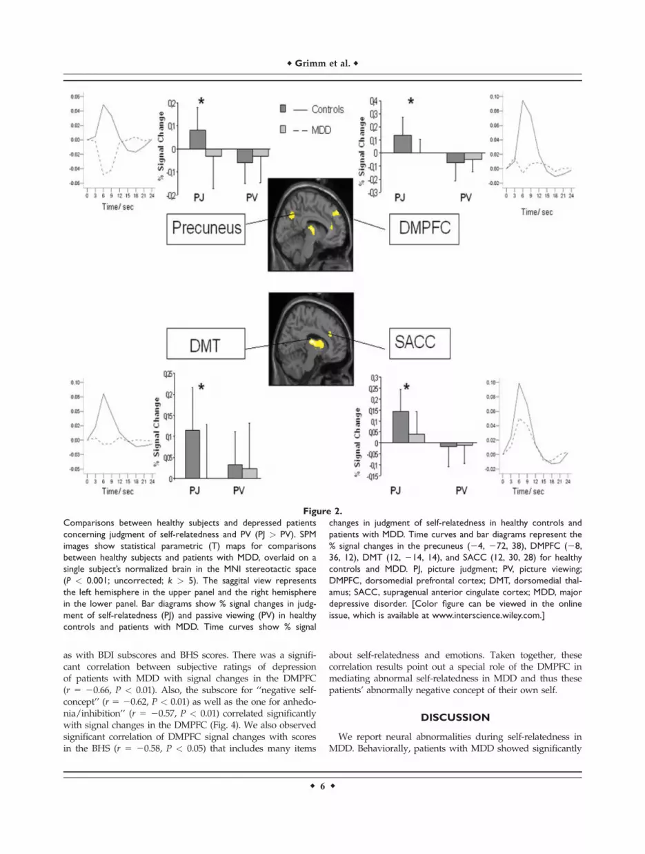
as with BDI subscores and BHS scores. There was a signifi-cant correlation between subjective ratings of depressionof patients with MDD with signal changes in the DMPFC(r 5 20.66, P < 0.01). Also, the subscore for ‘‘negative self-concept’’ (r 5 20.62, P < 0.01) as well as the one for anhedo-nia/inhibition’’ (r 5 20.57, P < 0.01) correlated significantlywith signal changes in the DMPFC (Fig. 4). We also observedsignificant correlation of DMPFC signal changes with scoresin the BHS (r 5 20.58, P < 0.05) that includes many items
about self-relatedness and emotions. Taken together, thesecorrelation results point out a special role of the DMPFC inmediating abnormal self-relatedness in MDD and thus thesepatients’ abnormally negative concept of their own self.
DISCUSSION
We report neural abnormalities during self-relatedness inMDD. Behaviorally, patients with MDD showed significantly
Figure 2.
Comparisons between healthy subjects and depressed patients
concerning judgment of self-relatedness and PV (PJ > PV). SPM
images show statistical parametric (T) maps for comparisons
between healthy subjects and patients with MDD, overlaid on a
single subject’s normalized brain in the MNI stereotactic space
(P < 0.001; uncorrected; k > 5). The saggital view represents
the left hemisphere in the upper panel and the right hemisphere
in the lower panel. Bar diagrams show % signal changes in judg-
ment of self-relatedness (PJ) and passive viewing (PV) in healthy
controls and patients with MDD. Time curves show % signal
changes in judgment of self-relatedness in healthy controls and
patients with MDD. Time curves and bar diagrams represent the
% signal changes in the precuneus (24, 272, 38), DMPFC (28,
36, 12), DMT (12, 214, 14), and SACC (12, 30, 28) for healthy
controls and MDD. PJ, picture judgment; PV, picture viewing;
DMPFC, dorsomedial prefrontal cortex; DMT, dorsomedial thal-
amus; SACC, supragenual anterior cingulate cortex; MDD, major
depressive disorder. [Color figure can be viewed in the online
issue, which is available at www.interscience.wiley.com.]
r Grimm et al. r
r 6 r

higher degrees of self-relatedness of negative emotionalstimuli and thus increased self-attribution of negative emo-tions, which is well in accordance with the clinical obser-vation of increased reflection and introspection of oneself,i.e., an increased self-focus. Neuronally, judgment of self-relatedness (when compared with mere perception of thevery same stimuli) was associated with reduced signalchanges in cortical and subcortical midline regions(DMPFC, SACC, Precuneus, VS, DMT) that are supposedto mediate the reflection and introspection of oneself. Thisis well in accordance with our hypothesis of reduced sig-nal changes that may be traced back to abnormally highresting state activity in these regions. Reduced signalchanges in subcortical midline structures (VS, DMT) corre-lated with abnormally high ratings of self-relatedness ofnegative emotional pictures whereas signal changes in theDMPFC correlated with psychopathological symptoms(BDI, BHS) of self, a negative self-concept, and anhedonia.
This corroborates our hypothesis that the increased nega-tive self-attribution in MDD may be modulated by reducedsubcortical-cortical midline signal changes. Taken together,our results provide first evidence for association of theincreased negative self-attribution in MDD with abnormalneural activity in subcortical-cortical midline regions.The DMPFC has often been observed to show reduced
neural activity during predominantly cognitive or cogni-tive-emotional tasks in MDD (Fitzgerald et al., 2008; May-berg, 2003; Phillipps et al., 2003). In healthy subjects, theDMPFC has been strongly associated with self-relatedness(Kelley et al., 2002; Northoff et al., 2006; Ochsner et al.,2005) and particularly the reflection and introspection ofoneself including self-awareness, self-reflection, self-evalu-ation, and self-recollection (Ochsner et al., 2005; Schmitzand Johnson, 2007). This and the often observed increasednegative self-attribution in MDD led us to suggest that theDMPFC may show altered activity specifically during
Figure 3.
Correlation of postscanning subjective ratings for self-related-
ness in negative emotional pictures with BOLD signals obtained
in the contrast PJ > PV in healthy and subjects with MDD. SPM
images shows statistical parametric (T) maps for the comparison
between healthy subjects and patients with MDD, overlaid on a
single subject’s normalized brain in the MNI stereotactic space
(P < 0.001; uncorrected; k > 5) during PJ > PV. The saggital
view represents the left hemisphere. The scatter plots show
subjects’ self-relatedness ratings for negative pictures on x-axis
and % signal change in left DMT (210, 214, 10) and VS (10, 10,
6) during PJ > PV on y-axis. Scatter plots are presented for both
healthy and depressed subjects (**P < 0.01; *P < 0.05). DMT,
dorsomedial thalamus; VS, ventral striatum; MDD, major depres-
sive disorder. [Color figure can be viewed in the online issue,
which is available at www.interscience.wiley.com.]
r Self-Focus and Neural Abnormalities in Depression r
r 7 r

judgment of self-relatedness in MDD mirroring negativeself-attribution. Our results demonstrated significantlyreduced signal intensities during judgment of self-related-ness in MDD. Most interestingly, this was specific for thejudgment period while during mere perception of the verysame stimuli no significant differences were observedbetween both groups in this region. This indicates thatabnormal stimulus-induced activity in DMPFC may bespecifically related to judgment of the self and thus emo-tional self-attribution. More specifically, reduced stimulus-induced activity in DMPFC seems to be specifically elicitedby reflection and introspection of the self as required injudgment of self-relatedness rather than mere perceptionwithout reflection and introspection.The apparent involvement of the DMPFC in self-abnor-
malities in MDD is further supported by the significantcorrelation between DMPFC signal intensities and theBDI/BHS scores as subjective measures of depression ingeneral and self-abnormalities in particular. The lowerDMPFC signal intensities during judgment of self-related-ness, the more severe MDD patients judged their symp-toms (BDI total) and the more self-abnormalities (BDI sub-score for negative self-concept and BHS) they experienced.These results indicate that psychopathological symptomsin MDD, such as the increased negative attribution as hall-mark of the increased self-focus, may be modulated byabnormal neural activity in the DMPFC.In addition to the DMPFC, we also observed abnormal-
ities in various other cortical midline regions. The SACChas been associated with monitoring of self-relatedness,which may be crucial in allowing to reflect about oneself(Northoff et al., 2006) and has often been observed to be
abnormal in MDD (Fitzgerald et al., 2008; Mayberg, 2002,2003; Phillips et al., 2003). Our observation of SACC defi-cits in MDD during judgment of self-relatedness mayindicate altered monitoring of self-relatedness, whichpsychologically may lead to increased self-relatednesswith less detachment specifically from negative emo-tional stimuli. The precuneus has been associated withretrieval of autobiographical and thus self-relevant infor-mation (Northoff et al., 2006) in healthy subjects. Patientswith MDD showed alterations in the precuneus, whichmay indicate altered retrieval of episodic or autobio-graphical memories.We also observed subcortical midline regions to be
altered in MDD. Patients with MDD showed significantlyreduced signal changes in the DMT and the VS duringjudgment of self-relatedness. These regions have beenshown to be specifically implicated in associating self andemotions in healthy subjects (Northoff et al., in press;Phan et al., 2004). Although the very same regions havealso been implicated in MDD (Epstein et al., 2006; Fitzger-ald et al., 2008; Grimm et al., 2008; Mayberg, 2002, 2003), itremained unclear whether they are also involved in media-ting the abnormally increased negative self-attribution andthus the increased self-focus in these patients. Our resultslend evidence to their involvement in the increased nega-tive self-attribution by showing reduced signal changesduring judgment of self-relatedness, which also correlatedparametrically with ratings of negative emotions. This indi-cates that subcortical midline regions seem to be crucial inassociating self and especially negative emotions in MDD.Neurally, the observation of reduced signal changes in
subcortical-cortical midline regions like the DMPFC,
Figure 4.
Correlation of depression symptom severity (BDI) with BOLD
signals obtained in the contrast PJ > PV in patients with MDD.
The SPM image shows a statistical parametric (T) map for the
comparison between healthy subjects and patients with MDD,
overlaid on a single subject’s normalized brain in the MNI ste-
reotactic space (P < 0.001; uncorrected; k > 5) during PJ > PV.
The saggital view represents the left hemisphere. The scatter
plot shows BDI subscore ratings for negative self-concept on x-
axis and % signal change in DMPFC (28, 36, 12) during PJ > PV
on y-axis. The scatter plot is presented for depressed subjects
(**P < 0.01). DMPFC, dorsomedial prefrontal cortex; BDI, Beck
Depression Inventory, MDD, major depressive disorder. [Color
figure can be viewed in the online issue, which is available at
www.interscience.wiley.com.]
r Grimm et al. r
r 8 r

SACC, DMT, and precuneus seem to be paradoxical.Increased self-focus should go along with increased neuralactivity rather than reduced signal changes as observedhere. This, however, is to neglect that self-related stimulido certainly not induce neural activity from zero in sub-cortical-cortical midline regions but modulate a preexistingstate of high resting state activity in these regions. Hence,the degree of stimulus-induced signal changes during self-relatedness may strongly depend upon the degree of rest-ing state activity. There is strong evidence that restingstate activity in subcortical-cortical midline regions isabnormally high in MDD (Greicius et al., 2007; Grimmet al., 2008; Mayberg, 2002, 2003; Phillips et al., 2003). Iftrue, this should lead to reduced stimulus-induced signalchanges in these patients, which has indeed been observedduring emotional stimulation (Grimm et al., 2008). Studiesin healthy subjects indicate that the degree of resting stateactivity in these regions may mirror the degree of self-relatedness (Mason et al., 2007; Schneider et al., in press;see D’Argembeau et al., 2005). Abnormally high restingstate activity in MDD should then be accompanied byincreased negative self-attribution and thus an increasedself-focus, which can indeed be observed both clinicallyand behaviourally. If the increased self-focus is indeedrelated to apparently abnormally high resting state activity,one would expect reduced signal changes in subcortical-cortical midline regions in these patients. This is exactlywhat we observed here and which was further confirmedby our parametric analysis. However, our results provide atbest indirect evidence because we neither investigated rest-ing state activity itself nor did we focus on those regionsthat were specifically active during the resting state. Hence,further studies with different experimental designs andanalyses are necessary that provide a less indirect relation-ship between resting state activity and self-relatedness.It should also be mentioned that other cortical regions
also showed altered neuronal activity including theDLPFC and the Insula. The DLPFC has been associatedwith self-relatedness, particularly its cognitive compo-nents like self-recognition and self-manipulation(Northoff et al., 2006; Ochsner et al., 2005; Mitchell et al.,2006). We also demonstrate altered neuronal activity inleft DLPFC during judgment of self-relatedness, whichcontributes in further characterizing the dysfunction inthis region in MDD (Grimm et al., 2008). Altered neuro-nal activity during judgment of self-relatedness was alsoobserved in the insula, which has often been shown to bedeficient in MDD (Bar et al., 2004; Fitzgerald et al., 2008).This dysfunction might be related to possible alterationsin interoceptive processing as the presumed main func-tion of this region in healthy subjects (Craig, 2002, 2003,2004; Critchley et al., 2005).Several limitations in our study should be acknowl-
edged. First, we did not investigate unmedicated MDDpatients so that antidepressant medication may have con-founded our results. Including medication dosage as cova-riate in our various analyses did not alter the obtained
results though. Second, one may criticize that we investi-gated only one particular symptom of the increased self-focus, the increased negative self-attribution, whereas ourdesign does not allow to make inferences about othersymptoms of the increased self-focus like ruminations,self-blame, etc., and the distinction between analytical andexperiential self-focus (Rimes and Watkins, 2005). Becausethis is certainly true more specific paradigms need to bedeveloped to test for the distinct components of theincreased self-focus in MDD. One may also be concernedabout our control condition, the perception of the verysame stimuli as used in the judgment condition. It shouldbe obvious, that we remain unable to exclude other typesof processing than self-related processing in the perceptioncondition. For instance it may be argued that the task wecall ‘‘picture viewing’’ will almost always involve somekind of judgment. Indeed, some kind of unconscious orpreconscious judgment that might implicate implicit self-related processing cannot be completely ruled out. Studiesthat focus on implicit versus explicit self-relatedness arenecessary to investigate whether both modes recruit sub-cortical-cortical midline structures or whether theseregions are specifically associated with explicit self-relatedprocessing such as introspection and reflection of self-relat-edness. Finally, the issue of task difficulty needs to bementioned. Though subjects had to press a button evenduring perception, both perception and judgment showdifferent task difficulties with only judgment requiring acognitive effort whereas perception does not. This differ-ence in task difficulty applied, however, equally to bothgroups healthy and subjects with MDD and is thereforecancelled out when comparing both groups. We also haveto mention that we cannot distinguish between state andtrait markers in our sample because we investigated thepatients only in the acute depressed state. One may forinstance imagine that the apparently high resting stateneural activity in midline regions could be a trait markerwhereas the stimulus-induced activity in these regionsmay be state rather than trait marker. Finally, the relation-ship of self-relatedness to other cognitive functions associ-ated with alterations in the same regions in MDD remainsunclear (Grimm et al., 2008). Future studies may conse-quently focus on the interaction between self-relatednessand cognitive functions to delineate the exact psychologi-cal function responsible for the reduced signal changes inthese regions. One may criticize the lack of neutral stimuliin our design, which makes it impossible to investigate theinteraction between emotion and self-relatedness. Becauseour main focus was on judgment of self-relatedness as dis-tinguished from mere perception, we refrained fromincluding neutral stimuli, which would have overextendedthe paradigm.In conclusion, our study provides first evidence of
altered neural activity in a subcortical-cortical midline net-work during judgment of self-relatedness in MDD. Thesechanges may be crucial in mediating patients’ abnormallyincreased self-attribution of negative emotions as hallmark
r Self-Focus and Neural Abnormalities in Depression r
r 9 r

of what clinically can be conceptualized as increased self-focus.
REFERENCES
American Psychiatric Association (1994): Diagnostic and StatisticalManual of Mental Disorders. Washington, DC: American Psy-chiatric Association.
Bar KJ, Greiner W, Jochum T, Friedrich M, Wagner G, Sauer H(2004): The influence of major depression and its treatment onheart rate variability and pupillary light reflex parameters.J Affect Disord 82:245–252.
Beck AT, Ward CH, Mendelson M, Mock J, Erbaugh J (1961): Aninventory for measuring depression. Arch Gen Psychiatry4:561–571.
Beck AT, Weissman A, Lester D, Trexler L (1974): The measure-ment of pessimism: the hopelessness scale. J Consult Clin Psy-chol 42:861–865.
Buckner RL, Andrews-Hanna JR, Schacter DL (2008): The brain’sdefault network: anatomy, function, and relevance to disease.Ann N Y Acad Sci 1124:1–38.
Craig AD (2002): How do you feel? Interoception: the sense of thephysiological condition of the body. Nat Rev Neurosci 3:655–666.
Craig AD (2003): Interoception: the sense of the physiological con-dition of the body. Curr Opin Neurobiol 13:500–505.
Craig AD (2004): Human feelings: why are some more aware thanothers? Trends Cogn Sci 8:239–241.
Critchley HD, Rotshtein P, Nagai Y, O’Doherty J, Mathias CJ,Dolan RJ (2005): Activity in the human brain predicting differ-ential heart rate responses to emotional facial expressions.NeuroImage 24:751–762.
Damasio AR (1999): How the brain creates the mind. Sci Am281:112–117.
D’Argembeau A, Collette F, Van der Linden M, Laureys S, DelFiore G, Degueldre C, Luxen A, Salmon E (2005): Self-referen-tial reflective activity and its relationship with rest: a PETstudy. NeuroImage 25:616–624.
de Greck M, Supady A, Thiemann R, Tempelmann C, Bogerts B,Forschner L, Ploetz KV, Northoff G (2008): Is our self based onreward? Self-relatedness recruits neural activity in the rewardsystem. NeuroImage 39:2066–2075.
Epstein J, Pan H, Kocsis JH, Yang Y, Butler T, Chusid J, HochbergH, Murrough J, Strohmayer E, Stern E, Silbersweig DA (2006):Lack of ventral striatal response to positive stimuli in depressedversus normal subjects. Am J Psychiatry 163:1784–1790.
Fitzgerald PB, Laird AR, Maller J, Daskalakis ZJ (2008): A meta-analytic study of changes in brain activation in depression.Hum Brain Mapp 29:683–695.
Greicius MD, Flores BH, Menon V, Glover GH, Solvason HB,Kenna H, Reiss AL, Schatzberg AF (2007): Resting-state func-tional connectivity in major depression: Abnormally increasedcontributions from subgenual cingulate cortex and thalamus.Biol Psychiatry 62:429–437.
Grimm S, Schmidt CF, Bermpohl F, Heinzel A, Dahlem Y, WyssM, Hell D, Boesiger P, Boeker H, Northoff G (2006): Segre-gated neural representation of distinct emotion dimensionsin the prefrontal cortex—An fMRI study. NeuroImage30:325–340.
Grimm S, Beck J, Schuepbach D, Hell D, Boesiger P, Bermpohl F,Niehaus L, Boeker H, Northoff G (2008): Imbalance betweenleft and right dorsolateral prefrontal cortex in major depres-sion is linked to negative emotional judgment: An fMRI
study in severe major depressive disorder. Biol Psychiatry63:369–376.
Grimm S, Boesiger P, Beck J, Schuepbach D, Bermpohl F, WalterM, Ernst J, Hell D, Boeker H, Northoff G (2008): Altered nega-tive BOLD-responses in the default-mode network duringemotion processing in depressed subjects. Neuropsychophar-macology (in press).
Grunebaum MF, Keilp J, Li S, Ellis SP, Burke AK, Oquendo MA,Mann JJ (2005): Symptom components of standard depressionscales and past suicidal behavior. J Affect Disord 87:73–82.
Hamilton M (1960): A rating scale for depression. J Neurol Neuro-surg Psychiatry 23:56–62.
Ingram RE (1990): Self-focused attention in clinical disorders:Review and a conceptual model. Psychol Bull 107:156–176.
Keenan JP, Nelson A, O’Connor M, Pascual-Leone (2001): A neu-rology: Self-recognition and the right hemisphere. Nature409:305.
Kelley WM, Macrae CN, Wyland CL, Caglar S, Inati S, HeathertonTF (2002): Finding the self? An event-related fMRI study.J Cogn Neurosci 14:785–794.
Lang PJ, Bradley MM, Cuthbert BN (1999): International AffectivePicture System (IAPS). The Center for Research in Psycho-physiology, University of Florida.
Mason MF, Norton MI, Van Horn JD, Wegner DM, Grafton ST,Macrae CN (2007): Wandering minds: the default network andstimulus-independent thought. Science 5810:393–395.
Mayberg H (2002): Depression, II: Localization of pathophysiology.Am J Psychiatry 159:1979.
Mayberg HS (2003): Modulating dysfunctional limbic-cortical cir-cuits in depression: towards development of brain-based algo-rithms for diagnosis and optimised treatment. Br Med Bull65:193–207.
Mitchell JP, Macrae CN, Banaji MR (2006): Dissociable medial pre-frontal contributions to judgments of similar and dissimilarothers. Neuron 50:655–663.
Northoff G (2007): Psychopathology and pathophysiology of theself in depression—Neuropsychiatric hypothesis. J Affect Dis-ord 104:1–14.
Northoff G (2008): What kind of neural coding and self does Hur-ley’s shared circuit model presuppose? Behav Brain Sci 2008;31:33–34.
Northoff G, Bermpohl F (2004): Cortical midline structures andthe self. Trends Cogn Sci 8:102–107.
Northoff G, Heinzel A, de Greck M, Bermpohl F, Dobrowolny H,
Panksepp J (2006): Self-referential processing in our brain—A
meta-analysis of imaging studies on the self. NeuroImage
31:440–457.Northoff G, Schneider F, Rotte M, Matthiae C, Tempelmann C,
Wiebking C, Bermpohl F, Heinzel A, Danos P, Heinze HJ,Bogerts B, Walter M, Panksepp J: Differential parametric mod-ulation of selfrelatedness and emotions in different brainregions. Hum Brain Mapp (in press).
Ochsner KN, Beer JS, Robertson ER, Cooper JC, Gabrieli JD, Kihsl-trom JF, D’Esposito M (2005): The neural correlates of directand reflected self-knowledge. NeuroImage 28:797–814.
Oldfield RC (1971): The assessment and analysis of handedness:The Edinburgh inventory. Neuropsychologia 9:97–113.
Phan KL, Wager TD, Taylor SF, Liberzon I (2004): Functionalneuroimaging studies of human emotions. CNS Spectr 9:258–266.
Phillips ML, Drevets WC, Rauch SL, Lane R (2003): Neurobiologyof emotion perception II: Implications for major psychiatric dis-orders. Biol Psychiatry 54:515–528.
r Grimm et al. r
r 10 r

PruessmannKP,WeigerM, ScheideggerMB, Boesiger P (1999): SENSE:sensitivity encoding for fastMRI.MagnResonMed 42:952–962.
Raichle ME, Gusnard DA (2005): Intrinsic brain activity sets thestage for expression of motivated behavior. J Comp Neurol493:167–176.
Raichle ME, MacLeod AM, Snyder AZ, Powers WJ, Gusnard DA,Shulman GL (2001): A default mode of brain function. ProcNatl Acad Sci USA 98:676–682.
Rimes KA, Watkins E (2005): The effects of self-focused rumina-tion on global negative self-judgements in depression. BehavRes Ther 43:1673–1681.
Schmitz TW, Johnson SC (2006): Self-appraisal decisions evoke dis-sociated dorsal—Ventral aMPFC networks. NeuroImage 30:1050–1058.
Schneider F, Bermpohl F, Heinzel A, Rotte M, Walter M, Tempel-mann C, Wiebking C, Dobrowolny H, Heinze HJ, Northoff G:The resting brain and our self: Self-relatedness modulates rest-ing state neural activity in cortical midline structures. Neuro-science (in press).
Schotte CKW, Maes M, Cluydts R, De Doncker D, Cosyns P(1997): Construct validity of the Beck Depression Inventory ina depressive population. J Affect Disord 46:115–125.
Siegle GJ, Carter CS, Thase ME (2006): Use of fMRI to predictrecovery from unipolar depression with cognitive behaviortherapy. Am J Psychiatry 163:735–738.
Stark CE, Squire LR (2001): When zero is not zero: The problem ofambiguous baseline conditions in fMRI. Proc Natl Acad SciUSA 98:12760–12766.
Steele JD, Kumar P, Ebmeier KP (2007): Blunted response to feed-back information in depressive illness. Brain 130:2367–2374.
Taylor SF, Phan KL, Decker LR, Liberzon I (2003): Subjective rat-ing of emotionally salient stimuli modulates neural activity.NeuroImage 18:650–659.
Treynor E (2003): Rumination reconsidered: A psychometric analy-sis. Cogn Ther Res 27:247–259.
Uddin LQ, Iacoboni M, Lange C, Keenan JP (2007): The self andsocial cognition: The role of cortical midline structures andmirror neurons. Trends Cogn Sci 11:153–157.
r Self-Focus and Neural Abnormalities in Depression r
r 11 r


