Embed Size (px)
Citation preview
© University of Pretoria
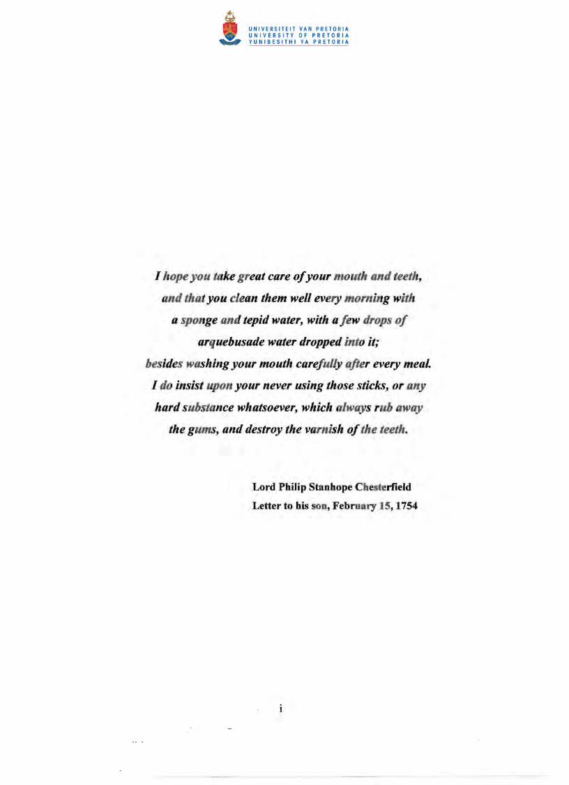
I hope you take great care ofyour mouth and teeth,
and that you clean them well every morning with
a sponge and tepid water, with a few drops of
arquebusade water dropped into it;
besides washing your mouth carefully after every meal
I do insist upon your never using those sticks, or ,any
hard substance whatsoever, which a/ways rub away
the gums, and destroy the varnish ofthe teeth.
Lord Philip Stanhope Chesterfield
Letter to his son, February 15, 1754

INDEX
DECLARATION ....................................................................................................... VI
DEDICATION ............................................................................................................ Vll
SUMMARY ................................................................................................................. Vlli
ACKNOWLEDGEMENTS ................................................................................... XlI
LIST OF FIGURES .................................................................................................. XlV
OPSOMMING ............................................................................................................ x
LIST OF TABLES .................................................................................................... xv
LIST OF ABBREVIATIONS ............................................................................... XVI
CHAPTER 1: INTRODUCTION
1.1 Introduction............................................ ... ...... ........... ... ........................... ..... .......... 1
1.2 Aim, Goals and Premise of the Study..................................................................... 2
1.2.1 Aim.......................................................................................................................... 2
1.2.2 Goals ....................................................................................................................... 2
1.2.3 Premise.................................................................................................................... 2
1.3 Delimitations and Limitations of the Study................................ ............................ 3
1.3.1 Delimitations... ................................................................ ............ ............. ............... 3
1.3.2 Linlitations .............................................................................................................. 3
1.4 Framework of Dissertation ..................................................................................... 4
1.5 Summary ................................................................................................................. 4
CHAPTER 2: LITERATURE REVIEW
2.1 Introduction............................................................................................................. 6
2.2 Periodontal disease and orthodontic treatment....................................................... 6
2.2.1 Pathogenesis ofperiodontal diseases...................................................................... 6
2.2.2 Long-term effects of orthodontic treatment on periodontal health ........................ 8
2.3 Dental caries and orthodontic treatment.. .............................. ...... ............ ............... 11
11

2.3.1 Aetiology..... .......... .................................................... ..................... ... .......... ............ 11
2.3.2 Clinical appearance of demineralisation................................................................. 12
2.3.3 Microstructural changes with demineralisation...................................................... 12
2.3.4 Orthodontic Patients ............................................................................................... 13
2.4 Development ofperiodontal indices .............. ..................... .................................... 16
2.5 Toothbrush comparisons......................................................................................... 19
2.5.1 Studies on Colgate Precision toothbrush................................................................ 21
2.5.2 Studies on the Oral-B Advantage toothbrush......................................................... 23
2.5.3 Studies on the Aquafresh toothbrush...................................................................... 23
2.6 Effectiveness of toothbrushing on oral hygiene during fixed orthodontic
treatment...................... ............................................................................................ 24
2.7 Effective oral hygiene for orthodontic patients ...................................................... 27
2.7.1 Toothbrushes........................................................................................................... 28
2.7.2 Interdental Adjuncts................................................................................................ 28
2.7.3 Fluoride ... ............. .......................... ..... ................. ....... ........................................ ... . 29
2.7.4 Oral Irrigators.......................................................................................................... 30
2.8 Summary ............... ................. .......................................... ..... ... ..... ............ ...... ..... ... 30
CHAPTER 3: MATERIALS AND METHODS
3.1 Introduction............................................................................................................. 32
3.2 Selection of the sample ........................................................................................... 32
3.3 Toothbrushes and toothpaste .................................................................................. 32
3.4 Instrumentation ....................................................................................................... 33
3.5 Evaluation criteria................................................. .................................................. 35
3.5.1 Plaque Index............................................................................................................ 35
3.5.2 Gingival Index ........................................................................................................ 36
3.5.3 Index ofOral Cleanliness........................................................................................ 37
3.6 Examination Procedure and Calibration................................................................. 37
3.7 Study Design.............................. .............................. ................... ............................ 38
3.7.1 Oral Hygiene Instructions....................................................................................... 38
3.7.2 Experimental design................................................................................................ 38
III

3.8 Data recording and analysis ........................... .......................... .............. ...... ........... 40
3.8.1 Exrunination data fonn............................................................................................ 40
3.8.2 Data recording......................................................................................................... 40
3.8.3 Data analysis ........................................................................................................... 40
3.9 Scheduling............................................................................................................... 41
3.10 Summary ................................................................................................................. 41
CHAPTER 4: RESULTS 4.1 Introduction............................................................................................................. 42
4.2 Analysis of Sample ................................................................................................. 42
4.3 Plaque Index............................................................................................................ 43
4.3.1 Plaque Index per tooth ........................... .................... ................................ ............. 43
4.3.2 Plaque Index per mouth .......................................................................................... 46
4.3.3 Statistical Analysis... .................. .................... ............ ...... ................. ....... ........ ....... 46
4.4 Gingival Index ........................................................................................................ 48
4.4.1 Gingival Index per tooth ..................................................... ......... ........................... 48
4.4.2 Gingival Index per mouth .......................................................... ........... .................. 48
4.4.3 Statistical Analysis.................................................................................................. 51
4.5 Index of Oral Cleanliness........................................................................................ 52
4.5.1 IOC (Before) vs IOC (After)................................................................................... 52
4.5.2 Statistical Analysis.................................................................................................. 53
4.6 Toothbrush Preference............................................................................................ 55
4.6.1 Statistical Analysis.................................................................................................. 56
4.7 Summary ................................................................................................................. 56
CHAPTER 5: DISCUSSION, CONCLUSIONS AND RECOMMENDA
TIONS
5.1 Discussion............................................................................................................... 58
5.1.1 Introduction............................................................................................................. 58
5.1.2 Summary of results ................................................................................................. 59
iv

5.2 Conclusion .............................................................................................................. 60
5.3 Recommendations................................................................................................... 61
REFERENCES........................................................................................................... 62
ADDENDUM A.......................................................................................................... 70
v

DECLARATION
I, Ashraf Laher, declare that Effectiveness of Manual Toothbrushes in
Patients with Fixed Orthodontic Appliances which I herewith submit to the
University of Pretoria for the MChD (Orthodontics) degree has not previously
been submitted by me to any other university.
ASHRAF LAHER DATE
vi

DEDICATION
To my parents, who gave me the
opportunity to succeed
and
To my wife, Tayyeba
and sons, Muhammed and Ebrahim,
for their love, support and understanding
Vll

SUMMARY
EFFECTIVENESS OF MANUAL TOOTHBRUSHES IN PATIENTS
WITH FIXED ORTHODONTIC APPLIANCES
Leader: Dr J Kroon, Department of Community Dentistry
Faculty of Dentistry, University of Pretoria
External Examiner: Dr W A Wiltshire, Department of Orthodontics
University of Manitoba, Canada
Department: Orthodontics
Degree: MChD (Orthodontics)
Maintaining good oral hygiene is a challenge for anyone, but particularly for orthodontic
patients whose appliances make them more susceptible to gingivitis, hyperplastic tissue,
decalcification and dental caries.
The aim of this study was to evaluate:
i) the effectiveness of 4 different manual toothbrushes III patients with fixed
orthodontic appliances
ii) the correlation of the Index of Oral Cleanliness (lOC) to both the Plaque Index (PI)
and Gingival Index (GI) and
iii) to detennine whether patient toothbrush preference is directly related to plaque
control.
A single-blind, cross-over study design was used to evaluate the toothbrushes. The brushes
evaluated were Orthodontic Oral B, Oral B Advantage 30, Colgate Precision and
Aquafresh. Forty-six patients, aged 11 to 27, undergoing fixed orthodontic appliance
therapy were screened and recruited with parental consent. These patients were randomly
viii

allocated into 4 groups. All the patients were given a scale and polish at week O. Baseline
recordings of PI, GI and IOC were done 4 weeks later and the first toothbrush given. After
using the toothbrush for a period of 2 weeks the PI, GI and IOC were again recorded and
the patients had another scale and polish. Aperiod of 4 weeks elapsed before new baseline
recordings were done and the sequence followed as described for the next toothbrush. This
was done until all patients had used all 4 toothbrushes. At the end of the clinical trial, each
patient was asked about their toothbrush preference.
The results showed that the PI and GI values were relatively low at baseline as well as after
the use of the toothbrushes. The Brown and Forsyths test for equality of variance was done
to enable testing of means. General linear model procedure showed no statistical
difference in Mean Plaque Index (MPI) before and after, and Difference in Mean Plaque
Index (DMPI) amongst the 4 toothbrushes. There was a slight difference in the Difference
in Mean Gingival Index (DMGI) between the Colgate Precision and Aquafresh toothbrush.
F or all the other comparisons general linear model procedure showed no difference in
MGI (before) and MGI (after).
Pearsons Correlation analysis showed that the IOC was significantly correlated to the PI,
but this correlation was not perfect. The high level of correlation indicates that the IOC
can be used as a screening procedure in orthodontic patients. There was no correlation
between the IOC and GI.
There was no correlation between patients preferred toothbrush and effectiveness of oral
hygiene as measured by DMPI and DMGI.
IX
OPSOMMING
DOELTREFFENDHEID VAN NIE-OUTOMATIESE TANDEBORSELS
IN PASleNTE MET VASTE ORTODONTIESE APPARAAT
Leier: Dr JKroon, Departement Gemeenskapstandheelkunde
Fakulteit Tandheelkunde, Universiteit van Pretoria
Eksterne Eksaminator: Dr W A Wiltshire, Departement Ortodonsie
Universiteit van Manitoba, Kanada
Departement: Ortodonsie
Graad: MChD (Ortodonsie)
Die handhawing van goeie mondhigiene is 'n uitdaging vir enige pasient, maar veral vir
ortodonsie pasiente waar hierdie apparate hulle vatbaarheid vir gingivitis, hiperplastiese
weefsel, dekalsifikasie en tandkaries verhoog.
Die doel van die studie was om die volgende te evalueer:
i) die effektiwiteit van 4 verskillende nie-outomatiese tandeborsels in pasiente met
vaste ortodonsie apparaat.
ii) die korrelasie van die "Index of Oral Cleanliness" (lOC) met be ide die Plaak
Indeks (PI) en Gingivale Indeks (GI) en
iii) om te bepaal of pasient voorkeur vir 'n tandeborsel direk verband hou met
plaakbeheer.
'n Enkle blinde ("single blind"), uitruilingstudie ("cross-over") ontwerp is gebruik om die
tandeborsels te evalueer. Die tandeborsels wat evalueer is was die Orthodontic Oral B,
Oral B Advantage 30, Colgate Precision en Aquafresh. Nadat siftingsondersoeke uitgevoer
is, is ses-en-veertig pasiente tussen die ouderdomme van 11 en 27 gewerf nadat
x

toe stemming van die ouers is verkry is. Hierdie pasiente was almal onder vaste ortodonsie
toestel terapie en is ewekansig in 4 groepe ingedeel. Alle pasiente het 'n skalering en
polering ontvang gedurende week O. Basislynmetings van PI, GI en IOC is 4 weke later
gedoen en die eerste tandeborsel is aan die pasiente uitgedeel. Nadat die tandeborsel vir 'n
peri ode van 2 weke gebruik is, is die PI, GI en IOC weer gemeet en 'n skalering en poleer
is weer uitgevoer. 'n Periode van 4 weke is toegelaat voordat nuwe basislynmetings
gedoen is en dieselfde prosedure gevolg is vir die volgende tandeborsel. Dit is gedoen
totdat al die pasiente alvier tandeborsels gebruik het. Aan die einde van die kliniese studie
is elke pasient uitgevra om die tandeborsel van voorkeur te bepaal.
Die resultate het getoon dat die PI en GI waardes relatief laag was tydens bepaling van die
basislyndata, asook na gebruik van die tandeborsels. Die Brown en F orsyths toets vir
gelykheid van variansie is gedoen om die gemiddeldes te toets. Algemene liniere model
prosedure het getoon dat daar geen statisties betekenisvolle verskil tussen Gemiddelde
Plaakindeks (MPI) voor en na, en Verskil in Gemiddelde Plaakindeks (DMPI) tussen die 4
tandeborsels was nie. Daar was 'n geringe verskil in die Verskil in Gemiddelde Gingivale
Indeks (VGGI) tussen die Colgate Precision en Aquafresh tandeborsels. Vir al die ander
vergelykings het algemene liniere model prosedure geen verskil getoon tussen MGI (voor)
en MGI (na) nie.
Pearsons Korrelasie analise het getoon dat die IOC betekenisvol korreleer met die PI, maar
dat die korrelasie nie perfek was nie. Die hoe vlak van korrelasie dui daarop dat die IOC
gebruik kan word as 'n siftingsprosedure in ortodonsie pasiente. Daar was geen korrelasie
tussen die IOC en GI nie.
Geen korrelasie is gevind nie tussen die tandeborsel wat deur die meeste pasiente verkies is
en die effektiwiteit van mondhigiene prosedures soos gemeet met die DMPI en DMGI.
xi

ACKNOWLEDGEMENTS
In the name ofAllah, most benejicient, most merciful
I wish to express my sincere appreciation to the following people:
Dr J Kroon, I express my heartfelt thanks for his inspiration, encouragement and support
throughout this study. I will be remiss in not acknowledging his guidance, patience and
continued interest in the progress of my work.
Dr W A Wiltshire, for his involvement at the beginning of this study and also for his
encouragement for me to continue with the MChD programme.
I am particularly indebted to Miss S Booyens for helping me with the recordings. Her
enthusiasm was and always will be an inspiration to me.
Prof CE Smit, from the Department of Statistics, for his invaluable guidance with the
statistical analysis of the data reported here.
Dr Mike van der Linde, of the Department of Information Technology for capturing the
data and computing all the analyses.
My wife, Tayyeba and children Muhammed and Ebrahim, for their forbearance and
moral support during the long hours that I spent without them during my 4 years of study.
Yvonne Skinner, a friend and colleague, for the encouragement throughout the project and
for typing this manuscript.
To the patients who participated in the study without whom this research would have not
been possible.
To the following companies:
SmithKline Beecham for supplying of toothbrushes as well as financial assistance.
Colgate and Gillette SA for supplying of toothbrushes and toothpaste.
Mrs J Hoon, Mrs R de Wet and Miss A Nell, who were of great assistance recording and
checking the results.
Mrs R de Bruyn, Mrs L du Bruyn, Miss C Niemand and the Staff of the Oral Hygiene
Division for organising the oral hygiene programme for the patients.
To the Oral Hygiene Students of 1998 for carrying out the oral hygiene programme.
xii

Mrs S Marsh and co-workers at the pre-clinical library.
Mr JS van der Merwe and Mrs BB Rothman at the photographic department.
My biggest thank you goes to my co-registrars, Drs Cobus Coetzee and Marcel Cucu. I
have learnt so much from you both - academically and socially. Without you these 4 years
would never have been so enjoyable. We will always be "one for all, and all for one"
To all those whom I have unintentionally omitted without whose understanding this study
would be incomplete.
xiii

LIST OF FIGURES
Figure 1: Toothbrushes evaluated... ..... ....................................... ............ .... .. ........... . 33
Figure 2: Instruments used. ....................................................................................... 33
Figure 3: The WHO Probe... ................. .. ....................... ..... ...................................... 34
Figure 4:. Experimental design .. .. .,........................................ .... .......... ..... ... ... .. .... ..... 40
Figure 5: MPI of tooth 16 ............................... .. .... ..... .. ............................................. 45
Figure 6: MPI of tooth 22 ..... ............ ... .... ...... ... .. .. ................................... .. ............... 45
Figure 7: MPI of tooth 24 .. ... ..... ..... .. .................... .. ........ .. ... .. ...... .. .. ............ .. .......... . 45
Figure 8: MPI of tooth 36 .......... ... .. .... ... ... .. .. ........... .... .... ........ ....... .. ...... .... ............. . 45
Figure 9: MPI of tooth 42 ...... ..... ... ..... .............. ...... ............... .. ... ..... .................. ....... 45
Figure 10: MPI of tooth 44 .... ... ... .. ............ ..... .... ....... ....... .. ........................................ 45
Figure 11: tvIPI per toothbrush................................... ......... ... .......... .... .. ..................... 47
Figure 12: MGI of tooth 16. .... .. ............... .. .. .. .... .. .... ... .... ... ......................................... 50
Figure 13: MGI of tooth 22............................................... .. ............ ...... ...................... 50
Figure 14: MGI of tooth 24... ....... ... ..... .. .. .... ..... .... .. ... .. ....... ....... .............. .. .... ... ..... ... .. 50
Figure 15: MGI of tooth 36... ...... .. .... ................. ...... .. .......... ........... ... ............. .. .. ........ 50
Figure 16: MGI of tooth 42. .. ... .......... ....... ... ... ...... ......... .............. .. .......... .... .............. . 50
Figure 17: MGI of tooth 44....... ..... .............................................. ... .. ...... ... .. ... ......... ... 50
Figure 18: MGI per toothbrush ..... .. ... ... .. ............................. ........ .................. .... ..... .... 51
Figure 19: MIOC per toothbrush ................................................................................ 53
Figure 20: Toothbrush Preference.. ... .. ...... ............. ... ..... .... ..... ... .... ...... ..... .... .. ..... ...... 55
XlV

LIST OF TABLES
Table 1: Available oral hygiene adjuncts together with recommendations for use
by orthodontic patients .......... ........... .............. ..................................... ..... 31
Table 2: Age distribution of study group................................................................ 42
Table 3: Sex distribution of patients....................................................................... 43
Table 4: Mean Plaque Index per tooth.................................................................... 44
Table 5: Mean Plaque Index per mouth ................................... .................. ............. 46
Table 6: p values for Plaque Index ......................................................................... 47
Table 7: Mean Gingival Index per tooth................................................................. 49
Table 8: Mean Gingival Index per mouth............................................................... 51
Table 9: p values for Gingival Index ........ ............................................ ......... ......... 52
Table 10: Mean IOC before and after brushing........................................................ 53
Table 11: Correlation coefficient and coefficient of determination ofPI vs IOC.... 54
Table 12: Correlation coefficient and coefficient of determination of GI vs IOC ... 54
xv

ANOVA
ADA
CAD
CPITN
DIOC
DMF
DMGI
DMIOC
DMPI
GI
lAB
IOC
MOl
MIOC
MPI
NaF
PI
PMA
PTNS
SEM
UK
WHO
LIST OF ABBREVIATIONS
Analysis ofVariance
American Dental Association
Computer Assisted Design
Community Periodontal Index ofTreatment Needs
Difference in Index ofOral Cleanliness
Decayed, Missing and Filled
Difference in Mean Gingival Index
Difference in Mean Index of Oral Cleanliness
Difference in Mean Plaque Index
Gingival Index
Interproximal Access Efficacy
Index of Oral Cleanliness
Mean Gingival Index
Mean Index ofOral Cleanliness
Mean Plaque Index
Sodium Fluoride
Plaque Index
Papilla-Margin-Attached Gingival Index
Periodontal Treatment Need System
Scanning Electron Microscopy
United Kingdom
World Health Organisation
xvi
CHAPTER 1: INTRODUCTION
1.1 Introduction
Maintaining good oral hygiene is a challenge for anyone, but particularly for
orthodontic patients whose appliances make them more susceptible to gingivitis,
hyperplastic tissue, decalcification and dental caries.
Loe (1971) has shown that gingivitis can be reversed in 5 days through effective
removal of plaque. Mechanical removal of plaque can be adequately achieved with
a manual toothbrush. The use of electric toothbrushes by patients undergoing
orthodontic treatment has also been reported amongst others by Heintze, Jost
Brinkman and Loundos (1996) and Trimpeneers et al (1997). The use of manual
toothbrushes has been by far the most cost-effective way in maintaining good oral
hygiene amongst orthodontic patients (Zachrisson, 1974).
Various other mechanical adjuncts such as oral irrigation have also been used to
reduce the level of plaque in patients undergoing orthodontic treatment (Burch,
Lanese and Ngan, 1994).
Other plaque control devices include the use of chemical agents. These agents are
used during the active phase of orthodontic treatment to reduce bacterial plaque
accumulation. According to Brightman et al (1991) the chemical agents should be
used for orthodontic patients who have difficulty in maintaining plaque control by
mechanical means alone. These patients should be reminded that the chemical
agents are not substitutes for thorough brushing and interproximal cleaning. For
most orthodontic patients these agents may be necessary only for short-term periods
to demonstrate how proper oral hygiene feels as this would provide an incentive for
the patients to redirect their oral hygiene methods.

1.2 Aim, Goals and Premise of the study
1.2.1 Aim
The aim of this study was to determine the effectiveness of four different manual
toothbrushes in plaque control in a group of patients undergoing fixed orthodontic
treatment.
1.2.2 Goals
The goals ofthis study were:
- to evaluate the effectiveness of four different manual toothbrushes using an
established plaque and gingival index
- to validate the Index of Oral Cleanliness (lOC) as described by Beam et al
(1996), in patients undergoing fixed orthodontic treatment
- to determine whether patient toothbrush preference is directly related to plaque
control.
1.2.3 Premise
A number of studies have reported on the most effective way in reducing plaque
deposits around orthodontic brackets. Heintze et al (1996) evaluated 3 different
types of electric toothbrushes during active appliance therapy and compared it to
manual toothbrushing. They concluded that only the Rota-dent electric toothbrush
showed statistically significantly lower plaque scores than the manual technique.
For the other toothbrushes no differences were found compared to the manual
toothbrushing technique.
Trimpeneers et al (1997) and Heasman et al (1998) also found no significant
difference between electric and manual toothbrushes. Kiliyoglu, Yildirim and
Polater (1997) investigated whether the specifically designed Orthodontic Oral B
toothbrush was superior to the conventional Oral B Plus 35 toothbrush. Their
results showed no difference between these two toothbrushes. However, the results
must be interpreted with caution as only 20 patients were used in the cross-over
design study.
2

The premise of this study is that there will be no difference in the effectiveness of
the four manual toothbrushes tested in plaque control of patients undergoing fixed
orthodontic therapy.
1.3 Delimitations and Limitations of the Study
1.3.1 Delimitations
The study was delimited to patients undergoing fixed Edgewise orthodontic
treatment at the Oral and Dental Hospital, University ofPretoria. The age range of
the patients was between 11-27 years. Details of patient selection will be discussed
in Chapter 3.
1.3.2 Limitations
Limitations of the study were:
- Patients taking part in the study, had 9 extra appointments in addition to their
regular orthodontic visits which might have interfered with their school.
- At the time of the study, there was no published research comparing different
types of manual toothbrushes from various manufacturers amongst orthodontic
patients thus no comparison could be made between various South African
studies.
- Co-ordination of scaling and polishing appointments often led to the distribution
of the "normal" oral hygiene programme at the Department of Oral Hygiene.
- Patients which seek treatment at the orthodontic department had a high dental
knowledge which might explain the low baseline levels for the gingival and
plaque indices as well as the small changes in before and after measurements.
3

1.4 Framework of Dissertation
CHAPTER 1
Introduction
~
CHAPTER 2
Literature Review
~
CHAPTER 3
Materials and Methods
J,
CHAPTER 4
Results
J,
CHAPTER 5
Discussion, Conclusion
And Recommendations
1.5 Summary
Effective oral hygiene is one of the most important aspects in achieving optimal
orthodontic results. With the ever increasing cost of oral health care world-wide,
research carried out on the effectiveness of various manual toothbrushes may be
significant in having cost-effective oral hygiene for orthodontic patients.
The aim, goals and premise of the study as well as delimitations and limitations
explaining how the study was designed were discussed.
The impact of orthodontic treatment on caries and periodontal diseases,
development of periodontal indices and studies where different toothbrushes were
compared, effectiveness of toothbrushing on oral hygiene during fixed orthodontic
4

treatment and effective oral hygiene for orthodontic patients will be reviewed and
discussed in Chapter 2.
5
CHAPTER 2: LITERATURE REVIEW
2.1 Introduction
Patients undergoing fixed orthodontic treatment are at a higher risk of developing
oral diseases such as dental caries and periodontal diseases. Various reports,
including those of Zachrisson (1975), Boyd (1983) and O'Reilly and Featherstone
(1987) have shown that orthodontic treatment is accompanied by an increased risk
of caries. Legott et al (1984) and Huser, Baehni and Lang (1990) report an
increased risk of gingivitis in patients undergoing orthodontic treatment. The
primary cause of these lesions is considered to be the increased retention of plaque
around fixed orthodontic appliances which creates an environment where effective
oral hygiene is more difficult to achieve. These aspects together with effective oral
hygiene modalities will be discussed.
2.2 Periodontal disease and orthodontic treatment
2.2.1 Pathogenesis of periodontal diseases
Epidemiological surveys, controlled clinical trials and basic research all indicate
that periodontal disease is caused by the bacteria colonising the tooth surface
(Lovdal et ai, 1961; Russell, 1963; Loe, Theilade and Jensen, 1965; Lindhe, Hamp
and Loe, 1975; Page and Schroeder, 1981). As long as the tooth surface is kept free
of bacterial deposits, the gingiva will remain healthy, however if plaque is allowed
to grow along the gingival margin, gingivitis will develop within a few weeks (Loe
et ai, 1965).
The next step in the pathogenesis of periodontal disease involves a spread of the
supragingival plaque into a subgingival location. Thus, the contact area between
the plaque and the gingival tissue becomes larger, resulting in an increased
tendency for bleeding from the inflamed pocket wall. Destruction of the
supragingival collagen fibres starts as soon as the dental plaque extends to a
distance of less than Imm from the apical border of the epithelial attachment. At
6

the same time, alveolar bone resorbs at a distance of 2mm from the plaque. The
progression of periodontal disease is thus determined by the rate of the apically
directed growth of dental plaque. A well known example of rapid periodontal
breakdown is that seen in juvenile periodontitis, in which a tooth may become
totally detached from its bony socket before the affected individual reaches 20 years
of age (Saxen, 1980).
The gingivaVperiodontal health status of young patients undergoing orthodontic
treatment has been the focus of attention by orthodontists and periodontists alike. It
might be reasonable to assume that poorly aligned teeth may complicate oral
hygiene procedures and lead to increased plaque accumulation and subsequent
gingival inflammation. (Davies et ai, 1991)
Although there is no scientific basis to support the concern that orthodontic tooth
movement may initiate gingivitis or cause periodontal attachment loss, it is
generally conceded that the greater plaque-retentive nature of orthodontic
appliances aids in the plaque accumulation at the gingival margin and thus may
contribute to the incidence and severity of gingival inflammation (Zachrisson and
Zachrisson, 1972). Most cases of gingivitis may remain stable for long periods of
time. However, the progression of some gingivitis lesions to periodontitis, resulting
in the irreversible loss of tooth-supporting tissues, has been well described. Page
and Schroeder (1981) and Buckley (1980) observed that plaque was the primary
etiologic agent in gingival disease, but when the amount of plaque and gingival
disease was low, a statistically significant relationship was found between irregular
teeth, plaque and gingival disease. This potential risk is unacceptable to many
orthodontists and their patients. It is therefore logical to ensure good gingival and
periodontal health before the commencement of orthodontic treatment. The
ongoing monitoring of gingival and periodontal health by orthodontists throughout
the treatment period and repeated reinforcement of acceptable oral hygiene routines
have become an integral part of modem orthodontic practice. Numerous clinical
studies have demonstrated the beneficial effect of an oral hygiene program carried
7

out in conjunction with orthodontic treatment (Boyd, 1983; Cohen, Moss and
William, 1983; McGlynn et aI, 1987).
All of the mentioned studies have evaluated the short-term effects of orthodontic
treatment on periodontal diseases. The next section deals with studies where the
long-term effect of orthodontic treatment on periodontal health was studied.
2.2.2 Long-term effects of orthodontic treatment on periodontal health
It is widely believed that an important rationale for performing orthodontic
treatment is to promote the health of the periodontium, thereby enhancing longevity
of the dentition (Kessler, 1976). It is therefore assumed that adults with untreated
malocclusions would be subject to a greater prevalence of periodontal disease than
if their malocclusions had been corrected orthodontically. The relationship between
malocclusion and periodontal disease has received much attention in the literature,
with little support for such a relationship.
Conversely, it has been maintained that orthodontic treatment may have some
adverse effects on the gingival and periodontal tissues which may hasten or
promote periodontal breakdown in later life (Burkett, 1963). Most of the studies on
the effects oforthodontic treatment on periodontal health have been concerned with
the effects during treatment and up to a few years after treatment, with no long-term
follow up. Also, most studies did not make use of a control group, which would be
desirable to permit interpretation of the findings.
Zachrisson and co-workers (1972, 1973, 1974) reported on a study of fifty young
patients with Class II, Division I malocclusions treated for an average of 19 months
with extraction of four first premolars. The health of the periodontal tissues was
evaluated periodically during and up to 2 years after treatment. Results were
compared to a control group of similar teenagers who received no treatment.
Despite good oral hygiene, a generalised moderate hyperplastic gingivitis was
evident 1 to 2 months after appliance placement. This condition persisted
8

throughout the treatment period and then improved during the first month after
appliance removal. Since Stuteville (1937) reported his research, similar changes in
the gingival tissues have been reported by many other authors, but the observations
were limited to the period of active treatment or immediately after appliance
removal. In Zachrisson's study, however, after 2 years ofpost-treatment follow-up,
the orthodontic group demonstrated a slightly increased loss of periodontal
attachment and alveolar bone compared to the untreated control group, but this was
considered to be within acceptable limits. However, approximately 10 percent of
the orthodontic patients demonstrated a more significant amount of loss of
attachment and marginal alveolar bone loss. It should be noted that the cases
studied involved severe malocclusions requiring extensive tooth movement.
In a longitudinal study by Alstad and Zachrisson (1979), conducted on thirty-eight
adolescent patients where the Class I and Class II malocclusions had been treated
with premolar extractions and results compared to a similar group of subjects with
almost ideal occlusions, no difference was found in the loss of periodontal
attachment up to 5 months after treatment. The orthodontic patients, however, did
participate in an oral hygiene program during treatment.
Similarly, Kloehn and Pfeifer (1974) evaluated the gingival health and periodontal
and alveolar bone support in fifty consecutively treated orthodontic extraction and
nonextraction adolescent patients during and up to 4 months after treatment. In
addition to noting a marked decrease in the hyperplastic gingivitis within 48 hours
after the conclusion of treatment, their findings indicated that orthodontic treatment
did not cause irreversible periodontal destruction.
Trossello and Gianelly (1979) also reported only minor differences in the health of
the periodontal tissues and alveolar bone in a group of thirty female patients
between 18 and 25 years of age at least 2 years after orthodontic treatment, as
compared to a similar group of subjects who have never received orthodontic
therapy.
9

Sadowsky and BeGole (1981) evaluated the periodontal health of a group ofninety
six patients who had received comprehensive fixed-appliance orthodontic treatment
during adolescence. Comparisons were made with a group of l03 adults who were
similar with regard to race, sex, age, socio-economic status, dental awareness, and
oral hygiene status but had malocclusions that had not been orthodontically treated.
There were no statistically significant differences in the general prevalence of
periodontal disease between the two groups. However, more detailed analysis
revealed that the orthodontic group had a greater prevalence of mild to moderate
periodontal disease in the maxillary posterior and mandibular anterior regions ofthe
mouth, as compared to the control group. The results suggested that orthodontic
treatment in adolescence is not a major factor in detennining the long-tenn
periodontal health status. No significant amount of either damage or benefit to the
periodontal structures could be directly attributed to orthodontic therapy.
Conversely, the lack of orthodontic therapy in adolescence does not appear to
influence subsequent development or non-development of periodontal disease in
adults.
Davies et at (1991) evaluated the relationship between orthodontic treatment and
subsequent periodontal health. Data from 417 children who were classified at
baseline as having significant occlusal variations and who were present at the
follow-up examination 3 years later were selected from an original group of 1015.
One hundred and fourteen of these children received orthodontic treatment over this
time period and provided two groups of children for comparison in this study.
Plaque indices, bleeding indices, and degree of dental irregularity were recorded for
each incisor and canine tooth. There were significant reductions in the plaque and
gingivitis scores on all tooth surfaces between the baseline and 3 year examination
in the two groups of children. The children who had received orthodontic treatment
had the greater reduction, but this appeared to be more related to behavioural
factors than to improved tooth alignment.
10

Orthodontic treatment, however, has been indicted for producing a large amount of
periodontal disease by Pritchard (1975), who reported a study of 100 consecutive
former orthodontic patients treated with four premolar extractions, who were
referred to him for periodontal treatment. He did acknowledge the bias of his
sample but succeeded in alerting orthodontists to potential periodontally hazardous
situations during and after orthodontic treatment.
Dorfman (1978) studied the mucogingival changes resulting from mandibular
incisor tooth movements in 1150 completed orthodontic cases. He found that a
small percentage of cases (1.3 percent) showed a decrease in width of keratinised
gingiva. These were statistically correlated with the magnitude and direction of
tooth movement.
From the discussion it can be concluded that there is no direct relationship between
orthodontic treatment and long-tenn periodontal disease. However, maintenance of
periodontal health during treatment will lead to a better long-tenn result. The
relationship between dental caries and orthodontics will now be discussed.
2.3 Dental caries and orthodontic treatment
There is general agreement that enamel caries begins beneath dental plaque (Shafer,
Hine and Levy, 1984). Dental caries is one of the most common diseases of
humans (Regezi and Sciubba, 1989). Dental caries may be defmed as a bacterial
disease of the calcified tissues of the teeth, characterised by demineralisation of the
inorganic and destruction of the organic substances of the tooth (Wilkins, 1989;
Soares and Southam, 1993).
2.3.1 Aetiology
Various theories for the aetiology of dental caries have been proposed, but there is
now overwhelming support for the acidogenic theory. This theory, which has
remained virtually unchanged since first postulated by Miller in 1889, proposes that
acid fonned from the fennentation of dietary carbohydrates by oral bacteria leads to
11

progressive decalcification of the tooth substance with a subsequent disintegration
of the organic matrix. Experiments with germ-free animals have shown that
bacteria are essential for the development of dental caries. Dietary sugars diffuse
rapidly through plaque where they are converted to acids (mainly lactic acid, but
also acetic and propionic acids) by bacterial metabolism. The pH of the plaque may
fall by as much as 2 units within 10 minutes after the ingestion of sugar. Some 30 to
60 minutes later, the pH of the plaque slowly rises to its original figure, due to the
diffusion of the sugar and some of the acid out of the plaque, and the diffusion into
the plaque of buffered saliva. At a critical pH, usually about 5.5, mineral ions are
liberated from the hydroxy-apatite crystals of the surface enamel and diffuse into
the plaque. Around a neutral pH the plaque is supersaturated with mineral ions
because of the extra ions from the enamel and some of the excess ions in the plaque
may be redeposited on the enamel crystal surfaces. There is, therefore, a see
sawing of ions across the plaque-enamel interface as the chemical environment
within the plaque changes. Repeated acidic episodes, however, lead to an overall
demineralisation and the initiation ofenamel caries (Soares and Southam, 1993).
2.3.2 Clinical appearance of demineralisation
Clinically, dental caries may be classified as pit or fissure caries and caries of
smooth surfaces. The earliest evidence of disease is a chalky-white etch on an
otherwise translucent tooth enamel surface. Measured by micro-hardness testing,
this altered enamel is softer than sound enamel. Scanning electron microscopy
(SEM) presents a tooth surface covered with a multitude of tiny pits, resembling a
honeycomb. Transmission electron microscopy at right angles to the surface reveals
that the mineral crystallite density is reduced not only on the surface, but also to an
even greater extent immediately beneath the surface (Regezi and Sciubba, 1989).
2.3.3 Microstructural changes with demineralisation
Initial decalcification assaults the enamel along its surface, dissolving individual
hydroxy-apatite crystallites. Thinning and shortening of the individual crystallites
is thus the initial evidence of decalcification. With time, acid dissolves crystallites
12

to create micro-cavities. If the process continues, the micro-cavities enlarge and
coalesce laterally across adjacent rods to form tunnels or interconnecting channels.
The surface layer of enamel remains relatively intact until it is almost completely
undermined.
The critical stage at which reversal of a lesion is no longer possible is believed to be
the point at which the amount of crystallites removed compromises the integrity of
the structural protein matrix (Regezi and Sciubba, 1989).
2.3.4 Orthodontic Patients
An increased cariogenic challenge is fonned around orthodontic brackets and
underneath bands. Early carious lesions in the enamel are observed clinically as a
white opaque spot. The area is slightly softer than the surrounding sound enamel.
The white is caused by an optical phenomenon and increases in whiteness when
dried by air (Ogaard, Rolla and Arends, 1988; Ogaard, 1989).
The presence of clinically detectable areas of enamel demineralisation following the
removal of orthodontic appliances is well recognised. The white spot lesion is
considered to be a precursor of enamel caries and in orthodontics has been
attributed to prolonged accumulation and retention of bacterial plaque on the
enamel surface adjacent to the appliance. Favoured sites for such accumulation are
around the cervical margins of the teeth, under the bands in areas where the
cementing medium has washed out, on the resin surfaces adjacent to bonded
attachments and at the junction of the bonding resin and the etched enamel (O'Reily
and Featherstone, 1987).
Lesions that develop on the facial surfaces on both anterior and posterior teeth
represent an unaesthetic side effect of orthodontic treatment that may counteract the
beneficial results of the treatment as such (Ogaard, et al 1988).
13

The prevalence reported among patients ranged from 2 to 96 percent. This large
variation is due to the variety of methods used to assess and score the presence of
decalcification, whether idiopathic enamel lucencies were included or excluded,
and the use or otherwise of a fluoride regime during treatment.
The distribution of affected teeth has been studied by several workers. Gorelick,
Geiger and Gwinnett (1982) found maxillary incisors and mandibular first molars to
be the teeth with the highest prevalence. Mizrahi (1983) found maxillary incisors
and first molars to be most commonly affected. He also reported that the enamel
opacities were found particularly on the cervical and middle thirds of the vestibular
surfaces of affected teeth. Ogaard (1989) found the first permanent molars in both
arches to have the highest prevalence. No difference was found between boys and
girls and left and right sides. In contrast, Geiger et al (1988) reported that lesions
occurred most frequently on maxillary lateral incisors and canines and on
mandibular premolars. According to Artun and Brobakken (1986) white spot
lesions are particularly evident in the gingival enamel parts.
Various experimental techniques like microradiography, polarised microscopy,
microhardness, and electron microscopy have been used to explore the
characteristics of enamel demineralisation (Ogaard et ai, 1988).
O'Reilly and Featherstone (1987) found that demineralisation occurs immediately
adjacent to orthodontic appliances after only 1 month, even with the daily use of a
sodium fluoride dentifrice. The demineralisation was as a result of plaque activity,
not the initial acid etching before bonding. Up to 15% mineral loss, both occlusal
to and cervical to orthodontic brackets was seen in patients. This loss was localised
to an area 50-75 ~m beyond the periphery of the bracket base. The rapidity of the
demineralisation was striking. It must be emphasised that this demineralisation
could not be observed clinically. This would suggest that considerable mineral loss
can occur without being observed by the clinician and clearly illustrates the
14

importance of early and constant preventive therapy if demineralisation is not to
continue.
The observation supported the work of Diedrich (1981) who used a SEM and
reported that even though bonded teeth apparently had a normal enamel
translucency, there was a physical lack of mineral in treated areas.
Ogaard et al (1988) used specially designed orthodontic bands for plaque
accumulation which were attached to premolars scheduled to be extracted as part of
an orthodontic treatment. Both microradiographic and SEM examinations showed
surface softening of the enamel. Visible white spots were seen within 4 weeks in
the absence of fluoride supplementation, the period of one orthodontic appointment
to the next. Careful inspection of the orthodontic bands and brackets should
therefore be carried out at every visit and preventative fluoride programs should be
instituted. The clinical significance of the present study is that enamel
demineralisation associated with fixed orthodontic therapy is an extremely rapid
process caused by a high and continuous cariogenic challenge in the plaque
developed around brackets and underneath ill-fitting bands. Gorelick et al (1982)
showed that 50% of the subjects experienced an increase in the number of white
spot lesions during treatment with fixed orthodontic appliances when no preventive
fluoride program was used.
Generally, as the proportion of tooth surface covered by an orthodontic bracket
increases, the more difficult it becomes for the patient to effectively clean the
remaining uncovered enamel. However, this does not mean that using bands, rather
than bonded attachments is more likely to result in decalcification. In practice, a
well cemented band appears to be protective of the tooth surface it covers, although
should the cement lute fail, extensive demineralisation can occur if the band is left
for a lengthy time (Mitchell, 1992; Ogaard et ai, 1988). Gorelick et al (1982) found
no difference in the prevalence of white spot lesions in bonded or banded teeth.
However, the site of plaque accumulation does differ between bonds and bands,
15

with the latter favouring the development ofplaque around the gingival margin with
an increase in the potential loss of periodontal support (Cianco, 1988; Alexander,
1991).
From the above discussion it is evident that demineralisation and subsequent dental
caries fonnation in patients undergoing orthodontic treatment will lead to
irreversible damage. It is of great importance that patients reduce the level of
plaque and consequently the level of dental caries to achieve an optimal orthodontic
result.
2.4 Development of periodontal indices
Dental epidemiology with all its index systems is a fairly young science (Ainamo
and Ainamo, 1978). Researchers in many fields have become increasingly aware of
measurement error in the clinical examination. Also, comparison of data from
various sources needs universal index systems. Consequently, calibrations systems
together with index systems have been formulated. Klein and Palmer (1938) first
introduced the Decayed, Missing and Filled (DMF) index. This index is only a
caries index and was included in this heading as it was the first index developed for
measuring a dental condition for epidemiological purposes.
Specific periodontal index systems were developed over the past four decades.
Schour and MassIer (1947) described the Papillae-Margin-Attached Gingival Index
(PMA). This index assesses the gingival condition of patients. The quantitative
evaluation rested on the assumption that the gingivae respond to local or systemic
disturbances most frequently by varying degrees of inflammation. The severity of
the inflammation was graded numerically according to increasing intensity and
extent of the disease. This index has some broad applications to surveys and clinical
trials. It has also served as the basis for subsequent indices.
Parfitt (1957), among others, modified the PMA Index which he used extensively in
periodontal epidemiology, especially with children. The main feature of the
16

modification was the addition of a severity rating to the diagnosis of the presence of
gingival inflammation. The condition of each interdental papilla, margin or attached
mucosa on the buccal or lingual surface of each tooth was observed and recorded.
Such a clinical assessment is dependent upon the judgement of an observer but the
grades are well defined, are well recognised in clinical practice and differ markedly
from one another. Even the grades of detectable and mild gingivitis are, in most
instances, well defined. However, as in all clinical assessments borderline cases
occur and reproducibility of findings of the same observer or between different
observers is not perfect.
In this method the presence of inflammation and the severity of inflammation in
each gingival area are recorded separately and the accuracy of one observation is
not submerged beneath the inaccuracy of the others; the advantages of each clinical
assessment are thus retained.
In the late 1950's the World Health Organisation (WHO) sponsored a series of
epidemiological studies in the Far East. The Periodontal Index (Russel, 1956), the
Periodontal Disease Index (Ramfjord, 1959) and finally the Oral Hygiene Index
(Greene and Vermillion, 1960) were formulated to allow for rapid examination of
large populations with advanced periodontal involvement.
While these indices proved too crude for use in experimental studies and short term
clinical trials new indices had to be formulated.
Loe and Silness formulated the Gingival Index (GI) (Loe, 1967) to determine the
different degrees of inflammation within the region of the marginal gingiva.
Silness and Loe (Loe,1967) then defined the Plaque Index (PI) to determine the
thickness of plaque at the gingival margin, instead of the coronal extension
suggested by Greene and Vermillion (1960). These two new indices proved to be
17

of great value and made it possible to demonstrate the indisputable correlation
between plaque formation and the initiation ofgingivitis.
In the early 1970's the WHO recognised that there was dissatisfaction with the
measurement of periodontal disease and treatment requirements in populations.
Johansen, Gjermo and Bellini (1973) proposed the Periodontal Treatment Need
System (PINS). This system expressed treatment needs according to clinical
findings, type of therapy and the corresponding time necessary to deliver the
treatment. This index assesses the gingival recession, gingivitis, amount of calculus
and pocket depth.
Following on the PINS, the Community Periodontal Index of Treatment Needs
(CPITN) was developed (Ainamo and Ainamo, 1978). The CPIIN evaluates both
periodontal status and the periodontal treatment needs in a population and estimates
the resources required in terms of time units and personnel (Ainamo et ai, 1982).
The most common use for CPIIN, to date, has been in identifying prevalence and
severity of periodontal conditions with respect to treatment needs. CPIIN has also
been widely used in private practice. Dental associations already recommending
the utilisation of CPIIN by their practitioners include Finland, New Zealand,
Australia, Japan, United Kingdom (UK) and regions in Norway; several other
countries are currently giving serious consideration to this matter (Cutress, Ainamo
and Sardo-Infrrri, 1987). In the UK a national plan for "Self assessment for gum
health" is being promoted and dental practitioners encouraged to participate in a
programme of assessing periodontal needs using CPIIN procedure. In New
Zealand, CPIIN is included in the dental undergraduate teaching curriculum. Also,
a periodontal awareness campaign initiated to reduce gingivitis and periodontitis
called for the introduction of CPITN into general dental practice in New Zealand
(Croxson, 1984). In Finland, the Public Dental Health Service has adopted the use
of the CPIIN for all children aged seven to nineteen years since January 1985. All
adults attending the Public Health Centres are also scored for CPIIN. Used with
18
common sense and an understanding of periodontal disease, the CPITN procedure
provides the epidemiologist and the practitioner with a practical means of assessing
periodontal treatment needs.
Recently, Bearn et al (1996) developed the Index of Oral Cleanliness (lOC). The
index was designed from studies of previously published reports on plaque
distribution. Lilienthal, Amerena and Gregory (1965), Loe et al (1965) and
Alexander (1971) all found similar patterns ofplaque distribution. Plaque deposits
increased from anterior to posterior in the mouth, and were greater in the
mandibular than maxillary dentition. USe et al (1965) and Cumming and USe
(1973) also found that plaque deposits were greater on lingual surfaces of the teeth
compared with buccal surfaces. The heaviest plaque deposits were recorded
interproximally, increasing from anterior to posterior. Lang, Cumming and Loe
(1973) confirmed these patterns of distribution and reported a correlation with
toothbrushing frequency. These reports suggested the potential of an index based
on an assessment of plaque distribution. Beam et al (1996) validated the IOC in
their study taking randomly selected adolescents. They recommend that this index
be further validated in a population undergoing orthodontic treatment.
In the presence of standardised methods to evaluate progress of diseases, control of
plaque and periodontal diseases may be assessed. Toothbrush comparisons will be
discussed using these standardised evaluation methods.
2.5 Toothbrush Comparisons
In The Toothbrush: Its Use and Abuse, Hirshfeld (1939) states: "Correct and
routine toothbrushing will soon iron out, so to speak, all the irregularities in, and
restore normal colour and contour to, the gingivae ....... Thus, since the toothbrush
may so readily aid in the resolution of these incipient symptoms, its potentiality in
their prevention is evident."
19

While toothbrushing continues to be the most widely used form of oral hygiene
procedures, Fraudsen (1986) concluded that toothbrushing is far fromsatisfactory
in controlling plaque. Toothbrushing clinical effectiveness is dependent on a
number of factors, including toothbrush design, toothbrushing methods, time and
frequency, and the evaluation methods. Demonstrating significant differences
between toothbrushes is difficult because of the number of factors needing to be
controlled.
Historically, manual toothbrush bristles had a hard texture since it was believed that
this feature would result in cleaner teeth and healthier gingiva (Fraudsen, 1986).
When reports began to surface in the second half of this century on the prevalence
of both hard and soft tissue trauma caused by the long-term use of these hard-bristle
brushes, toothbrush manufacturers began producing soft-textured brushes (Mintel
and Crawford, 1992). This was accomplished by using thinner diameter bristles
and by applying some degree of grinding or polishing to remove sharp burs on the
cut end of the bristles. These soft brushes, together with the production of low
abrasive toothpastes and better professional oral hygiene instruction on proper
brushing techniques, have done much to reduce the incidence of dental tissue
damage caused by daily toothbrushing.
In the field of toothbrush research, the advent of computer assisted design (CAD)
has spurred the product development cycle exponentially (Mintel and Crawford,
1992). New realms of possibility are open to the researcher, and multiple outcomes
can be realised for evaluation. The effect of this design tool has already been felt.
In recent times the toothbrush category, for so long represented by variations on a
flat-bristled theme, has undergone an unprecedented surge in bristle redesign
activity. New manual toothbrushes, designed to heighten the impact of the
toothbrushing regimen on oral health, have been introduced. These new designs
have shown significant activity in laboratory studies of artificial plaque removal
and interproximal access efficacy. (Yankell, Shi and Emling, 1992 and 1993;
20

Yankell et a/1993; Battista and Petrone, 1993, Yost, Miluszewski and Chen, 1994;
Volpenheim et ai, 1994)
A number of in vitro and in vivo studies have been carried out to compare various
toothbrush designs. Some of these studies rei event to this study will be discussed
and their outcomes evaluated.
2.5.1 Studies on Colgate Precision toothbrush
Yankell, Shi and Emling (1994) compared the Colgate Precision Compact soft
texture toothbrush and the Oral-B 35 toothbrush using a laboratory device designed
to simulate clinical toothbrushing motions and pressures. The toothbrushing time
was sixty seconds for each vertical or horizontal toothbrushing sequence, for each
of the three brushing weights tested (250,500 or 750g).
Interproximal access efficacy (lAE) was determined by measuring the maximum
width of the brushing stroke on pressure-sensitive paper placed around simulated
anterior or posterior teeth. Twenty-four toothbrushes ofeach design were evaluated
for each toothbrushing motion, tooth shape and toothbrushing weight. Using the
vertical toothbrushing motion on anterior teeth, IAE means for the Colgate
Precision Compact toothbrush were significantly higher (p<0.001) than the Oral-B
35 toothbrush at 250 and 750g of brushing weight. With vertical toothbrushing
across posterior-shaped teeth, lAE values for the Colgate Precision Compact
toothbrush were significantly higher (p<0.001) than the Oral-B 35 toothbrush at
each of the 250, 500 and 750g brushing weights tested. With horizontal
toothbrushing motions, the Colgate Precision Compact toothbrush had significantly
higher (p<0.001) lAE means, compared to the Oral-B 35 toothbrush, on both
anterior and posterior tooth shapes and at each of the brushing weights tested.
When all factors tested were combined, the total lAE for the Colgate Precision
Compact toothbrush was significantly superior (p<0.001) to the Oral-B 35
toothbrush, thus implying that the Colgate Precision toothbrush has a greater
interproximal access efficacy. This may be significant when using the Colgate
21
D 14-33>4-d"tb
J4 b I ~ 10 3 '"

Precision toothbrush in fixed orthodontic cases as greater access around brackets
may lead to an increase efficiency of brushing.
Shanna et al (1994) evaluated the Colgate Total along with the New Improved
Crest Complete, Reach Advance design and Oral-B Advantage. They reported that
the mean plaque and gingivitis values were not significantly different at baseline
between the four groups. Results from this three-month clinical study demonstrated
a significant reduction (p<O.OOO 1) in plaque levels and gingivitis for all of the four
toothbrushes compared to baseline. At both six weeks and three months, mean
gingivitis and plaque scores were significantly lower (p<0.01) in the Colgate Total
group compared to the other three toothbrushes tested. When changes from six
weeks to three months were statistically analysed, only the Colgate Total toothbrush
significantly reduced (p<0.001) mean gingivitis scores.
In an earlier study Sharma et al (1992) evaluated and compared the clinical
performance of three toothbrushes on plaque removal. This included the Colgate
Precision, Oral-B 40 and Reach Full-Head soft toothbrushes. They concluded that
the Colgate Precision toothbrush was significantly more effective (p<0.01) than
both the Oral-B 40 and Reach Full-Head soft toothbrushes in reducing whole mouth
plaque scores, as well as plaque at the gumline and at interproximal areas. The
Oral-B 40 and Reach toothbrushes were not significantly different from each other
with regard to plaque removal.
Deasy et al (1993) evaluated and compared the plaque removal performance of the
complete designed Colgate Precision with that of two commercially available
products, the Oral-B and Reach Full-head soft toothbrushes. Statistical analyses
indicated that the Colgate Precision Full-head soft toothbrush removed significantly
more plaque than either of the other two brushes.
Sing et al (1992) in a cross-over study to compare the ability to remove plaque of
two toothbrushes, namely Colgate Precision and Oral-B 40, concluded that the
22

Colgate Precision toothbrush was significantly more effective in reducing whole
mouth plaque scores, plaque scores at the gumline, and plaque scores at
interproximal areas.
2.5.2 Studies on the Oral-B Advantage toothbrush
Grossman, Dembling and Walley (1994), compared the plaque removal and
gingivitis reduction efficacy of Oral-B Advantage Plaque Remover to five manual
toothbrushes. Two long-term studies were conducted. In Study I, the Oral-B
Advantage Plaque Remover was compared to the Crest Complete and Colgate
Precision toothbrushes. In Study 2, the Oral-B Advantage Plaque Remover was
compared to the Reach Advanced Design, Colgate Plus and Jordan Exact
toothbrushes. The results of both studies were as follows: The Oral-B Advantage
Plaque Remover was significantly more effective than the Crest Complete, Colgate
Precision, Colgate Plus and Jordan Exact toothbrushes in whole mouth plaque
removal (p<O.05), and vs all brushes tested in gingivitis reduction (p<O.OI) and in
reducing gingival bleeding (p<O.OOI).
Rawls et al (1993), compared the bristle end rounding of three commercially
available toothbrushes under an electron microscope. The three toothbrushes were
Oral-B P-35, Colgate Precision and Crest Complete toothbrushes. The results
showed that the end-roundness fell in the order Oral-B P-35 more rounded than the
Crest Complete, which in tum was more rounded than the Colgate Precision. Thus
they concluded that the potential for harming dental tissues is less for the Oral-B P
35 toothbrush than for either the Colgate Precision or Crest Complete toothbrushes.
2.5.3 Studies on the Aquafresh toothbrush
Soparkar, Newman and De Paola (1991) compared the Aquafresh Flexsoft medium
and firm bristle versions to a widely available, standard brush with soft bristles.
Safety, as well as plaque and gingivitis were evaluated at baseline, two weeks, and
six weeks. At termination, all brands were considered to be safe. After two weeks,
the mean plaque scores for each of the four groups were reduced significantly,
23

although a difference between the control group and the test groups could not be
demonstrated. Between two and six weeks, the mean plaque scores for the test
brushes levelled off while the corresponding score for the control brush increased
significantly. The gingivitis scores showed a similar pattern. This pattern suggested
a more favourable used acceptance for the test brushes, which was consistent with
information provided by the subjects on a post-study questionnaire. Presumably,
this phenomenon was associated with the unique design of the test brushes.
To conclude on the comparison of toothbrushes, Reardon et al (1993) compared the
efficacy of the Oral-B P-35, Crest complete, Colgate Precision toothbrushes in four
independent clinical studies. They found that in all four studies, there were no
significant differences between any of the toothbrushes.
In a survey of dentists' personal preferences and recommendations for patients in
Australia, Gortjamanos, Singh and Strangio (1992) reported that 79% of dentists
surveyed received free samples, with Oral-B comprising 33% of all such samples,
followed by Colgate (16%) and Tek (13%). Fifty-three per cent of dentists surveyed
indicated they used all free samples received. Sixty-two per cent of dentists do not
consider that different brushes differ significantly in their plaque-removing ability.
Therefore, while an effective toothbrushing technique is important, selection of the
correct toothbrush from the wide range available may not be critical.
2.6 Effectiveness of toothbrushing on oral hygiene during fIXed
orthodontic treatment
A number of studies have reported on the most effective way in reducing plaque
deposits around orthodontic brackets.
Jackson (1991) reported that according to Rosendahl (1962), the first motor-driven
toothbrush was displayed at the American Dental Association (ADA) convention in
St Louis in 1938. Since its arrival, controversy over the relative effectiveness of
electric brushing over manual brushing has continued. Many authors have reported
24

superiority of the electric toothbrush in controlling different measures of oral
health, while other studies conclude that there is no appreciable difference between
the levels of oral hygiene that can be achieved with either device.
Boyd and Rose (1994) in their study investigated whether the rotary electric
toothbrush would be more effective than conventional toothbrushing for
maintaining periodontal health during fixed orthodontic treatment. The results of
this I8-month study show that the Rota-dent can be more effective than
conventional toothbrushing in maintaining the periodontal health of adolescents
undergoing treatment with fixed orthodontic appliances. Scores for the Plaque
Index, Gingival Index and bleeding tendency increased significantly for the control
group during the study, but remained reasonably stable for the treatment group.
Because gingival inflammation typically increases after fixed appliances are placed,
these results suggest that the lack of increase in the treatment group scores occurred
because the Rota-dent prevented gingival inflammation that would have occurred
had it not been used. The results also show that the short, pointed brush tip of the
Rota-dent effectively reduces interproximal plaque and prevents interproximal
gingivitis.
A study was conducted to detennine whether daily use of a rotary electric
toothbrush (Rota-dent) and a 0.05% sodium fluoride (NaF) rinse would
significantly reduce decalcification when compared with manual toothbrushing only
(control group) or manual toothbrushing and daily use of a NaF rinse (rinse group).
Boyd and Rose (1994) suggested that the twice daily use of the Rota-dent electric
toothbrush with a standard fluoride toothpaste and once daily use of a 0.05% NaF
rinse is more effective in preventing decalcification in adolescents during
orthodontic treatment with fixed appliances than either conventional toothbrushing
with a fluoride toothpaste, or similar toothbrushing and toothpaste with a once daily
NaF rinse.
25

A number of recent studies have shown that there is no significant difference in oral
hygiene when comparing manual to certain electric toothbrushes. Heintze et at (1996) evaluated the effectiveness of three different types of electric toothbrushes
during active appliance therapy under home conditions: Interplak, Rota-dent and
Braun Oral-B Plaque Remover. A manual technique, which included normal
toothbrush, interdental brush, and dental floss, served as reference. The study
concluded that the Rota-dent showed statistically significant lower plaque scores
than the manual technique. For all the other toothbrushes, no differences were
found in comparison to the manual technique. For plaque indices of specific sites,
statistical analysis revealed all electric toothbrushes to be equal to the manual
technique. No differences in gingival bleeding indices were found after 4 weeks
with either toothbrush, Patients with poor oral hygiene who used Rota-dent and
Braun Oral-B Plaque Remover OD5 had statistically significant lower plaque scores
compared to the manual technique (p<O.Ol; p>O.05 respectively); for patients with
good oral hygiene, these differences were neutralised. It may be concluded that
patients with poor oral hygiene may benefit from electric toothbrushes, especially
because plaque removal can be achieved easier and faster.
Trimpeneers et al (1997) compared the effectiveness of three different types of
electric toothbrushes, i.e. Interplak, Philips and Rota-dent, with a manual
multitufted toothbrush (Blend-a-Med), in removing supragingival plaque and in
preventing the development of gingivitis in adolescent patients with fixed
orthodontic appliances. The results demonstrated that for all parameters the manual
toothbrush was the most effective. Of the three electric toothbrushes tested, the
Philips toothbrush seemed to give slightly better results than the Interplak
toothbrush, whereas Rota-dent gave results inferior to all others.
Kili90glu et al (1997) investigated whether orthodontic toothbrushes were superior
to classical toothbrushes in the elimination of microbial dental plaque on teeth and
brackets and in the maintenance of periodontal tissue health in patients with fixed
appliances, ages 12 to 22 years. Twenty patients undergoing orthodontic treatment
26

with fixed appliances and brushing with the Bass technique were included in the
study. Ten patients used the Oral B Orthodontic type toothbrushes, whereas the
remaining 10 patients used the Oral BPlus 35 type toothbrushes. The Quigley-Hein
plaque index, bonded bracket index, sulcus bleeding index, and periodontal pocket
depth measurements were conducted at the beginning of the study as well as a
month later. No statistically significant difference was found for plaque, sulcus
bleeding, and periodontal pocket depth between the Oral-B Orthodontic and Plus 35
groups when the pre- and post-investigatory measurements for the vestibular and
proximal surfaces of upper and lower teeth were compared. This short-term study
concluded that the Orthodontic type toothbrush is not superior to the Plus 35-type
toothbrush. The results should be interpreted with caution as a very small sample
size was used and a cross-over design was not used in this study.
Heasman et al (1998) compared the new Dental Logic HP550 Philips electric
toothbrush to the Braun Oral-B Plaque Remover with a dedicated orthodontic brush
head and to the manual dedicated orthodontic toothbrush. Their results showed that
there was no significant effect on visible plaque or gingival bleeding indices with
any toothbrush for any tooth surface. They suggested that the new HP550 is
equally effective as the specifically designed orthodontic toothbrushes in removing
plaque from patients with fixed orthodontic appliances.
From the literature reviewed it is evident that there are conflicting reports on the
effectiveness of electric brushing over manual brushing and also between various
manual toothbrushes.
2.7 Effective oral hygiene for orthodontic patients
Fixed appliances make plaque removal more difficult because of the increase in
surfaces and the inaccessibility of some areas. Orthodontists must emphasise
patient education, motivation, and regular monitoring to ensure effective disease
prevention.
27

There are two main factors that orthodontists should consider in deciding which
oral hygiene adjuncts to prescribe: specific patient needs (susceptibility to disease,
oral condition) and individual characteristics (aptitude, dexterity, lifestyle).
Expectations of ideal home care may need to be tailored to the particular patient.
Some of the available oral hygiene adjuncts will be discussed.
2.7.1 Toothbrushes
A manual toothbrush with soft, rounded nylon bristles is effective when used
gingival to the brackets and archwires, with the bristles angled toward the gingival
margin. A scrub or Bass technique, using horizontal, short strokes and moderate
pressure, yields the best results (Zachrisson, 1974). Patients should also be advised
to modify their brushing habits after removal of their orthodontic appliances, since
brushing which is too intense can cause damage to teeth and gingivae.
A single-tufted brush (End-Tuft Brush) can be a useful supplement because it
adapts around and under loops, springs, ligatures, furcations, recessions, and
terminal molars. It is particularly appropriate for periodontally involved patients,
but it requires a greater degree of compliance. A study by Gjermo and Flotra
(1970) showed a 50 percent reduction in interproximal plaque among non
orthodontic patients using a single-tufted brush combined with floss or an
interdental stick.
Electric toothbrushes have been found to be effective, however, there have been
studies that show conflicting results among orthodontic patients (Heintze et aI,
1996; Trimpeneers et aI, 1997).
2.7.2 Interdental Adjuncts
For patients with wide embrasures, a variety of interdental brushes are available
including disposable travel brushes, brushes with wire handles and longer heads,
and brushes with interchangeable heads of different shapes. Interdental brushes can
28

be used for interproximal application of fluoride rinses or gels, and orthodontic
patients can hold them vertically to brush under archwires. In a study of non·
orthodontic patients (Gjenno and Flotra, 1970) interdental brushes were rated the
most effective interproximal cleaner for removal of food debris and plaque.
Dental floss is useful when tight contacts prevent access with an interdental brush.
A floss threader can be guided under archwires and a wrap technique with a gentle
"shoeshine" motion will avoid gingival trauma. Waxed and unwaxed flosses are
equally effective in plaque removal. Flavoured floss is often popular with patients.
Super Floss is a threader that holds a continuous strand of nylon filament and floss.
The threader can be used interdentally where space is adequate, or under arch wires
with the wrap technique. Wong and Wade (1985) found Super Floss slightly more
effective than waxed floss in removing interproximal plaque, possibly due to the
nylon filament.
It should be noted that flossing around orthodontic appliances is difficult and
tedious, and that either regular floss or Super Floss may present problems with
patient compliance.
2.7.3 Fluoride
Patients with fixed orthodontic appliances have higher levels of S mutans, perhaps
because of elevated plaque carbohydrate levels and residual debris, with a
consequent increase in bacterial acidity (Chatterjee and Kleinberg, 1979). Bonded
brackets promote decalcification by increasing the colonisation of S mutans on
adjacent enamel. Daily use of a fluoride gel or rinse will alleviate this increased
susceptibility to demineralisation.
Despite controversy over the most appropriate agents and methods of delivery,
there is general agreement that topical fluoride applications are beneficial in caries
prevention. Consistent home use under professional supervision produces the best
results. However, constant monitoring and regular professional fluoride treatments
29

are necessary, because it is not uncommon to find patient compliance as low as 50
percent (Geiger et aI, 1988).
2.7.4 Oral Irrigators
Oral irrigators are effective for orthodontic patients In conjunction with
toothbrushes and interdental adjuncts. When used with a chemotherapeutic agent,
oral irrigators have been shown to reduce plaque and gingivitis in non-orthodontic
patients (Aziz-Gandour and Newman, 1986; Agerbeck, Melsen and Rolla, 1975).
For patients whose plaque control is inadequate, daily application of a diluted
chlorhexidine solution can be prescribed. The lowest effective concentration is
recommended, since patient compliance can be reduced by the unpleasant taste,
staining property, and high cost of chlorhexidine. Application of chlorhexidine
should be at least 60 minutes before fluoride use to avoid an interaction that would
result in lessened effectiveness of both agents.
The various oral hygiene adjuncts as well as advantages and disadvantages of each
are summarised in Table 1.
2.8 Summary
From the discussion presented in this chapter, it is evident that effective plaque
control is the primary factor in reducing periodontal disease and caries in orthodontic
patients. Through various studies reported in the literature, a wide range of
toothbrushes have been shown to be effective in maintaining oral health. Apart from
toothbrushes, other oral hygiene adjuncts available to assist a patient with fixed
orthodontic appliances to maintain good oral hygiene have been discussed in this
chapter.
Chapter 3 discusses the materials and methods used in the project including selection
of the sample, evaluation criteria and study design.
30

Table 1: Available oral hygiene adjuncts together with recommendations for use by
orthodontic patients.
ADJUNCT USES ADVANTAGES DISADVANTAGES Manual toothbrush Buccal, lingual Good for buccal, Hand action is
Surfaces; scrub lingual surfaces inconsistent Bass techniques
Orthodontic Tooth surfaces above Adapts around Usable only for toothbrush and below brackets; archwires and brackets archwires, brackets
scrub, Bass techniques End·tuft toothbrush Supplement to manual Small head provides Time~nsuming;
brushing; furcations; access; flat or tapered extra adjunct around loops, ligatures; terminal
molars; recessions
Electric toothbrush Angled toward Small head; Does not adapt well
gingival margin Economical around brackets
Angled at 90° to Small heads; three Cost
gingival margin brush types
Interplak Horizontal - upper Tufts rotate Large head; cost; may
row at gingival independently be abrasive
margin, lower under
brackets
Interdental toothbrush Floss substitute in Many sizes; Useful only for wide
diastemas, wide accommodates embrasures
spaces; fluoride anatomical variations
application; held
vertically between brackets
Floss Tight contacts; used Flavours; waxed or Tedious; requires
with floss threader unwaxed dexterity; difficult
with brackets
Super Floss Narrow diastemas, Threader included; requiresTedious;
tight contacts easy to insert dexterity; may be
abrasive
Oral Irrigator Supplement to Reduces food debris, Unpleasant taste;
brushes, interdental plaque, gingivitis staining; cost; extra
adjuncts; used with adjunct; compliance
chlorhexidine
Fluoride Daily brush~n gel or Prevents decay, Taste; patient
rinse; in·office demineralisation compliance
treatment
Reproducedfrom Berglund and Small (1990)
31

CHAPTER 3: MATERIALS AND METHODS
3.1 Introduction
A cross-over longitudinal study design was used for this study involving subjects
undergoing fixed orthodontic treatment. Ethical approval by the Research
Committee of the University of Pretoria (Dental School) was granted. This Chapter
deals with all aspects of the study including selection of the sample, evaluation
criteria, study design and data recording and analysis.
3.2 Selection of the sample
After permission was obtained from the parent or patient, fifty subjects undergoing
Edgewise fixed orthodontic therapy at the Department of Orthodontics, Faculty of
Dentistry, University of Pretoria, were randomly selected to take part in the study.
Only patients who had full-arch fixed appliances with buccally bonded attachments
on both the upper and lower jaw were selected. Exclusion criteria included a
history of early onset ofperiodontal disease, a physical handicap that restricted free
movement of hands or fingers and patients that would have surgery as part of their
treatment. The ages of the subjects ranged between 11 and 27 years. During the
course of the study 4 subjects withdrew due to lack of compliance. All patients
were treated by either of 3 resident orthodontic registrars in the Department of
Orthodontics.
3.3 Toothbrushes and toothpaste
Colgate Maximum Cavity Protection toothpaste, sponsored by Colgate-Palmolive
South Africa, was given to all the subjects for the duration of the experimental
period in order to prevent any other variable that may be introduced by using
different toothpastes.
32


probe under a finger nail without causing pain. The probe is then run along the
gingival crevice to detect plaque and to elicit bleeding if inflammation is present.
3.5 Evaluation criteria
The effectiveness of the 4 toothbrushes in plaque control was evaluated by means
of a well established plaque and gingival index as well as the newly reported Index
of Oral Cleanliness (Beam et ai, 1996).
3.5.1 Plaque Index
The Plaque Index (PI) of Silness and USe (USe, 1967) was used to determine the
amount of plaque on the tooth surface. The presence of plaque was detected using
the WHO probe which was run in the gingival crevice from the distal to medial
papillae both lingually and buccally. The mesiofacial, buccal, distobuccal and
lingual surface of six teeth (16, 22, 24, 36, 42, 44) were scored as follows:
CODE CATEGORY CRITERIA 0 No plaque in the gingival area This score is given when the gingival area of the
tooth surface is literally free of plaque. The surface is tested by running a pointed probe across the tooth surface at the entrance of the
gingival crevice, and if no soft matter adheres to the point of the probe, the area is considered clean.
1 A film of plaque adhering to the
free gingival margin and adjacent area oftbe tooth.
This score is given when no plaque can be observed in situ by the unaided eye, but when the plaque is made visible on the point of the probe after this has been moved across the tooth
surface at the entrance of the gingival crevice.
2 Moderate accumulation of soft deposits within gingival pocket
on the gingival margin and/or
adjacent tooth surface, which can be seen by the naked eye.
This score is given when the gingival area is covered with a thin to moderately thick layer of
plaque. The deposit is visible with the naked
eye.
3 Abundance of soft matter within
the gingival pocket and/or on the
gingival margin and adjacent
tooth surface.
Heavy accumulation of soft matter, the thickness
of which fills out the nicks formed by the
gingival margin and the tooth surface. The interdental area is filled with soft debris.
35

Each of the four areas of the gingiva surrounding each tooth is given a score
ranging from 0 to 3. The scores from the four areas are added and divided by four
to give the PI score for each tooth. By adding the scores ofeach tooth and dividing
by the number of teeth examined, the PI score for each individual is obtained.
Scoring according to this criteria required light, drying of the teeth and gingiva,
and use of a mirror and WHO probe.
3.5.2 Gingival Index
The Gingival Index (GI) of Loe and Silness (Loe, 1967) was used for the
assessment of the gingival condition which clearly distinguished between the
quality of the gingiva (the severity of the lesion) and the location (quantity) in four
areas namely buccal, mesial, distal and lingual which make up the total
circumference of the marginal gingiva. A blunt instrument, such as a periodontal
probe was used to assess the qualitative changes in the gingival soft tissue. Each of
the four gingival areas of the tooth are given a score ranging from 0 to 3. The scores
for the four areas of the tooth are added and divided by four to give the GI for the
tooth. The GI scores obtained from each tooth is added together and the total
divided by the number of teeth examined to give the GI score for the individual.
For this study only positive or negative bleeding scores were recorded (codes 0 and
2). The mesiobuccal, buccal, distobuccal and lingual surfaces of the same teeth
used for the PI were scored. As with the PI, the GI for each individual was
obtained using the following criteria:
CODE CATEGORY CRITERIA
0 Absence of inflammation Gingivae are pale pink to pink No bleeding on probing
2 Moderate inflammation Moderate glazing, redness, oedema and
hypertrophy.
Bleeding on probing.
36

3.5.3 Index of Oral Cleanliness
Beam et al (1996) reported that the Index of Oral Cleanliness (lOC) provides a
reliable, rapid and quantitative method of scoring oral hygiene. However, the IOC
has not yet been validated in patients wearing either fixed or removable appliances.
The recordings of this index was done as follows:
CODE CRITERIA 4 facial surface plaque on two or more adjacent upper anterior (3211123) teeth;
3 facial surface plaque on two or more adjacent posterior teeth;
2 lingual surface plaque on two or more adjacent posterior teeth;
1 visible plaque deposits on one or more non-adjacent tooth surfaces;
0 no visible plaque
The teeth were dried with air and the examination was started on the facial surfaces
of the upper anterior teeth, progressing as necessary to buccal surfaces of posterior
teeth, lingual surfaces of posterior and then all other tooth surfaces, without the use
of disclosing solution or probes. The presence ofcalculus was ignored and only the
highest applicable score was recorded for the dentition.
3.6 Examination Procedure and Calibration
All examinations were carried out in the Department of Orthodontics at the
University of Pretoria Oral and Dental Hospital. The examinations were done
randomly by the author, an orthodontic registrar and a qualified oral hygienist. The
oral hygiene students together with other administrative personnel assisted in
recording of the results.
The examiners were trained and calibrated for PI, GI and IOC by an oral
epidemiologist (gold standard). The calibration of the two examiners involved
examination of 10 patients and calculating the correlation amongst them and the
gold standard. A correlation of more than 80% was achieved by both examiners.
Prior to the commencement of the study, the examiners and recording personnel
were trained in periodontal recording skills. The data form was explained to all the
37

•
team members prior to calibration. Personal details were recorded for each patient
at their first examination and only their unique identifier at subsequent visits. The
patients were seated in the dental chair. Examinations were done from behind
under the dental light using a plain mouth mirror and the WHO periodontal probe.
3.7 Study design
3.7.1 OralHygiene Instructions
Instructions on oral hygiene were given by oral hygiene students by means of charts
and models to all the patients prior to the use of the toothbrushes. Oral hygiene
students were assessed before each experimentation period for uniformity of
instructions.
The oral hygiene treatment was co-ordinated by the Oral Hygiene Division,
Faculty of Dentistry, University of Pretoria. Patients received their scaling and
polishing and oral hygiene instructions from the oral hygiene students and were
supervised and checked by the oral hygiene staff.
Each patient was provided with Colgate toothpaste together with the appropriate
toothbrush. The patients were instructed to brush using the scrub technique and to
floss. Each patient was given the same oral hygiene instructions both before and
after use of the toothbrush.
3.7.2 Experimental design
A cross-over longitudinal study design was used. The names of the 45 pupils were
arranged in descending order and divided into 4 groups. A random choice was
made for the first toothbrush allocation (TBI, TB2, TB3 and TB4) for each group
after which allocation alternated between groups A, B, C or D ensuring that each
patient used all of the 4 experimental toothbrushes (Figure 4).
38

The cross-over design was as follows:
Week 0: All participants are given a scaling and polishing together with oral hygiene
instructions.
Week 4: Baseline recordings ofPI, GI and IOC
Group A Group B Group C GroupD
TBl TB2 TB3 TB4 Use toothbrush for 2weeks Week 6: Recordings of PI, GI and IOC after which participants will be given a scaling and
polishing together with oral hygiene instructions.
Week 10:
Recordings of PI, GI and IOC
Group A Group B Group C GroupD
TB4 TBl TB2 TB3 Use toothbrush for 2 weeks
Week 12:
Recordings of PI, GI and IOC after which participants will be given a scaling and
polishing together with oral hygiene instructions.
Week 16: Recordings of PI, GI and IOC
Group A Group B Group C GroupD
TB3 TB4 TBI TB2
Use toothbrush for 2 weeks
Week 18: Recordings of PI, GI and IOC after which participants will be given a scaling and
polishing together with oral hygiene instructions.
Week 22: Recordings of PI, GI and IOC
Group A Group B Group C GroupD
TB2 TB3 TB4 TBI Used toothbrush for 2 weeks
Week 24: Recordings of PI, GI and IOC after which participants will be given a scaling and
polishing together with oral hygiene instructions.
39


compared using Mean 01 before (MOl (before)), Mean 01 after (MOl (after))
and the Difference in MGI (DMGI). These comparisons were done by means of
the commonly used Analysis of Variance (ANOVA) significance tests.
The Brown and Forsyth's statistical test was done to determine the
homogenicity in variance of the data. This is a prerequisite for testing of means.
• Correlation Analysis
It was deemed necessary to evaluate the level of agreement in IOC
measurements with measurements in PI and GI. A high level of agreement
would mean that a simpler and fast IOC measurement could be used instead of
the well established PI and GI. F or this purpose the Pearsons Correlation
Coefficient was used.
3.9 Scheduling
The clinical trial was planned to run for 24 weeks starting in April 1998.
Arrangements were made with the personnel of the Oral Hygiene Division to
arrange the patient examinations and subsequent prophylaxis.
3.10 Summary
This Chapter described the cross-over longitudinal study design as well as all
aspects involved in the implementation of the study. Chapter 4 gives a detailed
description of the results together with the statistical analyses.
41

CHAPTER 4: RESULTS
4.1 Introduction
In order to make any conclusion, results have to be statistically evaluated. This
chapter deals with comparing and evaluation of the results for PI, GI and IOC as
well as toothbrush preference.
4.2 Analysis of Sample
Initially fifty patients were selected of which 4 withdrew due to a lack of
compliance. Data was analysed for the remaining 46 patients. Ages ranged
between 11 and 27 years with a mean age of 16 years, 7 months.
The patients were divided randomly into 4 groups, namely groups A, B, C and D
(Table 2). Each group began with one of the 4 toothbrushes that were tested. The
cross-over design enabled all the subjects to use all 4 of the toothbrushes by the end
of the clinical trial.
Table 2: Age distribution of the study group
AGE GROUP A
D
GROUPB
D
GROUpe
D
GROUP D D
TOTAL D
11-13
14
15
16
17
18
19-20
21-27
1
2
3
2
1
1
3
1
3
-1
1
1
2
3
1
3 1
-1
-2
1
1
4
2
2
1
2
8
5
13
5
4
4
3
4
Total %
10
21,7
12
26,1
11
23,9
13 28,3
46
100
42

67,4% of the sample were female, 32,6% male (Table 3). There were no specific
criteria for having twice as many females.
Table 3: Sex distribution of patient
SEX GROUP A
n GROUPB
n GROUpe
n GROUPD
n TOTAL
n Female Male
6
4
4
8
9
2 12
1
31 (67,4%)
15 (32,6%)
Total 10 12 11 13 46
4.3 Plaque Index
4.3.1 Plaque Index per tooth
Details of the MPI (before) and MPI (after) as well as the DMPI for all patients and
for each tooth after using all 4 toothbrushes is shown in Table 4. The MPI per tooth
ranged from 0,37 to 0,8. The DMPI was calculated by subtracting MPI (before)
from MPI (after), thus a negative value meaning an improvement. The DMPI
ranged from -{),14 (indicating the greatest improvement) to 0,08 (indicating a
greater MPI after the use of a particular toothbrush). The DMPI is relatively small.
This may be ascribed to very low baseline levels.
Figures 5 to 10 illustrate the DMPI per tooth using the 4 toothbrushes.
43

Table 4: Mean Plaque Index per tooth
Tooth No.
PI Orthodontic OralB
OralB Advantage 30
Colgate Precision
Aquafresh
Mean SD Mean SD Mean SD Mean SD 16 Before 0,60 0,58 0,57 0,48 0,55 0,43 0,49 0,42
After 0,61 0,54 0,46 0,41 0,47 0,42 0,43 0,42 DMPI 0,01 0,57 ·0,11 0,52 ·0,08 0,42 ·0,06 0,47
22 Before 0,60 0,60 0,60 0,49 0,62 0,63 0,61 0,63 After 0,68 0,65 0,64 0,55 0,48 0,50 0,52 0,50 DMPI 0,08 0,65 0,04 0,67 ·0,14 0,49 -0.09 0,52
24 Before 0,71 0,54 0,78 0,64 0,68 0,57 0,72 0,52
After 0,73 0,55 0,8 0,54 0,61 0,42 0,65 0,49
OMPI 0,02 0,56 0,02 0,62 -0,07 0,52 -0,07 0,40
36 Before 0,55 0,53 0,42 0,45 0,45 0,47 0,48 0,53
After 0,49 0,61 0,47 0,41 0,37 0,47 0,48 0,54
OMPI -0,06 0,52 0,05 0,49 -0,08 0,42 0,00 0,55
42 Before 0,73 0,62 0,67 0,61 0,61 0,65 0,49 0,54
After 0,60 0,63 0,72 0,57 0,60 0,61 0,53 0,54
OMPI -0,13 0,87 0,05 0,56 -0,01 0,60 0,04 0,53
44 Before 0,67 0,53 0,71 0,58 0,72 0,64 0,67 0,55
After 0,64 0,66 0,71 0,51 0,63 0,50 0,56 0,46
OMPI -0,03 0,72 0,00 0,58 -0,09 0,62 -0,11 0,53
44


4.3.2 Plaque Index per mouth
The MPI before and after the use of each of the 4 toothbrushes is shown in Table 5.
The MPI for all toothbrushes tested is below 1, indicating a low level of gingivitis.
The DMPI ranges from -0,08 for the Colgate Precision to 0,00 for the Oral B
Advantage 30 indicating little or no difference between the brushes when
evaluating plaque removal.
Table 5: Mean Plaque Index per mouth
TOOTHBRUSHES PI O rthodontic
Oral B
OralB
Advantage 30
Colgate
Precision
Aquafresh
Mean SD Mean SD Mean SD Mean SD Before
After
DMPI
0,64 0,38
0,63 0,46
-0,01 0,35
0,63 0,39
0,63 0,33
0,00 0,35
0,61 0,42
0,53 0,36
-0,08 0,29
0,58 0,38
0,53 0,33
-0,05 0,22
Figure 11 illustrates the MPI for the various toothbrushes. There is little or no
difference in the DMPI when using any of the toothbrushes. However, the MPI is
relatively low, which may indicate a population with a relatively high dental IQ at
the onset of the study.
4.3.3 Statistical Analysis
• PI Scores
The ANOV A statistical procedure was used on a 5% level of significance to test
MPI (before), MPI (after) and DMPI for any differences between the 4
toothbrushes.
The p values ofMPI (before), MPI (after) and DMPI for each tooth as well as
for the PI per mouth are shown in Table 6.
46


4.4 Gingival Index
4.4.1 Gingival Index per tooth
As for the PI, each tooth was assessed for the GI as well. The results are shown in
Table 7. The MGI ranges from 0,18 at baseline for tooth 22 when using the
Aquafresh toothbrush to 0,67 after using the Oral B Advantage for tooth 16. These
low values are indicative of a relatively low gingival inflammation. The DMGI
ranges from -0,20 (indicating the best improvement) for tooth 24 using the Colgate
Precision toothbrush to 0,19 (indicating a worsening) for tooth 16 using the
Aquafresh toothbrush.
From the results it is evident that some teeth recorded a ''worsening'' of the GI after
using a particular toothbrush. This is indicated by a positive value in the DMGI. A
possible explanation for this is that the baseline values are very low and thus a
slight difference will be easily detected.
4.4.2 Gingival Index per mouth
The MGI using the 4 toothbrushes is shown in Table 8. As for the PI, little or no
difference was found in the DMGI for any of the 4 toothbrushes. The range of
DMGI is from -0,13 for the Oral B Advantage showing the best improvement to
0,05 for the Aquafresh showing a worsening in the GI. These values indicate that
they are not clinically significant as the difference is very small.
Figures 12 to 17 illustrate the effect of the various toothbrushes on the MGI per
tooth.
48

Table 7: Mean Gingival Index per tooth
Tootb
No. GI Ortbodontic
OralB Ora1B
Advantage 30 Colgate
Precision Aquafresb
MeaD SD Mean SD MeaD SD Mean SD 16 Before 0,45 0,60 0,5 0,67 0,58 0,73 0,33 0,52
After 0,58 0,76 0,67 0,73 0,48 0,64 0,52 0,65 DMPI 0,13 0,69 0,17 0,95 -0,10 0,65 0,19 0,65
22 Before 0,35 0,54 0,33 0,53 0,27 0,49 0,18 0,40
After 0,35 0,58 0,37 0,51 0,18 0,29 0,27 0,44
DMGI 0,00 0,54 0,04 0,63 -0,09 0,44 0,09 0,44
24 Before 0,37 0,55 0,39 0,63 0,47 0,56 0,36 0,50
After 0,29 0,57 0,40 0,55 0,27 0,50 0,39 0,49
DMGI -0,08 0,54 0,01 0,56 -0,20 0,57 0,03 0,52
36 Before 0,45 0,54 0,45 0,60 0,45 0,55 0,45 0,60
After 0,39 0,64 0,35 0,47 0,30 0,48 0,35 0,43 DMGI -0,06 0,63 -0,10 0,51 -0,15 0,65 -0,10 0,61
42 Before 0,40 0,59 0,60 0,70 0,48 0,61 0,34 0,53
After 0,43 0,65 0,46 0,58 0,32 0,57 0,38 0,45
DMGI 0,03 0,63 -0,14 0,76 -0,16 0,53 0,04 0,68
44 Before 0,35 0,57 0,40 0,53 0,29 0,60 0,33 0,56 After 0,36 0,66 0,35 0,42 0,29 0,54 0,36 0,42 DMGI 0,01 0,57 -0,05 0,62 0,00 0,57 0,03 0,16
49


The p values of each tooth and using the 4 different toothbrushes for MOl
(before), MGI (after) and DMGI are shown in Table 9.
Table 9: pvalues for Gingival Index
TOOTH NUMBER MGI (Before) MGI (After) DMGI
16 0,2895 0,5719 0,2l33
22 0,3929 0,2212 0,4192
24 0,7908 0,5399 0,18l3
36 1,0000 0,8812 0,9237
42 0,2036 0,6400 0,2789 44 0,8255 0,9198 0,9091
GI per moutb 0.6366 0.5051 0.2406
As for the PI the p values are all >0,05. It can therefore be concluded that the
toothbrushes are not significantly different from each other for all the teeth tested as
well as the GI per mouth in reducing gingivitis.
However, specific comparisons indicated that there may be a difference in the
DMGI between the Colgate Precision and Aquafresh toothbrushes. Colgate
Precision has a DMGI of -{),11 compared to 0,04 for the Aquafresh toothbrush
This indicates that for the GI the Colgate Precision may be slightly better than the
Aquafresh. It should be noted that the GI was only improved slightly in the case of
the Colgate Precision and this is not clinically significant.
4.5 Index of Oral Cleanliness
4.5.1 IOe (Before) vs IOe (After)
The loe was done to determine the validity ofthe index in a population undergoing
fixed orthodontic treatment. The Mean loe before and after (MIOC (before),
MIOC (after)) brushing with the 4 toothbrushes is shown in Table 10 and Figure 19.
The MIOC (before) and MIoe (after) values are relatively low (below 1,85),
indicating fairly good oral hygiene. There is little difference in MIoe (DMIOC)
when using either ofthe 4 toothbrushes. The range is between -{),09 when using the
Aquafresh toothbrush to 0,07 when using the Orthodontic Oral-B.
52
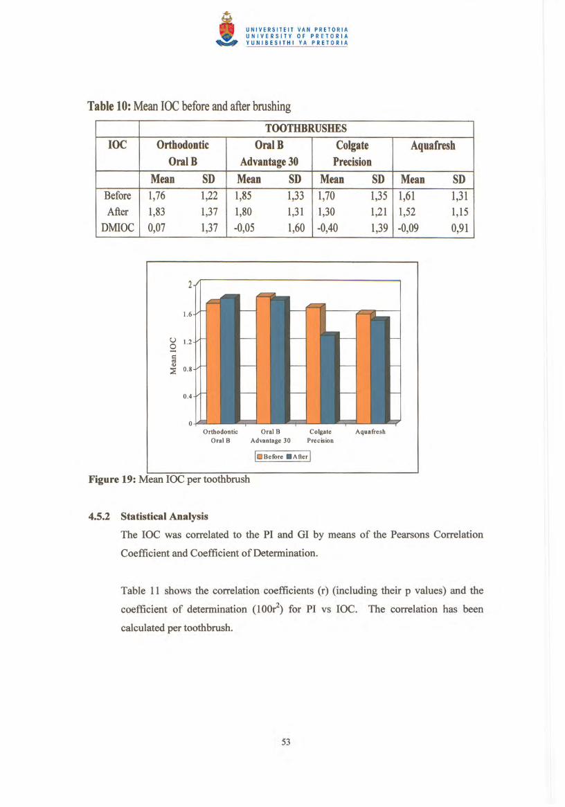

Table 11: Correlation coefficient and coefficient of determination for PI vs IOC
TOOTHBRUSHES Orthodontic
OralB
OralB
Advantage 30 Colgate
Precision Aquafresh
Before After Before After Before After Before After Correlation
Coefficient (r) P value
Coefficient of
Determination (lOO~) %
0,73527 0,68857 0,0001 0,0001
54,1 47,4
0,62443 0,73129 0,0001 0,0001
39,0 53,5
0,75466 0,73416 0,0001 0,0001
57,0 53,9
0,83581 0,73773 0,0001 0,0001
69,9 54,4
The range of the correlation coefficient is between 0,62443 to 0,83581. These are
all highly significantly different from °(all p values <0,01). The coefficient of
detennination ranged from 39,0% to 69,9%. These values indicate that for the IOC
compared to PI there was a highly significant correlation (p<0,01) but not a perfect
correlation. (A perfect correlation would need a coefficient of detennination of
100%).
Ideally a perfect correlation would be required to use the IOC as a substitute for the
PI. The high level of correlation however, indicates that the IOC can be used as a
screening procedure in orthodontic patients.
Table 12 shows the correlation between GI and IOC. As with the PI, the Pearsons
Correlation Coefficient and Coefficient ofDetennination were calculated.
Table 12: Correlation coefficient and coefficient ofdetennination for GI vs IOC
Orthodontic OralB
OralB Advantage 30
Colgate
Precision
Aquafresh
Before After Before After Before After Before After
Correlation
Coefficient (r)
P value
Coefficient of
Detennination (100r2) %
0,40669 0,23678
0,0050 0,1131
16,5 5,6
0,38615 0,32490
0,0080 0,0276
14,9 10,5
0,42565 0,42008
0,0032 0,0037
18,1 17,6
0,44513 0,27486
0,0019 0,0645
19,8 7,6
54


4.6.1 Statistical Analysis
In order to evaluate the degree of agreement between a patients preferred
toothbrush and their "best" toothbrush (as measured by the DMPI and DMGI) the
percentage where preference agreed with effectiveness was calculated. In only 9
patients (19,6%) did the DMPI (largest negative value) agree to their toothbrush
preference.
The same results was found for agreement between the DMGI (largest negative
value) and toothbrush preference (9 patients (19,6%)). There is thus no correlation
between toothbrush preference and effective oral hygiene in patients undergoing
orthodontic treatment.
When comparing the individual toothbrushes to DMPI, the Colgate Precision
toothbrush accounted for 44,5%, Aquafresh toothbrush for 33,3% while the Oral B
Advantage 30 accounted for 22,2% of the correlation. There was no correlation
between the Orthodontic Oral B and patient preference.
For the DMGI, the Colgate Precision toothbrush accounted for 66,7% of the
correlation, while the Oral B Advantage 30 and Aquafresh toothbrushes accounted
for 22,2% and 11,1 % respectively. Again there was no correlation between patient
preference and the specifically designed Orthodontic Oral B toothbrush. These
results can be accounted for by the patients preference for the Colgate Precision
toothbrush.
4.7 Summary
From the results and statistical analyses it can be seen that there is a low level of
plaque and gingivitis in the population studied before and after each toothbrush
used. Little difference was found between the after vs before recordings for both
the PI, GI and IOC. This may account for the statistical fmdings, i.e. there is no
statistical difference in the effectiveness of the four different manual toothbrushes
in patients with fixed orthodontic appliances.
56

The roc may be used as a screening for plaque, however, there was no correlation
to GI (bleeding).
There is no correlation between toothbrush preference and effective oral hygiene.
Chapter 5 will deal with a discussion of the results, conclusions drawn from the
study and recommendations as a result of this study.
57

CHAPTER 5: DISCUSSION, CONCLUSIONS AND
RECOMMENDATIONS
5.1 Discussion
5.1.1 Introduction
The study was designed to evaluate:
1. the effectiveness of 4 different toothbrushes In patients undergoing fixed
orthodontic therapy
ii. correlation between the Index of Oral Cleanliness (lOC) and either the Plaque
Index (PI) or Gingival Index (GI)
iii. correlation between patient toothbrush preference and effective oral hygiene.
Each of the manufacturers claim superiority of their particular toothbrush design.
The Oral-B Orthodontic has a longitudinal groove in the brushing surface to
facilitate brushing around brackets and arch wires. The Aquafresh toothbrush has a
unique flexible head. Soparkar et al (1991) claim that the Aquafresh design is more
acceptable to patients and the flexible head allows for better cleaning and thus
improved oral hygiene. The Colgate Precision toothbrush was designed to make up
for a less than adequate toothbrushing technique (Mintel and Crawford, 1992). It is
believed that this distinction in design accounts for Colgate Precision's clinical
superiority (Sharma et ai, 1992). The Oral B 30 is a toothbrush that has been
widely used and tested. Grossman et al (1994) showed increased plaque removal
with the Oral B 30. The softness of this toothbrush has also been well documented
(Rawls et ai, 1993). It is concluded that the potential for harming dental tissue is
less for the Oral B than any of the other toothbrushes tested.
The cross-over design enabled each brush to be tested on each patient. This
eliminated any potential for inter subject variation which may result from different
number of brackets or types of components in the appliance.
58

Patient instructions were standardised. It was recommended that each subject uses
the toothbrush for 2 minutes, twice per day.
5.1.2 Summary of results
The main findings of this study were:
i. a relatively low PI, GI and IOC at baseline and after brushing with each
toothbrush.
n. A two week brushing programme with any of the toothbrushes did not
111.
iv.
significantly affect the PI, GI and IOC.
There was a significant but not absolute correlation between the IOC and PI.
There was no correlation between the IOC and GI.
v. There was no correlation between toothbrush preference and effective oral
hygiene for the patients.
The results of this study showed that the PI and GI were already low at baseline.
This is different to that shown by Heasman et al (1998) and Heintze et al (1996)
where plaque scores at all surfaces were greater than 50% at baseline. Several
reasons may account for this difference. Firstly, the patients treated at the
University of Pretoria orthodontic programme undergo vigorous selection
procedures before being selected. Secondly, the dental IQ of these patients is
generally higher. Orthodontic therapy at the University of Pretoria is a privilege
offered to these patients. Thirdly, these patients are constantly monitored, and any
deterioration in oral hygiene is immediately rectified.
There was no statistical difference in the Difference in Mean Plaque Index (DMPI)
when comparing the 4 toothbrushes tested. However, when comparing the
Difference in Mean Gingival Index (DMGI), the Colgate Precision seems to be
more effective than the Aquafresh toothbrush. The p value obtained was 0,0495
which is <0,05 and thus these brushes were significantly different at the 95% level.
There was no statistical difference amongst the other toothbrushes.
59
According to the results of this study the IOC is correlated to the PI. There is no
correlation between the IOC and GI. The IOC does give a good and quick
evaluation of the oral hygiene status of patients as suggested by Beam et al (1996).
On the relation of patient preference of a toothbrush to effective oral hygiene one
would expect little or no correlation. Patient preference is often influenced by
factors such as marketing, previous experience and in today's economic climate,
cost. No correlation of patient preference and effective oral hygiene has been
reported in the literature. It is be suggested that such a correlation study could be
added to future toothbrush studies.
5.2 Conclusion
The following conclusions can be drawn from this study:
1. This study has confirmed the premise that there is no difference in the
effectiveness of the four toothbrushes tested in plaque control of patients
undergoing fixed orthodontic appliances.
11 . The IOC can be used as a screening of oral hygiene status in patients
undergoing orthodontic therapy (measure of plaque on the tooth surface). The
IOC cannot be used as a measure of gingivitis ( bleeding index).
lll. Patient preference is usually dependant on effective marketing and not on the
effectiveness of the brush.
60

5.3 Recommendations
1. Oral hygiene criteria should be used in the selection of orthodontic patients.
Patients with a low PI and 01 score showed little difference in oral hygiene
scores after the use ofeither of the 4 toothbrushes.
11. The IOC should be further investigated before it is used in evaluating
effectiveness oforal hygiene procedures in orthodontic patients.
lll. The IOC can be used as a screening procedure in patients undergoing fixed
orthodontic therapy.
IT IS NOT THE BRUSH WmCH DETERMINES EFFECTIVE
PLAQUE REMOVAL, BUT HOW YOU BRUSH!
61

REFERENCES
Agerbeck N, Melsen Band Rolla G(1975) Application of chlorhexidine by oral irrigation systems, Scandinavian Journal ofDental Research, 83 : 284-287.
Ainamo J and Ainamo A (1978) Development of oral health during dental studies in India
and Finland. International Dental Journal, 28 : 427.
Ainamo J, Bannes D, Beagrie G, Cutress T, Martin J and Sardo-Infirri J (1982) Development of the World Health Organisation (WHO) Community Periodontal Index of
Treatment Needs (CPITN) Internotjonol Dental Journal, 32: 281-291.
Alexander AG (1971) A study of the distribution of supra and sUbgingival calculus,
bacterial plaque and gingival inflammation in the mouths of 400 individuals. Journal of
Periodontology, 42 : 21-28.
Alexander SA (1991) Effects of orthodontic attaclunents on the gingival health of
permanent second molars. American Journol of Orthodontics and Dentofacial
Orthopedics, 100 :337-340.
Alstad S and Zachrisson BW (1979) Longitudinal study ofperiodontal condition associated
with orthodontic treatment in adolescents, American Journal ofOrthodontics, 76 : 277-286.
Artun J and Brobakken BO (1986) Prevalences of carious white spots after orthodontic
treatment with multibanded appliances. European Journal ofOrthodontics, 8 : 229-234.
Aziz-Gandour IA and Newman NH (1986) The effects of a simplified oral hygiene regime
plus supragingival irrigation with chlorhexidine or metronidazole on chronic inflammatory
periodontal disease. Journal ofClinical Periodontology, 13 : 228-236.
Battista GW and Petrone DM (1993) Comparison of the laboratory interproximal access
efficacy of the Colgate Precision toothbrush and the Crest Complete toothbrush. Journnl of
Clinical Dentistry, 4: D5-D7.
Beam DR, Aird JC, Jenkins WMM and Kinane DF (1996) Index of Oral Cleanliness
(l0C). A new oral hygiene index for use in clinical audit. British Journal ofOrthodontics,
23: 145-151.
Berglund LR and Small CL (1990) Effective oral hygiene for orthodontic patients. Journal
ofClinical Orthodontics, 25: 315-320.
62

Boyd RL (1983) Longitudinal evaluation of a system for self-monitoring plaque control
effectiveness in orthodontic patients. Journal ofClinical Periodontology, 10 : 380-388.
Boyd RL and Rose eM (1994) Effect of rotary electric toothbrush versus manual toothbrush on decalcification during orthodontic treatment. American Journal oj Orthodontics and Dento/adal Orthopedics, 105: 450-456.
Brightman LJ, Terezhalmy GT, Greenwell H, Jacobs Mand Enlow DH (1991) The effects
of a 0.12% chlorhexidine gluconate mouthrinse on orthodontic patients aged 11 through 17
with established gingivitis. American Journal oj Orthodontics and Dentojacial Orthopaedics. 100 : 324-329.
Buckley LA (1980) The relationships between irregular teeth, plaque, calculus and gingival
disease, British Dental Journal, 148 : 67-69.
Burch JG, Lanese R and Ngan P (1994) A two-month study of the effects of oral irrigation
and automatic toothbrush use in an adult orthodontic population with fixed appliances.
American Journal ofOrthodontics and Dentofadal Orthopaedics, 106 : 121-126.
Burkett L W (1963) Effects of orthodontic treatment on the soft periodontal tissues (1963)
American Journal ofOrthodontics, 49 : 660-671.
Chattetjee R and Kleinberg I (1979) Effect of orthodontic band placement on the chemical
composition of human incisor tooth plaque. Archives of Oral Biology, 24: 97-100.
Cianco SG (1988) Use of mouthrinses for professional indications. Journal of Clinical
Periodontology, 15 : 520-523.
Cohen AM, Moss JP and Williams DW (1983) Oral hygiene instruction prior to
orthodontic treatment. A preliminary study. British Dental Journal, 16 : 277-278.
Croxson LJ (1984) A simplified periodontal screening examination: The community
periodontal index of treatment needs (WHO) in general practice. International Dental
Journal, 34 : 28-34.
Cumming BR and Loe H (1973) Consistency of plaque distribution in individuals without
special home care instruction. Journal ofPeriodontal Research, 8 : 94-100.
Cutress TW, Ainamo J and Sardo-Infirri I (1987) The community periodontal index of
treatment needs (CPITN) procedure for population groups and individuals. International
Dental Journal, 9 : 192-196.
63

Davies TM, Shaw we, Worthington HV, Addy M, Dummer P and Kingdon A (1991) The
effect of orthodontic treatment on plaque and gingivitis, American Journal 0/Orthodontics
and Dento/adal Orthopaedics, 99: 155-162.
Deasy MJ, Singh SM, Kemp JH, Curtis JP, Rustogi KN and Fung K (1993) A clinical
comparison of plaque removal performance of three manual toothbrushes. Journal of Clinical Dentistry, 4 (Supplement D) : DI7-21.
Diedrich P (1981) Enamel alteration from bracket bonding and debonding. A study with
the electron microscope. American Journal a/Orthodontics, 79: 500-522.
Dorfinan HS (1978) Mucogingival changes resulting from mandibular incisor tooth
movement, American Journal o/Orthodontics, 74 : 286-297.
Emslie RD (1980) The 621 periodontal probe. International Dental Journal, 39 : 287.
Fraudsen A (1986) Mechanical oral hygiene practices. State-of-the-science review. In: Dental Plaque Control Measures and Oral Hygiene Practices Proceedings from a State-o/
the-Science Workshop, Loe H Kleinman DV, Eds., IRL Press Ltd., Oxford, 93-116.
Geiger AM, Gorelick L, Gwinnett AJ and Griswold PG (1988) The effect of a fluoride
program on white spot formation during orthodontic treatment, American Journal of
Orthodontics, 93 : 29-37.
Gjermo P and Flotra L (1970) The effect of different methods of interdental cleaning.
Journal ofPeriodontal Research, 5 : 230-236.
Gorelick L, Geiger AM and Gwinnett AJ (1982) Incidence of white spot fonnation after
bonding and banding. American Journal ofOrthodontics, 81 : 83-98.
Gortjamanos T, Singh H and Strangio T (1992) Choice of toothbrush: a survey of dentists'
personal preferences and recommendations for patients. Australian Dental Journal 37 :
290-295.
Greene JC and Vermillion JR (1960) The oral hygiene index: A method for classifYing oral
hygiene status. Journal ofthe American Dental Association, 61 : 172.
Grossman E, Dembling W and Walley DR (1994) Two long-term clinical studies
comparing the plaque removal and gingivitis reduction efficacy of the Oral-B Advantage
Plaque Remover to five manual toothbrushes. Journal ofClinical Dentistry 5: 46-53.
64

Heasman P, Wilson Z, Macgregor I and Kelly P (1998) Comparative study of electric and manual toothbrushes in patients with fixed orthodontic appliances. American Journal of
Orthodontics and Dentofacial Orthopedics, 114: 45-49.
Heintze SD, Jost-Brinkman PG andLoundos J (1996) Effectiveness of three different types of electric toothbrushes compared with a manual technique in orthodontic patients.
American Journal ojOrthodontics and Dentojacial Orthopaedics, 110 : 630-638.
Hirschfeld I (1939) The Toothbrush: Its Use and Abuse. Dental Items ofInterest Publishing
CO., Inc. Brooklyn, 43-70.
Huser MC, Baehni PC and Lang R (1990) Effects of orthodontic bands on microbiological and clinical parameters. American Journal o/Orthodontics and Dento/acial Orthopaedics,
97: 213-218.
Jackson CL (1991) Comparison between electric toothbrushing and manual toothbrushing,
with and without oral irrigation, for oral hygiene of orthodontic patients. American Journal
o/Orthodontics and Dento/acial Orthopedics 99: 15-20.
Johansen JR, Gjermo P and Bellini HT (1973) A system to classify the need of periodontal
treatment. Acta Odontology Scandinavia, 31 : 297-305.
Kessler M (1976) Interrelationships between orthodontics and periodontics. American
Journal o/Orthodontics, 70: 154-177.
Kiliyoglu M, Yildirim M and Po later H (1997) Comparison of the effectiveness of two
types of toothbrushes on the oral hygiene of patients undergoing orthodontic treatment with
fixed appliances. Journal o/Orthodontics and Dento/adal Orthopaedics, 111 : 591-594.
Klein H and Palmer CE (1938) Dental caries in American Indian children Public Health
Bulletin (Washington), No 239.
Kloehn JS and Pfeifer JS (1974) The effect of orthodontic treatment on the periodontium,
Angle Orthodontist, 44 : 127-134.
Lang NP, Cumming BR and Loe H (1973) Toothbrushing frequency as it related to plaque
development and gingival health. Journal 0/Periodontology, 44 : 396-405.
Legott PJ, Boyd RL, Quinin RS, Eakle WS and Chambers DW (1984) Gingival disease
pattern during fixed orthodontic treatment adolescents vs adults. Journal 0/ Dental
Research, 63 : 309 (Abstract 1245).
65

Lilienthal B, Amerena V and Gregory G (1965) An epidemiological study of chronic periodontal disease. Archives a/Oral Biology, 10 : 553-566.
Lindhe J, Hamp SE and Loe H (1975) Plaque induced periodontal disease in Beagle dogs.
Journal ofPeriodontology, 10 : 243.
Loe H (1967) The Gingival Index, the Plaque Index and the Retention Index systems.
Journal ofPeriodontology, 38 : 610.
Loe (1971) Human research model for the production and prevention ofgingivitis. Journal
ofDental Research, 50: 256-264.
L5e H, Theilade E and Jensen SB (1965) Experimental gingivitis in man. Journal 0/ Periodontology,36: 177-187.
Lovdal A, Arno A, Schei 0 and Waerhaug J (1961) Combined effect of sUbgingival scaling
and controlled oral hygiene on the incidence of gingivitis. Acta Odontology o/Scandinavia,
19: 537.
McGlynn FD, LeCompte OJ, Thomas RG, Courts KJ and Melamed BG (1987) Effects of
behavioural self-management on oral hygiene adherence among orthodontic patients.
American Journal 0/Orthodontics and Dento/acial Orthopaedics, 91 : 15-21.
Mintel TE and Crawford J (1992) The search for a superior toothbrush design technology.
Journal 0/Clinical Dentistry, 3: C I-C4.
Mitchell L (1992) Decalcification during orthodontic treatment with fixed appliances - an
overview. British Journal o/Orthodontics, 19 : 199-205.
Mizrahi E (1983) Surface distribution of enamel opacities following orthodontic treatment.
American Journal o/Orthodontics, 84 : 323-331.
Ogaard B, Rolla G and Arends J (1988) Orthodontic appliances and enamel
demineralisation. Part 1. Lesion development. American Journal 0/ Orthodontics and
Dento/acial Orthopedics, 94: 68-73.
Ogaard B (1989) Prevalence of white spot lesions in 19-year-olds: a study on untreated and
orthodontically treated persons 5 years after treatment. American Journal 0/Orthodontics
and Dento/acial Orthopedics, 96 : 423-427.
66

O'Reilly MM and Featherstone JD (1987) Demineralisation and remineralization around
orthodontic appliances: An in vivo study. American Journal of Orthodontics and
Dento!acial Orthopaedics, 92 : 33-40.
Page RC and Schroeder HE (1981) Current status of the host response in chronic marginal
periodontitis. Journal o/Periodontology, 52 : 477.
Parfitt OJ (1957) Afive year longitudinal study of the gingival condition of a group of children in England. Journal 0/Periodontology, 28 : 26.
Pritchard JF (1975) The effect of bicuspid extraction orthodontics on the periodontium, Journal ofPeriodontology, 46 : 534-542.
Ramfjord SP (1959) Indices for prevalence and incidence ofperiodontal disease. Journal of
Periodontology, 30 : 5 I.
Rawls HR, Smith NK, Lentz DL, Cobb GW Jr and Bailey MS (1993) An electron
microscopic comparison of bristle end-rounding of three commercial toothbrushes. Journal
ofClinical Dentistry 4 : 96-100.
Reardon RC, Cronin M, Balbo F, Schiff T, Menaker L, Weatherford TW 3rd, Walley D,
Vidra J, and Zib K (1993) Four clinical studies comparing the efficacy of flat-trim and
multi-level trim commercial toothbrushes. Journal ofClinical Dentistry, 4: 101-105.
Regezi JA and Sciubba JJ (1989) Oral Pathology: Clinical-Pathological Correlations. WB
Saunders Company, Philadelphia, PA, USA, 488-499; 508-512.
Rosendahl PO (1962) Toothbrushing, yesterday, tomorrow. Southern Californian Dental
Association Journal, 30: 210-213.
Russell AL (1956) A system of classification and screening of prevalence surveys of
periodontal disease. Journal ofDental Research, 35 : 350.
Russell AL (1963) International nutrition surveys: A summary of preliminary dental
fmdings. Journal ofDental Research, 42 : 232.
Sadowsky and BeGole (1981) Longterm effects of orthodontic treatment on periodontal
health, American Journal ofOrthodontics and Dentofacial Orthopaedics, 80: 156-172.
Saxen L (1980) Juvenile periodontitis. Journal ofClinical Periodontology, 7: 1.
67

Schour I and MassIer M (1947) Gingival disease in postwar Italy (1945). I Prevalence of
gingivitis in various age groups. Journal ofthe American Dental Association, 35 : 475,
Shafer WG, Hine MK and Levy BM (1984) A textbook of Oral Pathology, 4th edition. WB
Saunders Company, Tokyo, Japan, 406-468; 772-778.
Sharma NC, Galustians J, Rustogi KN, McCool 11, Petrone M, Volpe AR Kom LR and Petrone D (1992) Comparative plaque removal efficacy of three toothbrushes in two independent clinical studies. Journal ofClinical Dentistry 3: CI3-C20.
Sharma NC, Galustians J, McCool JJ, Rustogi KN and Volpe AR (1994) The clinical effects on plaque and gingivitis over three month's use of four complex design manual
toothbrushes. Journal ofClinical Dentistry, 5 : 114-118.
Singh SM, Rustogi KN, McCool JJ, Petrone M, Volpe AR, Korn LR and Petrone D (1992)
Clinical studies regarding the plaque removal efficacy of manual toothbrushes. Journal of
Clinical Dentistry, 3 (Supplement C) : C21-28.
2ndSoares N and Southam JC (1993) Oral Pathology, edition. Oxford Medical
Publications, Hong Kong, 19-33; 91-107.
Soparkar PM, Newman MB and De Paola PF (1991) The efficacy of a novel toothbrush
design. Journal ofClinical Dentistry, 2 : 107-10.
Stuteville OH (1937). Injuries caused by orthodontic appliances and methods ofpreventing
these injuries. Journal ofthe American Dental Association, 24 : 1494-1507.
Trimpeneers LM, Wijgaerts lA, Grognard NA, Dermaut LR and Adriaens PA (1997) Effect
of electric toothbrushes versus manual toothbrushes on removal of plaque and periodontal
status during orthodontic treatment. American Journal of Orthodontics and Dentofacial
Orthopaedics, 111 : 492-497.
Trossello VK and Gianelly AA (1979) Orthodontic treatment and periodontal status,
Journal ofPeriodontology, 50: 665-671.
Vo1penheim DW, Walsh ME, Dellerman PA and Burkett TA (1994) A new method for in
vitro evaluations of the interproximal penetration of manual toothbrushes. Journal of
Clinical Dentistry, 5: 27-33.
Wilkins EM (1989) Clinical Practice of the Dental Hygienist, 6th edition. Lea and Febiger,
Philadelphia, PA, USA, 235-246; 337-341; 366-358.
68

Wong CH and Wade AB (1985) A comparative study of effectiveness in plaque removal by
Super Floss and waxed dental floss. Journal ofClinical Periodontology, 12 : 788-795.
Yankell SL, Edvardsen S, Braaten S and Emling RC (1993) Laboratory and clinical
evaluations of the Jordan Exact toothbrush. Journal o/Clinical Dentistry, 4: 67-70.
Yankell SL, Shi X and Emling RC (1992) Comparative laboratory evaluation of three
toothbrushes regarding interproximal access efficacy. Journal ofClinical Dentistry 3: C5
C8.
Yankell SL, Shi X and Emling RC (1993) Comparative laboratory evaluation of two new
toothbrushes regarding interproximal access efficacy. Journal 0/ Clinical Dentistry, 4:
DI-D4.
Yankell SL, Shi X and Emling RC (1994) Laboratory interproximal access efficacy of two
compact manual toothbrushes. Journal o/Clinical Dentistry, 4 : 111-113.
Yost KG, Miluszewski KF and Chen AC (1994) Laboratory evaluations of toothbrush
interproximal penetration ratios. Journal o/Clinical Dentistry, 4: 125-127.
Zachrisson BU and Alnaes L (1973) Periodontal condition in orthodontically treated and
untreated children. Loss of attachment, gingival pocket depth and clinical crown heights,
Angle Orthodontist, 43 : 402-411.
Zachrisson BU (1974) Oral hygiene for orthodontic patients: Current concepts and practical
advice. American Journal 0/Orthodontics, 66 : 487-497.
Zachrisson BU (1975) Cause and prevention of injuries to teeth and supporting structures
during orthodontic treatment. American Journal o/Orthodontics, 69: 285-300.
Zachrisson BU and Alnaes L (1974) Periodontal condition in orthodontically treated and
untreated individuals, Angle Orthodontist, 44 : 48-55.
Zachrisson S and Zachrisson BU (1972) Gingival condition associated with orthodontic
treatment, Angle Orthodontist, 42 : 26-34.
69

ADDENDUM A TOOTHBRUSH PROJECT - DR A LAHER - T97157 - ORT9005
11. Examiner number I VJ:J
IL._2._R_e_gI_·s_tr_at_io_n_n_u_m_b_e_r___LI__....l--_ ----.-J1 V2:2·3
V3:4I 3. Gender I.
14. Date ofBirth (YYMMDD) 1 V4:S-10
I 5. Name ofpatient 1
6. Date of Examination V5:11-16\. (YYMMDD)
1 7_ Group V6:17
I8. Week Number V7:18
19. Toothbrush Type V8:19
10. Index of Oral Cleanliness (JOC)
D V9:20
11. Plaque Index (PI)
16 22 24 36 42 44
DB
B
MB
L
12. Gingival Index (GI)
VIO-V\S :2\-26
VI6-V21:27-32
V22-V27:33-38
V28-V33 :39-44
16 22 24 36 42 44
DB
B
MB
L
V34--V39:4S-S0
V40-V4S :SJ-S6
V46-VSl :S7-62
V52-V57:63-68
13. Fonn number VS8 :69-71
70


