Embed Size (px)
Citation preview

doi:10.1182/blood-2005-02-0471Prepublished online June 2, 2005;2005 106: 2138-2146
Junken Aoki, Michael J. O. Wakelam, Lawrence S. Young and Paul G. MurrayWei, Susan Morgan, Tanya Stankovic, Yasuhiro Kishi, Hiroyuki Arai, Marketa Nowakova, Guy Pratt, Karl R. N. Baumforth, Joanne R. Flavell, Gary M. Reynolds, Gillian Davies, Trevor R. Pettit, Wenbin and survival of Hodgkin lymphoma cellsInduction of autotaxin by the Epstein-Barr virus promotes the growth
http://bloodjournal.hematologylibrary.org/content/106/6/2138.full.htmlUpdated information and services can be found at:
(1930 articles)Signal Transduction � (4217 articles)Neoplasia �
(1739 articles)Free Research Articles �Articles on similar topics can be found in the following Blood collections
http://bloodjournal.hematologylibrary.org/site/misc/rights.xhtml#repub_requestsInformation about reproducing this article in parts or in its entirety may be found online at:
http://bloodjournal.hematologylibrary.org/site/misc/rights.xhtml#reprintsInformation about ordering reprints may be found online at:
http://bloodjournal.hematologylibrary.org/site/subscriptions/index.xhtmlInformation about subscriptions and ASH membership may be found online at:
Copyright 2011 by The American Society of Hematology; all rights reserved.Washington DC 20036.by the American Society of Hematology, 2021 L St, NW, Suite 900, Blood (print ISSN 0006-4971, online ISSN 1528-0020), is published weekly
For personal use only. by guest on June 8, 2013. bloodjournal.hematologylibrary.orgFrom
NEOPLASIA
Induction of autotaxin by the Epstein-Barr virus promotes the growth and survivalof Hodgkin lymphoma cellsKarl R. N. Baumforth, Joanne R. Flavell, Gary M. Reynolds, Gillian Davies, Trevor R. Pettit, Wenbin Wei, Susan Morgan, Tanya Stankovic,Yasuhiro Kishi, Hiroyuki Arai, Marketa Nowakova, Guy Pratt, Junken Aoki, Michael J. O. Wakelam, Lawrence S. Young, and Paul G. Murray
A proportion of patients with Hodgkinlymphoma carry Epstein-Barr virus (EBV),an oncogenic herpesvirus, in their tumorcells. Although it is generally assumedthat EBV contributes to the malignantphenotype of Hodgkin lymphoma cells,direct evidence in support of this is lack-ing. Here we show that EBV infection ofHodgkin lymphoma cells results in theinduction of autotaxin, a secreted tumor-associated factor with lysophospho-lipase-D activity. Up-regulation of auto-
taxin increased the generation oflysophosphatidic acid (LPA) and led tothe enhanced growth and survival ofHodgkin lymphoma cells, whereas spe-cific down-regulation of autotaxin de-creased LPA levels and reduced cellgrowth and viability. In lymphoma tis-sues, autotaxin expression was mainlyrestricted to CD30� anaplastic large-celllymphomas and Hodgkin lymphoma; inthe latter, high levels of autotaxin werestrongly associated with EBV positivity
(P � .006). Our results identify the induc-tion of autotaxin and the subsequent gen-eration of LPA as key molecular eventsthat mediate the EBV-induced growth andsurvival of Hodgkin lymphoma cells andsuggest that this pathway may provideopportunities for novel therapeutic inter-vention. (Blood. 2005;106:2138-2146)
© 2005 by The American Society of Hematology
Introduction
Epstein-Barr virus (EBV) is associated with B-lymphoid (eg,Burkitt lymphoma, posttransplantation lymphoma) and nonlym-phoid malignancies (eg, nasopharyngeal carcinoma). EBV is alsopresent in the malignant Hodgkin and Reed-Sternberg (HRS) cellsof some Hodgkin lymphomas (HLs)1,2 in which the viral genome ismonoclonal, implying that EBV infection is an early pathogenicevent.3 EBV also persists throughout the course of HL, suggestingthat it is important to the maintenance of the transformed pheno-type.4 However, the contribution of EBV to the pathogenesis of HLhas yet to be established.
In contrast to EBV-transformed B-lymphoblastoid cell lines andmost B-cell posttransplantation lymphomas in which all the knownlatent virus genes are expressed, other EBV-associated B-cellmalignancies show a more restricted pattern of virus gene expres-sion. For example, in most primary Burkitt lymphomas, EBVexpresses only the EBV-encoded RNAs (EBERs), the Epstein-Barrnuclear antigen-1 (EBNA1), and the BamH1A rightward tran-scripts5 (BARTs). Primary EBV-positive HRS cells express notonly these genes but also latent membrane protein 1 (LMP1)and LMP2.6-8
We show that EBV infection of HL cells leads to the inductionof autotaxin, an autocrine motility factor originally isolated fromA2058 human melanoma cells9 that has been shown to augmentmany cellular characteristics associated with tumor aggressive-ness,10 including cell proliferation,11 cell survival, invasion,10 andangiogenesis.12 Autotaxin is a phospholipase D (PLD) that can
generate lysophosphatidic acid (LPA) and sphingosine-1-phosphate (S1P) from lysophosphatidylcholine (LPC) and sphingo-sylphosphorylcholine (SPC), respectively.11,13 A role for LPA inhuman oncogenesis was suggested by the observation that LPA ispresent at elevated levels in ascites of patients with ovariancancer.14 Subsequently, it has been shown that LPA is a potenttumor-promoting small molecule in a range of biologic systems andthat it binds to specific cell surface heterotrimeric G-protein–coupled receptors (the endothelial cell differentiation gene [EDG]receptors) stimulating the proliferation, migration, and survival ofcancer cells.15,16 Therefore, mechanisms that lead to aberrant LPAproduction are likely to be associated with the initiation orprogression of cancer.16
We demonstrate that the induction of autotaxin by EBV leads tothe increased generation of LPA and to the proliferation andsurvival of HL cells. This is the first report that autotaxin and thetumor-promoting lipid LPA are the molecular targets of a transform-ing virus.
Materials and methods
Generation and phenotype of cell lines
EBV-negative KM-H2 cells17 were infected with Akata-derived recombi-nant EBV18 and cultured in 1 mg/mL G418. KM-H2 cells have been shownto be representative of HL cells.19,20 Control KM-H2 cells were generated
From the Cancer Research UK (CRUK) Institute for Cancer Studies and LiverResearch Laboratories, University of Birmingham, Birmingham, WestMidlands, United Kingdom; and the Graduate School of PharmaceuticalSciences, University of Tokyo, Tokyo, Japan.
Submitted February 7, 2005; accepted May 15, 2005. Prepublished online asBlood First Edition Paper, June 2, 2005; DOI 10.1182/blood-2005-02-0471.
Supported by the Leukaemia Research Fund.
K.R.N.B. and J.R.F. contributed equally to this study.
An Inside Blood analysis of this article appears in the front of this issue.
Reprints: Paul G. Murray, CRUK Institute for Cancer Studies, The MedicalSchool, University of Birmingham, Edgbaston, Birmingham, B15 2TT, UnitedKingdom; e-mail: [email protected].
The publication costs of this article were defrayed in part by page chargepayment. Therefore, and solely to indicate this fact, this article is herebymarked ‘‘advertisement’’ in accordance with 18 U.S.C. section 1734.
© 2005 by The American Society of Hematology
2138 BLOOD, 15 SEPTEMBER 2005 � VOLUME 106, NUMBER 6
For personal use only. by guest on June 8, 2013. bloodjournal.hematologylibrary.orgFrom
by electroporation with vector only (pzipLNSNeo). EBV-positive L591cells21 were serially diluted for up to 6 weeks to generate EBV-negativeclones. Clone L591-SD3 was used in later experiments. Immunohistochem-istry for EBNA1,22 reverse transcription–polymerase chain reaction (RT-PCR) and in situ hybridization for EBERs,23 and quantitative PCR for thedetection of viral DNA24 were used to demonstrate the presence of EBV.Immunoglobulin heavy (IgH ) gene sequence analysis of cell lines wasperformed as previously described.25 Viability of cell lines was determinedusing trypan blue reagent (Sigma-Aldrich, Poole, United Kingdom) andproliferation by WST-1 reagent (Roche Diagnostics, Lewes, United King-dom). The WST-1 reagent measures the activity of mitochondrial dehydro-genases in the sample. The tetrazolium salts in the reaction are cleaved toformazan, and the augmentation in enzyme activity leads to an increase inthe amount of formazan dye formed, which directly correlates with thenumber of metabolically active cells in the culture.
RT-PCR
RNA was extracted using the NucleoSpin RNA II kit (AB Gene, Epsom,United Kingdom). RT-PCR analysis for EBV gene expression was aspreviously described.26 Expression was detected using the followingprimers: autotaxin forward, 5�-CACCAGAGGCTAAATATGATGC-3�; au-totaxin reverse, 5�-GAAGGAGGACACAGAGAGAG-3�; EDG1 forward,5�-ATTCAGCCGCAGCAAATC-3�; EDG1 reverse, 5�-TAACTCTAC-CCACCAACACC-3�; EDG2 forward, 5�-AATTCAACTCTGCCAT-GAACCC-3�; EDG2 reverse 5�-ACTTTTCTCCTCTCTCACACCC-3�.Probes were 5�-CACCAGCTGTCTGGATTTCA-3� (autotaxin), 5�-AA-ATCTCTGGGCTTCGACTG-3� (EDG1), and 5�-CATCTTGGCTGGAGT-TCACA-3� (EDG2).
Gene expression analysis
Affymetrix HU133A arrays27 were used for all experiments. Total RNAfrom mycoplasma-free cell lines was used to prepare biotinylated RNA.28 Acomplete description of procedures is available at http://bioinf.picr.man.ac.uk/mbcf/downloads/GeneChip_Target_Prep_Protocol-CR-UK_v2.pdf. Ratios for GAPDH and �-actin (3�/5�) were within acceptablelimits (glyceraldehyde phosphate dehydrogenase [GAPDH], 0.80-1.18;�-actin, 0.83-1.89), and BioB spike controls were present on 5 of 8 chips,with BioC, BioD, and CreX also present in increasing intensity. Whenscaled to a target intensity of 100 (Affymetrix MAS 5.0), scaling factors forall arrays were within acceptable limits (HU133A chips, 0.591-1.134), aswere background, Q values, and mean intensities.
Images of GeneChips were analyzed using Affymetrix Microarray Suite5.0. Probe level quantile normalization29 and robust multiarray analysis30
on the raw .CEL files were performed using the Affymetrix package of theBioconductor (http://www.bioconductor.org) project. Differentially ex-pressed probe sets were identified using significance analysis of microar-rays (SAM)31,32; only those with positive or negative changes of 2.5-fold ormore and a false discovery rate of 5% or less were included. Hierarchicclustering was performed using dChip (http://www.dchip.org).
Generation of autotaxin-specific monoclonal antibodies
A polypeptide (amino acids 58-182 of human autotaxin) was expressed inEscherichia coli as a glutathione-S-transferase (GST) fusion protein usingpGEX-4T vector (Amersham-Pharmacia, Little Chalfont, United King-dom). Rats (WKY/Izm strain) were immunized with purified fusion protein,and medial iliac lymph nodes were used for fusion with PAI mousemyeloma cells. Hybridomas were selected by screening with enzyme-linked immunosorbent assay (ELISA), immunofluorescence, and Westernblotting. Two antibody-secreting hybridoma cell lines were generated(2A12 and 4F1).
Protein analysis
Cells were lysed in buffer (50 mM Tris, pH 7.5, 9 M urea, 0.15 M�-mercaptoethanol) and protein quantified by Bio-Rad DC Protein AssayKit (Bio-Rad, Hemel Hempstead, United Kingdom). Gel sample buffer wasadded to samples before sodium dodecyl sulfate–polyacrylamide gelelectrophoresis (SDS-PAGE), transfer to BioTrace NT membrane (VWR
International Ltd, Poole, United Kingdom), and incubation with monoclo-nal antibody (mAb) 2A12 (1:100) and then horseradish peroxidase (HRP)–conjugated rabbit anti–rat IgG (DAKO, Ely, United Kingdom) (1:1000).Detection was with enhanced chemiluminescence (ECL; Amersham-Pharmacia Biotech, Little Chalfont, United Kingdom). Immunohistochem-istry was performed on 4-�m sections, as previously described.33 HL wasclassified as autotaxinhi if autotaxin-staining intensity in HRS cells wasequivalent to or higher than that observed in endothelial cells in the samesection or as autotaxinneg/autotaxinlo if autotaxin-staining intensity in HRScells was lower than that in endothelial cells.
Conditioned media experiments and addition of exogenousLPA and S1P
Control KM-H2 cells (KM-H2-neo) were grown in their own conditionedmedia or in media from EBV-infected KM-H2 cells (KM-H2-Akata).KM-H2-Akata cells were grown in conditioned media from KM-H2-neocells or their own media. Cell proliferation was assessed by WST-1 assay.For assessment of the effects of exogenous LPA or S1P, 5 � 104 cellswere washed in serum-free media, resuspended in 100 �L RPMIcontaining 1% B-cell serum (BCS) alone (control), RPMI containing 1%BCS and 5 �M LPA, or RPMI containing 1% BCS and 1 �M S1P. ThenWST-1 assays were performed.
Analysis of autotaxin activity
Cells (7.5 � 106) were cultured in 15 mL serum-free and phenol red-freeOptiMEM (Invitrogen, Renfrew, United Kingdom) for 16 hours. After theaddition of phosphatase inhibitors, conditioned media were incubated with20 �M LPC or 20 �M SPC. Five hundred nanograms each of 17:0-LPA,D31-16:0-LPC, and C17 S1P were added as internal standards. Standardnomenclature was adopted in describing the lipids (ie, number of carbonatoms/number of double bonds). Samples were extracted with water-saturated butanol and dried. Contaminants were removed with chloroform/methanol/water. Samples were dissolved in chloroform/methanol/water(5:5:1) and subjected to liquid chromatography/mass spectrometry (LC-MS) separation. Detection was by selected ion monitoring (SIM) in �veand �ve electrospray ionization (ESI) modes using a probe voltage of � 4kV, a nebulizer nitrogen flow of 4 L/min, and a desolvation line temperatureof 300°C.
LPA-stimulated PLD activity
Cells were incubated in medium containing 4 �Ci/mL (0.148 MBq/mL)[3H]-palmitic acid before resuspension in RPMI containing 10 mM HEPES(N-2-hydroxyethylpiperazine-N’-2-ethanesulfonic acid) pH 7.4 and 0.1%bovine serum albumin (BSA). Cells were stimulated for 20 minutes in 30mM butan-1-ol with 5 �M LPA at 37°C, and PLD activity was determinedby analyzing the generation of [3H]phosphatidylbutanol.34
Knockdown of autotaxin expression
KM-H2-Akata cells in serum-free OptiMEM were incubated in thepresence of 10 nM autotaxin-specific siRNA (5�-GAAAGGCAGCAAAGU-CAUG-3�) in RiboJuice according to the protocol for 6-well plates(Novagen, Nottingham, United Kingdom). Cells were incubated at 37°C for4 hours, after which an equivalent volume of OptiMEM media containingPen/Strep and 20% BCS was added, followed by further incubation at 37°C.RT-PCR, immunoblotting for autotaxin and proliferation, and viabilityassays were performed as described earlier. Ribojuice only, OptiMEM only,and irrelevant siRNAs (scrambled LMP1 siRNA 5�-GGGUAGAUAGACU-CUCGCU-3�) acted as negative controls.
Statistical analysis
All quantifiable data were subjected to statistical analysis in MicrosoftExcel using a 2-tailed Student t test assuming the 2 samples displayedunequal variance. The generated P values were taken to indicate asignificant difference between the data sets when P was below .05. Any datathat satisfied these criteria are indicated by an asterisk above the relevantcolumn (Figures 1C, 1D, 4A, 4B, 4D-F, 5C, and 5D). On the charts
AUTOTAXIN AND HODGKIN LYMPHOMA 2139BLOOD, 15 SEPTEMBER 2005 � VOLUME 106, NUMBER 6
For personal use only. by guest on June 8, 2013. bloodjournal.hematologylibrary.orgFrom

themselves, the error bars represent the standard error of the means for aparticular data set.
Results
HL cells expressing a limited repertoire of virus genes displayenhanced growth and survival
We initially infected EBV-negative KM-H2 cells with Akata-derived recombinant EBV and confirmed its presence by RT-PCRor in situ hybridization for EBER and immunohistochemistry forEBNA1 (data not shown). During latency, EBV uses one of severalpromoters to transcribe EBNA1—the Qp promoter, which initiatestranscription of EBNA1 alone, or the Cp and Wp promoters, whichgenerate a long transcript that is multiply spliced to form EBNA1and the other EBNA proteins.35 RT-PCR analysis of EBV-infectedKM-H2 cells revealed that most EBNA1 transcripts originatedfrom Qp with very low levels of transcription from Cp/Wp andassociated low levels of EBNA2. LMP2 was expressed (Figure1A), but neither LMP1 RNA (Figure 1A) nor LMP1 protein (datanot shown) were detectable. There was no induction of thereplicative cycle in infected cells, as demonstrated by the absenceof BZLF1 expression (data not shown). Thus, EBV-infectedKM-H2 cells show a pattern of virus gene expression differentfrom that observed in primary HRS cells, including the notableabsence of LMP1 expression. Despite this restricted pattern ofvirus gene expression, EBV-infected KM-H2 cells displayedsignificantly increased proliferation (P � .001, P � .01, P � .01,P � .01) at 1, 48, 96, and 192 hours in 1% serum and enhancedviability (P .01, P � .05, P � .01) at 48, 96, and 192 hours,respectively, in 0.1% serum compared with EBV-negative cells(Figure 1C). To confirm these effects of EBV infection, EBV-negative L591 cells were isolated from parental EBV-positive cells(Figure 1B): loss of the EBV genome from L591 cells resulted in adramatic decrease in cell proliferation (data not shown) andviability (Figure 1D) (P .001, P � .05, P � .001) at 96, 144,and 192 hours in 1% serum. Sequence analysis of the IgH genefrom EBV-negative L591 cells and EBV-positive KM-H2 cellsconfirmed their origin from the respective parental lines (datanot shown).
Autotaxin is a transcriptional target of EBV in HL
Gene expression analysis revealed 11 up-regulated and 15 down-regulated probe sets after EBV infection of KM-H2 cells (Figure1E; Table 1). The most highly up-regulated probe was autotaxin.The induction of autotaxin mRNA in EBV-infected KM-H2 wasconfirmed by RT-PCR analysis (Figure 1F). Autotaxin mRNAlevels were decreased after the loss of EBV from L591 cells andwere low in other EBV-negative HL cell lines (Figure 1F).
Generation of autotaxin-specific monoclonal antibodies andup-regulation of autotaxin protein in EBV-infected HL cells
A purified polypeptide from human autotaxin was used to derive 2antibody-secreting hybridoma cell lines, 2A12 and 4F1. Immuno-fluorescence (data not shown) and immunoblotting (Figure 2A)were used to confirm the specificity of each monoclonal antibodyfor autotaxin. Immunoblotting confirmed the up-regulation ofautotaxin protein in EBV-infected KM-H2 and L591 cells (Figure2B). Analysis of other EBV-negative HL cell lines, such as L428,L1236, and HDLM2, revealed lower levels of autotaxin proteinthan was found in EBV-positive cell lines (Figure 2B). Autotaxinwas absent from EBV-negative BL cells (Ramos, DG75, BJAB)
and EBV-positive BL cells, including those displaying type 1latency (Akata, Rael, Daudi, Elijah) or type 3 latency (Raji,Namalwa) (Figure 2C). Of the EBV-transformed lymphoblastoidcell lines (LCLs) studied, only B95.8 cells expressed detectablelevels of autotaxin protein. Nasopharyngeal carcinoma cells,including EBV-negative HONE-1 cells and EBV-positive C666-1cells, lacked autotaxin protein (Figure 2D). HONE-1 cells infectedwith the same Akata-derived recombinant EBV used to generateEBV-positive KM-H2 cells failed to show induction of autotaxinafter infection (Figure 2D).
Expression of autotaxin in primary lymphoma tissues
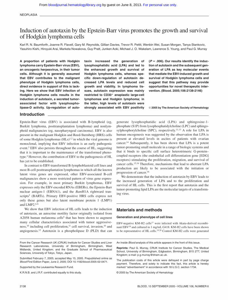
When immunohistochemistry of pelleted cell lines demonstratedthat both autotaxin-specific antibodies were effective on paraffin-embedded samples (data not shown), these were used to examinethe expression of autotaxin in a series of classic HL tumors withknown EBV status. HL samples that contained HRS cells withhigh-level expression of autotaxin (autotaxinhi) were significantlymore frequent in EBV-positive than in EBV-negative samples(Figure 3A-C; Table 2) (Fisher exact test, P .006). Autotaxin wasabsent from most EBV-positive and EBV-negative Burkitt lympho-mas (Figure 3E) and primary EBV-positive undifferentiated naso-pharyngeal carcinoma (NPC) (n 9; data not shown), confirmingthe results of in vitro studies suggesting that the induction ofautotaxin by EBV was restricted to HL cells. With respect to NPC,the absence of autotaxin expression might be considered surprisingbecause autotaxin expression has previously been reported in othertumors of epithelial origin. Analysis of other B-cell non-Hodgkinlymphomas (B-NHLs) (Table 3) revealed that autotaxin wasfrequently expressed in the CD30� and anaplastic variants ofdiffuse large B-cell lymphoma (DLBCL; Figure 3D) but wasabsent from most high-grade (including CD30� DLBCLs) andlow-grade B-NHLs. (Figure 3F-G). In normal lymphoid tissues,autotaxin was not detectable in most B-cell compartments, includ-ing B cells of germinal centers and mantle zones (Figure 3H), but itwas expressed by a subset of postgerminal center CD138� B cells(Figure 3I). These data suggest that the restricted expression ofautotaxin in tumors is in part a reflection of the stage of B-celldifferentiation from which each is derived.
Increased production of soluble autotaxin in the supernatantof EBV-infected KM-H2 cells is associated with enhancedcell growth
Because autotaxin can be cleaved from the cell membrane, it wasimportant to establish whether the induction of autotaxin alsoresulted in increased levels of soluble autotaxin. Although all HLcell lines generated soluble autotaxin, levels were higher inEBV-infected cells (Figure 4A). HL cell lines were grown inconditioned growth medium from either EBV-positive or EBV-negative KM-H2 cells. EBV-negative KM-H2 cells grown in theirown conditioned medium provided a baseline, and EBV-positiveKM-H2 cells provided a positive control. EBV-negative KMH2cells grown in conditioned medium from EBV-infected KM-H2cells showed an increase in cell proliferation after the first 24 hours,P equaling .028 (Figure 4A). This was followed by a reduction incell growth, presumably the result of gradual depletion of growthfactors in the conditioned medium. In contrast, EBV-infectedKM-H2 cells grown in conditioned medium from EBV-negativecells showed no change in proliferation after 24 hours, butthereafter cell growth increased.
2140 BAUMFORTH et al BLOOD, 15 SEPTEMBER 2005 � VOLUME 106, NUMBER 6
For personal use only. by guest on June 8, 2013. bloodjournal.hematologylibrary.orgFrom

Increased growth of EBV-negative KM-H2 cellsafter treatment with LPA or S1P
To determine whether HL cells were responsive to LPA or S1Pstimulation, KM-H2 cells were cultured in the presence of eitherLPA or S1P. Both LPA (Figure 4B) and S1P (data not shown)
significantly increased the growth of EBV-negative KM-H2 cells(P � .001) at 24, 48, and 72 hours. The same effect, but to a lesserdegree, was seen for the growth of EBV-positive cells above theirbaseline on the addition of LPA or S1P. These data suggest thatKM-H2 cells are responsive to the biologic mediators generated by
Figure 1. Increased growth and survival of EBV-positive HL cells expressing a limited repertoire of EBV genes is associated with the transcriptional up-regulationof autotaxin. (A) RT-PCR analysis demonstrates a pattern of virus gene expression in EBV-infected KM-H2 (KM-H2-Akata) cells predominated by expression of the EBERs,Qp-driven EBNA1, and the BamH1A transcripts. There was also very low-level transcription from Cp/Wp and associated low levels of EBNA2 and LMP2 transcripts in thesecells. Neither LMP1 RNA nor protein (data not shown) could be detected in these cells. (B) Loss of EBER expression in 2 clones (SD3 and SD5) of EBV-negative L591 cellsderived from the EBV-positive parental line by serial dilution. Quantitative PCR was also used to confirm the loss of the EBV genome from these cells (data not shown). CloneSD3 was used in later experiments. (C) (upper panels) WST1 assays showing relative metabolic activity (cell growth) of control cells (KMH2-neo) adjusted to a value of 1compared with EBV-infected KM-H2 cells (KM-H2 � Akata) in 10% and 1% sera. A clear and significant difference in cell growth between control and EBV-infected cells isobserved. (lower panels) Cell viability assays at 10% and 0.1% serum, demonstrating consistent and significant increases in the viability of EBV-infected KM-H2 cellscompared with controls. f indicates KM-H2-neo; and �, KM-H2 � Akata. (D) L591 SD3 cells showed dramatically reduced viability compared with EBV-positive parental L591cells in 10% and 1% sera. Cell proliferation was similarly affected by EBV loss (data not shown). f indicates L591; and �, L591-SD3. (E) Heat map showing gene expressiondifferences across 4 replicates of EBV-negative KM-H2 cells and EBV-infected KM-H2 cells (KM-H2 Akata); 26 probe sets met the criteria of a positive or negative change of2.5-fold or greater and a false discovery rate of 5% or less. Eleven probe sets were up-regulated and 15 were down-regulated after EBV infection. The most highly up-regulatedprobe set (mean fold increase, 4.16) was autotaxin (ENPP2). (F) Confirmation of increased autotaxin mRNA in EBV-infected KM-H2 (KM-H2 Akata) and L591 cells by RT-PCRanalysis compared with EBV-negative parental KM-H2, KM-H2 neo control, and L591-SD3 cells. Other EBV-negative HL cell lines (L428, L540, L1236) and theEBV-transformed lymphoblastoid cell line X50-7 expressed lower levels of autotaxin mRNA.
AUTOTAXIN AND HODGKIN LYMPHOMA 2141BLOOD, 15 SEPTEMBER 2005 � VOLUME 106, NUMBER 6
For personal use only. by guest on June 8, 2013. bloodjournal.hematologylibrary.orgFrom

autotaxin but that there is maximal activation by these mediators inEBV-infected cells. Although EBV-positive and EBV-negativeKM-H2 cells expressed the S1PR1 receptor (EDG1), they did notexpress the major LPA receptor, LPAR1 (EDG2), or the otherknown LPA or S1P receptors (Figure 4C). These findings wereconfirmed by RT-PCR analysis (Figure 4C). The expression of anunknown LPA receptor in KM-H2 cells was supported by thedetection of a small but significant stimulation of PLD activity by 5�M LPA (control, 0.126 � 0.011, � LPA 0.155 � 0.009; resultsare the mean � SD dpm in phosphatidylbutanol as a percentage ofdpm in total phospholipids).
Up-regulation of autotaxin in EBV-infected HL cells isassociated with increased use of LPC and generation of LPA
Although purified autotaxin can hydrolyze LPC and SPC, produc-ing LPA and S1P, respectively, it has not yet been demonstrated thatmedium conditioned with cell-derived autotaxin can generate thesesignaling lipids. Analysis of medium conditioned by either EBV-positive or EBV-negative KM-H2 cells and incubated with LPCand SPC showed that SPC was not hydrolyzed and that no S1P wasgenerated; in contrast, hydrolysis of LPC was significant, with4-fold more LPC hydrolyzed by the EBV-positive KM-H2 cell
Table 1. Differentially expressed genes in EBV-infected KM-H2 compared with control cells
Gene Accession no. Mean fold change Ontology
ENPP2 L35594 4.16 Cell motility, GPCR protein signaling pathway
CXCL12 U19495 4.12 Immune response, cell adhesion, chemotaxis, signal transduction
GLUL AL161952 3.33 Glutamine biosynthesis
SERPINA6 NM_001756 3.10 Transport
HMOX1 NM_002133 2.87 Positive regulation of IB kinase/NF-B cascade
CXCR4 AJ224869 2.80 Immune response, chemotaxis, GPCR protein signaling pathway
CCL20 NM_004591 2.77 Immune response, chemotaxis, cell-cell signaling
TPO M17755 2.77 Thyroid hormone generation
HSPA1A NM_005345 2.70 Ubiquitin-proteasome pathway, protein folding, signal transduction
CYP1B1 NM_000104 2.55 Eye morphogenesis
HAND1 NM_004821 2.55 Heart development, transcription from Pol II promoter
GAGE1 NM_001472 �2.54 Cellular defense response
OAS2 NM_016817 �2.60 Immune response
FLJ20035 NM_017631 �2.62 Protein biosynthesis
IFI44 NM_006417 �2.68 Viral response
ZC3HDC1 NM_022750 �2.72 Nucleic acid binding
ITIH5 NM_030569 �2.95 Regulation of localization, synthesis, and degradation of hyaluronan
PTPRK NM_002844 �2.99 Transmembrane receptor protein tyrosine phosphatase activity
CALD1 AL583520 �3.10 Muscle contraction and development
PCP4 NM_006198 �3.41 Central nervous system development
BCHE NM_000055 �3.50 Cocaine metabolism
CXCL11 AF002985 �3.60 Immune response, chemotaxis, cell-cell signaling
IFIT1 NM_001548 �3.75 Immune response
KIAA0992 NM_016081 �3.90 Cell shape, adhesion, and contraction
Clorf29 NM_006820 �6.01 Unknown
IFI27 NM_005532 �19.38 Immune response
Only genes that had a positive or negative change of 2.5-fold or greater and a false discovery rate of 5% or less were included. Autotaxin is represented by its gene symbol,ENPP2.
GPCR indicates G-protein-coupled receptor.
Figure 2. Generation of autotaxin-specific monoclonal antibodies andthe specific up-regulation of autotaxin protein by EBV-infection of HLcells. (A) Immunoblotting of sf9 cells infected with autotaxin (ATX)–baculovirususing 2A12 antibody demonstrates strong reactivity, whereas sf9 cells infectedwith wild-type (wt) baculovirus show no reactivity. Also shown here is reactivityagainst autotaxin in human serum, human plasma, culture supernatant andcells of breast cancer cell line MBA-MD-231, and supernatant and cells ofglioma cell line SF539. (B) Western blotting using 2A12 antibody demonstratesincreased autotaxin protein in EBV-infected KM-H2 cells (KM-H2 Akata)compared with uninfected KM-H2 cells (left panel). Most other EBV-negativeHL cell lines (L428, L540, L1236, HD-MyZ, HDLM2) expressed lower levels ofautotaxin protein. (right panel) Down-regulation of autotaxin protein in L591cells after the loss of the EBV genome. (C) Autotaxin could not be detected inEBV-negative BL cells (Ramos, DG75, BJAB) or in a number of EBV-positiveBL cells, including those displaying a type 1 form of EBV latency (Akata, Rael,Elijah), a type 3 form of EBV latency (Raji, Namalwa), or a Wp-restricted patternof EBV latency (Daudi) that expresses EBNA1 and the EBNA3 family. BL linesare negative for autotaxin, though there is an unidentified lower molecularweight band in the DG75, BJAB,Akata, Elijah, and Rael BL lines and in the IB4LCL line. Furthermore, of several lymphoblastoid cells lines (X50-7, B95.8 LCL,IB4), only B95.8 cells expressed detectable levels of autotaxin. (D) NPC cells,including EBV-positive C666-1 and EBV-negative HONE-1 cells, lacked auto-taxin protein. HONE-1 cells infected with the same Akata-derived recombinantEBV used to generate EBV-positive KM-H2 cells failed to up-regulate autotaxinexpression after infection.
2142 BAUMFORTH et al BLOOD, 15 SEPTEMBER 2005 � VOLUME 106, NUMBER 6
For personal use only. by guest on June 8, 2013. bloodjournal.hematologylibrary.orgFrom
medium than the EBV-negative medium (Figure 4D; P � .001). Inparallel, more LPA content was detected in the EBV-positive thanin the EBV-negative medium (Figure 4E; P � .05). The LPAcontent of the conditioned medium was low and was not equivalentto the loss of LPC; this was attributed to the short half-life of LPAin conditioned medium, with almost half the exogenously addedLPA degraded within 10 minutes (data not shown). Despite therelatively small amplification in total LPA generation by theEBV-positive KM-H2 cell medium, there was a selective increasein 16:0-LPA generation (P � .01) (Figure 4F). This acyl structurewas highly represented in the egg LPC added to the medium as thesubstrate and was not found in the unsupplemented culturemedium; thus, the increase was exclusively attributed to LPC-PLDactivity, which can be concluded to have been autotaxin.
Autotaxin promotes the growth and survivalof EBV-infected HL cells
To determine the contribution of autotaxin to the increased growthand survival of EBV-infected HL cells, we used siRNAs. AutotaxinmRNA levels in EBV-positive KM-H2 cells were reduced after 48hours of treatment with autotaxin-specific siRNAs compared withcontrol cells (cells treated with transfection reagent alone or withscrambled siRNA sequence); GAPDH transcription was unaffectedby these treatments (Figure 5A). Knockdown of autotaxin proteinin EBV-infected KM-H2 cells was also demonstrated after expo-sure to autotaxin-specific siRNAs (Figure 5B). Autotaxin knock-down resulted in significantly reduced cell growth (48 hours;
P .001) and viability (48 hours; P � .01) (Figure 5C) of HL cellscompared with Ribojuice-only controls. Importantly, knockdownof autotaxin expression also resulted in a substantial decrease in thelevels of LPA generated by conditioned medium derived fromEBV-infected KM-H2 cells (Figure 5D) (P � .001).
Discussion
Despite the frequent detection of EBV in the tumor cells ofHodgkin lymphoma, the mechanisms underlying the contributionof virus infection to the development and maintenance of thetransformed phenotype in this disease remains to be established.Here we studied the impact of EBV infection on the growth andsurvival of the HL-derived cell line, KM-H2. Although this cell line
Table 2. Autotaxin expression in EBV-positive and EBV-negative HL
Autotaxin status ofHRS cells EBV-positive EBV-negative
Autotaxinhi 10 6
Autotaxinneg/autotaxinlo 1 11
Autotaxinhi, % 91 (10 of 11) 35 (6 of 17)
HL cells were classified as autotaxinhi if autotaxin expression in HRS cells wasequivalent to or higher than that observed in endothelial cells in the same section oras autotaxinneg/autotaxinlo if autotaxin expression in HRS cells was lower than inendothelial cells. Fisher exact test results show there was a significantly higherproportion of autotaxinhi tumors in the EBV-positive group (P .006).
Table 3. Autotaxin expression in NHL
Lymphoma subtypeNo.
positive Total%
positive
Low-grade B-cell NHL
Follicular lymphoma 2 25 8
Chronic lymphocytic lymphoma 0 6 0
Mantle cell lymphoma 1 5 20
MALT lymphoma 1 1 100
High-grade B-cell NHL
Burkitt lymphoma 2 14 14
DLBCL
Centroblastic and immunoblastic* 7 26 27
CD30�/anaplastic* 12 12 100
Primary cutaneous 2 5 40
Posttransplantation lymphoproliferative disorder 1 1 100
T-cell neoplasms
Peripheral T-cell NHL unspecified 2 4 50
Mycosis fungoides 0 1 0
NK/T-cell lymphoma (angiocentric) 0 1 0
ALCL (null/T-cell type) 2 5 40
DLBCL indicates diffuse large B-cell lymphoma; ALCL, anaplastic large-celllymphoma.
*Morphologic variants of DLBCL.
Figure 3. Expression of autotaxin in lymphoid tis-sues. (A-B) Strong reactivity for autotaxin in HRS cellsfrom EBV-positive primary HL tumors. (C) Lower levels ofautotaxin expression in EBV-negative HRS cells despitestrong labeling of nontumor cells (arrow). The differencein expression between EBV-negative and EBV-positivetumors was highly significant (see “Expression of auto-taxin in primary lymphoma tissues”). In B-NHL, autotaxinexpression was largely restricted to CD30� ALCL (D),whereas most high-grade B-NHLs, such as BL (E) andCD30� DLBCL (F), and low-grade B-NHLs, such aschronic lymphocytic lymphoma (CLL) (G), were negative.(F-G, arrows) Strong labeling of endothelial cells. Thisrestricted pattern may be partly attributed to the findingthat among normal B cells, autotaxin expression wasabsent from germinal center (GC) and mantle zone (MZ)(H) but was expressed by CD138� postgerminal center Bcells (I). Images were acquired using Nikon TE2000 with60 �/1.4 NA oil immersion lens (Nikon, Kingston-upon-Thames, United Kingdom). Cells were stained with immu-noperoxidase. Images were captured with a Nikon Cool-pix 2100, and Paint Shop Pro 8.0 (Jase Software,Maidenhead, United Kingdom).
AUTOTAXIN AND HODGKIN LYMPHOMA 2143BLOOD, 15 SEPTEMBER 2005 � VOLUME 106, NUMBER 6
For personal use only. by guest on June 8, 2013. bloodjournal.hematologylibrary.orgFrom

has not been definitively shown (ie, by examination of immuno-globulin gene rearrangements) to arise from the primary tumor, it isclosely related cytogenetically19 and by gene expression20 to otherestablished HL-derived cell lines. EBV-infected KM-H2 cells,displaying a pattern of virus gene expression largely restricted toQp-driven EBNA1, LMP2, EBERs, and the BamH1A transcripts,show significantly increased cell growth and survival comparedwith uninfected controls. In vivo, EBV-infected HRS cells also
show expression of this restricted subset of virus genes but, inaddition, show very high levels of the latent membrane proteinLMP1.6-8 Although it is generally supposed that the major contribu-tion of EBV to the growth and survival of HRS cells is mediatedthrough LMP1, our data demonstrate an important contributionfrom any one or a combination of the other genes. This scenario issimilar to that of Burkitt lymphoma, in which the expression of arestricted subset of latent genes is associated with apoptosisresistance and increased tumorigenicity.36 Recently, Kis et al37 alsofailed to detect LMP1 expression in KMH2 cells infected withrecombinant EBV. Interestingly, in their study, LMP1 expressioncould be induced in infected KMH2 cells after exposure to CD40ligand and interleukin-4 (IL-4), suggesting that the latency IIphenotype observed in vivo may be a consequence of the tumormicroenvironment.
In the present study, the loss of EBV from L591 cells alsoresulted in dramatically impaired cell growth and survival, prob-ably the result of the combined loss of all latent virus geneexpression given that EBV-positive L591 cells displayed a latency3 form of virus gene expression in which all known latent genes,including LMP1 and LMP2, are expressed. Although the prov-enance of L591 remains uncertain, it is possible it originated from a
Figure 4. Increased soluble autotaxin production by EBV-infected HL cells isassociated with increased use of LPC and generation of LPA. (A) (upper panel)Immunoblotting demonstrates increase in autotaxin protein levels in the supernatant ofEBV-infected KM-H2 cells compared with controls. Other EBV-negative HL cells producedlower but detectable levels of soluble autotaxin compared with EBV-positive variants.(lower panel) EBV-negative KM-H2 cells cultured in supernatant from EBV-infected KM-H2cells (neo cells, EBV media) showed a transient increase in growth compared with control(EBV-negative KM-H2 cells grown in their own supernatant; neo cells, neo media).EBV-positive KM-H2 cells grown in medium from EBV-negative KM-H2 cells (EBV cells,neo media) showed reduced growth compared with control (EBV-positive KM-H2 cellsgrown in their own supernatant EBV cells, EBV media). f indicates Neo cells, Neo media;u, Neo cells, EBV media; o, EBV cells, Neo media; and �, EBV cells, EBV media. (B)KM-H2 cells treated with exogenously supplied LPAfollowed by cell growth assay (WST-1).LPA significantly increased the growth of EBV-negative KM-H2 cells (KM-H2-neo) but notof EBV-positive cells (KM-H2-EBV). f indicates KM-H2-neo; u, KM-H2-neo � LPA; o,KM-H2-EBV; and �, KM-H2-EBV � LPA. (C) (top panel) Microarray analysis of LPA/SIPreceptor (EDG receptor) expression in EBV-negative KMH-2 cells and EBV-positiveKM-H2 cells (KM-H2 Akata) represented as Affymetrix “calls.” P indicates mRNA present;A, mRNA absent; M, mRNA called marginal. Although EBV-negative and EBV-positiveKM-H2 cells expressed the S1PR1 receptor (EDG1), they lacked expression of the majorLPA receptor, LPAR1 (EDG2). The other known LPA or S1P receptors (EDG3-8) werelacking in these cells. (bottom panel)These findings were confirmed by RT-PCR analysis ofa range of EBV-positive and EBV-negative HL cells (data for EDG1 and EDG2 shown). (D)Hydrolysis of 20 �M LPC by serum-free–conditioned medium, collected after 16 hours ofculture over a 5-hour incubation. (E) Generation of LPA by incubation of 20 �M LPC for 3hours with KM-H2 or KM-H2-EBV cell–conditioned medium. (F) Acyl species analysis ofLPA generated by incubation of egg LPC with conditioned media. The 18:1 and 18:0 LPAswere generated by the cells without added LPC. � indicates KM-H2 neo; and f,KMH2-EBV. See the “Statistical analysis” section under “Materials and methods” forexplanation of error bars and asterisks.
Figure 5. Autotaxin promotes the growth and survival of EBV-infected HL cells.(A) (top left) Semiquantitative RT-PCR analysis of equivalent starting amounts ofcDNA (shown here are results of 30 cycles of amplification) from untreatedEBV-positive KM-H2 cells or these cells treated for 48 hours with transfection reagentalone (Ribojuice only), Ribojuice plus scrambled siRNA, or autotaxin (ATX)–specificsiRNAs. A clear difference in autotaxin mRNA levels between siRNA-transfected andcontrol cells was detectable. (bottom panel) GAPDH mRNA levels were unaffected bythese treatments. (B) Immunoblotting demonstrates knockdown of autotaxin proteinin EBV-positive KM-H2 cells treated with autotaxin-specific siRNAs compared withcontrols. (C) Treatment of EBV-infected KM-H2 cells with autotaxin-specific siRNAsresulted in significant reduction in proliferation after 48 hours (top panel) and cellviability after 72 hours (bottom panel) compared with cells treated with transfectionreagent alone or with scrambled siRNA (data not shown). RJ indicates ribojuicetransfection reagent. (D) Down-regulation of autotaxin expression in EBV-infectedKM-H2 cells resulted in reduced generation of LPA from LPC.
2144 BAUMFORTH et al BLOOD, 15 SEPTEMBER 2005 � VOLUME 106, NUMBER 6
For personal use only. by guest on June 8, 2013. bloodjournal.hematologylibrary.orgFrom

type 2 latency expressing primary tumor that subsequently driftedto latency 3 phenotype, analogous to a similar drift that occurs insome type 1 latency BL cells after serial passage. In this respect it isinteresting that, unlike LCLs, latency 3–expressing L591 cellsmaintained the expression of autotaxin.
We used microarray analysis to determine whether the growth-and survival-promoting effects of EBV we observed were aconsequence of cellular transcriptional changes. Of the genesdifferentially expressed in EBV-infected HL cells, autotaxin is notonly the most highly up-regulated probe set, it is also a criticalmediator of many of the biologic processes associated withmalignancy. In particular, autotaxin-expressing NIH3T3 cells dis-play a more invasive phenotype, and autotaxin expression inras-transformed NIH3T3 cells enhances tumorigenesis and meta-static potential compared with ras-transformed controls.10 Auto-taxin expression results in increased motility and invasiveness ofbreast cancer cells38 and is reported to be up-regulated in metastaticcompared with nonmetastatic variants of the MDA-MB-435 breastcancer cell line.39 Furthermore, autotaxin has been shown to be anangiogenic factor that stimulates human endothelial cells grown onMatrigel to form tubules similar to those induced by vascularendothelial growth factor.12 Many of the biologic effects ofautotaxin are the result of its PLD activity, which can generateLPA11 and S1P13 from LPC and SPC, respectively. LPA and S1Pbind to specific G-protein–coupled receptors (formerly the EDGreceptors) to exert their biologic effects. A potential role for LPA inhuman oncogenesis was first suggested by the observation that LPAis present at elevated levels in ascites of patients with ovariancancer.14 Subsequently, it has been shown that LPA is a potenttumor-promoting small molecule in a range of biologic sys-tems, stimulating the proliferation, migration, and survival ofcancer cells.15
Induction of autotaxin by EBV infection of KM-H2 cells wasinitially confirmed by RT-PCR analysis and subsequently byimmunoblotting using the monoclonal antibodies we had devel-oped. This effect was not unique to the KM-H2 HL cell linebecause the down-regulation of autotaxin expression also accompa-nied the loss of the EBV genome from L591 cells. Furthermore,most EBV-negative HL cell lines displayed low levels of autotaxinmRNA and protein. It should be noted that some of these lines maynot be representative of most HL cells given that L540 is derivedfrom T cells (it harbors TCR gene rearrangements) and HD-My-Z isprobably of monocytic origin. It should be noted that despite theexistence of a subset of HL tumors in which there is tumorexpression of T-cell antigens, only a small minority of these aregenuinely of T-cell origin, as defined by the presence of TCRrearrangements. Importantly, the highly significant association ofautotaxin expression with the presence of EBV in primary HRScells strongly suggests that EBV also induces autotaxin in primaryHL tumors. Unfortunately, it has been impossible to date to studycultured primary HRS cells from tissue biopsies, so we were unableto determine whether the induction of autotaxin in primary HRScells was also associated with phenotypic effects similar to thoseobserved in the cell lines. Absent or low-level expression ofautotaxin expression in EBV-transformed lymphoblastoid cells,and in a range of Burkitt lymphoma cells lines irrespective of EBVstatus, demonstrates that EBV-mediated induction of autotaxin islikely to be restricted to HL cells. In fact, we were also unable toobserve the induction of autotaxin by EBV in nasopharyngealcarcinoma cells, even when we infected such cells with the samevirus used to derive the EBV-positive KM-H2 cells. In addition tothese observations, we were able to show that autotaxin, though
frequently expressed in primary HL, was only infrequently ob-served in most NHLs with the exception of CD30� anaplasticlarge-cell lymphomas (ALCLs)—tumors phenotypically closelyrelated to HLs. Recent microarray analysis of primary effusionlymphomas that compared their gene expression with that of otherlymphoid cells lines identified autotaxin expression only in thePEL- and HL-derived cell lines, confirming the apparently re-stricted expression of autotaxin to tumors derived from certainspecific B-cell lineages.40
The mechanisms that underlie the cell-type specificity ofautotaxin induction by EBV infection of HL cells are intriguing.Recently, constitutively activated AP-1 with c-Jun and JunBoverexpression has been described in the tumor cells of HL andALCL but not in other lymphoma types.41 Furthermore, thetransformation of chick embryo fibroblasts by v-Jun has beenshown to result in an approximately 100-fold induction of auto-taxin mRNA,42 suggesting that AP-1 activity might be responsiblefor the restricted expression of autotaxin. It is also unclear which ofthe EBV genes expressed in the EBV-positive HRS cells isresponsible for the induction of autotaxin. This is under investiga-tion, as is whether autotaxin is induced through a pathwayinvolving the activation of AP-1.
We investigated whether the induction of autotaxin we observedin HL cells was of biologic significance. The demonstration ofincreased levels of soluble autotaxin in the supernatant of EBV-infected HL cells, together with the finding that conditioned mediafrom EBV-infected HL cells were able to increase the growth ofEBV-negative cells, strongly suggested this was the case. Further-more, elevated autotaxin levels were associated with the increaseduse of LPC and the increased generation of LPA. Secretedautotaxin can increase LPA production. Our finding that EBV-infected HL cells degraded greater than 50% of generated LPAwithin 10 minutes emphasized the significance of this. Thus, thesustained release of autotaxin is necessary for the continued LPAgeneration necessary for increased proliferation and survival. Wewere unable to detect the formation of S1P from SPC by theautotaxin-conditioned medium, demonstrating that though purifiedautotaxin can hydrolyze SPC, its physiologically relevant substrateis likely to be LPC. It is probable that S1P generation is through asphingosine kinase–catalyzed route.
Although these data strongly suggested that the induction ofautotaxin and the subsequent generation of LPA were responsiblefor the growth- and survival-promoting effects of EBV in HL cells,definitive proof of this required a more direct approach. Therefore,we used siRNAs to down-regulate autotaxin expression at themRNA and the protein levels. Reduction in autotaxin expressionwas associated with significant decreases in cell growth andviability of EBV-infected KM-H2 cells. Furthermore, the increasedgeneration of LPA we had observed after the induction of autotaxinin EBV-positive cells was partially reversed by treatment withautotaxin-specific siRNAs.
Taken together, our data demonstrate an important and specificconsequence of EBV infection of the malignant cells of HL:namely, the induction of autotaxin. In vitro, this induction leads tothe generation of LPA and to the increased growth and survival ofEBV-infected HRS cells. This is the first demonstration that virusinfection leads directly to the synthesis of the growth-promotinglipid LPA and that it could represent a more general pathway usedby herpesviruses during their normal life cycle or during theinitiation and maintenance of virus-associated tumors. Targetingthis pathway could represent a novel approach to the treatment ofEBV-associated HL. The development of specific inhibitors of
AUTOTAXIN AND HODGKIN LYMPHOMA 2145BLOOD, 15 SEPTEMBER 2005 � VOLUME 106, NUMBER 6
For personal use only. by guest on June 8, 2013. bloodjournal.hematologylibrary.orgFrom

autotaxin production or activity should lead to a decrease in LPAlevels within the tumor microenvironment. This, in turn, wouldlead to a reduction in the levels of LPA receptor activation and asubsequent decrease in LPA-mediated downstream intracellular
signaling. Within the context of EBV-associated primary HL, theincreased levels of autotaxin and LPA may function in an autocrineor a paracrine manner, directly affecting the malignant HRS cellsthemselves, the reactive lymphocytic infiltrate, or both.
References
1. Weiss LM, Movahed LA, Warnke RA, Sklar J. De-tection of Epstein-Barr viral genomes in Reed-Sternberg cells of Hodgkin’s disease. N EnglJ Med. 1989;320:502-506.
2. Wu TC, Mann RB, Charache P, et al. Detection ofEBV gene expression in Reed-Sternberg cells ofHodgkin’s disease. Int J Cancer. 1990;46:801-804.
3. Anagnostopoulos I, Herbst H, Niedobitek G, SteinH. Demonstration of monoclonal EBV genomes inHodgkin’s disease and Ki-1-positive anaplasticlarge cell lymphoma by combined Southern blotand in situ hybridization. Blood. 1989;74:810-816.
4. Coates PJ, Slavin G, D’Ardenne AJ. Persistenceof Epstein-Barr virus in Reed-Sternberg cellsthroughout the course of Hodgkin’s disease.J Pathol. 1991;164:291-297.
5. Tao Q, Robertson KD, Manns A, Hildesheim A,Ambinder RF. Epstein-Barr virus (EBV) in en-demic Burkitt’s lymphoma: molecular analysis ofprimary tumor tissue. Blood. 1998;91:1373-1381.
6. Deacon EM, Pallesen G, Niedobitek G, et al. Ep-stein-Barr virus and Hodgkin’s disease: transcrip-tional analysis of virus latency in the malignantcells. J Exp Med.1993;177:339-349.
7. Pallesen G, Hamilton-Dutoit SJ, Rowe M, YoungLS. Expression of Epstein-Barr virus latent geneproducts in tumour cells of Hodgkin’s disease.Lancet. 1991;337:320-322.
8. Murray PG, Young LS, Rowe M, Crocker J. Immuno-histochemical demonstration of the Epstein-Barr virus-encoded latent membrane protein in paraffin sectionsof Hodgkin’s disease. J Pathol. 1992;166:1-5.
9. Stracke ML, Krutzsch HC, Unsworth EJ, et al.Identification, purification, and partial sequenceanalysis of autotaxin, a novel motility-stimulatingprotein. J Biol Chem.1992;267:2524-2529.
10. Nam SW, Clair T, Campo CK, Lee HY, Liotta LA,Stracke ML. Autotaxin (ATX), a potent tumor mito-gen, augments invasive and metastatic potentialof ras-transformed cells. Oncogene. 2000;19:241-247.
11. Umezu-Goto M, Kishi Y, Taira A, et al. Autotaxinhas lysophospholipase D activity leading to tumorcell growth and motility by lysophosphatidic acidproduction. J Cell Biol. 2002;158:227-233.
12. Nam SW, Clair T, Kim YS, et al. Autotaxin (NPP-2), a metastasis-enhancing mitogen, is an angio-genic factor. Cancer Res. 2001;61:6938-6944.
13. Clair T, Aoki J, Koh E, et al. Autotaxin hydrolyzessphingosylphosphorylcholine to produce theregulator of migration, sphingosine-1-phosphate.Cancer Res.2003;63:5446-5453.
14. Westermann AM, Havik E, Postma FR, et al. Ma-lignant effusions contain lysophosphatidic acid(LPA)-like activity. Ann Oncol. 1998;9:437-442.
15. Moolenaar WH. LPA: a novel lipid mediator withdiverse biological actions. Trends Cell Biol. 1994;4:213-219.
16. Mills GB, Moolenaar WH. The emerging role of
lysophosphatidic acid in cancer. Nat Rev Cancer.2003;3:582-591.
17. Kamesaki H, Fukuhara S, Tatsumi E, et al. Cyto-chemical, immunologic, chromosomal, and mo-lecular genetic analysis of a novel cell line de-rived from Hodgkin’s disease. Blood. 1986;68:285-292.
18. Shimizu N, Yoshiyama H, Takada K. Clonalpropagation of Epstein-Barr virus (EBV) recombi-nants in EBV-negative Akata cells. J Virol. 1996;70:7260-7263.
19. Joos S, Granzow M, Holtgreve-Grez H, et al.Hodgkin’s lymphoma cell lines are characterizedby frequent aberrations on chromosomes 2p and9p including REL and JAK2. Int J Cancer. 2003;103:489-495.
20. Kuppers R, Klein U, Schwering I, et al. Identifica-tion of Hodgkin and Reed-Sternberg cell-specificgenes by gene expression profiling. J Clin Invest.2003;111:529-537.
21. Diehl V, Kirchner HH, Burrichter H, et al. Charac-teristics of Hodgkin’s disease-derived cell lines.Cancer Treatment Rep. 1982;66:615-632.
22. Grasser FA, Murray PG, Kremmer E, et al. Mono-clonal antibodies directed against the Epstein-Barr virus-encoded nuclear antigen 1 (EBNA1):immunohistologic detection of EBNA1 in the ma-lignant cells of Hodgkin’s disease. Blood. 1994;84:3792-3798.
23. Barletta JM, Kingma DW, Ling Y, Charache P,Mann RB, Ambinder RF. Rapid in situ hybridiza-tion for the diagnosis of latent Epstein-Barr virusinfection. Mol Cell Probes. 1993;7:105-109.
24. Junying J, Herrmann K, Davies G, et al. Absenceof Epstein-Barr virus DNA in the tumor cells ofEuropean hepatocellular carcinoma. Virology.2003;306:236-243.
25. Weston VJ, McConville CM, Mann JR, et al. Mo-lecular analysis of single colonies reveals a di-verse origin of initial clonal proliferation in B-pre-cursor acute lymphoblastic leukemia that canprecede the t(12;21) translocation. Cancer Res.2001;61:8547-8553.
26. Brooks LA, Lear AL, Young LS, Rickinson AB.Transcripts from the Epstein-Barr virus BAmHI Afragment are detectable in all three forms of viruslatency. J Virol. 1993;67:3182-3190.
27. GeneChip® arrays: human genome U133 set.Affymetrix Web site. Available at: http://www.affymetrix.com/products/arrays/specific/hgu133.affx. Accessed June 19, 2005.
28. Technical support: GeneChip® expression analy-sis technical manual. Affymetrix Web site. Avail-able at: http://www.affymetrix.com/support/technical/manual/expression_manual.affx. Ac-cessed June 19, 2005.
29. Bolstad BM, Irizarry RA, Astrand M, Speed TP. Acomparison of normalization methods for high-density oligonucleotide array data based on vari-ance and bias. Bioinformatics. 2003;19:185-193.
30. Irizarry RA, Bolstad BM, Collin F, Cope LM,Hobbs B, Speed TP. Summaries of AffymetrixGeneChip probe level data. Nucleic Acids Res.2003;31:e15.
31. Storey JD, Tibshirani R. Statistical methods foridentifying differentially expressed genes in DNAmicroarrays. Methods Mol Biol. 2003;224:149-157.
32. Tusher VG, Tibshirani R, Chu G. Significanceanalysis of microarrays applied to the ionizingradiation response. Proc Natl Acad Sci U S A.2001;98:5116-5121.
33. Reynolds GM, Billingham LJ, Gray LJ, et al. Inter-leukin 6 expression by Hodgkin/Reed-Sternbergcells is associated with the presence of “B” symp-toms and failure to achieve complete remission inpatients with advanced Hodgkin’s disease. Br JHaematol. 2002;118;195-201.
34. Cross MJ, Roberts S, Ridley AJ, et al. Stimulationof actin stress fibre formation in porcine aorticendothelial cells is mediated by the activation ofphospholipase D. Curr Biol. 1996;6:588-597.
35. Nonkwelo C, Skinner J, Bell A, Rickinson A,Sample J. Transcription start sites downstream ofthe Epstein-Barr virus (EBV) Fp promoter inearly-passage Burkitt lymphoma cells define afourth promoter for expression of the EBVEBNA-1 protein. J Virol. 1996;70:623-627.
36. Komano J, Sugiura M, Takada K. Epstein-Barrvirus contributes to the malignant phenotype andto apoptosis resistance in Burkitt’s lymphoma cellline Akata. J Virol. 1998;72:9150-9156.
37. Kis LL, Nishikawa J, Takahara M, et al. In vitroEBV-infected subline of KMH2, derived fromHodgkin lymphoma, expresses only EBNA-1,while CD40 ligand and IL-4 induce LMP-1 but notEBNA-2. Int J Cancer. 2005;113:937-945.
38. Yang SY, Lee J, Park CG, et al. Expression ofautotaxin (NPP-2) is closely linked to invasive-ness of breast cancer cells. Clin Exp Metast.2002;19:603-608.
39. Euer N, Schwirzke M, Evtimova V, et al. Identifi-cation of genes associated with metastasis ofmammary carcinoma in metastatic versus non-metastatic cell lines. Anticancer Res. 2002;22:733-740.
40. Jenner RG, Mailard K, Cattini N, et al. Kaposi’ssarcoma-associated herpesvirus-infected primaryeffusion lymphoma has a plasma cell gene ex-pression profile. Proc Natl Acad Sci U S A. 2003;100:10399-10404.
41. Mathas S, Hinz M, Anagnostopoulos I, et al. Aber-rantly expressed c-Jun and JunB are a hallmarkof Hodgkin lymphoma cells, stimulate prolifera-tion and synergize with NF-kappa B. EMBO J.2002;21:4104-4113.
42. Black EJ, Clair T, Delrow J, Neiman P, GillespieDA. Microarray analysis identifies Autotaxin, atumour cell motility and angiogenic factor withlysophospholipase D activity, as a specific targetof cell transformation by v-Jun. Oncogene. 2004;23:2357-2366.
2146 BAUMFORTH et al BLOOD, 15 SEPTEMBER 2005 � VOLUME 106, NUMBER 6
For personal use only. by guest on June 8, 2013. bloodjournal.hematologylibrary.orgFrom


![Pretransplantation [18-F]fluorodeoxyglucose positron emission tomography scan predicts outcome in patients with recurrent Hodgkin lymphoma or aggressive non-Hodgkin lymphoma undergoing](https://img.pdfslide.net/doc/110x75/63553e8721a0f893210b6bd2/pretransplantation-18-ffluorodeoxyglucose-positron-emission-tomography-scan-predicts.jpg)