Embed Size (px)
Citation preview

Page 1 of 13
Infectious complications after thoracic major trauma:peculiar aspects at Multidetector-CT's follow-up.
Poster No.: C-0589
Congress: ECR 2013
Type: Scientific Exhibit
Authors: V. Miele1, V. Di Giacomo1, I. Di Giampietro1, S. Ianniello1, G.
Menichini2, B. sessa2, M. Trinci3; 1Rome/IT, 2Roma/IT, 3Roma,ITALY/IT
Keywords: Trauma, Infection, Computer Applications-Detection, diagnosis,CT, Thorax, Emergency
DOI: 10.1594/ecr2013/C-0589
Any information contained in this pdf file is automatically generated from digital materialsubmitted to EPOS by third parties in the form of scientific presentations. Referencesto any names, marks, products, or services of third parties or hypertext links to third-party sites or information are provided solely as a convenience to you and do not inany way constitute or imply ECR's endorsement, sponsorship or recommendation of thethird party, information, product or service. ECR is not responsible for the content ofthese pages and does not make any representations regarding the content or accuracyof material in this file.As per copyright regulations, any unauthorised use of the material or parts thereof aswell as commercial reproduction or multiple distribution by any traditional or electronicallybased reproduction/publication method ist strictly prohibited.You agree to defend, indemnify, and hold ECR harmless from and against any and allclaims, damages, costs, and expenses, including attorneys' fees, arising from or relatedto your use of these pages.Please note: Links to movies, ppt slideshows and any other multimedia files are notavailable in the pdf version of presentations.www.myESR.org

Page 2 of 13
Purpose
Severe chest trauma remains a leading cause of trauma death after head injury (1).
The accurate assessment of thoracic trauma is difficult because of the variety of injuriesassociated with skeletal trauma and other complications risk factors.
The presence of a pulmonary contusion suggest major injury to the chest, with primaryetiological causes including falls and motor vehicle accidents (2).
In our specific population, the impairment of pulmonary function is frequent andmultifactorial (3).
The implication of chest trauma in mortality is related to the persistent respiratoryinsufficiency, the development of septic complications such as pneumonia, andmultisystemic organ failure.
The occurrence of pneumonia has been show to proceed and promote post-traumaticmultisystemic organ failure and late mortality (4,5).
Although the management of pulmonary trauma is mostly supportive, this plays a keyrole in the second phase of the management of the polytrauma patient.
Multidetector- CT (MDCT) exams, following the first one, are essential to follow theevolution of pulmonary parenchymal lesions previously reported, because they can helpus understand how
them will develop during hospitalization.
Some specific parenchymal pattern may represent the "tell-tale sign" of an infectiouscomplication, which can be managed and resolved early, without waiting for the completeappearance.
The identification of patter "risk" for development of infectious complications can helpin making precise directions in the choice of medical or pharmacological preventivemeasures .
Aim of this work is to evaluate, with MDCT, the incidence of infectious complications inmajor thoracic trauma, their main CT patterns, their timing of onset compared to firstexam, and outcomes.Our retrospective study would like to identify, if possible, earlyCT signs of infectious thoracic disease, to rapidly attend therapy and reduce long-termcomplications, morbidity and mortality.
Page 3 of 13
Methods and Materials
We revisioned 302 major trauma's patients (M 216, F 86, Mean Age 35+/- 10) that arrivedin our Emergency Department from January 2012 to December 2012, evaluating firstexam and all following thoracic exams of all Intensive Care's patients; all MDCT studieswere performed with same protocols and same CT unit (Ultra16Lightspeed, GE).
In all patients were separately reported for each lung, if present, which kind of prevalentpattern (ground-glass, reticular, micronodular, lobar consolidation, disomogeneousconsolidations), if mechanically-ventilated patient, if drainage pleuric was done, if othersrelated-lesions (pneumatocele, hematocele, abscess, empiema) were present; if known,which patogens were present.
The data were anonymized and evaluated randomly by an experienced radiologist, whohas studied the evolution of early lung lesions reviewing all the MDCT studies and hasclassified them according to a particular pattern of belonging. He has also indicated thetime of appearance of the pattern, considering as time 0 the day on which had occurredmajor trauma.
In a second step, an Intensive Care Unit (ICU) anesthesiologist, has associated to eachpatient the clinical diagnosis of pulmonary infection, if present during the hospitalization.
All patients with an Injury Severity Score (ISS) >15 were enrolled in the study.
Images for this section:

Page 4 of 13
Fig. 1: T.F. car-crash, first MDCT at the arrival in Emergency Department: limitated"ground-glass" contusive opacity of superior right lung, associated to hypertensivepneumothorax.

Page 5 of 13
Fig. 2: T.F, Follow-up MDCT (4 days later): little, disomogeneous consolidations, in thesame areas of contusion, with some centrolobular nodules. Pleuric tube is present onright side. (Klebsiella).

Page 6 of 13
Results
In our study, 45/302 (15%) major trauma patients have depeloved infectious thoracicpatterns, clinically confirmed.
All 45 patients affected by lung infections were intubated and receiving mechanicalventilation during hospitalization.
18/45 (40%)Patients showed ground- glass pattern, 1/45 (2,2%) reticular pattern,6/45 (13,3%) micronodular pattern, 8/45 (17,8%) lobar consolidation, 12/45 (26,7%)disomogenous consolidation.
The time median value of pattern appearance, respect to time 0, was of 3 days for groundglass pattern, 5 days for reticular pattern, 7days for the lobar consolidation and 9 daysfor the disomogeneous consolidations.
After the chest trauma 160/302 Patients required chest drainage and 31/45 Patients withpulmonary infections.
Pneumatocele and hematocele was found in 30/45 (66,7%) patients, abscess in 3/45(6,7%) , empiema in 4/45 (8,9%).
Haemophilus influenza is the bacteria most frequently identified in Early OnsetPneumonia (EOP).
Images for this section:
Page 7 of 13
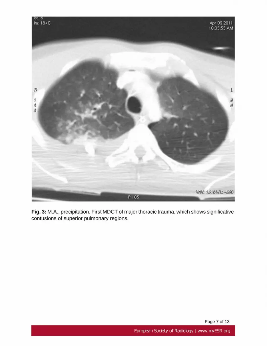
Fig. 3: M.A., precipitation. First MDCT of major thoracic trauma, which shows significativecontusions of superior pulmonary regions.

Page 8 of 13
Fig. 4: M.A., First MDCT, Coronal view.

Page 9 of 13
Fig. 5: M.A. Follow-up MDCT: after 5 days, development of confluent, disomogeneousopacity in the apical left lung (Staphilococcus A.).

Page 10 of 13
Conclusion
Ground glass and micronodular may be considered early development pattern evolvingtowards lobar parenchymal consolidation or disomogeneous consolidation, whichtogether account for approximately 44% of pulmonary complications in patients withpolytrauma.
The EOP develops in about 7 days after the event of major trauma, but the "ground glass"and "centrolobular nodules" patterns can be considered the precursors of this importantinvolvement.
On the basis of these results the pattern of early appearance can be considered as thenew "risk factor" for the development of a bronchus pneumonic process.
The ICU anesthesiologist can and should use these data to decide early medical therapyfeasible at least 3-4 days before the full-blown pneumonia appear.
This leads into an important reduction of the risk of multi-organ failure and late mortalityand in a lesser time of hospitalization of the polytraumatized patient for lung causes.
Particular attention should be paid to patients with chest drainage, which can beconsidered a predisposing factor but is not essential for the occurrence of an infectiouscomplication.
All 45 patients with infectious pulmonary complications were intubated duringhospitalization.
Several authors consider intubation and mechanical ventilation in the first hour animportant risk factor for the development of the EOP (2,6).
Our future perspective is to deepen this study applying this further distinction.
Images for this section:

Page 11 of 13
Fig. 6: F.S., moto-car crash. First MDCT: Massive right pneumothorax, associated toextensive contusive-lesions and hemothorax.

Page 12 of 13
Fig. 7: F.S., moto-car crash. Follow-up MDCT: Lobar consolidation, with air bronchogram(Haemophilus I.).

Page 13 of 13
References
1. LoCicero J III, Mattox KL. Epidemiology of chest trauma. Surg Clin NorthAm. 1989;69:15-19
2. Janus T J, Vaughan-Sarrazin MS, Baker LJ et al. Predictors ofPneumonua in Trauma Patients with pulmonary contusion. J trauma Nurs2012;19:139-47
3. Michelet P, Couret D, Brégeon F et al. Early Onset Pneumonia in SevereChest Trauma: a risk factor analysis. J Trauma. 2010; 68:395-400
4. Ciesla DJ, Moore EE, Johonson JL et al. The role of the lung in postinjurymultiple organ failure. Surgery 2005; 138:749-758
5. Sauaia A, Moore FA, Moore EE, et al. Epidemiology of trauma deaths:areassessment. J Trauma 1995;38:185-193
6. Stephan F, Mabrouk N, Decailliot F et al. Ventilator-associated pneumonialeading to acute lung injury after trauma. Anesthesiology 2006: 104:235-41
Personal Information


