Embed Size (px)
Citation preview
1 OF
z)
INFLAMMATORY CELLULAR RE SPONSEAND CYTOKINES IL-18, IL-6 AND TNFo
IN RAT AND HUMAN SPINAL CORD INJURY
Liqun Yang
Department of Surgery (Neurosurgery)University of AdelaideAdelaide, Australia
Department of NeuropathologyInstitute of Medical and Veterinary Science
Adelaide, Australia
Submified as part requirement for the degree of Doctor of Philosophy, June 2004
by
TABLE OF CONTENTS
ABSTRACTDECLARATIONACKNOWLEDGEMENTSFINANCIAL SUPPORTABBREVIATIONSPUBLICATIONS, PRESENTATIONS AND PzuZE
INTRODUCTION
ivviviiixX
xiv
1
1. TRAUMATTC SPINAL CORD INJURY (TSCI)1.1 EPIDEMIOLOGY1,2 NATURE OF THE INJURY1.3 SECONDARY SPINAL CORD INJURY1 .4 HISTOPATHOLOGICAL EVOLUTION1.5 NEUROPROTECTIVE TREATMENT1.6 ANIMAL MODELS
2. CYTOKINES2.1 OVERVIEW2.2 TNTERLEUKTN-I B GL-l B)2.3 TNTERLEUKTN-6 (rL-6)2.4 TUMOR NECROSIS FACTOR-o (TNF-a)
3. MICROGLIA3.l NOMENCLATURE3.2 CLASSIFICATION3.3 MICROGLIAL ACTIVATION3.4 FUNCTIONS OF MICROGLIA IN SCI
AIMS AND HYPOTHESES
1
1
2
J
t4t925
2929
JJ
42
49
575757
6062
õ5
1
RAT SCI EXPERIMENTS 66
1. MATERIALS AND GENERAL METHODS1.1 ANIMALS USED AND ETHICS APPROVAL1.2 ANAESTHESIA1.3 SURGICAL PROCEDURE1.4 NEUROLOGICAL EXAMINATION1 .5 PERFUSION-SACRIFICE1.6 TISSUE PROCESSING1.7 PHOTOGRAPHY
2. WEIGHT.DROP MODEL2.l METHODS2.2 RESULTS2.3 DISCUSSION
3. INFLAMMATORY CELLULAR RESPOI{SE3.1 METHODS3.2 RESULTS3.3 DISCUSSION
4. UPREGULATION OF IL-18, IL-6 AND TNFcr mRNAs ANI)PROTEINS AFTER MILD AND SEVERT, SCI
4.l METHODS4.2 RESULTS4.3 DISCUSSION
5. TREATMENT OF NF.TB.SPECIFIC ANTISENSEOLIGODEOXYNUCLEOTIDES AFTER SEVERE SCI
5.1 METHODS5.2 RESULTS5.3 DISCUSSION
66666868
69
70
707I
7272
73
80
848486
106
111
145
r45t46146
1i1119140
11
HUMAN SCI EXPERIMENTS 148
1. MATERIALS AND GENERAL METHODS1.1 SELECTION OF MATERIAL1.2 TISSUE PREPARATION
2. INFLAMMATORY CELLULAR RESPONSE2.l METHODS2.2 RESULTS2.3 DISCUSSION
3. EARLY EXPRESSION AND CELLULAR LOCALTZATIONOF IL-IB,IL-6 and TNF-cr
3.l METHODS3.2 RESULTS3.3 DISCUSSION
4. CO-LOCALTZATTO|{ Or IL-18 AND APP4.1 METHODS4.2 RESULTS4.3 DISCUSSION
FINAL DISCUSSION
148148
t49
151151
r52r69
173
173174180
182r82r82186
187
1. SUMMARY2. LIMITATION OF THIS PROJECT AND FUTURE WORK
187194
CONCLUSION 19s
APPENDICES 196
1. FIXATIVES AND BUFFERS2. STATISTICAL ANALYSES
t96t99
REF'ERENCES 218
111
ABSTRACT
The inflammatory response following spinal cord trauma plays an important role in the
secondary SCI. The goal of this study was to characterize the posttraumatic inflammatory
responses and localize cellular sources of IL-1P, IL-6 and TNF-cr following SCI. Thus, we
hypothesized that the pro-inflammatory cytokines IL-IP, IL-6 and TNF-cr may act as
messengers to coordinate the inflammatory cascade in the secondary SCI and that the
cytokine response should be greater in severe than in mild injury.
One hundred and twenty-six rats were used and rat spinal cord contusions were induced by
the wcight drop device. Mild and severe SCIs were respectively produced by dropping a 10-g
weight from 3.0 and 12.0 cm. Inflammatory cellular responses were studied using
immunohistochemistry and expressions of IL-l þ,IL-6 and TNF-cr mRNAs were analyzedby
RT-PCR. Thirteen human spinal cords removed at autopsy were studied using
immunohistochemistry and cellular sources of IL-18, IL-6 and TNF-a were localized using
double-label fluorescent confocal imaging.
In experimental SCI, neutrophils started to infiltrate primarily around blood vessels in the
central gray matter at6 hrs and peaked atl day. Macrophages were noted at 6 hrs and then
progressively increased for the first 3 days postinjury. Activated microglia were found as
early as I h after contusion and frequently wrapped around axonal swellings and healthy
neurons. RT-PCR showed an early and robust up-regulation of IL-IP, IL-6 and TNF-cr
mRNAs in spinal cord after severe contusion injury, maximal at 6 h postinjury with retum to
control levels by 24 h postinjury, the changes being significantly less in mild injury. In human
SCI, the inflammatory response paralleled the changes in experimental SCI, neurons and
IV
microglia were identified as the cellular sources of IL-18, IL-6 and TNF-cr, and IL-18 co-
existed with APP in the neurons and their axons.
RT-PCR analyses together with histological observations confirm that intrinsic CNS cells
(neurons and microglia), not peripheral inflammatory cells, are the main source of cytokines
because the peripheral inflammatory cells did not invade the injurcd spinal cord until 6 h
postinjury, a time when cytokine mRNA levels had peaked and started to decline. Microglia
around axons may have their possible beneficial effects on the injured axons by providing a
trophic local environment to promote the regeneration of the injured axons and the co-
existence of IL-IB and APP indicates a possible role of IL-IP in the production of APP,
Furthermore, our comparative RT-PCR analyses, showing significantly increased expression
of pro-inflammatory cytokine mRNAs in severe injury in contrast to mild injrry, support the
hypothesis that cy'tokine up-regulation is an important factor in the generation of the severity
of the inflammatory response and thus a suitable target for pharmacological intervention to
attenuate this response.
v

ACKNOWLEDGEMENTS
There are many people to whom I wish to express my gratitude for their help and support
during the last five years of my study at Adelaide, which has become a very exciting and
unforgettable experience in my life.
First and foremost, I would like to express my extreme and ineffable gratitude to my
supervisors, Professor Nigel Jones and Professor Peter Blumbergs, for their continuous
guidance, advice, support and dedication throughout the duration of this project. It would not
have been possible for me to even start my Ph.D. study without the support and
encouragement from my supervisors. Nobody coulcl have expected more supportive and
erudite supervisors. It has been a privilege to be your student.
My special thanks goes to Dr Corinna Van Den Heuvel, for her relentless and invaluable
guidance and advice, and for her never-ending support and friendship. Thank you so much for
everything, Corinna!
I am sincerely grateful to Mr Jim Manavis, for his constant and invaluable advice, discussion
and encouragement since day one, and for the very amusing experiences at the international
meeting. Not only was he there ready to answer my questions whenever I needed, but he has
always generously provided his help and patience endlessly.
Special thanks to Dr Barbara Koszyca and Dr Mark Gibson for allowing me to undertake a
large part of my research in the Pathology Department and for frequent invaluable discussion.
A very big thankyou to Ms Emma Moore for her endless assistance and patience, and for
teaching and helping me with western blotting experiments. A heart felt thankyou, Emma!
v1l
I would also like to thank:
1. Mr Chris Brown for providing technical advice at various phases of the research work.
2. NPrcf. Robert Vink for his expertise on statistical analysis and many interesting
discussions.
3. Dr. Mou¡i¡ Ghabriel for the many stimulating discussions pertaining to spinal cord
research.
4.Dr. Ghafar Sarvestani for his technical assistance in the confocal imaging and for his
continuous help.
5. The entire Neurosurgery Department including Ms Lyn Cockram, Dr Amal Abou-Hamden,
Dr Adam V/ells and Ms Melanie Smith, for making my time here enjoyable and for their
understanding, patience and lots of help. You are all wonderful people!
6. The entire Neuropathology Laboratory including Kathy, Bernice, Kathryn, Perury and
Margaret for making my life easier with their brilliant technical skills and their support'
7.The entire Pathology Department including Chris, Nigel, Eric, Fan-Ting and Maria for their
patience and technical skills in teaching and helping me with molecular biology experiments.
8. The entire research theatre staff especially Glenda Summersides for their brilliant technical
skills.
9. The staff of the Surgery Department: Mr Eric Smith, Mr Neville de Young, Dr Paul Drew
for sharing delightful conversations and jokes in the department.
10. Professor Glyn Jamieson for taking a genuine interest in my progress and his personal
interest in myself and my familY.
vlll
Special thanks to Dr. Jer¡rifer Brown, for her inspiration, help, fun and friendship, and for
allowing me to bother her with all different kinds of questions (not only scientific, but also
English and cultural!). It is a fantastic time to have her companionship.
Finally, I would like to thank my wife and my parents for the huge support that is required to
leave China for my study in Australia. Thank my family for giving the opportunity to fly, for
believing in me, and above all, for your tireless guidance through thick and thin.
FINANCIAL SUPPORT
This study was supported by the Adelaide Scholarship International from the University of
Adelaide, Australia.
IX
ABBREVIATIONS
APl
APP
ASD
BBB
BCIP
CAMP
C-Fos
C-Jun
CNS
COX-2
CRE
CRP
CSF
activator protein-1
amyloid precursor protein
acidic sphingomyelinase domain
blood brain barrier
5 -bromo-4-chloro-3 -indolyl phosphate
cyclic adenosine monophosphate
cytoplasmic fos
cytoplasmic jun
central nervous system
cyclooxygenase-2
oAMP response element
c-reactive protein
colony stimulating factors
deoxy ribonucleic acid
excitatory amino açid
epidermal. growth factor
extracellular regulated kinases
Fas-associating protein with death domain
factor associated with neutral sphingomyelinase activation
fibroblast growth factor
growth-associated phosphoprotein-43
glial fibrillary acidic protein
a glucocorticoid-responsive element
DNA
EAA
EGF
ERK
FADD
FAN
FGF
GAP-43
GFAP
GRE
X
IFN
GS I-B+
H&E
IKB
IL-1c¿
rL-18
IL-1R-AoP
IL-6
i-NOS
JAK
JNK
MAP
MEKK
MHC
MMP
MRE
mRNA
Mn-SOD
NASCIS
NBT
NFAT
NFBA
NF-IL6
NF-KB
Griffonia simplicifolia I-B¿
haematoxylin and eosin
interferon
inhibitory rB
interleukin-1c¿
interleukin-1B
IL-1 receptor accessory protein
interleukin-6
inducible-nitric oxide synthetase
janus activated kinase
c-Jun amino-terminal kinase
mitogen-activated protein
MAP kinase/ERK kinase
maj or histocompatibility complex
matrix metalloproteinase
multiresponse element
messenger ribonucleic acid
mitochondrial superoxide dismutases
national acute spinal cord injury study
nitro blue tetrazolium
nuclear factor ofactivated T cells
nuclear factor-BA
nuclear factor-interleukin-6
nuclear factor-kappaB
nerve growth factorNGF
XI
NHS
NMDA
NO
NOS
NSD
Par-4
PBS
PC-PLC
PDGF
PGIz
PLA-2
PNS
ROS
RT-PCR
SDS-PAGE
SHP-2
SRE
STAT
TGF-B
TNF
TNFR
TRADD
RIP
SCI
nonnal horse serum
N-methyl-D-aspartate
nitric oxide
nitric oxide synthase
neutral sphingomyelinase domain
prostate apoptosis response-
pho sphate-buffered saline
phosphatidylcholine-specifi c phospholipase c
platelet-derived growth factor
prostaglandin 12
phospholipase A-2
peripheral nervous system
receptor interacting protein
reactive oxygen species
reverse transcription polymerase chain reaction
spinal cord injury
sodium dodecyl sulfate-polyacrylamide gel electrophoresis
Src-homology phoshpatase-
serum responsive element
signal transducer and activator of transcription
transforming growth factor- p
tumour necrosis factor
tumour necrosis factor receptor
TNFR1 -associated death domain protein
TNF receptor-associat ed factor -2TRAF-2
x11
TSCI
TXAz
VCAM
traumatic spinal cord injury
thromboxane A2
vascular cell adhesion molecule
xlll
PUBLICATIONS, PRESENTATIOI{S AND PRIZE
Publications
Yans L., Blumbergs PC, Jones NR, Manavis J, Sarvestani GT and Ghabriel MN. Early
expression and cellular localization of pro-inflammatory cytokines in traumatic spinal cord
injury in the human. Spine 29(9):966-971;2004
Yang L, Van Den Heuvel C, Blumbergs PC, Jones NR, Manavis J. Inflammatory cellular
response and cytokines IL-1B, IL-6 and TNFa. J. Neurotrauma 19(10): 1295-1296;2002
Manuscripts in preparation
Yang L.. Jones NR, Blumbergs PC, Van Den Heuvel C, Moore EJ, Manavis J, Sarvestani GT
and Ghabriel MN. Severity-dependent expression of pro-inflammatory cytokines in traumatic
spinal cord injury in the rat. (Submitted to Journal of Clinical Neuroscience)
XlV
Presentations at scientific meetings
Yang L, Van Den Heuvel C, Blumbergs PC, Jones NR, Manavis J. Inflammatory cellular
response and cytokines IL-IP, IL-6 and TNFo in the human SCI. The 3 rd Asia Pacific
Symposium on Neural Regeneration, Perth,'WA, Australia, December 3-5, 2002.
Yang L, Van Den Heuvel C, Blumbergs PC, Jones NR, Manavis J. Inflammatory cellular
response and cytokines IL-18, IL-6 and TNFcr in the rat SCI. The 6th International
Neurtrauma Symposium, Tampa, FL, USA, October 27- November 1, 2002.
Yang L, Van Den Heuvel C, Jones NR, Manavis J, Blumbergs PC. Inflammatory cellular
response and cytokines IL-IB and IL-6 in experimental spinal cord injury. The L2th World
Congress of Neurosurgery, Sydney, NSW, Australia, September 16-20,2001.
Yang L, Van Den Heuvel C, Jones NR, Manavis J, Blumbergs PC. Inflammatory response in
the rat weight-drop model of spinal cord injury. Australian and New Zealand Society for
Neuropathology, Annual Scientific Meeting, Adelaide, SA, Australia, May 14,200I.
y34g¡, Van Den Heuvel C, Jones NR, Manavis J, Blumbergs PC. Inflammatory response in
the rat weight-drop model of spinal cord injury. Spinal Society of Australia, Annual
Scientifïc Meeting, Perth, WA, Australia, April 27-29,2001.
XV
PRIZE
Rob Johnson Award 2001
For the best paper by a trainee
Spinal Society of Australia
Annual Scientific Meeting
XVI
INTRODUCTION
When you examine a nran with a dislocation of a vertebra of his neck, and you find him
unable to move his anns and his legs. His penis is erect; urine drips from his penis
unknowingly. Then you have to say: A disease one cannot treat.
Edwin Smith Surgical Papynrs, about 25æ B.C.3r2
It has been more than forty-five centuries since an unknown Eglptian physician gave this
above description of the clinical features of traumatic tetraplegia 218, but TSCI is still a
prominent neurological health ca¡e issue for modern society. Until recentþ, the view of the
irreversibility of spinal cord lesions was accepted almost as a "la\il of natute".
However, there has been a rapid expansion in this field of neuroscience during the last
decades and we are now starting to understand the mechanisms of neuron survival and death,
the crucial molecula¡ aspects of neurite growth and the interaction of inflammatory cells with
the CNS. Obviously, in such a large and old field, a full and total coverage of the existing
literature is hardly possible. This review of the literature on TSCI in the following sections
concentrates on the inflammatory response following TSCI and the underlying cellular and
molecula¡ ¡scþanisms.
1. TRAUMATIC SPINAL CORD INJURY
I.I EPIDEMIOLOGY
The annual incidence of TSCI has been suggested worldwide to range from I2.7 to 50 cases
per millio¡ population tee. The incidence of TSCI in Australia is about 300 new cases per yoar
and is estimated to be 20-30 cases por million population per year 3e8.
I
Although the incidence of TSCI is increasing, the number of fatal cases has been decreasing
each year in Australia since the early 1980s as a result of legislation related to safety
measures, quicker retrieval from the accident site and imptoved emergency mânagement 3e8.
The most coÍlmon causes of TSCI are motor vehicle accidents (nearly SOVo), followed by
falls, penetrating injuries due to gunshot or knife wounds and sports accidents "t. Th"
majority of human TSCIs result from acute contusion due to displacement of bone or disk into
the spinal cord during fracture dislocation or burst fracture of the spine 41'188. The cervical
spinal cord as well as the thoracolumba¡ þnction a¡e the regions that a¡e most commonly
affected {58. Paralysis of the upper and lower extremities, so-called quadriplegia, accounts
for 54Vo of the injured patients and the remaining 46Vo accottnts for patients with paraplegia.
TSCI predominantþ occurs in young men (6l%o have an age between 16 and 30 yt), and only
20-3OVo of the patients are lvomen Lee'312.It is siguificantly uncommon for TSCI to occur in
children under the age of 14 56.
1.2 NATURE OF THE INJURY
The four vectors of forces applied to the spinal column in TSCI are flexion, extension,
rotation and compression. In the majority of cases, a combination of forces acts on the spinal
cord upon injury. The specific forces as well as the specific anatomic level to which the forces
are applied results in a specific pattem of posttraumatic spinal cord pathology'u.
The spinal cord is the major conduit through which motor and sensory signals pass between
brain and body. TSCI leads to a disruption of these pathways and a loss of motor and sensory
2
functions. It has been well established that much of the posttraumatic tissue damage and
subsequent neurological deficits a¡e due to secondary reactive processes 4m535'ee'18208. The
initial traumatic iojo.y triggers a cascade of molecular and cellular changes that occur within
minutes and continue for days or weeks. Therefore, the TSCI is a dpamic and complex
process rather than an event.
1.3 SECONDARY SPINAL CORD INIURY
Allen first proposed the concept of secondary ioþ.y in 1911 353, studying experimental acute
spinal cord injury in dogs. Today, it is well known that loss of neural function following acute
traumatic injury to the spinal cord only in part results from direct or immediate damage to
spinal gfey or white matter. The concept that secondary inju.y leads to much of the tissue
damage after traumatic SCI has been suggested by the slow progtess of histopathological
changes 3m and the continuing death of neurons in the spinal cord after primary trauma to the
co¡d 221. Strong evidence has been provided by the demonstration that neurological
impairment can be reduced by numerous postinjury treahents. Bracken et aL a7 in 1991
reported a large-scale clinical trial in patients with SCI, showing that high dose steroid
methylprednisolone can reduce the secondary injury and functional deficits in patients with
SCI by attenuating the biochemical and pathological changes in the cord after iojoty.
Now it is well established that damage to the spinal cord after SCI is produced by two distinct
mechanisms: the primary mechanical injury and a secondary injury initiated by the primary
injury 353. Trauma to the spinal cord not only produces the primary mechanical damage but
also causes progressive auto-destruction of tissue at the trauma site, the extent of which is
dependent on the severity of the trauma 83. Therefore, serondary injury processes play an
important role in the overall functional deficit tw'3e2.In the last 20 years, va¡ious putative
3
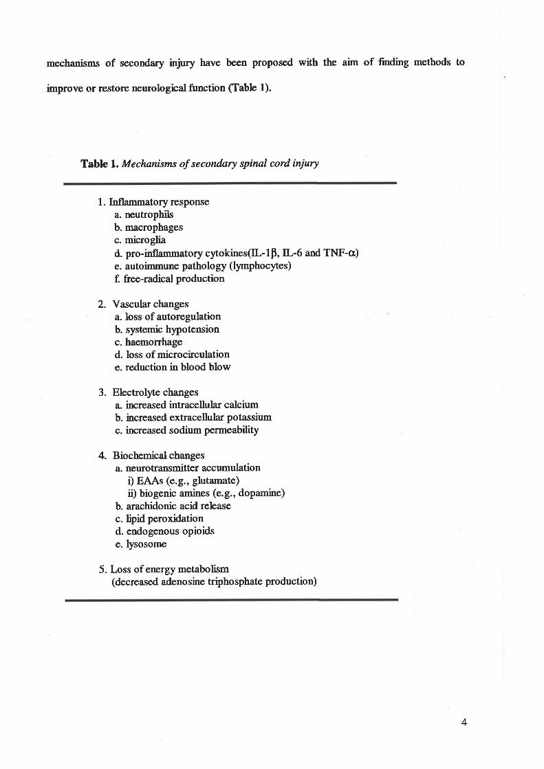
mechanisms of secondary injury have been proposed with the aim of finding methods to
improve or restore neurological function (Tabb 1).
Table l. Mechanisms of secondary spinal cord injury
1. Inflammatoryresponsea. neutrophilsb. macrophagesc. microgliad. pro-inflarnmatory cytokines(Il-1p, IL-6 and TNF-a)e. autoimmune pathology (lynphocytes)f. free-radical production
2. Vascular changesa. loss of autoregulationb. systemic hypotensionc. haemorrhaged. loss of microcirculatione. reduction in blood blow
3. Electrolyte changesa. increased intracellular calciumb. increased extracellular potassiumc. increased sodium permeability
4. Biochemical changesa. neurotransmitter accumuliation
Ð EAAs (e.g., glutanate)ü) biogenic amines (e.g., dopamine)
b. a¡achidonic acid releasec. lipid peroxidationd. endogenous opioidse. lysosome
5. Loss of energy metabolism(decreased adenosine trþhosphate production)
4
1.3.1 IM|I,AMMATORY RESPONSE
Usuall¡ an inflammatory rcsponse to traumatic injury brings about wound healing. However,
the posttraumatic inflammatory response at CNS lesion sites has been suggested to contribute
to secondary progressive tissue destruction 26333523e, which precludes any meaningful tissue
repair *. By causing damage to vascula¡, neuronal and glial cell populations, the primary
traumatic injury is supposed to render these and adjacent structures susceptible to secondary
injury by posttraumatic inflammation. The robust inflammatory tesponse begins within hours
and peaks within several days following TSCI ll'ry. This response mainly includes activation
of microglia, release of cytokines, immigration of perþheral inflammatory cells, increase in
vascular permeability and development of oedema t&'3s4.
1.3.1.1 MICROGLIA
It has been demonstrated in many studies that microglia are rapidly activated within hours
after SCI 65'te6'2oo2o8'276'312'32334t'382. Upon cellular activation, microglia, the resident cells of
the CNS, can produce large quantities of a number of neurotoxic mediators, including EAAs,
proteases, pro-inflammatory cytokines and NO, to further the secondary SCI. Details of
biologic effects of microglia after SCI will be discussed in section 3.4.
1.3.1.2 CYTOKINES
C¡okines, such as tr--lp, IL-6 and TNFc6 play a central role in the initiation, perpetuation
and regulation of inflammatory response following SCI 2ó. ¡ç¡ails of the functions and
significances of these pro-inflammatory cytokines will be discussed in section 2.
5
I.3. 1.3 NEUTROPHIL AND MONOCYTE INFIL'TRATION
There a¡e two main waves of cellular infiltration by leukocytes at the lesion site of the injured
spinal cord 1133'ry23e377. The first \f,rave consists of pollanorphonuclea¡ neutrophils and the
second is dominated by phagocytic macrophages.
Polynorphonuclear neutrophils sta¡t to infiltrate the lesion site and the adjacent tissue in the
first few hours after injury of the spinal cord in experimental animal models, reach a peak
within 24 h and disappear by 3 days 6s9e23e. It has been observed that the number of
polymorphonuclear neutrophils infiltrating the tissue is strongly correlated to the degree of
haemorrhage occurring at the lesion site, suggesting that bleeding into tissue and subsequent
degradation of haemoglobin products may be strong chemoattractant factors for
po\anorphonuclea¡ neutrophils tt323e. Activation of membrane phospholipases by certain
clotting factors such as thrombin and bradykinin, which extravasate into the spinal cord
tissue, may contribute to the initiation of the formation of chemoattractant factors for
polyrnorphonuclea¡ neutrophils tts. Based on in vitro observations, neutrophils are potentially
able to degrade connective tissue matrix and damage the tissue by their ability to generate
oxygen radicals and secrete granule constituents such as lysosomal enz5mes e'358. This view is
supported by some findings that in vivo application of antibodies agains¡ the membrane
glycoprotein complex CD18 (integ¡in pz-subunit), which has been shown to be critically
important for adhesion of leukocyt""t"'to', induced a decrease in tissue damage of the spinal
cotd 72. Furthermore, in a compression model of the spinal cord in the cat, Means and
Anderson 23e, observed signs of neuronophagia by pollmorphonuclear neutrophils in the
marginal zone of haemorrhagic necrosis, suggesting that neutrophils not only are general
mediators of inflarmatory events but also have specific cytotoxic effects on neuronal cell
populations. In contrast, this view on neutrophils has been challenged by some studies
showing that neutropenia induced by inþtion of vinblastine sulfate or methylprednisolone
6
has no effect on the extent of tissue damage following CNS in¡ry 20367. Therefore, the
detailed roles and functions of neutrophils in TSCI still remain çe¡1¡evsßial
The second wave of immigration of peripheral inflammatory cells into the a¡ea of injury
mainly consists of phagocytic macrophages 1e33. It has to be stressed that it is technically
difficult to distinguish between fully activated endogenous microglial cells and blood-derived
extrinsic macrophages 25'8s'101'272 and the roles and functions of microglia following TSCI will
be discussed in detail in section 3. In recent years and mainly based on in vitro studies,
macrophages have been found to not only participate in tissue destruction as phagocytes but
also have strong secretory properties 136271. Macrophages can secr€te a remarkable variety of
macromolecules, including many cytokines (e.g., ILla, [--1P, IL-6, TNF-c, interferons-(L -
Þ, -y, etc.), numerous growth factors (e.g., TGF-p, FGF, etc.) and various enzlmes (e.g.,
lysozlme, elastase, collagenase etc.) and these bioactive molecules exert a wide range of
biological effects, from mediating cytotoxicity to facilitation of tisslue rcpair2r2.
Some experimental evidence supports the involvement of macrophages in secondary tissue
damage following TSCI. It was observed that demyelination of axons that survived the initial
contusion injury is coincident in time with macrophage invasion 33'35. kt vitro studies showed
that neutral proteases secreted by stimulated macrophages lead to demyelinatioo 6'. Io lniuo
studies in the guinea pig demonstrated that macrophage depletion by intraperitoneal
application of silica dust at the time of spinal cord compression lesion produced a delay of up
to 2 days in the onset of secondary functional loss and a reduction in the loss of myelinated
fibres 34.
7
However, other experimental evidence has suggested that macrophages a¡e essential for the
regeneration of severed axons. Axonal transection in both the CNS and PNS results in
degeneration of the distal nerve stump, so-called Vfallerian degeneration. In the PNS, myelin
is rapidly degraded over a period of a few days, but in the CNS, myelin and a¡ronal debris can
persist for months due to the poor recruiûnent of macrophages to areas of injury in the CNS
compared with the massive recruitment in the PNS 30. The persistence of myelin and axonal
debris has been thought as an important factor in impeding the regeneration of injured Ð(ons
in the CNS and thus the poor recruitment of macrophages has been proposed to be an
important factor in the poor ability of the CNS to regenerate 5a'271. Futther-ore, the presence
of macrophages in the distal stump of a severed peripheral nerve is supposed to be not only
necessary for removing myelin and a:ronal debris but also for initiating mitosis in Schwann
cells and proliferation of fibroblasts 2e. Additionally, macrophages have a potential role in
reconstruction of the injured tissue. The first evidence that macrophages are essential in tissue
repair and remodelling was shown in 1975 by læibovich and Ross, who found that
macrophage depletion in the guinea pig was associated with impairment of skin wound
healing "4. Mol""ules produced by macrophages have been shown to have strong growth-
permissive or proliferative properties for glial and endotheli¿l cells in the CNS 3t2. Thus,
macrophages can enhance the production of NGF by non-neuronal cells t6e'220 and modiff
glial cell surface properties to produce a substrate permissive for neurite elongation 78.
Molecules produced by macrophages (e.g., IL-IP, IL-6, TNF-cr etc.) also have a direct
protective and trophic effect on the injure.d CNS tissue 24r'2e8. On the other hand, these
molecules are also known to have cytotoxic effects on the nervous system te4'3s4.
8
I.3.I.4LYMPHOCYTEINFL-TRATION
Relatively small nuurbers of diffe¡ent subpopulations of lyqphocytes have been seen in spinal
cord þsion sftos 163ø6275. Thus, the notion of an autoimmune process being initiated by TSCI
\ilas proposed by several research groups, but strong experimental evidence for this
hypothesis is still lacking 63. The majority of data indicate that a certain degree of bystander
damage to surviving nerve fibres may be due to the inflammatory response rather than a
targeted specific immune attack. However, it has to be ruled out by further experiments
whether a specific immune rcsponse plays a role in TSCI.
1.3.1.5 FREE REDICALS
It has also been hlpothesized that reperfrrsion injury of the endothelial cell after ischaemic
changes may contribute some cytotoxic substances in the inflammatory response 3r2.
Ischaemia can transform xanthine dehydrogenase in the endothelial cell into a xanthine
oxidase. Then, during reperfusion, oxygen activates the xanthine oxidase to oxidate xanthine,
transferring its electrons to molecular oxygen to generate superoxide radicals. These radicals,
which a¡e toxic to the endothelial cell, increase vascular permeability. This vascula¡ inj"ry is
exacerbated by neutrophils, which aggregate at sites of vascular damage, secreting additional
reactive oxygen species e'3s8. In this way, the inflammatory response causes damage to the
endothelial cellç disruption of vascula¡ integrity, oedema, extravasation of erythrocytes,
migration of neutrophils and monocytes into the injured region, and the subsequent release of
cytotoxic substances 406. Thus, the sustained release of cytotoxic substances in the
inflammæory response has been believed to play a key role in progressive necrosis in spinal
cord injury.
In summary, it has been suggested that the inflammatory response in spinal cord injury may
initiate and maintain progressive secondary tissue damage, This notion has been supported by
9
numerous repotts that the tissue necrosis can be significantþ reduced by treatment with anti-
inflammatory agents and that wound fiealing may be adversely affected by the presence of
tissue necrosis 3s3'M. However, the role of posttraumatic inflammatory r€sponse is still not
very clear at present. The need to reduce the inflammation to limit the spread of damage may
be in conflict with the need to permit the inflammation to promote tissue repair and
regeneration. Furthermore, although the posttraumatic inflammatory response of TSCI is well
documented, the functional significance of the inflammatory cells (e.g. noutrophils,
macrophages, microglia) in traumatic spinal cord lesions still remain controversial and the
posttraumatic inflammatory response ha,s not been studied in quantitative detail
I.3.¿LOCAL ISCHAEMIA
There is often a substantial decline in spinal cord blood flow after trauma; such ischemia is
believed to contribute to secondary tissue injury t*. M-y studies have shown a major
reduction in the microcirculation and a lack of perfusion at the injury site after experimental
cord trauma e62e3'3s3. Further, V/allace et aL378 demonstrated that there was a lack of perfrrsion
of the arterioles, capillaries and venules at the injury site and adjacent areas, considerably
cephalad and caudad from the injury site, while the large vessels of the cord (the anterior
spinal artery and the anterior sulcal arteries) always remained patent even after severe cord
inju.y. The ischaemic areas included a large part of the grey matter and the surrounding white
matter. Therefore, Tator et aL353 postulated that secondary thrombosis or vasospasm of
arterioles was due to the accumulation of noradrenaline at the injury site.
The normal cord can autoregulate blood flow to maintain a constant blood supply over a wide
range of arterial pressure. However, autoregulation is markedly impaired after spinal cord
trauma ton. Mo.eover, neurogenic shock can be induced by acute cord trauma. It has been
10
shown in numerous studies that posttraumatic hlpotension and decreased cardiac output are
caused after experimental acute spinal cord inþry. These declines result from a combination
of decreased sl4pathetic tone and myocardial effects 148.
These results have suggested that multþle factors, including reduction of spinal cord
microcirculation at and near the in¡ury sites, impaired autoregulation of spinal cord blood
flow, systemic h¡potension, and diminished cardiac output, contribute to ischaemia after
trauma in the spinal cord. Elevation of posttraumatic mean systemic arterial pressrre to more
than 160 mmHg failed to significantþ improve spinal cord blood flow at the inþry site and
caused marked hlperaemia at adjacent sites lae. A linea¡ relationship was found to exist
between the severity of cord injury and the reduction in blood flow at the inþry site. Further,
both the severity of cord injury and the degree of posttraumatic ischaemia were found to be
significantly related to posttraumatic anonal dysfunction (motor and somatosensory tracts of
the cord) 3s3.
Therefore, posttraumatic ischaemia is a direct and damaging reaction to trauma, and treatment
of the ischaemia contributes to the restoration of cord function 112.
1.3.3 EAA
The EAA, glutamate, is widely distributed within the CNS and serves as an excitatory
neurotransmitter 166. EAAs, such as glutamate and NMDA, can destroy neurons in the
mammalian CNS 24e and there are several tlpes of EAA posts¡maptic receptors, which
mediate the neurotoxicity of EAAs 368. It has been strongly suggested that EAAs are involved
in secondary SCI following TSCI. There have been numerous studies that demonstrate that
local levels of EAAs, especially glutamate, rise to toxic levels following TSCI 22r'22e'268'326
11
and that support the involvement of EAAs in the pathologic changes after spinal cord inþry.
It was also obserued that the extent of EAA increase was dependent on the severity of trauma
s3"26s. By means of the microdialysis technique, Liu et aL nr and other investigators 22e'326
demonstrated that extracellula¡ EAAs reached toxic levels upon impact injury to the spinal
cord.
Further evidence was provided by reports of exacerbation of neuronal damage by EAA
administration in the inþred cord and attenuation of neuronal damage by some selective EAA
antagonists (including antagonists of NMDA" KA and AMPA) following ttaumatic injury to
the spinal cord 83'tffi '1
w't4t'268'39o'3e2 -
Taken together, these results demonstrated that secondary damage to the spinal cord after
traumatic injory can be partialty attributed to delayed neurochemical change within CNS
tissue, produced by increased extracellular EAAs. Significantly increased EAAs are thought
to damage neurons by excessive activation mediated through specific membrane receptors
mainly by two mechanisms: (a) an early chloride and sodium ion influx, leading to acute
neuronal swelling and (b) a later calcium ion influx, leading to more delayed drmage
22122e'2ee. The later increased intracellula¡ calcium is responsible for a cascade of cell events
and further damage may occur in the absence sf s\ ,çtling 2ee'3s3. Thus, excessive stimulation
of membrane receptors by high extracellular concentration of EAAs may greatly increase
Ca2* influx to produce neuronal death in the CNS with accumulated intracellulÃr C** 161'183.
The increased intracellulat Ct* can activate a number of Ca2*-stimulated enzymes, such as
phospholipases, protein kinases, nitric oxide s5nthase, proteases and nucleases, which
subsequently damage some organellae, including mitochondrion and nuclei- All these
eventually result in cell damage 2ee. These hyrotheses have been supported by the
t2
observations of a rapid decrease of extracellular calcium within the spinal cord following
spinal iojory and the restoration of neurological function after SCI in rat models by cakium-
channel blockers 104"103. Intracellula¡ calcium influx and consequent accumulation is well
known as the final common pathway of toxic cell death in the neryous system 3tr.
1.3.4 OTHER SECOIYDARY BIOCHEIVIICAL INJURY T'ACTORS
Biogenic emines (dopamine, noradrenaline, serotonin and histamine) act as neurotransmitters
in the spinal cord. Some investþatoÍs s2'25t3ot4o7 focused considerable attention on biogenic
amines as possible mediators contributing to the secondary SCI. They reported that elevated
levels of biogenic amines were observed within the spinal cord after trauma and paralleled a
marked reduction in spinal cord blood flow in experimental trauma. However, ssffisting
results have been reported 35e, which failed to confirm previous reports of arrine alteration in
spinal cord trauma. An impact inþry to the thoracic cord did not lead to changes in
noradrenaline, dopamine or serotonin despite the presence of severe haemorrhage and
necrosis. Additionall¡ o"-methyl-p-tyrosine, which reduces the levels of central dopamine and
noradrenaline, did not demonstrate a protective effect after spinal cord trauma. Therefore, the
role of biogenic amines in spinal cord traum¿ remains to be determined.
The lysosome mechanism was proposed by several authors tt'. Th"y found that transection of
the spinal cord was followed by massivç accumulation of lysosomes and release of lysosomal
hydrolases within spinal cord stumps. They suggested that release of lysosomal hydrolases
resulted in secondary spinal cord damage.
The endogenous opioid mechanism was advanced by Faden et a1.107, who described the
therapeutic effects of the opiate receptor antagonist naloxone in experimental models. They
13
suggested that endogenous opioids might plzy a pathophysiological role in the spinal cord
following traumatic injury.
1.4 HISTOPATHOfuOGICAL EVOLATION
The sequential pathological changes in the cord following TSCI can be divided into acute,
subacute and late phases. The acute phase is characterised by haemorrhage, rapid necrosis and
glial activation, which is followed by reactive gliosis and recruiûnent of blood-born
inflammatory cells in the subacute phase. The final appearance of the injured spinal cords
some weeks after injury is surprisingly similar in the various lesion models: a cavity and scar
tissue are formed and in white matter, different stages of Wallerian degeneration a¡e observed.
1.4.1 ACUTE PHASE: HOURS AFTER INJURY
I.4.I.T HAEMORRHAGE
An alteration in the microvasculiature of central grey matter is the first detectable
morphological change in the spinal cord 303. Multifocal petechial haemorrhages at the siæ of
compression are observed within minutes following TSCI with the light microscope.
Postcapillary venules become distended with erythrocytes and red blood cells penetrate the
injured vascular endothelium and are found within the perivascular spaces 8'e1. The
haemorrhages at the very early time points are restricted to the central grey matter adjacent to
the central canala. The traumatic vascular lesions spread within the next few hours in a radial
direction in the grey matter and to a small extent also into white matter tt'". \ryfthio the first
24 h, microthrombi containing degranulated platelets and abundant fibrin a¡e frequentþ
observed in capillary-sized vessels with structurally intact as well as necrotic walls 11. The
progression from comparatively normal vessels to vascular congestion and blood
t4
extravasation can occur rapidly. As early as a few minutes following imFact injury, separation
of endothelial junctions and exposrrre of thc microvascular basal lamina were observed 142.
Studies in cats using scanning electron microscopy showed craters in capillary endothelium
and adherent noncellular material as early as I h posttrauma "t, toggett'rg that these
endothelial abnormalities might be due to free radical-induced injury to membrane lipids,
resulting in microcirculation failure. This view is supported by another finding that the
endothelial abnormalities were prevented by pre-treatment with antioxidants 385. lvithin hours
postinþry the hallmarks of ischaemic vascula¡ injury (vacuolation and sqrclling) were present
in endothelial cells of capillary and postcapillary venules throughout gley and white matter el.
1.4.1.2 NECROSIS
Another early morphological abnormality in the acute phase is neuronal necrosis. Neurons
with cytoplasmic eosinophilia, the first sþ of necrotic alteration, appear in random
distribution in grey matter within the first hour postinjory ttt. Other signs of necrotic
alterations in neurons are also frequentþ observed, including ghost cells, cytoplasmic
microvacuolation and shrunken neurons with indistinct nuclei smudged cytoplasm, loss of
Nissl bodies, irregular shape and hyperchromatization 341. Ultrastructural observations
revealed karyonhexis and swelling of the rough endoplasmic reticulum in necrotic neuronal
and glial cells 11. The necrotic alterations quantitatively increase in the grey matter within the
first several hours after injury ee, when many cell perikarya in the epicentre of the lesion have
undergone homogeneous eosinophilic changes on H&E staining and karyolytic or clumped
chromatin has been revealed in nuclei Additionall¡ both neurons and glial cells are equally
vulnerable and both cell populations undergo cell death in the same time interval ll'ee.
1.4.1.3 AXONAL INJIJRY
15
Axonal injury in white matter is characterized by a finely granular appearanoe associated with
a va¡iable amount of swelling within the first several hours postinþry 11. Characæristic eady
axonal abnormalities also include the development of a space between the axon and its myelin
sheath, particularly in the central injury zone. This axonal swelling was mostþ inærpreted as
due to oedem¿ and was described as an enlarged periaxonal space eo'177'374. However, electron
microscopic studies I'r45 have showed that the majority of these spaces are intramyelinic
vacuoles; the myelin sheaths are dilated or split. These focal changes of the myelin sheaths
appear mainly in the central parts of white matter and account for the progressive spongy
appearance of the white matter noted by light microscopy. By 4 h, numerous ÐKolts within the
central white matter and to a lesser extent axons scattered throughout the peripheral whiæ
matter showed swelling and accumulation of organelles (mitochondria, neurofilaments,
lysosomes and smooth endoplasmic reticulum) 11202. These axonal abnormalities continued in
a centrifugal fashion toward the pial surface over the next several days 50.
Immunoc)¡tochemical technologies using antibodies against APP ", slmaptophysin,
chromogranin A, cathepsio D "0, neurofilament protein subunits 277 nd ubiquitin 314 hove
revealed a far greater occurrence of axonal injury than had been appreciated by use of
classical silver staining techniques. Of these immunological ma¡kers, labelling for APP was
found to be the most sensitive and specific in demonstrating axonal i"jury ranging from
mildly damaged axons to axonal retraction bulbs, which represent severed axon cylinders
38,t32
1.4.1.4 MICROGLIAL ACTIVATION
As already described in section 1.3.1.1, rapid microglial activation rvas observed within hours
after SCI 341'382. Additionall¡ an in vitro study has demonstrated that microglia were activated
t6
\ilithin a period as short as 5-10 minutes 3t6. Details of microglial activation will be discussed
in section 3.3.
1.4.2 SIJBACUTE PIIASE: DAYS AI\[D \ilEEKS AFTER INJURY
This phase is characterized by the activation of astrocytes in the CNS as well as the
recruiûnent of different peripheral cell populations into the area of lesion.
I.4.2.1 INFLI.DGS OF NEUTROPHILS AI.ID MONOCYTES
As already described in section 1.3.1, two waves of infiltrating peripheral inflammatory cells,
an early peak of neutrophils and a later peak of phagocytic macrophages, atre seen following
TSCI.
I.4.2.2 ASTROCYTE ACTIVATION
Astrocytes undergo rema¡kable hlpertrophy and limited proliferation upon injury 25e.
Reactive astrocytes have increased numbers of intermediate filaments comprised largely of
GFAP and appear larger with more prominent and numerous processes than resting astrocytes
in the normal CNS 2e1. Reactive astrocytes also upmodulate the expression of a large number
of molecules (e.9., enz5¡rnes, cytokines, neurotrophic factors) to exert some beneficial and
injurious effects on the injured tissue. For example, upregulation of proteases and protease
inhibitors help remodel the extracellula¡ matrix, regulate the concentration of different
proteins in the neutrophil and clear up debris from degenerating cells 26o'2e1. Cytokines are key
mediators of immunity and inflammation and could play a critical role in the regulation of the
blood-central nervous system interface 2$2to'30s. Neurotrophic factors, transpofor molecules
and enzymes a¡e involved in the metabolism of EAAs and in the antioxidant pathway l003ee.
Microscopic evaluation of spinal cord stab wound lesions at various postoperative time
t7
intervals rpvealed a gradually increasing astrocytic reaction m. Between I and 2 days, mitotic
activity of astrocytes increased markedly, ¡ndbating that the increased number of astrocytes at
spinal cord lesion sites results in part from proliferation of the cell population 260. Astrocyte
reactivity upon injury extends in a radial direction into both adjacent grey and white matter,
often over long distanc"t*20'. The astrocytic response peaks at 14 days but is still present at
28 daYs 26o2et.
1.4.2.3INFIL'TRATION OF SCHWANN. MENINGEAL AND FIBROBLASTIC CELLS
Schwann cells, meningeal cells and fibroblasts a¡e additional peripheral cell types that have
been seen to invade spinal cord lesion siþs 32. However, the functional significance of these
infiltrating cells is still not clear and remains controversial-
1.4.3 LAIE PHASE: WEEKS TO MONTIIS AX"TER INJIIRY
1.4.3.1 POSTTRALJMAJIC SYRINGOIVTYELIA
Resolution of the acute,/subacute inflammatory rcsponse occurs several weeks after injury by
the disappearance of the neutrophils and macrophages from the lesion area, leaving a fluid-
filled cyst 'n7. 'W""kr after injury, the cysts become better defined and extensive scar tissue
forms a¡ound the cavity 3e7. At very late stages in humans (months to years after the injury)
secondary often very elongated syrinxes can appear preferentially rostral to the lesion 227 . T\e
process leading to so-called syringomyelia is very poorly understood at present. It is an
important late complication and can significantþ contribute to further functional losses 332.
1.4.3.2 DEIVTYELINATION AND REIVTYELINATION
18
Another important aspect of the laæ phase is the continuous loss of myelin and axons in whiæ
matter. Apparent axonel loss and demyelination of a number of fibres were observed between
7 days and 3 months postinþry 33. However, only a small proportion of the fibres were
completely denuded of myelin and the naþrity showed abnormally thin sheaths surrounding
Ð(ons of normal appearance 57'160. I¡'Lether this specific appearance can also be due to
demyelination is not completeþ clear. Interestingly, evidence of remyelination tryas also seen
at 3 weeks postinjury 140'160. Ma¡ked differences in the extent of remyelination between larger
and smaller lesions were shown and remyelination was observed more extensively in smaller
lesions t60. At least a part of the remyelination event soems to be due to oligodendrocytes 13e
and Schwann cells 57. Schwann cell remyelination was predominantþ observed in severe
contusive injury associated with extensive \Valletian degeneration 36.
I. 5 N EU ROPROTECTIVE TRBATMENT
The aim of neuroprotective therapies is to use pharmacologic agents to alter the detrimental
neurochemical cascades initiated by mechanical deformation of neural tissue 22240. Fromthe
numerous substances tested in experimental spinal cord injury models for their
neuroprotective efficacy, only one compound, methylprednisolone, \il¿rs shown in
well-controlled, multi-centre, large clinical trials to enhance functional outcome in the human.
Although controversy exists about the benefits of its application, methylprednisolone is
currently widely used in clinical practice and the only therapeutic agent approved by the Food
and Drug Administration of U.S.A. for treating acute TSCI in humans. So fa¡, the
experimental and clinic¿[ trials of a large number of substances have shown very difrerent and
inconsistent results 108 and few have been evaluated in extensive dose-response studies, and
rarely were the treatment schedules based on knowledge of the drug" phannacokinetics 312. In
t9
addition, behavioral tests that are difficult to interpret were often used as the main outcome
measurement, without ca¡eful and quantitative histology.
It is not proposed to undertake a comprehensive literature review of all these different
therapies but to provide a selective overview as a backgronnd to the use of NF-rB-specific
antisense oligodeox5mucleotides in our studies. In this section, we concentrate on thc fcw
groups of tested substances that showed interesting or promising results.
1.5.1 AIITI.IMT,AMMATORY AGENTS
1.5.1.1 STEROIDS
Steroids have been tested extensively in animal models as well as in clinical triats 47'7e'e7.
Methylprednisolone megadoses, if applied early after lesion, were shown to exert beneficial
effects in tle va¡ious experimental CNS injury models, although the exact mechanism of
action remained unclear 6AotAU2. Based on these results the NASCIS 1 study, the füst multi-
centre t¿¡demised clinical trial, was initiated to evaluate the steroid treaûnent of secondary
damage following acute spinal cord trauma. A total 330 patients were enrolled in the study
and were all treated within 48 h of injury. The study assessed the effects of high and low
doses of methylprednisolone on recovery of neurological function in patients with spinal cord
inj"ry. Neurological evaluations performed at 6 month and I year postinjury did not show a
significant difference in motor or sensory outcome between the high- and low-dose
methylprednisolone groups as. A series of animal experiments conducted while NASCIS I
was in progress suggested that higher doses than those used in NASCIS 1 might be needed to
achieve a therapeutic respons e 48'ts2. Thus, NASCIS 2 wus started. A total 487 patients wer€
randomised for treatments with methylprednisolone, naloxone, or placebo 47. Based on
20
experimental studies, the administration of methylprednisolone was 30 mgl kS followed by
5.a mgflrglhr for 23 h because these doses resulted in a positive effect on r€covery in animals
with TSCI. The maintenance dose was chosen according to theoretical calcul¿tions of the
plasma half-life of the drug in humans and, to avoid complications of glucocorticoid
a¡lministration, 24 h was chosen as the duration of drug application. Motor and sensory
functions \f,'ere assessed at 6 weeks, 6 months, and I year postinþry. The study found that the
group of patients receiving methylprednisolone within 8 h after injury had significantþ better
sensory and motor function than those receiving placebo. Furthermore, the differences in
motor improvement between the methylprednisolone and the placebo group were statistically
significant at all three follow-up periods. Compared to the placebo group, no positive effect
was shown in the group of patients who received naloxone or late methylprednisolone
administration. A more detailed analysis of the data of NASCIS 2 also indicated that delayed
treatment with methylprednisolone is associated with decreased neurological recovery 6.
Adverse effects of such high doses of methylprednisolone, such as urinary tract infections and
pneumonia, were commonly found in NASCIS 2123. The incidence of wound infection and
gastrointestinal haemorrhage in methylprednisolone-treated patients in NASCIS2 was also
slightly higher than in the control group.
An imFortant issue, which was not addressed in the NASCIS 2 study, is the mechanism of
action of methylprednisolone. It has been believed that the treatment of spinal cord injury
with steroids is based on their anti-inflammatory actions e7, Ê.8., methylprednisolone has been
recentþ found to reduce tr--lp and TNF-cr expression by SOVo and 55Vo respectively after SCI
in rats via inhibiting NF-KB and AP-l (two major proinflammatory transcription factors)
6e232'3e3'3e4. Additionally, very high doses of the slmthetic steroid methylpredni^solone were
2I
also found to exert non-specific radical scavenger effects 151 and reduce i-NOS expression by
7O7o arftsr SCI in rats 6e.
1.5.1.2 ANTISENSE OLIGODEOXYNUCLEOITIDES
Since the identification of the DNA double-stranded helix, the gene as a target of therapy and,
moreover, the use of DNA as a drug have been possibilities. Antisense oligonucleotides, short
strands of s¡mthetic nucleic acids, are designed to bind to a target gene promoter through
Watson-Crick base pairing (complementary base pairing) and the formation of this
oligonucleotide/promoter heteroduplex results in downregulation of mRNA slmthesis and
consequent inhibition of slmthesis of the protein product 174'376. 'Antisense' is used by some
living organisms, specifically viruses, to control gene replicationr2a. Only recentþ ha,s the use
of antisense DNA as a mechanism to control human gene tren^slation been appreciated. The
first antisense-based drug Vitravene rvas successfully developed to inhibit human
cytomegalovirus by Isis Pharmaceutical Inc., Carlsbad, California in 1998 255.
Antisense oligodeoxlmucleotides carry a high therapeutic potential for the treahent of human
diseases and these molecules have been applied in several different fields, including
oncolog¡ haematology and cardiovascular and infectious diseases t24't62.In recent years,
some studies have been initiated and performed to evaluate the therapeutic usefulness of
antisense technology in modulating physiological and pathological functions in the CNS.
These include cytokines, neurotransmitter receptors, neuropeptides, trophic factors and other
proteins t*. Mup." et aL 237 demonstrated that administration of a TNF-a-specific antisense
oligodeoxlmucleotide into striatal parenchyna reduced cell death and improved
neurobehavioral deficits in a rat model of intracerebral haemorrhage induced by disruption of
the BBB integrity by collagenase, suggesting potential benefits through interference with
22
TNF-a production in the CNS injury. To date, few studies have been performed to evaluate
the therapeutic usefulness of antisense oligodeoxlmucleotides in modulating cytokine
production to confer neuroprotection in TSCI. Thus, it would be of interest to examine such
pharmacological agents in TSCI models in which cytokines may be a maþr mephenism
leading to secondary tissue damage and functional deficits. For example, NF-rcB-specific
antisense oligodeoxlmucleotides would be a potential pharmacological agent because NF-KB
is a major transcrþtional factor for the upregulation of IL-lp,n--6 and TNFa to27't562ts and a
ubiquitous transcription factor involved in the proinflammatory rcsponse to cytokines such as
L-lp and TNFa 2e2 lfor details, see sections 2.2,2.3 and2.4).
However, like any other treatments in TSCI, the antisense oligodeoxlmucleotides have some
pitfalls and limitations. First, non-specific functional neuronal alteration and detrimental
effects on cellular morphology may be caused by the antisense oligodeoxlmucleotides in the
CNS 344 even though recent advances in antisense oligodeoxlmucleotides chemistry have
significantþ decreased these side effects 376. Second, while it is commonly believed that the
CNS has limited capacity to launch immune reactions, it has not yet been established whether
delivery of slmthetic antisense oligodeox5nrucleotides (a xenobiotic) into the CNS may
ultimately stimulate immune competent cells to generate antibodies against RNA and/or
DNA, triggering inflammation with severe CNS consequences. Finally, specificity of action is
a key issue. Non-specific binding of the antisense oligodeoxlnucleotides to nRNA species
other than the intended target may cause unwanted interference with transcription and
translation 174.
1.5.2 EAA ANTAGOIYISTS
23
Increased extracellular bvels of EAAs (glutamate and aspartate) accumularc in the lesion area
following CNS in¡ry 6s'tos22t2Æ245. Numerous studbs have demonstrated that glutamate and
related excitatory amino acids can induce death of neurons 262263383 and it has been
reßognizÊd fs¡ many years that excitatory amino acids may be implicated in the secondary
tissue damage following CNS in¡ury zolzz. Development of selective NMDA receptor
antagonists has shown that the NMDA receptor complex may be an important factor for the
excitotoxic action of EAAs 2e and several groups have shown that treatment with competitive
or noncompetitive NMDA antagonists improves neurological function after brain injury
105,rff,165
1.5.3 NORADRENERGrc AGOI\ISTS
Osterholm et aL 26s proposed 1þs mensemine theory of spinal cord injury, arguing that high
levels of accumulation of norepinephrine at the lesion site crucially contributes to the
pathological pattem of necrosis. The effect of catecholamine modulation using substances
such as oz-methyltyrosine and clonidine was therefore studied 2so'266. Mixed results were
obtained, and hence, the value of modulating noradrenergic transmission in the acute phase of
spinal cord inþry remains unclea¡ e. However, these phannacological studies attracted
general interest about possible pharmacological interventions. Barbeau and co-workers 15
reported that modulation of the monoamine level by noradrenergic agonists might be
important in the more chronic phase of spinal cord injury.
1.5.4 ITYPOTIIERMIA
Other strategies, ie., hypothermia, have been tested in experimental spinal cord injury in
addition to phannacological moans described above 23r.lt has been hlpothesized that low
temperature would protect the CNS tissue against the effects of hypoxia or ischaemia because
24
the oxygen demand of the CNS tissue may drop due to the low metabolism induced by
h¡ryothermia 2x.Lacalspinal cord cooling was used in a series of experimental animal studbs
and the experimental data led to the trial of local spinal se¡fl çssling in some cases of human
spinal cord inþry ttt. The data emerging from these studies h¿ve been difficult to interpret
mainly due to the fact that low numbers of cases were reported and in none of the studies
were appropriate controls included in the experimental desþ. Additionall¡ local spinal cord
çssling is a technique fraught with æchnical difficulties, and the high rate of mortality
reported in some of the triab 5l bd to conoerns about its clinical application.
1.6 ANIMAL MODEIß
Interest in exploring spinal cord function using experimental mòdeb of SCI dates back to 177
AD., when Galen performed spinal cord transections of dogs and other animals and
recognized that resulting functional loss differed with the segmental level of the injury 38e.
Numerous experimental animal models of TSCI have been developed during the last decades
in order to find effective methods to manage spinal cord injury in humans. To obtain the most
useful data, the experimental desþ should mimic as closely as possible the situation in the
human and it should be as reproducible as possible. This task is difficult because human SCIs
a¡e multifactorial and include variables that may be difficult to mimic. For example, the
majority of human SCIs occur in a closed vertebral system whereas 6s5¡ animal models use
an open laminectomy for lesionin1tt'. Horvever, these models have provided the best and
sometimes the only practical way to examine mechanism.s of injury and to test potential
treatments.
1.6.1 WEIGIIT.DROP MODEL
25
The first well-controlbd animal model was described by Allen 36 in 1911, who designed a
weight-drop technfolue to deliver a quantifiabb impact to the spinal cord of animals. ffis
technique of weight-drop contusion has been refined and adapted over the years, but the
central princþle of using gavity to standa¡dize 1¡ç ¡6phanic¿l imFact on the spinal cord has
persisted in TSCI research.
A primary advantage of weight-drop models of SCI lies in their clinical relevance. First, the
weight-drop method closely simulates some of the biomechanics of human SCI. For example,
most human SCIs a¡e believed to be due to rapid flexion-extension of the spinal column,
resulting in a contusion of the cord when the vertebral bone hits the spinal cord 3136'1253e1.
Other models such as photochemistry, transection, hemisection, and static loading cannot
model the clinical contusion injury 21. Second, the similarities in histopathological appearance
of the lesion site between rat weight-drop SCIs and human SCIs were observed by Balentine
et aL 11 and other investþators e5'188'324. The use of the weight-drop technique to produce
traumatic injuries to the spinal cord in animals and the resulting stepwise sequential
pathological changes in the cord were found 1s þ similar to the configuration found in human
spinal cord injuries. Spinal cord pathological changes, which were initially prominent in the
centre of the cord, started as haemorrhages and oedema in the grey matter and progressed
through central necrosis, adjacent white matter oedema, and demyelination, to finally involve
the entire cord. The central fusiform zone of spinal cord necrosis later evolved into a cyst.
Third, the weight-drop model has successñrlly predicted therapeutic potentials for steroids
Iater tested in clinical triah 47.
It has been found in the weight-drop model that the reproducibility of the impact on the spinal
cord at a given height is very good: the standard error is less than 2.5Vo of the mean force or
26
impulse 3l3el. Furthermore, the mean force or impulse of the impact is significantly correlated
to the height from which the weight is dropped 31, functional deficits of the onimals 1ã,
somatosensory evoked potentials 288 and histopatholo W 3t38e seen at one month after SCI.
Thus, the weightdrop model can be used to produce reproducible and graded spinal cord
contusion injuries in the animal models 3Ee3e1.
1.6.2 PNET]MATIC/ELECIROMECIIAIITICAL MODEL
The first of these devices, the constrained stroke pneumatic impactor was pioneered by the
¡esearch group in the General Motors Research Laboratories in the 1980s 7'1{. An advantage
of pneumatic or electromechanical production of spinal cord contusion is that injury
biomechanics (e.9., contact velocity, arnount of displacement of the surface of the tissue,
force, etc.) can be controlled independentþ and therefore greater precision or actual
determination of injury outcomes was allowed 33s'336.It has generally been used to injure the
cervical spinal region with the vertebral column intact "t. By maintaining the normal
structure of the spinal column, certain a¡tefacts associated with a laminecto¡y procedure
(e.g., size of laminectomy site, etc.) are thus avoided and close approximation of the human
injury process is achieved. However, such devices are complicated and very expensive, and
therefore their use has not been widespread.
1.6.3 COMPRESSION MODEL
Tarlov and his colleagues 352 itrit¡1e¿ the compression model and defined the gradation of
neurological deficits with increasing compression of the spinal cord. A balloon catheter or a
pneumatically driven mechanical plunger was used to compress the spinal cord in dogs. The
clinical relevance of slow and maintained compression models is somewhat limited and is
27
distinct from th¿t of the rapid contusion techniques, like the weight-drop
pneumatic/ebctromechanical techniques.
or
1.6.4 PHOTOCHEMICAL MODEL
Traumatic spinal cord inþry rcsults in degeneration of neural tissue by a number of
pathophysiological mechanisms. Local spinal cord ischaemia is one of these mechanisms. It
has been known that spinal cord blood flow is considetablyreduced after traumatic in¡ry 15e.
Thus, an entirely different method was introduced by Watson et aL 3s on the basis of injuring
the spinal cord vascula¡ endothelir¡m photochemically. After the inþtion a photosensitising
organic dye (e.g., bengal rose) into the bloodstream, the spinal cord is irradiated with a light
beam of appropriate wavelength. Damage to the vascular endothelium and subsequent
thrombosis lead to ischaemic lesions and vasogenic oedema 61. An advantage of the
photochemical approach is that laminectomy is not required because the vertebral dorsal
surfaces are sufficientþ translucent.
Bunge et al s7 and other resea¡chers 15e used the photochemical technique to induce local
spinal cord ischaemic injury in the rat, developing the photochemical lesion model with
minimal variability to better understand the ischaemia mechanism of secondary SCI.
Pathological changes after lesioning were studied by light and electron microscopy. The
photochemical lesions resulted in extensive necrotic regions bordered by swollen axons, a
massive influx of macrophages, an increase in the perivascular space in surrounding spared
tissue, demyelination in the early stage and late myelination, by both oligodendrocytes and
Schwann cells, in the tissue surrounding the necrotic lesion. These changes resembled those
observed after SCI caused by contusion or compression.
28
2.CYIOKII\ES
In rhis rcvbw of the literature on cytokines, qlecifu consideration has bee,n paid to three main
pro-infl ammatory cytokines: IL I p, IL6 and TNF-c.
2.1 oVERWEW
2.1.1 VYIIAT IS¡ A CYTOKINE ?
Most cytokines are soluble simple pollpeptides or glycoproteins with a molecular weight of
30 lt)a or less, but some cytokines form higher molecular weight oligomers (e.g., TNF-cr
molecules form trimers)6o2s6. Cytokines are produced by leucocytes and a variety of other
cells in thr body, and act as chemical communicators between cells and have regulatory
effects on haematopoietic and many other cell tlpes that participate in host defence and repair
processes 373. Cytokines arc secreæd into the extracellular fluid or are membrane exposed by a
cell There they exert their effects on the same cells (autocrine activity) or on neighbouring
cells þaracrine activiry) by binding to specific high-affinity cell surface receptors which are
coupled to intracellula¡ signal transduction and second messenger pathways 60'2s6.
2.1.2 RESEARCH IIISTORY
The field of cytokine resea¡ch has evolved from four originally independent sources to3'373.
The first and most significant source is the field of llmphokine resea¡ch. The origins of
lynphokine research can be dated back to the mid-1960s when it was demonstrated that
protein mediators secreted by lymphocytes regulated the growth and function of a variety of
white blood cells. It soon became apparent that monocytos too a¡ç the source of these
important proteins. The second source of cytokine research derives from the study of the
interferons, which gradually became recognized as proteins exerting a broad range of actions
on cell growth and differentiation. The third sorrce of cytokine research is the field of
29
haematopoietic growth factors, or CSFs. The fourth source of cytokine ¡esearch derives from
tbe study of growth factors acting on non-hacmatopobtic cells, such as PDGF, EGF, TGF-p,
FGF or NGF. Many of these factors, in addition to promoting cell growth, exert othrr
'cytokine-like' actions.
2.1.3 NOMEtr\¡CLATI]RE
A uniffing concept of cytokines was slow to emerge in the 1970s due to the fact that the
cytokine field evolved from several separate souroes. The term lprphokine', which originally
denoted the product of activated lyqphocytes ", *^ also used less discriminately for
secreted proteins from a variety of cell sou¡ces. To dispel the wrong notion that such proteins
could be produced by llanphocytes alone, Cohen et aL 73 proposed the term bytokines'. After
a long-standing reluctance, bytokine'hes þççs6s the generally accepted name for this group
of proteins. To designate individual cytokines, a group of participants at the Second
Intemational L¡mphokine Workshop held m 1979 proposed the term Tnterleukin' in order to
develop a system of nomenclature I and 'interleukin' was proposed on the basis of the ability
of these proteins to act as communication signals between different populations of leukocytes.
The interleukin series has now reached 18 373. Afthough the name 'interleukin'implies that
these agents function as communication signals between leukocytes, the term is not ¡eserved
for factors that can act only on leukocytes and a number of the proteins that have been
labelled as interleukins not only are produced by a variety of non-haematopoietic cells but
also affect the functions of many diverse somatic cells (e.g. IL-l or tr--O 1. Many cytokines
arc now termed interleukins while others continue to be known by their older names (e.9.
ml-o/Þ4, TNF-q, TGF-Þ and many others).
2.I.4 CHARACIERISTIC XEATT]RES
30
Cytokines a¡e ths major orchesúators of host defence processes and a¡c involved in responses
to trar¡ma, tumour and invading organisÍrs¡, repair and restoration of homeostasis 373. These
regulatory proteins have a set of unique features outlined below se'tso'tø2ut28s33137t.
a
o
o
Constitutive production of cytokines is usually low or absent except for growth
factors; production is tightþ regulated at the level of transcription or translation. The
production of growth factors tends to be constitutive and not as tlghtly regulated as
that of other cytokines.
Cytokines tend to be produced by less specialized cells and several unrelated cell
t5pes can produce the same cytokine (e.g., IL-l is produced by neurons, microglia,
monocytes-macrophages, fibroblasts, endotlielial cells, etc.)
Cytokines produce their actions by binding to specific high-atrnity cell surface
receptors. In addition to their cell surface (membrane-anchored) forms, many cytokine
receptors (e.g., IL-l, IL-6 and TNF receptors) exist also as soluble cytokine receptors,
lacking the residues that anchor the membrane, but retaining high-atrnity binding
properties. The soluble cytokine receptors play important roles as regulators of
cytokine activity and can function as competitive inhibitors by sequestering cytokines
auray from their respective cell surface receptors.
Most cytokine actions can be attributed to an altered pattern of gene expression in the
target cells. Phenotlpicall¡ cytokine actions lead to an increase (or decrease) in the
rate of cell proliferation, ¿ çþange in cell diffetentiation state and/or a change in the
expression of some differentiated functions.
The range of actions displayed by individual cytokines can be broad and diverse, some
actions targeting non-haematopoietic cells whilr other actions target haematopoietic
cells.
o
3l
o The 'redundancy' and 'pbiotropy' of cytokine actions, ie. structurally dissimilar
cytokines (e.g., TNF and ILI) can have an overlapping spectrum of actions and
individual cytokines tend to exert multþle actions on multþle target cells and tissues.
Cytokine action is contextual Actions of cytokines can be influenced profoundly by
the milieu in which they act and especially by the presence of other biologicaly active
agents (c.g., other cytokines). For example, a cytokine may increase (or decrease) the
production of another cytokine in multþle target cells, may increase (or decrease) the
expression of receptors in muþle target cells for another cyto\ine and may increase
(or decrease) signqlling by receptors in multþle target cells for another cytokine.
o
2.1.5 Sours of IL-lp,IL-6 and TIYX'c¡ in SCI
Although the inflammatory response to TSCI has been extensively investigated in animal
models, little is known about the cytokine response and the cellular sources of these cytokines
(tr--lp,IL-6 and TNFcr) in rat and human SCI.
32
2.2 rLrp
There are two forms of ILl, L.la and ILIP, and in most studbs their effects in terms of
biologic activity are indistinguishable 88. The sections below concentrate on ILlp.
2.2.1STRUCTTTRE
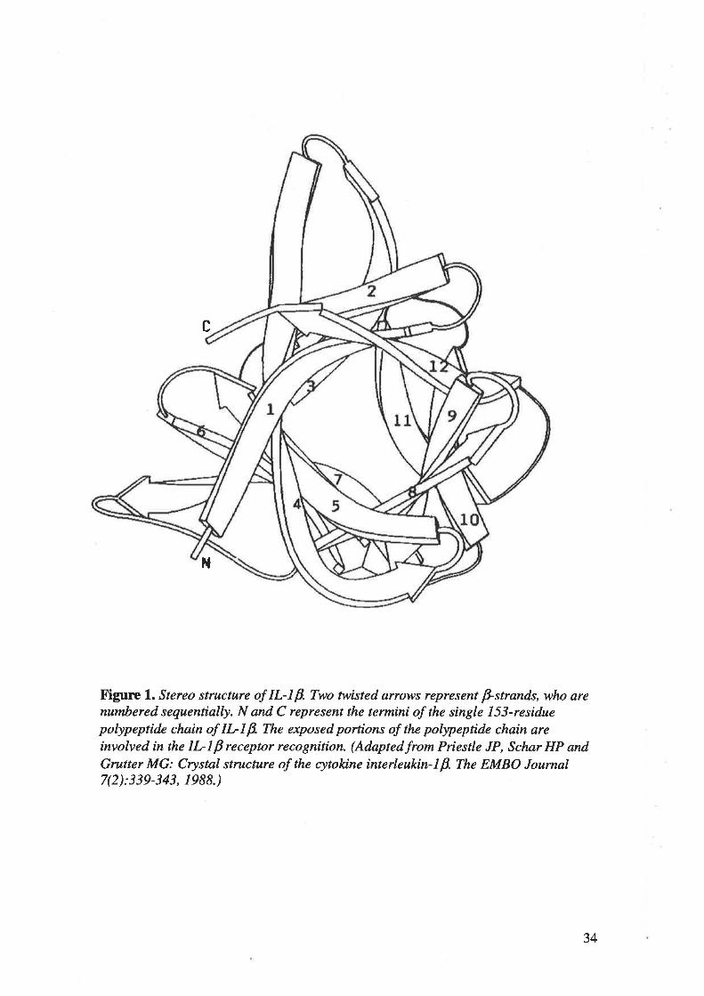
Human tr--lp is a single l53-residue pollpeptide chain, whose structure is composed of 12 p-
strands organizcd in a three-fold repeating motif (Figure 1). The core of the structure can best
be described as a tetrahedron whose 4 faces a¡e each made up of 3 p-strands, leaving only the
end supporting the chain termini fully exposed. The exposed portions of the pollpeptide chain
a¡e involved in the tr--lP receptor recognition 116'281'282'363.
33
c
1{
X'igurc l. Stereo structure of IL-I B. Two twisted arrows represent þstrands, who arenumbered sequentially. N and C represent the termini of the single Lí3-residuepolypeprtde chain of IL-Lp. The exposed portions of the polypeprtde chain areinvolved in the IL-lp receptor recognition. (Adaptedfrom Priestle JP, Schør HP andGrutter MG: Crystal structure of the cytokine interleukin-lp. The EMBO Joumal7(2):339-343, 1988.)
34
2.2.2rL-tþ GEI\E AI{D tr-lp BTOSYNTHESTS
The human tr-lp gene consists of 7 exorx¡ on the long arm of chromosome 2 at the locus
2ql3-2q2l6n.s.
Several potential transcriptional control elements, such as NFtcB, APl, CRE, NF-IL6 and
NFpA have been identified within the conserved region of the IL-lp promoter tes'1e8s2t342'3g,
as shown in Figure 2. These transcrþtional regulators are up-regulated in response to many
stimuli including in¡ry, microbial products (e.g., bacterial lipopolysaccaride), cytokines (IL-
lþ, IL-z, TNF, etc.), T celUantigen presenting cell interactions and immune complexes 88'8e.
For example, IL-IP nRNA levels in human monocytes rise rapidly within 15 min after
stimulation with bacterial lipopolysaccaride, but sta¡t to fall afte¡ 4h rla. The fall is caused by
the slmthesis of a transcriptional repressor and a decrease in nRNA half-life ls, suggesting
that the production of IL-1p is tightly regulated. This is further supported by in vivo studies
demonstrating a robust and transient increase of IL-lp mRNA expression that rapidly falls to
a low and constant level within 6 hr after CNS injury in rats te334t.
In addition to tightþ regulated transcription, this regulation also extends to translation of IL-
lP nRNA Stimulants such as complement component C5a 308 or h5perosmola¡ NaCl3te *ere
found to induce the slmthesis of large amounts of IL-lp mRNA in monocytic cells without
significant translation into the tr--lp protein. This dissociation between transcription and
translation suggests that the above stimuli a¡e not sufficient to provide a sþal for translation
despite a vigorous signal for transcription 301.
Following sSmthesis, the 31-kDa precrrsor of IL-lp remains c5rtosolic and only marginally
active until it is cleaved by IL-lp converting en4/Ine. The cleavage converts IL-lp precursor
35
to its 17 kDa mature form and then the mature tr-lp is secreted out of the cell 185. l1us, the
rebase of maturc tr-lp depends on cbavage by ILlp converting enz5mo.
Constitutive production of IL-lp is usually low or absent and its expression is tightly
regulated at the level of transcrþtion, translation and sec¡etion 88.
36
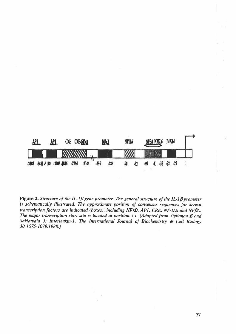
¡fl_ ¡[t clE 0Füt uü mu gry Tflil
.31ü .llil.3ll0 .3loj.ll{ú '276f ,1144 .l9J .186 .90 .lt 4 {l .ll 'll II I
X'igure 2. Structure of the IL- I P gene promoter. The general structure of the IL- I p promoteris schematically illustrated. The approximate position of consensus sequences for løoyvntranscription factors are indicated (boxes), including NFtß, APL, CRE, NF-IL6 and NFfr,.The major transcription start site is located at position +1. (Adapted from Stylianou E andSaklanala J: Interleukin-|. The Internartonal Journal of Biochemistry & Cell Biology30:1075-1079,1988.)
ai,{////t;,///////1tI I
37
2.2.3 TI-I RECEPTOR A¡TD SIGNAL TRAI\TSDUCTION
II-1 receptors are expressed in most cçlls, including neurons, endothelial cells, neutrophils, T
cells and B cells. tr'87. There a¡e two primary cell surface receptors for IL-lp. ILI t5'pe I
receptor transduces a signal whereas the type II receptor binds IL1P but does not transduce a
signat In fact, IL-l type II receptor acts as ¿ sink for IL-lp and has been termed a 'decoy'
receptor 74. A unique aspect of cytokine biology is a naturally occurring ILl receptor
antagonist, which is structurally similar to ILlp but lacking agonist activity 8e.
When IL-lp binds to IL-l type I receptor, a complex is formed which then binds to the IL-
lR-AcP resulting in high-atrnity binding 88. Although it remains unclear how IL-l t'lpe I
receptor triggers the phosphorylation and activation, it has be,en found that, within a few
minutes following binding to receptors, tr--lp induces intracellula¡ muþle protein
phosphorylations and activation of phosphatases via the receptor, resulting in cascades of
activation of protein kinases 42'342.
One main cascade leads to phosphorylation of h<B (ie. NF-rcB inhibitory factor), which is
rapidþ degraded by the proteasome following its phosphorylation. Degradation of IrB
activates NF-KB and releases it from the cytoplasmic NFKB-IKB complex t6. Theo, NF-KB
translocates to the nucleus and binds to its DNA recognition site in the gene promoter to up-
regulate the transcription of genes saçsding pro-inflammatory cytokines (e.9., L-1,2, 6,8,
TNF, etc.), adhesion molecules (e.g., VCAIvI-1), chemokines, growth factors (e.g., CSF),
inducible enz)mes (e.g., COX-2, iNOS, etc.), anti-apoptotic enzymes (e.g., Mn-SOD) and
growth proteins (e.g., c-myc) to'ts6'21s236'3s7 .
38
The other main cascacle is the activation of three MAP kinases, which is a complicated
pathway of phosphorylation of multþb protein kineses and is still poorþ understood s2. This
signal transduction pathway mainly bads to the activation of cytoplasmic AP-l (a dimer
composed of va¡ious Fos and Jun family proteins), which translocates to the nucleus and
binds to its DNA recognition siæ in the gene promoter to activate the transcription of many
Senes encoding proteolytic enzymes (e.g., MMPs), growth factors (e.g., NGF), growth protein
(e.g., GAP-43), p-APP, galanin, neurofilament proteins and a-tubulin s3,167,r782363@3e4.
Thus, tr--lp exerts most of the biological effects on cells through the activation of NF-lcB and
AP-l, two maje¡ proinflammtory transcription factors cornmon to many ll.-l-induced genes
88.
An unusual discrepancy has been noted that low concentrations of IL-lp can induce a potent
biologic response via a low number of receptors (fewer than 10-100 in some cells) 1262ø-,
suggesting the efficiency of so few IL-l tpe I receptors and the greatly amplified
transduction of the post-receptor signals.
2.2.4BtOLOcrC EITECTS Or IL-lp rN SCr
L-lP affects nearly every cell qpe, often in concert with other cytokines or small mediator
nolecules, e.9., TNF-c¡, as both tr--lÞ and TNF-a are potent activators of the transcription
factor NF-KB es3zl. t-tp is a highly inflammatory cytokine and the margin between clinical
benefit and unacceptable toxicity in humans is na¡row 8e. It has been generally accepted that
low concentrations of IL-lp protect neurons and are neurotrophic, whereas higher
concentrations are neurotoxic taz$.
39
Signfüantly increased tr-lÞ mRNA and protein expressions have been found in the cord
after experimental SCJ 1e'164'1e22t63413s7380. Some recent studies have demonstrated that
within hours following SCI, activated microglia within the cord express tr-lp, which
activates the endogenous glial cells 85 and stimulates the up-regulation of endothelial adhesion
molecules to enhance leukocyte recruitment (e.g., ICAM-I and VCAM-I) te'tl,'t7ei4t. These
findings suggesting that this early slmthesis of tr-lp may be an important signal for initiating
a progressive inflammatory response within the spinal çs¡d.
The role that the increased tr--1p plays in mediating secondary SCI is still controversial
Many in vitro and vivo studies demonstrated that IL-IP m¿y be neurotoxic. For example, it
has been demonstrated by in vitro investigations that tr--lp is toxic to neurons 67. Moreover,
in vivo studies have suggested that tr-lp may contribute to tissue destruction, increased
vascula¡ permeability and oedema formation as it up-regulates MMP expression via the
activation of AP-l, a major proinflamrnatory transcription factor t7s3e43es. MMP-I plays a
major role in tissue destruction in inflammation and MMP-9 degrades the extracellula¡ matrix
component of basement membrane leading to the loss of vascular integrity '*. n-tp is also
implicated in the up-regulation of iNOS and COX-2 via, the activation of NF-KB, another
major proinflammatory transcription factor 10, resulting in an increase in NO and ROS
production, and a TXAz-PGIz imbalance, respectively 6essz . An increased production of free
radicals causes secondary tissue damage and an imbalanced production between TXAz and
PGIz is involved in the microvascular thromboembolism and the secondary ischaemia
following SCI. Additionally, II--lp may stimulate formation of the astroglial ,"* ttt2tt,
which is thought to be one element inhibiting the regeneration of injured ðrolts through the
lesion. Recentl¡ Holmin and Mathiereo tt5 found that intracerebral administration of IL-IP
induces infiltration of inflammatory cells and apoptosis of intrinsic CNS cells. They also
n
found that ILIP caus€s higher expression bvel of the pro-apoptotic Bax gene than the anti-
apoptotic Bcl"2 gene. The most direct evilcnce implbating tr-lp in secondarSr SCI derives
from experiments that employed a recombinant ILI receptor antagonist. Systemic inþtions
of ILI receptor antagonist reduces neuronal damage, prevents apoptosis and improved
cognitive firnction in rodents after CNS injury 1222232s73o2360. Furthermore, the administration
of exogenous IL-lp markcdly exacerbates ischaemic or excitotoxic CNS injury 2ff300.
However, there a¡e instances where tr-lp appears to act as a neurotrophic factor. In support
o¡ ttis, tr--lp attenuated the increased neuronal death, which was induced by adding an
antiserum to IL-14 into dissociated mouse spinal cord cultures 4e. L-lp could produce a
neurotrophic effect indireÆtly via induction of some growth ,*,or., such as NGF and FGE in
astrocytes 2u,247. Additionall¡ tr--lp can stimulate blood vessel growth 138, which is essential
for the continued survival of tissue spared by the injury as well as any tissue ¡epair that may
contribute to functional recovery after SCI, which causes disruption of blood vessels and
consequent ischaemia.
Thus, L-lP is an important and modifiable component in the inflammatory rcsponse
following SCI and has complex and bi-directional biological efrects on the injured spinal
cord.
4l
2.311-6
2.3.1STRUCTI]RE
Human IL-6 is a glycoprotein with a mobcular rnass ranging from 2l to 28 l¡Da and consists
of 212 amino acids including a hydrophohic signal sequence of 28 amino acids 60345. The
structure is composed of four (4, g, C, D) helical bundles oriented in an 'up-up-down-down'
configuration and throe siæs a¡e involved in receptor interaction: site I is the binding site for
II-6 rcceptor o chain and sites 2 and 3 a¡e those for gp130 (IL-6 receptor p chain) (Figure 3)
269
42
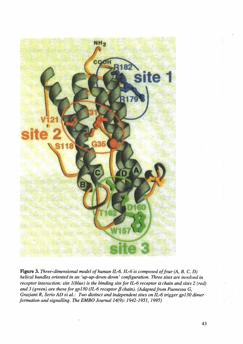
Figure 3. Three-dimensional model of human IL-6. IL-6 is composed of four (A, B, C, D)helical bundles oriented in an 'up-up-down-down' configuration. Three sites are involved inreceptor interaction: site l(blue) is the binding site for IL-6 receptor a chain and sites 2 (red)and 3 (green) are those for gp130 (IL-6 receptor p chain). (Adaptedfrom Paonessa G,Graziani R, Serio AD et al.: Two distinct and independent sites on IL-6 trigger gp130 dimerformation and signalling. The EMBO Journal 14(9): 1942-1951, 1995)
43
2.3.2TL4 GEI\E AI\D II,.6 BIOSYNTHESIS
The human IL6 gene is approximaæly 5 kilobases in bngth and consists of 5 exolls and 4
introns on the short arm of chromosome 7 at the locus 7p14-7p2160't7zl+s.
Within the conserved region of the IL6 promoter, there are several potential transcriptional
factors such as an NF-rcB binding site, a C-Fos SRE, a CRE, an AP-l binding site and GRE
t722ts'345. Three DNA sequences in the IL-6 promoter are identified as the main functional
transcriptional factors as shown in the figure: a 23-bp MRE, a l4-.bp NF-IL6 and an NF-KB
binding site (Figure 4). Additionally, the proposed stimuli to activate these three main
functional transcriptional factors are also shown in the Figure 4, including IL-l, TNF,L-6,
'LPS and serum.
The production of IL-6 is induced by a variety of stimuli, such as inj".y ts7'te72m'3ts'3s4365'388,
pro-inflammatory cytokines (e.g., IL-l, TNF,L-2, FI{-P etc.) 210'3053t', gro*th factors (e.g.,
PDGÐ tt', IPS 21036e and va¡ious viruses while its production is inhib,ited by glucocorticoids
tr6, L-4 133 orrd n--13 244.
Constitutive production of IL-6 is usually low or absent and its expression is tightly kept in
check by a complex network, including sex steroids, o.g., estrogen and testosterone3T2.
4
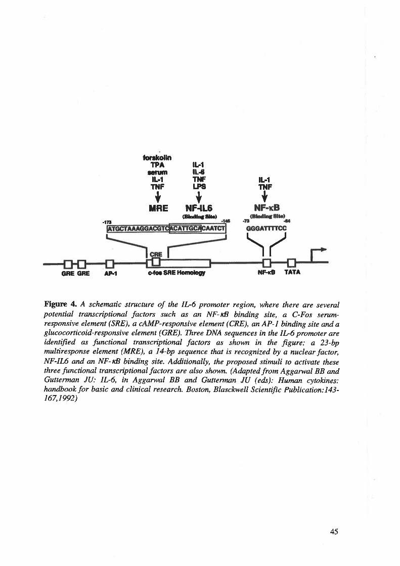
forrl¡ol[tTPA
aürmtL-lT]IF
TRE
t1|rtL{T}IFr"Fs
*NF-lL6
oru¡ç¡gftr)
\/
tbrTilF
t*
.ttt
GßE GFE AFtf o-lo. SRE ltotttologt llÊô 1A1A
Figure 4. A schematic structure of the IL-6 promoter region, where there are severølpotenrtd transcriptional factors such as an NF-IB binding site, a C-Fos serum-responsive eletnent (SRE), a cAMP-responsive element (CRE), an AP-1 binding site and aglucocorticoid-responsive element (GRE). Three DNA sequences in the IL6 profitoter areidentifted as functional transcriptional factors as shown in the ftgure: a 23-bpmulrtresponse element (MRE), a l4-bp sequence that is recognized by a nuclear factor,NF-II'6 and an NF-IB binding site. Additionally, the proposed stimuli to activate thesethree functional transcriptional factors are also shown. (Adnpted frorn Aggarwal BB andGutterman JU: IL-6, in Aggarwal BB and Gutterman JU (eds): Human cytokines:handbookfor basic and clinical research. Boston, Blasch,vell Scientiftc Publication:L43-167,1992)
45
2.33TL.6 RECEPTOR AIIID SIGNAL TRANSDUCTION
IL6 receptor is expressed on a wide va¡iety of cells, including neuronsr glial cells,
monocytes, fibroblasts, T cells and activated B cells owas, II-6 receptor consists of two
molecubs, one is an 80-kDa IL-6 binding protein (a chain) and the other a 130-kDa binding
protein, gp130 (Þ chain) wirh277-ammo-acid residues in its cytoplasmic domain 17226e. Thus,
IL6 receptor o chain and gp 130 (tr-6 receptor p chain) form a high-atrnity IL6 binding
site. The cytoplasmic domain of IL6 receptor a chain is not required for the ll-6-mediated
signal transduction.
The binding of IL-6 to its receptor induces the formation of a hexamer composed of two each
of IL-6, IL-6 receptor o chain and g¡l 130 26e, resulting in tyrosine phosphorylation of the
cytoplasmic domain of gp130 ã6. Then, intracellular signalling molecules (e.g., STAT)
specifically recognize and bind to the phosphorylated receptors (gp 130). Once bound to the
phosphorylated receptors, these signalling molecules themselves become phosphorylated by
receptor-associated kin¿sss, enabling them to disengage from the receptors (gp 130) and
interact with additional signalling molecules to form the intracellular IL-6 signal transduction
pathway. The IL-6 srgnal transduction pathway mainly consists of the JAK-STAT and Ras-
MAP kinase pathways t2o2s6.
JAK (named after Janus, the Roman god with two faces) a¡e gp l3O-associated kinases and
become activated by cross-phosphorylation after Il-6-induced phosphorylation of the gp130.
Although the JAK-STAT pathway is still not well understood, it is clear that JAK is critical
for the activation of a specific sþalling pathway, STAT t72'2s6. Once phosphorylated by
JAK STAT molecules disengage from the receptor (gI, 130) and translocate to the nucleus,
where they bind to specific DNA motifs in the gene promoters and activate or inhibit the
46
transcrþtion of these gones encoding anti-apoptotic proteins (e.g.,tr)l-2, pim-l, etc.), growth
proteins (e.9., c-þn, c-myc, c-myb, etc.) and acute-phase proteins (e.g., cRp) 172,173322.
In addition to the JAK-STAT signal tranduction pathwa¡ the other main pathway is the
activation of Ras-MAP kinases through SHP-2. This cascade of kinase reactions lead to the
phosphorylation and activation of NF-rB and ERIG 1722Ú'2s6. Once activated, tle NF-rB and
ERIß translocate to the nucleus to phosphorylate their corresponding transcription factors,
which up-regulaæ the expression of pro-inflarnmatory cytokines, adhesion molecules and
growth proteins to'ts62s6 .
2.3.4BIOLOGIC EFTECTS Or' IL-6 IN SCI
n-6, a multifunctional cytokine, is known to play an important role in a variety of CNS
functions such as cell-to-cell signaling coordination of inflammatory rcsponse, protection of
nerrons from insult, as well as neuronal and glial differentiation, growth and survival
147'173'1e734s, Íuny of which overlap with those of IL-lp and TNF-cr. It has been shown that a
variety of stimuli, including injury, viruses and cytokines (e.g., L-lP and TNF-a), can trigger
IL-6 production within the CNS 381 and IL-6 receptors are present in neurons and
o ligodendro cy4¡;s t7 2 2ro36eJ I 8.
Many experimental studies have demonstrated that IL-6 ¡RNA and protein are elevated in
the cord following SCI 164'216'341. It has been recentþ found that within hours following SCI,
IL-6 protein is expressed by neurons and activated microglia within the cord 1ú.
The functional significance of IL-6 in the CNS is still hotly debated. It has been observed that
IL-6 induces neurite outgrowth of neuronal cells in vitro 180, promotes oligodendrocyte
47
survival in vitro 18 and improves the survival of cultured rat cholinergic neurons with or
without initiating the slmthesis of neurotrophic factors 147'ts3'ts4't57228. Additionall¡ in vivo
studies have demonstrated that local adminisf¡¿1ion of IL6 attenuates the neurotoxic effects
of NMDA on rat striatal cholinergic nerrrons 3ól and prevents degeneration of the spinal
motoneurons of wobblermice 18r.
Conversely, it has been found in experimental SCI that anti.rat IL6 antibody significantþ
inhibits the up-regulation of iNos and reduces ultrastructural damages in the myelinated
nerye fibres to attenuate secondary spinal cord damag" 'ut. Additionally, a severe neurologic
s5mdrome with extensive neuropathologic changes (e.g., neuronal damage and reactive
astrocytosis) was induced in transgenic mice with cerebral overexpression of ¡--6 Ø.
Moreover, the clinical features and neuropathology in the transgenic mice correlated with the
level and spatial distribution of IL-6 overexpression.
Thus, IL-6 is both a neurotrophic and cytotoxic factor and is pa¡tially regulated by IL-lp and
TNF-q 375
48
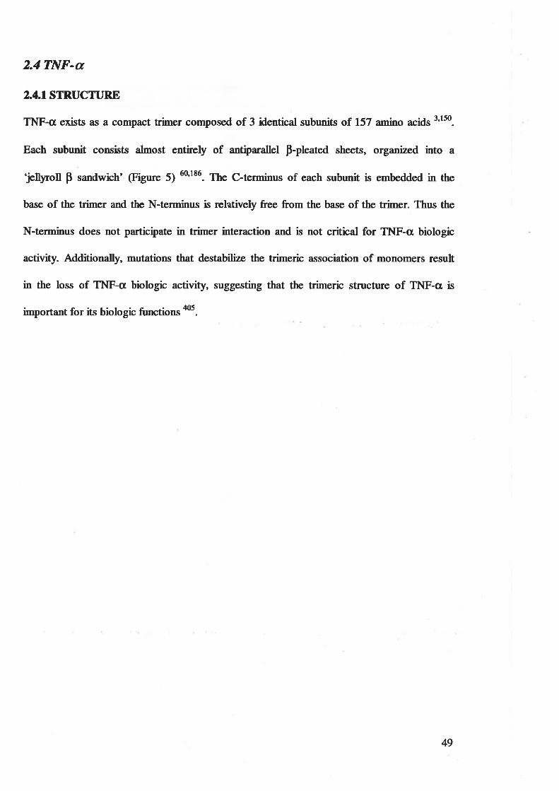
2.4 TNF-u
2.4.1STRUCTI]RE
TNF-cr exists as¡ a compact trimer composed of 3 identical subunits of 157 amino acids 3'150.
Each subunit consists almost entirely of antþarallel p-pleated sheets, organized into a
þltyroll p sandwich' @igure 5¡ 0o't40. The C-terminus of each subunit is embedded in the
base of the trimer and the N-terminus is rehtively free from the base of the trimer. Thus the
N-terminus does not participate in trimer interaction and is not critical for TNF-a biologic
activity. Additionall¡ mutations that destabilize the himeric association of monomers result
in the loss of TNF-a biologic activity, suggesting that the trimeric structure of TNF-o is
important for its biologic functions æ5.
49
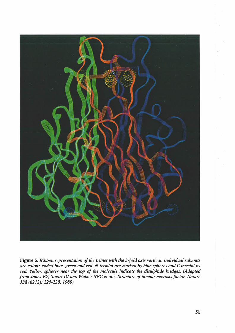
Figure 5. Ribbon representation of the trimer with the 3-fold axis verrtcaL Individual subunitsare colour-coded blue, green and red. N-termini are mnrked by blue spheres and C termini byred. Yellow spheres near the top of the molecule indicate the disulphide bridges. (Adapted
from Jones EY, Stuart DI andWalker NPC et al.: Structure of tumour necrosis factor. Nature338 (6212): 225-228, 1989)
50
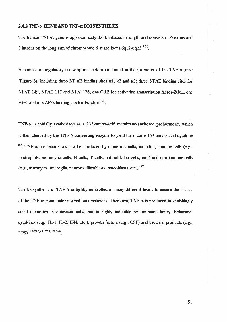
2.4.2 TNF-q GENE AND TIt{F-ct BIOSYNTIIESIS
The human TNF-cr gene is approximately 3.6 kilobases in length and consists of 6 exons and
3 introns on the long arm of chromosome 6 at the locus 6q12-6q233'6o.
A number of regulatory transcription factors are found in the promoter of the TNF-cr gene
(Figure 6), including three NF-KB binding sites 11, tO and 13; three NFAT binding sites for
NFAT-149, NFAT-I17 and NFAT-76; one CRE for activation transcription factor-2lJun, one
AP-l and one AP-2 binding site for Fos/Jun 405.
TNF-o is initially synthesized as a 233-amino-acid membrane-anchored prohormone, which
is then cleaved by the TNF-a converting enzyme to yield the mature l57-arrino-acid cytokine
60. TNF-cr has been shown to be produced by numerous cells, including immune cells (e.g.,
neutrophils, monocytic cells, B cells, T cells, natural killer cells, etc.) and non-immune cells
(e.g., astroc5rtes, microglia, neurons, fibroblasts, osteoblasts, etc.) aOs.
The bioslmthesis of TNF-a is tightly controlled at many different levels to ensure the silence
of the TNF-c gene under normal circumstances. Therefore, TNF-cl is produced in vanishingly
small quantities in quiescent cells, but is highly inducible by traumatic injury, ischaemia,
cytokines (e.g., IL-l, IL-2, IFN, etc.), growth factors (e.g., CSF) and bacterial products (e.g.,
I pS¡ 208,2t0,237,2s8,37e,3e6 .
51
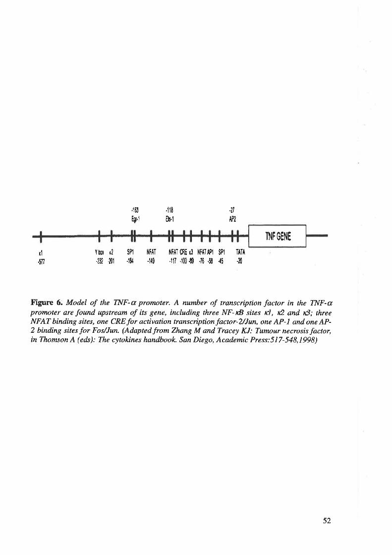
-21
AP2
.1 10
Ëþr
.1ü
É9r.1
TNF GENE
r1
.$ll
YM Í2 SP1
{32 .¿01 .164
t{tAT I'IFAI CRE rl i{FATAPI SPt TATA
.149 .t17.100$ .16.$ {5 .20
Figure 6. Model of the TNF-a promoter. A number of transcription factor in the TNF-apromoter are found upstream of its gene, including three NF-tß sites tcl, t2, and t6; threeNFAT binding sites, one CRE for activation transcription factor-2/Jun, one AP-1 and one AP-2 binding sites for Fos/Jun. (Adapted from Zhang M and Tracey KJ: Tumour necrosis factor,in Thomson A (eds): The cytokines handbook. San Diego, Academic Press:517-548,1998)
52
2.4.3 TNX'-o RECEPTOR AI\ID SIGNAL TRANSDUCTION
TNF-cr receptors are present on nearly all cell types with a few exceptions such as
erythrocytes and resting T cells. There are two distinct but structurally homologous TNF-cr
receptors, tlpe I and tlpe II with molecula¡ masses of 60 and 80 kDa, respectively. TNFRI is
found on most cell tlpes whereas TNFR2 is more restricted to endothelial and haematopoietic
cells óo'40s.
It is generally believed that TNFR1 is responsible for the majority of biologic actions of TNF-
s while sþal transduction through TNFR2 occurs less extensively and is confined mainly to
cells of the immune system. Thus, the section below concentrates on the signal transduction
through TNFRI.
Three intracellular functional domains of TNFRI, io., death ¡le1¡ein, NSD and ASD, are
responsible for transferring signals from TNF-c to their corresponding intracellular adaptor
proteins, TRADD, FAN, PC-PIJC, via protein-protein interactions.
Once TRADD has bound to the TNFRI death domain, it causes recruitment and activation of
FADD and RIP through the C-terminal death domain and recruitment and activation of
TRAF-2 through the N-tenninal death domain. Activated FADD and RIP then bind and
activate caspase-2 and caspase-8, respectively. These two caspases initiate a protease cascade,
resulting in the cleaving of death substrates and activation of death protein (e.g., Par-4) to
induce apoptosis 84'31u05. Meanwhile, activated TRAF-2 triggered activations of MEKK and
JNK lead to the activation NF-KB and AP-l to mediate the inflammatory response, by
induction of pro-inflammatory cytokines (e.9., IL-l,2,6, 8, TNF, etc.), adhesion molecules
(e.g., VCAM-l), inducible enz¡anes (e.g., COX-2, NOS, etc.), chemokines 10'156'zts'236'357, and.
53
anti-apoptosis molecules (e.g., Mn-SOD, CSF, etc.) t7t'2æ'37o4os. Thus, the TNFR1 death
domain mediates both apoptotic and anti-apoptotic pathways.
FAN links the NSD of TNFRI directþ to neutral sphingomyelinase, which is then activated
to mediate cell proliferation and the inflammatory response through MAP kinase ERK and
PLA-2, respectively 17 o¡os .
Once PC-PLC is activated, |,2-dncylglycerol is produced by PC-PLC and activates acidic
sphingomyelin¿5s, which converts lipid sphingomyelin to ceramide. The rapidly raised
intracellular level of ceramide acts as a secondary messenger, leading to NF-I<B activation to
mediate anti-apoptosis and the inflammatory respons e 37 04os .
Thus, the downstream pathways of these three adaptor proteins result in va¡ious cell activities,
including apoptosis, anti-apoptosis, inflammatory response and cell proliferation.
2.4.4B]0LOGIC EFT'ECTS OF TNF-o IN SCI
The presence of TNF-cr has been reported in various cells in the CNS, including microglia,
astrocytes and neurons 68 and these cells also express high-affinity TNF-a receptors 32e. It i.
generally believed that high levels of TNF-ct are injurious, but low levels can be beneficial
362,405
TNF-cr shares some similar properties with tr--1p in terms of pro-inflammatory effects, such
as activation of endogenous glial cells '66, prolif"ration and hypertrophy of astrocytos 37e,
enhanced pemreability of endothelial cells and enhanced recruitment of blood leucocytes 348.
54
This may be due to the fact that both TNF-cr and tr-lp are potent activators of NF-rcB, a
major pro-inflamrnatory transcription factor 10.
Many experimental studies have demonstrated that TNF-c mRNA and protein a¡e elevated in
the cord following SCI 1e'164'2o8'216'341'17e'3e3. It has been recently found that within hours
following SCI, TNF-cr protein is expressed by neurons and activated microglia within thc
cord teaSe6.
The functional significance of TNF-cr in the SCI is still not well understood. Some studies
have demonstrated cytotoxic roles of TNF-ø. It has been shown that TNF-a has a direct toxic
effect on oligodendrocytes in vitro ea and a single micro-inþction of TNF-c caused
demyelination in the spinal cord 325 and in the sciatic nerve 2e0. Furthermore, in transgenic
mice overexpressing TNF-o, a spontaneous inflammatory demyelination in the brain and
spinal cord occurred and was partially prevented by exogenously delivered neutralizing
antibodies to TNF-cr 283. In addition to the toxic effect on oligodendrocytes, TNF-cr can
induce apoptotic changes in cultured neurons and glial ceils 238'346'370 and cause BBB injury
with damage to endothelial cells 80. In support of cytotoxic roles of TNF-c, some in vivo
studies have also demonstrated that following CNS itjury, inhibition of TNF-cr by activated
protein C, IL-10 or a neutrahzng antibody significantþ improved neurological outcome
28,205,349
In contrast to its toxic actions, TNF-ø has also been demonstrated to induce IL-6, NGF and
other growth factors in astrocytes 305'3ee and exert a regenerative effect oo axons 313.
Additionally, TNF-cr prevents glutamate-induced cell death and cytotoxic effects of amyloid
p-peptides and iron by regulating intracellula¡ calcium levels and suppressing the
55
accumulation of ROS in pure hippocampal cultures 16'68. Attother important neurotrophic
effect of TNF-a is the induction of angiogenesis 213. Additionally, improved recovery of
motor function was observed in NOS-deficient, but not in TNF-cr-deficient mice after SCI 111.
Furthennore, a study using TNFR knockout mice (both TNFRI and TNFR2 deleted) has
shown that neurons from the TNFR knockout mice exhibit reduced survival in culture
compared with those from wild-tlpe mice and doses of kainic acid that cause minimal
damage in wild-type mice produce extensive neuronal loss in TNFR knockout mice 5s.
It has been recentþ proposed that the neurotrophic and neuroprotective effects of TNF-o may
depend on the ratio of TNFR1 to TNFR2 levels 26. One possibility is that higher levels of
TNFR2 may contribute to a cytoprotective response and increased levels of TNFR1 may
induce a cytotoxic response lo2.
In surnmary, TNF-a is a powerfrrl inflammatory mediator with both cytotoxic and
neurotrophic actions.
56
3. MICROGLIA
Microglia represent 5-l2%o of the cells in a given brain region 387 and these resident cells
comprise about ap to 2OVo of the total glial cell population 25. It has been shown that microglia
form an extremely stable cell population with little turnover and with less than lVo bone
rnanow precursor cells 85'170.
3.1 NOMENCIATURE
del Rio-Hortega in 1919 first used the term microglia to describe a type of glial cell that could
be differentiated from neurons, astrocytes and oligodendrocytes by its distinctive morphology
with silver impregnation techniques 8t. The nature and identity of microglial cells have
triggered much debate and resea¡ch since their discovery. However, it is now generally
accepted that microglia are ubiquitous, mesodermally derived glial cells of cha¡acteristic
morphology and phenotlpe which are evenly distributed throughout the central nervous
system, and whose functions are primarily related to phagocytosis, antþen presentation,
cytotoxicity and neuronal survival338. The term "macrophage" is used to refer to a monocyte-
derived phagocyte in the parenchyma of CNS e2. The distinction is complicated by the fact
that macrophages a¡e derived, at least in part, from intrinsic microglia in the CNS and that it
is technically difficult to distinguish between phagocytic microglia and monocyte-derived
macrophages 1r7J3e. Thus, microglia are also called as "resident macrophages of the CNS" 25.
3.2 CIA,SSIFICATION
In view of their morphological and functional characteristics in the mature CNS, three
different tlpes of microglia can be distinguished 25'33e:
1. the ramified (resting) microglia with long and branched processes, found
predominantly in the white matter of normal mature CNS;
57
2. the ameboid (activated), but non-phagocytic microglia with cellular hlpertroph¡ and
shortened and thickened processes, in response to sublethal injory without causing
terminal degeneration and necrosis such as follows nerve transection; and
3. the foamy (phagocytic) microglia in a¡eas of necrosis produced by varied mechanism
including neurotrauma.
Figure 7 demonstrates the progressive transforrnation in the morphological appearance
between ramified (resting) microglia and ameboid (activated) microglia.
Although neither the a¡rreboid nor ramified shapes may be reliable indicators of the functional
state of microglia, rarrified microglia are often referred to as "resting" microglia and ameboid
microglia as "activated" microglia in the mature CNS. However, there is no consensus as to
what constitutes the best marker for microglial activation, especially in situ 8s.
58
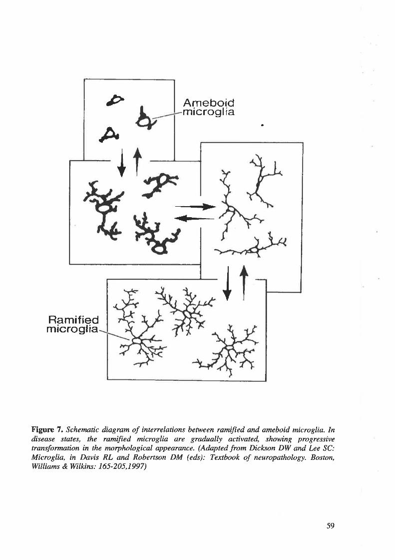
Þ
þ
+
+
Ameboidmicroglia
a
Ramifiedmicroglia
Figulrc 7. Schematic diagram of intenelations between ramified and ameboid microglia. Indisease states, the ramified microglia are gradually activated, showing progressivetransþrmarton in the morphological appearance. (Adapted from Dicl<son DW and Lee SC:Microglia, in Davis RL and Robertson DM (eds): Textbook of neuropathology. Boston,Williams & Wilkins : I 65 -205, I 997 )
59
3. 3 MICROGLTAL ACTIVATI ON
3.3.1 RESTING MICROGLIA
Resting microglia with numerous fine antler-like processes show a highly down-regulated
immunophenotlpe and are in a functionally quiescent state 272'13e. The cell surface antigens on
resting microglia in the physiologically quiescent state include complement receptor type 3
and Fc, but not MHC I, II and CD4 382. Microglia can most rapidly rcspond to injury in the
CNS. They respond not only to changes in the structural integnty of CNS but also to very
subtle alterations in their microenvironment, such as imbalances in ion homeostasis that
precede pathological changes detectable histologically ttn. Thus, microglial activation often
precedes reactions of any other cell type in the CNS 2m.
3.3.2 ACTIVATED MICROGLIA
To define stages of microglia activation in vivo, the model of facial nerve axotomy, which
leaves the BBB unimpaired, wff used as it allows the study of the activation of microglia in
the absence of infiltrating haematogenous macrophages 33e. In that study, the facial nerve was
cut outside the brain and the reactions of facial motoneurons and their glial environment were
studied in the brainstem. Following facial nerve transection, ramified microglia proliferated,
became ameboid with cellular hlpertrophy and shortened processes, and expressed several
new surface antigens, including MHC I, II and CD433e.It has also been noted that microglia
in a perineuronal position started to ensheath the injured motor neurons and interpose
processes between afferent synaptic terminals and the neuronal surface. The phenomenon,
originally described by Blinzinger and Kreutzberg ", i. oo* known as "synaptic stripping",
and its functional significance is not well understood. It has been suggested that detachment
of synaptic terminals from the motor neuron surface may influence synaptic reorganization of
injured motor neurons '5. Itt addition, APP is newly expressed by activated perineuronal
60
microglia 12. But, again, the significance of the presence of APP in activated microglia
rem¿ins enigmatic.
It has also been observed that, upon cellular activation, microglia are capable of producing
large quantities of a number of neurotoxic mediators, including EAAs, proteases, cytokines
and NO 2ffi'387. Details will be discussed in section 3.4.
3.3.3 PHAGOCYTIC MICROGIA
Under the condition of facial nerve axotom¡ microglia become activated but not phagocytic.
However, if motor neuron death is induced by inþction of toxic ricin into the facial nerve,
microglia rapidly transform into phagocytic microgliu too, and graduaþ migrated into the
injury site to remove the neuronal cell debris. In addition to complement receptor t5pe 3, Fc,
CD4, MHC I and II, these microglia-derived macrophages also express the EDI epitope
which is otherwise t¡pical of peripheral macrophages 25. Thor, it has to be stressed that it is
technically difficult to distinguish between intrinsic phagocytic microglia and monocyte-
derived macrophage s 2s'tt7'31e'382
.
3.3.4 ST]MMARY
In sunnnary, the microglial response to injury occurs in a rather stereotlpic pattern,
irrespective of the cause of the lesion 128'33e. Results obtained from the facial-nerve transection
model show that activation of microglia displays a repertoire in terms of proliferation,
migration to the site of injury, cha¡acteristic morphological, immunophenotlpical and
functional changes, and that microglial activation takes place in a graded fashion. In the first
stage of activation, microglia become activated, but do not become phagocytic. In the second
61
stage of activation, following neuronal and/or axonal terminal degeneration, microglia
transform into phagocytes, also known as microglia-derived brain macrophages.
Thus, the activation process is a graded event that is dependent on the severity of the insult
and is most probably regulated by specific signals, e.g., cytokines, in a well-controlled
mannef L28
3.4 FUNCTIONS OF MICROGLIA IN SCI
Functional significance of activated microglia in SCI is still poorly understood. Microglia
have shown both cytotoxic and neurotrophic effects on the injured tissue, suggesting a
possible dual-edged role 25.
3.4.1 CYTOTOXICITY
Many studies have demonstrated a swift activation of microglia following SCI 323'341'382.
There are two principal ways by which microglia may act as cytotoxic cells.
First, phagocytic microglia express Fc and complement receptors on their surfaces 85. In
particular, during antibody-mediated demyelination, activated microglia can lyse antibody-
coated target cells via interaction of Fc and complement receptors on their surfaces with
immune complexes and complement-opsonized antigens as.
Second, activated microglia are capable of releasing several potentially cytotoxic substances
in vitro such as cytokines (e.g., TNF-o, IL-IP and IL-6), free oxygen intermediates, NO,
proteinases, arachidonic acid derivates and EAAs t2'7s'242'323'355'387. This is further supported
by findings in animal models of CNS trauma that at the lesion site activated microglia have
62
been shown by microdialysis measurements and in situ hybridisation to release high levels of
cytokines (e.g., ILl) within 24 h following injury 388. Sh.rman et aL 323 observed microglia
with an activated morphology in the same regions as oligodendrocyte apoptosis and apparent
contact between some of the apoptotic oligodendrocytes and microglial processes after spinal
cord contusion in the rat, suggesting microglia-produced TNF-o may cause oligodendrocyte
apoptosis. Microglia-produced TNF-cr may also cause direct damage to myelin, furthering
demyelination 317. Additionally, Combs et al. 75 demonstrated in a tissue culture model that
conditioned media from p-amyloid- and prion- stimulated microglia are neurotoúc to mouse
neurons.
Interestingly, the cytotoxic properties of microglia can be modulated by c5rtokines,
glucocorticoids and neurotransmitters 224. Cytokines such as IFN1 prime microglia to become
activated whereas other cytokines such as TGFP1 or IJ'-4 downregulate microglial
cytotoxicity ^t. Neuronal activity may also control microglial activation. p-adrenergic
agonists like isoproterenol have been shown to reduce IL-1 and TNF-c production by
microglia 2m. Additionall¡ glucocorticoids, such as dexamethasone and corticosterone, are
also potent inhibitors of microglial activation 224. Furthermore, potent inhibitors of
macrophage/microglial activation have been shown to reduce the extent of tissue damage and
improve the motor function after ischaemic inj"ry in the rabbit spinal cord 137.
However, most of the information on the cytotoxic properties of activated microglia pertains
to in vitro observations and still remains to be confirmed in vivo.
3.4.2 I\EUROTROPHY
63
Although activated microglia are generally regarded as cytotoxic effector cells, they also exert
neurotrophic effects on the injured tissue to promote tissue repair and regeneration after
injory. For example, TGFpI produced by activated microglia after CNS trauma or after
ischaemic injory may either promote tissue repair directly or indirectly by reducing astrocytic
scar formatioî2te'386'388. Microglia secrete both the urokinase-type plasminogen activator and
plasminogen, which have been shown to promote neurite outgrowth 2s3'2s4. Microglia-secreted
plasminogen may also be involved in tissue remodelling after injury 25. In fact, tissue
remodelling by activated microglia precedes the regeneration of optic nerve of fish or
amphibia which, unlike the mammalian CNS, has a high capacity for regeneration, 2o,
suggesting microglial activation may support regeneration. Furthermore, a recent study has
demonstrated that transplantation of activated macrophage/mciroglial cells can significantly
promote the regeneration of sensory axons into the injured spinal cord in the rat 280.
It is also reasonable to assume that following injury, immediately activated microglia release
mediators such as IL-l, which may induce astrocyte activation 110, âs IL-l is capable of
inducing h¡perplasia and hlpertrophy of astrocytes when injected intracerebrally 138. Reactive
astrocfes may, in turn, contribute to CNS regeneration by secreting neurotrophic factors such
¿ts neurotrophins or insulin-like growth factors, which promote neuronal and/or
oligodendrocyte survival 130.
64
2.
3.
AIMS AI\D HYPOTHESES
ITYPOTHESES
1. The pro-inflammatory cytokines IL-lp, IL-6 and TNF-o act as messengers to
coordinate the inflammatory cascade in secondary SCI.
The cytokine response should be greater in severe than in mild SCI.
The NF-tcB antisense should significantly inhibit the upregulation of IL-lp, IL-6 and
AIMS
To investigate early posttraumatic inflammatory cellular response in rat and human
SCIs.
To demonstrate and compare temporal mRNA expression patterns for IL-lß, IL-6 and
TNFo in mild and severe rat SCIs.
To demonstrate and compare temporal protein expression patterns for IL-lß, IL-6 and
TNFo in mild and severe rat SCIs.
To compare temporal mRNA and protein expression patterns for IL-lß, IL-6 and
TNFa in severe rat SCI with the patterns in rats treated with the NF-KB antisense after
SCI.
To localize cellular sources of interleukin-lp G,-lp), interleukin-6 (IL-6) and tumour
necrosis factor-cr (TNF-a) in rat and human SCI.
TNF-a after SCI byblocking the activation of NF-tcB.
1
2.
3.
4.
5
65
RAT SCI EXPERIMENTS
1. MATERIALS AI\D GEI\ERAL METHODS
I.I ANIMAI.S USED AND ETHICS APPROVAL
All the experimental procedures were approved by the Animal Ethics Committees of the
Institute of Medical and Veterinary Science and the University of Adelaide. One hundred and
twenty-six male Sprague-Dawley rats were used in the experiments and were supplied by and
housed in the Institute of Medical and Veterinary Science (Table 2). Prior to surgery, rats
were kept in cages in groups of up to six. Rats were weighed at the first anaesthetic.
Postopcratively, rats were housed in individual cages and given food and water ad libidum.
Analgesia was administered as required.
66
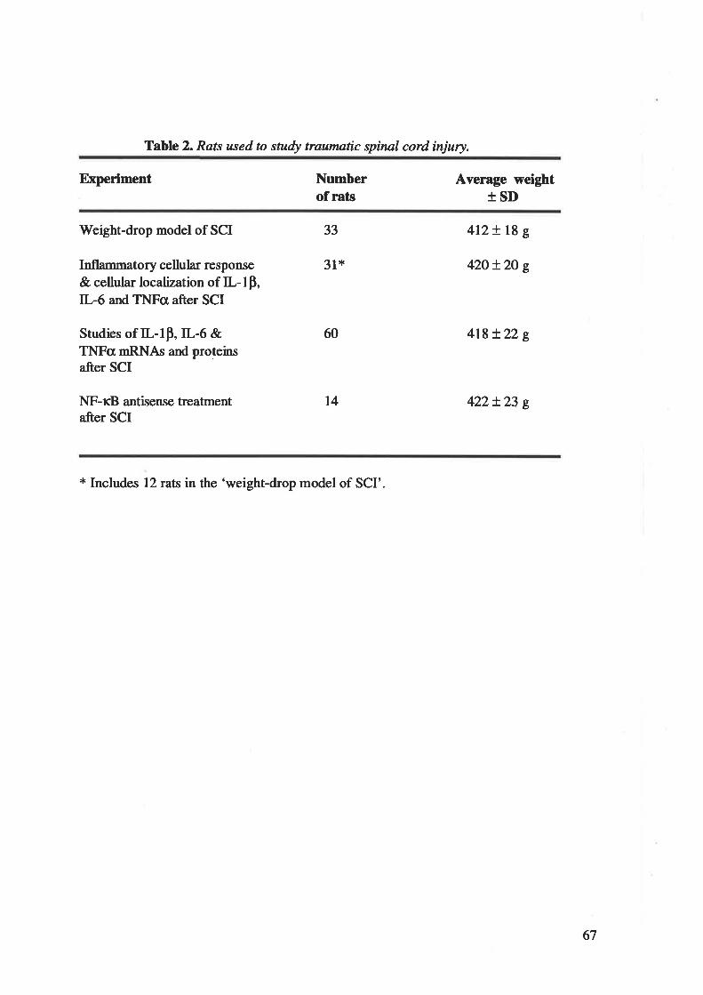
Table 2. Rats used to study traumntic spinal cord ínjury.
Experiment Numberof rats
Average weightISD
Weight-drop model of SCI
Inflammæory cellular response
& cellular localization of IL-lp,IL-6 and TNFa after SCI
Studies of IL-1p,IL-6 &TNFcr mRNAs and proteinsafter SCI
NF-KB antisense treatmentafter SCI
33
31*
+12+ 18 g
42O+2O g
418+22 g
422+23 t
60
t4
* Includes 12rats in the 'weight-drop model of SCI'.
67
1.2 ANAESTHESIA
Anaesthesia was induced in each rat with 4Vo ßoflirane in oxygen (3 Umin) in an anaesthetic
chamber. Rats were positioned prone. Anaesthesia was maintained with the animal self
ventilating 2.5Vo isoflurane in oxygen (1.5 I/min) through a nose cone. Core body
temperature was continuously monitored and maintained at 38 + I oC by a heating pad.
Following the surgical procedure, anaesthesia was withdrawn and the rat supplied with lù0o/o
oxygen for l-2 minutes before placing the rat in the postoperative recovery cage.
1.3 SURGICAL PROCEDURE
A 3 cm midline incision was made in the skin, followed by blunt dissection to expose the
lower thoracic spine. Paraspinal muscles were strþped from the spinous processes of T10 to
T13 under a dissecting microscope. A laminectomy of T12 was performed to expose a
circular region of dura approximately 8 mm in diameter. The spinal column was stabilized by
clamping the spinous processes of T11 and T13 with angled Allis clamps held in custom-
machined holders. The clamps were then raised so that the lumbar region of the rat was
suspended by the spinous processes. The prepared rat was positioned under a weight-drop
device, which was used to induce the spinal cord contusions (details seen rrn2.1.2). Mild and
severe SCIs were respectively produced by dropping a 10-g weight from 3.0 or 12.0 cm onto
the exposed dura of rat spinal cord at the Tl2 vertebral level. After injury, the wound was
closed in layers using 3-0 synthetic absorbable sutures.
68
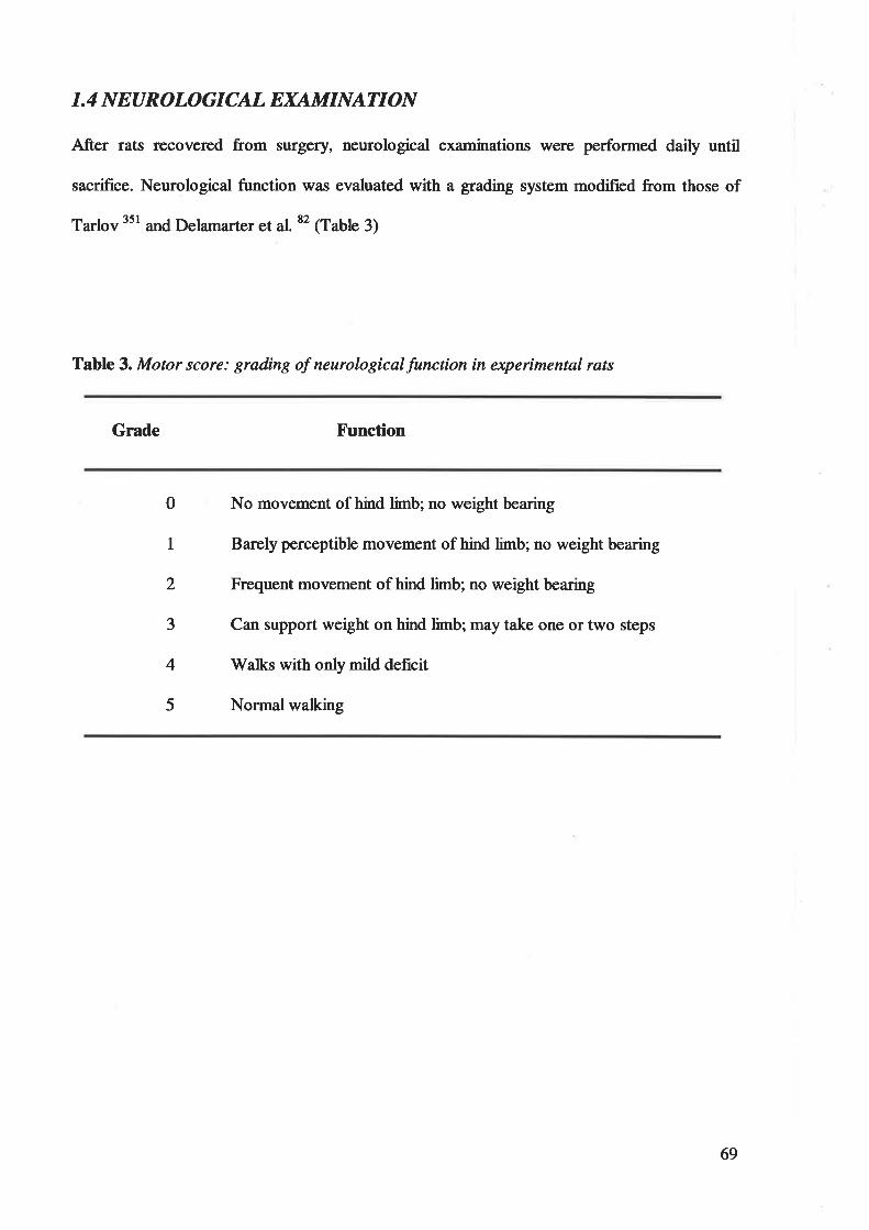
1.4 N EUROLOGICAL EXAMINATI ON
After rats recovered from surgery, neurological examinations were perforrred daily until
sacrifice. Neurological function was evaluated with a grading system modified from those of
Ta¡lov 351 and Delarna¡ter et al82 (Table 3)
Table 3. Motor score: grading of neurologicalfunction in experimental rats
Grade X'unction
0
1
2
3
4
5
No movement of hind limb; no weight bearing
Barely perceptible movement of hind limb; no weight bearing
Frequent movement of hind limb; no weight bearing
Can support weight on hind limb; may take one or two steps
rWalks with onlymild deficit
Normal walking
69
I. 5 PERF U SI ON -SACRI F I CE
Following a survival period of I h - 14 days, rats were induced and anaesthetized with
isoflurane. The rat was positioned supine and the thoracic cage opened to expose the heart and
aort¿. A blunt 19 gauge needle was introduced through the apex of the heart into the
ascending aorta. Two millilitres of 1000 ru/d heparin was inþted into the aorta. The hea¡t
was clarnped transversely at the apex to secure the needle in place. The right atrium was
opened. The rat was then perfused with either freshly prepared 4Vo paraformaldehyde in 0.1
M phosphate buffer (pH 7.4, at room temperature) or normal saline for 10 minutes at a
pressure of l2O mmHg. Perfüsion pressures were generated with compressed air in a closed
system and monitored with a sphygmomanometer 3ß.
1.6 TISSAE PROCESSING
1.6.1 REMOVAL OF SPINAL CORDS FROM RATS PERFUSEDWII]H Vo
PARAFORMALDETTYDE
The entire spinal column was removed from each rat immediately after perfrrsion-fixation and
postfixed n 4Vo paraformaldehyde in 0.1 M phosphate buffer (pH7.Ð overnight. The spinal
cord was then dissected out and divided into nine l-cm segments. Each segment was marked
at the rostral end on the right side with a small vertical incision to allow orientation.
Tissue blocks were dehydrated in graded alcohol baths and cleared with chloroform, prior to
processing into paraffin wax. Five micrometer serial sections were cut at (150 ¡rm) levels with
a microtome and mounted onto slides coated with APT (Sigma Chemical Corporation, US).
The sections were dried at 37"C for at least 12 hours prior to use. Sections from each level
were stained with H&E and immunohistochemically.
70
1.6.2 REMOVAL OF SPINAL CORDS FROM RATS PERFUSED \ryTIT{ NORMALSALINE
Spinal cord segments (2 cm in length and 19[.-210 mg in weight) encompassing the lesion
epicenhe were quickly removed from rats, snap-frozen in dry ice and placed in a sûerile bag
immediately after perfusion. Tissue samples were stored at -7OoC until RT-PCR and Western
blotting analyses.
1.7 PHOTOGRAPHY
Photomicrographs were taken with an Olympus PM20 camera system and an Ollmpus BX50
microscope. Film used for microscopic photography was Kodak EPY-36 Tungsten 647
Ektachrome.
7l
2. WEIGHT.DROP MODEL
This experiment was performed to determine whether or not a graded (mild and severe) and
consistent SCI could be produced in the rat with the weight-drop device.
2.1 METHODS
2.1.I ANIMALS
Thirty-three adult male Sprague-Dawley rats were used and divided into three groups;
liaminectomy control, mild and severe SCI. The control group (N=3) received a Tl2
laminectomy only and were sacrificed at 14 d postinjury. The mild SCI group consisted of
fifteen rats, which were injured by dropping tho 10-g weight 3 cm onto the spinal cord and
were sacrificed at I d, 7 d ot 14 d postinjury (n=5 for each time point). In the severe SCI
group, fifteen rats were injured by dropping the 10-g weight 12 cm onto the spinal cord and
were sacrificed at 1 d.,7 d or 14 d postinjury (n=5 for each time point).
2.1.2 WETGHT- DROP TECHNTQUE
The weight-dtop device consisted of a Teflon impounder, 2 g weight, aligned but loosely
fitted over the end of a brass rod inserted through a 10-g weight. The brass rod was attached
to a stereotactic frame that allowed precise positioning along the X, Y, and Zcootdtnates. The
spinal column was stabilized by clamping the spinous processes of T11 and T13 with angled
Allis clamps held in custom-machined holders and the impounder was then centred over the
laminectomy and lowered onto the dura. The 10-g weight was held in place by a pin which,
when removed, allowed the weight to fall. The 10-g weight was then dropped 3.0 or l2.O cm
onto the impounder and 5 seconds later, the entire rod assembly was raised off the dura. For
the laminectomy control group, the impounder was lowered onto the dura for 2 minutes but
no weight was dropped.
72
2.1.3 MOTOR SCORE
Motor performances of hind limbs were analyzrd with a grading system modified from those
of Tarlov 351 and Delamarter et aL 82 (Table 3). A motor score was assigned to the left and
right hind limb separately and the motor scores of the left and right hind limbs were averaged
together to yield one score for a rat per test session.
2.1.4 HISTOPATIIOLOGY
Tissue sections were stained with haematoxylin and eosin. The epicentre of the injury was
identified histologically by reviewing serial transverse sections and selecting the section that
reflected the maximal extent of the lesion with light microscopy. The area of the maximal
lesion was measured with NIH image 1.62 software (NIH, Bethesda, MA) and the length of
the spinal cord lesion was derived from the number of serial transverse sections showing
histolo gical abnormality.
2.1.5 STATISTICS
Wilcoxon Rank Sum Test and T-test were respectively used to compare the differences in
Tarlov score and histopathology between mild and severe rat SCI. A P value less than 0.05
was considered significant.
2.2 RESULTS
2.2.I ]jÙ.fOTOR PERFORMANCE
As a motor score was assigned to the left and right hind limbs separately, as¡rrnmetry in the
lesion was detected in some rats. However, for the most part, the deficits appeared
73
slmmetrical with the same score given to both hind limbs. In no case did the score between
hind limbs and vary by more than one point.
Table 4 shows the motor scores for laminectomy control, mild and severe SCI groups and
statistical comparisons of pairs of the mild and severe SCI groups with respect to the motor
score at I,7 and 14 days after injury (refer to Appendix 2).
The laminectomy control group did not show any motor function deficits while mild and
severe SCI groups presented varying degrees of deficits. In no case did the score between rats
in the same group vary by more than one point.
Motor functions of the hind limbs in the mild SCI group demonstrated mild and transient
declines and then gradually and fully recovered to the normal level, whereas motor functions
of rats with severe SCI were significantly impaired and improved marginally ovet a period of
two weeks after injury. The mild SCI group had significantly better motor scores than the
severe SCI group at 1,7 and 14 days after injory.
74
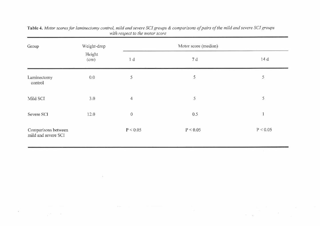
Table 4. Motor scoresfor laminectomy control, mild and severe SCI groups & comparisons of pairs of the mild and severe SCI groups
with respect to the motor score
Group
Laminectomycontrol
MiId SCI
Severe SCI
Comparisons betweenmild and severe SCI
Weight-drop
Height(cm)
0.0
3.0
12.0
1d
P < 0.05
5
5
1
5
5
5
4
0
Motor score (median)
7d
0.5
P < 0.05
14d
P < 0.05
2.2.2HISTOPATHOLOGY
The 3-cm weight-drop contusions in the group of mild SCI produced central core lesions,
which were charactenzed by cavities that replaced some of the grey and white matter. There
were also multiple small microcysts in the white matter, some of which contained swollen
axons or axonal debris. A representative transverse section at the lesion centre in mild SCI at
14 days after injury is shown in Figure 84.
The 12-cm weight-drop contusions in the group of severe SCI created subtotal losses of grey
and white matter and only tiny and isolated patches of white matter remained on peripheral
edges of the ventral funiculus. A representative transverse sections in severe SCI at 14 days
after injury is shown in Figure 88.
No tissue loss or pathological changes were observed in the larninectomy control group
(Figure 8C).
Statistical comparisons were done between the mild and severe SCI groups with respect to the
area of maximal lesions and the length of lesions at 14 days after injury, showing significantly
greater areas of maximal lesion (6.70 t M7 rrrÑ) and lengths of lesion (13.23 + 0.81 mm) in
severe SCI than in mild SCI (area of maximal lesion: l.2l + 0.11 mm2 & lesion length 2.88 +
0.26 mm)(refer to Appendix 2). P values are both less than 0.05.
76
Figure 8. Representative transverse secrtons at the epicentre of the lesionfor a 3 cm (A) and12 ctn (B) weight-drop contusion SCI, 2 weel<s after injury. C shows a transverse section of alaminectonty control rat spinal cord at 712 segment,2 weel<s after laminectonay. H&8, X10.
77
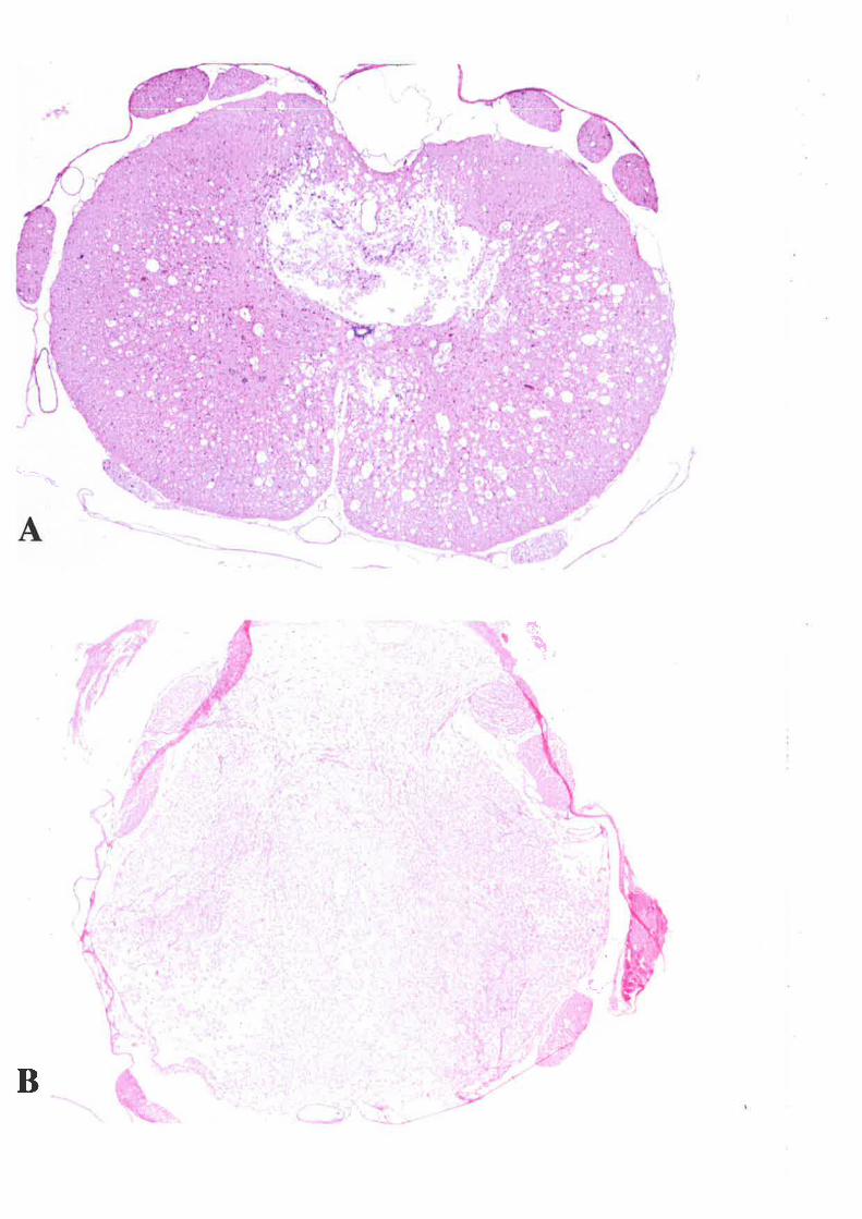
-è
*1
B
I
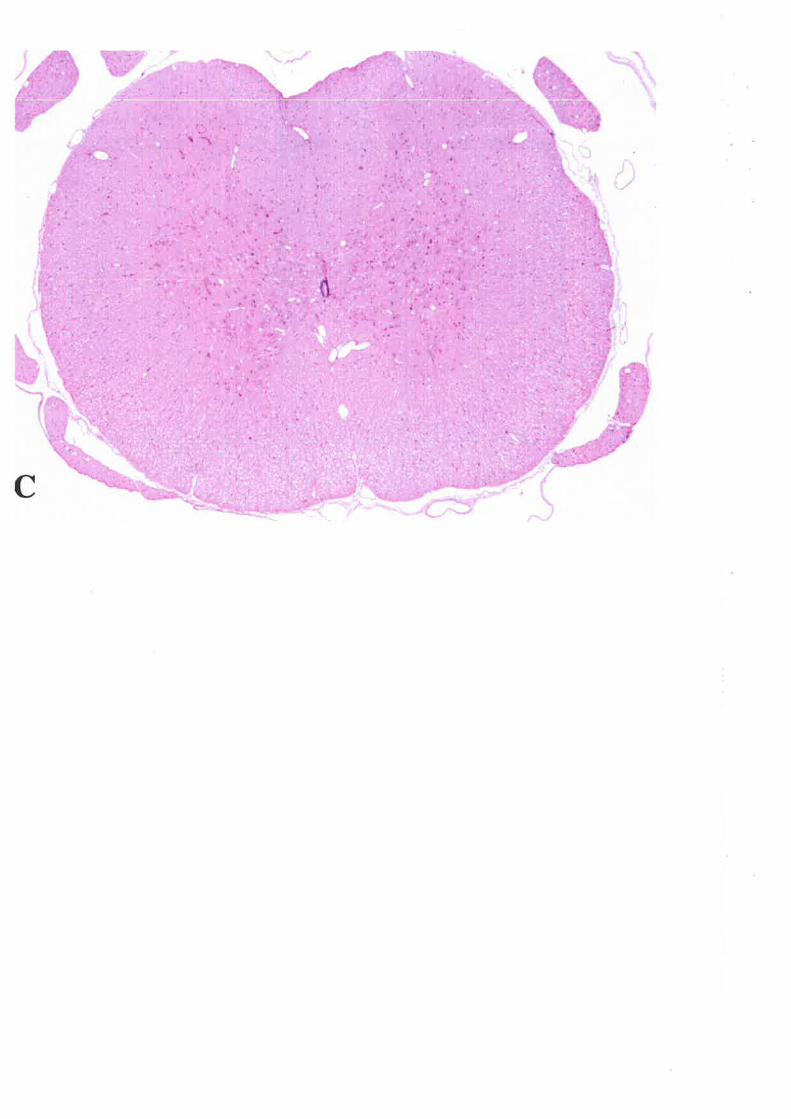
2.3 DISCUSSION
The results indicate that by using our weight-drop device, a graded SCI can be produced in
rats with significant differences in outcomes of motor function and histopathology between
mild and severe SCI groups. Furthermore, the spinal cord lesion induced by the weight-drop
device is consistent: the standard error is less than L0 percent of the mean maximal lesion area
or mean lesion length in the mild and severe SCI group.
Histopathology of spinal cords (area of maximal lesions and length of lesions) at 14 days after
mild and severe SCI was chosen for statistical comparisons because it is more accurate to
define the lesion area and intact a¡ea in the damaged spinal cord at 14 days postinjury
@igures 8A-B) than at ea¡lier time points, such as t hr postinjury @igures 9A-B).
There are two important differences in our weight-drop techni4ue if compared to the classic
Allen weight-drop technique2t'1et. The first is the extent of the laminectomy. We carried out a
limited, one-level larrinectomy just large enough to allow the impounder to contact the dura.
An ideal experimental model of SCI would require minimal surgical preparation of the animal
and multiple-level laminectomy itself is a major injuty to the animal. The second is the extent
to which the spinal column was stabilized prior to weight-drop injury. We stabilized the
spinal column via clarrps on the spinous processes of the vertebrae immediately rostral and
caudal to the laminectomy. The rat was actually lifted by those clamps so that the weight-drop
impact could not be cushioned by the body as a whole. Such stabilization also reduced the
effects of respiratory movements that may alter the position of the spinal cord with respect to
the impounder at the moment of impact and thus contribute to variability.
80
In conclusion, our weight-drop device produces graded and consistent contusion injuries of
the spinal cord in the rat.
81
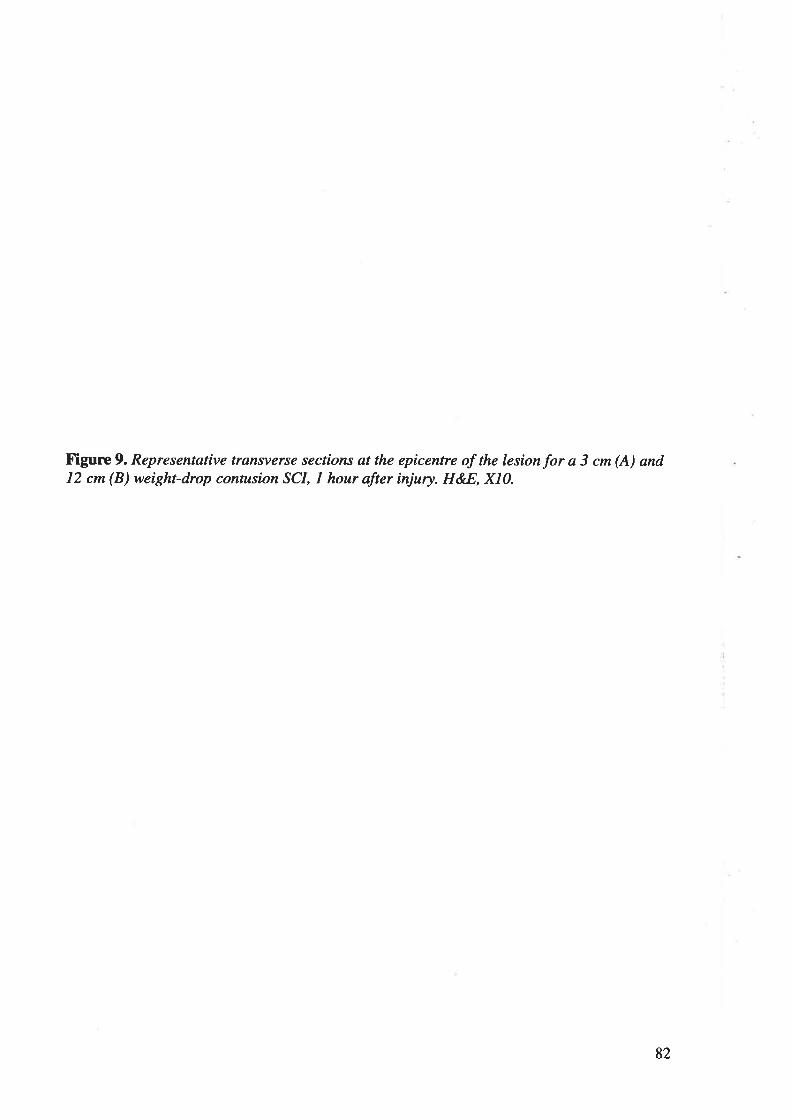
Figure 9. Representartve tansverse secrtons at the epicentre of the lesionfor a 3 cm (A) and12 cm (B) weight-drop contusion SCI, I hour after injury. H&-8, Xl}.
82
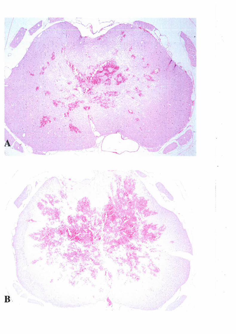
¡
II
t
r{
"' .. ''
ri
-#¡
ç
1Yl!
i¡ú
[,ilu
" : ;ìr^ ..¡.
*t¡-''h
m
3. II\FLAMMATORY CELLT]LAR RESPONSE
This experiment was performed to investþate the posttraumatic inflammatory cellular
response in the rat after SCI.
3.1 METHODS
3.1.I ANIMALS
Thirty-one rats were used and divided into four groups: mild SCI, severe SCI, laminectomy
control and narve control Fourteen rats with mild SCI were killed at t h (n=2),3 h (n=2), 6 h
(n2), I d (n=2), 3 d (n=2), 7 d (rJ) or 14 d (n=2) postinjury. Severe SCI group also
includesfourtecnrats,whichweresacrificedatlh(n=2),3h(n=2),6h(n=2), 1d(n=2),3d
(n=2),7 d (n=2) or 14 d (n=2) postrnjury. Two rats with laminectomy only were killed at I or
24h (n=I for each time point) after surgery and one norrnal rat was killed as a narve control.
3.I.2 HISTOLOGICAL STAINING
Sections from each level were stained with haematoxylin and eosin to assess the extent and
distribution of any pathologic abnormality of the cord with particular attention to
haemorrhage, necrosis and migration of inflammatory cells (neutrophils and macrophages).
Immunocytochemical staining was carried out on 5-Fm paraffin sections using standard
irnmunoperoxidase staining protocols 334. The sections were deparaffinized in xylene and
cleared in graded ethanol baths. Endogenous peroxidase activity in the tissue was blocked by
incubation wlth 37o HzOz in PBS and non-specific binding was suppressed by incubation with
blocking serum (157o NHS in PBS). Sections were then incubated overnight with the primary
antibody diluted in NHS and stained with 3,3-diaminobenzidine tetrahydrochloride (DAB;
Sigma, St. Louis, MO) using avidin-biotin peroxidase (Vectastain ABC kit; Vector
84
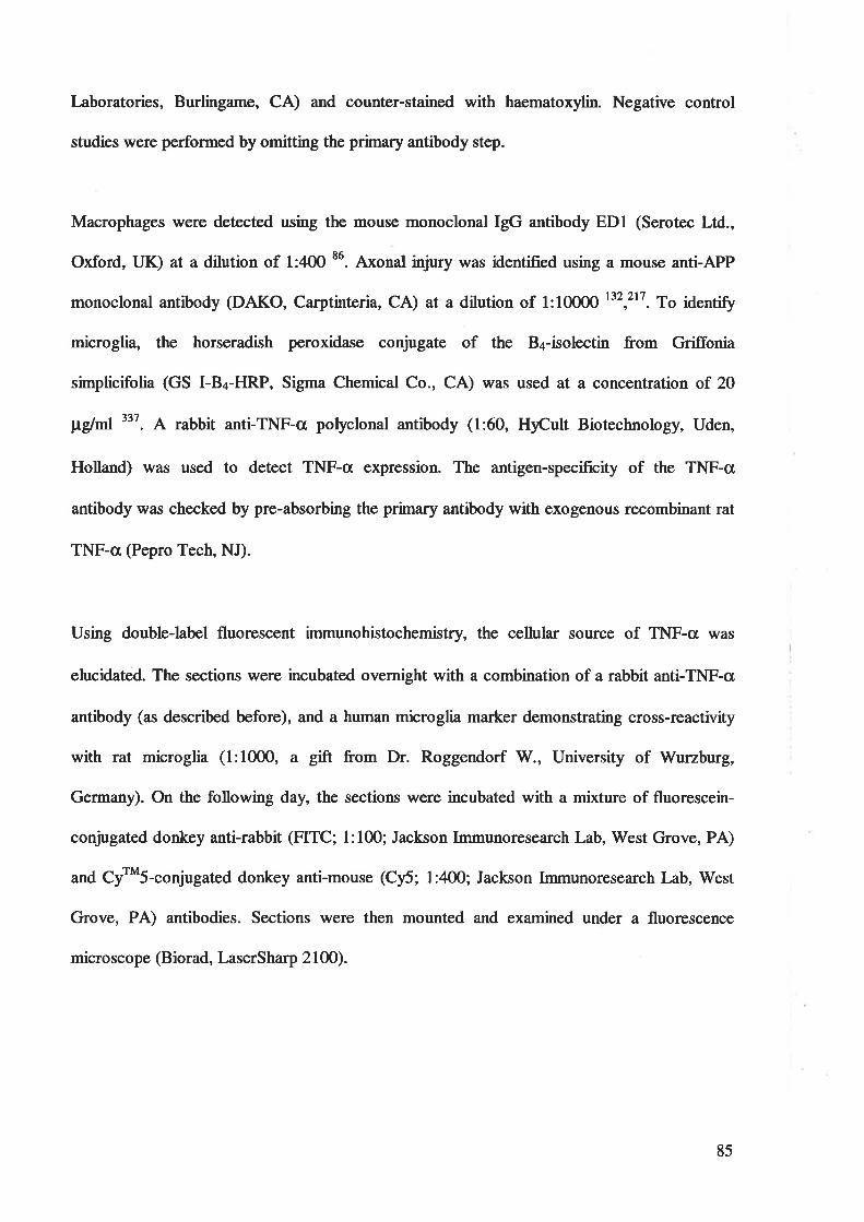
Laboratories, Burlingame, CA) and counter-stained with haematoxylin. Negative control
studies were performed by omitting the primary antibody step.
Macrophages were detected using the mouse monoclonal IgG antibody EDI (Serotec Ltd.,
Oxford, UK) at a dilution of 1:4O0 86. Axonal injury was identified using a mouse anti-APP
monoclonal antibody (DAKO, Carptinteria, CA) at a dilution of 1:10000 132,217. To identiff
microglia, the horseradish peroxidase conjugate of the B¿-isolectin from Griffonia
simplicifolia (GS I-B4-HRP, Sigma Chemical Co., CA) was used at a concentration of 2O
lLgrnl 337. A rabbit anti-TNF-cr polyclonal antibody (1:60, HyCult Biotechnology, Uden,
Holland) was used to detect TNF-a expression. The antigen-specificity of the TNF-cr
antibody was checked by pre-absorbing the primary antibody with exogenous recombinant rat
TNF-c¡ @epro Tech, NJ).
Using double-label fluorescent immunohistochemistry, the cellular source of TNF-c¡ was
elucidated. The sections were incubated overnight with a combination of a rabbit anti-TNF-a
antibody (as described before), and a human microglia marker demonstrating cross-reactivity
with rat microglia (1:1000, a gift from Dr. Roggendorf 'W., University of W'urzburg,
Germany). On the following day, the sections were incubated with a mixture of fluorescein-
conjugated donkey anti-rabbit (FITC; 1:100; Jackson Immunoresearch Lab, West Grove, PA)
and Cyru5-conjugated donkey anti-mouse (CV5; 1:4O0; Jackson Immunoresearch Lab, West
Grove, PA) antibodies. Sections were then mounted and examined under a fluorescence
microscope (Biorad, LaserSharp 2 100).
85
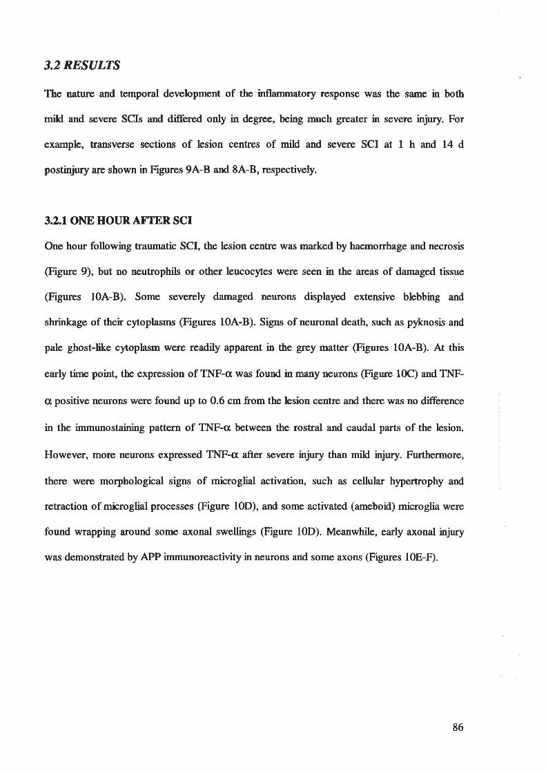
3.2 RESULTS
The nature and temporal development of the inflammatory response was the same in both
mild and severe SCIs and differed only in degree, being much greater in severe injury. For
ç¡¡emFlo, transverse sections of lesion centres of mild and severe SCI at t h and 14 d
postinjury are shown in Figures 9A-B and 8A-B, respectively.
3.2.1 OI\E HOUR AFIER SCI
One hour following traumatic SCI, the lesion centre was marked by haemorrhage and necrosis
@igure 9), but no neutrophils or other leucocytes were seen in the areas of damaged tissue
(Figures 104-8). Some severely darnged ner¡rons displayed extensive blebbing and
shrinkage of their cytoplasms (Figures 104-B). Signs of neuronal death, such as pyknosis and
pale ghost-like cytoplasm were readily apparent in the grey matter (Figures 104-B). At this
early time point, the expression of TNF-cl was found in many neurons (Figure 10C) and TNF-
cr positive neurons were found up to 0.6 cm from the lesion centre and there was no difference
in the immunostaining pattern of TNF-a between the rostral and caudal parts of the lesion.
However, more neurons expressed TNF-a after severe injr.,ry than mild injury. Furthermore,
there were morphological sþs of microglial activation, such as cellula¡ hypertrophy and
retraction of microglial processes (Figure 10D), and some activated (ameboid) microglia were
found wrapping around some axonal swellings @igure 10D). Meanwhile, early axonal injury
was demonstrated by APP immunoreactivity in neurons and some Ð(ons (Figures 10E-Ð.
86
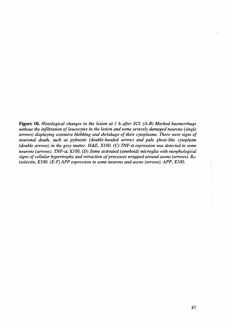
Figure 1.:O. Histological changes in the lesion at I h after SCI. (A-B) Marked haemorrhagewithout the infiltration of leucocytes in the lesion and some severely dnmnged neurons (singlearrows) displaying extensive blebbing and shrinl<nge of their cytoplasms. There were signs ofneuronal death, such as pyløosis (double-headed anow) and pale ghost-like cytoplasm(dauble arrows) in the grey matter. H&8, X100. (C) TNF-a expression was detected in some
neurons (anows). TNF-a, X100. (D) Some activated (ameboid) microgliawith morphologicalsigns of cellular hypertophy and retraction of processes wrapped around axons (anows). Ba-isobcrtn, X100. (E-F) APP expression in some neurons and ax,ons (anows). APP, X100.
87
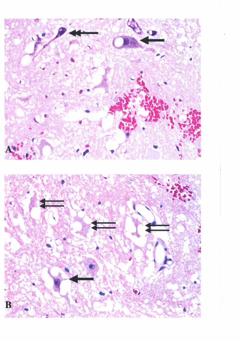
¡u
l¡ì
fùb '#
I¡
T,
tì
T
ì )
,
,,lui
lr*'
I \I a
(r
at
hÇ
\ll
ö"{
' .h
-¡.
c{
rl-
fa
af"
f
'lfll
tü
ll,
,*n¡
ffi
,ffiff
if,|,,
,I
rI
)
î;
\
îî îl
II
t ì o
q;i
L
ãI
{a-
ìr I \
I/
*)
ì¡
¡ ì0
.a>
t
i1
a
åt
I
,lt
Ç t4
rJ
,r,
a
/I
ilh M'*
*.r
r(t
aù
,l
i
¡' I
!t"
"t
, I
ìIr
or[t
,{
tf
üirt
o
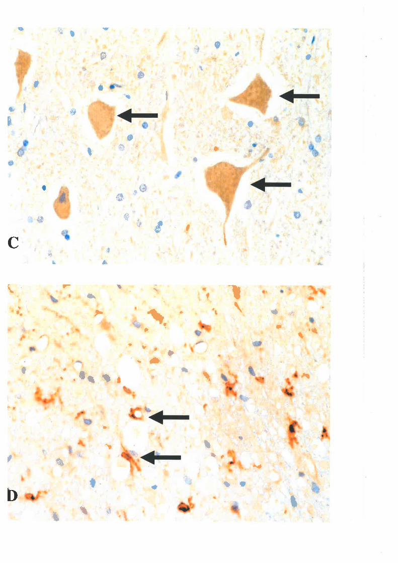
;t$[,:':- rr
'|-
+
ryd#.
'*s
#*lr,ûf 'o
Çö
\r; ''
&ff¡
¡üI
ü0
',tP'
,tf
#flÞffi
s:
,
,#t
4
#ü
,#f
rI,#.r t ,
.fh t*-#l,
\
t
ü#
r
tr'
t
ü
4lF{öË
üa
pI
å-
tÐ
t,ltÒt
ît
f
.¡&'-\
4
5ü
I
't¡"ú
#t¡lf>
.{ft
fþü
.'û'
-¡fr
{&ffi,
wtf
{
{åI
tt'
utFA
ftþ,l
{",,#ffi#
,$'t*
þr
ß
l.t
"üå
,'$
cÌ'
#f
-,#IF
flF
^ rll
&r
rFdH
@t
*
ü,.;'
l)'
¡il"rË
q{
6' dii'
Çt
#,s,
d *+
{Ën
(':
aI
#t
r*rrt.t":T
qFffi.
@It
t,i
übù
fl øru
ú
offi
()
rfi
.,!D
t
/t4,¡t
#
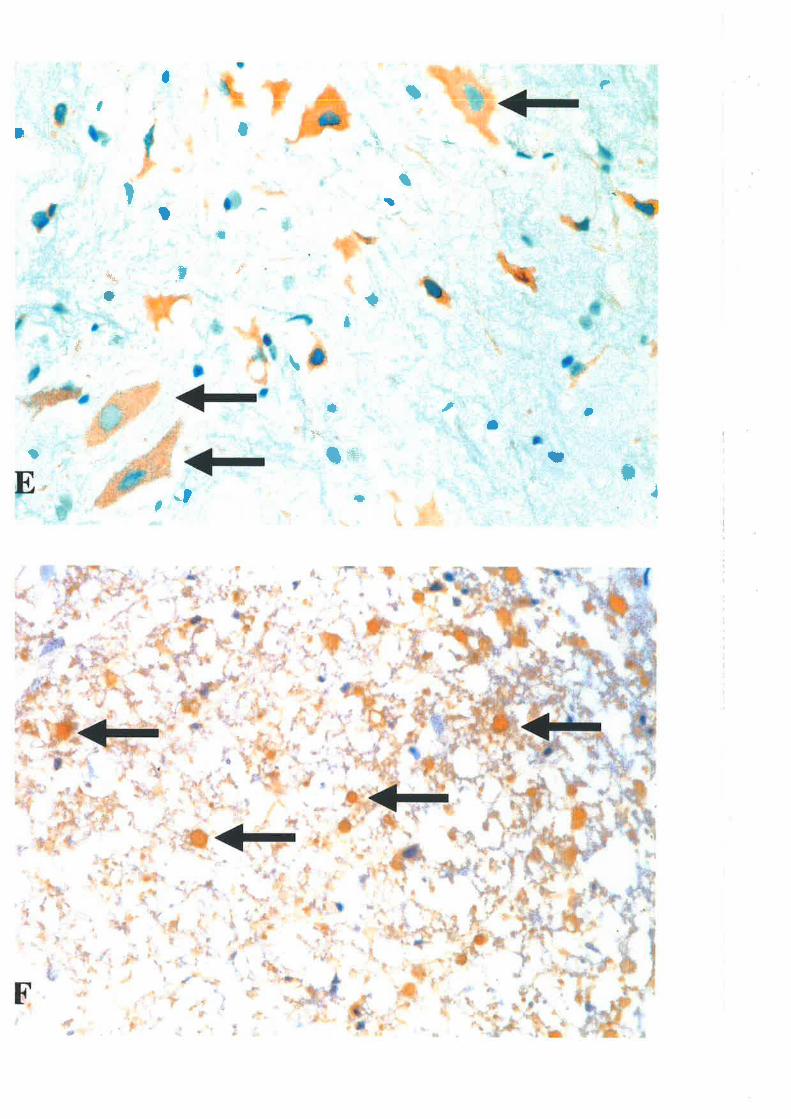
*,dt -
*
*
'È
:
ö int\ ft
+l-d> =-?
*tI
Ìn
*i
:
t
-1ß t_
\
I+
a*Þ-
+
*
*
q+C
t
,t --+
II ::q-' !-\ 'a
t
ihI
a,
I
d.* =*_I
I
3.2.2 SD( HOURS AFTER SCI
Similar pathology was observed at 3 h after SCI compared to t h postinjury. However, at 6 h
postinjury, a dramatic increase in the number of multinuclear neutrophils was found primarily
in and around blood vessels (Figure 114). Very occasional mononuclear cells in the walls of
blood vessels were observed. TNF-o expression was detected not only in many neurons but
also in some glial cells with microglial morphological appearance (Figure 118). Some of
these TNF-cr positive glial cells were found surrounding axons (Figure 118). TNF-a positive
cells were found up to 1 cm from the lesion centre and there was no difference in the
immunostaining pattern of TNF-o between the rostral and caudal parts of the lesion. There
were few neluons and glial cells expressing TNF-cr after mild injury compared with severe
injury. More activated (ameboiQ microglia were detected and some of them were found
wrapping around axons as well as neurons (Figure 11C). Axonal injury was shown by APP
immunoreactivity of many neurons and axons (Figure l lD).
To determine if TNF-cr positive glial cells were microglia, double-label fluorescent
immunohistochemistry was used, showing ss-loselization of TNF-cr and the microglial
marker KM1P (Figure 12).
9T
Figurc ll. Histological changes in the lesion at 6 h after SCI. (A) Neutrophil rnargination inthe wall of a blood vessel (ønows). H&8, X100. (B) Glial cells expressed TNF-a and some ofthetn sunounded axotts (ørrows). TNF-a, X1500. (C) More activated (ameboid) microgliawere found and some of themwrapped around neurons (anows). Ba-isolectin, X100. (D) APPpositive neurons (double-headed anow) and axons (anows). APP, X100.
92
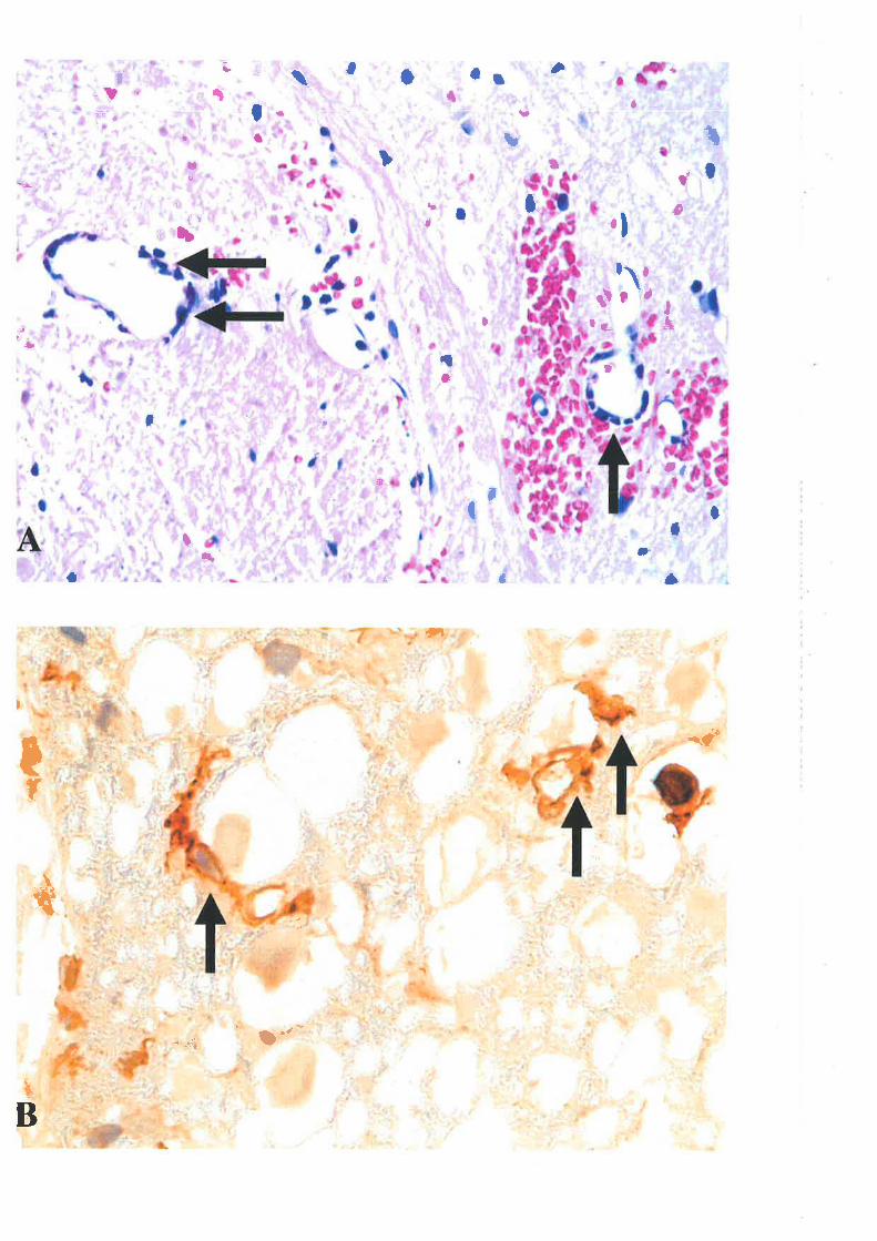
EË
or
¿*'
; tu
I
t" .
/ ':
*,'
ú{'"
*tfm
ü;Ç .,'* *,
"Ë'tn "'fi
rì
$
aL I
.ftr
,ti '
I I r.
"F
,, 1*
<>
h,{
I
,fli I {ì
.*,t
.
ü,,, ìú .ìri
,
s' .f .
a
,, ,:.
',
14 I
,, *$ ;" ît
fli
{¡ iil, a n t t
\ ltr
lh: *;
,
W'
, rf
üt
ll$ u"
t ltt tJ
pe(i t"'
a
r.lt,
uqi
te
t t ifiltt
tt
t
i:
ra
tJ
{f
t
Ðih
J
?
, ¡ t ¡ ,
ffi.,
ü
rr,l
.t,Ç
*"{*
'f"
I* ,.
''It
. .i
I -¡ (*.
{, ,
I llt
'i
,i5'-;
,.)/
ll-
t "'e
e-
t$$
Jú,
T¡ü
I
iilñ
Ëti
.a
üI
I
çr,
) mi
a*j
r| -
{_
D{D
lÇ
þr-
tttl
)*
I 't't
OO
drl
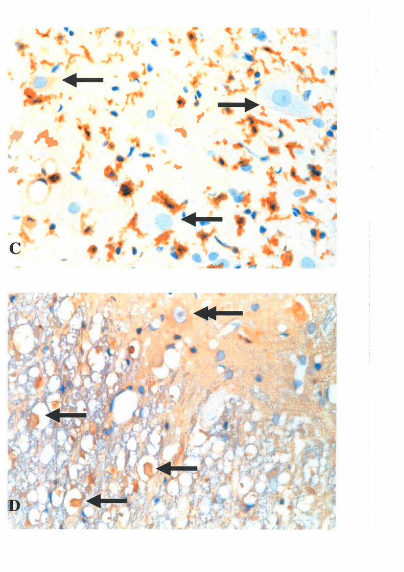
ffi1-'¡tr
T¡¡
fd\
ï#f¡
r&
3iJt'
{h.
{
\.t,
{Ì
{
rlI #+à
h-i t#
&
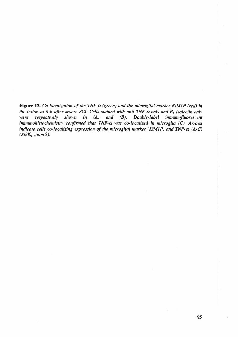
Figure 12. Co-localizarton of the TNF-a (green) and the microglial marker KLMIP (red) inthe lesion at 6 h after severe SCI. Cells stained with anti-TNF-a only and B¿-isolectin onlywere respectively shovwt in (A) and, (B). Double-label immunofluorescentimmunohistochemistry confirmcd that TNF-a was co-Iocalized in microglia (C). Arrowsindicate cells co-localizing expression of the tnicroglial marlcer (KLMIP) and TNF-a (A-C)(X600, zoom2).
95
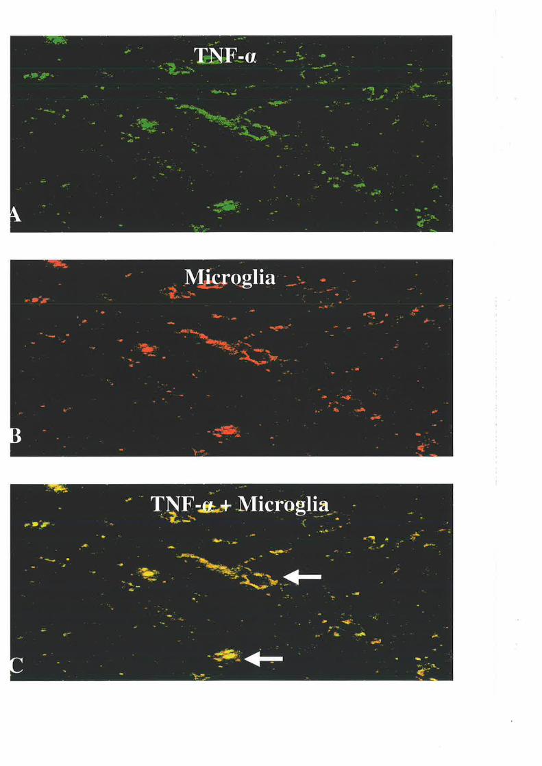
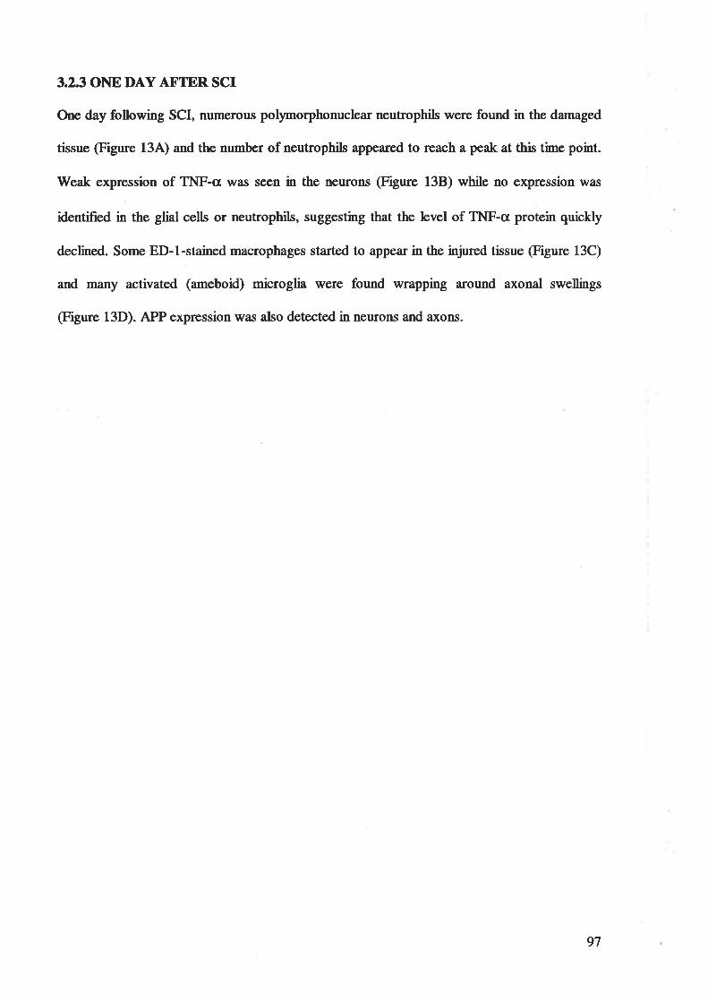
3.2.3 OIITE DAY AT"IER SCI
One day following SCI, numerous poþmorphonuclear neutrophils were found in the danaged
tissue (Figure 134) and the number of neutrophils appealed to reach a peak at this time point.
rJVeak expression of TNF-a was seen in the neurons @igure l3B) while no expression was
identified in the glial cells or neutrophils, suggesting that the level of TNF-ct protein quickly
declined. Some ED-l-stained macrophages st¿¡ted to appear in the injured tissue (Figure 13C)
and many activated (ameboid) microglia were found wrapping around axonal s1¡rsllings
@igure 13D). APP expression was also detected in neurons and axons.
97

Figure 13. Histological changes in the lesion at I d after SCI. (A) Numerous neutrophilsinfiltrated the dnmaged spinal cord (anow). HEE, X100. (B) Weak expression of TNF-ø inneurons (anows). TNF-u, X100. (C) Some møcrophages størted to appear in the lesion. ED-1, X1500. (D) Axons were surrounded by acrtvated (ameboid) microglia (arrows). Ba-isolectin, X|00.
98
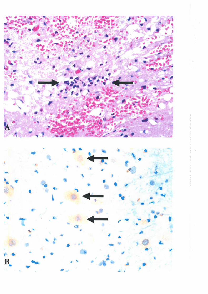
\ û
ìl
'u;i rr
I
öt
I
:.
'l
d', ü
il ,t ,# ì
#
ü
ü".
t
tr
#
II o Ç l*l
i
1Ñq
I
üt
p tü
dl
D f' "'r
'l
t a
p
I ,| I
,ffi
I¡ll
t
I
I
.#.
î \ t il #
fl *î
,î:D
+,
Itf
I t
I{lr)
Ê
¡ I\
fttr t
l)-*
ìt
bI
# \ü
$"""
""""
''
t
F
il
t
il[
\{r
l
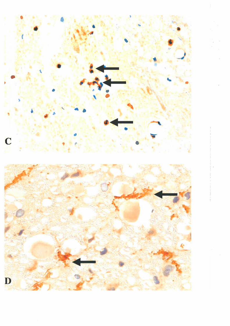
F
- *.-'
# L
il*ú
tÞ\ç+(,
t
aJt
r>r=
b+
È
ç
t
+
''=êLaaJI
1aI
IÌÈ'lf
{å/)
tI
I * t
Ê
ì
" *E+I a :i1= li
f'
t't {¡trlÊtr.*
ln;EI
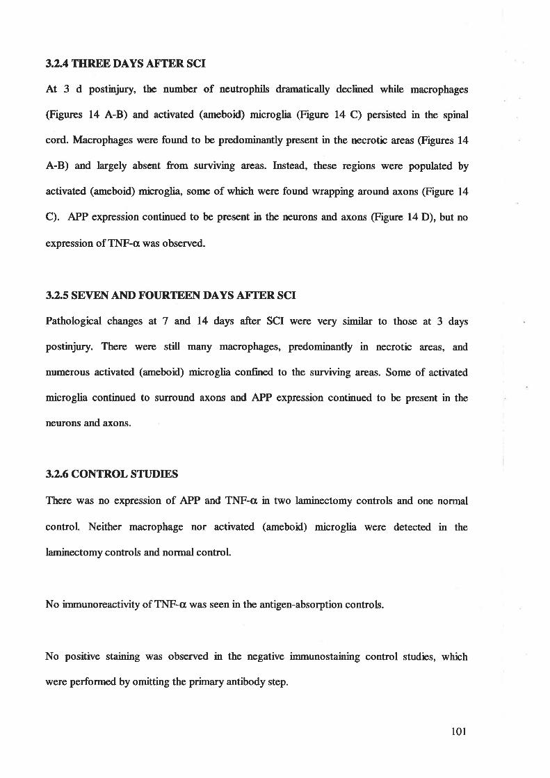
3.2.4 TIIREE DAYS AFTER SCI
At 3 d postinjury, the number of neutrophils dramatically declined while macrophages
@igures 14 A-B) and activated (ameboid) microglia @igure 14 C) persisted in the spinal
cord. Macrophages were found to be predomina¡fly present in the necrotic areas (Figures 14
A-B) and largely absent from surviving areas. Instead, these regions were populated by
activated (ameboiQ microglia, some of which were found wrapping around axons (Figure 14
C). APP expression continued to be present in the neurons and axons @igure 14 D), but no
expression of TNF-a was observed.
3.2.5 SEVEI\I AI{D FOURTEEN DAYS AFTER SCI
Pathological changes at 7 and 14 days after SCI were very similar to those at 3 days
postinjury. There were still many macrophages, predominantly in necrotic areas, and
numerous activated (ameboid) microglia confined to the surviving areas. Some of activated
microglia continued to surround ÐKons and APP expression continued to be present in the
neurons and axons.
3.2.6 CONTROL STTJDIES
There was no expression of APP and TNF-cr in two laminectomy controls and one normal
control. Neither macrophage nor activated (ameboid) microglia were detected in the
laminectomy controls and norrnal control
No immunoreactivity of TNF-cl was seen in the antþen-absorption controls.
No positive staining was observed in the negative immunostaining control studies, which
were perforrned by omitting the primary antibody step.
101
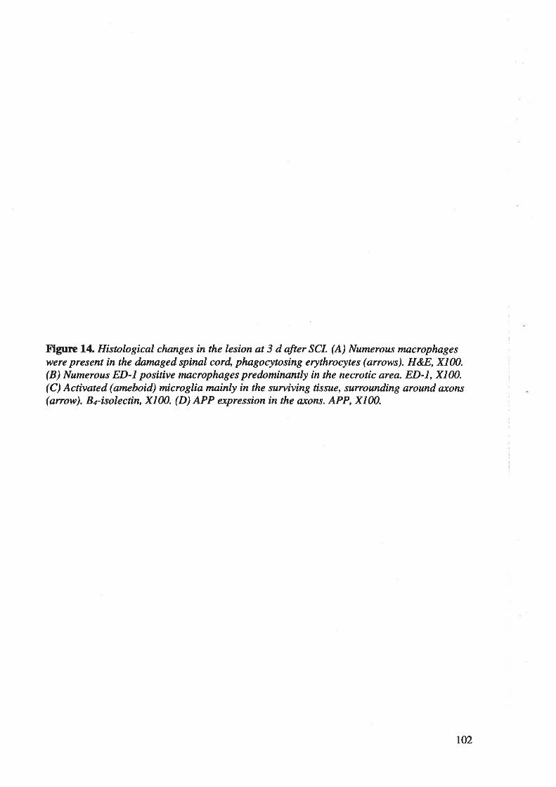
Figure 14. Histological changes in the lesion at 3 d after SCI. (A) Numcrous macrophageswere present in the damaged, spinal cord, phagocytosing erythrocytes (anows). H&8, X100.(B) Numerous ED-l positive macrophages predominanþ in the necrotic area. ED-L, X100.(C) Activated (amcboid) rnicroglia mainly in the surviving tissue, surrounding around. a,x,ons
(anow). B¿-isolectin, X100. (D) APP expression in the axons. APP, X100.
102
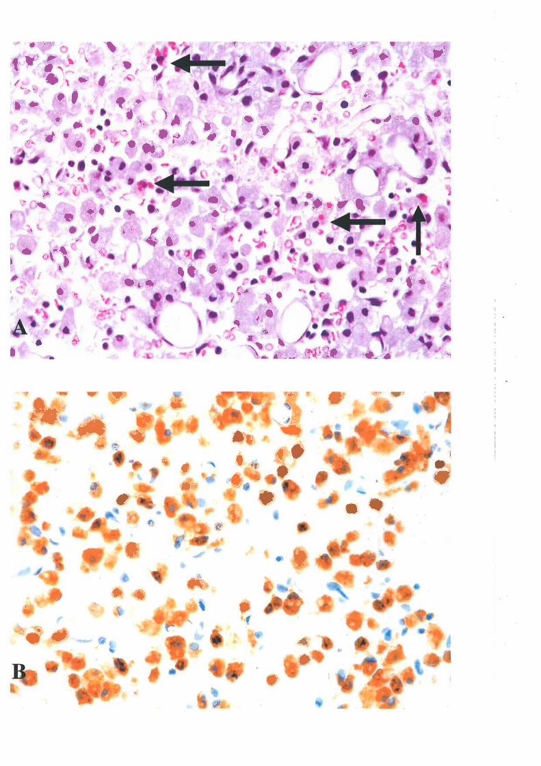
3 j't,#
=äÜ
.ü
ö
aï
t
*
t
=
Ü,.#f_
i*
a*t. å
T
*S* t+!*
å
#*
t
{þ*üt"
E=* *
a
e*
*
'J _È
+r
==.*+
tI
==ËG'
É
a
+{
*
:'
at &l\-à¡*
aç+ =€*å i'=
3Ë-=æ'ry æ¡ìJ
-:.'ç
E,+F *-a¡.¡-*
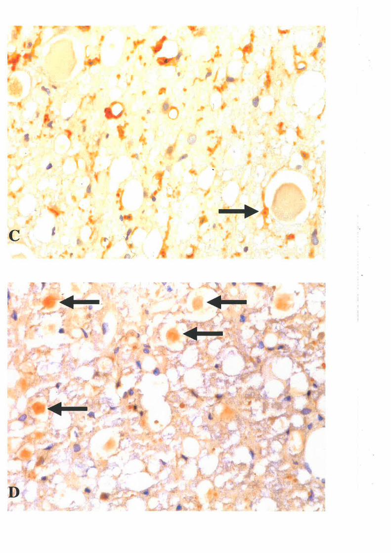
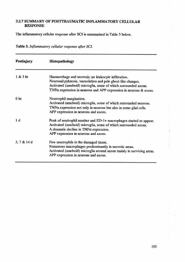
3.2.7 STJMMARY OF POSTTRAT]MATIC INTT,AMMATORY CELLTJLARRESPONSE
The inflammatory cellular response after SCI is srrmmarised in Table 5 below.
Table 5.Inflammatory cellular response after SCI.
Postiqiury Histopatholory
1&3hr
6hr
1d
3,7 &14d
Haemorrhage and necrosis; no leukocyte infiltration.Neuronal pyknosis, vacuolation and pale ghost-like changes.Activated (ameboiQ microglia, some of which surrounded axons.TNFc¡ expression in neurons and APP expression in neurons & axons.
Neutrophil margination.Activated (ameboid) microglia, some of which surroundcd ncurons.TNFcr expression not only in neurons but also in some glial cells.APP expression in neurons and axons.
Peak of neutrophil number and ED-l+ macrophages started to appear.Activated (ameboid) microglia, some of which surrounded axons.A dramatic decline in TNFcr expression.APP expression in neurons and axons.
Few neutrophils in the damaged tissue.Numerous macrophages predominantly in necrotic areas.Activated (ameboid) microglia around axons mainly in surviving areas.APP expression in nenrons and axons.
105
3.3 DISCUSSION
3.3.1 STAINING METHODS X'OR RAT MICROGLIAL CELLS
The lack of a unique ma¡ker for microglia has made their study challenging. Many of the best
ma¡kers for mic¡sglia a¡e cell surface antþens that are unstable with routine histologic
methods. Except for a few markers such as lectin, markers for microglia a¡e either useless or
suboptimal in paraformaldehyde-füed or formalin-fixed paraffin sections 85'338.
Therefore, a simple method of using the lectin from GS I-B¿ for histochemical visualization
of rat microglial cells was used in this study. In addition to providing good structural
preservation, this method achieves complete labelling of all microglial cells and superior
visualization of microglial processes. Furthermore, no other glial cell tlpes aside from
microglia demonstrate binding with the lectin from GS l-Bott'.Therefore, this lesl¡ is a very
reliable microglial marker.
3.3.2 APP T]PREGTJLATION
APP has been used extensively as a sensitive marker of axonal injury in human studies
3e't3t'32o and experimental studies 234'37r, but the significance of APP upregulation in neurons
and axons in response to TSCI is unknown. It has been suggested that induction of APP in the
CNS is a normal "acute phase" response to neuronal stress 7t't3t'2e4'2es similar to the induction
of heat shock proteins 225. However, other studies have suggested that APP upregulation may
have neuroprotective functions 143'23s'33o. In the studies described here, it is not known
whether APP expression increases early to perform some neuroprotective function. A
possibility is that APP upregulation has no specific role in TSCI as alteration in APP
expression could result from a number of factors common to local or diffi¡se areas of tissue
damage such as oxidative stress and local inflammatory cytokines 71. T'hus, increased APP
106
immunoreactivity within neurons and axons following SCI may be an adaptive response to
injury.
3.3.3 CHEIVÍOATTRACTANTS OF IMTLTRATING LEUKOCYIE'S
Haematogenous inflammatory cells (neutrophils and macrophages) did not start to infiltrate
the injured spinal cord until 6 h postinjury suggesting that these inflammatory cells were not
spilled from darnaged vessels, but recruited by some chemoattractants, including pro-
inflammatory cytokines, haemorrhage and necrosis 88'tt3'23e'347.It has been reported that direct
inþction of TNFa or ILlp into normal CNS causes widespread leukocyte margination 341.
This is further supported by our findings that a dramatic elevation of TNFc expression as
early as I h after SCI, which may contribute to the recruitrnent of leukocytes into the
damaged cord.
3.3.4 INTT,AMMATORY CELLS
Three cell tlpes are predominantly involved in the early aspects of an inflammatory response:
microglia, neutrophils and macrophages. In contrast to some in vivo studies that failed to
detect morphological signs of microglial activation by I d postinjury'u''to', activated
(ameboiQ microglia with cellular hypertrophy and retraction of processes were observed in
'rris study as early as t h after TSCI and some of them were found surrounding axonal
swsllings. This novel finding challenged a current view that there is no direct topographical
association between microglial cells and axons, and no association of axonal injury with
activated microglia t27'261'278. More studies a¡e needed to elucidate the effect of activated
microglia surrounding axons because activated microglia are able to produce pro-
inflammatory cytokines (observed in this study), cytotoxic substances 25'387 and growth factors
341. Activated microglia may provide a trophic local environment to promote axonal
t07
regeneration as some studies have shown that activated microglia promoted regeneration of
sen¡¡ory axons in the injured rat spinal cord ø8280 and improved recovery of motor function in
paraplegic rats 28e.
The cur¡ent study also found that activated (ameboid) microglia surrounded a¡ound neurons.
This phenomenon originally found by the Blinzinger group 37 in the study of facial nerve
axotomy, is now known as "sSmaptic stripping" 25. Activated (arneboid) microglia are thought
to interpose processes between afferent slmaptic terminals and the neuronal surface and to be
involved in limited phagocytosis of individual spaptic termini. It has been suggested that
neuronal energy expenditure is minimised by cessation of the 'transmission" function 2s2 and
"s¡maptic stripping" may also have an imFact on the slmaptic reorganiz¿lion of the injured
neurons, but the effect of synaptic stripping on neurons is, as yet, largely unknown.
Neutrophil.s are the first peripheral inflammatory cells to migrate into the tissue. It has been
suggested that neutrophils may be involved in neuronophagia 23e, a virew supported by in vitro
literature about cytotoxic effects of neutrophils e, but not by other in vivo data, which could
not correlate secondary tissue damage with the presence of polymorphonuclear neutrophils
ee'118. In this study we have not found neutrophil-mediated neuronophagia and our data have
shown that neutrophil influx in the contused spinal cord follows a time corrse of a detectable
increase at 6 h and a rapid influx thereafter that peaks at 24 h. This is consistent with time
corlrses described for other studies of SCI 6s'ee'34t . The fact that necrosis precedes the arival
of neutrophil.s at the injury site indicates that the tissue necrosis may not be caused by the
invading leukocytes, but that it may, instead, provide a chemotactic stimulus for attracting
neutrophils as the first line of recruited phagocytic cells to begio removal of cellula¡ debris.
108
The second wave of peripheral inflammatory cells to enter damaged tissue in the CNS is
monocyte-derived macrophages. It has been well-known that the population of macrophages
is heterogeneous, consisting of microglia- and monocyte-derived 25'85'2N'2t6'338. Activated
(ameboid) microglia can be distinguished from monocyte-derived macrophages by the
morphology (microglial processes) before they further transform into round-shaped
macrophages 338. However, when activate.d (ameboid) microglia further tran.sfom into round-
shaped macrophages, it is very difficult to determine their origin. This is because most
techniques that label microglia will also label peripheral monocyte-derived macrophages
25'338. 'I'hus, the quantitative contributions of endogenous microglia-derived and monocyte-
derived macrophages to the total macrophage pool are still not clear 25. Macrophages arising
from both sources are specifically labelled by the monoclonal antibody EDl, which does not
label untransformed resident microglia, and is therefore the best label for macrophages 77.
Regardless of their origin, various reports have shown that macrophages can pþ both
negative and positive roles in responding to injury. Macrophages (including microglia and
monocytes) can be induced to produce several potentially cytotoxic substances in vitro, such
as free oxygen intermediates, NO, proteases, EAAs and pro-inflarnmatory cytokines 13'200.
Thus, these cells may contribute to the secondary tissue destruction after TSCI. In support of
this, depletion of macrophages has been shown to decrease tissue damage in the cord and
delay functional loss after SCI 34'274. In contrast, macrophages play a key role in the removal
of cellular debris and erythroc5/tes, and produce the trophic cytokines, such as TGF-pl and
n--4, to promote tissue repair and neuronal survival t4'78'271. This is supported by some recent
demonstrations that transplantation of cultured macrophage or microglia cause enhanced
neurite outgrowth and better recovery of motor function t2t'286'28e. Thus, the role of
macrophages (including microglia and monocytes) is far from clear in studies of TSCI and
109
their function is likely to be dependent on the time point following injury and their location
relative to the lesion.
3.3.5 CELLIILAR SOURCES OF TNFcr
It has been observed in our study that the rapid elevation of TNFcr expression at 1,3 and 6 h
after SCI was followed by a dramatic decline of TNFa expressions prior to the peak
apllearance of neutrophils and macrophages, indicating that TNFcr and other pro-
inflammatory cytokines may be primarily produced by endogenous CNS cells rather than
infiltrating blood-borne leukocytes. This is supported by our detection of TNFcI expression in
nerrons and some microglial cells, using double-label fluorescent immunohistochemistry.
Similar studies of other pro-inflammatory cytokines, such as IL-lp and IL-6, could not be
done due to the unavailability of IL-lp and IL-6 specific antibodies for rat paraffin tissue.
110
4. UPREGIILATION OF IL-lÞ, IL-6 AI\D TI\Fa mRNAs AI\DPROTEINS AFTER MILD AI\D SEVERE SCI
This experiment was performed to investigate the response of pro-inflammatory cytokines
(tr--lp,IL6 and TNFa) at nRNA and protein levels in the rat after SCI.
4.1 METHODS
4.1.1ANIMALS
Sixty rats were used and divided into four groups: mild $Q¡, severe SCI, laminectomy control
and narve control. Twenty-one rats with mild SCI were killed at t h (n4), 3 h (n=5), 6 h
(o4), 1 d (n=4) or 3 d (n4) postinjury. The severe SCI group included twenty rats, which
weresacrificedatlh(n--4),3h(n-4),6h(n4), 1d(n4)or3d(n4)postinjury.Sixteen
ratswithlaminectomyonlywerekilledatlh(n=3),3h(n=3),6h(n=3), 1d(n=3)or3d
(n4) after surgery and three nonnal rats served as narve controls.
4.1.2 RT-PCR
RT-PCR was used to analyse the mRNA expressions of IL-lp,IJ--6 and TNFcr in the rat
spinal cord after TSCI.
4. 1.2. T MOLECIJLAR REAGENTS
One ml of Trizol@ reagent was utilised in the RNA extraction process. Materials made use of
in reverse transcription include 500ng random hexamers, lx Reverse Transcription Buffer
(50tnM Tris-HCl, 50mM KCI" 10mM MgClr, 0.5mM spermidine, L0mM DTT), lmM
deooxlmucleoside triphosphates (dNTPs) and 2OU avian myclobastosis virus (AMV)
(Promega, Madison, WI).
111
Reagents involved in PCR amplification comprise of 0.6pM each of sense and antisense
oilgonuleotide primer, 1.7 mM MgCt, 2(X)pM dNIPs, lx PCR buffer (20rnM Tris-HCl,
50tnM KCI) and lU of PlatinumTaq DNA pol¡nnerase and the internal control l8s primer
mix.
SamFles werc prcparcd for visualisation with 6x Loading Buffer (5OVo glycerol, O.25Vo
bromophenol,0.25Vo xylene cyanol) and run on l.5Vo agarose in IxTAE solution with DNA
marker (207o DNA matker, lOVo loadms buffer, TOVo water).
4.1,.2.2 RNA EXTRACTION
For total RNA extraction, a 2cm segment of spinal cord encompassing the lesion epicentre
was placed in 4 ml of TRIzol@ Reagent (Life Technologies, Gaithersbug, MD). Tissue
samples were completely homogenized and 800 pl of chloroform was added to the samples.
Following a brief vortex, the samples were centrifuged at l2ûffi x g for 15 minutes at oC.
The top layer (aqueous phase) containing total RNAs was carefully removed and transferred
to sterile tubes while the bottom layer (organic phase) containing total DNAs and proteins
was used for westem blot analysis (refer to 4.1.3). An equal volume of isopropanol was added
to the RNA samples, the tubes inverted to mix, and left at -2O"C for 15 minutes to precipitate
the RNA. RNA sarnples were centrifuged at 12000 x g for 15 minutes at 4"C. The
supernatants were removed with care not to disturb the pellets and the pellets were washed in
TOVo ethanol. The RNA sarnples were centrifuged at I20t0 x g for 10 minutes at 4t. The
supernatants were removed and the tubes briefly re-spun. Residual 70Vo ethanol was removed
and RNA pellets allowed to air dry. RNA samples were re-suspended in 100p1 of DEPC
water and stored at -70t until required.
lL2
Two ml of RNA sample was diluted l:25O in DErc water and the concentration detennined
¡sing a LIV-1601 Shimadzu Recording Spectrophotometer (Shimadzu Corporation, Kyoto,
Japan), measuring the absorbance at a wavelength of 26ùnm. Purity of the RNA was
analysed by comparing absorbance at26Ûnmwith absorbance at 280ffrr.
4.1.2.3 REVERSE TRANSCRIPTION
The gene expression levels of IL-IB, IL-6, TNFc¡ and cyclophilin control were determined by
RT-PCR. One microgram of total RNA, 0.5 pl random hexamers (1 pglpt) and DEPC water
added to a volume of 12 ¡rl was heated for 5 minutes at 70t in a GeneAmp@ PCR system
9700 thermal cycler to denature the RNA. Following the denaturation, 4 pl Reverse
Transcription Buffer (5X), 2 pl dNTPs (10 mM) and 2 ¡rl purified AMV Reverse
Transcriptase (10 U/pl) was added to each sample to make a final volume of 20¡rl. Reverse
transcription was performed at 30t for 10 minutes and then 42"C fot 45 minutes and this was
followed by an incubation at 95t for 5 minutes to inactivate the enzyme.
4.1.2.4 POLYMERASE CHAIN REACTION
The FCR amplification reaction was performed n a 2O ¡rl volume containing 1 ¡rl of the RT
reaction solution, 0.8 mM each of sense and antisense primer (refer to Table 7), 50 mM
MgCl2, 2 pl dNTPs (2mM), 2 pl PCR buffer (lOX), lU of PlatinumTaq DNA polymerase
(Life Technologies, Gaithersburg, MD) and DEPC water. Samples were amplified in
GeneAmp@ PCR system 9700 thermal cycler for initial denaturisation of 94oC for 3 minutes,
followed by 35 cycles of denaturation (94 oC, 30 seconds), annealing (60 oC, 30 seconds) and
extension (72 "C, 30 seconds) followed by a final extension of 72"C for 5 minutes to allow
the completion of any partial extension products and the annealing of single-stranded
complementary products. The PCR products were stored at -2OoC until needed. Primers and
113
expected PCR product sizes for each target gene (IL-lp, tr--6 and TNFcI) and cyclophilin
control are listed in Table 7.
The PCR products were electrophorated on 2Vo agarose gels, stained with ethidium bromide
and visualized by ultraviolet (UV) illumination. Digital images of the FCR products were
produced under LIV light and the intensity of PCR product staining was determined using
NIH image 1.62 (NIH, Bethesda, I\D) 343. The amount of each PCR product of IL-lp, IL-6
and TNF-a was normalized to cyclophilin, a transcript whose levels do not change in
contused spinal cord 318.
ll4
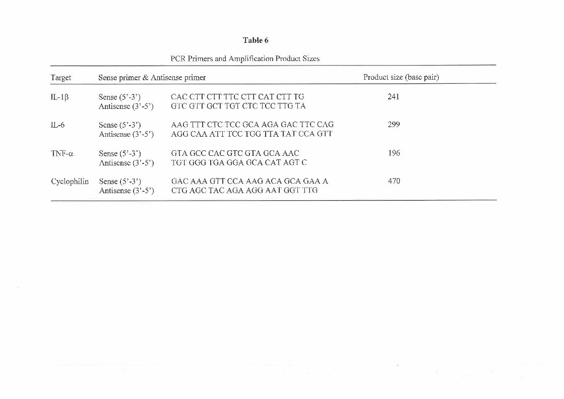
Table 6
PCR Primers and Amplification Product Sizes
Target Sense primer & Antisense primer
IL-lp Sense (5'-3')Antisense (3'-5')
TL-6 Sense (5'-3')Antisense (3'-5')
TNF-o Sense (5'-3')Antisense (3'-5')
Cyclophilin Sense(5'-3')Antisense (3'-5')
CAC CTT CTT TTC CTT CAT CTT TGGTC GTT GCT TGT CTC TCC TTG TA
AAG TTT CTC TCC GCA AGA GAC TTC CAGAGG CAA ATT TCC TGG TTA TAT CCA GTT
GTA GCC CAC GTC GTA GCA AACTGT GGG TGA GGA GCA CAT AGT C
GAC AJA.A' GTT CCA AAG ACA GCA GAA ACTG AGC TAC AGA AGG AAT GGT TTG
Product size (base pair)
24t
299
196
470
4.1.3 \ilF"sTERN BLOT ANALYSß
The protein levels of ILlp, IL-6 and TNF-a were analysed by western blotting.
4.1.3.1 PROTEIN EXTRACTION AND PREPARATION
For total protein extraction, a 2 cm segment of spinal cord encompassing the lesion epicentre
was placed in 4 ml of TRIzol@ Reagent. Tissue samples rvere completely homogenized and
800 ¡rl of chloroform was added to the sarrples. Following a brief vortex, the samples were
centrifuged at 12000 x g for 15 minutes at 4"C. The top layer (aqueous phase) containing total
RNAs was carefully removed and transferred to sterile tubes for RT-PCR while the bottom
layer (organic phase) containing total DNAs and proteins was used for western blot assay.
The bottom layer was mixed with 1.2 mI of lOÙVo ethanol and the mixture was centrifuged at
2000 x g for 5 minutes at 4"C to sediment DNA. The supernatant containing total proteins
was then mixed with isopropyl alcohol and the protein precipitate was sedimented at 12000 x
g for 10 minutes at 4"C. The protein pellet was washed 3 times in a solutiol ço¡laining 0.3 M
guanidine hydrochloride n 95Vo ethanol. The protein pellet was dissolve d n 2Vo SDS by
pipetting and complete dissolution of the protein pellet may require incubating the sample at
50t. The protein concentration was determined by Bio-Rad (Hercules, CA, USA) protein
assay. The samFle was then mixed with laemlis buffer and stored at -20oC for use in western
blot analysis.
Recombinant IL-IP, IL-6 and TNF-o @eprotech, Rocky Hill, NÐ were also dissolved in
laemlis buffer, serving as positive controls.
4.1.3.2 }VESTERN BLOTTING
116
Samples and recombinant controls were heated in boiling water for 3 minutes and then cooled
to room temperature. Twenty-five micrograms protein from each sample, recombinant
positive controls and BenchMarkru protein marker (GIBCO BRL, Baithersburg, MD) were
loaded onto líVo acrylamide El'ot. The gelwas run at2} mAin 1x SDS-PAGE running
buffer for 1.5 hours. Following electrophoresis, the separating gel, a piece of nitrocellulose
membrane (Millipore, Bedford, MA) of the same size, together with 4 pieces of blotting paper
was soaked in Semi-Dry transfer buffer for approximately l0 minutes. The gel-membrane
sandwich was then assembled in the Hoefer Semi-Dry transfer apparatus and the 1¡ansfer
ca¡ried out at 0.8 mA/cm2 for 90 minutes. After the transfer, the membrane was blocked in
5Vo ntik in Tris-buffered saline with O.lVo thimersoal (pH 7.5) at 4"C for overnight. The
membrane was incubated for 2 hours with a primary antibody, which was rabbit anti-rat IL-
1B, rabbit anti-rat IL-6 or rabbit anti-rat TNF-c antibodies (Peprotech, Rocky Hill" NI)
inl:500 dilution. Following the incubation with the primary antibody, the membrane was
washed three times with Tris-buffered saline with O.lVo thimersoal (pH 7.5) and incubated for
I hour with a secondary antibody (goat against rabbit IgG conjugated with alkaline
phosphatase, 1: 10000) (Sigma, St. Louis, MO). The membrane was then incubated with
substrate development solution (10 ml alkaline phosphatase substrate buffer, 100 pl NBT and
100 ¡tl BCIP) for 5 minutes and rinsed in deionised water. The membrane was allowed to air
dry on a paper towel and intensities of protein bands were quantified densitometrically by
NIH image 1.62 (NIH, Bethesda, MD). In case where levels of IL-IB, IL-6 and TNF-cr were
not detectable, the data were recorded as 1 arbitrary densitometric unit. The anount of each
product was normalized to the recombinant control for comparison.
4.1.3.3 STATISTICAL ANALYSES
It7
Multiple samples urere 4nalysed by one-way analysis of variance (ANOVA). Differences
be¡neen 1¡e mild and severc SCI groups were analysed by a post-hoc test of Student Neuman
IGuls. AP value less than 0.05 was considered significant.
118
4.2 RESULTS
4.2.1RT-PCR
The profile of cytokine transcription in contused spinal cord was marked by an early, robust
but transient increase in IL-lp, IL6 and TNF-a mRNAs.
4.2.1.1IL-1ß mRNA
RT-PCR products representing tr--lp mRNA levels were shown in Figure 15 with a size of
241 buse pairs. The sfaining intensity of RT-PCR products (amplified for 35 cycles) was
directly proportional to the amount of DNA because pixel density units of these products were
within the linear range of RT-PCR (refer to appendix 2).
The levels of IL-1p mRNA in the group of severe SCI were elevated at I and 3 h after injury,
peaked at 6 h and declined to near baseline levels by I d postinjury @igures 154 and 16).
Compared to the mild injury group @igures 158 and 16), the mRNA levels of IL-lp in the
group of severe SCI were significantly higher at 3 and 6 h postinjury (Pd.05). The maximum
level of the IL-lp transcript in the severe SCI group at 6 h postinjury represented a 5-fold
increase over the level of mild injury group. Additionally, the levels of the IL-IP mRNA in
the mild SCI were significantly increased at I h, 3 h and 6 h postinjury @<O.05) compared to
laminectomy controls (Figures 15C and 16). In the group of laminectomy controls,
fluctuations in IL-lp mRNA were hardly noted and their expression levels were quite similar
to those in the naive control group (Þ0.05) (refer to appendix 2).
It9

Figure 15. RT-PCR products for the ILIP gene. Column M: molecular weight marl<¿rs.Colutnns th, 3h, 6h, Id and 3d: RT-PCR products of IL-LQ at I hour, 3 hours, 6 hours, Idayand. 3 days postinjury. A: II-IQ n,RNA expressions after severe SCI; B: IL-IP mRNAexpressions after mild SCI; and C: II-IQnRNA expressions in laminectony controls.
r20
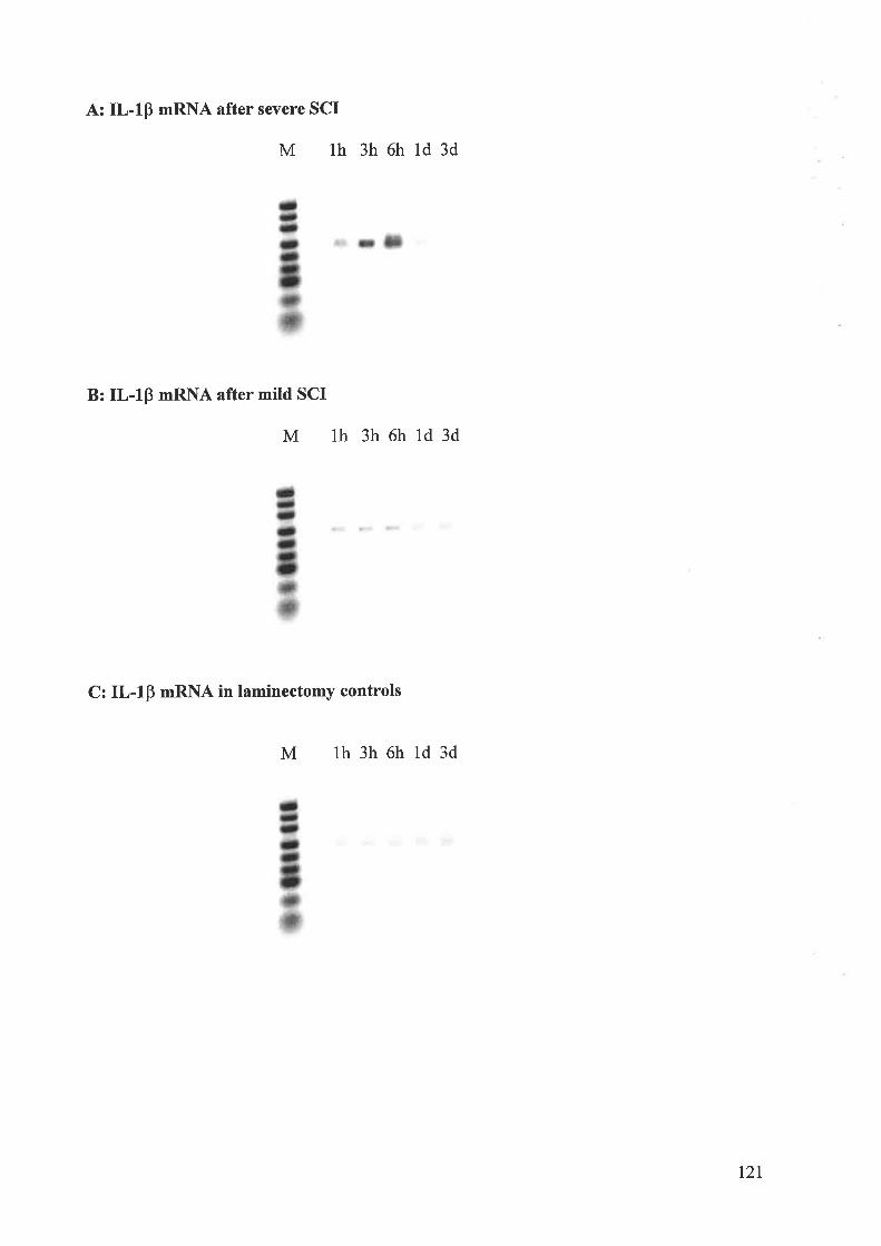
A: IL-IB mRNA after severe SCI
M th 3h6hld3d
B: IL-IB nRNA after mild SCI
M th 3h6hld3d
C: IL-lp mRNA in laminectomy controls
M th3h6hld3d
t2r
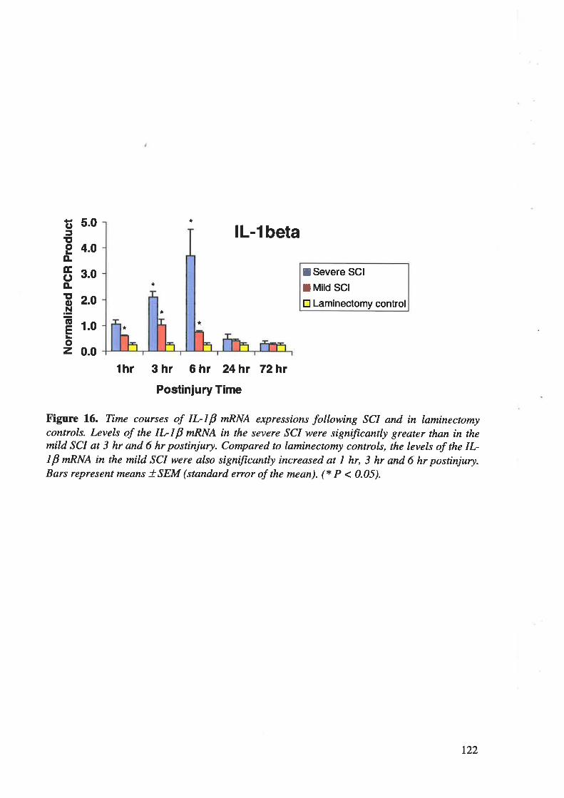
5.0
4.0
3.0
2.0
I
0.0
C'5ItoLo.EC)ÀEoN=(EELoz
*
lL-l beta
*I Severe SCI
I Mird scttr Laminectomy control
t
.0 **
thr 3hr 6hr 24hr 72hr
Postinjury Time
tr'igure 16. Time courses of IL-IP nRNA expressions following SCI and in laminectomycontrols. Levels of the IL-I P nRNA in the severe SCI were significantly greater than in themild SCI at 3 hr and 6 hr postinjury. Compared to laminectomy controls, the levels of the IL-1B mRNA in the mild SCI were also significantþ increased at I hr, 3 hr and 6 hr postinjury.Bars represent means tSEM (standard enor of the mean). (* P < 0.05).
r22
4.2.1.2IL-6 mRNA
RT-PCR products representing IJ.,-6 mRNA levels are shown in Figure 17 with a size of 299
base pairs. Jfte steining intensity of RT-PCR products (amplified for 35 cycles) was directly
proportional to the amount of DNA because pixel density units of these products were within
the linear range of RT-FCR (refer to appendix 2).
The expression profile of IL-6 nRNA in spinal cord was vinually identical to the IL-IP
pattern, shown in Figures 15 and 16. The levels of IL-6 mRNA in the group of severe SCI
were elevated at I and 3 h after injury, peaked at 6 h and declined to nea¡ baseline levels by I
d postinjury (Figures l7A and 18). Compa¡ed to the mild injury group (Figures 178 and 18),
the nRNA levels of IL-6 in the group of severe SCI were significantly higher at 3 and 6 h
postinjury e<o.05). The maximum level of the IL-6 transcript in the severe SCI group at 6 h
postinjury represented a7-fold increase over the level of mild injury group. Additionally, the
levels of the IL-6 mRNA in the mild SCI were significantly increased at t h, 3 h and 6 h
postinjury e<o.05) compared to laminectomy controls (Figures 17C and 18). In the group of
laminectomy controls, fluctuations in IL-6 mRNA were hardly noted and their expression
levels were quite similar to those in the naive control group (P>0.05) (refer to appendix 2).
123
figure 17. RT-PCR products for the II-6 gene. Column M: molecular weight marl<crs.Colunms Ih, 3h, 6h, Id and 3d: RT-PCR prodacts of IL6 at I hour, 3 hours, 6 hours, Idayand, 3 døys postinjury. A: IL6 wRNA expressions after severe SCI; B: II-6 nRNAexpressions afier mild SCI; and C: IL6 ntRNA expressions in laminectonty controls.
124
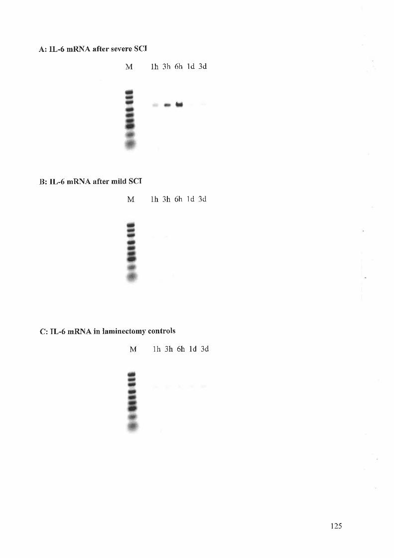
A: IL-6 mRNA after severe SCI
M th3h6hld3d
B: IL-6 mRflA after mild SCI
M th3h6hld3d
C: IL-6 mRl\lA in laminectomy controls
M th3h6hld3d
t25
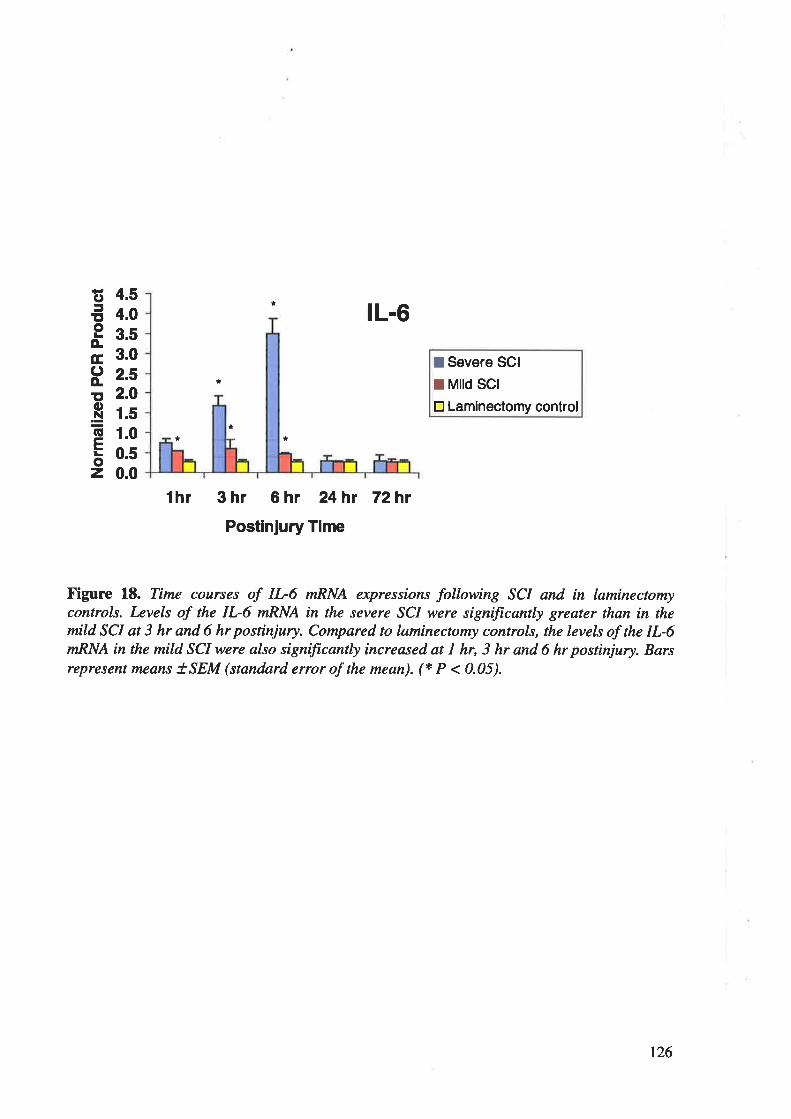
lL-6
I Severe SCI
I Mird scrtr Laminectomy control
thr 3hr 6hr 24hr 72h¡
Postinjury Time
X'igure 18. Time courses of IL-6 mRNA expressions following SCI and in laminectomycontrols. Levels of the IL-6 wRNA in the severe SCI were significanþ greater thøn in themild SCI at 3 hr and 6 hr postinjury. Compared to laminectomy controls, the levels of the IL-6mRNA in the mild SCI were also signiftcanþ increased at I hr, 3 hr and 6 hr postinjury. Barsrepresent means ISEM (standnrd eruor of the mean). (* P < 0.05).
Fo5ttoLÀÉ,oo.Eo.NGELoz
*
*
*
*
*
4.54.03.53.02.52.01.51.00.50.0
t26
4.2.1.3 TNFa mRNA
RT-PCR products representing TNF-ct, mRNA levels are shown in Figure 19 with a size of
196 base pairs. The staining intensity of RT-PCR products (amplified for 35 cycles) was
directþ proportional to the amount of DNA because pixel density units of these products were
within the linea¡ range of RT-PCR (refer to appendix 2).
The levels of TNF-a nRNA in the group of severe SCI were elevated at I and 3 h after
injury, peaked at 6 h and declined to near baseline levels by I d postinjury (Figures 194 and
20). Compa¡ed to the mild i"j".y group @igures 198 and 20), the mRNA levels of TNF-a in
the group of severe SCI were significantly higher at I,3 and 6 h postinjury @<O.05). The
maximum level of the TNF-a transcript in the severe SCI group at 6 h postinjury represented
a 6-fold increase over the level of mild injury group. Additionally, the levels of the TNF-cr
mRNA in the mild SCI were significantly increased at t h, 3 h and 6 h postinjury (P<O.05)
compared to laminectomy controls (Figures 19C and 20). In the lanrinectomy controls, no
significant differences in TNF-cl mRNA were soen when compared with narve controls
(P>O.05) (refer to appendix 2).
127
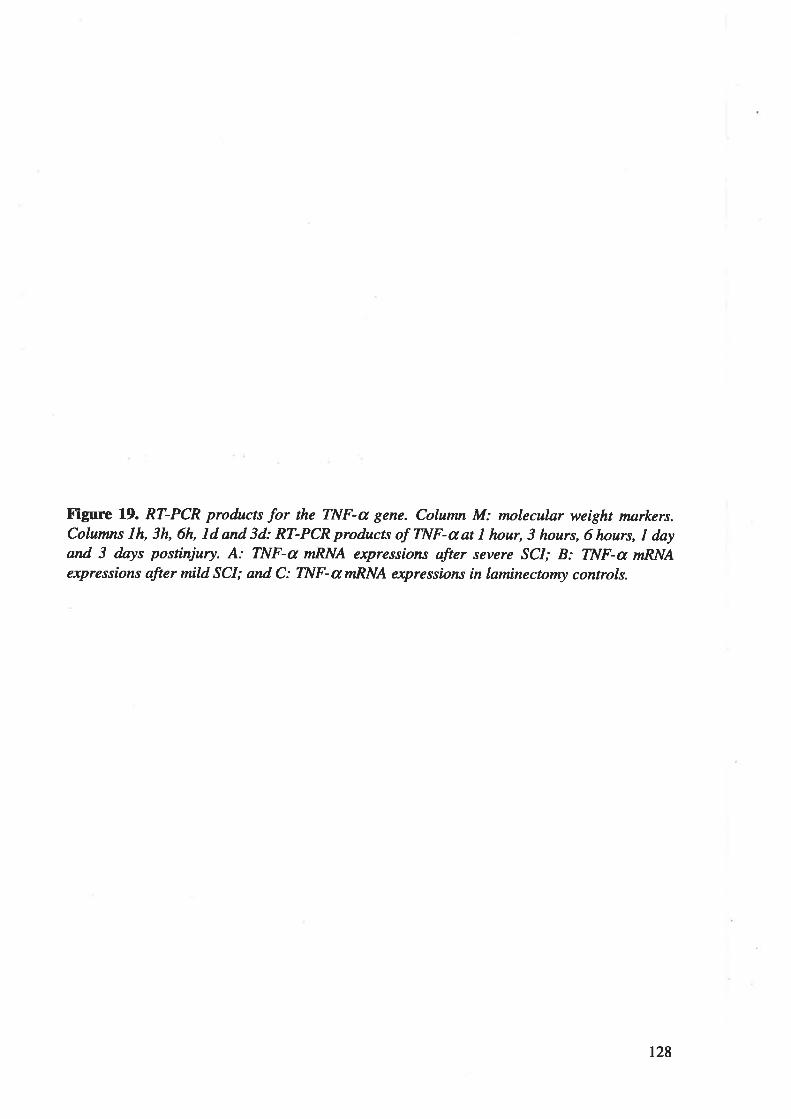
Figure 19. RT-PCR products for the TNF-u gene. Column M: molecular weight markers.Columns Ih, 3h, 6h, Id and 3d: RT-PCR products of TNF-øat I hour, 3 hours,6 hours, I doyand 3 days postinjury. A: TNF-a nRNA expressions øfter severe SCI; B: TNF-a nRNAexpressions after mild SCI; and C: TNF-amRNA expressions in laminectomy controls.
128
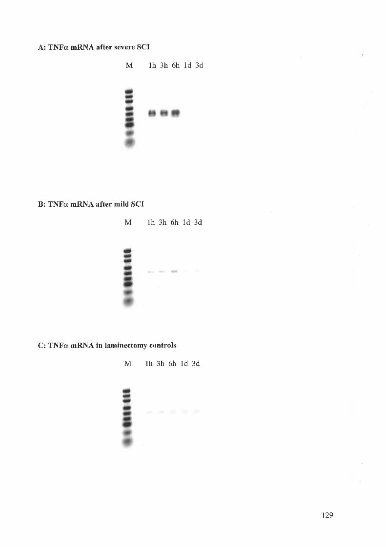
A: TNFcr mRNA after severe SCI
B: TNFg mRNA after mild SCI
M th3h6hld3d
M th3h6hld3d
C: TNFa mRNA in laminectomy controls
M th3h6hld3d
129
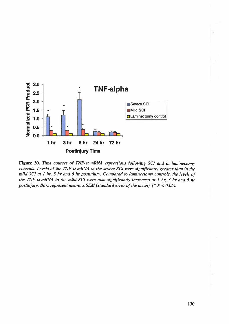
3.0
0.5
0.0
lHC'
=t,oLÀEoÀt,o.N(ú
ELoz
2.5
2.0
1.5
1.0
* TNF-alpha
* ISerære SCI
rMild scrtrLaminectomy contrcl
*
** *
t hr 3 hr 6 hr 24h¡ 72hr
Postinjury Time
Figure 2O. Time courses of TNF-a nRNA expressions following SCI and in laminectomycontrols. Levels of the TNF-a wRNA in the severe SCI were signiftcanþ greater than in themild SCI at I hr, 3 hr and 6 hr postinjury. Compared to laminectomy controls, the levels ofthe TNF-a nRNA in the mild SCI were also signiftcanþ increased at I hr, 3 hr and 6 hrpostinjury. Bars represent means tSEM (standard etor of the mean). (* P < 0.05).
130
4.2.2WESIERN BLOTTING
The profile of cytokine proteins in contused spinal cord was also marked by an early, robust
but transient increase in IL-IP, IL-6 and TNF-cr proteins. However, the increase in the pro-
inflammatory cytokines was only observed in the group of severe SCI and no immunoreactive
cytokine proteins were detected in the groups of mild SCI, laminectomy controls or naïve
controls.
4.2.2.1 rl.-lp PROIErN
\Vestern blotting showed labelling of single bands with a molecula¡ weight of 17.3 kDa for
tr--lp in Figure 21. The intensity of protein bands was directly proportional to the amount of
protein because arbitrary densitometric units of these protein bands were within the linear
range of western blot analysis (refer to appendix 2).
The levels of IL-lp protein in the group of severe SCI were elevated at 1 and 3 h after inju.y,
peaked at 6 h and declined to undetectable levels by I d postinjury while no immunoreactive
tr--lp was detectable in the spinal cord extracts from rats in the groups of mild SCI,
larninectomy controls and narve controls (Figures 2I). Thus, comFared to mild SCI,
liaminectomy controls and naïve controls, the levels of IL-lp protein at th, 3h and 6h
following severe SCI were significantþ higher (Pd.05) (refer to appendix 2).
13t
Figure 21. Temporal expressions of IL-Ip at the protein level in the severe SCI detnonstratedby western blot analysis. Lcvels of the IL-lp protein after severe SCI began to increase at Iand 3 hr posrtnjury, peaked at 6 hr and quicHy decline to non-detectable levels by I d (A) II-I p protein bands of the group of severe SCI. (B) Qtnntificarton of IL-I p protein bands of thegroup of severe SCI. Bars represent mcans tSEM.
t32
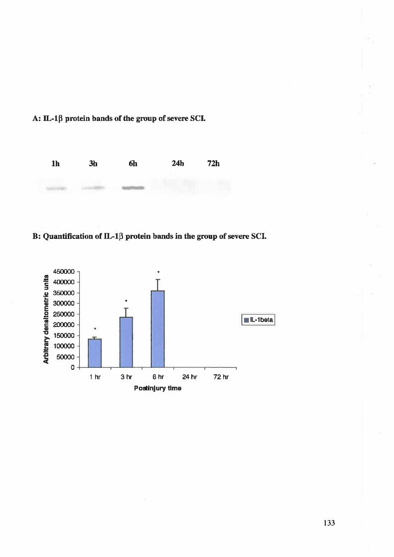
A: IL-IB protein bands of the group of severe SCI.
lh 3h 6h 24h 72b'
*ËË
B: Quantification of IL-IB prctein bands in the group of severe SCI.
450000
¿|OO0O0
350000
300000
250000
200000
150000
100000
50000
0
I lL-1beta
thr 3 hr 6 hr 24hr
Postlnlury tlme
72hr
ttr3C'
oEoËotroEL6
o
133
4.2.2.2IL-6 PROTEIN
Western blotting showed labelling of single bands with a molecula¡ weight of 23 kDa for IL-6
in Figure 22. T\e intensity of protein bands was directþ proportional to the amount of protein
because arbitrary densitometric units of these protein bands were within the linea¡ range of
western blot analysis (refer to appendix 2).
The levels of IL-6 protein in the group of severe SCI were elevated at I and 3 h after injury,
peaked at 6 h and declined to undetectable levels by I d postinjury while no immunoreactive
IL-6 was detectable in the spinal cord extracts from rats in the groups of mild SCI,
laminectomy controls and naïve controls (Figures 22). Thus, compared to mild SCI,
laminectomy controls and narve controls, the levels of IL-6 protein at th, 3h and 6h following
severe SCI were significantly higher (Pd.05) (refer to appendix 2).
134
Flgure 22. Temporal expressions of IL6 at the protein level in the severe SCI demonstratedby western blot analysis. Lcvels of the II-6 protein after severe SCI began to increase at Iand 3 hr posrtnjury, peal<cd at 6 hr ønd quicHy decline to non-detectable levels by I d.. (A) IL6 protein bands of the Broup of severe SCI. (B) Quantification of IL-6 protein bands of thegroup of severe SCI. Bars represent m¿ans !SEM.
135
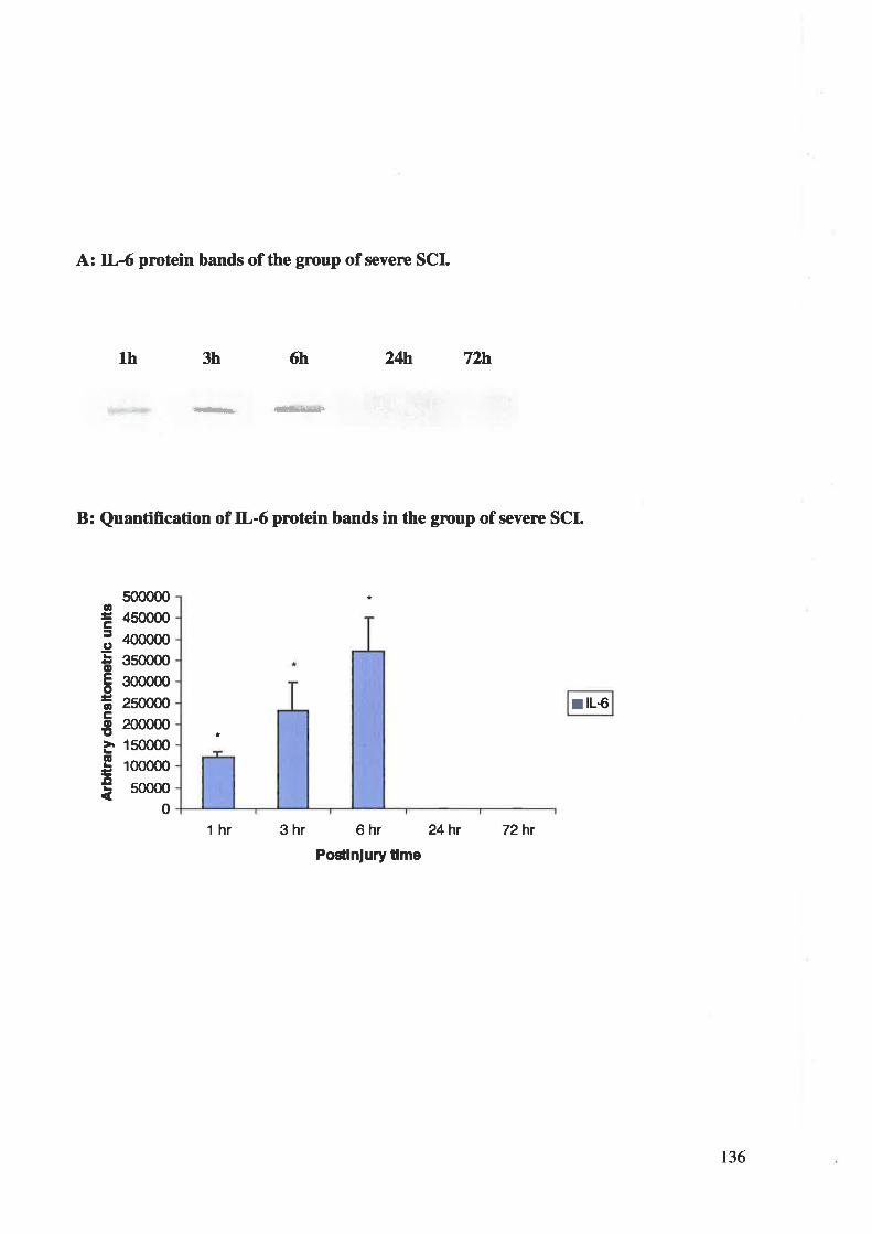
A: IL-6 protein bands of the group of severe SCI.
th 3h 6tr 24}f, 72h
{-ts* --æ
B: Quantifrcation of IL-6 protein bands in the group of severr SCI.
500000
450000
400000
3s0000
300000
2s0000
200000
1s0000
100000
50000
oc:goEoacoE
EL
o
I IL€
0
thr 3hr 6 hr 24h¡
Postlnlury tlme
72hr
136
4.2.2.3 TNFcr PROTEIN
'Western blotting showed þþlling of single bands with a molecular weight of 17 kDa for
TNFa in Figure 23. The intensity of protein bands was directly proportional to the amount of
protein because arbitrary densitometric units of these protein bands were within the linear
range of western blot analysis (refer to appendix 2).
The levels of TNFa protein in the group of severe SCI were elevated at 1 and 3 h after injury,
peaked at 6 h and declined to undetectable levels by I d postinjury while no immunoreactive
TNFct was detectable in the spinal cord extracts from rats in the groups of mild gg¡,
laminectomy controls and narve controls (Figures 23). Thus, compared to mild SCI,
liaminectomy controls and narve controls, the levels of TNFa protein at th, 3h and 6h
following severe SCI were siguificantly higher (P4.05) (refer to appendix 2).
t37
Ftgure 23. Temporal expressions of TNFu øt the protein level in the severe SCI demonstratedby western blot analysis. I*vels of the TNFa protein after severe SCI began to increase at Iand 3 hr posrtnjury, pealccd at 6 hr and quicWy decline to non-detectable levels by I d- (A)TNFa protein bands of the group of severe SCI. (B) Quanrtftcation of TNFaprotein bands ofthe group of severe SCI. Bars represent mcans tSEM.
138
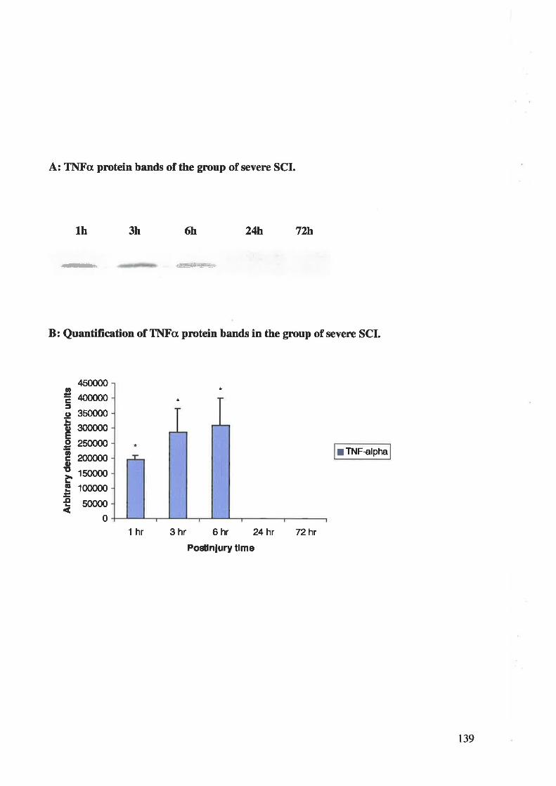
B: Quantification of TI\{Fcr protein bands in the group of sevene SCI.
A: Tl\lFcl protcin bands of the grcup of severe SCI.
th 3h 6h 24tn 72h
.*. #
4s0000
¿100000
350000
300000
250000
200000
150000
100000
s0000
0
thr 3 hr 6 hr 24hr
Postlnlury tlme
72h¡
0Ê5goEootroE
6
.o
ITNFalpha
t39
4.3 DISCTISSION
4.3.1 CELLIILAR SOURCES OF IL-IP,IL-6 and TI{X'-cr
The rapid elevation of IL-lp, IL6 and TNF-c mRNA expressions at 1,3 and 6 h after SCI
was followed by a dramatic decline of these expressions prior to the peak appearance of
neutrophils and macrophages, indicating that these pro-inflammatory cytokines are primarily
produced by endogenous CNS cells rather than infiltrating blood-borne leucocytes. This is
supported by our detection of TNF-cr expression in neurons and microglial cells after SCI (see
section 3.2). These data suggest that neurons and microglial cells, not blood-born leucocytes
proposed by many authors L3s'347'354, may play a major role in the initiation and regulation of
early posttraumatic inflammatory response of SCI by producing tr--lp, IL-6 and TNF-a.
4.3.2 DISPARITIES BETWEEN TRANSCRTPTION AND TRANSLATION OF IL-lp,IL.6 AI\D TI\F-A
To our knowledge, t\is is the first study to address the disparities between s5mtheses of pro-
inflammatory cytokine mRNAs and their bioactive proteins in SCI. Expressions of IL-lp, IL-
6 and TNF-a at both nRNA and protein levels in severe SCI were dramatically elevated at 1,
3 h after injury, peaked at 6 h and declined to nea¡ baseline levels by I d postinjury. Thus, in
the severe SCI, the temporal profiles of IL-lp, IL-6 and TNF-cr protein expressions closely
parallel their nRNA syntheses. Although IL-lp, IL-6 and TNF-c mRNA levels increased in
mild injury, we were unable to detect the corresponding proteins histochemically.
The dissociation between mRNA and protein expression levels of these pro-inflammatory
cytokines in the mild SCI may be attributed to a combination of two factors. First, different
experimental techniques may cause the dissociation because two different techniques, RT-
PCR and western blotting, were respectively used to detect the mRNA and protein
IQ
expressions of IL-IP, IL-6 and TNF-c" The RT-PCR process is exquisitely sensitive and a
single copy of target nucleic acid can be detected by PCR because the number of target
nucleic acid copies approximately doubles every cycle and 35 cycles of PCR in this study
may ampliry the target nucleic acid by about thirty billion t¡mes (235) "'ttt. W".tern blot
analysis, when compared to RT-PCR, is relatively insensitive as the detection limits for IL-
lP, IL-6 and TNF-cr proteins are about 1.5-2.0 ng according to the antibody manufacturer's
protocol @eprotech, Rocþ Hill, NI). Second, mild SCI may not be sufficient to provide a
signal for enough transliation of the mRNAs into the bioactive cytokine proteins despite a
vigorous signal for slntheses of the pro-inflammatory cytokine mRNAs. The s¡mtheses of
mature tr--lp, IL-6 and TNF-o proteins are tightþ controlled at many different levels to
ensure the silence of the pro-inflammatory cytokines in the CNS under normal circumstances
and each one of the slmthetic steps from sensing the presence of SCI to cytokine secretion is
controlled by different molecular events regulating the cytokine gene, mRNA and protein
t8'00s. Thus, in mild SCI, the s¡mtheses of mature tr--lp, IL-6 and TNF-cr proteins may not be
able to overcome the apparentþ intrinsic inhibition to sufficiently translate the mRNAs. This
is further confirmed by our finding that TNFcr expression by immunohistochemistry occurs in
fewer neurorls and glial cells in the mildly injured spinal cord compared to severely injured.
In summary, the symtheses of mature L-lp, IL-6 and TNF-cr proteins did not mirror their
nRNA productions following mild SCI and were not sufficient for detection with western
blot analysis.
4.3.3 SEVERITY-DEPENDENT E)PRESSIONS OF IL-lp,IL-6 AI\D TI\X'-a
Our results demonstrated significant elevations in IL-lp, IL-6 and TNF-a following severe
SCI compared to mild SCI. To our knowledge, this is the first report of severity-dependent
l4t
expr€ssions of IL1p, IL-6 and TNF-a in SCI, suggesting that these cytokines may be suitable
targets for phannacological intervention.
These data also suggest a positive association between neurological impairment and excessive
expressions of IL-lp, IL-6 and TNF-cl as mild SCI did not cause either lasting functional
deficits or substantially increased expression of pro-inflammatory cytokines. Thesc rcsults are
consistent ryi1þ simila¡ changes observed previously in traumatic brain injury where no
neurologic deficits were caused in a mild contusion injury with a small up-regulation of TNF-
cr expression 176're4. Therefore, expression levels of the L-lp, IL-6 and TNF-c may be
associated with severity of SCI, especially with reference to resultant neurological deficits.
tr--lp, IL-6 and TNF-a are important and modifiable components of the inflanmatory
response to traumatic CNS injury. The production of IL-6 is regulated by a variety of stimuli,
including IL-lp and TNF-cr r72'36e.In experimental traumatic injury of the CNS, it has been
shown that the IL-6 receptor was present in neurons and oligodendroc¡es 381, and that IL-6
induced neurite outgrowth aI1d improved neuronal survival by inducing the spthesis of
neurotrophic factors 147'ts7'1e7. Conversel¡ anti-rat IL-6 antibody was found to significantþ
attenuate the upregulation of inducible nitric oxide synthase and reduce secondary spinal cord
damage 36t. Thur, IL-6 is thought as a kind of neurotrophic and cytotoxic factor and is
partially regulated by IL-lp and TNF-cr "t. tr--lp is a highly inflammatory cytokine, which is
implicated in neuronal necrosis and apoptosis, leukocyte infiltration, edema, activation of
Blial cells, induction of cytokines and slmthesis of nitric oxide 88. For example, tr--lþ is able
to induce IL-6 secretion from astrocytes a, stimulate astrocytes to proliferate 135'211 a.d
enhance the slmthesis of nerve growth factor (NGÐ and basic fibroblast growth factor
(bFGÐ in astrocytes247'2$4.It has been also reported that systemic injections of IL-l receptor
t42
antagonist reduced neuronal cell death and improved cognitive function after lateral fluid
percussion head injury in the tat 3M. Thus, ILI has complex and bi-directional biological
effects in the inflammatory response of CNS tissue to trauma. TNF-a shares some similar
properties with IL-lp in terms of pro-inflammatory effects, such as the promotion of
apoptosis in neurons 238'346, the activation of glial cells 366, the stimulation of astrocyte
proliferation 37e and thc induction of IL-6 and growth factors in astrocytes 305'3ee. TNF-a is
also believed to enhance the infiltration of blood leucocytes by modulating the expression of
various surface antþens and adhesion molecules on endotheli¿l cells 348. Additionall¡ the
cytotoxic effects of TNF-a on oligodendrocytes, leading to demyelination, have been shown
in vitro ea and in vivo 2eo'3e6. In contrast to its toxic actions, TNF-o has also been
demonstrated to have a regenerative effect on Ð(ons 313 and to have a neurotrophic effect by
inducing s¡mthesis of NGF in astrocytes 3Ð. Thus, TNF-a is a powerful inflammatory
mediator with both cytotoxic and neurotrophic actions.
Taken together, tr--lp, IL-6 and TNF-cr have been shown to produce tissue damage whereas
they may also induce the production of neurotrophic molecules to rescue the injured tissue in
the posttraumatic response. The explanation of these apparent contradictions requires a
complete understanding of dynarrics of the posttraumatic inflammatory response in SCI. The
early and robust rise in tr--lp, IL-6 and TNF-a following SCI observed in this study may be
detrimental because they may be overproduced beyond the level of neurotrophic effects and
the margins between benefit and toxicity are quite narrow a0s. 1¡ is supported by the finding
of spontaneous inflammatory demyelination in the CNS in transgenic mice overexpressing
TNF-a 283. In the present study no neurologic deficit was observed in mild SCI with a small
upregulation of proinflammatory cytokine expression. The neurotrophic effects induced by
143
tr--lp, IL-6 and TNFc may not oocur because the cells producing the neurotrophic molecules
may be damaged or killed in CNS in¡ry.
Therefore, these data suggest that there may be dual roles of IL-lp, IL-6 and TNF-a in SCI
and the final effects of these pro-inflammatory cytokines rnay be greatþ determined by many
factors.
a
t4
5. TREATMENT OF NF-'G.SPECIFIC ANTISENSEOLIGODEOXYNUCLEOTIDES AFTER SEVERE SCI
5.1 METHODS
5.1.1 ITX'.KB-SPECIFIC AI\TISENSE OLIG'ODEOXYI\TUCLEOTIDES
The NF-rB-specific antisense oligodeoxymucleotides in albumin microencapsulation weÍìe a
generous gift from Dr. C. Oettinger and Prof. M.J. D'Souza at Deparhnent of Pharmaceutical
Sciences, College of Phannac¡ Mercer University, Atlanta, GA 30308, USA The NF-rB-
specific antisense oligodeox5nucleotides in their studies have been shown to significantly
reduce the upregulation of TNFcr, IL-lp and IL-6 in septic shock model in the rat, slashing
lAÙVo death rate after endotoxin inþction to only 207o.
5.1.2 REX'ORMIILATION OX' NF-KB-SPECTFIC ANTISENSEOLIGODEOXYI\TUCLEOTIDES
Six hundred micrograms of oligodeox¡mucleotides were weighed and re-suspended in 1 ml
normal saline containing 2Vo Twe,en 80 as a suspending agent (pre-sterilized by autoclaving).
The suspension sample was then sonicated for 30 seconds and ready for use.
5.1.3 INJECTION OF NI'-KB-SPECFIC ANTISENSEOLIOGODEOXYIITUCLEOTIDES
Fourteen rats received a subarachnoid inþction of 5 pl of suspension containing 3 pg NF-KB-
specific antisense oligodeoxynucleotides and an intravenous inþction of 955 pl of suspension
containing 597 ¡tg NF-tcB-specific antisense oligodeoxlmucleotides immediately after severe
SCI produced by 12 cm weight-drop. Rats were sacrificed at I hr (N=4), 3 hr (N=5) and 6 hr
(N=5) postinjury.
5.1.4 mRNA AND PROTEIN EXTRACTION
145
The nRNA and protein of IL-lp, IL-6 and TNFa were extracted from rat spinal cords
according to the methods described in section 4.1.
5.2 RESULTS
The upregulation of IL-lp, IL-6 and TNFa at the levels of mRNA and protein after severe
SCI was not significantþ inhibited by the NF-rcB-specific antisense oligodeox5mucleotides in
the treatment group compared to non-treatment group. At 1, 3 and 6 hr postinjury, large
increases in the production of IL-lp, IL-6 and TNFc¡ mRNA and protein were still observed
in spinal cord extracts from rats treated with antisense afrer severe SCI.
5.3 DISCUSSION
Although the NF-rB-specific antisense oligodeoxlmucleotides have been shown to
significantly reduce the upregulation of TNFcr, IL-lp and IL-6 in a septic shock model in the
rat, the souÌces of upregulated cytokines (tr--lp, IL-6 and TNFcr) in SCI models a¡e different
from those in septic shock models. The upregulation of IL-IB, IL-6 and TNFa after CNS
iojury is mainly observed in the spinal cord and brain 168'267'3s4'37e whereas the increase in
these cytokines is largely present in the systemic circulation after septic shock 230. Thus, the
NF-tcB-specific antisense oligodeoxynucleotides administered intravenously in the septic
shock model can easily get to the circulation to inhibit the cytokine upregulation. In contrast,
most of the NF-rB-specific antisense oligodeoxlmucleotides were administered via the
femoral vein due to the limited subarachnoid space. Therefore, the main factor behind the
failure of inhibition by the NF-rcB-specific antisense oligodeox5mucleotides may be a lack of
vehicle to deliver the antisense into the targeted cells (e.g., neurons and microglia), which are
the main sources of upregulated cytokines, since our studies have shown endogenous cells
146
including neurons and microglia contribute substantially to the large increase in IL-lp, IL-6
and TNFo observed in the severely contused spinal çs¡d.
Support from this comes from an unpublished study presented at the 6û Inteinational
Neurotrauma Syrnposium in which haemagglutinating virus of Japan-liposome complex
çs¡laining the NF-rB-specific antisense oligodeoxlmucleotides was used in their study to
significantly reduce the mRNA levels of IL-lp, IL6 and TNFo in the rat spinal cord in the
ischaemic SCI model The use of NF-tcB-specific antisense oligodeoxlmucleotides for the
treatment of traumatic SCI requires further study.
147
HUMAN SCI EXPERIMENTS
1. MATERIALS AND GENERAL METHODS
1.1 SELECTION OF MATERIAL
1.1.1 TRAUMA CASES
Eleven cases of human SCI resulting from acute trauma as a result of a motor vehicle accident
(9 men, 2 women; age range, 18-83 years) were selected for the study (Table 4). The study
samples were selected from a series of spinal cord injury cases that survived long enough to
be admitted to the Spinal Unit at the Royal Adelaide Hospital. All subsequently died and
underwent a full post-mortem examination during the period 1978-1997. Al1 the spinal cords
were delivered to the Neuropathology Department at the Institute of Medical and Veterinary
Science for neuropathological examination.
Injuries that resulted from chronic compression of the spinal cord were excluded. Clinical
information was obtained from hospital case notes and accident reports. Only those cases with
adequate records of the circumstance of the injury, adequate patient clinical information and
adequate neuropathologic data were included in the study. Details of a concomitant
hypotension episode that may have compounded the traumatic event were also recorded
(Table 4).
1.1.2 CONTROL CASES
Due to the difficulty of obtaining normal spinal cords from post-mortem, only two human
spinal cords were available as controls (Table 4). These were from patients with traumatic
brain injury only and had no evidence of pathological abnormality in the spinal cord.
148
1.2 TISSUE PREPAR, TION
All spinal cords were removed from the human body within24h after death and were then
lrxed in 40%o formalin for a minimum of ten days. All the cords were then examined
according to standard neuropathological techniques. Individual cord segment levels were
determined by counting the segments above and below ventral nerve root of T2 (a landmark
due to the marked anatomical disparity in size between T1 and T2 nerve roots).
Tissue blocks were dehydrated in graded alcohol baths and cleared with chloroform, prior to
processing into parafhn wax. Five micrometer serial sections were cut at (150 prm) levels with
a microtome ancl mountecl onto slides coated with APT (Sigma Chemical Corporation, US)
The sections were dried at 37"C for at least 12 hours prior to use. Sections from each level
were stained with H&E and immunohistochemically
r49
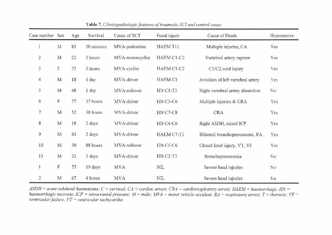
Table 7 . Clinicopathologic features of traumatic SCI and control cases
Case number Sex Age Survival Cause of SCT Focal injury
2
aJ
4
5
6
7
8
9
1 M 83 30 minutes MVA-pedestrian HAEM T11
M22 5 hours
5 hours
1 day
1 day
37 hours
38 hours
2 days
2 days
88 hours
5 days
19 days
4 hours
MVA-motorcyclist HAEM Cl-C2
MVA-cyclist
M18
10 M30
F73
M18
M48
F77
M52
HAEM CI-C2
MVA-driver HAEM Cl
MVA-rollover HN C5-T2
MVA-driver HN C3-C6
MVA-driver HN C7-C8
MVA-driver HN C4-C6
MVA-driver HAEM C]-TI
MVA-rollover HN C5-C6
MVA-driver HN C2-T7
MVA
Cause of Death
Multiple injuries, CA
Vertebral artery rupture
CIIC2 cord injury
Arulsion of left vertebral artery
Right vertebral artery dissection
Multiple injuries & CRA
CRA
Right ASDH, raised ICP
Bilateral bronchopneumonia, RA
Closed head injury, VT, VF
Bronchopneumonia
Severe head injuries
Severe head injuries
Hypotensive
Yes
Yes
Yes
Yes
No
Yes
Yes
Yes
Yes
Yes
No
63M
11 M32 No
1 F75 NIL
No
ASDH: acute subdural haematoma; C : cervical; CA: cardiac arrest; CRA : cardiorespiratory arrest; HAEM: haemoruhage; HN :haemorrhagic necrosis; ICP : intracranial pressure; M: male; MVA : motor vehicle accident; RA : respiratory arrest; T : thoracic; VF :ventricular failure; W : ventricular tachycardia.
2 M67 MVA NIL
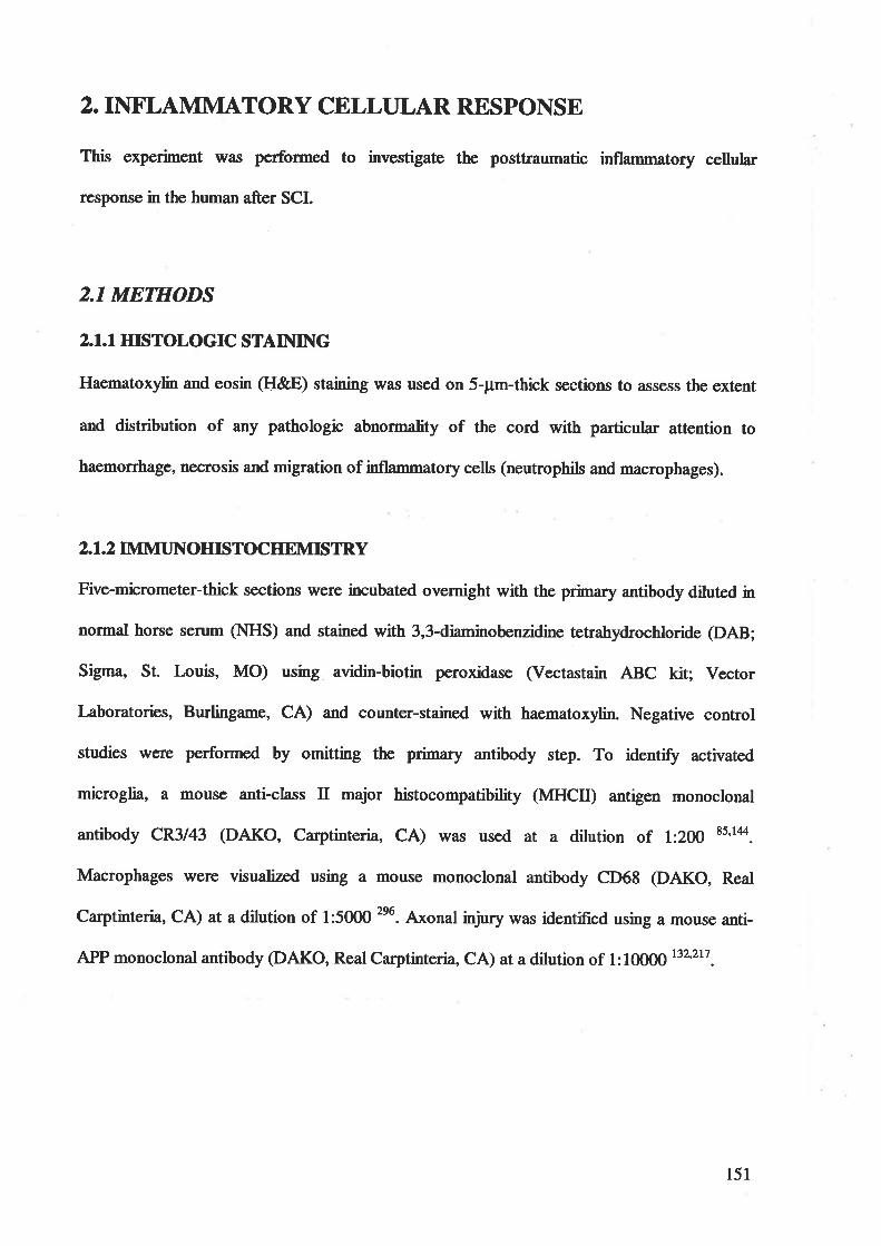
2. II\FLAMIVIATORY CELLT]LAR RESPONSE
This experiment lvas performed to investigate the posttraumatic inflammatory cellular
respor¡se in the human after SCI.
2.1 METHODS
2.1.1 HISTOLOGIC STAII\IING
Haematoxylin and eosin (H&E) staining was used on 5-pm-thick sections to assess the extent
and distribution of any pathologic abnormality of the cord with particular attention to
haemorrhage, necrosis and migration of inflammatory cells (neutroph;ls and macrophages).
2. 1.2 IMMT]NOHISTOCHEIVIIS TRY
Five-micrometer-thick sections were incubated overnight with the primary antibody diluted in
normal horse serum (N[HS) and stained with 3,3-diaminobenzidine tetrahydrochloride (DAB;
Sigma, St. Louis, MO) using avidin-biotin peroxidase (Vectastain ABC kit; Vector
Laboratories, Burlingame, CA) and counter-stained with haematoxylin. Negative control
studies were performed by omitting the primary antibody step. To identiff activated
microglia, a mouse anti-class II major histocompatibility (MHCtr) antigen monoclonal
antibody CR3/43 (DAKO, Carptinteria, CA) rvas used at a dilution of 1:200 85'1'14.
Macrophages we¡e visualized using a mouse monoclonal antibody CD68 @AKO, Real
Carptinteria, CA) at a dilution of 1:5000 2e6. Axonal injory was identified using a mouse anti-
APP monoclonal antibody (DAKO, Real Carptinteria, CA) at a dilution of 1:10000 t32'2t7.
151
2.2 RESULTS
2.2JN MIIITUTES PIOST SCI
At 30 minutes after traumatic SCI, the lesion centre was marked by haemorrhage mainly in
the central grey matûer, but no neutrophils or other leucocytes tvere soen in the areas of
damaged tissue (Figures 24A-B). Meanwhile , early axonal injury was demonstrated by APP
immunoreactivity in neurons and some axons (Figure 24C). At this early time point, some
activated microglia were detected, wrapping around some Ð(ons @igure 24D).
t52
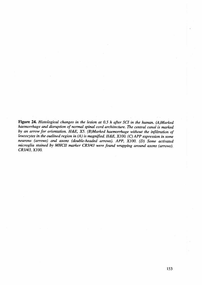
Figure A. Hístological changes in the lesion at 0.5 h after SCI in the human. (A)Markedhaemonhage and disruprton of nortnal spinal cord architecture. The central canal is marl<¿dby an arrow for orientation. H&8, X5. (B)Marl<cd haemorrhage without the infiltration ofleucocytes in the outlined region in (A) is magnified. H&8, X100. (C) APP expression in someneurons (anows) and axons (double-headed anows). APP, X100. (D) Some activatedmicroglia stained by MHCII marl<¿r CR3/43 were found wrapping around a.xons (anows|cR3/43, X100.
153
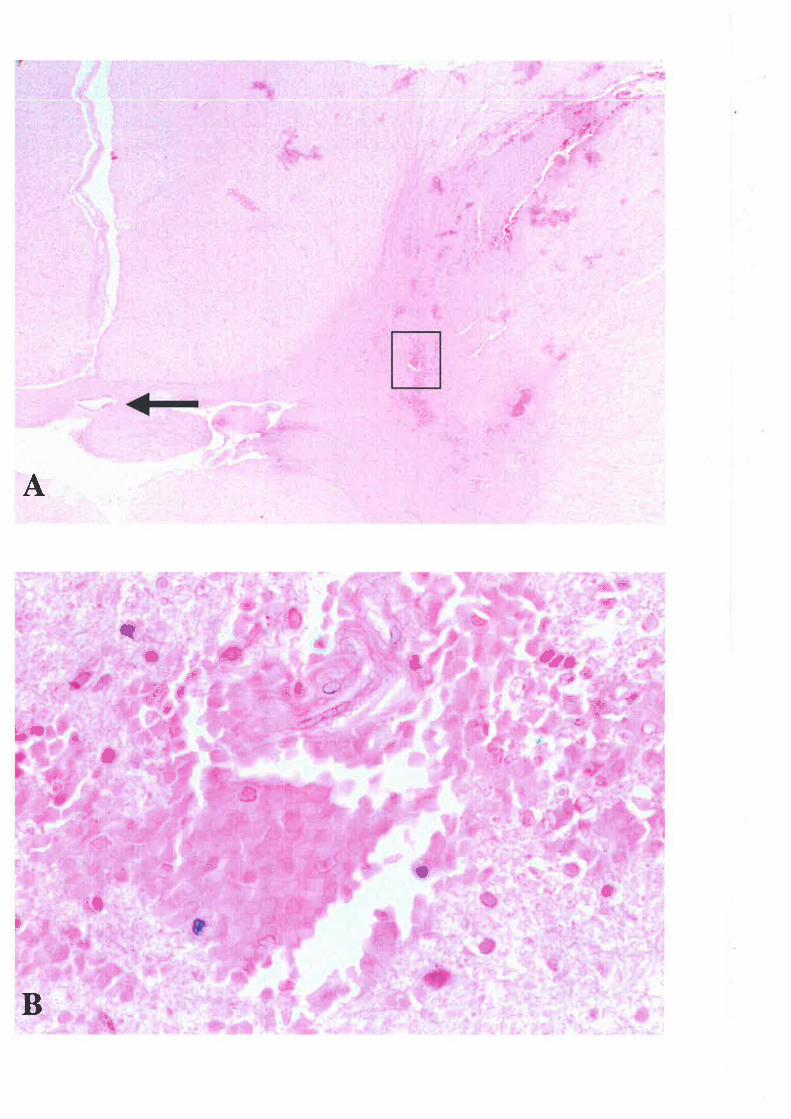
=+
*
A
rc
€**
*
=#
+
-
=æ-J'
I
ütsE,
#t
t=Þ+
*5r
+ü
t.,Ê
h
3
B
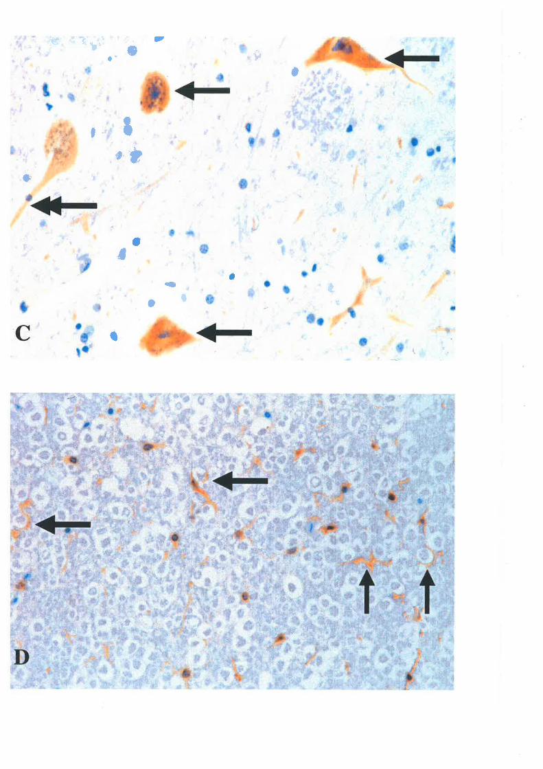
..t., ..
.- ,-$r =+
..J*
t
'*
s
a
6
Iå
tè)
ÈGl}
¡¡
.=,uã: {
)
Ç*
=: .::t or:*
I *'."'-'''1
tl: -. rì-:
å
.+ -¡11l :.Í È.
i
..¡
å*J -
-F7 ,'- *,,,- .':
{
JF*=I
='4 L* t
I€?
7
.= ! - ':
¡ ' . :.: i',. i
J5ã.
¿É !a'+.'i 'sls:
4a
r.#' *
2.2.28rn HOURS POST SCr
At 5 h postinjury, pollmorphonuclear neutrophil n \il¿ls observed @igure 25A).
Very occasional mononuclear cells in the walls of blood vessels were observed. Axonal injury
was shown by APP immunoreactivity in many neurons and axons (Figure 258). Activated
hlpertrophied microglia were detected and some of their processes wrapped around swollen
axons (Figure 25C).
156
Figure 25. Histological clnnges in the lesíon at 5 h after SCI in the human. (A) Neutrophilmargination in the wall of a blood vessel (anows). H&8, X100. (B) Expression of APP in,nony a,xons. APP, X100. (C) More activated microglia were found ønd some of them wrappedaround swollen ax/ons (arrows). CR3/43, XIM.
157
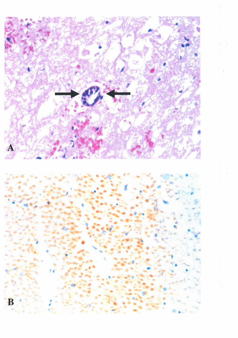
T
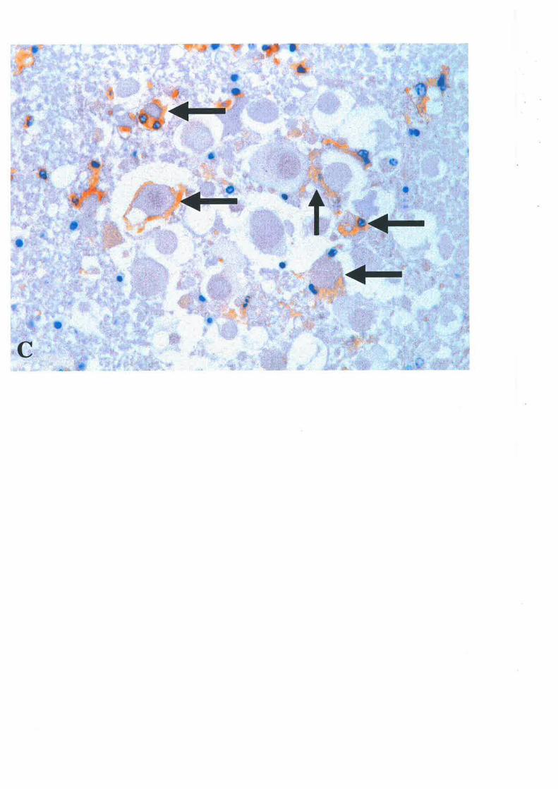
2.2.34 HOURS POST SCr
One day following SCI in the human, numerous polynorphonuclear neutrophils were widely
spread throughout the damaged tissue (Figure 264) and the number of neutrophils appeared
to reach a peak at this time point. Many activated microglia were found wrapping a¡ound both
axonal swellings and neuronal cell bodies @igure 268). Some CD68 positively-stained
macrophages started to appear in the injüed tissue (Figure 26C) and APP expression
persisted in neurons and axons (not shown).
160
Figurc Xi. Histological changes in the lesion at I d after SCI in the human. (A) Nu¡nerouspolymorphonuclear neutrophils ínfiltrated the datnaged spinal cord.. HEE, X100. (B)Neurons were surrounded by activated microglia (anows). CR3/43, X100. (C) Some CD68positively-stained macrophnges started to appear in the lesion. CD68, X100.
161
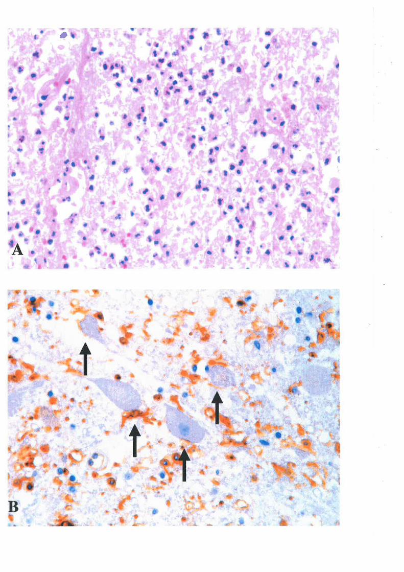
Û*
I
r*,
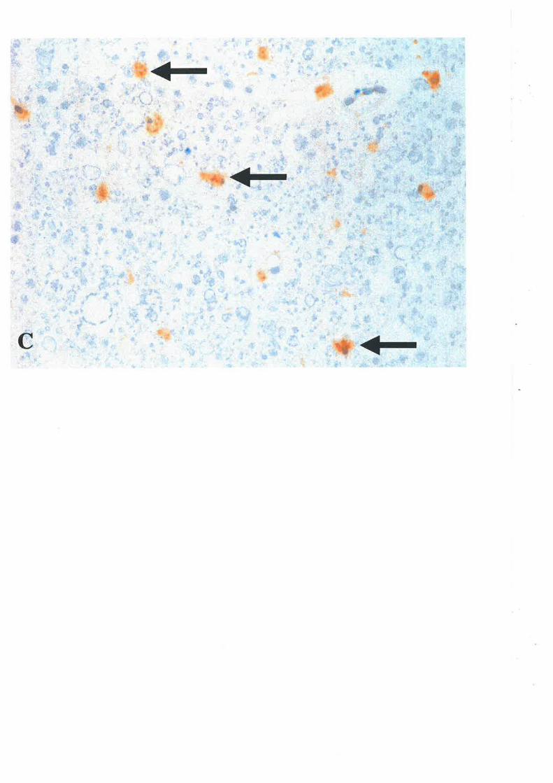
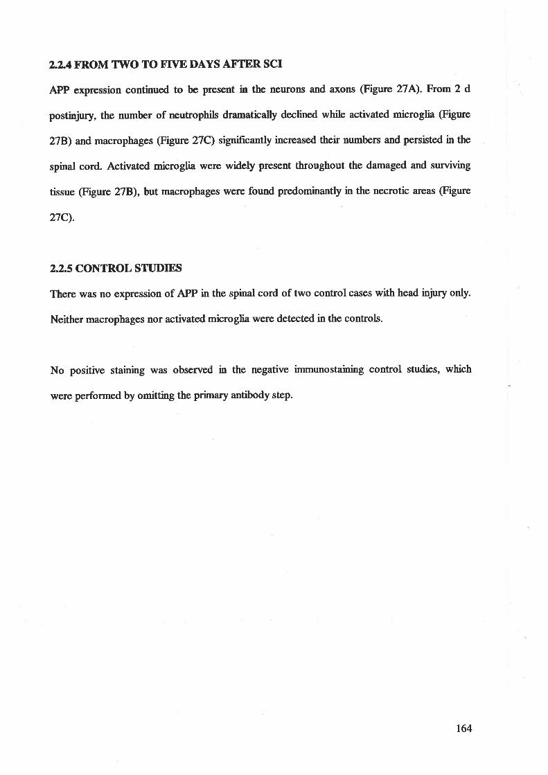
2.2.4IINOM TWO TO XTVE DAYS AT"TER SCI
APP expression continued to be present in the neurons and axons @igure 27A). From 2 d
postinþry, the number of neutrophils dramatically declined while activated microglia (Figure
278) and macrophages (Figure 27C) significantly increased their numbers and persisted in the
spinal cord. Activated microglia were widely present throughout the damaged and surviving
tissue (Figure 278), but macrophages were found predominantþ in the necrotic areas (Figure
27C).
2.2.5 CONTROL STT.]DIF"S
There was no expression of APP in the spinal cord of two control cases with head injury only
Neither macrophages nor activated microglia were detected in the controls.
No positive staining was observed in the negative immunostaining control studies, which
were performed by omitting the primary antibody step.
164

Ftgure 2il. Histological clunges in the lesion from 2 to 5 days after SCI in the hutnan. (A)
APP expression in the neurons (anows) and axons (double-headed anows). APP, X100. (B)
Activated microglia tnainly in the surviving tissue (anows). CR3/43, X100. (C) Numerous
Íoamy macorphages were lørgely present in the necrotic area (anows). CD68, XIN.
165
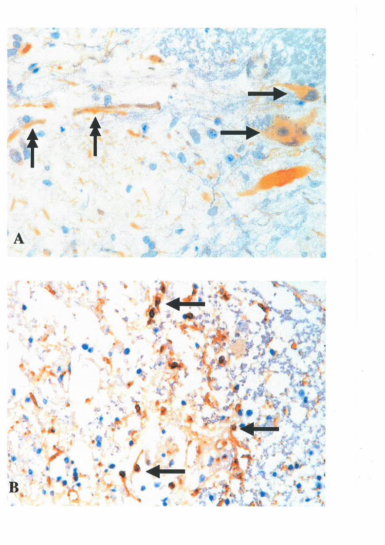
,
I
.*
aç'
B
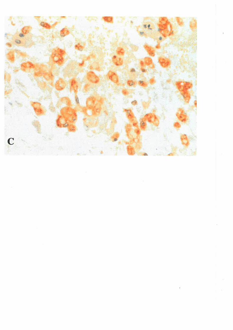
*,
@
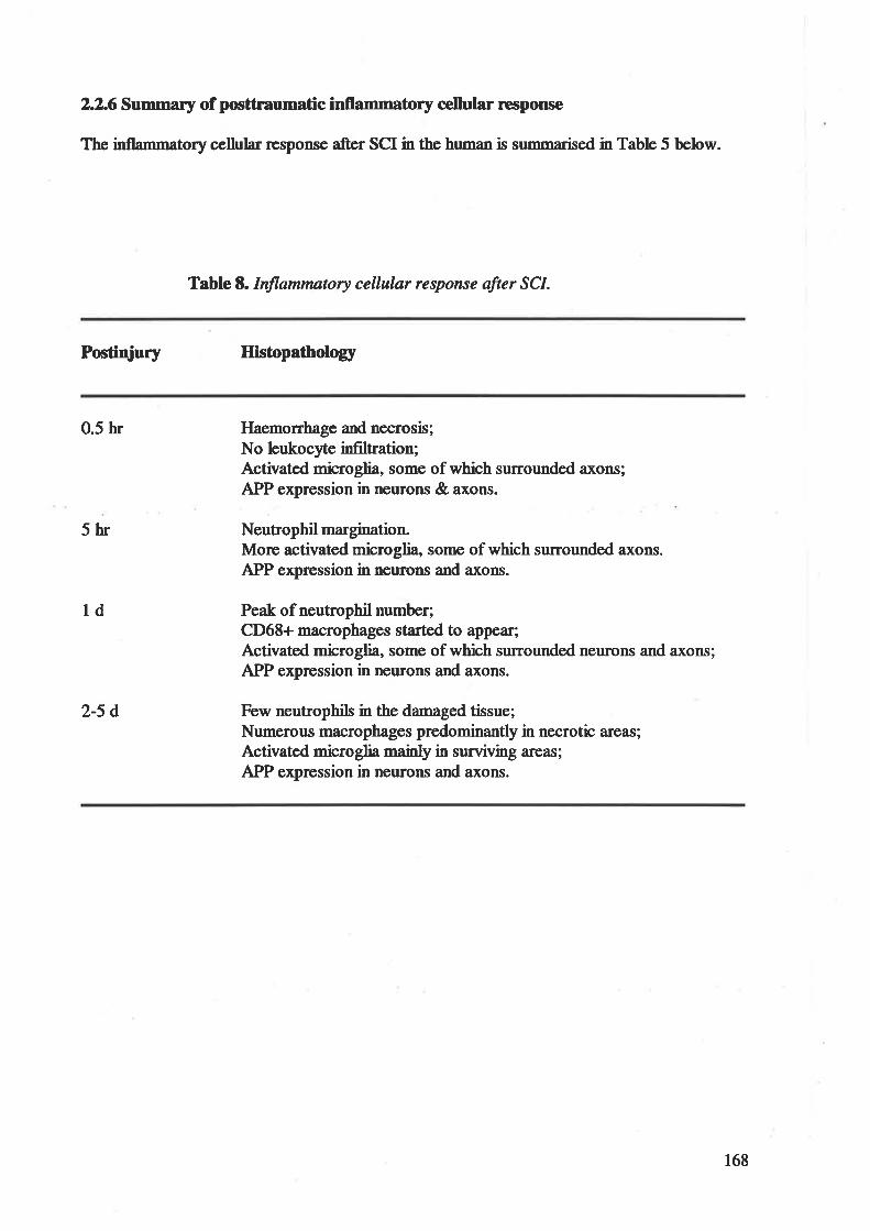
2.2.6 Summarlr of posttraumatic inflammatory cellular neqrcru¡e
The inflammatory cellular r€sponse after SCI in the human is summarised in Table 5 below.
Table E.Inflammatory cellular respot.se after SCI.
Postidury Hisúopatholory
0.5 hr
5hr
ld
2-5 d
Haemorrhage and necrosis;No leukocyte infiltration;Activated microglia, some of which surrounded axons;APP expression in neurons & axons.
Neutrophil margination.More activated microglia, some of which surrounded axons.APP expression in neurons and axons.
Peak of neutrophil number;CD68+ macrophages started to appear;Activated microglia, some of which surrounded neurons and axons;APP expression in nerrons and axons.
Few neutrophils in the damaged tissue;Numerous macrophages predominantly in necrotic ar€as;Activated microglia mainly in surviving ar€as;
APP expression in neurons and axons.
168
2.3 DISCUSSION
23.1 MARKERS FOR ACTTVAÏED HI'MAI\ MICROGLIA
The MHC class II specific monoclonal antibody CR3/43 was used in this study to investigate
activated microglia in routinely processed, paraffn-embedded human spinal cord. CR3l43 ß
directed ageinst the p-chain of all products of the MHC class tr gene subregions HLA-DR,
HLA-DQ and HLA-DP r4.
CR3/43 þþlling characteristics were systematically evaluated by Graeber and colleagues 14,
using a panel of 50 autopsies and surgical cases. CR3/43 labelling was compared to the
macrophage/microglia markers L-CÀ KPl and RCA-I. It has been found ¡þ¿1¡ssting
microglia of histologically normal tissue were not stained by CR3/43 and CR3/43 was the
most sensitive antibody to detect activated microglia in subtle tissue pathology. More
importantly, astrocytes were never found to be labelled by CR3/43.
fa srrmmary, CR3l43 is an excellent ma¡ker for activated human microglia and it is specific
for microglia in the sense that it does not label other glia. Visualization of microglial
activation in this study was greatþ aided by using this antibody.
2.3.2 APP T]PREGT]LATION
This study demonstrated the presence of widespread APP immunoreactivity in the neurons
and axons in all 11 human cases following SCI. APP positive staining was observed as early
as haH an hour after injury and persisted for at least 5 days. These observations parallel those
observed in the rat SCI (described in Table 5, page 108). APP immunoreactivity occurs not
only as a result of primary mechanical i"ju.y but also as a result of secondary complicating
factors such as h¡'poxia, ischaemia and infarction2'1e't3t, which are frequently found following
r69
injury. Thus, APP is upregulated as an acute phase cell-stress response to inþry. However,
exactþ where APP fits into the compbx inflemmatory cellular rcsPonses following SCI is
unclear and requires further study.
2.3.3 INTI,AMMATORY CELLI]LAR RESPONSE
The inflammatory cellular response following SCI in the human parallels the changes
observed in rat SCI (described in Table 5, page 108). Like the response in rat SCI, there are
mainly three cell tlpes involved in fts inflammatory rcsponse following SCI in the human:
microglia, neutrophils and macrophages. The time course of inflammatory cellular r€sponse
in human SCI is also very similar to that observed in rat SCI (described in Table 5, page 108).
In contrast to some in vivo studies that failed to detect morphological signs of microglial
activation by I d postinjury 261341, activated microglia in this study were detected as early as
0.5 h after injury and found surrounding axonal swellings and otherwise "normal" axons. This
novel finding is in agreement with that seen in rat SCI in this study (described on page 110)
and has further challenged the current view that there a¡e no direct topographical association
between microglial cells and axons, and no association of anonal inj"ry with activated
microglia t27261'278. More studies a¡e needed to elucidate the effect of activated microglia
surrounding axons because activated microglia are able to produce pro-inflammatory
cytokines (observed in this study), cytotoxic substances 25'387 and growth factors 3al. Activated
microglia may provide a trophic local environment to promote the axonal regeneration
because some studies have shown that activated microglia promoted the regeneration of
sensory Ðrons in the injured rat spinal cord 248'280. It has been also found in this study that
activated microglia surrounded neurons. This is consistent with the finding in rat SCI
(described on page I 11) and is referred to as synaptic stripping. Microglia are thought to be
170
involved in limit€d phagocytosis of individual slmaptic tenrrini and to have an imPact on the
spaptb reorganization of the neurons, but the effect of slmaptic stripping on neurons is, as
yet, largely unknown.
Haematogenous inflammatory cells (neutrophils and macrophages) did not start to infiltrate
the injure.d spinal cord until 6 h postinjury suggesting that these peripheral inflammatory cells
we¡e not spille.d from damaged vessels, but recruite.d by some chemoattract¿nts including
cytokines (e.g. IL-lp,IL-6 and TNFa) 88'348.
Neutrophils are the first peripheral inflammatory cells to migrate into the tissue. It has been
suggesæd that infiltrating neirtrophils may participate in tissue destruction and enlargement of
the lesion because they can produce reactive oxygen species and other substances with
potentially tissue-damaging effects 6s'3473s8. Taoka and colleagues 3a7 recentþ found that
inhibition of neutrophil activation alleviated the motor disturbances observed in a rat model of
SCI. However, other studies reported that neutropenia had no effect on the extent of tissue
damage following CNS injury 2o'tt8'367. The observation in this study that necrosis precedes
the a¡rival of neutrophils at the injury site indicates that the tissue necrosis may not be caused
by the invading leukocytes, but that it may, instead, provide a chemotactic stimulus for
attracting neutrophils as the first line of recruited phagocytic cells to begin removal of cellula¡
debris.
The second wave of peripheral inflammatory cells to enter damaged tissue in the spinal cord
is monocyte-derived macrophages. Macrophages in the damaged spinal cord arise from two
different sorrces; from the non-resident population of circulating blood monocytes recruited
into injured cord and transformed into macrophages, and from the resident population of
17t
ramifüd microglia in spinal cord activated and transformed into macrophages25'8s2û216338.
The quantitative contributions of endogenous microglia-derived and monocyte-derived
macrophages to the tot¿l macrophage pool are still not clear ã.
Regardless of their origin, various reports have shown that macrophages can play both
negative and positive roles in responding to injury. Macrophages (including microglia and
monocytes) can be induced to produce several potentially cytotoxic substances in vitro, such
as fr,ee oxygen inærmediates, NO, proteases, EAAs and pro-inflarnmatory cytokines 13200.
Thus, these cells rnay cause "bystandet'' damage to inøct tissue and contribute to the
secondary tissue destruction after TSCI. In support of this view, Blight 3a and Popovich 27a
have observed a better anatomical and functional outcome in animals with CNS injuries when
macrophage numbers a¡e reduced experimentally. In addition, several studies have shown the
damaging effects of pro-inflammatory cytokines produced by macrophages 172ss. However,
another view holds that macrophages plzy a key role in the removal of cellula¡ debris which
a¡e essential for tissue rç¡edslling, and produce the trophic cytokines, such as TGF-p1 and
IL-4, to promote tissue repair and neuronal survival t4'7827t. In support, several studies
transplanting macrophages/microglia into an injure.d CNS showed increases in axonal
regeneration t2t28o. However, this interpretation is flawed by a lack of quantitative,
çomlìarative cell counts conducted in the same animal Therefore, the role of macrophages
(including microglia and monocytes) is far from clea¡ in studies of TSCI and their function is
likely to be dependent on the time point following inj"ry and their location relative to the
lesion.
172
3. EARLY EIPRESSION AI\D CBLLT]LAR LOCALIZATION[-1Þ, IL-6 and TI\Fcr
This experiment was performed to investigate the expression of tr-lp, IL-6 and TNFa in the
human spinal cord after SCI.
3.1 METHODS
3. I. 1 IMMI]NOHISTOCHEIVIISTRY
For staining of IL-lp, IL-6 and TNF-o, a rabbit anti-L-lp polyclonal antibody (1:200,
Rockland, PA), a rabbit anti-Il-6 polyclonal antibody (l:25, HyCult Biotechnology, Uden,
Holland) and a mouse anti-TNF-a monoclonal antibody (l:75, HyCult Biotechnology, Uden,
Holland) were used to identiff these pro-inflamnatory cytokines. 'We also checked the
antigen-specificrty of the tr--lp, IL-6 and TNF-c¡ antibodies by preabsorbing these primary
antibodies with exogenous recombinant human tr--lp, IL-6 and TNF-ct @epro Tech, NJ).
3.I.2 IX)T]BLF-LABEL TT,UORF.SCENT CONFOCAL IMAGING
The cellula¡ sources of IL-lp, IL-6 and TNF-cr were elucidated using immunofluo¡escence
double labelling confocal imaging. The sections were incubated overnight with combinations
of anti-Il-lp, anti-Il--6 or anti-TNF-a antibody (as described before), and activated
microglia-specific markers CR3/43 (as described on page 154) or anti-glucose transporter-S
(Glut-5) antibody (l:500), a gift from Dr. F. Mahe¡, University of Melbourne)'7o.On the
following day, the sections were incubated with a mixture of fluorescein-conjugated donkey
anti-rabbit (FITC; 1:100; Jackson Tmmunoresea¡ch Lab, V/est Grove, PA) and Cy*5-
conjugated donkey anti-mouse (Cy5; 1:4O0; Jackson Immunoresearch Lab, West Grove, PA)
173
antibodies. Sections were then mounted and examined under a fluorescence microscope
@iorad, LaserSha¡p 2læ).
3.2 RESULTS
The staining intensity of ILlp, IL-6 and TNF-a markedly increased at 0.5 and 5 h after
traumatic SCI and quicHy declined at I d postrnjury (Figure 28). ILlp,L-6 or TNF-ct,
positive cells were found up to 2 cord segments away from the lesion centre. There was no
difference in the immunostaining pattern of ILlp, IL-6 and TNF-cl between the rostral and
caudal parts of the lesion.
Half an hour after injury, the expression of tr--lp was found in some neurons and axons
(Figure 28A), and IL-6 and TNF-a ws¡s mainly localized in the neurons @igures 28D and
28G). At 5 h after injury tr--lp, IL-6 and TNF-o were detected not only in many neurons but
also in some microglial-like cells @igures 288, 28E and 28Ð. At 1 d after injury, vory weak
staining of IL-lp, IL-6 and TNF-cr was seen in the neurons @igures 28C,28F and 28I) while
no expression was identified in the glial cells. From 2 d postinjury no sf¿ining of IL-lp, IL-6
and TNF-a was observed.
174
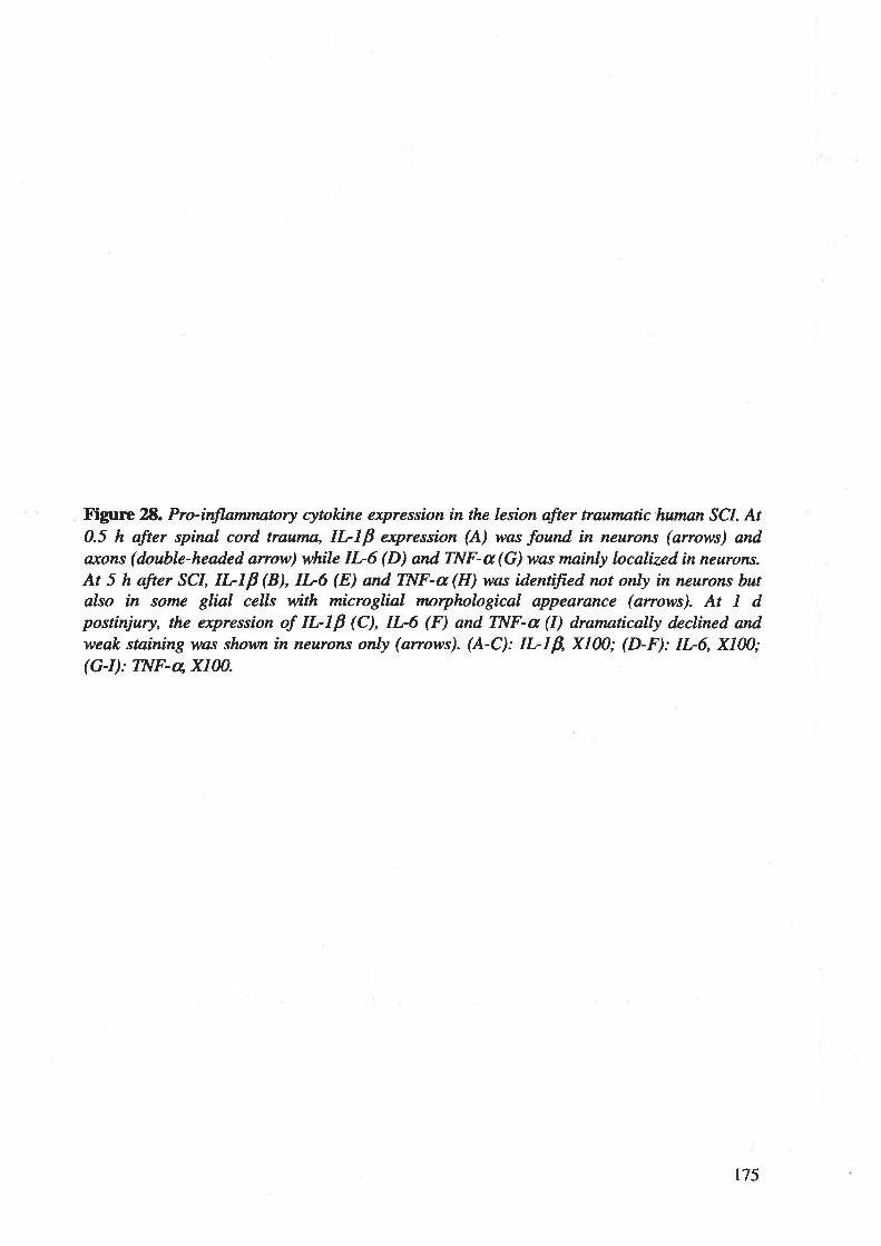
Figure 2E. Pro-inflatwrotory cytokine expression in the lesion after traumntic'human SCI. At0.5 h after spinal cord tranûÌtn, II-IP expression (A) was found in neurons (anows) andaxons (double-headed anow) while IL6 (D) and TNF-u (G) was tnainly localized in neurons.At 5 h after SCI, ilÅP (B), IL6 (E) ønd TNF-u (H) was identified not only in neurons butalso in somc glial cells with microglial rnorphological appeorance (anows). At I dpostinjury, the expressíon of ILIP (C), IL6 (F) and TNF-a (I) dramatically declined andweak staining was shown in neurons only (anows). (A-C): IIÅP, X100; (D-F): II-6, Xl00;(G-I): TNF-u, X100.
175
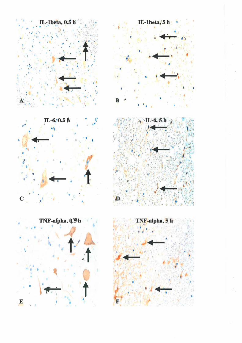
If,-lbeta 5 h
rêt\
fII;':1I
I
¡Q
I
,iI
)-la
tI
EE
å
t- Ë=
é
*
!
= i.t. .. .
.{I
,I
l_
Bt]I
t'a
t
-rl.-6r.0.511
IË I
t i\
I
<FcH'
,drI
I
ã;=*
êÊ
=
lÞ
G
Içt
11 ¡"
,5
11
Ç-
a\E
C a
TNF-atþha, ÇEhê€
,,1
1t.
Iå
\
q À
a
Fa
I 1fT*l
]E
Using double-label fluorescent immunohistochemistry, co-localization of IL-lP, tr--6 or TNF-
a and microglial markers CR3143 or glut-5 was found, confirming that ILlp, IL6 or TNF-a
positive glial cells observed at 5 h postinþry were microglia (Figure 29).
In antigen-absorption experiments, the immu¡ereactivity of IL-1p, IL-6 and TNF-c¡ was
absent, showing the specificity of these antibodies in this study.
There was no expression of pro-inflammatorycytokines (IL-lp,IL-6 and TNF-o) in the two
normal control cases.
177
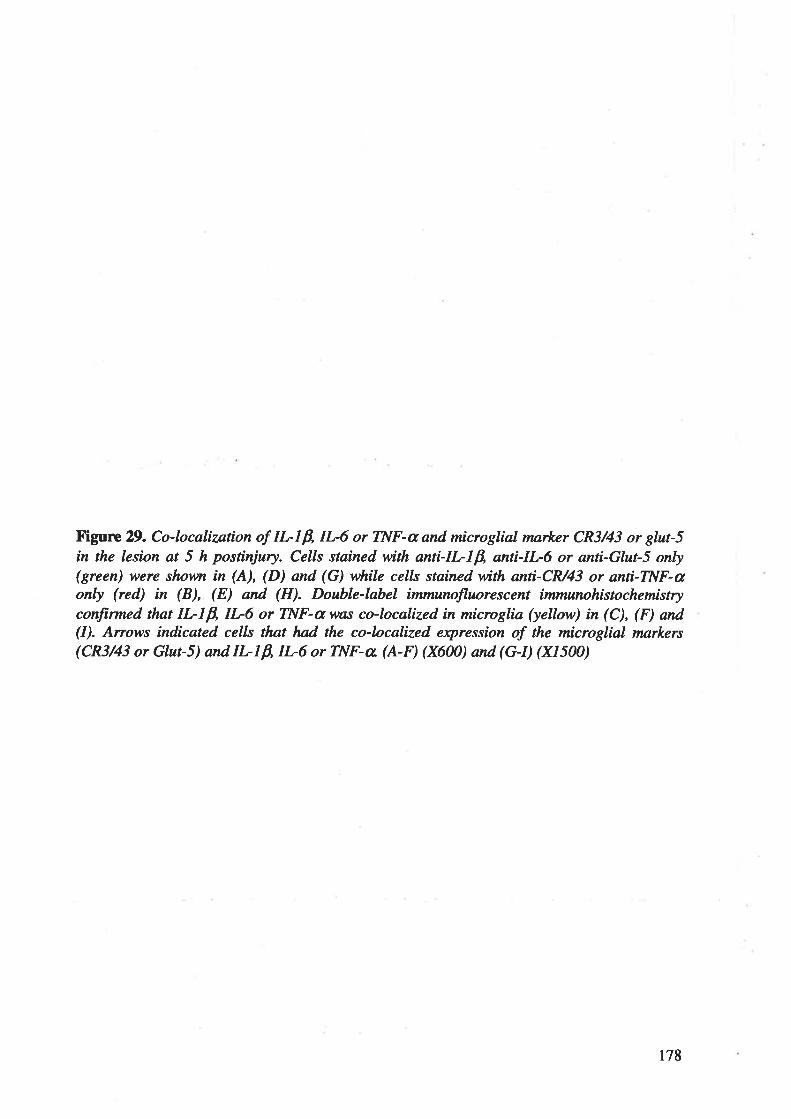
Figurc 29. Co-localization of IL-Lfl IL-6 or TNF-tand miuoglial m¿rl<¿r CR3/43 or glut-Sin the lesion at 5 h postinjury. Cells stained with anti-IL-Lfl anti-IL-6 or anti-Glut-S only(green) were shown in (A), (D) and (G) while cells stained with anrt-CR/43 or anrt-TNF-aonly (red) in (B), (E) and (H). Double-label immunofluorescent immunohistochemistryconfirmcd that IL-|fl II-6 or TNF-awas co-localized in microglia (yellow) in (C), (F) ønd(I). Anows indicated cells that had the co-localized expression of the microglial mnrkers(CR3/43 or Glut-S) andIL-Lfl IL-6 orTNF-a (A-F) (X600) and(G-I) (X1500)
t78
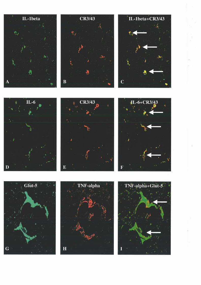
J.3 DISCUSSION
Our results have shown a rapid elevation of ILlp, IL6 and TNF-o expressions at 0.5 and 5 h
after injury and then a dramatic decline of these expressions prior to the appearance of
neutrophils and macrophages at I d postinþry (described in Table 8 on page 171), suggesting
that these pro-inflammatory cytokines are primarily produced by endogenous CNS cells
rather than infiltrating blood-borne leucocytes in human SCI. These findings in human SCI
have confirmed results observed in our weight-drop rat model of SCI and in other animal
models of SCI te'164'te3 that neutrophils and mactophages are not the cellula¡ sources of ILlp,
IL6 and TNF-c.
Further, the present study demonstrates the production of IL-IP, IL-6 and TNF-a in neurons
at 0.5 h after injury and in both neurons and microglia at 5 h after injury. To our knowledge,
this study is the first to demonstrate the cellular sources of pro-inflammatory cytokines, such
as IL-lp, IL-6 and TNF-c, in the injured human spinal cord. This novel finding is consistent
with recent in vivo studies in rat models of SCI that identified TNF-cr in neurons and
microglia te4'3e6 and in vitro studies that showed the production of a variety of cytokines in
neurons and microglia 134'2ro. These data have confirmed that neurons and microglia
(especially neurons), not blood-born leucocytes proposed by many authors t3s'347'354, may play
a major role in the initiation and regulation of early posttraumatic inflarrmatory response of
the human SCI by producing tr--lp, IL-6 and TNF-c.
Thus, for the first time, human SCI has been shown in this study to induce the expression of
the proinflammatory cytokines IL-lp, IL-6 and TNF-cr in neurons within as little as 0.5 h
following injury. These c5rtokines, a group of proteins that evoke remarkable responses in a
variety of cell t¡pes, initiate a complex cascade leading to inflarnmatory responses, including
180
increased vascular permeabilit¡ leukocyte infiltration and induction of acute-phase response
t9,rØ20834137938O
Inflammæion may contribute to secondary tissue damage after SCI, for example by
promoting tissue oedema and ischaemia The proinflamatory cytokines induce several
molecules capable of increasing vascular permeability as well as inducing cellular fluid loss
267. These molecules include components of the complement cascade (C3a and C5a, which in
furn may cause release of histamine, prostaglandins and leukotrienes from resident mast
cells), proteases such as plasminogen activator, and bradykinin '6' . lt has been shown that
direct inþtion of IL-lp into spinal cord enhanced vascular permeability 310 *¿ increased
expression of pro-inflammatory cytokines in transgenic mice exace¡bated neurological
functional impairment after SCI 187. Additionall¡ systemic administration of the anti-
inflammatory cytokine IL-10 has been reported to limit neuronal damage and promote
functional r€covery following SCI in rats 28273. These results suggest that inflammation-
promoted cellular damage exacerbates lesion volumes and SCI functional outcomes.
On the other hand, inflammatory rcsponses have been found to promote beneficial response
after SCI. It has been demonstrated that tissue loss after SCI was reduced by direct application
of pro-inflamrnatory cytokines te2. Furthermore, IL-1p or TNF-a receptor-knockout mice
demonstrated reduced remyelination and diminished functional recovery tet'233.
Taken together, the role of pro-inflarnmatory cytokines after SCI is still controversial and it
may be dependent on the balance of the injurious and beneficial effects of these cytokines.
181
4.CO-LOZÁ'.LLZ,ATTON OF tr -lp AI\D APP
Because tr-lp immunoreactivity was localized in some nerrrons and axons (Figure 284), we
used immunofluorescence double þþlling confocal imaging to examine whether IL-lp co-
existed with amyloid precursor protein (APP) in the neurons and axons.
4.1 METHODS
Co-existence of IL-lp with APP was demonstrated using immunofluorescence double
labelling confocal imaging. The sections were incubated overnight with a combination of
anti-Il,-lp (as described before) and anti-APP antibodies (as described before). On the
followin¿ day, the sections were incubated with a mixture of fluorescein=conjugated donkey
anti-rabbit (FITC; l:100; Jackson rmmunoresea¡ch Lab, \Vest Grove, PA) and Cy*5-
conjugated donkey anti-mouse (C!5; 1:4O0; Jackson rmmunoresea¡ch Lab, West Grove, PA)
antibodies. Sections were then mounted and examined under a fluorescence mic¡ssçsps
@iorad, LaserSharp 2100).
4.2 RBSULTS
In the immunofluorescence experiments, L-lp and APP were found co-localizing in the
nerrons and axons at 0.5 h and 5 h postinjury (Figure 30).
182
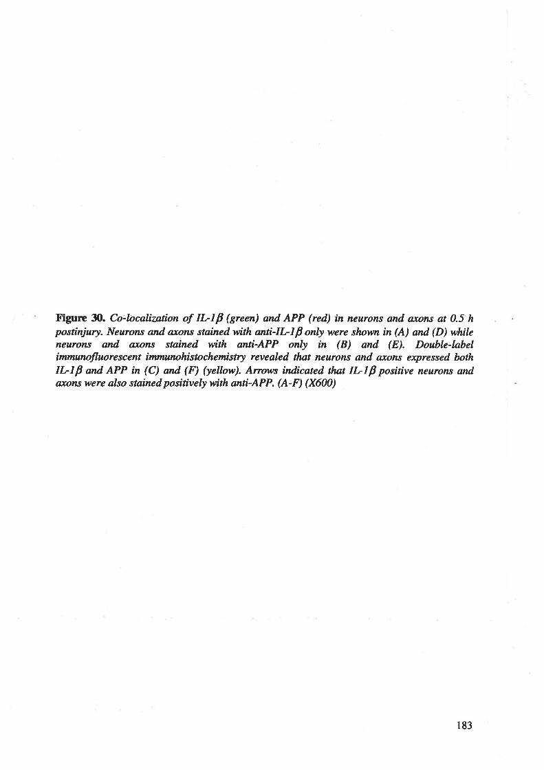
Figurc 30. Co-localízption of ILIQ (green) and APP (red) ín neurons and axons at 0.5 hpostinjury. Neurons and axons stained with anti-ILlp only were slwwn in (A) and (D) whileneurons and. a.xons stained with anti-APP only in (B) and (E). Double-labelimrnunofluorescent immarchistochemistry revealed that neurons and axons expressed bothilÅP and. APP in (C) and (F) (yellow). Arrows indicated that II-I| posirtve neurons andaxons were also stained positively with anti-APP. (A-F) (X600)
183
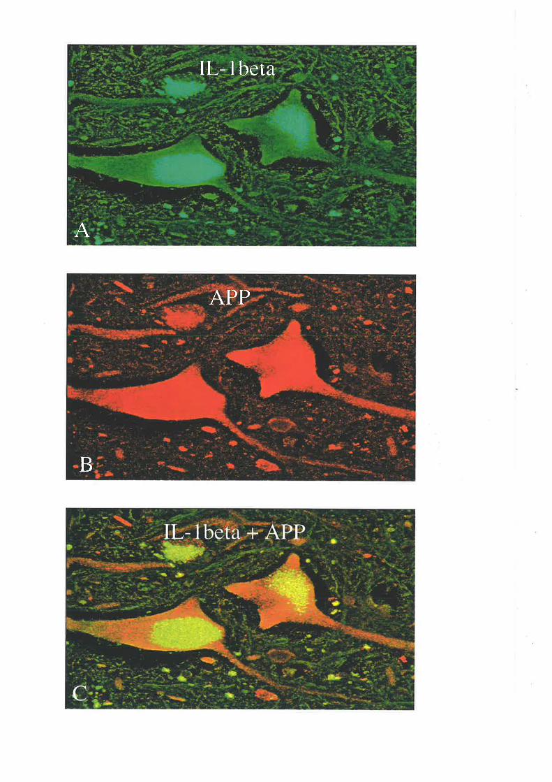
IL-lbeta
A
APP
B
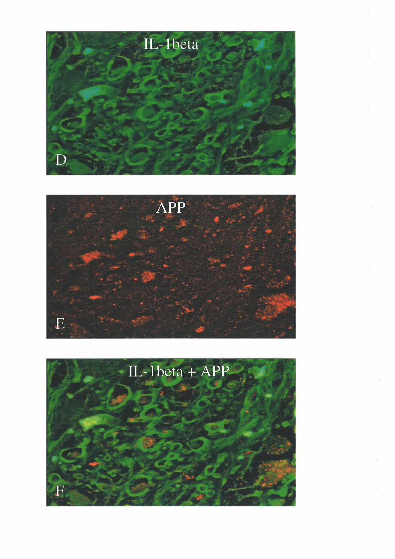
L- lbeta
D
APP
E
IL-lbet¿r + APP
F
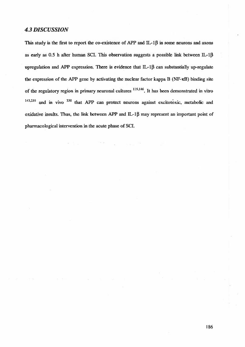
4.3 DISCUSSION
This study is the first to report the co-existence of APP and tr--lp in some neurons and axons
as early as 0.5 h after human SCI. This observation suggests a possible link between ILlp
upregulation and APP expression. There is evidence that IL-lp can substantially up-regulate
the expression of the APP gene by activating the nuclea¡ factor kappa B (NF-tcB) binding site
of the regulatory region in primary neuronal cultures tte't46.It has been demonstrated in vitro
14323s and in r¡vo 330 that APP can protect neurons ageinsl excitotòxic, metabolic and
oxidative insults. Thus, the link between APP and L-1p may represent an imFortant point of
pharmacological inærvention in the acute phase of SCI.
186
FINAL DISCUSSION
1. SUMI\,IARY
Traumatic injury to the spinal cord initiates a cascade of secondary tissue damage beyond the
original siæ of trauma, leading to a fufher loss of tissue and function 1e'35'182"rc0. The cascade
of secondary tissue damage is accompanied by an inflammatory response marked by
infiltration of neutrophils and macrophages, activation of glial cells and up-regulated
expression of pro-inflammatory cytokine s ee'tø354.
Neutrophils, macrophages and activated microglia may act as scavenger cells to remove
cellular debris and release cytotoxic or neurotrophic m¡lecules intg the injured spinal cord
3s'6s'8s2ffi'34t'"0, bot the precise functions and effects of these cells remain unclea¡. Pro-
inflammatory cytokines, such as interleukin-lp (IL-lp), interleukin-6 (IL-6) and tumour
necrosis factor-ct (TNF-a), a¡e involved in the regulation of the leukocyte recruitment and
microglial activation te23to, but their functional significances for secondary tissue damage
following traumatic spinal cord inþry (SCD remain controversü2ï7. tr--lp, IL-6 and TNF-a
have been shown to have cytotoxic effects on the nervous system but also induce neurotrophic
molecules involved in the repair of injured tissue 4'147'te4'241'2e8'3s4. Furthermore, the
relationship between severity of SCI and expression levels of these pro-inflammatory
cytokines has not been addressed and relationships between mRNA and protein levels of pro-
inflammæory cytokines (e.9. tr--lp, IL-6 and TNF-c) in the central nervous system (CNS)
have not been investþated. The cellular sourcos of IL-lp, IL-6 and TNF-o following
traumatic SCI a¡e controversial Some authors believe that tlese cytokines are produced
prima¡ily by neutrophils and macrophages 23e347, but other studies have suggested that
endogenous CNS cells including microglia may also produce pro-inflammatory cytokines
187
under various injury conditions re43e6. Further studies of the posttraumatic inflamrnatory
respoil¡es to SCI are required to resolve these controversies.
We hlpothesized that the pro-inflammetory cytokines ILlp, TNF-o and IL-6 produced by
intrinsic cells in the spinal cord may act as messengers to coordinate the inflammatory
cascade and the influx of neutrophils and monocytes to the site of damage and that the
cytokine response should be greater in severe than in mild injury.
An in vivo model of SCI (the New York University [npactor model) was used in this study to
induce acute SCI and inflammatory changes in rats. Analyses of the histological changes and
inflammatory responses in rat SCI showed that activated microglia we¡e found in mild and
seve¡e SCI as early as I h afrer contusion and increased dramatically at I d postinjury while
neutrophils and macrophages were not noted in the injured spinal cord until 6 hrs postinjury.
Increased immunoreactivity of TNF-a in mild and severe SCI was detected in neurons at I h
postinþry, and in neuroru; and microgln at 6 h postinjury with much less increase in mild
SCI, but the expression was short-lived and declined sharply by 1 d postinury. Furthermore,
we quantified the expressions of n--lp, IL-6 and TNF-c¡ at both mRNA and protein levels
over time following mild and sevetre SCI, utilizing RT-PCR and western blotting, and
compared the expression levels of IL-lp, IL-6 and TNF-cr between mild and seve¡e traumatic
SCI. Our results demonstrated an early significant up-regulation of IL-lp, IL-6 and TNF-a
mRNAs, maximal at 6 h postinjury with return to control levels by 24 h postinjury, the
sþanges being statistically significantþ less in mild injury. The results also showed early
transient increases of IL-lp, IL-6 and TNF-ct proteins in severe SCI but not mild SCI.
188
ImmunocSÉochemical, western blotting and RT-PCR analyses suggest that endogenous cells
(neurons and microglia) in the rat spinal cord, not the blood-bonre leucocytes, contribute to
tr--lp, IL-6 and TNF-o production in the posttraumatic inflammatory response and that their
up-regulation is greater in severe than mild SCI. This paper is the first to demonstrate
significantþ increased expression of IL-lp, IL-6 and TNF-a messengcr ribonucleic acids
(mRNAs) in severe SCI in contrast to mild SCI and that the temporal profiles of ILlp, IL-6
and TNF-cr protein expression in severe SCI paralleled their mRNA transcription whereas
mild SCI resulted in increased slmthesis of mRNAs but no detectable protein increase on
western blotting. The disparities between transcription and translation of IL-lp, IL-6 and
TNF-o in mild SCI could be due to the factors previously described (see Chapter RAT SCI
ÐGERIMENTS, section 4.3.2). Significantly increased expression of pro-inflammatory
cytokines in severe inj"ry in contrast to mild injury supports the hlpothesis that cytokine up-
regulation is an important factor in the generation of the severity of the inflammatory
response. Thus, the significantþ upregulated expression of cytokines ILlß, IL-6 and TNFo
in severe SCI is a suitable target for pharmacological intervention to attenu¿1e this response.
Almost all observations have been confined to animal models of CNS trauma and little is
known of the cellular sources of IL-lp, IL-6 and TNF-cr in the human. Therefore, we
investigated the posttraumatic inflammatory response and the cellula¡ sources of IL-IB, IL-6
and TNF-cr in human spinal cord following contusion injury. This study is the first to
demonstrate that, as early as 0.5 h after SCI, activated microglia were detected and their
processes surrounded axonal s1¡r6llings, suggesting that microglia a¡e involved in the early
response to traumatic axonal injury (see Chapter HUMAN SCI EXPERIMENTS, section
2.3.3).
189
Another novel finding in this study is that increased immunoreactivity of tr--lÞ, IL-6 and
TNF-a was detected in neurons at 0.5 h postinjury, and in neurons and microglia at 5 h
postinþry but the expression of these pro-inflammatory cytokines was short-lived and
declined sharply to baseline by 2 d postinþry. In human SCI as in our rat modef neutrophils
and macrophages a¡e not the cellula¡ sources of ILlp, IL6 and TNF-c. 'We have found that
endogenous cells in the hum¿n spinal cord, neurons and microglia, contribute to IL-lp, IL6
and TNF-a production in the posttraumatic inflamrnatory response, suggesting that neurons¡
and microglia, not 'þrofessional' blood-borne inflammatory cells, may play a major role in
the initiation and regulation of the early post-traumatic inflammatory response of human SCI.
More studies a¡e needed to elucidate the effect of activated microglia surrounding axons in
both the rat and human SCI because activated microglia a¡e able to produce pro-inflammatory
cytokines (observed in this study), cytotoúc substances 25'387 and growth factors 3al. Activated
microglia may provide a trophic local environment to promote axonal regeneration because
some studies 248280 have shown that activated microglia promoted the regeneration of sensory
axons in the inþred rat spinal cord. It has been also found in this study that activated
microglia surrounded nerrons. This phenomenon, originally described by the Blinzinger
group 3t, is noo, referred to as slmaptic stripping. Microglia are thought to be involved in
limited phagocytosis of individual slmaptic termini and to have an impact on the synaptic
rcorgantzation of the neurons, but the effect of slmaptic stripping on neurons is, as yet, largely
unknown.
Finall¡ the combined results of rat and human SCI in this study indicate that expression of
the pro-inflarnmæory cytokines IL-lß, IL-6 and TNFa following SCI is robust, but short-
lived, and that cytokine upregulation plays an important role in the initiation and regulation of
190
the early posttraumatic inflanmatory response of SCI, where IL-1ß, IL-6 and TNFo are
important molecules for the inærcellular srosstalk between microgli4 neurons, endothelial
cells, oligodendrocyûes and astrocytes. tr-lp activates microglia to express more IL-lß,n--6
and TNFc, stimulates astrocytes to form astroglial scars, upregulates expression of endothelial
adhesion molecules to enhance leukocyte recuitnrent and produces MMP and free radicals to
cause tissue ischaemia and destruction. IL-6 produces neuronal damage and reactive
astrocytosis to inhibit the axonal regeneration. TNFa has a direct toxic effect on
oligodendrocytes, .lamages vascular cells and activates the astrocytes and microglia. The
production of IL6 is regulated by a variety of stimuli including L-1P and TNFcr @igur€ 31)
(se,e Chapter INTRODUCTION, sections 2.2.4, 2.3.4 & 2.4.4; Chapter HUMAN SCI
ÐGERIMENTS, section 3.3 and Chapter RAT SCI ÐGERIMENTS, section 4.3.3).
191
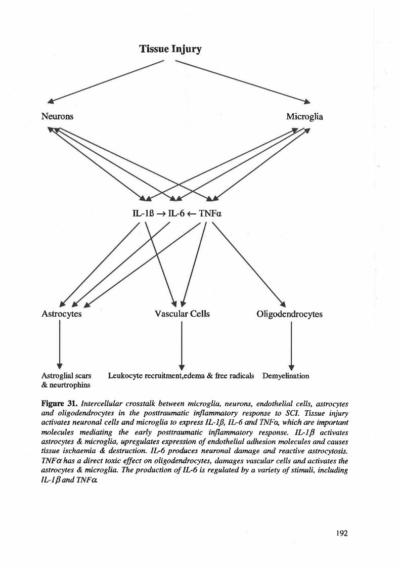
Tissue Injury
Neurons Microglia
IL-lß+IL6eTNFo
Astrocytes Vascular Cells Oligodendrocytes
Astroglial scars
& neurtrophinsLeukocyte recruitment,edema & free radicals Demyelination
Figure 31. Intercellular crosstalk between microglia, neurons, endathelial cells, astrocytesand oligodendrocytes in the posttraumartc inflammatory response to SCI. Tissue injuryacrtvatus neuronal cells and microglia to express IL-Iß, II-6 and INFx, which are importantmolecules mediartng the earþ posttrau¡matic inflammatory response. II-IP activatesastrocytes & microglia, upregulates expression of endothelial adhesion molecules and causestissue ischaetnia & destruction. II-6 produces neuronal dnmage and reactive astrocytosis.TNFu has a direct toxic effect on oligodendrocytes, dnmages vascular cells and activates theastrocytes & microglia. The producrton of IL-6 is regulated by a variety of stimuli, includingILI p and TNFa
192
ILIß, IL6 and TNFa are important and modifiable components of the inflammatory
response to trar¡matic CNS inþry. The production of ILIP, tr-6 and TNF-cl is regulated by a
variety of factors and these cytokines may function as both neurotrophic and cytotoxic factors
(see Chapter RAT SCI ÐPERIMENTS, section 4.3.3). tr--lp, IL6 and TNF-a have been
shown to produce tissue damage but may also rescue injured tissue in the posttraumatic
response by inducing the production of neurotrophic molecules. There are several
explanations for these apparent conflicting roles. First, the early and robust rise in ILIP, IL-6
and TNF-a following SCI observed in this study may be detrimental if they are overproduced
as the margins between neurotrophy and neurotoxicity are quite narrow 005. lhis concept is
supported by the finding of spontaneous inflammatory CNS demyelination in transgenic mice
ovenexpressing TNF-a 283. Second, the neurotrophic effects induced by IL-lp, IL-6 and
TNFc¡ may not occur in vivo because in vitro studies have shown that the production of
neurotrophic molecules required intact cells. Third, cellular effects of cytokines are likely to
be affected by other ongoing secondary in¡ury events after SCI, e.g., APP expression can be
substantially upregulated by IL-lp (see Chapter HTJMAN SCI ÐPERIMENTS, section 4.3)
and the toxicity of excitatory amino acids, which a¡e also upregulated early after CNS iqiury
105, caû be enhanced by TNFa tr. Foorth, the timing of the cytokine upregulation may
influence their effects. Several studies 26're2 de onstrated that an inþction of a cytokine
cochail of IL-lp, IL-6 and TNF-a into the spinal cord at 4 days postinjury resulted in a
reduction in the recruitrnent of periphetal macrophages, microglial activation and tissue loss.
However, the posttraumatic inflammatory response was enhanced and there was greater tissue
loss when the cytokines \re¡e delivered 1 day after injury. Lastly, the cellular actions of
cytokines are complex and subþt to various regulation factors, including the number,
subtlpe and proportion of activated receptors 43'327. Al*o, the toxicity of excitatory amino
acids may be enhanced by TNF-a 66.
193
Therefore, these daø suggest that ILIP, ÍI-6 and TNF-c are powerñrl inflamrnatory
mediators in the posttraumatic inflammatory response of SCI and the final effects of these
pro-inflamrnatory cytokines rnay be geatþ determined by many factors, including the
temporal and spatial relation of cytokines to their receptor-bearing target cells of the spinal
cord.
z.LßVIJTATION OF'THIS PROJECT AI\D FI]TURE WORK
This study failed to demonstrate inhibition of the upregulation of cytokines IL-lp, IL-6 and
TNFa in severe SCI by the NF-tcB-specific antisense oligodeoxlmucleotides. A lack of a
suiøble vehicle to deliver the antisense oligodeoxlmucleotides into the targeted cells (e.g.
neurons and microglia) is the main factor behind the failure. Further work is required to find
an appropriate delivery mechanism for the antisense oligodeoxlmucleotides in order to inhibit
cytokine expression in severe SCI. This would provide information regarding the role of
excessive cytokine expression.
The observation that activated microglia surrounded neurons and axons in both rat and human
SCI requires further studies to determine exactly how microglia, neurons and ¿DKons interact
with each other in SCI because information on intercellular communication is crucial for our
understanding of the posttraumatic inflammatory response.
Other important future questions to be addressed a¡e: (1) how and when cytokine receptors
arc expressed in the spinal cord following injury; (2) whether the expression of cytokine
receptors is affected by the inj"ry severity and (3) when, during the posttraumatic response to
SCI, cytokines are either helpful or hannfuL
194
CONCLUSIONS
There is an early, robust but transient upregulation of ILIß, IL-6 and TNFa mRNAs by I day
after SCI in the rat. The cytokine mRNA r€sponses are significantþ greater in severe than in
mild SCI in the rat. Protein levels of ILlß, IL6 and TNFo in severe rat SCI were
dramatically elevated in contrast to non-detectable levels in mild SCI. Furthermore, similar
results in the expression of pro-inflammatory cytokines IL-lß, IL6 and TNFa were seen in
human SCI. Our observations support the h¡pothesis that the cytokine upregulation is an
important factor in the generation of the spinal cord injury, and is thus a suitable target for
phannacological intervention to attenuate this response.
Our studies of rat and human SCI have found that intrinsic CNS cells (neurons and
microglia), not peripheral inflammatory cells, are the mein ssu¡çe of pro-inflammatory
cytokines. Thus, we believe that the inflammatory response in SCI is controlled by the central
nervous system itself (neurons and microglia) rather than 'þrofessional' blood-borne
inflammæory cells.
Our observations that microglial processes wrapped a¡ound Ð(ons in the rat and human SCI
need further study
195
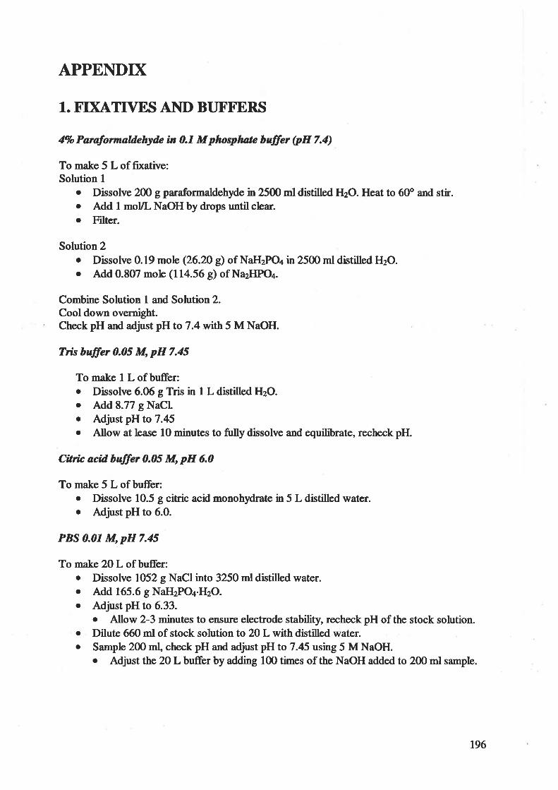
APPENDX
1. trTXATIYES AI\D BI.ITT'ERS
47o Parøformaldehyde ín 0.1 M phosplute buffer (pH 7.4)
To make 5 L of füative:Solution I
o Dissolve 2AO gparaformaldehyde in 2500 ml distilled HzO. Heat to 60o and stir.o Add I moUL NaOH by drops until clear.o Filær.
Solution 2a Dissolve 0.19 mole (26.20 g) of NaIIzPO+ in 2500 ml distilled I{2O.
Add 0.807 mole (114.56 g) of NafiPOa.a
Combine Solution I and Solution 2.Cool down overnight.Check pH and adþst pH to 7.4 with 5 M NaOH.
Trís buffer 0.05 M, pH 7.45
To make 1 L of buffer:o Dissolve 6.M STris in 1 L distilled HzO.o Add 8.77 gNaCLo Adjust plJto7.45o Allow at lease l0 minutes to fully dissolve and equilibrate, recheck pH.
C¡tr¡c æid buffer 0.05 M, pH 6.0
To make 5 L of buffer:o Dissolve 10.5 g citric acid monohydrate in 5 L distilled water.o Adjust pH to 6.0.
PBS 0.U M, pH 7.45
To make 20 L of buffer:o Dissolve IO52 g NaCl into 3250 ml distiled water.o Add 165.6 g NatIzPO¿-HzO.o Adjust pH to 6.33.
o Allow 2-3 minutes to ensu¡e electrode stability, recheck pH of the stock solution.o Dilute 660 ml of stock solution to 2OL with distilled water.o Sample 200 ml, check pH and adjust pH to 7.45 using 5 M NaOH.
o Adjust the 20 L buffer by adding 100 times of the NaOH added to 200 ml sample.
196
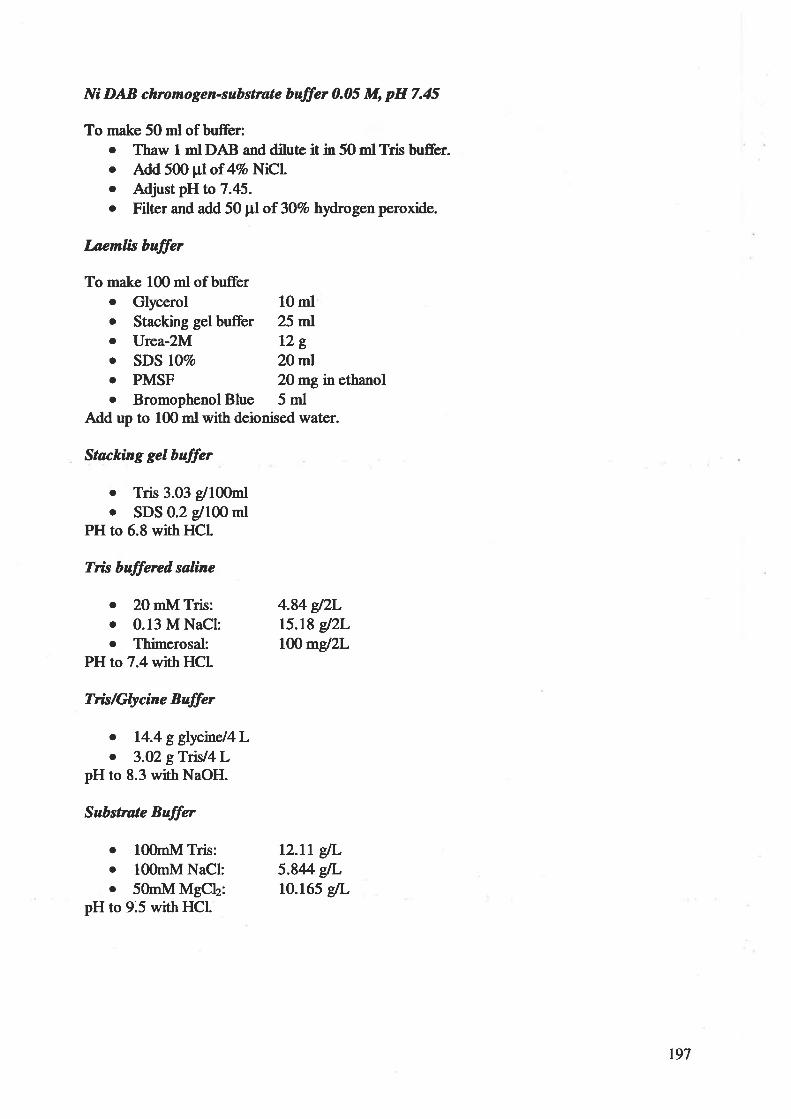
N¡ DAB chromogen-substrate buffer 0.05 A pH 7.45
To make 50 ml of buffer:o Thaw I ml DAB and dilute it in 50 ml Tris buffer.o Add 500 pl of 4Vo NiCLo Adjust pH to 7.45.o Filter and add 50 pl o13O7o hydrogen peroxide.
Læmlß buffer
To make 100 ml of buffero Glycerol 10 mlr Stacking gel buffer 25 ßlo Urea-2M 12 go SDS 107o 20mlr PMSF 20mg in ethanolo Bromophenol Blue 5 ml
Add up to 100 ml with deionised water.
Stackíng gelbuffer
o Tris 3.03 g/100m1o SDS 0.2 g/100 ml
PH to 6.8 with HCL
Trß buffered søline
o 2O mM Tris:o 0.13 M NaCl:o Thimerosal:
PH to 7.4 with HCL
TríslGlycine Baffer
o 14.4 g glycnel4Lo 3.02 gTrßl4L
pH to 8.3 with NaOH.
Substmte Buffer
o 100mM Tris:o 10OmM NaCl:o 50mM MgClz:
pH to 9.5 with HCL
4.84 Bl2L15.18 glzLItO mgl2L
l2.lI glL5.84 glLt0.t65 EtL
r97
TAE buffer
o Æ mM Tris Aminomethaneo 2O mM glacial acetic acido I mMEDTA
pH to 8.0
198
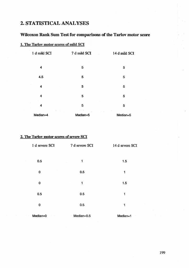
2. STATISTICAL ANALYSES
Wilcoxon Rank Sum Test for comparisons of the Tarlov motor score
I d mild $g¡ 7 dmild SCI 14 d mild SCI
2. Ihe Tarlov motor scores of severe SCI
1 d severe SCI 7 d severe SCI 14 d severe SCI
1.5
1.5
5
5
5
5
5
4
4.5
4
4
4
5
5
5
5
5
Median=5Median=4 Median=5
10.5
0.50
0 1
0.5
0.5
1
0.5
0
1
1
Median=0 Median=1
r99
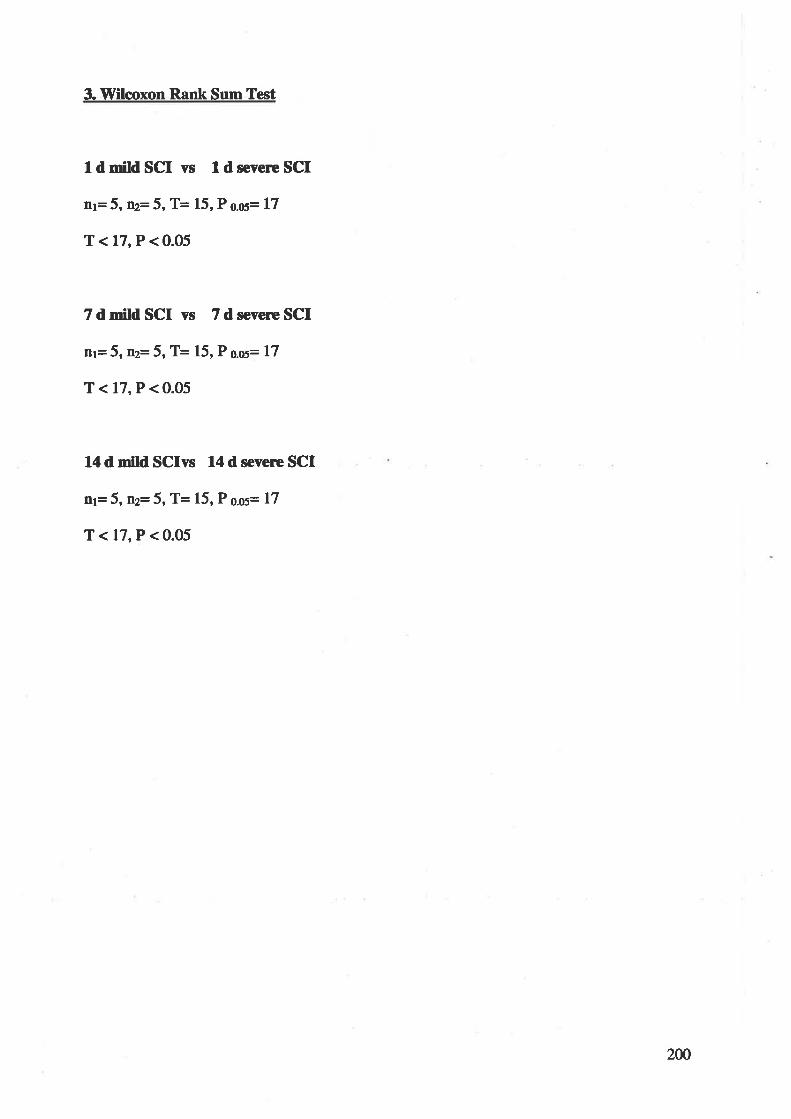
3. VYilcoxon Rank Sum Test
1d mild SCI vs I d severe SCI
nl= 5, mt=5, T= 15, P o.os= 17
T<17,P<0.05
7 d mild SCI vs 7 d severe SCI
nt= 5, nz=5, T= 15, P O.oS= 17
T<17,P<0.05
14 d mild SCIvs 14 d severc SCI
ttt= 5, ft2= 5, T= 15, P O.OS= 17
T <17,P < 0.05
2m
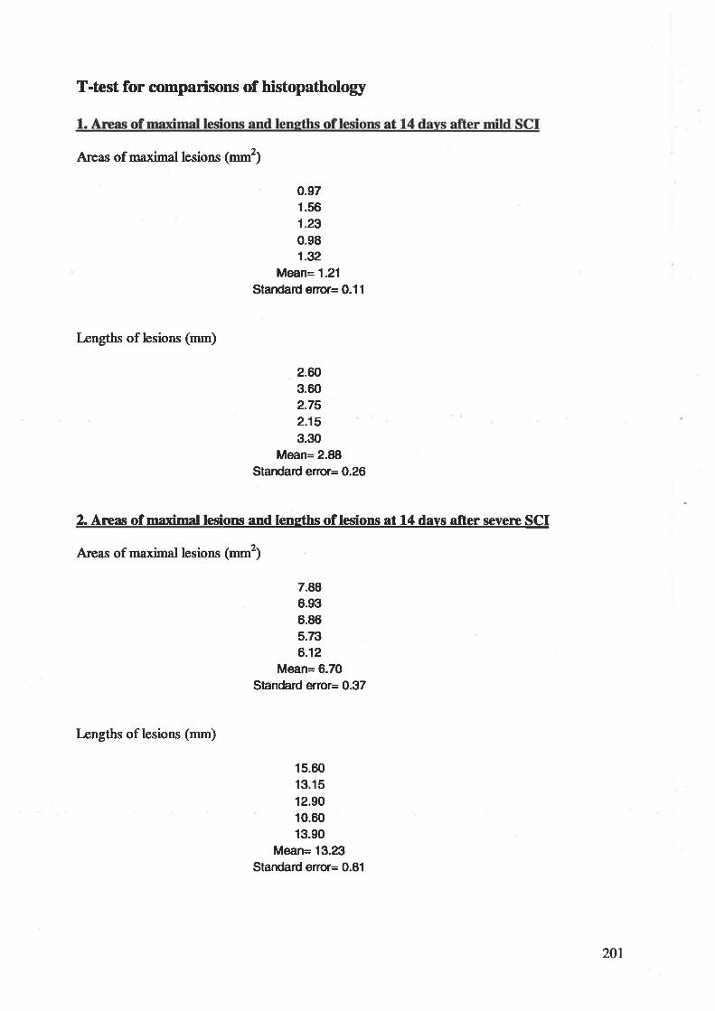
T-úest for comparisons of histopathology
Areas of maximal lesions (- 1
0.971.561.29
0.981.32
Mean= 1.21
Standard êÍor= 0.11
Lengths of lesions (--)
2.603.602.752.153.30
Mean= 2.88Stardard êrror= 0.26
2. Areas of maximal lesions and leneths of lesions at 14 davs after severc SCI
Areas of maximal lesions (--1
7.886.936.865.7ts
6.12Mean= 6.70
Standard êrror= 0.37
Lengths of lesions (mm)
15.60
13.15
12.9010.6013.90
Mean= 13.23Standad êrror= 0.81
201
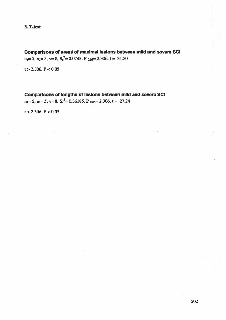
3. T-test
Gomparisons of aleas of maxlmal leslons between mlld and severe SCI
rr= 5, ¡¡2=5,v= 8, Sê O.UI4|,P o.os= 2.3O6,t = 31.80
t>2.306, P < 0.05
Comparlsons of lengths of leslons between mlld and severc SCI
nl= 5, W=5,v= 8, S"2= 0.36185, P o.os= 2.306,t= 27.24
t>2.3M,P<0.05
202
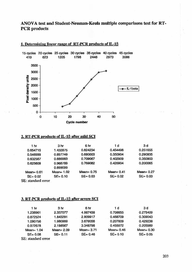
AI\OVA test and Student-Neuman-Keuls multþle comparisons tcst for RT-PCR products
l. Determinine linear ranse of RT-FCR prÐducts of IL-18
lScycles 2Ocycles 25cycles 3Ocycles 3Scycles 40cycles (5cycles419 623 1205 1798 2Æ 2973 3086
+ll-lbeta
10 æ30Cþle number
N 50
2. RT-PICR oroducts of IL-18 afrer mild SCI
3500
3m0
2500
2000
l5(x)
1000
500
otr=ocoEoxÂ-
o
0
thr0.6547150.5469990.632s670.625609
Mean= 0.61
SE= 0.02SE: standard error
3hr1.432875o.8977490.8869830.9687890.899099
Mean= 1.02SE= 0.10
6hro.82423r',0.69o(trt0.7090670.769082
Mean= 0.75SE= 0.Gì
6hr4.987¡158
2.8099173.6780563.348796
Mean= 3.71
SE= 0.46
1d0.4s4980.3509040.,102909
0.426904
Mean= 0.41
SE= 0.02
1d0.7088550.¿168709
0.207809o.455972
Mean= 0.46SE= 0.10
3d0.2516550.2909350.3s090Íl0.200065
Mean= 0.27SE= 0.03
3do.27ilOg0.3090,1i1
o.4292380.205989
Meån= 0.30SE= 0.05
3. RT-PCR nroducts of IL-18 afrer severe SCI
t hr 3hr1.238961 2.3370770.872524 1.84Í1291
1.090756 1.9809890.972678 2.198567
Mean= 1.04 Mean= 2.09SE= 0.08 SE= 0.11
SE: standa¡d error
203
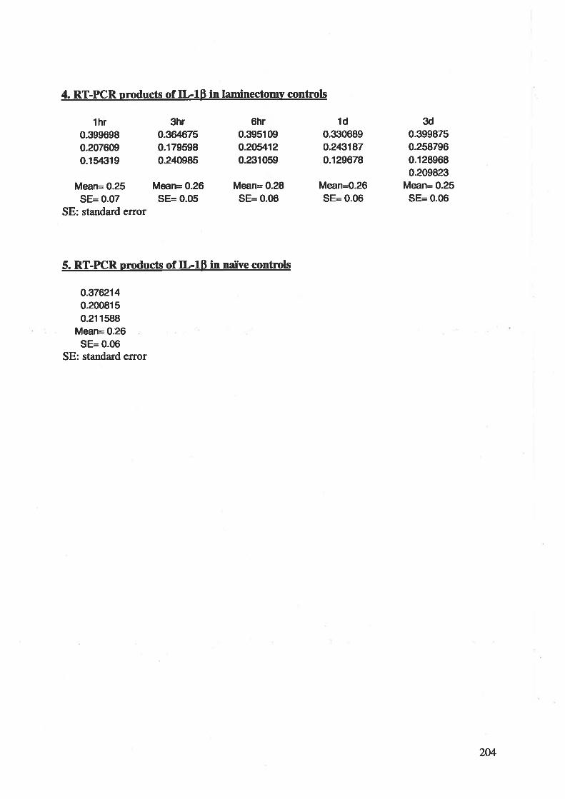
4. RT-PCR oroducts of IL-lÞ in laminectomy controls
Mean= 0.25 Mean= 0.26SE= 0.07 SE= 0.05
SE: standard error
thr0.3996980.2076090.154Í119
3hr0.36¿1675
0.179s980.2¿10985
6hr0.395109o.20il120.231059
Mean= 0.28SE= 0.06
1d0.trr0689o.2431870.129678
Mean=0.26SE= 0.06
3d0.3998750.2587960.1289680.209823
Mean= 0.25SE= 0.06
5. RT-PICR products of IL-IB in nailve controls
o.3762140.2008150.211588
Mean= 0.26SE= 0.06
SE: standard error
204
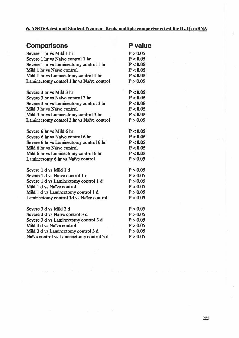
6. ANOVA test and Student-Neuman-Keuls multiple comparisons test for IL-lÞ nRNA
GomparisonsSeve¡e I hrvs Mild t hrSevere I hr vs Naivc control I hrSevere t hr vs I¿minectomy control I hrIVIild I hr vs Narve controlMild I hr vs l¿minectomy control I hrI¿minectomy control I hr vs Narve control
Severe 3 hr vs Mild 3 hrSevere 3 hr vs Naive control3 hrSevere 3 hr vs Laminectomy control 3 hrMild 3 hr vs Narve controlùÍild 3 hr vs Laminectomy control 3 hrI-aminectomy control3 hr vs Narve control
Severe 6 hr vs Mild 6 hrSevere 6 hr vs Nai¡e control6 hrSevere 6 hr vs I¡minectomy control 6 hrMild 6 hr vs Narve controlNlild 6 hr vs Laminectomy control6 hrLaminectomy 6 hr vs Narve control
SevereldvsMildldSevere I d vs Naive control L dSevere I d vs I¿minectomy control I dMild I d vs Narve controlMild I d vs l¿minectomy control 1 dI¿minectomy control ld vs Narve control
Severe3dvsMild3dSevere 3 d vs Naive control3 dSevere 3 d vs Laminectomy control3 dVlild 3 d vs Narve controlMild 3 d vs Laminectomy control 3 dNarve control vs Laminectomy control3 d
P valueP > 0.05P < 0.05P < 0.05P < 0.05P < 0.05P > 0.05
P < 0.05P < 0.05P < 0.05P < 0.05P < 0.05P > 0.05
P < 0.05P < 0.05P < 0.05P < 0.05P < 0.05P > 0.05
P > 0.05P > 0.05P > 0.05P > 0.05P > 0.05P > 0.05
P > 0.05P > 0.05P > 0.05P > 0.05P > 0.05P > 0.05
205
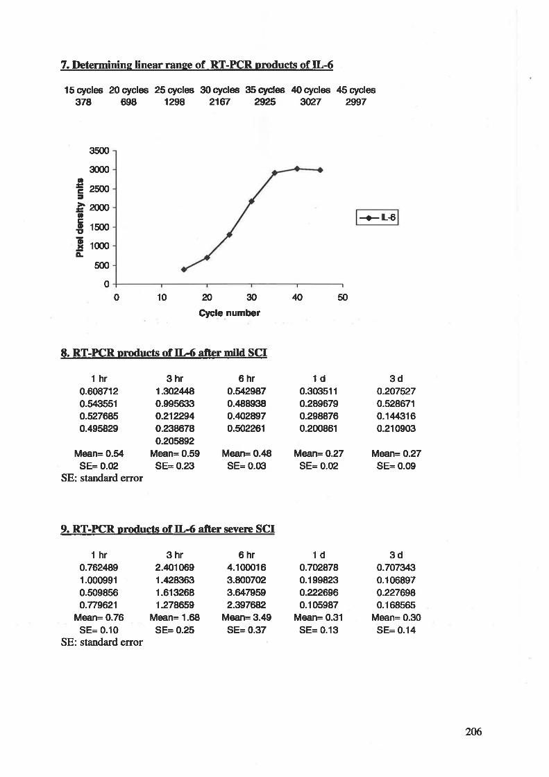
7.I)eterurinine linear ranee of RT-PCR pmducts of IL-6
lScycles 2Ocycles 25cycles 3Ocycles S5cycles 40cycles A5cydes378 698 1298 2167 Æ5 gt27 2997
35æ
3æ0
mãno
15(x)
1000
5(n
CÊf
otrouoxÀ
thr0.6087120.5435510.5276850.495829
Mean= 0.34SE= 0.02
SE: standa¡d error
3hr1.302Æ0.995trt3o.2122940.2386780.205892
Mean= 0.59SE= 0.23
6hro.il29870.488938o.40/2897
0.s02261
Mean= 0.48SE= O.Gl
6hr4.1000163.8007023.6479s92.397æ2
Mean= 3.49SE= 0.37
N 50
1d0.3G1511
0.2896790.2988760.200861
Mæn=O.27SE= 0.02
1do.7028780.199823o.2226960.105987
Mean= 0.31
SE= 0.13
+lL€
3do.2075270.5286710.1¿14Íì16
0.210903
Mean= 0.27SE= 0.09
3do.7073430.1068970.2276980.168s6s
Mean= 0.30SE= 0.14
0
10 2030Clcle number
E. RT-PICR products of IL-6 after mild SCI
0
9. RT-FCR pmducts of IL-6 afrer severc SCI
t hr 3hr0.762ß9 2.4010691.000991 1.428Íttrl0.509856 1.6132680.779621 1.278659
Mean= 0.76 Mean= 1.68
SE= 0.10 SE= 0.25SE: standard error
206
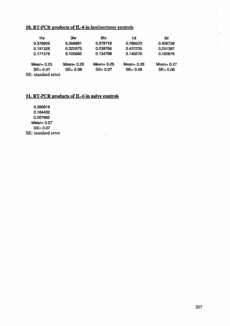
10. RT-PCR products of IL-6 in laminectomv controls
Mean= 0.25 Mean= 0.26SE= 0.07 SE= 0.08
SE: standard error
thr0.3789050.1913260.171579
3hr0.356981o322rr30.105982
6hro.3787190.2397650.134768
Mean= 0.25SE= 0.07
1d0.286523o.422235o.143276
Mean= 0.28SE= 0.08
3d0.¿t09739
0.2513870.160876
Mân=O.27SE= 0.06
11. RT-PICR uroducts of IL-6 in na,ive contr,ols
0.390919o.16/432o.267892
Mean= 0.27SE= 0.07
SE: st¿ndard error
207
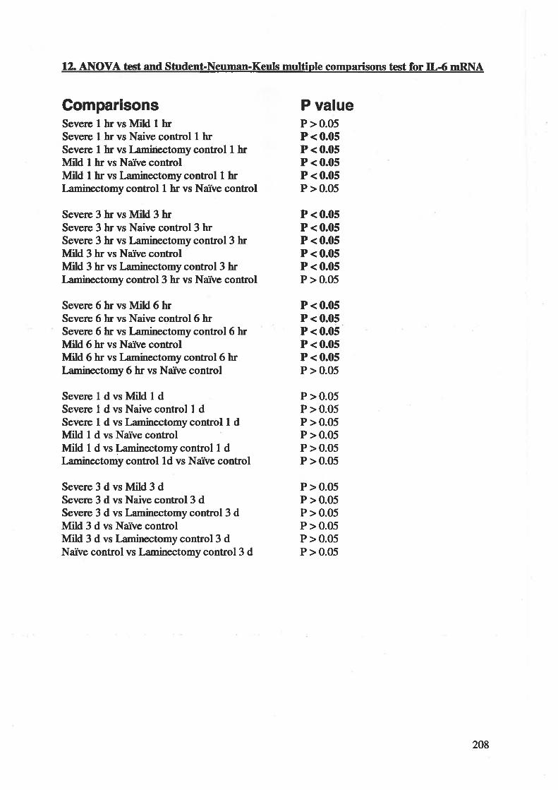
12. AIIOVA test and Student-Neuman-Keuls multÍple comparisons test for IL-6 nRNA
Gompar¡sonsSevere I hr vs Mild I hrSevere I hr vs Naive control I hrSeve¡e I hr vs I¿minectomy control I hrVlild I hr vs Narve controlIVIild I hr vs Laminectomy control t hrLaminectomy control I hr vs Narve control
Severe 3 hr vs Mild 3 hrSevere 3 hr vs Naive control3 hrSevere 3 hr vs Laminectomy control 3 hrMild 3 hr vs Narve controlllild 3 hr vs I¿minectomycontrol3 hrI¿minectomy control3 hr vs Narve control
Severe 6 hr vs Mild 6 hrSevere 6 hr vs Naive control6 hrSevere 6 hr vs l-aminectomy control6 hrÙfild 6 hr vs Narve controlMild 6 hr vs l-aminectomy control6 hrI-aminectomy 6 hr vs Naïve control
SevereldvsMildldSevere 1 d vs Naive control I dSevere 1 d vs I-aminectomy control I dMild 1 d vs Narve controlMild 1 d vs I-aminectomy control I dLaminectomy control ld vs Narve control
Severe3dvsMiH3dSevere 3 d vs Naive control3 dSevere 3 d vs Laminectomy control3 dIvfild 3 d vs Narve controlIvlild 3 d vs Laminectomy control3 dNarve control vs Laminectomy control 3 d
P valueP > 0.05P< 0.05P < 0.05P < 0.05P < 0.05P > 0.05
P < 0.05P < 0.05P < 0.05P < 0.05P < 0.05P > 0.05
P < 0.05P < 0.05P < 0.05P < 0.05P < 0.05P > 0.05
P > 0.05P > 0.05P > 0.05P > 0.05P > 0.05P > 0.05
P > 0.05P > 0.05P > 0.05P > 0.05P > 0.05P > 0.05
208
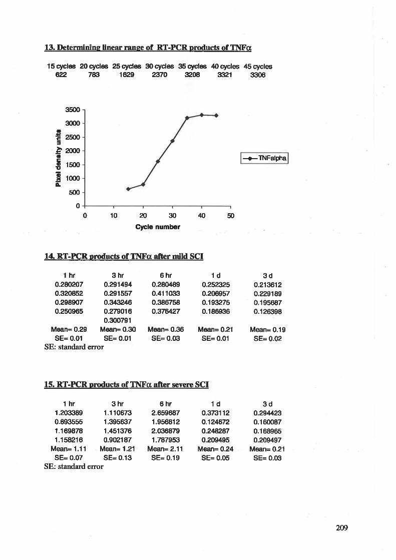
13. Determinine linear range of RT-PCR pmducts of Tl\[X'cr
l5cyclee 20cyclæ 2Scycles S0cycles S5cycles 40cycles 45cycles622 783 1629 2970 3208 3Ít21 3306
+TNFalpha
ãJ30Clcle number
ß50
14. RT-PCR products of ThlFa after mild SCI
3s00
3000
2ffiãþ0
1500
1(n0
s00
otr=à6Êouo.xÀ
0
100
t hr 3hr0.2æ207 0.2914940.320852 0.2915570.298907 0.3432ß0.250965 0.279016
0.300791Mean= 0.29 Mean= 0.30
SE= 0.01 SE= 0.01SE: standard error
t hr 3hr1.209389 1.1106730.893sss 1.395trt71.169878 1./+51376
1.158216 0.902187Mean= 1.11 Mean= 1.21
SE= 0.07 SE= 0.13SE: standard error
15. RT-PICR omducts of II\Fcr afrer severe SCI
6hr0.2804890.411Gì30.3867s8o.37æ27
Mean= 0.36SE= 0.Gì
6hr2.6596871.9568122.0Í16879
1.787953Mean= 2.11
SE= 0.19
1do.2523250.2069570.1932750.186936
Mean= 0.21
SE= 0.01
1do.373112o.12ß72o.2ß2870.209495
Mean= 0.24SE= 0.05
3do.21æ120.2291890.1956870.126398
Mean= 0.19SE= 0.02
3do.294É,23
0.1600870.168965o.209497
Mean= 0.21
SE= 0.0Íl
2W
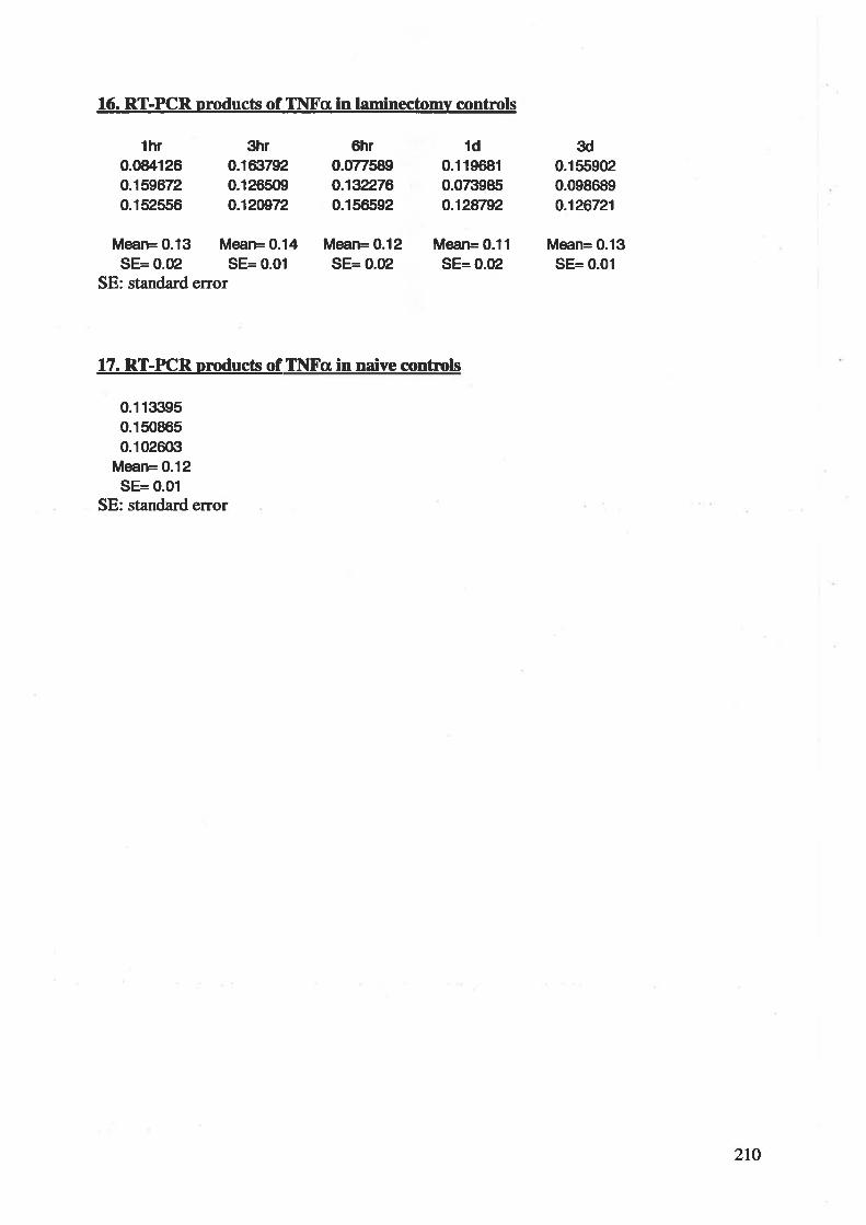
16. RT-PCR oroducts of TI{Fcr in laminectomv contrcls
Mean= 0.13 Mean= 0.14SE= 0.02 SE= 0.01
SE: standard error
thr0.084126o.1596720.152556
3hr0.16Ít7920.126509o.12æ72
6hr0.077589o.1922760.156592
Mean= 0.12SE= 0.û2
1d0.1196810.0its985o.128792
Mean= 0.11
SE= 0.02
3d0.1559020.098689o.126721
Mean= 0.13SE= 0.01
17. RT-PICR prcducts of Ihlf,'a in naive controls
0.11tr¡950.1508650.1026G¡
Mean= 0.12SE= 0.01
SE: standa¡d error
zto
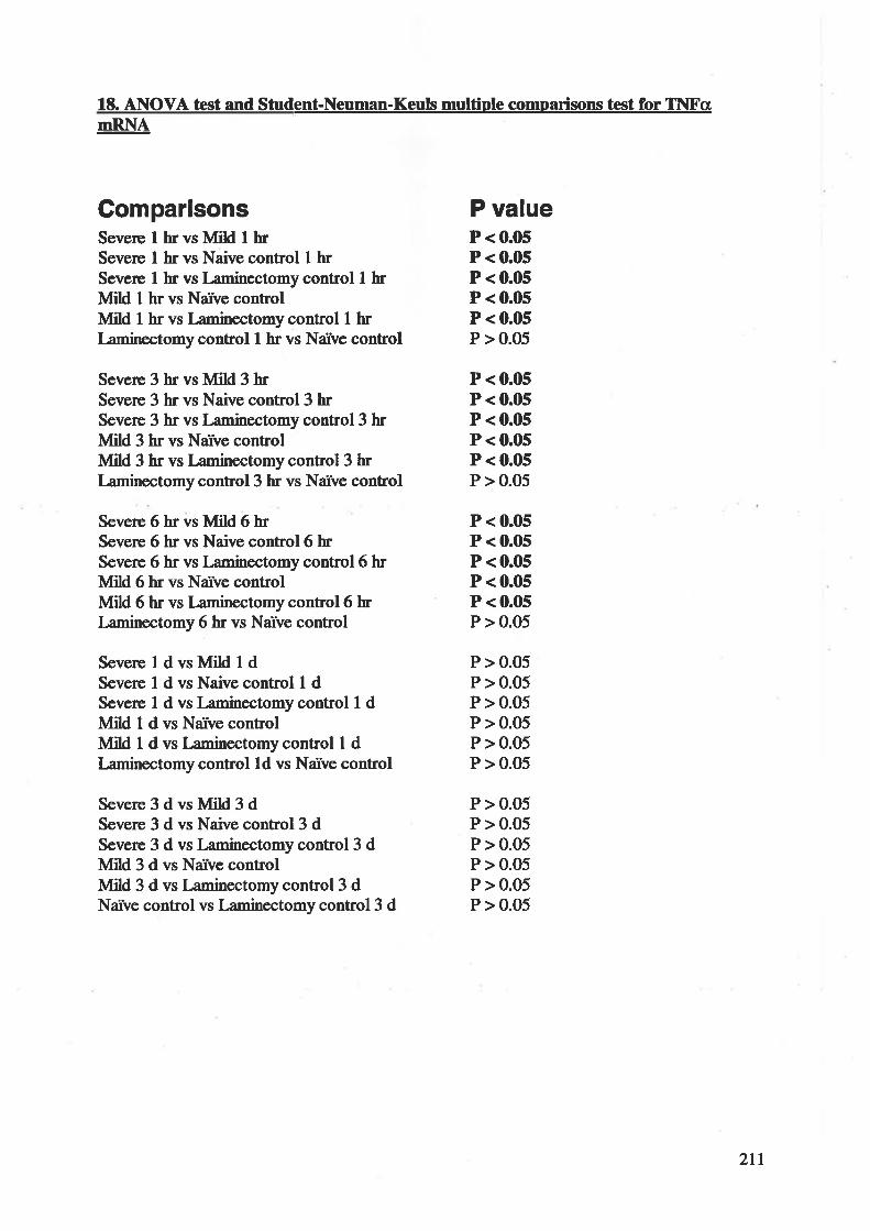
lE. ANOVA test and Student-Neuman-Keuls multiple comparisons test for lhlFanRNA
GomparisonsSevere I hr vs MiH I hrSevere I hr vs Naive control I hrSevere I br vs Laminectomy control I hrMild I hr vs Narve controlMild I hr vs l¿minectomy control t hrLaminectomy control I hr vs Narve control
Severe 3 hr vs Ivfild 3 brSevere 3 br vs Naive control3 hrSevere 3 hr vs I¡minectomy control3 hrMild 3 hr vs Narve controlMild 3 hr vs Laminectomy control 3 hrLaminectomy control3 hr vs Narve control
Severe 6 hr vs Mild 6 hrSevere 6 hr vs Naive control6 hrSevere 6 hr vs Laminectomy control6 hrMild 6 hr vs Narve controlMitd 6 hr vs l¿minectomy control6 hrLaminectomy 6 hr vs Narve control
SevereldvsMildldSevere 1 d vs Naive control I dSevere I d vs Laminectomy control 1 dIVIild 1 d vs Narve controlMild 1 d vs I¿minectomy control I dI¿minectomy control ld vs Narve control
Severe3dvsMild3dSevere 3 d vs Naive control3 dSevere 3 d vs Laminectomy control 3 dIvlild 3 d vs Narve controlMild 3 d vs Laminectomy control3 dNaïve control vs I¿minectomy control3 d
P valueP < 0.05P < 0.05P < 0.05P < 0.05P < 0.05P > 0.05
P < 0.05P < 0.05P < 0.05P < 0.05P < 0.05P > 0.05
P < 0.05P < 0.05P < 0.05P < 0.05P < 0.05P > 0.05
P > 0.05P > 0.05P > 0.05P > 0.05P > 0.05P > 0.05
P > 0.05P > 0.05P > 0.05P > 0.05P > 0.05P > 0.05
2rt
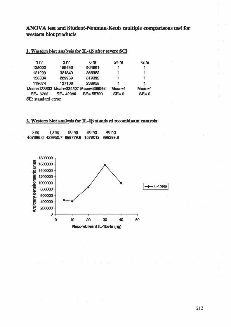
ANOVA test and Student-Neuman-Keuts multþle comparisons tcst forwestem blot products
1. Vl¡estern blot analvsis for IL-lß after severe SCI
thr 3hr 6hr138002 189,135 504861121299 321il9 368982156834 289939 319392119074 137106 238958
Mean=133802 Mæn=2 5O7 Mean=358048SE=8762 SE=42980 SE=55790
SE: standard error
72hr24hr1
1
1
1
Mean=1
SE= 0
1
1
1
1
Mean=1
SE= 0
2. Vt¡estcm blot analvsis for IL-lÞ standard recombinant controls
Sng 10ng 20ng 30ng 40ng¡157396.6 423950.7 869779.8 1579012 996iÍ199.8
1800000
r600000
1400000
12000æ
10000æ
800000
6000æ¿lO00O0
200000
0
+ll-lbeta
10?o3040Recornbinant lL-l beta (ng)
50
og¡.9oEoocoEàG
.o
0
212

3. AI\OVA test and Student-Neuman-Keuls multiple comoarisons test for IL-lß pr.otein
ComparisonsSevere I hr vs Mild I hrSeve¡e I hr vs Naive control I hrSevere I hr vs I-aminectomy control I hrMild I hr vs Narve controlMild I hr vs Laminectomycontrol I hrI-aminectomy control t hr vs Naïve conhol
Severe 3 hr vs Mild 3 hrSeve¡e 3 hr vs Naive control3 hrSevere 3 hr vs Laminectomy control3 hrþIild 3 hr vs Narve controlMild 3 hr vs Laminectomy control 3 hrI¿minectomy control 3 hr vs Narve control
Severe 6 hr vs Mild 6 hrSevere 6 hr vs Naive control6 hrSevere 6 hr vs Laminectomy control6 hrùIild 6 hr vs Naîve controlMild 6 br vs I¿minectomy control6 hrI-aminectomy 6 hr vs Narve control
SevereldvslvlildldSevere I d vs Naive control I dSevere I d vs Laminectomy control 1 dIvlild I d vs Narve controlIUiH f d vs Laminectomy control 1 dI¿minectomy control 1d vs Narve control
Severe3dvsMild3dSevere 3 d vs Naive control3 dSevere 3 d vs Laminectomy control 3 dMild 3 d vs Narve controlIvIild 3 d vs Laninectomy control3 dNarve control vs Laminectomy control3 d
P valueP <0.05P <0.05P < 0.05P > 0.05P > 0.05P > 0.05
P < 0.05P < 0.05P < 0.05P > 0.05P > 0.05P > 0.05
P < 0.05P < 0.05P < 0.05P > 0.05P > 0.05P >0.05
P > 0.05P > 0.05P > 0.05P > 0.05P > 0.05P > 0.05
P > 0.05P > 0.05P > 0.05P > 0.05P > 0.05P > 0.05
2t3
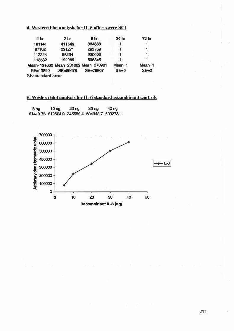
4. Western blot analvsÍs for IL-6 after severe SCf
thr 3hr 6hr161141 411ffi 364388971V2 221271 292769111224 9823/. 2306021135Í|2 19298s 595845
Mean=l 21 000 Mean=231 009 Mean--370901
SE=13890 SE=6.5678 SE=79807SE: standard error
10 ?o30Recomblnant lL6 (ng)
72hr24hr
Mean=1
1
1
1
1
Mean=1SE=0
1
1
1
1
SE=0
5. TYestern blot analvsis for IL-6 standard recombinant contnols
Sng 10ng 20ng 30ng 40ng81413.75 2196e1.9 34s559.4 5M942.7 609273.1
700000
600000
s00000
400000
300000
200000
100000
oÊa.9oEootrooI6o
+lL€
00 N 50
214
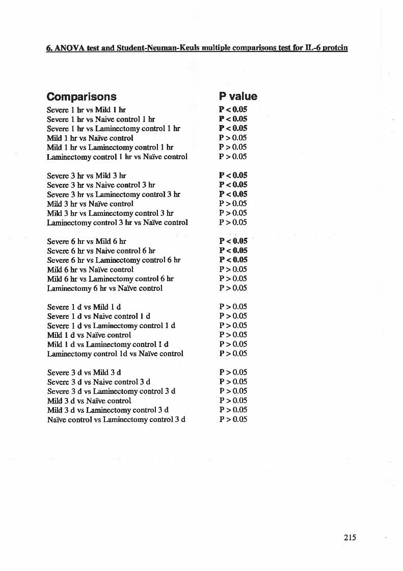
6. A¡IOVA test and Student-Neuman-Keuls multiple comparisons test for IL-6 prctein
ComparisonsSevere I hr vs ùIild I hrSevere I hr vs Naive control I hrSevere I hr vs I¿minectomy control I hrMiH I hr vs Narve controlMild I hr vs Laminectomy control I hrLaminectomy control I hr vs Narve control
Severe 3 br vs Mild 3 brSevere 3 hr vs Naive control3 hrSevere 3 hr vs I-aminectomy control 3 hrMild 3 hr vs Narve controlMild 3 hr vs I¿minectomy control 3 hrLaminectomy control3 hr vs Narve control
Severe 6 hr vs Mild 6 hrSevere 6 hr vs Naive control6 hrSevere 6 hr vs I-aminectomy control6 hrMild 6 hr vs Narve controlIvlild 6 hr vs I-aminectomy control6 hrLaminectomy 6 hr vs Narve control
SevereldvsMildldSevere 1 d vs Naive control I dSevere I d vs l¿minectomy control 1 dMild 1 d vs Naîve controlIvlild I d vs Laminectomy control 1 dLaminectomy control ld vs Narve control
Severe3dvsMild3dSevere 3 d vs Naive control3 dSevere 3 d vs I-aminectomy control 3 dMild 3 d vs Narve controlMild 3 d vs l¡minectomy control 3 dNarve control vs Laminectomy control 3 d
P valueP < 0.05P < 0.05P < 0.05P > 0.05P > 0.05P > 0.05
P < 0.05P < 0.05P < 0.05P > 0.05P > 0.05P > 0.05
P < 0.05P < 0.05P < 0.05P > 0.05P > 0.05P > 0.05
P > 0.05P > 0.05P > 0.05P > 0.05P > 0.05P > 0.05
P > 0.05P > 0.05P > 0.05P > 0.05P > 0.05P > 0.05
215
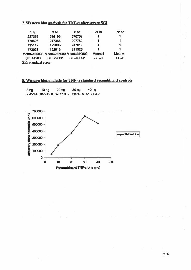
7. \üestern blot analvsis for T1\[tr'-a after severe SCI
thr 3hr 6hræ7æ5 51519ft s7rrcz178526 2773æ 207789195112 192888 24761917æ28 162913 211526
Mean=l 96008 Mean=287090 Mean=3l 0909
SE=145ff1 SE=79802 SE=89052SE: standard error
72hr24hr
Mean=1
1
1
1
1
1
1
I1
Mean=1SE=0SE=0
E. T[estem blot analvsis for TI\F-a standard recombinant controls
Sng 10ng 20ng 30ng 40ng50450.4 187245.8 370216.6 628742.9 515664.2
+TNFclpha
0 10?o3040Recomblnant TNFalpha (ng)
50
700000
600000
500000
400000
300000
200000
100000
oÊãgoEooCoo6
Â
0
2t6
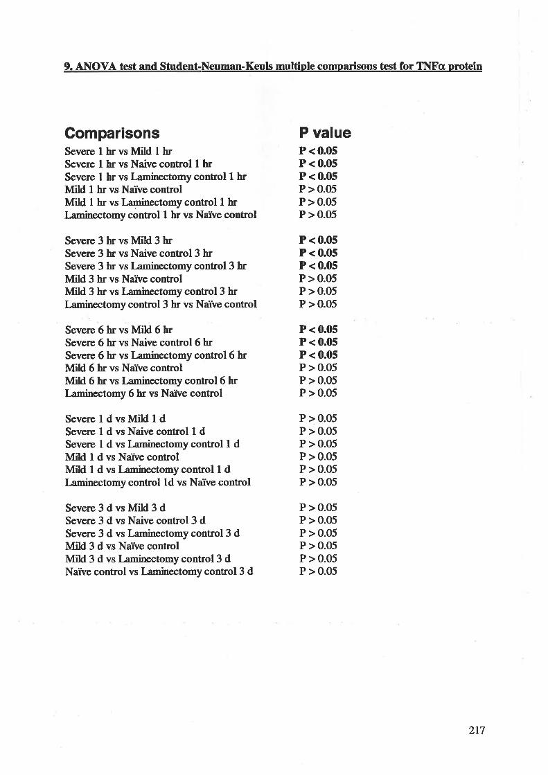
9. AIIIOVA test and Student-Neuman-Keuls multiple comparisons test for II\IFa orotein
Compar¡sonsSeve¡e I hr vs Mild t hrSevere I hr vs Naive control t hrSevere I hr vs Laminectomy control I hrMild I hr vs Narve controlùfild I hr vs Laminectomy control t hrI-aminectomy control I hr vs Narve control
Seve¡e 3 hr vs Mild 3 hrSevere 3 hr vs Naive control3 hrSevere 3 hr vs Laminectomy control 3 hrI\[ild 3 hr vs Naïve controlÙfild 3 hr vs I¿minectomy control3 hrI¿minectomy control3 hr vs Naïve control
Severe 6 hr vs ÙIild 6 hrSevere 6 hr vs Naive control6 hrSevere 6 hr vs Laminectomy control6 hrÞfild 6 hr vs Narve controlMild 6 hr vs Laminectomy control6 hrLaminectomy 6 hr vs Narve control
Severeldvsl[ild1dSevere I d vs Naive control I dSevere I d vs Laminectomy control 1 dMild I d vs Narve controlIvlild I d vs I¡minectomy control I dLaminectomy control Ld vs Narve control
Severe3dvsMild3dSevere 3 d vs Naive control3 dSevere 3 d vs Laminectomy control3 dI\dild 3 d vs Narve controlMild 3 d vs Laminectomycontrol3 dNarve control vs Laminectomy control 3 d
P valueP <0.05P < 0.05P < 0.05P > 0.05P > 0.05P > 0.05
P < 0.05P < 0.05P < 0.05P > 0.05P > 0.05P > 0.05
P < 0.05P < 0.05P < 0.05P > 0.05P > 0.05P > 0.05
P > 0.05P > 0.05P > 0.05P > 0.05P > 0.05P > 0.05
P > 0.05P > 0.05P > 0.05P > 0.05P > 0.05P > 0.05
217
I
REFERENCES
10.
Aarden LA Bnrnner TK Cerottini lC, et al Revised nomenclature for antigen-nonspecific T cell proliferation and heþr factors. J. Immunol.l?.322928-2929, 1979Abe K St4eorge-Hyslop, P.H., Taîù R.E. and Kogure, trC: Induction of amyloidprecursor protein mRNA after heat shock in cultured human lynphoblastoid cells.
Neurosci l*tt 125z169-17 l, l9 IAggarwal BB: Tumor necrosis factor, in Aggarwal BB, Gutterman JU (eds): Humancytokines: handbook for basic and clinical researth. Boston: Blackwell scientificpublications, 1992, Vol l, pp 27U287Aloisi F, Care, A., Borsellino, G.: Production of hemol¡aqphopoietic cytokines bynormal human astorcytes in response to IL-l beta and tumor necrosis factor-alpha. J.Immunol 14922358-2366, 1992Amff AP, lævy ML: Pathogenesis and pharmacological strategies for mitigatingsecondary damage in acute spinal cord inþry. .Neumsurgery 44rz1027-1039, 1999Anderson DK Means ED, \Vaters TR, et at Mcrovascular perfusion and metabolismin injured spinal cord after methylprednisolone treatment. J Neurosurg 56:106-113.,1982Anderson TE: A controlled pneumatic technique for experimental spinal cordcontusion. J.Neurosci.Methods 62327 -333, 1982Assenmacher DR, Ducker TB: Experimental traumatic paraplegia. The vascula¡ andpathological changes seen in reversible and irreversible spinal-cord lesions. J BoneJoint Surg Am 53:671-680.,1971Baggiolini M, Imboden P, fÞtmers P: Neutrophil activation and the effects ofinterleukin-8/neutrophil-activating peptide I (IL-8/NAP-I). Cytokines 4:1-17.,1992Baldwin AS, Jr.: The NF-kappa B and I kappa B proteins: new discoveries andinsights. Annu Rev rmmunoll4z649-683., 1996Balentine JD: Pathology of experimental spinal cord trauma- tr. Ultrastructure ofaxons and myelin. .Lab.InvesL 39:25L266, 197 8
BanatiRB, Gehrmann l,Cz¡*hC, et aL Early and rapid de novo slmthesis ofAlzheimer beta A4-amyloid precursor protein (APP) in activated microglia. Glia9zlÐ-210.,1993Banati RB, Gehrmann J, Schubert P, et at Cytotoxicity of microglia. Glia 7:111-118.,IÐ3Banati RB, Graeber MB: Surveillance, intervention and cytotoxicity: is there aprotective role of microglia? Dev Neurosci l6zllLl27.,1994Barbeau H, Chau C, Rossignol S: Noradrenergic agonists and locomo¡s¡ treiningaffect locomotor rocovery after cord transection in adult cats. Brain Res Bull30:387-393.,1993Barger S'W, Horster D, Furukawa K et at Tumor necrosis factors alpha and betaprotect neurons agains¡ amyloid beta-peptide toxicity: evidence for involvement of akappa B-binding factor and attenuation of peroxide andCa2+ accumulation. hrocNatl Acad Sci U S l¡92t9328-9332.,1995Ba¡one FC, FeuerstenGZ: Iqflammatory mediators and stroke: new opportuniÏes fornovel therapeutics. J Cereb Blood Flow Metab 19:819-834.,1999Barres BA" Scbmid R, Sendnter M, et at Multþle extracellula¡ signals are requiredfor long-term oligodendrocyte survival Development ll8z283-295., 1993Bartholdi D, Schwab ME: Expression of pro-inflammatory cytokine and chemokinemRNA upon experimental spinal cord inþry in mouse: an in situ hybridization study..EurJ.Neurosci . 9 ¿ I 422- t 438, 1997
11.
12.
13
14.
15.
16.
17.
18.
2.
3.
4.
5.
6.
7.
8.
9
19.
218
20. Bartholdi D, Schwab ME: Methyþrednisolone inhibits early inflammatory Processosbut not ischemic cell death after experimental spinal cord bsion in the rat. Brain Res112z177-186.,1995
21. Basso DM, Beattþ MS, Bresnahan JC: Graded histological and locomotor outcomesafter spinal cord contusion using the NYU weight-drop device versus transection.Experimental Neurclo g 139 z2&256, 1996
22. Bavetta S, Hamlp Pl Burnstock G, et aL The effects of FI(506 on dorsal columnÐKolls following spinal cord inþry in adult rats: neuroprotection and localregeneration Exp Neurol 158:382-393., 1999
23. Becker-Andre M, Hahlbrock K: Absolute mRNA quantification using the pollmerasechain reaction (PCR). A novel approach by a PCR aided transcript titration assay(PATTÐ. Nucleic Adds Res 17:9437-9446.,1989
24. Beggs JL, Waggener JD: The acute mbrovascular riesponses to spinal cord inþry.Adv Neurcl X2zl7 9-189., 1979
25. Berry M, Butt AIVÍ: Cellular pathology of the central nervous systom, in Graham DI,I-antos PL (eds): Greenfield's neumpatholory, ed 6th. London: Arnold, L997,pp78-156
26. Bethea JR: Spinal cord injury-induced inflamrnation: a dual-edged sword. Pnog BrainRes 12E:33-42.,2m
27. Bethea JR, Castro M, Keane RW et at Traumatic spinal cord injury induces nuclearfactor-kappaB activation J Neumsci lEz325l-3260., 1998
28. Bethea JR, Nagashima H, Acosta MC, et at Systemically administered interleukin-lOreduces tumor necrosis factor-alpha production and significantþ improves functionalrecovery following traumatic spinal cord inþry in rats. J Neurotrauma 16:851-863.,1999
29. Beuche \Y, Friede RL: The role of non-resident cells in Wallerian degeneration. JNeurocyûol l3z7 67 -796., 1984
30. Bignami À Ralston HI, 3rd: The cellula¡ reaction to \Yallerian degeneration in thecentral nervous system of the cat. Brain Res 13:444 ß1.,1969
31. Black P, Markowiø RS, Damjanov I, et at Models of spinal cord injury: Pa¡t 3.
Dpamic load technique. .Neumsurgery 22:51-60, 1988
32. Blakemore WF: Remyelination by Schwann cells of axons demyelinated byintraspinal inþtion of 6-aminonicotinamide in the rat. J Neurocytol 4¿145-757.,1975
33. Blight AR: Delayed demyelination and macrophage invasion: a candidate forsecondary cell damage in spinal cord inþry. Cent NeIv Syst Trauma 222Ð-315.,1985
34. Blight AR: Effects of silica on the outcome from experimental spinal cord injury:imFlication of macrophages in secondary tissue damage. Neurosciencrc 6O¿263-273.,r994
35. Blight AR: Macrophages and inflammatory damage in spinal cord injury.J.Neurotraum¡. 9 Suppl 1:583-91, 1992
36. Blight AR: An overview of spinal cord inþry models, in Narayan RIÇ Wilberger JE,PovlishockJT (eds): Neurotrauma. New York McGraw-Hill" 1996,pp 1367-1379
37. Nlin-inger K lGeutzberg, G.: Displacement of s¡maptic terminals from regeneratingmotor neurons by microglial celß. Z Zellforscù Mikmsk Anat 85:145-157, 1968
38. Blumbergs FC, Scott G, Manavis J, et aL Staining of amyloid pr€cursor protein tostudy axonal damage in mild head injury. Lancet 344:1055-1056., 1994
219
39. Blumbergs FC, Scott G, Manavis I ct at Topography of axonal injury as defined byamyloid precursor protein and the sector scoring method in mild and severe closedhead injury. J Neunotrauma 122565-572., 1995
ß. Bohlman HH: Acute fractures and dislocations of the cervical spine. An analysis ofth¡ee hundred hospitalized patients and revbw of the literature. J Bone Joint SurgAm 61:1119-1142.,1979
41. Bohlman HH, Ducker TB: Spine trauma in adults, in Rorhman RH, Simeone FA (eds):
lhe spine. Philadelphia: Saunders, lÐ2, pp 973-llW42. BomalaskiJS, Steiner MR, Simon PL, et aL ILI increases phospholipuse M activity,
expression of phospholipase A2-activating protein, and release of linoleic acid fromthe murine T helper cell line F,L-4. J Immunol 14E:155-16O., 1992
43. Botchkina GI, Meistrell ME, 3rd, Botchkina IL, et at Expression of TNF and TNFreceptors þ55 and p75) in the rat brain after focal cerebral ischemia. Mol Med 3:765-781., L997
4. Boyeson MG, FeeneyDM: Intraventricular norepinephrine facilitates motor recoveryfollowing sensorimotor cortex injury. Pharmaol Biochem Behav 352497-5O1., 1990
45. Bracken MB, Collins WF, Freeman DF, et aL Efficacy of methylprednisolone in acute
spinal cord injury. Jama 251245-52.,19844. Bracken MB, Holford TR: Effects of timing of methylprednisolone or naloxone
administration on recovery of segmental and long-tract neurological function inNASCIS 2. J Neuruurg 79:500-507.;1993
47. Bracken MB, Shepard MJ, Collins WF, et at A randomi?rÃ, controlled trial ofmethylprednisolone or naloxone in the treatment of acute spinal cord injury. N E"gl ¡Med:14O5-l4ll, lW
48. Braughler JM, Hall ED: I-actate and pynrvate metabolism in injured cat spinal cordbefore and after a single large intravenous dose of methylprednisolone. J Neurosurg59t256-26L, 1983
49. Brenneman DE, Schultzberg M, Barúai T, et aL Cytokine reguliation of neuronalsurvival J Neurochem 58:454- 4ó¡0., 1992
50. Bresnahan JC: An electron-microscopic analysis of axonal alterations following bluntcontusion of the spinal cord of the rhesus monkey (Macaca mulatta). J Neurol Sci37259-82.,1978
51. Bricolo A., Ore GD, Da Pian R, et at Local cooling in spinal cord injury. Surg Neurol6:101-106.,1976
52. Brodner RA" Dohrmann GJ, Roth RH, et at Correlation of cerebrospinal fluidserotonin and altered spinal cord blood flow in experimental trauma. .Surg.Neurol.132337-343, 1980
53. Broude E, McAtee M, Kelley MS, et aL Fetal spinal cord transplants and exogenous
neurotrophic support enhance c-Jun expression in mature anotomized neurons afterspinal cord injury. F'-I¡ Neurol 155:65-78.,1999
54. Brown MC, Perry VH, Lunn ER, et aL Macrophage dependence of peripheral sensory
nerve regeneration: possible involvement of nerve growth factor. Neumn 62359-37O.,
t99L55. Bruce AJ, Boling W, Kindy MS, et aL Altered neuronal and microglialresponses to
excitotoxic and ischemic brain injury in mice lacking TNF receptors. Nat Med 2:788-794.,1996
56. Buchanan L, Nawoczenski D: An overview, in Buchanan L (ed): Spinal cord i4iury:concepts and menagement appmaches Baltimore: Williams and \üilkins, 1987, pp3-19
220
57. Bunge MB, Hobts VR, Bates ML, et at Characterization of photochemically inducedspinal cord inþry in the rat by light and electron microscopy. .Exp.Neuml l27z7G93,1994
58. Burke DC, Brown Dl Burby HT, et at Data collection on spinal cord injuries:urological outcome. Paraplegia 25z3ll-317 ., 1987
59. Burke F, Naylor MS, Davbs B, et at The cytokine wall chart. Immunol Todayl4zl65-t70.,lÐ3
l). Callard RE, Gearing AJH: The cyúokine factsboolc London ; San Diego: AcademicPr,ess, 1994
61. Cameron T, Prado R, Watson BD, et aL Photochemically induced cystic lesion in therat spinalcord. I. Behavioral and morphological analysis. Exp Neurol lt9z2lÇ223.,1990
62. Cammer W, Bloom BR, Norton WT, et at Degradation of basic protein in myelin byneutral proteases secreted by stimulated macrophages: a possible mechanism ofinflammatory demyelination. Proc NatI Acåd Sd U S A 75:1554-1558., 1978
63. Campagnolo DI, Keller SE, Delisa JA et at Alteration of immune system function intetraplegics. A pilot study. Am J Phys Med Rehabil 732387-393., 1994
&. Campbell IL, Abraham CR, Masliah E, et at Neurologic disease induced in transgenicmice by cerebral overiexpression of interleukin 6. Pnoc NatI Acsd Sci U S A90:10061-10065., 1993
65. Carlson SL, Parish ME, Springer JE, et aL Acute inflammatory rcsponse in spinalcord following impact injury. f,'*I¡ Neurol l5l:77-88., 1998
66. Chao CC, Hu S: Tumor necrosis factor-alpha potentiates glutamate neurotoxicity inhuman fetal brain cell cultures. I)ev Neurosci 16z172-179., 1994
67. Chao CC, Hu S, Sheng WS, et at Cytokine-stimulated astrocytes damage humanneurons via a nitric oxide mechanism. Glial6z276-284.,199,6
68. Cheng B, Christakos S, Mattson MP: Tumor necrosis factors protect neurons againstmetabolic-excitotoxic insults and promote maintenance of calcium homeostasis.Neurcn l2zl39 -153., 1994
69. Chikawa T, Ikata T, Katoh S, et aL Preventive effects of lecithinized superoxidedismutase and methylprednisolone on spinal cord injury in rats: transcriptionalregulation of inflammatory and neurotrophic genes. J Neurotrauma 1t:93-103., 2001
70. Cho KH, Iwasaki Y, Imamura H, et aL Experimental model of posttraumaticsyringomyelia: the role of adhesive arachnoiditis in syrinx formation. J.Neurosurg.E0:133-139,1994
71. Ciallella JR, Rangnekar W, McGillis JP: Heat shock alters Alzheimer's beta amyloidprecursor protein expression in human endothelial cells. J Neurucci Res 37:769-776.,r994
72. Clark WM, Madden KP, Rothlein R, et aL Reduction of central nervous systemischemic injury in rabbits using leirkocyte adhesion antibody treaûnent. Stroke222877-883., l99l
73. Cohen S, Bigazzi PE, Yoshida T: Commentary. Similarities of T cell function in cell-mediated immunity and antibody production. Cell Immunol 12:150-159., 1974
74. Colotta F, Re F, Muzio M, et aL Interleukin-l type II receptor: a decoy target for IL-lthat is regulated by IL-4. Science 261¿472-475.,1993
75. Combs CÇ Johnson DE, Cannady SB, et at Identification of microglial signaltransduction pathways mediating a neurotoxic response to amyloidogenic fragments ofbeta-amyloid and prion proteins. J Neurcsci 19:928-939.,1999
221
76. Croston GE, Cao Z Goddel DV: NF-kappa B activation by inærleukin-l (tr-l)requires an IL-l receptor-associated protein kinase activity. J Biol Chem 2il0zl65lL16517.,1995
77. Damoiseaux JG, Dopp, E.A, Celame, W., Choa" D., MacPherson, G.G. and Diþtra,C.D.: Rat macrophage lysosomal membnane antigen recognized by monoclonalantibody EDl. Immunology E3zl4l+-l47, 1994
78. David S, Boucha¡d C, Tsatas O, et at Macrophages can modiff the nonpermissivenature of the adult mammalian central neryous system- Neurcn 5zM3-469.. 1990
79. De Ley G, Leybaert L: Effect of flunarizine and methylprednisolone on functionalrçcovery after experimental spinal injury. J Neurotrauma 10:25-35.,1993
80. de Vries [IE, Blom-Roosemalen MC, van Oosten M, et aL The influence of cytokineson the integrity of the blood-brain barrbr in vitro. J NeuroimmunolØz3T-43.,1996
81. del Rio-Hortega P: Mbrogþ in PenfieH W (ed): Cyûologgr and cellular pathologgrof the nervour¡ sysúem. New York: Hafrer Publ" 1965,pp 483-534
82. Delamarter RB, Bohlman HH, Dodge LD, et at Experimental lumba¡ spinal stenosis.Analysis of the cortical evoked potentials, microvasculature, and histopathology. JBone Joint Surg Am 72tlll.l-l20., 1990
83. Demediuk P, Daly MP, Faden AI: Effect 6f imFact trauma on neurotransmitter andnonneurotransmitter amino acids in rat spinal cord [published erratum appears in JNeurochem 1990 Feb;5 4Q):72Ç51. J.Neurochem. 52t1.529- 1536, 1989
84. Dhillon HS, Dong GX, Yurek DM, et aL Regional expression of Par-4 nRNA andprotein after fluid percussion brain injury in the rat. Ex¡l Neurol l70zl4Èl48., 2001
85. Dickson DW lr,e SC: Microglia in Davis RL, Robertson DM (eds): Textbook ofNeurcpathologr, ed Îd. Baltimore: Williams &V/ilkins, 1997,pp 165-2O5
86. D[þtra CD, Dopp EA" Joling P: The heterogeneþ of mononuclear phagocytes inlymphoid organs: distinct macrophage subpopulations in the rat recognized bymonoclonal antibodies EDl, ED2 and ED3. Immunology:589-599, 1985
87. Dina¡ello CA: The biological properties of interleukin-l. Eur Cytokine Netw 52517-531.,1994
88. Dina¡ello CA: Interleukin-l, in Thomson AW (ed): The CyûoHne Handbook, ed 3rd. San Diego: Academic Press Limited, 1998, pp 35-73
89. Dinarello CA: Interleukin-l. CytoHne Growth Facúor Rev 8:253-265.,199790. DiStefano PS, Friedman B, Radzieþwski C, et aL The neurotrophins BDNF, NT-3,
and NGF di.play distinct patterns of retrograde axonal transport in perþheral andcentral neurons. Neuron E:983-993., 1992
91. Dohrmann GJ, Wagner FC, Jr., Wick KM, et at Fine structural alterations intransitory traumatic paraplegia. hoc Veterans Adm Spinal Cord Iqi Conf 1E:6-8.,t97l
92. Douglas SD: Morphology of monocyûes and macrophages, in Beutler E, LichhanMA Coller BS, et al (eds): lVilliam hematologr, ed sth. New York: McGraw-Hill"l95,pp 861-894
93. Dower SK: Interleukin-l, in Aggarwal BB, Gutterman JU (eds): Human cytokines :handbook for basic and clinical researth. Boston: Blackwell Scientific Publications,1992, Vol l, pp 46-81
94. DSouza S, Alinauskas, trL, McCrea, E., Goodyer, C. and Antel, J.P.: Differentialsusceptibility of human CNS-derived cell populations to TNF-dependent andindependent immune-mediated inþry. J. Neumsci. 1527293-7300, 1995
95. Ducker TB, Kindt G\Y, Kempf L;G: Pathological findings in acute experimental spinalcord trauma. J.Neurosurg. 35:70G7O8, 197 I
222
96.
n.
98.
99.
100.
101.
to2.
103.
rM.
105.
106.
to7.
108.
r09.
110.
111.
lt2.
I 13.
t14.
115.
Ducker TB, Sabman M, Perot PL, Jr., et aL Experimental spinal cord trauma, f:Correlation of blood flow, tissue oxygen and neurologic status in the dog. SurgNeuml 102tu63.,1978Ducker TB,7r;iÃma¡ SM: Spinalcord inþry. Role of steroid therapy. Spine 19¿2281-2287.,1994Dumonde DC,'Wolstencroft RA" PanayrGS, et at "Lyrnphokinesn: non-antibodymediators of cellula¡ immunity by lprphocyte activation. Nature 24238-42.,1969Dusart I, Schwab ME: Secondary celldeath and the inflammatoryreaction after dorsalhemisection of the rat spinal cord. Eumpean Joumal of Neuroccienæ 62712-724,1994Eddleston M, Mucke L: Molecular profile of reactive astrocytes--implications for theirrole in neurologic disease. Neumscience 54215-36.,1993Esiri MM, Morris CS: Immunocytochemical study of macrophages and microglialcells and extracellular matrix components in human CNS disease. 2. Non-neoplasticdiseases. J.NeurolSci. l0lz59-72, l99lEugster HP, Frei K Bachmann R, et aL Severity of sl4ptoms and demyelination inMOG-induced EAE depends on TNFRI. Eur J Immunol 29¿626-632., L999f,,yans CH: Cytokines: molecular keys to homeostasis, development, andpathophysiology. J Cell Biochem 532277 -279., 1993Faden AI: Recent pharmacological advances in experimental spinal injury:theorecticaland methodological considerations. TINS 6¿375-377, 1983Faden AI, Demediuk P, Panter SS, et aL The role of exciøtory amino acids and
NMDA receptors in traumatic brain inþry. .Science 2Æ¿798-800., 1989Faden AI, Lemke M, Simon RP, et at N-methyl-D-aspartate antagonist MK801improves outcome following traumatic spinal cord injury in rats: behavioral, anatomic,and neurochemical studies. J.Neurotrauma. 5:33-45, 1988
Faden AI, Molineaux CJ, Rosenberger JG, et at Endogenous opioid immunoreactivityin rat spinal cord following traumatic injury. .Ann Neurol.17z386-390, 1985
Faden AI, Salzman S: Phannacological strategies in CNS trauma. Trends PharmacolSci 13:29-35.,1Ð2Faden Af, Simon RP: A potential rob for excitotoxins in the pathophysiology ofspinal cord inþry. .Ann Neuml.23:623-626, 1988Fagan AM, Gage FH: Cholinergic sprouting in the hippocampus: a proposed role forIL-l. E¡rp Neurol 110: 105-120., 1990Farooque M, Isaksson J, Olsson Y: Improved recovery after spinal cord injury inneuronalnitric oxide slmthasedeficient mice but not in TNF-alpha-deficient mice. JNeurctraum¡ lt: 105 -LI4., 2ælpshlings MG, Tator CH, Linden RD: The effect of nimodipine and dextran on axonalfunction and blood flow following experimental spinal cord injury. J Neurosutg:403-4t6,1989Feigin I: The pathological changes produced by focal cerebral anoxia. Res hrbt AssocRes Nerv Ment Dß 4l¡23-39.,1966Fenton MJ, Vermeulen MW', Clark BD, et at Human pro-Il-l beta gene expression inmonocytic cells is regulated by two distinct pathways. J Immunol 14O22267-2273.,1988Fernandez HN, Henson PM, Otani A et at Chemotactic response to human C3a andC5a anaphylatoxins. I. Evaluation of C3a and C5a leukotaxis in vitro and understimulated in vivo conditions. J Immunol 120:109-115., 1978
223
116. Finzel BC, Clancy r r ., Holland DR, et al Crystal stnrcture of recombinant humaninterleukin-l beta at2.O A resolution J Mol Biol2t9zl79-791.,1989
ll7. Flaris NA Densmorc TL, Molbston MC, et at Characterization of microglia andmacrophages in the central nervous system of rats: definition of the differentialexpression of molecules usin8 standa¡d and novel monoclonal antibodies in normalCNS and in four modrls of parenchymal reaction. Glia 7:34-N., 1Ð3
118. Forbes AD, Slimp JC,lvinn RK et aL Inhibition of neutrophil adhesion does notprevent ischemic spinal cord inþry. Ann lhorac Surg 58:106+1068.,1994
119. Forloni G, Demicheli F, Giorgi S, et aL Expression of amyloid precursor proteinmRNAs in endothelial, neuronal and glial cells: modulation by interleukin-l. BrainRes Mol Brain Res 16:128-134.,1992
l2O. FoxwellBMJ, Hunt AE, LaliFV, et al Cytokine signalling, in BalkwillFR (ed):CytoHne molecular biologr, ed 3rd. Oxford: Oxford University Press, 2OùO, pp 105-133
l2l. Franzen R, Schoenen J, Iæprince P, et at Effects of macrophage transplantation in theinjurd adult rat spinal cord: a combined immunocytochemical and biochemical study.
J.Neurosci.Res. 51 : 3 I 6-327, lÐ8122. Friedlander RM, Gagliardini V, Rotello RJ, et aL Functional role of interleukin I beta
(IL-l beta) in IL-l beta-converting enz5nne-mediaæd apoptosis. J Exp Med 184:717-724.,1996
I23. Galandiuk S, Raque G, Appel S, et at The two-edged sword of large.dose steroids forspinal cord trauma. Ann Surg 21t:419-425; discussion 425-417.,1993
124. Galderisi U, Cascino A Giordano A: Antisense oligonucleotides as therapeutic agents.
J Celt Physiol l.Elz25l-257.,|W125. Galo K Kerasidis H, Wrathall JR: Spinal cord contusion in the rat: behavioral analysis
of functional neurologic impairment. .Í'I¡.NeuroL 82123-134. 1985126. Gallis B, Prickett KS, Jackson J, et at ILI induces rapid phosphorylation of the IL-1
receptor. J Immunol 14323235-32û., 1989127. Geddes JF, Vowles GH, Beer T'W, et at The diagnosis of diffi¡se axonal injury:
implications for forensic practice. Neuropathol Appl Neumbiol 232339-347., 1997128. Gehrmann J, Matsumoto Y, I(reutzbery GW: Mcroglia: intrinsic immuneffector cell
of the brain. Brain Res Brain Res Rev N2269-287.,1995129. Gehrmann J, Mies G, Bonnekoh P, et aL Microglial reaction in the rat cerebral cortex
induced by cortical spreading depression. Bra¡n Pathol 3zll-17., 1993130. Gehrmann J, Yao DL, Bonetti B, et at Expression of insulin-like growth factor-I and
related peptides during motoneuron regeneration. Exp Neurol l2ft202-210.,I994l3l. Gentleman SM, Graham DI, Robets GIV: Molecular pathology of head trauma:
altered beta APP metabolism and the aetiology of Alzheimer's disease. hog BrainRes !)ú:237-2M.,1993
132. Gentleman SM, Nash MJ, Sweeting CJ: Amyloid precursor protein as a marker foraxonal injury after head injury. Neumscienoe Letters:139-14,1993
133. Gibbons R, Martinez O, Matli M, et at Recombinant IL-4 inhibits IL-6 synthesis byadherent peripheral blood cells in vitro. LymphoHne Res 9:283-293., I99O
134. Giulian D, Baker, T.J., Shih, L.C. and Lachman, L.B.: Interleukinl of the centralnervous system is produced by ameboid microglia. J. Exp. Med. 1:il¿594-60/', 1986
135. Giulian D, Chen, J., Ingeman, J.E., George, J.IC and Noponen, M.: The role ofmononuclear phagocytes in wound healing after traumatic inj"ry to adult mammalianbrain. Journal of Neurosci enæ, 9t4É.16429, 1989
224
136.
137.
138.
139.
t40..
l4t.
1,42.
143.
tM.
145.
tM.
147.
148.
149.
150.
151.
r52.
r53.
154.
155.
Giulian D, Corpuz M, Chapman S, et aL Reactive mononuclear phagocytes release
neurotoxins after ischemic and ûaumatþ injury to the central rervous system- JNeurosci Res 36:68 I -693., 1993Giulian D, Robertson C: Inhibition of mononucbar phagocytes reduces ischemic
injury in the spinal cord. Ann Neurcl t123342.,l99OGiulian D,'Woodwafd J, Young DG, et aL Interbukin-l inþted into mammalian
brain stimulaæs astrogliosis and neovascularization. J Neurosci E22485-2490.' 1988
Gledhill RF, Harrison BM, McDonaH WI: Demyelination and remyelination after
acute spinal cord compression. Í'.'I¡ Neurol ß2472-487., 1973
Gledhill RF, McDonald WI: Morphological characteristics of central demyelinationand remyelination: a singh-fiber study. Ann Neuml 1:552-560., 1977
Gomez Pinilla R TramH, Cotman CW, et at Neuroprotective effect of MK-801 and
U-50488H after contusive spinal cord inþry. .Ex¡l.Neuml l04zllS-124,1989Goodman JH, Bingham WG, Jr., Hunt WE: Platelet aggregation in experimentalspinal cord injury. Ultrastructural observations. Arch Neurol 36¿197-2O1., 1979
Çsedman Y, Maßon MP: Secreted forms of beta-amyloid precursor protein protecthippocampal neurons against amyloid beta-peptide-induced oxidative injury. nxpNeurol t?8¡zl-L2.,1994Graeber MB, Bise K Mehraein P: CR3/43, a ma¡ker for activated human microglia:application to diagnostic neuropathology. .NeuropatholApplNeurobioL 20:4O6-
Æ8,1994Griffiths T, Evans MC, Meldrum BS: Intracellular calcium accumulation in rathippocampus during seizures induced by bicuculline or L-allylglycine. .Neumscience10:385-395, 1983Crrilli M, Goffi F, Memo M, et at Inærleukin-lbeta and glutamate activate the NF-kB/Relbinding site from the regulatoryregion of the amyloid precrrsor protein gene
in primary neuronal cultures. The Jour¡al of Biological Chemisfi 271215002-
t5æ7,199,6Gruol DL, Nelson TE: Physiological and pathological roles of interleukin-6 in the
central nervous system. Mol Neurobiol 15:307-339., 1997
Guha A, Tator CH: Acute cardiovascular effects of experimental spinal cord injury. JTrauma:481-490, 1988Guha A" Tator CH, Rochon J: Spinal cord blood flow and systemic blood pressure
after experimental spinal cord injury in ¡ats. Stroke:372'377, 1989Hakoshima T, Tomita K: Crystallization and preliminary X-ray investþation teveals
that tumor necrosis factor is a compact trimer furnished with 3-fold s¡mmeûry. J MolBiol 201:455-457., 1988
Hall ED, Braughler JM: Acute effects of intravenous glucocorticoid pretreahent onthe in vitro peroxidation of cat spinal cord tissue. Exp Neuml 732321-324., 1981
Hall ED, Braughler JM: Effects of intravenous methylprednisolone on spinal cordlipid peroxidation and Na+ + K+)-ATPase activity. Dose-response analysis during 1st
hour after contusion injury in the cat. J Neurosu¡g 572247-253.,1982Hama T, Kushima Y, Miyamoto M, et at Interleukin-6 improves the survival ofmesencephalic catecholaminergic and septal cholinergic neurons from postnatal" two-week-old rats in cultures. Neumscience 402445-452.,1991Hama T, Miyamoto M, Tsukui H, et aL Interleukin-6 as a neurotrophic factor forpromoting the survival of cultured basal forebrain cholinergic neurons from postnatal
rats. Neumsci Lett 104:3 Æ-34., 1989Hamoui S, Benedetto JP, Garret M, et aL Quantitation of mRNA species by RT-PCRon total mRNA population. PCR Methods Appl4:160-166.,1994
225
156.
157.
158.
r59.
160.
161.
162.
163.
tg.
165.
166.
t67.
168.
169.
170.
t7t.
t72.
r73.
174.
Hanada T, Yoshimura A: Regulation of cytokine signaling and inflammation.
CytoHne Growth Factor Rev 13:413.,2W2Hans VH, Kossmann T, I-enzlinger PM, et at Experimental axonal inþry triggers
interbukin-6 mRNA, protein slmthesis and rehase into cerebrospinal fluid. J CerebBlood Flow Metab 192184.194.,1999Hansebout RR, Tanner JA" Romero-Sierra C: Current status of spinal cord cooling inthe treatment of acute spinal cord injury. Spine 9:508-511.,1984Hao JX, Watson BD, Xu XJ, et at Protective effect of the NMDA antagonist MK-801on photochemically induced spinal lesions in the rat. .Ex¡l.Neuml. 118:143-152,1992Ha¡rison BM, McDonald WI: Remyelination after transient experimental compression
of the spinal cord. Ann Neurol 12542-551.,1977Hartley DM, Kufth MC, Bþrkness L, et aL Glutamate receptor-induced 45Ca2+
accumulation in cortical cell cultue correlates with subsequent neuronal degeneration.
J.Neumsci . l3zl993-2ffi, 1993Ha¡tmann G, Bidlingmaier M, Eigler À et at Cytokines and therapeutic
oligonucleotides. Cytokines Cell Mol Ther 32241 -256., lÐ7Hauben E, Nevo U, Yoles E: Autoimmune T cells as potential neuroprotective therapy
for spinal cord inþry. The Lancet 3542286-287, 2000Hayashi M, Ueyama T, Nemoto K et aL Sequential mRNA expression for immediaæ
early genes, cytokines, and neurotrphins in spinal cord injury. Journal ofNeurotrau mt 17 z2O3 -218, 2WHayes RL, Jenkins LW, Lyeth BG, et at Pretreatment with phencyclidine, ffi N-methyl-D-asparrtlrte antagonist, attenuates long-term behavioral deficits in the ratproduced by traumatic brain inþry. J.Neumtrauma. 5:259-274,1988Headþ PM, Grillner S: Exciøtory amino acids and slmaptic transmission: the
evidence for a physiological function. .Trends.PharmacoLScí. 11¡205-211, l99OHengerer B, Lindhohn D, Heumann R, et at læsion-induced increase in nerve growth
factor nRNA is mediated by c-fos. hoc Natl Acad Sd U S A t/:3899-3n3., t99OHensler T, Sauerland S, Bouillon B, et aL Association between injury pattern ofpatients with muþle injuries and circulating levels of soluble tumor necrosis factorreceptors, interleukin-6 and interleukin-lO, and polymorphonuclear neutrophilelastase. J Traum¡ 522962-97 O., 2ú2Heumann R, LindholmD, Bandtlow C, et at Differentialregulation of mRNAencoding nerve growth factor and its receptor in rat sciatic nerve during development,
degeneration, and regeneration: role of macrophages. Pnoc Natl Acad Sd U S AM¿8735-8739.,1987Hickey WF, Vass K Lassmann H: Bone marrow-derived elements in the centralnervous system: ¿¡ immunohistochemical and ultrastructural survey of rat chimeras. JNeumpathol Exp Neurol 5l¡246-256., 1992Hirano M, Osada S, Aoki T, et at MEK kinase is involved in tumor necrosis factoralpha-induced NF-kappaB activation and degradation of IkappaB-alpha. J Biol Chem271¿1323Ç13238., 1996Hirano T: Interleukin-6, in Thomson AW (ed): The Cytokine Handbook.' ed 3 rd.San Diego: Academic Press Limited, 1998, pp L97-229Hirano T: Interleukin 6 and its receptor: ten years later. Int Rev Immunoll6t249-284.,1998Ho PI, Parkinson DR: Antisense oligonucleotides as therapeutics for malignant
diseases. Semin Oncol 24¿187 -202., 1997
226
175. Holmin S, Mathiesen T: Intracerebral administration of interleukin-lbeta and
induction of inflammation, apoptosis, and vasogenic edema J Neurosurg 92:108-120.,2w
t76. f{slmin S, Schalling lvf, Hoþberg B, ct aL Delayed cytokine expression in rat brain
following experimental contusion J Neumsurg 86:493-5M., 1997
177. Holtz A, Nystrom B, Gerdin B, et at Neuropathological changes and neurologicalfunction after spinal cord compression in the rat. J Neurotrauma 72155-167., 1990
178. Houle JD, Schramm P, Herdegen T: Trophic factor modulation of c-Jun expression insupraspinal neurons after chronic spinal cord inþry. Exp Neurol 154z6012-611., 1998
179. Iadecola C: Bright and dark sides of nitric oxide in ischemic brain inþry. TrendsNeurosci 20zl32- 139., 1997
180. Ihara S, Nakajima K Fukada T, et at Dual control of neurite outgrowth by STAT3and MAP kinase in FC12 cells stimulated with inærleukin-6. Embo I1625345-5352.,t997
181. Ikeda K Kinoshita M, Tagaya N, et at Coadministration of interleukin-6 (IL-6) and
soluble IL6 receptor delays progression of wobbler mouse motor neuron disease.
Brain Res 726:9 1 -97 ., 1996182. Ito T, Qanagi IC, Wakabayashi trC, Ikuta, F.: Traumatic spinal cord inþry: A
neuropathological study on the longitudinal spreading of the lesions. ActaNeuropathot (Berl) 93¿13-18, 1997
183. Jancso G, Karcsu S, Kiraly E, et at Neurotoxin induced nerve cell degeneration:possible involvement of calcium. .Bra¡n Res. 295: 2 ll-216, 1984
184. Ja¡rous N, Kaempfer R: Induction of human interleukin-l gene expression by retinoicacid and its regulation at processing of precursot transcripts. J Biol Chem 2fi9z23l4l'23149.,1994
185. Jobling SÀ Auron PE, Gurka G, et at Biological activity and receptor binding ofhuman prointerleukin-l beta and subpeptides. J Biol Chem 2fí3216372-16378., 1988
186. Jones EY, Stuart DI, Walker NP: Stn¡cture of tumour necrosis factor. Nature3ß2225-228., 1989
187. Jones TB, Basso, D.M., Sodhi, A, Pan, l.Z.,Hart,R.P., MacCallum, R.C.,I,ge, S.,'Whitacre, C.C. and Popovich, P.G.. Pathological CNS autoimmune disease triggeredby traumatic spinal cord inþry: implications for autoimmune vaccine therapy. J.Neurosci. 22226W27 m, 2tO2
188. Kakulas BA, Taylor JR: Pathology of injuries of the vertebral column and spinal cord.,
in Frankel HL (ed): Handbook of dinic¿l neumlory. Amsterdam: Elsevier, 1992,pp2l-51
189. Kao CC, Chang LW, Bloodworth JM, Jr.: The ¡eçþanism of spinal cord cavitationfollwing spinal cord transection. Part 2. Electron microscopic observations.
J.Neurnsurg . 46;7 45'7 56, 197 7
190. Keanrey PA" Ridella SA, Viano DC, et at Interaction of contact velocity and cordcompression in determining the severity of spinal cord inþry. J.Neurotrauma.5:187-208, 1988
l9l. Kim GM, Xu, J., Xu, J., Song, S.IC, Yan, P., Ku, G., Xu, X.M. and [Isu, C.Y.. Tumornecrosis factor receptor deletion reduces nuclear factor-kappaB activation, cellularinhibitor of
apoptosis protein 2 expression, and functional recovery after traumatic spinal cord injury. JNeurnsci 2lz6617 -6625, 2ffi1
I92. Klusman I, Schwab ME: Effects of pro-inflammatory cytokines in experimental spinal
cord injury. Brain Res. 762:173-184, L997
227
193. Iftoblach SM, Faden AI: Cortical inærleukin-l beta elevation after traumatb brain
injury in the rat: no effect of two sebctive antagonists on motor recovery. NeurneciLatt89z5-8., 2000
194. I(noblach SM, Fan L, Faden AI: Early neuronal expression of tumor necrosis factor-alpha after experimental brain injury contributes to neurological impairment. JNeurcimmunol 95: I 15'125., 1999
195. Kominato Y, Galson D,'Waterman WR, et at Monocyte expression of the human
prointerkukin I bet¿ gene (ILIB) is dependent on promoter sequences which bind the
hematopoietic transcription factor Spi-l/PU.l. Mol Cell Biol 15:59-68., 1995
196. Koshinaga M, Whittemore SR: The temporal and spatial activation of microglia infiber tracts undergoing anterograde and retrograde degeneration following spinal cord
lesion. J.Neurotrauma. l2t2Ú -222, 1995197. Kossmann T, Hans V, Imhof HG, et at Interleukin-6 released in human cerebrospinal
fluid following traumatic brain inþry may trigger nerve growth factor production inastrocytes. Brain Res 713: 143'1.52., 1996
198. Krauer KG, Belzer DK Liaskou D, et at Regulation of interleukin-lbota transcriptionby Epstein-Barr virus involves a number of latent proteins via their interaction withRBP. Virolory 252¿418-430., 1998
199. Kraus J: Epidemiological aspects of acute spinal cord injury: A review of incidence,
prevalence, causes and outcome., in Decker DaP, J. (ed): Central Nervous SystemIhauma Stafus Report Washington" D.C.: National Institute of Neurological and
Communicative Disorders and Stroke, National Institute of Health., 1985, pp 313-325
20fJ'. I(reutzberg GW: Mcroglia: a sensor for pathological events in the CNS. TrendsNeumsci l9z3l2-318., 1996
2Ol. I-aemmli UtrC Cleavage of structural proteins during the assembly of the head ofbacteriopha ge T 4. Natur¡ ?iül 268G685., 197 O
2O2. Lampert PW, Cressman MR: Fine-structural çhanges of myelin sheaths after axonal
degeneration in the spinal cord of rats. Am J Pathol 49zll39-1155., 1966
2O3. I-apchak PA, Araujo DM, Hefti F: Systemic interleukin-l beta decreases brain-derived
neurotrophic factor messenger RNA expression in the rat hippocampal formation.Neumscienæ 53¿297-301., 1993
2M. Lau LT, Yu AC: Astrocytes produce and release interleukin-l, interleukin-6, tumornecrosis factor alpha and interferon-gamma following traumatic and metabolic inþry.
J Neurctrauma 1E:35I -359.,2æl2O5. Lavine SD, Hofinan FM, Zokovic BV: Circulating antibody againsl tumor necrosis
factor-alpha protects rat brain from reperñrsion injury. J Cereb Blood trlow Metab18:52-58., 1Ð8
206. Lawrence CB, Allan SM, RothwellNJ: Interleukin-lbeta and the interleukin-lreceptor antagonist act in the striatr¡m to modiff excitotoxic brain damage in the rat.
BurJ Neumsci 10:1188-1195., 1998
2O7. Le J, Vilcek J: Tumor necrosis factor and interleukin l: cytokines with multþleoverlapping biological activities. Lab Invest 56¿234248., L987
208. I-c YL, Shih K Bao P, et aL Cytokine chemokine expression in contused rat spinal
cord. Neumchen Int 362417'425., 20002O9. Lee FS, Hagler J, Chen ZI, et at Activation of the IkappaB alpha kinase complex by
MEKKI, a kinase of the JNK pathway. Cell 82213-222., 1997
2lO. Lee SC, Liu W, Dickson DW, et at Cytokine production by human fetal microgliaand astrocytes. Differential induction by lipopolysaccharide and IL-l beta. J Imnunol15O22659-2667., 1993
228
2lt.
212.
213.
214.
215.
216.
217.
2t8.2t9.
220.
221
222.
223.
224.
225.
226.
228.
227
I-ees GJ: The possible contribution of microglia and macrophages to delayed neuronal
death after ischemia J Neurcl Scí 114:119-122.,1993Iæhfcr RI, Ganz T: Biochemistry and function of monocytes and macrophages., inBeutbr E, Lichtman MÀ Coller BS, et al (eds): \ililliams Hematolory.' ed fiflù.New York: McGraw-Hill, 1995, pp 869-894I-eibovich SJ, Polverini P.J., Shepad H.M.,'Wiseman, D.M., Shively, V., Nuseir, N.:Macrophage-induced angiogenesis is modiated by tumour necrosis factor-alphaNahrre 329¿63U632, 1987Iribovich SJ, Ross R: The tole of the mactophage in wound repair. A study withhydrocortisone and antimacrophage sonmr. Am J Pathol 7E:7 1 - 100., 197 5
Iænardo MI, Baltimore D: NF-kB: A pleiotropic mediator of inducible and tissue-
specific gene control Cell 5E2227-229, 1989
Leskovar A" Moriarty IJ, Turek JJ, et at The macrophage in acute neural inþry:changes in cellnumbers over time and hvels of cytokine production in mammalian
central and perþheral neryous systems. J Exp Biol ã)3:1783-1795.,2mLewis SB, Finnie J\il', Blumbergs FC: A head impact model of early axonal inþry inthe sheep. Joumal of Neumtrauma 13:505-514,1996Lexa F, Jirasek A: Papynrs of Edwin Smith. Roz,hl. Chir. 2/0279,1941Lindhoh D, Castren E, Kiefer R, et at Transforming growth factor-beta I in the ratbrain: increase after inþry and inhibition of astrocyte proliferation. J Cell Biolll7z395-ßO., 1992Lindholm D, Heumann R, Meyer M, et aL Interleukin-l regulates s¡mthesis of nerve
growth factor in non-neuronal cells of rat sciatic nerve. Natr¡re 330:658-659.,1987Liu D, Thangnþon W, McAdoo DJ: Excitatory amino acids rise to toxic levels upon
impact injury to the rat spinal cord. .Brain Res. 547:3+348, l99l'Liu S, Ruenes GL, Dancausse HA" et ak Non-NMDA receptor antagonists protectagainst neurotoxic spinal cord inþry in the rat, Society for Neumscience Abstrac{20¡z153Ç1535,1994Loddick SA, Rothwell NJ: Neuroprotective effects of human ¡ecombinant interleukin-I receptor antagonist in focal cerebral ischaemia in the rat. J Cereb Blood trlowMetab 16¿932-94O., 1996Loughlin AJ,'lVoodroofe MN, Cuzner ML: Modulation of interferon-gamma-inducedmaþr histocompatibility complex class tr and Fc receptor expression on isolated
microglia by transforming growth factor-beta 1, interleukin-4, noradrenaline and
glucocorticoicls. rmmunologgr 792125-13O., 1993Lowe J, Mayer RJ: Ubþuitin, cell shess and diseases of the nervous system.
Neuropathol Appl Neurobiol 16z28l'291^, 1990
Lucas JH, Emery DG, \Mang G, et at In vitro investþations of the effects ofnonfreezing low temperatures on lesioned and uninjured mammalian spinal neurons. JNeurotraum¡ 11:35- 61., 1994Madsen PW, Yezierski RP, Holets VR: Syringomyelia: clinical observations and
experimental studies. J.Neurotrauma. llz24l-254, 1994Maeda Y, Matsumoto M, Hori O, et aL Hypoxia/reoxygenation-mediated induction ofastrocyte interleukin 6: a paracrine mechanism potentially enhancing neuton survivalJ Exp Med 180:2297-2308.,1994Marsala M, Sorkin LS, Yaksh TL: Transient spinal ischemia in rat: characterization ofspinal cord blood flow, extracellular amino acid release, and concurrenthistopathological damage. J.Cereb.Btood Flow Metab. 1426O4614, 1994
229.
229
230. Martin C, Boisson C, Haccoun M, et at Patterns of cytokine evolution (tumor necrosis
factor-aþha and interbukin-6) after se,ptic shock, hemorrhagic shock, and severe
trauma Crit Care Med 25:1813-1819.,1997Iù/raftmez-Anzanø'A, Green BA: Hlpothermia in spinal cord inþry. J Neur,otrauma9:5497-505.,1992Marx J: How the glucocorticoids suPlrress immunity. Science TIO:232-233.,1Ð5Mason JL, Suzuki, IC, Chaplin, D.D. and Matsushilna, G.IC. Inærleukin-lbetapromotes repair of the CNS. J Neurosci 2lz7o46-7o52,2ælMasumura M, Hata R, Uramoto H, et al Altered expression of amyloid precursors
proteins after traumatic brain in¡ry in ¡ats: in situ hybridization and
immunohistochemical study. J Neumtrauma 17 zl23-L34., 2WMattson MP, Cheng B, CulwellAR, et al Evidence for excitoprotective and
intraneuronal calcium-regulating roles for secreted forms of the beta-amyloidprecnrsor protein. Neuron lO¿243-254., 1993Mattson MP, Culmsee C, Yu Æ: Apoptotic and antiapoptotic mechanisms in stroke.
Cell Tissue Res 301:173-187.,2000 -
Ma¡me M, Ni lV, Yan Hf, et at Antisense oligodeoxlmucleotide inhibition of tumornecrosis factor-alpha expression is neuroprotective after intracerebral hemorrhage.
Stnoke 32224o?-48, 2001
Mclaurin J, DSouza, S., Stewart, J.: Effect of tumour necrosis factor alpha and beta
on human ligodendrocyûes and neurons in culture. Int J. Dev. Neurosci 122369-38I,L995Means EDaA D.IC: Neuronophagia by leukocytes in experimental spinal cord injury.
J. Neurcpathol Erç. Neuml AzTUI'719, 1983
Meldrum B: Possible therapeutic applications of antagonists of excitatory nmino acid
neurotransmitters. .Clin.Sci. ßzll3'12? 1985
Merrill JE, Benveniste EN: Cyokines in inflammatory brain lesions: helpful and
harmtuL TINS 19:33 1-338, 1996Merrill JE, Ignarro LI, Sherman MP, et at Microglial cell cSttotoxicity ofoligodendrocytes is mediate.d through nitric oxide. J Immunol 15122132-2141.,1993Merrill JE, Timmerman RP: Natural and induced cytotoxicþ of oligodendrocytes bymicroglia is inhibitable by TGF beta. Glis 42327'331.,1991MintyA Chalon P, Derocq JM, et aL Interleukin-l3 is a new human llmphokineregulating inflammatory and immune responses. Nahrre 3622248-250., 1993
MiæhellJJ, Anderson KI: Quantiøtive autoradiographic analysis of excitatory arnino
acid receptors in the cat spinalcord. NeurosciJ,€tt lAz269-272,199IMoalem G, Leibowitz Amit R, Yoles E, et at Autoimmune T cells protect neurons
from secondary degeneration after central nervous system axotomy. .Nat-Med. 5249-
55,1999Mocchetti I, Wrathall JR: Neurotrophic factors in central nervous system trauma. JNeumtrauma 12: 853-870., 1995
Moore S, Thanos S: The concept of microglia in relation to central nervous system
disease and regeneration. .hrog.Neurcbiol 48;4/.l-46O, 1996
Nadler JV, Evenson DÀ Cuthbertson GJ: Comparative toúcþ of kainic acid and
other acidic amino acids toward rat hippocampalneurons. .Neumscience 625O5-2517, t98lNaftchi NE: Functional restoration of the traumatically injured spinal cord in cats byclonidine. Science 217 zl0/.2-[0/y'.., 1982Naftchi NE, Demeny M, DeCrescito V, et at Biogenic amine concentrations intraumatized spinal cords of cats. Effect of drug therapy. J.Neurcsur¿.4O252-57,1974
237
231.
232.233.
234.
235.
236.
238.
239.
2N.
241.
242.
243.
24.
245.
2M.
247.
248.
249.
250.
251.
230
252. Nakajima K Kohsaka S: Functionalrobs of microglia in the brain. Neurosci Res
l7zl87-2O3.,1993253. Nakajime K Tsuzaki N, Nagata K et aL Production and secretion of plasminogen in
cultured rat brainmicroglia IEBS Lett 30E:179-182.,1Ð2254. Nakaj¡ma K Tsuzaki N, Shimoþ IVÍ, et aL Mbroglia isolated from rat brain secrete a
urokinase-type plasminogen activator. Brain Res 577:28 5-292., lÐ2255. Nedbal lV, Teichmann B: Advantages of antisense drugs for the treatment of oral
diseases. Antisense NucleicAcid Drug Dev 12:183-191.,2m2256. Nelms IC Cytokine signal transduction, in Balhvill FR (ed): The cytoHne networ*.
Oxford: Oxford University Press, 2OfJfJ,, pp 2Ù49257. Nesic O, Xu GY, McAdoo D, et aL IL-f receptor antagonist prevents apoptosis and
caspase-3 activation after spinal cord inþry. J NeunotraumalEz94T-956.,2Ú1258. Nomoto Y, Yamamoto M, Fukushima T, et at Expression of nuclear factor kappaB
and tumor necrosis factor ulpha in the mouse brain after experimental thermal ablation
in¡ury. Neumsnrgery 4t: 158- 166., 2W259. Norton WT, Aquino DA, Hozumi I, et at Quantitative aspects of reactive gliosis: a
review. Neuroelem Res 17:877 -885., 19922ñ. Otsrien MF, Lenke LG, Lou J, et aL Astrocyte nesponse and transforming growth
factor-beta localization in acute spinal cord injury. Spine l9z232l-2329; discussion
2330.,1994261. Oehmichen M, Theuerkauf I, Meissner C: Is traumatic axonal iojury (AI) associated
with an early microglial activation? Application of a double-labeling technique forsimultaneous detection of microglia and AI. Acta Neuropathol (Berl) llz49l-494.,1999
262. Ohey JW: Neurotoxicity of excitatory amino acids, in McGeer EG, Olney JW,
McGeer PL (eds): Kainic acid as a tool in neurobiolory. New York Raven Press,
1978,pp95-l2l263. Olney JW, Rhee V, Ho OL: Kainic acid: a powerfrrl neurotoxic analogue of glutanate.
.Brain Res. 77:507 -512, 197 42@. Orencole SF, Dina¡ello CA: Cha¡acærization of a subclone (D10S) of the D10.G4.1
helper T-cell line which proliferates to attomolar concentrations of interleukin-l in the
absence of mitogens. Cytokine lzlÇ22.,1989265. Osterholm JL, Mathews GJ: Altered norepinephrine metabolism following
experimental spinal cord inþry. 1. Relationshþ to hemorrhagic necrosis and post-
wounding neurological deficits. J Neurosurg 36:386-394., 1972266. Osterhoh JL, Mathews GJ: Altered norepinephrine metabolism, following
experimental spinal cord inþry. 2. Protection ageinst traumatic spinal sst¿hemorrhagic necrosis by norepinephrine slmthesis blockade with alpha methyltyrosine. J Neurosurg 36¡395-Q1., 1972
267. PanIZ, ¡i l, $e¡lhi A" et aL Cytokine activity contributes to induction ofinflammatory cytokine mRNAs in spinal cord following contusion. J Neurnsci Res
ß2315-322.,2W2268. Panter SS, Yum S'lV', Faden AI: Alteration in extracellula¡ arrino acids after traumatic
spinal cord injury [see comments]. .AnnNeurol. 27 296-99, l99O269. Paonessa G, GrazianiR, De Serio A, et aL Two distinct and independent sites on IL-6
trigger gp 130 dimer formation and sigualling. Embo J 14¿1942-1951., 1995
27O. Palme J, Maher F, Simpson I, et al Glucose transporter Glut 5 expression inmicroglial cells. Glia 212327 -331, 1997
271. Perry VH, Brown MC, Andersson PB: Macrophage r€sponses to central and
peripheral nerve injury. .AdnNeurol. 59:309-314, 1993
23t
272.
273.
274.
275.
276.
277.
278.
279.
280.
281.
282.
283.
284.
285.
286.
287.
288.
289.
290.
Perry VH, Gordon S: Macfophages and microglia in the nefvous system- TrendsNeunosci llz27 3-T77., 1988Plunkett JÀ Yu, C.G., Easton, J.M., Bethea, J.R., and Yezierski RP-: Effects ofinærbukin-I0 (ILl0) on pain behavior and gene expression following excitotoxicspinalcord in¡uryinth€ratF,xpNeurcllßzl4/=-154,2ælPopovich PG, Guan 7-Y,IeiP, et aL Depletion of hematogenous macrophages
promotes partial hindlimb r€covery and neuroanatomical repair after experimental
spinal cord inþry. .Exp.Neurol 158:35 I -36 5, lÐ9Popovich PG, Stokes BT, Whitacre CC: Concept of autoimmunity following spinal
cord inþry: possible robs for T lymphocytes in the traumatized central nervous
system- J.Neumsci.Res. 45: 349 -363, 1996
Popovich FG, 'Wei P, Stokes BT: Cellular inflammatory fesponse after spinal cordinþry in Sprague-Dawley and Lewis rats. J.Comp.NeurcL 377t443-ß4,1997Povlishock JT: Pathobiology of traumatically induced axonal injury in animals and
man. Ann Emerg Mdt2298G986., 1993
Povlishock JT, Becker DP: Fate of reactive axonal swellings induced by head inþry.Lab Invest 52254ù552., 1985
Povlishock JT, Kontos HA: The pathophysiology of pial and intraparenchynavascula¡ dysfunction, in Grossman RG, Gildenberg PL (eds): Head idury: basic andclinicat aaspects. New York: Raven, l982,pp 15-29
Prewitt CM, Niesman IR, Kane CJ, et at Activated macrophagdmicroglial cells can
ptomote the regeneration of sensory axons into the injured spinal cord. .Exp.Neurol.lß2433-M3,1997Priestle JP, Schar HP, Grutter MG: Crystal structure of the cytokine interleukin-l beta.
Embo J7¿339-343., 1988
Priestle JP, Schar HP, Grutter MG: The three-dimensional structure of human
interleukin-l beta refined to 2.O A rcsolution. Prrog Clin Biol Res 349:297'3O7., lg9oProbert L, Akassoglou K Kassiotis G, et at TNF-alpha transgenic and knockoutmodels of CNS inflammation and degeneration. J Neuroimmunol T2zl3T-141.,lÐ7Pshenichkin SP, Szekel¡ AM., and lVise, B.C.: Transcriptional and
posttranscriptional mechanisms involved in the IL-l, steroid, and protein kinase C
regulation of nerve growth factor in cortical astrocytes. J. Neurtc:hem" 632419'428,
1994Pugh-Humphreys RGP, Thomson AW: Cytokine and their receptors as potential
therapeutic targets, in Thomson AW (ed): The cytokine handbook' ed 3 rd. San
Diego: Academic Press, 1998, pp 885-931Rabchevsþ AG, Streit WJ: Grafting of cultured microglial cells into the lesioned
spinal cord of adult ras enhances neurite outgrowth. J Neumsci Res 47:34-48.,1997
Raghupathi R, Mclntosh TIÇ Smith DH: Cellula¡ responses to experimental brain
injury. Brain Pathol 5'.437'442., 1995
Raines A, Dretchen KL, Mam K et at Spinal cord contusion in the rat:
somatosensory evoked potentials as a firnction of graded injury. J Neurotraum¡5:151-160., 1988Rapalino O, Lazarov-Spiegler O, Agranov E, et at Implantation of stimulated
homologous macrophages results'in partial rccovery of paraplegic rats. Nat Med42814821.. 1998Redford EJ, Hall, S.M., and Smith, ICJ.: Vascula¡ changes and demyelination induced
by the intraneural inþtion of tumour necrosis factor. Brain 118:869-878, 1995
232
291.
292.
293.
294.
295.
296.
297.
298.
z99.
300.
301.
302.
303.
30/..
305.
306.
307.
308.
309.
Reier Pj: Gliosis following CNS injury: the anatomy of astrocytic scars and theirinfluences on axonal elongation, in FedoroffS, Vernadakis A (eds): Astrocyûes: cellbiology and pathology of astnocyteo. Orlando: Academic, 1986, pp263-324Renard P, Raes M: The proinflammatory transcription factor NFkappaB: a potentialtarget for novel therapeutical strategbs. Cell Biol Toxicol l5z34l-34.,|WRivlin AS, Tator CH: Effect of duration of acute spinal cord compression in a ne\f,'
acute cord inþrymodel in the rat. .Surg.Neuml 10:38-43, 1978
Roberts GW, Gentleman SM, Llmch À ct aL beta A4 amyloid protein deposition inbrain after head trauma. Lancet 33t21422-1423.,1991Roberts Gril, Gentleman $llf, Lynch À et at Beta amyloid protein deposition in the
brain after severe head injury: implications for the pathogenesis of Alzheimer's
disease. J Neurol Neumsurg kychiatry 572419-425.,1994Roggendorf W, Strupp S, Paulus W: Dishibution and charactptuation ofmicroglia/macrophages in human brain tumors. Acta Neumpathol.Berl 922288-293,
t996Rostworowski M, Balasingam V, Chabot S, et aL Astrogliosis in the neonatal and
adult murine brain post-trauma: elevation of inflammatory cytokines and the lack ofrequircment for endogenous interferon-gâmma. J Neurosci l7 236fu367 4., lÐ7Rothewell NJatI, S.J.: Cytokines and the nen/ous system II: Actions and mechanisms
of action. Trends Neurosci 1E:130-136,1995Rothman SM, Olney ñV': Excitotoxbity and the NMDA receptor. TINS lOz299-3O2,
1987Rothwell NJ, Loddick SA" Stroemer P: Interleukins and cerebral ischaemia. Int RevNeurobiol 40z28l -298., lÐ7Salzman SIÇ Hirofuji E, Llados Eckman C, et at ¡'ds¡eaminergic responses to spinal
trauma. Participation of serotonin in posttraumatic progression of neural damage.
J.Neurosurg . 662431 -439, 1987
Sanderson KL, Raghupathi R., Saatman, ICE., Marin, D., Miller, G. and Mclntosh,T.IC: Interleukin-l receptor antagonist affenuates regional neuronal cell death and
cognitive dysfunction after experimentalbrain injury. J. Cereb. Blood Flow Metab.17:1118-1125, 1999Sandler AN, Tator CH: Review of the effect of spinal cord trama on the vessels and
blood flow in the spinal cord. J Neurosurg 45:638-646.,1976Santoreneos S, Stoodley MÀ Jones NR: A technique for in vivo vascula¡ perfirsionfixation of the sheep centralnervous system- Jounal of Neuruscience 79:195-19,1998Sawada M, Suzumura, A, Ma¡unouchi" T.: TNF-alpha induces IL-6 production byastrocytes but not by microglia. Brain Rese¿rch 5832296-299, 1992
Schanne FA)ç Kane AB, Young EE: Calcium dependence of toxic cell death: a finalcommon pathway. Science:70G7Ù2, 1979Schindler R, Clark BD, Dinarello CA: Dissociation between interleukin-l beta mRNAand protein s¡mthesis in human peripheral blood mononuclear cells. J Biol ChemXi5zlO232-1O237., lg9oSchindlerR, Gelfand JA, Dina¡ello CA: Recombinant C5a stimulates transcriptionrather th"n translation of interleukin-l (IL.l) and tumor necrosis factor: translational
srgnal provided by lipopolysaccharide or IL-l itself. Blood 76¿1631-1638., 1990
Schmitt AB, Breuer S, Voell M, et at GAP-43 (B-50) and C-Jun afe up-fcgulated inaxotomized neurons of Cla¡ke's nucleus after spinal cord injury in the adult rat.
Neurobiol Dis 62122-130., 1999
233
310.
311.
312.
313.
314.
315.
316.3r7.
318.
319.
320.
321.
322.
324.
326.
328.
329.
323
325
Schnell L, Fearn S, Schwab ME, et aL Cytokine-induced acute inflammation in the
brain and spinal cord. Joumal of neumpatholory and Experimental Neurolory58zVt5-?54,1999Schutze S, Kronke M: Cytokine recqttor signalling pathways: PC-specificphospholipase C and sphingomyelinases, in Balkrrill F (ed): Cytokine molecularbiology. Oxford: Oxford Universityhress, 20[fJ, pp 133-155Schwab ME, BarthoHi D: fÞgeneration and regeneration of axons in the lesioned
spinal cord. Physiol Rev 762319-37O.,1996Schwartz I\4 Solomon A, I-avie V, et aL Tumor necrosis factor facilitates regeneration
of injured central nervous system axons. Brain Res 545:33+338.,1991Schweitzer JB, Park MR, Einhaus SL, et aL Ubiquitin marks the reactive swellings ofdiffirse axonal iojory. Ac{a Neuropathol (Berl) 85:503-507.,1993Segal fL,Gonzales E, Yousefi S, et aL Circulating levels of IL-2R,ICAM-I, and IL6in spinal cord injuries. Arrh Phys Med Rehabil 7Ez&47, lÐ7Sehgal PB: Regulation of IL6 gene expfession. Res Immunoll43z72+734.,19925elmaj KIV', Raine CS: Tumor necrosis factor mediates myelin and oligodendrocytedamage in vitro. Ann Neuml 232339-346., 1988
Semple-rowland SL, Mahame A, Popovich FG, et at Analysis of TGF-betal gene
expression in contused rat spinal cord using quantitative RT-PCR. Journal ofNeurotrauma 12: 1003-1014, 1995Shapiro L; Dina¡çlto CA: Osmotic rcgulation of cytokine slmthesis in vitro. Pnoc NatlAcad Sd U S A 92'.12230.12234., 1995SherriffFE, Bridges LR, Gentleman SM, et at Ma¡kers of axonal injnry in postmortem human brain. Acta Neuropathol (Berl) 82433-439.,1Ð4Shirakawa F, Saito K, Bonagura CA et aL The human prointerleukin 1 beta gene
requires DNA sequenaes both proximal and distal to the transcription start site fortissue-specific induction. Mol Cell Biol l.3zl332'134,y'.., 1993
Shirogane T, Fukada T, Muller JM, et at Spergistic roles for Pim-l and c-Myc inSTAT3-mediated cell cycle progression and antiapoptosis. Immunity llzTÞ-719.,t9ÐShuman SL, Bresnahan JC, Beattie MS: Apoptosis of microglia and oligodendrocytes
after spinal cord contusion in rats. JNeurnsci.Res. 50:798-808, 1997
Siddall P, Xu CL, Cousins M: Allod¡mia following traumatic spinal cord inþry in the
rat. .Neumre¡rort 6z124l-124, 1995Simmons RD, Willenborg DO: Diroct inþtion of cytokines into the spinal cordcauses autoimmune encephalomyelitis-like inflammation. J Neurcl Sci lü):37-42.,1990Simpson RK Jr., Robertson CS, Goodman JC: Spinal cord ischemia-induced elevation
of amino acids: extracellular measurement with microdialysis. .Neuroc.hem.Res.15:635-639,1990Sipe KJ, Srisawasdi D, Dantzer R, et aL An endogenous 55 kDa TNF receptor
mediates cell death in a neural cell line. Brain Res Mol Brain Rlæß¡222-232.,1996Smith CW', Rothlein R, Hughes BJ, et aL Recognition of an endothelial determinant
for CD l8-dependent human neutrophil adherence and transendothelial migration. JClin Invest E2¿1746-1756., 1988
Smith RA, Baglioni C: Cha¡acterization of TNF receptors. Immunol Ser 56:131-147.,1992Smith-Swintosþ VL, Pettigrew L,C, Craddock SD, et at Secreted forms of beta-amyloid prccnrsor protein protect against ischemic brain injury. J Neurochem632781-784.,1994
327
330.
234
335
33t.
332.
333.
334.
336.
337.
338.
339,
340,.
342.
343.
Sporn MB, Roberts AB: Peptide growth factors a¡e multifunctional Natr¡re 3322217-
219.,1988Squbr MV, L€hr RP: Post-traumatb syringomyelia [see comments].
J.NeumlNeurozurg.Psychi atry lI zl09.í'l09.8, 199,4
Stephenson DT, Rosh, IC and Cbmens, J.A: Amyloid precursor protein accumulates
in regions of neurodegeneration following focal cerebral ischaemia in the rat. BrainRes 593:128-135,1992Sternberger LA: in Sternberger LA (ed): Immunocytochemistry' ed 2nd. New York:John Wiley & Sons, 1979Stokes BT, Horner PJ: Spinal cord inþry modeling and outcome assessment., inNarayanRK PovlishockJT, WilbergerJE (eds): Neurctrauma. New York McGraw-Hill, 1996, pp 1395-14O3Stokes BT, Noyes DH, Behnnann DL: An electromechanical spinal injury technique
with dpamic sensitivity. JNeurotrauma. 92187-195, tÐ2Sheit \YJ: An improved staining method for rat microglial cells using the lectin fromGriffonia simplicifolia The Joumal of Histochenistry and Cytochenistry3t:1683-1686, 1990Sheit WJ, Graeber MB: Microglia: a pictorial .hrog.Histoehem"Cytochem. 31:1-89,tÐ6Str€it WJ, Graeber MB, I(reutzbrgGW: Functional plasticity of microglia: a review.
.G[a l:301-307,1988Streit WJ, I(reutzberg GW: Response of endogenous glial cells to motor neuron
degeneration induced by toxic ricin. J Comp Neurol 2ß2248-263., 1988
Sheit WJ, Semple-Rowland SL, Hurby SD, et aL Cytokine nRNA profiles incontused spinal cord and axotomized facial nucleus suggest a beneficial role forinflammation and gliosis. F,xp.NeuroL 152¿7 L87, 1998SrylianouE, SaklatvalaJ: Inærleukin-l. IhelntemationalJournalof Biochemistry& Cell Biologr 30:1075-1079,1998Sutherland JC, Lin, B., Monteleone, D.C., Mugavero, J., Sutherland, 8.M., Trunk,J.:
Electronic imaging system for direct and rapid quantitation of fluorescence fromelectrophoretic gels: Application to ethidium bromide-stained DN,{. Anal. Biochem.1632446-457,1987Szkla¡cf/k AW, Ka¡czma¡ok L: Brain as a unique antisense environment. AntisenseNucleic Acid Drug Dev 9:105-115, 1999
Taga T, Kishimoto T: IL-6, in Aggaru'al BB, Gutterman JU (eds): Human cyÛokines:
handbook for basic and clinical researth. Boston: Blackwell Scientjfic Publications,
l992,pp 143-168Talley AK Dewhurst, S., Perry, S.IJV.: Tumour necrosis factor alpha-induced
apoptosis in human neuronal cells: protection by the antioxidant N-acetylcysteine and
the genes bcl-2 and crmA Mol. Cell Biol. 15:2359-2366,1995Taoka Y, Okajima K: Role of leukocyûes in spinal cord injury in rats. J Neurotrauma17z2l9-229.,2mTaoka Y, Okajima K: Spinal cord injury in the rat. Pnog.Neurcbiol. 56:341-358, 1998
Taoka Y, Okajima K Uchiba M, et aL Activated protein C reduces the severity ofcompression-induced spinal cord inþry in rats blr inhibiting activation of leukocytes. JNeurosci 18: 1393-1398., 1998Taoka Y, Okajima K Uchiba M, et at Gabexate mesilate, a slmthetic protease
inhibitor, prevents compression-induced spinal cord injury by inhibiting activation ofleukocytes in rats [see comments]. .CriLCare Med. ?.5287L879,1997Tarlov IM: Acute spinal cord compression paralysis. J Neumsury 36:lG2O.,1972
341
345
347
34r'..
3Æ.
348.349.
350.
35r.
235
352.
353.
354.
355.
356.
357.
358.
359.
360.
361.
362.
363.
3@..
365.
366.
367.
368.
Tarlov IM, Klinger M, Vitale S: Spinal cord compression studbs: I. Experimentaltechniques to produce acute and gradual compression. Arth Neurol Psychiatry70:813-819,1953Tator C[I, Fehlings MG: Revbw of the secondary injury theory of acute spinal çs¡¿trauma with emphasis on vascular mechanisms [see comments]. J.Neumsurg. 75:15-26, l99lTaupin V, Toulmond S, Serano A et aL Increase in IL-6, ILI and TNF levels in ratbrain following traumatic lesion: influence of pre- and post-traumatic treatment withRo5 4864, a peripheral-t)"e (p siæ) benzodiazepine ligand. J Neuroimmunol42¿177-185., 1993Thery C, Chamak B, Mallat M: Cytotoxic Effect of Brain Macrophages onDeveloping. EurJ Neurosci 3:1155-1164., tÐlThoma^s IVE: Cha¡actnnzationof the dpamic natu¡e of microglial cells. Brain Res
Bull 25:351-354., lWTonai T, Taketani Y, Ueda N, et aL Possible involvement of interleukin-l incyclooxygenase-2 induction after spinal cord inþry in rats. J NeurochemT2¿302-3W.,lWTonnesen MG, Worthen GS, Johnston RB: Neutrophil emigration, activation, and
tissue damage., in Clark RAF, Henson PM (eds): Ihe molecrrlar and oellular biologrof wound rcpa¡r. New York: Plenum" 1988, pp 162-196Torre JC, Johnson CM, Harris LH, et at Monoamine changes in experimental head
and spinal cord trauma: failure to confirm previous observations. .Sury.Neurcl.2z5-ll,1974Touhond S, Rothwell NJ: Interleukin-l receptor antagonist inhibits neuronal danagecaused by fluid percussion injury in the rat. Brain Res. 671:261'266, 1995
Toulmond S, Vige X, Fage D, et at Local infusion of inærleukin-6 attenuates the
neurotoxic effects of NMDA on rat striatal cholinergic neurons. Neumsci Lett14¿49-52.,19D2Tracey KI, Vlassara H, Cerami A: Cachectin/tumour necrosis factor. Lancet lzll22'1126.,1989Treha¡ne AC, Ohlendorf DH, \Veber FC, et al X-ray structural studies of the cytokine
interlenkin l-bcta- Pr,og Clin Biol Res 349z3Ú'319., 1990
Tsukada J, Misago M, Serino Y, et aL Hr¡man T-cell leukemia virus type I Taxtransactivates the promoter of hum¿n prointerleukin-lbeta gene through association
with two transcrþtion factors, nuclear factor-interleukin-6 and Spi-l. Blood 9O¿3142-
3153., t997Tuna M, Polat, S., Erman, T.,Ildan, F., Gocer, A, Tuna, N., Tamer, L., Kaya, M.,Cetinalp E.: Effect of anti-rat interleukin 6 antibody aftet spinal cord injury in the rat:
inducible nitric oxide slmthase expression, sodium- and potassium-activated,
magnesium-dependent adenosine-5'-trþhosphatase and superoxide dismutase
activation, and ultrastructural changes. J Neumeurg (Spine l) 95z6L73,2OOITznng SF, Kahn M, Liva S, et at Tumor necrosis factor-alpha regulation of the Idgene family in astrocytes and microglia during CNS inflammæory injury. GliaXi¿139-152.,1999Uhl MW, Biagas I(V, Grundl PD, et aL Effects of neutropenia on edema, histolog¡and cerebral blood flow after traumatic brain injury in mts. J Neurotrauma 11:303-315.,1994Urca G, Urca R: Neurotoxic effects of excitatory amino acids in the mouse spinal
cord: quisqualate and kainate but not N-methyl-D-aspartate induce permanent neural
damage. .Brain Res.529:7-15, 1990
236
369.
370.
371.
372.
373.
374.
375.
376.
377.
378.
379.
380.
381.
382.
383.
384.
385.
386.
387.
388.
Vallþres L, Rivest S: Regulation of the genes encoding inærleukin-6, its receptor, and
g¡r130 in the rat brain in response to tbe immune activator lipopolysaccharide and the
proinflammatory cytokine interbrftin- lbeta J Neurochem 69: 1 668- I 683.' 1997
Van Anrwerp DJ, Martin SJ, Verma III4 et aL Inhibition of TNF-induced apoptosis by
NF-kappa B. Trends Cell Biol 3:1ü/-111., 1998
Van Den Heuvel C, Finnb JW, Blumbergs FC, et at Upregulation of neuronal
amyloid pfecufsor protein (APP) and APP nRNA following magnesium sulphate
(MgSOa) therapy in traumatic brain h¡ry. J NeurotraumatTzlMl-1053.' 2(nO
Vanden Berghe W, Vermeulen L, De Wilde G, et aL Signal transduction by tumor
necrosis factor and gene regulation of the inflammatorycytokine interleukin-6.Bioclem Phamacol ó0: I I 85- 1 1 95., 2WVilcek J: The cytokines: an overview., in Thom.son AW (ed): The CytokineHandbook, ed 3 rd" San Diego: Academic Press, 1998, pp 1-15'Wagner
FJC, Jr., VanGilder JC, Dohrmann GJ: Pathological changes from acute tochronic in experimental spinal cord trauma J Neurosur¿ß292-98., 1978
lVagner JA: Is IL6 both a cytokine and a neurotrophic facßr? J. Exp. Med.18322417-2419,1996V/ahþstedt C, Salmi P, Good L, et at Potent and nontoxic antisense oligonucleotides
containing locked nucleic acids. hoc Natl Acad Sci U S A 9/:5633-5638.' 2000
\Vakefield CL, Eidelberg E: Electron microscopic observations of the delayed effects
of spinal cord compression. F-F Neurol &'637'6Æ., 1975rüallace MC, Tator CÍl,Frazr,e P: Relationship between posttraumatic ischemia and
hemorrhage in the injured rat spinal cord as shown by colloidal carbon angiography.
Neurosurgery 2433 -439, 1986Wang CX, Nuttin B, Heremans H, et at Production of tumor necrosis factor in spinal
cord following traumatic injury in rats. J Neurnimmunol 69:151-156., 1996
Wang CX, Olschowka JA, lVrathall JR: Increase of interleukin-lbeta mRNA and
protein in the spinal cord following experimental trar¡matic injury in the rat. BrainRes 759:19G196., L991
\Matanabe D, Yoshimura R, KhalilM, et at Cha¡acteristic localization of gp130 (the
signat-transducing receptof component used in common for IL-6[IL-|IICNIF/LIF/OSÙÐ in the rat brain. EurJ Neumsci 8:1630-164O-,1996Watanabe T, Yamamoto T, Abe Y, et aL Differential activation of microglia after
experimental spinal cord injury. J.Neumtrauma" 16:255 -265, 19ÐWatkins JC: Exciøtory amino acids, in McGeer EG, Olney JW', McGeer PL (eds):
N(ninic acid as a ûool in neurobiolog. New York Raven Press, 1978, pp 37-69
Watson BD, Prado R, Dietrich WD, et at Photochemically induced spinal cord injuryin the rat. Brain Res 367:296-300., 1986
Wei EP, Kontos HA, Dietrich WD, et aL Inhibition by free radical scavengeß and by
cyclooxygenase inhibitors of pial arteriolar abnormalities from concussive brain inþryin cats. Circ Res 48:95-103., 1981
Wiessner C, Gehrmann J, Lindhoh D, et at Expression of transforming growthfactor-beta I and interleukin-l beø mRNA in rat brain following transient forebrain
ischemia. Acta Neuropathol (Berl) K2439-446., 1993
Wood PL: Miroglia as a unþue cellular target in the treatment of stroke: Potential
nerotoxic mediators produced by activated microglia. Neumlogical Researth 17¿24-248,1Ð5Woodroofe MN, Sa¡na GS, Wadhwa M, et at Detection of interleukin-l and
interleukin-6 in adult rat brain, following mechanical injury, by in vivo microdialysis:
237
389.
390.
39t.
392.
393.
394.
395.
396.
398.399.
400.
N2.
n3.
M.
ß6.
397
nl
evidence of a role fe¡ microglia in cytokine production J.NeumimmunoLS3¿227-236, t99lWrathall JR: \ileightdtop models of experimental spinal cord inþry, in Povlishock
JT, Wilberger fü,Narayan RK (eds): Neurotrauma. New York McGraw-Hill, 1996,
pp 1381-1394\Mrathall JR, Choinbre D, Teng YD: Dose-dependent reduction of tissue loss and
functional impairment after spinal co¡d trauma with the AlvfPAftainate antagonist
NBQX. J.Neumsci . 1426598-6607, 1994Wrathall JR, Pettegrew RK Harrrey F: Spinal cord contusion in the rat: production ofgraded, reproducibb, in¡ry grouPs. .Exp.Neurol. 8E: 108- I 22, 1985
Wrathall JR, Teng YD, Choiniere D, et at Evidence that local non-NMDA receptors
contribute to functional deficits in contusive spinal cord inþry. .Brain Res. 586:14{Þ
143,1992Xu f, Fan G, Chen S, et at Methyþrednisolone inhibition of TNF-alpha expression
and NF-kB activation after spinal cord inþry in rats. Brain Res Mol Brain Res
592135-142., 1998Xu J, Kim GM, Abmed SH, et aL Glucoconicoid receptor-mediated suppression ofactivator protein-l activation and matrix metalloproteinase expression after spinal
cord inþry. J Neurosci 21292-97.,?.t0lYamasaki Y, Matsuura N, Shozuhara H, et at Interleukin-l as a pathogenetic
mediator of ischemic 6¡¿¡¡ demage in rats. Stroke 2fiz676-680: discussion 681., 1995
Yan P, Li Q, Kim GM, et at Celtular localization of tumor necrosis factor-alpha
following acute spinal cord injury in adult rats. J Neurotrauma 1E:563-568., 2001
Yang L, Jones NR, StoodleyMÀ et at Excitotoxic model of post-traumatic
syringomyelia in the rat. Spine 2'6¡1842'1849.,2WLYeo J: Spinal cord injuries: diagnosis and first aid. Mod. Med. Aust 37¿36-45,1994
Yoshida K Gage FH: Cooperative regulation of nerve growth factor s¡mthesis and
secretion in fibroblasts and astrocytes by fibroblast growth factor and other cytokines.
Brain Res 569: 14-?5., 1992Young W: Secondary injury mechanisms in acute spinal cord inþry. J. Emerg. Med.11 (Suppl l)tl3-22,1Ð3Young W, Mrescito V, FtammES, et at Pharmacological therapy of acute spinal
cord injury: studies of high dose methyþrednisolone and naloxone. Clin NeumsurgA2675-697., 19881'oung w, Flamm ES: Effect of highdose corticosteroid therapy on blood flow,evoked potentials, and extracellula¡ calcium in experimental spinal injury. JNeurusury 57 ¿667 -67 3., 1982Young W, Koreh I: Potassium and calcium changes in injur€d spinal cords. .BrainRes.365:42-53, 19867aþekJP, Wing M, Scolding NJ, et aL Interactions between oligodendrocytes and
microglia. I major role for complement and tumour necrosis factor in oligodendrocyte
adherence and killing. Brain 1 15: 1 6 1 1 - 1 631., 1992-\ang M, Tracey KI: Tumor Necrosis Factor., in Thomson AW (ed): The CytokineHandbook", d 3 rd. San Diego: Academic P¡ess Limited, 1998, pp 517-549
hangZ Iftebs CJ, Guth L: Experimental analysis of progressive necrosis after spinal
cord trauma in the rat: etiological rob of the inflammatory response. ExperimentalNeurcloggr 143: 141- 152, 1997
Zivin JA, Doppman JL, Reid fL, etat Biochemical and histochemical studies ofbiogenic amines in spinal cord trauma. Neurologr Xiz99-1O7, 1976
405
û7
238


