Embed Size (px)
Citation preview

1
Infliximab Therapy in Patients with Chronic Sarcoidosis and Pulmonary Involvement
Authors: Robert P. Baughman, MD1; Marjolein Drent, MD2; Mani Kavuru, MD3; Marc A.
Judson, MD4; Ulrich Costabel, MD5; Roland du Bois, MD6; Carlo Albera, MD7; Martin
Brutsche, MD8; Gerald Davis, MD9; James F. Donohue, MD10; Joachim Müller-Quernheim,
MD11; Rozsa Schlenker-Herceg, MD12; Susan Flavin, MSN12; Kim Hung Lo PhD12; Barry
Oemar, MD12; and Elliot S. Barnathan, MD12 on behalf of the Sarcoidosis Investigators
1University of Cincinnati Medical Center, Cincinnati, OH, United States; 2University Hospital
Maastricht, Maastricht, Netherlands; 3Cleveland Clinic, Cleveland, OH, United States; 4Medical
University of South Carolina, Charleston, SC, United States; 5University of Essen, Medical
Faculty, Ruhrlandklinik Essen, Essen, Germany; 6Imperial College of Science, Technology, and
Medicine, London, England; 7Università Di Torino, Dipartimento di Scienze Cliniche e
Biologiche, ASO San Luigi, Orbassano (Torino), Italy; 8University Hospital Basel, Basel,
Switzerland; 9University of Vermont, Burlington, VT, United States; 10University of North
Carolina, Chapel Hill, NC, United States; 11Unviversity of Freiburg Medical Center, Freiburg,
Germany; 12Centocor, Inc, Malvern, PA, United States.
Running Title: Infliximab in chronic sarcoidosis
Descriptor number: 81
No. of Words in Text: 3,098
Support: Centocor, Inc., Malvern, PA, United States, supported this research.
AJRCCM Articles in Press. Published on July 13, 2006 as doi:10.1164/rccm.200603-402OC
Copyright (C) 2006 by the American Thoracic Society.
2
Address Correspondence to:
Robert P. Baughman, MD
University of Cincinnati Medical Center
Cincinnati, OH
: (513)-584-5225
: (513)-584-5110
This article has an online supplement, which is accessible from this issue’s table of content online at www.atsjournals.org

3
ABSTRACT
Rationale: Evidence suggests that tumor necrosis factor alpha (TNFα) plays an important role in
the pathophysiology of sarcoidosis.
Objectives: To assess the efficacy of infliximab in sarcoidosis.
Methods: A Phase 2, multicenter, randomized, double-blind, placebo-controlled study was
conducted in 138 patients with chronic pulmonary sarcoidosis. Patients were randomized to
receive intravenous infusions of infliximab (3 or 5 mg/kg) or placebo at weeks 0, 2, 6, 12, 18,
and 24 and followed through week 52.
Measurements and Main Results: The primary endpoint was the change from baseline to week
24 in percent of predicted forced vital capacity. Major secondary efficacy parameters included
Saint George’s Respiratory Questionnaire, 6-minute walk distance, Borg’s CR10 dyspnea score,
and the proportion of Lupus Pernio Physician’s Global Assessment responders for patients with
facial skin involvement. Patients in the combined infliximab groups (3 and 5 mg/kg) had a mean
increase of 2.5% from baseline to week 24 in the percent of predicted forced vital capacity,
compared with no change in placebo-treated patients (p=0.038). No significant differences
between the treatment groups were observed for any of the major secondary endpoints at week
24. Results of post hoc exploratory analyses suggested that patients with more severe disease
tended to benefit more from infliximab treatment.
Conclusions: Infliximab therapy resulted in a statistically significant improvement in percent of
predicted FVC at week 24. The clinical importance of this finding is not clear. The results of this
Phase 2 clinical study support further evaluation of anti-TNFα therapy in severe, chronic,
symptomatic sarcoidosis.
Number of words in Abstract: 249 Key words: prednisone, tumor necrosis factor, pulmonary function tests, clinical trial

4
INTRODUCTION
Sarcoidosis is a systemic granulomatous disease that primarily affects the lung and lymphatic
systems of the body. The etiology of the disease remains unknown. (1) Currently, no agent is
approved for the treatment of sarcoidosis by health authorities. The majority of patients with
chronic sarcoidosis receive systemic corticosteroids, considered to be the standard of care. In
patients whose disease requires persistent corticosteroid administration, alternate therapies such
as antimalarial, cytotoxic and nonsteroidal anti-inflammatory agents have also been used. (2)
Since treatment with these agents is nonspecific and has shown considerable long-term toxicity
and uncertain or unproven efficacy, (3,4) there is a considerable unmet need for more effective
and safer therapies to combat this debilitating disease.
The anti-tumor necrosis factor-alpha (TNFα) antibody infliximab (REMICADE, Centocor, Inc.,
Malvern, PA) binds to and neutralizes TNFα, thereby inhibiting its action after release from
pulmonary macrophages and other cells. TNFα, along with other cytokines, is critical to the
development of the noncaseating granulomas that are the hallmark of sarcoidosis. (5) Since
infliximab has previously been reported to be efficacious in the treatment of refractory
sarcoidosis (6-8), we conducted the current multicenter, randomized, double-blind, placebo-
controlled study of infliximab for the treatment of chronic sarcoidosis with pulmonary
involvement. Results derived from the current study were previously presented at the 2005
Annual American Thoracic Society Meeting, the 2005 Annual Meeting of the European
Respiratory Society, the 2005 WASOG Conference on Diffuse Lung Diseases Annual Meeting,
and the 2005 Annual Meeting of the American College of Rheumatology. (9-18)

5
METHODS (word count=557)
Eligible adult patients had histologically proven sarcoidosis, diagnosed at least 1 year prior to
screening, evidence of parenchymal disease on chest radiograph, a forced vital capacity (FVC)
≥50% and ≤85% of the predicted value, and a Medical Research Council (MRC) dyspnea score
(19) of at least Grade 1. Patients must have been treated with at least 10 mg/day of prednisone or
equivalent or one or more immunosuppressants for ≥3 months prior to screening. Doses of these
medications had to be stable for ≥1 month prior to study entry. During the study, background
medication regimen and doses were to remain stable. Patients with skin or eye involvement were
encouraged to participate. Major exclusion criteria included any serious infections (within
2 months of screening) or opportunistic infections (within 6 months of screening), Class III or IV
congestive heart failure (CHF), current signs and symptoms of systemic lupus erythematosus,
history of malignancy within the prior 5 years, lymphoproliferative disease, and history of
treated or untreated latent tuberculosis.
This was a Phase 2, multicenter, double-blind, placebo-controlled study in which patients were
randomized in a 1:1:1 ratio to receive intravenous infusions of either placebo, infliximab
3 mg/kg, or infliximab 5 mg/kg at weeks 0, 2, 6, 12, 18, and 24. Patients were followed through
week 52. Centocor, Inc., Malvern, PA provided the study agent, and infusions were to be
administered over at least a 2-hour period.
One hundred thirty-eight patients from 34 centers in the United States and Europe were
randomized between 30 September 2003 and 31 August 2004. Institutional Review

6
Boards/Ethics Committees at the participating sites approved the study, and patients provided
written informed consent.
The primary endpoint was the change from baseline in the percent of predicted FVC at week 24.
Pulmonary function testing employed a standardized calibrated laptop spirometer. Major
secondary endpoints included the Saint George’s Respiratory Questionnaire (SGRQ) total score
(20), 6-minute walk distance (6-MWD) test (21,22), Borg’s CR10 dyspnea score (23, prior to
6-MWD test) and the proportion of Lupus Pernio Physician’s Global Assessment (LuPGA)
responders for the subset of patients with facial skin involvement at baseline.
The LuPGA, specifically developed for use in this protocol, was a semi-quantitative rating scale
representing the physician’s assessment of the patient’s lupus pernio status relative to baseline.
Scores range from 6 (worsening of the patient’s facial skin involvement) to 1 (100% clearing of
the patient’s lupus pernio). Patients with a score of 1 (clear) or 2 (excellent) were considered
‘responders.’
Blood samples were collected for determination of serum infliximab concentrations. Serum
angiotensin-converting enzyme (ACE) levels were also measured.
The chest radiograph scoring system (24) used in this study allowed quantitative comparison of
chest radiographs obtained at baseline and weeks 6 and 24. Chest radiographs were scored by
two independent reviewers who remained blinded to the time point the radiograph was
performed and the patient’s treatment assignment. Images were evaluated for extent (score 0-4)

7
and profusion (score 0-4) for each of four types of shadows commonly seen in sarcoidosis:
reticulonodular (R), mass (M), confluent (C), and fibrosis (F).
The sample size of 40 patients per treatment group provided a statistical power of 80% to detect
a 10% improvement in the primary endpoint for at least one of the infliximab groups compared
with placebo. In the analysis of the primary endpoint treatment group comparisons were made
using the analysis of covariance (ANCOVA).

8
RESULTS
Baseline characteristics and patient disposition
A total of 138 patients were randomized to treatment: 45 to placebo, 46 to infliximab 3 mg/kg,
and 47 to infliximab 5 mg/kg. Two patients (one in the placebo group and one in the infliximab
3-mg/kg group) withdrew consent prior to administration of the first infusion of study agent.
One patient in the infliximab 5-mg/kg group withdrew consent after week 24, but prior to the
week-52 visit. One hundred thirty-three (96%) patients completed the study (Figure 1).
The treatment groups were generally well balanced for baseline demographic characteristics. The
infliximab 3-mg/kg group had fewer patients of African descent and a heavier average body
weight compared with the other groups, but these differences were not significant (p>0.05 for
both comparisons). Baseline pulmonary function, symptoms, SGRQ total score, 6MWD, and
background corticosteroid doses were similar across the treatment groups (Table 1).
Change from baseline in the percentage of predicted FVC
In the primary efficacy analysis, the group of patients randomized to infliximab therapy had a
least squares (LS) mean increase of 2.5% from baseline to week 24 in the percent of predicted
FVC, compared with no change in the placebo group (p=0.038). The mean increase in the
infliximab 3-mg/kg group (2.8%, p=0.041 vs. placebo) was similar to that observed in the
infliximab 5 mg/kg group (2.2%, p=0.116 vs. placebo) (Figure 2). Response to infliximab
therapy was observed as early as week 2. The mean changes in percent of predicted FVC that
were observed at week 24 were slightly reduced through week 52 (Figure 3).

9
Change from baseline in SGRQ total score, Borg’s CR10 dyspnea score (prior to 6-MWD),
and 6-MWD
Improvement in the SGRQ total score was observed in all groups at both weeks 24 and 52, with
no significant differences between the groups (Table 2). There were no significant treatment-
group differences with regard to changes from baseline to week 24 or week 52 in Borg’s CR10
dyspnea score (Table 2).
At week 24, the LS mean (SE) changes in 6-MWD, using last-observation-carried–forward
methodology, were –5.9 (10.7), -1.7 (10.8), and 1.4 (10.6) for the placebo, infliximab 3-mg/kg,
and infliximab 5-mg/kg groups, respectively. At week 52, there was a nominally significant
difference between the combined infliximab and placebo groups, with an average difference of
27.5 meters (p=0.019). This difference was primarily due to an average decrease (worsening) of
19.9 meters from baseline in the placebo group (Table 2).
Proportion of LuPGA Responders and Patients with Extrapulmonary Involvement
Nineteen of the randomized patients presented with facial skin involvement (lupus pernio) at
baseline. Two patients assigned to the infliximab group did not receive any infliximab treatment.
Classifying these two patients who had missing data for week 24 as nonresponders, there was
one responder in the placebo group, no responders in the infliximab 3-mg/kg group, and four
responders in the infliximab 5-mg/kg group (p=NS). Exclusion of the missing week 24 data did
not alter these findings. Similar results were observed at week 52.

10
The proportions of patients with extrapulmonary involvement at baseline were similar between
the placebo (66.7%) and combined infliximab (68.1%) groups (Table 1). By week 24, 59.1% and
57.3% of patients in the placebo and combined infliximab groups had extrapulmonary
involvement.
Chest radiograph scores
Reticulonodular opacities, as measured by the R-score, improved in patients in both the
infliximab 3-mg/kg and 5-mg/kg groups by week 6 (p<0.05); this improvement was maintained
at week 24 when compared with that of placebo (Table 2). Overall, infliximab treatment resulted
in an approximate 1-point improvement, representing an approximate 26% decrease in the extent
of reticulonodular infiltrates. No significant changes occurred in any of the other scores assessed
(C, M, F scores). Chest radiographs were not obtained after week 24.
Serum infliximab concentrations and ACE levels
Serum infliximab concentrations increased in a dose-dependent manner following infliximab 3-
or 5-mg/kg infusions given at 6-week intervals (after the induction regimen at weeks 0, 2, and 6)
and reached steady state by week 24. Median trough concentrations at week 24 were 3.4 and
7.5 µg/ml for the infliximab 3- and 5-mg/kg maintenance regimens, respectively. After the last
infusion of infliximab at week 24, serum infliximab concentrations gradually declined over time,
generally falling below the lower limit of quantification by week 36 in the infliximab 3-mg/kg
group and by week 44 in the infliximab 5-mg/kg group.

11
When compared with the placebo group, treatment with infliximab 3 mg/kg or 5 mg/kg resulted
in a reduction of serum ACE levels by week 12; this effect was maintained through week 24
(Table 2). Serum ACE levels returned towards baseline levels by week 52 in both infliximab
groups. Mean levels remained above baseline at weeks 12, 24 and 52 weeks in the placebo
group.
Subgroup Analyses
Prespecified subgroup analyses, based on age, race, gender, use of immunosuppressants at
baseline, and extrapulmonary involvement, were performed for the primary endpoint and major
secondary endpoints. Treatment benefit did not differ significantly by any of the prespecified
variables.
Post-hoc exploratory analyses were also performed to assess the effect of baseline percent of
predicted FVC, baseline SGRQ, disease duration, and baseline MRC dyspnea score on treatment
outcome. Infliximab treatment resulted in a larger improvement in percent of predicted FVC at
week 24 in patients with longer disease duration, lower FVC, higher SGRQ total score, or more
symptoms, i.e., those with more severe disease (Figure 4). In addition, infliximab therapy
appeared to be more beneficial in patients receiving immunosuppressants or higher doses of
corticosteroids, or in those with multi-organ extrapulmonary involvement (data not shown).
Similar trends were observed in the change from baseline in the 6-MWD while no such
differences were noted in the change from baseline in the SGRQ total score (data not shown).

12
Adverse events
The proportions of patients who had adverse events were similar across the treatment groups.
Respiratory system disorders, most notably upper respiratory tract infection, coughing, dyspnea,
and bronchitis, were the most commonly reported adverse events (Table 3).
Low proportions of patients discontinued study agent due to an adverse event in the placebo (2 of
44 patients, or 4.5%) and combined infliximab (5 of 91 patients, 5.5%) groups. Serious adverse
events occurred in 5 of 44 (11.4%) placebo-treated patients and in 10 of 91 (11.0%) infliximab-
treated patients through week 24 (Table 3). A similar pattern was observed through week 52.
Pneumonia was the most common serious adverse event, reported in 4 of 91 (4.4%) infliximab-
treated patients and no placebo patients. One patient, a 58-year-old woman in the infliximab
3-mg/kg group, had a squamous cell carcinoma that was diagnosed 6 weeks after the fifth
infusion. This patient, who had been receiving azathioprine for several years prior to study
participation, had a skin lesion before the study started. The patient completed the safety follow-
up phase and the squamous cell carcinoma lesion was reported to have resolved. One patient who
had received six infusions of infliximab 5 mg/kg had an epithelioid sarcoma that was reported
approximately 9 months after the last study infusion. The diagnosis was made after the patient
presented with spinal cord compression and abdominal thrombophlebitis 2 weeks earlier. The
patient died of the epithelioid sarcoma approximately 3 months after the diagnosis was made. A
62-year-old female in the placebo group was hospitalized for pulmonary hypertension and Class
IV heart failure, as categorized by New York Heart Association Functional Class. The patient
died of respiratory failure secondary to progression of sarcoidosis, approximately 6 weeks after
the last placebo infusion was administered.

13
Infusion reactions occurred with 2.3% of infusions in both the placebo (6 of 258 infusions) and
combined infliximab (12 of 529 infusions) groups. There were no anaphylactic or delayed
hypersensitivity reactions reported during the study. The occurrence of infections was similar
across treatment groups through week 52 (Table 3), with the exception of pneumonia, which
occurred more frequently in both the infliximab 3-mg/kg (n=3, 6.7%) and 5-mg/kg (n=3, 6.5%)
groups than in the placebo group (n=1, 2.3%). Four of these seven pneumonia cases were
simultaneously reported as serious adverse events.

14
DISCUSSION
While the stimulating antigen(s) of sarcoidosis remains unknown, the alveolar macrophages in
active disease have been shown to produce a variety of proinflammatory cytokines, including
TNFα, interleukin (IL)-1β, and IL-6. (25) TNFα seems to be critical to the development of the
noncaseating granulomas that are the hallmark of the disease. (5) In prior case reports, therapy
with the anti-TNFα monoclonal antibody, infliximab, was associated with improvement of lung
function in patients with refractory pulmonary sarcoidosis who had worsening lung function
despite high doses of corticosteroids and other immunosuppressants. (7,26,27)
This study is the first randomized, double-blind, placebo-controlled clinical study to demonstrate
statistically significant improvement in lung function in patients with symptomatic pulmonary
sarcoidosis who were receiving infliximab in addition to stable doses of background
corticosteroid and/or immunosuppressive therapy. The improvement in percent of predicted FVC
was 2.5% in the combined infliximab group. This improvement in FVC was similar in magnitude
to that reported with corticosteroid therapy alone for acute pulmonary sarcoidosis. (28,29) The
clinical importance of the 2.5% improvement in FVC is unclear, particularly since there was no
treatment benefit demonstrated by the other major secondary clinical endpoints.
Patients were required to continue on the same stable background therapy throughout the study
that they had been receiving at baseline. Therefore, only patients with stable disease were
eligible for participation. The stability of the disease in the study population was confirmed by
the finding that the FVC in the placebo group did not change during the 24-week treatment
period. By studying patients with stable disease, background therapy may have diminished the

15
response to infliximab therapy. In addition, exploratory analyses revealed that patients with more
severe disease were more likely to benefit from infliximab therapy as measured by FVC and
6-MWD. Thus, future evaluation of anti-TNFα therapy in sarcoidosis should focus on patients
more apt to benefit from such therapy, i.e., patients with unstable or more severe disease.
Improvement in chest radiographs has been noted with corticosteroid therapy. (30) In the current
study, an improvement in the reticulonodular pattern was demonstrated with infliximab therapy.
Improvement was not noted in the other chest radiograph scores assessed, including the mass
(M), confluent (C), and fibrosis (F) scores.
Variations in the serum ACE level have been associated with disease activity in patients with
sarcoidosis. (31,32) Corticosteroid therapy has also been associated with decreases in serum
ACE levels. (33,34) Dosages of corticosteroids in the current study were stable throughout the
observation period. Thus, the observed decrease in serum ACE level suggests an actual decrease
in disease activity resulting from infliximab therapy.
In general, infliximab was well tolerated in this group of patients with symptomatic pulmonary
sarcoidosis. One patient in the placebo group died of respiratory failure secondary to progression
of sarcoidosis. A second death was reported after study completion; this patient in the infliximab
5-mg/kg group died of epithelioid sarcoma.
Although no clear causal relationship has been established, an increased incidence of certain
types of malignancies such as lymphoma has been associated with the use of anti-TNFα agents.

16
(35,36) No cases of lymphoma were observed in this study. Two patients randomized to
infliximab had malignancies. The patient (infliximab 3 mg/kg) who developed skin cancer had a
several year history of azathioprine therapy. The use of azathioprine in combination with other
immunosuppressants has been associated with an increased occurrence of skin cancer. (37,38)
Another patient (infliximab 5 mg/kg) was diagnosed with metastatic epithelioid sarcoma
approximately 9 months after the last infliximab infusion, and died approximately 3 months after
completing the study. This rare soft tissue sarcoma comprises less than 1% of sarcomas. If
metastatic disease occurs, the prognosis is generally poor, with a median postmetastatic survival
of 8 months. (39) TNFα has been used with variable success as an adjunct to chemotherapy in
some forms of sarcoma. (40) At least one study has demonstrated some in vitro anti-proliferative
effect of TNFα in some epithelioid sarcoma cell lines. (41) While there has been some suggestion
that sarcoidosis itself is associated with an increased risk of cancer (42), other studies have not
supported this notion. (43)
Since the clinical presentation of sarcoidosis can mimic that of tuberculosis, tuberculosis must be
ruled out in all cases of sarcoidosis. (44,45) Further, use of anti-TNFα therapies has been
associated with increased risk of tuberculosis infection and reactivation. (46) Therefore, patients
with cavitary (Stage IV) disease, or a history or current evidence of latent or active tuberculosis
were excluded from the study. Since clinically significant fungal infections have been reported
with the use of immunosuppressant therapies in sarcoidosis (47), patients with fungal infections
were not eligible for participation. There were no cases of fungal infection, tuberculosis or
opportunistic infections reported in the study.
17
Pneumonia was more common in infliximab-treated patients (7% of patients) than in those
receiving placebo (2% of patients). These findings are similar to those observed across all other
infliximab clinical trials conducted in other indications (e.g., rheumatoid arthritis and Crohn’s
disease).
Infusion reactions have been reported as a potential reaction to the chimeric antibody component
of infliximab. (48,49) In this study, infusion reactions were generally mild and occurred in
similar proportions of infliximab- and placebo-treated patients.
In conclusion, while infliximab therapy was associated with a statistically significant
improvement in percent of predicted FVC after 24 weeks of therapy, treatment benefit was not
demonstrated in endpoints such as SGRQ, 6-MWD, or Borg’s CR10 dyspnea score in the overall
study population. Thus, the clinical relevance of the FVC improvement remains unclear. Results
of post hoc analyses suggest a greater benefit with infliximab treatment in patients with more
severe disease. However, since these were post hoc analyses, these findings must be viewed with
caution and considered only as hypothesis generating. These results support further evaluation of
anti-TNFα therapy in patients with severe chronic sarcoidosis.

18
ACKNOWLEGMENTS
The authors would like to acknowledge all of the investigators and patients who participated in
this study. The study investigators included J. Grutters, Nieuwegein, NL; L. Tanoue, New
Haven, CT, USA; A. Teirstein, NY, NY, USA; R. Bonnet, Bad Berka, GER; F. Kanniess,
Grosshansdorf, GER; H. Patrick, Philadelphia, PA, USA; O. Sharma, Los Angeles, CA, USA; H.
Yeager, Washington, D.C., USA; M. Thomeer, Leuven, BE; N. Vetter, Wien, AT; P. Chanez,
Montpellier, FR; C. Fogarty, Spartanburg, SC, USA; M. Kaye, Minneapolis, MN, USA; D.
Wilkes, Indianapolis, IN, USA; H. Hoogsteden, Rotterdam, NL; G. Hunninghake, Iowa City, IA,
USA; M. Mandel, Larchmont, NY, USA; D. McNally, Farmington, CT, USA; L. Newman,
Denver, CO, USA; L. Nicod, Bern, SWIT; G. Raghu, Seattle, WA, USA; M. Rossman,
Philadelphia, PA, USA; N. Sweiss, Chicago, IL, USA; and D. Valeyre, Bobigny, FR. The
authors would also like to thank Michelle Perate, of Centocor, for her editorial assistance.

19
REFERENCES
1. Newman LS, Rose CS, Bresnitz EA, Rossman MD, Barnard J, Frederick M, Terrin ML,
Weinberger SE, Moller DR, McLennan G, et al. A case control etiologic study of sarcoidosis.
Am J Respir Crit Care Med 2004;170:1324-1330.
2. Baughman RP, Lower EE, Lynch JP 3rd. Treatment modalities for sarcoidosis. Clin Pulm Med
1994;1:223-231.
3. Martin WJ II, Iannuzzi MC, Gail DB, Peavy HH. Future directions in sarcoidosis research.
Am J Respir Crit Care Med 2004;170:567-571.
4. Lower EE, Baughman RP. Prolonged use of methotrexate for sarcoidosis. Arch Intern Med
1995;155:846-851.
5. Newman LS, Rose CS, Maier LA. Sarcoidosis. [Erratum in: N Engl J Med 1997;337:139]. N
Engl J Med 1997;336:1224-1234.
6. Baughman RP, Bradley DA, Lower EE. Infliximab in chronic ocular inflammation. Int J Clin
Pharmacol Ther 2005;43:7-11.
7. Baughman RP, Lower EE. Infliximab for refractory sarcoidosis. Sarcoidosis Vasc Diffuse
Lung Dis 2001;18:70-74.

20
8. Doty JD, Mazur JE, Judson MA. Treatment of sarcoidosis with infliximab. Chest
2005;127:1064-1071.
9. Baughman RP, on the behalf of the Sarcoidosis Investigators. Clinical characteristics of
patients participating in a multicenter, randomized, double-blind, placebo-controlled trial
evaluating the safety and efficacy of infliximab in patients with chronic sarcoidosis with
pulmonary involvement. Presented at the 2005 Annual American Thoracic Society Meeting, San
Diego, California. [Poster: 706]
10. Baughman RP, on behalf of the Sarcoidosis Investigators. Results of a randomized,
double blind trial of anti-tumor necrosis antibody for sarcoidosis. Presented at the 2005 Annual
Meeting of the European Respiratory Society Copenhagen, Denmark, 2005.
11. Baughman R, Judson M, Costabel U, DuBois R, Drent M, Kavuru M, Lo K, Andresen C,
Schlenker-Herceg R, Barnathan E. A randomized, double-blind, placebo-controlled trial of
infliximab in patients with chronic pulmonary sarcoidosis. Presented at the 2005 WASOG
Conference on Diffuse Lung Diseases Annual Meeting, Denver, Colorado.
12. Baughman RP, Judson MA, Costabel U, DuBois RM, Drent M, Kavuru M, Lo KH,
Andresen C, Schlenker-Herceg R, Barnathan ES. Randomized, double-blind, placebo-controlled
trial of infliximab in patients with chronic pulmonary sarcoidosis. Chest 2005;128:202S.

21
13. Baughman RP, Lo KH, Barnathan E. Dyspnea in patients with chronic sarcoidosis with
pulmonary involvement enrolled in a multicenter, randomized, double-blind, placebo-controlled
trial. Presented at the 2005 Annual American Thoracic Society Meeting, San Diego, California.
[Poster: 705]
14. Baughman RP, Lower EE, Judson MA, Teirstein A, Schlenker-Herceg R, Barnathan E.
Lupus pernio in sarcoidosis: clinical features and a target for therapy. Presented at the 2005
Annual American Thoracic Society Meeting, San Diego, California. [Poster: 704]
15. Drent M, Judson MA, Costabel U, duBois RM, Kavuru M, Lo KH, Andresen C,
Schlenker-Herceg R, Barnathan ES, Baughman RP. Responder analyses in patients receiving
infliximab for chronic sarcoidosis with pulmonary involvement. Chest 2005;128:316S.
16. Judson MA, Costabel U, Drent M, Kavuru M, duBois RM, Lo KH, Schlenker-Herceg R,
Barnathan ES, Baughman, RP. Assessment of infliximab efficacy in extrapulmonary sarcoidosis
using a novel assessment tool: results from a randomized trial. Chest 2005;128:202S-203S.
17. Kavuru M, duBois RM, Costabel U, Judson MA, Drent M, Lo KH, Andresen C,
Schlenker-Herceg R, Barnathan ES, Baughman RP, et al. Chest x-ray assessment using a
detailed scoring method in a randomized trial of infliximab in subjects with chronic pulmonary
sarcoidosis. Chest 2005;128:203S.
18. Sweiss NJ, Barnathan ES, Lo K, Schlenker-Herceg R, Baughman R. Reactive protein
(CRP) as a predictor of infliximab treatment outcome in patients with chronic sarcoidosis.

22
Presented at the 2005 Annual Meeting of the American College of Rheumatology, San Diego,
California.
19. Fletcher C. Standardized questionnaire on respiratory symptoms: a statement prepared and
approved by the MRC Committee on the Aetiology of Chronic Bronchitis (MRC breathlessness
score). Brit Med J 1960;2:1665.
20. Jones PW, Quirk FH, Baveystock CM, Littlejohns P. A self-completed measure of health
status for chronic airflow limitation. The St. George’s Respiratory Questionnaire. Am Rev Respir
Dis 1992;145:1321-1327.
21. Guyatt GH, Sullivan MJ, Thompson PJ, Fallen EL, Pugsley SO, Taylor DW, Berman LB.
The 6-minute walk: a new measure of exercise capacity in patients with chronic heart failure.
Can Med Assoc J 1985;132:919-923.
22. American Thoracic Society. ATS statement: guidelines for the six-minute walk test. Am J
Respir Crit Care Med 2002;166:111-117.
23. Borg G. Borg’s perceived exertion and pain scales. Champaign, IL: Human Kinetics; 1998.
24. Muers MF, Middleton WG, Gibson GJ, Prescott RJ, Mitchell DN, Connolly CK, Harrison
BD. A simple radiographic scoring method for monitoring pulmonary sarcoidosis: relations
23
between radiographic scores, dyspnea grade and respiratory function in the British Thoracic
Society Study of Long-Term Corticosteroid Treatment. Sarcoidosis Vasc Diffuse Lung Dis
1997;14:46-56.
25. Steffen M, Petersen J, Oldigs M, Karmeier A, Magnussen H, Thiele HG, Raedler A.
Increased secretion of tumor necrosis factor-alpha, interleukin-1-beta, and interleukin-6 by
alveolar macrophages from patients with sarcoidosis. J Allergy Clin Immunol 1993;91:939-949.
26. Ulbricht KU, Stoll M, Bierwirth J, Witte T, Schmidt RE. Successful tumor necrosis factor
alpha blockade treatment in therapy-resistant sarcoidosis. Arthritis Rheum 2003;48:3542-3543.
27. Pritchard C, Nadarajah K. Tumour necrosis factor alpha inhibitor treatment for sarcoidosis
refractory to conventional treatments: a report of five patients. Ann Rheum Dis 2004;63:318-320.
28. Gibson GJ, Prescott RJ, Muers MF, Middleton WG, Mitchell DN, Connolly CK, Harrison
BD. British Thoracic Society Sarcoidosis study: effects of long term corticosteroid treatment.
Thorax 1996;51:238-247.
29. Pietinalho A, Tukiainen P, Haahtela T, Persson T, Selroos O. Oral prednisolone followed
by inhaled budesonide in newly diagnosed pulmonary sarcoidosis: a double-blind, placebo-
controlled, multicenter study. Chest 1999;116:424-431.
24
30. Paramothayan S, Jones PW. Corticosteroid therapy in pulmonary sarcoidosis: a systematic
review. JAMA 2002;287:1301-1307.
31. DeRemee RA, Rohrbach MS. Serum angiotensin-converting enzyme activity in evaluating
the clinical course of sarcoidosis. Ann Intern Med 1980;92:361-365.
32. Gronhagen-Riska C, Selroos O. Angiotensin converting enzyme. IV. Changes in serum
activity and in lysozyme concentrations as indicators of the course of untreated sarcoidosis.
Scand J Respir Dis 1979;60:337-344.
33. Baughman RP, Ploysongsang Y, Roberts RD, Srivastava L. Effects of sarcoid and steroids
on angiotensin-converting enzyme. Am Rev Respir Dis 1983;128:631-633.
34. Gronhagen-Riska C, Selroos O, Niemisto M. Angiotensin converting enzyme. V. Serum
levels as monitors of disease activity in corticosteroid-treated sarcoidosis. Eur J Respir Dis
1980;61:113-122.
35. Askling J, Fored CM, Baecklund E, Brandt L, Backlin C, Ekbom A, Sundstrom C, Bertilsson
L, Coster L, Geborek P, et al. Haematopoietic malignancies in rheumatoid arthritis: lymphoma
risk and characteristics after exposure to tumour necrosis factor antagonists. Ann Rheum Dis
2005;64:1414-1420.

25
36. Geborek P, Bladström A, Turesson C, Gulfe A, Petersson IF, Saxne T, Olsson H, Jacobsson
LT. Tumour necrosis factor blockers do not increase overall tumour risk in patients with
rheumatoid arthritis, but may be associated with an increased risk of lymphomas. Ann Rheum Dis
2005;64:699-703.
37. Taylor AE, Shuster S. Skin cancer after renal transplantation: the causal role of azathioprine.
Acta Derm Venereol 1992;72:115-119.
38. Koranda FC, Dehmel EM, Kahn G, Penn I. Cutaneous complications in immunosuppressed
renal homograft recipients. JAMA 1974;229:419-424.
39. Spillane A, Thomas JM, Fisher C. Epithelioid Sarcoma: The clinicopathological
complexities of this rare soft tissue sarcoma. Ann Surg Oncol 2000;7:218-225.
40. Eggermont AMM, Brunstein F, Grunhagen D, ten Hagen TLM. Regional treatment of
metastasis: role of regional perfusion. State of the art isolated limb perfusion for limb salvage.
Ann Oncol 2004;15:iv107-112.
41. Engers R, van Roy F, Heymer T, Ramp U, Moll R, Dienst M, Friebe U, Pohl A, Gabbert
HE, Gerharz CD. Growth inhibition in clonal subpopulations of a human epithelioid sarcoma
cell line by retinoic acid and tumour necrosis factor alpha. Br J Cancer 1996;73:491-498.

26
42. Askling J, Grunewald J, Eklund A, Hillerdal G, Ekbom A. Increased risk for cancer
following sarcoidosis. Am J Resp Crit Care Med. 1999;160:1668-72.
43. Romer FK, Hommelgaard P, Schou G. Sarcoidosis and cancer revisited: a long-term follow-
up study of 555 Danish sarcoidosis patients. Eur Respir J 1998;12: 906-912.
44. American Thoracic Society. Statement on sarcoidosis. Joint Statement of the American
Thoracic Society (ATS), the European Respiratory Society (ERS) and the World Association of
Sarcoidosis and Other Granulomatous Disorders (WASOG) adopted by the ATS Board of
Directors and by the ERS Executive Committee, February 1999. Am J Respir Crit Care Med
1999;160:736-755.
45. Baughman RP, Lower EE, du Bois RM. Sarcoidosis. Lancet 2003;361:1111-1118.
46. Keane J, Gershon S, Wise RP, Mirabile-Levens E, Kasznica J, Schwieterman WD, Siegel
JN, Braun MM. Tuberculosis associated with infliximab, a tumor necrosis factor-alpha
neutralizing agent. N Engl J Med 2001;345:1098-1104.
47. Baughman RP, Lower EE. Fungal infections as a complication of therapy for sarcoidosis.
QJM 2005;98:451-456.

27
48. Gottlieb AB, Chaudhari U, Mulcahy LD, Li S, Dooley LT, Baker DG. Infliximab
monotherapy provides rapid and sustained benefit for plaque-type psoriasis. J Am Acad
Dermatol 2003;48:829-835.
49. Hanauer SB. Review article: safety of infliximab in clinical trials. Aliment Pharmacol Ther
1999;13 (Suppl 4):16-22.

28
FIGURE LEGENDS
Figure 1. Patient disposition.
Figure 2. Summary of change from baseline to week 24 in percent of predicted FVC with last
observation carried forward.
Figure 3. Mean (±SD) changes from baseline in percentage of predicted FVC through week 52
(randomized patients, no imputation for missing data). * p<0.05 vs. placebo group.
Figure 4. Summary of change from baseline in percent of predicted FVC at week 24 by baseline
percent of predicted FVC, baseline SGRQ, disease duration, and baseline MRC dyspnea score.
The median for baseline percent of predicted FVC is 69% and the median for baseline SGRQ
total score is 45.

29
TABLES
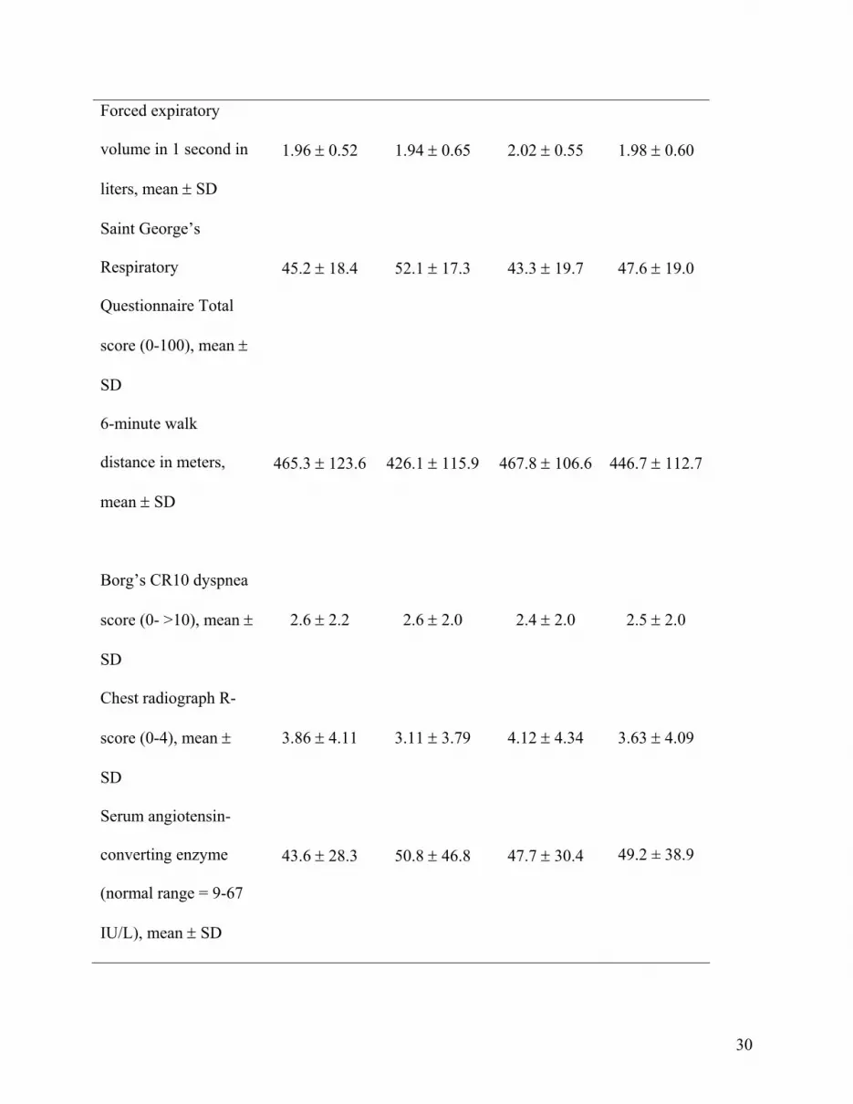
Table 1. Baseline demographics and disease characteristics, and concomitant medications
(randomized patients)
Characteristics
Placebo
(n=45)
Infliximab
3 mg/kg
(n=46)
Infliximab
5 mg/kg
(n=47)
Combined
Infliximab
(n=93)
Age, mean ± SD 45.3 ± 9.4 49.3 ± 9.4 46.5 ± 8.7 47.8 ± 9.1
Male, n (%) 26 (57.8) 24 (52.2) 28 (59.6) 52 (55.9)
Race, n (%)
Caucasian
Black
Asian
Other
29 (64.4)
16 (35.6)
0 (0.0)
0 (0.0)
36 (78.3)
8 (17.4)
1 (2.2)
1 (2.2)
28 (59.6)
17 (36.2)
1 (2.1)
1 (2.1)
64 68.8)
25 (26.9)
2 (2.2)
2 (2.2)
Extrapulmonary
involvement, n (%)
30 (66.7) 32 (72.7) 30 (63.8) 62 (68.1)
Years since
histologically proven
sarcoidosis, mean ±
SD
7.0 ± 6.2
8.0 ± 6.2
5.8 ± 6.1
6.9 ± 6.2
Forced vital capacity,
mean ± SD
Percent of predicted
Liters
68.8 ± 11.1
2.86 ± 0.77
67.7 ± 9.6
2.81 ± 0.86
69.5 ± 8.6
2.84 ± 0.70
68.6 ± 9.1
2.82 ± 0.78
30
Forced expiratory
volume in 1 second in
liters, mean ± SD
1.96 ± 0.52
1.94 ± 0.65
2.02 ± 0.55
1.98 ± 0.60
Saint George’s
Respiratory
Questionnaire Total
score (0-100), mean ±
SD
45.2 ± 18.4
52.1 ± 17.3
43.3 ± 19.7
47.6 ± 19.0
6-minute walk
distance in meters,
mean ± SD
465.3 ± 123.6
426.1 ± 115.9
467.8 ± 106.6
446.7 ± 112.7
Borg’s CR10 dyspnea
score (0- >10), mean ±
SD
2.6 ± 2.2
2.6 ± 2.0
2.4 ± 2.0
2.5 ± 2.0
Chest radiograph R-
score (0-4), mean ±
SD
3.86 ± 4.11
3.11 ± 3.79
4.12 ± 4.34
3.63 ± 4.09
Serum angiotensin-
converting enzyme
(normal range = 9-67
IU/L), mean ± SD
43.6 ± 28.3
50.8 ± 46.8
47.7 ± 30.4
49.2 ± 38.9

31
Concomitant, n (%)
Corticosteroids only
Immunomodulator
only
Corticosteroid +
Immunomodulator
26 (57.8)
2 (4.4)
17 (37.8)
20 (43.5)
4 (8.7)
22 (47.8)
24 (51.1)
4 (8.5)
19 (40.4)
44 (47.3)
8 (8.6)
41 (44.1)
Corticosteroid dose
(mg prednisone
equivalent per day),
mean ± SD
13.9 ± 9.3
11.7 ± 6.9
12.2 ± 5.6
11.9 ± 6.2

32
Table 2. Change from baseline in secondary efficacy parameters (nonparametric analysis,
randomized patients, p values computed based on analysis of covariance)
Characteristics Placebo
(n=45)
Infliximab
3 mg/kg
(n=46)
Infliximab
5 mg/kg
(n=47)
Combined
Infliximab
(n=93)
Mean ± SD Change from Baseline*
Saint George’s Respiratory
Questionnaire Total score
(0-100)†
Week 24‡
Week 52
-4.5 ± 2.1
-2.4 ± 2.1
-3.2 ± 2.2
-2.9 ± 2.2
-4.1 ± 2.1
-3.4 ± 2.1
-3.7 ± 1.5
-3.1 ± 1.6
6-meter walk distance (6-
MWD) in meters§
Week 24‡
Week 52
-5.9 ± 10.7
-19.9 ± 9.4
-1.7 ± 10.8
17.1 ± 9.4
p=0.007
1.4 ± 10.6
-1.8 ± 9.5
-0.2 ± 7.8
7.6 ± 6.6
p=0.019
Borg’s CR10 dyspnea score
(prior to 6-MWD)
Week 24
Week 52
0.2 ± 2.0
0.7 ± 2.4
0.1 ± 1.2
0.5 ± 2.2
-0.2 ± 2.2
0.1 ± 2.1
-0.1 ± 1.8
0.3 ± 2.1

33
Chest radiograph R-score
Week 6
Week 24
0.17 ± 1.58
0.29 ± 2.21
-0.84 ± 1.63
p=0.002
-1.04 ± 2.02
p=0.001
-0.90 ± 2.90
p=0.008
-0.85 ± 3.62
p=0.016
-0.87 ± 2.36
p<0.001
-0.94 ± 2.93
p=0.001
Serum angiotensin-
converting enzyme
Week 12
Week 24
Week 52
6.9 ± 30.2
7.9 ± 33.9
4.6 ± 22.0
-13.2 ± 27.4
p<0.001
-13.2 ± 41.8
p=0.004
-5.3 ± 35.0
p=0.188
-9.8 ± 23.0
p=0.009
-9.0 ± 23.9
p=0.013
-1.2 ± 23.9
p=0.317
-11.5 ± 25.1
p<0.001
-11.0 ± 33.3
p=0.002
-3.1 ± 29.4
p=0.178
*Values are LS mean ± SE for SGRQ and 6-MWD.
†Lower scores indicated better health-related quality of life.
‡Utilizes last observation carried forward.
§Longer distances indicate better function.

34
Table 3. Summary of adverse event data (all treated patients). Safety assessments were
performed through week 52. All adverse events that occurred between visits were reported.
Infusion reactions were defined as any adverse event that occurred during or within 1-2 hours
after completing the study agent infusion.
Placebo
(n=44)
Infliximab
3 mg/kg
(n=45)
Infliximab
5 mg/kg
(n=46)
Combined
Infliximab
(n=91)
Adverse events through week 24, n (%)
35 (79.5) 39 (86.7) 35 (76.1) 74 (81.3)
Commonly reported adverse events*
Upper respiratory tract infection
8 (18.2) 14 (31.1) 8 (17.4) 22 (24.2)
Dyspnea 5 (11.4) 10 (22.2) 5 (10.9) 15 (16.5)
Pain 6 (13.6) 9 (20.0) 5 (10.9) 14 (15.4)
Coughing 4 (9.1) 7 (15.6) 5 (10.9) 12 (13.2)
Adverse events through week 52, n (%)
41 (93.2) 40 (88.9) 40 (87.0) 80 (87.9)
Commonly reported adverse events*
Upper respiratory tract infection
14 (31.8) 20 (44.4) 11 (23.9) 31 (34.1)
Coughing 6 (13.6) 16 (35.6) 8 (17.4) 24 (26.4)
Dyspnea 7 (15.9) 15 (33.3) 6 (13.0) 21 (23.1)
Pain 7 (15.9) 9 (20.0) 8 (17.4) 17 (18.7)
Sarcoidosis 5 (11.4) 6 (13.3) 7 (15.2) 13 (14.3)
Bronchitis 9 (20.5) 7 (15.6) 4 (8.7) 11 (12.1)

35
Headache 6 (13.6) 4 (8.9) 7 (15.2) 11 (12.1)
Back pain 2 (4.5) 2 (4.4) 8 (17. %) 10 (11.0)
Serious adverse events through week 24, n (%) 5 (11.4) 6 (13.3) 4 (8.7) 10 (11.0)
Commonly reported serious adverse events†
Pneumonia 0 (0.0%) 0 (0.0%) 2 (4.3%) 2 (2.2%)
Serious adverse events through week 52, n (%) 8 (18.2) 11 (24.4) 10 (21.7) 21 (23.1)
Commonly reported serious adverse events†
Pneumonia 0 (0.0) 1 (2.2) 3 (6.5) 4 (4.4)
Sarcoidosis 2 (4.5) 0 (0.0) 1 (2.2) 1 (1.1)
Cardiac failure 2 (4.5) 0 (0.0) 0 (0.0) 0 (0.0)
Deaths during the study, n (%) 1 (2.3) 0 (0.0) 0 (0.0) 0 (0.0)
Infusion reactions through week
24 (per patient) n (%)
6 (13.6)
3 (6.7) 5 (10.9) 8 (8.8)
Infusion reaction through week
24 (per infusion) n (%)
6/258
(2.3%)
3/260
(6.7%)
9/269
(3.3%)
12/529
(2.3%)
Infections through week 52,
n (%)
All infections
Infections requiring
antimicrobial treatment
Serious infections
32 (72.7)
27 (61.4)
4 (9.1)
29 (64.4)
26 (57.8)
6 (13.3)
25 (54.3)
23 (50.0)
4 (8.7)
54 (59.3)
49 (53.8)
10 (11.0)
* Defined as AEs reported in >15% of patients in any treatment group.
† Defined as SAEs reported in >3% of patients in any treatment group.

36
FIGURES
Figure 1.

37
Figure 2.
0Placebo(n = 45)
3 mg/kg(n = 46)
5 mg/kg(n = 47)
CombinedInfliximab(n = 93)
1
2
3
4
5
LS M
ean
± S
E
0.0
2.8
2.22.5
p = 0.041
p = 0.116 p = 0.038
1279
5

38
Figure 3.

39
Figure 4.
40
Infliximab Therapy in Patients with Chronic Sarcoidosis and Pulmonary Involvement
Authors: Robert P. Baughman, MD1; Marjolein Drent, MD2; Mani Kavuru, MD3; Marc A.
Judson, MD4; Ulrich Costabel, MD5; Roland du Bois, MD6; Carlo Albera, MD7; Martin
Brutsche, MD8; Gerald Davis, MD9; James F. Donohue, MD10; Joachim Müller-Quernheim,
MD11; Rozsa Schlenker-Herceg, MD12; Susan Flavin, MSN12; Kim Hung Lo PhD12; Barry
Oemar, MD12; and Elliot S. Barnathan, MD12 on behalf of the Sarcoidosis Investigators
Online Data Supplement

41
EXPANDED METHODS for WEB Based Repository
Eligible adult patients ≥18 years of age had histologically proven sarcoidosis, diagnosed at least
1 year prior to screening, evidence of parenchymal disease (Stage II or III) on chest radiograph, a
forced vital capacity (FVC) ≥50% and ≤85% of the predicted value, and a Medical Research
Council (MRC) dyspnea score (19) of at least Grade 1. The screening chest x-rays (both
posterioranterior and lateral views) were required to be obtained within 3 months prior to the
first study infusion, and were required to be interpreted by a qualified radiologist and indicate
that the patient was free from any evidence of tuberculosis. Patients must have been treated with
at least 10 mg/day of prednisone or equivalent or one or more immunosuppressant for at least
3 months prior to screening. Doses of these medications had to be stable for at least 1 month
prior to study entry. During the study, the background medication regimen and doses were to
remain stable. Men and women of childbearing potential were required to use adequate birth
control measures (e.g., abstinence, oral contraceptives, intrauterine device, barrier method with
spermicide, or surgical sterilization) for the duration of the study and were to continue such
precautions for the 6 months after receiving the last infusion of study agent. Patients were
obliged to meet their respective country’s tuberculosis eligibility requirements in terms of
screening, eligibility assessment, and prevention rules. They were to be capable of reading and
understanding the consent form and the subject assessment forms, and were to be willing and
able to adhere to the study visit schedule and other protocol-specified procedures. Patients with
skin or eye involvement were encouraged to participate.
Patients were excluded from the study if they had used any investigational drug within 1 month
prior to screening or within five half-lives of the investigational agent (whichever was longer), or

42
had received previous treatment with any other therapeutic agent targeted at reducing TNFα
(e.g., pentoxifyline, thalidomide, etanercept, CDP 870, adalimumab) within 3 months prior to
screening. Patients were not eligible if they had received previous administrations of infliximab.
Patients could not have received any live virus or bacterial vaccinations within 3 months before
the first dose of study agent (or were expected to receive any of these vaccinations during the
trial or up to 3 months after the last dose of study agent). Patients were not eligible for the trial if
they had had any previous adverse reactions or allergic reactions (e.g., anaphylaxis) associated
with the administration of monoclonal antibodies or antibody fragments. Patients were not
eligible if they had Class III or IV congestive heart failure (CHF) according to the New York
Heart Association Classification System, or a history of severe right-sided heart failure or cor
pulmonale. Those who had mild heart failure (NYHA Class I or II) were to be closely monitored.
Exclusion criteria also included any serious infections (within 2 months of screening) or
opportunistic infections (within 6 months of screening), or a known infection with human
immunodeficiency virus (HIV). Patients with current signs and symptoms of systemic lupus
erythematosus, or with severe, progressive, or uncontrolled renal, hepatic, hematologic,
endocrine, pulmonary, cardiac, neurologic or cerebral diseases (with the exception of
sarcoidosis) were excluded. Patients were also not eligible if they underwent an organ transplant
(with the exception of a corneal transplant >3 months prior to screening); had a known
malignancy or history of malignancy within the 5 years prior to screening (except for basal cell
carcinoma of the skin that had been treated with no evidence of recurrence); had a history of
lymphoproliferative diseases including lymphoma, or signs and symptoms suggestive of possible
lymphoproliferative disease, such as lymphadenopathy of unusual size or location (e.g., nodes in
the posterior triangle of the neck, infraclavicular, epitrochlear, or periaortic areas); or had

43
splenomegaly. Patients were excluded if they were pregnant, nursing, or planning to become
pregnant during the trial or within the 6-month period thereafter or had a known substance abuse
or dependency (other than caffeine and/or nicotine) within 3 years of screening. If patients had a
poor tolerability of venipuncture or lack of adequate venous access for required blood sampling
and infusion administration, had a known history of demyelinating disease (such as multiple
sclerosis or optic neuritis), had a non-sarcoidosis condition affecting survival, had a mental
health problem interfering with study participation, or had a history of treated or untreated latent
tuberculosis, they were not eligible for participation in the study.
This was a Phase 2, multicenter, double-blind, placebo-controlled study in which patients were
randomized in a 1:1:1 ratio to receive intravenous infusions of either placebo, infliximab
3 mg/kg, or infliximab 5 mg/kg at weeks 0, 2, 6, 12, 18, and 24. Patients were followed through
week 52. Centocor, Inc., Malvern, PA provided the study agent, and infusions were to be
administered over at least a 2-hour period.
One hundred thirty-eight patients from 34 centers in the United States and Europe were
randomized between 30 September 2003 and 31 August 2004. Institutional Review
Boards/Ethics Committees at the participating sites approved the study, and patients provided
written informed consent.
The primary endpoint was the change from baseline in the percent of predicted FVC at week 24.
Pulmonary function testing employed a standardized calibrated laptop spirometer. Major
secondary endpoints included the Saint George’s Respiratory Questionnaire (SGRQ) total score

44
(20), 6-minute walk distance (6-MWD) test (21,22), Borg’s CR10 dyspnea score (23, prior to
6-MWD test) and the proportion of Lupus Pernio Physician’s Global Assessment (LuPGA)
responders for the subset of patients with facial skin involvement at baseline.
The LuPGA, specifically developed for use in this protocol, was a semi-quantitative rating scale
representing the physician’s assessment of the patient’s lupus pernio status relative to baseline.
Scores range from 6 (worsening of the patient’s facial skin involvement) to 1 (100% clearing of
the patient’s lupus pernio). Patients with a score of 1 (clear) or 2 (excellent) were considered
‘responders.’
Blood samples were collected for determination of serum infliximab concentrations. Serum
angiotensin-converting enzyme (ACE) levels were also measured.
The chest radiograph scoring system (24) used in this study allowed quantitative comparison of
chest radiographs obtained at baseline and weeks 6 and 24. Chest radiographs were scored by
two independent reviewers who remained blinded to the time point the radiograph was
performed and the patient’s treatment assignment. Images were evaluated for extent (score 0-4)
and profusion (score 0-4) for each of four types of shadows commonly seen in sarcoidosis:
reticulonodular (R), mass (M), confluent (C), and fibrosis (F).

45
Statistical analyses
The sample size of 40 patients per treatment group provided a statistical power of 80% to detect
a 10% improvement in the primary endpoint for at least one of the infliximab groups compared
with placebo. The sample size calculation was based on an assumed standard deviation of
approximately 17%. In the analysis of the primary endpoint, i.e., change from baseline in the
percent of predicted FVC at week 24, treatment group comparisons were made using the analysis
of covariance (ANCOVA). The model included the change from baseline in the percent of
predicted FVC as the dependent variable and treatment group and clinical center as factors and
baseline percent of predicted FVC as a covariate. Clinical center was considered a random
effect. Data from all randomized patients were analyzed according to assigned treatment group
(intention-to-treat analysis). Least squares means are reported. A last-observation-carried-
forward (LOCF) technique was used to impute missing data. A p-value less than 0.05 was
considered statistically significant.
The ANCOVA model was also employed for evaluation of the secondary efficacy parameters.
Analyses included all randomized patients according to the randomized treatment group.
Missing data were imputed for the first two major secondary endpoints (SGRQ, 6-MWD) using
LOCF.


