Embed Size (px)
Citation preview

JOURNAL OF VIROLOGY, Apr. 2007, p. 3058–3067 Vol. 81, No. 70022-538X/07/$08.00�0 doi:10.1128/JVI.02082-06Copyright © 2007, American Society for Microbiology. All Rights Reserved.
Influenza A Virus NS1 Protein Activates the PI3K/Akt PathwayTo Mediate Antiapoptotic Signaling Responses�
Christina Ehrhardt,1 Thorsten Wolff,2 Stephan Pleschka,3 Oliver Planz,4 Wiebke Beermann,2Johannes G. Bode,5 Mirco Schmolke,1 and Stephan Ludwig1*
Institute of Molecular Virology, ZMBE, Westfaelische-Wilhelms-University, Von Esmarch-Str. 56, D-48149 Muenster, Germany1;Robert-Koch-Institute, P15, Nordufer 20, D-13353 Berlin, Germany2; Institute of Medical Virology, Justus-Liebig-University,
Frankfurter Strasse 107, D-35392 Giessen, Germany3; Friedrich-Loeffler-Institute, Paul-Ehrlich-Strasse 28,D-72076 Tubingen, Germany4; and Klinik fuer Gastroenterologie, Hepatologie und Infektiologie,
Heinrich-Heine-University, D-40225 Duesseldorf, Germany5
Received 22 September 2006/Accepted 22 December 2006
Recently we have shown that influenza A virus infection leads to activation of the phosphatidylinositol3-kinase (PI3K)/Akt pathway and that this cellular reaction is dependent on the expression of the viralnonstructural protein 1 (NS1). These data also suggested that PI3K activation confers a virus-supportingactivity at intermediate stages of the infection cycle. So far it is not known which process is regulated by thekinase that supports virus replication. It is well established that upon infection with influenza A virus, theexpression of the viral NS1 keeps the induction of beta interferon and the apoptotic response within a tolerablelimit. On a molecular basis, this activity of NS1 has been suggested to preclude the activation of cellulardouble-stranded RNA receptors as well as impaired modulation of mRNA processing. Here we present a novelmode of action of the NS1 protein to suppress apoptosis induction. NS1 binds to and activates PI3K, whichresults in the activation of the PI3K effector Akt. This leads to a subsequent inhibition of caspase 9 andglycogen synthase-kinase 3� and limitation of the virus-induced cell death program. Thus, NS1 not only blocksbut also activates signaling pathways to ensure efficient virus replication.
Influenza virus infection results in the activation of a variety ofintracellular signaling pathways that are in part required to mountan antiviral response to infection but also may be exploited by thevirus to support its replication (18, 19). A recent addition to theseinfluenza virus-induced signaling mediators is phosphatidylinosi-tol 3-kinase (PI3K) and its downstream effector Akt/protein ki-nase B (PKB) (12). PI3K consists of regulatory (p85) and enzy-matic (p110) subunits, each existing in several isoforms. Theactive enzyme exhibits both a protein kinase and a lipid kinaseactivity (9). The kinase controls various cellular processes, such asmetabolic regulation, cell growth, proliferation, and survival (3, 5,14). The consequence of PI3K activation is the generation ofphosphatidylinositol 3,4,5-trisphosphate from phosphatidylinosi-tol 4,5-bisphosphate in the membrane, which functions as a sec-ond messenger to recruit pleckstrin homology domain-containingproteins, such as Akt (also known as PKB) and phosphoinositide-dependent kinase 1. Akt/PKB is a major PI3K effector and be-comes further activated by phosphorylation at Thr308 and atSer473 (3).
With regard to the function of the kinase in virus-infected cells,it has been recently shown that PI3K is involved in double-stranded RNA (dsRNA)-induced activation of the antivirally act-ing transcription factor interferon-regulatory factor 3 (27), sug-gesting a function in the defense against viral infection. While wewere able to confirm such an activity in influenza virus-infectedcells, we could also demonstrate that the virus exploits the path-
way for a virus-supportive function, namely, to regulate the pro-cess of viral entry (12). However, despite this function, whichwould match the very early activation peak observed, a muchstronger activation in later stages of the replication cycle couldalso be detected. Inhibitor studies revealed that in addition toentry, PI3K further regulates a virus-supportive function duringlater stages of replication (12).
It was interesting to note that a virus mutant lacking the viralNS1 protein (delNS1) failed to activate the PI3K/Akt pathway(12). Although a surprising multitude of activities had been orig-inally assigned to the RNA binding NS1 protein, recent analyseshave shown that a major in vivo function is to antagonize theantiviral type I interferon (IFN) system (15). Comparisons ofwild-type and delNS1 viruses revealed that NS1 inhibits the acti-vation of the IFN-� gene, which is controlled by the transcrip-tional activators NF-�B, interferon-regulatory factor 3, and ATF-2/c-Jun (20, 28, 31). Mechanistically, it was shown that the NS1protein interferes with the intracellular signaling events that trig-ger activation of these dsRNA-responsive transcription factors(15). Significantly, this activity involves the targeting of the RNAhelicase RIG-I, a cellular sensor for viral RNA (24, 34). In clearcontrast to the dsRNA-activated IFN-�-inducing signaling path-ways, the PI3K/Akt pathway, although activated by dsRNA, re-mained silent rather than being activated upon infection bydelNS1 (12). This prompted us to analyze whether NS1 is in-volved in the activation of PI3K and which virus-supportive func-tion the pathway may fulfill in later stages of the replication cycle.
MATERIALS AND METHODS
Viruses, cells, and viral infections. Avian influenza virus A/FPV/Bratislava/79(H7N7) (FPV), human influenza virus A/Puerto Rico/8/34 (H1N1) (PR8), the cor-responding mutant delta NS1 (delNS1) (16), the human influenza virus strain
* Corresponding author. Mailing address: Institute of MolecularVirology, Westfaelische-Wilhelms-University, Von-Esmarch-Str. 56,D-48149 Muenster, Germany. Phone: 49 251 83 57791. Fax: 49 251 8357793. E-mail: [email protected].
� Published ahead of print on 17 January 2007.
3058
on June 23, 2015 by guesthttp://jvi.asm
.org/D
ownloaded from

FIG. 1. Influenza A virus NS1 induces activation of the PI3K/Akt signaling pathway. (A) A549 cells were left untreated (lane 1), were mock infected(lane 11), or were infected with influenza A virus strain Victoria/3/75 (H3N2) (lanes 2, 5, and 8), Thailand/KAN-1/2004 (H5N1) (lanes 3, 6, and 9), orPR8 (H1N1) (lane 4, 7, and 10) at a MOI of 5 for the indicated times. (B) A549 cells were left untreated (lane 4), mock infected (lane 1), or infectedwith influenza A virus strain PR8 (lane 2) or delNS1 (�NS1) (lane 3) at a MOI of 5 for 8 h. (C) A549 cells were transfected with empty vectors (lanes1 and 4) or expression constructs for wild-type PR8 NS1 (PR8 NS1wt) (lane 2), NS-IAmut1 expressing a NS1 protein of wild-type length with five aminoacid replacements at positions 181 to 185 (LIGGL to KQRRS) (lane 3), wild-type WSN NS1 (WSN NS1wt) (lane 5), and the corresponding NS1 witha R38AK41A mutation in the dsRNA binding site (lane 6). (D) A549 cells were transfected with constructs expressing NS1wt (lane 2) or truncated NS1consisting of the N-terminal amino acids 1 to 125 (lane 3) or 1 to 126(�3) (lane 5), as well as their corresponding dsRNA binding site mutants bearingan R38AK41A mutation (lanes 4 and 6). (E) A549 cells were transfected with an empty vector or expression constructs for WSN NS1wt (lanes 3 and4) and the corresponding NS1 with a R38AK41A mutation (lanes 5 and 6). Cells were left untreated (lanes 1, 3, and 5) or were transfected with thedsRNA analog poly(IC) (5 �g/ml) in the presence of DOTAP for 45 min and subsequently harvested (lanes 2, 4, and 6). (F) A549 cells were transfectedwith constructs expressing NS1wt (lanes 3 and 4) or truncated NS1 consisting of the N-terminal amino acids 1 to 126(�3) (lanes 5 and 6) or thecorresponding dsRNA binding site mutant bearing an R38AK41A mutation (lanes 7 and 8). Note that cells in panels C to F were cotransfected with aplasmid expressing wild-type Akt. In all assays phosphorylated Akt (Ser473) was detected by Western blotting. Equal protein loading of the kinase wasverified in Akt Western blots. Ongoing viral replication was demonstrated by accumulation of the viral NP in panel B. Equal NS1 production fromplasmids was monitored in Western blots detecting NS1 (panel A, lanes 2 to 10; panel C, lanes 2, 3, 5, and 6; and panel E, lanes 3 to 6) or HA-taggedNS1 (panel D, lanes 2 to 6, and panel F, lanes 3 to 8). Relative Akt phosphorylation was normalized to Akt and NS1 content, upon quantification withthe Lumi-Analyst program (Boehringer Mannheim). Akt phosphorylation levels of vector-transfected cells (panel C, lane 4, and panel E, lane 1) werearbitrarily set at 1, while phosphorylation of Akt in untreated WSN NSwt-expressing cells (panel D, lane 3) or untreated PR8 NS1wt-expressing cells(panel F, lane 1) was arbitrarily set at 100%.
3059
on June 23, 2015 by guesthttp://jvi.asm
.org/D
ownloaded from
A/Victoria/3/75 (H3N2) (Victoria), and the reassortant virus WSN-HK (H1N1)were propagated and used as described earlier (11, 20, 26, 30, 33). The humaninfluenza virus isolate of a highly pathogenic avian virus strain, A/Thailand/KAN-1/2004 (H5N1), was a kind of gift from P. Puthavathana (Mahidol, University,Bangkok, Thailand) and has been passaged on Madin-Darby canine kidney(MDCK) cells. The recombinant PR8 virus and the corresponding truncated mu-tants comprising amino acids 1 to 80, 1 to 125, and 1 to 126 were propagated andused as described earlier (10, 31). For infection, cells were washed with phosphate-buffered saline (PBS) and incubated with virus at the indicated multiplicities ofinfection (MOI) diluted in PBS containing 0.2% bovine serum albumin (BSA), 1mM MgCl2, 0.9 mM CaCl2, 100 U/ml penicillin, and 0.1 mg/ml streptomycin for 30min at 37°C. The inoculum was aspirated, and cells were incubated with eitherminimal essential medium or Dulbecco modified Eagle medium (DMEM) (Invitro-gen) containing 0.2% BSA and antibiotics. MDCK and African green monkeykidney (Vero) cells were grown in minimal essential medium. HEK 293 cells and thehuman lung epithelial cell line A549 were grown in DMEM. All media were sup-plemented with 10% heat-inactivated fetal bovine serum (Invitrogen).
Plasmids, reagents, and inhibitors. Expression constructs for wild-type NS1 ofthe influenza virus strain A/Puerto Rico/8/34 (H1N1) (PR8) and the correspond-ing mutant NS-IAmut1, which carries amino acid substitution at position 181 to185 (LIGGL3KQRRS), were described earlier (20). Wild-type NS1 of theA/WSN/33 strain and the corresponding NS1 mutant with amino acid substitu-tions R38A and K41A, which is no longer able to bind to dsRNA, were describedpreviously (20). Plasmids coding for the truncated PR8 NS1 mutant forms com-prising amino acids 1 to 125 and 1 to 126 were originally constructed by insertingthe corresponding cDNAs into pcDNA3. The latter mutant additionally codesfor three nonviral amino acids and thus is slightly shifted in polyacrylamide gelelectrophoresis gels (see Fig. 1D and F). Additional mutations affecting thedsRNA binding activity of the encoded NS1 proteins were introduced by usingthe QuikChange mutagenesis kit (Stratagene). The PR8 NS1 mutant proteinswere inserted in a pTracer-CMV vector (Invitrogen) containing a hemagglutinin(HA) tag. Expression constructs for a wild-type form of Akt in pCMV5 werekindly provided by Jakob Troppmair, Daniel Swarovski Research Laboratory,University of Innsbruck, Austria, and have been described before (29).
An expression construct for a wild-type form of p85� C-terminally tagged withthe yellow fluorescent protein (YFP) in pCDNA3 was kindly provided by BerndNurnberg, Institute of Biochemistry and Molecular Biology, Heinrich-Heine-University of Dusseldorf, and was previously described (4). The dsRNA analogpoly(IC) was purchased from Amersham Biosciences and transfected at a con-centration of 5 �g/ml with DOTAP (Roth) for 45 min according to the manu-facturer’s protocol. The specific PI3K inhibitor LY294002 (Promega) was dis-solved in dimethyl sulfoxide (DMSO) and was added directly after infection asindicated. The apoptosis inducer staurosporine dissolved in DMSO (Sigma) wasadded directly to the medium at the indicated concentration.
Transient transfections. A549 cells were transfected with Lipofectamine 2000(Life Technologies) according to a protocol described previously (2). HEK 293cells were transfected with polyethylenimine as described previously (13).
Immunoprecipitations and Western blotting. For immunoprecipitations, cellswere lysed on ice with Triton lysis buffer (20 mM Tris-HCl, pH 7.4; 137 mMNaCl; 10% glycerol; 1% Triton X-100; 2 mM EDTA; 50 mM sodium glycero-phosphate, 20 mM sodium pyrophosphate; 5 �g ml�1 aprotinin; 5 �g ml�1
leupeptin; 1 mM sodium vanadate; and 5 mM benzamidine) for 30 min. ForWestern blotting, cells were lysed on ice with radioimmunoprecipitation assaylysis buffer (25 mM Tris, pH 8.0; 137 mM NaCl; 10% glycerol; 0.1% sodiumdodecyl sulfate; 0.5% natrium-deoxycholate; 1% NP-40; 2 mM EDTA, pH 8.0; 5�g ml�1 aprotinin; 5 �g ml�1 leupeptin; 1 mM sodium vanadate; and 5 mMbenzamidine) for 30 min. Cell lysates were cleared by centrifugation, and theprotein concentration was determined by the Bradford method. Cell lysates wereused for immunoprecipitation with antibodies or antisera against green fluores-cent protein (GFP) (B-2) or p85� (B-9) (Santa Cruz Biotechnologies), p85�(Serotec), or NS1 (polyclonal antiserum) coupled to protein A or G agarose(Roche). Mouse or rabbit sera were used for control purposes. Alternatively,lysates were directly subjected to sodium dodecyl sulfate-polyacrylamide gelelectrophoresis and subsequent blotting. The phosphorylated active form of Akt(Ser473) was detected in crude cell lysates by a phosphospecific Akt (Ser473)rabbit antiserum (Biosource). Different influenza A virus proteins were visual-ized using either a nucleoprotein (NP)-specific mouse antiserum (Serotec), aPB1-specific (vK-20) goat antiserum (Santa Cruz Biotechnologies), or NS1-specific rabbit antisera that were kind gifts of Ilkka Julkunen, National PublicHealth Institute, Finland, and Juan Ortin, CNB-CSIC, Spain. HA-tagged PR8NS1 expression constructs were detected with an anti-HA monoclonal antibody(3F10; Roche). Different apoptosis-specific markers were visualized with thefollowing specific antisera: a phospho-specific glycogen synthase-kinase 3 �
(GSK-3�) (Ser9) rabbit antibody (Cell Signaling Technologies), a caspase 9(Asp330)-specific rabbit antiserum (Cell Signaling Technologies), or a poly-(ADP-ribose) polymerase (PARP)-specific mouse antibody (BD TransductionLaboratories). In some of the assays, loading controls were performed with apan-ERK2 antiserum (Santa Cruz Biotechnologies) or a pan-Akt antiserum(Cell Signaling Technologies). Protein bands were visualized in a standard en-hanced chemiluminescence reaction.
Indirect immunofluorescence microscopy. MDCK or A549 cells were grownonto 15-mm coverslips, and 24 h later cells were left uninfected or were infectedwith the influenza A virus strain FPV (MOI � 0.01) or PR8 (MOI � 20),respectively. LY294002 (50 �M) or the same volume of the solvent (DMSO) wasadded directly upon infection. To detect the viral nucleoprotein, cells werestained with an anti-NP antibody as described earlier (12). To visualize cell nucleiin these assays, cells were additionally stained with 1 �M DAPI (4,6-diamidino-2-phenylindole) (Molecular Probes). For detection of apoptotic cell nuclei at 6 hpostinfection (p.i.), cells were incubated with DMEM containing 2 �g/ml ofpropidium iodide (PI) for 15 min and fixed with 3.7% paraformaldehyde at 4°Cfor 20 min, as described previously (21). Cells were washed with PBS andincubated with 0.1% BSA for 5 min and phalloidin-Alexa 488 (MolecularProbes) for 30 min to detect the actin cytoskeleton. Cells were washed threetimes with PBS and once with water before being covered with fluorescentmounting medium (DAKO). Fluorescence was visualized using a Leitz DMRBfluorescence microscope.
Nicoletti assay. Transfected or untransfected MDCK cells were infected withFPV (MOI � 0.005) for 18 h. Cells were treated with LY294002 (50 �M) or withthe same volume of the solvent (DMSO) directly after infection. For determi-nation of late stages of apoptotic cell death, apoptotic hypodiploid nuclei weredetected by fluorescence-activated cell sorter analysis (22).
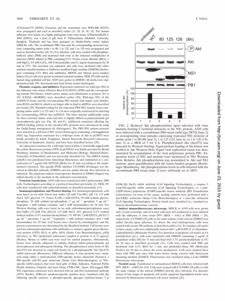
FIG. 2. Reduced Akt phosphorylation upon infection with virusmutants bearing C-terminal deletions in the NS1 protein. A549 cellswere infected with a recombinant PR8 strain (wild type [WT]) (lane 2)or corresponding virus mutants expressing truncated NS1 proteins ofamino acids 1 to 80 (80, lane 3), 1 to 125 (125, lane 4), or 1 to 126 (126,lane 5) at a MOI of 5 for 8 h. Phosphorylated Akt (Ser473) wasdetected by Western blotting. Equal protein loading of the kinase wasverified in Akt Western blots. Equal viral replication status was dem-onstrated by accumulation of the viral polymerase protein PB1. Ex-pression levels of NS1 and mutants were monitored in NS1 Westernblots. Relative Akt phosphorylation was normalized to Akt and NS1content, upon quantification with the Lumi-Analyst program (Boehr-inger Mannheim), Akt phosphorylation levels in cells infected with therecombinant PR8 strain (lane 2) were arbitrarily set at 100%.
3060 EHRHARDT ET AL. J. VIROL.
on June 23, 2015 by guesthttp://jvi.asm
.org/D
ownloaded from

RESULTS
Expression of NS1 results in activation of the PI3K/Aktsignaling pathway. Activation of the PI3K/Akt pathway wasobserved upon infection with a variety of influenza A virusstrains, including laboratory strains of the H1N1 subtype suchas A/Puerto-Rico/8/34 (PR8), the human H3N2 isolate Victo-ria/3/75, and a human H5N1 isolate of the highly pathogenicavian influenza strains of the Asia type (Fig. 1A). Since infec-tion of cells with the delNS1 virus mutant results in an almostcomplete abrogation of PI3K activation in A549 cells (Fig. 1B)or MDCK cells (12), we analyzed whether NS1 itself inducesPI3K and Akt activation. Full-length NS1 or different mutantsof the protein were expressed, and activation of the PI3K/Aktpathway was determined using a phosphospecific Akt antibodythat detects active Akt when phosphorylated at S473. Thisphosphorylation/activation event has been shown to occur in astrictly PI3K-dependent manner (1, 12). Expression of full-length NS1 derived from the virus strain PR8 or WSN readilyresulted in Akt phosphorylation and thereby activation of the
kinase (Fig. 1C, lanes 2 and 5). Similar results were also ob-tained with NS1 proteins from other virus isolates, includingthose of highly pathogenic avian H7N1 and H5N1 strains orcontemporary human virus isolates, such as A/New Caledonia/2007/99 (H1N1) (data not shown). Interestingly, a mutant ofthe NS1 protein that carries amino acid exchanges in the RNAbinding site did not activate Akt as efficiently as the wild type(Fig. 1C, lane 6) although it was expressed at similar levels.Reduced Akt phosphorylation compared to that induced bywild-type NS1 was also observed with a mutant that carriesamino acid replacements at positions 181 to 185 (LIGGL toKORRS) (Fig. 1C, lane 3). These mutations abrogate the bind-ing to the NS1 binding protein NS1-BP (32) and cause a pre-dominant cytoplasmic localization of the protein (20). Expres-sion of truncated NS1 proteins with deletions of the C terminusof NS1 up to amino acid 125 or 126 resulted in an even weakeractivation of PI3K/Akt (Fig. 1D, lanes 3 and 5), which was insome cases further reduced if the RNA binding domains ofthese fragments were mutated (Fig. 1D, lane 4). This suggests
FIG. 3. NS1 coimmunopreciptates with p85� and -�, two isoforms of the regulatory subunit of PI3K. (A and B) HEK 293 cells were transfectedwith a plasmid expressing a YFP-tagged version of p85� (p85�-YFP). At 24 h posttransfection cells were left untreated or infected with theinfluenza A virus strain PR8 (A) or the reassortant WSN-HK (B) (MOI � 5) for 4 h and subsequently harvested. Cells were subjected toimmunoprecipitation (IP) with an anti-GFP antibody directed against the YFP tag (lanes 1 and 2) or a serum control (lanes 3). Coimmunopre-cipitated NS1 was detected by Western blotting (WB). Equal protein loads of p85�-YFP in the immunoprecipitates were verified using theanti-GFP antibody detecting the YFP tag. The viral NS1 protein input of crude cell lysates served as control (lower panels). (C to F) A549 cells(C and D) or HEK 293 cells (E and F) were left untreated or infected with the influenza A virus strain PR8 (MOI � 5) for 4 h and subsequentlyharvested. Cells were subjected to immunoprecipitation of endogenous p85� with an anti-p85� antibody (panel C, lanes 2 and 3, and panel E, lanes1 and 2), an anti-NS1 antiserum (panel D, lanes 2 and 3), an anti-p85� (panel F, lanes 1 and 2), or serum as a control (panels C and D, lanes 4,and panels E and F, lanes 3). Coimmunoprecipitated NS1 (C, E, and F) or coimmunoprecipitated p85� (D) was detected by Western blotting.Equal protein loads in the immunoprecipitates were verified in p85� (C and E), p85� (F), or NS1 (D) Western blots. The viral NS1 protein andendogenous p85� or -� input of crude cell lysate served as a control.
VOL. 81, 2007 NS1 ACTIVATES THE ANTIAPOPTOTIC PI3K/Akt PATHWAY 3061
on June 23, 2015 by guesthttp://jvi.asm
.org/D
ownloaded from

FIG. 4. An active PI3K/Akt pathway suppresses viral apoptosis induction via inactivation of the Akt effectors GSK-3� and caspase 9. (A) MDCK cellswere infected with a recombinant PR8 strain (wild type [WT]) (lane 4) or corresponding virus mutants expressing truncated NS1 proteins of amino acids1 to 80 (80) (lane 2) and 1 to 126 (126) (lane 3) at a MOI of 1 for 8 h. (B, C, and D) MDCK cells were treated with the specific PI3K inhibitor LY294002(50 �M) or equal volumes of the solvent (DMSO) right after infection with influenza A virus strain PR8 at a MOI of 20 (C) or with FPV at a MOI of0.01 (B) or 1 (D) for 6 h (C and D) or 18 h (B). Thereafter cells were lysed and phosphorylated Akt (Ser473) was detected by Western blotting. Inductionof apoptosis-regulating markers, such as cleavage of PARP and caspase 9 or phosphorylation of GSK-3�, was detected with specific antibodies againstPARP (cleaved [cl.] or uncleaved), phosphorylated GSK-3�, and caspase 9. Note that in panels B and D the caspase 9 antibody detected only thefull-length form of the caspase. Equal protein loads were verified with Akt or ERK2 blots. Ongoing viral replication was demonstrated by accumulationof the viral NS1 or PB1 protein in Western blots. Additionally, ongoing viral replication was verified via NP staining of MDCK cells which were infectedwith FPV and/or treated with LY294002 (50 �M) under the same conditions as described for the experiments shown in panel B. (E and F) Vero cellswere treated with the specific PI3K inhibitor LY294002 (50 �M) or equal volumes of the solvent (DMSO) right after infection with the influenza A virusstrain FPV (MOI � 1) (E) or PR8 (MOI � 7) for 6 h. Phosphorylated Akt (Ser473) and induction of PARP cleavage were detected as described above.Equal protein loads were verified with ERK2 blots. Ongoing viral replication was demonstrated by accumulation of the viral NS1 protein.
3062 EHRHARDT ET AL. J. VIROL.
on June 23, 2015 by guesthttp://jvi.asm
.org/D
ownloaded from

that an intact C terminus and the integrity of amino acids in theRNA binding domain as well as amino acids 181 to 185 arerequired for the full capacity of NS1 to activate the PI3K/Aktpathway. Since it has been shown previously that dsRNA canactivate PI3K (27), the capacities of NS1 and the mutants toactivate PI3K/Akt in the presence of the dsRNA analogpoly(IC) was analyzed. Wild-type NS1- and mutant-expressingcells were transfected with poly(IC) and subsequently analyzedfor Akt phosphorylation. As expected from earlier studies (12,27), poly(IC) treatment resulted in a weak activation of thePI3K/Akt pathway also in the absence of NS1 (Fig. 1E and F,lanes 2). Strikingly higher levels of activation were detected inuntreated cells that express wild-type NS1 (Fig. 1E and F, lanes3). dsRNA treatment of wild-type NS1-expressing cells re-sulted in an enhanced PI3K/Akt activity (Fig. 1E and F, lanes4). While this may indicate a synergism of dsRNA and NS1, analmost similar additive effect was also observed with the mu-tant lacking the RNA binding domain, although the overallactivation of the PI3K/Akt pathway was much weaker in thiscase (Fig. 1E, lane 6, and F, lane 8). These findings indicatethat dsRNA binding of NS1 is not a prerequisite for the ca-pacity of the protein to activate the PI3K pathway and thatboth activation pathways might act independently of eachother. Nevertheless, the integrity of the NS1 RNA bindingdomain appears to be required for full activation.
Infection of cells with mutant influenza viruses expressingtruncated NS1 proteins results in reduced PI3K activity. Sin-gular expression of a viral protein may yield misleading results,since this scenario does not fully reflect the situation duringvirus propagation. Thus, besides the delNS1 virus, we alsoemployed isogenic PR8 mutant viruses with truncated NS1proteins to infect cells and assess PI3K/Akt activation. Consis-tent with the results obtained by transfection of NS1, we ob-served a reduction of PI3K/Akt activation with decreasinglength of the protein (Fig. 2). This further supports the as-sumption that the C terminus of NS1 is required for full acti-vation of the signaling pathway and confirms the previousfindings in the context of a genuine virus infection.
The NS1 protein coimmunoprecipitates with the p85� and -�regulatory subunits of PI3K. A remaining question concerns themechanism by which NS1 expression leads to PI3K activation.
Most strikingly this may occur by direct interaction of NS1 withPI3K. Thus, we initially analyzed whether the � form of theregulatory subunit of PI3K, p85�, would precipitate with NS1in infected cells. Indeed, we were able to coimmunoprecipitatethe NS1 of either the PR8 or the WSN-HK strain with eithera transfected p85�-YFP fusion protein (Fig. 3A and B) or theendogenous p85� protein (Fig. 3C and E). Furthermore wewere also able to coimmunoprecipitate endogenous p85� pro-tein with NS1 of the PR8 strain (Fig. 3D) and vice versa (Fig.3F), confirming recent findings by Hale et al. (17). Interest-ingly, this coimmunoprecipitation was observed only in in-fected cells and not in cells where both proteins were expressedfrom plasmids (data not shown).
Viral PI3K activation is required to suppress apoptotic sig-naling responses. The fact that the viral NS1 protein, which sofar was known only to suppress antiviral signaling responses,now was found to specifically activate a signaling pathwaypoints to a virus-supportive rather than a virus-antagonizingfunction of PI3K. In support of this consideration, inhibition ofthe kinase by wortmannin, a specific inhibitor of PI3K, still ledto reduced progeny virus titers even if added as late as 2 hpostinfection, suggesting a requirement of PI3K in intermedi-ate or late stages of the infection cycle (12).
Besides its capacity to suppress activation of dsRNA-in-duced transcription factors, NS1 has also been shown to inhibitviral apoptosis induction (35). This effect of the protein wasthought to occur primarily indirectly through the suppressionof type I interferons, which are known initiators of pro-grammed cell death. However, the finding that delNS1 virusinfection resulted in a hyperinduction of apoptosis also in Verocells that are devoid of functional IFN-�/� genes indicates thatthere are other, type I IFN-independent modes of NS1 actionto suppress induction of cell death (35).
Among the various functions of PI3K, the protein is alsoknown to mediate cell survival via activation of Akt (8). Aktdirectly phosphorylates caspase 9 and thereby inhibits activa-tion of this apoptotic protease (6). Furthermore, among otheractivities, Akt phosphorylates and inactivates GSK-3� (25).This protein is involved in glycogen metabolism and also trig-gers proapoptotic metabolic events. Thus, phosphorylation byAkt silences the apoptosis-promoting activity of the kinase.
FIG. 4—Continued.
VOL. 81, 2007 NS1 ACTIVATES THE ANTIAPOPTOTIC PI3K/Akt PATHWAY 3063
on June 23, 2015 by guesthttp://jvi.asm
.org/D
ownloaded from
Besides the existing data regarding the delNS1 virus, theresults shown in Fig. 4A further indicated that the capacity ofinfluenza viruses to activate the PI3K/Akt pathway inverselycorrelates with viral induction of apoptosis. Here, virus mutantstrains with truncated NS1 proteins were used that, accordingto the results shown in Fig. 2, only inefficiently activate Akt.While infection of cells with wild-type PR8 virus for 8 h did notlead to apoptosis induction, as measured by cleavage of PARP,a prominent substrate of apoptotic caspases, a cleaved PARPband is clearly visible upon infection with the truncated mu-tants (Fig. 4A). To further test whether NS1-mediated PI3Kactivation is involved in suppression of apoptosis, we treateduninfected and infected cells with the specific PI3K inhibitorLY294002. At 18 h postinfection (MOI � 0.01), cells wereharvested and cell lysates were examined for Akt phosphory-lation (Fig. 4B, panel a) and PARP cleavage (Fig. 4B, panel e).Furthermore, the activating cleavage of caspase 9 (Fig. 4B,panel h) and the phosphorylation of GSK-3� (Fig. 4B, panel f)as direct downstream effector functions of PI3K/Akt were an-alyzed. In uninfected cells the PI3K inhibitor was able to re-duce basal Akt phosphorylation (Fig. 4B, panel a, lane 2).Neither PARP cleavage nor activating cleavage of caspase 9was observed in these uninfected cells. Infection of solvent-treated cells showed induction of PARP cleavage (Fig. 4B,panel e, lane 3), consistent with earlier findings that influenzavirus infection of cells results in caspase activation (33). How-ever, in the same sample we also found enhanced levels ofphosphorylated Akt (Fig. 4B, panel a, lane 3), which is thoughtto confer an antiapoptotic signal. As a consequence, in thesecells, GSK-3� is phosphorylated and cleavage of caspase 9 isbarely visible, most likely due to inactivation by active Akt (Fig.4B, panels f and h, lane 3). Once PI3K/Akt activity is sup-pressed by LY294002, a strong increase of PARP cleavageconcomitant with an activating cleavage of caspase 9 is ob-served (Fig. 4B, panels e and h, lane 4). This also coincideswith a reduced phosphorylation of GSK-3�, an event thatkeeps this proapoptotic kinase active (Fig. 4B, panel f, lane 4).In addition, enhanced cleavage of caspase 3 and caspase 7 wasalso detectable in LY294002-treated and infected cells, indi-cating a massive onset of an apoptotic response in the absenceof PI3K activation (data not shown). The detected effects werenot due to an impact of PI3K inhibition on viral entry (12), asimmunofluorescence analysis of the viral NP showed that thesame number of cells were infected at 18 h p.i. in LY294002and solvent-treated cells (Fig. 4B, bottom panels).
However, to further rule out that the observed effects aredue to secondary autocrine or paracrine events that may haveoccurred during the 18-h infection period, we also analyzedlysates of cells that were infected for only 6 h with differentinfluenza A virus strains (Fig. 4C and D), a time point withinthe primary replication cycle postinfection. At this earlier timepoint, viral activation/phosphorylation of Akt in untreated orsolvent-treated cells coincided with a complete suppression ofPARP cleavage, as well as enhanced phosphorylation andthereby inactivation of GSK-3� (Fig. 4C, lane 3, and D, lanes4 and 5). Inhibition of PI3K by LY294002 in infected cellsresulted in reduced phosphorylation of Akt and GSK-3� aswell as cleavage of full-length caspase 9 and onset of PARPcleavage (Fig. 4C, lane 4, and D, lane 6). Finally, an indirectimpact of IFN-�/� could be ruled out, since enhanced PARP
cleavage was also detected in LY294002-treated Vero cells(Fig. 4E and F).
The apoptosis-suppressing effect of PI3K could further bedetected when later events of apoptosis induction were ana-lyzed, e.g., the onset of DNA fragmentation as measured by
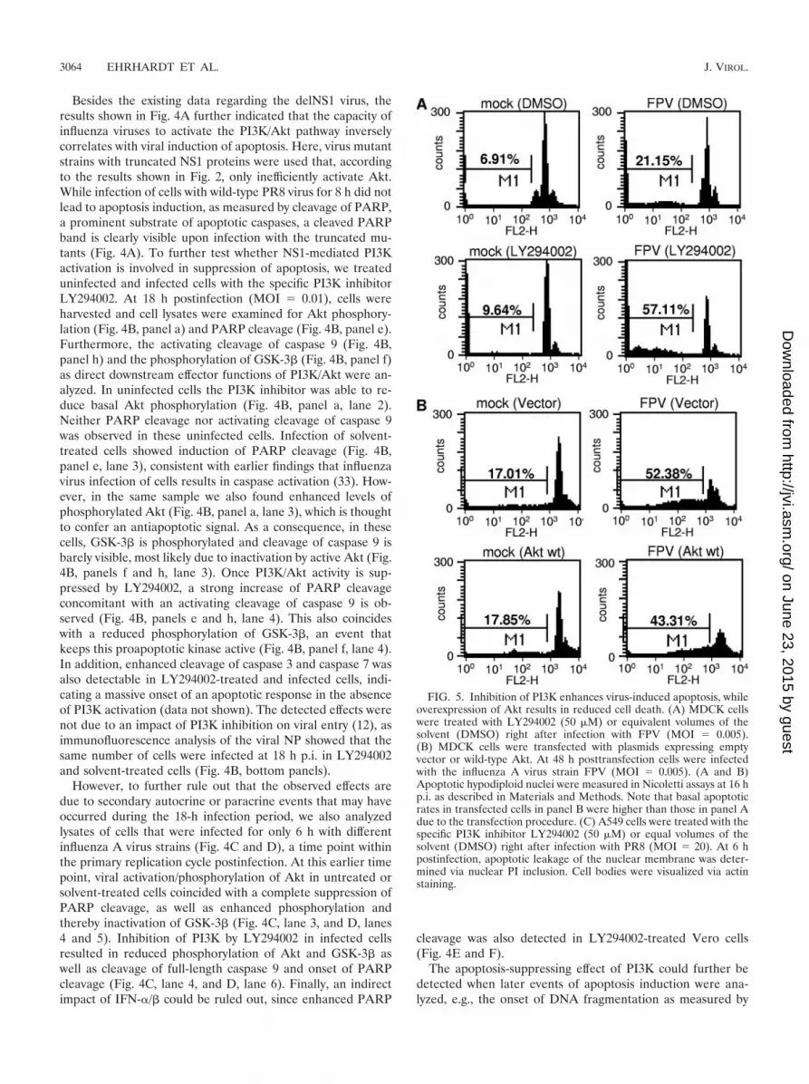
FIG. 5. Inhibition of PI3K enhances virus-induced apoptosis, whileoverexpression of Akt results in reduced cell death. (A) MDCK cellswere treated with LY294002 (50 �M) or equivalent volumes of thesolvent (DMSO) right after infection with FPV (MOI � 0.005).(B) MDCK cells were transfected with plasmids expressing emptyvector or wild-type Akt. At 48 h posttransfection cells were infectedwith the influenza A virus strain FPV (MOI � 0.005). (A and B)Apoptotic hypodiploid nuclei were measured in Nicoletti assays at 16 hp.i. as described in Materials and Methods. Note that basal apoptoticrates in transfected cells in panel B were higher than those in panel Adue to the transfection procedure. (C) A549 cells were treated with thespecific PI3K inhibitor LY294002 (50 �M) or equal volumes of thesolvent (DMSO) right after infection with PR8 (MOI � 20). At 6 hpostinfection, apoptotic leakage of the nuclear membrane was deter-mined via nuclear PI inclusion. Cell bodies were visualized via actinstaining.
3064 EHRHARDT ET AL. J. VIROL.
on June 23, 2015 by guesthttp://jvi.asm
.org/D
ownloaded from

accumulation of cells with hypodiploid DNA content in Nico-letti assays (Fig. 5A and B). Virus-induced DNA fragmenta-tion was strongly enhanced in infected cells treated withLY294002 (Fig. 5A, lower panels), while the number of cellswith hypodiploid DNA content was reduced in infected cellsthat overexpress a wild-type form of Akt (Fig. 5B, lowerpanels).
Another hallmark of apoptosis induction is disintegration ofthe nuclear membrane, a process that allows DNA-stainingcompounds such as PI to easily enter the apoptotic cell nucleus(21). While a weak nuclear PI staining could be detected uponinfluenza virus infection (Fig. 5C, panels b), this was dramat-ically enhanced in infected cells that were treated withLY294002 (Fig. 5C, panels d). The compound alone had noeffect (Fig. 5C, panels c).
Finally, Fig. 6 shows that the ability of NS1 to enhance Aktphosphorylation in cells treated with the nonviral apoptosisstimulus staurosporine correlated with its capacity to reducestaurosporine-induced PARP cleavage (lanes 3 and 4).
Taken together, these data clearly indicate that NS1-medi-ated activation of the PI3K/Akt pathway suppresses the onsetof premature virus-induced caspase activation and apoptosis.
DISCUSSION
Influenza virus infection leads to the activation of a varietyof signaling pathways, including the PI3K/Akt signaling cas-cade. The kinetics of activation were previously shown to be
biphasic, peaking at early and late phases of infection (12).While activation of the early phase most likely is mediated bybinding of the virus to the cell surface, which leads to signalsthat promote a very early step in virus uptake (12), it remainedenigmatic for which virus-supportive event the later activationphase of PI3K may be required. Here we show that late acti-vation of PI3K confers an antiapoptotic signal via activation ofAkt and the Akt effectors GSK-3� and caspase 9. The influ-enza virus NS1 protein plays a major role in influenza virusactivation of PI3K, presumably by association with the regula-tory subunits p85� and -�. Thus, NS1, via activation of PI3Kand Akt, appears to prevent premature apoptosis induction toensure efficient replication.
This reveals an additional function of the NS1 protein, whichapparently not only suppresses dsRNA-induced pathways (15)but is shown here also to activate a dsRNA-dependent signal-ing process. The precise contribution of the NS1 dsRNA bind-ing domain remains to be fully understood, since poly(IC)treatment resulted in the same relative enhancement of PI3Kactivity in the presence of NS1, regardless of the integrity of itsRNA binding domain. Nevertheless, the finding that a dsRNAbinding-deficient NS1 mutant shows a weaker PI3K activationthan the corresponding wild-type protein points to a functionalinvolvement of this protein domain. Other structural con-straints of the NS1 protein that appear to be required foroptimal PI3K activation are an intact C terminus downstreamof position 125 and a domain at amino acids 181 to 185.Currently it is unclear whether the respective domains are
FIG. 5—Continued.
VOL. 81, 2007 NS1 ACTIVATES THE ANTIAPOPTOTIC PI3K/Akt PATHWAY 3065
on June 23, 2015 by guesthttp://jvi.asm
.org/D
ownloaded from

directly involved in the PI3K-NS1 interaction or whether mu-tations of these sites alter the secondary structure of the pro-tein, which indirectly may prevent full PI3K activation.
Interestingly, interaction of p85� and -� with NS1 could bedetected only in infected cells and not in transfected cells.Consistent with that, preliminary results from yeast two-hybridassays showed only a very weak interaction of NS1 with p85�,indicating that an important cofactor for efficient binding is notpresent in yeast. This factor is most likely not dsRNA, sincetransfection of poly(IC) could not rescue coimmunoprecipita-tion of transfected p85 and NS1 proteins, again arguing againstan essential role of dsRNA in NS1-mediated PI3K activation.
While the present paper was in preparation, another publi-cation demonstrated direct binding of NS1 to the � isoform butnot the � isoform of p85, which resulted in activation of PI3K(17). In our experiments we also observed an interaction withthe � isoform of p85, although at a somewhat lower level,which may point to a slightly higher affinity of the NS1 to the� isoform of p85. In the study by Hale et al. (17), the authorsdescribe amino acid 89 in the NS1 protein as being critical forbinding to p85. This is consistent with our observation that amutant virus expressing a NS1 protein of amino acids 1 to 80induces a weaker Akt phosphorylation than the wild type.However, since both a virus bearing a NS1 protein of 125N-terminal amino acids and a virus with a NS1 protein withmutations of amino acids 181 to 185 are also weak PI3K/Aktinducers, additional sites beside amino acid 89 in the protein
appear to be required. Thus, our findings confirm the obser-vations by Hale et al. (17) and expand the data by identificationof the functional consequences of NS1-mediated PI3K activity,namely, suppression of viral apoptosis induction.
It might appear puzzling that influenza virus expresses aprotein that suppresses apoptosis via PI3K while another viralprotein, PB1-F2, even enhances apoptosis in infected cells (7).Furthermore, while the observations described here point to apredominant antiviral role of apoptosis induction, there arealso data showing that apoptosis-inducing factors or earlycaspase activation is beneficial for virus replication (23, 33).However, these data are not contradictory if one takes intoaccount that the virus not only might have evolved strategies tokeep the overinduction of an apoptotic response at a tolerablelimit but also may have acquired the capability to exploit theremaining activities to support its replication. This would alsoexplain why NS1 does not completely suppress apoptotic re-sponses or induction of other antiviral activities. Thus, influ-enza virus has evolved strategies to adjust the delicate balanceof pro- and antiviral activities for an optimum of replicationefficiency.
ACKNOWLEDGMENTS
We thank Gudrun Heins, Sandra Neumann, Andrea Stadtbaumer,Carmen Theseling, and Ludmilla Wixler for excellent technical assis-tance. We also thank Ilkka Julkunen and Juan Ortin for generouslyproviding antibodies and Andrej Egorov for recombinant viruses.
This work was supported by grants from the Deutsche Forschungs-gemeinschaft (DFG), the DFG Graduate Schools (GRK1045 andGRK1409), and the fund “Innovative Medical Research” as well as theIZKF of the University of Muenster Medical School. This work is alsopart of the activities of the European Union STREP EUROFLU andVIRGIL European Network of Excellence on Antiviral Drug Resis-tance, supported by a grant (LSHMCT-2004-503359) from the Priority1 “Life Sciences, Genomics and Biotechnology for Health” program inthe 6th Framework Programme of the European Union.
REFERENCES
1. Alessi, D. R., M. Andjelkovic, B. Caudwell, P. Cron, N. Morrice, P. Cohen,and B. A. Hemmings. 1996. Mechanism of activation of protein kinase B byinsulin and IGF-1. EMBO J. 15:6541–6551.
2. Basler, C. F., X. Wang, E. Muhlberger, V. Volchkov, J. Paragas, H. D. Klenk,A. Garcia-Sastre, and P. Palese. 2000. The Ebola virus VP35 protein func-tions as a type I IFN antagonist. Proc. Natl. Acad. Sci. USA 97:12289–12294.
3. Brazil, D. P., Z. Z. Yang, and B. A. Hemmings. 2004. Advances in proteinkinase B signalling: AKTion on multiple fronts. Trends Biochem. Sci. 29:233–242.
4. Brock, C., M. Schaefer, H. P. Reusch, C. Czupalla, M. Michalke, K. Spicher,G. Schultz, and B. Nurnberg. 2003. Roles of G beta gamma in membranerecruitment and activation of p110 gamma/p101 phosphoinositide 3-kinasegamma. J. Cell Biol. 160:89–99.
5. Cantley, L. C. 2002. The phosphoinositide 3-kinase pathway. Science 296:1655–1657.
6. Cardone, M. H., N. Roy, H. R. Stennicke, G. S. Salvesen, T. F. Franke, E.Stanbridge, S. Frisch, and J. C. Reed. 1998. Regulation of cell death pro-tease caspase-9 by phosphorylation. Science 282:1318–1321.
7. Chen, W., P. A. Calvo, D. Malide, J. Gibbs, U. Schubert, I. Bacik, S. Basta,R. O’Neill, J. Schickli, P. Palese, P. Henklein, J. R. Bennink, and J. W.Yewdell. 2001. A novel influenza A virus mitochondrial protein that inducescell death. Nat. Med. 7:1306–1312.
8. Cooray, S. 2004. The pivotal role of phosphatidylinositol 3-kinase-Akt signaltransduction in virus survival. J. Gen. Virol. 85:1065–1076.
9. Dhand, R., I. Hiles, G. Panayotou, S. Roche, M. J. Fry, I. Gout, N. F. Totty,O. Truong, P. Vicendo, K. Yonezawa, et al. 1994. PI 3-kinase is a dualspecificity enzyme: autoregulation by an intrinsic protein-serine kinase ac-tivity. EMBO J 13:522–533.
10. Egorov, A., S. Brandt, S. Sereinig, J. Romanova, B. Ferko, D. Katinger, A.Grassauer, G. Alexandrova, H. Katinger, and T. Muster. 1998. Transfectantinfluenza A viruses with long deletions in the NS1 protein grow efficiently inVero cells. J. Virol. 72:6437–6441.
11. Ehrhardt, C., C. Kardinal, W. J. Wurzer, T. Wolff, C. von Eichel-Streiber, S.
FIG. 6. NS1 expression results in reduced PARP cleavage inducedby the nonviral apoptosis stimulus staurosporine. MDCK cells weretransfected with plasmids expressing empty vectors or the PR8 NS1wild-type protein (PR8 NS1 wt). At 24 h posttransfection cells were leftuntreated or treated with 1 �M staurosporine for 5 h. PhosphorylatedAkt (Ser473) was detected by Western blotting. Induction of PARPcleavage as a hallmark of apoptosis was detected with specific antibod-ies against PARP (cleaved [cl.] or uncleaved). Protein loads werecontrolled with an ERK2 blot. Equal viral protein expression wasverified in NS1 Western blots.
3066 EHRHARDT ET AL. J. VIROL.
on June 23, 2015 by guesthttp://jvi.asm
.org/D
ownloaded from
Pleschka, O. Planz, and S. Ludwig. 2004. Rac1 and PAK1 are upstream ofIKK-epsilon and TBK-1 in the viral activation of interferon regulatory fac-tor-3. FEBS Lett. 567:230–238.
12. Ehrhardt, C., H. Marjuki, T. Wolff, B. Nurnberg, O. Planz, S. Pleschka, andS. Ludwig. 2006. Bivalent role of the phosphatidylinositol-3-kinase (PI3K)during influenza virus infection and host cell defence. Cell Microbiol.8:1336–1348.
13. Ehrhardt, C., M. Schmolke, A. Matzke, A. Knoblauch, C. Will, V. Wixler,and S. Ludwig. 2006. Polyethylenimine, a cost-effective transfection reagent.Signal Transduction 6:179–184.
14. Franke, T. F., C. P. Hornik, L. Segev, G. A. Shostak, and C. Sugimoto. 2003.PI3K/Akt and apoptosis: size matters. Oncogene 22:8983–8998.
15. Garcia-Sastre, A. 2004. Identification and characterization of viral antago-nists of type I interferon in negative-strand RNA viruses. Curr. Top. Micro-biol. Immunol. 283:249–280.
16. Garcia-Sastre, A., A. Egorov, D. Matassov, S. Brandt, D. E. Levy, J. E.Durbin, P. Palese, and T. Muster. 1998. Influenza A virus lacking the NS1gene replicates in interferon-deficient systems. Virology 252:324–330.
17. Hale, B. G., D. Jackson, Y. H. Chen, R. A. Lamb, and R. E. Randall. 2006.Influenza A virus NS1 protein binds p8beta and activates phosphatidylino-sitol-3-kinase signaling. Proc. Natl. Acad. Sci. USA 103:14194–14199.
18. Ludwig, S., O. Planz, S. Pleschka, and T. Wolff. 2003. Influenza-virus-in-duced signaling cascades: targets for antiviral therapy? Trends Mol. Med.9:46–52.
19. Ludwig, S., S. Pleschka, O. Planz, and T. Wolff. 2006. Ringing the alarmbells: signalling and apoptosis in influenza virus infected cells. Cell Micro-biol. 8:375–386.
20. Ludwig, S., X. Wang, C. Ehrhardt, H. Zheng, N. Donelan, O. Planz, S.Pleschka, A. Garcia-Sastre, G. Heins, and T. Wolff. 2002. The influenza Avirus NS1 protein inhibits activation of Jun N-terminal kinase and AP-1transcription factors. J. Virol. 76:11166–11171.
21. McGahon, A. J., S. J. Martin, R. P. Bissonnette, A. Mahboubi, Y. Shi, R. J.Mogil, W. K. Nishioka, and D. R. Green. 1995. The end of the (cell) line:methods for the study of apoptosis in vitro. Methods Cell Biol. 46:153–185.
22. Nicoletti, I., G. Migliorati, M. C. Pagliacci, F. Grignani, and C. Riccardi.1991. A rapid and simple method for measuring thymocyte apoptosis bypropidium iodide staining and flow cytometry. J. Immunol. Methods 139:271–279.
23. Olsen, C. W., J. C. Kehren, N. R. Dybdahl-Sissoko, and V. S. Hinshaw. 1996.bcl-2 alters influenza virus yield, spread, and hemagglutinin glycosylation.J. Virol. 70:663–666.
24. Opitz, B., A. Rejaibi, B. Dauber, J. Eckhard, M. Vinzing, B. Schmeck, S.Hippenstiel, N. Suttorp, and T. Wolff. 28 November 2006. IFN-� inductionby influenza A virus is mediated by RIG-I which is regulated by the viral NS1protein. Cell Microbiol. [Epub ahead of print.]
25. Pap, M., and G. M. Cooper. 1998. Role of glycogen synthase kinase-3 in thephosphatidylinositol 3-kinase/Akt cell survival pathway. J. Biol. Chem. 273:19929–19932.
26. Pleschka, S., T. Wolff, C. Ehrhardt, G. Hobom, O. Planz, U. R. Rapp, and S.Ludwig. 2001. Influenza virus propagation is impaired by inhibition of theRaf/MEK/ERK signalling cascade. Nat. Cell Biol. 3:301–305.
27. Sarkar, S. N., K. L. Peters, C. P. Elco, S. Sakamoto, S. Pal, and G. C. Sen.2004. Novel roles of TLR3 tyrosine phosphorylation and PI3 kinase indouble-stranded RNA signaling. Nat. Struct. Mol. Biol. 11:1060–1067.
28. Talon, J., M. Salvatore, R. E. O’Neill, Y. Nakaya, H. Zheng, T. Muster, A.Garcia-Sastre, and P. Palese. 2000. Influenza A and B viruses expressingaltered NS1 proteins: a vaccine approach. Proc. Natl. Acad. Sci. USA 97:4309–4314.
29. von Gise, A., P. Lorenz, C. Wellbrock, B. Hemmings, F. Berberich-Siebelt,U. R. Rapp, and J. Troppmair. 2001. Apoptosis suppression by Raf-1 andMEK1 requires MEK- and phosphatidylinositol 3-kinase-dependent signals.Mol. Cell. Biol. 21:2324–2336.
30. Wagner, R., T. Wolff, A. Herwig, S. Pleschka, and H. D. Klenk. 2000. Inter-dependence of hemagglutinin glycosylation and neuraminidase as regulatorsof influenza virus growth: a study by reverse genetics. J. Virol. 74:6316–6323.
31. Wang, X., M. Li, H. Zheng, T. Muster, P. Palese, A. A. Beg, and A. Garcia-Sastre. 2000. Influenza A virus NS1 protein prevents activation of NF-�Band induction of alpha/beta interferon. J. Virol. 74:11566–11573.
32. Wolff, T., R. E. O’Neill, and P. Palese. 1998. NS1-binding protein: a novelhuman protein that interacts with the influenza A virus nonstructural NS1protein is relocalized in the nuclei of infected cells. J. Virol. 72:7170–7180.
33. Wurzer, W. J., O. Planz, C. Ehrhardt, M. Giner, T. Silberzahn, S. Pleschka,and S. Ludwig. 2003. Caspase 3 activation is essential for efficient influenzavirus propagation. EMBO J. 22:2717–2728.
34. Yoneyama, M., M. Kikuchi, T. Natsukawa, N. Shinobu, T. Imaizumi, M.Miyagishi, K. Taira, S. Akira, and T. Fujita. 2004. The RNA helicase RIG-Ihas an essential function in double-stranded RNA-induced innate antiviralresponses. Nat. Immunol. 5:730–737.
35. Zhirnov, O. P., T. E. Konakova, T. Wolff, and H. D. Klenk. 2002. NS1 proteinof influenza A virus down-regulates apoptosis. J. Virol. 76:1617–1625.
VOL. 81, 2007 NS1 ACTIVATES THE ANTIAPOPTOTIC PI3K/Akt PATHWAY 3067
on June 23, 2015 by guesthttp://jvi.asm
.org/D
ownloaded from


