Embed Size (px)
Citation preview

Inhibition of c-Abl Kinase Activity Renders Cancer CellsHighly Sensitive to MitoxantroneKemal Alpay1., Mehdi Farshchian2., Johanna Tuomela3, Jouko Sandholm4, Kaappo Aittokallio1,
Elina Siljamaki2, Marko Kallio5, Veli-Matti Kahari2, Sakari Hietanen1*
1 Department of Obstetrics and Gynecology and Joint Clinical Biochemistry Laboratory of Turku University Hospital, Medicity Research Laboratory, University of Turku,
Turku, Finland, 2 Department of Dermatology and MediCity Research Laboratory, University of Turku and Turku University Hospital, Turku, Finland, 3 Department of Cell
Biology and Anatomy, University of Turku, Turku, Finland, 4 Cell Imaging Core, Turku Centre for Biotechnology, University of Turku and Abo Akademi University, Turku,
Finland, 5 VTT Health, VTT Technical Research Centre of Finland, Turku, Finland
Abstract
Although c-Abl has increasingly emerged as a key player in the DNA damage response, its role in this context is far fromclear. We studied the effect of inhibition of c-Abl kinase activity by imatinib with chemotherapy drugs and found a strikingdifference in cell survival after combined mitoxantrone (MX) and imatinib treatment compared to a panel of otherchemotherapy drugs. The combinatory treatment induced apoptosis in HeLa cells and other cancer cell lines but not inprimary fibroblasts. The difference in MX and doxorubicin was related to significant augmentation of DNA damage.Transcriptionally active p53 accumulated in cells in which human papillomavirus E6 normally degrades p53. Thecombination treatment resulted in caspase activation and apoptosis, but this effect did not depend on either p53 or p73activity. Despite increased p53 activity, the cells arrested in G2 phase became defective in this checkpoint, allowing cellcycle progression. The effect after MX treatment depended partially on c-Abl: Short interfering RNA knockdown of c-Ablrendered HeLa cells less sensitive to MX. The effect of imatinib was decreased by c-Abl siRNA suggesting a role forcatalytically inactive c-Abl in the death cascade. These findings indicate that MX has a unique cytotoxic effect when thekinase activity of c-Abl is inhibited. The treatment results in increased DNA damage and c-Abl–dependent apoptosis, whichmay offer new possibilities for potentiation of cancer chemotherapy.
Citation: Alpay K, Farshchian M, Tuomela J, Sandholm J, Aittokallio K, et al. (2014) Inhibition of c-Abl Kinase Activity Renders Cancer Cells Highly Sensitive toMitoxantrone. PLoS ONE 9(8): e105526. doi:10.1371/journal.pone.0105526
Editor: Stephan Neil Witt, Louisiana State University Health Sciences Center, United States of America
Received December 9, 2013; Accepted July 24, 2014; Published August 22, 2014
Copyright: � 2014 Alpay et al. This is an open-access article distributed under the terms of the Creative Commons Attribution License, which permitsunrestricted use, distribution, and reproduction in any medium, provided the original author and source are credited.
Funding: This study was financially supported by grants from Finnish Medical Society, Turku University Foundation and Cancer Society of South-Western Finland,the Academy of Finland (projects 137687 and 268360), the Finnish Cancer Research Foundation, Sigrid Juselius Foundation, Turku University Hospital EVO grant(project 13336). The funders had no role in study design, data collection and analysis, decision to publish, or preparation of the manuscript.
Competing Interests: The authors have declared that no competing interests exist.
* Email: [email protected]
. These authors contributed equally to this work.
Introduction
Chemotherapy in tumor treatment works mainly through
causing DNA damage that induces a complex network of cellular
responses ultimately leading to cancer cell death. At the core of the
response are pathways that recognize the damage, halt the cell
cycle, and enact the death cascade. In cancer therapy, radiother-
apy and most chemotherapy agents function by directly damaging
DNA or interfering with DNA replication. The DNA damage
response of malignant and normal cells determines the efficacy
and side effects of the treatment. The fate of the cell lies in the
complex DNA repair pathways evoked by numerous types of DNA
damage that can arise after genotoxic treatment [1]. Successful
repair is critical for normal tissue to overcome the side effects of
the therapy but in the tumor can result in treatment resistance.
Cancer cells usually have accumulated mutations in genes involved
in DNA repair, offering a variety of therapeutic opportunities for
agents that modulate the remaining functional repair pathways.
After DNA damaging treatment, damaged bases, mismatches, or
DNA adducts are usually tolerated up to a certain quantitative
threshold but can give rise to mutations if they remain unrepaired
[2].
c-Abl inhibition has been recently proposed to lead to an altered
DNA damage response [3]. c-Abl is a non–receptor tyrosine kinase
that plays a role in differentiation, adhesion, cell division, death,
and stress responses and binds to several proteins involved in
apoptosis pathways [4]. The changes in c-Abl protein conforma-
tion vary, and the binding partners consequently differ [4–6].
Several proteins such as ATM, DNA-PK, BRCA1, and the
transcription factors p73 and RFX1 interact with c-Abl [5]. Most
notably, c-Abl has been reported to interact with the homologous
recombination-repair protein Rad51, elevate [7] its expression at
the gene level, and activate it by phosphorylation. Active c-Abl can
be inhibited by the small molecule drug imatinib (Gleevec; STI-
571), which was developed against the aberrant BCR/Abl fusion
protein found in chronic myeloid leukemia (CML) [8]. In CML
cells, the first exon of c-Abl is replaced by the BCR gene sequence,
resulting in constitutively active c-Abl expression. This aberrant
kinase activity results in enhanced proliferation, which can be
inhibited with imatinib. Imatinib is an ATP-competitive inhibitor
stabilizing inactive c-Abl conformation [8]. The kinase activity of
c-Abl is increased after DNA damage and then increases the
activity of Atm and Atr [9]. Treatment with imatinib decreases the
PLOS ONE | www.plosone.org 1 August 2014 | Volume 9 | Issue 8 | e105526

level of elevated RAD51 involved in double-strand break (DSB)
repair and sensitizes several cell types to chemotherapy [10–13].
Direct interaction has also been demonstrated between c-Abl and
DNA-PK, which regulates non-homologous end joining [14].
The development of uterine cervical cancer is a multistep
process that involves cervical mucosal cell transformation by
oncogenic human papillomavirus (HPV) E6 and E7 proteins. E7
inactivates the cell cycle regulator pRb, inhibiting cell cycle arrest,
while E6 inactivates the tumor suppressor protein p53, the key
regulator of apoptosis and genotoxic stress response [15]. Because
cervical cancer cells almost always carry wild-type p53, which is
degraded by high-risk HPV, p53 was formerly regarded as
completely non-functional in cervical cancer cells. However, the
work of several groups has recently made evident that p53
inactivation may be reverted in HPV E6–carrying cells and that
p53 status in cervical cancer cells is not equal to that of cancer cells
with a mutated p53 gene [16]. We previously observed that
chemoradiation reactivates p53 in cervical cancer cells and
promotes cell death synergistically. However, when analyzed in
detail, the p53 protein may either enhance or inhibit the
cytotoxicity of the chemotherapy drug [17,18]. Mouse embryonic
fibroblasts null for c-Abl are defective in p53 phosphorylation and
resistant to death after genotoxic damage. Inhibition of c-Abl by
imatinib diminishes hydroxyurea-induced p53 phosphorylation
[9]. We hypothesized that the active p53 may enhance DNA
repair and thus wanted to study the effect on cell death of repair
modulation. c-Abl is generally believed to relay pro-apoptotic
signaling from Atm and Atr to p53 and p73, among other targets
[3]. We studied here the effect of c-Abl inhibition on p53 activity
in HPV-positive cells and how it relates to the damage and death
responses.
A comprehensive panel of drugs representing alkylating agents,
platinum drugs, and topoisomerase I and II inhibitors was studied
together with imatinib in cervical cancer cells carrying HPV and
in HPV-negative cell lines. We report here that c-Abl inhibition by
imatinib in combination with MX genotoxic treatment results in
impaired DNA repair, abrogation of the G2 phase checkpoint, and
massive apoptosis.
Materials and Methods
Cell lines and cytotoxicity assaysThe human cervical cancer cell lines SiHa (HPV 16+), CaSki
(HPV16+), and HeLa (HPV 18+), breast cancer cell line MCF7,
and vulvar cancer cell line A431 were obtained from the American
Type Culture Collection (Manassas, VA, USA). The primary
human fibroblasts have been described before [19]. The cells were
grown as monolayers in DMEM supplemented with 10% fetal
bovine serum, 2 mM L-glutamine, non-essential amino acids
(Euroclone, Wetherby, UK), and 50 mg/ml gentamycin (Calbio-
chem, San Diego, CA, USA). The HeLa p53 reporter cell line,
carrying the p53 reporter plasmid ptkGC3p53-luc, has been
described previously [18]. Despite the presence of HPV E6, even
the HPV-positive cell lines show some p53 activity after genotoxic
stress, but the activity can be degraded by dominant-negative p53
(DDp53) or ectopic E6 driven by a strong promoter. The SiHa
DDp53, SiHa CMV, HeLa DDp53, HeLa CMV (empty vector),
and SiHa E6 cell lines have also been described previously [18].
The dominant-negative p53–expressing HeLa cell line (HeLa
DD) was derived by transfecting the parental cell line with a
plasmid that expresses a truncated mouse p53 containing amino
acid residues 1–14 and 302–390 under the control of the CMV
promoter. Stable transfectants were selected with 0.8 mg/ml
G418.
In short-term cell viability assays, 1–26104 cells per well
(depending on cell line) were seeded into 96-well plates, and the
medium was replaced with drugs diluted with medium. The cell
viability was measured by WST-1 agent (Roche, Mannheim,
Germany) or MTT agent (Sigma-Aldrich Inc., St. Louis, MO,
USA), and absorbance was measured at 450 nm (Multiskan plate
microreader; Labsystems, Finland) or at 570 nm (Tecan multi-
plate reader; Tecan, Switzerland), respectively.
For clonogenic growth assays, SiHa and HeLa cells were seeded
into 6-well plates 48 hours before treatment. The cells were
exposed to treatment for 6 hours and then trypsinized and
suspended in fresh medium and seeded into 6-well plates with
3 mM imatinib. SiHa cells were incubated for 14 days and HeLa
cells for 7 days. Following incubation, cells were fixed with 1:1
acetone–methanol and stained with Giemsa (Merck, Whitehouse
Station, NJ, USA). Then clones were either counted manually or
analyzed as described previously [20]. Caspase 3/7 activity of cells
undergoing apoptosis was determined using the Apo-ONE
Caspase-3/7 homogenous caspase assay (Promega).
Reagents, drugs, and antibodiesThe chemotherapy compounds mitoxantrone (MX) (Wyeth-
Lederle, Finland), doxorubicin (DXR) (Nycomed, Roskilde, Den-
mark), cyclophosphamide (Orion Pharma, Espoo, Finland),
topotecan (GlaxoSmithKline, Uxbridge, Middlesex, UK), etopo-
side (Pfizer), cisplatin (Bristol-Myers Squibb, Princeton, NJ, USA),
docetaxel (Aventis), and carboplatin (Bristol-Myers Squibb,
Princeton, NJ, USA) were stored and prepared as described
[17]. Imatinib was a gift from Novartis Pharmaceuticals (Basel,
Switzerland). The stock solution of imatinib at 200 mM was
prepared by dissolving the compound in DMSO. The c-kit and
PDGF-a and b receptor blocker AG1296 was purchased from
Calbiochem (cat. no. 658551). PDGF-BB was purchased from
Sigma-Aldrich. The following antibodies were used for Western
blotting: mouse monoclonal DO-1 for p53 (Santa Cruz Biotech-
nology, Santa Cruz, CA, USA), rabbit polyclonal anti-GADD45a(Cell Signaling, cat. no. 3518), rabbit polyclonal anti-phospho-
PDGF b receptor (Cell Signaling, cat. no. 3161), monoclonal
mouse anti-RAD51 (Invitrogen, Carlsbad, CA, USA; cat. No. 35–
6500), monoclonal mouse anti-cyclin B1 (BD Biosciences, cat. no.
554178) and monoclonal anti-p73 (Santa Cruz Biotechnology,
Santa Cruz, CA, USA). The RNA was isolated using the RNeasy
kit (Qiagen, Hilden, Germany).
Short interfering RNAs and transfectionsThe c-Abl short interfering RNAs (siRNAs) were obtained from
Invitrogen (Stealth, Carlsbad, CA, USA; cat. no. 1299003), and
non-targeting siRNA (Qiagen, cat. no. 1027281) was used as
control. Transfection of the cells was performed with 75 nM of
three individual siRNAs targeting c-Abl and control siRNA using
the siLentFect Lipid Reagent (Bio-Rad).
p53 reporter assayStable ptkGC3p53luc-bsd SiHa, CaSki, and HeLa cell lines as
well as the composition of the p53 reporter plasmid ptkGC3p53-
luc have been described earlier [17]. The cells were seeded into
96-well plates (104 cells/well). After allowing for cell attachment
for 24 hours, the treatments were begun for the indicated
durations. The living cells in each well were determined
colorimetrically with the WST-1 assay. Thereafter, the cells were
rinsed with PBS and overlaid with 100 ml of a mixture containing
50% PBS and 50% Bright-Glo luciferase assay reagent (Promega,
Madison, WI, USA). The luciferase activity was quantified with
the aid of a hybrid capture luminometer (Digene, Gaithersburg,
Mitoxantrone and c-Abl Kinase Activity
PLOS ONE | www.plosone.org 2 August 2014 | Volume 9 | Issue 8 | e105526

MD, USA). Luciferase readings were divided by WST-1 value to
obtain normalized reporter activity.
Western blottingThe cells were harvested using 200 ml of standard 16SDS
sample buffer. The resulting whole cell extracts were boiled and
then separated by 10% SDS-PAGE and transferred to Immobilon-
P polyvinylidene fluoride membranes (Millipore, Billerica, MA,
USA). The membranes were probed with the indicated primary
antibodies, and secondary detection was done with anti-mouse
horseradish peroxidase (HRP) (GE Healthcare, NJ, USA), anti-
rabbit HRP (DAKO, Glostrup, DK), and ECL (GE healthcare,
NJ, USA). Beta-actin was used as a loading control. The Western
blot films were digitized with a ChemiDoc MP gel analysis
platform (Bio-Rad, Hercules, CA, USA). and analyzed with Fiji
(ImageJ) ver 1.47q (Wayne Rasband, NIH, USA) using the Gels
option.
Flow cytometryCell cycle analysis was performed by flow cytometry. Cells were
harvested with trypsinization together with floating non-viable
cells. The cells were washed once with PBS and suspended in
sodium citrate buffer (40 mM Na-Citrate, 0.3% Triton X-100,
0.05 mg/ml propidium iodide, PBS) 20 minutes prior to analysis.
Cell cycle analysis was performed using FACSCalibur (Becton
Dickinson, CA, USA) and CellQuest Pro software (Becton
Dickinson). Cell cycle and apoptosis analyses were performed
with ModFit LT (Verity Software House, Inc., Topsham, ME,
USA) and Flowing Software ver. 2.5 (Mr. Perttu Terho, Turku
Centre for Biotechnology, Finland, www.flowingsoftware.com),
respectively. To further analyze the apoptosis induction after MX
and MX + imatinib treatment HeLa cells were grown on 6-well
plates and treated with indicated drugs for 24 and 48 hours.
Medium and cells were collected and the samples were stained
with Annexin-V-FITC kit (ab14085; Abcam, Cambridge, UK)
according to manufacturer’s instructions. Data were acquired with
a FACSCalibur flow cytometer, and analyzed with Flowing
Software.
Real-time quantitative reverse transcription PCRThe RT-PCR method has been described before [18]. The
primers were HPV 18 E6, forward, 59-TGGCGCGCTTTGAGGA-
39, and reverse, 59-TGTTCAGTTCCGTGCACAGATC-39; and
EF1a, forward, 59-CTGAACCATCCAGGCCAAAT-39, and re-
verse, 59-GCCGTGTGGCAATCCAAT-39. The amounts of HPV
18 E6 transcripts were normalized against the readings for EF1a.
Comet assayDNA damage was studied with a single cell gel electrophoresis
kit (Trivigen, Gaithersburg, MD, USA). The assay was performed
in alkaline conditions to detect both single- and double-stranded
DNA damage. The single-cell gel electrophoresis of DNA was
performed as described by the manufacturer. Images were
captured using an Olympus BX60 fluorescence microscope (Zeiss
AxioVert 200 M) at 620 magnification.
Time-lapse microscopyImages were captured in one hour interval for 72 hours with
IncuCyte ZOOM kinetic imaging system (Essen Bioscience,
Michigan, USA). Representative wells were selected and movies
were constructed with ImageJ ver 1.47d (Wayne Rasband, NIH,
USA). Bar represents 200 mm.
StatisticsTo evaluate differences between groups, we used Student’s t-
tests. A p value below 0.05 was considered to indicate statistical
significance.
Results
Inhibition of c-Abl by imatinib potentiates the cytotoxiceffect of mitoxantrone in cancer cells but not in primaryfibroblasts
The potency of imatinib in enhancing cytotoxicity was screened
in a large panel of chemotherapy drugs. In the short-term
cytotoxicity assays, imatinib alone was not cytotoxic to any of the
cell lines at 3 mM to 10 mM. When HeLa, CaSki, and SiHa cells
were treated with the topoisomerase I inhibitors (topotecan and
etoposide), nucleoside analogues (gemcitabine, fluorouracil, and
cytarabine), alkylating agents (cyclophosphamide and dacarba-
zine), or cisplatin, imatinib did not enhance cytotoxicity (data not
shown). Neither did it affect anthracycline-(doxorubicin,DXR)
treated cells (Figure 1A). The effect of carboplatin was enhanced
two fold by addition of imatinib for 48 hours (data not shown). In
contrast to all the other chemotherapy drugs tested, imatinib
showed a dramatic effect together with mitoxantrone (MX), a
topoisomerase II inhibitor (Figure 1A). The effect of imatinib was
equal between 3 mM and 10 mM indicating that the kinase activity
is blocked in the studied cell lines even at 3 mM.
The enhanced effect was also seen in CaSki and SiHa cells
although to a lesser extent (Figure S1). The cell lines that we
primarily wanted to test were derived from HPV-positive cervical
cancer. However, the effect appears to not have been restricted to
these cells. The drugs were tested with two cancer cell lines of
different origin: The vulvar carcinoma cell line A431 has a
missense mutation in the p53 gene, and the MCF7 breast cancer
cell line has a wild-type p53. The survival was strikingly similar to
the HPV-positive cell lines (Figure 1B). In contrast, primary non-
transformed fibroblasts were not more sensitive when imatinib was
added to MX treatment (Figure 1B). This result indicates that the
observed effect is not restricted to HPV-positive cancer cells but
can occur more broadly regardless of p53 status. However, non-
transformed cells may exhibit resistance to this treatment.
The enhanced effect of imatinib was also seen in native and
differently modified HeLa and SiHa cells in a manner indepen-
dent of either p53 or E6 (Figure S2). MX was further studied in
clonogenic growth assays to monitor the long-term recovery
capacity of the cells. MX concentrations as low as 1 nM together
with 3 mM imatinib inhibited HeLa cell clonal growth completely,
both p53-null and empty vector–carrying cells (Figure 1C). In
SiHa cell lines, 3 mM imatinib alone did not affect clonal growth.
The sensitivity of CaSki cells for imatinib addition was between
that of SiHa and HeLa cells (Figure S3).
Mitoxantrone induces caspase 3/7, which is enhanced byblocking c-Abl with imatinib
The treatment with MX increases the release of caspase 3 and 7
from the mitochondria indicative of apoptosis. Imatinib alone is
not able to alter these levels (Figure 2). Cells treated with 0.6 mM
MX and 0.8 mM DXR increased the caspase activity 2.5 fold,
whereas the MX + imatinib combination treatment increased the
activity 4 fold. Imatinib did not increase the activity induced by
DXR alone.
Mitoxantrone and c-Abl Kinase Activity
PLOS ONE | www.plosone.org 3 August 2014 | Volume 9 | Issue 8 | e105526

Inhibition of c-Abl kinase activity increases DNA damagein MX-treated cells
Adding imatinib to MX increased comet tailing in HeLa cells
whereas imatinib alone did not have any effect (Figure 3A).
Imatinib did not induce tailing in DXR-treated cells. The amount
of GADD45a protein increased slightly even in the whole cell
lysates with MX + imatinib (5 fold, Figure 3B). This increase
seems to be regulated at the transcriptional level because we also
have observed a marked increase in GADD45a transcript levels
after the combination therapy in microarray RNA analyses
(unpublished data). Like p53 protein accumulation in cell stress,
GADD45a transcription increases under stress, including with
DNA damage. Both DXR and MX increased RAD51 levels (10
fold), and imatinib pretreatment inhibited the increase equally
(20%) but enhanced the accumulation of p53 when added to MX
(13 fold, Figure 4).
Figure 1. Imatinib increases cytotoxicity of mitoxantrone (MX) but not doxorubicin (DXR) in HeLa cells and this effect is not specificfor cervical cancer cell lines or dependent on p53 status. (A) Human cervical cancer (HeLa) cells were treated with MX (0.6 mM), DXR (0.8 mM),imatinib (10 mM) or their combinations. WST cell viability assay was performed at 12 h, 24 h and 48 h. The results were normalized with cell numberin medium only containing wells. *** p,0.001 independent samples T-test. (B) Both A-431 vulvar carcinoma cell line which has a missense mutationin the p53 gene and MCF-7 breast cancer cell line which has a wild type p53 gene show an enhanced effect when MX and imatinib are combined.Imatinib does not increase the cytotoxicity of MX in primary fibroblasts. Measurements were performed at 48 h using WST cell viability assay. ** p,0.01, *** p,0.001 independent samples T-test. (C) Clonogenic assay. Imatinib enhances MX cytotoxicity both in in HeLa CMV cell line with wild-typep53 and empty vector and HeLa DDp53 cell line carrying a dominant negative p53. Cells were treated with each drug for 12 h. Then, medium wasreplaced with fresh medium without drugs. The concentration of imatinib was 3 mM whereas MX was used in different concentrations ranging from1 nM to 40 nM. Clones were counted under microscope. Experiment was done in triplicate, mean 6 SD. *** p,0.001.doi:10.1371/journal.pone.0105526.g001
Mitoxantrone and c-Abl Kinase Activity
PLOS ONE | www.plosone.org 4 August 2014 | Volume 9 | Issue 8 | e105526

Mitoxantrone-induced p53 activation is enhanced byinhibiting c-Abl kinase activity
The chemotherapy drugs used in this study induce various
forms of DNA damage, which p53 in the target cells senses. p53 is
activated in cervical cells despite the degradation activity of E6
[16,19]. We measured p53 reporter activity in HeLa, CaSki, and
SiHa cell lines with DXR, MX, and cisplatin. When the drugs
were compared, cisplatin induced the reporter more than MX in
HeLa and CaSki cells but similarly in SiHa cells, and 0.6 mM MX
alone did not activate the reporter at all in HeLa cells at 48 hours.
DXR induced the reporter more than cisplatin and MX in all cell
lines (Table 1). Imatinib alone did not significantly alter the
activity in any of the cell lines. Adding imatinib to cisplatin slightly
reduced the activity, but there was no effect for DXR. In contrast,
adding imatinib to 0.6 mM MX increased the activity in all cell
lines, by 50% in SiHa cells but four fold in HeLa cells and eight
fold in CaSki cells. The limitation of this approach is that a direct
comparison between cell lines cannot be made because of different
amounts of integrated reporter plasmid. Consistent with the
reporter assays, p53 protein level was also significantly increased in
Western blot analyses (Figure 4). We saw no decrease in p53 level
with imatinib alone, results that were in accordance with the
reporter analysis outcomes.
p73 is a member of the p53 protein family and interacts with c-
Abl directly and can also induce apoptosis. The p73 protein levels
did not change after the treatments (Figure S4).
Imatinib does not alter HPV E6 levelsMost of the chemotherapy drugs reduce the amount of E6
mRNA [17]. We wanted to know whether imatinib causes
reduction in E6 expression in HeLa cells either alone or combined
with MX. We found that 1 mM MX decreases E6 mRNA levels in
HeLa cells by approximately 50% and that 5 mM imatinib alone
does not reduce the level of E6 mRNA in these cells. A
combination of 5 mM imatinib and 1 mM MX did not reduce
the level of E6 mRNA in HeLa cells more than 1 mM MX alone.
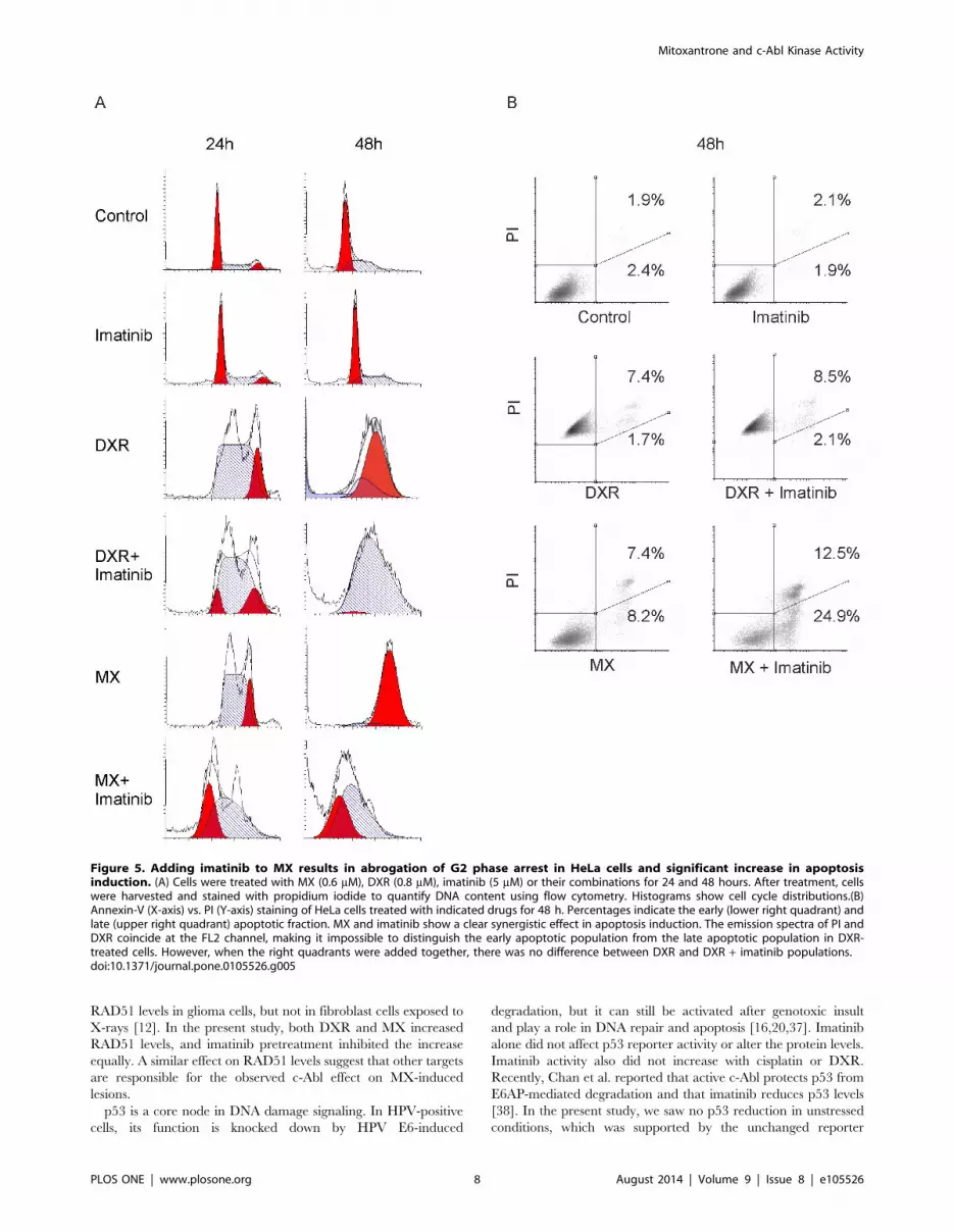
Imatinib abrogates S-phase arrest caused by MX andincreases apoptosis
In HeLa cells treated with imatinib alone, at 24 hours, cell cycle
distribution was the same as in cells with medium alone, but there
were slightly more cells in S phase after 48 hours (Figure 5A). At
24 hours and especially 48 hours, DXR accumulated the cells at
G2 phase. Adding imatinib induced S phase arrest in the software
analysis; however, at 48 hours with DXR + imatinib, there
appeared to be a small G2 population that the analysis software
failed to detect. MX-treated cells progressed at 24 hours to S and
G2, but the extended incubation after 48 hours showed a clear G2
arrest. Adding imatinib to MX showed at 24 hours a population
progressing to G1, indicating abrogation of the arrest. At
48 hours, there were no cells beyond S phase but still also a clear
G1 population. This finding suggests that the cells had prema-
turely entered directly from S phase to mitosis and that the
imatinib drove this effect. Alternatively, MX + imatinib may
induce a delay from G1/S to G2. However, several unsuccessful
mitoses were detected in the time-lapse video of MX+imatinib-
treated cells favoring the interpretation that the cell cycle arrest
was abrogated (Video S1). We also determined the cyclin B1
levels, a well-acknowledged mitotic marker, in the drug treated cell
populations to collect further evidence for the notion that the
MX+imatinib co-treated cells are capable of proceeding in cell
cycle and entering M-phase. We found that treatment of HeLa
cells with DXR, DXR+imatinib, MX and MX+imatinib leads to
accumulation of cyclin B1. The protein level of imatinib treated
cells was under level of detection similarly to the non-treated
controls exhibiting basal level cyclin B1 expression (Figure S5).
Imatinib increased the sub G1 events in MX-treated HeLa cells,
partly indicative of apoptosis. The sub G1 population was the
largest (38.6%) in the MX + imatinib group at 48 hours. Imatinib
also increased DXR-induced sub G1 but to a lesser extent (17.8%)
at 48 hours (Table S1). To further analyze the possible apoptosis
we stained the cells with annexin V which is a marker of early
apoptosis. We found that imatinib significantly increases the
proportion of apoptotic cells in MX-treated cells, but failed to
detect any increase when imatinib was added to DXR (Figure 5B)
Figure 2. Combination of MX and imatinib activates caspase cascades. HeLa cells were treated with indicated drugs for 48 h. Then, freshmedium was replaced and caspase 3/7 activity was measured using ELISA assay. Experiment was done in triplicate, mean 6 SD. *** p,0.001 T-test.doi:10.1371/journal.pone.0105526.g002
Mitoxantrone and c-Abl Kinase Activity
PLOS ONE | www.plosone.org 5 August 2014 | Volume 9 | Issue 8 | e105526

Flow cytometry results are in line with the caspase experiment
supporting the notion that imatinib with MX is more potent
inducer of cell death than with DXR. Imatinib has been previously
reported to cause G1 arrest in head and neck cancer cell lines [21]
whereas MX and DXR cause G2 arrest [22]. We saw no G1 arrest
with imatinib alone.
Downregulation of c-Abl with siRNA impedes thecytotoxicity of MX + imatinib
Imatinib and MX showed an additive effect in reducing the
number of control siRNA HeLa cells. When c-Abl was knocked
down with siRNA, we found that the proliferation of untreated
cells was reduced by 30–50% in repeated experiments (Figure 6,
Video S2). Both the growth curves and time-lapse microscopy data
show a slight growth inhibition in c-Abl siRNA HeLa cells.
However, neither control siRNA nor c-Abl siRNA alone induced
cell death. This result suggests that c-Abl is required for the normal
proliferation of these cells. Targeting of c-Abl in HeLa cells by
siRNA rendered the cells less sensitive to MX. CaSki cells were
also less sensitive to MX when c-Abl was downregulated with
siRNA, but no inhibition of proliferation was observed. These cells
were harder to transfect with siRNA and only 40% efficacy was
achieved. This may also explain why the proliferation was not
inhibited in these cells. Moreover, the combinatory effect of
imatinib was significantly reduced, implicating c-Abl as the pivotal
target of imatinib in this outcome. The latter finding is also in line
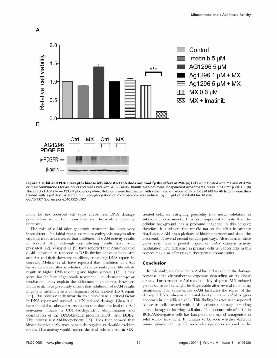
with experiments with other targets of imatinib, PDGF and c-Kit.
AG1296 is known to potently inhibit signaling of human PDGF a-
and b-receptors as well as of the related stem cell factor receptor c-
Kit, at 1 mM and 5 mM concentrations, respectively. AG1296
could not mimic the action of imatinib with MX (Figure 7A).
Moreover MX does not have any effect on phosphorylation of
PDGF receptor (Figure 7B). Furthermore, the siRNA experiment
implied that the kinase-active c-Abl is responsible for cell survival
after MX damage. Of importance, this experiment points to
different roles for kinase-active and kinase-inactive c-Abl. Kinase-
inactive c-Abl appears to be required for apoptosis with MX, but
kinase-active c-Abl is a key player for DNA damage repair after
MX in HeLa cells.
Figure 3. Imatinib increases DNA-damage induced by MX butnot by DXR. (A) DNA damage in the HeLa cells was studied usingComet assay after treatment with MX (0.6 mM), DXR (0.8 mM) andimatinib (5 mM), or their combinations. Cometting (tailing) indicatesDNA damage. (B) Imatinib combined with MX increases the level ofGADD45a. Western blot of GADD45a protein levels from whole celllysates. HeLa cells were treated with MX (0.6 mM), DXR (0.8 mM),imatinib (5 mM), or their combinations for 30 h.doi:10.1371/journal.pone.0105526.g003
Figure 4. Imatinib increases p53 levels when combined withMX, but not with DXR. Western blot of p53 and RAD 51 in HeLa cells.Imatinib combined with MX increases p53 protein levels in HeLa cells.RAD51 level was slightly reduced in cells treated with imatinib and MX.Cells were treated with MX (0.6 mM), DXR (0.8 mM), imatinib (5 mM) ortheir combinations for 30 hours.doi:10.1371/journal.pone.0105526.g004
Mitoxantrone and c-Abl Kinase Activity
PLOS ONE | www.plosone.org 6 August 2014 | Volume 9 | Issue 8 | e105526

Discussion
One of the major findings in this study is that the synergizing
effect of imatinib was specifically seen with MX. Previous studies
have shown that imatinib may enhance the cytotoxicity of a wide
range of chemotherapy drugs in solid tumor–derived cells [13,23].
Nevertheless, one group found that colon cancer cells become less
sensitive to TRAIL-induced apoptosis after imatinib [24]. These
reports did not involve testing the possible sensitivity differences
between chemotherapeutics in this context. Interestingly, Pinto et
al. very recently found in a comparable screening setting that
imatinib and MX additively inhibit the proliferation of PC-3
prostate cancer cells [25,26].
MX is an anthracenedione that targets topoisomerase II. It can
also induce DNA intercalation and free radical generation [27].
The effect of imatinib with MX observed in the present study
cannot merely be explained by topoisomerase II inhibition
because DXR (also a topoisomerase II inhibitor) did not have
similar activity with imatinib. The effect of imatinib is linked to its
ability to increase DNA damage in target cells, but it did not
notably increase the DNA damage induced by DXR in HeLa
cells. The difference in these closely related compounds is not
entirely clear, but there are some reported differences in these
drugs that may be related to the multi-drug resistance (MDR)
clearance of the drug.
Several tyrosine kinase inhibitors can selectively modulate
MDR-dependent drug efflux [28]. Imatinib has been reported to
reverse the resistance to topotecan and SN-38, an active
metabolite of irinotecan, and this activity has been attributed to
inhibition of the ABCG2 transporter [29]. MX resistance is
additionally mediated by transport proteins MRP-1 and ABCB1
[30]. Nilotinib, an imatinib derivative, inhibits the activity of both
ABCG2 and ABCB1, leading to enhanced DXR accumulation.
Different cells may behave in opposite ways because imatinib has
been reported to increase DXR concentration and synergize
cytotoxicity in breast cancer cells [31] but not in sarcoma cells
[28,32]. The concentration of the drug in the cell is not likely
solely to explain the outcome, because, for example, raising the
concentration of the drug does not lead to such a profound p53
accumulation and activity as seen with the MX + imatinib
combination (data not shown). Moreover, the cells can be rescued
by knocking down c-Abl. Therefore, without c-Abl commitment,
the effect is profoundly hampered.
c-Abl knockdown by siRNA rendered HeLa cells resistant to
MX. Taken together with the fact that kinase-inactive c-Abl
sensitized the cells to MX, this finding indicates that maximal MX
cytotoxicity depends on kinase-inactive c-Abl. The results suggest
that either 1) kinase-active c-Abl is a direct inhibitor of apoptosis in
these cells or 2) kinase-inactive c-Abl is pro-apoptotic in stress
conditions. Of relevance, kinase-inactive c-Abl has an important
function that is not present in knocked-down cells. It has been
shown that in addition to BCR/Abl cell lines, even in solid tumor–
derived cells that have increased c-Abl activity, imatinib may
inhibit aberrant growth. Moreover, c-Abl activation may either
enhance or decrease cell cycle progression in a cell type–specific
way or inhibit apoptosis and induce cell death [23,33]. We saw no
difference in cell proliferation or clonogenic growth after imatinib
treatment, but c-Abl knockdown diminished proliferation of HeLa
cells. This finding suggests that kinase-inactive c-Abl supports cell
cycle progression in these cells in unstressed conditions. In fact, a
positive mitogenic role for c-Abl has been shown in studies
exploiting abl-deficient cells [34,35]. When DNA is damaged, c-
Abl is activated, and the cell cycle is stalled. If the catalytic activity
is inhibited, the lack of c-Abl enhances cell cycle progression,
allowing no time for repair, highlighting the key role of c-Abl in
cell cycle control in these cells. These results suggest that kinase
activity of c-Abl may either enhance or inhibit proliferation
depending on either cell type–specific differences or protein
conformation properties and is not simply a binary choice.
c-Abl facilitates a repair checkpoint following moderate DNA
damage but promotes death after severe damage. This checkpoint
depends on the catalytic activity of c-Abl and is ruptured by
imatinib [3]. Checkpoint activation and mitosis block occur
especially in cells that have defective replication. Cells lacking the
S phase checkpoint with disordered replication eventually die [36].
HeLa cells were arrested in G2 phase after 48 hours of exposure to
MX and DXR. Of importance, we found that inhibition of c-Abl
kinase activity repressed the G2 phase arrest induced by MX, but
not that induced by DXR G1 and the number of annexin positive
cells also increased significantly, indicating defective replication
that was followed by apoptosis with enhanced caspase 3/7
activation. This finding, together with the comet assay results,
suggests a different form of DNA damage recognition that depends
on active c-Abl function in the case of MX damage. Consistently,
microarray analyses exploring the different outcomes of MX and
DXR reveal that there are differences in MX and DXR damage
responses (unpublished data). The cell cycle analyses hint at an
important connection between c-Abl and cell cycle control for
certain chemotherapy drugs. This possibility is in line with c-Abl
being a binding partner to a number of substrates linked to cell
cycle checkpoint control [4].
After DNA damage, RAD51 is centrally involved in homolo-
gous recombination repair and mediates the DNA strand-pairing
step. Russell et al. have previously shown that imatinib reduces
Table 1. p53 reporter activity changes in HeLa, SiHa and CaSki cells.
Hela SiHa CaSki
Cisplatin 2.760.2 3.660.3 14.760.6
Cisplatin + Imatinib 1.960.1 2.660.1 10.360.0
Doxorubicin 4.360.2 9.560.3 32.660.6
Doxorubicin + Imatinib 4.260.23 8.660.35 31.160.4
Mitoxantrone 0.960.1 3.860.1 3.060.2
Mitoxantrone + Imatinib 4.160.0 4.760.2 24.060.2
Imatinib 1.060.0 1.260.1 1.260.1
The results are given in fold-change. The concentrations used were MX 0.6 mM, 1 mM and 1 mM in HeLa, SiHa and CaSKi cell lines, respectively; DXR 0.8 mM, 1.4 mM and1 mM in HeLa, SiHa and CaSKi cell lines, respectively; Cisplatin 40 mM, 70 mM and 40 mM in HeLa, SiHa and CaSKi cell lines respectively. Imatinib 10 mM in each cell line.doi:10.1371/journal.pone.0105526.t001
Mitoxantrone and c-Abl Kinase Activity
PLOS ONE | www.plosone.org 7 August 2014 | Volume 9 | Issue 8 | e105526
RAD51 levels in glioma cells, but not in fibroblast cells exposed to
X-rays [12]. In the present study, both DXR and MX increased
RAD51 levels, and imatinib pretreatment inhibited the increase
equally. A similar effect on RAD51 levels suggest that other targets
are responsible for the observed c-Abl effect on MX-induced
lesions.
p53 is a core node in DNA damage signaling. In HPV-positive
cells, its function is knocked down by HPV E6-induced
degradation, but it can still be activated after genotoxic insult
and play a role in DNA repair and apoptosis [16,20,37]. Imatinib
alone did not affect p53 reporter activity or alter the protein levels.
Imatinib activity also did not increase with cisplatin or DXR.
Recently, Chan et al. reported that active c-Abl protects p53 from
E6AP-mediated degradation and that imatinib reduces p53 levels
[38]. In the present study, we saw no p53 reduction in unstressed
conditions, which was supported by the unchanged reporter
Figure 5. Adding imatinib to MX results in abrogation of G2 phase arrest in HeLa cells and significant increase in apoptosisinduction. (A) Cells were treated with MX (0.6 mM), DXR (0.8 mM), imatinib (5 mM) or their combinations for 24 and 48 hours. After treatment, cellswere harvested and stained with propidium iodide to quantify DNA content using flow cytometry. Histograms show cell cycle distributions.(B)Annexin-V (X-axis) vs. PI (Y-axis) staining of HeLa cells treated with indicated drugs for 48 h. Percentages indicate the early (lower right quadrant) andlate (upper right quadrant) apoptotic fraction. MX and imatinib show a clear synergistic effect in apoptosis induction. The emission spectra of PI andDXR coincide at the FL2 channel, making it impossible to distinguish the early apoptotic population from the late apoptotic population in DXR-treated cells. However, when the right quadrants were added together, there was no difference between DXR and DXR + imatinib populations.doi:10.1371/journal.pone.0105526.g005
Mitoxantrone and c-Abl Kinase Activity
PLOS ONE | www.plosone.org 8 August 2014 | Volume 9 | Issue 8 | e105526

activity. In contrast, the potentiating effect of imatinib for p53 was
profound after MX treatment. These findings indicate a difference
between cell lines treated with different chemotherapy drugs in
respect to p53 activity after c-Abl kinase inhibition. Second, p53
activation is in line with the observation in comet assays that c-Abl
inhibition by imatinib added DNA tailing after MX but not after
DXR. Despite the accumulation of transcriptionally active p53,
cell survival and clonogenic growth did not alter when p53 was
inactivated by overexpression of dominant-negative p53. p73 is a
direct substrate of c-Abl and is phosphorylated upon DNA damage
[1,39]. We found no evidence that p73 was responsible for the
combined effect in HeLa cells in Western analyses. Upon DNA
damage, p73 accumulates in an active c-Abl-dependent manner
[40]. We blocked the c-Abl kinase activity by imatinib; therefore, it
is not surprising that we saw no p73 dependence for the MX +imatinib effect. Taken together, these results implicate pathways
other than p53 or p73 as being involved here in the kinase-inactive
c-Abl–mediated apoptosis. The more detailed gene level determi-
Figure 6. Down-regulation of c-Abl by siRNA counteracts MX induced apoptosis in HeLa and CaSki cells. (A) HeLa and CaSki Cells weretransfected with non-targeting or c-Abl siRNA (75 nM). Western blot analysis was performed to examine the effect of siRNA. Level of c-Abl wasquantitated and corrected for b-actin level. Values are proportioned to levels of control siRNA for each cell line. (B) HeLa, and (C) CasKi cells weretransfected with control or c-Abl siRNA. After treansfection cells were treated with MX (0.6 mM), imatinib (5 mM) and their combination for 48 h.Relative amount of surviving cells was determined by WST-1 assay. Results are from two independent experiments in triplicates. Data are shown asmean 6 SD. * p,0.05, ** p,0.01, *** p,0.001. NS, not significant.doi:10.1371/journal.pone.0105526.g006
Mitoxantrone and c-Abl Kinase Activity
PLOS ONE | www.plosone.org 9 August 2014 | Volume 9 | Issue 8 | e105526
nants for the observed cell cycle effects and DNA damage
potentiation are of key importance and the work is currently
underway.
The role of c-Abl after genotoxic treatment has been very
inconsistent. The initial report on mouse embryonic oocytes after
cisplatin treatment showed that inhibition of c-Abl activity results
in survival [41], although contradicting results have been
presented [42]. Wang et al. [9] have reported that Atm-mediated
c-Abl activation in response to DSBs further activates both Atm
and Atr and their downstream effects, enhancing DNA repair. In
contrast, Meltser et al. have reported that inhibition of c-Abl
kinase activation after irradiation of mouse embryonic fibroblasts
results in higher DSB rejoining and higher survival [43]. It may
seem that the form of genotoxic treatment - i.e., chemotherapy or
irradiation - may explain the difference in outcomes. However,
Fanta et al. have previously shown that inhibition of c-Abl results
in genetic instability as a consequence of diminished DNA repair
[44]. Our results clearly favor the role of c-Abl as a critical factor
in DNA repair and survival in MX-induced damage. Chen et al.
have found that ultraviolet irradiation that does not lead to c-Abl
activation induces a CUL-4A-dependent ubiquitination and
degradation of the DNA-binding proteins DDB1 and DDB2.
This process is c-Abl-independent [45]. They then showed that
kinase-inactive c-Abl may negatively regulate nucleotide excision
repair. This activity would explain the dual role of c-Abl in MX-
treated cells, an intriguing possibility that needs validation in
subsequent experiments. It is also important to note that the
cellular background has a profound influence in this context;
therefore, it is relevant that we did not see the effect in primary
fibroblasts. c-Abl has a plethora of binding partners and sits at the
crossroads of several crucial cellular pathways. Alterations in these
genes may have a pivotal impact on c-Abl catalytic activity
modulation. The difference in primary cells vs. cancer cells in this
respect may also offer unique therapeutic opportunities.
Conclusion
In this study, we show that c-Abl has a dual role in the damage
response after chemotherapy exposure depending on its kinase
activity. Furthermore, c-Abl may be a key player in MX-induced
genotoxic stress but might be dispensable after several other drug
treatments. The kinase-active c-Abl facilitates the repair of the
damaged DNA whereas the catalytically inactive c-Abl triggers
apoptosis in the afflicted cells. This finding has not been reported
before in cells treated with c-Abl-activating damage including
chemotherapy or ionizing radiation. The obscure role of c-Abl in
BCR/Abl-negative cells has hampered the use of antagonists in
solid tumor treatment. It remains to be seen whether different
tumor subsets with specific molecular signatures respond to the
Figure 7. C-kit and PDGF receptor kinase inhibitor AG1296 does not modify the effect of MX. (A) Cells were treated with MX and AG1296or their combinations for 48 hours and measured with WST-1 assay. Results are from three independent experiments, mean 6 SD. *** p,0.001. (B)The effect of AG1296 on PDGFR phosphorylation. HeLa cells were first treated with either medium alone (Ctrl) or 0.6 mM MX for 48 h. Cells were thentreated with 5 mM AG1296 for 15 min. Phosphorylation of PGDF receptor was induced by 0.1 mM of PDGF-BB for 10 min.doi:10.1371/journal.pone.0105526.g007
Mitoxantrone and c-Abl Kinase Activity
PLOS ONE | www.plosone.org 10 August 2014 | Volume 9 | Issue 8 | e105526

combined treatment with specific chemotherapy drugs together
with c-Abl inhibition.
Supporting Information
Figure S1 Imatinib enhances MX induced cytotoxicityalso in CaSKi and SiHa cell lines. Short-term cytotoxicity
assay. Results were from three independent experiments, mean 6
SD. *** p,0.001.
(TIF)
Figure S2 Enhancement of MX induced cytotoxicity byimatinib is not p53 dependent. p53 activity was abolished
with either dominant negative p53 (DDp53) or ectopic HPVE6.
CMV depicts the empy vector. Treatment duration 48 h. A.
Stably transfected HeLa cells B. Stably transfected SiHa cells.
(TIF)
Figure S3 Imatinib enhances MX induced cytotoxicityin CaSKi and SiHa cell lines with growing in clonaldensities. Targeting of residual p53 activity with ectopic E6 does
not rescue SiHa cells from the imatinib enhanced cytotoxicity.
Cells were treated in the clonogenic assay with each drug for 12 h.
Then, fresh medium was replaced. The concentration of imatinib
was 3 mM whereas MX was used in concentrations from 1 nM to
32 nM in experiments done with CaSKi cell line and from 1 nM
to 15 nM with SiHa CMV cell line. Results were from three
independent experiments, mean 6 SD. *** p,0.001.
(TIF)
Figure S4 p73 protein levels after indicated treatments.Western blot image from whole cell lysates at 48 h after treatment.
(TIF)
Figure S5 Cyclin B1 accumulation in HeLa cells treatedwith imatinib, DXR, DXR+imatinib, MX, and MX+ima-
tinib. Cyclin B1 protein level was examined with Western blot
analysis 48 h after the treatment.
(TIF)
Table S1 Cell cycle distributions of HeLa cells. Cell cycle
distribution percentages from experiment shown in Fig. 5. Cells
were treated with indicated drugs for 24 and 48 h. Cell cycle phase
percentages were calculated using ModFit LT cell cycle modelling
software. Results are shown as percentages of G1, S and G2/M
populations.
(DOCX)
Video S1 Combination of MX (0.6 mM) and imatinib(5 mM) induces more abnormal mitoses than MX alonein HeLa cells. Cells were seeded on 96-well plates and images
were acquired at 1 hour interval. Bar represents 200 mm.
(MP4)
Video S2 c-Abl siRNA induces mild growth inhibitionbut not cell death in HeLa cells. Cells were seeded on 96-well
plates and images were acquired at 1 hour interval. Bar represents
200 mm.
(MP4)
Acknowledgments
We thank Mr. Jaakko Lehtimaki, Ms. Johanna Markola-Warn, and Ms.
Sari Pitkanen for skillfull technical assistance.
Author Contributions
Conceived and designed the experiments: SH MF K. Alpay JS JT.
Performed the experiments: K. Alpay MF JT JS K. Aittokallio ES.
Analyzed the data: SH JS JT MF MK K. Alpay VMK. Contributed
reagents/materials/analysis tools: SH VMK MK. Wrote the paper: SH
MF JT JS VMK K. Alpay. Conceived and supervised the study: SH.
References
1. Agami R, Blandino G, Oren M, Shaul Y (1999) Interaction of c-Abl and
p73alpha and their collaboration to induce apoptosis. Nature 399: 809–813.
2. Hoeijmakers JH (2007) Genome maintenance mechanisms are critical for
preventing cancer as well as other aging-associated diseases. Mech Ageing Dev
128: 460–462.
3. Maiani E, Diederich M, Gonfloni S (2011) DNA damage response: the emerging
role of c-Abl as a regulatory switch? Biochem Pharmacol 82: 1269–1276.
4. Colicelli J (2010) ABL tyrosine kinases: evolution of function, regulation, and
specificity. SciSignal 139. re6. doi: 10.1126/scisignal.3139re6
5. Wang JY (2004) Controlling Abl: auto-inhibition and co-inhibition? Nat Cell
Biol 6: 3–7.
6. Nagar B (2007) c-Abl tyrosine kinase and inhibition by the cancer drug imatinib
(Gleevec/STI-571). J Nutr 137: 1518S–1523S.
7. Slupianek A, Schmutte C, Tombline G, Nieborowska-Skorska M, Hoser G,
et al. (2001) BCR/ABL regulates mammalian RecA homologs, resulting in drug
resistance. Mol Cell 8: 795–806.
8. Buchdunger E, O’Reilly T, Wood J (2002) Pharmacology of imatinib (STI571).
Eur J Cancer 38 Suppl 5: S28–S36.
9. Wang X, Zeng L, Wang J, Chau JF, Lai KP, et al. (2011) A positive role for
c-Abl in Atm and Atr activation in DNA damage response. Cell Death Differ 18:
5–15.
10. Chen G, Yuan SS, Liu W, Xu Y, Trujillo K, et al. (1999) Radiation-induced
assembly of Rad51 and Rad52 recombination complex requires ATM and
c-Abl. J Biol Chem 274: 12748–12752.
11. Kubler HR, van Randenborgh H, Treiber U, Wutzler S, Battistel C, et al. (2005)
In vitro cytotoxic effects of imatinib in combination with anticancer drugs in
human prostate cancer cell lines. Prostate 63: 385–394.
12. Russell JS, Brady K, Burgan WE, Cerra MA, Oswald KA, et al. (2003) Gleevec-
mediated inhibition of Rad51 expression and enhancement of tumor cell
radiosensitivity. Cancer Res 63: 7377–7383.
13. Choudhury A, Zhao H, Jalali F, Al Rashid S, Ran J, et al. (2009) Targeting
homologous recombination using imatinib results in enhanced tumor cell
chemosensitivity and radiosensitivity. Mol Cancer Ther 8: 203–213.
14. Kharbanda S, Yuan ZM, Weichselbaum R, Kufe D (1997) Functional role for
the c-Abl protein tyrosine kinase in the cellular response to genotoxic stress.
Biochim Biophys Acta 1333: O1–O7.
15. zur Hausen H (2002) Papillomaviruses and cancer: from basic studies to clinical
application. Nat Rev Cancer 2: 342–350.
16. Hietanen S (2009) Apoptosis in Carcinogenesis and Chemoherapy of the Uterine
Cervix. In: Chen GG, Lai PBS, editors. Apoptosis in Carcinogenesis and
Chemotherapy: Springer Netherlands. pp.51–73.
17. Koivusalo R, Hietanen S (2004) The cytotoxicity of chemotherapy drugs varies
in cervical cancer cells depending on the p53 status. Cancer Biol Ther 3: 1177–
1183.
18. Koivusalo R, Krausz E, Ruotsalainen P, Helenius H, Hietanen S (2002)
Chemoradiation of cervical cancer cells: targeting human papillomavirus E6 and
p53 leads to either augmented or attenuated apoptosis depending on the
platinum carrier ligand. Cancer Res 62: 7364–7371.
19. Hietanen S, Auvinen E, Syrjanen K, Syrjanen S (1998) Anti-proliferative effect
of retinoids and interferon-alpha-2a on vaginal cell lines derived from squamous
intra-epithelial lesions. Int J Cancer 78: 338–345.
20. Koivusalo R, Krausz E, Helenius H, Hietanen S (2005) Chemotherapy
compounds in cervical cancer cells primed by reconstitution of p53 function
after short interfering RNA-mediated degradation of human papillomavirus 18
E6 mRNA: opposite effect of siRNA in combination with different drugs. Mol
Pharmacol 68: 372–382.
21. Wang-Rodriguez J, Lopez JP, Altuna X, Chu TS, Weisman RA, et al. (2006)
STI-571 (Gleevec) potentiates the effect of cisplatin in inhibiting the proliferation
of head and neck squamous cell carcinoma in vitro. Laryngoscope 116: 1409–
1416.
22. Potter AJ, Rabinovitch PS (2005) The cell cycle phases of DNA damage and
repair initiated by topoisomerase II-targeting chemotherapeutic drugs. Mutat
Res 572: 27–44.
23. Lin J, Arlinghaus R (2008) Activated c-Abl tyrosine kinase in malignant solid
tumors. Oncogene 27: 4385–4391.
24. Huang DY, Chao Y, Tai MH, Yu YH, Lin WW (2012) STI571 reduces TRAIL-
induced apoptosis in colon cancer cells: c-Abl activation by the death receptor
leads to stress kinase-dependent cell death. J Biomed Sci 19: 35.
25. Pinto AC, Angelo S, Moreira JN, Simoes S (2011) Schedule treatment design
and quantitative in vitro evaluation of chemotherapeutic combinations for
metastatic prostate cancer therapy. Cancer Chemother Pharmacol 67: 275–284.
Mitoxantrone and c-Abl Kinase Activity
PLOS ONE | www.plosone.org 11 August 2014 | Volume 9 | Issue 8 | e105526

26. Pinto AC, Moreira JN, Simoes S (2011) Liposomal imatinib-mitoxantrone
combination: formulation development and therapeutic evaluation in an animal
model of prostate cancer. Prostate 71: 81–90.
27. Hande KR (2008) Topoisomerase II inhibitors. Update Cancer Ther 3: 13–26.
28. Tiwari AK, Sodani K, Wang SR, Kuang YH, Ashby CR Jr, et al. (2009)
Nilotinib (AMN107, Tasigna) reverses multidrug resistance by inhibiting the
activity of the ABCB1/Pgp and ABCG2/BCRP/MXR transporters. Biochem
Pharmacol 78: 153–161.
29. Houghton PJ, Germain GS, Harwood FC, Schuetz JD, Stewart CF, et al. (2004)
Imatinib mesylate is a potent inhibitor of the ABCG2 (BCRP) transporter and
reverses resistance to topotecan and SN-38 in vitro. Cancer Res 64: 2333–2337.
30. Morrow CS, Peklak-Scott C, Bishwokarma B, Kute TE, Smitherman PK, et al.
(2006) Multidrug resistance protein 1 (MRP1, ABCC1) mediates resistance to
mitoxantrone via glutathione-dependent drug efflux. Mol Pharmacol 69: 1499–
1505.
31. Sims JT, Ganguly SS, Bennett H, Friend JW, Tepe J, et al. (2013) Imatinib
reverses doxorubicin resistance by affecting activation of STAT3-dependent NF-
kappaB and HSP27/p38/AKT pathways and by inhibiting ABCB1. PLoS One
8: e55509.
32. Villar VH, Vogler O, Martinez-Serra J, Ramos R, Calabuig-Farinas S, et al.
(2012) Nilotinib counteracts P-glycoprotein-mediated multidrug resistance and
synergizes the antitumoral effect of doxorubicin in soft tissue sarcomas. PLoS
One 7: e37735.
33. Srinivasan D, Sims JT, Plattner R (2008) Aggressive breast cancer cells are
dependent on activated Abl kinases for proliferation, anchorage-independent
growth and survival. Oncogene 27: 1095–1105.
34. Plattner R, Kadlec L, DeMali KA, Kazlauskas A, Pendergast AM (1999) c-Abl is
activated by growth factors and Src family kinases and has a role in the cellular
response to PDGF. Genes Dev 13: 2400–2411.
35. Furstoss O, Dorey K, Simon V, Barila D, Superti-Furga G, et al. (2002) c-Abl is
an effector of Src for growth factor-induced c-myc expression and DNAsynthesis. EMBO J 21: 514–524.
36. Labib K, De Piccoli G (2011) Surviving chromosome replication: the many roles
of the S-phase checkpoint pathway. PhilosTrans R Soc Lond B Biol Sci 366:3554–3561.
37. Hietanen S, Lain S, Krausz E, Blattner C, Lane DP (2000) Activation of p53 incervical carcinoma cells by small molecules. Proc Natl Acad Sci USA 97: 8501–
8506.
38. Chan AL, Grossman T, Zuckerman V, Campigli DG, Moshel O, et al. (2013)c-Abl phosphorylates E6AP and regulates its E3 ubiquitin ligase activity.
Biochemistry 52: 3119–3129.39. White E, Prives C (1999) DNA damage enables p73. Nature 399: 734–735, 737.
40. Yuan ZM, Shioya H, Ishiko T, Sun X, Gu J, et al. (1999) p73 is regulated bytyrosine kinase c-Abl in the apoptotic response to DNA damage. Nature 399:
814–817.
41. Gonfloni S, Di Tella L, Caldarola S, Cannata SM, Klinger FG, et al. (2009)Inhibition of the c-Abl-TAp63 pathway protects mouse oocytes from
chemotherapy-induced death. Nat Med 15: 1179–1185.42. Kerr JB, Hutt KJ, Cook M, Speed TP, Strasser A, et al. (2012) Cisplatin-induced
primordial follicle oocyte killing and loss of fertility are not prevented by
imatinib. Nat Med 18: 1170–1172.43. Meltser V, Ben Yehoyada M, Reuven N, Shaul Y (2010) c-Abl downregulates
the slow phase of double-strand break repair. Cell Death Dis 1: e20.44. Fanta S, Sonnenberg M, Skorta I, Duyster J, Miething C, et al. (2008)
Pharmacological inhibition of c-Abl compromises genetic stability and DNArepair in Bcr-Abl-negative cells. Oncogene 27: 4380–4384.
45. Chen X, Zhang J, Lee J, Lin PS, Ford JM, et al. (2006) A kinase-independent
function of c-Abl in promoting proteolytic destruction of damaged DNA bindingproteins. Mol Cell 22: 489–499.
Mitoxantrone and c-Abl Kinase Activity
PLOS ONE | www.plosone.org 12 August 2014 | Volume 9 | Issue 8 | e105526


