Embed Size (px)
Citation preview
cells
Article
iNOS Interacts with Autophagy Receptor p62 and isDegraded by Autophagy in Macrophages
Jing Wang 1,†, Ming-Yue Wu 1,†, Huanxing Su 1, Jinjian Lu 1, Xiuping Chen 1, Jieqiong Tan 2 andJia-Hong Lu 1,*
1 State Key Laboratory of Quality Research in Chinese Medicine, Institute of Chinese Medical Sciences,University of Macau, Taipa, Macao; [email protected] (J.W.); [email protected] (M.-Y.W.);[email protected] (H.S.); [email protected] (J.L.); [email protected] (X.C.)
2 Center for Medical Genetics, School of Life Sciences, Central South University, Changsha 410008, Hunan,China; [email protected]
* Correspondence: [email protected]; Tel.: +853-88224508† Both authors contributed equally to this work.
Received: 23 September 2019; Accepted: 14 October 2019; Published: 15 October 2019�����������������
Abstract: Nitric oxide (NO) is an important mediator of inflammation response and the productionof NO has been linked to a variety of diseases, including tumors, inflammation and central nervoussystem diseases. In macrophages, a high level of NO is generated by iNOS during inflammatoryresponses triggered by cytokines or pathogens. Autophagy, a cellular bulk degradation process vialysosome, has been implicated in many disease conditions including inflammation. In this study, wehave reported the previously unknown role of autophagy in regulating iNOS levels in macrophages,both under basal and Lipopolysaccharides (LPS)-induced conditions. Our data showed that iNOSlevels accumulated upon autophagy inhibition and decreased upon autophagy induction. iNOSinteracted and co-localized with autophagy receptor p62/SQSTM1, especially under LPS-stimulatedcondition in macrophages. Moreover, the immunostaining data revealed that iNOS also co-localizeswith the autophagosome marker LC3 and lysosome marker LAMP1, especially under lysosomalinhibition conditions, indicating iNOS is an autophagy substrate. Finally, we showed that autophagynegatively regulated the generation of NO in macrophages, which is consistent with the changes ofiNOS levels. Collectively, our study revealed a previously unknown mechanism by which autophagyregulates iNOS levels to modulate NO production during inflammation.
Keywords: iNOS; NO; p62/SQSTM1; autophagy; macrophage
1. Introduction
NO is an important inflammation signaling molecule which is synthesized from L-arginine bynitric oxide synthase (NOS). The NOS enzymes family contains three isoforms: endothelial nitricoxide synthase (eNOS) mainly located at epithelial cells, neuronal nitric oxide synthase (nNOS) mostlydistributed in neurons and inducible nitric oxide synthase (iNOS) widely expressed in immune cells,including macrophages, dendritic cells, microglia and T cells during inflammatory response [1]. iNOSlevel is extremely high in macrophages after cytokines or bacterial stimulating, generating large amountof NO in a short time to mediate inflammation response as well as cause cytotoxicity [2]. In addition totranscriptional and translational regulation, iNOS level can also be regulated via ubiquitin-proteasomesystem by degradation [3,4].
Autophagy is an intracellular process for lysosome degradation and it usually means “self-eating”.It originates from a bilayer membrane structure called the autophagosome, which forms anautolysosome after fusion with lysosomes for degradation [5]. Autophagy was initially considered
Cells 2019, 8, 1255; doi:10.3390/cells8101255 www.mdpi.com/journal/cells
Cells 2019, 8, 1255 2 of 13
to be non-selective, but recent studies have found that this process can be selective, partially via theautophagy receptor proteins [6], to precisely degrade target proteins [7]. In early studies, LPS hasbeen found to induce autophagy in human macrophages and in the RAW 264.7 cell line via a Toll-likereceptor 4 (TLR4)-dependent pathway [8,9]. P62 is the classic autophagy receptor which recruits cargosto the autophagosome for degradation. Inhibition of autophagy leads to intracellular p62 aggregationand the amount of p62 protein can be used as an indicator of autophagic flux [10]. P62 expressionis increased after LPS stimulation and it plays important roles in inflammation-related pathwaysregulation [11,12].
Autophagy is involved in inflammation regulation by multiple mechanisms including: degradationof inflammasome, clearance of pathogens, cytokine secretion and antigen presentation [13]. In thisstudy, we find a new mechanism by which autophagy regulates inflammation response by affectingthe NO production via controlling autophagic degradation of iNOS.
2. Materials and Methods
2.1. Reagents
Chloroquine (CQ), SAR405, Torin 1, Rapamycin, Lipopolysaccharides (LPS), 1400 wdihydrochloride and Cycloheximide were purchased from Sigma-Aldrich (St. Louis, MO, USA).Nitric oxide Griess reagent and RIPA buffer were obtained from Beyotime Biotechnology (Shanghai,China). Dynabeads protein Immunoprecipitation kit, TNF-α and IL-6 ELISA kit were from ThermoFisher Scientific (Dreieich, Germany). The mouse monoclonal antibody against iNOS (Cat. No: 610328)was purchased from Biosciences (Heidelberg, Germany). The rabbit monoclonal anti-p62 for Westernblot and immunofluorescence (Cat. No: ab109012) was purchased from Abcam (Cambridge, UK) andrabbit polyclonal anti-p62 for co-immunoprecipitation (Cat. No: P0068) was obtained from sigma.The rabbit polyclonal anti-LC3 (Cat. No: NB100-2220) for western blot was from Novus Biologicals(Littleton, CO, USA) and rabbit polyclonal anti-LC3 (Cat. No: PM036) for immunofluorescence stainingwas obtained from MBL International Corporation (Woburn, MA, USA). Anti-GAPDH (Cat. No: 5174)and anti-Lamp1 (Cat. No: 9091) were obtained from Cell Signaling Technology (Boston, MA, USA).
2.2. Cell Culture and Treatment
Raw 264.7 cells were cultured in incubator at 37 ◦C with 5% CO2 and 95% humidified atmosphere.The cells grew in Dulbecco’s Modified Eagle’s medium (DMEM, Gibco, Darmstadt, Germany),containing 10% fetal bovine serum (FBS, Gibco) and 1% penicillin/streptomycin. Bone-marrow-derivedmacrophages (BMDMs) were obtained from the femur and tibia of C57BL/6 mice. Bone marrow cellswere obtained by flushing femur and tibia with 1 X HBSS. The flushed out-bone marrow cells werecultured for 7 days in Dulbecco’s Modified Eagle’s medium (DMEM), containing 10% fetal bovineserum (FBS), 1% penicillin/streptomycin and 10% L929 cell culture medium. After 7 days, above 97%cells that attached to the bottle of dish are macrophages and can be used for experiments.
2.3. Western Blot Analysis
Raw 264.7 cells and BMDMs were seeded at a density of 4 × 10⁵ per well in 12-well platesovernight, then treated with autophagy inhibitors (Chloroquine, 30 µM; SAR405, 1 µM) and inducers(Torin 1,1 µM; Rapamycin, 1 µM) for 24 h. Under LPS stress condition, Raw 264.7 treated with LPS (200ng/mL) for 12 h, then replaced with new medium and treated with autophagy inhibitors or inducersfor another 12 h. Raw 264.7 cells and BMDMs were washed with ice-cold phosphate buffer saline(PBS) twice, then lysed with RIPA buffer (50 mM Tris-HCl pH 8.0, 0.1 M NaCl, 20 mM EDTA, 1% SDS,contained with protease and phosphatase inhibitor cocktails). The lysates were denatured at 99 ◦Cin sample loading buffer, proteins (20 µg) were resolved with SDS-PAGE and then transferred to apolyvinylidene difluoride membrane. Membranes were blocked with 5% fat-free milk in Tris-buffedsaline supplemented with 0.1% Tween-20 (TBST) for 1h at room temperature. After that, membranes
Cells 2019, 8, 1255 3 of 13
were incubated with primary antibodies overnight at 4 ◦C. After washing with TBST for 30 min,membranes were incubated with HRP-conjugated secondary antibodies for 2 h at room temperatureand washed for another 30 min. Finally, HRP substrate (GE healthcare) was used to detect blotsthrough chemiluminescence. The densitometric analysis of western blotting bands were calculatedvia software (Image Lab 5.1, Bio-Rad, Munich, German). Briefly, the file of bands was import intothe software, the bands to be quantified were selected in each group, and the densitometric valueof each bands were subtracted with background value. The adjusted densitometric value of eachband was firstly normalized with GAPDH to get the relative densitometric value, then all the relativedensitometric values were normalized with that of CTRL group to obtain the fold of control value.
2.4. Immunofluorescence Assay
Raw 264.7 cells were plated on glass coverslips in 24-well plates. Under basal condition, cellswere treated with CQ and SAR for 12 h. While under stress condition, cells were treated withLPS (1 µg/mL) for 12 h, then replaced with fresh medium and treated with autophagy inhibitors orinducers for another 12 h. After treatment, cells were fixed with 4% paraformaldehyde for 10 min, andpermeabilized with 0.3% Triton X-100 for 15 min (Sigma-Aldrich). Cells then were blocked with 5%BSA (Beyotime Biotechnolog) for 2 h and stained with iNOS antibody (1:100, Santa cruz) or p62 (1:100,Abcam) antibody overnight at 4 ◦C. The iNOS and p62 signals were visualized by incubating withAlexa Fluor488 (green) and Alexa Fluor555 (red) conjugated secondary antibodies (1:500) for 2 h atroom temperature. The nuclei were stained with Hoechst, and the coverslips were mounted with afluoromount aqueous mounting medium (Sigma-Aldrich). Cells were visualized through a confocalfluorescence imagine microscope (Leica TCS SP8; Leica Microsystems, Bensheim, Germany). Hoechst,Fluor488 (Green) and Fluor555 (Red) fluorescence is excited under 405 nm, 488 nm, and 552 nm laserexcitation, respectively, and used sequential scanning for image capture.
Colocalization is analyzed via Image J (National Institutes of Health, Bethesda, MD, USA), andimage distribution in the article is refer to the previous study [14].
2.5. Co-Immunoprecipitation Assay
Under basal condition, Raw 264.7 cells were plated in 6-well plates overnight. Under stresscondition, Raw 264.7 cells treated with LPS (1 µg/mL) for 12 h. Then cells were washed with ice-coldPBS and lysed with IP lysis buffer (10 mM Tris-HCl, pH 7.5, 2 mM EDTA, 1% NP40, 150 mM NaCl,supplemented with protease and phosphates inhibitor cocktail). The lysates incubated with anti-iNOSantibody (1:100, Cell Signaling Technology) or p62 antibody (1:100, Sigma) overnight at 4 ◦C. 25 µLprotein-G conjugated magnetic beads (Dynabeads Protein Immunoprecipitation kit, Thermo scientific)was added into each IP tube and incubate at 4 ◦C for 6 h. The supernatants were removed by aspirationand the beads were washed three times with the lysis buffer. Finally, the beads were denatured at 99 ◦Cin 1×sample loading buffer and the IP products were detected by western blotting as mentioned above.
2.6. ELISA Assay
Raw 264.7 cells were seeded in 12-well plates. Treatment with LPS (200 ng/mL) for 12 h, thenreplaced with fresh medium and treated with autophagy inhibitors or inducers for another 12 h. 200 µLof medium supernatant was collected after treatment. The levels of TNF-α and IL-6 were determinedwith enzyme-linked immunosorbent assay (ELISA) kit (Thermo Fisher Scientific) according to thetechnical guide protocol. Samples were measured in a 96-well plate by a fluorometer equipped with450 nm. The absorbance of the samples was valued by comparing with a simultaneously generatedstandard curve.
Cells 2019, 8, 1255 4 of 13
2.7. Nitric Oxide Detection Assay
Raw 264.7 cells were seeded in 12-well plates and treated with LPS (200 ng/mL) for 12 h, then theculture medium was replaced with fresh medium and treated with autophagy inhibitors, autophagyinducers or 1400 w (100 µM) for another 12 h. After treatment, the medium supernatant was collectedto detect the production of nitric oxide by Griess reagent (Beyotime Biotechnology) according to themanufacturer’s protocols. Samples were plated in a 96-well plate by a fluorometer equipped with540 nm emission filter. The nitrite concentration was calculated from a nitrite standard curve inthis method.
2.8. Statistics
For comparing different groups, statistical analysis was performed by one-way ANOVA withTurkeys as post hoc tests. Data were analyzed with GraphPad Prism and statistically significant atp < 0.05.
3. Results
3.1. Autophagy Regulates the Level of iNOS in Macrophages
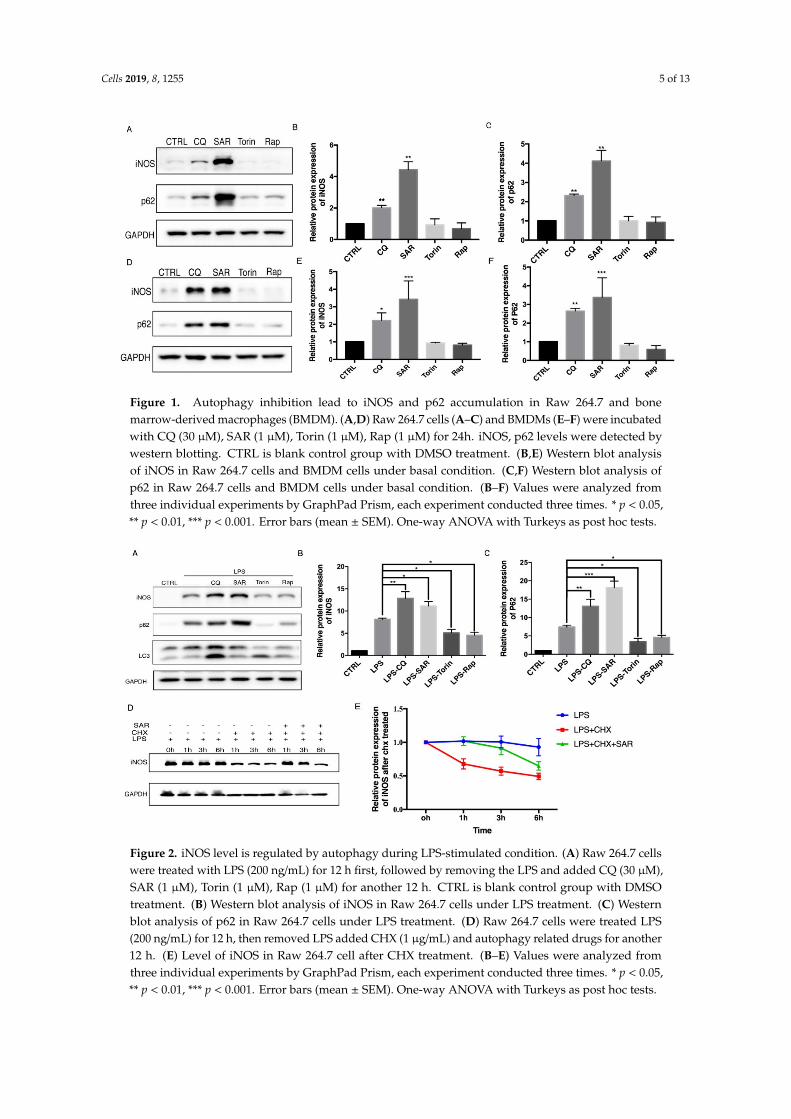
To explore the role of autophagy in iNOS degradation, Raw 264.7 macrophage cells were treatedwith autophagy inducers and inhibitors. Chloroquine (CQ) is an commonly used autophagy inhibitorby raising lysosome pH and decreasing autophagosome-lysosome fusion [15,16] and SAR405 is anotherautophagy inhibitor through PI3KC3 inhibition [17]. Torin 1 and rapamycin were commonly usedas inducers of autophagy by inhibiting the mTOR pathway [18]. We found that the protein levels ofiNOS are consistent with p62 level which dramatically accumulated upon autophagy inhibition inRaw 264.7 cells (Figure 1A–C). Consistently, we also detected the iNOS and p62 protein levels in bonemarrow-derived macrophages (BMDMs) (Figure 1 D–F). We further sought to figure out whether theiNOS would display similar pattern after autophagy modulators treatment under the inflammationcondition induced by LPS, a stronger inducer of inflammation and iNOS expression. After stimulationwith LPS, the iNOS and p62 levels were dramatically increased. Interestingly, autophagy inhibitorsfurther increased the iNOS and p62 levels while autophagy inducers decreased the iNOS and p62levels in Raw 264.7 cells (Figure 2A–C). These data drove us to propose that iNOS is an autophagysubstrate during inflammation. To understand whether the iNOS is degraded by autophagy, we useda eukaryote protein synthesis inhibitor cycloheximide to block protein translation and observed thatthe degradation rate of iNOS was impaired by addition of autophagy inhibitor SAR405 (Figure 2D,E).Thus, we found that iNOS can be degraded through the autophagy pathway, in a similar trend like p62in Raw 264.7 cells.
Cells 2019, 8, 1255 5 of 13
Figure 1. Autophagy inhibition lead to iNOS and p62 accumulation in Raw 264.7 and bonemarrow-derived macrophages (BMDM). (A,D) Raw 264.7 cells (A–C) and BMDMs (E–F) were incubatedwith CQ (30 µM), SAR (1 µM), Torin (1 µM), Rap (1 µM) for 24h. iNOS, p62 levels were detected bywestern blotting. CTRL is blank control group with DMSO treatment. (B,E) Western blot analysisof iNOS in Raw 264.7 cells and BMDM cells under basal condition. (C,F) Western blot analysis ofp62 in Raw 264.7 cells and BMDM cells under basal condition. (B–F) Values were analyzed fromthree individual experiments by GraphPad Prism, each experiment conducted three times. * p < 0.05,** p < 0.01, *** p < 0.001. Error bars (mean ± SEM). One-way ANOVA with Turkeys as post hoc tests.
Figure 2. iNOS level is regulated by autophagy during LPS-stimulated condition. (A) Raw 264.7 cellswere treated with LPS (200 ng/mL) for 12 h first, followed by removing the LPS and added CQ (30 µM),SAR (1 µM), Torin (1 µM), Rap (1 µM) for another 12 h. CTRL is blank control group with DMSOtreatment. (B) Western blot analysis of iNOS in Raw 264.7 cells under LPS treatment. (C) Westernblot analysis of p62 in Raw 264.7 cells under LPS treatment. (D) Raw 264.7 cells were treated LPS(200 ng/mL) for 12 h, then removed LPS added CHX (1 µg/mL) and autophagy related drugs for another12 h. (E) Level of iNOS in Raw 264.7 cell after CHX treatment. (B–E) Values were analyzed fromthree individual experiments by GraphPad Prism, each experiment conducted three times. * p < 0.05,** p < 0.01, *** p < 0.001. Error bars (mean ± SEM). One-way ANOVA with Turkeys as post hoc tests.
Cells 2019, 8, 1255 6 of 13
3.2. iNOS Interacts and Colocalizes with Autophagy Receptor p62/SQSTM1
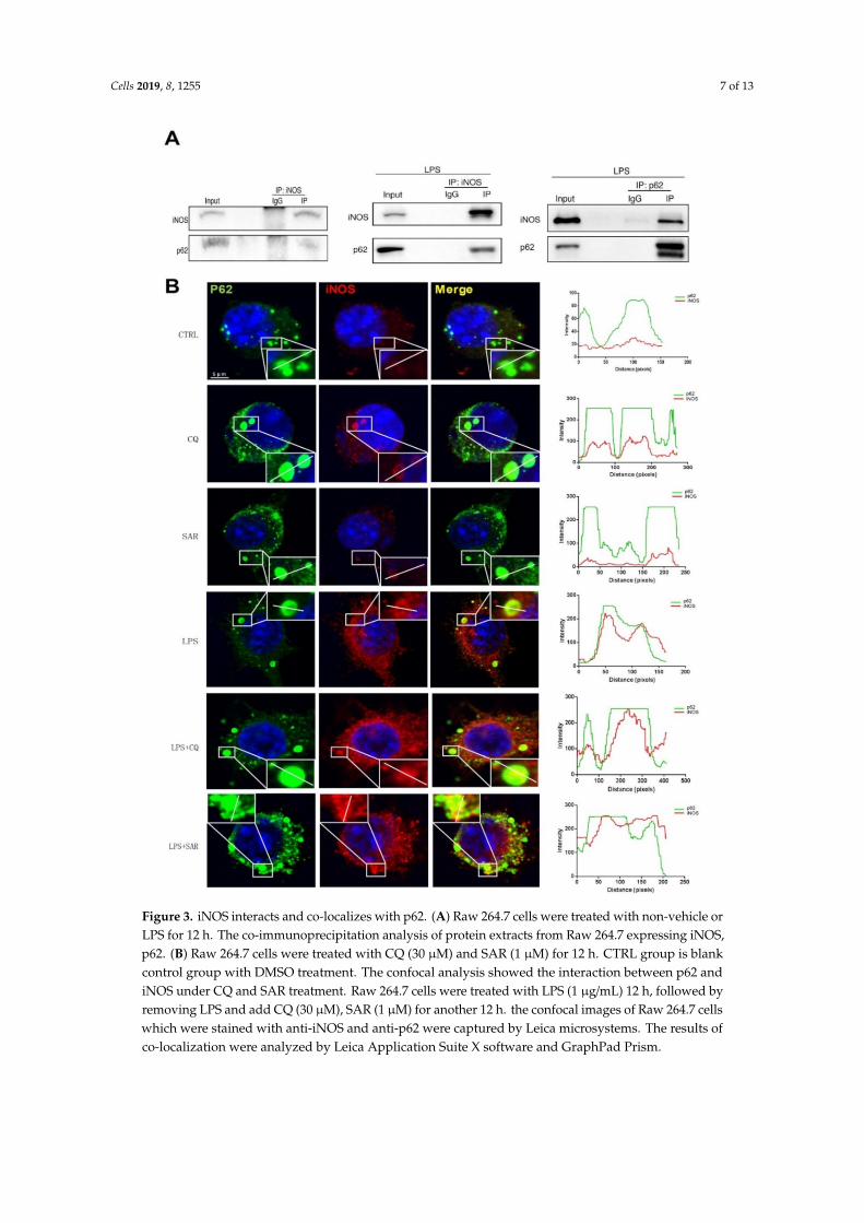
The observation that iNOS displayed similar trend as p62 under basal and inflammation conditionsdrove us to explore the potential relationship between iNOS and p62. We first examined whether iNOSinteracts with p62 by immunoprecipitation. The result showed that iNOS antibody could pull downp62 in Raw 264.7 cells. To make the iNOS enriched obviously, we treated Raw 264.7 cells with LPS andthe amount of p62 being pulled down dramatically increased under LPS-treated conditions. Meanwhile,p62 antibody could also pull down iNOS in Raw 264.7 cells after LPS stimulation (Figure 3A). To furthersupport the interaction between iNOS and p62, the immunofluorescence assay was performed toobserve the intracellular co-localization of p62 and iNOS under different conditions. Under basalcondition, despite the low level of iNOS protein, we could see iNOS forms some puncta-like structurewhich partially co-localized with p62 in Raw 264.7 cells (Figure 3B). Since LPS can induce autophagyand inflammatory response-induced iNOS expression, the fluorescent signals of p62 and iNOS wereboth dramatically increased after LPS treatment. Compared to the LPS-only treatment, cells weretreated with LPS plus CQ or SAR405 displayed stronger iNOS expression, which obviously colocalizedwith p62 puncta (Figure 3B). This result is consistent with the WB data. The co-localization efficiencywas further demonstrated by line profiles. The data indicates that iNOS interacts with autophagyreceptor p62 and forms puncta-like structure with p62 in macrophages.
Cells 2019, 8, 1255 7 of 13
Figure 3. iNOS interacts and co-localizes with p62. (A) Raw 264.7 cells were treated with non-vehicle orLPS for 12 h. The co-immunoprecipitation analysis of protein extracts from Raw 264.7 expressing iNOS,p62. (B) Raw 264.7 cells were treated with CQ (30 µM) and SAR (1 µM) for 12 h. CTRL group is blankcontrol group with DMSO treatment. The confocal analysis showed the interaction between p62 andiNOS under CQ and SAR treatment. Raw 264.7 cells were treated with LPS (1 µg/mL) 12 h, followed byremoving LPS and add CQ (30 µM), SAR (1 µM) for another 12 h. the confocal images of Raw 264.7 cellswhich were stained with anti-iNOS and anti-p62 were captured by Leica microsystems. The results ofco-localization were analyzed by Leica Application Suite X software and GraphPad Prism.
Cells 2019, 8, 1255 8 of 13
3.3. iNOS Partially Colocalizes with Autophagosome Marker LC3 and Lysosome Marker LAMP1
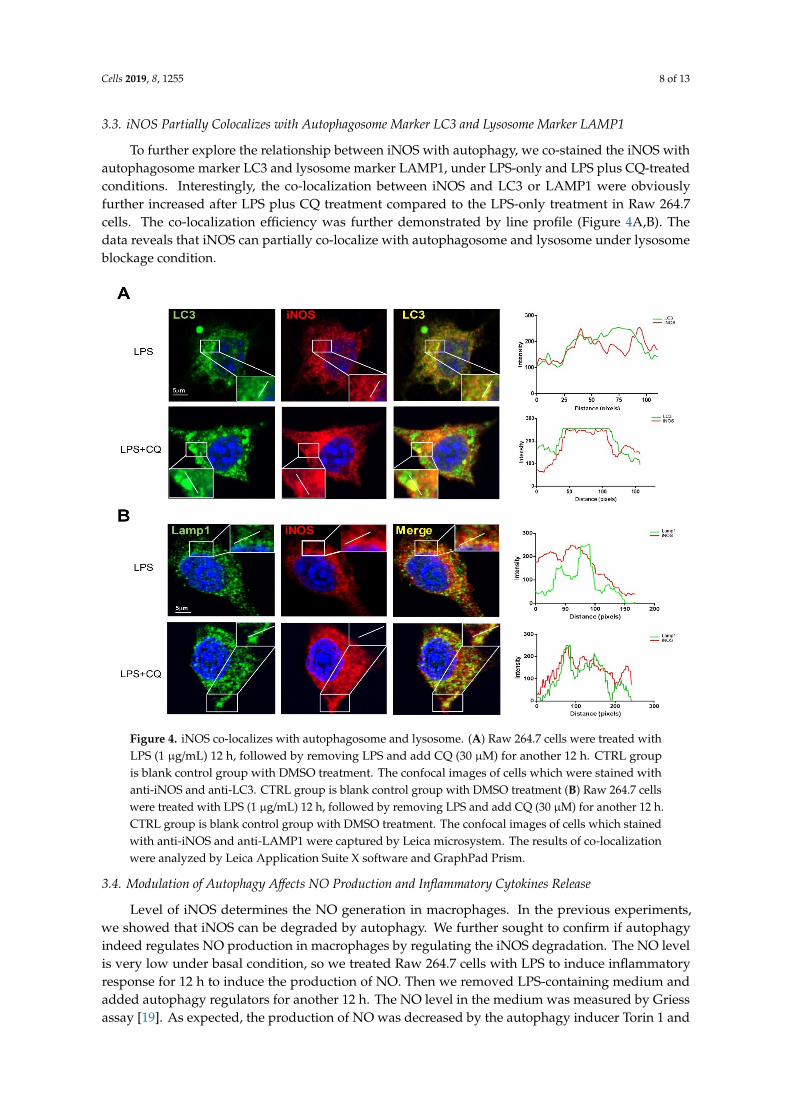
To further explore the relationship between iNOS with autophagy, we co-stained the iNOS withautophagosome marker LC3 and lysosome marker LAMP1, under LPS-only and LPS plus CQ-treatedconditions. Interestingly, the co-localization between iNOS and LC3 or LAMP1 were obviouslyfurther increased after LPS plus CQ treatment compared to the LPS-only treatment in Raw 264.7cells. The co-localization efficiency was further demonstrated by line profile (Figure 4A,B). Thedata reveals that iNOS can partially co-localize with autophagosome and lysosome under lysosomeblockage condition.
Figure 4. iNOS co-localizes with autophagosome and lysosome. (A) Raw 264.7 cells were treated withLPS (1 µg/mL) 12 h, followed by removing LPS and add CQ (30 µM) for another 12 h. CTRL groupis blank control group with DMSO treatment. The confocal images of cells which were stained withanti-iNOS and anti-LC3. CTRL group is blank control group with DMSO treatment (B) Raw 264.7 cellswere treated with LPS (1 µg/mL) 12 h, followed by removing LPS and add CQ (30 µM) for another 12 h.CTRL group is blank control group with DMSO treatment. The confocal images of cells which stainedwith anti-iNOS and anti-LAMP1 were captured by Leica microsystem. The results of co-localizationwere analyzed by Leica Application Suite X software and GraphPad Prism.
3.4. Modulation of Autophagy Affects NO Production and Inflammatory Cytokines Release
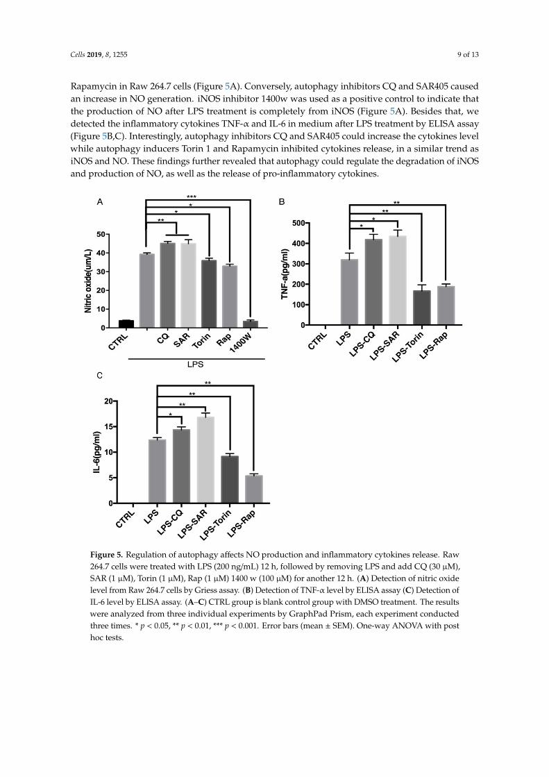
Level of iNOS determines the NO generation in macrophages. In the previous experiments,we showed that iNOS can be degraded by autophagy. We further sought to confirm if autophagyindeed regulates NO production in macrophages by regulating the iNOS degradation. The NO levelis very low under basal condition, so we treated Raw 264.7 cells with LPS to induce inflammatoryresponse for 12 h to induce the production of NO. Then we removed LPS-containing medium andadded autophagy regulators for another 12 h. The NO level in the medium was measured by Griessassay [19]. As expected, the production of NO was decreased by the autophagy inducer Torin 1 and
Cells 2019, 8, 1255 9 of 13
Rapamycin in Raw 264.7 cells (Figure 5A). Conversely, autophagy inhibitors CQ and SAR405 causedan increase in NO generation. iNOS inhibitor 1400w was used as a positive control to indicate thatthe production of NO after LPS treatment is completely from iNOS (Figure 5A). Besides that, wedetected the inflammatory cytokines TNF-α and IL-6 in medium after LPS treatment by ELISA assay(Figure 5B,C). Interestingly, autophagy inhibitors CQ and SAR405 could increase the cytokines levelwhile autophagy inducers Torin 1 and Rapamycin inhibited cytokines release, in a similar trend asiNOS and NO. These findings further revealed that autophagy could regulate the degradation of iNOSand production of NO, as well as the release of pro-inflammatory cytokines.
Figure 5. Regulation of autophagy affects NO production and inflammatory cytokines release. Raw264.7 cells were treated with LPS (200 ng/mL) 12 h, followed by removing LPS and add CQ (30 µM),SAR (1 µM), Torin (1 µM), Rap (1 µM) 1400 w (100 µM) for another 12 h. (A) Detection of nitric oxidelevel from Raw 264.7 cells by Griess assay. (B) Detection of TNF-α level by ELISA assay (C) Detection ofIL-6 level by ELISA assay. (A–C) CTRL group is blank control group with DMSO treatment. The resultswere analyzed from three individual experiments by GraphPad Prism, each experiment conductedthree times. * p < 0.05, ** p < 0.01, *** p < 0.001. Error bars (mean ± SEM). One-way ANOVA with posthoc tests.
Cells 2019, 8, 1255 10 of 13
4. Discussion
Autophagy is a recycling mechanism in eukaryotes and plays a key role in embryonic development,cell defending and survival. Under stress, the cells initiate the autophagy to remove damagedproteins [20], organelles or invading pathogens [21], through the lysosomal pathway and recyclethe degradation products to cope with the adverse environments [22]. Inflammation is a defendingresponse normally activated by microbial pathogen infection or tissue damage [23], which will recruitimmune cells such as neutrophils, macrophages and monocytes to the lesion site [24]. Defects ofautophagy pathway are closely related to the development of inflammatory diseases including Crohn’sdisease [25], rheumatoid arthritis and cancer [26]. In this study, we firstly found that upon autophagyinduction or inhibition, iNOS levels displayed similar change pattern as autophagy substrate p62,indicating iNOS is possible to be the substrate of autophagy. Secondly, further experiments ofimmunoprecipitation and Immunofluorescence assays confirmed the interaction and colocalizationbetween iNOS and p62, especially under LPS-simulated condition. iNOS also partially colocalizes withautophagosome marker LC3 and lysosome marker LAMP1, implicating that iNOS can be degradedvia autophagy. At last, nitric oxide, the product of iNOS, displayed same trend as iNOS duringautophagy induction and inhibition. The results reveal that iNOS regulation via autophagy can be anaspect to explain the autophagy function in inflammation. All the above data demonstrate that iNOSinteracted with autophagy receptor p62 and degraded by autophagy in macrophages.Our studiesrevealed that iNOS can be degraded by autophagy in macrophages and inhibition of autophagyresulted in accumulation of iNOS and its product NO. The results also reveal a novel mechanism bywhich autophagy regulates inflammation.
Autophagy is deeply implicated in inflammatory response in macrophages with diversemechanisms, such as degradation of inflammasome, clearance of pathogens, cytokine secretionand regulation of NF-kappa B signaling [13]. Although there are discoveries revealing that activationof autophagy pathway would affect level of iNOS and inflammatory-related cytokines production inmicroglia cells [27], a clear elaboration of the relationship between autophagy and iNOS still lack [28].Our results revealed that iNOS interacts with autophagy receptor p62 and is degraded by autophagyin macrophages, suggesting that autophagy could regulate the iNOS protein level, as well as the NOproduction during inflammation. This mechanism could partially explain why autophagy regulationhas been linked to iNOS level and NO production level change in a previous study [29]. However,our study does not exclude the potential role of autophagy in the transcriptional regulation of iNOSlevel by other signaling pathways including NF-kappa B. Cycloheximide (CHX) is known to suppressprotein synthesis and has been widely used to analyze the protein degradation rate. We can see therapid degradation of iNOS after CHX treatment, while inhibition of autophagy by SAR405 partiallyblocked iNOS degradation, indicating that iNOS can be degraded via autophagy. Considering thatCHX can inhibit autophagy in the early stage via inhibition of newly synthesized protein needed inautophagy or activation of mTORC1 activity [30], the inhibition effect of SAR405 on iNOS degradationmay be more obvious than it appeared because CHX may already partially impair the autophagicdegradation of iNOS. As one necessary signaling molecules, NO plays an important role in physiologyand pathology [2]. The overexpression of iNOS and NO have been shown to result in various damagein inflammation microenvironment [31]. Our study thus offers a new strategy to control iNOS leveland NO production during inflammatory process via regulating autophagy. Interaction between iNOSand autophagy receptor p62 reveals a selective degradation of iNOS via autophagy. However, there isno evidence to support the direct interaction between iNOS and p62, further study will be performedto understand whether iNOS directly interacts with p62 and the interaction domains, as well as tounderstand whether the interaction can be regulated via post-translational modifications. Furthermore,whether iNOS-p62 interaction will affect the function of p62 worth further examination.
iNOS level keeps at a relative low level and can be strongly up-regulated during inflammation.However, prolonged iNOS upregulation will generate high-level NO which may cause tissues damage,thus efficient degradation of iNOS is a critic mechanism for elimination of inflammation [32]. iNOS
Cells 2019, 8, 1255 11 of 13
has been revealed to be degraded by UPS [33,34] and our data revealed for the first time that iNOS canalso be degraded by autophagy, in a selective manner via the interaction with p62. Compared withthe dramatic accumulation of iNOS when proteasome is inhibited [35], the increase of iNOS level ismoderate during autophagy inhibition. Furthermore, under protein translation inhibition condition,the autophagy inhibition can only partially impair the iNOS degradation. These data suggest thatUPS may play a major role for iNOS degradation while autophagy can be a fine-turn for selectiveiNOS degradation. The collaboration of UPS and autophagy pathway may help eliminate the NOover-production during inflammation via promoting the iNOS degradation.
Author Contributions: J.W., M.-Y.W., and J.-H.L. designed the experiments and analyzed the data. J.-H.L.supervised the whole project. J.W. and M.-Y.W. performed the experiments. J.W., M.-Y.W., and J.-H.L. wrotethe manuscript. J.-H.L., M.-Y.W., H.S., X.C., J.L. and J.T. analyzed the data and reviewed the draft. All authorscontributed to the writing and final approval of the manuscript.
Funding: This study was funded by The Science and Technology Development Fund, Macau SAR (File No.FDCT-0110/2018/A3, FDCT-024-2017-AMJ, FDCT-092-2015-A3), and the grants of China NSFC-31871024, and theUniversity of Macau grants MYRG2016-0019-ICMS-QRCM and MYRG2017-00147-ICMS awarded to JHL.
Acknowledgments: Not applicable.
Conflicts of Interest: The authors declare that they have no conflicts of interest with the contents of this article.
Abbreviations
iNOS, inducible nitric synthase; eNOS, endothelial nitric synthase; nNOS, neuronal nitric synthase; NO, nitricoxide; CQ, Chloroquine; LPS, Lipopolysaccharides; BMDM, bone marrow-derived macrophages.
References
1. Alderton, W.K.; Cooper, C.E.; Knowles, R.G. Nitric oxide synthases: Structure, function and inhibition.Biochem. J. 2001, 357, 593–615. [CrossRef] [PubMed]
2. Aktan, F. iNOS-mediated nitric oxide production and its regulation. Life Sci. 2004, 75, 639–653. [CrossRef][PubMed]
3. Kolodziejski, P.J.; Musial, A.; Koo, J.-S.; Eissa, N.T. Ubiquitination of inducible nitric oxide synthase isrequired for its degradation. Proc. Natl. Acad. Sci. USA 2002, 99, 12315–12320. [CrossRef] [PubMed]
4. Pautz, A.; Art, J.; Hahn, S.; Nowag, S.; Voss, C.; Kleinert, H. Regulation of the expression of inducible nitricoxide synthase. Nitric Oxide 2010, 23, 75–93. [CrossRef] [PubMed]
5. Klionsky, D.J.; Abdelmohsen, K.; Abe, A.; Abedin, M.J.; Abeliovich, H.; Acevedo, A.A.; Adeli, K.; Agholme, L.;Agostinis, P.; Aguirre-Ghiso, J.A.; et al. Guidelines for the use and interpretation of assays for monitoringautophagy. Autophagy 2016, 12, 1–222. [CrossRef] [PubMed]
6. Alirezaei, M.; Kemball, C.C.; Whitton, J.L. Autophagy, inflammation and neurodegenerative disease. Eur. J.Neurosci. 2011, 33, 197–204. [CrossRef]
7. Liu, Z.; Chen, P.; Gao, H.; Gu, Y.; Yang, J.; Peng, H.; Xu, X.; Wang, H.; Yang, M.; Liu, X.; et al. Ubiquitylationof autophagy receptor Optineurin by HACE1 activates selective autophagy for tumor suppression. CancerCell 2014, 26, 106–120. [CrossRef]
8. Xu, Y.I.; Jagannath, C.; Liu, X.D. Toll-like Receptor 4 Is a Sensor for Autophagy Associated with InnateImmunity. Immunity 2007, 27, 135–144. [CrossRef]
9. Paul, W.; Carchman, E.H.; Young, A.C.; Jayashree, R.; Rosengart, M.R.; David, K.; Zuckerbraun, B.S.Lipopolysaccaride induces autophagic signaling in macrophages via a TLR4, heme oxygenase-1 dependentpathway. Autophagy 2011, 7, 6.
10. Stolz, A.; Ernst, A.; Dikic, I. Cargo recognition and trafficking in selective autophagy. Nat. Cell Biol. 2014, 16,495. [CrossRef]
11. Park, S.; Ha, S.D.; Coleman, M.; Meshkibaf, S.; Kim, S.O. p62/SQSTM1 enhances NOD2-mediated signalingand cytokine production through stabilizing NOD2 oligomerization. Plos One 2013, 8, e57138. [CrossRef][PubMed]
Cells 2019, 8, 1255 12 of 13
12. Prabakaran, T.; Bodda, C.; Krapp, C.; Zhang, B.C.; Christensen, M.H.; Sun, C.; Reinert, L.; Cai, Y.; Jensen, S.B.;Skouboe, M.K.; et al. Attenuation of cGAS-STING signaling is mediated by a p62/SQSTM1-dependentautophagy pathway activated by TBK1. EMBO J. 2018, 37, e97858. [CrossRef] [PubMed]
13. Netea-Maier, R.T.; Plantinga, T.S.; van de Veerdonk, F.L.; Smit, J.W.; Netea, M.G. Modulation of inflammationby autophagy: Consequences for human disease. Autophagy 2016, 12, 245–260. [CrossRef]
14. Hol, J.; Otterdal, K.; Breland, U.M.; Stang, E.; Pedersen, T.M.; Hagelsteen, K.; Ranheim, T.; Kasprzycka, M.;Halvorsen, B.; Haraldsen, G.; et al. Statins affect the presentation of endothelial chemokines by targeting tomultivesicular bodies. PLoS ONE 2012, 7, e40673. [CrossRef] [PubMed]
15. Kimura, T.; Takabatake, Y.; Takahashi, A.; Isaka, Y. Chloroquine in cancer therapy: A double-edged sword ofautophagy. Cancer Res. 2013, 73, 3–7. [CrossRef] [PubMed]
16. Mauthe, M.; Orhon, I.; Rocchi, C.; Zhou, X.; Luhr, M.; Hijlkema, K.-J.; Coppes, R.P.; Engedal, N.; Mari, M.;Reggiori, F.; et al. Chloroquine inhibits autophagic flux by decreasing autophagosome-lysosome fusion.Autophagy 2018, 14, 1435–1455. [CrossRef]
17. Ronan, B.; Flamand, O.; Vescovi, L.; Dureuil, C.; Durand, L.; Fassy, F.; Bachelot, M.F.; Lamberton, A.;Mathieu, M.; Bertrand, T.; et al. A highly potent and selective Vps34 inhibitor alters vesicle trafficking andautophagy. Nat. Chem. Biol. 2014, 10, 1013. [CrossRef]
18. Thoreen, C.C.; Sabatini, D.M. Rapamycin inhibits mTORC1, but not completely. Autophagy 2009, 5, 725–726.[CrossRef]
19. Wu, Q.-L.; Buhtoiarov, I.N.; Sondel, P.M.; Rakhmilevich, A.L.; Ranheim, E.A. Tumoricidal effects of activatedmacrophages in a mouse model of chronic lymphocytic leukemia. J. Immunol. 2009, 182, 6771–6778. [CrossRef]
20. Shibutani, S.T.; Saitoh, T.; Nowag, H.; Münz, C.; Yoshimori, T. Autophagy and autophagy-related proteins inthe immune system. Nat. Immunol. 2015, 16, 1014. [CrossRef]
21. Shi, C.-S.; Shenderov, K.; Huang, N.-N.; Kabat, J.; Abu-Asab, M.; Fitzgerald, K.A.; Sher, A.; Kehrl, J.-H.Activation of autophagy by inflammatory signals limits IL-1β production by targeting ubiquitinatedinflammasomes for destruction. Nat. Immunol. 2012, 13, 255. [CrossRef] [PubMed]
22. Doria, A.; Gatto, M.; Punzi, L. Autophagy in human health and disease. New Engl. J. Med. 2013, 368,1845–1846. [PubMed]
23. Licastro, F.; Candore, G.; Lio, D.; Porcellini, E.; Colonna-Romano, G.; Franceschi, C.; Caruso, C. Innateimmunity and inflammation in ageing: A key for understanding age-related diseases. Immun. Ageing 2005,2, 8. [CrossRef] [PubMed]
24. Chung, H.Y.; Cesari, M.; Anton, S.; Marzetti, E.; Giovannini, S.; Seo, A.Y.; Carter, C.; Yu, B.P.; Leeuwenburgh, C.Molecular inflammation: Underpinnings of aging and age-related diseases. Ageing Res. Rev. 2009, 8, 18–30.[CrossRef] [PubMed]
25. Wu, M.-Y.; Song, J.-X.; Wang, S.-F.; Cai, C.-Z.; Li, M.; Lu, J.-H. Selective autophagy: The new player in thefight against neurodegenerative diseases? Brain Res. Bulletin. 2018, 137, 79–90. [CrossRef] [PubMed]
26. Deretic, V.; Saitoh, T.; Akira, S. Autophagy in infection, inflammation and immunity. Nat. Rev. Immunol.2013, 13, 722. [CrossRef] [PubMed]
27. Han, H.-E.; Kim, T.-K.; Son, H.-J.; Park, W.J.; Han, P.-L. Activation of autophagy pathway suppresses theexpression of iNOS, IL6 and cell death of LPS-stimulated microglia cells. Biomol. Ther. 2013, 21, 21. [CrossRef]
28. Hämäläinen, M.; Nieminen, R.; Vuorela, P.; Heinonen, M.; Moilanen, E. Anti-inflammatory effects offlavonoids: Genistein, kaempferol, quercetin, and daidzein inhibit STAT-1 and NF-κB activations, whereasflavone, isorhamnetin, naringenin, and pelargonidin inhibit only NF-κB activation along with their inhibitoryeffect on iNOS expression and NO production in activated macrophages. Mediat. Inflamm. 2007, 2007.
29. Zhu, T.; Yao, Q.; Wang, W.; Yao, H.; Chao, J. iNOS induces vascular endothelial cell migration and apoptosisvia autophagy in ischemia/reperfusion injury. Cell. Physiol. Biochem. 2016, 38, 1575–1588. [CrossRef]
30. Watanabe-Asano, T.; Kuma, A.; Mizushima, N. Cycloheximide inhibits starvation-induced autophagythrough mTORC1 activation. Biochem. Biophys. Res. Commun. 2014, 445, 334–339. [CrossRef]
31. Zhong, Z.; Umemura, A.; Sanchez-Lopez, E.; Liang, S.; Shalapour, S.; Wong, J.; He, F.; Boassa, D.; Perkins, G.;Ali, S.R.; et al. NF-κB restricts inflammasome activation via elimination of damaged mitochondria. Cell 2016,164, 896–910. [CrossRef] [PubMed]
32. Golde, S.; Coles, A.; Lindquist, J.A.; Compston, A. Decreased iNOS synthesis mediates dexamethasone-inducedprotection of neurons from inflammatory injury in vitro. Eur. J. Neurosci. 2003, 18, 2527–2537. [CrossRef] [PubMed]
Cells 2019, 8, 1255 13 of 13
33. Wang, T.; Luo, S.; Qin, H.; Xia, Y. Hsp90 inhibition renders iNOS aggregation and the clearance of iNOSaggregates by proteasomes requires SPSB2. Free Radic. Biol. Med. 2018, 117, 90–98. [CrossRef] [PubMed]
34. Kovács, J. Effect of cycloheximide on induced autophagy in epithelial cells of the seminal vesicle of mice.Acta Morphol. Acad. Sci. Hung. 1974, 22, 69. [PubMed]
35. Musial, A.; Eissa, N.T. Inducible nitric-oxide synthase is regulated by the proteasome degradation pathway.J. Biol. Chem. 2001, 276, 24268–24273. [CrossRef] [PubMed]
© 2019 by the authors. Licensee MDPI, Basel, Switzerland. This article is an open accessarticle distributed under the terms and conditions of the Creative Commons Attribution(CC BY) license (http://creativecommons.org/licenses/by/4.0/).


