Embed Size (px)
Citation preview

of November 28, 2015.This information is current as into CTL Escape
InsightsTwo Immunogenic SIV Epitopes and Structures of Mamu-A*01 Complexed withRhesus Macaque MHC Class I: Crystal First Glimpse of the Peptide Presentation by
and George F. GaoGao, Lili Zong, Abdul Hamid Khan, John I. Bell, Zihe Rao Fuliang Chu, Zhiyong Lou, Yu Wai Chen, Yiwei Liu, Bin
http://www.jimmunol.org/content/178/2/944doi: 10.4049/jimmunol.178.2.944
2007; 178:944-952; ;J Immunol
Referenceshttp://www.jimmunol.org/content/178/2/944.full#ref-list-1
, 25 of which you can access for free at: cites 60 articlesThis article
Subscriptionshttp://jimmunol.org/subscriptions
is online at: The Journal of ImmunologyInformation about subscribing to
Permissionshttp://www.aai.org/ji/copyright.htmlSubmit copyright permission requests at:
Email Alertshttp://jimmunol.org/cgi/alerts/etocReceive free email-alerts when new articles cite this article. Sign up at:
Print ISSN: 0022-1767 Online ISSN: 1550-6606. Immunologists All rights reserved.Copyright © 2007 by The American Association of9650 Rockville Pike, Bethesda, MD 20814-3994.The American Association of Immunologists, Inc.,
is published twice each month byThe Journal of Immunology
by guest on Novem
ber 28, 2015http://w
ww
.jimm
unol.org/D
ownloaded from
by guest on N
ovember 28, 2015
http://ww
w.jim
munol.org/
Dow
nloaded from
First Glimpse of the Peptide Presentation by Rhesus MacaqueMHC Class I: Crystal Structures of Mamu-A*01 Complexedwith Two Immunogenic SIV Epitopes and Insights into CTLEscape1
Fuliang Chu,*� Zhiyong Lou,† Yu Wai Chen,* Yiwei Liu,† Bin Gao,* Lili Zong,*‡§
Abdul Hamid Khan,*� John I. Bell,¶ Zihe Rao,† and George F. Gao2*¶
The infection of rhesus macaques (Macaca mulatta) by the SIV is the best animal model for studying HIV infection and for AIDSvaccine development. A prevalent MHC class I allele, Mamu-A*01, is known to correlate with containment of SIV, which has beenextensively explored in studies of CTL-based vaccination concepts. We determined the crystal structures of Mamu-A*01 com-plexed with two immunodominant SIV epitopes: the nonamer CM9 of group-specific Ag (Gag, 181–189; CTPYDINQM) and theoctamer TL8 of transcription activator (Tat, 28–35; TTPESANL). The overall structures of the two Mamu-A*01 complexes aresimilar to other MHC class I molecules. Both structures confirm the presence of an absolutely conserved proline anchor residuein the P3 position of the Ag, bound to a D pocket of the Mamu-A*01 H chain with optimal surface complementarity. Like otherMHC/peptide complex structures, the P2 and C-terminal residues of the epitopes are also important for anchoring to the MHCmolecule, whereas the middle residues form an arch and their side chains are directed into solvent. These two structures revealdetails of how Mamu-A*01 interacts with two well-studied epitopes at the atomic level. We discuss the structural basis of CTLescape, based on molecular models made possible by these two structures. The results we present in this study are most relevantfor the rational design of Mamu-A*01-restricted CTL epitopes with improved binding, as a step toward development of AIDSvaccines. The Journal of Immunology, 2007, 178: 944–952.
H uman immunodeficiency virus is the causative agent ofAIDS, which currently poses one of the greatest globalthreats to human health. According to the World Health
Organization, the population infected with HIV rose in 2004 to itshighest level ever: an estimated 36–44 million (World Health Or-ganization, http://www.unaids.org/wad2004/report.html). This factunderscores the urgent need for an effective AIDS vaccine. Despitevigorous research efforts, preventive (sterilizing) immunizationbased on raising neutralizing Abs against HIV components has
proved fruitless (1–3). In contrast, it is known that the host CTLresponses provide powerful defense to contain the virus postinfec-tion (4, 5), and CTL-based immunization is currently the mostpromising approach toward vaccine development (6). At present,the most relevant animal model for investigating HIV pathology isthe SIV infection of the nonhuman primate rhesus monkeys, withboth the viruses and the infected hosts being genetically close (7–10). SIV and HIV have high nucleotide sequence homology, asimilar tropism for CD4� T cells, and induce similar pathologies(5, 7, 9). Moreover, immune system components of rhesus mon-keys and humans are highly conserved. For example both haveMHC and TCR genes that present the same regions of lentiviralGag and Env proteins (7, 11–14). At the molecular level, it hasbeen reported that the binding motif of the peptide to MHC classI molecules between human and rhesus monkeys is conserved ingeneral (15), although Mamu-A*01 shows a special preference forP3 proline as anchor residue, which is not a case for any otherknown human HLAs (15). For all of these reasons, the SIV/rhesusmodel has been extensively explored as an invaluable tool for vac-cine evaluation (16, 17).
Among the host CTL defense system of rhesus monkey, an al-lele of class I MHC, Mamu-A*01, has been associated with slowprogression to AIDS-like syndrome (18–21), and animals that ex-pressed this allele show the best control over SIV replication (22).Systematic studies were performed to discover its epitopes andtheir specific CTLs (7, 23, 24). Two well-characterized epitopes,“TL8” from the SIV transcriptional transactivator (Tat) protein(residues 28–35, TTPESANL; note that in some studies, the“SL8,” STPESANL, sequence was used) and “CM9” from the SIVgroup-specific Ag (Gag) protein (residues 181–189, CTPY
*Center for Molecular Immunology, Institute of Microbiology, Chinese Academy ofSciences, Beijing, People’s Republic of China; †Laboratory of Structural Biology,Tsinghua University, Beijing, People’s Republic of China; ‡Department of Obstetricsand Gynaecology, Zhujiang Hospital, Nanfang Medical University, Guangzhou, Peo-ple’s Republic of China; §Nuffield Department of Obstetrics and Gynaecology, Uni-versity of Oxford, Oxford, Oxfordshire, United Kingdom; ¶Nuffield Department ofClinical Medicine, University of Oxford, Oxford, Oxfordshire, United Kingdom; and�Graduate School, Chinese Academy of Sciences, Beijing, People’s Republic of China
Received for publication November 17, 2005. Accepted for publication October27, 2006.
The costs of publication of this article were defrayed in part by the payment of pagecharges. This article must therefore be hereby marked advertisement in accordancewith 18 U.S.C. Section 1734 solely to indicate this fact.1 This work was supported by a grant from Chinese Academy of Sciences KnowledgeInnovation Project Grant no. KSCX2-SW-227, a grant from the National Basic Re-search Program (Project 973) of the Ministry of Science and Technology of thePeople’s Republic of China (Grant no. 2006CB504204), and grants from NationalNatural Science Foundation of China (Grant nos. 30440020 and 30671903). G.F.G. isa distinguished young investigator of the Natural Science Foundation of China (Grantno. 30525010).2 Address correspondence and reprint requests to Dr. George F. Gao, Center forMolecular Immunology, Institute of Microbiology, Chinese Academy of Sciences, 13Beiyitiao, Zhongguancun, Beijing 100080, People’s Republic of China. E-mail ad-dress: [email protected]
Copyright © 2007 by The American Association of Immunologists, Inc. 0022-1767/07/$2.00
The Journal of Immunology
www.jimmunol.org
by guest on Novem
ber 28, 2015http://w
ww
.jimm
unol.org/D
ownloaded from

DINQM) have been found to be immunodominant, and CTLs spe-cific for these two epitopes are detected in the acute stage of SIVinfection (7, 25–27). Following the initial peak expression, TL8-specific CTLs declined precipitously after the acute phase, whereasCM9-specific CTLs remained at a constant level throughout thechronic phase of infection (25, 27). Unfortunately, these powerfulhost defense mechanisms also impose strong selective pressure onthe evolution of the viruses (27–29). TL8 escape variants weredetectable during the very early stage of infection, whereas CM9escape variants appeared much later (27, 30). Eventually, the mu-tants will dominate the virus population and result in the progres-sion to AIDS, as reported in both rhesus monkey and human hosts(27). Evasion of the host immune system via CTL escape wasfound to be a major obstacle in CTL-based vaccine strategies (27,31–33).
The availability of the SIV/rhesus model makes it possible toperform systematic studies on CTL escape. A number of escapemutants of various Mamu-A*01-restricted epitopes, includingCM9 and TL8, were documented (20, 26). As a step toward de-lineating the molecular basis of CTL escape, we undertook struc-tural studies to reveal how Mamu-A*01 binds these two very dif-ferent SIV peptides for presentation to the TCR. Previously,several studies have probed the interactions and structures of pep-tide and class I MHC molecules (pMHC)3 from human and mice(34–38). This is the first nonhuman primate pMHC structure eversolved, and the results show that the nonamer and octamer peptidesbind to the Mamu-A*01 in a similar way with P3 as an anchorresidue. The results we describe in this study provide a structuralbasis for T cell immune escape and have implications in the ra-tional design of CTL-based vaccines against AIDS.
Materials and MethodsPeptide synthesis
The Gag (181–189) CTPYDINQM peptide (denoted CM9) and Tat (28–35) TTPESANL peptide (denoted TL8) were synthesized and purified by
HPLC reverse phase chromatography (SciLight Biotechnology). The pu-rities of the peptides are �90% as assessed by HPLC (data not shown).
Crystallization and data collection
The overexpression and purification of Mamu-A*01 and �2-microglobulin(�2m) and the crystallization of the Mamu-A*01/CM9 complex has beenreported previously (39). The Mamu-A*01/TL8 complex was prepared bythe same procedures. Crystals of the Mamu-A*01/CM9 and Mamu-A*01/TL8 complexes were obtained under the optimized condition (0.1 M Tris(pH 8.5), 1.8 M ammonium sulfate) in 8 wk. Data for the CM9 and TL8crystals were collected on a Rigaku R-AXIS IV�� image plate with aRigaku MM007 rotation Cu K� anode home x-ray generator at 40 kV and20 mA (� � 1.5418Å). The crystals were soaked for several minutes in thereservoir solution supplemented with 20% glycerol as a cryoprotectant andthen flash cooled directly in liquid nitrogen. Diffraction data collected at100 K to 2.8 Å were processed using the program HKL2000 (40) (Table I).
3 Abbreviations used in this paper: pMHC, peptide and class I MHC molecule com-plex; �2m, �2-microglobulin; r.m.s., root-mean-squared.
FIGURE 1. Overview of Mamu-A*01 structure with peptide-CM9. TheH chain, composed of the �1, �2, and �3 domains, is shown in ribbonrepresentation and colored purple. The L chain (�2m) is shown in ribbonrepresentation and colored light blue; the peptide is shown in stick modelcolored by atom types (C, yellow; N, blue; O, red; S, green).
Table I. Data collection and refinement statistics
CM9 TL8
Data processingSpace group I422 I422Cell parameters (A, °) a�b�183.7, c�155.2 a�b�182.1, c�156.7Resolution range (Å) 50.0–2.8 (2.9–2.8)a 50.0–2.8 (2.9–2.8)a
Total reflections 62116 211955Unique reflections 28622 31890Completeness (%) 87.1 (75.6)a 97.5 (96.3)a
Rmerge (%)b 12.9 (56.3)a 15.4 (62.7)a
I/� 7.0 (1.3)a 6.3 (2.3)a
RefinementR factor (%)c 22.8 21.7Rfree (%)c 27.8 25.7r.m.s. deviation
Bonds (Å) 0.015 0.007Angles (°) 2.001 1.411
Average B factor 49.3 38.2Number of all nonhydrogen atoms 6328 6296Number of solvent atoms Ramachandran plotd 343 309Most favored (%) 89.3 90.2Disallowed (%) 0.0 0.0
a Numbers in parentheses correspond to the highest resolution shell. r.m.s.d., Root-mean-square deviations from idealgeometry.
b Rmerge � �h�I �Iih��Ih��/�h�I �Ih�, where �Ih� is the mean intensity of the observations Iih of reflection h.c R factor � � (��Fobs��Fcalc��)/��Fobs�; Rfree is the R factor for a subset (5%) of reflections that was selected prior to
refinement calculations and not included in the refinement.d Ramachandran plots were calculated using the program PROCHECK.
945The Journal of Immunology
by guest on Novem
ber 28, 2015http://w
ww
.jimm
unol.org/D
ownloaded from

Structure determination and refinement
The structures of the Mamu-A*01/CM9 and Mamu-A*01/TL8 complexesbelong to the I422 space group. The structure of Mamu-A*01/CM9 wassolved by molecular replacement using the HLA-B*5301 molecule (Pro-tein Data Bank code: 1A1M; with the peptide excluded) as a search model,using the program CNS (38, 41). Two clear solutions in both the rotationand translation functions correspond to the two molecules in the asymmet-ric unit. Residues that differ between Mamu-A*01 and the search modelwere manually rebuilt in the program O (42) under the guidance of Fo-Fcand 2Fo-Fc electron density maps. After refinement of the model with theCNS program using simulated annealing, energy minimization, restrainedindividual B factors, and the addition of 343 water molecules, the respec-tive working R factor and Rfree dropped from 0.45 and 0.42 to 0.23 and0.28 for all data from 50 to 2.8 Å. The course of refinement was monitoredby calculating Rfree based on a subset containing 5% of the total number ofunique reflections. The final model of Mamu-A*01 in complex with CM9was subsequently used to solve the structure of the Mamu-A*01/TL8 com-plex by molecular replacement. After the same refinement steps and theaddition of 309 water molecules, the working R factor and Rfree droppedfrom 0.27 and 0.31 to 0.22 and 0.26. The coordinate errors estimated byLuzzati plot in CNS (41), and for the Mamu-A*01/CM9 and the Mamu-
A*01/TL8 complex structures are 0.41Å and 0.35 Å, respectively. Theaverage real-space fit values, calculated by the O program (42), for theMamu-A*01/CM9 and the Mamu-A*01/TL8 complex structures are 0.95and 0.94, respectively. Model geometries were verified using the programPROCHECK (43). The atomic coordinates of these two crystal structureshave been deposited in the Protein Data Bank with accession nos. 1ZLNand 1ZVS.
ResultsOverall structures of Mamu-A*01/peptide complexes
Both of the complex crystal structures contain residues 1 to 276 ofthe Mamu-A*01 H chain, residues 1 to 99 of human �2m, and SIVpeptides (CM9 or TL8). The overall structure of Mamu-A*01 issimilar to other class I MHC molecules, with a H chain consistingof the characteristic �1/�2 and �3 domains, and the �2m chain(Fig. 1). Mamu-A*01 shares significant sequence homology withother MHC molecule, including �85% sequence identities amongprimates and 70% with mice (Fig. 2). We compared the structureof the Mamu-A*01/CM9 complex with representative structures of
FIGURE 2. Structure-based sequence alignment of Mamu-A*01 and 11 different types of MHC class I molecules, including Mamu-A*02, HLA-A*0101,HLA-A*0201, HLA-A*1101, HLA-A*2402, HLA-B*5301, HLA-Cw4, Patr-A*0101, Patr-A*0201, H-2Kb, and H-2Kd. Black arrows above the alignmentindicate �-strands; cylinders denote �-helices. Residues highlighted in red are absolutely conserved, whereas those highlighted in yellow are highly (�80%)conserved. Green numbers denote residues that form disulfide-bonds. The alignment was generated using the program Clustal X (56) and drawn withESPript (57).
946 STRUCTURE OF RHESUS MACAQUE PEPTIDE-Mamu-A*01 COMPLEX
by guest on Novem
ber 28, 2015http://w
ww
.jimm
unol.org/D
ownloaded from

human and mouse origins. The root-mean-squared (r.m.s.) devia-tions between Mamu-A*01/CM9 and other class I MHC moleculesfor superposition over all C� atoms are below 1.6 Å. In the twocomplex structures reported in this study, the respective Mamu-A*01 molecules have a r.m.s. deviation of 0.58 Å for all C� atoms,suggesting that the two Mamu-A*01 molecules are identicalwithin the limits of experimental error. Detailed interaction be-tween the bound peptides and the Mamu-A*01 H chain are listedin Table II.
Conformations of the bound peptides
The electron densities for the bound peptides CM9 and TL8 arewell-defined inside the peptide-binding grooves of Mamu-A*01(Fig. 3, A and B). The side chains of the central residues of bothpeptides are directed into solvent and make few contacts with res-idues in the �1 and �2 helical domains of the Mamu-A*01 H chain(Fig. 3, A and B). Both peptides make extensive polar and nonpolarcontacts with residues of Mamu-A*01 (Table II).
By superimposing the binding domains of these complexes, wefound that the TL8 and CM9 peptides adopt similar extended con-formations with the P2, P3, and C-terminal residues in near-iden-tical positions to serve as anchor residues to the Mamu-A*01-binding groove (Fig. 4A). In the central region of CM9, from P4 toP6, the backbone of the peptide arches out of the binding groove.
The TL8 peptide is one residue shorter and the effects of archingof its central residues (P4 and P5) are not as pronounced asthose of CM9. To accommodate an extra residue, the P4 and P5residues of CM9 follow a small zigzag across the width of thebinding groove.
The P3 anchor residue, the C-terminal residues, and P2 residueare totally buried. The P4, P5, and P8 residues of CM9 and P4, P5,and P7 residues of TL8 remain solvent accessible after binding,with relative solvent accessibilities varying from �40% to 80%(Table III).
P3 anchor residue
It has been known for some time that proline in the P3 position isan absolutely conserved feature shared by all Mamu-A*01-re-stricted epitopes reported to date (7, 15, 23, 24). The two Mamu-A*01/peptide complex models provide a structural explanation forthis observation. The peptide-binding groove of Mamu-A*01 con-tains a well-defined hydrophobic pocket (D pocket) that is optimalfor binding the P3 proline side chain, constituted by the side chainsof residues Tyr9, Arg97, and Tyr159; and the allele-specific residuesVal99 and Met156 (Fig. 5). In addition, the pyrrolidine ring of theP3 proline stacks against the aromatic ring of Tyr159, with a nearestdistance of 2.9 Å, and offers extra stability (Fig. 5). These dataargue for a stringent stereochemical requirement for proline in
Table II. Hydrogen bonds and van der Waals interactions between SIV peptides and Mamu-A*01 residues
Complex
PeptideHydrogen Bond
Partner
Van der Walls Contact ResiduesResidue Atom Residue Atom
CM9
P1-Cys O Tyr159 OH Met5, Tyr7, Tyr59, Arg62, Glu63, Gln163,Trp167, Tyr171N Tyr7 OH
N Tyr171 OHP2-Thr N Glu63 OE2 Tyr7, Tyr9, Met45, Glu63, Asn66, Met67
N Glu63 OE1OG1 Glu63 OE1OG1 Asn66 ND2O Glu63 OE1O Asn66 ND2
P3-Pro N Tyr159 OH Tyr9, Arg97, Val99, Met156, Tyr159
P4-Tyr OH Gln163 NE2P5-Asp OD1 Ser155 OGP6-Ile Thr69, Glu70
P7-Asn Val152, Ser155
P8-Gln O Trp147 NE1P9-Met O Tyr84 OH Trp147, Tyr123, Tyr84, Leu81, Tyr116, Tyr118
W348 Arg97 NH1Glu70 OE2P7-Asn OP7-Asn N
TL8 P1-Thr N Tyr171 OH Met5, Tyr7, Tyr59, Arg62, Glu63, Gln163,Trp167, Tyr171N Tyr7 OH
OG1 Trp167 NE1O Tyr159 OH
P2-Thr N Glu63 OE2 Tyr7, Tyr9, Met45, Glu63, Asn66, Met67
OG1 Asn66 ND2OG1 Glu63 OE1
P3-Pro Tyr9, Arg97, Val99, Met156, Tyr159
P4-GluP5-Ser OG Asn73 ND2
OG Glu70 OE1P6-Ala Val152, Met156
P7-Asn O Trp147 NE1P8-Leu O Tyr84 OH Trp147, Tyr123, Tyr84, Leu81, Tyr116, Tyr118
O Thr143 OG1N Asn77 OD1
W24 P8-Leu OThr80 OG1Asn77 OD1
947The Journal of Immunology
by guest on Novem
ber 28, 2015http://w
ww
.jimm
unol.org/D
ownloaded from

position P3: a more bulky residue cannot be accommodated,whereas a smaller residue cannot form the optimal hydrophobicinteractions required for binding.
In most MHC class I molecules, tyrosine in position 99 is con-served, except in H-2Kb, where its counterpart is serine, which can
form hydrogen bonds. However, in Mamu-A*01 and H-2Dd, theresidues at position 99 are valine and alanine, respectively. Theside chains of valine and alanine are relatively small, which pro-vide more space to accommodate the P3 proline side chain of thepeptide. Furthermore, valine and alanine cannot form hydrogenbonds. As in H-2Ld, in pocket B, position 63 is alanine, whichcannot form any hydrogen bonds to P2-Pro. Thus, we postulatethat it is the nonpolar residues at position 99 that ensure the P3proline to packs in the right place complementary to the pocket(34, 38, 44, 45). Similarly, in H-2Dd, which also has an absolutepreference for proline as the third residue of the epitope (34), it hasalanine, another residue with a small hydrophobic side chain, atposition 99.
FIGURE 4. Comparisons of SIV and HIV peptide antigenic conforma-tions. Side view superpositions of the different peptides are shown here tocompare the position of the anchor residues that tether the peptide to theMHC surface, and the exposed residues that are most likely to be involvedin TCR binding. The peptides are shown as stick models colored by atomtypes (N, blue; O, red; S, green) with carbon atoms colored differently foreach peptide: yellow for TL8, purple for CM9, and green for TL9. A,Superposition of the CM9 and TL8 peptides. B, Superposition of the CM9,with TL9 (PDB ID, 1A1M) peptides.
FIGURE 5. Binding of TL8 peptide with emphasis of Pocket D. Themolecule surface of Mamu-A*01 is shown in gray, Pocket D in green, andthe peptide is represented as stick model colored in yellow. The residues ofTL8 peptide and the important residues forming the Pocket D in Mamu-A*01 are labeled. Pocket A–F are labeled in red letters. The P3 proline isthe core of hydrophobic interactions. The pyrrolidine ring of P3-Pro isparallel to the phenyl ring of Tyr159. Species-specific residues Val99 andMet156 are shown in the pocket. Tyr9 and Val99 are forming the bottom ofPocket D, while Arg97 are forming the other side. The figure was generatedby Pymol (http://www.pymol.org/).
Table III. Accessible surface area (ASA) calculations of bound SIVepitopesa
Free Peptide ASA Bound Peptide ASA
Absolute, Å2 Relative, % Absolute, Å2 Relative, %
CM9P1-Cys 178 133 17 12P2-Thr 109 78 1 0P3-Pro 105 77 3 2P4-Tyr 183 86 115 54P5-Asp 99 70 56 40P6-Ile 155 89 40 23P7-Asn 118 82 40 28P8-Gln 173 97 104 58P9-Met 250 129 8 4
TL8P1-Thr 183 131 20 14P2-Thr 120 86 3 2P3-Pro 118 87 1 1P4-Glu 175 101 132 77P5-Ser 111 96 45 39P6-Ala 92 86 27 25P7-Asn 143 100 75 52P8-Leu 237 133 2 1
a Accessible surface areas were calculated with the program NACCESS using asolvent probe of radius 1.4 Å (http://wolf.bms.umist.ac.uk/naccess/). Relative acces-sibility of each residue was calculated as the percentage of accessibility compared tothe accessibility of that residue in an extended Ala-Xaa-Ala tripeptide conformation (60).
FIGURE 3. Electron densities of bound SIV peptides. Shown here arefinal 2Fo-Fc-stimulated annealing omit maps contoured at 1.0 �. Only thedensities corresponding to the peptides are drawn. The peptides are shownas stick models colored by atom types (C, yellow; N, blue; O, red; S,green), and all peptide residues are labeled. The peptides are shown in stickrepresentation and colored yellow. Parts of the �1 helix and � sheet ofMamu-A*01 are shown in ribbon representation and colored purple. A,CM9 peptide; B, TL8 peptide. Figs. 3, 4, and 6 were generated by Bob-script (58) and Raster3D (59).
948 STRUCTURE OF RHESUS MACAQUE PEPTIDE-Mamu-A*01 COMPLEX
by guest on Novem
ber 28, 2015http://w
ww
.jimm
unol.org/D
ownloaded from
C terminus anchor residue
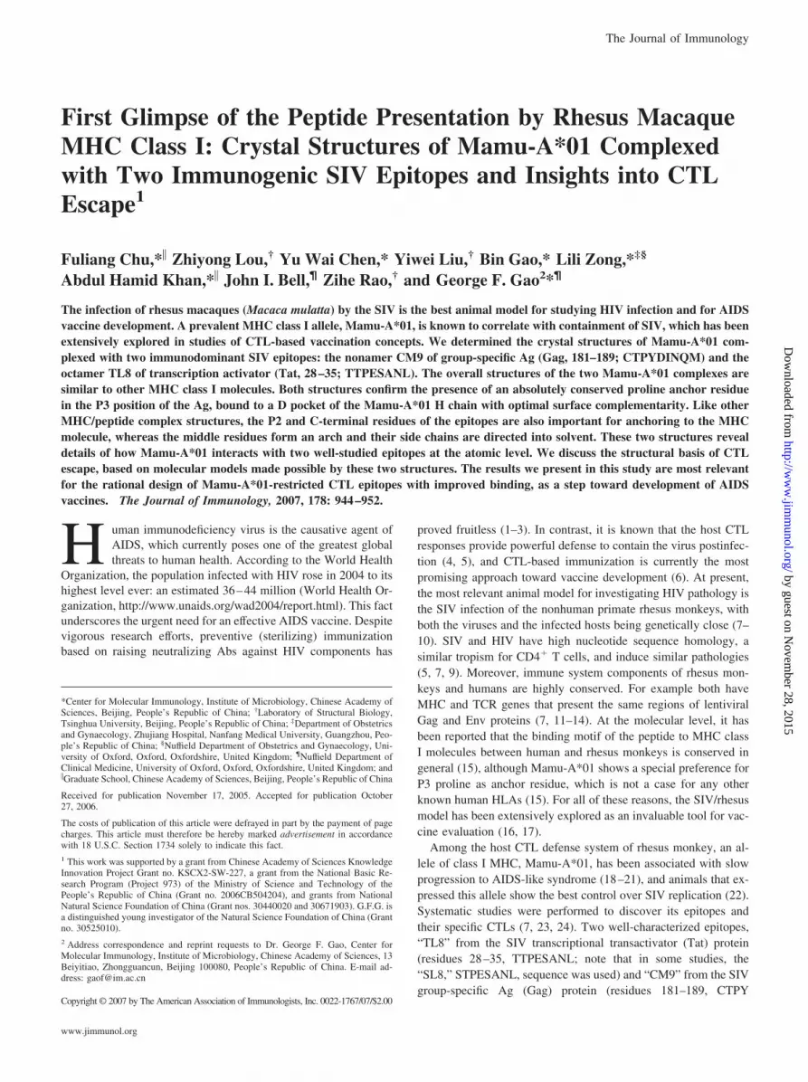
The F pocket of Mamu-A*01 is deep and hydrophobic, formed bythe absolutely conserved residues Tyr84, Tyr123, Thr143, Lys146,and Trp147, (Figs. 2 and 6, A and B), as well as the less conservedresidues Asn77, Thr80, Leu95, and Tyr116. The carboxylate group ofthe CM9 C terminus makes two hydrogen bonds with Tyr84 andThr143 and extends deeply into the hydrophobic pocket togetherwith the side chain of P9-Met, which firmly anchors the C terminusof the peptide (Fig. 6A). In contrast to CM9, the TL8 peptidebinds to Mamu-A*01 with a C-terminal P8-Leu that has its sidechain slightly reoriented from the CM9 P9-Met side chain (Fig.6, A and B).
Compared with the human and mouse MHC complex structures,the residues forming the F pocket of Mamu-A*01, Tyr84, Thr143,and Asp77 are rather conserved. These residues usually form hy-drogen bonds to the C-terminal residues. There are water mole-cule-mediated hydrogen bonds with Asp77 and Thr80 (Table II andFig. 6, A and B). The pattern of C-terminal hydrogen bonds in theMamu-A*01/peptide complexes is very similar to those of humanand mouse complexes. Like most class I MHC molecules, the Fpocket of Mamu-A*01 can accept aliphatic P-omega side chains(Leu, Met, Ile, and Val) (46).
Unique P2 residue
The B pockets of MHC class I molecules including Mamu-A*01,HLA-A*0101, and HLA-B*5801 are constituted by a conservedpeptide-binding motif with Met/Thr at residue 45, Asn at residue66, and Met at residue 67. There is a strong preference for a smallresidue, either threonine, serine, or alanine, in the P2 position ofthe peptide (15, 24). From the two Mamu-A*01/peptide structuresreported in this study, the P2-Thr residues are found to be in con-tact with these three residues’ conserved peptide-binding motifresidues (Table II). In addition, two bulky tyrosine residues Tyr7
and Tyr9 on the bottom of the pocket protrude upwards and definea small volume to accommodate the P2 side chain. In both of theMamu-A*01 complex structures, we found that the side chains ofP2-Thr form hydrogen bonds with Glu63 and Asn66 (Fig. 6, C andD, and Table II). Presumably serine in position P2 can be accom-modated in the way as a threonine, thus maintaining these twohydrogen bonds.
Buried P1 residue
In both complex structures, the N-terminal (P1) residue of eachpeptide is buried in pocket A with a reduction of �160 Å2 inaccessible surface area (Table III). The main-chain atoms of thisresidue contribute to the overall binding of the peptides. Pocket Ais formed by the residues Met5, Tyr7, Tyr167, and Tyr171, which arehighly conserved in most class I molecules (Fig. 2). The aminogroup of the P1 residue of CM9 forms hydrogen bonds with bothTyr7 and Tyr171 of Mamu-A*01, whereas the carbonyl group ofthis residue forms a hydrogen bond with Tyr159. The P1 residue ofTL8 is able to adopt a similar conformation and interacts withthese same tyrosine residues (Fig. 6, C and D). Hydrogen bondingwith the N-terminal residue of the bound epitope is a recurringtheme among class I MHC molecules (24, 37). Therefore, we con-clude that the hydrogen bonding pattern of the N-terminal residueof the bound peptide, similar to the C-terminal residues, is rela-tively conserved.
Interactions of the middle residues in the peptide and the rolesof water molecules
In CM9, the P6-Ile and P7-Glu residues show no interaction withthe floor of the binding pocket (the �-sheet) and they are largelyburied, having relative solvent accessibilities of 23 and 28%, re-
FIGURE 6. Network of hydrogen bonds at the C- and N-terminal ends ofthe binding groove. The backbone of Mamu-A*01 is represented as purpleribbon; the residues of the peptides is shown as golden stick, and relevantresidues of Mamu-A*01 are shown as stick models colored by atom types (N,blue; O, red; S, green) with carbon atoms of peptides in yellow and those ofMamu-A*01 in green. Hydrogen bonds are illustrated as dotted lines. Watermolecules are shown as red spheres indicated by red letter W. A, Pocket F forCM9. Only P7, P8, P9, and one water molecule (W348) are shown. B, PocketF for TL8. Only P7, P8, and one water molecule (W24) are shown. C, PocketA for CM9. Only P1-Cys, P2-Thr, and P3-Pro are shown. D, Pocket A forTL8. Only P1-Cys, P2-Thr, and P3-Pro are shown.
949The Journal of Immunology
by guest on Novem
ber 28, 2015http://w
ww
.jimm
unol.org/D
ownloaded from

spectively (Table III). In some human pMHC complexes (of HLA-A*1101), the side chains of residues in the central P6 (of nonamer)or P7 (of decamer) positions are directed into the binding pocketand contribute significant secondary interactions to anchor the pep-tide (47). With Mamu-A*01, however, the equivalent residues (P6and P7 of CM9; P6 of TL8) adopt different conformations. Insteadof pointing downwards, their side chains extend parallel to the�-sheet floor of the binding groove and make van der Waals in-teractions with the �1 and �2 helices of Mamu-A*01 (Fig. 3 andTable II).
In the Mamu-A*01/CM9 structure, water molecule W348 me-diates four important hydrogen bonds between the Mamu-A*01residues Glu70 and Arg97 and the CM9 residue P7-Gln within theC and E pockets (Table II and Fig. 6A). In the Mamu-A*01/TL8complex, water molecules W24 form three hydrogen bonds be-tween the TL8 residue P8-Leu, and the Mamu-A*01 residuesAsn77 and Thr80 within the F pocket (Table II and Fig. 6B). Thelocations of the two water molecules are not conserved betweenthe two structures.
DiscussionThe structures reported in this study provide detailed informationregarding peptide presentation by the rhesus monkey Mamu-A*01,an important MHC allele that has been used in many systematicinvestigations into SIV infection. These are the first-ever solvednonhuman primate pMHC complex structures and allow manypublished functional data to be interpreted from a structural per-spective. Apart from AIDS, nonhuman primate models are invalu-able in studying other human diseases, such as rheumatoid arthri-tis, hepatitis, malaria, etc. (15).
SIV epitopes bind to Mamu-A*01 with a two-point anchorageresembling the motif of binding by several known human andmouse class I MHC molecules, including HLA-A*01, HLA-B*08,and H-2Db (7, 24). Similar to other pMHC complex structures, theSIV epitopes have the characteristic P2 and C-terminal anchor res-idues in the peptide-binding motif. The unique structural feature ofthe Mamu-A*01-restricted epitope is an absolute requirement forproline in the P3 position as one of its major anchor residues (23).Precedents for P3 anchors have been reported in the context ofboth the murine (H-2Dd) and human (HLA-A*01 and -B*08)MHC molecules (7, 24). Interestingly, the murine structure alsohas a P3-Pro anchor that is stabilized by the ring stacking withMHC residue Tyr159 (34).
In addition to interactions between protein and peptide atoms,water molecules have been shown to be important in bridging hy-drogen bonds in almost every MHC peptide structure determinedto date, including the two structures we solved in this study (48).Our structural observations provide two examples of water mole-cules contributing additional hydrogen bonds and fine-tuning thebinding interface between a MHC molecule and its epitopes.
It is interesting to compare the structure of the CM9 (CTPYDINQM) peptide reported in this study here with that of a highlysimilar HIV-2 epitope (TPYDINQML, TL9) that is part of thecomplex with HLA-B*53 (38). The HIV-2 epitope is identical insequence to SIV Gag 182–190 (i.e., CM9 shifted by one residue tothe C-terminal), such that CM9 and TL9 are related by a single-residue offset relative to each other. Despite having eight of theirnine residues in common, the two nonamers have totally differentconformations when induced to fit into optimal binding to theirrespective MHC molecules (Fig. 4B). Therefore, it cannot be in-ferred that epitopes with similar sequences bind to MHC mole-cules with similar peptide conformations. In a previous study, itwas found that HIV-2-derived TL9 binds 25-fold weaker thanCM9 to Mamu-A*01 (7).
Similarly, another interesting epitope that is highly related toCM9 is Gag CL10 (CTPYDINQML), which is essentially CM9plus one extra residue at the C terminus. CL10 is restricted by adistinctly different CTL clone (7), but it binds to Mamu-A*01 witha similar affinity to CM9 (7). The bound conformation of the CL10epitope may result in a different C-terminal anchorage from CM9.
For the CM9 peptide, residues P4-Tyr, P5-Asp, and P8-Glnshow the highest degree of solvent exposure (Table III) and are thebest candidates for TCR binding. For TL8, residues P4-Glu, P5-Ser, and P7-Asn also display a high degree of solvent exposure. Inthe crystal structures of HLA-A*1101 complexed with two HIVepitopes, the side chains of the P4, P5, P7, and P8 residues (for thenonamer peptide), or the P4, P5, P8, and P9 residues (for thedecamer peptide) have the highest solvent exposure and are pro-posed as the best candidates for TCR recognition (47). We exam-ined previous work on natural and artificial mutations of SIV/HIVto gain insights into the TCR binding site on the Mamu-A*01/peptide structures. It has been shown that alanine substitutions ineither the P7 or P8 positions of a HLA-A*11-restricted peptideeliminate TCR recognition without affecting the binding affinity,whereas substitutions at P4 and P5 have no effect (47, 49). Theseresults for the human MHC class I homologue are in striking con-trast to those of Mamu-A*01. A recombinant SIV that has an ala-nine substitution at the CM9 P8-Gln residue (Gag Q188A; alsoknown as “Q53A”) behaves very much like the wild-type virus andstimulates CM9-specific CTL responses (50). This argues againstP8-Gln of CM9 playing a role in TCR binding. In contrast, a nat-urally occurring P5-Ser to leucine (Tat S32L) variant is resistant toCTL lysis while its binding to Mamu-A*01 is not lost, suggestingthat this residue is important in TCR recognition (26). Taken to-gether, residues in the P4 and P5 positions are the more likelydeterminants for TCR binding, whereas the involvement of resi-dues at the C terminus are debatable. It is noteworthy that the workon HLA-A11 was performed using a CTL clone, whereas the workon Mamu-A*01 was conducted using a cell line. Therefore, itshould be possible to isolate a CTL clone from this cell line thatrecognizes the P8-residue. Studies using these specific conditionsshould be performed in the future to address these issues.
MHC class I molecules bind the dimeric CD8�� coreceptormainly via its �3 domain (51, 52). It can be inferred that the CD8binding site of Mamu-A*01 is also located on this surface becausethe �3 domain of Mamu-A*01 is highly conserved (Figs. 1 and 2).
The SIV/rhesus monkey model has contributed significantly toour current understanding of the effects of CTL escape. The eva-sion of CTL responses can occur at two levels: either the mutatedepitope does not bind to the MHC molecule, or the pMHC com-plex is no longer recognized by the TCR. The two Mamu-A*01-restricted epitopes we used in this study have been extensivelystudied and their escape variants have been characterized.
CTL escape variants of CM9 are rare and they arise at a laterstage of infection, consistent with CM9 being derived from a struc-turally and/or functionally constrained part of Gag where muta-tions cannot be tolerated (28). Most consistently, the T182I variant(P2 location of the epitope; or “T47I” in some studies) is the onlyone that turned up in several investigations (20, 31, 53). Accordingto a previous study, the T182I mutation of CM9 leads to decreasein binding to Mamu-A*01 by two orders of magnitude (24, 31).We studied the Mamu-A*01/CIPYDINQM complex and observeda dramatically reduced complex yield in refolding experiments (F.Chu, G. F. Gao, unpublished results), suggesting a significantlyreduced binding affinity. From the Mamu-A*01/CM9 structure,pocket B is simply not large enough to accommodate the isoleu-cine side chain that is just slightly bigger. Furthermore, the two
950 STRUCTURE OF RHESUS MACAQUE PEPTIDE-Mamu-A*01 COMPLEX
by guest on Novem
ber 28, 2015http://w
ww
.jimm
unol.org/D
ownloaded from

hydrogen bonds contributed by P2-Thr are lost as a result ofmutation.
In contrast, escape variants of TL8 restricted by Mamu-A*01are diversified and they appear very early postinfection, implyingthat this part of the Tat protein is nonessential and can toleratemutations without compromising survival (26, 27, 30). Althoughthe most dramatic loss in binding to Mamu-A*01 was caused bymutations in the P2 and P8 anchor positions, CTL escape can beachieved by mutations in any of the eight residues (26). In thisstudy, we discuss the structural consequences of several predom-inant mutations, namely, T28P (P1 residue, also known as “S28P”in some studies), T29I (P2), S32L (P5), L35Q, L35P, and P35R(P8 residue) (26, 30). The Tat T29I mutation is analogous to theGag T182I mutation at the P2 location, resulting in a side chainthat is unable to fit in the B pocket, as discussed above. From theMamu-A*01/TL8 structure, the P8-Leu residue is an importantresidue that is completely buried in the F pocket (Table III), whichhas a strong preference for hydrophobic residues in the C-terminalanchor position (24). Replacement of P8-Leu with either proline,arginine, or glutamine would result in a loss of this optimal C-terminal anchorage due to the mutant side chain being too bulky,or sterically unfit, or due to burial of polar or charged atoms. TheT28P (P1 position) and S32L (P5 position) mutants are 2- and7-fold weaker in binding, respectively. As revealed by the Mamu-A*01/TL8 structure, the P1 and P5 residues do not contribute sig-nificantly to the stability of the complex. Presumably, these twomutations would compromise the shape complementarity at theprotein-peptide interface but will not abolish binding. It is morelikely that these two mutants escape CTL via evasion of TCRrecognition.
The two SIV epitopes used in this study could represent twovery different strategies for viral control: TL8-specific CTLs arestimulated by very low concentration of the epitope (high “func-tional avidity”) and are therefore promising in initial containmentof acute infection (29), whereas the CM9-specific CTLs may forcethe virus to escape with a severely compromised fitness and theweakened viruses may subsequently be controlled by secondarymeasures (27). The two structures reported are most relevant inproviding a detailed molecular understanding of the specific CTLescape, one of the major obstacles in current AIDS vaccine re-search. From the electron density for TL8, we observe a relativelyflat peptide in the middle of the epitope, and position P5 is occu-pied by serine with a short side chain. In CM9, its counterpart isisoleucine in position P6. As reported by Price et al. (54), TCRCDR3 usages for many TL8 epitope-restricted CTL clones usedarginine in the middle of the CDR3. This arginine has been shown,in a HLA-A2-restricted TCR recognition, to insert into a notchformed on the Ag surface between the largely flat peptide and theMHC class I �2 helix (55). This pattern of recognition of TL8 maylimit the flexibility of Ag recognition and facilitates viral escape.The T cell response is mainly stimulated by the interaction of theepitope with the CDR3, but different immunodominant epitopesinduce different responses. Based on this, a vaccine with a spec-trum of different peptide epitopes, including the future mutant vari-ants, would offer a stronger immune response. This strategy shouldbe tested for the TL8 and its escape mutant S5L immunization inthe rhesus model. Furthermore, the structures reported in this studyalso pave the way for rational vaccine design, e.g., by virtualscreening in silico.
AcknowledgmentsWe thank Prof. Norman L. Letvin and Dr. Marcelo Kuroda from HarvardMedical School for providing us with the Mamu-A*01 plasmid. We aregrateful to Prof. Po Tien and Dr. Minghai Zhou for their assistance. We
also thank Dr. Justin L. Merritt from University California Los AngelesSchool of Dentistry and Dr. Mark Bartlam from Tsinghua University fortheir critical reading of the manuscript. Dr. Yu Wai Chen (Randall Divisionof Cell and Molecular Biophysics, King’s College London, London, U.K.)is a visiting professor at the Laboratory of Molecular Immunology andMolecular Virology, Institute of Microbiology, Chinese Academy ofSciences.
DisclosuresThe authors have no financial conflict of interest.
References1. Friedrich, T. C., A. B. McDermott, M. R. Reynolds, S. Piaskowski, S. Fuenger,
I. P. De Souza, R. Rudersdorf, C. Cullen, L. J. Yant, L. Vojnov, et al. 2004.Consequences of cytotoxic T-lymphocyte escape: common escape mutations insimian immunodeficiency virus are poorly recognized in naive hosts. J. Virol. 78:10064–10073.
2. Poignard, P., E. O. Saphire, P. W. Parren, and D. R. Burton. 2001. gp120: Bio-logic aspects of structural features. Annu. Rev. Immunol. 19: 253–274.
3. Wagner, R., V. J. Teeuwsen, L. Deml, F. Notka, A. G. Haaksma, S. S.Jhagjhoorsingh, H. Niphuis, H. Wolf, and J. L. Heeney. 1998. Cytotoxic T cells andneutralizing antibodies induced in rhesus monkeys by virus-like particle HIV vac-cines in the absence of protection from SHIV infection. Virology 245: 65–74.
4. Kuroda, M. J., J. E. Schmitz, W. A. Charini, C. E. Nickerson, M. A. Lifton,C. I. Lord, M. A. Forman, and N. L. Letvin. 1999. Emergence of CTL coincideswith clearance of virus during primary simian immunodeficiency virus infectionin rhesus monkeys. J. Immunol. 162: 5127–5133.
5. Stott, J., and N. Almond. 1995. Assessing animal models of AIDS. Nat. Med. 1:295–297.
6. Kaufmann, S. H., and A. J. McMichael. 2005. Annulling a dangerous liaison:vaccination strategies against AIDS and tuberculosis. Nat. Med. 11: S33–S44.
7. Allen, T. M., J. Sidney, M. F. del Guercio, R. L. Glickman, G. L. Lensmeyer,D. A. Wiebe, R. DeMars, C. D. Pauza, R. P. Johnson, A. Sette, and D. I. Watkins.1998. Characterization of the peptide binding motif of a rhesus MHC class Imolecule (Mamu-A*01) that binds an immunodominant CTL epitope from sim-ian immunodeficiency virus. J. Immunol. 160: 6062–6071.
8. Bontrop, R. E., N. Otting, B. L. Slierendregt, and J. S. Lanchbury. 1995. Evo-lution of major histocompatibility complex polymorphisms and T-cell receptordiversity in primates. Immunol. Rev. 143: 33–62.
9. Johnson, R. P. 1996. Macaque models for AIDS vaccine development. Curr.Opin. Immunol. 8: 554–560.
10. Sauermann, U. 2001. Making the animal model for AIDS research more precise:the impact of major histocompatibility complex (MHC) genes on pathogenesisand disease progression in SIV-infected monkeys. Curr. Mol. Med. 1: 515–522.
11. Boyson, J. E., C. Shufflebotham, L. F. Cadavid, J. A. Urvater, L. A. Knapp,A. L. Hughes, and D. I. Watkins. 1996. The MHC class I genes of the rhesusmonkey. Different evolutionary histories of MHC class I and II genes in primates.J. Immunol. 156: 4656–4665.
12. Levinson, G., A. L. Hughes, and N. L. Letvin. 1992. Sequence and diversity ofrhesus monkey T-cell receptor � chain genes. Immunogenetics 35: 75–88.
13. Voss, G., and N. L. Letvin. 1996. Definition of human immunodeficiency virustype 1 gp120 and gp41 cytotoxic T-lymphocyte epitopes and their restrictingmajor histocompatibility complex class I alleles in simian-human immunodefi-ciency virus-infected rhesus monkeys. J. Virol. 70: 7335–7340.
14. Watkins, D. I. 1995. The evolution of major histocompatibility class I genes inprimates. Crit. Rev. Immunol. 15: 1–29.
15. Dzuris, J. L., J. Sidney, E. Appella, R. W. Chesnut, D. I. Watkins, and A. Sette.2000. Conserved MHC class I peptide binding motif between humans and rhesusmacaques. J. Immunol. 164: 283–291.
16. Kindt, T. J., V. M. Hirsch, P. R. Johnson, and S. Sawasdikosol. 1992. Animalmodels for acquired immunodeficiency syndrome. Adv. Immunol. 52: 425–474.
17. Smith, S. M. 2002. HIV vaccine development in the nonhuman primate model ofAIDS. J. Biomed. Sci. 9: 100–111.
18. Miller, M. D., H. Yamamoto, A. L. Hughes, D. I. Watkins, and N. L. Letvin.1991. Definition of an epitope and MHC class I molecule recognized by gag-specific cytotoxic T lymphocytes in SIVmac-infected rhesus monkeys. J. Immu-nol. 147: 320–329.
19. Muhl, T., M. Krawczak, P. ten Haaft, G. Hunsmann, and U. Sauermann. 2002.MHC class I alleles influence set-point viral load and survival time in simianimmunodeficiency virus-infected rhesus monkeys. J. Immunol. 169: 3438–3446.
20. O’Connor, D. H., B. R. Mothe, J. T. Weinfurter, S. Fuenger, W. M. Rehrauer,P. Jing, R. R. Rudersdorf, M. E. Liebl, K. Krebs, J. Vasquez, et al. 2003. Majorhistocompatibility complex class I alleles associated with slow simian immuno-deficiency virus disease progression bind epitopes recognized by dominant acute-phase cytotoxic-T-lymphocyte responses. J. Virol. 77: 9029–9040.
21. Zhang, Z.-Q., T.-M. Fu, D. R. Casimiro, M.-E. Davies, X. Liang, W. A. Schleif,L. Handt, L. Tussey, M. Chen, A. Tang, et al. 2002. Mamu-A*01 allele-mediatedattenuation of disease progression in simian-human immunodeficiency virus in-fection. J. Virol. 76: 12845–12854.
22. Mothe, B. R., J. Weinfurter, C. Wang, W. Rehrauer, N. Wilson, T. M. Allen,D. B. Allison, and D. I. Watkins. 2003. Expression of the major histocompati-bility complex class I molecule Mamu-A*01 is associated with control of simianimmunodeficiency virus SIVmac239 replication. J. Virol. 77: 2736–2740.
951The Journal of Immunology
by guest on Novem
ber 28, 2015http://w
ww
.jimm
unol.org/D
ownloaded from
23. Allen, T. M., B. R. Mothe, J. Sidney, P. Jing, J. L. Dzuris, M. E. Liebl,T. U. Vogel, D. H. O’Connor, X. Wang, M. C. Wussow, et al. 2001. CD8�
lymphocytes from simian immunodeficiency virus-infected rhesus macaques rec-ognize 14 different epitopes bound by the major histocompatibility complex classI molecule mamu-A*01: implications for vaccine design and testing. J. Virol. 75:738–749.
24. Sidney, J., J. L. Dzuris, M. J. Newman, R. P. Johnson, A. Kaur, K. Amitinder,C. M. Walker, E. Appella, B. Mothe, D. I. Watkins, and A. Sette. 2000. Definitionof the Mamu A*01 peptide binding specificity: application to the identification ofwild-type and optimized ligands from simian immunodeficiency virus regulatoryproteins. J. Immunol. 165: 6387–6399.
25. Allen, T. M., L. Mortara, B. R. Mothe, M. Liebl, P. Jing, B. Calore,M. Piekarczyk, R. Ruddersdorf, D. H. O’Connor, X. Wang, et al. 2002. Tat-vaccinated macaques do not control simian immunodeficiency virus SIVmac239replication. J. Virol. 76: 4108–4112.
26. Allen, T. M., D. H. O’Connor, P. Jing, J. L. Dzuris, B. R. Mothe, T. U. Vogel,E. Dunphy, M. E. Liebl, C. Emerson, N. Wilson, et al. 2000. Tat-specific cyto-toxic T lymphocytes select for SIV escape variants during resolution of primaryviraemia. Nature 407: 386–390.
27. Goulder, P. J., and D. I. Watkins. 2004. HIV and SIV CTL escape: implicationsfor vaccine design. Nat. Rev. Immunol. 4: 630–640.
28. McMichael, A., and P. Klenerman. 2002. HIV/AIDS. HLA leaves its footprints onHIV. Science 296: 1410–1411.
29. O’Connor, D. H., T. M. Allen, T. U. Vogel, P. Jing, I. P. DeSouza, E. Dodds,E. J. Dunphy, C. Melsaether, B. Mothe, H. Yamamoto, et al. 2002. Acute phasecytotoxic T lymphocyte escape is a hallmark of simian immunodeficiency virusinfection. Nat. Med. 8: 493–499.
30. O’Connor, D., T. Friedrich, A. Hughes, T. M. Allen, and D. Watkins. 2001.Understanding cytotoxic T-lymphocyte escape during simian immunodeficiencyvirus infection. Immunol. Rev. 183: 115–126.
31. Barouch, D. H., J. Kunstman, M. J. Kuroda, J. E. Schmitz, S. Santra, F. W. Peyerl,G. R. Krivulka, K. Beaudry, M. A. Lifton, D. A. Gorgone, et al. 2002. Eventual AIDSvaccine failure in a rhesus monkey by viral escape from cytotoxic T lymphocytes.Nature 415: 335–339.
32. Chen, Z. W., A. Craiu, L. Shen, M. J. Kuroda, U. C. Iroku, D. I. Watkins,G. Voss, and N. L. Letvin. 2000. Simian immunodeficiency virus evades a dom-inant epitope-specific cytotoxic T lymphocyte response through a mutation re-sulting in the accelerated dissociation of viral peptide and MHC class I. J. Im-munol. 164: 6474–6479.
33. Friedrich, T. C., E. J. Dodds, L. J. Yant, L. Vojnov, R. Rudersdorf, C. Cullen,D. T. Evans, R. C. Desrosiers, B. R. Mothe, J. Sidney, et al. 2004. Reversion ofCTL escape-variant immunodeficiency viruses in vivo. Nat. Med. 10: 275–281.
34. Achour, A., K. Persson, R. A. Harris, J. Sundback, C. L. Sentman, Y. Lindqvist,G. Schneider, and K. Karre. 1998. The crystal structure of H-2Dd MHC class Icomplexed with the HIV-1-derived peptide P18–I10 at 2.4 A resolution: impli-cations for T cell and NK cell recognition. Immunity 9: 199–208.
35. Bjorkman, P. J., M. A. Saper, B. Samraoui, W. S. Bennett, J. L. Strominger, andD. C. Wiley. 1987. Structure of the human class I histocompatibility antigen,HLA-A2. Nature 329: 506–512.
36. Bouvier, M., and D. C. Wiley. 1994. Importance of peptide amino and carboxyltermini to the stability of MHC class I molecules. Science 265: 398–402.
37. Persson, K. S., and G. Schnieder. 2000. Three-dimensional structures of MHCclass I-peptide complexes: implications for peptide recognition. Arch. Immunol.Ther. Exp. 48: 135–142.
38. Smith, K. J., S. W. Reid, K. Harlos, A. J. McMichael, D. I. Stuart, J. I. Bell, andE. Y. Jones. 1996. Bound water structure and polymorphic amino acids act to-gether to allow the binding of different peptides to MHC class I HLA-B53. Im-munity 4: 215–228.
39. Chu, F., Z. Lou, B. Gao, J. I. Bell, Z. Rao, and G. F. Gao. 2005. Complexassembly, crystallization and preliminary X-ray crystallographic studies of rhesusmacaque MHC Mamu-A*01 complexed with an immunodominant SIV-Gag non-apeptide. Acta Crystallograph Sect. F Struct. Biol. Cryst. Commun. 61: 614–616.
40. Otwinoski, Z. M., and W. Minor. 1997. Processing of X-ray diffraction datacollected in oscillation mode. Methods Enzymol. 276: 307–326.
41. Brunger, A. T., P. D. Adams, G. M. Clore, W. L. DeLano, P. Gros,R. W. Grosse-Kunstleve, J. S. Jiang, J. Kuszewski, M. Nilges, N. S. Pannu, et al.
1998. Crystallography & NMR system: a new software suite for macromolecularstructure determination. Acta Crystallogr. D Biol. Crystallogr. 54: 905–921 (Pt. 5).
42. Jones, T. A., J. Y. Zou, S. W. Cowan, and M. Kjeldgaard. 1991. Improvedmethods for building protein models in electron density maps and the location oferrors in these models. Acta Crystallogr. A 47: 110–119 (Pt. 2).
43. Laskowski, R. A., D. S. Moss, and J. M. Thornton. 1993. Main-chain bondlengths and bond angles in protein structures. J. Mol. Biol. 231: 1049–1067.
44. Balendiran, G. K., J. C. Solheim, A. C. Young, T. H. Hansen, S. G. Nathenson,and J. C. Sacchettini. 1997. The three-dimensional structure of an H-2Ld-peptidecomplex explains the unique interaction of Ld with �-2 microglobulin and pep-tide. Proc. Natl. Acad. Sci. USA 94: 6880–6885.
45. Smith, K. J., S. W. Reid, D. I. Stuart, A. J. McMichael, E. Y. Jones, and J. I. Bell.1996. An altered position of the �2 helix of MHC class I is revealed by the crystalstructure of HLA-B*3501. Immunity 4: 203–213.
46. Zhang, C., A. Anderson, and C. DeLisi. 1998. Structural principles that governthe peptide-binding motifs of class I MHC molecules. J. Mol. Biol. 281:929–947.
47. Li, L., and M. Bouvier. 2004. Structures of HLA-A*1101 complexed with im-munodominant nonamer and decamer HIV-1 epitopes clearly reveal the presenceof a middle, secondary anchor residue. J. Immunol. 172: 6175–6184.
48. Fremont, D. H., E. A. Stura, M. Matsumura, P. A. Peterson, and I. A. Wilson.1995. Crystal structure of an H-2Kb-ovalbumin peptide complex reveals the in-terplay of primary and secondary anchor positions in the major histocompatibilitycomplex binding groove. Proc. Natl. Acad. Sci. USA 92: 2479–2483.
49. Threlkeld, S. C., P. A. Wentworth, S. A. Kalams, B. M. Wilkes, D. J. Ruhl,E. Keogh, J. Sidney, S. Southwood, B. D. Walker, and A. Sette. 1997. Degenerateand promiscuous recognition by CTL of peptides presented by the MHC class IA3-like superfamily: implications for vaccine development. J. Immunol. 159:1648–1657.
50. Peyerl, F. W., H. S. Bazick, M. H. Newberg, D. H. Barouch, J. Sodroski, andN. L. Letvin. 2004. Fitness costs limit viral escape from cytotoxic T lymphocytesat a structurally constrained epitope. J. Virol. 78: 13901–13910.
51. Gao, G. F., J. Tormo, U. C. Gerth, J. R. Wyer, A. J. McMichael, D. I. Stuart,J. I. Bell, E. Y. Jones, and B. K. Jakobsen. 1997. Crystal structure of the complexbetween human CD8�� and HLA-A2. Nature 387: 630–634.
52. Kern, P. S., M. K. Teng, A. Smolyar, J. H. Liu, J. Liu, R. E. Hussey, R. Spoerl,H. C. Chang, E. L. Reinherz, and J. H. Wang. 1998. Structural basis of CD8coreceptor function revealed by crystallographic analysis of a murine CD8��ectodomain fragment in complex with H-2Kb. Immunity 9: 519–530.
53. Peyerl, F. W., D. H. Barouch, W. W. Yeh, H. S. Bazick, J. Kunstman,K. J. Kunstman, S. M. Wolinsky, and N. L. Letvin. 2003. Simian-human immu-nodeficiency virus escape from cytotoxic T-lymphocyte recognition at a struc-turally constrained epitope. J. Virol. 77: 12572–12578.
54. Price, D. A., S. M. West, M. R. Betts, L. E. Ruff, J. M. Brenchley, D. R. Ambrozak,Y. Edghill-Smith, M. J. Kuroda, D. Bogdan, K. Kunstman, et al. 2004. T cell receptorrecognition motifs govern immune escape patterns in acute SIV infection. Immunity21: 793–803.
55. Stewart-Jones, G. B., A. J. McMichael, J. I. Bell, D. I. Stuart, and E. Y. Jones.2003. A structural basis for immunodominant human T cell receptor recognition.Nat. Immunol. 4: 657–663.
56. Thompson, J. D., T. J. Gibson, F. Plewniak, F. Jeanmougin, and D. G. Higgins.1997. The CLUSTAL_X windows interface: flexible strategies for multiple se-quence alignment aided by quality analysis tools. Nucleic Acids Res. 25:4876–4882.
57. Gouet, P., X. Robert, and E. Courcelle. 2003. ESPript/ENDscript: Extracting andrendering sequence and 3D information from atomic structures of proteins. Nu-cleic Acids Res. 31: 3320–3323.
58. Esnouf, R. M. 1997. An extensively modified version of MolScript that includesgreatly enhanced coloring capabilities. J. Mol. Graph. Model. 15: 132–134,112–133.
59. Merritt, E. A., and D. J. Bacon. 1997. Raster3D: photorealistic molecular graph-ics. Methods Enzymol. 277: 505–524.
60. Hubbard, S. J., S. F. Campbell, and J. M. Thornton. 1991. Molecular recognition:conformational analysis of limited proteolytic sites and serine proteinase proteininhibitors. J. Mol. Biol. 220: 507–530.
952 STRUCTURE OF RHESUS MACAQUE PEPTIDE-Mamu-A*01 COMPLEX
by guest on Novem
ber 28, 2015http://w
ww
.jimm
unol.org/D
ownloaded from


