Embed Size (px)
Citation preview

Inter-Ictal Spike Detection using a Database of Smart
Templates
Shaun S. Loddera,∗, Jessica Askampa, Michel J.A.M. van Puttena,b,∗
aClinical Neurophysiology, MIRA-Institute for Biomedical Technology and Technical
Medicine, University of Twente, The NetherlandsbDept of Neurology and Clinical Neurophysiology, Medisch Spectrum Twente, Enschede,
The Netherlands
Keywords: Electroencephalography (EEG), Epilepsy, Epileptiform
discharges, Automated detection, Template matching, Computerized
interpretation
1. Introduction
Epilepsy is estimated to affect around 50 million people worldwide. With
a high temporal resolution that can capture inter-ictal epileptiform discharges
(IEDs), EEGs play an important role in the diagnosis of epilepsy. A major
drawback however, is that reviewing them is time-consuming. Also, the di-
agnostic yield is low, partially due to the relative short duration of routine
EEG recordings, and due to this, multiple routine recordings are typically
required before signs of inter-ictal epileptiform activity are found (Doppel-
bauer et al., 1993). Given that reviewers have different levels of training
∗Correspondence to: Dept. Clinical Neurophysiology, Building Carre, Science andTechnology, P.O. Box 217, 7500 AE Enschede, The NetherlandsPhone: +31 53 489 [email protected] (S.S. Lodder)[email protected] (M.J.A.M. van Putten)
Preprint submitted to Clin Neurophysiol February 3, 2014
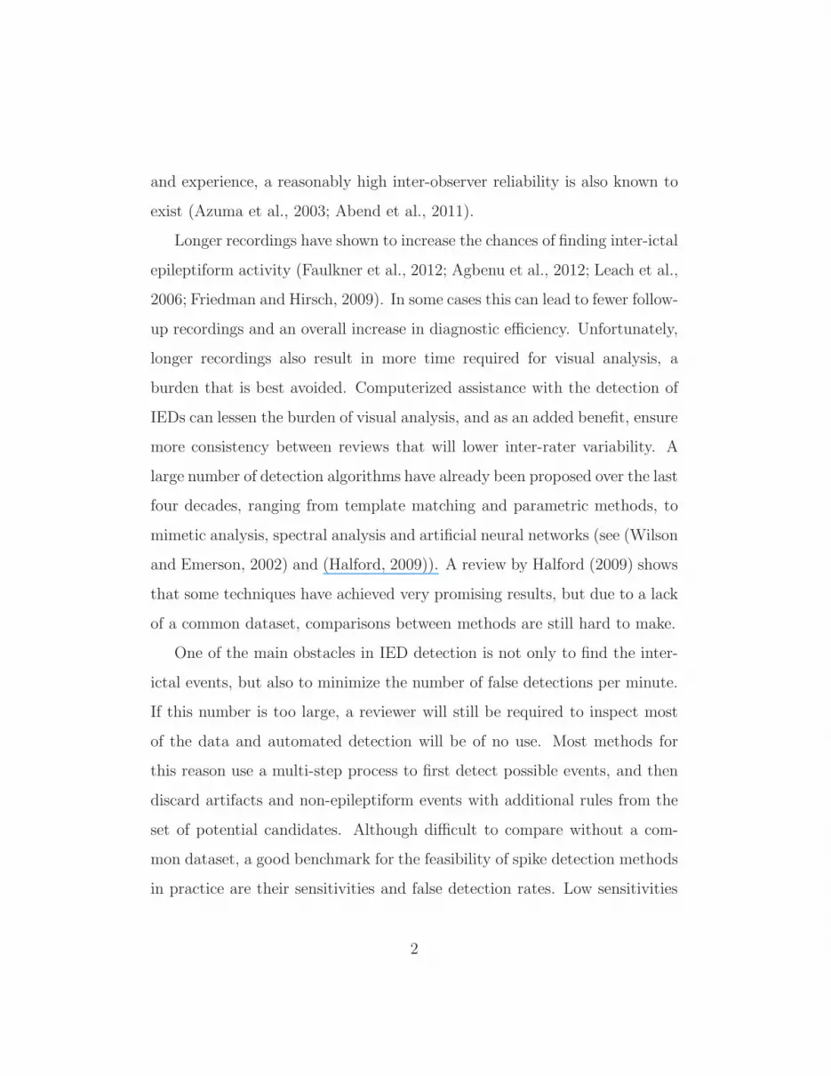
and experience, a reasonably high inter-observer reliability is also known to
exist (Azuma et al., 2003; Abend et al., 2011).
Longer recordings have shown to increase the chances of finding inter-ictal
epileptiform activity (Faulkner et al., 2012; Agbenu et al., 2012; Leach et al.,
2006; Friedman and Hirsch, 2009). In some cases this can lead to fewer follow-
up recordings and an overall increase in diagnostic efficiency. Unfortunately,
longer recordings also result in more time required for visual analysis, a
burden that is best avoided. Computerized assistance with the detection of
IEDs can lessen the burden of visual analysis, and as an added benefit, ensure
more consistency between reviews that will lower inter-rater variability. A
large number of detection algorithms have already been proposed over the last
four decades, ranging from template matching and parametric methods, to
mimetic analysis, spectral analysis and artificial neural networks (see (Wilson
and Emerson, 2002) and (Halford, 2009)). A review by Halford (2009) shows
that some techniques have achieved very promising results, but due to a lack
of a common dataset, comparisons between methods are still hard to make.
One of the main obstacles in IED detection is not only to find the inter-
ictal events, but also to minimize the number of false detections per minute.
If this number is too large, a reviewer will still be required to inspect most
of the data and automated detection will be of no use. Most methods for
this reason use a multi-step process to first detect possible events, and then
discard artifacts and non-epileptiform events with additional rules from the
set of potential candidates. Although difficult to compare without a com-
mon dataset, a good benchmark for the feasibility of spike detection methods
in practice are their sensitivities and false detection rates. Low sensitivities
2

show that reviewers can miss important IEDs if any are present, and as men-
tioned, high false detection rates make evaluating the output of the algorithm
more time consuming than a visual review itself. Although selectivity mea-
sures also provide useful information about the algorithm, it does not take
into account the number of IEDs per minute in the dataset, which is critical
when comparing methods from different datasets. Recent methods report
average sensitivities of 0.90 (Nonclercq et al., 2012), 0.65 (De Lucia et al.,
2008), 0.82 (Argoud et al., 2006), 0.93 (Subasi, 2006), 0.78 (Halford et al.,
2012), 0.69 (Ji et al., 2011a), and 0.92 (Van Hese et al., 2008), and average
false detections per minute of 6 (De Lucia et al., 2008), 13.4 (Argoud et al.,
2006), 0.92 (Indiradevi et al., 2008), and 0.09 (Ji et al., 2011a). In most
studies that report high sensitivities, a high false detection rate is present
if given, otherwise data epochs are used instead of continuous EEG, or the
number of false detections are not reported (Nonclercq et al., 2012; Subasi,
2006; Halford et al., 2012; Indiradevi et al., 2008; Van Hese et al., 2008). On
the other hand, studies with low false detection rates show lower sensitivities.
This shows the trade off for all methods between choosing high sensitivities
with more false detections, or lower sensitivities with fewer false detections.
Regardless of the many algorithms available, few implementations have
made it to clinical practice. With the aim of achieving a practical and reli-
able system for IED detection, we introduce an automated detection method
based on template matching to find IEDs in EEG recordings. The method
is unique in the sense that it uses a large database of templates extracted
from a training set of example EEGs. Unlike other template matching algo-
rithms that only rely on a single or a small number of templates to search for
3

inter-ictal events, our system can match more variations in spike morphology
and is less vulnerable to variability. In addition, the algorithm is designed
to incorporate experience into the templates from past classifications during
training, which can be extended even further so that they can gain additional
experience with repeated use. The presented method is intended to assist
instead of replace visual inspection, with the aim of significantly reducing
review time for clinicians and neurologists.
2. Methods
2.1. Subjects and data
An EEG dataset for this study was obtained from the department of
Clinical Neurophysiology at the Medisch Spectrum Twente (MST) in the
Netherlands. Recordings were made with a standard 20-30 minute protocol
using a Brainlab EEG system and standard Ag-AgCl electrode caps placed
according to the international 10-20 guidelines. Impedances were kept below
5 kΩ to reduce polarization effects. A sample rate of either 250 Hz or 256 Hz
was used for each recording. Afterwards it was band-pass filtered between
0.5-30 Hz, and downsampled to 100 Hz to increase efficiency of the algorithm.
IEDs were marked and reviewed by experienced electroencephalographers on
the channels where they were clearly visible (MvP and JA). Each IED con-
sisted of one of the following patterns: (i) spike, (ii) sharp wave, (iii) spike
and slow wave, (iv) sharp wave and slow wave (v) slow wave with absent
or very small preceding spike, or (vi) polyspike and slow wave. IEDs were
marked in three montages: common reference, bi-polar and source.
4

The dataset consisted of 23 records with 723 IEDs in total and a com-
bined recording length of 481 min. The patient group consisted of 15 males
and 8 females with ages ranging from 4 to 52 (mean 23.2± 17.1) years.
Thirteen subjects were diagnosed with generalized epilepsies (4 absence, 1
juvenile myoclonic, 8 idiopathic), and the remaining ten with focal epilepsies
(5 temporal lobe, 1 rolandic, 4 other). The dataset was split into two parts,
one for template collection and training, and the other for an evaluation set.
Eight EEGs were used for training and fifteen for testing. The training set
contained 482 marked IEDs with a total recording length of 175 min. The
testing set contained 241 marked IEDs with a total recording length of 306
min.
2.2. Preprocessing
Independent component analysis was used to reduce the influence of eye
blink artifacts. After calculating the independent components with the Fas-
tICA algorithm1, each component was compared to the electrooculogram
(EOG) channel recorded with the EEG. If a component showed a substan-
tial correlation with the EOG channel (>0.5), it was removed by setting all
its values to zero. The remaining components were projected back to their
channel space by applying the inverse transform.
2.3. Method outline
The presented method uses a database of templates resembling typical
waveforms of epileptiform activity, and finds matches between the templates
1Available at http://research.ics.aalto.fi/ica/fastica/
5

and inter-ictal events in the EEG. The main concepts that make this method
different from others are the following: (i) a substantially larger set of
templates is used, (ii) the templates are designed to have experience by re-
membering past classifications and using this to improve future predictions,
and (iii) an agreement system is used to combine detected events from mul-
tiple templates and thus lower false detections. The sections that follow give
a detailed outline of the various parts of the method, which can be divided
into template collection, learning, and detection. An illustrative summary
of the template collection and learning phase is shown in Fig. 1, and one for
the detection phase in Fig. 2.
2.4. Template collection
Starting with a blank database, templates are added using a training
dataset. For each EEG in the training set, a collection of epochs is extracted
at the locations where IEDs were marked by the reviewers. Each epoch rep-
resents a template and is added to the database. Three montages are used
during collection: common reference, bi-polar, and source. Templates are
extracted per channel, i.e. if an IED is marked over two channels in the
common reference montage and one channel in the bi-polar montage, three
templates will be created. Single spikes or sharp waves were not included
as templates, due to a high degree of false detections. An example of ex-
tracted templates is shown in Fig. 3. Note that they also have differences in
magnitude (amplitudes were not normalized, but used ‘as recorded’).
6

2.5. Learning
After extracting the templates, their ability to find and discriminate be-
tween other IEDs from the same or other recordings and non-epileptiform
activity is determined. This is done by finding a time-shifted correlation
between each template and all EEG channels for all EEGs in the training
set with the same montage. Due to the computational cost involved, this
part of the algorithm was implemented with parallel processing. Locations
are found where the templates have correlations above 0.85, and the under-
lying EEG segment is extracted to calculate additional properties to further
determine the relationship between them and the template by which they
were detected. For this, let a template consisting of N samples (N differs
per template) be defined as Stem(n) : n ∈ [1..N ], and each segment it finds
as Sseg(n) : n ∈ [1..N ], as illustrated in Fig. 4. In addition, let the pre-
ceding N samples of each segment (same length as segment) be defined as
Sprec(n) : n ∈ [1..N ] and the variance of the channel on which the segment
7

is located as σ2ch. Then, the following properties are calculated:
fCRR = corr(Stem, Sseg), (1)
fDCRR = corr(S ′
tem, S′
seg), (2)
fMAD =1
N
N∑
n
‖Stem(n)− Sseg(n)‖, (3)
fVRCHAN =σ2seg
σ2seg + σ2
ch
, (4)
fVRTEM =σ2seg
σ2seg + σ2
tem
, and (5)
fVRPREC =σ2seg
σ2seg + σ2
prec
, (6)
where
σ2seg = var(Sseg), (7)
σ2tem = var(Stem), and (8)
σ2prec = var(Sprec). (9)
In addition to having a high correlation with the template (fCRR ≥ 0.85),
these properties are used to determine the similarity between each detected
segment and its template. Property fDCRR measures the correlation between
the derivatives of Sseg and Stem, fMAD finds the mean amplitude difference
between Sseg and Stem, and fVRCHAN, fVRTEM and fVRPREC calculates the
variance ratio between σ2seg and the sum of σ2
seg with σ2ch, σ2
tem and σ2prec
respectively.
Using these properties, the location of each segment is converted to a
“nomination” of an IED event if it satisfies both of the following conditions:
8

(i) fMAD < 20µV, and (ii) fVRCHAN > 0.75 or fVRPREC > 0.67. Given that
the correlation between two segments is scale invariant, the first requirement
ensures that their magnitude is similar. The second requirement is based
on the assumption that, for scalp EEG, the amplitude changes within an
IED segment will be significantly larger than its preceding segment and the
channel variance. Threshold values were chosen arbitrarily. Given that the
true locations of IEDs are known in the training set, each nomination is either
marked as correct or as a false detection, depending on if any of its samples
overlap in time with a marked IED. At this point, the false detection rate of
a template can be calculated:
FDRtem =false detections
true detections + false detections. (10)
Using only the criteria above to nominate events as IEDs lead to many
false detections. Therefore, a linear support vector machine (SVM) is trained
using the nominations in the training set to give the template additional
experience in discriminating between true IEDs and false predictions. Vectors
consisting of fCRR, fDCRR, fVRTEM are used as input. By having an SVM
trained on past classifications, the template can now find events with high
correlation to itself and predict if it is in fact an epileptiform event or not.
After a template’s SVM is trained, its reliability (Rtem) is calculated as the
accuracy of re-classifying its own training vectors. The reliability reflects a
templates ability to separate IEDs from non-epileptiform events, and is used
as a weight factor during detection.
After all training steps are complete, templates that do not comply with
the following conditions are discarded: (i) Templates with zero detections,
(ii) templates with only false detections, (iii) templates with a small number
9

of correct detections and no false detections. The last condition avoids tem-
plates that only find themselves in the training set and which have no use in
finding other IEDs.
2.6. Detection
For detection, a similar approach is followed as during learning: For each
template in the database, a time-shifted correlation is calculated on each
EEG channel. Locations with correlations above 0.85 are nominated as IEDs
if they also satisfy the conditions: (i) fMAD < 20µV and (ii) fVRCHAN >
0.75 or fVRPREC > 0.67, and using each template’s own SVM, a prediction
is made if the event is in fact an IED or only a similar looking artifact.
False predictions are discarded and the remaining events are converted to
nominations and added to a global pool. Nominations are stored with the
following information about themselves: (a) its onset and duration, (b) the
channel on which it was found, (c) fCRR, and (d) Rtem.
Next, nominations are grouped together if they reside on the same channel
and overlap with more than 75%. Groups with fewer than 3 nominations are
considered unreliable and are discarded. Each of the remaining groups is
considered a detected event, and the onset and duration is taken from the
mean of the nominations in the group. The event is also assigned a detection
certainty, which is done by calculating the mean product of each nomination’s
correlation and template reliability:
certainty =1
M
M∑
m
Rtem(m)fCRR(m), (11)
with M the number of nominations in that group. Lastly, events from indi-
vidual channels are merged into one if they overlap in time, and once again
10

the mean onset, duration and certainty is used to describe the event.
3. Results
A total of 2632 templates were collected during training, of which 472 were
discarded for not passing the constraints described in Section 2.5. To gain
some insight on the behavior of the remaining 2160 templates, histograms
of FDRtem and Rtem are provided in Fig. 5(a) and Fig. 5(b) respectively.
The mean false detection rate before training SVMs is 0.74 and the mean
template reliability after SVM training 73.8%. The two histograms show that
the templates by themselves have high false detection rates and suboptimal
accuracies in finding IEDs. Fig. 5(b) shows however how the use of SVMs
and additional properties can help reduce the number of false detections and
improve accuracy. Fig. 6(a) gives an ROC curve from all detected events
in the test EEGs, and it shows that although low accuracies are achieved
at template level, the combined information from individual templates lead
to reliable detections on a global level. As seen in Fig. 6, the certainty
threshold can be adjusted to either maximize the sensitivity or minimize the
false detection rate. Figures 6(b) and 6(c) respectively show the sensitivity
and false detection rate as a function of minimum certainty, and Table 1
gives corresponding values to a number of points on the curves in Fig. 6. For
all test EEGs combined, a maximum sensitivity of 0.99 is reached at a false
detection rate of 7.24 false positives per minute. Note that the IED rate in
the evaluation set is only 0.79 IEDs per minute, and so a false detection rate
of 7.24 will be unacceptable for practical use. Although far from ideal, we
consider a more acceptable false detection rate for reviewing to be below three
11

per minute. The mean sensitivities obtained after adjusting the certainty
threshold of each EEG individually is shown in Table 2. The thresholds were
chosen in such a way as to limit the false positives per minute to no more
than three. After this, a mean sensitivity of 0.90 and mean false detection
rate of 2.36 min−1 is achieved. The selection of a threshold cannot be chosen
a priori to limit the false detection rate to a known maximum, but this shows
that detections with higher certainties are more reliable. In a typical usage
scenario, a user may choose to view events with the highest certainties first,
and then gradually reduce the threshold if he wishes to see more.
In addition to the detection algorithm, the described method was also
implemented into an EEG viewer. Fig. 7 shows how each of the detected
events are presented, and using the options provided, the reviewer can decide
to either confirm, reject, or indicate doubt concerning each detected event.
In such a way, the method can be used to assist during a review by pointing
out areas of interest in the EEG and allowing the reviewer to give the final
verdict.
4. Discussion
Automated detection of inter-ictal epileptiform discharges is a crucial step
towards improving the efficiency of epilepsy diagnostics and monitoring. Not
only does it reduce the review time significantly and make longer recordings
feasible, but it also allows for more objective analysis with less inter-observer
variability. The presented method uses a large collection of templates to de-
tect inter-ictal epileptiform discharges. It keeps track of past classifications
during training in order to provide the templates with additional statistical
12

experience, and the size of the database allows it to contain example spike-
and-slow-wave patterns of many morphologies so that IEDs can be detected
across multiple patients and recordings, regardless of whether their inter-ictal
patterns look the same. In addition, using the experience of each template,
the system can determine its reliability during detection. This approach re-
sembles the way in which humans search for IEDs: by finding waveforms that
have properties associated with IEDs, and mentally comparing them to pat-
terns observed in the past. In addition to high correlations between templates
and detected EDs, additional features were chosen to add information and
improve detection accuracy. Given that correlations are scale independent,
the mean amplitude difference (fMAD) ensures that matching events are on
the same scale, and the variance ratios (fVRCHAN, fVRTEM, fVRPREC) ensure
that IEDs are significantly different from its surrounding activity. By adding
these properties, the method is capable of not only using wave morphology,
but also temporal context.
To improve accuracy, the system removes uncertainties of single templates
by using a network effect to make decisions. In other words, even though
individual templates by themselves miss events and make many false detec-
tions, the collective value of their votes result in accurate detections. Using
this approach, the system is less vulnerable to variability in spike morphol-
ogy than other methods which rely on single features or a small number of
templates. Still, the system is highly dependent on the templates contained
within its database. Our results show that mosts IEDs can be detected with
the current set of templates and thresholds described. However, the ideal
set (and number) of templates to find all common inter-ictal patterns is not
13

known, and it could be that additional templates with more variations in
spike and wave morphology will allow the system to match events more ac-
curately, allowing higher thresholds for individual detections, and thereby
lowering the overall false detection rate.
A major advantage of this technique is that the system remains dynamic
and that more templates can be added if required. By further extending
the method and adding a feedback loop during reviews, the database can
become adaptive. As an example, missed events can be added as templates
if a reviewer finds any IEDs visually during a review. Another example
is if template reliabilities change when new EEGs are reviewed and false
detections are marked by a reviewer. As currently implemented, templates
search for matching IEDs in parallel to each other. Although not strictly
neccesary, this speeds up the detection process significantly and makes the
system scalable and fast enough for clinical use. Our implementation of the
method runs on a standard desktop computer (Intel i7-2600 3.40 GHz CPU,
4 GB RAM) and requires no additional hardware.
Reviews by Wilson and Emerson (2002) and Halford (2009) summarize
most of the reported techniques over the last four decades. In addition to
these, newer methods have also been reported (Nonclercq et al., 2009, 2012;
Scherg et al., 2012; Ji et al., 2011a,b; Igasaki et al., 2011; Argoud et al.,
2006; Nenadic and Burdick, 2005; Wang et al., 2010; Zhou et al., 2012).
Comparisons between different methods are hard to make because there is
no common dataset currently available. Without a benchmark dataset, re-
ported results rely heavily on the data used. For example, some studies use
standard 20 minute recordings to evaluate their methods, whereas others use
14

longer recordings or smaller EEG segments simply categorized as either con-
taining IEDs or not. If the number of IEDs per minute are not within the
same range between datasets, measures such as the specificity, selectivity and
the false detection rate cannot be compared in a fair manner. As suggested
by Halford (2009), a multi-center project is needed to create a dataset for
reliable benchmarks. The author later presents such a dataset, and although
still under development, it may suffice for benchmark testing and future com-
parisons (Halford et al., 2011, 2012). Regardless of using different datasets,
our method compares well to others. Depending on the confidence threshold
chosen, the system achieves sensitivities of up to 0.99. With thresholds cho-
sen as to limit the false detection rate, a mean sensitivity of 0.90 and mean
false detection rate of 2.36 is achieved. We feel that the threshold should be
set interactively by the reviewer during runtime, which can start by viewing
the most probable IEDs first with a high threshold value, and then gradually
view more detections (with greater false detection rate) if needed by lowering
it.
Apart from achieving high accuracies in epileptiform event detection, au-
tomated methods should also be responsible for presenting their findings
to the reviewer in a fast and efficient manner. Although some commercial
applications already exist, automated detection is mostly ignored by prac-
ticing neurologists. One reason for this could be that the software is still
too complicated and time consuming to use. Fig. 7 shows an example of
how a detected event is presented to a clinician. To make review time faster,
events can be grouped by location or morphology and reviewing can take
place on a macro scale to save time. This is already shown in some studies
15

(and some products) who use clustering techniques to group similar looking
events together (Nonclercq et al., 2012; Scherg et al., 2012; Ji et al., 2011a).
Although our method shows promising results, it is still far from ideal and
some reported methods appear to perform better. Many of them however,
have already been refined to the point where further improvement is limited.
Our method, as presented here, is only a first step in testing a novel approach
where many improvements can still be made. In work to follow, we will try
to use more of the spatial context available to discard artifacts and lower
the false detection rate. One way that we can achieve this is by introducing
event clustering. Events with similar properties can be clustered, and each
cluster’s validity as IEDs can be determined based on the spatial distribution
and frequency of occurrence of the events it contains. Apart from clustering,
a number of ad hoc rules can also be applied as a post-detection step to
mimic the procedures followed by reviewers when determining the validity
of IEDs. Further work will also focus on testing the system on long-term
EEG recordings such as in-home ambulatory monitoring, where the burden
of visual inspection is even greater and more problematic for the reviewer.
In summary, automated detection of inter-ictal epileptiform activity will
become invaluable to current reviewing techniques that rely mostly on visual
analysis. A reliable system for a wide range of EEGs with different spike
morphologies is needed, which should be tested with a gold-standard dataset
for comparisons with other techniques. With minor improvements and an in-
tuitive interface, the presented method is capable of providing more efficiency
with reviewing and in turn improve the diagnostic efficiency of epilepsy.
16

Acknowledgements
The authors would like to extend their gratitude to the reviewers for their
valuable comments and suggestions that contributed to the quality of this
work.
Abend NS, Gutierrez-Colina A, Zhao H, Guo R, Marsh E, Clancy RR,
et al. Interobserver reproducibility of electroencephalogram interpretation
in critically ill children. J Clin Neurophysiol, 2011; 28:15–9.
Agbenu J, Newton RW, Martland T, Ismayl O, Hargreaves S. Effect of
reducing the recording time of standard EEGs on the detection of EEG-
abnormalities in the management of the epilepsies of childhood. Seizure,
2012; 21:422–5.
Argoud FIM, De Azevedo FM, Neto JM, Grillo E. SADE3: an effective
system for automated detection of epileptiform events in long-term EEG
based on context information. Med Biol Eng Comput, 2006; 44:459–70.
Azuma H, Hori S, Nakanishi M, Fujimoto S, Ichikawa N, Furukawa TA. An
intervention to improve the interrater reliability of clinical EEG interpre-
tations. Psychiatr Clin Neurosci, 2003; 57:485–9.
De Lucia M, Fritschy J, Dayan P, Holder DS. A novel method for automated
classification of epileptiform activity in the human electroencephalogram-
based on independent component analysis. Med Biol Eng Comput, 2008;
46:263–72.
17

Doppelbauer A, Zeitlhofer J, Zifko U, Baumgartner C, Mayr N, Deecke L.
Occurrence of epileptiform activity in the routine EEG of epileptic patients.
Acta neurologica Scandinavica, 1993; 87:345–52.
Faulkner HJ, Arima H, Mohamed A. Latency to first interictal epileptiform
discharge in epilepsy with outpatient ambulatory EEG. Clin Neurophysiol,
2012; 123:1732–1735.
Friedman DE, Hirsch LJ. How long does it take to make an accurate diagnosis
in an epilepsy monitoring unit? Clin Neurophysiol, 2009; 26:213–7.
Halford JJ. Computerized epileptiform transient detection in the scalp elec-
troencephalogram: Obstacles to progress and the example of computerized
ECG interpretation. Clin Neurophysiol, 2009; 120:1909–1915.
Halford JJ, Pressly WB, Benbadis SR, TatumWO, Turner RP, Arain A, et al.
Web-based collection of expert opinion on routine scalp EEG: software
development and interrater reliability. J Clin Neurophysiol, 2011; 28:178–
84.
Halford JJ, Schalkoff RJ, Zhou J, Benbadis SR, Tatum WO, Turner RP,
et al. Standardized Database Development for EEG Epileptiform Transient
Detection: EEGnet Scoring System and Machine Learning Analysis. J
Neurosci Meth, 2012; 212:308–316.
Igasaki T, Higuchi T, Hayashida Y, Murayama N, Neshige R. Proposal for
patient-specific automatic on-line detection of spike-and-wave discharges
utilizing an artificial neural network. In 2011 4th International Conference
on Biomedical Engineering and Informatics (BMEI). IEEE, 2011; 813–817.
18

Indiradevi KP, Elias E, Sathidevi PS, Dinesh Nayak S, Radhakrishnan K. A
multi-level wavelet approach for automatic detection of epileptic spikes in
the electroencephalogram. Comput Biol Med, 2008; 38:805–16.
Ji Z, Sugi T, Goto S, Wang X, Ikeda A, Nagamine T, et al. An automatic
spike detection system based on elimination of false positives using the
large-area context in the scalp EEG. IEEE Trans Biomed Eng, 2011a;
58:2478–88.
Ji Z, Wang X, Sugi T, Goto S, Nakamura M. Automatic spike detection based
on real-time multi-channel template. 2011 4th International Conference on
Biomedical Engineering and Informatics (BMEI), 2011b; 648–652.
Leach JP, Stephen LJ, Salveta C, Brodie MJ. Which electroencephalography
(EEG) for epilepsy? The relative usefulness of different EEG protocols
in patients with possible epilepsy. J Neurol Neurosurg Psychiatry, 2006;
77:1040–2.
Nenadic Z, Burdick JW. Spike detection using the continuous wavelet trans-
form. IEEE Trans Biomed Eng, 2005; 52:74–87.
Nonclercq A, Foulon M, Verheulpen D, De Cock C, Buzatu M, Mathys P,
et al. Spike detection algorithm automatically adapted to individual pa-
tients applied to spike-and-wave percentage quantification. Neurophysiol
Clin Clin Neurophysiol, 2009; 39:123–31.
Nonclercq A, Foulon M, Verheulpen D, De Cock C, Buzatu M, Mathys P,
et al. Cluster-based spike detection algorithm adapts to interpatient and
19

intrapatient variation in spike morphology. J Neurosci Methods, 2012;
210:259–265.
Scherg M, Ille N, Weckesser D, Ebert A, Ostendorf A, Boppel T, et al.
Fast evaluation of interictal spikes in long-term EEG by hyper-clustering.
Epilepsia, 2012; 53:1196–204.
Subasi A. Automatic detection of epileptic seizure using dynamic fuzzy neural
networks. Expert Syst Appl, 2006; 31:320–328.
Van Hese P, Vanrumste B, Hallez H, Carroll GJ, Vonck K, Jones RD, et al.
Detection of focal epileptiform events in the EEG by spatio-temporal dipole
clustering. Clin Neurophysiol, 2008; 119:1756–70.
Wang C, Zou J, Zhang J, Wang M, Wang R. Feature extraction and recogni-
tion of epileptiform activity in EEG by combining PCA with ApEn. Cogn
Neurodyn, 2010; 4:233–40.
Wilson SB, Emerson R. Spike detection: A review and comparison of algo-
rithms. Clin Neurophysiol, 2002; 113:1873–81.
Zhou J, Schalkoff RJ, Dean BC, Halford JJ. Morphology-based wavelet fea-
tures and multiple mother wavelet strategy for spike classification in EEG
signals. Conference proceedings : Annual International Conference of the
IEEE Engineering in Medicine and Biology Society, 2012; 2012:3959–62.
20

Table 1: Choosing to maximize the sensitivity or minimize the false detection rate can be
done by adjusting the minimum confidence threshold. This allows a reviewer to view the
most probable IEDs first, and then to look at additional events if desired. Values below
correspond to points on Fig. 6(a)-(c).
Min confidence Mean sensitivity Mean Fp/min
0 0.99 7.24
0.02 0.97 4.12
0.04 0.67 1.29
0.06 0.44 0.87
0.08 0.27 0.62
0.1 0.2 0.54
0.12 0.15 0.41
0.14 0.08 0.37
0.16 0.07 0.31
0.18 0.06 0.29
0.2 0.04 0.26
0.22 0.03 0.22
0.24 0.02 0.19
0.26 0.02 0.16
21

Table 2: Mean sensitivities and false detection rates per EEG in the test set using a varying
confidence threshold value per EEG and limiting false detections to a maximum of three
per minute.
EEG Dur (min) IEDs SEN Fp/min
S1 16.3 13 1.00 1.59
S2 22.5 11 0.82 2.93
S3 20.0 5 1.00 1.40
S4 20.0 36 0.97 2.60
S5 22.3 19 0.79 1.93
S6 19.5 6 1.00 2.72
S7 20.3 79 0.97 1.82
S8 21.2 5 0.80 1.98
S9 20.0 2 1.00 2.95
S10 20.0 12 1.00 2.00
S11 21.5 7 0.38 2.88
S12 20.3 7 0.86 2.95
S13 20.7 19 0.95 2.08
S14 21.0 14 1.00 2.95
S15 20.0 6 1.00 2.60
mean 20.4 16.07 0.90 2.36
22

Figure 1: Outline of the template extraction and learning steps. After the
templates are trained, each of them contributes to the detection by nominat-
ing possible IEDs.
Figure 2: Outline of the detection method. Trained templates detect and
nominate possible IEDs. The system combines these nominations and iden-
tifies IEDs based the the mutual agreement of multiple templates.
Figure 3: Nine templates taken from the trained database consisting of
2160 templates. Template lengths are allowed to differ and amplitudes are
preserved.
Figure 4: Template features: To find matching events, templates use corre-
lation coefficients, amplitude differences and variance ratios between a tem-
plate and its detected segment. Variance ratios add temporal context to a
detected event by comparing it to its surrounding activity.
Figure 5: Histograms combining individual template statistics: (a) false
detection ratios; and (b) Rtem: the ability of each template’s SVM to sepa-
rate its training vectors. The two histograms show that by themselves the
templates have high false detection rates and in many cases poor separability
between IEDs and non-IED events. The power of the system lies in combin-
ing the information and using a network effect to allow accurate predictions.
23

Figure 6: Evaluation of the described method on a test set of 15 EEGs: (a)
an ROC curve shows how the sensitivity of detecting IEDs can be improved
at the price of more false detections. The dashed rectangle gives an approx-
imate range of false detection rates that we consider as more feasible for
practical use; (b) higher confidence values reduce the sensitivity, (c) but also
lowers the false detection rate.
Figure 7: A detected IED highlighted on top of the EEG, as displayed to
the reviewer in a bi-polar montage.
24

Figure 1
DB(untrained)
com
mon
ref
bi-
pola
r
sou
rce
EX
TR
AC
TIO
NL
EA
RN
ING
Extract per channel
all segments marked
by a reviewer as IED.
Store as templates.
T1
T2T3 T4 T5
T6
For each extracted template:
- nd all segments (all montages and channels)
with high correlation to itself
- compare detections with IEDs marked by reviewer.
- use detection outcomes and train classi ers to
improve future predictions.
DB(trained)
Figure 1:
25

Figure 2
Figure 2:
26

Figure 3
0 0.2 0.4 0.6−30
−20
−10
0
10
20
s
µV
0 0.2 0.4 0.6−100
−50
0
50
sµ
V
0 0.2 0.4 0.6−40
−20
0
20
40
s
µV
0 0.2 0.4 0.6−80
−60
−40
−20
0
20
40
s
µV
0 0.2 0.4 0.6
−50
0
50
s
µV
0 0.2 0.4 0.60
20
40
60
80
s
µV
0 0.2 0.4 0.6
−50
0
50
s
µV
0 0.2 0.4 0.6−40
−20
0
20
40
60
s
µV
0 0.2 0.4 0.6−100
−50
0
s
µV
Figure 3:
27

Figure 4
SsegSprec
Stemtem
segprecch
Figure 4:
28

Figure 5
0 0.2 0.4 0.6 0.8 10
50
100
150
200
250
300
350
False detection ratios of templates
FDR
Cou
nt
(a)
0 20 40 60 80 1000
20
40
60
80
100
120
SVM separability using training vectors
Rtem
(%)
Cou
nt(b)
Figure 5:
29

Figure 6
0 2 4 60
0.2
0.4
0.6
0.8
1
Se
nsi
tivity
Sensitivity vs FDR
FDR (fp/min)
(a)
0 0.5 10
0.2
0.4
0.6
0.8
1Sensitivity vs Certainty
Se
nsi
tivity
Certainty
(b)
0 0.5 10
2
4
6
FDR vs Certainty
Certainty
FD
R (
fp/m
in)
(c)
Figure 6:
30

Figure 7
5V
1
Figure 7:
31


