Embed Size (px)
Citation preview
Interference with Hemozoin Formation Represents anImportant Mechanism of Schistosomicidal Action ofAntimalarial Quinoline Methanols
Juliana B. R. Correa Soares1, Diego Menezes2, Marcos A. Vannier-Santos2, Antonio Ferreira-Pereira3,
Giulliana T. Almeida4, Thiago M. Venancio4¤, Sergio Verjovski-Almeida4, Vincent K. Zishiri5, David
Kuter5, Roger Hunter5, Timothy J. Egan5, Marcus F. Oliveira1*
1 Laboratorio de Bioquımica Redox, Programa de Biologia Molecular e Biotecnologia, Instituto de Bioquımica Medica, Universidade Federal do Rio de janeiro, Rio de
Janeiro, Brazil, 2 Instituto Goncalo Moniz, FIOCRUZ, Salvador, Brazil, 3Departamento de Microbiologia Geral, Instituto de Microbiologia Prof. Paulo de Goes, Universidade
Federal do Rio de Janeiro, Rio de Janeiro, Brazil, 4Departamento de Bioquımica, Instituto de Quımica, Universidade de Sao Paulo, Sao Paulo, Brazil, 5Department of
Chemistry, University of Cape Town, Rondebosch, Cape Town, South Africa
Abstract
Background: The parasitic trematode Schistosoma mansoni is one of the major causative agents of human schistosomiasis,which afflicts 200 million people worldwide. Praziquantel remains the main drug used for schistosomiasis treatment, andreliance on the single therapy has been prompting the search for new therapeutic compounds against this disease. Our grouphas demonstrated that heme crystallization into hemozoin (Hz) within the S. mansoni gut is a major heme detoxification routewith lipid droplets involved in this process and acting as a potential chemotherapeutical target. In the present work, weinvestigated the effects of three antimalarial compounds, quinine (QN), quinidine (QND) and quinacrine (QCR) in a murineschistosomiasis model by using a combination of biochemical, cell biology and molecular biology approaches.
Methodology/Principal Findings: Treatment of S. mansoni-infected female Swiss mice with daily intraperitoneal injectionsof QN, and QND (75 mg/kg/day) from the 11th to 17th day after infection caused significant decreases in worm burden(39%–61%) and egg production (42%–98%). Hz formation was significantly inhibited (40%–65%) in female worms recoveredfrom QN- and QND-treated mice and correlated with reduction in the female worm burden. We also observed that QNtreatment promoted remarkable ultrastructural changes in male and female worms, particularly in the gut epithelium andreduced the granulomatous reaction to parasite eggs trapped in the liver. Microarray gene expression analysis indicatedthat QN treatment increased the expression of transcripts related to musculature, protein synthesis and repair mechanisms.
Conclusions: The overall significant reduction in several disease burden parameters by the antimalarial quinoline methanolsindicates that interference with Hz formation in S. mansoni represents an important mechanism of schistosomicidal actionof these compounds and points out the heme crystallization process as a valid chemotherapeutic target to treatschistosomiasis.
Citation: Correa Soares JBR, Menezes D, Vannier-Santos MA, Ferreira-Pereira A, Almeida GT, et al. (2009) Interference with Hemozoin Formation Represents anImportant Mechanism of Schistosomicidal Action of Antimalarial Quinoline Methanols. PLoS Negl Trop Dis 3(7): e477. doi:10.1371/journal.pntd.0000477
Editor: Malcolm K. Jones, Queensland Institute of Medical Research, Australia
Received February 19, 2009; Accepted June 3, 2009; Published July 14, 2009
Copyright: � 2009 Correa Soares et al. This is an open-access article distributed under the terms of the Creative Commons Attribution License, which permitsunrestricted use, distribution, and reproduction in any medium, provided the original author and source are credited.
Funding: This work was supported in part by WHO/TDR, South-South Initiative grant A60265 (MFO-TJE), International Centre for Genetic Engineering andBiotechnology (ICGEB-CRP) grant (CRP/BRA07-03, contract CRP/07/008), CNPq, FAPERJ (MFO through Jovens Cientistas do Nosso Estado 2007) and FAPESP (toSVA). MFO, MAVS and SVA are research scholars from CNPq. The funders had no role in the study design, data collection and analysis, decision to publish, orpreparation of the manuscript.
Competing Interests: All authors have read and approved the contents of the manuscript and none of the authors have competing financial interests. There isno significant overlap between the submitted manuscript and any other papers from the same authors under consideration or in press elsewhere.
* E-mail: [email protected]
¤ Current address: National Center for Biotechnology Information, National Library of Medicine, National Institutes of Health, Bethesda, MD, United States ofAmerica
Introduction
Schistosomiasis is a major parasitic disease that affects 170
million people in sub-Saharan Africa and close to 30 million
people in north of Africa, Asia and South America [1]. Recent
estimates indicate that 779 million people live in risk areas across
70 countries [2,3,4] raising the possibility that morbidity associated
with the disease may be considerably under-estimated [5]. The
disease pathology is typically a consequence of an inflammatory
granulomatous reaction due to parasite egg deposition in the liver
and other host tissues [6]. The major etiological agent of human
schistosomiasis is the platyhelmith Schistosoma mansoni which is
known to digest large amounts of blood in order to complete its
development and sexual maturation [7]. During this process, host
hemoglobin is degraded by several proteolytic enzymes [8,9]
forming peptides, amino acids and the prosthetic group heme [10].
Heme is an amphyphilic molecule of low molecular weight that
plays essential biological roles, from cell respiration to drug
www.plosntds.org 1 July 2009 | Volume 3 | Issue 7 | e477
detoxification [11]. A large body of evidence has demonstrated
that once in a ‘‘free’’ state, heme is able to induce oxygen-derived
free radicals formation [12,13], lipid peroxidation [14,15] and
protein [16] and DNA [17] oxidation. Due to its amphyphilic
nature, ‘‘free’’ heme also interferes with phospholipid membrane
stability and solubility, in a mechanism independent of its pro-
oxidant effects [18,19], eventually resulting in cell lysis. As a
consequence, it is apparent that blood-feeding organisms evolved
efficient adaptations in order to circumvent the deleterious effects
of ‘‘free’’ heme [20]. A particular mechanism present in some
blood-feeders, such as shown in malaria parasites (Plasmodium sp.)
[21], the kissing bug Rhodnius prolixus, a Chagas’ disease vector [22]
and the blood fluke S. mansoni consists of the crystallization of heme
into a dark brown pigment known as hemozoin (Hz) [23]. Our
group has shown that heme crystallization represents a major
heme detoxification mechanism in both R. prolixus and S. mansoni,
acting as a preventive antioxidant defense [24]. We have also
shown that adult females of S. mansoni produce large amounts of
Hz within the gut [23], involving extracellular lipid droplets
present in the gut lumen in this process [25,26]. Moreover, the
hydrophilic-hydrophobic interface provided by the gut lipid
droplets, seems to play a key catalytic role in heme crystallization,
adding a strong biological support to the interface-mediated heme
crystallization model recently proposed by Egan and co-workers
[27].
Due to the essential nature of Hz formation in Plasmodium [27]
the development of antimalarials throughout the years has been
largely limited to the 4-aminoquinoline compounds, such as
chloroquine (CLQ) and quinoline methanols such as quinine (QN)
and quinidine (QND). These compounds exert potent action
against the blood stages of Plasmodium in a mechanism that impairs
Hz formation [28]. It was shown that 4-aminoquinolines interact
with ‘‘free’’ heme, hindering its crystallization into Hz. The ‘‘free’’
heme interacts with membranes and exerts severe toxic effects,
ultimately killing the parasite through oxidative stress [29]. An
additional theory suggests that heme-quinoline complexes incor-
porate into a growing crystal face influencing its external
appearance, and blocking its growth [30,31]. Regardless of the
mechanism by which 4-aminoquinolines act on Hz formation, our
group has shown that CLQ inhibits heme crystallization in vivo in
both R. prolixus [32], and also in S. mansoni [33,26]. In this regard,
we showed that in vivo treatment of S. mansoni -infected mice with
CLQ decreased the overall severity of experimental murine
schistosomiasis [33]. These results indicated for the first time that
interfering with Hz formation in this parasite is a valuable
approach for chemotherapeutic development. In addition, the Hz
formation pathway is peculiar to blood-feeding parasites (including
S. mansoni) and is absent in the host, which makes it an
exceptionally attractive drug target. Very recent evidence indicates
that the antimalarial mefloquine exerts potent antischistosomal
effects, reducing worm burden [34], although its mechanism of
action remains to be elucidated.
Currently, the main control strategy for schistosomiasis relies on
chemotherapy [35], praziquantel (PZQ) being the main drug of
choice for this purpose, as recommended by the World Health
Organization. PZQ is safe, well tolerated and effective in a single
oral dose treatment against the adult stages of all forms of
schistosomiasis [36–38]. Despite the threat of resistance develop-
ment of S. mansoni to PZQ [39–40], the establishment of true
resistance so far is not conclusive [41]. Nevertheless, reliance on
single PZQ therapy raises real concern and, as a result, this has
prompted the search for new therapeutic targets and drugs against
this disease. Other studies have suggested the antimalarial
artemether as a new drug for schistosomiasis [42] due to its
potent action against young schistosomula [43–45]. Alternative
approaches, such as inhibition of the cysteine protease cathepsin
B1 by K11777 early in the infection, have drastically decreased
both worm and eggs burdens, delaying the egg-associated organ
pathology [46]. The recent discoveries on the role of thioredoxin-
glutathione reductase (TGR) activity for parasite redox balance
and survival have prompted new chemotherapeutic development
studies targeting this enzyme [47–49]. Inhibitors of S. mansoni
TGR, such as oxadiazoles [49], and other drugs discovered in a
massive screening [48], has provided new lead compounds that
specifically inhibited parasite TGR with high potency, showing
that an imbalance in the redox cascade is deleterious to S. mansoni,
regardless of parasite stage [47–49].
Here we investigated the effects of the antimalarial quinolines
QN, QND and QCR on S. mansoni-infected mice, by evaluating
infection, biochemical, pathological, ultrastructural and molecular
parameters.
Methods
Ethics statementAll animal care and experimental protocols were conducted
following the guidelines of the institutional care and use committee
(Comission for Evaluation of Animal Use for Research from the
Federal University of Rio de Janeiro, CAUAP-UFRJ) and the
NIH Guide for the Care and Use of Laboratory Animals (ISBN 0-
309-05377-3). The protocols were approved by CAUAP-UFRJ
under the registry #IBQM002. Technitians dedicated to the
animal facility at the Institute of Medical Biochemistry (UFRJ)
carried out all aspects related to the mice husbandry under strict
guidelines to assure careful and consistent handling of the animals.
Parasites and host animalsS. mansoni LE strain was maintained in the laboratory using
Biomphalaria glabrata snails and Swiss mice as intermediate and
definitive hosts, respectively. Cercariae released from snails were
Author Summary
Heme is an essential molecule to most living organisms,but once in a free state it exerts toxic effects. Blood-feeding organisms evolved efficient ways to detoxify freeheme derived from hemoglobin digestion. A key mecha-nism present in some hematophagous organisms consistsof the crystallization of heme into a pigment namedhemozoin. Schistosoma mansoni is one of the etiologicagents of human schistosomiasis, a parasitic disease thataffects over 200 million people in tropical and subtropicalareas. Hemozoin formation represents the main hemedetoxification pathway in S. mansoni. Here, we report thatthe antimalarial quinoline methanols quinine and quini-dine exert schistosomicidal effects notably due to theircapacity to interfere with hemozoin formation. Whenquinine or quinidine were administered intraperitoneallyduring seven days to S. mansoni-infected mice (75 mg/kg/day), both worm and eggs burden were significantlyreduced. Interestingly, hemozoin content in female wormswas drastically affected after treatment with eithercompound. We also found that quinine caused importantchanges in the cellular organization of worm gastrodermisand increased expression of genes related to musculature,protein synthesis and repair mechanisms. Together, ourresults indicate that interference with hemozoin formationis a valid chemotherapeutic target for development of newschistosomicidal agents.
Effects of Antimalarial Quinolines on Schistosoma
www.plosntds.org 2 July 2009 | Volume 3 | Issue 7 | e477
injected in mice cervices through a subcutaneous route. Mice were
kept in a animal care facility at Institute of Medical Biochemistry
(UFRJ). Forty-two days after infection, adult worms were obtained
from the mice by mesenteric perfusion with saline as previously
described [50].
Regurgitant isolationAbout 150 female adult worms were obtained by mesenteric
perfusion of mice, placed in 1 mL of ultrapure water at room
temperature for 80 minutes and gently shaken every 5 minutes.
After that, the precipitated worms were discarded and superna-
tant, hereafter referred to as ‘‘regurgitant’’, was colleted and kept
at 270uC, as previously described by our group [26]. Protein
content from regurgitant were measured relative to bovine serum
albumin as a standard [51].
Syntheses of new quinolinesThe syntheses of compound 3 (C3), compound 6 (C6),
compound 7 (C7), and compound 8 (C8) were accomplished by
following a method previously described [52]. Synthesis of
compound 9 (C9) was carried out using the protocol described
in the literature [28]. For synthesis of compound 10 (C10), 4,7-
dichloroquinoline (300 mg, 1.51 mmol) was added to a stirred
solution of cyclopentylmethylamine (1.00 mL, 5.83 mmol) under
N2. The mixture was heated under pressure in a cyclo-addition
tube at 120uC overnight. After, the mixture was allowed to cool to
room temperature, it was poured into saturated brine (20 mL) and
extracted with ethyl acetate (3650 mL). The organic layer was
dried (Na2SO4) and concentrated in vacuo to afford a crude
product, purified by flash column chromatography using a mixture
of ethyl acetate: hexane (90:10) as eluent to give C10 (295 mg,
87%) as a light-yellow solid, m.p. (EtOAc :Hex) 147–151uC; dH(CDCl3, 400 MHz) 8.51 (1H, d, J=5.5 Hz), 7.94 (1H, d,
J=1.8 Hz), 7.63 (1H, d, J=9.2 Hz), 7.32 (1H, dd, J=1.8,
7.3 Hz), 6.43 (1H, d, J=5.5 Hz), 5.05 (1H, br s), 4.01 (1H, m),
2.15 (2H, m), 1.80–1.61 (8H, m); dC (CDCl3, 100.5 MHz) 151.8,
149.3, 149.0, 134.8, 128.7, 125.2, 121.0, 117.2, 100.0, 54.3, 33.3.
Heme crystallization inhibition assaysThe assay utilized to evaluate inhibition of heme crystallization
by quinoline antimalarials was based on a previous method
described by Ncokazi et al, with minor modifications [53]. This
method makes use of the known physico-chemical properties of
heme that slowly and spontaneously crystallize into Hz particles in
vitro in acidic conditions, in such a way that the reaction kinetics
depends on the temperature, ionic strength, and time [54]. After
reaction is complete, pyridine is added to the medium to promote
the solubilization of the non-crystallized heme, since it is unable to
solubilize heme within the Hz crystals. Then, aliquots of the
supernatant were taken to determine the light absorption of non-
crystallized heme at 405 nm. Samples corresponding to 15 mg of
protein from whole regurgitant were incubated overnight at 37uC
in 0.5 M sodium acetate buffer, pH 4.8, in the presence of
100 mM hemin (prepared in 0.1 N sodium hydroxide) in a 96-well
plate with different concentrations of compounds (from 5 mM to
100 mM) in a total volume of 170 mL. Commercial quinolines and
non-quinoline drugs were prepared in 100% ethanol whereas new
quinolines were in 100% methanol. After that, 80 mL of 30.0% (v/
v) pyridine solution in 20 mM Hepes sodium salt, pH 7.5 was
added to each well, mixed and incubated for 15 minutes at room
temperature, in order to allow sedimentation of Hz crystals. Then,
38 mL of supernatant were transferred to another 96-well plate,
diluted to 250 mL in 30.0% (v/v) pyridine solution and the amount
of free heme was determined using a microplate reader at 405 nm
as previously described [53]. Hz content was determined by
subtracting the amount of total heme added to the samples minus
the values found in the experimental group. After that, data were
plotted with GraphPad Prism 4.0 software and the IC50 was
determined.
Quinolines treatment in vivoFemale mice aged around 30 days were infected with S. mansoni
by subcutaneous injection of 250 cercariae in the cervical region.
Mice were treated in two different protocols, being the first one from
day 11 to 17 after infection, with daily intraperitoneal injections of
75 mg/kg quinine hydrochloride dihydrate (QN), quinacrine
(QCR) or quinidine (QND) prepared as 30.0% ethanol solutions.
Control mice were treated with 30.0% ethanol alone (control). The
volumes injected in each mouse was about 100 mL. Worms were
collected by mesenteric perfusion 42 days after infection. In the
second protocol, mice were treated from day 42 to 45 after infection
with daily intraperitoneal injections of 100 mg/kg QND in 30.0%
ethanol and the worms were collected by mesenteric perfusion 46
days after infection. The worms were separated by sex, counted and
utilized for biochemical, ultrastructural and molecular assays. Mice
small and large intestines, liver and plasma were collected for
subsequent analyses. Non-infected mice were also treated with QN
or 30.0% ethanol alone during the same period.
Egg countingLiver, small and large intestines of each mouse were collected
after mesenteric perfusion, washed with phosphate buffered saline,
weighed, sliced into small pieces, and subsequently digested
overnight at 37uC in 10 mL of 4.0% potassium hydroxide
(prepared in ultrapure water), as previously described [55].
Aliquots of digested tissues (10 mL) were placed onto a glass
coverslip, and the number of eggs was determined by counting
with a Zeiss stereomicroscope (Stemi SV11 MC80).
Transmission electron microscopy (TEM)Adult S. mansoni worms collected after mesenteric perfusion from
mice were fixed in 1 mL of 2.5% glutaraldehylde and 4.0%
formaldehyde in 0.1 M sodium cacodylate buffer, pH 7.4. The
worms were fixed and post-fixed in 1.0% osmium tetroxide and
0.8% potassium ferricyanide in the same buffer, dehydrated in
acetone and embedded in epoxy polybed resin. Thin cuts of the
blocks were produced, contrasted in uranyl acetate and lead citrate
and observed in a transmission electron microscopy Zeiss CEM
902, as described earlier by our group [25].
Hz quantificationHz in adult worms was extracted and quantified based on
methods previously described by our group [23]. Worms were
separated by sex, counted and homogenized in protease inhibitor
cocktail (containing 0.1 mg/mL leupeptin trifluoroacetate salt,
2 mM benzamidine hydrochloride and 0.1 mg/mL soybean
trypsin inhibitor in ultrapure water), hereafter referred as
‘‘homogenate’’. For Hz extraction, 1 mL of female and male
homogenates of S. mansoni were centrifuged at 15.0006g for
15 minutes at 25uC and then their supernatants were discarded.
The pellets were washed three times in 1 mL of extraction buffer
(contained a mixture of 0.1 M sodium carbonate and 2.5%
sodium dodecyl sulphate), pH 9.1 and twice with 1 mL of
ultrapure water. The final pellet was solubilized in 1 mL 0.1 N
sodium hydroxide and shaken for 30 minutes. Hz content was
determined spectrophotometrically at 400 nm in a GBC-UV/Vis-
920 (Australia).
Effects of Antimalarial Quinolines on Schistosoma
www.plosntds.org 3 July 2009 | Volume 3 | Issue 7 | e477
HistopathologyLivers collected after mesenteric perfusion from control and
QN-treated mice were washed with phosphate buffered saline,
sliced into small pieces, fixed in 5 mL of 2.5% glutaraldehyde and
4.0% formaldehyde in 0.1 M sodium cacodylate buffer, pH 7.4
and kept at 4uC. Tissue slices were embedded in paraffin, stained
with hematoxylin and eosin dyes and observed in an Olympus
BX51 microscope. The mean granuloma area was analysed by
using the ImageJ software available at the website (http://rsb.info.
nih.gov/ij/).
Microarray analysisFreshly perfused adult female worms were incubated with cold
RNAlater solution (Ambion, USA) and kept at 4uC until RNA
extraction. Total RNA was extracted using Trizol reagent
(Invitrogen, USA) according to the manufacture’s instructions.
RNA was quantified using a Nanodrop ND-1000 UV/Vis
spectrophotometer and its quality was assessed with an Agilent
2100 Bioanalyzer, a micro fluidics-based electrophoresis platform.
1 mg of total RNA from each sample was amplified using the T7-
RNA polymerase based SuperScript Indirect RNA amplification
system (Invitrogen, USA) and subsequently 3 mg of aminoallyl-
modified amplified RNA was labeled using Cy3 or Cy5 reactive
dye (GE Healthcare, USA). Amplification and labeling were done
according to manufacturer specifications. Pools of RNA from
worms treated with QN were hybridized against RNA from
control worms. In order to ensure the uniformity of data, all
experiments were performed using slides from the same printing
batch and dyes from the same lot. Technical and biological
replicates were performed. Dye swap was employed in order to
account for dye biases. A total of eight different replicated data
values were obtained for each condition and for each probe on the
array. The Cy3- and Cy5-labeled samples were combined, dried
and re-suspended in hybridization buffer (50% formamide, 25%
RNase free water and 25% Microarray Hybridization buffer 46);
hybridization and washings were done according to the manufac-
turer GE Healthcare (USA). Samples were hybridized overnight to
cDNA microarray slides containing 4,000 elements in duplicate
(GEO accession: GPL3929) [56] using ASP hybridization
chambers (GE Healthcare, USA) at 42uC. After washings, slides
were air dried and scanned using a microarray dual channel laser
scanner (GenePix 4000B, Molecular Devices, USA) at 5 mm
resolution, 100% laser power and PMT levels were adjusted in
order to obtain similar average intensities of red and green signal.
Data were extracted using the program Array Vision 8.0. To
correct for systematic biases on the data originated from small
differences in the labeling and/or detection efficiencies between
the fluorescent dyes, expression ratios were logged (base 2) and
normalized using a locally weighted linear regression (LOWESS)
algorithm [57]. Normalized log2 (ratios) were further analyzed
with the Significance Analysis of Microarrays tool (SAM) [58]
using a 0.1% false discovery rate (FDR) to find differentially
expressed genes. Genes identified in the previous step were filtered
using a 1.7 fold change cutoff in at least 4 out of the 8 data points.
This step is critical in identifying biological relevant changes. The
fold change filter was used after identification of significant
differentially expressed genes, thus avoiding the bias caused by
data trimming before significance testing [59]. Our microarray
data was deposited in GEO with the accession number
GSE14751.
Aminotransferase assayThe activities of aspartate aminotransferase (AST) and alanine
aminotransferase (ALT), indicators of hepatocellular damage,
were measured in the plasma of control and QN-treated mice.
Heparinized blood samples were obtained by cardiac puncture,
centrifuged at 1.0006g for 10 minutes at room temperature and
the plasma collected. AST and ALT activities were spectropho-
tometrically determined, as previously described [60].
Determination of total thiol contentThe assay utilized to quantify the total thiol content was based
on a previous method described by Sedlack and Lindsay [61].
Aliquots of 50 mL of the total homogenates (corresponding to
150 mg of protein) were mixed with 150 mL of 200 mM TRIS
buffer, pH 8.2, 10 mL of 10 mM DTNB (5, 59-dithiobis-(2-
nitrobenzoic acid) and 790 mL of 100% methanol. A reagent
blank (without homogenate) and the sample blank (without
DTNB) were prepared in a similar manner. The samples with
or without DTNB were incubated at 25uC for 15 minutes. After
that, samples were centrifuged at 3.0006g for 15 minutes at 25uC
and the supernatants were used to estimate the thiols content by
measuring light absorption at 412 nm in a GBC-UV/Vis-920.
The total thiol content in the homogenate was determined by
subtracting the values found in the experimental group minus the
values found in the sample blank. The molar extinction coefficient
of DTNB (13600 M21 cm21) was used to calculate the amount of
DTNB reduced in each sample. The cellular content of thiol was
plotted with GraphPad� Prism 4.0 software and expressed as
nanomols of reduced DTNB/mg protein.
Determination of lipid peroxidation by the thiobarbituricacid reactive substances assay (TBARS)Samples corresponding to 120 mL of S. mansoni male or female
homogenates were added to 180 mL sodium phosphate buffer,
pH 7.4 plus 200 mL of 1.0% TBA in 50% acetic acid. The samples
were incubated at 95uC for 15 minutes, cooled and added 500 mL
n-butanol. The samples were centrifuged at 20.0006g for
10 minutes at room temperature and the absorbance of organic
phase was measured spectrophotometrically at 532 nm in a GBC-
UV/Vis-920. For quantification purposes, the millimolar extinc-
tion coefficient of malondialdehyde (MDA) of 156 mM/cm21 was
utilized.
Assessment of intracellular reactive speciesFemale and male adult worms obtained by mesenteric perfusion
of mice treated or not with QN were incubated with 1 mg/mL of
29,79-dichlorofluorescin diacetate (CMH2-DCFH-DA, Molecular
Probes) in 200 mL of RPMI 1640 medium supplemented with 5%
of fetal bovine serum, 2 mM of glutamine and 10 U.I/mL of
penicillin and 10 mg/mL streptomycin for 30 minutes in the dark.
After that, the worms were washed with saline solution and were
observed in the fluorescence microscope (Axio Observer. Z1,
Zeiss, Germany) using the excitation and emission wavelengths of
480 nm and 530 nm, respectively.
S. mansoni cultureFor cultivation of schistosomula, cercariae released from
Biomphalaria glabrata snails were processed and converted to
schistosomula by following a method previously described [62].
A water solution containing approximately 7200 cercariae was
passed through a syringe needle (0.7 mm) four times and, after
that, was left undisturbed for 90 minutes at room temperature to
separate schistosomula from cercariae tails. In the laminar flow,
the schistosomula pellet was washed five times in 5 mL of sterile
saline solution containing 20 U.I/mL of penicillin and 20 mg/mL
streptomycin, counted and transferred to 24 wells plates at a
Effects of Antimalarial Quinolines on Schistosoma
www.plosntds.org 4 July 2009 | Volume 3 | Issue 7 | e477
density of 150 schistosomula per well. Then, 2 mL of RPMI 1640
medium supplemented with 5% of fetal bovine serum, 2 mM of
glutamine and 10 U.I/mL of penicillin and 10 mg/mL strepto-
mycin was added in each well, followed by 2 mL of human red
blood cells (hRBC). The culture was kept at 37uC and 4% CO2
and the medium supplemented with hRBC was changed everyday.
Different concentrations of QN were added to the culture medium
five days after starting the culture and was left for additional five
days. In control group, ethanol was added to the culture medium
to reach 0.1% of final concentration
Culture of adult worms was carried out by following the method
described earlier with minor modifications [63]. Briefly, adult
worms were obtained by mesenteric perfusion of mice with sterile
saline, forty-two days after infection. In the laminar flow, the
worms were washed three times with 5 mL of RPMI 1640
supplemented with 5% of fetal bovine serum, 2 mM of glutamine,
20 U.I/mL penicillin and 20 mg/mL streptomycin. A worm
couple was plated per each well of a 24-wells plate in the presence
of 2 mL of RPMI 1640 medium supplemented with 5% of fetal
bovine serum, 2 mM of glutamine, 10 U.I/mL of penicillin and
10 mg/mL streptomycin. Human RBC (10 mL) was added to each
well everyday and cultures were kept at 37uC and 4% CO2. The
medium supplemented with hRBC was changed everyday. After
two days of culture, QN was added to medium to reach 14.3 mM
and 28.6 mM of final concentrations and kept for the following five
days of culture. In control group, ethanol was added to the culture
medium to reach 0.1% of final concentration. On the sixth day of
culture the worms were collected and prepared for TEM as
described above.
Data analysisThe statistical analyses were done by non-paired Student’s t test
or one-way ANOVA analysis of variance and a posteriori Tukey’s
test for pair-wise comparisons. For all tests, a difference of p,0.05
was considered to be significant. Student’s t test, ANOVA, Tukey’s
test and correlation analysis were performed by using GraphPad
� Prism version 4.00 for Windows (GraphPad Software, USA).
Results
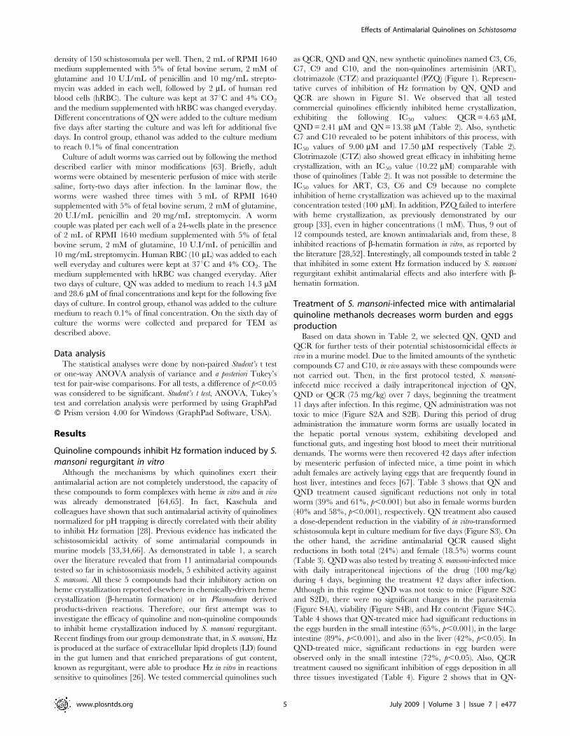
Quinoline compounds inhibit Hz formation induced by S.mansoni regurgitant in vitroAlthough the mechanisms by which quinolines exert their
antimalarial action are not completely understood, the capacity of
these compounds to form complexes with heme in vitro and in vivo
was already demonstrated [64,65]. In fact, Kaschula and
colleagues have shown that such antimalarial activity of quinolines
normalized for pH trapping is directly correlated with their ability
to inhibit Hz formation [28]. Previous evidence has indicated the
schistosomicidal activity of some antimalarial compounds in
murine models [33,34,66]. As demonstrated in table 1, a search
over the literature revealed that from 11 antimalarial compounds
tested so far in schistosomiasis models, 5 exhibited activity against
S. mansoni. All these 5 compounds had their inhibitory action on
heme crystallization reported elsewhere in chemically-driven heme
crystallization (b-hematin formation) or in Plasmodium derived
products-driven reactions. Therefore, our first attempt was to
investigate the efficacy of quinoline and non-quinoline compounds
to inhibit heme crystallization induced by S. mansoni regurgitant.
Recent findings from our group demonstrate that, in S. mansoni, Hz
is produced at the surface of extracellular lipid droplets (LD) found
in the gut lumen and that enriched preparations of gut content,
known as regurgitant, were able to produce Hz in vitro in reactions
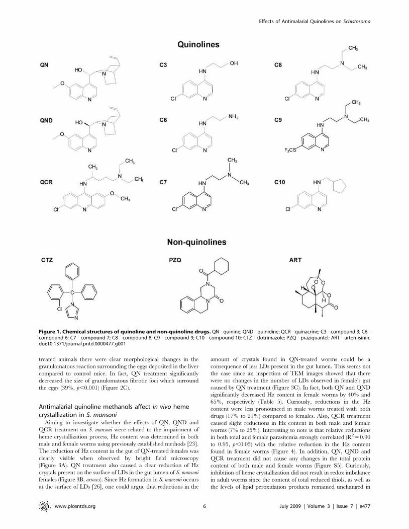
sensitive to quinolines [26]. We tested commercial quinolines such
as QCR, QND and QN, new synthetic quinolines named C3, C6,
C7, C9 and C10, and the non-quinolines artemisinin (ART),
clotrimazole (CTZ) and praziquantel (PZQ) (Figure 1). Represen-
tative curves of inhibition of Hz formation by QN, QND and
QCR are shown in Figure S1. We observed that all tested
commercial quinolines efficiently inhibited heme crystallization,
exhibiting the following IC50 values: QCR=4.63 mM,
QND=2.41 mM and QN=13.38 mM (Table 2). Also, synthetic
C7 and C10 revealed to be potent inhibitors of this process, with
IC50 values of 9.00 mM and 17.50 mM respectively (Table 2).
Clotrimazole (CTZ) also showed great efficacy in inhibiting heme
crystallization, with an IC50 value (10.22 mM) comparable with
those of quinolines (Table 2). It was not possible to determine the
IC50 values for ART, C3, C6 and C9 because no complete
inhibition of heme crystallization was achieved up to the maximal
concentration tested (100 mM). In addition, PZQ failed to interfere
with heme crystallization, as previously demonstrated by our
group [33], even in higher concentrations (1 mM). Thus, 9 out of
12 compounds tested, are known antimalarials and, from these, 8
inhibited reactions of b-hematin formation in vitro, as reported by
the literature [28,52]. Interestingly, all compounds tested in table 2
that inhibited in some extent Hz formation induced by S. mansoni
regurgitant exhibit antimalarial effects and also interfere with b-
hematin formation.
Treatment of S. mansoni-infected mice with antimalarialquinoline methanols decreases worm burden and eggsproductionBased on data shown in Table 2, we selected QN, QND and
QCR for further tests of their potential schistosomicidal effects in
vivo in a murine model. Due to the limited amounts of the synthetic
compounds C7 and C10, in vivo assays with these compounds were
not carried out. Then, in the first protocol tested, S. mansoni-
infecetd mice received a daily intraperitoneal injection of QN,
QND or QCR (75 mg/kg) over 7 days, beginning the treatment
11 days after infection. In this regime, QN administration was not
toxic to mice (Figure S2A and S2B). During this period of drug
administration the immature worm forms are usually located in
the hepatic portal venous system, exhibiting developed and
functional guts, and ingesting host blood to meet their nutritional
demands. The worms were then recovered 42 days after infection
by mesenteric perfusion of infected mice, a time point in which
adult females are actively laying eggs that are frequently found in
host liver, intestines and feces [67]. Table 3 shows that QN and
QND treatment caused significant reductions not only in total
worm (39% and 61%, p,0.001) but also in female worms burden
(40% and 58%, p,0.001), respectively. QN treatment also caused
a dose-dependent reduction in the viability of in vitro-transformed
schistosomula kept in culture medium for five days (Figure S3). On
the other hand, the acridine antimalarial QCR caused slight
reductions in both total (24%) and female (18.5%) worms count
(Table 3). QND was also tested by treating S. mansoni-infected mice
with daily intraperitoneal injections of the drug (100 mg/kg)
during 4 days, beginning the treatment 42 days after infection.
Although in this regime QND was not toxic to mice (Figure S2C
and S2D), there were no significant changes in the parasitemia
(Figure S4A), viability (Figure S4B), and Hz content (Figure S4C).
Table 4 shows that QN-treated mice had significant reductions in
the eggs burden in the small intestine (65%, p,0.001), in the large
intestine (89%, p,0.001), and also in the liver (42%, p,0.05). In
QND-treated mice, significant reductions in egg burden were
observed only in the small intestine (72%, p,0.05). Also, QCR
treatment caused no significant inhibition of eggs deposition in all
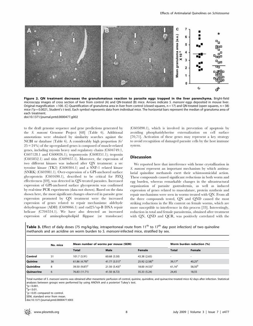
three tissues investigated (Table 4). Figure 2 shows that in QN-
Effects of Antimalarial Quinolines on Schistosoma
www.plosntds.org 5 July 2009 | Volume 3 | Issue 7 | e477
treated animals there were clear morphological changes in the
granulomatous reaction surrounding the eggs deposited in the liver
compared to control mice. In fact, QN treatment significantly
decreased the size of granulomatous fibrotic foci which surround
the eggs (39%, p,0.001) (Figure 2C).
Antimalarial quinoline methanols affect in vivo hemecrystallization in S. mansoniAiming to investigate whether the effects of QN, QND and
QCR treatment on S. mansoni were related to the impairment of
heme crystallization process, Hz content was determined in both
male and female worms using previously established methods [23].
The reduction of Hz content in the gut of QN-treated females was
clearly visible when observed by bright field microscopy
(Figure 3A). QN treatment also caused a clear reduction of Hz
crystals present on the surface of LDs in the gut lumen of S. mansoni
females (Figure 3B, arrows). Since Hz formation in S. mansoni occurs
at the surface of LDs [26], one could argue that reductions in the
amount of crystals found in QN-treated worms could be a
consequence of less LDs present in the gut lumen. This seems not
the case since an inspection of TEM images showed that there
were no changes in the number of LDs observed in female’s gut
caused by QN treatment (Figure 3C). In fact, both QN and QND
significantly decreased Hz content in female worms by 40% and
65%, respectively (Table 5). Curiously, reductions in the Hz
content were less pronounced in male worms treated with both
drugs (17% to 21%) compared to females. Also, QCR treatment
caused slight reductions in Hz content in both male and female
worms (7% to 25%). Interesting to note is that relative reductions
in both total and female parasitemia strongly correlated (R2=0.90
to 0.95, p,0.05) with the relative reduction in the Hz content
found in female worms (Figure 4). In addition, QN, QND and
QCR treatment did not cause any changes in the total protein
content of both male and female worms (Figure S5). Curiously,
inhibition of heme crystallization did not result in redox imbalance
in adult worms since the content of total reduced thiols, as well as
the levels of lipid peroxidation products remained unchanged in
Figure 1. Chemical structures of quinoline and non-quinoline drugs. QN - quinine; QND - quinidine; QCR - quinacrine; C3 - compound 3; C6 -compound 6; C7 - compound 7; C8 - compound 8; C9 - compound 9; C10 - compound 10; CTZ - clotrimazole; PZQ - praziquantel; ART - artemisinin.doi:10.1371/journal.pntd.0000477.g001
Effects of Antimalarial Quinolines on Schistosoma
www.plosntds.org 6 July 2009 | Volume 3 | Issue 7 | e477
QN-treated S. mansoni females (Figure S6). Finally, fluorescence
microscopy analyses revealed that in QN-treated worms the levels
of reactive species is undistinguishable from control ones assessed
by the fluorescent reactive probe CMH2-DCFDA (Figure S7).
Quinine treatment causes remarkable ultrastructuralchanges in the gastrodermis of adult wormsIn an attempt to investigate whether inhibition of heme
crystallization promoted by QN treatment affected parasite
ultrastructure, we observed control and QN-treated adult worms
by transmission electron microscopy (TEM) as shown in figure 4.
An initial inspection of different parasite regions indicated that
QN indeed caused ultrastructural modifications in S. mansoni.
Curiously, we did not observe substantial alterations in the
tegument and musculature of both female (Figure S8A and S8B)
and male (Figure S8C and S8D) worms recovered from QN-
treated mice. More importantly, detailed analyses indicated that
QN treatment caused remarkable ultrastructural changes in the
gastrodermis of both female (Figure 5A and 5B) and male worms
(Figure 5C and 5D). Noteworthy is the extensive loss of electron
density and clear indications of both cytoskeleton disorganization
and mitochondrial swelling observed in female gastrodermis
(Figure 5B). Regarding the male worms, the most striking change
observed in QN-treated worms was the washed-out aspect of the
cytosol of gastrodermis cells (Figure 5D). In addition, mitochon-
dria were found inside autophagic vacuoles in the gastrodermis
(Figure S9A) as well as with swollen appearance, washed-out
matrix and with remnants of inner membranes in the sub-
tegumentar region of QN-treated female worms (Figure S9C).
Finally, adult females cultured in vitro in the presence of QN
(14.3 mM) for 48 h promoted gastrodermis vacuolization and the
appearance of huge electrondense particles within the tissue
(Figure 6B). Interestingly, QN caused a complete destruction of
females gastrodermis after 72 h of treatment as shown in figure 6C
where LD usually found in the gut lumen were found in close
association in mitochondria, possibly derived from the gastro-
dermis.
Quinine treatment increased expression of transcriptsrelated to sexual differentiation, musculature,cytoskeleton and repair mechanisms in S. mansoni
femalesIn order to characterize the effect of QN treatment at the
molecular level, we performed large-scale gene expression analyses
using cDNA microarrays with approximately 4,000 different S.
mansoni gene fragments (GEO accession number: GPL3929) [58].
Gene expression levels were compared between female adult
worms recovered from infected mice that were treated with QN
and female worms recovered from control (non-treated) infected
mice. We have found 25 transcripts significantly up-regulated in
QN treated parasites (Figure S10). These transcripts were mapped
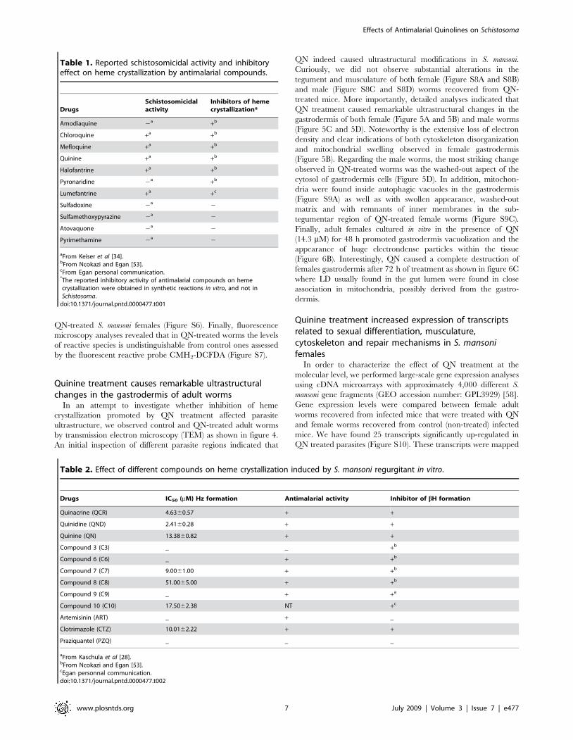
Table 1. Reported schistosomicidal activity and inhibitoryeffect on heme crystallization by antimalarial compounds.
Drugs
Schistosomicidal
activity
Inhibitors of heme
crystallization*
Amodiaquine 2a
+b
Chloroquine +a
+b
Mefloquine +a
+b
Quinine +a
+b
Halofantrine +a
+b
Pyronaridine 2a
+b
Lumefantrine +a
+c
Sulfadoxine 2a
2
Sulfamethoxypyrazine 2a
2
Atovaquone 2a
2
Pyrimethamine 2a
2
aFrom Keiser et al [34].bFrom Ncokazi and Egan [53].cFrom Egan personal communication.*The reported inhibitory activity of antimalarial compounds on hemecrystallization were obtained in synthetic reactions in vitro, and not inSchistosoma.doi:10.1371/journal.pntd.0000477.t001
Table 2. Effect of different compounds on heme crystallization induced by S. mansoni regurgitant in vitro.
Drugs IC50 (mM) Hz formation Antimalarial activity Inhibitor of bH formation
Quinacrine (QCR) 4.6360.57 + +
Quinidine (QND) 2.4160.28 + +
Quinine (QN) 13.3860.82 + +
Compound 3 (C3) _ _ +b
Compound 6 (C6) _ + +b
Compound 7 (C7) 9.0061.00 + +b
Compound 8 (C8) 51.0065.00 + +b
Compound 9 (C9) _ + +a
Compound 10 (C10) 17.5062.38 NT +c
Artemisinin (ART) _ + _
Clotrimazole (CTZ) 10.0162.22 + +
Praziquantel (PZQ) _ _ _
aFrom Kaschula et al [28].bFrom Ncokazi and Egan [53].cEgan personnal communication.doi:10.1371/journal.pntd.0000477.t002
Effects of Antimalarial Quinolines on Schistosoma
www.plosntds.org 7 July 2009 | Volume 3 | Issue 7 | e477
to the draft genome sequence and gene predictions generated by
the S. mansoni Genome Project [68] (Table 6). Additional
annotations were obtained by similarity searches against the
NCBI nr database (Table 6). A considerably high proportion (6/
25= 24%) of the up-regulated genes is composed of muscle-related
genes, including myosin heavy and regulatory chains (C604749.1,
C607128.1 and C600026.1); tropomyosin (C608351.1); troponin
(C605852.1) and titin (C609457.1). Moreover, the expression of
two different kinases was induced after QN treatment: a src
tyrosine kinase (TK3) (C600304.1) and a SNF-1 related kinase
(SNRK) (C603981.1). Over-expression of a GPI-anchored surface
glycoprotein (C601690.1), described to be critical for PZQ
effectiveness [69], was detected in QN-treated parasites. Increased
expression of GPI-anchored surface glycoprotein was confirmed
by real-time PCR experiments (data not shown). Based on the data
shown here, the most significant changes observed in parasite gene
expression promoted by QN treatment were the increased
expression of genes related to repair mechanisms: aldehyde
dehydrogenase (ADH) (C609066.1) and rad25/xp-B DNA repair
helicase (C704554.1). We have also detected an increased
expression of aminophospholipid flippase (or translocase)
(C605890.1), which is involved in prevention of apoptosis by
avoiding phosphatidylserine externalization on cell surface
[70,71]. Activation of these genes may represent a key strategy
to avoid recognition of damaged parasite cells by the host immune
system.
Discussion
We reported here that interference with heme crystallization in
S. mansoni represent an important mechanism by which antima-
larial quinoline methanols exert their schistosomicidal action.
These compounds caused significant reductions in both worm and
egg burden, whereas remarkable changes in the ultrastructural
organization of parasite gastrodermis, as well as induced
expression of genes related to musculature, protein synthesis and
repair mechanisms were seen in worms treated with QN. From all
the three compounds tested, QN and QND caused the most
striking reductions in the Hz content on female worms, which are
more susceptible to interference in this process [33]. Interestingly,
reduction in total and female parasitemia, obtained after treatment
with QN, QND and QCR, was positively correlated with the
Figure 2. QN treatment decreases the granulomatous reaction to parasite eggs trapped in the liver parenchyma. Bright-fieldmicroscopy images of cross section of liver from control (A) and QN-treated (B) mice. Arrows indicate S. mansoni eggs deposited in mouse liver.Original magnification6100. (C) Quantification of granuloma area in liver from control (closed squares, n = 17) and QN-treated (open squares, n = 38)mice (*p= 0.0021, Student’s t test). Each symbol represents data from individual mice. The horizontal bars represent the median of granuloma area ofeach treatment.doi:10.1371/journal.pntd.0000477.g002
Table 3. Effect of daily doses (75 mg/kg/day, intraperitoneal route from 11th to 17th day post infection) of two quinolinemethanols and an acridine on worm burden to S. mansoni-infected mice, stratified by sex.
No. mice Mean number of worms per mouse (SEM) Worm burden reduction (%)
Total Male Female Total Female
Control 51 101.7 (5.91) 60.68 (3.50) 43.38 (2.65) - -
Quinine 36 61.86 (4.79)a 41.77 (3.51)a 25.92 (2.58)b 39,17a 40,25a
Quinidine 8 39.50 (9.87)a 21.50 (5.43)a 18.00 (4.53)c 61,16a 58,50b
Quinacrine 6 76.83 (11.71) 41.50 (6.72) 35.33 (5.24) 24,45 18,55
Total number of S. mansoni worms was obtained after mesenteric perfusion of control, quinine, quinidine, and quinacrine-treated mice 42 days after infection. Statisticalanalyses between groups were performed by using ANOVA and a posteriori Tukey’s test.ap,0.001.bp,0.01.cp,0.05 compared to control.SEM, standard error from mean.doi:10.1371/journal.pntd.0000477.t003
Effects of Antimalarial Quinolines on Schistosoma
www.plosntds.org 8 July 2009 | Volume 3 | Issue 7 | e477
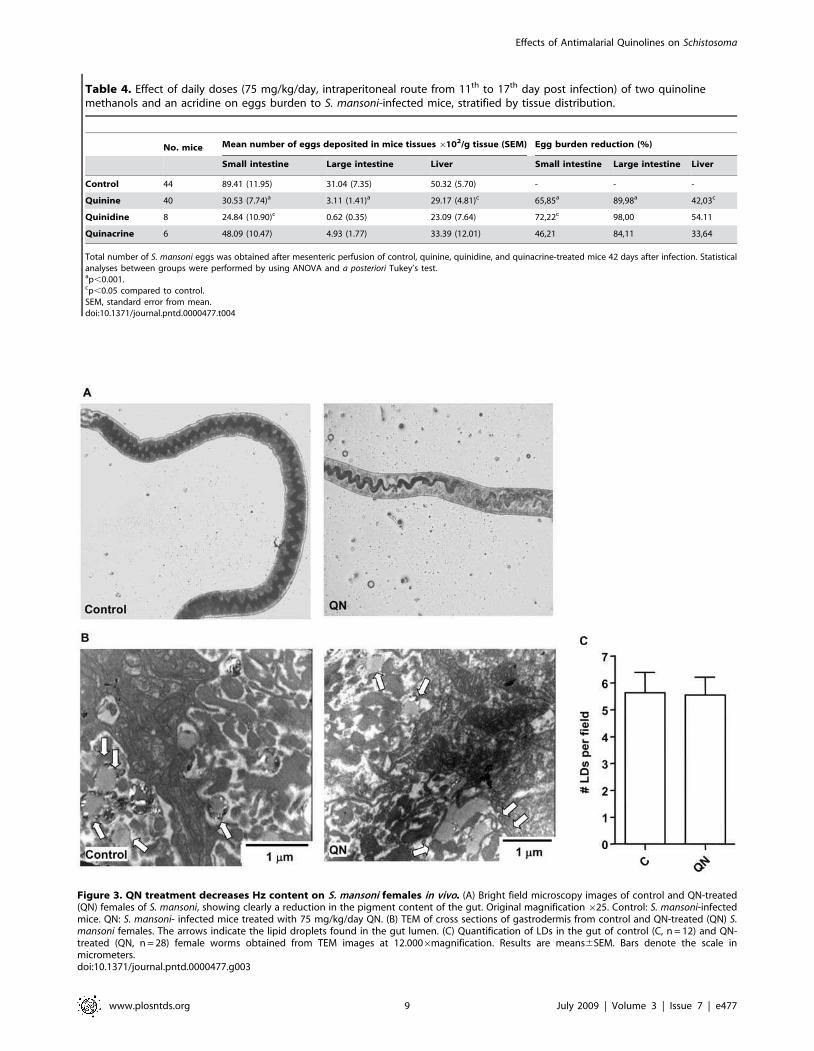
Table 4. Effect of daily doses (75 mg/kg/day, intraperitoneal route from 11th to 17th day post infection) of two quinolinemethanols and an acridine on eggs burden to S. mansoni-infected mice, stratified by tissue distribution.
No. mice Mean number of eggs deposited in mice tissues6102/g tissue (SEM) Egg burden reduction (%)
Small intestine Large intestine Liver Small intestine Large intestine Liver
Control 44 89.41 (11.95) 31.04 (7.35) 50.32 (5.70) - - -
Quinine 40 30.53 (7.74)a 3.11 (1.41)a 29.17 (4.81)c 65,85a 89,98a 42,03c
Quinidine 8 24.84 (10.90)c 0.62 (0.35) 23.09 (7.64) 72,22c 98,00 54.11
Quinacrine 6 48.09 (10.47) 4.93 (1.77) 33.39 (12.01) 46,21 84,11 33,64
Total number of S. mansoni eggs was obtained after mesenteric perfusion of control, quinine, quinidine, and quinacrine-treated mice 42 days after infection. Statisticalanalyses between groups were performed by using ANOVA and a posteriori Tukey’s test.ap,0.001.cp,0.05 compared to control.SEM, standard error from mean.doi:10.1371/journal.pntd.0000477.t004
Figure 3. QN treatment decreases Hz content on S. mansoni females in vivo. (A) Bright field microscopy images of control and QN-treated(QN) females of S. mansoni, showing clearly a reduction in the pigment content of the gut. Original magnification625. Control: S. mansoni-infectedmice. QN: S. mansoni- infected mice treated with 75 mg/kg/day QN. (B) TEM of cross sections of gastrodermis from control and QN-treated (QN) S.mansoni females. The arrows indicate the lipid droplets found in the gut lumen. (C) Quantification of LDs in the gut of control (C, n = 12) and QN-treated (QN, n = 28) female worms obtained from TEM images at 12.0006magnification. Results are means6SEM. Bars denote the scale inmicrometers.doi:10.1371/journal.pntd.0000477.g003
Effects of Antimalarial Quinolines on Schistosoma
www.plosntds.org 9 July 2009 | Volume 3 | Issue 7 | e477
percentage of Hz content reduction in female worms (Figure 3),
strenghtening the concept that interference with heme crystalliza-
tion plays an important role on the mechanism of schistosomicidal
action of antimalarial quinoline methanols. The present work,
together with previous evidence describing schistosomicidal
activity of antimalarial quinolines [33,34,66], strongly indicates
that heme crystallization in Schistosoma is a promising target for the
development of new schistosomicidal compounds.
Hz formation represents the main heme detoxification mech-
anism in S. mansoni, accounting for more than 50% of total heme
content in female worms [23]. Our group firstly proposed that
interference in Hz formation could be a potential target for new
therapies against schistosomiasis [33] by showing that treatment of
S. mansoni-infected mice with multiple intraperitoneal injections of
CLQ caused an important decrease in the overall severity of
disease. In this regard, it is important to notice some aspects
related to the results of inhibition of heme crystallization by
antimalarial quinoline methanols obtained in the present work, as
following: i) Hz content observed in female worms after QN and
QND treatment were normalized by the worm’s protein content
(Table 5), a parameter that was not affected after quinoline
methanols treatment (Figure S5). Thus, reductions in the Hz
content observed (Table 5) were not related to reduced worm
counts (Table 3) or to increased protein levels found upon
treatment; ii) since the LDs found in the gut lumen are responsible
for Hz production in S. mansoni [26] reduced Hz formation
(Table 5) is not a consequence of less LDs found in the gut lumen,
since treatment with antimalarial quinoline methanols did not
affected LD counts (Figure 3C); iii) the relative reduction in
parasitemia was positively correlated with the relative reduction of
Hz content in females (Figure 4), which, once again strongly
denotes the role of heme crystallization to parasite survival; iv)
previous evidence from the literature, demonstrating schistosomi-
cidal activity of antimalarial drugs, show a high agreement with
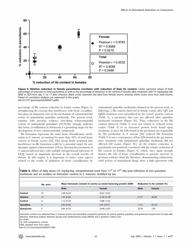
Figure 4. Relative reduction in female parasitemia correlates with reduction of their Hz content. Circles represent values of bothpercentage of reduction in total parasitemia as well as the percentage of reduction in Hz content in females after treatment with 75 mg/kg/day QN,QND or QCR from day 11 to 17 after infection. Black circles represent the data from female worms whereas white circles were from total worms.Values of correlation analyses are expressed in the graph.doi:10.1371/journal.pntd.0000477.g004
Table 5. Effect of daily doses (75 mg/kg/day, intraperitoneal route from 11th to 17th day post infection) of two quinolinemethanols and an acridine on hemozoin content in S. mansoni, stratified by sex.
No. mice Mean hemozoin content in worms as nmols heme/mg protein (SEM) Reduction in Hz content (%)
Male Female Male Female
Control 27 2.48 (0.35) 36.67 (3.97) - -
Quinine 27 1.95 (0.20) 21.90 (2.19)a 21.37 40.28c
Control 8 1.12 (0.19) 19.86 (1.52) - -
Quinidine 6 0.93 (0.30) 6.92 (0.91)a 17.31 65.16a
Quinacrine 6 0.84 (0.19) 18.46 (2.07) 25.54 7.05
Hemozoin content was obtained from S. mansoni worms recovered after mesenteric perfusion of control, quinine, quinidine, and quinacrine-treated mice 42 days afterinfection. Statistical analyses between groups were performed by using ANOVA and a posteriori Tukey’s test.ap,0.001.cp,0.05 compared to control.SEM, standard error from mean.doi:10.1371/journal.pntd.0000477.t005
Effects of Antimalarial Quinolines on Schistosoma
www.plosntds.org 10 July 2009 | Volume 3 | Issue 7 | e477
those compounds that also inhibit synthetic b-hematin formation
(Table 1). The only exception is amodiaquine, since the drug
interferes with heme crystallization in vitro, although did not cause
any change in parasitological parameters described by Keiser and
collagues [34] (Table 1). Conceivably, the metabolic processing of
amodiaquine, and its short plasmatic half-life (5.2 minutes), may
affect not only the drug accucmulation inside Schistosoma gut, but
also the final concentrations necessary to inhibit Hz formation,
ultimately failing to cause parasite death [34]; v) in table 2, from 12
compounds tested, 9 are known antimalarials and, from these, 8
are known inhibitors of b-hematin formation in vitro, as reported
by the literature [28,52]. Interestingly, 6 out of these same 8
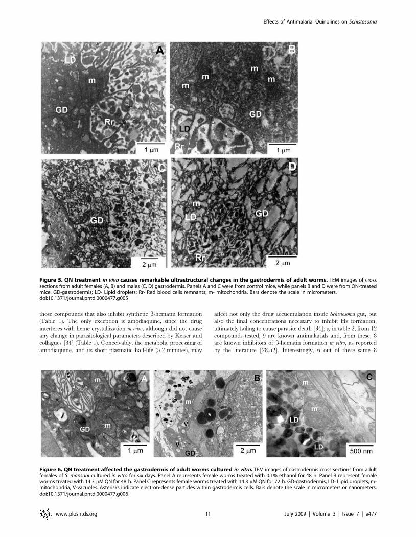
Figure 5. QN treatment in vivo causes remarkable ultrastructural changes in the gastrodermis of adult worms. TEM images of crosssections from adult females (A, B) and males (C, D) gastrodermis. Panels A and C were from control mice, while panels B and D were from QN-treatedmice. GD-gastrodermis; LD- Lipid droplets; Rr- Red blood cells remnants; m- mitochondria. Bars denote the scale in micrometers.doi:10.1371/journal.pntd.0000477.g005
Figure 6. QN treatment affected the gastrodermis of adult worms cultured in vitro. TEM images of gastrodermis cross sections from adultfemales of S. mansoni cultured in vitro for six days. Panel A represents female worms treated with 0.1% ethanol for 48 h. Panel B represent femaleworms treated with 14.3 mM QN for 48 h. Panel C represents female worms treated with 14.3 mM QN for 72 h. GD-gastrodermis; LD- Lipid droplets; m-mitochondria; V-vacuoles. Asterisks indicate electron-dense particles within gastrodermis cells. Bars denote the scale in micrometers or nanometers.doi:10.1371/journal.pntd.0000477.g006
Effects of Antimalarial Quinolines on Schistosoma
www.plosntds.org 11 July 2009 | Volume 3 | Issue 7 | e477
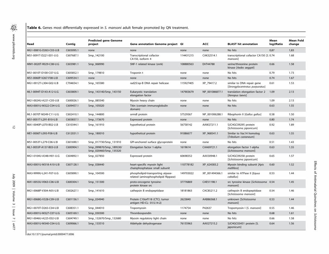
Table 6. Genes most differentially expressed in S. mansoni adult female promoted by QN treatment.
Read Contig
Predicted gene Genome
project Gene annotation Genome project GI ACC BLAST hit annotation
Mean
log2Ratio
Mean Fold
change
MG1-0081G-D263-C03-U.B C603095.1 none none none none No hits 0,87 1,83
MS1-0091T-D221-E01-U.G C607687.1 Smp_142100 Transcriptional cofactorCA150, isoform 4
114431215 CAK32514.1 transcriptional cofactor CA150 [S.mansoni]
0.74 1.68
MM1-0020T-R029-C08-U.G C603981.1 Smp_068990 SNF-1 related kinase (snrk) 108880563 EAT44788 serine/threonine proteinkinase [Aedes aegypti]
0.66 1.58
MS1-0010T-D100-C07-U.G C605852.1 Smp_179810 Troponin t none none No hits 0.79 1.73
MS1-0060P-V267-F08-U.B C609534.1 none none none none No hits 0.74 1.67
MG1-0012T-L304-G02-U.B C704654.1 Smp_165580 rad25/xp-B DNA repair helicase 115927405 XP_79417.2 similar to DNA repair gene[Strongylocentrotus purpuratus]
0.61 1.53
ML1-0094T-D143-A12-U.G C603809.1 Smp_143140/Smp_143150 Eukaryotic translationelongation factor
147903679 NP_001086877.1 translation elongation factor 2[Xenopus laevis]
1.09 2.13
MG1-0024U-A221-C05-U.B C600026.1 Smp_085540 Myosin heavy chain none none No hits 1.09 2.13
MA3-0001U-M322-C04-U.G C609457.1 Smp_105020 Titin (contain immunoglobulindomain)
none none No hits 0.63 1.55
ML1-0070T-M249-C11-U.G C602410.1 Smp_144800 ormdl protein 57529367 NP_001006288.1 Ribophorin II [Gallus gallus] 0.58 1.50
MS1-0051T-L291-B10-U.B C603837.1 Smp_173670 Expressed protein none none No hits 0.80 1.74
MA1-0040P-L070-B02-U.B C610344.1 Smp_161910 hypothetical protein 56758142 AAW27211.1 SJCHGC09295 protein[Schistosoma japonicum]
0.92 1.89
ME1-0006T-L093-F08-U.B C612031.1 Smp_180010 hypothetical protein 91086677 XP_968541.1 Similar to Vac14 homolog[Tribolium castaneum]
0.63 1.55
MS1-0053T-L279-C06-U.B C601690.1 Smp_017730/Smp_131910 GPI-anchored surface glycoprotein none none No hits 0.51 1.43
ML1-0055P-A137-B03-U.B C600904.1 Smp_099870/Smp_189530/Smp_020460/Smp_135320
Elongation factor 1-alpha 1619614 CAA69721.1 elongation factor 1-alpha[Schistosoma mansoni]
0.63 1.55
MG1-0104U-A348-H01-U.G C604892.1 Smp_027850 Expressed protein 60698352 AAX30948.1 SJCHGC09258 protein[Schistosoma japonicum]
0.65 1.57
MA3-0001U-M318-A10-U.B C607128.1 Smp_038440 heart-specific myosin lightchainphosphatase small subunit
110778182 XP_624583.2 Myosin binding subunit [Apismellifera]
0.60 1.52
MA3-9999U-L241-F07-U.G C605890.1 Smp_104500 phospholipid-transporting atpase-related (aminophospholipid flippase)
149703022 XP_001494366.1 similar to ATPase II [Equuscaballus]
0.53 1.44
MA1-0053U-V063-C06-U.B C600304.1 Smp_151300 proto-oncogene tyrosine-protein kinase src
37776869 CAE51198.1 src tyrosine kinase [Schistosoma
mansoni]0.54 1.45
MG1-0068P-V304-A05-U.B C602627.1 Smp_141610 cathepsin B endopeptidase 18181863 CAC85211.2 cathepsin B endopeptidase[Schistosoma mansoni]
0.54 1.46
MG1-0068G-V328-C09-U.B C601156.1 Smp_034940 Protein C10orf118 (CTCL tumorantigen HD-CL- 01/L14-2)
2623840 AAB86568.1 unknown [Schistosoma
mansoni]0.53 1.44
MG1-0070T-D265-C04-U.B C608351.1 Smp_044010 Tropomyosin 1174754 P42637 Tropomyosin I [S. mansoni] 0.55 1.46
MA3-0001U-M327-C07-U.G C605169.1 Smp_030300 Thrombospondin none none No hits 0.68 1.61
MG1-0046U-A225-E02-U.B C604749.1 Smp_132670/Smp_132680 Myosin regulatory light chain none none No hits 0.66 1.58
MA3-0001U-M340-C04-U.G C609066.1 Smp_133510 Aldehyde dehydrogenase 76155963 AAX27215.2 SJCHGC03451 protein [S.japonicum]
0.64 1.56
doi:10.1371/journal.pntd.0000477.t006
Effects
ofAntim
alarialQuinolin
esonSch
istosoma
www.plosntds.o
rg12
July
2009
|Volume3
|Issu
e7
|e477
compounds inhibited Hz formation induced by S. mansoni
regurgitant with different intensities. Therefore, although alterna-
tive explanations can be raised, interference with Hz formation
seems to be the simplest explanation for the findings presented
here.
Several reports proposed the relevance of a chlorine atom at 7-
position of the quinoline ring as an important structural
determinant for the inhibitory effects on heme crystallization in
vitro [28,53]. Despite its obvious relevance, this feature alone is not
sufficient to determine their antimalarial effect because another
important aspect must be considered. Drugs accumulation within
the Plasmodium food vacuole play also a key role since this allow
quinoline compounds reach intracellular concontrations necessary
to inhibit with Hz formation. In fact, Kaschula and colleagues
obtained an excellent correlation between antiplasmodial activity,
normalized for pH trapping, and beta-hematin inhibitory activity
[28]. In this regard, despite the lower IC50 values obtained in vitro,
neither Hz formation in vivo nor parasite burden were affected by
QND when it was administered later on infection (Figure S4).
However, reduction in both Hz content in female worms (Table 5),
as well as parasite and eggs burden were achieved when QND was
administered early on the infection (Tables 3 and 4), indicating
that interference with heme crystallization is an important aspect
to determine its schistosomicidal effects. Also, QCR treatment
caused no changes in worm burden and Hz formation as well
(Tables 3 and 5). Thus, based on the data presented here, we
cannot exclude the possibility that drug accumulation, through pH
trapping, could be also determining some of the schistosomicidal
effects of antimalarial quinoline methanols. Since these com-
pounds are weak bases, it is possible that their accumulation may
affect the parasite gut pH and then interfering with parasite
proteolytic enzymes. In fact, Bogitsch and Davenport demonstrat-
ed that lysosomotropic agents, such as chloroquine, accumulates
within S. mansoni schistosomula cultured in vitro [72]. Further
research on these aspects will provide a better understanding on
the contribution of these factors in determining the schistosomi-
cidal action of quinolines.
Praziquantel (PZQ) administration represents the main strategy
for schistosomiasis control worldwide, but the threat of resistance
development to PZQ has prompted the search for new
schistosomicidal compounds [39]. In this sense, quinoline
compounds have been used as the main therapeutic option for
malaria treatment for more than 300 years [73]. Their wide use in
the treatment of malaria is in part due to their high safety and
efficacy, in addition to their well known pharmacokinetics.
Consensus over the mechanism of antimalarial action of CLQ
suggests that it relies on its interaction with heme [64] by forming
a complex that accumulates within the acidic food vacuole [74]
which impairs heme crystallization therein [75]. The complex
formed between heme and quinolines increased heme affinity for
biological membranes, enhancing its toxic effects [29,74,75] and
also inhibiting heme crystallization in Plasmodium [76,77,78].
Possibly, heme molecules derived from hemoglobin digestion
would be diverted from their natural fate (Hz) by forming a stable
complex with quinoline methanols, which, in turn, would increase
their association with hydrophilic-hydrophobic interfaces present
not only in LD but also in other biological membranes, such as
those of the gastrodermis cells (Figures 3B, 5B, 5D and 6C).
Moreover, increased association of heme-QN complex with the
plasma membrane of gastrodermis cells would explain the
remarkable cytoskeleton disorganization observed in QN-treated
worms (Figures 3B, 5B, 5D and 6C), caused by heme-induced
changes in membrane stability and oxidative modifications. An
interesting observation in table 2 is that PZQ caused no effect on
heme crystallization in vitro, reinforcing the potential of therapeutic
regimens combining PZQ with antimalarial quinoline methanols,
as these compounds affect distinct metabolic pathways. Further
research is necessary to determine whether this combined
treatment turns out to be a valuable clinical strategy for
schistosomiasis chemotherapy.
Quinoline methanols administration did not result in complete
elimination of parasites from S. mansoni-infected mice, and a
possible explanation could be the induction of genes that enable
parasite survival upon impaired heme crystallization. Changes
observed in parasite gene expression would be a consequence of
increased levels of non-crystallized heme complexed to quinoline
methanols. Noteworthy, QN-exposed parasites showed up-regu-
lation of 25 genes (Table 6), which can be grouped into 8
categories: sexual differentiation, musculature, cytoskeleton, signal
transduction, transcription/translation, protein digestion, recogni-
tion, and repair mechanisms. Curiously, among the induced genes
was TK3 (probe C600304.1), a gene encoding a src tyrosine kinase
predominantly expressed in the reproductive organs, as well as in
female’s vitellarium [79], being involved in signal transduction
pathways related to the cytoskeleton in Schistosoma gonads [79].
TK3 is a herbimycin probable target, having key functions in
regulating gonad development and egg production [80]. Over-
expression of a kinase with putative cell-architecture regulatory
function may indicate that a cytoskeleton reorganization process
takes place as part of an effective survival response to QN
administration.
In QN-treated worms we have detected over-expression of a
gene encoding a GPI-anchored surface glycoprotein (probe
C601690.1), described to be critical for PZQ effectiveness [69].
It has been postulated that antibodies that develop against some
unique glycoproteins exposed at the parasites’ tegument surface
are important for an effective action of PZQ [42], as PZQ
effectiveness is impaired in immune-deficient animals [42]. In fact,
monoclonal antibodies that recognize the GPI-anchored surface
glycoprotein are able to restore PZQ efficacy in B-cell depleted
mice [69]. It is tempting to hypothesize that the observed QN-
induced over-expression of the tegument glycoprotein gene could
favor an augmented action of PZQ, thus favoring the idea of a
QN-PZQ combined treatment; since the threat of PZQ resistance
development represents a real concern, QN would be a good PZQ
partner for schistosomiasis treatment.
Despite the schistosomicidal effects of quinoline methanols,
direct evidence linking this property to increased oxidative stress
conditions, due to impaired heme crystallization in parasite gut
(Figure 3B, and Table 5) remains inconclusive. Measurements of
total reduced thiol content (Figure S6A) as well as lipid
peroxidation products (Figure S6B) and the assessment of reactive
species in QN-treated worms by using the fluorescent probe
CMH2-DCFA (Figure S7), failed to demonstrate changes in these
parameters. We speculate that the absence of oxidative stress
markers in QN-treated parasites could be explained by an
increased expression of genes related to repair mechanisms. In
fact, we observed that 4 out of 25 genes over-expressed in QN-
treated worms were related to mechanisms involved in cellular
repair and maintenance (Table 6). Noteworthy is the increased
expression of aldehyde dehydrogenase (ADH) gene, which encodes
an enzyme involved in detoxification of lipid peroxidation
products such as reactive malondialdehyde and hydroxynonenal
[81], and of rad25/xp-B DNA repair helicase gene. Increased
expression of both ADH and rad25/xp-B DNA repair helicase
genes would explain the preserved ultrastructure in surviving
parasites recovered at the end of the treatment period. Further,
ultrastructural evidence (Figure S9A) indicates that mitochondrial
Effects of Antimalarial Quinolines on Schistosoma
www.plosntds.org 13 July 2009 | Volume 3 | Issue 7 | e477
autophagy in the gastrodermis of QN-treated female worms would
play a cellular protective role by eliminating dysfunctional
organelles and preventing mitochondrial-induced apoptosis.
In conclusion, the results presented here show that interference
with heme crystallization in S. mansoni by quinoline methanols
exerts promising schistosomicidal effects, causing significant
reductions in several pathologic parameters of disease. Since the
endemic areas of malaria and schistosomiasis overlap in many
regions of the globe and, considering that Hz formation occurs
both in Plasmodium and Schistosoma parasites, retrospective clinical
analyses of malaria patients treated with quinolines in these
regions would be quite informative concerning the potential use of
heme crystallization inhibitors as anti-parasitic agents for both
diseases.
Supporting Information
Figure S1 Dose-dependent inhibition of S. mansoni regurgitant-
driven Hz formation in vitro by QN, QND and QCR.
Representative IC50 curves of inhibition of Hz formation in
reactions promoted by S. mansoni female regurgitants were
conducted in the presence of different concentrations of (A) QN,
(B) QND and (C) QCR, as described in the methods section. All
drugs were tested in concentrations ranging from 5–100 mM.
Found at: doi:10.1371/journal.pntd.0000477.s001 (7.16 MB TIF)
Figure S2 QN and QND treatment were not toxic to S. mansoni-
infected mice. Alanine aminotransferase (ALT) (A and C) and
aspartate aminotransferase (AST) (B and D) activities were assayed
in plasma samples from C, QN (A, B) or QND (C, D) treated mice
infected with S. mansoni as markers for hepatocellular damage. QN
treatment means S. mansoni-infected mice treated with 75 mg/kg/
day QN from day 11 to 17 after infection, whereas in QND
treatment mice were treated from day 42 to 45 after infection with
daily intraperitoneal injections of 100 mg/kg QND. Results were
expressed as mean6SEM (n= 10, for A and B; n = 3, for C and
D).
Found at: doi:10.1371/journal.pntd.0000477.s002 (4.14 MB TIF)
Figure S3 QN caused a significant reduction in the viability of
cultured in vitro transformed schistosomula. Effect of QN
treatment on the viability of cultured in vitro-transformed
schistosomula assessed by MTT reduction. Control means
schistosomula treated with 0.1% ethanol, whereas in the
experimental groups, QN was added in concentrations ranging
from 10–100 mM. Results were expressed as mean6SEM. *
p,0.001, QN 10–100 mM vs. control (C) (one-way ANOVA and
a posteriori Tukey’s test).
Found at: doi:10.1371/journal.pntd.0000477.s003 (1.30 MB TIF)
Figure S4 QND treatment later on infection did not affect
parasite burden, viability or Hz content. (A) Effect of QND
treatment on total number of S. mansoni female and male worms in
control (n = 5) and QND-treated (n= 5) mice. (B) Effect of QND
treatment on the viability of S. mansoni female and male worms in
control (n = 5) and QND-treated (n = 5) mice. (C) Hz content in
female and male S. mansoni worms in control (n = 5) and QND-
treated (n = 5) mice. Hz was extracted from S. mansoni and
quantified as described in materials and methods. Control (C): S.
mansoni-infected mice treated with about 100 mL of 30.0% ethanol.
QND: S. mansoni-infected mice treated with 100 mg/kg/day QND
from day 42 to 45 after infection. Results are expressed as
mean6SEM.
Found at: doi:10.1371/journal.pntd.0000477.s004 (3.35 MB TIF)
Figure S5 QN and QND treatment did not affect protein
content in adult worms. (A) Effect of QN treatment on protein
content in female and male S. mansoni worms. Results are
expressed as mean6SEM (n= 27). (B) Effect of QND and QCR
treatment on protein content in female and male S. mansoni worms
(n= 4–9). Control (C) means S. mansoni-infected mice treated with
about 100 mL of 30.0% ethanol. QN, QND and QCR mean S.
mansoni-infected mice treated with 75 mg/kg/day of each
compound from day 11 to 17 after infection. Results were
expressed as mean6SEM. * p,0.001 one-way ANOVA and a
posteriori Tukey’s test, for males vs. their respective female groups.
Found at: doi:10.1371/journal.pntd.0000477.s005 (2.50 MB TIF)
Figure S6 QN treatment did not affect total thiol levels nor
induce lipid peroxidation in adult worms. (A) Effect of QN
treatment on the total thiol content in female and male S. mansoni
worms (n= 27). (B) Effect of QN treatment on lipid peroxidation in
female and male S. mansoni worms (n= 12), assessed by the TBARS
method. Control (C) means S. mansoni-infected mice treated with
about 100 mL of 30.0% ethanol, whereas QN means S. mansoni-
infected mice treated with 75 mg/kg/day QN from day 11 to 17
after infection. Results are expressed as mean6SEM.
Found at: doi:10.1371/journal.pntd.0000477.s006 (2.41 MB TIF)
Figure S7 QN did not increase reactive species formation in
adult worms. Effect of QN treatment on the reactive species
formation in female (A) and male (B) S. mansoni worms.
Intracellular reactive species from S. mansoni worms were
quantified as described in methods section using the fluorescent
probe CMH2-DCFDA. Control means S. mansoni-infected mice
with about 100 mL of 30.0% ethanol, whereas QN means S.
mansoni-infected mice treated with 75 mg/kg/day QN from day 11
to 17 after infection. Images of worms were acquired in both
bright field (left) and epifluorescence (right) microscopy.
Found at: doi:10.1371/journal.pntd.0000477.s007 (8.43 MB TIF)
Figure S8 QN treatment did not cause ultrastructural changes
in the tegument of adult worms. TEM images of tegument cross
sections from females (A, B) and males (C, D) of S. mansoni. Panels
A and C were from control worm, while panels B and D were from
QN-treated worm. T- tegument and MF- muscle fibers. Bars
denote the scale in micrometers.
Found at: doi:10.1371/journal.pntd.0000477.s008 (9.60 MB TIF)
Figure S9 QN treatment promotes mitochondrial autophagy
and morphological changes in female worms. TEM of cross-
sections images from S. mansoni adult females obtained from QN-
treated mice. (A) Gastrodermis of QN-treated female worm
showing a mitochondrion inside of an autophagic vacuole depicted
inside the white-dashed box. Panels B and C were from sub-
tegumentar region of a female worm obtained from control (B) and
QN-treated mice (C). MF-muscular fiber. Arrows indicate a clear
swelling of the mitochondria and arrowhead indicates remnants of
inner mitochondrial membrane. The asterisk indicates a washed-
out mitochondrial matrix and the inset in panel C depicts a
magnification of a swollen mitochondrion. Control means S.
mansoni-infected mice treated with about 100 mL of 30.0% ethanol,
whereas QN means S. mansoni-infected mice treated with 75 mg/
kg/day QN from day 11 to 17 after infection. Bars denote the
scale in micrometers.
Found at: doi:10.1371/journal.pntd.0000477.s009 (9.43 MB TIF)
Figure S10 QN treatment changed gene expression of S. mansoni
female worms. Genes identified as differentially up regulated in
female worms treated with quinine when compared with control
females (see methods for details). Heat map representing the 25
genes identified as significantly (FDR 0.1%) over-expressed after
treatment. Each line represents one gene and each two adjacent
columns represent replicas of one experiment, as indicated at the
Effects of Antimalarial Quinolines on Schistosoma
www.plosntds.org 14 July 2009 | Volume 3 | Issue 7 | e477
bottom of the panel. Expression levels of genes are represented by
the log2 (treatment/control ratio). Sample 1.x and 2.x represent
the dye swap replicates for sample 1 and 2, respectively.
Found at: doi:10.1371/journal.pntd.0000477.s010 (0.82 MB TIF)
Acknowledgments
We are grateful to Mr. Claudio P. Figueira, Mrs. Elisangela Sodre, Mr.
Joao V. de Oliveira Neto and Mrs Maria Marta Freire for the excellent
technical assistance provided. The authors would also like to express our
gratitude to all staff of Laboratory of Redox Biochemistry for the valuable
help on the manuscript revision.
Author Contributions
Conceived and designed the experiments: MAVS SVA RH TJE MFO.
Performed the experiments: JBRCS DM MAVS AFP GTA TMV SVA
VKZ DK RH TJE MFO. Analyzed the data: JBRCS DM MAVS AFP
GTA TMV SVA VKZ DK RH TJE MFO. Contributed reagents/
materials/analysis tools: MAVS AFP SVA VKZ DK RH TJE MFO.
Wrote the paper: MAVS GTA TMV SVA TJE MFO.
References
1. WHO (2003–2004) Making health research work for poor people. TropicalDisease Research TDR/GEN/05.1.
2. Fenwick A (2006) New initiatives against Africa’s worms. Trans R Soc TropMed Hyg 100: 200–207.
3. Chitsulo L, Engels D, Montresor A, Savioli L (2000) The global status ofschistosomiasis and its control. Acta Trop 77: 41–51.
4. Steinmann P, Keiser J, Bos R, Tanner M, Utzinger J (2006) Schistosomiasis andwater resources development: systematic review, meta-analysis, and estimates ofpeople at risk. Lancet Infect Dis 6: 411–425.
5. King CH, Dickman K, Tisch DJ (2005) Reassessment of the cost of chronichelmintic infection: A meta-analysis of disability-related outcomes in endemicschistosomiasis. Lancet 365: 1561–1569.
6. Boros DL (1989) Immunopathology of Schistosoma mansoni infection. ClinMicrobiol Rev 2: 250–269.
7. Lawrence JD (1973) The ingestion of red blood cells by Schistosoma mansoni.J Parasitol 59: 60–63.
8. Delcroix M, Sajid M, Caffrey CR, Lim KC, Dvorak J, Hsieh I, Bahgat M,Dissous C, McKerrow JH (2006) A multienzyme network functions in intestinalprotein digestion by a platyhelminth parasite. J Biol Chem 281: 39316–39329.
9. Morales ME, Rinaldi G, Gobert GN, Kines KJ, Tort JF, Brindley PJ (2008)RNA interference of Schistosoma mansoni cathepsin D, the apical enzyme of thehemoglobin proteolysis cascade. Mol Biochem Parasitol 157: 160–168.
10. Kloetzel K, Lewert RM (1966) Pigment formation in Schistosoma mansoniinfections in the white mouse. Am J Trop Med Hyg 15: 28–31.
11. Ponka P (1999) Cell biology of haem. Am J Med Sci 318: 241–256.
12. Davies MJ (1988) Detection of peroxyl and alkoxyl radicals produced by reactionof hydroperoxides with heme-proteins by electron spin resonance spectroscopy.Biochim Biophys Acta 964: 28–35.
13. Van der Zee J, Barr DP, Mason RP (1996) ESR spin trapping investigation ofradical formation from the reaction between hematin and tert-butyl hydroper-oxide. Free Radic Biol Med 20: 199–206.
14. Tappel AL (1955) Unsaturated lipide oxidation catalyzed by hematincompounds. J Biol Chem 217: 721–733.
15. Gutteridge JM, Smith A (1988) Antioxidant protection by haemopexin of haem-stimulated lipid peroxidation. Biochem J 256: 861–865.
16. Aft RL, Mueller GC (1984) Hemin-mediated oxidative degradation of proteins.J Biol Chem 259: 301–305.
17. Aft RL, Mueller GC (1983) Hemin-mediated DNA strand scission. J Biol Chem258: 12069–12072.
18. Chou AC, Fitch CD (1981) Mechanism of hemolysis induced by ferriprotopor-phyrin IX. J Clin Invest 68: 672–677.
19. Schmitt TH, Frezzatti WA Jr, Schreier S (1993) Hemin-induced lipid membranedisorder and increased permeability: a molecular model for the mechanism ofcell lysis. Arch Biochem Biophys 307: 96–103.
20. Graca-Souza AV, Maya-Monteiro C, Paiva-Silva GO, Braz GR, Paes MC, et al.(2006) Adaptations against heme toxicity in blood-feeding arthropods. InsectBiochem Mol Biol 36: 322–335.
21. Slater AF, Swiggard WJ, Orton BR, Flitter WD, Goldberg DE, et al. (1991) Aniron–carboxylate bond links the heme units of malaria pigment. Proc Natl AcadSci USA 88: 325–329.
22. Oliveira MF, Silva JR, Dansa-Petretski M, De Souza W, Lins U, et al. (1999)Haem detoxification by an insect. Nature 400: 517–518.
23. Oliveira MF, d’Avila JC, Torres CR, Oliveira PL, Tempone AJ, et al. (2000)Haemozoin in Schistosoma mansoni. Mol Biochem Parasitol 111: 217–221.
24. Oliveira MF, Timm BL, Machado EA, Miranda K, Attias M, et al. (2002) Onthe pro-oxidant effects of haemozoin. FEBS Lett 512: 139–144.
25. Oliveira MF, Kycia SW, Gomez A, Kosar AJ, Bohle DS, et al. (2005) Structuraland morphological characterization of hemozoin produced by Schistosomamansoni and Rhodnius prolixus. FEBS Lett 579: 6010–6016.
26. Correa Soares JB, Maya-Monteiro CM, Bittencourt-Cunha PR, Atella GC,Lara FA, et al. (2007) Extracellular lipid droplets promote hemozoincrystallization in the gut of the blood fluke Schistosoma mansoni. FEBS Lett581: 1742–1750.
27. Egan TJ (2006) Interactions of quinoline antimalarials with hematin in solution.J Inorganic Biochem 100: 916–926.
28. Kaschula CH, Egan TJ, Hunter R, Basilico N, Parapini S, et al. (2002)Structure-Activity Relationships in 4-Aminoquinoline Antiplasmodials. TheRole of the Group at the 7-Position. J Med Chem 45: 3531–3539.
29. Ginsburg H, Demel RA (1983) The effect of ferriprotoporphyrin IX andchloroquine on phospholipid monolayers and the possible implications toantimalarial activity. Biochim Biophys Acta 732: 316–319.
30. Bohle DS, Kosar AD, Madsen SK (2002) Propionic acid side chain hydrogenbonding in the malaria pigment beta-hematin. Biochem Biophys Res Commun294: 132–135.
31. Sullivan Jr DJ, Gluzman IY, Russell DG, Goldberg DE (1996) On the molecularmechanism of chloroquine, s antimalarial action. Proc Natl Acad Sci USA 93:11865–11870.
32. Oliveira MF, Silva JR, Dansa-Petretski M, de Souza W, Braga CMS, et al.(2000) Hemozoin formation in the midgut of the blood sucking insect Rhodniusprolixus. FEBS Lett 477: 95–98.
33. Oliveira MF, d’Avila JCP, Tempone AJ, Soares JBR, Runjanek FD, et al. (2004)Inhibition of heme aggregation by chloroquine reduces Schistosoma mansoniinfection. J Infect Dis 190: 843–852.
34. Keiser J, Chollet J, Xiao SH, Mei JY, Jiao PY, et al. (2009) Mefloquine-anaminoalcohol with promising antischistosomal properties in mice. PLoS NeglTrop Dis 3(1): e350.
35. Ribeiro-dos-Santos G, Verjovski-Almeida S, Leite LC (2006) Schistosomiasis–acentury searching for chemotherapeutic drugs. Parasitol Res 99: 505–521.
36. WHO (2002) Prevention and control of schistosomiasis and soil-transmittedhelminthiasis: report of a WHO expert committee. Geneva: WHO Tech RepSer No 912.
37. Cioli D, Pica-Mattoccia L (2003) Praziquantel. Parasitol Res 90: 53–59.
38. Utzinger J, Keiser J (2004) Schistosomiasis and soil- transmitted helminthiasis:common drugs for treatment and control. Expert Opin Pharmacother 5:263–285.
39. Ismail M, Botros S, Metwally A, William S, Farghally A, et al. (1999) Resistanceto praziquantel: direct evidence from Schistosoma mansoni isolated fromEgyptian villagers. Am J Trop Med Hyg 60: 932–935.
40. Danso-Appiah A, de Vlas SJ (2002) Interpreting low praziquantel cure rates ofSchistosoma mansoni infections in Senegal. Trends Parasitol 18: 125–129.
41. Botros S, Sayed H, Amer N, et al. (2005) Current status of sensitivity topraziquantel in a focus of potential drug resistance in Egypt. Int J Parasitol 35:787–791.
42. Cioli D (1998) Chemotherapy of schistosomiasis: an update. Parasitol Today 14:418–422.
43. Xiao SH (2005) Development of antischistosomal drugs in China, withparticular consideration to praziquantel and the artemisinins. Act Trop 96:153–157.
44. Xiao SH, Booth M, Tanner M (2000) The prophylactic effect of artemetheragainst Schistosoma japonicum infection. Parasitol Today 16: 122–126.
45. Li YS, Chen HG, He HB, Hou XY, Ellis M, et al. (2005) A double-bind fieldtrial on the effects of artemether on Schistosoma japonicum infection in a highlyendemic focus in southern China. Acta Trop 96: 184–190.
46. Abdulla MH, Lim KC, Sajid M, McKerrow JH, Caffrey CR (2007) Schistosomamansoni: novel chemotherapy using a cysteine protease inhibitor. PLoS Medicine4: e14.
47. Kuntz AN, Davioud-Charvet E, Sayed AA, Califf LL, Dessolin J, et al. (2007)Thioredoxin glutathione reductase from Schistosoma mansoni: an essentialparasite enzyme and a key drug target. PLoS Medicine 4: e206.
48. Simeonov A, Jadhav A, Sayed AA, Wang Y, Nelson ME, et al. (2008)Quantitative High-Throughput Screen Identifies Inhibitors of the Schistosomamansoni Redox Cascade. PLoS Negl Trop Dis 2: e127.
49. Sayed AA, Simeonov A, Thomas CJ, Inglese J, Austin CP, et al. (2008)Identification of oxadiazoles as new drug leads for the control of schistosomiasis.Nat Med 14: 407–412.
50. Smithers SR, Terry RJ (1965) The infection of laboratory hosts with cercariae ofSchistosoma mansoni and the recovery of adult worms. Parasitol 55: 695–700.
51. Lowry OH, Rosebrough NJ, Farr AR, Randall RJ (1951) Protein measurementwith the Folin phenol reagent. J Biol Chem 193: 265–275.
52. Egan TJ, Hunter R, Kaschula CH, Marques HM, Misplon A, et al. (2000)Structure-function relationships in aminoquinolines: effect of amino and chlorogroups on quinoline-hematin complex formation, inhibition of b-hematinformation, and antiplasmodial activity. J Med Chem 43: 283–291.
53. Ncokazi KK, Egan TJ (2005) A colorimetric high-throughput beta-hematininhibition screening assay for use in the search for antimalarial compounds. AnalBiochem 338: 306–319.
Effects of Antimalarial Quinolines on Schistosoma
www.plosntds.org 15 July 2009 | Volume 3 | Issue 7 | e477
54. Egan TJ, Mavuso WW, Ncokazi KK (2001) The mechanism of beta-hematinformation in acetate solution. Parallels between hemozoin formation andbiomineralization processes. Biochemistry 40: 204–213.
55. Cheever AW (1968) Conditions affecting the accuracy of potassium hydroxidedigestion techniques for counting Schistosoma mansoni eggs in tissues. BullWorld Health Org 39: 328–331.
56. DeMarco R, Oliveira KC, Venancio TM, Verjovski-Almeida S (2006) Genderbiased differential alternative splicing patterns of the transcriptional cofactorCA150 gene in Schistosoma mansoni. Mol Biochem Parasitol 150: 123–131.
57. Yang YH, Speed T (2002) Design issues for cDNA microarray experiments. NatRev Genet 3: 579–588.
58. Tusher VG, Tibshirani R, Chu G (2001) Significance analysis of microarraysapplied to the ionizing radiation response. Proc Natl Acad Sci USA 98:5116–5121.
59. Larsson O, Wahlestedt C, Timmons JA (2005) Considerations when using thesignificance analysis of microarrays (SAM) algorithm. BMC Bioinformatics 29:129.
60. Amador E, Wacker W (1962) Serum glutamic oxalacetic transaminase activity.Clin Chem 8: 343–350.
61. Sedlak J, Lindsay RH (1968) Estimation of total, protein-bound, and nonproteinsulfhydryl groups in tissue with Ellman’s reagent. Anal Biochem 25: 192–205.
62. Basch PF (1981) Cultivation of Schistosoma mansoni in vitro. I. Establishment ofcultures from cercariae and development until pairing. J Parasitol 67: 179–185.
63. Clegg JA (1965) In vitro cultivation of Schistosoma mansoni. Exp Parasitol 16:133–147.
64. Cohen SN, Phifer KO, Yiekding KL (1964) Complex formation betweenchloroquine and ferrihaemic acid in vitro, and its effect on the antimalarialaction of chloroquine. Nature 202: 805–806.
65. Macomber PB, Sprinz H (1967) Morphological effects of chloroquine onPlasmodium berghei in mice. Nature 214: 937–939.
66. Pellegrino J, Katz N (1974) Experimental therapy of schistosomiasis mansoni. IX.Observations on the activity of quinine and its isomers. Rev Inst Med Trop SaoPaulo 16: 301–304.
67. Loker ES (1983) A comparative study of the life-histories of mammalianschistosomes. Parasitology 87: 343–369.
68. Haas BJ, Berriman M, Hirai H, Cerqueira GG, Loverde PT, El-Sayed NM(2007) Schistosoma mansoni genome: Closing in on a final gene set. ExpParasitol 117: 225–228.
69. Hall TM, Joseph GT, Strand M (1995) Schistosoma mansoni: molecular cloning
and sequencing of the 200-kDa chemotherapeutic target antigen. Exp Parasitol
80: 242–249.
70. Kagan VE, Fabisiak JP, Shvedova AA, Tyurina YY, Tyurin VA, Schor NF,
Kawai K (2000) Oxidative signaling pathway for externalization of plasma
membrane phosphatidylserine during apoptosis. FEBS Lett 477: 1–7.
71. Lopez-Revuelta A, Sanchez-Gallego JI, Garcıa-Montero AC, Hernandez-
Hernandez A, Sanchez-Yague J, et al. (2007) Membrane cholesterol in the
regulation of aminophospholipid asymmetry and phagocytosis in oxidized
erythrocytes. Free Radic Biol Med 42: 1106–1118.
72. Bogitsch BJ, Davenport GR (1991) The in vitro effects of various lysosomotropic
agents on the gut of Schistosoma mansoni schistosomula. J Parasitol 77: 187–193.
73. Slater AF (1993) Chloroquine: mechanism of drug action and resistance in
Plasmodium falciparum. Pharmacol Ther 57: 203–235.
74. Chou AC, Chevli R, Fitch CD (1980) Ferriprotoporphyrin IX fulfills the criteria
for identification as the chloroquine receptor of malaria parasites. Biochemistry
19: 1543–1549.
75. Chou AC, Fitch CD (1980) Hemolysis of mouse erythrocytes by ferriprotopor-
phyrin IX and chloroquine: chemotherapeutic implications. J Clin Invest 66:
856–858.
76. Slater AFG, Cerami A (1992) Inhibition by chloroquine of a novel haem
polymerase enzyme activity in malaria trophozoites. Nature 355: 167–169.
77. Dorn A, Stoffel R, Matlle H, Bubendorf A, Ridley RG (1995) Malarial
haemozoin/b- haematin supports haem polymerization in the absence of
protein. Nature 374: 269–271.
78. Fitch CD, Cai GZ, Chen YF, Shoemaker JD (1999) Involvement of lipids on
ferriprotoporphyrin IX polymerization in malaria. Biochim Biophys Acta 1454:
31–37.
79. Kapp K, Knobloch J, Schussler P, Sroka S, Lammers R, et al. (2004) The
Schistosoma mansoni Src kinase TK3 is expressed in the gonads and likely involved
in cytoskeletal organization. Mol Biochem Parasitol 138: 171–182.
80. Knobloch J, Kunz W, Grevelding CG (2006) Herbimycin A suppresses mitotic
activity and egg production of female Schistosoma mansoni. Int J Parasitol 36:
1261–1272.
81. Haliwell B, Gutteridge J (2007) Free Radicals in Biology and Medicine.
Biosciences Oxford. Fourth edition.
Effects of Antimalarial Quinolines on Schistosoma
www.plosntds.org 16 July 2009 | Volume 3 | Issue 7 | e477


![2,2-Diphenylbenzo[ c ]quinoline-1-oxyl](https://img.pdfslide.net/doc/110x75/635e8ce561ed6123ee0711e5/22-diphenylbenzo-c-quinoline-1-oxyl.jpg)


