Embed Size (px)
Citation preview
Copyright © 2017 Daben Janet Moses et al. This is an open access article distributed under the Creative Commons Attribution License, which
permits unrestricted use, distribution, and reproduction in any medium, provided the original work is properly cited.
International Journal of Advanced Chemistry, 5 (1) (2017) 39-53
International Journal of Advanced Chemistry
Website: www.sciencepubco.com/index.php/IJAC
doi: 10.14419/ijac.v5i1.7511
Research paper
Gas chromatography-mass spectral structural analysis,
phytochemical screening and antimicrobial activity
of n-hexane leaf extract of Corymbia torelliana
Daben Janet Moses 1*, Dashak Dayil Albert 2, Isaac Rahab Uwhomagbejo 2
1 Department of Science Laboratory Technology, Faculty of Natural Sciences, University of Jos, P M B. 2084, Jos,
PlateauState, Nigeria 2 Department of Chemistry, Faculty of Natural Sciences,University of Jos, P M B. 2084, Jos, Plateau State, Nigeria
*Corresponding author E-mail: [email protected]
Abstract
The chemical studies and antimicrobial activity of n-hexane leaf extract of Corymbiatorelliana was evaluated for medicinal im-
portance. The phytochemical constituents present were steroids, tannins, cardiac glycosides alkaloids and terpenes. The result of
sodium fussion test revealed the presence of Phosphorus Nitrogen and Chlorine. The Column Chromatography gave several fractions
that were pulled together by Thin Layer Chromatography based on their Rf values, colours and resolutions on different solvent sys-
tems. GC-MS was used to identify compounds like: Hexadecanoic acid methyl ester, 9,12-Octadecadienoic acid methyl ester,
2,2,4,4-tetramethyl-1,3-cyclobutanediene, Pentadecanoic acid-14-methyl methyl ester, Hexadecanoic acid-2-hydroxyl propyl ester,
2(4H)-Benzofuranone-5,6,7,7a-tetrahydro-4,4,7a-trimethyl and many others. Antimicrobial screening was carried out on Escherichia
coli, Staphylococcus aureus and Aspergillus niger using the agar well diffusion technique. The result shows that the extract exhibit
antimicrobial activity with zones of inhibition in diameter. These results show that the plant exhibit antimicrobial activity and possess
pharmacological characteristics, which could be applied in the production of potent drugs.
Keywords: Antimicrobial Activity; Corymbia torelliana; Sodium Fusion Test; Gas Chromatography-Mass Spectometry; Phytochemical Screening.
1. Introduction
Man has learned to search for drugs in fruits, seeds, barks, leaves
and other parts of plants due to many years of struggle against
illness. Medicinal plants have shown great promises as sources of
easily available effective therapy for diseases particularly tropical
developing country (Roopashree et al. 2009, p. 20). Africa and
indeed Nigeria has large collection of these plants and herbs that
are of medicinal importance. The total combination of knowledge
and traditional practices used in diagnosing, eliminating or pre-
venting ailment, which may rely exclusively on experiences, ver-
bally, or written as used to define traditional medicine by WHO
( 2002) has intensified scientific search and recovery of new me-
tabolites from traditional and medicinal plants.
Phytochemistry or plant chemistry studies of flora cannot be ex-
hausted, but has followed since the beginning of exploration and
therefore developed alongside the growth in sciences of chemistry
as a distinct discipline (Harborne 1984, p. 1). Thin Layer Chroma-
tography (TLC) serves as one of the many analytical methods in
providing a chromatographic plant extract fingerprint (Azra et al.
2012, p. 146). The extraction of bioactive agent from plants is one
of the most appealing areas among scientists and non-scientists
alike today because of the emergence of bacterial resistance like
Pseudomonas aeruginosa with its ability to rapidly develop re-
sistance to multiple classes of antibiotic is still lingering (Lister et
al. 2009, p. 583).
Corymbia torelliana has found familiarization in the area of re-
search due to its diverse applications in areas of traditional medi-
cine. The essential oils of the plant parts are rich in natural com-
pounds such as hydrocarbon monoterpenol, spatulenol, α and β-
pinenes, ocimene, aromadendrene and caryophyllene oxide as its
characteristic constituents (Alian et al. 2012, p. 6). Dashak & his
co-workers (2016, p.59) have reported that the essential oils of the
fruits contained compounds, which could be use as fragrance in
manufacturing industries.
In Nigeria, C. torelliana leaves have been known as curative agent
for sore throat, bacterial infections of respiratory and urinary tract,
wounds, gastric and duodenal ulcers and cough associated with
most pulmonary diseases (Farah et al. 2002, p. 395; Adeniyi et al.
2006; p. 34, Alian et al. 2012, p. 6).
In many developing countries, particularly in Africa these plants at
present are used in local traditional medicine and above all reputed
as having useful medicinal activities. Some of which has been
proven by researches as alternatives to improved drugs. These
plants can provide basis for establishing of local pharmaceutical
industries where new substances or drugs could be synthesized for
use against diseases for which suitable cures are found or not yet
available.
This work assesses the activities of the n-hexane extract from the
leaves of Corymbia torelliana on Escherichia coli, Staphylococcus
arueus and Aspergellus niger micro-organisms. The results of the
secondary metabolites will suggest the types of bioactive natural
products, and the GCMS analysis will proffer headway to the
40 International Journal of Advanced Chemistry
compounds that might be responsible for the activities of the
leaves extract.
2. Materials and methods
2.1. Collection and preparation of plant sample
The fresh leaves of the plant were obtained from the plantation in
Ishong Agwom community, Furaka Road, Plateau State, Nigeria.
It was authenticated and deposited in the Herbarium, with a
Voucher No. FHJ 028 in the Department of Horticulture, Federal
College of Forestry, Jos, Plateau State, Nigeria. The leaves sam-
ples were stored in plastic containers and brought into the labora-
tory after which was cleaned, air dried under shade, milled and
stored for analysis.
2.2. Extraction and eoncentration of extract
The pulverizes leaf through sample weighing 130g was extracted
with n-hexane by reflux (soxhlet) methods. The extracts were
concentrated by vacuum rotary evaporator (R-205) at 35oC, and
stored in an air tight container for further analysis.
2.3. Column chromatography analysis
The slurry was prepared using 150g of silica gel (200-400 mesh)
in 500ml ethyl acetate and was gently poured into the column,
ensuring no air bubbles were trapped. The packed column was
allowed to settle evenly. 5g of the crude extract was dissolved in
10ml of ethyl acetate, which was then adsorbed on 20g of the
silica gel (200-400 mesh) and then placed on the column. The
height of the mobile phases above the packed column was 5-10cm
(Thomas 1975, p. 92). The flow rate of the mobile phase in the
column was kept constant. The effluent was collected in small
fraction of (50cm3) in a beaker, so that the separated compounds
on the column remain resolved. The n-hexane extract was separat-
ed using n-hexane, ethyl acetate and methanol mobile phases in
ratios and several fractions were obtained.
2.4. Thin layer chromatography analysis
The dried prepared plates were spotted with the fractions from
Column Chromatography of the n- hexane extracts. The extracts
were spotted in duplicate at equal distances of 1.5cm to each other
and allowed to dry. The plate was transferred into a developing
tank already saturated with the mobile phases (chloroform, metha-
nol) in ratios (9:1, 4:1, 3: 1) for all the fractions. Chromatograms
were observed under uv-light 254nm and separated components
were viewed, circled and Rf values calculated, based on this, the
fractions were pooled together (Harborne1984, p. 11). The pooled
fractions 1,2 and 3 were analyzed by GC-MS analysis.
2.5. Gas chromatography and mass-spectrophotometer
analysis
Analysis of the leaves of Corymbia torelliana using Gas Chroma-
tography and Mass-Spectrophotometer (Shimadzuma Japan
QP2010 PLUS); under the following conditions: AOC-20i auto-
injection, column flow rate 1.58ML/ min, injection volume of 1μL
at 2500C with initial temperature of column at 800C, pressure of
108pKa, total flow of 6.2mL/min and total run time-28mins. Car-
rier gas Helium at a constant flow rate of 0.99ml/min.
2.6. Identification of GC-MS chromatograms
Identification of leaves chromatograms were compared with pub-
lished Electron Impact-Mass Spectral (EI-MS) in the NIST (Na-
tional Institute of Standards of Technology), Shimadzu’s Flavours
and Fragrance of Natural Synthetic Compounds (FFNSC), and
published spectral data. The retention indices were determined
based on a homologous series of n-alkanes internal standard ana-
lyzed under the same operating conditions. Calibration based on
the Automatic Adjustment of Compound Retention Time
(AACRT) function of the GC-MS. Relative concentration of the
leaves extract component were calculated based on GC peak area
with computer matching using NIST libraries provided with com-
puter controlling the GC-MS System. The spectrum of unknown
component was compared with the spectrums of known compo-
nents stored in the libraries. The name, molecular weight and
structure of the components of the test materials ascertained (Sil-
verstein et al. 1974, p. 41-71. Lee 1998, p. 1-21).
2.7. Phytochemical screening
The extract was screened for the presence of these secondary me-
tabolites: saponins, tannins, cardiac glycosides, anthraquinones,
flavonoids, alkaloids, terpenes, and steroids.
2.7.1. Test for saponins
The frothing tests (Wall et al.1954, p. 1-7).
2.7.2. Test for tannins
Reduction test (Trease & Evans 1989, p. 244-248).
2.7.3. Test for cardiac glycosides
Keller Killiani test. (Trease & Evans 1989)
2.7.4. Test for anthraquinones
Bourntrager’s test and Liebermann Burchard (Trease& Evans
1989)
2.7.5. Test for alkaloids
Mayer’s Reagent and Picric acid test (Trease & Evans 1989).
2.7.6. Test for terpenes and steroids
Salkowski test (Sofowora 1982, p. 54-56).
2.7.7. Test for flavonoid
Lead acetate test and Sodium hydroxide test (Segelman et al. 1971,
p. 52-55).
2.8. Sodiumfussion (lassaigne) test
Sodium fussion (Lassaigne) Test was used in elemental analysis
for the qualitative determination of the presence of Halogens,
Nitrogen, Sulphur and Phosphorus. The leaves sample was fused
with sodium metal then plunged into water and qualitative analy-
sis were carried out on the resultant solution to obtain various
constituents (Vishnoi 1979, p. 40-42).
2.9. Test organisms and their preparations
Escherichia coli, Staphylococcus arueus and Aspergillus niger
were obtained from the Department of Microbiology, University
of Jos, Plateau State, Nigeria. The bacterial were kept on nutrient
Agar (NA) slant at 40C. Inoculations were obtained from over-
night culture grown on NA slant at 370C.
2.10. Determination of anti-bacterial activity
Agar well diffusion method as described by (Sanchez et al. 2005,
p.430-431 ) was use for the antibacterial screening. 0.9g of the
crude extract was dissolved in 9cm3 of distilled water to obtain
90mg/cm3 as the highest stock solution. It was then serially diluted
using the procedures of (Atlas 1995, p.765, Ochei & Kochatkar
2007, p. 795-817) Gentamycin at 4mg/cm3 was included as posi-
International Journal of Advanced Chemistry 41
tive control. The sterilized molten nutrient Agar at 450C was set
on the disinfected plates and equidistant wells on the surfaces of
the agar were bored using a sterile cork borer of 4mm diameter.
0.2ml of prepared extracts of different concentrations as well as
the standard drug was transferred into the made holes of the agar.
The culture plates were allowed to stand for 30mins for pre-
diffusion and the bacterial were incubated for 24hours at 370C
after which the zone of inhibition were measured.
2.11. Determination of Minimum Inhibition
Concentration. (MIC)
A double dilution of the extracts solutions 90mg/ml, 45mg/ml,
22.5mg/ml, 11.25mg/ml were prepared in the nutrient broth accu-
rate volume of 0.1ml of the suspension of an overnight culture of
the test bacterial were added to respective sets of the test tube.
After shaking to mix, the test tube were incubated at 370C for
24hours in an incubator. The test tubes were examined for turbidi-
ty. The presence of the turbidity indicated growth in the test bacte-
rial. The highest concentration that inhibited visible growth of the
bacterial was observed and recorded as Minimum Inhibitory Con-
centration (MIC) of the extracts for that particular organism. The
test was conducted under aseptic conditions.
2.12. Determination of minimum bactericidal
concentration (MBC)
The Minimum Bactericidal Concentration of the extracts that
eliminate the test bacteria is known as Minimum Bactericidal
Concentration. This is done in sub-culturing the contents of the
test tubes that shows no growth in the (MIC) determination. Sub-
culturing was done by streaking of loopful of the required MIC
test tubes over the surface of the already set agar. This was incu-
bated overnight at 370C for 24hours. The MBC was recorded as
the lowest concentration with no growth observed on the nutrient
agar plates.
3. Results and discussion
This research work has presented the phytochemical, elemental,
antimicrobial activity and Gas Chromatography-Mass Spectrome-
try analysis in Tables 1-6.
Table 1 shows phytochemical components of n-hexane leaf extract
of C. torellaina. The result revealed the presence of tannins, ster-
oids, cardiac glycosides, alkaloids and terpenes while other com-
ponents are absent. These secondary metabolites are vital to its
medicinal values and physiological activity. Tannins are essential
in invitro protein digestion while Steroids are associated with
compounds used as sex hormones. Alkaloids contribute to plant
fitness and survival often have pharmacological effects and are
used in medicine so also terpenes are recognized for their aromatic
qualities (Bwai et al. 2014, p. 179). The presence of alkaloids in
the leaf extract as obtained in this research work does not con-
tained in the methanolic extract of the leaf as earlier determined
by (Ogbole et al. 2016, p. 24, Adeniyi and Ayepola 2008, p. 34).
However, saponins, anthraquinones and flavonoids have been
reported present from the same authors as against this work even
though no n-hexane leaf extract study have been reported. The
reasons could be due to several factors such as solvent, climate,
habitant, soil nutrients, time of harvest, stress and physiological
age of the plant. (Vagahasiya 1997, p. 754, Glasby 1999, p. 125)
had reported that the eucalyptus species contain a variety of phyto-
constituents that are effective in the treatment of ulcer.
The result of Sodium fusion test in table 2 indicate the presence of
nitrogen, phosphorous and chlorine in the plant, which agree with
the compounds identified by the GC-MS spectral analysis and
supportedby other earlier authors (Alianet al. 2012, p. 10, Ololade
and Olawore 2013, p. 6-8) as constituents of the leaves plant ex-
tract.
3.1. Phytochemical screening
Table 1: Phytochemical Constituents of the Leaf Extract of Corymbia
torelliana
Phytochemical components Leaf extract
Saponins -
Alkaloids +
Tannins + Anthraquinones -
Flavonoids -
Cardiac glycosides + Steroids +
Terpenes +
Note: + = Present − =Absent
3.2. Sodium fusion test
Table 2: Sodium/ Lassaigne’s Test for S, N, P and the Halogens
Test Observation Inference
2cm3 of sample filtrate + conc. Yellow ppt soluble in aq. NH3 Phosphorus present
HNO3 (0.5cm3) + 5% solution
+ ammoniummolybdate + heat Filtrate + sodium nitroprussideNocolour change Sulphur absent
Solution
Filtrate + FeSO4+dil. NaOH Green heavy ppt observed solution + heat
Cooled + dil. H2SO4 Iron (ii) hydroxide obtained Nitrogen present
Filtrate + excess dil. HNO3 No visible colour change Iodine absent +HgCl
Filtrate + dil. HNO3 White clear solution Chlorine suspected
Solution +NH3 White ppt soluble in Chlorine present Aq. NH3
3.3. Antimicrobial screening
Table 3: Antimicrobial Activity of N-Hexane Leaf Extract of Corymbiato-relliana
Concentrations (mg/cm3) Control
Organisms 9045 22.511.25 4(mg/cm3) Escherichia coli15±0.41 14±0.2612±0.08 10±0.06 26±1.02
Staphylococcus18±0.46 14±0.23 13±0.70 10±0.08 32±0.63
Aureus Aspergillus - - - --
Niger
Key: – = No inhibition + = inhibition± =SEM
Table 4: Minimum Inhibitory Concentration (MIC) of corymbiatorelliana
Leaves Extract
Concentrations (mg/cm3)
Organisms 90 45 22.5 11.25
Escherichia - - - - Coli
Staphylococcus- - - +
Aureus Aspergillus - - - -
Niger
Key: – = No inhibition + = inhibition
Table 5: Minimum Bacteriocidal Concentration (MBC) of Corymbiatorel-
liana Leavesextract
Concentrations (mg/cm3)
Organisms 90 45 22.5 11.25 Escherichia - - - +
Coli
Staphylococcus - - - + Aureus
Aspergillus- - - -
Niger Key: – = No inhibition + = inhibition
Tables 3-5 presents the antimicrobial activity of the leaf extract
against Escherischia coli, (gram negative), Staphylococcus aureus
(gram positive) and Aspergillus niger (fungi).
Table 3 shows the effect of the leaf extract on the test organisms.
The results demonstrated that the extract inhibited the growth of E.
coli and S. aureus but below the control. There was no inhibited
42 International Journal of Advanced Chemistry
growth of A. niger at the concentration used, this could be at-
tributed to the action of phyto-constituents of the plant (Ayepola
and Adeniyi 2008, p. 38) such as the terpenes may be responsible,
for it is known not to possess the possibility to attack, to link up
the membrane cell and to destroyed it (Alain et al. 2012, p. 9).
The MIC result of n-hexane leaf extract is presented in Table 4.
Aspergillus niger and E. coli represent poor activity as agreed with
the earlier work of (Alain et al.2012, p. 9) on the essential oil of
the leaves by hydro-distillation method but against the methanol
and dichloromethane extracts reported by (Adeniyi and Ayepola,
2008, p. 38). The n-hexane leaf extract activity on S. aureus is
effective at 11.25mg/ml and below. The MBC result in Table 5
shows that the concentration of the extract that prevents the activi-
ty of the bacteria is 11.25 mg / ml.
The antimicrobial activities demonstrated by the crude extract of
n-hexane is of utmost significance since both gram negative and
gram positive micro-organisms shows relative sensitivity on lower
concentrations and possibly on higher concentration as suggested.
These organisms were isolated from infected wounds, these results
can justify the use of the plant in the treatment of wounds and
hence the antibacterial and antifungal activities. Also, agreeing
with the assertion of (Bruneton1999, p. 555-559) which stated that
the decoction of the leaves of Corymbia torelliana could be used
as a remedy for sore throat and bacterial infections
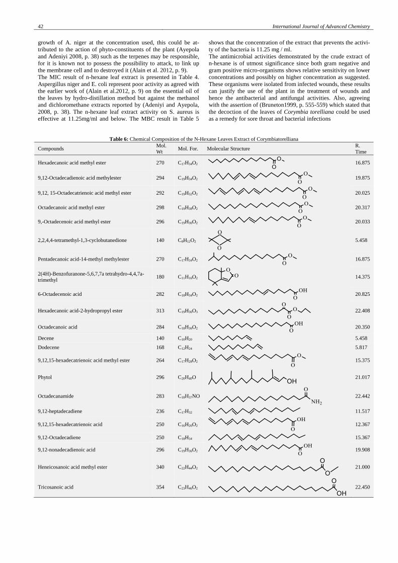
Table 6: Chemical Composition of the N-Hexane Leaves Extract of Corymbiatorelliana
Compounds Mol.
Wt Mol. For. Molecular Structure
R.
Time
Hexadecanoic acid methyl ester 270 C17H34O2
16.875
9,12-Octadecadienoic acid methylester 294 C19H34O2
19.875
9,12, 15-Octadecatrienoic acid methyl ester 292 C19H32O2
20.025
Octadecanoic acid methyl ester 298 C19H38O2
20.317
9,-Octadecenoic acid methyl ester 296 C19H36O2
20.033
2,2,4,4-tetramethyl-1,3-cyclobutanedione 140 C8H12O2
5.458
Pentadecanoic acid-14-methyl methylester 270 C17H34O2
16.875
2(4H)-Benzofuranone-5,6,7,7a tetrahydro-4,4,7a-
trimethyl 180 C11H16O2
14.375
6-Octadecenoic acid 282 C18H34O2
20.825
Hexadecanoic acid-2-hydropropyl ester 313 C19H36O3
22.408
Octadecanoic acid 284 C18H36O2
20.350
Decene 140 C10H20 5.458
Dodecene 168 C12H24 5.817
9,12,15-hexadecatrienoic acid methyl ester 264 C17H28O2
15.375
Phytol 296 C20H40O
21.017
Octadecanamide 283 C18H37NO
22.442
9,12-heptadecadiene 236 C17H32
11.517
9,12,15-hexadecatrienoic acid 250 C16H26O2
12.367
9,12-Octadecadiene 250 C18H34
15.367
9,12-nonadecadienoic acid 296 C19H36O2
19.908
Heneicosanoic acid methyl ester 340 C22H44O2
21.000
Tricosanoic acid 354 C23H46O2
22.450
International Journal of Advanced Chemistry 43
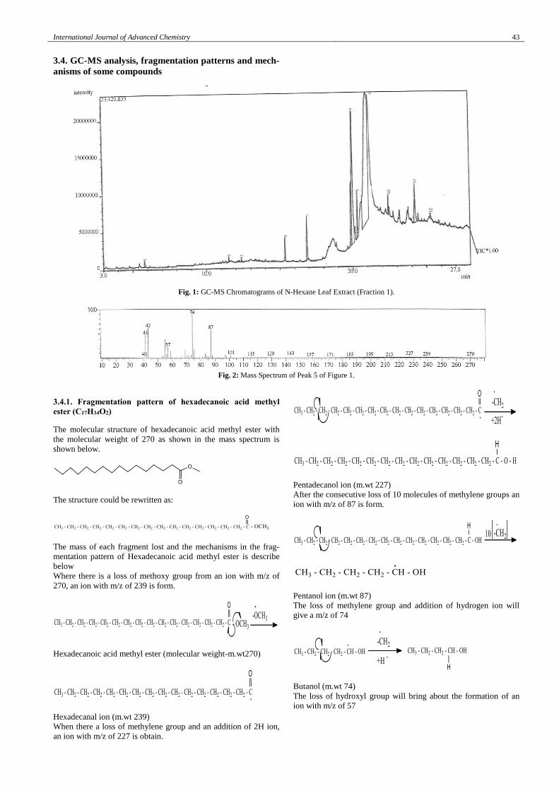
3.4. GC-MS analysis, fragmentation patterns and mech-
anisms of some compounds
Fig. 1: GC-MS Chromatograms of N-Hexane Leaf Extract (Fraction 1).
Fig. 2: Mass Spectrum of Peak 5 of Figure 1.
3.4.1. Fragmentation pattern of hexadecanoic acid methyl
ester (C17H34O2)
The molecular structure of hexadecanoic acid methyl ester with
the molecular weight of 270 as shown in the mass spectrum is
shown below.
The structure could be rewritten as:
The mass of each fragment lost and the mechanisms in the frag-
mentation pattern of Hexadecanoic acid methyl ester is describe
below
Where there is a loss of methoxy group from an ion with m/z of
270, an ion with m/z of 239 is form.
Hexadecanoic acid methyl ester (molecular weight-m.wt270)
Hexadecanal ion (m.wt 239)
When there a loss of methylene group and an addition of 2H ion,
an ion with m/z of 227 is obtain.
Pentadecanol ion (m.wt 227)
After the consecutive loss of 10 molecules of methylene groups an
ion with m/z of 87 is form.
Pentanol ion (m.wt 87)
The loss of methylene group and addition of hydrogen ion will
give a m/z of 74
Butanol (m.wt 74)
The loss of hydroxyl group will bring about the formation of an
ion with m/z of 57
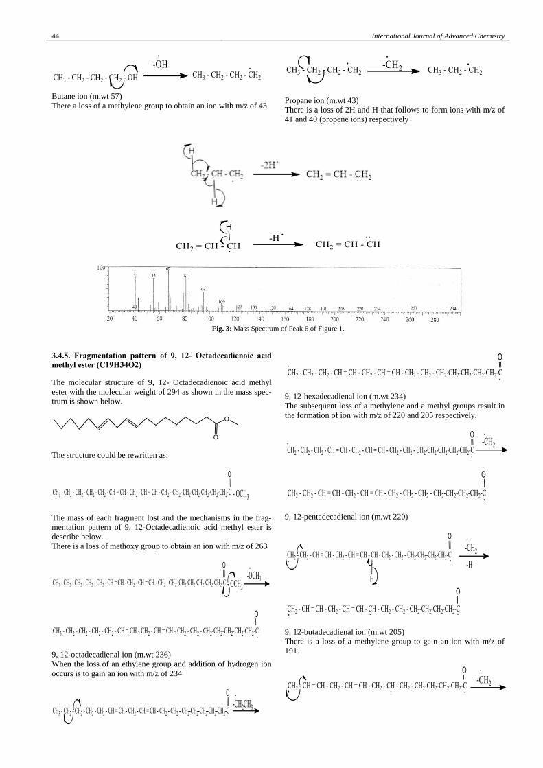
44 International Journal of Advanced Chemistry
Butane ion (m.wt 57)
There a loss of a methylene group to obtain an ion with m/z of 43
Propane ion (m.wt 43)
There is a loss of 2H and H that follows to form ions with m/z of
41 and 40 (propene ions) respectively
Fig. 3: Mass Spectrum of Peak 6 of Figure 1.
3.4.5. Fragmentation pattern of 9, 12- Octadecadienoic acid
methyl ester (C19H34O2)
The molecular structure of 9, 12- Octadecadienoic acid methyl
ester with the molecular weight of 294 as shown in the mass spec-
trum is shown below.
The structure could be rewritten as:
The mass of each fragment lost and the mechanisms in the frag-
mentation pattern of 9, 12-Octadecadienoic acid methyl ester is
describe below.
There is a loss of methoxy group to obtain an ion with m/z of 263
9, 12-octadecadienal ion (m.wt 236)
When the loss of an ethylene group and addition of hydrogen ion
occurs is to gain an ion with m/z of 234
9, 12-hexadecadienal ion (m.wt 234)
The subsequent loss of a methylene and a methyl groups result in
the formation of ion with m/z of 220 and 205 respectively.
9, 12-pentadecadienal ion (m.wt 220)
9, 12-butadecadienal ion (m.wt 205)
There is a loss of a methylene group to gain an ion with m/z of
191.
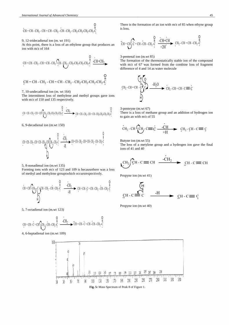
International Journal of Advanced Chemistry 45
9, 12-tridecadienal ion (m. wt 191)
At this point, there is a loss of an ethylene group that produces an
ion with m/z of 164
7, 10-undecadienal ion (m. wt 164)
The intermittent loss of methylene and methyl groups gave ions
with m/z of 150 and 135 respectively.
6, 9-decadienal ion (m.wt 150)
5, 8-nonadienal ion (m.wt 135)
Forming ions with m/z of 123 and 109 is becausethere was a loss
of methyl and methylene groupswhich occursrespectively.
5, 7-octadienal ion (m.wt 123)
4, 6-heptadienal ion (m.wt 109)
There is the formation of an ion with m/z of 85 when ethyne group
is loss.
3-pentenal ion (m.wt 85)
The formation of the thermostatically stable ion of the compound
with m/z of 67 was formed from the combine loss of fragment
difference of 4 and 14 as water molecule
3-penteyne (m.wt 67)
There is a loss of methane group and an addition of hydrogen ion
to gain an with m/z of 55
Butyne ion (m.wt 55)
The loss of a metylene group and a hydrogen ion gave the final
ions of 41 and 40
Propyne ion (m.wt 41)
Propyne ion (m.wt 40)
Fig. 5: Mass Spectrum of Peak 8 of Figure 1.
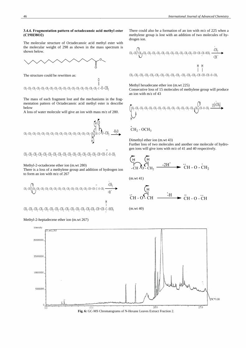
46 International Journal of Advanced Chemistry
3.4.4. Fragmentation pattern of octadecanoic acid methyl ester
(C19H38O2)
The molecular structure of Octadecanoic acid methyl ester with
the molecular weight of 298 as shown in the mass spectrum is
shown below.
The structure could be rewritten as:
The mass of each fragment lost and the mechanisms in the frag-
mentation pattern of Octadecanoic acid methyl ester is describe
below
A loss of water molecule will give an ion with mass m/z of 280.
Methyl-2-octadecene ether ion (m.wt 280)
There is a loss of a methylene group and addition of hydrogen ion
to form an ion with m/z of 267
Methyl-2-heptadecene ether ion (m.wt 267)
There could also be a formation of an ion with m/z of 225 when a
methylene group is lost with an addition of two molecules of hy-
drogen ion.
Methyl hexadecane ether ion (m.wt 225)
Consecutive loss of 15 molecules of methylene group will produce
an ion with m/z of 43
Dimethyl ether ion (m.wt 43)
Further loss of two molecules and another one molecule of hydro-
gen ions will give ions with m/z of 41 and 40 respectively.
(m.wt 41)
(m.wt 40)
Fig. 6: GC-MS Chromatograms of N-Hexane Leaves Extract Fraction 2.
International Journal of Advanced Chemistry 47
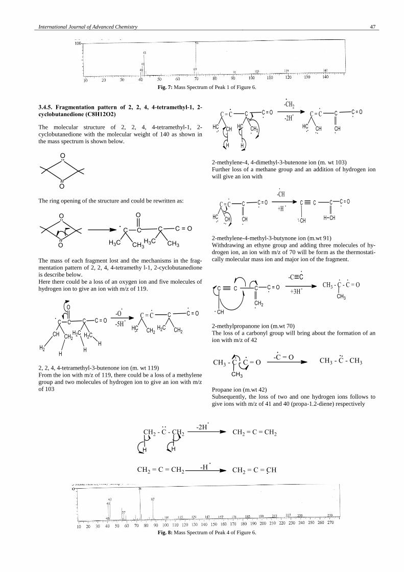
Fig. 7: Mass Spectrum of Peak 1 of Figure 6.
3.4.5. Fragmentation pattern of 2, 2, 4, 4-tetramethyl-1, 2-
cyclobutanedione (C8H12O2)
The molecular structure of 2, 2, 4, 4-tetramethyl-1, 2-
cyclobutanedione with the molecular weight of 140 as shown in
the mass spectrum is shown below.
The ring opening of the structure and could be rewritten as:
The mass of each fragment lost and the mechanisms in the frag-
mentation pattern of 2, 2, 4, 4-tetramethy l-1, 2-cyclobutanedione
is describe below.
Here there could be a loss of an oxygen ion and five molecules of
hydrogen ion to give an ion with m/z of 119.
2, 2, 4, 4-tetramethyl-3-butenone ion (m. wt 119)
From the ion with m/z of 119, there could be a loss of a methylene
group and two molecules of hydrogen ion to give an ion with m/z
of 103
2-methylene-4, 4-dimethyl-3-butenone ion (m. wt 103)
Further loss of a methane group and an addition of hydrogen ion
will give an ion with
2-methylene-4-methyl-3-butynone ion (m.wt 91)
Withdrawing an ethyne group and adding three molecules of hy-
drogen ion, an ion with m/z of 70 will be form as the thermostati-
cally molecular mass ion and major ion of the fragment.
2-methylpropanone ion (m.wt 70)
The loss of a carbonyl group will bring about the formation of an
ion with m/z of 42
Propane ion (m.wt 42)
Subsequently, the loss of two and one hydrogen ions follows to
give ions with m/z of 41 and 40 (propa-1.2-diene) respectively
Fig. 8: Mass Spectrum of Peak 4 of Figure 6.
48 International Journal of Advanced Chemistry
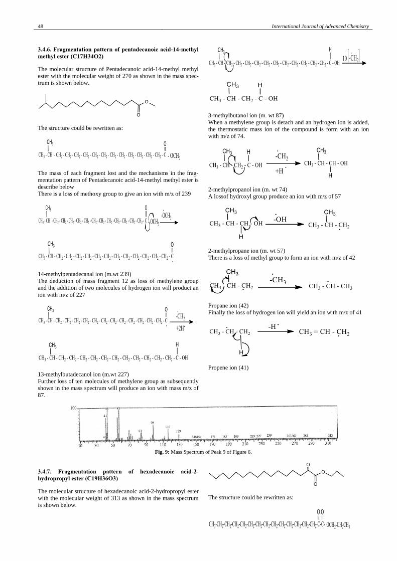
3.4.6. Fragmentation pattern of pentadecanoic acid-14-methyl
methyl ester (C17H34O2)
The molecular structure of Pentadecanoic acid-14-methyl methyl
ester with the molecular weight of 270 as shown in the mass spec-
trum is shown below.
The structure could be rewritten as:
The mass of each fragment lost and the mechanisms in the frag-
mentation pattern of Pentadecanoic acid-14-methyl methyl ester is
describe below
There is a loss of methoxy group to give an ion with m/z of 239
14-methylpentadecanal ion (m.wt 239)
The deduction of mass fragment 12 as loss of methylene group
and the addition of two molecules of hydrogen ion will product an
ion with m/z of 227
13-methylbutadecanol ion (m.wt 227)
Further loss of ten molecules of methylene group as subsequently
shown in the mass spectrum will produce an ion with mass m/z of
87.
3-methylbutanol ion (m. wt 87)
When a methylene group is detach and an hydrogen ion is added,
the thermostatic mass ion of the compound is form with an ion
with m/z of 74.
2-methylpropanol ion (m. wt 74)
A lossof hydroxyl group produce an ion with m/z of 57
2-methylpropane ion (m. wt 57)
There is a loss of methyl group to form an ion with m/z of 42
Propane ion (42)
Finally the loss of hydrogen ion will yield an ion with m/z of 41
Propene ion (41)
Fig. 9: Mass Spectrum of Peak 9 of Figure 6.
3.4.7. Fragmentation pattern of hexadecanoic acid-2-
hydropropyl ester (C19H36O3)
The molecular structure of hexadecanoic acid-2-hydropropyl ester
with the molecular weight of 313 as shown in the mass spectrum
is shown below.
The structure could be rewritten as:
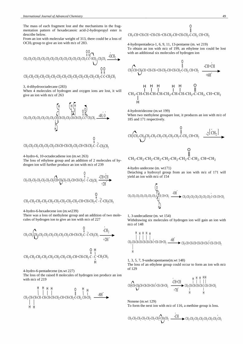
International Journal of Advanced Chemistry 49
The mass of each fragment lost and the mechanisms in the frag-
mentation pattern of hexadecanoic acid-2-hydropropyl ester is
describe below.
From an ion with molecular weight of 313, there could be a loss of
OCH2 group to give an ion with m/z of 283.
3, 4-dihydrooctadecane (283)
When 4 molecules of hydrogen and oxygen ions are lost, it will
give an ion with m/z of 263
4-hydro-6, 10-octadecadiene ion (m.wt 263)
The loss of ethylene group and an addition of 2 molecules of hy-
drogen ion will further produce an ion with m/z of 239
4-hydro-6-hexadecene ion (m.wt239)
There was a loss of methylene group and an addition of two mole-
cules of hydrogen ion to give an ion with m/z of 227
4-hydro-6-pentadecene (m.wt 227)
The loss of the raised 8 molecules of hydrogen ion produce an ion
with m/z of 219
4-hydropentadeca-1, 6, 9, 11, 13-pentaene (m. wt 219)
To obtain an ion with m/z of 199, an ethylene ion could be lost
with an additional six molecules of hydrogen ion
4-hydrotridecene (m.wt 199)
When two methylene groupare lost, it produces an ion with m/z of
185 and 171 respectively.
4-hydro undecene (m. wt171)
Detaching a hydroxyl group from an ion with m/z of 171 will
yield an ion with m/z of 154
1, 3-undecadiene (m. wt 154)
Withdrawing six molecules of hydrogen ion will gain an ion with
m/z of 148
1, 3, 5, 7, 9-undecapentaene(m.wt 148)
The loss of an ethylene group could occur to form an ion wih m/z
of 129
Nonene (m.wt 129)
To form the next ion with m/z of 116, a methine group is loss.
50 International Journal of Advanced Chemistry
Octane ion (m.wt 116)
With the loss of methyl group and three molecules of hydrogen
ion, an ion with m/z of 98 is form.
2-heptane ion (m.wt 98)
Now there could be a loss of methylene group and a hydrogen
ionto yield an ion with m/z of 83
1, 3-hexadiene ion (m.wt 83)
There is a loss of an ethylene group togain an ion with m/z of 57
Butene ion (57)
The loss of methylene would be appropriate to form an ion with
m/z of 43.
Propene ion (m.wt 43)
Finally, the loss of two and one hydrogen ion one after the other
occur to form an ion with m/z of 41 and 40 (propyne ions) respec-
tively.
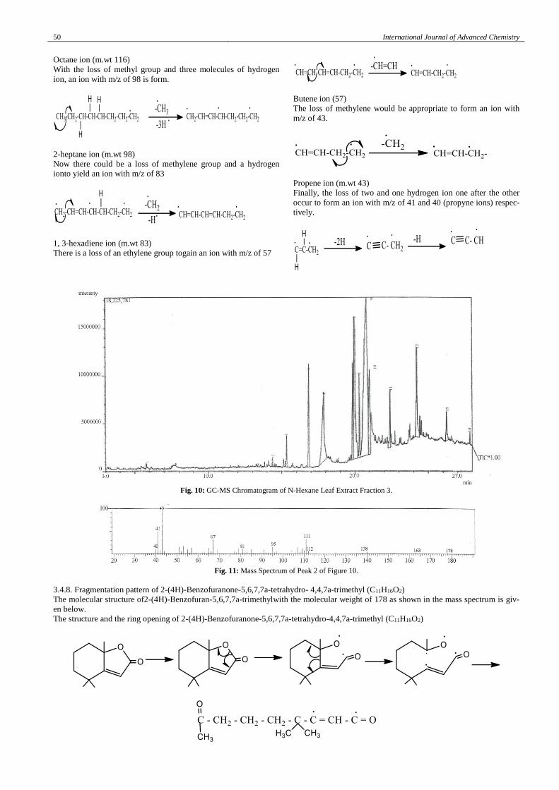
Fig. 10: GC-MS Chromatogram of N-Hexane Leaf Extract Fraction 3.
Fig. 11: Mass Spectrum of Peak 2 of Figure 10.
3.4.8. Fragmentation pattern of 2-(4H)-Benzofuranone-5,6,7,7a-tetrahydro- 4,4,7a-trimethyl (C11H16O2)
The molecular structure of2-(4H)-Benzofuran-5,6,7,7a-trimethylwith the molecular weight of 178 as shown in the mass spectrum is giv-
en below.
The structure and the ring opening of 2-(4H)-Benzofuranone-5,6,7,7a-tetrahydro-4,4,7a-trimethyl (C11H16O2)
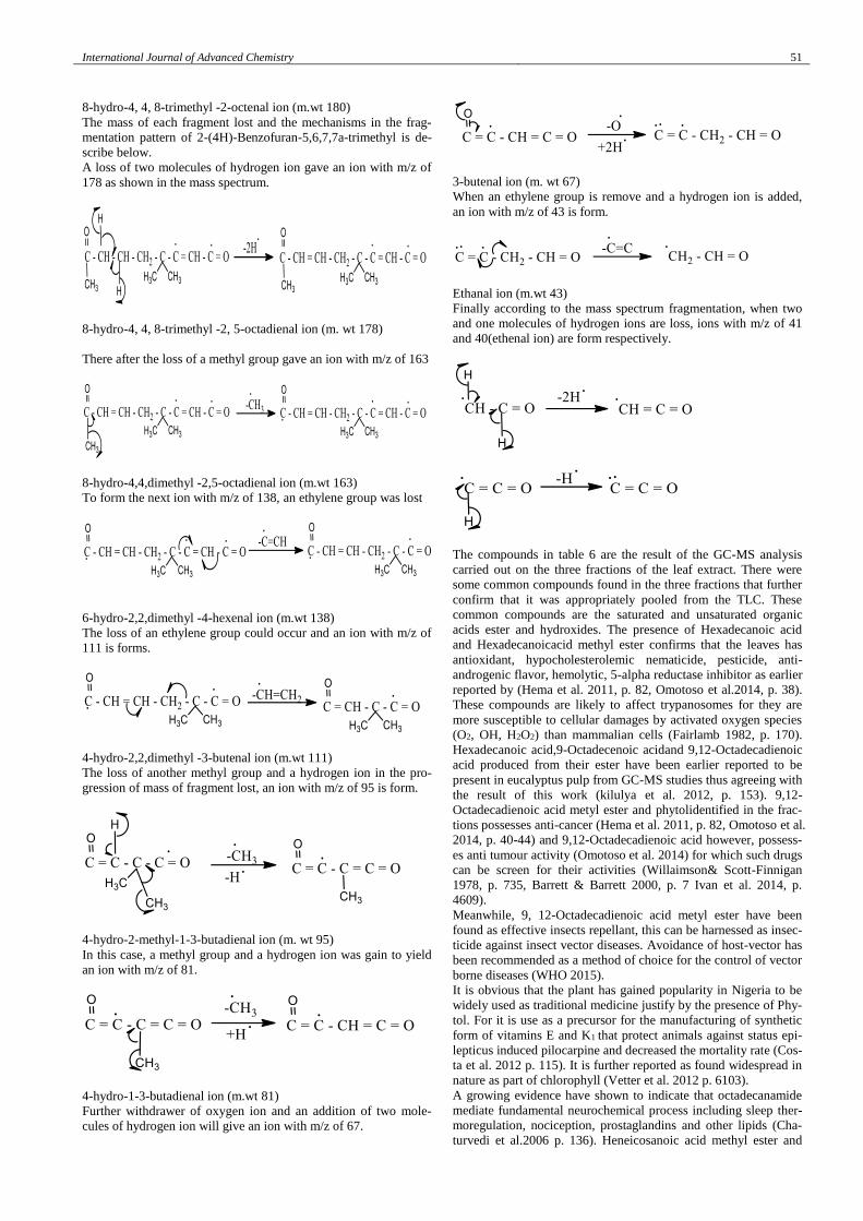
International Journal of Advanced Chemistry 51
8-hydro-4, 4, 8-trimethyl -2-octenal ion (m.wt 180)
The mass of each fragment lost and the mechanisms in the frag-
mentation pattern of 2-(4H)-Benzofuran-5,6,7,7a-trimethyl is de-
scribe below.
A loss of two molecules of hydrogen ion gave an ion with m/z of
178 as shown in the mass spectrum.
8-hydro-4, 4, 8-trimethyl -2, 5-octadienal ion (m. wt 178)
There after the loss of a methyl group gave an ion with m/z of 163
8-hydro-4,4,dimethyl -2,5-octadienal ion (m.wt 163)
To form the next ion with m/z of 138, an ethylene group was lost
6-hydro-2,2,dimethyl -4-hexenal ion (m.wt 138)
The loss of an ethylene group could occur and an ion with m/z of
111 is forms.
4-hydro-2,2,dimethyl -3-butenal ion (m.wt 111)
The loss of another methyl group and a hydrogen ion in the pro-
gression of mass of fragment lost, an ion with m/z of 95 is form.
4-hydro-2-methyl-1-3-butadienal ion (m. wt 95)
In this case, a methyl group and a hydrogen ion was gain to yield
an ion with m/z of 81.
4-hydro-1-3-butadienal ion (m.wt 81)
Further withdrawer of oxygen ion and an addition of two mole-
cules of hydrogen ion will give an ion with m/z of 67.
3-butenal ion (m. wt 67)
When an ethylene group is remove and a hydrogen ion is added,
an ion with m/z of 43 is form.
Ethanal ion (m.wt 43)
Finally according to the mass spectrum fragmentation, when two
and one molecules of hydrogen ions are loss, ions with m/z of 41
and 40(ethenal ion) are form respectively.
The compounds in table 6 are the result of the GC-MS analysis
carried out on the three fractions of the leaf extract. There were
some common compounds found in the three fractions that further
confirm that it was appropriately pooled from the TLC. These
common compounds are the saturated and unsaturated organic
acids ester and hydroxides. The presence of Hexadecanoic acid
and Hexadecanoicacid methyl ester confirms that the leaves has
antioxidant, hypocholesterolemic nematicide, pesticide, anti-
androgenic flavor, hemolytic, 5-alpha reductase inhibitor as earlier
reported by (Hema et al. 2011, p. 82, Omotoso et al.2014, p. 38).
These compounds are likely to affect trypanosomes for they are
more susceptible to cellular damages by activated oxygen species
(O2, OH, H2O2) than mammalian cells (Fairlamb 1982, p. 170).
Hexadecanoic acid,9-Octadecenoic acidand 9,12-Octadecadienoic
acid produced from their ester have been earlier reported to be
present in eucalyptus pulp from GC-MS studies thus agreeing with
the result of this work (kilulya et al. 2012, p. 153). 9,12-
Octadecadienoic acid metyl ester and phytolidentified in the frac-
tions possesses anti-cancer (Hema et al. 2011, p. 82, Omotoso et al.
2014, p. 40-44) and 9,12-Octadecadienoic acid however, possess-
es anti tumour activity (Omotoso et al. 2014) for which such drugs
can be screen for their activities (Willaimson& Scott-Finnigan
1978, p. 735, Barrett & Barrett 2000, p. 7 Ivan et al. 2014, p.
4609).
Meanwhile, 9, 12-Octadecadienoic acid metyl ester have been
found as effective insects repellant, this can be harnessed as insec-
ticide against insect vector diseases. Avoidance of host-vector has
been recommended as a method of choice for the control of vector
borne diseases (WHO 2015).
It is obvious that the plant has gained popularity in Nigeria to be
widely used as traditional medicine justify by the presence of Phy-
tol. For it is use as a precursor for the manufacturing of synthetic
form of vitamins E and K1 that protect animals against status epi-
lepticus induced pilocarpine and decreased the mortality rate (Cos-
ta et al. 2012 p. 115). It is further reported as found widespread in
nature as part of chlorophyll (Vetter et al. 2012 p. 6103).
A growing evidence have shown to indicate that octadecanamide
mediate fundamental neurochemical process including sleep ther-
moregulation, nociception, prostaglandins and other lipids (Cha-
turvedi et al.2006 p. 136). Heneicosanoic acid methyl ester and
52 International Journal of Advanced Chemistry
tricosanoic acid found in this plant are fatty acids commonly in
plant oils and extracts, can be utilized as relaxant. 9,12-
octadecadienoic acid (antibacterial), octadecanoic acid (antimicro-
bial, hardener and thickener use as skin cleaner in soap industries)
9,12,15 octadecatrienoic acid methyl ester for antibacteri-
al,anticandidal, antiinflammatory, hypocholesterolemic, cancer
preventive,hepatoprotective, nematicide, insectifuge antihistamin-
ic,antiarthritic, anticoronary, antieczemicantiacne, 5-
alphareductase inhibitor antiandrogenic and2(4H)-Benzofuran-
5,6,7,7a-tetrahydro-4,4,7a-trimethyl for antimicrobial (Mujeeb
2014, table 6). 2,2,4,4-tetrametyl-1,3-cyclobutanedione is well
known building block for the sterically congested system (Brunck
2001, p. 227) These compounds are synergiscally responsible for
the activities of the plant which can be harness for the develop-
ment of our developing countries in the area of pharmacological
techniques and economic improvement.
4. Conclusion
The results of our finding indicate that the n-hexane extract of C.
torelliana is a rich source of bioactive agent of natural background,
which might have potentials for use in the classification of drugs
in pharmaceutical industries. This study has contributed and justi-
fies the claim of the plant as traditional medicine without any
adverse side effect as reported in developing countries compare
with synthetic drugs. The spectra therein have been identified as
shown by the fragmentation patterns and mechanisms as possibly
those of the compounds identified which have medicinal and
pharmacological properties. In spite of the medicinal importance
of C. torelliana, it has short rotation hardwood for variety of
products and ornamentals with specific emphasis on existing and
emerging markets for revenue generation if domesticated.
References
[1] Adeniyi BA &Ayepola OO (2008) the phytochemical screening and
antimicrobial activity of leaf extracts of eucalyptus camaldulensis and eucalyptustorelliana / (myrtaceae). Research Journal of Medic-
inal Plants 2, 34-38. https://doi.org/10.3923/rjmp.2008.34.38.
[2] Adeniyi AG, Odufowoke RO &Olaleye SB (2006) Antimicrobial and gastroprotective properties of eucalyptus torelliana / (myr-
taceae) crude extracts. International Journal of Pharmacology
2,362-365. https://doi.org/10.3923/ijp.2006.362.365. [3] Alian AG, Felician A, Boniface Y, Alian KY, Chantal M &
Dominique S (2012) Chemical and biological investigation of
leaves of eucalyptus torellianaessentialoil from Benin. International
Research Journal of Biological Sciences 15, 6-12.
[4] Atlas RM (1995) Microorganisms in our World 2ndedn. Mosby Publishers Inc. Baltimore, pp. 765.
[5] Azra A, Ekwenchi MM, Dashak DA &Dildar A (2012) Gas Chro-
matography-Mass Spectrometry (GC-MS) analysis of Phthalate iso-late in n-hexane extract of Azadirachta A. Juss(Neem)
leaves.Journal ofAmerican Science 8(12), 146-155.
[6] Barret SV and Barret MP (2000) Anti-sleeping sickness drugs and cancer. Chemotherapy and Parasitology Today 16, 7-9.
https://doi.org/10.1016/S0169-4758(99)01560-4.
[7] Brunck JS, Koch A, Grzegorz M, Lehnhoff S, Margaretha P, Pra-kash GKS, Rasul G, Bau R &Olah GA (2001) 1,2 addition of TMS-
CF3 TMS-CN toserially crowded 2,2,4,4-tetramethyl-1-3-
cyclobutanedione. Journal of Indian Institute of Science 81, 227-237.
[8] Bruneton J (1999) Pharmacognosy: Phytochemistry. In Medicinal
Plants 2ndedn. London Intercept Ltd pp. 555-559.
[9] Bwai MD, Afolabi M, Odukomaiya D, Ikokoh P & OrishadipeA
(2014) Proximate composition, mineral and phytochemical constit-
uents of Eleusinecoracana (finger millet).International Journal of Advance Chemistry 2(2), 171-174.
https://doi.org/10.14419/ijac.v2i2.3496.
[10] Chaturvedi S1, Driscoll WJ, Elliot BM, Faraday MM, Grunberg NE& Mueller GP (2006) In vivo evidence that N-oleoylglycine act
independently of its conversion to oleamide.Prostaglandins and
other Lipid Mediat 81(3-4), 136-149. https://doi.org/10.1016/j.prostaglandins.2006.09.001.
[11] Costa JP1, Ferreira PB, De Sousa DP, Jordan J&Freitas RM (2012)
Anticonvulsant effect of phytol in a pilocarpine model in mice.
Neuroscience letter523 (2), 115-118.
https://doi.org/10.1016/j.neulet.2012.06.055.
[12] Dashak D A &Ano J (2007) Chemical composition and phytochem-ical studies of Crinum zeylanicum. Journal of Sciences Engineering
and Technology 14 (2), 7355-7365.
[13] Dashak D A, Daben J M,Olaoye FM, Ogunbiade AT&OgboleE (2016) Evaluation of the essential oils constitutes from the leaves,
seed buds and fruits of EucalytustorellianaF. Muel plant by Gas Chromatography- Mass Spectral analysis. IOSR-Journal ofApplied
Chemistry 9 (10), 45-60.
[14] Fairlamb AH (1982) Trends Biochemistry Science: Difluoro-methyornithine and the rationale development of polyamine antag-
onism in the cure ofprotozoa infection. In Mechanism of Drug Ac-
tion, Academic press, USA, pp.159-173. [15] Farah A, Fechtal M, ChouchA&Zarira S (2002) The Essential oil of
Eucalyptus camaldulensis and its natural hybrid (clone 583) from
Morocco. Flavour Fragrance Journal 17, 395-397. https://doi.org/10.1002/ffj.1114.
[16] Ivan S, Pablo T, Juan CE, Natalia Q, Mauricio AC, Juan V, Chris-
tian E, Angelica F, Ricardo AT, Juan DM, Rodrigo L, Bruce KC, Ramon JE & Christian OS (2014) 2-Phenylaminonaphthoquinones
and related compounds: Synthesis, trypanocidal and cytotoxic ac-
tivities. In Bioorganicand Medicinal Chemistry 22, 4609-4620. https://doi.org/10.1016/j.bmc.2014.07.030.
[17] Glasby JS (1999) Dictionary of Plants Containing Secondary Me-
tabolites.Taylor and Francis Ltd, Londonpp. 125-225. [18] Harborne JB (1984). Phytochemical methods. A guide to modern
techniques of plant analysis. 2ndedn. Chapman and Hall, London,pp
1, 11. https://doi.org/10.1007/978-94-009-5570-7. [19] Hema R, Kumaravel S &Lagusundaram A (2011). GC/MS deter-
mination of bioactive components of Murrayakoenigii. Journal of
American Science 7 (1), 80-83. [20] Kilulya KF, Msagati TAM, Mamba BM, Ngila JC & Bush T (2012)
Ionic liquid-liquid extraction and supported liquid membrane anal-
ysis of lipophilic wood extractives from dissolving pulp. Chroma-tographia 75, 513-520. https://doi.org/10.1007/s10337-012-2225-5.
[21] Lee TA (1998) A Beginner’s Guide to Mass Spectral Interpretation.
JohnWiley and Sons Inc. (NY) pp 1-21. [22] Lister PD, Wolter DJ & Hanson ND (2009) Antibacterial-resistant
Pseudomonas aeruginosa: Clinical impact and complex regulation
of chromosomally encoded resistance mechanisms. Clinical Micro-biology Revision 22(4), 582-610.
https://doi.org/10.1128/CMR.00040-09.
[23] Mujeeb F, Bajpai P &Pathak N (2014) Phytochemical evaluation, antimicrobial activity and determination of bioactive compounds
from leaves of Aeglemarmelos. BioMed Research Interna-
tionalAvailable at: http://dx.doi.org/10.1155/2014/497606 (ac-cessed 25 March 2016). https://doi.org/10.1155/2014/497606.
[24] Ochei J&Kochatkar A (2007) Medical Laboratory Science, Theory
and Practice.Tata McGraw-Hill Ltd, pp. 795-817.
[25] Ogbole E, Dashak DA,Nvau JB, Daben MR, Abongaby G, Obaloto
OB, Oladipo OO, Igweh AC (2016) Phytochemical screening and
in vitro evaluation of the antitrypanosomal action of the methanolic leaf extract ofCorymbiatorelliana. International Journal of Ethno-
medicine and Pharmacology 3(1), 20-29.
https://doi.org/10.14194/ijep.3.1.3. [26] Ololade ZS and Olawore NO (2013) Chemistry and medicinal po-
tentials of the seed essential oil of Eucalyptus torel-
lianaF.Muellgrown in Nigeria. Global Journal of Science Frontier Research Chemistry 13(3), 1- 11.
[27] Omotoso AE, Eseyin OO & Suleiman M (2014) Phytochemical analysis ofCnidoscolusaconitifolius(Euphorbiaceae) leaf with spec-
trometric techniques. Nigerian Journal of Pharmaceutical and Ap-
plied Science Research 3 (1), 38-49. [28] Roopashree TS, Dang R, Rani SRH, Narendra C (2009) Antibacte-
rial activity ofantipsoriatic herb: Cassia tora, MomordicaCharantia
and Calendula officinalis. International Journal of Applied Re-
search in Natural products 1(3), 20-28.
[29] Sanchez NR, Garcia DA, Shiavini MS, Nakamura CV &Filho BPD
(2005) an evaluation of antibacterial activities of Psidiumguajava. Brazilian Journal of biotechnology48, 429-436.
[30] Segelman AB, Farnsworth NR, and Quimby MD (1969) Biological
and phytochemical evaluation of plants 111: False-negative sapo-nins test results induced by the presence of tannins.Lloydia 32, 52-
55.
[31] Silverstein RM, Bassler GC & Morrill TC (1974) Spectrometric Identification of Organic Compounds.3rdedn.John Wiley and Sons
Inc. (NY) pp 41-71.
International Journal of Advanced Chemistry 53
[32] Sofowora A (1982) Medicinal plants and traditional medicine in
Africa. John Wiley and Sons Ltd, New York, pp.54-56.
[33] Thomas KW (1975) Principles and Techniques of Practical Bio-
chemistry. (Williams BL & Wilson K ed.), Edward Arnold Ltd,
London pp52-98. [34] Trease GE & Evans MD (1989) A textbook of Pharmacognosy.
13thedn. Braillier, Tindaland Caussel, London, pp. 244-248.
[35] Vagahasiya YNR, Chanda S (2008) Antibacterial and preliminary phytochemical and physiochemical analysis of eucalyptus citriodo-
ra HK leaf.Natural Product Research 22(9), 754-762. https://doi.org/10.1080/14786410701628788.
[36] Vetter W1, Schröder M&Lehnert K (2012) Differentiation of re-
fined and virgin edible oils by means of the trans- and cis-phytol isomer distribution.Journal of Agriculture and Food Chemistry
60(24), 6103- 6107. https://doi.org/10.1021/jf301373k.
[37] Vishnoi NK (1979) Advance Practical Organic Chemistry.Vikas Publishing House PVT Ltd pp. 40-42.
[38] Wall ME, Kreider MM, Krewson CF, Eddy CR, Williams JJ, Cor-
del DS & Gentry HS (1954) Survey of plants for steroidal sapogen-ins and other constituents. Journal of AmericanPharmaceutical As-
sociation 43, 1-7. https://doi.org/10.1002/jps.3030430102.
[39] Williamson J & Scott-Finnigan TJ (1978) Trypanocidal activity of antitumour antibiotics and other metabolic inhibitors. Antimicrobial
Agents andChemotherapy 13,735-744.
https://doi.org/10.1128/AAC.13.5.735. [40] World Health Organization (2001) Global Strategy for Containment
of Antimicrobial Resistance. Available at: www.who.int/emc- doc-
ument/antimicrobial resistance/docs/global start.pdt(accessed 23 March 2015).
[41] World Health Organization (2016) African Trypanosomiasis
(Sleeping sickness). Fact sheet, no. 259. Available at: www.who.int/mediaccentre/factsheets/fs259/en/(accessed 25 Au-
gust 2016).


