Embed Size (px)
Citation preview

BRIEF COMMUNICATION
Intrathecal anti-CD20 efficiently depletes meningeal B cellsin CNS autoimmunityKlaus Lehmann-Horn1, Silke Kinzel2, Linda Feldmann2, Florentine Radelfahr1, Bernhard Hemmer1,Sarah Traffehn2, Claude C. A. Bernard3, Christine Stadelmann2, Wolfgang Br€uck2 &Martin S. Weber2,4
1Department of Neurology, Technische Universit€at M€unchen, Munich, Germany2Department of Neuropathology, University Medical Center, Georg August University, G€ottingen, Germany3Australian Regenerative Medicine Institute, Monash University, Melbourne, Australia4Department of Neurology, University Medical Center, Georg August University, G€ottingen, Germany
Correspondence
Martin S. Weber, Departments of
Neuropathology and Neurology, University
Medical Center, Georg August University,
Robert-Koch-Str. 40, 37099 G€ottingen,
Germany. Tel: +49-551 39-7706; Fax: +49-
551-39-10800; E-mail: martin.weber@med.
uni-goettingen.de
Present address
Klaus Lehmann-Horn, Department of
Neurology, University of California, San
Francisco, CA
Funding Information
K. L.-H. is supported by the Kommission f€ur
Klinische Forschung (KKF) of the Technische
Universit€at M€unchen and is a fellow of the
Deutsche Forschungsgemeinschaft (DFG; Le
3079/1-1). M. S. W. is supported by the
National Multiple Sclerosis Society (NMSS; PP
1660), the Deutsche Forschungsgemeinschaft
(DFG; WE 3547/4-1), and the ProFutura
Programm of the Universit€atsmedizin
G€ottingen. B. H. is supported by a grant
from the German Ministry for Education and
Research (BMBF, “German Competence
Network Multiple Sclerosis” (KKNMS),
Control-MS, 01GI0917) and the DFG
(He2386/7-1). C. C. A. B. is supported by
grants from the National Health and Medical
Council of Australia.
Received: 19 February 2014; Revised: 9 May
2014; Accepted: 19 May 2014
doi: 10.1002/acn3.71
Abstract
Clinical trials revealed that systemic administration of B-cell-depleting anti-
CD20 antibodies can hold lesion formation in the early relapsing-remitting
phase of multiple sclerosis (MS). Throughout the secondary-progressive (SP)
course of MS, pathogenic B cells may, however, progressively replicate within
the central nervous system (CNS) itself, which is largely inaccessible to sys-
temic anti-CD20 treatment. Utilizing the murine MS model of experimental
autoimmune encephalomyelitis, we show that intrathecal (i.t.) administration
of anti-CD20 alone very efficiently depletes meningeal B cells from established
CNS lesions. In SP-MS patients, adding i.t. administration of anti-CD20
to its systemic use may be a valuable strategy to target pathogenic B-cell
function.
ª 2014 The Authors. Annals of Clinical and Translational Neurology published by Wiley Periodicals, Inc on behalf of American Neurological Association.
This is an open access article under the terms of the Creative Commons Attribution-NonCommercial-NoDerivs License, which permits use and
distribution in any medium, provided the original work is properly cited, the use is non-commercial and no modifications or adaptations are made.
1

Introduction
Clinical trials testing B-cell-depleting anti-CD20 (ritux-
imab, ocrelizumab) in relapsing-remitting multiple sclero-
sis (RR-MS) generated encouraging results1,2 and
indirectly consolidated the concept that B cells play an
important pathogenic role in MS. B cells may contribute
to MS pathogenesis as antigen-presenting cells for acti-
vation of encephalitogenic T-cells,3 as precursors of
antibody-secreting plasma cells,4 and as source of
pro-inflammatory cytokines.5 Emerging evidence suggests
that throughout the chronic course of MS, pathogenic B-
cell function may gradually shift from the periphery into
the inflamed central nervous system (CNS). The recent
discovery of ectopic B-cell follicle-like structures in the
meninges of a proportion of patients with secondary-pro-
gressive MS (SP-MS)6,7 suggests that CNS B cells may
locally reproduce and differentiate, providing one expla-
nation why in later stages of MS, clinical progression
decreasingly correlates with magnetic resonance imaging
(MRI)-detectable CNS infiltration. As data indicate that
only about 0.1% of systemically infused anti-CD20
reaches the cerebrospinal fluid (CSF),8 intrathecal (i.t.)
application of anti-CD20, that may provide a valuable
strategy to more efficiently target B cells within the CNS.
Based on this assumption, a clinical trial combining i.t.
anti-CD20 with systemic B-cell depletion in SP-MS is on
its way (ClinicalTrials.gov Identifier: NCT01212094). In
order to determine the effectiveness of i.t anti-CD20 by
itself in depleting CNS B cells, we investigated this novel
therapeutic approach in the murine MS model of experi-
mental autoimmune encephalomyelitis (EAE).
Methods
Female C57BL/6 mice were immunized with 75-lg mur-
ine recombinant myelin oligodendrocyte glycoprotein
(MOG) 1-117 in Complete Freund’s Adjuvant (CFA) sub-
cutaneously and 150ng pertussis toxin intraperitoneal
(i.p.) twice. This induction regimen is associated with a
high frequency of B cells infiltrating the CNS compared
to classical induction regimens like immunization with
MOG peptide 35-55.9 EAE severity was scored as follows:
0 = no paralysis, 1 = loss of tail tone, 2 = mild monopa-
resis or paraparesis, 3 = severe paraparesis, 4 = paraplegia
and/or quadriparesis, 5 = moribund or death. At an EAE
score ≥2, mice were randomly assigned to one of four
groups and concomitantly received 100 lg of murine
anti-CD20 or anti-ragweed isotype per week either i.t. (in
10 lL phosphate buffered saline (PBS); percutaneously
into the cisterna magna with a 30-gauge needle in 45° an-teflexion of the head10) or systemically (i.p.). Anti-CD20
and ragweed isotype control were kindly provided by
Genentech Inc (South San Francisco, CA). Immune cells
were isolated from blood, spleen, draining lymph nodes,
brain, or spinal cord. Regarding the latter two, cells were
isolated by Percoll gradient after collagenase/DNAse
digestion of CNS tissue for 1 h.11 Cells were stained for
CD19, CD3, and CD11b (all BD Biosciences, Heidelberg,
Germany). In the brain and spinal cord, additional stain-
ing for CD45 (eBioscience, Frankfurt, Germany) identified
infiltrating cells of hematopoietic origin. For histological
analysis, CNS tissue was sectioned following perfusion
with PBS and cryofixation and stained with Luxol fast
blue and Periodic acid Schiff (LFB/PAS), hematoxylin and
eosin (H&E) or by B220/CD3 immunohistochemistry as
described previously.9 For comparison of clinical scores
between groups, the Mann–Whitney test was used. For
comparison of groups in our Fluorescence-activated cell
sorting (FACS) study (Fig. 2B), a two-way analysis of var-
iance (ANOVA) with correction for multiple testing (Tu-
key) was performed. Only near-significant values
comparing i.t. and i.p. anti-CD20 groups are shown. This
study was approved by the government of Upper Bavaria
(protocol number 55.2-1-54-2531-67-09).
Results
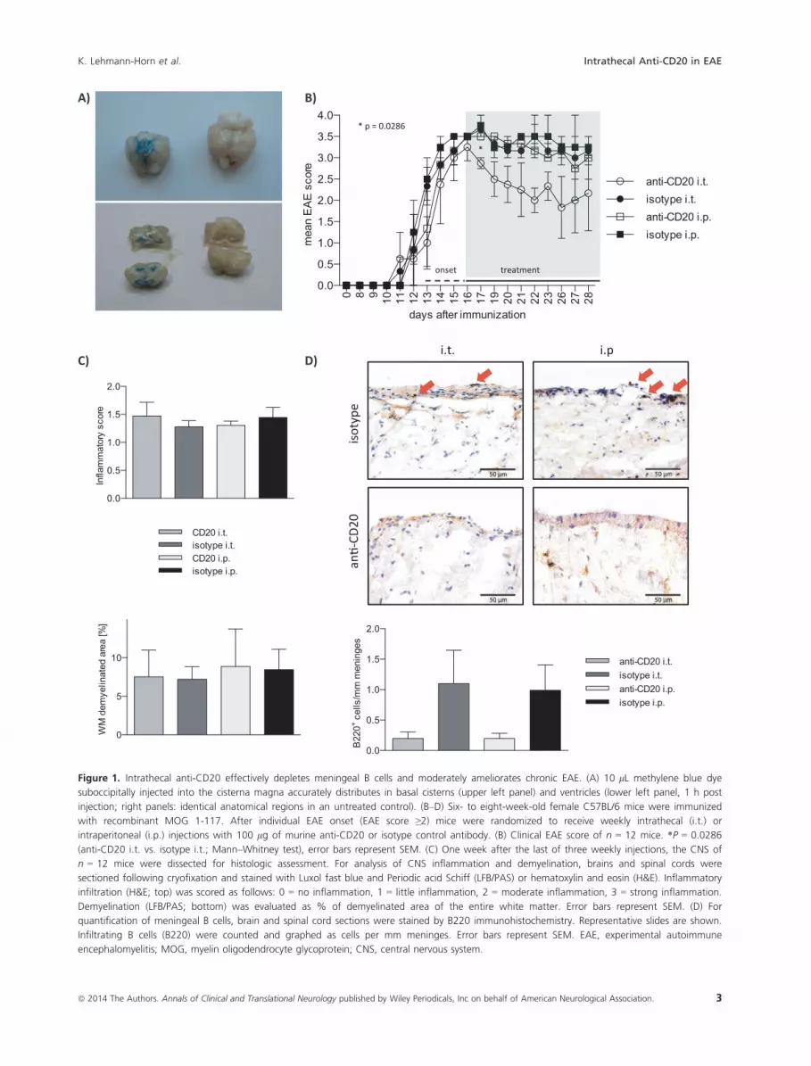
A first experiment intended to validate the method of i.t.
injection in mice. As shown in Figure 1A, 10-lL methy-
lene blue dye injected into the cisterna magna stained
inner and outer CSF spaces, while no leakage of the dye
to the periphery was detected. Anti-CD20 or the respec-
tive control was then injected i.t. or i.p. into mice
with established rMOG-induced EAE. As indicated in
Figure 1B, weekly i.t. anti-CD20 moderately ameliorated
disease severity, whereas systemic anti-CD20 had a mild,
but not significant effect. After three weekly injections, all
four groups showed a comparable extent of inflammatory
CNS infiltration and demyelination (Fig. 1C). In contrast,
i.t. application of anti-CD20 as well as its systemic use
strongly reduced the number of B220+ B cells within
meningeal lesions (Fig. 1D), where the vast majority of
CNS B cells are located in this model.9 In order to quan-
tify the frequency of B cells more precisely (including the
entire CNS with adjacent meninges) and to assess the
kinetic of their depletion, B cells were isolated from
blood, spleen, lymph nodes, brain, and spinal cord 7 days
after 1 (day 7), 2 (day 14), or 3 (day 21) weekly injections
(Fig. 2). Paralleling the histologic analysis, i.t. as well as
systemic anti-CD20 treatment was associated with a
marked decline in B cells in the brain and spinal cord,
while there was a trend that i.t. anti-CD20 may have been
slightly superior to its systemic use (Fig. 2A and B). Of
note, depletion of B cells from the brain, but in particular
from the spinal cord showed a delayed kinetic with the
2 ª 2014 The Authors. Annals of Clinical and Translational Neurology published by Wiley Periodicals, Inc on behalf of American Neurological Association.
Intrathecal Anti-CD20 in EAE K. Lehmann-Horn et al.
0 8 9 10 11 12 13 14 15 16 17 19 20 21 22 23 26 27 28
0.0
0.5
1.0
1.5
2.0
2.5
3.0
3.5
4.0
days after immunizationm
ean
EA
E s
core
anti-CD20 i.t.isotype i.t.anti-CD20 i.p.isotype i.p.
* p = 0.0286
treatmentonset
*
0.0
0.5
1.0
1.5
2.0
Infla
mm
ator
y sc
ore
CD20 i.t.isotype i.t.CD20 i.p.isotype i.p.
i.pi.t.iso
type
an-C
D20
0
5
10
WM
dem
yelin
ated
are
a [%
]
0.0
0.5
1.0
1.5
2.0
B22
0+ cel
ls/m
m m
enin
ges
anti-CD20 i.t.isotype i.t.anti-CD20 i.p.isotype i.p.
A) B)
C) D)
Figure 1. Intrathecal anti-CD20 effectively depletes meningeal B cells and moderately ameliorates chronic EAE. (A) 10 lL methylene blue dye
suboccipitally injected into the cisterna magna accurately distributes in basal cisterns (upper left panel) and ventricles (lower left panel, 1 h post
injection; right panels: identical anatomical regions in an untreated control). (B–D) Six- to eight-week-old female C57BL/6 mice were immunized
with recombinant MOG 1-117. After individual EAE onset (EAE score ≥2) mice were randomized to receive weekly intrathecal (i.t.) or
intraperitoneal (i.p.) injections with 100 lg of murine anti-CD20 or isotype control antibody. (B) Clinical EAE score of n = 12 mice. *P = 0.0286
(anti-CD20 i.t. vs. isotype i.t.; Mann–Whitney test), error bars represent SEM. (C) One week after the last of three weekly injections, the CNS of
n = 12 mice were dissected for histologic assessment. For analysis of CNS inflammation and demyelination, brains and spinal cords were
sectioned following cryofixation and stained with Luxol fast blue and Periodic acid Schiff (LFB/PAS) or hematoxylin and eosin (H&E). Inflammatory
infiltration (H&E; top) was scored as follows: 0 = no inflammation, 1 = little inflammation, 2 = moderate inflammation, 3 = strong inflammation.
Demyelination (LFB/PAS; bottom) was evaluated as % of demyelinated area of the entire white matter. Error bars represent SEM. (D) For
quantification of meningeal B cells, brain and spinal cord sections were stained by B220 immunohistochemistry. Representative slides are shown.
Infiltrating B cells (B220) were counted and graphed as cells per mm meninges. Error bars represent SEM. EAE, experimental autoimmune
encephalomyelitis; MOG, myelin oligodendrocyte glycoprotein; CNS, central nervous system.
ª 2014 The Authors. Annals of Clinical and Translational Neurology published by Wiley Periodicals, Inc on behalf of American Neurological Association. 3
K. Lehmann-Horn et al. Intrathecal Anti-CD20 in EAE

lymph node
day 7 day 14 day 210
20
40
60
80
% o
f CD1
9+
B)
brain
day 7 day 14 day 210
5
10
15
% o
f CD4
5+
CD19
+
p = 0.066
spinal cord
day 7 day 14 day 210
5
10
15
% o
f CD4
5+
CD19
+
blood spleen LN brain spinal cord
an -CD20 i.t.
isotypei.t.
an -CD20 i.p.
isotypei.p.
CD19
FS gated on CD45 +
A)
blood
day 7 day 14 day 210
20
40
60
80
% o
f CD1
9+
an -CD20 i.t.isotype i.t.an -CD20 i.p.isotype i.p.
spleen
day 7 day 14 day 210
20
40
60
80
% o
f CD1
9+
0.542% 6.00% 5.00% 2.76%
58.0%
0.376%
64.3%
10.0%
9.86% 12.5% 53.0%
0.375% 0.583% 3.40% 4.76% 8.58%
57.2% 48.2% 44.1% 11.7%
4 ª 2014 The Authors. Annals of Clinical and Translational Neurology published by Wiley Periodicals, Inc on behalf of American Neurological Association.
Intrathecal Anti-CD20 in EAE K. Lehmann-Horn et al.

most prominent effect occurring after the second injec-
tion. Interestingly, particularly in i.p. anti-CD20, in some
instances B cells accumulated in the CNS under continu-
ous anti-CD20 treatment (brain – day 7 to day 14; spinal
cord – day 14 to day 21). Unexpectedly, weekly injections
of anti-CD20 into the cisterna magma also led to a virtu-
ally complete depletion of B cells in all peripheral com-
partments analyzed (Fig. 2A and B). Already one i.t.
injection of anti-CD20 (day 7) was followed by a broad
depletion of B cells from blood, spleen, and lymph nodes
in its extent indistinguishable from systemic anti-CD20
treatment. In order to evaluate the immunological conse-
quences of anti-CD20-mediated B-cell depletion, we
included an analysis of CD3+ T cells and CD11b+ myeloid
cells. As indicated in Figure S1A and B, all peripheral
compartments showed an expected compensatory increase
in the frequency of T cells and myeloid cells upon deple-
tion of B cells. In contrast, frequency of T cells and mye-
loid cells remained roughly stable in brain and spinal
cord. In these compartments, B cells only represented 5–12% of infiltrating cells. Thus, it would be expected that
their depletion may only lead to a mild compensatory fre-
quency increase of T cells and myeloid cells. To quantify
T-cell infiltration by absolute numbers, CD3 immunohis-
tochemistry was performed. While systemic anti-CD20
had no dampening effect on T-cell infiltration into the
CNS, i.t. application of anti-CD20 was indeed associated
with a trend toward a decline of CNS T cells (Fig. S1C).
Discussion
I.t. administration of anti-CD20 efficiently reduced the
frequency of CNS meningeal B cells at an EAE disease
stage, when CNS lesions are largely established and infil-
tration of immune cells into the CNS gradually declines.12
This finding supports the concept that effector mecha-
nisms of anti-CD20-mediated eradication of B cells are
sufficiently provided within the CNS and that accordingly
anti-CD20 is generally capable of locally depleting CNS B
cells. Our study was neither designed nor sufficiently
powered to detect clinical effects of i.t. or i.p. anti-CD20
compared to isotype control antibody. Yet, in Figure 1B,
there was a trend that suggested that i.p. anti-CD20 may
be beneficial compared to isotype i.p., which, although in
our current study a lower dose was used, would be in
agreement with our previously published results.9 Fur-
thermore, there was a trend that i.t. anti-CD20 may be
slightly superior to its systemic counterpart. Additional
studies are needed to verify this observation and to eluci-
date the underlying mechanisms, for example, the genera-
tion of plasma cells with their various effector functions
and other factors. In the model used, the majority of
CNS B cells are found within meningeal areas (Fig. 1D
and 9). Given the immediate access of i.t. administered
agents to the meninges, it is plausible that injecting anti-
CD20 into the CSF may result in its highest concentration
in the vicinity of meningeal B cells. In our FACS analysis,
there was indeed a trend that i.t. anti-CD20 may be supe-
rior to its systemic use in depleting infiltrated B cells
from the brain or spinal cord. This finding is of particular
relevance, as ectopic lymphoid follicles presumably con-
taining and replicating pathogenic B cells are found in
meningeal regions adjacent to the subarachnoid space in
patients with SP-MS.6 Interestingly, we observed in some
occasions, more pronounced when anti-CD20 was applied
systemically, that B cells accumulated in the CNS despite
almost complete peripheral B-cell depletion. This finding
may reflect local expansion of B cells, which would render
targeting B cells locally in the CNS even more important.
Unexpectedly, anti-CD20 injected into the cisterna
magna depleted peripheral B cells in a manner comparable
to systemic anti-CD20, indicating that the compound did
not remain within the CNS. Paralleling these findings, in a
recent trial in subjects with CNS B-cell lymphoma serial i.t.
application of rituximab showed an unexpected short CSF
half-life of the compound, whereas it accumulated in the
serum.13 While the CSF is drained primarily into the
venous system where transmission of the larger anti-CD20
molecule should be restricted, an alternative route of CSF
drainage leads into deep cervical lymphatics,14 which may
represent the link to the periphery. In the clinical context,
the subsequent depletion of peripheral B cells following i.t.
administration of anti-CD20 should enhance the projected
benefit in treatment of MS. In our preclinical model,
Figure 2. Intrathecal anti-CD20 alone is modestly superior to its systemic application in depleting infiltrating CNS B cells, while it equally reduces
B-cell frequencies in peripheral compartments. Six- to eight-week-old female C57BL/6 mice were immunized with recombinant MOG 1-117. Mice
received a total of 1–3 weekly injections of 100 lg of intrathecal (i.t.) anti-CD20, i.t. isotype control, systemic (i.p.) anti-CD20, or i.p. isotype
control starting on the day of individual EAE onset (EAE score ≥2). (A) Flow cytometric staining of CD19+ B cells in blood (first column), spleen
(second column), lymph node (third column), brain, and spinal cord (fourth and fifth column, both gated on CD45+) on day 21 after treatment
onset. Plots shown are representative of the mean values shown below. (B) Bars represent mean percentages of CD19+ B cells in the blood and
spleen (upper panel), lymph node, brain, and spinal cord (lower panel; the latter two gated on CD45+), detected by FACS on days 7, 14, and 21
after treatment initiation (n = 3 mice per group per time point). Error bars represent SEM. Statistical analysis (two-way ANOVA with correction for
multiple testing) comparing anti-CD20 i.t. versus anti-CD20 i.p. and isotype i.t. versus isotype i.p. groups, respectively, were performed for each
time point and near-significant results are indicated in the graph. CNS, central nervous system; MOG, myelin oligodendrocyte glycoprotein; EAE,
experimental autoimmune encephalomyelitis; ANOVA analysis of variance.
ª 2014 The Authors. Annals of Clinical and Translational Neurology published by Wiley Periodicals, Inc on behalf of American Neurological Association. 5
K. Lehmann-Horn et al. Intrathecal Anti-CD20 in EAE

however, this finding makes it difficult to distinguish pri-
mary i.t. effects from secondary systemic effects of i.t. anti-
CD20. Concomitantly, this observation indicates, however,
that selective targeting of CNS B cells, which may be desir-
able in some settings to reduce systemic side effects, may
not be feasible by this approach.
Taken together, our mechanistic data provide evidence
that CNS B cells may be targeted by anti-CD20 and that,
herein, its i.t. application is at least as potent as its sys-
temic application. In the context of treating patients,
where there is evidence of poor accessibility of anti-CD20
to the CNS compartment, adding i.t. anti-CD20 adminis-
tration to its systemic use may indeed be the most promis-
ing strategy to eradicate CNS-established pathogenic B-cell
function. Results from the on-going clinical trial applying
this regimen to patients with SP-MS will determine
whether such facilitated elimination of CNS B cells may
translate into clinical benefit.
Acknowledgments
The authors wish to thank Veronika Husterer for excellent
technical support, Uwe K€odel (Ludwig-Maximilians-Uni-
versity, Munich) for sharing his expertise on i.t. injections
in mice and Anne Winkler for sharing her expertise on
CNS histology. K. L.-H. is supported by the Kommission
f€ur Klinische Forschung (KKF) of the Technische Univer-
sit€at M€unchen and is a fellow of the Deutsche Forschungs-
gemeinschaft (DFG; Le 3079/1-1). M. S. W. is supported by
the National Multiple Sclerosis Society (NMSS; PP 1660),
the Deutsche Forschungsgemeinschaft (DFG; WE 3547/4-
1), and the ProFutura Programm of the Universit€atsmedi-
zin G€ottingen. B. H. is supported by a grant from the
German Ministry for Education and Research (BMBF,
“German Competence Network Multiple Sclerosis”
(KKNMS), Control-MS, 01GI0917) and the DFG (He2386/
7-1). C. C. A. B. is supported by grants from the National
Health and Medical Council of Australia.
Conflict of Interest
None declared.
References
1. Hauser SL, Waubant E, Arnold DL, et al. B-cell depletion
with rituximab in relapsing-remitting multiple sclerosis. N
Engl J Med 2008;358:676–688.
2. Kappos L, Li D, Calabresi PA, et al. Ocrelizumab in
relapsing-remitting multiple sclerosis: a phase 2,
randomised, placebo-controlled, multicentre trial. Lancet
2011;378:1779–1787.
3. Weber MS, Hemmer B. Cooperation of B cells and T cells
in the pathogenesis of multiple sclerosis. Results Probl Cell
Differ 2010;51:115–126.
4. Weber MS, Hemmer B, Cepok S. The role of antibodies in
multiple sclerosis. Biochim Biophys Acta 2011;1812:239–
245.
5. Barr TA, Shen P, Brown S, et al. B cell depletion therapy
ameliorates autoimmune disease through ablation of IL-6-
producing B cells. J Exp Med 2012;209:1001–1010.
6. Serafini B, Rosicarelli B, Magliozzi R, Stigliano E, Aloisi F.
Detection of ectopic B-cell follicles with germinal centers
in the meninges of patients with secondary progressive
multiple sclerosis. Brain Pathol 2004;14:164–174.
7. Magliozzi R, Howell O, Vora A, et al. Meningeal B-cell
follicles in secondary progressive multiple sclerosis
associate with early onset of disease and severe cortical
pathology. Brain 2007;130:1089–1104.
8. Rubenstein JL, Combs D, Rosenberg J, et al. Rituximab
therapy for CNS lymphomas: targeting the leptomeningeal
compartment. Blood 2003;101:466–468.
9. Weber MS, Prod’homme T, Patarroyo JC, et al. B-cell
activation influences T-cell polarization and outcome of
anti-CD20 B-cell depletion in central nervous system
autoimmunity. Ann Neurol 2010;68:369–383.
10. Rupprecht TA, Angele B, Klein M, et al. Complement C1q
and C3 are critical for the innate immune response to
Streptococcus pneumoniae in the central nervous system. J
Immunol 2007;178:1861–1869.
11. Weber MS, Prod’homme T, Youssef S, et al. Type II
monocytes modulate T cell-mediated central nervous
system autoimmune disease. Nat Med 2007;13:935–943.
12. Steinman L. Assessment of animal models for MS and
demyelinating disease in the design of rational therapy.
Neuron 1999;24:511–514.
13. Rubenstein JL, Fridlyand J, Abrey L, et al. Phase I study of
intraventricular administration of rituximab in patients
with recurrent CNS and intraocular lymphoma. J Clin
Oncol 2007;25:1350–1356.
14. Kida S, Pantazis A, Weller RO. CSF drains directly from
the subarachnoid space into nasal lymphatics in the rat.
Anatomy, histology and immunological significance.
Neuropathol Appl Neurobiol 1993;19:480–488.
Supporting Information
Additional Supporting Information may be found in the
online version of this article:
Figure S1. Anti-CD20 B-cell depletion leads to a compen-
satory increase of T-cell and myeloid cell frequency in the
periphery but not in the CNS. Six- to eight-week-old
female C57BL/6 mice were immunized with recombinant
MOG 1-117. Mice received a total of 1–3 weekly injec-
tions of 100 lg of intrathecal (i.t.) anti-CD20, i.t. isotype
6 ª 2014 The Authors. Annals of Clinical and Translational Neurology published by Wiley Periodicals, Inc on behalf of American Neurological Association.
Intrathecal Anti-CD20 in EAE K. Lehmann-Horn et al.

control, systemic (i.p.) anti-CD20, or i.p. isotype control
starting on the day of individual EAE onset (EAE score
≥2). Bars represent mean percentages of (A) CD11b+
myeloid cells and (B) CD3+ T cells in the blood, spleen,
lymph node, brain, and spinal cord detected by FACS on
days 7, 14, and 21 after treatment initiation (n = 3 mice
per group per time point). Error bars represent SEM. (C)
For quantification of absolute T-cell numbers infiltrating
brain and spinal cord, sections were stained by CD3
immunohistochemistry. CD3+ T cells were counted and
graphed as cells per mm meninges. Error bars represent
SEM.
ª 2014 The Authors. Annals of Clinical and Translational Neurology published by Wiley Periodicals, Inc on behalf of American Neurological Association. 7
K. Lehmann-Horn et al. Intrathecal Anti-CD20 in EAE
![[Complete radiological response of meningeal hemangiopericytoma after adjuvant radiotherapy to incomplete excision]](https://img.pdfslide.net/doc/110x75/6328d65c2ebd19e34c0124a7/complete-radiological-response-of-meningeal-hemangiopericytoma-after-adjuvant-radiotherapy.jpg)


![Radioimmunotherapy of B-Cell Lymphoma with [ 131 I]Anti-B1 (Anti-CD20) Antibody](https://img.pdfslide.net/doc/110x75/633e415ac90f08c5af0fa454/radioimmunotherapy-of-b-cell-lymphoma-with-131-ianti-b1-anti-cd20-antibody.jpg)


