Embed Size (px)
Citation preview
23
4
Copyright © 2009 John Wiley & Sons, Ltd. Phytochem. Anal. 2010, 21, 234–245
Research Article
Received: 8 May 2009; Revised: 14 October 2009; Accepted: 16 October 2009 Published online in Wiley Interscience: 2 December 2009
(www.interscience.wiley.com) DOI 10.1002/pca.1190
Lipophilic Constituents from Aerial and Root Parts of Mercurialis perennis L.
Peter Lorenz, Marc Hradecky, Melanie Berger, Julia Bertrams, Ulrich Meyer and Florian C. Stintzing*
ABSTRACT:Introduction – Dog’s mercury (Mercurialis perennis L.) is a perennial herb used in remedies for medicinal purposes. The plant is supposed to contain potentially active substances but its constituents have only been rarely studied.Objective – Detailed studies on the phytochemical composition are of great interest to broaden the knowledge on the chemotaxonomy and pharmacognosy of M. perennis.Methodology – Chloroform and hexane extracts from roots and aerial parts were investigated using GC/MS and LC/MS.Results – The whole plant exhihited a broad spectrum of structurally diverse constituents, mainly alkaloids, terpenes, sterols and simple aromatic compounds. Closer inspection of the piperidine alkaloid hermidin revealed its inherent instability towards air oxygen. To obtain quantitative data on these alkaloids the synthesis of the more stable reference compound 4-methoxy-1-methylpyridine-2,6(1H,3H)-dione (MMPD) was required. In this study, MMPD was detected for the fi rst time as a genuine compound in Mercurialis. Hermidine quinone and hermidin dimers originating from hermidin via a free anionic radical reaction were also confi rmed by GC/MS. Moreover, volatile compounds such as benzylalcohol, 2-phenylethanol, 4-methoxy- and 3,4-dimethoxyphenol, (−)-cis- and (+)-trans-myrtanol, (−)-cis-myrtanal as well as squalene were predominan-tely present in Mercurialis roots. In contrast, aerial parts mainly contained phytol derivatives, sterols and tocopherols. By changing solvent polarity, lipid and wax-containing fractions were obtained. LC/MS-studies on hexane extracts showed the presence of several mixed triglycerides constituted by linolenic, linoleic, oleic, stearic and palmitic acids, as well as lutein, carotenes and pheophytins.Conclusions – The phytochemical data presented complement our knowledge on the rarely studied plant M. perennis and may broaden its use in future phytotherapy. Copyright © 2009 John Wiley & Sons, Ltd.
Keywords: Mercurialis; alkaloids; carotenoids; hermidin; hermidin quinone; phenolics; terpenes; triacylglycerols; tocopherols; sterols
* Correspondence to: F. C. Stintzing, WALA Heilmittel GmbH, Dorfstrasse 1,
D-73087 Bad Boll/Eckwälden, Germany. E-mail: fl [email protected]
WALA Heilmittel GmbH, Dorfstrasse 1, D-73087 Bad Boll/Eckwälden, Germany
Introduction
Dog’s mercury (Mercurialis perennis L.) is a perennial herbaceous
plant belonging to the Euphorbiaceae family. Notably, as a
member of the subfamily Acalypheae, it does not produce white
sap, an otherwise common taxonomic feature of this plant family.
M. perennis may typically be encountered in shady woodlands
throughout northern Europe, Russian Asia and some parts of
North America (Jeff erson, 2008). The fresh parts of the plant are
malodorous and supposed to be poisonous to livestock (Bismarck
and Floehr, 1974; Watson, 1998). Poisoning in humans caused by
mistaken consumption of the herb has also been described in
the literature (Rugman and Meecham, 1983) causing nausea,
vomiting, haemorrhagic infl ammation of the gastrointestinal
tract and of the kidneys as typical symptoms of an intoxication.
On the other hand, positive eff ects can be found in the litera-
ture. For example, in the Middle Ages a tea made from aerial parts
of the plant was recommended for dropsy, constipation, bron-
chial catarrh, loss of appetite, rheumatism and gout (Madaus,
1979). In phytotherapy dog’s mercury was used for a long time
as a purgative (laxative) and to treat women diseases such as
menstrual molimina (Berger, 1954; Madaus, 1979). Nowadays,
M. perennis is increasingly applied in homeopathy and anthropo-
sophic remedies for treatment of infl ammation, poorly healing
wounds and sore, dry or infl amed eyes.
Leaves and roots are the source of an unstable blue dye formed
upon air oxidation when plants are dried or freshly cut. Yet only
a few constituents have been identifi ed from Mercurialis sp.:
piperidine alkaloids (Swan, 1984, 1985; Matsui et al., 1986), fl avo-
noid glycosides (Dumkow, 1969; Aquino et al., 1987), maltose in
the roots (Jeremias and Kull, 1966) and methylamine (Cromwell,
1949). However, the latter was most probably isolated as an
artefact from the piperidine alkaloid hermidin under alkaline
conditions (Swan, 1985). Moreover, dog’s mercury is reported to
Constituents from Mercurialis Perennis
Phytochem. Anal. 2010, 21, 234–245 Copyright © 2009 John Wiley & Sons, Ltd. www.interscience.wiley.com/journal/pca
23
5
contain saponins, cyanogenic glycosides and an essential oil
(Hahn and Hahn, 2001). However, to the best of our knowledge
their specifi c structures have not been identifi ed. In the course
of a phytochemical screening a comprehensive investigation of
the chemical composition of M. perennis was performed. With the
exception of Acalypha indica (Hungeling et al., 2009) the phyto-
chemical composition of members of Acalypheae has not been
recently studied. Therefore chloroform and hexane extracts of
the aerial and root parts from M. perennis were investigated using
GC/MS and LC/MS techniques to assess the whole spectrum of
lipid constituents. Special attention was devoted to the quantifi -
cation of the piperidine-2,6-dione alkaloids in the plant since
these neutral alkaloids may play a key role in biological and phar-
macological functions.
Experimental
Chemicals and reagents
Reference standards of campesterol, glyceryl trilinolenate, linoleic acid,
(−)-cis-myrtanol, (+)-trans-myrtanol, β–sitosterol, stigmasterol, squalene
and (+)-γ–tocopherol were obtained from Sigma-Aldrich (Steinheim,
Germany). β–Carotene, eicosane, linolenic acid, nonacosane, oleic acid,
palmitic acid, phytol (cis/trans-mixture) and the silylating mixture Fluka I
according to Sweeley were purchased from Fluka (Buchs, Switzerland).
2-Phenylethanol and DL-α–tocopherol were obtained from Carl Roth
GmbH (Karlsruhe, Germany). 3,4-Dimethoxyphenol and benzylalcohol
were obtained from Alfa Aesar GmbH & Co KG (Karlsruhe, Germany) and
Riedel-de-Haën (Seelze, Germany), respectively. (−)-cis-myrtanal was
obtained by synthesis from (−)-cis-myrtanol via pyridinium chlorochro-
mate oxidation (Corey and Suggs, 1975) in dichloromethane (GC-purity
of the crude material: 70%, data not shown). All other chemicals of
analytical or synthetic grade were purchased from VWR (Darmstadt,
Germany), e.g. 4-methoxyphenol, 3-oxoglutaric acid, the aqueous solu-
tion of methylamine (40% w/v), trimethylformate, potassium peroxodi-
sulfate and sodium dithionite (sodium hydrosulfi te). A marigold extract
(Tagetes erecta L.) used to prove identity of lutein via LC/MS was received
from IMCD Deutschland GmbH (Cologne, Germany).
Plant material
Aerial and root parts from M. perennis were collected during the growing
period between May and October 2008 in the mountain forest above Bad
Boll/Eckwälden (Baden-Wuerttemberg, Germany). The fresh plant mate-
rial was cleaned by rinsing with water, dried with tissue paper and kept
at −80°C until analysis. Another portion of the plant material (herbal
parts) was also air-dried in a shady place. M. perennis was identifi ed by
Professor O. Spring (Department of Botany, Hohenheim University,
Stuttgart, Germany). Specimens of the plant were deposited at the her-
barium of Hohenheim University (voucher numbers HOH-006229 to
HOH-006232).
Extraction of the plant material
Method A. For analytical GC/MS profi ling of the volatile constituents the
deep frozen (−80°C) roots or aerial parts from M. perennis (20 g) were
immersed in chloroform (200 mL). Subsequently, the plant material was
minced for 1 min by an ultrathurrax (21,000 rpm; IKA-Werke GmbH & Co.
KG, Staufen, Germany) and the slurry allowed to stand for 24 h. The sedi-
ment was recovered by vacuum fi ltration over Celite and the fi lter cake
re-extracted in the same manner again and fi nally washed with chloro-
form (50 mL). Remaining water was removed from the combined fi ltrates
and the chloroform fraction evaporated to dryness under vacuum
rotovaporation. For quantitative GC/MS analyses, the residue was dis-
solved in chloroform (20 mL) containing the internal reference com-
pound eicosane (n-C20). Three separate extractions were analysed by GC/
MS each measured in triplicate (n = 3).
Method B. In a modifi ed procedure a stream of nitrogen was bubbled
through the extraction mixture for 10 min before and then after ultra-
thurrax treatment, to exclude atmospheric oxygen during the 24 h
extraction process. The slurry was worked up in the same manner as
described in method A.
Method C. The plant material was extracted according to method B but
in the presence of sodium dithionite (1.20 g) dissolved in water (12 mL),
for reduction of Herm-Q.
Method D. A total lipid fraction was derived from the air-dried, pow-
dered (<2 mm) aerial parts (150.5 g) by extraction with hexane (2 L) for
24 h. After vacuum fi ltration over Celite the extract was concentrated by
vacuum rotovaporation to yield an orange-brown tarry residue (1.4 g;
0.93% of the plant material).
Synthesis of the reference compound hermidin
Hermidin was synthesised from 4-methoxy-1-methylpyridine-2,6(1H,3H)-
dione (MMPD) according to a modifi ed procedure described by Swan
(1985).
4-Methoxy-1-methylpyridine-2,6(1H,3H)-dione. Dimethyl-2-
oxopropane-1,3-dicarboxylate (24.6 g; 0.131 mol), obtained as a cis/trans
mixture according to Swan’s (1985) procedure starting from 3-oxoglutaric
acid and trimethylformate, was cooled to −80°C and treated under nitro-
gen atmosphere with an aqueous solution of methylamine (40% w/v,
39.5 mL). The mixture was stirred for 2 h, while the temperature was
allowed to reach room temperature. The red-brownish liquid thus
obtained was kept overnight in the refrigerator. After removing the
solvent together with unreacted methylamine by vacuum rotovapora-
tion, toluene (2 × 100 mL) was added and removed again under reduced
pressure to azeotrope the water. The resulting product was refl uxed for
about 90 min with a sodium methoxylate solution, freshly prepared from
metallic sodium (3.2 g; 0.139 mol) and methanol (150 mL). Subsequently,
the solvent was distilled off and the residual solid dissolved in water
(300 mL). Unreacted starting material was removed by extraction with
diethyl ether (4 × 100 mL). Acetic acid (22 mL) was added to the aqueous
solution, and the latter extracted with chloroform (3 × 100 mL). The
strawberry-red chloroform extract was dried over sodium sulfate, fi ltered
and the solvent evaporated under vacuum to yield crude MMPD (GC
purity 80%). Repeated recrystallisation from a mixture of chloroform–
ether yielded MMPD (11.26 g; 55.5% of the theoretical value) as faintly
pink crystals, m.p. 111–112°C (m.p.Lit = 114–115°C; Swan, 1985); UV–vis
254 nm (logε = 3.89); GC purity 96%; GC/MS (tR 17.2 min) m/z (%BPI): 155
(M+, 100), 127 (M − CO+, 11), 126 (M − NCH3+, 9), 112 (M − CO − CH3
+, 15),
98 (M − NCH3 − CO+, 8), 69 (26), 68 (39). IR (KBr, cm−1): 521 (w), 614 (w),
662 (w), 823 (m, C=C), 963 (w), 994 (w), 1102 (w), 1165 (w),1192 (w), 1237
(s), 1293 (s), 1390 (s), 1436 (s), 1636 (s), 1673 (s, C=O), 1722 (s), 2907 (w,
C=CH), 3446 (br).
5-Hydroxy-4-methoxy-1-methylpyridine -2,6(1H,3H)-dione
(hermidin). 4-Methoxy-1-methylpyridine-2,6(1H,3H)-dione (MMPD;
2.3 g; 14.82 mmol) was dissolved under nitrogen atmosphere in an ice-
cold solution of 2.95 g (73.75 mmol) sodium hydroxide in water (53 mL).
While stirring, potassium peroxodisulfate (potassium persulfate 4.9 g,
18.12 mmol) was added rapidly and stirring continued for 15 min during
which the solution turned blue-green. To the solution kept for 2 days in
the refrigerator (4°C), sulfuric acid (96% w/w, 4.4 mL) was added drop-
wise with cooling (ice water bath) under a nitrogen atmosphere. The
P. Lorenz et al.
www.interscience.wiley.com/journal/pca Copyright © 2009 John Wiley & Sons, Ltd. Phytochem. Anal. 2010, 21, 234–245
23
6
reaction mixture was heated under refl ux for 1 h. Subsequently, solutions
of anhydrous sodium sulfi te (10.00 g; 79.34 mmol) in water (25 mL) and
sodium dithionite (5.00 g; 28.72 mmol) in water (25 mL) were added at
+4°C and the mixture stirred for 10 min. The resulting solution was
treated with 50 mL water and extracted with oxygen free chloroform
(3 × 100 mL). The combined chloroform extracts were dried (sodium
sulfate) and the solvent removed by rotovaporation to yield 1.84 g crude
material. By use of vacuum liquid chromatography (VLC) on a silica
column (78 g TLC grade Merck silica 60, preconditioned with chloroform),
hermidin was purifi ed by elution with chloroform. The fractions contain-
ing the pure compound (checked by GC/MS) were combined, the solvent
distilled off and the product recrystallised from a chloroform–ether
mixture. Total yield: 0.255 g (9.2% of the theoretical value); pale yellow
crystals, m.p. 124–126°C; UV–vis 292 nm (logε = 3.795); GC purity 98%;
GC/MS (tR 16.4 min) m/z (%BPI): see Table 1. IR (KBr, cm−1): 610 (m), 760
(m), 1083 (m), 1132 (s), 1234 (s), 1292 (s), 1356 (m), 1456 (s), 1621 (s, C=O),
1692 (s, C=O), 1721 (s), 3387 (s, br, OH).
Analytical and spectral analyses
IR-analyses. The IR spectra were recorded on a Bruker Tensor 27-FT-IR
spectrometer at the SGS Institute Fresenius (Taunusstein, Germany).
GC/MS-analyses. GC/MS was performed with a PerkinElmer Clarus 500
gas chromatograph with split injection (split ratio 30 : 1, injection volume
1.0 μL) coupled to a mass detector. The column used was a Zebron
ZB-5ms capillary column (60 m × 0.25 mm inner diameter × 0.25 μm
fi lm thickness, 5% phenylpolysiloxane and 95% dimethylpolysiloxane
coating; Phenomenex, Torrance, CA, USA). Helium was the carrier gas at
a fl ow rate of 1 mL/min. The injector used was a PSS (programmed-tem-
perature split/splitless injector, temperature 250°C). The temperature
program for the column oven was 100–320°C at 4°C/min with a fi nal hold
time of 30 min. The mass spectrometer was run in the electron ionisation
mode (70 eV). Alkaloids, aromatic alcohols, phenolics, terpenes, tocoph-
erols and sterols were identifi ed via reference standards or by the help of
the NIST spectral database. The most abundant components were quan-
tifi ed by GC/MS in the TIC mode (m/z 50–610) via external calibration
using eicosan (n-C20) as internal standard. Calibration curves were
established for each single compound in the range of 0.001–0.2 mg/mL
(r2 = 0.999).
HPLC-DAD-MS/MS analyses. Chromatographic analyses were carried
out with an Agilent 1200 HPLC system (Agilent Technologies Inc., Palo
Alto, CA, USA), equipped with a binary pump, a micro vacuum degasser,
an autosampler, a thermostatic column compartment and a UV–vis diode
array detector. A YMCTM Carotenoid reversed-phase column (5 μm, 250 ×
3.0 mm i.d., Waters Corporation, Milford, MA USA) was used for
Table 1. Gas chromatography–mass spectrometric (GC/MS) data of compounds, identifi ed in the chloroform extract from the
roots and aerial parts of M. perennis L.
Compound tR (min) Characteristic mass fragments, m/z (% BPI)
Benzylalcohol 7.0 108 (M+, 100), 107 (64), 91 (14)a, 79 (69), 77 (42)2-Phenylethanol 8.6 122 (M+, 33), 103 (5), 92 (51), 91 (100)a, 77 (5), 65 (12)
(−)-cis-Myrtanal 10.6 152 (M+, 1), 137 (43)b, 123 (77)c, 109 (51)d, 82 (100), 67 (78) 4-Methoxyphenol 10.8 124 (M+, 100), 109 (97)b, 81 (40), 65 (4), 53 (15)
(−)-trans-Myrtanol
(+)-cis-Myrtanol
12.1
12.4154 (M+, 0.1), 136 (9)e, 123 (100), 107 (12), 93 (54), 81 (52)
154 (M+, 0.1), 136 (14)e, 123 (97), 107 (24), 93 (100), 81 (66)Hermidinj 16.5 171 (M+, 99), 156 (36)b, 142 (100), 128 (11), 114 (26), 69 (24)3,4-Dimethoxyphenol 16.6 154 (100), 139 (80)b, 111 (47), 93 (16), 81 (6), 69 (9)Hermidin-quinonek 20.3 169 (M+, 38), 140 (0.3), 112 (36), 84 (18), 69 (100)Phytadiene isomersj 28.0–29.1 278 (3), 137 (11), 123 (53), 109 (27), 95 (74), 82 (64), 68 (100)trans-Phytol 34.7 278 (2)e,f, 123 (37), 111 (15), 95 (29), 81 (31), 71 (100)Squalene 48.8 410 (M+, 1), 341 (9)g, 149 (18), 136 (29), 121 (23), 81 (54), 69 (100)
δ-Tocopherol 51.1 403 (M + 1, 68), 402 (M+, 100), 281 (5), 177 (43), 137 (67)
γ-Tocopherol 52.8 417 (M + 1, 89), 416 (M+, 100), 368 (9), 191(24), 151 (96)
α-Tocopherol 54.1 430 (M+, 100), 205 (9), 165 (56), 136 (3), 121 (3)Campesterol 56.0 400 (M+, 100), 382 (75)e, 367 (37)h, 315 (77)i, 289 (41), 255 (50), 213 (55)Stigmasterol 56.4 412 (M+, 89), 395 (11)e, 379 (21)h, 351 (29), 327 (7)i, 300 (45), 271 (40), 255 (100),
213 (54)
β-Sitosterol 57.5 414 (M+, 100), 396 (29)e, 381 (20)h, 329 (43)i, 303 (24), 273 (26), 255 (28), 213 (32)
a [C6H5CH2]+; b [M − CH3]+; c [M − C2H5]+; d [M − C3H7]+; e [M − H2O]+; f molecular ion not detectable; g [M − C5H9]+; h [M − H2O − CH3]+; i [M − C6H13]+; j tentatively assigned; MS data of one representative isomer are shown. k Proposed fragmentation for hermidin (left)
and hermidin quinone (right) in the MS:
NO O
OH
OCH3
CH3
(156)(142)
(128)(114)
[M-CH3]+[M-N-CH3]+
[M-CH3-CO]+[M-N-CH3-CO]+
(m/z 171)
[M]+
NO
O
O
CH3
OCH3
[M-N-CH3]+
(140)
[M-N-CH3-CO]+
(112)
[M-N-CH3-2CO]+
(84)
[M-N-2CH3-2CO]+
(69)
[M]+
(m/z 169)
Constituents from Mercurialis Perennis
Phytochem. Anal. 2010, 21, 234–245 Copyright © 2009 John Wiley & Sons, Ltd. www.interscience.wiley.com/journal/pca
23
7
chromatographic separation at 25°C. The UV-detection of carotenoids
and chlorophyll derivatives was performed at 419 nm.
The mobile phase consisted of methanol–tert-butyl methyl ether–
water (81 : 15 : 4; v/v/v; mobile phase A) and methanol–tert-butyl methyl
ether–water (6 : 90 : 4; v/v/v; mobile phase B) with a fl ow rate of 0.60 mL/
min. Starting with 0% B at 0 min, a linear gradient was followed to 55%
B at 50 min, keeping 55% B until 54 min, then increasing to 100% B
at 55 min, continuing for 10 min, before re-equilibration to starting
conditions.
The lipid fraction of M. perennis (2.7 mg) obtained via extraction
method D was dissolved in 2-propanol (10 mL). The resulting clear solu-
tion was fi ltered through a 0.45 μm GHP acrodisc membrane (PALL Life
Sciences, Dreieich Germany) before use. The injection volume of each
sample was 20 μL.
The LC system was coupled to an HCT ultra ion trap (Bruker Daltonic
GmbH, Germany) with an APCI source operating in the positive mode
with the following parameters: HV voltage −4000 V; dry gas N2 4 L/min
with a dry temperature set at 300°C; nebuliser 35 psi, vaporiser tempera-
ture 350°C. Full-scan mass spectra of the HPLC eluates were recorded
during the chromatographic separation yielding [M + H]+ and [M + H2O]+
ions. In the case of triacylglycerols [M + K+Na]+ were also observed. To
obtain further structural information, these ions were trapped and frag-
mented to yield the precursor product patterns of the analytes. The mass
range was recorded from m/z 50 to 1200 with a compound stability and
trap drive level of 100%. MSn data were acquired in the auto MS/MS
mode. The instruments were controlled by an Agilent Chemstation and
EsquireControl Software.
Determination of the dry mass amount
Fresh plant material (10 g) was dried at 50°C for 2 days. Afterwards the
dry mass was determined [dry mass amount (%) = dry weight × 100/fresh
weight]. A mean value of four measurements for each sample was ascer-
tained (n = 4).
Determination of the fatty acid and sterol composition
For the qualitative analysis of the fatty acids and sterol composition the
lipid fraction (obtained via extraction method D) was saponifi ed and
derivatised as follows: the lipid fraction (0.60 g) was treated with potas-
sium hydroxide solution (50 mL, 10% in methanol, w/v). After sonication
(1.5 min) the suspension was kept at room temperature for 24 h.
Afterwards the mixture was acidifi ed by addition of 1 M hydrochloric acid
(85 mL) and then extracted with hexane (3 × 50 mL). The combined
hexane extracts were dried over sodium sulfate. After fi ltration, the
solvent was removed by rotovaporation to yield 0.51 g of an orange
residue (stored at −20°C until analysis). For GC/MS analyses the saponi-
fi ed material (7.3 mg) was dissolved in chloroform (0.5 mL) and silylated
at 105°C for 1 h, corresponding to a previously reported procedure
(Lorenz et al., 2008) using 0.2 mL silylating mixture Fluka I by Sweeley.
Results and Discussion
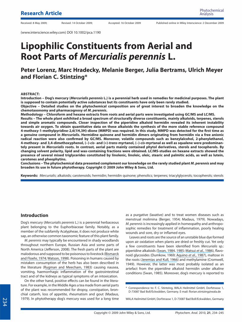
Profi ling of lipophilic constituents by GC/MS
Roots and aerial parts from M. perennis were extracted by use of
chloroform to yield a broad spectrum of lipophilic constituents.
Typical total ion chromatograms (TIC) of aerial part and root
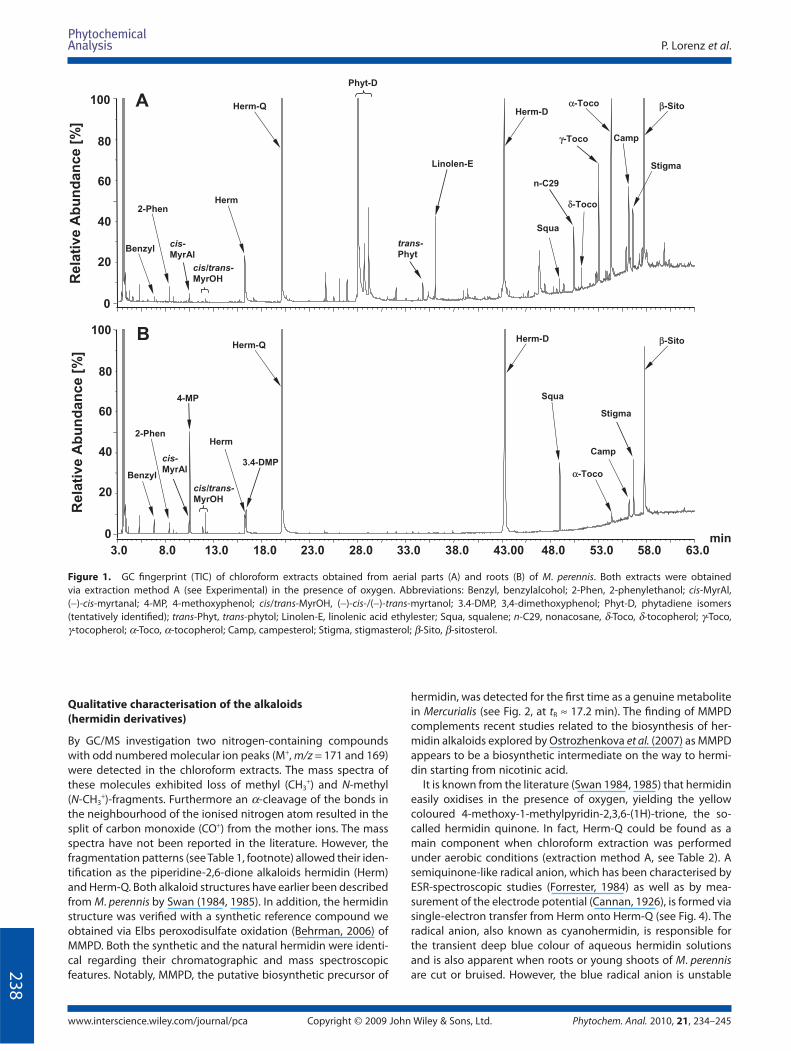
extracts are shown in Fig. 1A and B, respectively.
The main constituents were identifi ed by comparison of the
mass spectra with the NIST spectral database, as well as via refer-
ence compounds (Table 1). It is obvious that the aerial part
extracts (Fig. 1A) contain a larger variety of compounds than the
roots (Fig. 1B). However monoterpenes, simple phenolics and
squalene were present in much higher amounts in the root parts
(for quantitative data see below).
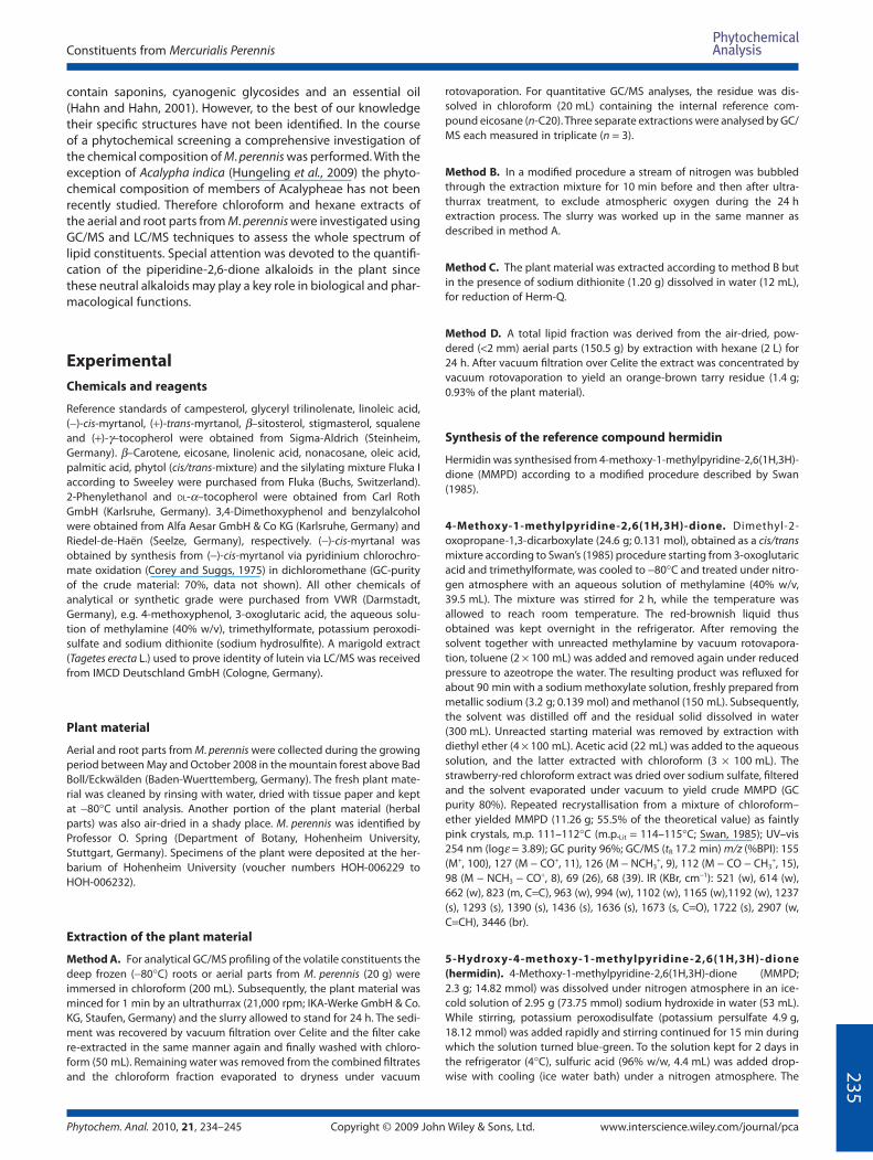
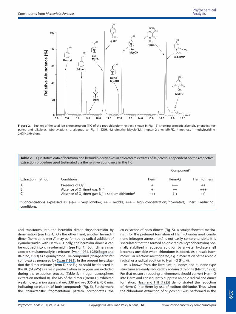
At short retention times of the TIC (tR 6–18 min) simple aro-
matic constituents like benzylalcohol (Benzyl), 2-phenylethanol
(2-Phen), 4-methoxyphenol (4-MP) and 3,4-dimethoxyphenol
(3.4-DMP) were identifi ed (Fig. 2). Moreover, bicyclic monoter-
penes of the pinane type such as (−)-cis-myrtanal (cis-MyrAl),
(−)-cis- and (+)-trans-myrtanol (cis/trans-MyrOH) were detected
and another monoterpene 6,6-dimethyl-bicyclo[3,1,1]heptan-2-
one (DBH) tentatively assigned by its specifi c mass data. All of the
compounds determined are typical essential oil constituents
detected in M. perennis for the fi rst time. Only 2-phenylethanol,
myrtanol and myrtanal have been reported earlier as constitu-
ents in the essential oil of Yamaai (Mercurialis leiocarpa) (Kameoka
et al., 1988). In the intermediate and long retention time range of
the TIC (tR 23–63 min; Fig. 1A), various polyisoprene compounds
were detected in the aerial parts of M. perennis. Between 27 and
30 min, three compounds with similar mass spectra were
observed (m/z 278, 137, 123; Table 1). Because of MS similarities
to trans-phytol (trans-Phyt; tR 34.7 min), these compounds were
tentatively assigned as phytadiene isomers (Phyt-D). Such phyta-
diene isomers (2,6,10,14-tetramethyl hexadienes) have been
earlier reported as pyrolysis artefacts from pheophytin a (Hites,
1974). Furthermore, α-, γ- and δ-tocopherols (α-, γ-, δ-Toco),
phytosterols like campesterol (Camp), stigmasterol (Stig) and
β-sitosterol (β-Sito), as well as linolenic acid ethylester (Linolen-E),
squalene (Squa) and nonacosane (n-C29) were assigned (Fig. 1).
The hermidin quinone (Herm-Q) and hermidin dimers (Herm-D;
co-elution in one peak at tR 43.0 min) were formed from hermidin
(Herm; see below).
Quantifi cation of terpenoid, aromatic and
phytosterol constituents
Quantitative data on the main constituents of aerial and root
parts were obtained by GC/MS via external calibration with
reference compounds. As mentioned before, some of the com-
pounds with short retention times were more frequently found
in the roots than in aerial parts. In roots collected in October,
4-methoxyphenol, (−)-cis-/(+)-trans-myrtanol and 3,4-dime-
thoxyphenol concentrations of 179.5, 35.8 and 99.6 mg/kg were
determined, while in aerial parts 9.5, 13.2 and 7.4 mg/kg were
found (see Fig. 3). Only 2-phenylethanol was present in slightly
higher amounts in aerial (32.8 mg/kg in May and 43.2 mg/kg in
October) than in root parts (17.4 mg/kg in October). The tenta-
tive phytadiene isomers (Phyt-D) and trans-phytol (trans-Phyt)
were exclusively present in aerial parts (1185.2 and 1184.0 mg/
kg Phyt-D and 77.4 and 108.0 mg/kg trans-Phyt in May and
October, respectively Fig. 3), corroborating the hypothesis that
these constituents are derived from chlorophylls (Hites, 1974) not
present in the roots.
Also, α-Toco was 8–18-fold higher in the aerial (369.8
and 814.7 mg/kg collected in May and October, respectively)
compared with root parts (44.2 mg/kg). Other tocopherol
isomers (δ- and γ-Toco) were detected only in aerial parts (not
quantifi ed).
Considering the total phytosterol concentration (sum of
campesterol, stigmasterol and β-sitosterol), approximately
double was found in aerial parts (1363.7 mg/kg in May and
1101.2 mg/kg and October) compared with root (580.6 mg/kg
in October). In summary, distinct qualitative and quantitative
diff erences in the composition of root and aerial parts of
M. perennis exist.
P. Lorenz et al.
www.interscience.wiley.com/journal/pca Copyright © 2009 John Wiley & Sons, Ltd. Phytochem. Anal. 2010, 21, 234–245
23
8
Qualitative characterisation of the alkaloids
(hermidin derivatives)
By GC/MS investigation two nitrogen-containing compounds
with odd numbered molecular ion peaks (M+, m/z = 171 and 169)
were detected in the chloroform extracts. The mass spectra of
these molecules exhibited loss of methyl (CH3+) and N-methyl
(N-CH3+)-fragments. Furthermore an α-cleavage of the bonds in
the neighbourhood of the ionised nitrogen atom resulted in the
split of carbon monoxide (CO+) from the mother ions. The mass
spectra have not been reported in the literature. However, the
fragmentation patterns (see Table 1, footnote) allowed their iden-
tifi cation as the piperidine-2,6-dione alkaloids hermidin (Herm)
and Herm-Q. Both alkaloid structures have earlier been described
from M. perennis by Swan (1984, 1985). In addition, the hermidin
structure was verifi ed with a synthetic reference compound we
obtained via Elbs peroxodisulfate oxidation (Behrman, 2006) of
MMPD. Both the synthetic and the natural hermidin were identi-
cal regarding their chromatographic and mass spectroscopic
features. Notably, MMPD, the putative biosynthetic precursor of
hermidin, was detected for the fi rst time as a genuine metabolite
in Mercurialis (see Fig. 2, at tR ≈ 17.2 min). The fi nding of MMPD
complements recent studies related to the biosynthesis of her-
midin alkaloids explored by Ostrozhenkova et al. (2007) as MMPD
appears to be a biosynthetic intermediate on the way to hermi-
din starting from nicotinic acid.
It is known from the literature (Swan 1984, 1985) that hermidin
easily oxidises in the presence of oxygen, yielding the yellow
coloured 4-methoxy-1-methylpyridin-2,3,6-(1H)-trione, the so-
called hermidin quinone. In fact, Herm-Q could be found as a
main component when chloroform extraction was performed
under aerobic conditions (extraction method A, see Table 2). A
semiquinone-like radical anion, which has been characterised by
ESR-spectroscopic studies (Forrester, 1984) as well as by mea-
surement of the electrode potential (Cannan, 1926), is formed via
single-electron transfer from Herm onto Herm-Q (see Fig. 4). The
radical anion, also known as cyanohermidin, is responsible for
the transient deep blue colour of aqueous hermidin solutions
and is also apparent when roots or young shoots of M. perennis
are cut or bruised. However, the blue radical anion is unstable
3.0 8.0 13.0 18.0 23.0 28.0 33.0 38.0 43.00 48.0 53.0 58.0 63.0min
100
0
80
60
40
20
100
0
80
60
40
20
Rela
tive A
bu
nd
an
ce
[%]
Rela
tive A
bu
nd
an
ce
[%]
Stigma
α-Toco
Camp
Squa
Herm
cis/trans-MyrOH
Benzyl
2-Phen
Herm-Q
cis-MyrAl
γ-Toco
δ-Toco
n-C29
trans-Phyt
Phyt-D
Linolen-E
Herm-D
β-Sito
Benzyl
2-Phen
cis-MyrAl
Herm-DHerm-Q
Herm
Stigma
α-Toco
Camp
Squa
cis/trans-MyrOH
4-MP
3.4-DMP
β-SitoA
B
Figure 1. GC fi ngerprint (TIC) of chloroform extracts obtained from aerial parts (A) and roots (B) of M. perennis. Both extracts were obtained
via extraction method A (see Experimental) in the presence of oxygen. Abbreviations: Benzyl, benzylalcohol; 2-Phen, 2-phenylethanol; cis-MyrAl,
(−)-cis-myrtanal; 4-MP, 4-methoxyphenol; cis/trans-MyrOH, (−)-cis-/(−)-trans-myrtanol; 3.4-DMP, 3,4-dimethoxyphenol; Phyt-D, phytadiene isomers
(tentatively identifi ed); trans-Phyt, trans-phytol; Linolen-E, linolenic acid ethylester; Squa, squalene; n-C29, nonacosane, δ-Toco, δ-tocopherol; γ-Toco,
γ-tocopherol; α-Toco, α-tocopherol; Camp, campesterol; Stigma, stigmasterol; β-Sito, β-sitosterol.
Constituents from Mercurialis Perennis
Phytochem. Anal. 2010, 21, 234–245 Copyright © 2009 John Wiley & Sons, Ltd. www.interscience.wiley.com/journal/pca
23
9
7.0 8.0 9.0 10.0 11.0 12.0 13.0 14.0 15.0 16.0 17.0 18.0
min0
100
40
OH
OH
O
OH
OHNO O
OCH3
CH3
OH
OH
OCH3
OCH3
NO O
OCH3
CH3
Benzyl
2-Phen
cis-
MyrAl
Herm
4-MP
cis-MyrOH
trans-MyrOH
MMPD
3.4-DMP
OH
OCH3
Rela
tive A
bu
nd
an
ce
[%]
20
60
80
DBH
6.0
OH
Figure 2. Section of the total ion chromatogram (TIC of the root chloroform extract, shown in Fig. 1B) showing aromatic alcohols, phenolics, ter-
penes and alkaloids. Abbreviations: analogous to Fig. 1; DBH, 6,6-dimethyl-bicyclo[3,1,1]heptan-2-one; MMPD, 4-methoxy-1-methylpyridine-
2,6(1H,3H)-dione.
Table 2. Qualitative data of hermidin and hermidin derivatives in chloroform extracts of M. perennis dependent on the respective
extraction procedure used (estimated via the relative abundance in the TIC)
Componenta
Extraction method Conditions Herm Herm-Q Herm-dimers
A Presence of O2b + +++ ++
B Absence of O2 (inert gas: N2)c + ++ +++C Absence of O2 (inert gas: N2) + sodium dithionited +++ (+) (+)
a Concentrations expressed as: (+)/+ = very low/low, ++ = middle, +++ = high concentration; b oxidative; c inert; d reducing
conditions.
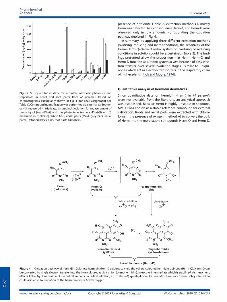
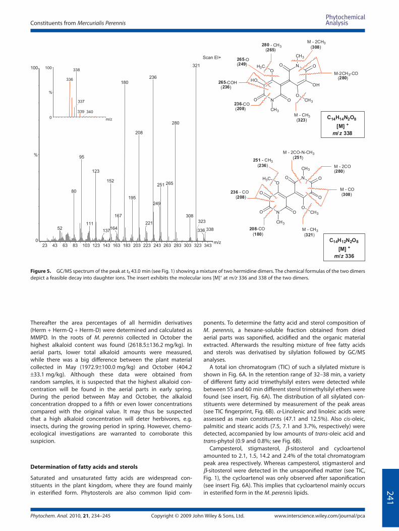
and transforms into the hermidin dimer chrysohermidin by
dimerisation (see Fig. 4). On the other hand, another hermidin
dimer (hermidin dimer A) may be formed by radical addition of
cyanohermidin with Herm-Q. Finally, the hermidin dimer A can
be oxidised into chrysohermidin (see Fig. 4). Both dimers may
appear simultaneously in a mixture (Swan, 1984, 1985; Boger and
Baldino, 1993) as a quinhydrone-like compound (charge-transfer
complex) as proposed by Swan (1985). In the present investiga-
tion the dimer mixture (Herm-D; see Fig. 4) could be detected in
the TIC (GC/MS) as a main product when air oxygen was excluded
during the extraction process (Table 2, nitrogen atmosphere,
extraction method B). The MS of the dimers (Herm-D) exhibited
weak molecular ion signals at m/z 338 and m/z 336 at tR 43.0 min,
indicating co-elution of both compounds (Fig. 5). Furthermore
the characteristic fragmentation pattern corroborates the
co-existence of both dimers (Fig. 5). A straightforward mecha-
nism for the preferred formation of Herm-D under inert condi-
tions (nitrogen atmosphere) is not easily comprehensible. It is
speculated that the formed anionic radical (cyanohermidin) nor-
mally stabilised in aqueous solution by a water hydrate shell
becomes unstable when chloroform is added. As a result inter-
molecular reactions are triggered, e.g. dimerisation of the anionic
radical or a radical addition to Herm-Q (Fig. 4).
As is known from the literature, quinones and quinone-type
structures are easily reduced by sodium dithionite (March, 1992).
For that reason a reducing environment should convert Herm-Q
into Herm and consequently suppress anionic radical and dimer
formation. Haas and Hill (1925) demonstrated the reduction
of Herm-Q into Herm by use of sodium dithionite. Thus, when
the chloroform extraction of M. perennis was performed in the
P. Lorenz et al.
www.interscience.wiley.com/journal/pca Copyright © 2009 John Wiley & Sons, Ltd. Phytochem. Anal. 2010, 21, 234–245
24
0
presence of dithionite (Table 2, extraction method C), mostly
Herm was detected. As a consequence Herm-Q and Herm-D were
observed only in low amounts, corroborating the oxidation
pathway depicted in Fig. 4.
In summary, by applying three diff erent extraction methods
(oxidising, reducing and inert conditions), the sensitivity of the
Herm–Herm-Q–Herm-D redox system on oxidising or reducing
conditions in solution could be ascertained (Table 2). The fi nd-
ings presented allow the proposition that Herm, Herm-Q and
Herm-D function as a redox system in vivo because of easy elec-
tron transfer over several oxidation stages—similar to ubiqui-
nones which act as electron transporters in the respiratory chain
of higher plants (Rich and Moore, 1976).
Quantitative analysis of hermidin derivatives
Since quantitative data on hermidin (Herm) in M. perennis
were not available from the literature, an analytical approach
was established. Because Herm is highly unstable in solutions,
MMPD was chosen as a stable reference compound for external
calibration. Roots and aerial parts were extracted with chloro-
form in the presence of oxygen (method A) to convert the bulk
of Herm into the more stable compounds Herm-Q and Herm-D.
Co
ncen
trati
on
[mg
/kg
] in
dry
mass
0
200
400
600
800
1000
1200
Ben
zyl
2-Phen
4-M
P
cis/
trans-
Myr
OH
3.4-
DM
P
trans-
Phyt
Phyt-D
Squa
αα-Toco
Cam
p
Stigm
a
β-Sito
Figure 3. Quantitative data for aromatic alcohols, phenolics and
terpenoids in aerial and root parts from M. perennis, based on
chromatograms exemplarily shown in Fig. 1 (for peak assignment see
Table 1). Compound quantifi cation was performed via external calibration
(n = 3, measured in triplicate; ± standard deviation; for measurement of
trans-phytol [trans-Phyt) and the phytadiene isomers (Phyt-D) n = 2,
measured in triplicate]. White bars, aerial parts (May); grey bars, aerial
parts (October); black bars, root parts (October).
N
O
HO
CH3
OO
CH3
N
O
O
CH3
O O
CH3
N
O
O
CH3
O O
CH3
H
N
O
OO
CH3
O
H3C
N
O
CH3
OO
CH3
O
Herm(colorless)
cyanohermidin(blue)
Herm-Q(yellow)
N
O
OO
CH3
HO
H3C
hermidin dimers (Herm-D)
N
CH3
OCH3
O
O
ON
OCH3
O
OH
O
CH3
dimerization
+ e
- 2e
radical addition
- e (+ 2H )
- 2 e
(- 2H )
[O]
hermidin dimer A(yellow)
chrysohermidin(yellow-brown)
Figure 4. Oxidation pathway of hermidin. Colorless hermidin (Herm) oxidises to yield the yellow-coloured hermidin quinone (Herm-Q). Herm-Q can
be converted by single electron transfer into the blue coloured radical anion (cyanohermidin), a reactive intermediate which is stabilised via mesomeric
eff ects. Either by dimerisation of the radical anion or by radical addition, e.g. to Herm-Q, quinhydrone-like hermidin dimers are formed. Chrysohermidin
could also arise by oxidation of the hermidin dimer A with oxygen.
Constituents from Mercurialis Perennis
Phytochem. Anal. 2010, 21, 234–245 Copyright © 2009 John Wiley & Sons, Ltd. www.interscience.wiley.com/journal/pca
24
1
Thereafter the area percentages of all hermidin derivatives
(Herm + Herm-Q + Herm-D) were determined and calculated as
MMPD. In the roots of M. perennis collected in October the
highest alkaloid content was found (2618.5±136.2 mg/kg). In
aerial parts, lower total alkaloid amounts were measured,
while there was a big diff erence between the plant material
collected in May (1972.9±100.0 mg/kg) and October (404.2
±33.1 mg/kg). Although these data were obtained from
random samples, it is suspected that the highest alkaloid con-
centration will be found in the aerial parts in early spring.
During the period between May and October, the alkaloid
concentration dropped to a fi fth or even lower concentrations
compared with the original value. It may thus be suspected
that a high alkaloid concentration will deter herbivores, e.g.
insects, during the growing period in spring. However, chemo-
ecological investigations are warranted to corroborate this
suspicion.
Determination of fatty acids and sterols
Saturated and unsaturated fatty acids are widespread con-
stituents in the plant kingdom, where they are found mainly
in esterifi ed form. Phytosterols are also common lipid com-
ponents. To determine the fatty acid and sterol composition of
M. perennis, a hexane-soluble fraction obtained from dried
aerial parts was saponifi ed, acidifi ed and the organic material
extracted. Afterwards the resulting mixture of free fatty acids
and sterols was derivatised by silylation followed by GC/MS
analyses.
A total ion chromatogram (TIC) of such a silylated mixture is
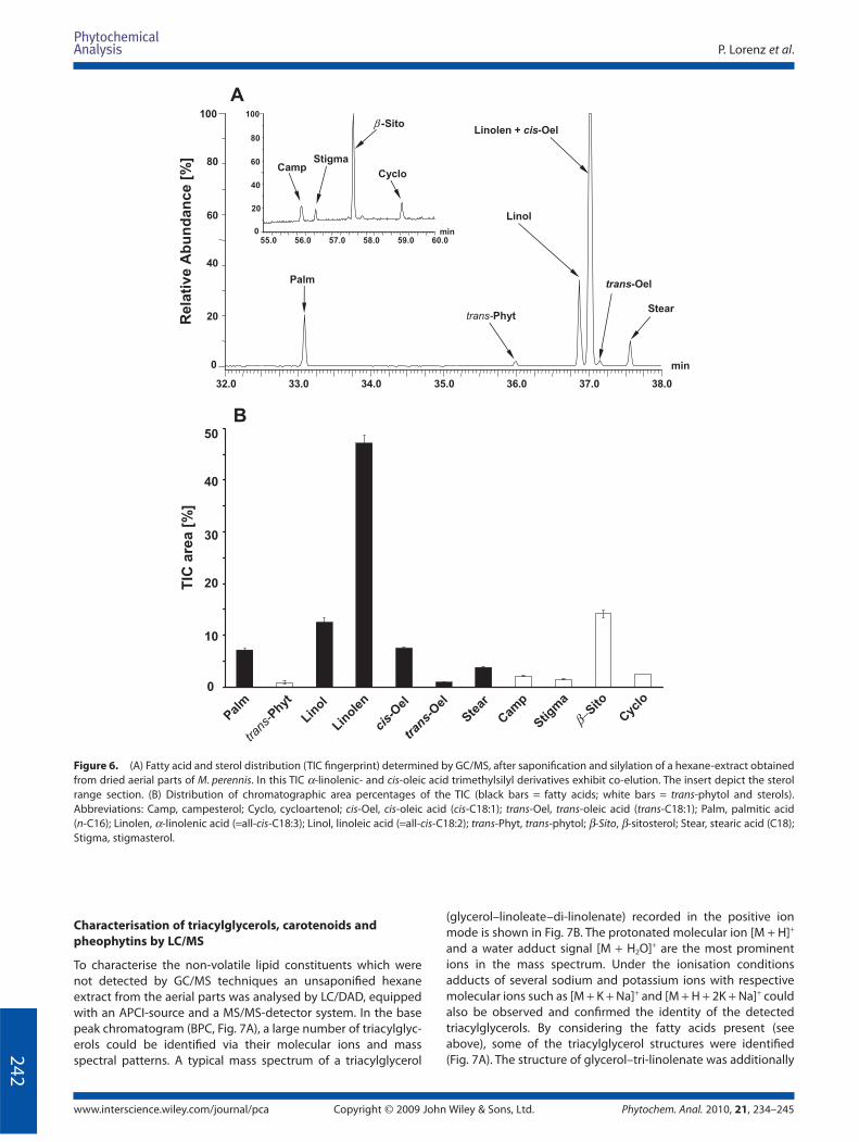
shown in Fig. 6A. In the retention range of 32–38 min, a variety
of diff erent fatty acid trimethylsilyl esters were detected while
between 55 and 60 min diff erent sterol trimethylsilyl ethers were
found (see insert, Fig. 6A). The distribution of all silylated con-
stituents were determined by measurement of the peak areas
(see TIC fi ngerprint, Fig. 6B). α-Linolenic and linoleic acids were
assessed as main constituents (47.1 and 12.5%). Also cis-oleic,
palmitic and stearic acids (7.5, 7.1 and 3.7%, respectively) were
detected, accompanied by low amounts of trans-oleic acid and
trans-phytol (0.9 and 0.8%; see Fig. 6B).
Campesterol, stigmasterol, β-sitosterol and cycloartenol
amounted to 2.1, 1.5, 14.2 and 2.4% of the total chromatogram
peak area respectively. Whereas campesterol, stigmasterol and
β-sitosterol were detected in the unsaponifi ed matter (see TIC,
Fig. 1), the cycloartenol was only observed after saponifi cation
(see insert Fig. 6A). This implies that cycloartenol mainly occurs
in esterifi ed form in the M. perennis lipids.
23 43 63 83 103 123 143 163 183 203 223 243 263 283 303 323 343m/z0
100
%
Scan EI+
321
236180
95
80
52
123
111
152
137
167
164
208
195
221
280
265251
249
308
338336
m/z0
100
%
338
336
337
339 340
323
N
N
CH3
CH3
O OO
H3C
OCH3O O
HOOH
M - CH3(323)
M - 2CH3
(308)
C14H14N2O8
[M] +
m/ z 338
M-2CH3-CO(280)
280 - CH3(265)
265-O(249)
265-COH(236)
236-CO(208)
N
N
CH3
CH3
O OO
H3C
OCH3O O
OO
M - CH3
(321)
(308)
208-CO
(180)
251 - CH3
(236)
236 - CO(208)
C14H12N2O8
[M] +
m/z 336
M - CO
M - 2CO(280)
M - 2CO-N-CH3
(251)
Figure 5. GC/MS spectrum of the peak at tR 43.0 min (see Fig. 1) showing a mixture of two hermidine dimers. The chemical formulas of the two dimers
depict a feasible decay into daughter ions. The insert exhibits the molecular ions [M]+ at m/z 336 and 338 of the two dimers.
P. Lorenz et al.
www.interscience.wiley.com/journal/pca Copyright © 2009 John Wiley & Sons, Ltd. Phytochem. Anal. 2010, 21, 234–245
24
2
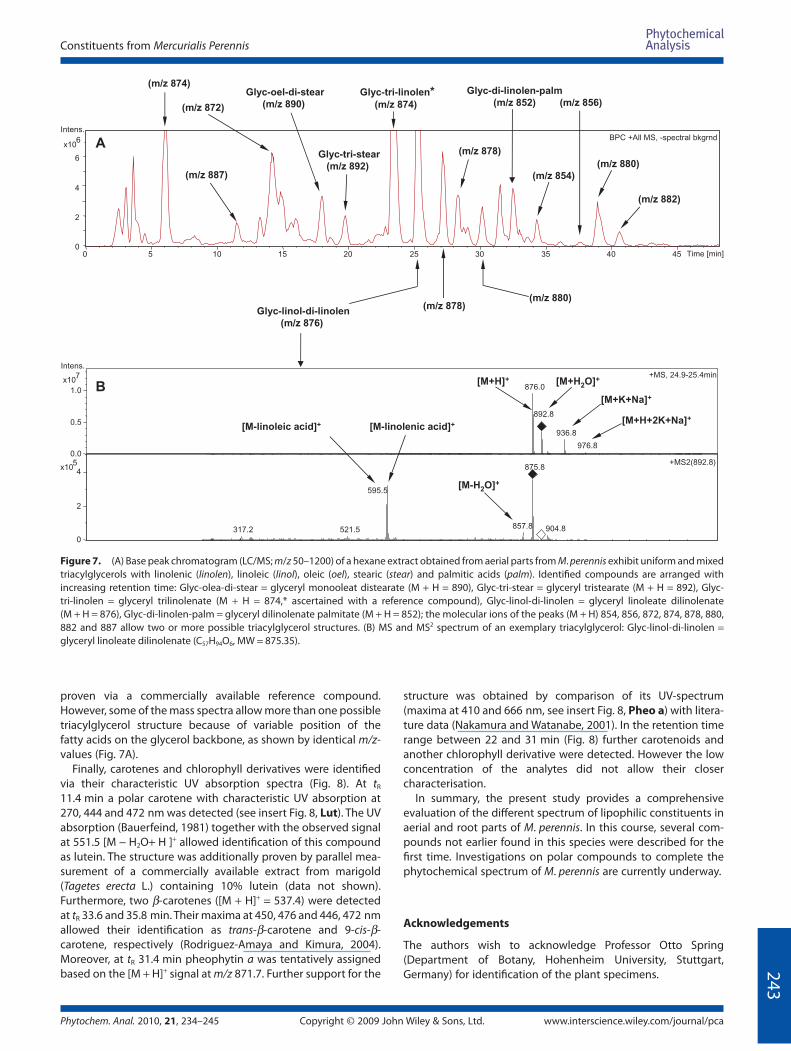
Characterisation of triacylglycerols, carotenoids and
pheophytins by LC/MS
To characterise the non-volatile lipid constituents which were
not detected by GC/MS techniques an unsaponifi ed hexane
extract from the aerial parts was analysed by LC/DAD, equipped
with an APCI-source and a MS/MS-detector system. In the base
peak chromatogram (BPC, Fig. 7A), a large number of triacylglyc-
erols could be identifi ed via their molecular ions and mass
spectral patterns. A typical mass spectrum of a triacylglycerol
32.0 33.0 34.0 35.0 36.0 37.0 38.0
min
100
0
20
40
60
80
Rela
tive A
bu
nd
an
ce
[%]
Palm
trans-Phyt
Linol
Stear
Linolen + cis-Oel
trans-Oel
55.0 56.0 57.0 58.0 59.0 60.0min
20
100
40
60
80
0
CampStigma
β -Sito
Cyclo
A
TIC
are
a[%
]
0
10
20
30
40
50
B
Linole
n
Palm
trans
-Phyt
Linol
cis-
Oel
Stear
Cam
p
trans-
Oel
Stigm
a
β−Sito
Cyc
lo
(glycerol–linoleate–di-linolenate) recorded in the positive ion
mode is shown in Fig. 7B. The protonated molecular ion [M + H]+
and a water adduct signal [M + H2O]+ are the most prominent
ions in the mass spectrum. Under the ionisation conditions
adducts of several sodium and potassium ions with respective
molecular ions such as [M + K + Na]+ and [M + H + 2K + Na]+ could
also be observed and confi rmed the identity of the detected
triacylglycerols. By considering the fatty acids present (see
above), some of the triacylglycerol structures were identifi ed
(Fig. 7A). The structure of glycerol–tri-linolenate was additionally
Figure 6. (A) Fatty acid and sterol distribution (TIC fi ngerprint) determined by GC/MS, after saponifi cation and silylation of a hexane-extract obtained
from dried aerial parts of M. perennis. In this TIC α-linolenic- and cis-oleic acid trimethylsilyl derivatives exhibit co-elution. The insert depict the sterol
range section. (B) Distribution of chromatographic area percentages of the TIC (black bars = fatty acids; white bars = trans-phytol and sterols).
Abbreviations: Camp, campesterol; Cyclo, cycloartenol; cis-Oel, cis-oleic acid (cis-C18:1); trans-Oel, trans-oleic acid (trans-C18:1); Palm, palmitic acid
(n-C16); Linolen, α-linolenic acid (=all-cis-C18:3); Linol, linoleic acid (=all-cis-C18:2); trans-Phyt, trans-phytol; β-Sito, β-sitosterol; Stear, stearic acid (C18);
Stigma, stigmasterol.
Constituents from Mercurialis Perennis
Phytochem. Anal. 2010, 21, 234–245 Copyright © 2009 John Wiley & Sons, Ltd. www.interscience.wiley.com/journal/pca
24
3
proven via a commercially available reference compound.
However, some of the mass spectra allow more than one possible
triacylglycerol structure because of variable position of the
fatty acids on the glycerol backbone, as shown by identical m/z-
values (Fig. 7A).
Finally, carotenes and chlorophyll derivatives were identifi ed
via their characteristic UV absorption spectra (Fig. 8). At tR
11.4 min a polar carotene with characteristic UV absorption at
270, 444 and 472 nm was detected (see insert Fig. 8, Lut). The UV
absorption (Bauerfeind, 1981) together with the observed signal
at 551.5 [M − H2O+ H ]+ allowed identifi cation of this compound
as lutein. The structure was additionally proven by parallel mea-
surement of a commercially available extract from marigold
(Tagetes erecta L.) containing 10% lutein (data not shown).
Furthermore, two β-carotenes ([M + H]+ = 537.4) were detected
at tR 33.6 and 35.8 min. Their maxima at 450, 476 and 446, 472 nm
allowed their identifi cation as trans-β-carotene and 9-cis-β-
carotene, respectively (Rodriguez-Amaya and Kimura, 2004).
Moreover, at tR 31.4 min pheophytin a was tentatively assigned
based on the [M + H]+ signal at m/z 871.7. Further support for the
structure was obtained by comparison of its UV-spectrum
(maxima at 410 and 666 nm, see insert Fig. 8, Pheo a) with litera-
ture data (Nakamura and Watanabe, 2001). In the retention time
range between 22 and 31 min (Fig. 8) further carotenoids and
another chlorophyll derivative were detected. However the low
concentration of the analytes did not allow their closer
characterisation.
In summary, the present study provides a comprehensive
evaluation of the diff erent spectrum of lipophilic constituents in
aerial and root parts of M. perennis. In this course, several com-
pounds not earlier found in this species were described for the
fi rst time. Investigations on polar compounds to complete the
phytochemical spectrum of M. perennis are currently underway.
Acknowledgements
The authors wish to acknowledge Professor Otto Spring
(Department of Botany, Hohenheim University, Stuttgart,
Germany) for identifi cation of the plant specimens.
Time [min]
BPC +All MS, -spectral bkgrnd
0
2
4
6
6x10
Intens.
0 5 10 15 20 25 30 35 40 45
876.0
936.8
976.8
892.8
+MS, 24.9-25.4min
5.1252.713 857.8 904.8
595.5
875.8+MS2(892.8)
0.0
0.5
1.0
7x10
Intens.
0
2
45
x10
[M+H]+ [M+H2O]+
[M-H2O]+
[M+K+Na]+
[M+H+2K+Na]+
[M-linoleic acid]+ [M-linolenic acid]+
(m/z 882)
(m/z 880)
(m/z 856)
(m/z 854)
Glyc-di-linolen-palm
(m/z 852)
(m/z 878)
Glyc-tri-linolen*(m/z 874)
Glyc-tri-stear
(m/z 892)
Glyc-oel-di-stear
(m/z 890)
(m/z 880) (m/z 878)
Glyc-linol-di-linolen
(m/z 876)
A
B
(m/z 874)
(m/z 887)
(m/z 872)
Figure 7. (A) Base peak chromatogram (LC/MS; m/z 50–1200) of a hexane extract obtained from aerial parts from M. perennis exhibit uniform and mixed
triacylglycerols with linolenic (linolen), linoleic (linol), oleic (oel), stearic (stear) and palmitic acids (palm). Identifi ed compounds are arranged with
increasing retention time: Glyc-olea-di-stear = glyceryl monooleat distearate (M + H = 890), Glyc-tri-stear = glyceryl tristearate (M + H = 892), Glyc-
tri-linolen = glyceryl trilinolenate (M + H = 874,* ascertained with a reference compound), Glyc-linol-di-linolen = glyceryl linoleate dilinolenate
(M + H = 876), Glyc-di-linolen-palm = glyceryl dilinolenate palmitate (M + H = 852); the molecular ions of the peaks (M + H) 854, 856, 872, 874, 878, 880,
882 and 887 allow two or more possible triacylglycerol structures. (B) MS and MS2 spectrum of an exemplary triacylglycerol: Glyc-linol-di-linolen =
glyceryl linoleate dilinolenate (C57H94O6, MW = 875.35).
P. Lorenz et al.
www.interscience.wiley.com/journal/pca Copyright © 2009 John Wiley & Sons, Ltd. Phytochem. Anal. 2010, 21, 234–245
24
4
Intens.
[mAU]
+All MS, -spectral bkgrnd
UV chromatogram, 417-421 nm
x10
0
2
4
6
6
4
6
8
10
0 5 10 15 20 25 30 35 40 Time [min]
Lut
all-trans-β-CaroPheo a
cis-β-Caro
887.8
551.5+MS, 11.2-11.7min
174.9208.9
265.0
345.2429.3
495.3
533.4+MS2 (551.9)
0.0
0.5
1.0
6x10
Intens.
0
1
2
3
4x10
HO
OH
nm250
300 350 400 450 500
mAU
0
0.5
1
1.5
2
2.5Lutdi-Olea-Stear
[M-H2O+H]+
663.5
871.7+MS, 31.6-31.8min
461.3
533.3
593.3+MS2(871.7)
0
1
6x10
Intens.
0.0
0.5
1.0
6x10
Pheo a [M+H]+
NH
NH
N
N
O
O
O
O
O
[M-phytol]+
nm300 400 500 600 700
mAU
0
1
2
3
4
5
6
7
[M-2H2O+H]+
Figure 8. Base peak traces and UV traces (recorded at 419 nm) of a hexane extract obtained from dried aerial parts of M. perennis; MS+ and MS2 spectra
of lutein (Lut, C40H56O2, MW = 568.87) and pheophytin a (Pheo a, C55H74N4O5, MW = 871.20). The inserts show UV spectra (with UV maxima) of both
compounds at the specifi ed retention times in the LC.
References
Aquino R, Behar I, D’Agostino M, Simone FD, Schettino O, Pizza C. 1987. Phytochemical investigation on Mercurialis annua. Biochem Syst Ecol 15: 667–669.
Bauerfeind JC. 1981. Carotenoids as Colorants and Vitamin A Precursors. Academic Press: New York.
Behrman EJ. 2006. The Elbs and Boyland–Sims peroxydisulfate oxida-tions. Beilstein J Org Chem 2: 22.
Berger F. 1954. Handbuch der Drogenkunde. Verlag für Medizinische Wissenschaften Wilhelm Maudrich: Vienna; 348–351.
Bismarck R, Floehr W. 1974. Poisoning with Mercurialis perennis in a grazing cow herd. Short communication from practice. Dtsch Tierärztl Wochenschr 81: 433–434.
Boger DL, Baldino CM. 1993. D,L- and meso-Isochrysohermidin: total synthesis and interstrand DNA crosslinking. J Am Chem Soc 115: 11418–11425.
Cannan RK. 1926. Electrode potentials of hermidin, the chromogen of Mercurialis perennis. Biochem J 20: 927–937.
Corey EJ, Suggs, W. 1975. Pyridinium chlorochromate. An effi cient reagent for oxidation of primary and secondary alcohols to carbonyl com-pounds. Tetrahedron Lett 16: 2647–2650.
Constituents from Mercurialis Perennis
Phytochem. Anal. 2010, 21, 234–245 Copyright © 2009 John Wiley & Sons, Ltd. www.interscience.wiley.com/journal/pca
24
5
Cromwell BT. 1949. The micro-estimation and origin of methylamine in Mercurialis perennis L. Biochem J 45: 84–86.
Dumkow K. 1969. Die Flavonoide einheimischer Euphorbiaceen. 3. Mitt. Isolierung und Identifi zierung der Flavonoidglycoside von Mercurialis perennis L. Planta Med 17: 391–392.
Forrester AR. 1984. Autoxidation of hermidin: an ESR study. Experientia 40: 688–689.
Haas P, Hill TG. 1925. Mercurialis. III. A consideration of the physiological signifi cance of the chromogen. Ann Bot 39: 861–865.
Hahn G, Hahn M. 2001. Der Wolf aus dem Garten Eden—Wolfsmilchgewächse: giftig, dennoch medizinisch und wirtschaftlich genutzt. Oesterr Apoth Ztg 26: 1252–1262.
Hites RA. 1974. Phytadienes from the pyrolysis of pheophytine a. J Org Chem 39: 2634–2635.
Hungeling M, Lechtenberg M, Fronczek FR, Nahrstedt A. 2009. Cyanogenic and non-cyanogenic glucosides from Acalypha indica (Euphorbiaceae). Phytochemistry 70: 270–277.
Jeff erson RG. 2008. Biological fl ora of the British isles: Mercurialis perennis L. J Ecol 96: 386–412.
Jeremias K, Kull U. 1966. Zum Vorkommen von Maltose in vegetativen Pfl anzenteilen. Experientia 22: 141–142.
Kameoka H, Masui Y, Amano K, Kyo A. 1988. Components of essential oil of Mercurialis leiocarpa. Nippon Nogei Kagaku Kaishi (in Japanese) 62: 1217–1219.
Lorenz P, Berger M, Bertrams J, Wende K, Wenzel K, Lindequist U, Meyer U, Stintzing FC. 2008. Natural wax constituents of a supercritical fl uid CO2 extract from quince (Cydonia oblonga Mill.) pomace. Anal Bioanal Chem 391: 633–646.
Madaus G. 1979. Lehrbuch der biologischen Heilmittel, 2nd edn. Nachdruck der Ausgabe Leipzig 1938. Medimed Verlag: Hildesheim.
March J. 1992. Advanced Organic Chemistry: Reactions, Mechanisms, and Structure. 4th edn. John Wiley & Sons Inc.: New York; 912.
Matsui Y, Kawabe C, Mastumoto K, Abe K, Miwa T. 1986. A 2-oxo-3-pyrroline dimer from Mercurialis leiocarpa. Phytochemistry 25: 1470–1471.
Nakamura A, Watanabe T. 2001. Separation and determination of minor photosynthetic pigments by reversed-phase HPLC with minimal alteration of chlorophylls. Anal Sci 17: 503–508.
Ostrozhenkova E, Eylert E, Schramek N, Golan-Goldhirsh A, Bacher A, Eisenreich W. 2007. Biosynthesis of the chromogen hermidin from Mercurialis annua L. Phytochemistry 68: 2816–2824.
Rich PR, Moore AL. 1976. The involvement of the protonmotive ubiquinone cycle in the respiratory chain of higher plants and its relation to the branchpoint of the alternate pathway. FEBS Lett 65: 339–344.
Rodriguez-Amaya DB, Kimura M. 2004. HarvestPlus Handbook for Carotenoid Analysis. HarvestPlus Technical Monograph 2. International Food Policy Research Institute and International Center for Tropical Agriculture, Washington, DC.
Rugman F, Meecham J. 1983. Mercurialis perennis (dog’s mercury) poison-ing: a case of mistaken identity. Br Med J 287: 1924.
Swan GA. 1984. Hermidin, a chromogen from Mercurialis perennis L. Experientia 40: 687–688.
Swan GA. 1985. Isolation, structure and synthesis of hermidin, a chromo-gen from Mercurialis perennis L. J Chem Soc Perkin Trans I: 1757–1766.
Watson PJ. 1998. Suspected dog’s mercury (Mercurialis perennis) poison-ing in cattle. Vet Rec 142: 116–117.


