Embed Size (px)
Citation preview

Involvement of 9-O-Acetyl GD3 Ganglioside in Mycobacteriumleprae Infection of Schwann Cells*□S
Received for publication, May 24, 2010, and in revised form, August 10, 2010 Published, JBC Papers in Press, August 25, 2010, DOI 10.1074/jbc.M110.147272
Victor Tulio Ribeiro-Resende‡§1, Michelle Lopes Ribeiro-Guimaraes‡, Robertha Mariana Rodrigues Lemes‡,Isis Cristina Nascimento§, Lucineia Alves‡, Rosalia Mendez-Otero§, Maria Cristina Vidal Pessolani‡,and Flavio Alves Lara‡
From the ‡Laboratorio de Microbiologia Celular, Pavilhao de Hanseníase, Instituto Oswaldo Cruz, Fundacao Oswaldo Cruz,21045-900 Rio de Janeiro, Brazil and the §Laboratorio de Neurobiologia Celular e Molecular, Instituto de Biofísica Carlos ChagasFilho, Centro de Ciencias da Saude, Universidade Federal do Rio de Janeiro, 21941-902 Rio de Janeiro, Brazil
Mycobacterium leprae (ML), the etiologic agent of leprosy,mainly affects the skin and peripheral nerves, leading to demy-elization and loss of axonal conductance. Schwann cells (SCs)are the main cell population infected by ML in the nerves, andinfection triggers changes in the SC phenotype from a myeli-nated to a nonmyelinated state. In the present study, we showthat expression of 9-O-acetyl GD3, a ganglioside involved in cel-lular anti-apoptotic signaling and nerve regeneration, increasesin SCs following infection with ML. Observation by confocalmicroscopy together with coimmunoprecipitation suggestedthat this ganglioside participates in ML attachment and inter-nalization by SC. Immunoblockage of 9-O-acetyl GD3 in vitrosignificantly reduced adhesion ofML to SC surfaces. Finally, weshow that activation of the MAPK (ERK 1/2) pathway and SCproliferation, two known effects of ML on SCs that result indemyelization, are significantly reduced when the 9-O-acetylGD3 ganglioside is immunoblocked. Taken together, these datasuggest the involvement of 9-O-acetyl GD3 in ML infection onSCs.
Leprosy, one of the oldest recorded diseases, remains animportant cause of morbidity, with �250,000 new cases/year.Mycobacterium leprae (ML),2 the causative agent of leprosy, isan obligate intracellular pathogen with high tropism forSchwann cells (SCs) (1). Data collected over the past 15 yearssuggest that cell surfacemolecules are involved inML adhesionto SCs (2). The G domains of the laminin-�2 chain and thedystroglycan receptor have been shown to play roles in mediat-ing ML attachment to the SC surface (3). Phenolic glycolipid-I,a major unique glycoconjugate on the ML surface, binds lami-nin-2, which explains the predilection of the bacterium forperipheral nerves (4). In vitro and in vivo studies in Rag-1�/�
mice, which lack B and T lymphocytes, showed thatML attach-ment to the SC surface is sufficient to cause demyelination inperipheral nerves (5). This effect was found to be dependent onneuregulin receptor, ErbB-2, and ERK 1/2 activation by ML,leading to MAPK signaling and proliferation (6). Additionally,conversion of SCs from the myelinated to nonmyelinated phe-notype is directly related to increased proliferative capacity (7).Tapinos et al. (8) have shown that ML is able to induce SCproliferation through ERK 1/2 activation via MEK-indepen-dent and p56LcK-dependent pathways.9-O-Acetyl GD3 ganglioside is an acetylated glycolipid pres-
ent in the cell membrane of many types of vertebrate cells (9).This molecule plays an important role in the development, dif-ferentiation, and regeneration of the nervous system. Amongits multiple functions are: cell attachment to the extracellularmatrix, neuronal migration, neurite outgrowth in vitro, andcontrol of apoptosis (10–17). 9-O-Acetyl GD3 ganglioside ishighly expressed after sciatic nerve crush in the regeneratingnerve, as well as in the dorsal root ganglia and lumbar spinalcord. During peripheral nerve regeneration, 9-O-acetyl GD3 ismainly expressed by SCs, which concurrently undergo changesin phenotype and proliferative state, followed by lower expres-sion in a few axons (18).The up-regulation of 9-O-acetyl GD3 expression in prolifer-
ating SCs led us to test the hypothesis that this molecule alsoplays a role in SC leprosy cytopathology, because proliferativeand anti-apoptotic states are also induced by ML during infec-tion (19, 20). We analyzed ML-induced expression of this gan-glioside and performed immunoblockage assays in SCs exposedtoML.Moreover, we investigated the association between 9-O-acetyl GD3 and well knownML cell ligands (laminin-2, dystro-glycan, and ErbB-2 receptors) using laser scanning microscopyand coimmunoprecipitation. These data demonstrate for thefirst time the involvement of 9-O-acetyl GD3 in cellular infec-tion, suggesting a relevant biological role for this ganglioside asa possible receptor for ML in SCs.
EXPERIMENTAL PROCEDURES
Mycobacteria—Armadillo-derived lethally irradiatedM. lep-rae was kindly provided by Dr. Patrick J. Brennan (ColoradoState University, Fort Collins, CO, through the NIAID,National Institutes of Health under contract 1Al 25469). M.leprae (viable and lethally irradiated) derived from the footpadsof athymic nu/nu mice were provided by J. L. Krahenbuhl
* This work was supported by grants from the Fundacao Carlos Chagas Filhode Amparo a Pesquisa do Estado do Rio de Janeiro and from the FundacaoOswaldo Cruz and by funds from the American Leprosy Missions and theOrder of St. Lazarus.
□S The on-line version of this article (available at http://www.jbc.org) containssupplemental Figs. S1–S5.
1 To whom correspondence should be addressed: Pavilhao Hanseníase IOC,Av. Brasil 4365 Manguinhos, 21045-900 Rio de Janeiro-RJ, Brazil. Tel.: 55-21-25626554; Fax: 55-21-22808193; E-mail: [email protected].
2 The abbreviations used are: ML, M. leprae; FITC�, M. leprae conjugated withfluorescein; SC, Schwann cell; MOI, multiplicities of infection; HLP, histone-like protein; HPTLC, high performance thin layer chromatography; ANOVA,analysis of variance; MBP, myelin basic protein; LAM, lipoarabinomannan.
THE JOURNAL OF BIOLOGICAL CHEMISTRY VOL. 285, NO. 44, pp. 34086 –34096, October 29, 2010© 2010 by The American Society for Biochemistry and Molecular Biology, Inc. Printed in the U.S.A.
34086 JOURNAL OF BIOLOGICAL CHEMISTRY VOLUME 285 • NUMBER 44 • OCTOBER 29, 2010

(National Hansen’s Disease Programs Laboratory, BatonRouge, LA).Mycobacterium smegmatismc2 155 andMycobac-terium bovis BCG Pasteur strains were grown at 37 °C inMiddlebrook 7H9 broth (BD) supplemented with 0.05%Tween80 under constant agitation on a magnetic plate. The cultureswere harvested in themid-log phase, counted according to pro-tocol of Shepard and McRae (21), and kept frozen at �70 °Cuntil use. Mycobacteria were conjugated with fluorescein(FITC�; Sigma) by incubation with carbonate-bicarbonatebuffer, pH8.2, for 1 h followed by threewasheswith 10mMPBS,pH 7.4.Culture Procedures—Human SCs derived from the tumor
cell line ST-8814 were seeded on coverslips pretreated withpoly-L-lysine (100 �g/ml; Sigma-Aldrich) and laminin (10�g/ml; Invitrogen) in 24-well cell culture plates (Greiner Bio-one). The cultures were then incubated at 37 °C, 5% CO2 inRPMI 1640 medium (Invitrogen) plus 10% FBS (Cutilab, SaoPaulo, Brazil), penicillin, and streptomycin (100 units/ml and100�g/ml, respectively; Sigma). Forty-eight hours later, FITC�
M. leprae were added to the culture medium at various multi-plicities of infection (MOI, 1:10, 1:25, and 1:50). The cultureswere then incubated for 2, 12, 24, 48, and 72 h, followed bygentle removal of the medium. The cells were then washedtwice for 2 min with 10 mM PBS, fixed for 15 min with 4%paraformaldehyde (Sigma-Aldrich) at 37 °C, and washed twiceagain with 10 mM PBS, pH 7.4.Mouse Infection with M. leprae—Two-month-old male
Foxnu/nu (NUDE) mice derived from the Balb-C strain werehoused at the Fundacao Oswaldo Cruz rodent facility underinternal license number P-0328/07. All of the experiments wereperformed following the National Institutes of Health Guide-lines for the Care and Use of Laboratory Animals and wereapproved by FundacaoOswaldoCruzCommittee for theUse ofExperimental Animals. One injection of 10�l of RPMImediumcontaining �107/ml live bacteria was administered to eachforepaw. Four months after the injection, we were able toobserve swelling, indicating that the infection was well estab-lished in the injectedmice. Tenmonths after infection, themicewere sacrificed in a CO2 chamber. The sciatic nerves (n � 4)were carefully removed and fixed in 4% paraformaldehydeovernight. Sciatic nerves from noninfected NUDE (n � 4) andBalb-C (n� 3) mice were also removed and fixed for later anal-ysis as control groups. After fixation, all of the nerve segmentswere cryoprotectedwith 30% sucrose in 0.1 M phosphate buffer,pH 7.4, for 48 h.Immunofluorescence—Dissected nerves were embedded in
Tissue-Tek� O.C.T. compound (Sakura Finetechnical, Tokyo,Japan). Frozen longitudinal sections were cut at 16 �m on acryostat (Leica CM 1850), mounted on gelatin-coated slides,and frozen. For immunofluorescence, the nerve sections werewashed three times with 10 mM PBS, pH 7.4, and then blockedby incubation with 10% normal goat serum (Invitrogen) and 1%BSA (Sigma) for 1 h at room temperature. Incubation with theprimary antibodies was performed overnight at 4 °C, followedby three washes with 0.001% Triton X-100 (5 min each) andthen incubation with the secondary antibodies for 2 h at roomtemperature. The following primary antibodies were used indouble or triple labeling combinations for nerve staining in the
in vivo studies: polyclonal IgG anti-�-S100 (1:400; Dako),monoclonal IgM anti-�-9-O-acetyl GD3 (Jones clone, 1:100;Sigma), polyclonal IgG anti-�-myelin basic protein (MBP,1:200; Abcam), andmousemonoclonal IgG anti-�-histone-likeprotein (HLP, 1:600) or rabbit polyclonal IgG anti-�-Lam (1:35,both kindly donated by Dr. Patrick Brennan).For immunostaining of SC cultures, fixed cells were washed
three times with 10mM PBS, pH 7.4, and blocked by incubationwith 5% normal goat serum (blocking solution) for 40 min in ahumid chamber. Primary antibodies were diluted in the block-ing solution, and coverslips were incubated for 2 h in a humidchamber at room temperature. Next, the coverslips werewashed with 10 mM PBS, pH 7.4 (three times for 5 min each)and incubated with secondary antibodies diluted in the sameblockage solution for 90 min at room temperature. After a finalseries of washes in PBS (three times for 5 min each), the cellnuclei were counterstained with DAPI (Sigma-Aldrich), andeach coverslip was placed upside down on a slide containing adrop of SlowFade� antifade solution (Molecular Probes). Thefollowing antibodies were used for immunostaining: rabbitpolyclonal anti-�-S100 (1:400; Dako), goat polyclonal anti-�-laminin-2 (1:100; Santa Cruz Laboratories), rabbit polyclonalIgG anti-�-laminin (1:1000; Sigma), rabbit polyclonal IgGanti-�-ErbB-2 (1:150; Abcam), mouse monoclonal IgM anti-�-9-O-acetyl GD3 (Jones, 1:100; Sigma), mouse monoclonal IgGanti-�-dystroglycan (1:100; Abcam), and mouse monoclonalIgG anti-�-HLP (histone-like protein, 1:600, kindly donated byDr. Patrick Brennan). The following secondary antibodies wereemployed: Cy3-conjugated �-mouse (1:800; Jackson Immuno-chemicals), Cy3-conjugated �-� chain IgM (1:600; JacksonImmunochemicals), Alexa 488-conjugated �-rabbit (1:400;Invitrogen), Alexa 633 �-mouse (1:400; Invitrogen), Alexa 555�-rabbit (1:400; Invitrogen), and fluorescein-conjugated rabbit�-goat (1:100; Sigma-Aldrich).Immunoblockage and Deacetylation of 9-O-Acetyl GD3—ST-
8814 SCs (5 � 104/coverslip) were allowed to grow for 48 h instandard medium (RPMI 1640 plus 10% FBS) until semi-con-fluent. For immunoblockage assay, the cells were incubated infour different conditions: 1) RPMI medium plus 10% FBS as acontrol condition; 2) Jones antibody added to the standardmedium diluted 1:100, as described by Santiago et al. (22); 3)mouse IgM immunoglobulin (1:200; Caltag, Burlingame, CA)added to the standard medium as an antibody control; and 4)anti-A2B5 antibody against gangliosides derived from theC-se-ries of the biosynthetic pathway (1:50; Roche Applied Science).For the deacetylation assay, cultured Schwann cells were incu-bated for 1 h at 4 °C in the absence of light in three differentconditions: 1) RPMI medium plus 10% FBS as a control condi-tion; 2) 10 mM of meta sodium periodate (NaIO4) (Sigma), asdescribed byVan Lenten andAshwell (23) andManzi et al. (24);and 3) Jones antibody added to the standard medium diluted1:100 in control medium. The cells were incubated for 2 h withthe Jones antibody before bacteria addition to the cultures. ThemediumcontainingNaIO4was removed, and cells werewashedtwice in 10 mM PBS, pH 7.4, and incubated with controlmedium.Twohours after incubation in both experimental con-ditions, FITC�M. leprae (MOI 1:25) were added to the culturesand incubated for additional 24 h. The SCs were then washed
9-O-Acetyl GD3 Involvement in M. leprae Infection
OCTOBER 29, 2010 • VOLUME 285 • NUMBER 44 JOURNAL OF BIOLOGICAL CHEMISTRY 34087
and fixed, immunolabeled for S100, and mounted as describedabove. Two independent experimentswere performed in a totalof six wells (n � 6) for each experimental condition.For proliferation assays, SCs were incubated for 48 h after
immunoblockage, followed by fixation and immunostaining forKi-67 (1:150; Abcam), and the cell nuclei were counterstainedwith DAPI. Neutralizing antibody to GD3 ganglioside, thedirect precursor of 9-O-acetyl GD3, was added to the SC cul-ture as a ganglioside blockage control group, as was GD2 gan-glioside, which belongs to the b-series of ganglioside biosynthe-sis. Two independent experiments, each with six replicates,were performed for each experimental condition.Blockage of �-1 Integrin Receptor—Human Schwann cells
(ST-8814) were cultured until semi-confluence in RPMImedium supplemented with 10% FBS. Then 50 ng/ml of �-in-tegrin blocking peptide (Santa Cruz Laboratories) was added tothemedium. The cells were incubated for 2 h before immnuno-blockage procedure with mAb Jones, and controls (RPMI,mouse IgM, and A2B5) were repeated as described previously.Again, 2 h after immunoblockage, FITC� M. leprae (MOI 1:25)were added to the cultures. Both blocking peptide and mAbJones were kept for 24 h during incubation with bacteria.Microscopy Image Capture and Analysis—Slides containing
immunolabeled SCs were analyzed by epifluorescence micros-copy (Apotome; Zeiss) using rhodamine, fluorescein, andDAPIfilters. The images were captured by digital camera (Axiocam;Zeiss) coupled to imaging software (Axiovision; Zeiss). Opticalsections, images for colocalization analysis, and tri-dimen-sional reconstruction for both in vivo and in vitro experimentswere obtained by confocal microscopy (LSM 510 Meta; Zeiss)using lasers with 488-, 583-, and 633-nm wavelengths.Ganglioside Extraction and Identification by High Perform-
ance Thin Layer Chromatography (HPTLC)—The total lipids ofcells were extracted from 1 mg of total protein for each exper-imental condition with 10 volumes of chloroform/methanol(1:1 v/v; Merck) and separated into two equal samples. Onesample was treated with amild alkaline solution of 2 NNaOH at37 °C for 1 h and neutralized with a solution of 2 N acetic acid.This sample was called base-treated. Both samples were thenseparated using a Lipophilic Sephadex LH-20 column chro-matograph, and only the ganglioside fraction was recovered.Individual gangliosides were separated by HPTLC using platesof silica gel 60 (Merck). GM1 and GM2 gangliosides were usedas standards. The separation was performed with chloroform/methanol 1:1 (v/v) for 10 min, followed by chloroform/metha-nol/CaCl2 (50:45:10 v/v/v) for 45 min. To detect glycolipids inthe sample, the plates were stained with a solution of 5%H2SO4and 0.1% orcinol and baked at 120 °C for�3min. For immuno-logical overlay assays, the HPTLC plate was treated with 0.4%polyisobutylmethacrylate in hexane/chloroform. Nonspecificbinding sites were blocked with 1% BSA in PBS for 45 min. Theplate was then incubated with Jones mAb (1:200) in 10% fetalcalf serum overnight at 4 °C and washed five times with 10 mM
PBS at pH 7.4, followed by incubation with an anti-IgM HRP-conjugated antibody (1:400; Sigma) in 10% FCS for 2 h at roomtemperature and five further washes with PBS. The immunore-active bands were observed using an enhanced chemilumines-cence ECL kit (Amersham Biosciences).
Coimmunoprecipitation and Western Blotting—After incu-bation, SCs were washed twice with 10 mM PBS at pH 7.4 plus0.2mM sodiumorthovanadate and 1mMPMSF (Sigma) at roomtemperature. Radioimmune precipitation assay buffer wasadded to the plates to lyse cells, and the mixture was incubatedfor 20 min at 4 °C, followed by DNA shearing. The lysate wastransferred to a small tube and centrifuged for 15min at 2500�g. The supernatant was then centrifuged again at 13,000� g for20 min. The protein content of supernatants was assayed by aBCA kit (Pierce). Jones IgM mAb (1:100) was added to equalamounts of protein (1 mg), and the lysates were incubated at4 °C for 2 h with light shaking. Next, 50 �l of protein L-agarosewas added and incubated at 4 °C for 1 h. The immunocom-plexes were recovered by centrifugation at 1000 � g at 4 °C for1 min and washed twice with radioimmune precipitation assaybuffer plus 1 mM PMSF, once with TBS, twice with 0.5 M Tris,pH 6.8, and stored at �70 °C.For Western blotting, the immunocomplexes were treated
with sample buffer and resolved by SDS-PAGE on 8.5 or 6%gels, followed by transfer to nitrocellulose membranes. Afterblocking with TBS plus 3% BSA for 2 h at room temperature,the membranes were incubated with anti-laminin-2 (1:200,goat polyclonal; Santa Cruz Biotechnology), anti-�-dystrogly-can (1:100, rabbit polyclonal; Abcam), or anti-ErbB-2 (1:200,rabbit polyclonal; Abcam,) for 2 h at room temperature. Proteinbands were visualized by incubation with mouse anti-goat orgoat anti-rabbit horseradish peroxidase-conjugated antibody(1:400; Sigma) and chemiluminescent Supersignal substrate(Pierce).Quantitative and Statistical Analysis—Data were analyzed
using one-way analysis of variance (ANOVA) with Neuman-Keuls post-test for multiple comparisons. The results are ex-pressed as the means � S.E.For quantification of 9-O-acetyl GD3 expression in SCs
infected with ML, five photomicrographs were taken per cov-erslip, and the percentage of 9-O-acetyl GD3 positive SCs wascalculated compared with the total number of SCs in each field.Three independent experiments with two coverslips each wereperformed. Colocalization between 9-O-acetyl GD3 and M.leprae, laminin-2, �-distroglycan, ErbB-2, and ErbB-3 recep-tors in both in vivo and in vitro assays was assessed with thecolocalization graph function of the confocal software (Axiovi-sion; Zeiss).For quantification of the 9-O-acetyl GD3 immunoblockage
assay, two independent experiments were performed on a totalof eight wells for each experimental condition. Five photomi-crographs were taken per coverslip, and the percentage ofSCs with associated M. leprae was calculated for each experi-mental group. The number of bacteria per SCwas also counted,and criteria were defined as low charge (1–5 bacteria/SC),medium charge (6–10 bacteria/SC), and high charge (11–15bacteria/SC).
RESULTS
Expression of 9-O-Acetyl GD3 Ganglioside by Schwann CellsAssociated withM. leprae—The first objective of this study wasto determine whether the interaction between M. leprae (M.leprae-FITC�, MOI � 1:10, 24 h of incubation) and Schwann
9-O-Acetyl GD3 Involvement in M. leprae Infection
34088 JOURNAL OF BIOLOGICAL CHEMISTRY VOLUME 285 • NUMBER 44 • OCTOBER 29, 2010

cells (immunostained for S100) was able to modify 9-O-acetylGD3 abundance or distribution, as observed by epifluorescentmicroscopy. Fig. 1A shows internalized and surface-associatedbacteria in SCs. Expression of 9-O-acetyl GD3 ganglioside wasdetected by compression of several optical sections of the samearea obtained by laser scanning microscopy (Fig. 1B, bi-dimen-sional reconstruction). Not all SCs expressed the ganglioside,but a higher number of ganglioside-positive cells were visiblewhen inactivatedMLwas added to the culture (Fig. 1C). Differ-ent patterns of 9-O-acetyl GD3 expression were observed ineach of the two conditions, from largemembrane clusters alongthe surface on noninfected cells (Fig. 1B) to a punctate mem-brane signal similar to lipid rafts in bacillus-exposed cells (Fig.1C). Many points of colocalization of 9-O-acetyl GD3 and MLwere visible on SC surfaces (Fig. 1C, thin arrows). However,there were also non-colocalized bacteria and gangliosides pres-
ent on the SC surfaces (Fig. 1C, thick arrows). The distributionof 9-O-acetyl GD3 on SCs was observed in a time course ofincubation withML (2, 12, 24, 48, and 72 h) and compared withthe control group, where ML was not added (Fig. 1, D–F). Thecontrol cells displayed weak and homogeneous expression of9-O-acetyl GD3 (Fig. 1D). This differed markedly from theincreased and punctuated expression of the ganglioside alongSC surfaces from early to late incubation times, which can beclearly seen in Fig. 1 (E and F). Quantitative analysis of thenumber of 9-O-acetyl GD3-positive SCs was performed after24 h of exposure to ML (Fig. 1G, MOI � 10:1) and showed astatistically significant increase, from 8.1 � 0.2 to 15.2 � 0.3%.Biochemical analysis by HPTLC of samples from SC cultures inthe presence or absence of ML confirmed the increase in 9-O-acetyl GD3 synthesis in the experimental group compared withcontrol (Fig. 1H).
FIGURE 1. M. leprae modulates 9-O-acetyl GD3 expression in Schwann cells. A, epifluorescence image of SCs at low confluence incubated with FITC-labeledM. leprae (green) for 24 h and immunolabeled using IgG-Cy3 anti-S100 (red). The nuclei were counterstained with DAPI for epifluorescence imaging (blue).B, optical section of SCs incubated for 24 h without ML and immunolabeled using anti-9-O-acetyl GD3 IgM (red). The nuclei were counterstained with To-Profor confocal imaging (blue). C, optical section of SCs incubated with FITC-labeled M. leprae for 24 h and immunolabeled for 9-O-acetyl GD3. Points of colocal-ization can be observed in yellow (thin arrows), and non-colocalized bacteria are indicated by thick arrows. D–F, high magnification epifluorescence images ofSCs immunolabeled for 9-O-acetyl GD3 (red): noninfected (D) and incubated with ML for 24 h (E) or 72 h (F). G, percentage of cultured SCs expressing 9-O-acetylGD3 after incubation for 2 or 24 h with ML (ML�, multiplicity of infection: 1:10) or in the absence of ML (ML-). H, HPTLC followed by overlay and immunoblotting,showing increasing amounts of 9-O-acetyl GD3 in SCs incubated with or without ML. *, p � 0.01; *, p � 0.001 (ANOVA). n � 8 samples for each experimentalcondition. Scale bars, A, B, and D–F, 20 �m; C, 50 �m.
9-O-Acetyl GD3 Involvement in M. leprae Infection
OCTOBER 29, 2010 • VOLUME 285 • NUMBER 44 JOURNAL OF BIOLOGICAL CHEMISTRY 34089
SCs were also incubated with other mycobacteria (M. bovisBCG, pathogenic; and M. smegmatis, nonpathogenic) for 24 h(supplemental Fig. S1, C–F). Quantitative analysis showed thatbothM. smegmatis andM. bovisBCGdisplayed less associationwith SCs (supplemental Fig. S1G; ANOVA p � 0.01) and lessassociationwith 9-O-acetyl GD3 ganglioside than didML (sup-plemental Fig. S1H; ANOVA p � 0.01).Expression of 9-O-Acetyl GD3 Ganglioside in Nerve Fibers Is
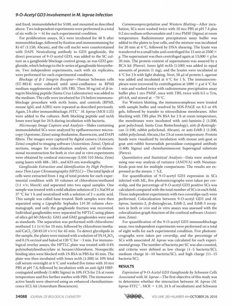
Positively Affected by M. leprae Infection—The second aim ofthis study was to determine whether the SC ganglioside modu-lation observed in vitro is biologically relevant in vivo. Toaddress this point, Balb-C Foxnu/nu (NUDE mice) mice wereinfected with ML in the booth posterior foot pad, as describedby Colston andHilson in 1976 (26). Tenmonths after infection,the animals were sacrificed, and the sciatic nerves were isolatedand processed for histological analysis. Optical longitudinalsections of the sciatic nerves of NUDE mice triple-immunos-tained for 9-O-acetyl GD3, MBP, and HLP (ML antigenmarker) showed increased expression and redistribution of9-O-acetyl GD3 in nerves isolated frommice infected with MLcompared with the control group (Fig. 2, A and E). We did notobserve changes in MBP levels in infected mice, even at 10months after infection (Fig. 2, B and F). On the other hand,9-O-acetyl GD3-MBP colocalization seems to increase in
nerves infected with ML (Fig. 2H,yellow). As seen in vitro, gangliosidedistribution changed from diffuse(noninfected) to clustered patternsalong the infected nerve fiber (Fig. 2,A, E, I, and J). The Ranvier’s nodearea is shown in high magnificationconfocal images (Fig. 2, I and J,arrows). HLP staining in blue can beobserved at the Ranvier’s node, aswell as at some points along thenerve fiber (Fig. 2J). Is importantto notice that the HLP antigenimmunolocalization signal did notnecessarily represent an intact andviable bacillus; it seems that the tis-sue came from an animal after 10months of chronic infection. Fluo-rescence analysis by confocalmicroscopy showed significant up-regulation of 9-O-acetyl GD3 ininfected NUDE mice (Fig. 2K).Taken together, these data suggestthatML infection leads to increasedamounts of 9-O-acetyl GD3 andchanges its cell membrane distribu-tion along the nerve fiber. Further-more, these changes are probablyassociated with the presence of thepathogen or pathogen-derived anti-gens. Immunostaining of anotherML antigen (LAM) in the sameinfected tissue also showed points ofcolocalization between ML and
9-O-acetyl GD3 ganglioside (supplemental Fig. S2D, arrows).9-O-Acetyl GD3 Ganglioside Associated with �-Dystroglycan
and ErbB-2 Receptors in Schwann Cells Exposed to M. leprae—Because we observed a high degree of colocalization betweenML and 9-O-acetyl GD3 during in vitro SC infection (Fig. 1), wenext investigated whether the membrane pool of gangliosidewas associated with molecules known to be related to MLattachment and invasion following ML exposure. SCs wereincubated with ML for 24 h, and coimmunoprecipitation andcolocalization experiments were performed to monitor theassociation between 9-O-acetyl GD3 and laminin-2, �-dystro-glycan, or ErbB-2 receptors.Following ganglioside immunoprecipitation from SCs incu-
bated with or without ML for 24 h, Western blots were per-formed for each ML ligand (Fig. 3A). Levels of the �-chain oflaminin-2 (300 kDa) were not significantly different betweenuntreated and bacteria-exposed SCs. However, quantitativeanalysis of the immunoblots against �-dystroglycan andErbB-2 receptor showed 3- and 5-fold higher levels, respec-tively, in ML-treated SCs compared with the control group.�-Dystroglycan was not coimmunoprecipitated using anti-A2B5 antibody or isotype mouse IgM, but a visible �-dystro-glycan signal was obtained by immunoprecipitating withanti-laminin-2 (Fig. 3B).
FIGURE 2. M. leprae modulates 9-O-acetyl GD3 expression and distribution in the sciatic nerve in vivo.Optical longitudinal sections by confocal microscopy of noninfected (A–D) and infected (E–H) sciatic nerve,triple-immunostained for 9-O-acetyl GD3 (A and E), MBP (B and F), and ML HLP (C and G). Merged images areshown in D and H. I and J, confocal optical slices at high magnification of noninfected (I) or ML-infected (J)isolated sciatic-nerve fibers triple immunostained for 9-O-acetyl GD3, MBP, and HLP. The arrows in J indicateclusters of the ganglioside at the Ranvier’s node area and along the infected nerve fiber. n � 4 mice for eachexperimental condition. K, histogram of quantitative analysis by confocal microscopy of the expression of9-O-acetyl GD3 ganglioside in longitudinal sections of infected (ML�) or noninfected (ML-) sciatic nerve ofNUDE mice. ***, p � 0.0001 (Mann-Whitney). Scale bars, A–H, 50 �m; I and J, 200 �m.
9-O-Acetyl GD3 Involvement in M. leprae Infection
34090 JOURNAL OF BIOLOGICAL CHEMISTRY VOLUME 285 • NUMBER 44 • OCTOBER 29, 2010

We also investigated the colocalization of 9-O-acetyl GD3and ML receptor association by laser confocal microscopy.Semi-confluent cultures were double-labeled for 9-O-acetylGD3 and laminin-2, �-dystroglycan, or ErbB-2 receptor, andcolocalization ratios weremeasured. Quantitative analysis gavecolocalization values 14.2% for laminin-2 (Fig. 3C), 2.3% fordystroglycan (Fig. 3D) and 3.12% for ErbB-2 (Fig. 3E) inuntreated cells. Colocalization ratios increased dramatically inML-exposed cultures to 16.12% for laminin-2 (Fig. 3F), 5.9% fordystroglycan (Fig. 3G), and 11.6% for ErbB-2 (Fig. 3H). Themost significant increases in colocalization with 9-O-acetylGD3 were observed for dystroglycan (Fig. 3B) and ErbB-2 (Fig.3C), whereas colocalization with laminin-2 was not signifi-
cantly affected (Fig. 3A). Yellow points on the SC surface (Fig. 3,F–H, right column) show the colocalization between 9-O-acetylGD3 and the previously described ML receptors. As a colocal-ization control, SC cultures were also double-labeled for lami-nin-2 (ligand) and �1-integrin (receptor subunit) in the pres-ence or absence of ML. �1-Integrin is not involved in ML-SCadhesion, and we observed no differences in the pattern ofexpression of �1-integrin or colocalization with laminin-2 ineither experimental condition (supplemental Fig. 3, A–F).In vivo analyses of 9-O-acetyl GD3 colocalization with the
three ML receptors were performed to confirm the in vitrofindings. Longitudinal frozen sections of infected sciatic nervesof NUDEmice were double-labeled for 9-O-acetyl GD3 gangli-
FIGURE 3. 9-O-Acetyl GD3 ganglioside associates with laminin-2, �-dystroglycan, and ErbB-2 receptor on the Schwann cell surface in vitro and in vivo.A, coimmunoprecipitation (IP) of 9-O-acetyl GD3 with laminin-2, �-dystroglycan, and ErbB-2 receptor in the absence (ML-) or presence (ML�) of M. leprae withhistograms of quantitative analysis by densitometry for each protein analyzed by Western blotting (WB). B, as coimmunoprecipitation controls, anti-A2B5,mouse IgM isotype, and anti-laminin-2 were used, followed by Western blotting for �-dystroglycan. Control groups (ML-, n � 3) were normalized to 1 forcomparison (ML�, n � 3). C–H, confocal sections of semi-confluent Schwann cells, uninfected (C–E) or after 48 h of ML exposure (F–H), double-immunolabeledfor 9-O-acetyl GD3 and laminin-2 (C and F), �-dystroglycan (D and G), and ErbB-2 receptor (E and H). Right-hand columns (C–E and F–H) show merged images,where yellow dots represent colocalization. The percentages in C–E and F–H represent the degree of colocalization of 9-O-acetyl GD3 with laminin-2, �-dys-troglycan, or ErbB-2 receptor in the presence or absence of ML. I–K, confocal longitudinal optical sections of NUDE mice sciatic nerves infected by M. lepraedouble-labeled for 9-O-acetyl GD3 and laminin-2, �-dystroglycan, or ErbB-2 receptor. Points of colocalization are indicated by arrows. The percentages in I–Krepresent the degree of colocalization. *, p � 0.01 (Mann-Whitney). n � 3 experiments for each ML ligand analyzed. Scale bars, C–K, 20 �m.
9-O-Acetyl GD3 Involvement in M. leprae Infection
OCTOBER 29, 2010 • VOLUME 285 • NUMBER 44 JOURNAL OF BIOLOGICAL CHEMISTRY 34091

oside and laminin-2, �-dystroglycan, or ErbB-2. Optical sec-tions obtained by laser scanning microscopy allowed us toobserve areas of colocalization (Fig. 3, I–K). These assays dem-onstrated colocalization between the ganglioside and�-dystro-glycan or ErbB-2 receptor in NUDE infected nerves but notbetween the ganglioside and laminin-2 (Fig. 3, I–K, arrows).Taken together, the colocalization and coimmunoprecipitationsuggest that ML adhesion to the SC surface induces the associ-ation of 9-O-acetyl GD3 ganglioside with �-dystroglycan andErbB-2.Neutralization of 9-O-Acetyl GD3 Reduces Association of M.
leprae to Schwann Cells—We have shown the associationbetween ML and 9-O-acetyl GD3 ganglioside on the mem-branes of SCs prior to invasion (Fig. 1) and demonstrated thatthis association induces 9-O-acetyl GD3 association with mol-ecules already known to participate in ML adhesion and infec-tion in SCs (Fig. 3). We next asked whether we could block MLinvasion using anti-9-O-acetyl GD3 antibody in vitro. Indeed,the neutralization of 9-O-acetyl GD3 by its monoclonal anti-body (Jones clone; Fig. 4B) dramatically reduced ML adhesionto SCs (Fig. 4A), leading to a reduction in the percentage of SCswith high numbers of ML (11–15 bacilli/cell; Fig. 4E) and anincrease in the percentage of SCs with low numbers ofML (1–5bacilli/cell; Fig. 4E). Mouse IgM immunoglobulin, a control forimmunoglobulin isotype, did not induce any significant changein ML association (Fig. 4C). The addition of anti-A2B5 alsofailed to reduceML associationwith SCs (Fig. 4D). The levels of
laminin-2,�-dystroglycan, andErbB-2 on SC surfaces 24 h afterincubation with the neutralizing Jones mAb (supplemental Fig.S4) did not change, which rules out the possibility of antibody-induced down-regulation of the receptors.Previous work described that Jones antibody could also rec-
ognize integrins (25). To ensure that the immunoblockage ofML internalization by Jones is a specific effect exerted by 9-O-acetyl GD3 and not by integrin blockage, we performed a con-trol using �1-integrin blocking peptide. After 2 h of incubationwith 100ng/ml of�1-integrin blocking peptide followed by 24 hof incubation with mAb Jones, we observed significantlyreduced percentage of Schwann cells associated with ML com-pared with the control condition with the blocking peptidealone and RPMI medium (supplemental Fig. S5). The minorreduction of ML-Schwann cell association observed when cellswere preincubated with �1-integrin blocking peptide is proba-bly due to the role of �1-integrin as a laminin-binding protein,because laminin is recognized by ML and exerts a crucial con-tribution to cellular invasion (27).We also performed another approach to neutralize 9-O-
acetyl GD3 ganglioside incubating live cultured Schwann cellswith 10mMNaIO4 for 1 h at 4 °C in the absence of light. It is wellknown that periodate (IO4
�) breaks the bond between carbons 7and 8 of the sialic acid moiety of 9-O-acetyl GD3 (Fig. 5, A andB) (23, 24). We did not observe any signal of 9-O-acetyl GD3 inSchwann cells treated with 10 mM periodate (Fig. 5E), whencomparedwith untreated cells (Fig. 5F). Treatment of Schwann
FIGURE 4. Immunoblockage of 9-O-acetyl GD3 reduces the association of M. leprae with Schwann cells in vitro. A–D, epifluorescence images of SC nuclei(DAPI) incubated for 24 h with FITC-labeled M. leprae or RPMI alone control medium (A), Jones (mAb �-9-O-acetyl GD3) antibody added to the control medium(B), mouse IgM isotype added to the control medium (C) or A2B5 antibody added to the control medium (D). E, percentage of SCs associated with ML withdifferent numbers of bacteria/cell (1–5, 6 –10, and 11–15 bacteria/cell). F, Western blotting and quantitative analysis of the active ERK 1/2 (p-ERK 1/2) from SCscultured for 48 h with ML or incubated with RPMI (standard medium), A2B5 mAb, and Jones mAb. Western blotting for active ERK 1/2 (p-ERK 1/2) and total ERK1/2 following the same experimental conditions. ***, p � 0.0001; **, p � 0.001 (ANOVA). Two independent experiments were performed for each experimentalcondition (n � 8). Scale bars, A–C, 50 �m.
9-O-Acetyl GD3 Involvement in M. leprae Infection
34092 JOURNAL OF BIOLOGICAL CHEMISTRY VOLUME 285 • NUMBER 44 • OCTOBER 29, 2010

cell with 10 mM periodate significantly reduced the number ofcells associated with ML (Fig. 5, C, D, and G).M. leprae-induced ERK 1/2 Activation and Schwann Cell
Proliferation Is Abolished by Immunoblockage of 9-O-AcetylGD3 Ganglioside—ERK 1/2 activation by internalized ML,which leads to demyelination of SCs, is also associated with SCproliferation (6). Because ERK 1/2 activation is a part of theMAPK pathway, and both proliferation and demyelination areactivated by intracellular ML (8), we investigated whether cellproliferation induced by ML through ERK 1/2 could bereversed by 9-O-acetyl GD3 ganglioside immunoblockage. ERK1/2 phosphorylation was measured by Western blotting in SCcultures exposed to inactivatedML (Fig. 4E). Quantitative anal-
ysis showed that activation of ERK1/2was reduced to low levelswhen 9-O-acetyl GD3 was preincubated with blocking anti-body (Fig. 4E). A2B5, mouse IgM, or anti-GD3 treatment didnot reduce phosphorylation of ERK 1/2 (Fig. 4E). IncubationwithML for 48 h increased the number of Ki-67-positive SCs by19% (Fig. 6, A, B, and E, p � 0.01 ANOVA). Conversely, thenumber of Ki-67-positive cells in ML-exposed cultures inwhich 9-O-acetyl GD3 was neutralized was reduced by 21%(Fig. 6,B,C, andE, p� 0.001ANOVA). Jones antibody alone, inthe absence of ML, did not significantly reduce cell prolifera-tion compared with the control group (Fig. 6E). We also testedimmunoblockage on infected cells usingmouse IgM,A2B5, and�-GD3 neutralizing antibodies. The percentages of Ki-67-pos-
FIGURE 5. Deacetylation of 9-O-acetyl GD3 reduces the association of M. leprae with Schwann cells in vitro. A, molecular structure of 9-O-acetyl GD3ganglioside. The second sialic acid is demonstrated by a red circle. B, suggested mechanism for the removal of O-acetyl group by breaking the bondbetween carbons number seven and eight of the second sialic acid. C and D, epifluorescence images of cultured Schwann cells 24 h after exposition toFITC-labeled M. leprae treated (D) or not (C) with sodium periodate (NaIO4) for 1 h at 4 °C in the absence of light. The cell nuclei were counterstained withDAPI. E and F, immunofluorescence for 9-O-acetyl GD3 ganglioside (E) was also performed after deacetylation of the ganglioside by NaIO4 (F). All of theexperimental conditions were performed for 1 h at 4 °C in the absence of light. G, quantitative analysis of the percentage of Schwann cells associatedwith ML (24 h of exposition) after incubation with RPMI medium, NaIO4, or Jones (mAb �-9-O-acetyl GD3) neutralizing antibody. **, p � 0,001 ANOVA.Two independent experiments were performed for each experimental condition (n � 4). Ac, acetyl group; R � radical. Scale bars, C and D, 50 �m; E andF, 20 �m.
9-O-Acetyl GD3 Involvement in M. leprae Infection
OCTOBER 29, 2010 • VOLUME 285 • NUMBER 44 JOURNAL OF BIOLOGICAL CHEMISTRY 34093

itive SCs were similar to controls in each of these antibody-treated groups (Fig. 6E). In conclusion, immunoblockage of9-O-acetyl GD3 was sufficient to inhibit SCs proliferation andERK 1/2 activation induced by ML association.
DISCUSSION
In this study, we showed that ML induced increases in thecell surface expression and changes in the distribution of the9-O-acetyl GD3 ganglioside in SCmembranes in vitro as well asin the infected peripheral nerves of NUDE mice in vivo. Thetotal amount of this ganglioside was increased by 70% (Fig. 1H)in the cultures incubated with ML (Fig. 1C). Changes in theexpression of this ganglioside in Ml-infected nerves, especiallyconcentrated at Ranvier’s node, support the hypothesis that the9-O-acetylGD3pool is somehowmodified duringML infectionin the nerves (Fig. 2J). Because increased ganglioside synthesishas been observed in ML-infected armadillos, this is likely anevolutionarily conserved response to the infection (28). Thus,we propose that ML evolved antigens able to generate thesechanges in ganglioside presentation, which will help and sup-port bacillus infection of human nerves.It is generally accepted that aggregated ML derived from
lymphatic and blood vessels reach and bind to the endoneurialcompartment of nerves through the circulatory system (29–31). Furthermore,ML can infect the dermis and directly bind todermal SCs, where it can migrate along nerve fibers toward thedorsal roots (32). In both strategies of nerve infection, the inter-nodal area seems to be a trophic niche for ML. These experi-ments showed that 9-O-acetyl GD3 is much more abundant ininfected nerves (Fig. 1) and is concentrated on the internodalarea (Fig. 2, I–J), which may facilitate subsequent ML attach-ment and invasion, because 9-O-acetyl GD3 immunoblockagedramatically reducedML attachment (Fig. 4). A high amount of9-O-acetyl GD3 on the SC membrane of infected nerves couldinduce cell proliferation signal transduction through the ERK1/2 pathway (Figs. 4E and 6), leading to an increase in the num-
ber of unmyelinated cells, the pri-mary cell target of the bacillus (8),with a resulting loss of myelination.Other mycobacterium species
with a similar hydrophobic envelop,such as M. smegmatis, Mycobacte-rium tuberculosis, and Mycobacte-rium chelonae, are able to bindlaminin-2 and interact with SCs aswell, suggesting a conserved featurewithin the genus Mycobacterium(33). However, the association of9-O-acetyl GD3 on the SC mem-brane with other Mycobacteriumspecies (M. bovis andM. smegmatis)is significantly reduced comparedwith ML (supplemental Fig. S1H),suggesting a specific adaptation ofML surface antigens to bind to theganglioside, rather than a simplehydrophobic interaction.The neutralization or deacetyla-
tion of 9-O-acetyl GD3 ganglioside with Jones mAb or metasodiumperiodate significantly reduced the association ofML toSCs and effectively reduced levels of infection (bacilli/cell)(Figs. 4 and 5). Only a small fraction of the cells presented highinfection rates (11–15 bacilli/cell) in control groups (Fig. 4) andthat population was reduced by two-thirds with 9-O-acetylGD3 immunoblockage. It is possible that the heterogeneity ofML infection in vitro is due to the fact that only a small fractionof the cells endogenously express the 9-O-acetyl GD3 ganglio-side (Fig. 1).Internalized ML is known to trigger the alternative MAPK
signaling pathway (PKC�-LcK-ERK 1/2) (8); therefore, thereduced activation of ERK 1/2 in the presence of Jones mAbprobably reflects the reduced amount ofML inside the SCs. Theaddition of anti-A2B5 antibody, which neutralizes gangliosidesof the C-path synthesis, did not have the same effect (data notshown). Finally, neutralizing 9-O-acetyl GD3 ganglioside abol-ished the proliferation induced by ML in SCs (Fig. 6). In thisstudy, we examined SC proliferation after immunoblockage of9-O-acetyl GD3 48 h after incubating with ML. Whereas thismay be too short a time to measure proliferation assay in nor-mal SCs, the ST-8814 Schwannoma cell line has an increasedproliferation rate compared with primary SC cultures (19).Thus, this cell type allowed us to analyze SC proliferation in theabsence of ganglioside in a relatively short time period.Gangliosides are smaller in size than most cell surface pro-
teins or glycoproteins. The extracellular tail of 9-O-acetyl GD3comprised of four carbohydrates aligned toward the extracel-lular matrix (9). This reduces the possibility that this moleculeacts as a direct receptor for ML, but it supports the hypothesisthat this ganglioside is an important coreceptor to ML. Addi-tionally, several published reports indicate that deacetylated orneutralized 9-O-acetyl GD3 ganglioside has important roles inneural and immune cell processes, such as cell adhesion,migra-tion, and survival (13, 34). Neutrophil motility is sharplyreduced when GM1 gangliosides are neutralized (35). Migrat-
FIGURE 6. Immunoblockage of 9-O-acetyl GD3 reduces Schwann cell proliferation induced by M. leprae invitro. A–D, epifluorescence images of SCs incubated for 48 h with RPMI alone (A), RPMI plus ML (B), Jones mAbplus ML (C), and mouse IgM plus ML (as isotype control for Jones mAb) (D). Fixed cultures were immunolabeledfor Ki-67 and counterstained with DAPI. E, percentage of Ki-67� cells plus Jones mAb in the presence ofabsence of ML or with anti-A2B5 or anti-�-GD3 antibodies in the presence of ML as controls. *, � 0.01; **, p �0.001 (ANOVA). Two independent experiments were performed for each experimental condition (n � 8). Scalebars, A–D, 50 �m.
9-O-Acetyl GD3 Involvement in M. leprae Infection
34094 JOURNAL OF BIOLOGICAL CHEMISTRY VOLUME 285 • NUMBER 44 • OCTOBER 29, 2010

ing neurons in the developing cerebral cortex, subventricularzone, or cerebellum are also affected after immunoblockage of9-O-acetyl GD3 ganglioside (11, 13, 14). Gangliosides areknown to interact laterally in the membrane, regulating theresponsiveness of receptors for insulin, epidermal growth fac-tor, and vascular endothelial growth factor (36). Gangliosidesalso act as regulatory elements in the immune and nervous sys-tems, inmetabolism, during cancer progression, and in apopto-sis inhibition (10, 36–39). The ganglioside 9-O-acetyl GD3could thus be involved in regulating the extracellular localiza-tion ofmembrane proteins recognized byML such as�-dystro-glycan and ErbB-2. Because 9-O-acetyl GD3 has been reportedto be present in lipid rafts (40), we hypothesize that the punc-tate distribution of this ganglioside induced by the bacilluscould be an effect of its sequestration to lipid rafts (Fig. 2J).Studies by Rambukkana and co-workers (3, 6) demonstrated
the involvement ofmolecules such as dystroglycan, laminin-�2,and ErbB-2 receptor as key players for SC infection by ML. Inthe present work we demonstrated the association of 9-O-acetyl GD3with two of threemolecules described above,�-dys-troglycan and ErbB-2, after ML exposure (Fig. 3). Laminin-2 isnot attached directly to the cell surface of SCs, as opposed to�-dystroglycan or ErbB-2 receptor but can be found in asoluble form in the extracellular matrix or bound to theextracellular matrix or cell membrane (41). We did notobserve points of colocalization of laminin-2 and 9-O-acetylGD3 in the infected nerve tissue, whereas such points wereclearly seen for �-dystroglycan and ErbB-2 at high magnifi-cations with confocal microscopy (Fig. 3, I–K). This suggeststhat, unlike the other ligands for ML, laminin-2 is not incontact with 9-O-acetyl GD3. In addition, increased levels ofErbB-2 were found after immunoprecipitation of 9-O-acetylGD3 in SCs incubated with ML, which suggests a universalinvolvement of this ganglioside during infection of myeli-nated and nonmyelinated SCs. The colocalization of ErbB-2and 9-O-acetyl GD3 in infected nerves supports this hypoth-esis because ErbB-2 is the ML receptor in myelinated SCs(Fig. 3, A, E, and K). Interestingly, dystroglycan, which ishighly expressed at the nodal area in the nodes of Ranvier, isessential for regular myelination and establishment of nodalarchitecture (42). Among its biological roles is clustering ofvoltage-gated sodium channels at nodes of Ranvier in SCs(43). We observed ML antigen as well as colocalization ofdystroglycan and 9-O-acetyl GD3 in the same area at thesenodes (Fig. 3J). Therefore, it is possible that ML disruptsdystroglycan function at nodal areas, causing a loss of func-tion of myelinating fibers through an unknown molecularprocess.In conclusion, the data presented here suggest a biological
role for 9-O-acetyl GD3 in the infection of Schwann cells withM. leprae. This is the first report on the important role of a hostcell ganglioside in mycobacterium infection. Our study shedslight on the adhesion of ML to SCs, an event that depends on acombination of several elements on the SCmembrane. Furtherstudies are necessary to clarify whether other SC membraneelements can interact with 9-O-acetyl GD3 and to determinethe impact of these associations on nerve demyelination duringleprosy.
Acknowledgments—We thank Dr. Janet W. Reid for the English revi-sion, Dr. Jose Oswaldo Previato and Dr. Adriane Todeschini fromUniversidade Federal do Rio de Janeiro for the excellent scientificsupport, the American LeprosyMissions and the Order of St. Lazarus,which support Dr. James Krahenbuhl and Dr. Ramanuj Lahiri fromNational Hansen’s Disease Program, who kindly donated the Myco-bacterium leprae used in this work and trained us to produce ML inthe nude mice model, and Dr. Patrick Brennan from Colorado StateUniversity for kindly providing the IgG anti-�-HLP and IgGanti-�-Lam.
REFERENCES1. Scollard, D. M., Adams, L. B., Gillis, T. P., Krahenbuhl, J. L., Truman,
R. W., and Williams, D. L. (2006) Clin. Microbiol. Rev. 19, 338–3812. Rambukkana, A. (2000) Lepr. Rev. 71, (suppl.) S168–S1693. Rambukkana, A., Yamada, H., Zanazzi, G., Mathus, T., Salzer, J. L., Yurch-
enco, P. D., Campbell, K. P., and Fischetti, V. A. (1998) Science 282,2076–2079
4. Ng, V., Zanazzi, G., Timpl, R., Talts, J. F., Salzer, J. L., Brennan, P. J., andRambukkana, A. (2000) Cell 103, 511–524
5. Rambukkana, A., Zanazzi, G., Tapinos, N., and Salzer, J. L. (2002) Science296, 927–931
6. Tapinos, N., Ohnishi, M., and Rambukkana, A. (2006) Nat. Med. 12,961–966
7. Jessen, K. R., and Mirsky, R. (2008) Glia 14, 1552–15658. Tapinos, N., and Rambukkana, A. (2005) Proc. Natl. Acad. Sci. U.S.A. 102,
9188–91939. Thompson, T. E., and Tillack, T. W. (1985) Annu. Rev. Biophys. Biophys.
Chem. 14, 361–38610. Kniep, B., Kniep, E., Ozkucur, N., Barz, S., Bachmann, M., Malisan, F.,
Testi, R., and Rieber, E. P. (2006) Int. J. Cancer 119, 67–7311. Mendez-Otero, R., andCavalcante, L. A. (2003)Prog.Mol. Subcell Biol. 32,
97–12412. Mendez-Otero, R., and Santiago, M. F. (2003) Braz. J. Med. Biol. Res. 36,
1003–101313. Santiago,M. F., Costa,M. R., andMendez-Otero, R. (2004) J. Neurosci. 24,
474–47814. Miyakoshi, L. M., Mendez-Otero, R., and Hedin-Pereira, C. (2001) Braz. J.
Med. Biol. Res. 34, 669–67315. Mendez-Otero, R., and Friedman, J. E. (1996) Eur. J. Cell Biol. 71,
192–19816. Malisan, F., and Testi, R. (2002) Biochim. Biophys. Acta 1585, 179–18717. Schlosshauer, B., Blum, A. S., Mendez-Otero, R., Barnstable, C. J., and
Constantine-Paton, M. (1988) J. Neurosci. 8, 580–59218. Ribeiro-Resende, V. T., Oliveira-Silva, A., Ouverney-Brandao, S., San-
tiago,M. F., Hedin-Pereira, C., andMendez-Otero, R. (2007)Neuroscience147, 97–105
19. Svaren, J., and Meijer, D. (2008) Glia 56, 1541–155120. Rodrigues, L. S., da SilvaMaeda, E.,Moreira,M. E., Tempone,A. J., Lobato,
L. S., Ribeiro-Resende, V. T., Alves, L., Rossle, S., Lopes, U. G., and Pesso-lani, M. C. (2010) Cell Microbiol. 12, 42–54
21. Shepard, C. C., and McRae, D. H. (1968) Int. J. Lepr. Other Mycobact. Dis.36, 78–82
22. Santiago, M. F., Berredo-Pinho, M., Costa, M. R., Gandra, M., Cavalcante,L. A., and Mendez-Otero, R. (2001)Mol. Cell Neurosci. 17, 488–499
23. Van Lenten, L., and Ashwell, G. (1971) J. Biol. Chem. 246, 1889–189424. Manzi, A. E., Dell, A., Azadi, P., and Varki, A. (1990) J. Biol. Chem. 265,
8094–810725. Yang, C. R., Liour, S. S., Dasgupta, S., and Yu, R. K. (2007) J. Neurosci. Res.
85, 1381–139026. Colston, M. J., and Hilson, G. R. (1976) Nature 262, 399–40127. Rambukkana, A., Salzer, J. L., Yurchenco, P.D., andTuomanen, E. I. (1997)
Cell 88, 811–82128. Harris, E. B., Li, Y. T., and Li, S. C. (1986) Int. J. Lepr. Other Mycobact. Dis.
54, 289–293
9-O-Acetyl GD3 Involvement in M. leprae Infection
OCTOBER 29, 2010 • VOLUME 285 • NUMBER 44 JOURNAL OF BIOLOGICAL CHEMISTRY 34095

29. Khanolkar, V. R. (1964) Indian J. Med. Res. 52, 139–15030. Katoch, V. M. (1999) Indian J. Lepr. 71, 45–5931. Scollard, D. M., McCormick, G., and Allen, J. L. (1999) Am. J. Pathol. 154,
1611–162032. Scollard, D. M. (2000)Microbes Infect. 2, 1835–184333. Marques, M. A., Antonio, V. L., Sarno, E. N., Brennan, P. J., and Pessolani,
M. C. (2001) J. Med. Microbiol. 50, 23–2834. Mukherjee, P., Faber, A. C., Shelton, L. M., Baek, R. C., Chiles, T. C., and
Seyfried, T. N. (2008) J. Lipid Res. 49, 929–93835. Gong, Y., Tagawa, Y., Lunn, M. P., Laroy, W., Heffer-Lauc, M., Li, C. Y.,
Griffin, J. W., Schnaar, R. L., and Sheikh, K. A. (2002) Brain 125,2491–2506
36. Regina Todeschini, A., and Hakomori, S. I. (2008) Biochim. Biophys. Acta1780, 421–433
37. Vyas, A. A., Patel, H. V., Fromholt, S. E., Heffer-Lauc, M., Vyas, K. A.,Dang, J., Schachner, M., and Schnaar, R. L. (2002) Proc. Natl. Acad. Sci.
U.S.A. 99, 8412–841738. Vyas, K. A., Patel, H. V., Vyas, A. A., and Schnaar, R. L. (2001) Biol. Chem.
382, 241–25039. Vyas, A. A., and Schnaar, R. L. (2001) Biochimie 83, 677–68240. Simons,M., Schwarz, K., Kriz,W.,Miettinen, A., Reiser, J.,Mundel, P., and
Holthofer, H. (2001) Am. J. Pathol. 159, 1069–107741. Hall, H., Bozic, D.,Michel, K., andHubbell, J. A. (2003)Mol. Cell. Neurosci.
24, 1062–107342. Saito, F., Moore, S. A., Barresi, R., Henry, M. D., Messing, A., Ross-Barta,
S. E., Cohn, R. D., Williamson, R. A., Sluka, K. A., Sherman, D. L., Brophy,P. J., Schmelzer, J. D., Low, P. A., Wrabetz, L., Feltri, M. L., and Campbell,K. P. (2003) Neuron 38, 747–758
43. Occhi, S., Zambroni, D., Del Carro, U., Amadio, S., Sirkowski, E. E.,Scherer, S. S., Campbell, K. P., Moore, S. A., Chen, Z. L., Strickland, S., DiMuzio, A., Uncini, A., Wrabetz, L., and Feltri, M. L. (2005) J. Neurosci. 25,9418–9427
9-O-Acetyl GD3 Involvement in M. leprae Infection
34096 JOURNAL OF BIOLOGICAL CHEMISTRY VOLUME 285 • NUMBER 44 • OCTOBER 29, 2010


![[Leprosy: Mycobacterium leprae and HIV coinfection]](https://img.pdfslide.net/doc/110x75/6353ad9baf90c188350a1b6e/leprosy-mycobacterium-leprae-and-hiv-coinfection.jpg)


