Embed Size (px)
Citation preview

Involvement of death receptor Fas in germ celldegeneration in gonads of Kit-deficient Wv/Wv mutantmice
S Sakata1, K Sakamaki1, K Watanabe1, N Nakamura2,S Toyokuni3, Y Nishimune4, C Mori5 and S Yonehara*,1
1 Graduate School of Biostudies and Institute for Virus Research, KyotoUniversity, Shogoin-Kawahara-cho, Sakyo-ku, Kyoto 606-8507, Japan
2 Graduate School of Medicine, Nagoya University, Showa-ku, Nagoya 466-8550, Japan
3 Graduate School of Medicine, Kyoto University, Sakyo-ku, Kyoto 606-8501,Japan
4 Institute for Microbial Diseases, Osaka University, Suita 536-0871, Japan5 Graduate School of Medicine, Chiba University, Chuoh-ku, Chiba 260-8670,
Japan* Corresponding author: S Yonehara, Graduate School of Biostudies and
Institute for Virus Research, Kyoto University, Shogoin-Kawahara-cho, Sakyo-ku, Kyoto 606-8507, Japan. Tel: 81 75 751 4783; Fax: 81 75 751 4784; E-mail:[email protected]
Received 8.11.02; accepted 30.12.02Edited by H Ichijo
AbstractKit and its ligand stem cell factor (SCF) play a fundamentalrole in hematopoiesis, melanogenesis and gametogenesis.Homozygous Wv mutant mice with a mutation in kit showabnormalities in these cell lineages. Fas is a member of thedeath receptor family inducing apoptosis. In this study, wegenerated double-mutant mice (Wv/Wv:Fas�/�) and analyzedhistologically their reproductive organs. In testes and ovariesof the double-mutant mice, testicular germ cells and oocyteswere detected, respectively, whereas the same-aged Wv/Wv
mice contained neither cells. In addition, inhibition of Kitsignals by administration of anti-Kit mAb, which inducesdegeneration of testicular germ cells in vivo in wild-type mice,did not cause degeneration in Fas-deficient mice. In testiculargerm cells of Wv/Wv mutant mice, an increase of Fasexpression was observed in spermatogonia. Further, in vitrotreatment with SCF was shown to downregulate Fas onfibroblasts expressing exogenous Kit through activation ofPI3-kinase/Akt. All the results clearly indicate that Fas-mediated apoptosis is involved in germ cell degenerationaccompanied by defects in Kit-mediated signals, and Kitsignaling negatively regulates Fas-mediated apoptosis invivo.Cell Death and Differentiation (2003) 10, 676–686. doi:10.1038/sj.cdd.4401215
Keywords: Fas; germ cell; kit receptor; stem cell factor; Wv
mutant mouse
Abbreviations: ERK, extracellular signal-regulated kinase;
MEK, MAP kinase-ERK kinase; PI3K, phosphatidylinositol 3-
phosphate kinase; RTK, receptor tyrosine kinase; SCF, stem cell
factor.
Introduction
Kit is a receptor tyrosine kinase (RTK) belonging to the PDGFreceptor/CSF-1 receptor family.1–3 Kit and its ligand, stem cellfactor (SCF), play a fundamental role during the establish-ment and maintenance of the three stem cell populations:melanoblasts, hematopoietic stem cells and germ cells.Genes encoding Kit and SCF are mapped to the whitespotting (W) and steel (Sl) loci in mice, respectively.4–8 Bothmutant mice of W and Sl loci show similar phenotypes such assevere macrocytic anemia, a decreased number of mast cells,a complete lack of coat pigmentation and infertility.9 Further,many alleles of variable severity at both W and Sl loci havebeen identified.10 For example, Wv mutant mice with theC57BL/6 background have a point mutation in the cytoplasmicregion of Kit, resulting in no coat pigmentation, severeanemia, a deficiency of mast cells and infertility in homo-zygotes (Wv/Wv).11,12
In reproductive organs, Kit plays essential roles inspermatogenesis and oogenesis.10 In the testes, Kit receptorexpression is restricted to differentiating type A to type Bspermatogonia, primary spermatocytes and Leydig cells.13–16
In particular, type A spermatogonia cells express a maximalamount of Kit at both the transcript and protein level, followedby type B spermatogonia.13,17,18 Therefore, the germ cells ofthe homozygous W mutant mice are impossible to developand degenerate accompanied by apoptosis leading toinfertility owing to a lack of Kit signals.18 In the ovaries, Kit isexpressed on not only primordial and growing oocytesthroughout the development of follicles but also on interstitialtheca cells.13,18 In the ovaries of W mice, oogenesiscompletely fails because of defects of Kit signals resulting inno follicles and oocytes.10
Members of the RTK family play essential roles in theproliferation, differentiation, migration and survival of variouscells.19 Kit as well as other RTKs interacts with severalintracellular signal transducers including PI3-kinase, JAK2,STAT1, src-related kinases, SH2-containing tyrosine phos-phatases and adaptor proteins such as SOCS1, Shc, GRB2and GRB7. These signal transducers function in variouscellular processes by activating target molecules includingPKC, Akt and MAP kinases such as ERK and JNK.20 Kit isstrongly suggested to be indispensable for not only amitogenic response but also an antiapoptotic response.
Fas is a cell surface receptor belonging to the deathreceptor family, along with tumor necrosis factor receptor
Cell Death and Differentiation (2003) 10, 676–686& 2003 Nature Publishing Group All rights reserved 1350-9047/03 $25.00
www.nature.com/cdd

type1 (TNF-R1) and TRAIL receptors.21 Fas can induce celldeath in a variety of tumors and lymphocytes by ligation withFas ligand or an agonistic anti-Fas monoclonal antibody(mAb).22–25 While the physiological role of Fas has been wellstudied in the immune system,26–29 Fas mRNA is expressedat high levels in not only the thymus and spleen, but also theliver, heart, lungs and ovaries of mice.26,30 Therefore, it couldbe that Fas acts in the physiological deletion of potentiallyharmful or unnecessary cells in these tissues.28
In female gonads, we and others demonstrated that Fas isexpressed on granulosa cells of various follicles and lutealcells.31–38 A few reports showed Fas expression on oocytes,but we have not been able to confirm this.32,38,39 Theexpression on granulosa and luteal cells suggests that Fasis involved in follicular atresia and luteolysis for the gonadalhomeostasis of adult mice. In male gonads, Fas and Fasligand are detected in the testes, but their function is stillunclear. As Fas-deficient mice showed no abnormality in thetestes,40 it was thought that Fas is not essential in germ cellhomeostasis. However, recent reports insisted on theinvolvement of Fas in the physiological death of germcells.41–43 In addition, Fas expression was shown to increasein the testes after drug treatment, ischemia–reperfusion orheat shock.44–47 In these damaged testes, Fas may play a roleas a regulator of pathological germ cell death, although thishas not been clearly determined.
To investigate the relation between Kit-mediated survivalsignals and Fas-mediated apoptotic signals in gonads, wegenerated double-mutant mice by intercrossing Kit-deficientWv mutant mice with Fas knockout mice. Our study clearlydemonstrated that a defect of Kit signaling leads to activationof a Fas-mediated apoptotic signal in both spermatogenesisand oogenesis, and Fas is involved in germ cell loss in Kitsignaling-deficient mice. Moreover, our findings provide newevidence of crosstalk of signals via Kit and Fas, indicating thatKit-induced signals negatively regulate Fas expression in thegonads.
Results
Analysis of double-mutant mice
In the gonads of Kit-deficient W mutant mice, the degenera-tion of testicular germ cells and oocytes was reported to beaccompanied by cell death.10 We investigated whether Fas isinvolved in these events because we and other groupspreviously showed the expression of Fas in both male andfemale gonads.31–38,41,43–46 To clarify whether Fas is involvedin germ cell degeneration in the gonads of W mutant mice, wegenerated double-mutant mice (Wv/Wv:Fas�/�) by intercross-ing the spontaneous Kit mutant Wv mice with Fas-deficientmice against the same C57BL/6 background.
We first examined whether the number of germ cells in thegonads of the mutant mice changes with the Fas gene status.The number of Wv/Wv homozygous newborns was less thanthat expected as a result of Mendel’s laws, while a significanteffect of Fas gene status was not observed. In addition, Wv/Wv homozygous mice often died within 2 weeks because ofanemia (data not shown), while some mutant mice survivedfor more than 8 weeks. Their testes and ovaries could then be
examined. We measured the size of the testes of the survivingWv mutant mice, and found the testes of Wv/Wv homozygousmice to be smaller (approximately 10–20% in size) than thoseof Wv/+ or wild-type mice regardless of Fas status (data notshown).
Histological analysis of testes in the double-mutant mice
Next, we histologically examined the testes of the 8-week-oldmutant mice by staining with hematoxylin and eosin (Figure 1).Testes of Wv/+:Fas�/� mice were histologically normal. Germcells at all developmental stages including spermatogonia,primary spermatocytes, secondary spermatocytes, haploidspermatids and sperm were observed (Figure 1a,d). Fas-deficiency did not induce hyperplasia of germ cells in thesemutant male mice. In the testes of Wv/Wv:Fas+/+ mice,seminiferous tubules were abnormal, resulting in no differ-entiated germ cells (Figure 1b,e), as described previously inWv mutant mice.10 Interestingly, in the testes of Wv/Wv:Fas�/�
mice, spermatocytes, spermatids and sperm were detected inseveral seminiferous tubules (Figure 1c,f), indicating that Fasdeficiency can rescue testicular germ cells in Wv/Wv mice. Assummarized in Table 1, about 90% of seminiferous tubules inWv/Wv:Fas�/� mice had only Sertoli cells with or without avery few spermatogonia, while we observed a significant
Figure 1 Histological analyses of testes from 8-week-old mutant mice.Sections prepared from testes of Wv/+:Fas�/� (a,d), Wv/Wv:Fas+/+ (b,e) and Wv/Wv:Fas�/� (c,f) mice were stained with hematoxylin and eosin. Photographswere taken under a phase-contrast microscope at � 32 (a–c) and � 160 (d–f).Arrows in (c) show the tubules containing differentiated germ cells indicated byarrowheads in (f). Scale bars correspond to 125mm
Signaling crosstalk between Fas and KitS Sakata et al
677
Cell Death and Differentiation

number of spermatocytes that differentiated into the pachy-tene stage in the other 10% of tubules. Further, differentiatedspermatids and sperm could be also observed in about 5% ofseminiferous tubules. The seminiferous tubules containingdifferentiated germ cells at the meiotic or a more advancedstage of spermatogenesis seemed to be located aroundrather than in the center of the testes of Wv/Wv:Fas�/� mice(Figure 1c). However, we could not detect sperm in theepididymis of Wv/Wv:Fas�/� mice by histological analysis(data not shown). Taking all the results into consideration, Faswas shown to be involved in the degeneration of testiculargerm cells in Wv mutant mice lacking signals from Kit, whileother signals might also play a role in the degeneration.
We also examined the propagative activity of Wv/Wv:Fas�/�
male mice. However, we observed no fertility in Wv/Wv:Fas�/�
male mice mating with wild-type females. The number ofundegenerated germ cells and/or differentiated sperm in thedouble-mutant mice might be too small for fertilization.
Histological analysis of ovaries in the double-mutant mice
Female gonads were also analyzed in Wv/+:Fas�/�, Wv/Wv:Fas+/+ and Wv/Wv:Fas�/� mice. Figure 2 shows the resultsof a histological analysis of ovaries in 4-week-old animals.Ovaries of Wv/+:Fas�/� mice displayed a normal histology,where oocytes and follicles at various stages of developmentwere detected (Figure 2a,d). This phenotype of Wv/+:Fas�/�
mice is similar to that of lpr mice, which exhibit a spontaneousloss of function in the Fas gene. On the other hand, ovaries ofWv/Wv:Fas+/+ mice were shown to be only very small (Figure2b,e), with no oocytes or follicles. Importantly, in ovaries ofWv/Wv:Fas�/� mice, we detected primary follicles, growingfollicles and Graffian follicles with oocytes (Figure 2c,f). Thus,Kit deficiency affected the gametogenesis of female gonadsmore than male gonads in younger mice, and inactivation ofFas resulted in the diminution of the degeneration of oocytesand follicles in the Kit-deficient ovaries. Results for ovaries aresummarized in Table 2. They clearly indicate that loss offunction of Fas prevents the degeneration of germ cells in bothmale and female gonads of Wv/Wv mice.
Although Wv/Wv:Fas�/� female mice were examined interms of their fertility, they did not become pregnant (data notshown). In addition, oocytes of Wv/Wv:Fas�/� mice wereshown to be impaired with aging, and most of the oocytesdegenerated in the ovary of 8-week-old Wv/Wv:Fas�/� femalemice (data not shown). All the results indicate that Fas isinvolved in the degeneration of oocytes in 4-week-old Wv/
Wv:Fas�/� female mice, while mechanisms other than Fas-mediated apoptosis might play a role in the infertility and thedegeneration of oocytes of 8-week-old Wv/Wv:Fas�/� mice.
Figure 2 Histological analyses of ovaries isolated from 4-week-old mutantmice. Sections of ovaries of Wv/+:Fas�/� (a,d), Wv/Wv:Fas+/+ (b,e) and Wv/Wv:Fas�/� (c and f) mice were stained with hematoxylin and eosin. Photographswere taken under a phase-contrast microscope at � 32 (a–c) and � 80 (d–f).Scale bars correspond to 125mm
Table 1 Histological data on testes from 8-week-old mice
Percentage of tubules with
Genotype Number of mice examined spermatocyte spermatid sperm
Wv/Wv:Fas+/+ 10 0 0 0Wv/Wv:Fas�/� 12 9.673.3 5.172.0 4.371.8
Numerical values show the percentage (mean7S.D.) of tubules containing spermatocytes, spermatids or sperm. Themean7S.D. was counted under the microscope from histological images depicted in Figure 1. Almost all tubules of atestis were examined for counting by analyzing five cross-sections/testis
Table 2 Histological data of ovaries from 4-week-old mice
GenotypeNumber of mice
examinedNumber of oocytes
in an ovary
Wv/Wv:Fas+/+ 8 0Wv/Wv:Fas�/� 12 6.675.1
Numerical values show the number (mean7S.D.) of differentiated oocytes(oocytes in secondary follicles and Graffian follicles) in an ovary. Themean7S.D. of oocytes/ovary was counted under the microscope from serialcross sections of the entire ovary. The numbers of differentiated oocytes in wild-type and Fas�/� mice were essentially the same (about 50/ovary) as describedpreviously.35
Signaling crosstalk between Fas and KitS Sakata et al
678
Cell Death and Differentiation

In vivo blocking of Kit signals by anti-Kit mAbcauses germ cell degeneration in wild-type but notFas-deficient mice
We next examined the effects of Fas deficiency on thedegeneration of testicular germ cells in another mouse systemwith suppressed Kit signals. We artificially blocked the Kitsignaling pathway through injection of an antagonistic anti-c-kit mAb ACK2 into wild-type C57BL/6 and Fas-deficient mice.We analyzed the population of testicular germ cells from bothmice after injection of ACK2. It has been demonstrated thatdifferentiating spermatogonia and early preleptotene sperma-tocytes expressing Kit degenerated after injection of ACK2which can block the function of Kit.18 We examined the testesof the antibody-treated animals by an immunohistochemicalmethod with an anti-germ cell-specific mAb, TRA98, andstained another serial section with Hoechst (Figure 3). At 48 hafter injection of ACK2, most differentiating type A andintermediate spermatogonia had disappeared in the wild-typetestes (Figure 3a), although differentiated germ cells andinterstitial cells were observed in Hoechst-stained sections(Figure 3c). In contrast, many spermatogonia still existedeven after 48 h in Fas-deficient mice (Figure 3b). Theseresults supported our results for Wv/Wv:Fas�/� double-mutantmale mice (Figure 1 and Table 1) and strongly indicate thatFas is involved in testicular germ cell degeneration induced bydefects of Kit signaling.
Expression of Fas in testes from wild-type andhomozygous Wv/Wv mutant mice
Our data thus suggest that Kit-mediated signals inhibit theFas-mediated apoptosis-inducing signal. To clarify the mole-cular mechanism of the Kit-induced inhibition, we analyzedthe expression level of Fas because activation of Ras, whichmust occur downstream of Kit, was previously reported todecrease the expression.48 In our case, we speculated thatthe deficiency of Kit signaling might cause the induction of Fasexpression on the germ cells. At first, we analyzed theexpression at the RNA level by Northern blot hybridization.We examined total RNA isolated from the testes of 6-week-oldwild-type C57BL/6 and homozygous Wv/Wv mutant mice(Figure 4). Fas transcripts were significantly detected in thetestes from Wv/Wv mutant mice, but we could not detect FasmRNA in the testes of wild-type mice. This result suggestedthat Fas is expressed on spermatogonia and/or interstitialcells. Alternatively, the difference in Fas expression mightreflect the difference of the cell population in the testes of wild-type and Wv/Wv mutant mice.
To identify the cell surface expression of Fas, we performedflow cytometric analysis with the anti-Fas mAb Jo-2. Fasexpression was examined on the surface of testicular cellsprepared from 3- to 4-week-old wild-type C57BL/6 andhomozygous Wv/Wv mutant mice. In wild-type testes, fewcells expressing Fas were observed, while such cells weredetected in Wv/Wv mutant mice (Figure 5a). Then, we carriedout a two-dimensional flow cytometric analysis with the Jo-2and EE2 mAbs in order to clarify whether Fas is expressed onspermatogonia or not. Previous study demonstrated theexpression of EE2 antigen on the germ cell surface; the
expression was predominant prior to meiosis, and graduallydecreased as the differentiation proceeded.49 EE2-bright cellscorrespond to type A and primitive spermatogonia, while EE2-intermediate cells correspond to intermediate and type B
Figure 3 Immunohistochemistry of testes from ACK2-injected adult mice.Frozen sections were prepared from testes of wild-type C57BL/6 mice (a,c) andFas�/� mice (b,d), to which anti-c-kit mAb ACK2 was administered 48 h earlier,as described in Materials and Methods. The section was then stained with thegerm cell-specific mAb TRA98 (a,b), and another serial section was stained withHoechst (c,d). Experiments were repeated five times using different mice and thedata presented show representative results. Photographs were taken under aphase-contrast microscope at � 160. Arrows and arrowheads show sperma-togonia and differentiated haploid spermatids, respectively. Scale barscorrespond to 25 mm
Figure 4 Northern blot analysis of Fas transcripts in testes. Northern blottingwas carried out with total RNA isolated from testes of 5- to 6-week-old wild-typeC57BL/6 and Wv/Wv mutant mice as described in Materials and Methods using32P-labeled mouse Fas (upper panel) and human EF1-a (lower panel) cDNAs asDNA probes.
Signaling crosstalk between Fas and KitS Sakata et al
679
Cell Death and Differentiation

spermatogonia, preleptotene, leptotene and zygotene sper-matocytes. EE2-negative cells correspond to pachytenespermatocytes or cells in the more advanced stages ofspermatogenesis and interstitial cells. Flow cytometric analy-sis clearly indicated that Fas was significantly expressed onEE2-positive cells in Wv/Wv mutant mice although littleexpression of Fas was detected on EE2-positive cells in
wild-type mice (Figure 5b). We then examined germ cells of 2-week-old mice, and found the induction level of Fas expres-sion in 2-week-old than 3- to 4-week-old Wv/Wv mutant mice(Figure 5b). Fas expression was indicated to be upregulatedin testicular germ cells of 3- to 4-week-old mice mainlybecause of a deficiency of Kit.
To clearly distinguish expression levels of Fas betweenSCF-dependent and -independent spermatogonia, we carriedout flow cytometric analyses by triple staining with EE2, ACK2and Jo-2. Figure 5c shows the results of two-dimensional flowcytometric analyses with EE2 and ACK2 in wild-type (leftpanel), Wv/Wv mutant (center panel) and Wv/Wv:Fas�/�
double-mutant (right panel) mice. Kit-positive and EE2-positive cells (squares a, d and g) correspond to spermato-gonia that require SCF-induced Kit signals for differentiation.Kit-negative and EE2-bright cells (squares b, e and h)correspond to primitive spermatogonia such as testicularstem cells which have the ability to self-proliferate. Kit-negative and EE2-negative cells (squares c, f and i)correspond to Sertoli cells and pachytene spermatocytes ormore advanced stages of spermatogenesis. In Wv/Wv mutantmice, primitive spermatogonia, encircled with the square e,were decreased in number when compared with those of wild-type mice. In Wv/Wv:Fas�/� double-mutant mice, however, a
Figure 5 Flow cytometric analyses of Fas expression on mouse testicular germcells. (A) Single-cell suspensions isolated from testes of 3- to 4-week-old wild-type C57BL/6 mice (left panel) and Wv/Wv mutant mice (right panel) were stainedwith (solid lines) or without (dotted lines) FITC anti-Fas mAb Jo-2. (B) Testicularcells of 3- to 4-week-old or 2-week-old wild-type C57BL/6 mice and Wv/Wv mutantmice were stained with PE-anti-testicular germ cell mAb EE2, together with (rightpanels) or without (left panels) FITC-Jo-2. (C) Single-cell suspensions isolatedfrom testes of 3- to 4-week-old wild-type C57BL/6 mice (left panel), Wv/Wv mutantmice (center panel) and Wv/Wv:Fas�/� mice (right panel) were stained with EE2and ACK2. The squares, indicated as a, d and g, correspond to spermatogonia;b, e and h to primitive spermatogonia; and c, f and i to interstitial cells and well-differentiated germ cells. (D) Fas expression on testicular cells in the squares in(C). Cells were stained with (solid lines) or without (dotted lines) FITC Jo-2. Allexperiments were repeated five times, and the data presented showrepresentative results
Signaling crosstalk between Fas and KitS Sakata et al
680
Cell Death and Differentiation

drastic recovery of primitive spermatogonia was not ob-served. Although the percentage of wild-type spermatogoniarequiring Kit signals for differentiation, encircled with thesquare a (containing 10% of total cells), was about one-third ofthat of Wv/Wv spermatogonia (square d, 24%), the total cellnumber of spermatogonia/testis in wild-type mice was morethan six times that in Wv/Wv mutant mice, because total cellnumber/testis for cytofluorometric analysis in wild-type micewas more than 20 times that in Wv/Wv mutant mice.Interestingly, Fas deficiency increased the number of sper-matogonia requiring Kit signals for differentiation more thantwo-fold when compared with that of Wv/Wv mutant mice,because (1) the percentage of spermatogonia requiring Kitsignals for differentiation in Wv/Wv and Wv/Wv:Fas�/� mutantmice was comparable (square d, 24%, and square g, 29%,respectively) and (2) total cell number/testis for cytofluoro-metric analysis in Wv/Wv:Fas�/� mice was more than twotimes that in Wv/Wv mutant mice.
Next, we examined Fas expression on Kit-positive andEE2-positive cells, Kit-negative and EE2-bright cells and Kit-negative and EE2-negative cells encircled with the squares a–f in Figure 5c (Figure 5d). Wv/Wv mutant mice were shown tosignificantly express Fas in all the testicular cells, while wild-type mice expressed little Fas in any testicular cells.
All the results indicated that the expression of Fas on germcells was upregulated in the testes of Wv/Wv mutant mice, andgerm cell degeneration induced by defects in the Kit signalingsystem was suggested to be caused by activation of the Fas-mediated signaling pathway.
SCF downregulates Fas expression on Kit-expressing NIH3T3 cells
To confirm the downregulation of Fas by Kit-induced signals,we examined the effects of SCF on Kit-expressing cells invitro. We transfected c-kit cDNA into NIH3T3 cells andestablished several Kit-positive stable clones. Then, weanalyzed the expression of Fas on these clones by flowcytometry after treatment with 10 mg/ml of mouse recombinantSCF for 48 h. A significant decrease of Fas was detected onthe Kit-expressing stable clone NIH-K11 but not parentalNIH3T3 cells after treatment with SCF (Figure 6a,b). Theother Kit-positive stable clones showed essentially similarcharacteristics to those of NIH-K11 cells (data not shown).These results directly show that the Kit signal induces thedownregulation of Fas.
We then examined which downstream signal of Kit plays arole in the downregulation of Fas expression. Since thedownregulation was speculated to be induced by survivalsignals mediated through Kit, we focused on two signalingpathways downstream of Kit, PI3K/Akt and Ras/MEK/ERK.Activation of the Ras/MEK/ERK pathway was reported toinhibit Fas-mediated apoptosis,50 while activation of PI3K/Aktpathway was suggested to suppress the expression level ofcell surface Fas.48 In addition, Kit-mediated activation of thePI3K/Akt pathway was demonstrated to be significant ingametogenesis.51,52 We analyzed the effects of two kinds ofinhibitors that specifically prevent Kit-mediated signaling: thePI3K-specific inhibitor wortmannin and the MEK1/2-specific
inhibitor U0126. These inhibitors were added to the culturemedium of NIH-K11 cells together with SCF and Fasexpression was examined 48 h later with a flow cytometer(Figure 6c and d). Downregulation of Fas was suppressed bythe treatment with wortmannin, while U0126 showed nosignificant effect. Similar results were obtained with the PI3Kinhibitor LY294002 and MEK1 inhibitor PD98059 (data notshown). Thus, PI3K inhibitors but not MEK inhibitors blockedthe downregulation of Fas by SCF.
To biochemically detect the activation of downstreamsignaling molecules of Kit, which was inhibited by wortmannin
Figure 6 Flow cytometric analyses of Fas expression on SCF-treatedfibroblasts expressing exogenous Kit. Parental NIH3T3 (a) and Kit-expressingstable clone NIH-K11 (b) cells were stimulated with (bold lines) or without (finelines) 10 ng/ml of SCF for 48 h and then stained with (dotted lines) or without(solid lines) FITC-Jo-2. NIH-K11 cells were also stimulated with SCF for 48 h inthe presence of 100 nM wortmannin (c) or 10 mM U0126 (d). Cells werepretreated with each inhibitor for 30 min prior to stimulation with SCF.Experiments were repeated five times, and the data presented showrepresentative results
Signaling crosstalk between Fas and KitS Sakata et al
681
Cell Death and Differentiation
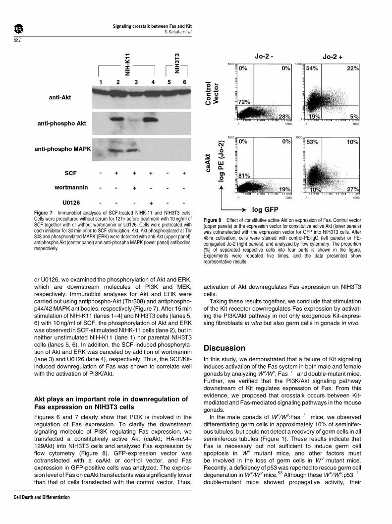
or U0126, we examined the phosphorylation of Akt and ERK,which are downstream molecules of PI3K and MEK,respectively. Immunoblot analyses for Akt and ERK werecarried out using antiphospho-Akt (Thr308) and antiphospho-p44/42 MAPK antibodies, respectively (Figure 7). After 15 minstimulation of NIH-K11 (lanes 1–4) and NIH3T3 cells (lanes 5,6) with 10 ng/ml of SCF, the phosphorylation of Akt and ERKwas observed in SCF-stimulated NIHK-11 cells (lane 2), but inneither unstimulated NIH-K11 (lane 1) nor parental NIH3T3cells (lanes 5, 6). In addition, the SCF-induced phosphoryla-tion of Akt and ERK was canceled by addition of wortmannin(lane 3) and U0126 (lane 4), respectively. Thus, the SCF/Kit-induced downregulation of Fas was shown to correlate wellwith the activation of PI3K/Akt.
Akt plays an important role in downregulation ofFas expression on NIH3T3 cells
Figures 6 and 7 clearly show that PI3K is involved in theregulation of Fas expression. To clarify the downstreamsignaling molecule of PI3K regulating Fas expression, wetransfected a constitutively active Akt (caAkt; HA-mD4–129Akt) into NIH3T3 cells and analyzed Fas expression byflow cytometry (Figure 8). GFP-expression vector wascotransfected with a caAkt or control vector, and Fasexpression in GFP-positive cells was analyzed. The expres-sion level of Fas on caAkt transfectants was significantly lowerthan that of cells transfected with the control vector. Thus,
activation of Akt downregulates Fas expression on NIH3T3cells.
Taking these results together, we conclude that stimulationof the Kit receptor downregulates Fas expression by activat-ing the PI3K/Akt pathway in not only exogenous Kit-expres-sing fibroblasts in vitro but also germ cells in gonads in vivo.
Discussion
In this study, we demonstrated that a failure of Kit signalinginduces activation of the Fas system in both male and femalegonads by analyzing Wv/Wv, Fas�/� and double-mutant mice.Further, we verified that the PI3K/Akt signaling pathwaydownstream of Kit regulates expression of Fas. From thisevidence, we proposed that crosstalk occurs between Kit-mediated and Fas-mediated signaling pathways in the mousegonads.
In the male gonads of Wv/Wv:Fas�/� mice, we observeddifferentiating germ cells in approximately 10% of seminifer-ous tubules, but could not detect a recovery of germ cells in allseminiferous tubules (Figure 1). These results indicate thatFas is necessary but not sufficient to induce germ cellapoptosis in Wv mutant mice, and other factors mustbe involved in the loss of germ cells in Wv mutant mice.Recently, a deficiency of p53 was reported to rescue germ celldegeneration in Wv/Wv mice.53 Although these Wv/Wv:p53�/�
double-mutant mice showed propagative activity, their
Figure 7 Immunoblot analyses of SCF-treated NIHK-11 and NIH3T3 cells.Cells were precultured without serum for 12 hr before treatment with 10 ng/ml ofSCF together with or without wortmannin or U0126. Cells were pretreated witheach inhibitor for 30 min prior to SCF stimulation. Akt, Akt phosphorylated at Thr308 and phosphorylated MAPK (ERK) were detected with anti-Akt (upper panel),antiphospho Akt (center panel) and anti-phospho MAPK (lower panel) antibodies,respectively
Figure 8 Effect of constitutive active Akt on expression of Fas. Control vector(upper panels) or the expression vector for constitutive active Akt (lower panels)was cotransfected with the expression vector for GFP into NIH3T3 cells. After48 hr cultivation, cells were stained with control-PE-IgG (left panels) or PE-conjugated Jo-2 (right panels), and analyzed by flow cytometry. The proportion(%) of separated respective cells into four parts is shown in the figure.Experiments were repeated five times, and the data presented showrepresentative results
Signaling crosstalk between Fas and KitS Sakata et al
682
Cell Death and Differentiation

sperm counts were less than 7% of those in wild-type mice.Inactivation of p53 may not be sufficient to rescue germ celldegeneration induced by a failure of Kit signaling. Wesuppose that both Fas and p53 work cooperatively as anexecutioner in germ cell death. Our hypothesis is supportedby a previous report describing that Fas was responsible forthe p53-independent germ cell apoptosis in response to heatstress in lpr/lpr:p53�/� double-mutant mice.54 Thus, both Fas-and p53-dependent signaling pathways play a role in theapoptosis of germ cells.
Interestingly, a deficiency of p53 did not rescue oocytes ofWv/Wv mice from degeneration.53 In contrast, our Wv/Wv:Fas�/� mice displayed an apparently normal oogenesisand folliculogenesis even though the Kit signaling wasdefective (Figure 2). These results indicate that loss offunction of Fas, but not that of p53, leads to the recovery ofboth oocytes and granulosa cells in Wv mutant mice. Manyprevious reports, including ours, demonstrated expression ofFas on granulosa cells.31–38 A few reports suggestedexpression of Fas on oocytes32,38,39 although we could notdetect it. On the other hand, Kit expression was shown to berestricted on oocytes but not on granulosa cells in folli-cles,13,18,55 while deficiency of Kit leads to Fas-induced loss ofgranulosa cells as well as oocytes. We wondered why Kit-positive and Fas-negative oocytes and Kit-negative and Fas-positive granulosa cells are similarly degenerated in Wv
mutant mice. In oocytes, failure of Kit signaling might induceFas expression. To explain the loss of granulose cells, twoprevious reports are helpful, which insisted on the presence ofparacrine factor(s) moving between oocytes and granulosacells in the follicles.55,56 Loss of the paracrine factor(s) fromoocytes in Kit-defective mice may activate Fas-mediatedapoptotic signals in granulosa cells.
Kit signals are essential not only in gametogenesis but alsoin hematopoiesis and melanogenesis. Therefore, we exam-ined bone marrow cells isolated from Wv/Wv:Fas+/+ and Wv/Wv:Fas�/� mice, but we could not observe a noticeabledifference in their cell populations by flow cytometric analyses(data not shown). Moreover, Fas expression was previouslyreported on granulocytes expressing Kit.57 Thus, Fasexpression is not regulated by Kit-mediated signals inhematopoietic cells. In addition, our double-mutant micedisplayed the same coat color as Wv/Wv:Fas+/+ mice,suggesting that inactivation of Fas has no significant effecton the survival of melanocytes. Thus, the downregulation ofFas expression by Kit-mediated signals is suggested to be acell type-specific event. This might explain the moderate levelof SCF-induced downregulation of Fas in Kit-expressingfibroblasts (Figure 6).
In this study, we demonstrated the existence of crosstalkbetween Fas-mediated apoptotic signals and Kit-mediatedsurvival signals. In vivo, Fas expression was upregulated onKit-deficient Wv spermatogonia (Figure 5). On 4-week-old Wv
spermatogonia, which soon undergo apoptosis, the expres-sion of Fas is strong, while on 2-week-old Wv spermatogonia,which survive for some time, it is negligible. These resultscorrelate well with the germ cell apoptosis associated with theaging of Wv/Wv mutant mice. From the results of two-dimensional flow cytometric analyses with EE2 and ACK2,the number of primitive spermatogonia was shown to
dramatically decrease in Wv/Wv mutant mice. While primitivespermatogonia of Wv/Wv mutant mice expressed Fas, thenumber of these cells in Wv/Wv:Fas�/� double-mutant micerecovered only slightly. It is considered that Fas may not bedirectly involved in the degeneration of primitive spermatogo-nia in Wv/Wv mutant mice, and other factor(s) might contributeto the loss. In contrast, the number of SCF-requiringspermatogonia recovered in Wv/Wv:Fas�/� double-mutantmice compared with Wv/Wv mutant mice. All the resultsindicated that a defect in Fas can rescue SCF-dependentspermatogonia from apoptosis, resulting in an increase in thenumber of more differentiated cells, although Fas deficiencycannot rescue primitive spermatogonia.
In addition, we also showed that Fas is downregulated invitro on stimulation with SCF in Kit-expressing NIH3T3 cells(Figure 6). Our in vitro analyses with inhibitors for PI3K ortransfectants expressing caAkt clearly verified the correlationbetween the activation of Akt and downregulation of Fasexpression (Figures 6–8). The significance of the Kit-mediated PI3K/Akt signaling pathway in gametogenesis waspreviously demonstrated by analyzing mutant Kit knockinmice.51,52 These mutant mice, expressing mutated Kit atTyr719 which cannot interact with the p85 subunit of PI3K,were shown to be defective in spermatogenesis and oogen-esis but not in hematopoiesis and melanogenesis. Theseresults coincided well with our results in this study. In addition,Akt1-deficient mice were recently generated and showed asimilar phenotype to that observed in the knockin mice of theKit mutant.58 Thus, the PI3K/Akt signaling pathway via Kit isessential for the survival of testicular germ cells and oocytes,which suppresses Fas-mediated apoptotic signaling by down-regulating the expression of Fas in the gonads.
In this study, we demonstrated that Fas expression isdownregulated via the activation of PI3K/Akt signaling. Thisresult coincided with a previous report indicating down-regulation of Fas on the activation of Ras/PI3K/Akt in NIH3T3cells.48 However, several lines of evidence indicated that thePI3K/Akt signaling pathway negatively controls apoptosis byregulating multiple signaling molecules. By inactivating aforkhead transcription factor, FKHRL-1, Akt was reported todownregulate Fas ligand and upregulate expression of c-FLIP, which is known as an antiapoptotic molecule againstFas-induced apoptosis.59,60 As we have not shown theregulation of these target molecules by activated PI3K/Akt inthis study, it is necessary to clarify them with further studies.
We used NIH3T3 cells to investigate the downregulation ofFas by Kit-mediated and caAkt-induced signals (Figures 6and 8). Further, Ras and its downstream molecule PI3K werereported to downregulate Fas expression, although activationof Akt was not shown.48 However, we could not detect anoticeable reduction of endogenous Fas expression onBalb3T3 cells expressing caAkt as described in a previousreport, where we showed that Fas-mediated apoptosis wasstrongly inhibited by treatment with basic FGF, a ligand foranother RTK, through inhibiting intracellular signals via Fas byactivation of ERK.50 These results indicate that inhibitorymechanisms for Fas-mediated apoptosis are regulated bymultiple signaling molecules including Akt and ERK, whichaffect not only the Fas expression level but also theintracellular signaling pathway via Fas. Kit-mediated signals
Signaling crosstalk between Fas and KitS Sakata et al
683
Cell Death and Differentiation

might inhibit Fas-mediated apoptosis both in vivo and in vitroby not only suppressing Fas expression but also blocking theintracellular signaling pathway via Fas.
In this study, we revealed the negative regulation of Fas-mediated signaling by survival signals induced by Kit/SCFthrough in vivo and in vitro analyses of Wv mutant mice andfibroblasts, respectively. Our results provide a clue to therelation between various survival signals and death signals.
Materials and Methods
Materials and antibodies
The nylon membrane Hybond-N was purchased from AmershamBioscience, PI3K inhibitors wortmannin and LY294002 from SIGMA andCell Signaling, respectively, the MEK1/2 inhibitor U0126 and MEK1inhibitor PD98059 from Cell Signaling and the polyvinylidene difluoride(PVDF) membrane Immobilon from Millipore.
Monoclonal antibodies EE249 and TRA9861 that recognize testiculargerm cells and anti-c-kit mAb ACK262 were prepared as describedpreviously. Fluorescein isothiocyanate (FITC)- and phycoerythrin (PE)-conjugated anti-Fas mAb Jo-2 were purchased from BD-Pharmingen.Horseradish peroxidase (HRP)-conjugated anti-Rat IgG Ab was fromCappel, biotin-conjugated anti-rat k+l light chains mAb from Sigma, HRP-conjugated anti-mouse Ig from Amersham Bioscience, PE- and APC-conjugated avidin from BD-Pharmingen and PE-conjugated ACK2 fromeBioscience. Anti-Akt mAb, anti-Phospho-Akt (Thr308) Ab and anti-Phospho-p44/42 MAPK mAb and HRP-linked anti-rabbit IgG Ab werepurchased from Cell Signaling.
Mice
Wv/Wv:Fas�/� mice were generated by intercrossing heterozygous Wv/+mutant mice (SLC, Shizuoka, Japan) with Fas-deficient mice63 having thesame C57B/6 background. A Fas-deficient C57B/6 background wasgenerated by backcrossing seven times with C57B/6 mice. The genotypeof the Fas mutants was identified by PCR assay with DNA isolated fromtails. The Wv mutation was distinguished by coat color. For the dissectionof reproductive organs, mice were killed using carbon dioxide gas.
Histological analysis
Testes and ovaries dissected from mice were fixed overnight in Bouin’ssolution, embedded in paraffin after dehydration, and cut at 5 mm.Sections were stained with hematoxylin and eosin according to standardprocedures.
Administration of anti-c-kit mAb ACK2
To block c-kit function, 100 mg/mouse of purified anti-c-kit mAb ACK2 wasintravenously injected into adult (8- to 12-week-old) mice as describedpreviously.18 For a control, the same volume of phosphate-buffered saline(PBS) was injected.
Immunohistochemistry
Dissected testes were embedded in an O.C.T compound (Tissue-Tec,Sakura, Tokyo, Japan), frozen quickly in liquid nitrogen, and sectioned at8 mm with a cryostat (Carl Zeiss). Sections were fixed for 30 min in PBScontaining 4% paraformaldehyde at room temperature, and then stained
with the germ cell-specific mAb TRA98 as follows. The sections werepreincubated in PBS containing 1% goat serum for 1 h, and then incubatedwith TRA98 in PBS containing 3% skimmed milk and 0.1% Tween 20(PBS-MT) at room temperature for 1 h. After three washes with PBScontaining 0.1% Tween 20 (PBS-T), the sections were incubated withHRP-conjugated anti-Rat IgG (1:1000) in PBS-MT for 2 h. After three morewashes with PBS-T, samples were developed using a metal-enhanceddiaminobendizine (DAB) substrate kit (PIERCE).
Northern blot analysis
Cellular total RNA was prepared from testes of C57BL/6 and Wv/Wv
mutant mice using ISOGEN (Nippon Gene, Tokyo, Japan) according tothe manufacturer’s instructions. Total RNA (20 mg) was loaded onformaldehyde–agarose gel for electrophoresis and transferred to nylonmembrane. DNA probes for mouse Fas and EF1-a were labeled with[a-32P] dCTP (specific activity >3000 Ci/mmol, ICN) using an RTG-DNAlabeling kit (Amersham Bioscience) with full-length mouse Fas cDNA26
and full-length EF1-a cDNA,64 respectively, and purified using a NICKColumn (Pharmacia Biotech) according to the manufacturer’s instructions.The membrane was hybridized with probes using PerfectHyb Hybridiza-tion Solution (TOYOBO, Osaka, Japan) at 681C overnight, and washedwith 2� SSC/0.1% SDS for 5 min twice at 681C and then 0.1� SSC/0.1% SDS for 30 min twice at 681C. The membrane was developed byplacing it in contact with X-ray film.
Flow cytometric analysis
Testes dissected from 2- to 6-week-old wild-type C57B/6 and Wv/Wv:Fas+/+
mice were cut into small pieces with a razor blade and pipetted severaltimes in PBS to isolate testicular cells, and the testicular single-cellsuspension was collected through a nylon mesh. Aliquots of 1� 106 cellswere incubated for 1 h at 41C with or without 1mg of FITC-conjugated anti-Fas mAb Jo-2 in flow cytometric buffer (PBS containing 5% FCS and0.04% sodium azide). In the case of double staining with both EE2 and Jo-2 or triple staining with EE2, ACK2 and Jo-2, cells were initially incubatedwith an EE2 mAb for 1 h at 41C, then with a biotin-conjugate anti-rat k+llight chain mAb for 30 min. After blocking with rat IgG for 30 min, cells werefinally incubated with PE-conjugated avidin and FITC-conjugated Jo-2 orAPC-conjugated avidin, PE-conjugated ACK2 and FITC-conjugated Jo-2for 1 h. NIH3T3 and its derivative NIH-K11 were stimulated with mouserecombinant SCF (Calbiochem) with or without wortmannin, LY294002,U0126 or PD98059 for 48 h. Cells were then scraped with a rubberpoliceman and stained with Jo-2 as described above. All the samples,rinsed with flow cytometric buffer twice and fixed with PBS containing 3.5%formaldehyde, were analyzed by flow cytometry on an EPICS Elite(Beckman Coulter).
Establishment of stable transfectants
Mouse c-kit cDNA subcloned into the vector pcDNA3 (Invitrogen) wasprovided by Drs. Ueda and Kanakura (Osaka University) and wasintroduced into NIH3T3 cells using Lipofectamin Plus reagent (Invitrogen).After selection with G418 and subsequent cloning, clones stablyexpressing Kit were established.
Western blot analysis
Total cell lysates of NIH3T3 cells and transfectants were separated bySDS-PAGE and transferred to PVDF membranes. Then, the membranes
Signaling crosstalk between Fas and KitS Sakata et al
684
Cell Death and Differentiation

were immersed in Tris-buffered saline (pH 7.6) with 0.1% Tween 20 (TBS-T), 5% skimmed milk and 2% FCS at room temperature for 1 h andincubated with the primary antibodies at room temperature for 1 h. Themembranes were washed with TBS-T three times, and incubated with thesecondary antibodies at room temperature for 1 h. Specific signals weredetected using the enhanced Western Lightning (PerkinElmer LifeScience) detection system on X-ray film.
Acknowledgements
We thank Drs. Ueda and Kanakura, Dr. Kikkawa and Dr. Nishikawa forproviding mouse c-kit cDNA, caAkt cDNA and anti-c-kit mAb ACK2,respectively. We thank Drs. Hisahiro Yoshida and Shin-ichi Nishikawa fortheir useful advice. This study was supported in part by Grants-in-Aid fromthe Ministry of Education, Culture, Sports, Science and Technology of theJapanese Government.
References
1. Qiu FH, Ray P, Brown K, Barker PE, Jhanwar S, Ruddle FH and Besmer P(1988) Primary structure of c-kit: relationship with the CSF-1/PDGF receptorkinase family–oncogenic activation of v-kit involves deletion of extracellulardomain and C terminus. EMBO J. 7: 1003–1011
2. Besmer P, Murphy JE, George PC, Qiu FH, Bergold PJ, Lederman L, Snyder JrHW, Brodeur D, Zuckerman EE and Hardy WD (1986) A new acutetransforming feline retrovirus and relationship of its oncogene v-kit with theprotein kinase gene family. Nature 320: 415–421
3. Yarden Y, Kuang WJ, Yang-Feng T, Coussens L, Munemitsu S, Dull TJ, ChenE, Schlessinger J, Francke U and Ullrich A (1987) Human proto-oncogene c-kit:a new cell surface receptor tyrosine kinase for an unidentified ligand. EMBO J.6: 3341–3351
4. Chabot B, Stephenson DA, Chapman VM, Besmer P and Bernstein A (1988)The proto-oncogene c-kit encoding a transmembrane tyrosine kinase receptormaps to the mouse W locus. Nature 335: 88–89
5. Geissler EN, Ryan MA and Housman DE (1988) The dominant-white spotting(W) locus of the mouse encodes the c-kit proto-oncogene. Cell 55: 185–192
6. Copeland NG, Gilbert DJ, Cho BC, Donovan PJ, Jenkins NA, Cosman D,Anderson D, Lyman SD and Williams DE (1990) Mast cell growth factor mapsnear the steel locus on mouse chromosome 10 and is deleted in a number ofsteel alleles. Cell 63: 175–183
7. Huang E, Nocka K, Beier DR, Chu TY, Buck J, Lahm HW, Wellner D, Leder Pand Besmer P (1990) The hematopoietic growth factor KL is encoded by the Sllocus and is the ligand of the c-kit receptor, the gene product of the W locus.Cell 63: 225–233
8. Zsebo KM, Williams DA, Geissler EN, Broudy VC, Martin FH, Atkins HL, HsuRY, Birkett NC, Okino KH and Murdock DC (1990) Stem cell factor is encodedat the Sl locus of the mouse and is the ligand for the c-kit tyrosine kinasereceptor. Cell 63: 213–224
9. Russell ES (1979) Hereditary anemias of the mouse: a review for geneticists.Adv. Genet. 20: 357–459
10. Besmer P, Manova K, Duttlinger R, Huang EJ, Packer A, Gyssler C andBachvarova RF (1993) The kit-ligand (steel factor) and its receptor c-kit/W:pleiotropic roles in gametogenesis and melanogenesis. Development, Suppl.:125–137
11. Nocka K, Tan JC, Chiu E, Chu TY, Ray P, Traktman P and Besmer P (1990)Molecular bases of dominant negative and loss of function mutations at themurine c-kit/white spotting locus: W37, Wv, W41 and W. EMBO J. 9: 1805–1813
12. Reith AD, Rottapel R, Giddens E, Brady C, Forrester L and Bernstein A (1990)W mutant mice with mild or severe developmental defects contain distinct pointmutations in the kinase domain of the c-kit receptor. Genes Dev. 4: 390–400
13. Manova K, Nocka K, Besmer P and Bachvarova RF (1990) Gonadal expressionof c-kit encoded at the W locus of the mouse. Development 110: 1057–1069
14. Manova K, Huang EJ, Angeles M, De Leon V, Sanchez S, Pronovost SM,Besmer P and Bachvarova RF (1993) The expression pattern of the c-kit ligandin gonads of mice supports a role for the c-kit receptor in oocyte growth and inproliferation of spermatogonia. Dev. Biol. 157: 85–99
15. Rossi P, Dolci S, Albanesi C, Grimaldi P, Ricca R and Geremia R (1993)Follicle-stimulating hormone induction of steel factor (SLF) mRNA in mouseSertoli cells and stimulation of DNA synthesis in spermatogonia by soluble SLF.Dev. Biol. 155: 68–74
16. Sandlow JI, Feng HL and Sandra A (1997) Localization and expression of the c-kit receptor protein in human and rodent testis and sperm. Urology 49: 494–500
17. Sorrentino V, Giorgi M, Geremia R, Besmer P and Rossi P (1991) Expressionof the c-kit proto-oncogene in the murine male germ cells. Oncogene 6: 149–151
18. Yoshinaga K, Nishikawa S, Ogawa M, Hayashi S, Kunisada T and Fujimoto T(1991) Role of c-kit in mouse spermatogenesis: identification of spermatogoniaas a specific site of c-kit expression and function. Development 113: 689–699
19. Schlessinger J (2000) Cell signaling by receptor tyrosine kinases. Cell 103:211–225
20. Linnekin D (1999) Early signaling pathways activated by c-Kit in hematopoieticcells. Int. J. Biochem. Cell B 31: 1053–1074
21. Ashkenazi A and Dixit VM (1999) Apoptosis control by death and decoyreceptors. Curr. Opin. Cell Biol. 11: 255–260
22. Yonehara S, Ishii A and Yonehara M (1989) A cell-killing monoclonal antibody(anti-Fas) to a cell surface antigen co-downregulated with the receptor of tumornecrosis factor. J. Exp. Med. 169: 1747–1756
23. Oehm A, Behrmann I, Falk W, Pawlita M, Maier G, Klas C, Li-Weber M,Richards S, Dhein J and Trauth BC (1992) Purification and molecular cloning ofthe APO-1 cell surface antigen, a member of the tumor necrosis factor/nervegrowth factor receptor superfamily. Sequence identity with the Fas antigen. J.Biol. Chem. 267: 10709–10715
24. Suda T, Takahashi T, Golstein P and Nagata S (1993) Molecular cloning andexpression of the Fas ligand, a novel member of the tumor necrosis factorfamily. Cell 75: 1169–1178
25. Rouvier E, Luciani MF and Golstein P (1993) Fas involvement in Ca(2+)-independent T cell-mediated cytotoxicity. J. Exp. Med. 177: 195–200
26. Watanabe-Fukunaga R, Brannan CI, Itoh N, Yonehara S, Copeland NG,Jenkins NA and Nagata S (1992) The cDNA structure, expression, andchromosomal assignment of the mouse Fas antigen. J. Immunol. 148: 1274–1279
27. Yonehara S, Nishimura Y, Kishil S, Yonehara M, Takazawa K, Tamatani T andIshii A (1994) Involvement of apoptosis antigen Fas in clonal deletion of humanthymocytes. Int. Immunol. 6: 1849–1856
28. Nagata S and Golstein P (1995) The Fas death factor. Science 267: 1449–1456
29. Nishimura Y, Ishii A, Kobayashi Y, Yamasaki Y and Yonehara S (1995)Expression and function of mouse Fas antigen on immature and mature T cells.J. Immunol. 154: 4395–4403
30. Suda T, Okazaki T, Naito Y, Yokota T, Arai N, Ozaki S, Nakao K and Nagata S(1995) Expression of the Fas ligand in cells of T cell lineage. J. Immunol. 154:3806–3813
31. Leithauser F, Dhein J, Mechtersheimer G, Koretz K, Bruderlein S, Henne C,Schmidt A, Debatin KM, Krammer PH and Moller P (1993) Constitutive andinduced expression of APO-1, a new member of the nerve growth factor/tumornecrosis factor receptor superfamily, in normal and neoplastic cells. Lab.Invest. 69: 415–429
32. Guo MW, Mori E, Xu JP and Mori T (1994) Identification of Fas antigenassociated with apoptotic cell death in murine ovary. Biochem. Biophys. Res.Commun. 203: 1438–1446
33. Quirk SM, Cowan RG, Joshi SG and Henrikson KP (1995) Fasantigen-mediated apoptosis in human granulosa/luteal cells. Biol. Reprod.52: 279–287
34. Hakuno N, Koji T, Yano T, Kobayashi N, Tsutsumi O, Taketani Y and NakanePK (1996) Fas/APO-1/CD95 system as a mediator of granulosa cell apoptosisin ovarian follicle atresia. Endocrinology 137: 1938–1948
35. Sakamaki K, Yoshida H, Nishimura Y, Nishikawa S, Manabe N and Yonehara S(1997) Involvement of Fas antigen in ovarian follicular atresia and luteolysis.Mol. Reprod. Dev. 47: 11–18
36. Kim JM, Boone DL, Auyeung A and Tsang BK (1998) Granulosa cell apoptosisinduced at the penultimate stage of follicular development is associated with
Signaling crosstalk between Fas and KitS Sakata et al
685
Cell Death and Differentiation

increased levels of Fas and Fas ligand in the rat ovary. Biol. Reprod. 58: 1170–1176
37. Roughton SA, Lareu RR, Bittles AH and Dharmarajan AM (1999) Fas and Fasligand messenger ribonucleic acid and protein expression in the rat corpusluteum during apoptosis-mediated luteolysis. Biol. Reprod. 60: 797–804
38. Cataldo NA, Dumesic DA, Goldsmith PC and Jaffe RB (2000)Immunolocalization of Fas and Fas ligand in the ovaries of women withpolycystic ovary syndrome: relationship to apoptosis. Hum. Reprod. 15: 1889–1897
39. Kondo H, Maruo T, Peng X and Mochizuki M (1996) Immunological evidencefor the expression of the Fas antigen in the infant and adult human ovary duringfollicular regression and atresia. J. Clin. Endocr. Metab. 81: 2702–2710
40. Adachi M, Suematsu S, Kondo T, Ogasawara J, Tanaka T, Yoshida N andNagata S (1995) Targeted mutation in the Fas gene causes hyperplasia inperipheral lymphoid organs and liver. Nat. Genet. 11: 294–300
41. Tres LL and Kierszenbaum AL (1999) Cell death patterns of the ratspermatogonial cell progeny induced by sertoli cell geometric changes and Fas(CD95) agonist. Dev. Dynam. 214: 361–371
42. Koji T (2001) Male germ cell death in mouse testes: possible involvement ofFas and Fas ligand. Med. Electron Microsc. 34: 213–222
43. Francavilla S, D’Abrizio P, Cordeschi G, Pelliccione F, Necozione S, Ulisse S,Properzi, G. and Francavilla, F. (2002) Fas expression correlates with humangerm cell degeneration in meiotic and post-meiotic arrest of spermatogenesis.Mol. Hum. Reprod. 8: 213–220
44. Lee J, Richburg JH, Younkin SC and Boekelheide K (1997) The Fas system is akey regulator of germ cell apoptosis in the testis. Endocrinology 138: 2081–2088
45. Koji T, Hishikawa Y, Ando H, Nakanishi Y and Kobayashi N (2001) Expressionof Fas and Fas ligand in normal and ischemia–reperfusion testes: involvementof the Fas system in the induction of germ cell apoptosis in the damaged mousetestis. Biol. Reprod. 64: 946–954
46. Ogi S, Tanji N, Yokoyama M, Takeuchi M and Terada N (1998) Involvement ofFas in the apoptosis of mouse germ cells induced by experimentalcryptorchidism. Urol. Res. 26: 17–21
47. Yin Y, DeWolf WC and Morgentaler A (1998) Experimental cryptorchidisminduces testicular germ cell apoptosis by p53-dependent and -independentpathways in mice. Biol. Reprod. 58: 492–496
48. Peli J, Schroter M, Rudaz C, Hahne M, Meyer C, Reichmann E and Tschopp J(1999) Oncogenic Ras inhibits Fas ligand-mediated apoptosis bydownregulating the expression of Fas. EMBO J. 18: 1824–1831
49. Koshimizu U, Nishioka H, Watanabe D, Dohmae K and Nishimune Y (1995)Characterization of a novel spermatogenic cell antigen specific for early stagesof germ cells in mouse testis. Mol. Reprod. Dev. 40: 221–227
50. Kazama H and Yonehara S (2000) Oncogenic K-Ras and basic fibroblastgrowth factor prevent Fas-mediated apoptosis in fibroblasts through activationof mitogen-activated protein kinase. J Cell Biol. 148: 557–566
51. Kissel H, Timokhina I, Hardy MP, Rothschild G, Tajima Y, Soares V, AngelesM, Whitlow SR, Manova K and Besmer P (2000) Point mutation in kit receptortyrosine kinase reveals essential roles for kit signaling in spermatogenesis andoogenesis without affecting other kit responses. EMBO J. 19: 1312–1326
52. Blume-Jensen P, Jiang G, Hyman R, Lee KF, O’Gorman S and Hunter T (2000)Kit/stem cell factor receptor-induced activation of phosphatidylinositol 3’-kinaseis essential for male fertility. Nat. Genet. 24:157–162
53. Jordan SA, Speed RM and Jackson IJ (1999) Deficiency of Trp53 rescues themale fertility defects of Kit(W-v) mice but has no effect on the survival ofmelanocytes and mast cells. Dev. Biol. 215: 78–90
54. Yin Y, Stahl BC, DeWolf WC and Morgentaler A (2002) P53 and Fas aresequential mechanisms of testicular germ cell apoptosis. J. Androl. 23:64–70
55. Yoshida H, Takakura N, Kataoka H, Kunisada T, Okamura H and Nishikawa SI(1997) Stepwise requirement of c-kit tyrosine kinase in mouse ovarian follicledevelopment. Dev. Biol. 184: 122–137
56. Eppig JJ, Schultz RM, O’Brien M and Chesnel F (1994) Relationship betweenthe developmental programs controlling nuclear and cytoplasmic maturation ofmouse oocytes. Dev. Biol. 164: 1–9
57. Inazawa Y and Yonehara S (1999) Fas-induced in vivo apoptosis in bonemarrow: anti-Fas mAb-induced elimination and successive proliferation of Fas-expressing cells especially those of myeloid lineage. Cell Struct. Funct. 24:151–159
58. Chen WS, Xu PZ, Gottlob K, Chen ML, Sokol K, Shiyanova T, Roninson I,Weng W, Suzuki R, Tobe K, Kadowaki T and Hay N (2001) Growth retardationand increased apoptosis in mice with homozygous disruption of the Akt1 gene.Genes. Dev. 15: 2203–2208
59. Brunet A, Bonni A, Zigmond MJ, Lin MZ, Juo P, Hu LS, Anderson MJ, ArdenKC, Blenis J and Greenberg ME (1999) Akt promotes cell survival byphosphorylating and inhibiting a Forkhead transcription factor. Cell 96: 857–868
60. Panka DJ, Mano T, Suhara T, Walsh K and Mier JW (2001)Phosphatidylinositol 3-kinase/Akt activity regulates c-FLIP expression intumor cells. J. Biol. Chem. 276: 6893–6896
61. Tanaka H, Pereira LA, Nozaki M, Tsuchida J, Sawada K, Mori H and NishimuneY (1997) A germ cell-specific nuclear antigen recognized by a monoclonalantibody raised against mouse testicular germ cells. Int. J. Androl. 20: 361–366
62. Nishikawa S, Kusakabe M, Yoshinaga K, Ogawa M, Hayashi S, Kunisada T,Era T and Sakakura T (1991) In utero manipulation of coat color formation by amonoclonal anti-c-kit antibody: two distinct waves of c-kit-dependency duringmelanocyte development. EMBO J. 10: 2111–2118
63. Senju S, Negishi I, Motoyama N, Wang F, Nakayama KI, Nakayama K, LucasPJ, Hatakeyama S, Zhang Q, Yonehara S and Loh DY (1996) Functionalsignificance of the Fas molecule in naive lymphocytes. Int. Immunol. 8: 423–431
64. Lu XA and Werner D (1989) The complete cDNA sequence of mouseelongation factor 1 alpha (EF 1 alpha) mRNA. Nucleic Acids Res. 17: 442
Signaling crosstalk between Fas and KitS Sakata et al
686
Cell Death and Differentiation


