Embed Size (px)
Citation preview
American Journal ofPathology, Vol. 150, No. 1, January 1997Copyright X) American Societyfor Investigative Pathology
Involvement of Nitric Oxide in the Pathogenesisof Cyclophosphamide-Induced HemorrhagicCystitis
Marcus Vinfcius Ponte Souza-Filho,*Marcos Venfcio Alves Lima,*Margarida Maria Lima Pompeu,tGustavo Ballejo,* Fernando Queiroz Cunha,*and Ronaldo de Albuquerque Ribeiro*From the Department ofPhysiology and Pharmacology*and Department ofPatbology,t Health Science Center,Federal University of Ceara, Fortaleza, and the DepartmentofPharmacology,* Faculty ofMedicine, USP, RibeirdoPreto, Brazil
The involvement of nitric oxide (NO) and thepotential modulation ofNO synthase (NOS) ac-tivity by platelet-activating factor were investi-gated in a rat model of cyclophosphamide-in-duced hemorrhagic cystitis. Male Wistar ratsreceived a single intraperitoneal injection of cy-clophosphamide, and cystitis was evaluated 6,12, 24, 48, and 72 hours later by determining thechanges in bladder wet weight and plasma pro-tein extravasation and the macro- and micro-scopic morphological alterations. In addition,NOS activity and NADPH-diaphorase histochem-istry were studied in bladder tissues. Normalbladders showed extensive NADPH-diaphorasestaining and a high level of constitutive NOSwhereas the activity ofinducible NOS was almostundetectable. Cyclopbosphamide dose- and time-dependently increased the bladder wet weigbtand bladder plasma protein extravasation.These events were accompanied at a microscopiclevel by urotbelial necrosis, sloughing, ulcer-ation, hemorrhage, and leukocyte infiltration.Cyclophosphamide also increased the levelsof inducible NOS but reduced those of constitu-tive NOS. The NOS inhibitors L-NG-nitroargininemethyl ester and L-NG-nitroarginine significantlyreduced the cyclopbosphamide-induced plasmaprotein extravasation and urothelial damage.This reduction was completely reversed by L-ar-ginine but not by D-arginine. The administration
of the platelet-activating factor antagonist BN52021 decreased the cyclopbosphamide-inducedplasma protein extravasation as wel as the risein inducible NOS activity but bad no effect on thefaU in constitutive NOS activity. These resultssuggest that endogenous NO participates in theurothelial damage and in the inflammatoryevents leading to cyclophosphamide-inducedhemorrhagic cystitis. Platelet-activating factoralso seems to be involved in the patbogenesis ofthis condition, possibly by inducing NOS. (AmJPathol 1997, 150.247-256)
Since the introduction of cyclophosphamide in clin-ical therapeutics in 1958, hemorrhagic cystitis hasbeen recognized as a common and distressing com-plication that limits its use.1'2 The incidence of thisside effect varies from 2 to 40% in patients on long-term, low-dose treatment with cyclophosphamidewhereas it can be as high as 75% in patients receiv-ing a high intravenous dose.3'4
Although it has been proposed that urothelialdamage occurs through direct contact with acrolein,a urotoxic metabolite of cyclophosphamide thatcauses edema, necrosis, ulceration, hemorrhage,leukocyte infiltration, and neovascularization, the en-dogenous inflammatory mediators involved in blad-der damage still remain unknown.5
Increasing evidence indicates that nitric oxide(NO) is involved in acute and chronic inflammation.NO is an inorganic, free radical gas released duringthe metabolism of L-arginine by a family of isoen-zymes called NO synthases (NOSs).6 Two of theseenzymes are constitutively expressed (cNOS)
Supported by CNPq, FUNCAP, and FAPESP. M. V. P. Souza-Filhoand M. V. A. Lima were recipients of CNPq and CAPES (Brazil)fellowships, respectively.Accepted for publication September 6, 1996.
Address reprint requests to R. A. Ribeiro, Departamento de Fisio-logia e Farmacologia, CCS-UFC, Rua Coronel Nunes de Melo,1127, Rodolfo Te6filo, 60430-270 Fortaleza, CE, Brasil.
247

248 Souza-Filho et alAJPJanuary 1997, Vol. 150, No. 1
whereas a third (iNOS) can be induced by immuno-logical stimuli such as interferon-y, interleukin-1, andtumor necrosis factor.7 The NO released by cNOSacts as an important signaling molecule in the car-diovascular and nervous systems whereas that gen-erated by the iNOS over long periods by cells of theimmune system has been shown to be cytostatic/cytotoxic for tumors and other cells and for a varietyof microorganisms.7-' The administration of NOS in-hibitors reduces the severity of the inflammatory re-sponse to carrageenin'O and to Freund's adjuvant11in rats, whereas L-arginine enhances them. Immune-complex-induced vascular injury in rat lungs and inthe dermal vasculature can also be attenuated byNOS inhibitors.12 Clinically, the colonic mucosa ofpatients with ulcerative colitis has an increased NOsynthesis,13 and NOS inhibitors have been shown toameliorate experimentally induced chronic ileitis.14In addition, nitrite concentrations in plasma and sy-novial fluid are increased in patients with rheumatoidarthritis and osteoarthritis. 15The aim of the present study was to investigate the
involvement of NO in the pathogenesis of cyclophos-phamide-induced hemorrhagic cystitis in rats. In ad-dition, as platelet-activating factor (PAF) is an impor-tant inflammatory mediator16 17 and has beenproposed to participate in the induction of NOS inseveral different tissues,18 we also examined theeffect of a PAF antagonist in the regulation of NOSactivity in this model of cystitis.
Materials and Methods
AnimalsMale Wistar rats weighing 180 to 200 g were housedin temperature-controlled rooms and received waterand food ad libitum until used.
DrugsCyclophosphamide (Enduxan) was from Abbott (SaoPaulo, Brazil), BN 52021 was from the Institute Pas-teur and L-[U-14C]arginine (0.05 ,uCi, -1 ,umol/L)was from Amersham (Little Chalfont, UK). All otherreagents were from Sigma Chemical Co. (St. Louis,MO).
Induction and Quantification of CystitisCystitis was induced by the intraperitoneal injectionof cyclophosphamide (50, 100, and 200 mg/kg). Atdifferent times thereafter, the animals were killed,and their bladders were removed by careful dissec-
tion, emptied of urine, and evaluated for the differentparameters described below.
Measurement of Vesical Edema
Vesical edema was quantified either by an in-crease in bladder wet weight1'5 or by the determina-tion of vesical vascular permeability.19 The bladderwet weights (in milligrams) are reported as themean ± SEM/100 g of body weight. Vesical vascularpermeability was quantified by the Evans blue dyeextravasation technique. Evans blue (25 mg/kg) wasadministered intravenously via the penial venous si-nus 1 hour before the animals were sacrificed. Thebladders were then dissected and placed in glasstubes containing 1.5 ml of formamide, after whichthey were incubated at 56°C overnight. The opticaldensity of the extracted dye was estimated at 600nm, and the results are reported as the mean ± SEM(in micrograms) of Evans blue per bladder.
Assay of NOS Activity
NOS activity was evaluated using a citrulline pro-duction assay.20,21 Bladders from control and cyclo-phosphamide-treated (100 mg/kg) animals were ho-mogenized in appropriate buffer (50 mmol/L Tris/HCI, pH 7.4, containing 3.2 mmol/L sucrose, 1mmol/L dithiothreitol, 10 ,g/ml leupeptin, 10 ,ug/mlsoybean trypsin inhibitor, and 2 ,ug/ml aprotinin) at40C. The homogenate was centrifuged at 10,000 x gfor 20 minutes at 40C, and the NOS activity in thesupernatant fractions was assayed for the conver-sion of [14C]L-arginine into [14C]L-citrulline. The as-say buffer (pH 7.2) contained 50 mmol/L KH2PO4, 1mmol/L MgCI2, 0.2 mmol/L CaCI2, 50 mmol/L valine,20 ,umol/L L-citrulline, 20 ,umol/L L-arginine, 1 mmol/Ldithiothreitol, 100 ,umol/L NADPH, 3 ,umol/L tetrahy-drobiopterin, 3 ,umol/L flavin adenine dinucleotide, 3,umol/L flavin mononucleotide, and 0.05 sCi of L-[U-14C]arginine. After a 20-minute incubation at 370C,the reaction was terminated by removing the sub-strate through the addition of a 1:1 mixture (v/v) ofMilli-Q water/Dowex-AG50W (200-400, 8% cross-linked, Na+T-form). The activity of the Ca2+-depen-dent enzyme was determined as the difference be-tween the [U-14C]arginine to [U-14C]citrullineconversion in control samples and in samples con-taining EGTA (3 mmol/L). The activity of the Ca2+-independent enzyme was determined from the dif-ference between samples containing EGTA (3mmol/L) and samples containing the NOS inhibitorL-NMMA (1 mmol/L). The protein content of the frac-
1-
Eu
3O
TIME (h)
tions was determined by the Coomassie Blue bind-ing method (Pierce Chemical, Rockford, IL). NOSactivity was expressed as picomoles of citrulline permilligram of protein per minute.
Histochemistry of NADPH-Diaphorase
For the histochemical demonstration of NADPH-diaphorase activity, bladders were fixed in freshlyprepared 4% paraformaldehyde in phosphate-buff-ered saline (PBS) for 2 hours and subsequently cryo-preserved in a 15% sucrose solution for 24 hours.The bladders were then frozen in liquid nitrogen, and12- to 16-,um sections were cut. For NADPH-diaph-orase staining, sections were incubated with 1mmol/L f3-NADPH and 0.5 mmol/L nitroblue tetrazo-lium in 50 mmol/L Tris/HCI (pH 8.0) containing 0.2%Triton X-100 for 60 minutes at 37°C in a humid cham-ber.22
20- (a) (b)
i I
io5
C - 5 10 20 40 - - 150 3L-NAME (mg/kg) L-
Cyclophosphamide (100mg/kg) (mlL-NAM
Cyclophosphan
NO in Cyclophosphamide Hemorrhagic Cystitis 249AJPJanuary 1997, Vol. 150, No. 1
120g
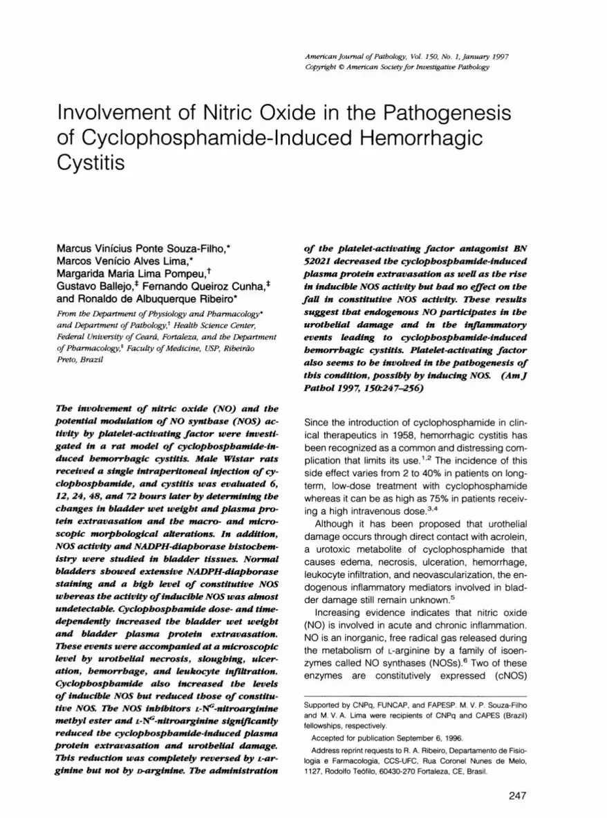
Figure 1. L-NAME inhibits the cyclophospha--100.* mide-induced time-dependent increase in weta weight and plasma protein extravasation in rat
. 80 k bladders. a: Bladder wet weight (hatched bars)c andplasma protein extravasation (solid bars) in_ normal animals (C, control) and in those treated
60 S 48 hours earlier with cyclophosphamide (50,100, and 200 mg/kg). b: Plasma protein extrav-asation (0) and bladder wet weight (0) in an-
.40~ imals treated 6, 12, 24, 48, or 72 hours earlierM with cyclophosphamide (100 mg/kg); A, plasma
20 protein extravasation induced by cyclophospha-t mide (100 mg/kg) in L-NAME-treated (20 mg/kg)
animals 6, 12, 24, 48, and 72 hours after the
72 0 C induction of cystitis. The results are reported asthe mean + SEM (n > 6). P < 0.05 comparedwith the respective control (C and vehicle) byANOVA, Fisher's F-test.
Macroscopic and Microscopic Evaluation of theBladder
Bladders were excised, freed from the surround-ing connective tissue, and examined grossly foredema and hemorrhage. Histological examinationwas performed by a pathologist in a single-blindfashion. Edema, bleeding, and histological changeswere evaluated according to the criteria of Gray eta123 as follows. Edema was considered severe (3+)when fluid was seen externally and internally in thewalls of the bladder, moderate (2+) when the edemawas confined to the internal mucosa, mild (1 +) be-tween normal and moderate, and none (0) as normal.Hemorrhage was scored as follows: 3+, intravesicalclots; 2+, mucosal hematomas; 1+, telangiectasiaor dilatation of the bladder vessel; 0, normal. Histo-pathology was scored as follows: 0, normal epithe-lium and absence of inflammatory cell infiltration and
*
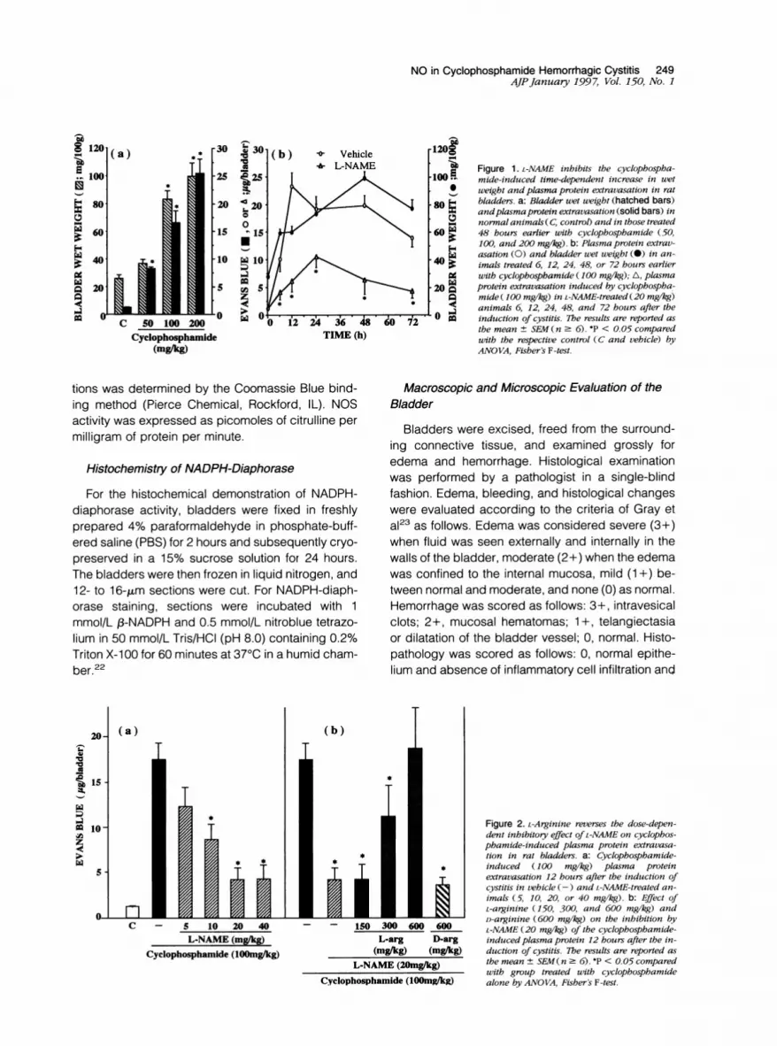
Figure 2. L-Arginine reverses the dose-depen-dent inhibitory effect OIL-NAME on cyclophos-phamide-induced plasma protein extravasa-tion in rat bladders. a: Cyclophosphamide-
* induced (100 mg/kg) plasma proteinextravasation 12 hours after the induction ofcystitis in vehicle (-) and L-NAME-treated an-imals (5, 10, 20, or 40 mg/kg). b: Effect ofL-arginine (150, 300, and 600 mg/kg) andD-arginine (600 mg/kg) on the inhibition by
100600600 L-NAME (20 mg/kg) of the cyclophosphamide-*arg D-arg induced plasma protein 12 hours after the in-
glkg) (mgAkg) duction of cystitis. The results are reported as
[E (20mg/kg) the mean ± SFM(n :- 6). *P < 0.05 comparedwith group treated with cyclophosphamide
nide (100muiWkg) alone by ANO VA, Fisher's F-test.

250 Souza-Filho et alAJPJanuary 1997, Vol. 150, No. 1
25j
20-
15-
Il-OJ 1
Tr
C - _
L-NOARG (2bm&)
Figure 3. L-Arginine reverses the inhibitory effect ofL-NOARG on thecyclophosphamide-induced plasma protein extravasation in rat blad-ders. The bars show the increase in plasma protein extravasationinduced by cyclophosphamide (100 mg/kg) in vehicle (-), L-NOARG(20 mg/kg) alone, and L-NOARG plus L-arginine-treated (600 mg/kg)animals. The results are reported as the mean ± SEM(n 6). P <0.05 compared with group treated with cyclophosphamide alone by
ANOVA, Fisher's F-test.
ulceration; 1, mild changes involving diminished ep-
ithelial cells, flattening with submucosal edema, mildhemorrhage, and few ulcerations; 2, severe changesincluding mucosal erosion, inflammatory cell infiltra-tion, fibrin deposition, hemorrhage, and multiple ul-cerations.
Effect of L-NG-Nitroarginine Methyl Ester(L-NAME), L-NG-Nitroarginine (L-NOARG),and BN 52021 on Cyclophosphamide-Induced Hemorrhagic Cystitis
Effect of L-NG-Nitroarginine Methyl Ester(L-NAME)
The animals received the NOS inhibitor L-NAME(5, 10, 20, and 40 mg/kg, intraperitoneal (i.p.)) 30minutes before and 6, 24, and 30 hours after an
injection of cyclophosphamide (100 mg/kg, i.p.).Their bladders were then analyzed 6, 12, 24, and 48hours after cyclophosphamide injection, as de-scribed above. In another set of experiments, L-argi-nine (150, 300, or 600 mg/kg, i.p.) or D-arginine (600mg/kg, i.p.) was administered simultaneously withL-NAME (20 mg/kg), and the cystitis was analyzed 12hours after cyclophosphamide injection.
Effect of L-NG-Nitroarginine (L-NOARG)
The rats were injected with another NOS inhibitor,L-NOARG (20 mg/kg, i.p.) 30 minutes before and 6hours after cyclophosphamide (100 mg/kg, i.p.). L-
Arginine (600 mg/kg, i.p.) was also administered to agroup of animals simultaneously with L-NOARG. Thebladders were analyzed 12 hours after the inductionof cystitis.
Effect of BN 52021
The animals were treated with BN 52021 (10 mg/kg, subcutaneous) 30 minutes before and 6, 24, and30 hours after cyclophosphamide (100 mg/kg, i.p.),and their bladders were analyzed 6, 12, and 48hours after cyclophosphamide administration. TheNOS activity in the bladders of one group of theseanimals was determined 12 hours after cyclophos-phamide, as described above.
Statistical AnalysisThe results are expressed as the mean ± SEM.Statistical significance (P < 0.05) was assessed byanalysis of variance (ANOVA) followed by Fisher'stest. The morphological data were analyzed by thenonparametric Mann-Whitney U-test.
Results
Cyclophosphamide (50 to 200 mg/kg, i.p.) dose-dependently increased the bladder wet weight andEvans blue extravasation in rats (Figure la) but didnot alter the bladder dry weight (not shown). Theincreases in bladder wet weight increase and Evansblue extravasation observed with cyclophosphamide(100 mg/kg) were time dependent (Figure ib). Fig-ure lb also shows that the Evans blue extravasationwas significantly inhibited by L-NAME. The latter alsoinhibited the bladder wet weight increase measured48 hours after cyclophosphamide administration(data not shown).The inhibitory effect of L-NAME (5 to 40 mg/kg) on
Evans blue extravasation was dose dependent andwas reversed by the simultaneous administration of
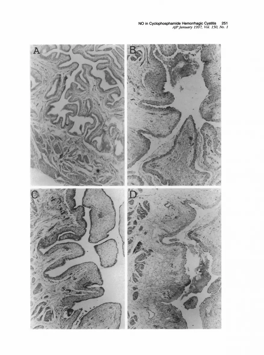
Figure 4. Histological analysis of representative bladder walls in cross section. A: Bladder of a control rat. B: Cystitis in a rat treated 12 hourspreviously with cyclophosphamide. The epithelium is thin and sometimes completely denuded with fibrin deposition and areas of ulcerations beingpresent. Edema and leukocyte infiltration can also be seen. C: Cystitis induced by cyclophosphamide in an L-NAME-treated rat. The epithelium is morepreserved, the edema and leukocyte infiltration are attenuated, and ulceration is not present. D: The cystitis induced by cyclophosphamide in anL-NAMEplus L-arginine-treated rat. The epithelium is completely denuded with more ulcerations and mucosal erosions. Severe edema and several clotsare present. H&E; magnification, X 40.
I 1:=an_
NO in Cyclophosphamide Hemorrhagic Cystitis 251AJPJanuaty 1997, Vol. 150, No. 1
,¢,,\\.
; > S. , _ * e.
* * ,; flP>: *
*ee ,. , iw, wB ,s,3k.,,SJ\Z s '&';L. v.'.'* '> , ., ' t -^ w ).£

252 Souza-Filho et alAJPJanuary 1997, Vol. 150, No. 1
Table 1. Median Values (and Ranges) of theMacroscopic and Microscopic Scores (Gray'sScores) for Cyclophosphamide-InducedHemorrhagic Cystitis after 12 Hours inAnimals Treated with Saline, L-NAME (20 mg/kg), and L-NAME (20 mg/kg) plus L-arginine(600 mg/kg)
Treatment
Morphologicalanalysis
L-NAMEplus
Saline L-NAME L-arginine
Microscopic 2 (1-2) 1 (0-1)* 2 (2-2)Macroscopic 2 (2-3) 1 (0-1)* 2 (2-3)
*Mann-Whitney U test; P < 0.05.
L-arginine but not of D-arginine (Figure 2). Similarly,L-NOARG (20 mg/kg) significantly reduced the in-crease in plasma protein extravasation induced bycyclophosphamide after 12 hours. This reductionwas completely reverted when L-arginine (600 mg/kg) was given simultaneously with L-NOARG (Figure3).
Cyclophosphamide induced severe cystitis char-acterized macroscopically by the presence of intra-vesical clots, mucosal hematomas, and severeedema. Microscopically, extensive mucosal erosionwith ulceration, fibrin deposition, hemorrhage,edema, and leukocyte infiltration was observed.These alterations were reduced by L-NAME butcould be reinstated by L-arginine (Figure 4 and Table1). Interestingly, in the animals that received L-argi-nine, the cyclophosphamide-induced cystitis tendedto be more severe.NOS activity in the normal bladder was mainly of
the cNOS (calcium-dependent) isoform (>95%). Cy-clophosphamide administration increased signifi-cantly the activity of iNOS (calcium-independent),which was already evident 6 hours after injection andremained elevated for up to 48 hours (Figure 5B). Incontrast, cNOS decreased over a similar time course
in the presence of cyclophosphamide (Figure 5A).Consistent with these findings, intense NADPH di-aphorase staining was observed in the urothelium ofcontrol rats (Figure 6, A and B). This activity de-creased dramatically 12 hours after cyclophospha-mide administration and coincided with an intensesloughing of the urothelium (Figure 6, C and D),which persisted up to 48 hours after treatment withcyclophosphamide (data not shown). NumerousNADPH-diaphorase-positive cells were also ob-served in the lamina propria within 12 hours (Figure6D) and were still present 48 hours after the induc-tion of cystitis (data not shown).
The cyclophosphamide-induced increase inplasma protein extravasation at 12 and 48 hoursafter its administration was significantly reduced bytreatment of the animals with the PAF antagonist BN52021 (Figure 7A). This treatment also significantlyinhibited the cyclophosphamide-induced bladderwet weight gain measured 48 hours after the induc-tion of cystitis (data not shown). The increase inbladder iNOS activity caused by cyclophosphamidewas significantly reduced by BN 52021 (Figure 7C).Interestingly, BN 52021 did not significantly modifythe alteration in cNOS activity induced by cyclophos-phamide (Figure 7B).
DiscussionIn the present paper, we have shown that endoge-nous NO is involved in the inflammatory events lead-ing to hemorrhagic cystitis after the administration ofcyclophosphamide, as demonstrated by the follow-ing observations: 1) two NOS inhibitors, L-NAME andL-NOARG, dose-dependently inhibited the cyclo-phosphamide-induced increase in plasma proteinextravasation and bladder wet weight; this inhibitioncould be reversed by L-arginine; 2) NOS inhibition by
(a) cNOS (b) INOS
Figure 5. Time course ofNOS activity in cyclo-phosphamide-induced hemorrhagic cystitis.The bars represent the cNOS (a) and iNOS (b)activities in the bladders of rats treated 6, 12,24, and 48 hours previously with cyclophospha-mide (100 mg/kg). NOS activities were assayedby a citrullineproduction assay. Thefirst bar inboth panels (0 represents the cNOS and iNOSactivities in naive animals, respectively. The re-
sults are reported as the means ± SEMt(n = 6).*P < 0.05 compared with the naive animals (C)by ANOVA, Fisher's F-test.
.12
*3'S I-
la
ow
15
T I
10-
5.
C 6 12 24 48
Cyclophosphamide
*

NO in Cyclophosphamide Hemorrhagic Cystitis 253AJPJanuary 1997, Vol. 150, No. 1
;w. x , =a. -
Figure 6. Histochemical localization ofNADPH-diaphorase/NOS-positive structures in the urinary bladder ofcontrol rats (A and B) and in rats 12hours after the administration of cyclophosphamide (C and D). In bladders from control rats, the urothelium is strongly positive for NADPH-diaphorase whereas in the bladdersfrom cyclophosphamide-treated rats a complete sloughing ofthe urothelium is observed and NADPH-diaphorase-positive cells are now observed in the lamina propria. Magnification, x 6.25 (A and C) and x50 (B and D).
IN

254 Souza-Filho et alAJPJanuary 1997, Vol. 150, No. I
(a)15
.P2I2
-E10
."
0-
DfI...... . I I VA0 6 12 18 24 30 36 42 48 C PBS BN
TIME (hours) CyclophosphamideC PBS BN
CyclophosphamideFigure 7. Inhibitory effect ofBN 52021 on plasma protein extravasation and iNOS activity in cyclophosphamide-induced hemorrhagic cystitis. a:Increase in plasma protein extravasation induced by cyclophosphamide (100 mg/kg) in vehicle (0) and by BN-52021 (10 mg/kg; A) in animals 6,12 and 48 hours after the induction of cystitis. b and C: cNOS and iNOS activities, respectively, in the cytosolic fraction of bladders 12 hours afterthe induction of cystitis by cyclophosphamide in vehicle (PBS) and BN 52021 (BA) -treated rats. The results are reported as the means ± SEM(n =
6). P < 0.05 compared with the PBS-treated animals (C) by ANOVA, Fisher's F-test.
L-NAME significantly reduced the mucosal damage,hemorrhage, edema, and leukocyte infiltration in thebladders of cyclophosphamide-treated rats; 3)NADPH-diaphorase histochemistry showed thatNOS-containing cells present in the urothelium ofcontrol rats disappeared in cyclophosphamide-treated animals and that positive cells, most likelyimmune cells, appeared in the lamina propria of theinflamed mucosa; 4) cyclophosphamide markedlyincreased calcium-independent NOS activity in thebladder with a time course similar to that of thehistopathological alterations observed. At the same
time, the activity of calcium-dependent NOS de-creased, possibly reflecting the urothelial sloughingobserved in the histopathological analysis.
The participation of NO in cyclophosphamide-in-duced cystitis is consistent with previous reportsshowing that NO is cytotoxic to different cells7-9 andthat it participates in the inflammatory events.L-NMMA has been reported to dose-dependentlyreduce the cell damage observed in several patho-logical conditions, such as endotoxic shock.24 Inaddition, both L-NMMA and L-NAME diminished theedema formation induced by substance p,25 by car-
rageenin and dextran,10 and by BK, 5-hydroxytryp-tamine, and polycations.26,27 This latter effect ofNOS inhibitors could be reversed by the co-admin-istration of a vasodilator agent such as iloprost (aPGI2 analogue), suggesting that the anti-edema ef-fect of L-NAME is a consequence of the inhibition ofblood flow. However, experiments performed in the
hamster cheek pouch microvasculature have shownthat L-NAME can also reduce the increase in vascu-
lar permeability in response to bradykinin and aden-osine-5'-diphosphate,28 suggesting that NO by itselfis able to modulate vascular permeability. It hasbeen recently demonstrated that iNOS-deficientmice generated by gene knockout have a reducedinflammatory response to carrageenin.29
Our data further indicated that PAF could be one
of the inflammatory mediators contributing to theactivation of the L-arginine-NO pathway. The involve-ment of PAF and its modulation of NOS activity incyclophosphamide-induced hemorrhagic cystitis issuggested by the observations that 1) pretreatingthe rats with the PAF antagonist BN 52021 signifi-cantly reduced the cyclophosphamide-inducedplasma protein extravasation and the increase inbladder wet weight and that 2) BN 52021 reducediNOS activity. Interestingly, BN 52021 did not pre-vent the decrease in cNOS activity when comparedwith the bladders of rats treated with cyclophospha-mide alone. This finding indicates that PAF may par-
ticipate in the inflammatory response after urothelialdamage has occurred and is consistent with theurothelial sloughing observed in the BN-52021-treated animals. The reduction of iNOS activity in theBN-52021-treated rats suggests that PAF may beinvolved in the recruitment of inflammatory cells con-
taining iNOS (based on the diaphorase staining) or inthe induction of NOS in the already emigrated cells.This hypothesis is strengthened by evidence that
(b) cNOS ( c ) iNOS
gm
_/
Po
do

NO in Cyclophosphamide Hemorrhagic Cystitis 255AJPJanuary 1997, Vol. 150, No. 1
PAF may contribute to the induction of NOS in in-flammatory cells.18We have recently shown that in addition to PAF
and NO, the inflammatory cytokines tumor necrosisfactor-a and interleukin-1 are also important in theappearance of cyclophosphamide-induced cysti-tis.19 As these cytokines are able to induce the NOSin inflammatory cells,7'8 it is likely that they also con-tribute to the induction of NO production in the blad-der after cyclophosphamide administration.
In conclusion, our results suggest that endoge-nous NO is involved in urothelial damage and in theinflammatory events leading to hemorrhagic cystitisafter cyclophosphamide administration. The induc-tion of NOS in the inflammed bladder appears torequire the action of PAF.
AcknowledgmentsWe gratefully acknowledge the technical assistanceof Fabfola Leslie A. C. Mestriner and Eleni LuizaTamburus Gomes.
References
1. Philips FS, Sternberg SS, Cronin AP, Vidal PM: Cyclo-phosphamide and urinary bladder toxicity. Cancer Res1961, 21:1577-1589
2. Stillwell TJ, Benson RC: Cyclophosphamide-inducedhemorrhagic cystitis: a review of 100 patients. Cancer1988, 61:451-457
3. Foad BSI, Hess EU: Urinary bladder complications withcyclophosphamide therapy. Arch Intern Med 1976,136:616-625
4. Droller MJ, Saral R, Santos G: Prevention of cyclophos-phamide-induced hemorrhagic cystitis. Urology 1982,20:256-258
5. Cox PJ: Cyclophosphamide cystitis: identification ofacrolein as the causative agent. Biochem Pharmacol1979, 28:2045-2049
6. Forstermann U, Schmidt HHHW, Pollock JS, Sheng H,Mitchell JA, Warner TD, Nakane M, Murad F: Isoformsof nitric oxide synthase: characterization and purifica-tion from different cell types. Biochem Pharmacol 1991,42:1849-1857
7. Nathan CF: Nitric oxide as a secretory product of mam-malian cells. FASEB J 1992, 6:3051-3064
8. Moncada S, Palmer RMJ, Higgs EA: Nitric oxide: phys-iology, pathophysiology, and pharmacology. Pharma-col Rev 1991, 43:109-142
9. Estrada C, Gomez C, Martfn C, Moncada S, GonzalezC: Nitric oxide mediates tumor necrosis factor-a cyto-toxicity in endothelial cells. Biochem Biophys Res Com-mun 1992, 186:475-482
10. lalenti A, lanaro A, Moncada S, Di Rosa M: Modulation
of acute inflammation by endogenous nitric oxide. EurJ Pharmacol 1992, 211:177-182
11. lalenti A, Moncada S, Di Rosa M: Modulation of adju-vant arthritis by endogenous nitric oxide. Br J Pharma-col 1993, 110:701-706
12. Mulligan MS, Hevel JM, Marletta MA, Ward PA: Tissueinjury caused by deposition of immune complexes isI-arginine dependent. Proc Natl Acad Sci USA 1991,88:6338-6342
13. Middleton SJ, Shorthouse M, Hunter JO: Increasednitric oxide synthesis in ulcerative colitis. Lancet 1993,341:465-466
14. Miller MJS, Sadowska-Krowicka H, Chotinaruemol S,Kakkis JL, Clark DA: Amelioration of chronic ileitis bynitric oxide synthase inhibition. J Pharmacol Exp Ther1993, 264:11-16
15. Farrel AJ, Blake DR, Palmer RMJ, Moncada S: In-creased concentrations of nitrite in synovial fluid andserum samples suggest increased nitric oxide synthe-sis in rheumatic diseases. Ann Rheum Dis 1992, 51:1219-1222
16. Braquet P, Touqui L, Shen TY, Vargaftig BB: Perspec-tives in platelet-activating factor research. PharmacolRev 1987, 39:97-145
17. Snyder F: Platelet-activating factor and related acety-lated lipids as potent biologically active mediators.Am J Physiol 1990, 259:C697-C708
18. Szabo C, Wu CC, Mitchell JA, Gross SS, ThiemermannC, Vane JR: Platelet-activating factor contributes to theinduction of nitric oxide synthase by bacterial lipopoly-saccharide. Circ Res 1993, 73:991-999
19. Gomes TNA, Santos CC, Souza-Filho MVP, Cunha FQ,Ribeiro RA: Participation of TNF-a and IL-1 in thepathogenesis of cyclophosphamide-induced hemor-rhagic cystitis. Brazilian J Med Biol Res 1995, 28:1103-1108
20. Salter M, Knowles RG, Moncada S: Widespread tis-sue distribution, species distribution, and changes inactivity of calcium-dependent and calcium-indepen-dent nitric oxide synthases. FEBS Lett 1991, 291:145-149
21. Assreuy J, Cunha FQ, Liew FY, Moncada S: Feedbackinhibition of nitric oxide. Br J Pharmacol 1993, 108:833-837
22. Hope BT, Vincent SR: Histochemical characterizationof neuronal NADPH diaphorase. J Histochem Cyto-chem 1989, 37:653-661
23. Gray KJ, Engelmann UH, Johnson EH, Fishman IJ:Evaluation of misoprostol cytoprotection of the bladderwith cyclophosphamide (cytoxan) therapy. J Urol 1986,133:497-500
24. Nava E, Palmer RMJ, Moncada S: The role of nitricoxide in endotoxic shock. Biology of Nitric Oxide: Phys-iological and Clinical Aspects. Edited by S Moncada,MA Marietta, JB Hibbs, EA Higgs. London, PortlandPress, 1992, pp 231-233
25. Hughes SR, Williams TJ, Brain SD: Evidence that nitric

256 Souza-Filho et alAJPJanuary 1997, Vol. 150, No. 1
oxide modulates oedema formation induced by sub-stance P. Eur J Pharmacol 1990, 191 :481-484
26. Antunes E, Mariano M, Cirino G, Levi S, de Nucci G:Pharmacological characterization of polycation-in-duced rat-hind paw oedema. Br J Pharmacol 1990,101:986-990
27. Antunes E, Giraldelo CMM, Cirino G, de Nucci G: Ef-fects of NG-monomethyl-L-arginine and its D-enantio-mer on rat hind paw oedema. Biology of Nitric Oxide:Physiological and Clinical Aspects. Edited by S
Moncada, MA Marletta, JB Hibbs, EA Higgs. London,Portland Press, 1992, pp 264-266
28. Mayhan WG: Role of nitric oxide in modulating perme-ability of hamster cheek pouch in response to adeno-sine 5-diphosphate and bradykinin. Inflammation 1992,16:295-305
29. Wei X, Charles IG, Smith A, Ure J, Feng GF, Huang F,Xu D, Muller W, Moncada S, Liew FY: Altered immuneresponses in mice lacking inducible nitric oxide syn-thase. Nature 1995, 375:408-411


