Embed Size (px)
Citation preview

Involvement of uracil nucleotides in protection ofcardiomyocytes from hypoxic stress
Smadar Yitzhakia, Vladimir Shneyvaysa, Kenneth A. Jacobsonb, and Asher Shainberga,*
aFaculty of Life Sciences, Bar-Ilan University, Ramat-Gan 52900, IsraelbLaboratory of Bioorganic Chemistry, NIDDK, NIH, Bethesda, MD, USA
AbstractCardiomyocytes express one or more subtypes of P2 receptors for extracellular nucleotides. P2purinoceptors, which are activated by nucleotides, are classified as P2X or P2Y: P2X receptors areligand-gated intrinsic ion channels, and P2Y receptors are G protein-coupled receptors.Extracellular pyrimidine and purine nucleotides are released from the heart during hypoxia.Although the cardioprotective effects of purines acting via purinoceptors were studied intensively,the physiological role of uracil nucleotide-responsive P2Y2, P2Y4, P2Y6, and P2Y14 receptors isstill unclear, especially in the cardiovascular system. This study revealed that uridine-5′-triphosphate (UTP) protected cultured rat cardiomyocytes during hypoxia and explored the UTPsignaling pathway leading to this cardioprotection. We found that UTP, but not UDP or uridine,significantly reduced cardiomyocyte death induced by hypoxia. Incubation with UTP for 1 h,before exposure to hypoxic conditions, protected the cells 24 h later. The cardioprotective effect ofUTP was reduced in the presence of the P2 antagonist suramin. In addition, UTP caused atransient increase of [Ca2+]i in cardiomyocytes. Pyridoxal-5′-phosphate-6-azophenyl-2,4-disulfonate (PPADS) or Reactive blue 2 (RB-2), other antagonists of P2 receptors, abolished the[Ca2+]i elevation caused by UTP. We used various inhibitors of the Ca2+ signaling pathway toshow that UTP elevated levels of [Ca2+]i, originating from intracellular sources, via activation ofphospholipase C and the IP3 receptor. Interestingly, these inhibitors of the Ca2+ signaling pathwaydid not prevent the immediate protective effect caused by UTP. Although mitochondrial KATPchannels are involved in other preconditioning mediator pathways, the involvement of thesechannels in the cardioprotective effect induced by UTP was ruled out, because 5-hydroxydecanoicacid (5-HD), a specific inhibitor of these channels, did not prevent the protection.
KeywordsP2Y2 nucleotide receptor; G protein-coupled receptor; Pyrimidines; Cardioprotection; Ischemia;Preconditioning
1. IntroductionMyocardial infarction and heart failure are leading causes of morbidity and mortality inhumans. Considerable effort has been devoted to improving functional recovery andreducing the extent of infarction after ischemic episodes. A step in this direction was thediscovery that the heart is significantly protected against ischemic injury when firstpreconditioned by a brief ischemia [1]. Adenosine, a metabolite of adenine nucleotides,simulates the protection afforded by preconditioning (PC) via A1 and A3 receptors.
© 2005 Elsevier Inc. All rights reserved.*Corresponding author. Tel.: +972 3531 7038; fax: +972 3 736 9231. [email protected] (A. Shainberg).
NIH Public AccessAuthor ManuscriptBiochem Pharmacol. Author manuscript; available in PMC 2012 September 23.
Published in final edited form as:Biochem Pharmacol. 2005 April 15; 69(8): 1215–1223. doi:10.1016/j.bcp.2005.01.018.
NIH
-PA Author Manuscript
NIH
-PA Author Manuscript
NIH
-PA Author Manuscript

However, accumulating evidence suggests that adenosine receptors are not the onlymediators of PC [2-4], but that adrenoceptors [5], bradykinin receptors [6], opioid receptors[7], and ATP receptors [8] may also contribute to PC.
Purine and pyrimidine nucleotides have widespread and specific extracellular signalingactions in the regulation of a variety of functions in many tissues. In 1978 Burnstockproposed that specific receptors for extracellular nucleosides (P1) or nucleotides (P2)mediate the physiological effects of adenosine and ATP, respectively [9]. According topharmacological evidence, mechanisms of signal transduction, and molecular structure,receptors that mediate the extracellular actions of purine and pyrimidine nucleotides aredivided into two major families: ligand-gated receptors (P2X) and G protein-coupledreceptors (P2Y). In contrast to P2X receptors, the class of P2Y receptors is activated notonly by ATP and its derivatives, but also by other naturally occurring nucleotides. The P2Y2and P2Y4 receptors are activated by the uracil nucleotide UTP (uridine-5′-triphosphate)acting as an autocrine regulator [10,11]. These receptors are preferentially coupled to Gq andtherefore activate phospholipase Cβ, leading to the formation of inositol trisphosphate, Ca2+
elevation, and production of diacylglycerol, which activates protein kinase C (PKC) andmitogen-activated protein kinases [12,13]. Thus, activation of P2Y2 and P2Y4 receptors andsubsequently kinases can have long-term, tropic effects on cardiomyocyte activity.
Whereas the effects of purine nucleosides and nucleotides in myocardial infarction andischemia have been intensively studied [14], the role of pyrimidine nucleotides underhypoxic conditions has not been well explored. Our principal aim was to elucidate theprotective effects of UTP and pyrimidinergic receptor activation against detrimental factorsof ischemia/hypoxia. For this reason we sought to explore whether P2Y2 and P2Y4 receptorsare present on the surface of cultured newborn rat cardiomyocytes and the expression levelsof these receptors. Receptor functionality was demonstrated by evaluation of the calciumresponse following receptor activation and by the initiation of signal transduction pathways,which resulted in attenuating cardiomyocyte injury in hypoxia. For this purpose we studiedthe expression and localization of P2Y2 and P2Y4 receptor proteins in culturedcardiomyocytes through immunohistochemistry and Western blotting. Changes inintracellular Ca2+ concentration ([Ca2+]i) after the application of nucleotides were studiedby indo-1 cytofluorometry. Morphological evaluation of cultured cells, measurement oflactate dehydrogenase (LDH) release to the culture medium, and determination of ATPcontent were used to characterize the protection from hypoxic damage upon UTP receptoractivation.
The present study provides evidence for the first time that the extracellular pyrimidinenucleotide, UTP, could play a substantial role in mediating cardioprotection from hypoxicdamage through specific receptors.
2. Experimental procedures2.1. Cell culture
Rat hearts (2–3 d old) were removed under sterile conditions and washed three times in PBSto remove excess blood cells. The hearts were minced and then gently agitated in RDB, asolution of proteolytic enzymes (Life Science Research Inst. [15]), prepared from a fig treeextract. After treatment with RDB, cell viability was found to be 98%, in contrast to 85% intrypsin-treated monolayer cultures. The RDB was diluted 1:100 in Ca2+- and Mg2+-freePBSat 25 °C for a fewcycles of 10 min each, as described previously [16]. Dulbecco’smodified Eagle’s medium, supplemented with inactivated 10% horse serum (BiologicalIndustries) and 0.5% chick embryo extract, was added to supernatant suspensions containingdissociated cells. The mixture was centrifuged at 300 × g for 5 min. The supernatant phase
Yitzhaki et al. Page 2
Biochem Pharmacol. Author manuscript; available in PMC 2012 September 23.
NIH
-PA Author Manuscript
NIH
-PA Author Manuscript
NIH
-PA Author Manuscript

was discarded, and the cells were resuspended in the same medium. The suspension of thecells was diluted to 1.0 × 10⁶ cells/ml, and 1.5 ml of the suspension was placed in 35-mmplastic culture dishes on collagen/gelatin-coated coverglasses. The cultures were incubatedin a humidified atmosphere of 5% CO2, 95% air at 37 °C. Confluent monolayers exhibitingspontaneous contractions were developed in culture within 2 d. All experiments wereperformed between days 5 and 7 in culture.
2.2. [Ca2+]i measurementsIntracellular free calcium ion concentration was estimated from indo-1 fluorescence with theratio method (the SAMPLE program) described elsewhere [17,18].
2.3. Hypoxic conditionsMyocyte cultures 5–7 d old were washed from the medium with glucose-free PBScontaining 5 μM HEPES at pH 7.4 before exposing the myocytes to the various conditionsat 37 °C. The hypoxic condition consisted of 120 min in a hypoxic incubator in which theatmosphere was replaced by the inert gas argon (100%) in glucose-free media [16,19]. Thehypoxic damage was characterized at the end of the hypoxic period by morphological andbiochemical evaluation. Continuous monitoring of [Ca2+]i during hypoxia was realized in aspecial barrier well, where cells were protected from oxygen by a laminar counterflowinglayer of argon gas as previously described [20]. The coverglasses with cultured cells wereplaced at the bottom of the well. This chamber was mounted on a specially modified Zeissinverted epifluorescence microscope.
2.4. Assays of intracellular ATP levelAfter hypoxia, control and experimental cells were harvested in 1 ml cold 5% trichloroaceticacid. The cell extract was used for the measurement of ATP content with the luciferin–luciferase bioluminescence kit (CLSII, Boehringer) following the manufacturer’s protocol.The values are expressed as nmol/mg of protein [17].
2.5. Experiments with purinergic and pyrimidinergic ligandsUTP at concentrations of 3–50 μM was applied to the cell cultures for 15 min following a15-min preincubation with various antagonists (Sigma): pyridoxal-5′-phosphate- 6-azophenyl-2,4-disulfonate (PPADS), suramin, or Reactive blue 2 (RB-2). The cell cultureswere washed with PBS prior to the experimental treatment.
2.6. Release of LDHProtein content and LDH activity were determined according to El-Ani et al. [21]. Briefly,25 μl supernatant was transferred into a 96-well dish, and the LDH activities weredetermined with an LDH-L kit (Sigma), as described by the manufacturer. The product ofthe enzyme was measured spectrometrically at 30 °C at a wavelength of 340 nm asdescribed previously [19]. The results were expressed relative to the control (X-fold) in thesame experiment. Each experiment was done in triplicate and was repeated at least threetimes.
2.7. Staining2.7.1. Propidium iodide—The assay is based on vital binding of propidium iodide (5 μg/ml) to nuclei of cells whose plasma membranes have become permeable because of celldamage. The assay was performed according to Nieminen et al. [22]. For counter-stainingwe used Hoechst 33342 (10 μM), which stains the nuclei of all cells.
Yitzhaki et al. Page 3
Biochem Pharmacol. Author manuscript; available in PMC 2012 September 23.
NIH
-PA Author Manuscript
NIH
-PA Author Manuscript
NIH
-PA Author Manuscript

2.7.2. Lysosome staining by neutral red—Neutral red is a widely used marker ofmembrane-bound compartments with an acidic lumen (lysosomes and several other acidiccompartments). Live cardiomyocytes were incubated for 30 min in PBS containing 4 μg/mlneutral red (Molecular Probes).
2.7.3. Immunostaining—Cells were fixed in absolute methanol for 10 min,permeabilized with 0.1% Triton X-100 for 5 min, and blocked in 2% normal goat serum for10 min. Anti-P2Y2 or anti-P2Y4 antibodies (Alomone Labs) were diluted in PBS to a finalconcentration of 1:100 and then incubated overnight at room temperature with the cells. As areporter we used goat antirabbit fluorescein isothiocyanate (FITC) that was incubated for 30min at room temperature.
2.7.4. Western blotting—Aliquots (~70 μg) from each protein sample were loaded on11% SDS polyacrylamide gels and blotted onto nitrocellulose membranes. Filters weresaturated with Tris-buffered saline in 10% milk for 1 h at room temperature to avoid non-specific binding of the primary antibody and incubated overnight at 4 °C with anti-P2Y2(1:200) and anti-P2Y4 (1:300) antibodies. Each P2Y receptor was also tested in the presenceof neutralizing peptides (ratio 1:1 between peptide and antiserum). Blots were then washedin Tris-buffered saline (1 mM Tris–HCl, pH 8.0, 15 mM NaCl, 0.1% Tween X-100, finalconcentration), incubated for 1 h with antirabbit IgG coupled to FITC. Immunoreactionswere analyzed with enhanced chemiluminescence.
2.8. StatisticsResults were expressed as mean ± S.D. Data were analyzed by analysis of variance withapplication of a post hoc Tukey-Kremer test. Statistical significance was determined as *P <0.01 and **P < 0.05 in different experiments.
3. Results3.1. Cardiomyocytes express UTP-responsive P2Y receptors
The presence of P2Y2 and P2Y4 receptors in cultured rat cardiomyocytes was demonstratedwith immunofluorescent staining and Western immunoblot assay. Newborn ratcardiomyocytes exhibited P2Y2 and P2Y4 receptors on the cell surface, as seen from thefluorescence intensity level. The specificity of receptor immunostaining was indicated bylack of staining in the presence of competitive peptide antigens, derived from the receptorsequence. Anti-P2Y2 or -P2Y4 antibodies stained the whole cardiomyocyte membrane, butno staining was observed in fibroblasts that were present in the same culture dish (Fig. 1Aand B). An immunoblot assay showed that anti-P2Y2 antibodies detected four major proteinbands of the predicted molecular weight at 42, 55, 68, and 84 kDa, and P2Y4 receptors wereidentified by two protein bands, which were ~50 and ~80 kDa. These bands disappearedfollowing preincubation of the antibodies with their competitive peptide antigens (Fig. 1C).These results demonstrated the existence of P2Y2 and P2Y4 receptors in neonatalcardiomyocytes.
3.2. Effect of UTP on cardiomyocytes subjected to hypoxiaTo investigate the role of UTP in attenuation of myocyte injury during prolonged (120 min)hypoxia, cultured cardiomyocytes were incubated with various concentrations of UTP (3–50μM) for 15 min prior to hypoxia. The pyrimidine (10 μM) did not cause any cell injury butelicited a significant attenuation of the cellular injury produced by the subsequent 120-minhypoxia, as measured by the level of LDH released from the cells (Fig. 2A). The decrease inLDH release suggested that UTP receptor activation delayed acute hypoxia-inducedcardiomyocyte injury. For investigation of the time dependence of UTP in protecting the
Yitzhaki et al. Page 4
Biochem Pharmacol. Author manuscript; available in PMC 2012 September 23.
NIH
-PA Author Manuscript
NIH
-PA Author Manuscript
NIH
-PA Author Manuscript

cardiocytes, the cells were pretreated with 50 μM UTP for 1 h and then exposed to 2 hhypoxia after incubation in full medium for 15 min to 72 h. UTP protected the cells fromhypoxia even when treated 48 h before hypoxia, as revealed by LDH release (Fig. 2B).These results suggest that UTP activates a long, persistent mechanism of protection (asecond window of protection). On the other hand, the related derivatives uridine and UDP(50 μM) did not exhibit a protective effect on cardiomyocytes against hypoxia (Table 1).This ruled out the P2Y6 receptor as the site of action of protective uracil nucleotides.
To verify whether the protective effect caused by UTP is mediated via P2 receptors,cardiomyocytes were treated with a P2 receptor antagonist, e.g. RB-2, PPADS, or suramin,before exposing them to 50 μM UTP and hypoxia. Treatment with suramin or PPADS, butnot RB-2, inhibited the protective effect of UTP, as determined by LDH released to themedium (Table 1), indicating the involvement of P2Y2 receptors and at least partly of P2Y4receptors in UTP-induced protection of cultured cardiomyocytes.
3.3. Effect of P2 agonists/antagonists on intracellular ATP levels during hypoxiaThe protective effect caused by extracellular UTP was also demonstrated by protection oftotal ATP content after hypoxia. The results showed that the ATP level decreaseddramatically after 2 h exposure to hypoxia (2.92 ± 0.17 nmol/mg protein compared with20.12 ± 2.31 nmol/mg protein in normoxic cultures; Fig. 3). Pretreatment of cardiomyocyteswith 50 μM UTP prevented the dramatic decrease of ATP levels (16.62 ± 4.62 nmol/mgprotein) in cultures subjected to hypoxia. However, protection against ATP depletion did notoccur when cells were pretreated with P2 receptor antagonist PPADS (1.84 ± 1.31 nmol/mgprotein) or suramin (2.56 ± 0.87 nmol/mg protein) before exposure to UTP, indicating thatthe protective effect of UTP is mediated via P2 receptors.
The protective effect of UTP was also demonstrated through cardiomyocyte morphology.Following hypoxia the cells were fixed and stained with hematoxylin and eosin, whichshowed typical irreversible oncotic damage of cardiomyocytes: vacuoles and disorder of themyofilaments, pyknotic nuclei, and edematous areas in the cytoplasm and around thenucleus. In addition, hypoxic conditions increased the accumulation of the lysosomotropicvital dye neutral red in cytoplasmic granules and around the nucleus and increased the sizeof the lysosomes and the intensity of their color (Fig. 4). The damage to culture cells treatedwith UTP before hypoxia was greatly reduced. Cell loss (percentage of cell death) waspresented as the number of dead cells (propidium iodide stained) as a percentage of the totalnumber of cells (Hoechst 33341 stained). After 120 min of hypoxia cell loss was 40% ± 5%.Activation of the UTP receptors attenuated cell loss caused by hypoxia to 18% ± 2%;control untreated cells had 3.1% ± 0.2% cell loss.
The contribution of the mitochondrial KATP channel to the protective effects of UTP wasexamined by application of a mKATP channel blocker, 5-hydroxydecanoic acid (5-HD; 200μM), 15 min before treatment with UTP and hypoxia. The presence of 5-HD did not alterthe protective effects of UTP, as revealed by LDH release to the medium (Table 1). Thesefindings indicate that UTP exerts its protective effect via a pathway bypassing themitochondrial KATP channel.
3.4. Effect of UTP receptor activation on intracellular [Ca2+]i[Ca2+]i levels were determined by using indo-1 as a fluorescent probe. Treatment with 10μM UTP induced transient accelerations of the beating rate and a transient elevation of[Ca2+]i baseline, which lasted 30–40 s, and finally restored the normal beating activity (Fig.5A). Application of PPADS (10 μM) or RB-2 (50 μM) prevented [Ca2+]i elevation (Fig. 5Band C). Suramin (300 μM), which acts as an antagonist of P2Y2 receptors among other P2
Yitzhaki et al. Page 5
Biochem Pharmacol. Author manuscript; available in PMC 2012 September 23.
NIH
-PA Author Manuscript
NIH
-PA Author Manuscript
NIH
-PA Author Manuscript

subtypes, did not prevent [Ca2+]i elevation following activation of the UTP receptors (Fig.5D). A possible pathway of UTP receptor signaling leading to an increase in [Ca2+]i isactivation of phospholipase C (PLC), which mediates inositol 1,4,5-trisphosphate (IP3)production and calcium release from IP3 receptors [23]. Application of (1-(6-((17beta-3-methoxyestra-1,3,5(10)-trien-17-yl)amino)hexyl)-1H-pyrrole-2,5-dione) (U73122; 2 μM aninhibitor of PLC activity) or 2-aminoethoxydiphenylborane (2APB), an IP3 receptorinhibitor (50 μM) abolished Ca2+ elevation after activation of UTP receptors (Fig. 6).
3.5. Effect of UTP on [Ca2+]i level during hypoxiaContinuous monitoring of [Ca2+]i during hypoxia was accomplished with a speciallydesigned chamber, where cells were protected from oxygen by a laminar counterflowinglayer of the inert gas argon. The level of [Ca2+]i began to be elevated ~10–12 min after theinitiation of hypoxia, with the cessation of beating activity (Fig. 7A). When thecardiomyocytes were pretreated with UTP (50 μM) for 15 min and then subjected tohypoxia, a delay in [Ca2+]i elevation was observed, and spontaneous contractions lasted for30–40 min after the initiation of hypoxia (Fig. 7B). Pretreatment with UTP 24 h before thehypoxia resulted in maintenance of spontaneous contractility even after 40 min of hypoxia(Fig. 7C).
4. DiscussionIn this work we demonstrate that short-term activation of UTP receptors protects newbornrat cardiomyocytes against hypoxic damage and significantly reduces the cell death causedby hypoxia via activation of P2Y2 and P2Y4 receptors.
It was shown in rat cardiomyocytes that UTP, an alternate agonist to ATP at P2Y2purinergic receptors, is as effective as ATP in stimulating PLC, and – as with ATP – theresponse is Gq mediated [23,24]. The uridine nucleotide-responding receptor P2Y4, whichwhen cloned from rat heart [25] is equipotently activated by ATP and UTP, is coupled, inpart, to Gi/o [24,26]. The presence of P2Y2 and P2Y4 receptors was demonstrated in thehuman fetal heart [27], but in adult myocytes, P2Y4 receptors could hardly be detected [28].Therefore, it is important to establish the expression of P2Y2 and P2Y4 receptors in newbornrat cardiomyocytes by immunohistochemical methods and immunoblotting. As was found inthis study, rat newborn cardiomyocytes abundantly express P2Y2 receptors, and anti-P2Y2antibodies detected four major proteins, indicating heterogeneity of these receptors. HigherMW bands may reflect receptor dimerization. Expression of P2Y4 receptors also was foundin newborn rat cardiomyocytes, although less abundant and represented by two proteinbands.
The mechanism of action by which UTP induces cardioprotection is not known. Recentstudies on UTP signaling showed that these receptors are coupled to the IP3 pathway, whichcauses Ca2+ elevation [29]. As our study shows, the application of UTP induces animmediate increase in [Ca2+]i level. This increase is not abolished by suramin, antagonist toP2Y2 but not to P2Y4 receptors [30], indicating that the immediate Ca2+ response to UTP ismediated through P2Y4 receptors. The observed Ca2+ response may be a mechanism ofUTP-mediated contraction (i.e., P2Y4 receptor-mediated contraction), but evidently thispathway of [Ca2+]i elevation is not involved in acute myocyte protection from hypoxia,because other uridine derivatives (UDP, uridine), although causing a transient elevation ofintracellular Ca2+, do not protect the cells from hypoxia. These data are consistent withknown effects of uridine derivatives on heart performance [12]. These results imply that inspite of its central physiological role in cardiac cells, [Ca2+]i elevation is not one of thecritical elements in the cell-signaling pathway required for UTP-mediated classicalcardioprotection (first window of protection).
Yitzhaki et al. Page 6
Biochem Pharmacol. Author manuscript; available in PMC 2012 September 23.
NIH
-PA Author Manuscript
NIH
-PA Author Manuscript
NIH
-PA Author Manuscript

This study shows that activation of UTP receptors protects cultured newborn ratcardiomyocytes from damage induced by hypoxia. It appears that the P2Y2 receptor triggersa cardioprotective response, although we cannot exclude a role of the P2Y4 receptor,because selective antagonists for this receptor are still not available. P2Y4 receptors havebeen reported to couple via pertussis toxin-sensitive G proteins, and as Oldenburg et al.wrote, “Virtually all Gi-coupled receptors present on the cardiomyocyte are capable ofpreconditioning the myocardium, even if their ligands (exogenous agonists) are not releasedduring a PC protocol” [13].
The present study found that the mitochondrial ATP-sensitive potassium channel, theopening of which is considered as an important step in the anti-infarct effect of ischemicpreconditioning, plays no role in protection induced through UTP receptor activation.
Arthur et al. showed that ATP causes a robust activation of PLC via activation of Gq innewborn rat cardiomyocytes [31] and would be expected to cause hypertrophic responseslike those caused by other factors that activate Gq-coupled receptors, such as α-adrenergicagonists, endothelin, and angiotensin II during long-term exposure [24]. However, ATP wasreported to inhibit hypertrophic responses [32]. On the contrary, UTP is as effective as ATPin stimulating PLC [33] and causes hypertrophic responses in rat cardiomyocytes [24]. Asthe present study shows, short-term exposure to UTP stimulates both classical protection anda long-term, persistent mechanism of protection (second window of protection). It wasreported that activation of phosphoinositide-3-kinase (PI3K) leads to cell survival,cytoskeletal rearrangement, and Ca2+ signaling. Cardiac cells express several PI3Ks, andtheir activity, together with the tyrosine kinase, is required to mediate Ca2+ spiking ofneonatal rat cardiomyocytes [34,35]. PI3Ks phosphorylate phosphatidylinositol 4,5-bisphosphate, which after hydrolysis by PLCs produces IP3.
It was recently shown that P2Y receptor activation leads to IP3 generation, which inducesmitochondrial Ca2+ loading and indirectly depletes Ca2+ from the sarcoplasmic reticulum[34,35]. The mitochondria can accumulate and release large amounts of Ca2+. Increasedmitochondrial Ca2+ activates respiratory enzymes and increased ATP production, butpathological states such as ischemia cause a significant decrease of the ATP-ADP ratio andmassive Ca2+ entry into the mitochondrial matrix, leading to cell death. As this study shows,short-term activation of UTP receptors protects mitochondria from Ca2+ overload andmaintains increased levels of ATP production. These data complemented recent findings ofBelous et al. [36], which described mitochondrial P2Y2-like receptors linking cytosolic UTPto mitochondrial calcium uptake. It was found that UTP strongly inhibits mitochondrialCa2+ uptake, which may promote respiratory chain activity and ATP production.
The involvement of UTP receptors in mediating cardioprotection reflects biologicalredundancy in the life-saving signals of P2Y2 and P2Y4 pyrimidinoceptors. Therapeutictargeting of pyrimidinergic receptors for protection against ischemic myocardial damagemay be more effective than targeting the purinergic receptors. The breakdown product ofUTP, uridine, unlike adenosine, has limited ancillary pharmacologic effects [37]. ATP isdegraded to adenine nucleotides and nucleosides, so that the response of cardiomyocytesshould be the mixture of various effects of these multiple compounds [38]. Thus, UTP maybe used in preference to ATP as a P2Y2 and P2Y4 receptor agonist, because it does not formcardiovascularly active metabolites such as adenosine.
AcknowledgmentsThis research was partially supported by the Horowitz Foundation at Bar-Ilan University and the Israel Ministry ofHealth. We are indebted to Ms. Avrille Goldreich for helping to prepare this manuscript and to Tova Zinman andAhuva Isaac for their valuable technical assistance.
Yitzhaki et al. Page 7
Biochem Pharmacol. Author manuscript; available in PMC 2012 September 23.
NIH
-PA Author Manuscript
NIH
-PA Author Manuscript
NIH
-PA Author Manuscript

References1. Murry CE, Jennings RB, Reimer KA. Preconditioning with ischemia: a delay of lethal cell injury in
ischemic myocardium. Circulation. 1986; 74:1124–36. [PubMed: 3769170]
2. Miki T, Miura T, Bunger R, Suzuki K, Sakamoto J, Shimamoto K. Ecto-5′-nucleotidase is notrequired for ischemic preconditioning in rabbit myocardium in situ. Am J Physiol. 1998;275:H1329–37. [PubMed: 9746483]
3. Miura T, Ishimoto R, Sakamoto J, Tsuchida A, Suzuki K, Ogawa T, et al. Suppression ofreperfusion arrhythmia by ischemic preconditioning in the rat: is it mediated by the adenosinereceptor, prostaglandin, or bradykinin receptor. Basic Res Cardiol. 1995; 90:240–6. [PubMed:7575377]
4. Li Y, Kloner RA. The cardioprotective effects of ischemic preconditioning are not mediated byadenosine receptors in rat hearts. Circulation. 1993; 87:1642–8. [PubMed: 8491020]
5. Hu K, Nattel S. Mechanisms of ischemic preconditioning in rat hearts. Involvement of alpha1B-adrenoceptors, pertussis toxin-sensitive G proteins, and protein kinase C. Circulation. 1995;92:2259–65. [PubMed: 7554210]
6. Goto M, Liu Y, Yang XM, Ardell JL, Cohen MV, Downey JM. Role of bradykinin in protection ofischemic preconditioning in rabbit hearts. Circ Res. 1995; 77:611–21. [PubMed: 7641331]
7. Miki T, Cohen MV, Downey JM. Opioid receptor contributes to ischemic preconditioning throughprotein kinase C activation in rabbits. Mol Cell Biochem. 1998; 186:3–12. [PubMed: 9774179]
8. Ninomiya H, Otani H, Lu K, Uchiyama T, Kido M, Imamura H. Enhanced IPC by activation ofpertussis toxin-sensitive and -insensitive G protein-coupled purinoceptors. Am J Physiol Heart CircPhysiol. 2002; 282:H1933–H1943. [PubMed: 11959661]
9. Burnstock, G. A basis for distinguishing two types of purinergic receptor. In: Bolis, L.; Straub, RW.,editors. Cell Membrane Receptors for Drugs and Hormones: A Multidisciplinary Approach. NewYork: Raven Press; 1978. p. 107-18.
10. Communi D, Janssens R, Suarez-Huerta N, Robaye B, Boeynaems JM. Advances in signalling byextracellular nucleotides. The role and transduction mechanisms of P2Y receptors. Cell Signal.2000; 12:351–60. [PubMed: 10889463]
11. Jacobson KA, Jarvis MF, Williams M. Purine and pyrimidine (P2) receptors as drug targets. J MedChem. 2002; 45:4057–93. [PubMed: 12213051]
12. Eliseev VV, Rodionova OM, Sapronov NS, Selizarova NO. The effect of uridine and uridinenucleotides on isolated rat heart performance in regional myocardial ischemia. Patol Fiziol EkspTer. 2002; 2:13–5. [PubMed: 12152421]
13. Oldenburg O, Cohen MV, Yellon DM, Downey JM. Mitochondrial K(ATP) channels: role incardioprotection. Cardiovasc Res. 2002; 55:429–37. [PubMed: 12160940]
14. Abbracchio MP, Burnstock G. Purinergic signalling: pathophysiological roles. Jpn J Pharmacol.1998; 78:113–45. [PubMed: 9829617]
15. Shneyvays V, Nawrath H, Jacobson KA, Shainberg A. Induction of apoptosis in cardiac myocytesby an A3 adenosine receptor agonist. Exp Cell Res. 1998; 243:383–97. [PubMed: 9743598]
16. Safran N, Shneyvays V, Balas N, Jacobson KA, Shainberg A. Cardioprotective effects ofadenosine A1 and A3 receptor activation during hypoxia in isolated rat cardiac myocytes. MolCell Biochem. 2001; 217:143–52. [PubMed: 11269659]
17. Shneyvays V, Mamedova L, Zinman T, Jacobson K, Shainberg A. Activation of A3 adenosinereceptor protects against doxorubicin-induced cardiotoxicity. J Mol Cell Cardiol. 2001; 33:1249–61. [PubMed: 11444927]
18. Fixler D, Tirosh R, Zinman T, Shainberg A, Deutsch M. Differential aspects in ratio measurementsof [Ca2+]i relaxation in cardiomyocyte contraction following various drug treatments. CellCalcium. 2002; 31:279–87. [PubMed: 12098217]
19. Shneyvays V, Safran N, Halili-Rutman I, Shainberg A. Insights into adenosine A1 and A3receptors function: cardiotoxicity and cardioprotection. Drug Dev Res. 2000; 50:324–37.
20. Stern MD, Silverman HS, Houser SR, Josephson RA, Capogrossi MC, Nichols CG, et al. Anoxiccontractile failure in rat heart myocytes is caused by failure of intracellular calcium release due toalteration of the action potential. Proc Natl Acad Sci USA. 1988; 85:6954–8. [PubMed: 3413129]
Yitzhaki et al. Page 8
Biochem Pharmacol. Author manuscript; available in PMC 2012 September 23.
NIH
-PA Author Manuscript
NIH
-PA Author Manuscript
NIH
-PA Author Manuscript
21. El-Ani D, Jacobson KA, Shainberg A. Characterization of adenosine receptors in intact culturedheart cells. Biochem Pharmacol. 1994; 48:727–35. [PubMed: 8080445]
22. Nieminen AL, Gores GJ, Bond JM, Imberti R, Herman B, Lemasters JJ. A novel cytotoxicityscreening assay using a multiwell fluorescence scanner. Toxicol Appl Pharmacol. 1992; 115:147–55. [PubMed: 1641848]
23. Puceat M, Vassort G. Purinergic stimulation of rat cardiomyocytes induces tyrosinephosphorylation and membrane association of phospholipase C gamma: a major mechanism forInsP3 generation. Biochem J. 1996; 318:723–8. [PubMed: 8809068]
24. Pham TM, Morris JB, Arthur JF, Post GR, Brown JH, Woodcock EA. UTP but not ATP causeshypertrophic growth in neonatal rat cardiomyocytes. J Mol Cell Cardiol. 2003; 35:287–92.[PubMed: 12676543]
25. Bogdanov YD, Wildman SS, Clements MP, King BF, Burnstock G. Molecular cloning andcharacterization of rat P2Y4 nucleotide receptor. Br J Pharmacol. 1998; 124:428–30. [PubMed:9647463]
26. Filippov AK, Simon J, Barnard EA, Brown DA. Coupling of the nucleotide P2Y4 receptor toneuronal ion channels. Br J Pharmacol. 2003; 138:400–6. [PubMed: 12540532]
27. Bogdanov Y, Rubino A, Burnstock G. Characterisation of subtypes of the P2X and P2Y families ofATP receptors in the foetal human heart. Life Sci. 1998; 62:697–703. [PubMed: 9489506]
28. Hou M, Malmsjo M, Moller S, Pantev E, Bergdahl A, Zhao XH, et al. Increase in cardiac P2X1-and P2Y2-receptor mRNA levels in congestive heart failure. Life Sci. 1999; 65:1195–206.[PubMed: 10503935]
29. Harden TK, Boyer JL, Nicholas RA. P2-purinergic receptors: subtype-associated signalingresponses and structure. Annu Rev Pharmacol Toxicol. 1995; 35:541–79. [PubMed: 7598506]
30. von Kügelgen I, Wetter A. Molecular pharmacology of P2Y-receptors. Naunyn SchmiedebergsArch Pharmacol. 2000; 362:310–23. [PubMed: 11111826]
31. Arthur JF, Matkovich SJ, Mitchell CJ, Biden TJ, Woodcock EA. Evidence for selective couplingof alpha 1-adrenergic receptors to phospholipase C-beta 1 in rat neonatal cardiomyocytes. J BiolChem. 2001; 276:37341–6. [PubMed: 11489909]
32. Zheng JS, Boluyt MO, Long X, O’Neill L, Lakatta EG, Crow MT. Extracellular ATP inhibitsadrenergic agonist-induced hypertrophy of neonatal cardiac myocytes. Circ Res. 1996; 78:525–35.[PubMed: 8635209]
33. Podrasky E, Xu D, Liang BT. A noval phospholipase C- and cAMP-independent positive inotropicmechanism via a P2 purinoceptor. Am J Physiol. 1997; 273:2380–7.
34. Jaconi M, Bony C, Richards SM, Terzic A, Arnaudeau S, Vassort G, et al. Inositol 1,4,5-trisphosphate directs Ca2+ flow between mitochondria and the endoplasmic/sarcoplasmicreticulum: a role in regulating cardiac autonomic Ca2+ spiking. Mol Biol Cell. 2000; 11:1845–58.[PubMed: 10793156]
35. Bony C, Roche S, Shuichi U, Sasaki T, Crackower MA, Penninger J, et al. A specific role ofphosphatidylinositol 3-kinase gamma. A regulation of autonomic Ca2+ oscillations in cardiaccells. J Cell Biol. 2001; 152:717–28. [PubMed: 11266463]
36. Belous A, Wakata A, Knox CD, Nicoud IB, Pierce J, Anderson CD, et al. Mitochondrial P2Y-likereceptors link cytosolic adenosine nucleotides to mitochondrial calcium uptake. J Cell Biochem.2004; 92:1062–73. [PubMed: 15258927]
37. Vassort G. Adenosine 5′-triphosphate: a P2-purinergic agonist in the myocardium. Physiol Rev.2001; 81:767–806. [PubMed: 11274344]
38. Williams M, Jarvis MF. Purinergic and pyrimidinergic receptors as potential drug targets. BiochemPharmacol. 2000; 59:1173–85. [PubMed: 10736418]
Abbreviations
2APB 2-aminoethoxydiphenylborane
ATP adenosine 5′-triphosphate
Yitzhaki et al. Page 9
Biochem Pharmacol. Author manuscript; available in PMC 2012 September 23.
NIH
-PA Author Manuscript
NIH
-PA Author Manuscript
NIH
-PA Author Manuscript

FITC fluorescein isothiocyanate
5-HD 5-hydroxydecanoic acid
IP3 inositol 1,4,5-trisphosphate
LDH lactate dehydrogenase
PBS phosphate buffered saline
PC preconditioning
PI3K phosphoinositide-3-kinase
PLC phospholipase C
PKC protein kinase C
PPADS pyridoxal-5′-phosphate-6-azophenyl-2,4-disulfonate
RB-2 Reactive blue 2
U73122 (1-(6-((17beta-3-methoxyestra-1,3,5(10)-trien-17-yl)amino)-hexyl)-1H-pyrrole-2,5-dione)
UDP uridine-5′-diphosphate
UTP uridine-5′-triphosphate
Yitzhaki et al. Page 10
Biochem Pharmacol. Author manuscript; available in PMC 2012 September 23.
NIH
-PA Author Manuscript
NIH
-PA Author Manuscript
NIH
-PA Author Manuscript

Fig. 1.Determination of P2Y receptors in cardiomyocytes by immunofluorescent staining andimmunoblotting. Cardiomyocytes were fixed in methanol, permeabilized with 1% TritonX-100, blocked and incubated in the presence of antibodies against P2Y2 (A) or P2Y4 (B)receptors. For detection of the bound antibodies, cells were incubated with goat antirabbitFITC (green) following counterstaining with propidium iodide (red). Bars = 10 μm. Proteinsisolated from rat cardiomyocyte homogenates were subjected to SDS-PAGE on an 11%acrylamide gel and transferred onto a nitrocellulose membrane. Filters were probed with theindicated P2Y antibodies simultaneously with the neutralizing peptide (np). Protein bandswere detected with a secondary antibody coupled to peroxidase by enhancedchemiluminescence (C).
Yitzhaki et al. Page 11
Biochem Pharmacol. Author manuscript; available in PMC 2012 September 23.
NIH
-PA Author Manuscript
NIH
-PA Author Manuscript
NIH
-PA Author Manuscript

Fig. 2.Concentration- and time-dependent effect of UTP on cardiomyocytes subjected to hypoxia.Six-day-old cardiomyocytes were treated for 15 min with various concentrations of UTP (3–50 μM). The cells were then washed twice and subjected to hypoxia for 2 h in glucose-freePBS, at 37 °C. The amount of LDH released to the medium was determined and comparedto the total activity of control homogenate (100%; A). Cardiomyocytes were treated with 50μM UTP for 15 min or 24 h before hypoxia. Other groups of cells were incubated with 50μM UTP for 1 h, washed and replaced with normal medium and then subjected to hypoxia24, 48, or 72 h subsequent to the treatment with UTP (B). Data are means of at least threereplicates in five separate experiments ± S.D. *P < 0.01, **P < 0.05 compared with hypoxia.
Yitzhaki et al. Page 12
Biochem Pharmacol. Author manuscript; available in PMC 2012 September 23.
NIH
-PA Author Manuscript
NIH
-PA Author Manuscript
NIH
-PA Author Manuscript
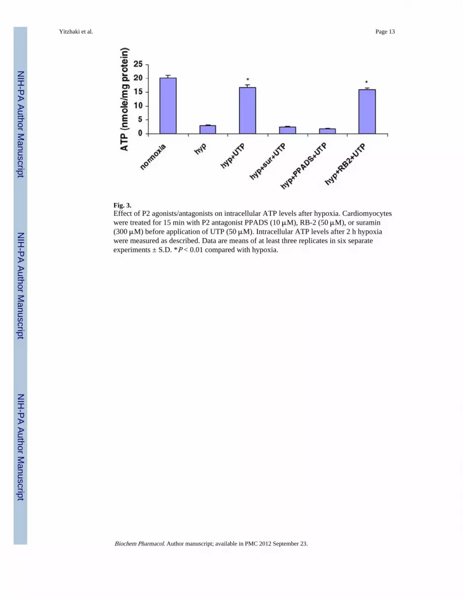
Fig. 3.Effect of P2 agonists/antagonists on intracellular ATP levels after hypoxia. Cardiomyocyteswere treated for 15 min with P2 antagonist PPADS (10 μM), RB-2 (50 μM), or suramin(300 μM) before application of UTP (50 μM). Intracellular ATP levels after 2 h hypoxiawere measured as described. Data are means of at least three replicates in six separateexperiments ± S.D. *P < 0.01 compared with hypoxia.
Yitzhaki et al. Page 13
Biochem Pharmacol. Author manuscript; available in PMC 2012 September 23.
NIH
-PA Author Manuscript
NIH
-PA Author Manuscript
NIH
-PA Author Manuscript

Fig. 4.Effect of UTP on cardiomyocyte morphology in hypoxic conditions. Six-day-old culturedcardiomyocytes under normoxic conditions were subjected to 2 h hypoxic environment orpretreated with 50 μM UTP and then subjected to hypoxia. One group of these cells wasstained with propidium iodide (red), which marks damaged cells, and with Hoechst 33342(blue), which stains live-cell nuclei (first column). A second group of cells was stained withhematoxylin and eosin (second column). A third group of cells was stained with neutral red,which stains lysosomes (third column). The results shown are representative of sixexperiments. Bars = 10 μm.
Yitzhaki et al. Page 14
Biochem Pharmacol. Author manuscript; available in PMC 2012 September 23.
NIH
-PA Author Manuscript
NIH
-PA Author Manuscript
NIH
-PA Author Manuscript

Fig. 5.Effect of antagonists on the [Ca2+]i response. Indo-1-loaded cardiomyocytes were pretreatedwith UTP (A) or with P2 antagonist PPADS (10 μM, B), RB-2 (50 μM, C), or suramin (300μM, D) before application of UTP (50 μM). The fluorescence ratio of 410:490 nm, which isproportional to changes in Ca2+ levels, is demonstrated. One experiment shown isrepresentative of seven.
Yitzhaki et al. Page 15
Biochem Pharmacol. Author manuscript; available in PMC 2012 September 23.
NIH
-PA Author Manuscript
NIH
-PA Author Manuscript
NIH
-PA Author Manuscript

Fig. 6.Effect of UTP on [Ca2+]i elevation in Ca2+-free medium or in the presence of PLC inhibitor.Indo-1-loaded cardiomyocytes were exposed to 2 μM U73122, a PLC inhibitor (A), or to2APB an IP3 receptor inhibitor (50 μM) (B) 15 min before UTP (50 μM) application. Thechanges in Ca2+ levels are demonstrated. One experiment shown is representative of five.
Yitzhaki et al. Page 16
Biochem Pharmacol. Author manuscript; available in PMC 2012 September 23.
NIH
-PA Author Manuscript
NIH
-PA Author Manuscript
NIH
-PA Author Manuscript

Fig. 7.Effect of UTP on [Ca2+]i accumulation during hypoxia. Cardiomyocytes received UTP for15 min just before hypoxia (B) or 24 h before hypoxia (C). Calcium was monitored duringhypoxia with indo-1. The fluorescence ratio of 410:490 is demonstrated. Each recording wasdone for 10 s at times 0, 20, 40, and 60 min after the initiation of the hypoxia. The effect ofhypoxia alone is also shown (A). One experiment shown is representative of five.
Yitzhaki et al. Page 17
Biochem Pharmacol. Author manuscript; available in PMC 2012 September 23.
NIH
-PA Author Manuscript
NIH
-PA Author Manuscript
NIH
-PA Author Manuscript

NIH
-PA Author Manuscript
NIH
-PA Author Manuscript
NIH
-PA Author Manuscript
Yitzhaki et al. Page 18
Table 1
Effect of uracil derivatives or P2 receptors antagonists on cardiomyocytes subjected to hypoxia
Treatment LDH release (%)
Normoxia 4.2 ± 0.6
Hyp 40 ± 2.4
Hyp + UTP 10 ± 1.6*
Hyp + uridine 29 ± 2.5
Hyp + UDP 36 ± 2.6
Hyp + 5-HD 37 ± 0.6
Hyp + UTP + 5-HD 12 ± 1.6*
Hyp + UTP + PPADS 32 ± 3.6
Hyp + UTP + suramin 25 ± 3.5
Hyp + UTP + RB-2 11 ± 0.8*
Cardiomyocytes were treated with 50 μM uridine, UDP, or UTP or with P2 receptor antagonist PPADS (10 μM), RB-2 (50 μM), or suramin (300μM) or with 5-HD, a mitochondrial KATP channel inhibitor (300 μM), for 15 min before hypoxia. The cells were then washed twice and
subjected to hypoxia for 2 h in glucose-free PBS, at 37 °C. The amount of LDH released to the medium was determined and compared to the totalactivity of control homogenate (100%). Data are means of at least three replicates in three separate experiments ± S.D.
*P < 0.01 compared with hypoxia.
Biochem Pharmacol. Author manuscript; available in PMC 2012 September 23.


![Dysfunction of Diastolic [Ca2+] in Cardiomyocytes Isolated From Chagasic Patients](https://img.pdfslide.net/doc/110x75/63547b7ab17b833cd801f904/dysfunction-of-diastolic-ca2-in-cardiomyocytes-isolated-from-chagasic-patients.jpg)


![Collision-induced dissociation mechanisms of [Li(uracil)]+](https://img.pdfslide.net/doc/110x75/634c7bb41983efcda60572c0/collision-induced-dissociation-mechanisms-of-liuracil.jpg)


