Embed Size (px)
Citation preview

APPLIED AND ENVIRONMENTAL MICROBIOLOGY, Mar. 2011, p. 2153–2160 Vol. 77, No. 60099-2240/11/$12.00 doi:10.1128/AEM.02164-10Copyright © 2011, American Society for Microbiology. All Rights Reserved.
Iodide Accumulation by Aerobic Bacteria Isolated from SubsurfaceSediments of a 129I-Contaminated Aquifer at the Savannah
River Site, South Carolina�
Hsiu-Ping Li,1 Robin Brinkmeyer,1 Whitney L. Jones,2 Saijin Zhang,1 Chen Xu,1Kathy A. Schwehr,1 Peter H. Santschi,1 Daniel I. Kaplan,2 and Chris M. Yeager2*
Department of Marine Sciences, Texas A&M University, Galveston, Texas 77553,1 and Savannah River National Laboratory,Aiken, South Carolina 298082
Received 13 September 2010/Accepted 14 January 2011
129I is of major concern because of its mobility in the environment, excessive inventory, toxicity (it accu-mulates in the thyroid), and long half-life (�16 million years). The aim of this study was to determine ifbacteria from a 129I-contaminated oxic aquifer at the F area of the U.S. Department of Energy’s SavannahRiver Site, SC, could accumulate iodide at environmentally relevant concentrations (0.1 �M I�). Iodideaccumulation capability was found in 3 out of 136 aerobic bacterial strains isolated from the F area that wereclosely related to Streptomyces/Kitasatospora spp., Bacillus mycoides, and Ralstonia/Cupriavidus spp. Two previ-ously described iodide-accumulating marine strains, a Flexibacter aggregans strain and an Arenibacter troitsensisstrain, accumulated 2 to 50% total iodide (0.1 �M), whereas the F-area strains accumulated just 0.2 to 2.0%.Iodide accumulation by FA-30 was stimulated by the addition of H2O2, was not inhibited by chloride ions (27mM), did not exhibit substrate saturation kinetics with regard to I� concentration (up to 10 �M I�), andincreased at pH values of <6. Overall, the data indicate that I� accumulation likely results from electrophilicsubstitution of cellular organic molecules. This study demonstrates that readily culturable, aerobic bacteria ofthe F-area aquifer do not accumulate significant amounts of iodide; however, this mechanism may contributeto the long-term fate and transport of 129I and to the biogeochemical cycling of iodine over geologic time.
129I is a major by-product of nuclear fission that is of concernbecause of its mobility in the environment, excessive inventory,long half-life (�16 million years), and potential toxicity due tobioaccumulation through the food chain and bioconcentrationin the thyroid gland (16, 17, 22). Currently, 146 Ci of 129I isinventoried in soils at two U.S. Department of Energy (DOE)sites, the Hanford Site and the Savannah River Site (SRS),where it has been identified as a key risk driver in contami-nated soils and groundwater (21, 24). Furthermore, the globalinventory of 129I will increase significantly if just a fraction ofthe expected “Nuclear Renaissance” is realized (e.g., betweenChina and India alone, 50 to 60 new nuclear reactors areexpected to come online by the year 2020) (16, 50). Thus, it iscritical to understand the environmental behavior of 129I inorder to rigorously assess storage and disposal options forcurrent and future stockpiles of 129I.
In general, little information is available about the chemicalproperties and mobility of iodine in subsurface aquifers, par-ticularly its tendency to form organo-iodine or its mobility asorgano-iodine. However, our own measurements of groundwa-ter from several of the F-area aquifer bore holes found thatorgano-iodine could account for up to 25% of total iodine (43,52). The various isotopes of iodine can be strongly bound tomacromolecular organic matter, which can significantly de-crease or increase its transport, bioavailability, and transfer tohumans, depending on the molecular weight and physicochem-
ical properties of the resulting iodine-organic matter species(15, 19, 41–43, 49).
Microbial activity has been linked to the production of or-gano-iodine and sorption of iodine to soil, and a small butgrowing body of literature has implicated microbial oxidases,perhydrolases, and particularly peroxidases in the halogena-tion of soil organic matter (4, 10, 20, 23, 27, 32–34, 37, 38). Yet,details concerning the mechanisms and bacterial species orgroups involved are lacking. The most-recent advances in re-search concerning microbial-iodine interactions have beencontributed by Amachi et al. (1–8, 18). In a series of papers,Amachi’s research group has isolated (i) iodide-accumulatingbacteria (IAB) from marine sediments that concentrate iodideby a factor of 6 � 103 (6), (ii) iodide-oxidizing bacteria fromseawater and natural gas brine water that transform iodide intoI2 and volatile organo-iodine species (8), and (iii) iodide-meth-ylating bacteria from a variety of soil and seawater samples (4,5). For the marine IAB, Amachi et al. (2) proposed a modelwhereby extracellular H2O2, generated by glucose oxidase, ox-idizes iodide to I2 or hypoiodous acid (HIO) via an unidenti-fied haloperoxidase. HIO is then transported across the cellmembrane via a facilitated diffusion-type mechanism. Onceinside the cell, HIO either is reduced to iodide or forms or-gano-iodine.
Notably, iodide-oxidizing and iodine-accumulating strainswere readily obtained from marine or brine waters but notfrom surface soils (22 samples from a rice paddy, upland field,and forest soils) (6, 7). The only study to date examining thepotential for microorganisms from subsurface aquifers to as-sociate with iodine was performed using enrichments fromanoxic natural gas formation waters that contain exceedingly
* Corresponding author. Mailing address: Savannah River NationalLaboratory, 999-W, Aiken, SC 29808. Phone: (803) 819-8403. Fax:(803) 819-8432. E-mail: [email protected].
� Published ahead of print on 28 January 2011.
2153
high concentrations of iodine (�120 mg liter�1) and a set of 16anaerobic bacterial strains obtained from culture collectionsthat included sulfate and iron reducers, denitrifiers, and me-thanogens (3). The results indicated very limited adsorption oraccumulation of iodine by anaerobic microorganisms.
Overall, the research conducted thus far demonstrates thatmicrobial activity is an important factor in iodine biogeochemi-cal cycling (particularly in enhancing iodine binding to high-molecular-weight organic matter) and leads us to hypothesizethat select soil and sediment bacteria could be capable ofinfluencing the chemical behavior of iodide (the most commonform of 129I found in groundwater) via accumulation, volatil-ization, and oxidation under aerobic conditions. In particular,we wish to develop an understanding of the role that microor-ganisms play in the formation of organo-iodine in the subsur-face and how they influence iodine mobility. As an initial steptoward this overarching goal, the aim of this study was todetermine if naturally occurring bacteria from a 129I-contam-inated oxic aquifer at the Savannah River Site, SC, could ac-cumulate iodide at environmentally relevant concentrations.
MATERIALS AND METHODS
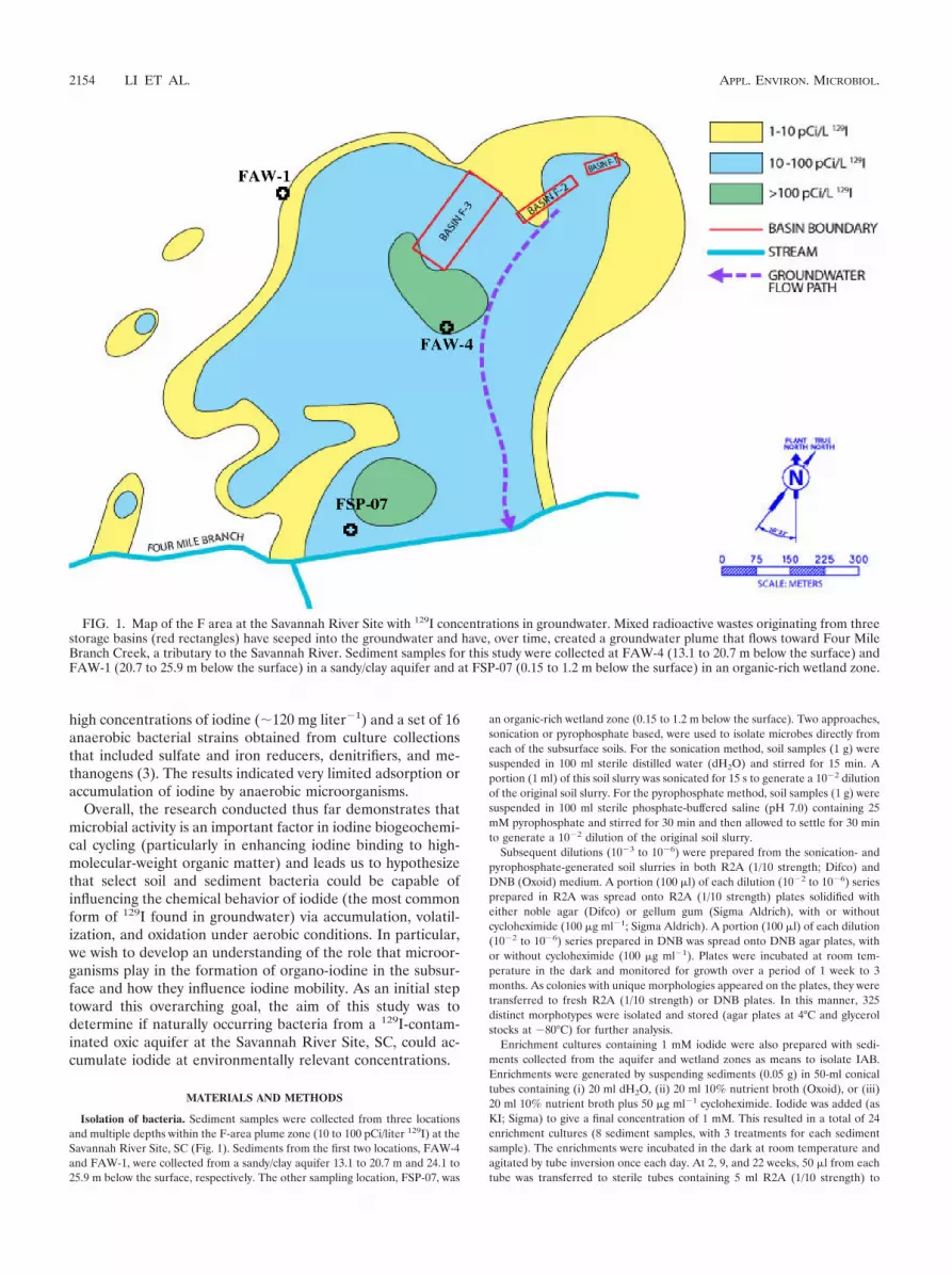
Isolation of bacteria. Sediment samples were collected from three locationsand multiple depths within the F-area plume zone (10 to 100 pCi/liter 129I) at theSavannah River Site, SC (Fig. 1). Sediments from the first two locations, FAW-4and FAW-1, were collected from a sandy/clay aquifer 13.1 to 20.7 m and 24.1 to25.9 m below the surface, respectively. The other sampling location, FSP-07, was
an organic-rich wetland zone (0.15 to 1.2 m below the surface). Two approaches,sonication or pyrophosphate based, were used to isolate microbes directly fromeach of the subsurface soils. For the sonication method, soil samples (1 g) weresuspended in 100 ml sterile distilled water (dH2O) and stirred for 15 min. Aportion (1 ml) of this soil slurry was sonicated for 15 s to generate a 10�2 dilutionof the original soil slurry. For the pyrophosphate method, soil samples (1 g) weresuspended in 100 ml sterile phosphate-buffered saline (pH 7.0) containing 25mM pyrophosphate and stirred for 30 min and then allowed to settle for 30 minto generate a 10�2 dilution of the original soil slurry.
Subsequent dilutions (10�3 to 10�6) were prepared from the sonication- andpyrophosphate-generated soil slurries in both R2A (1/10 strength; Difco) andDNB (Oxoid) medium. A portion (100 �l) of each dilution (10�2 to 10�6) seriesprepared in R2A was spread onto R2A (1/10 strength) plates solidified witheither noble agar (Difco) or gellum gum (Sigma Aldrich), with or withoutcycloheximide (100 �g ml�1; Sigma Aldrich). A portion (100 �l) of each dilution(10�2 to 10�6) series prepared in DNB was spread onto DNB agar plates, withor without cycloheximide (100 �g ml�1). Plates were incubated at room tem-perature in the dark and monitored for growth over a period of 1 week to 3months. As colonies with unique morphologies appeared on the plates, they weretransferred to fresh R2A (1/10 strength) or DNB plates. In this manner, 325distinct morphotypes were isolated and stored (agar plates at 4°C and glycerolstocks at �80°C) for further analysis.
Enrichment cultures containing 1 mM iodide were also prepared with sedi-ments collected from the aquifer and wetland zones as means to isolate IAB.Enrichments were generated by suspending sediments (0.05 g) in 50-ml conicaltubes containing (i) 20 ml dH2O, (ii) 20 ml 10% nutrient broth (Oxoid), or (iii)20 ml 10% nutrient broth plus 50 �g ml�1 cycloheximide. Iodide was added (asKI; Sigma) to give a final concentration of 1 mM. This resulted in a total of 24enrichment cultures (8 sediment samples, with 3 treatments for each sedimentsample). The enrichments were incubated in the dark at room temperature andagitated by tube inversion once each day. At 2, 9, and 22 weeks, 50 �l from eachtube was transferred to sterile tubes containing 5 ml R2A (1/10 strength) to
FIG. 1. Map of the F area at the Savannah River Site with 129I concentrations in groundwater. Mixed radioactive wastes originating from threestorage basins (red rectangles) have seeped into the groundwater and have, over time, created a groundwater plume that flows toward Four MileBranch Creek, a tributary to the Savannah River. Sediment samples for this study were collected at FAW-4 (13.1 to 20.7 m below the surface) andFAW-1 (20.7 to 25.9 m below the surface) in a sandy/clay aquifer and at FSP-07 (0.15 to 1.2 m below the surface) in an organic-rich wetland zone.
2154 LI ET AL. APPL. ENVIRON. MICROBIOL.
generate a 10�2 dilution. Dilutions of 10�4 and 10�6 were then prepared in R2A(1/10 strength), and 25 �l of each dilution was spread on both 25% DNB-IS (ISconsists of 1.2 g liter�1 KI and 1g liter�1 soluble starch) and 25% R2A-IS agarplates. In this manner, 29 distinct morphotypes were isolated and stored (agarplates at 4°C and glycerol stocks at �80°C) for further analysis.
16S rRNA gene sequencing and phylogenetic analysis. To determine thetaxonomic identity of the isolates, a portion of the 16S rRNA gene was amplifiedand sequenced. Crude bacterial lysate was collected by placing a small amount ofisolated colonies into 10 �l Tris-EDTA (TE) buffer (pH 7.5) with sterile pipettetips. Amplification was performed using 1 �l of the crude lysate as a template in50-�l reaction mixtures containing 5 �l 10� buffer, MgCl2 (2.5 mM), the 27fprimer (5�-GAGTTTGATCMTGGCTCAG-3�) (10 pmol), the 1492r primer (5�-GGTTACCTTGTTACGACTT-3�) (10 pmol), deoxynucleoside triphosphates(200 �M each), and 1.25 U of HotStar Taq DNA polymerase (Qiagen). Reac-tions were amplified in a PTC-100 thermocycler (MJ Research, Inc.) as follows:95°C for 10 min and 32 cycles of 95°C for 1 min, 45°C for 45 s, and 72°C for 1 min,followed by a 7-min elongation step. PCRs were visualized on a 1.2% agarose gelto ensure amplification of a single product of the expected size. The 16S rRNAgene amplicon was purified with a MinElute PCR purification kit (Qiagen), andthe concentration of the purified product was adjusted to �15 ng �l�1 in dH2Ousing a Nanodrop 1000 spectrophotometer (Thermo Scientific). Sequencing ofthe purified PCR products was performed by the Georgia Sequencing Facility atthe University of Georgia, Athens, GA.
Phylogenetic similarity of 16S rRNA sequences was first determined withBLAST searches of the GenBank (http://www.ncbi.nlm.nih.gov/genbank/index.html) and the RDP classifier (13), followed by maximum-likelihood reconstruc-tion of phylogenetic trees with ARB (48).
Culture conditions. Two marine iodide-accumulating bacterial (IAB) strains,Flexibacter aggregans NBRC15975 and Arenibacter troitsensis JCM11736, werepurchased from the NITE Biological Research Center, Chibin, Japan, and usedas reference strains for the iodide accumulation assay. The two reference strainsand all microbial isolates from the F area were grown on marine agar 2216(Difco) and 1/4-strength R2A agar medium (EMD), respectively.
Iodide accumulation screening. Two methods using 125I� as a tracer werecombined to determine the iodide-accumulating ability from F-area microbes.The first used autoradiography, similar to the procedure of Amachi et al. (7). Amixture of I�, 0.016 �M stable I� (as NaI; Sigma), and 0.80 kBq ml�1 125I� wasapplied evenly to R2A agar plates. Microbial isolates from the F area wereinoculated onto the plates using sterile toothpicks and incubated for 3 days in thedark at room temperature (�27°C). A small portion of microbial colonies, �0.25mm in diameter, was transferred with sterile toothpicks into 7-ml glass vialscontaining 4 ml of scintillation cocktail (Ecolume). After the colonies werevortexed for 1 min, the radioactivity of accumulated 125I was determined by aliquid scintillation counter (Beckman Coulter LS6500) for 10 min as a prelimi-nary screen. Colonies testing positive for 125I accumulation were then transferredfrom the agar plates via a traditional plate lift technique onto a nitrocellulosemembrane filter (82-mm diameter; Whatman Optitran BA-S 85). As a controlfor the background, liquid on the agar surface was also transferred onto thenitrocellulose membrane. The membranes were dried at room temperature for30 min and then exposed to a maximum-sensitivity autoradiography film (KodakBioMax) at �20°C for 14 days in the dark. Film was processed with developer,a deionized-water stop bath, and fixer according the manufacturer’s instructions(Kodak).
For the second method for assessment of iodide accumulation, microbialisolates from the F area and the two reference marine IAB strains (the F.aggregans and A. troitsensis strains) were cultivated in 50-ml conical tubes con-taining 5 ml of nutrient broth and marine broth, respectively. Stable I� wasadded to the cultures at a concentration (0.1 �M) that reflected the ambientconcentration of 127I� (stable iodine) in the F area. 125I� (1.33 kBq liter�1) wasadded to the cultures as the tracer. Incubations were performed at room tem-perature in the dark with continuous shaking (150 rpm). After a 24-h incubationperiod, a subsample (100 �l) was collected and was immediately transferred intoa scintillation vial. Radioactivity of 125I was determined by liquid scintillationcounting for 10 min and referred to as “125I activity in the microbial culture.” Toinvestigate the effect of hydrogen peroxide on cellular iodide accumulation,H2O2 (final concentration, 5 mM) was added to a portion (4 ml) of the cell-iodide mixture after the initial 24-h incubation period, and the incubation wascontinued for an additional 4 to 24 h. Portions of the cell suspension (900 �l)were collected immediately after the initial 24 h of incubation (before H2O2
addition) or 4 or 24 h after H2O2 addition. The collected cell suspension wascentrifuged at 16,000 � g for 20 min at 4°C. The supernatant was discarded, andthe cell pellet was washed 3 times and suspended in 900 �l of fresh medium(without added stable I� and 125I�). To determine “cell-associated 125I,” a
portion (100 �l) of the washed cell suspension was measured by scintillationcounting for 10 min (count errors were consistently below 20%). The iodide-accumulating ability (percent accumulation) of microbial cells was calculated asthe ratio of “cell-associated 125I” versus “125I activity in the microbial culture.”Cell densities in aliquots were measured at 600 nm with a spectrophotometer(Turner SP8001). Correlations of bacterial dry weight with optical density at 600nm (OD600) for each target bacterium were obtained during different periods ofexponential growth through linear regression, resulting in an r2 value higher than0.98 for each strain.
Effect of chloride on iodide-accumulating abilities. Bacteria were grown in twotypes of aqueous minimal media, M9 and Cl�-deficient M9 (M9X). M9 con-tained 47.75 mM Na2HPO4, 22.04 mM KH2PO4, 8.56 mM NaCl, 18.70 mMNH4Cl, 2 mM MgSO4, 0.1 mM CaCl2, 22.22 mM glucose, and 0.5% yeast extract.M9X was prepared by replacing NaCl, NH4Cl, and CaCl2 with the same con-centrations of Na2SO4, (NH4)2SO4, and Ca(OH)2, respectively. Cells grown inM9 and M9X media were exposed to 2 different concentrations of stable I� (0.1and 10 �M) while maintaining the same concentration of 125I� (1.33 kBq li-ter�1). Incubations were performed in the dark at room temperature with con-tinuous shaking at 150 rpm for 24 h. H2O2 (final concentration, 5 mM) was thenadded to the microbial culture. After an additional 24-h incubation, aliquotswere collected for the determination of cellular 125I activity and cell density asdescribed above.
Effect of pH and sodium azide on iodide-accumulating abilities. To evaluateiodide accumulation as a function of pH, IAB from the F area were grown in theM9 medium containing 0.5% yeast extract at room temperature in the dark withshaking (150 rpm). When the optical density (600 nm) of the cultures reached�0.8, cells were harvested by centrifugation (3,500 � g at 20°C for 15 min) andwashed 2 times with pH-adjusted minimal medium (pH 4 to 9). The minimalmedium contained 8.6 mM NaCl, 18.7 mM NH4Cl, 2 mM MgSO4, 0.1 mMCaCl2, 22.2 mM glucose, and 50 mM KH2PO4. Acetate buffer, phosphate buffer,and borate buffer were used to adjust the pH from 4 to 5, from 6 to 8, and to 9,respectively. To avoid bursting or shrinking of cells by transferring them from M9to pH-adjusted medium, the ionic strength of pH-adjusted medium was main-tained at 177 mM, which was the same ionic strength as in the M9 medium. Cellpellets were suspended in pH-adjusted minimal medium containing stable I�
(0.1 �M), 125I� (1.33 kBq liter�1) and H2O2 (5 mM).In order to investigate the role of heme-containing enzymes or other active cell
processes on iodide accumulation, a parallel assay was performed with theaddition of sodium azide (NaN3). NaN3 (10 mM) was added to the cell suspen-sion to inhibit enzymatic activities before addition of I� and H2O2. Cell suspen-sions were then incubated at room temperature in the dark for 24 h, after whichaliquots were collected for the determination of 125I activity and cell density asdescribed above.
Iodide desorption. A procedure similar to that of MacLean et al. (30), withslight modifications, was performed to determine if iodine desorption couldoccur after accumulation. After iodide accumulation at pH 4 to 9 (describedabove), cells were collected via centrifugation (16,000 � g for 20 min at 4°C),suspended in M9 medium (pH 7), and incubated with end-over-end shaking (150rpm) for 2 h. Aliquots were then collected for the determination of 125I activityand cell density as described above.
Statistical analyses. Analyses of variance (ANOVA) were performed to eval-uate the significant differences in iodide-accumulating abilities as a function ofiodide concentrations, as well as the presence versus absence of chloride ions.Tukey’s post hoc tests were used to compare means of values for iodide-accu-mulating abilities from significant (P � 0.05) ANOVA test results. The statisticalanalyses were performed using SPSS 16.0 (SPSS Institute, Inc.).
RESULTS
Isolation and phylogenetic analysis of F-area soil bacteria.A total of 325 aerobic microbes were directly isolated fromsubsurface sediments of the F area at the Savannah River Site(Fig. 1). Of these isolates, 32% were cultured from sandy/claysediments of an aquifer (sites FAW-1 and FAW-4, 13.1 to25.9 m below the surface) and 29% and 39% were culturedfrom the dark, organic rich sediments of the seep zone (siteFSP-07), 0.15 to 1 m and 1 to 1.2 m below the surface, respec-tively. Analysis of the 16S rRNA gene revealed that the isolateswere members of four phyla within the domain Bacteria. Themajority of isolates (55%) were classified as Proteobacteria,
VOL. 77, 2011 IODIDE-ACCUMULATING TERRESTRIAL BACTERIA 2155

followed by Firmicutes (20%), Actinobacteria (17%), and Bac-teroidetes (8%). No obvious trends relating the phylogeny ofthe isolates to their environmental source (seep zone versussand/clay aquifer or depth of the sediment) or the isolationmethod employed (pyrophosphate versus sonication or gellangum versus noble agar) were observed (data not shown).
A yellow coloration was noted on the tubes of 2/24 enrich-ment cultures (8 sediment sources, with 3 enrichment condi-tions for each culture; see Materials and Methods) after 22weeks of incubation in the dark. A strong I2 smell was alsodetected after the caps of the discolored conical tubes wereopened, indicating the transformation of I� to I2 (8). The twoenrichment cultures that exhibited yellow coloration were de-rived from the seep zone (FSP-07) sediments collected 0.15 to1 and 1 to 1.2 m below the surface, and both were incubated indH2O with 1 mM I�. Isolation efforts from the 24 enrichmentcultures yielded 29 distinct morphotypes. Analysis of 16SrRNA gene sequences revealed that these 29 strains weremembers of the same 4 phyla, as the isolates recovered directlyfrom sediments. The majority of isolates from the enrichmentcultures (76%) were classified as Proteobacteria, followed byActinobacteria (14%), Bacteroidetes (7%), and Firmicutes (3%).
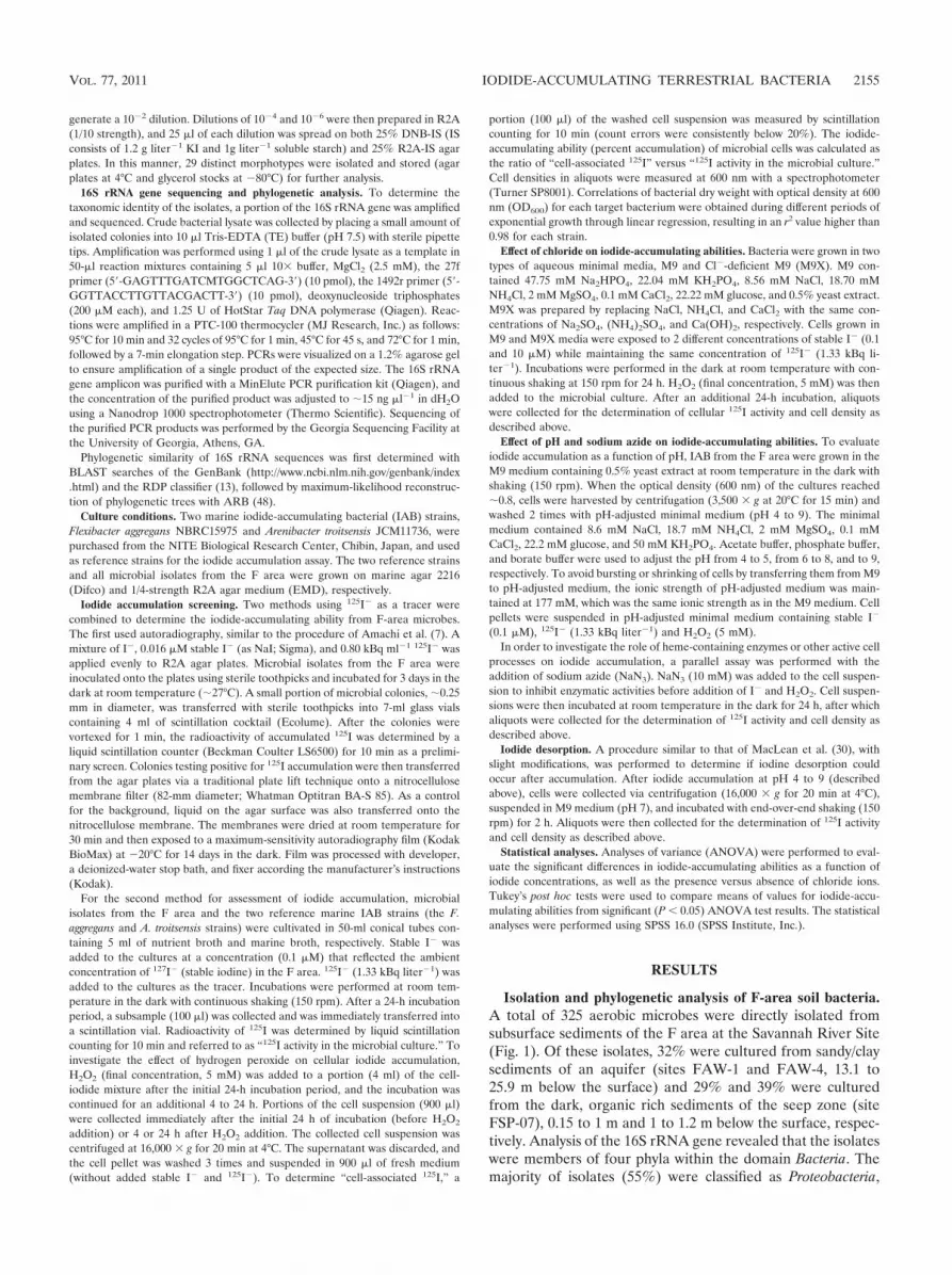
Screening for iodide-accumulating strains. Forty-two strains,representing the phylogenetic diversity of isolates from F-areasoils and enrichment cultures, were screened for their ability toaccumulate 125I� during growth on R2A agar. Cell materialfrom 14 strains exhibited detectable radioactivity by liquid scin-
tillation counting following growth in the presence of 125I�.Cell mass from colonies of these 14 isolates and liquid from thesurface of the 125I� R2A plates was transferred to a nitrocel-lulose membrane, which was exposed to film for 14 days. Anobvious solid black circle was observed where the film hadbeen exposed to cell material from strain FA-30, indicatingthat this strain accumulated iodide relatively strongly (Fig. 2).Autoradiographic impressions from four of the isolates, FA-5,FA-16, FA-17B, and FA-18, showed a slightly darker region inthe center of each image, whereas impressions from FA-15,FA-2B-NB, and FA-2B-B* showed a dark halo around theouter border of each of their respective images (Fig. 2). Thesedistinct, strain-specific autoradiographic patterns were repeat-able, suggesting that the strains accumulate or interact with125I� differently.
A second method, in which bacteria were grown in liquidmedium containing 125I�, was used to assess iodide accumu-lation among 139 of the F-area isolates (the 42 strains initiallyscreened by autoradiographic analysis of colony material and97 additional strains) as well as the two I�-accumulating ma-rine bacterial strains, the F. aggregans strain and the A. troit-sensis strain. With the use of this method, nine of the F-areastrains exhibited the potential to accumulate iodide. How-ever, upon reexamination using the same procedure with 3additional washes, only one F-area strain (FA-30) and themarine strains consistently exhibited appreciable 125I-accu-mulating abilities (Table 1).
It has been reported that H2O2 plays a role in iodide accu-mulation and oxidation by marine bacteria (6, 18); thus, iodideaccumulation was assessed for the same 139 strains and 2marine IAB strains in liquid cultures as described above, ex-cept that H2O2 was added to the cultures either 4 or 24 h priorto harvesting of the cells. H2O2 addition resulted in greateriodide accumulation for FA-30 and the two marine strains andenabled the identification of two additional iodide-accumulat-ing strains, FA-2C-B* and FA-191 (Table 1). In each of thesestrains, iodide accumulation was greater in cells that had beenexposed to H2O2 for a longer period of time (24 versus 4 h).However, the three IAB strains isolated from the F area, FA-30, FA-2C-B*, and FA-191, exhibited an iodide-specific accu-mulation that was 2 orders of magnitude less than that ob-served for the marine IAB strains, the F. aggregans and A.troitsensis strains (Table 1). Among the F-area isolates, theonly strain that exhibited an iodide accumulation phenotype
FIG. 2. Autoradiographic image of cell material from 14 F-areaisolates (FA-30 was blotted in duplicate) that had been grown on R2Aplates containing 125I�. The “Agar Surface” dot shows the backgroundradioactivity from the surface of an R2A plate containing 125I�.
TABLE 1. Iodide accumulation by F-area bacterial isolates and two known IAB isolates
Bacterial isolate(s)Without H2O2 With H2O2 added at 4 h With H2O2 added at 24 h
Cell activity (cpm)a Accumulation (%)b Cell activity (Bq/ml) Accumulation (%) Cell activity (Bq/ml) Accumulation (%)
F. aggregans NBRC15975c 726 2.410 637 2.096 2,097 7.246A. troitsensis JCM11736c 10,170 25.482 10,365 25.609 17,635 41.489FA-30 105 0.383 120 0.428 315 1.100FA-2C-B* 33 0.157 61 0.296 121 0.593FA-191 27 0.099 40 0.154 67 0.251Other strainsd 31 (�18) 0.143 (�0.087) 26 (�13) 0.125 (�0.066) 28 (�17) 0.134 (�0.082)
a Activity associated with cells washed 3 times following incubation of cells in the presence of 125I for 24, 28, or 56 h. H2O2 was added after 24 h for the 28- and 56-hincubations.
b (Number of cpm 125I in cell pellet after incubation/number of cpm 125I in supernatant after incubation) � 100.c Marine strains previously identified as IAB (6).d Average values � SD for the 133 strains that did not consistently accumulate iodide.
2156 LI ET AL. APPL. ENVIRON. MICROBIOL.

with both the plate and the liquid culture assays was FA-30(among the F-area isolates, FA-30 also exhibited the strongest125I� accumulation phenotype with the use of both assays).
Phylogenic analysis of the partial 16S rRNA genes (750 to900 bp) from FA-30, FA-2C-B*, and FA-191 revealed thatthese strains were members of the Actinobacteria, Firmicutes,and Proteobacteria phyla, respectively. FA-30 was most closelyrelated to various Streptomyces and Kitasatospora species (16SrRNA gene similarity, �94%), FA-2C-B* was closely relatedto Bacillus mycoides L2S8 (EU221418; 98% similarity), andFA-191 was most closely related to various Ralstonia and Cu-priavidus species (97 to 98% similarity). FA-191 and FA-30were cultured from seep zone (FSP-07) sediments, 0.15 to 1and 1 to 1.2 m below the surface, respectively, whereas strainFA-2C-B* was isolated from one of the I� enrichment cul-tures. No IAB were found among the isolates obtained fromthe deeper, oligotrophic sandy/clay aquifer material.
Impact of chloride on iodide accumulation. To evaluatewhether Cl� present in M9 medium could inhibit iodide accu-mulation by the F-area IAB, cells of FA-30, FA-2C-B*, andFA-191 were incubated in medium with and without addedchloride (27 mM total Cl� concentration, added as 8.56 mMNaCl, 18.70 mM NH4Cl, and 0.1 mM CaCl2). Strains FA-30and FA-2C-B* accumulated more iodide in the presence ofchloride (P � 0.05), whereas chloride did not have a significanteffect on I� accumulation in strain FA-191 (Fig. 3).
Impact of pH and sodium azide on iodide accumulation.The iodine-accumulating ability of strains FA-30, FA-2C-B*,and FA-191 in liquid cultures grown in pH-adjusted medium(pH 4 to 9) containing iodide (0.1 �M) was evaluated. BetweenpH 4 and pH 6, there were significant decreases in cellulariodine content in strains FA-30 and FA-191 as the pH in-creased (Fig. 4). Above pH 6, the cellular iodine content waslow, �0.1 �g/g dry cell weight, and either gradually decreasedwith increasing pH (FA-30) or remained at a constant low levelwith increasing pH (FA-191). A relationship between pH and
iodide accumulation was not observed for FA-2C-B* (data notshown).
Strains FA-30 and FA-191 were tested in a parallel pH assaywhere sodium azide (NaN3; 10 mM) was included. Both strainsexhibited lower I� accumulation when incubated with NaN3,particularly below pH 6.0 (Fig. 4). At pH 4.0, cells of FA-191and FA-30 accumulated 40% and 90% less I�, respectively, inthe presence of NaN3. At pH levels of �6.0, I� accumulationwas essentially unaffected by NaN3 (it should be noted, how-ever, that the cellular iodine content of cells incubated inmedium with a pH value of �6.0 was barely above backgroundlevels).
Desorption of iodine from IAB. Desorption experimentswere conducted with strains FA-30 and FA-191 to evaluate thenature of the interaction between iodide and the cells. Desorp-tion of iodine as a function of pH was not observed in cells ofFA-30 or FA-191 (only �10% variation in the cellular iodinecontent of the cells before and after the desorption step) (datanot shown).
DISCUSSION
Mobility of radioactive iodine in the subsurface environmentis affected by the iodine’s chemical speciation and interactionswith soil constituents, including minerals, organic matter, andmicroorganisms. Based on thermodynamic principles, the mainiodine species in SRS F-area groundwater and sediments
FIG. 3. Iodide accumulation by FA-30, FA-2C-B*, and FA-191 inM9 medium containing 27 mM chloride ions (gray bars) versus that inmedium without chloride salts (white bars). The experiment was per-formed using 0.1 �M iodide. Bars represent average amounts of iodideaccumulated per cell culture biomass (OD600), and error bars showstandard deviations (SD) (n 3).
FIG. 4. Correlation between pH and iodide accumulation by FA-30(A) and FA-191 (B) in the presence (squares) or absence (triangles) ofsodium azide. Symbols represent average amounts of iodide accumu-lated per cell culture biomass (�g g dry cell weight�1), and error barsshow standard deviations (n 3).
VOL. 77, 2011 IODIDE-ACCUMULATING TERRESTRIAL BACTERIA 2157

should be iodide, which is thought to have the highest subsur-face mobility, i.e., to be least sorbed or taken up by sediments,compared to iodate and organo-iodine (33, 40, 43). However,organo-iodine has been found to contribute a significant frac-tion (up to 25%) of total iodine in groundwater from the Farea (43, 52). The extent to which iodide binds to or is incor-porated within bacterial cells in oxic subsurface aquifers hasnot previously been investigated; thus, we tested the iodide-accumulating ability of 139 phylogenetically distinct bacterialisolates from F-area sediments.
Three aerobic bacterial strains from the F area, FA-30, FA-2C-B*, and FA-191, were shown to accumulate iodide. Iodideaccumulation by these three strains was significantly differentfrom the background value observed for 136 other strainsevaluated (consistently 2 to 10 times higher), and in the case ofFA-30, this result was validated by two different approaches, (i)autoradiography of cell material grown on 125I� agar platesand (ii) liquid scintillation analysis of washed cell material thathad been grown in liquid medium containing 125I�. Comparedto that of IAB isolated from marine sources, however, theiodide-accumulating capacity of the F-area strains was quitesmall. Amachi et al. (6) reported accumulation of 80 to 90% oftotal iodide by various marine strains, including F. aggregansNBRC15975 and A. troitsensis JCM11736, when these strainswere incubated in the presence of 0.1 �M I�. Under ourexperimental conditions (i.e., much more rigorous cell washingand higher pH [7.0 versus 6.0] than those used by Amachi etal.), F. aggregans and A. troitsensis accumulated 2 to 50% totaliodide (0.1 �M), whereas F-area strains FA-30, FA-2C-B*, andFA-191 accumulated 0.2 to 1.5% of the total iodide (0.1 �M).
Another difference between the IAB isolated from F-areasediments and the IAB previously identified from marine sed-iments (6) is that the latter microorganisms were classifiedexclusively within the Flavobacteriaceae family of the Bacte-roidetes, whereas the F-area IAB represented three phyla, Ac-tinobacteria (FA-30, most closely related to Streptomyces andKitasatospora spp.), Firmicutes (FA-2C-B*, a putative Bacillussp.), and Betaproteobacteria (FA-191, closely related to Ralsto-nia/Cupriavidus spp.). As these three phyla are often dominantmembers of terrestrial soil microbial communities (they com-prise �90% of the isolates obtained from F-area sediments)whereas Bacteroidetes spp. are more common in marine envi-ronments, it appears that iodine accumulation is not restrictedto a distinct phylogenetic lineage(s) but rather is manifested inselect bacterial taxa adapted to their respective environments.
There are two mechanisms that provide the most parsimo-nious explanation for iodide accumulation by bacteria. One iselectrostatic adsorption of I� by positively charged functionalgroups, such as amines from proteins and peptides, present onthe surface of the cell. For example, MacLean et al. (30) usedBacillus subtilis to model iodide-bacterium adsorption throughelectrostatic interaction and found that it readily adsorbediodide at pH values of �4 and that iodide could then be quicklydesorbed (�2 h) by raising the pH to 7.0. In that study, anaqueous solution of diluted HNO3 was used as the experimen-tal wash solution, precluding other halide ions from competingwith iodide for electrostatic interaction with bacterial cells. Inthe present study, the three terrestrial IAB strains were incu-bated and maintained in a M9 minimal medium solution thatcontained �27 mM chloride ions, which is �2.7 � 105 times
higher than the iodide concentration (0.1 �M) present in theexperimental assays. Iodide accumulation by FA-30, FA-2C-B*, and FA-191 was not inhibited by chloride ions (27 mM)(Fig. 3). Indeed, at low iodide concentrations (0.1 �M), I�
accumulation by FA-30 and FA-2C-B* was greater in M9 me-dium than in chloride-deficient M9 medium (we note thatthere was a difference in ionic strength between the standardM9 medium and chloride-deficient M9 medium used in thisexperiment). Furthermore, pH-dependent desorption testsfailed to reveal reversibility of the interaction between iodideand cells of FA-30 and FA-191. These results indicate that I�
accumulation by the F-area terrestrial strains was not due toelectrostatic surface adsorption when pH levels were �4.0.
The second possible mechanism for adsorption between io-dide and cellular constituents is electrophilic substitution re-sulting in iodination of organic molecules. Strong electrophilessuch as HOI/I2/I3
� produced by biotic or abiotic processesfrom I� could attack aromatic rings or other organic moietiesof the cell and replace -H with -I to form a stable organo-iodine bond (12, 31, 44). Given the high concentration oforganic matter in a bacterial culture, it is reasonable to expectthat the oxidized I species, i.e., HOI/I2/I3
�, could react with it.Several observations signify that the iodide accumulation phe-notype exhibited by F-area strains FA-30, FA-2C-B*, and FA-191 likely proceeds via this mechanism. First, the iodide-accu-mulating abilities from FA-30, FA-2C-B*, FA-191 were allfacilitated by the addition of H2O2. Since H2O2 is a strongoxidant, I� is readily oxidized to I2/I3
� without enzymaticcatalysis, especially under acidic conditions. However, at thenear-neutral pH values (pH 6 to 7) that were used during thescreening portion of our study, H2O2 consistently stimulatediodide accumulation in just 5 of 141 strains tested (includingthe two marine strains). Thus, it is unlikely that H2O2, itselfacting as an oxidizing agent, transformed I� into a highlyreactive species capable of binding nonspecifically to cell ma-terial (though at lower pH values [�4 to 5], this mechanismcould be more relevant). Alternatively, haloperoxidases, whichare found in animals, plants (including algae), fungi, and bac-teria, utilize H2O2 as a cosubstrate and are considered theprimary enzyme system responsible for nonspecific halogena-tion of organic substrates in nature (11, 45, 46). NaN3 signifi-cantly inhibited I� accumulation in the F-area strains, impli-cating the involvement of an enzymatic driven process, such asan active transport system or heme haloperoxidases, which areNaN3 sensitive (45). The F-area strains also exhibited in-creased I� accumulation with decreasing pH, characteristic ofmany haloperoxidases, which exhibit optimal activity underacidic conditions (14, 36, 38). Although our data support elec-trophilic substitution or internalization rather than electro-static adsorption to the cell surface, the precise nature andlocation of bacterially bound I� in these terrestrial strains andthe accumulation mechanism remain to be determined.
Background concentrations of stable I in F-area groundwa-ter range from 10 to 100 nM, and plume concentrations of 129Iare typically �60 pCi/liter (2.4 nM) at “hot spots” but canreach levels of �900 pCi/liter in the organic-rich seep zone.Our results demonstrate that the majority (98%) of aerobicbacteria isolated from F-area sediments do not accumulateiodide (�0.2% accumulation) at ambient I� concentrations(0.1 �M), and the three IAB strains that were identified accu-
2158 LI ET AL. APPL. ENVIRON. MICROBIOL.

mulate less than 2% I� under environmental conditions (aer-obic; pH 4 to 9; 0.1 �M total I�) associated with most of theF-area plume (at the center of the plume, pH values as low as3.2 have been documented, where electrostatic adsorption ofI� by bacterial cells as demonstrated by MacLean et al. [30]could play a role in I� transport). Our experiments were con-ducted with dense cell cultures (�1 � 109 cells ml�1), whereascell numbers in groundwater from the sandy/clay aquifer of theF area are lower than 1 � 104 cells ml�1 (data not shown). Atthese cell concentrations, cellular accumulation of I� would beexceedingly low. Furthermore, each of the IAB strains identi-fied in this study was isolated from the seep zone sediments,not the sandy/clay aquifer material. These results indicate thatIAB are most likely not responsible for the high fraction oforgano-iodine (up to 25% of total iodine) that has been mea-sured in groundwater of the F-area subsurface aquifer abovethe seep zone (43, 51). However, our ongoing experiments withiodine-oxidizing bacteria from F-area soils thus far indicatethat this pathway for organo-iodine formation is more signifi-cant.
Our multifaceted, carefully controlled approach allowed usto definitively identify an IAB phenotype that was 1 to 2 ordersof magnitude less, in terms of specific iodine accumulationactivity, than that previously established for bacteria from verydifferent environments (i.e., brines). This is important for sev-eral reasons. 129I has an extremely long half-life (�16 millionyears), and its production is increasing each year. The DOEand other entities are tasked with modeling the long-term(centuries to thousands of years) fate and transport of 129I.Over decades or centuries, I� accumulated by bacterial cellsand covalently attached to cellular constituents could conceiv-ably make its way to the organo-iodine pool through cell lysisand possible incorporation into more-refractory organic soilmaterial (e.g., humic or fulvic acids). Even when bacteria,whose biomass typically accounts for 1% or less of sedimentaryorganic matter (such as the F-area seep zone sediments) (25),incorporate less than 2% of iodine into their cells, this processcould contribute appreciably to the organo-iodine pool overthe long term. Similar mechanisms have been proposed toexplain chloride retention in forest soils and peat bogs overdecades to centuries (10). Carefully controlled, long-term col-umn studies are needed to examine the extent that IAB, suchas those identified in this study, affect 129I speciation and mo-bility in F-area seep zone sediments. Finally, uncultivated bac-terial species yet to be discovered from the F area or fungi maybe capable of much higher levels of iodide accumulation (9).We are currently examining that possibility through a microau-toradiography-fluorescence in situ hybridization (MAR-FISH)approach (29).
ACKNOWLEDGMENTS
We thank Melanie Dunn for providing assistance in the pH effectassays.
This work was funded by the U.S. Department of Energy’s Subsur-face Biogeochemical Research Program within the Office of Science(DE-FG02-08ER64567, modification no. 002) and partially supportedby Welch Grant BD0046.
REFERENCES
1. Amachi, S. 2008. Microbial contribution to global iodine cycling: volatiliza-tion, accumulation, reduction, oxidation, and sorption of iodine. MicrobesEnviron. 23:269–276.
2. Amachi, S., K. Kimura, Y. Muramatsu, H. Shinoyama, and T. Fujii. 2007.Hydrogen peroxide-dependent uptake of iodine by marine Flavobacteriaceaebacterium strain C-21. Appl. Environ. Microbiol. 73:7536–7541.
3. Amachi, S., K. Minami, I. Miyasaka, and S. Funkanaga. 2010. Ability ofanaerobic microorganisms to associate with iodine: 125I tracer experimentsusing laboratory strains and enriched microbial communities from subsur-face formation water. Chemosphere 79:349–353.
4. Amachi, S., et al. 2003. Microbial participation in iodine volatilization fromsoils. Environ. Sci. Technol. 37:3885–3890.
5. Amachi, S., Y. Kamagata, T. Kanagawa, and Y. Muramatsu. 2001. Bacteriamediate methylation of iodine in marine and terrestrial environments. Appl.Environ. Microbiol. 67:2718–2722.
6. Amachi, S., Y. Mishima, H. Shinoyama, Y. Muramatsu, and T. Fujii. 2005.Active transport and accumulation of iodide by newly isolated marine bac-teria. Appl. Environ. Microbiol. 71:741–745.
7. Amachi, S., Y. Muramatsu, H. Shinoyama, and T. Fujii. 2005. Application ofautoradiography and a radiotracer method for the isolation of iodine-accu-mulating bacteria. J. Radioanal. Nucl. Chem. 266:229–233.
8. Amachi, S., et al. 2005. Isolation of iodide-oxidizing bacteria from iodide-rich natural gas brines and seawaters. Microb. Ecol. 49:547–557.
9. Ban-nai, T., Y. Muramatsu, and S. Amachi. 2006. Rate of iodine volatiliza-tion and accumulation by filamentous fungi through laboratory cultures.Chemosphere 65:2216–2222.
10. Bastviken, D., et al. 2007. Chloride retention in forest soil by microbialuptake and by natural chlorination of organic matter. Geochim. Cosmochim.Acta 71:3182–3192.
11. Butler, A., and M. Sandy. 2009. Mechanistic considerations of halogenatingenzymes. Nature 460:848–854.
12. Christiansen, J. V., and L. Carlsen. 1991. Iodinated humic acids, p. 467–474.In B. Allard, H. Boren, and A. Grimvall (ed.), Humic substances in theaquatic and terrestrial environment, vol. 33. Springer-Verlag Berlin, Berlin,Germany.
13. Cole, J. R., et al. 2009. The Ribosomal Database Project: improved align-ments and new tools for rRNA analysis. Nucleic Acids Res. 37:D141–D145.
14. Colin, C., et al. 2003. The brown algal kelp Laminaria digitata reaturesdistinct bromoperoxidase and iodoperoxidase activities. J. Biol. Chem. 278:23545–23552.
15. Cook, P. L. M., P. D. Carpenter, and E. C. V. Butler. 2000. Speciation ofdissolved iodine in the waters of a humic-rich estuary. Mar. Chem. 69:179–192.
16. Denham, M., D. Kaplan, and C. Yeager. 2009. Groundwater radioiodine:prevalence, biogeochemistry, and potential remedial approaches. SRNL-STI-2009-00463; TRN: US200921%%175. Savannah River National Labo-ratory, Aiken, SC.
17. Fuge, R. 2005. Soils and iodine deficiency, p. 417–433. In O. Selinus et al.(ed.), Essentials of medical geology. Elsevier, Amsterdam, Netherlands.
18. Fuse, H., H. Inoue, K. Murakami, O. Takimura, and Y. Yamaoka. 2003.Production of free and organic iodine by Roseovarius spp. FEMS Microbiol.Lett. 229:189–194.
19. Gilfedder, B. S., M. Petri, and H. Biester. 2009. Iodine speciation and cyclingin fresh waters: a case study from a humic rich headwater lake (Mummelsee).J. Limnol. 68:396–408.
20. Heumann, K. G., et al. 2000. Aging of dissolved halogenated humic sub-stances and the microbiological influence on this process. Acta Hydrochim.Hydrobiol. 28:193–201.
21. Hiergesell, R. A., et al. 2008. Inventory of residual radioactive material at theprojected Savannah River Site end state. SRNL-STI-2008-00380, Rev. 0.Savannah River National Laboratory, Aiken, SC.
22. Hou, X. L., et al. 2009. A review on speciation of iodine-129 in the environ-mental and biological samples. Anal. Chim. Acta 632:181–196.
23. Johanson, K. J. 2000. Iodine in soil. SKB technical reports, no. TR-00-21.Swedish Nuclear Fuel and Waste Management Company, Stockholm, Swe-den.
24. Kincaid, C. T., et al. 2006. Inventory data package for Hanford assessments.PNNL-15829, Rev. 0. Pacific Northwest National Laboratory, Richland, WA.
25. Kindler, R., A. Miltner, H.-H. Richnow, and M. Kastner. 2006. Fate ofgram-negative bacterial biomass in soil-mineralization and contribution toSOM. Soil Biol. Biochem. 38:2860–2870.
26. Reference deleted.27. Kupper, F. C., et al. 1998. Iodine uptake in Laminariales involves extracel-
lular, haloperoxidase-mediated oxidation of iodide. Planta 207:163–171.28. Reference deleted.29. Li, H. P., et al. 2010. The potential role of microbes on iodine-129 mobility
in groundwater relevant to long-term stewardship of DOE sites, abstr.LBNL-43E-2010, p. 199. Abstr. Subsurf. Biogeochem. Res. Program Princ.Invest. Meet., Washington, DC, 28 to 31 March 2010.
30. MacLean, L. C. W., R. E. Martinez, and D. A. Fowle. 2004. Experimentalstudies of bacteria—iodide adsorption interactions. Chem. Geol. 212:229–238.
31. Moulin, V., P. Reiller, B. Amekraz, and C. Moulin. 2001. Direct character-ization of iodine covalently bound to fulvic acids by electrospray mass spec-trometry. Rapid Commun. Mass. Spectrom. 15:2488–2496.
VOL. 77, 2011 IODIDE-ACCUMULATING TERRESTRIAL BACTERIA 2159
32. Muramatsu, Y., S. Yoshida, S. Uchida, and A. Hasebe. 1996. Iodine desorp-tion from rice paddy soil. Water Air Soil Pollut. 86:359–371.
33. Muramatsu, Y., S. Yoshida, U. Fehn, S. Amachi, and Y. Ohmomo. 2004.Studies with natural and anthropogenic iodine isotopes: iodine distributionand cycling in the global environment. J. Environ. Radioact. 74:221–232.
34. Ortiz-Bermudez, P., C. H. Kolby, E. Srebotnik, and K. E. Hammel. 2007.Chlorination of lignin by ubiquitous fungi has a likely role in global organo-chlorine production. Proc. Natl. Acad. Sci. U. S. A. 104:3895–3900.
35. Reference deleted.36. Pommier, J., L. Sokoloff, and J. Nunez. 1973. Enzymatic iodination of pro-
tein: kinetics of iodine formation and protein iodination catalyzed by horse-radish peroxidase. Eur. J. Biochem. 38:497–506.
37. Radlinger, G., and K. G. Heumann. 2000. Transformation of iodide innatural and wastewater systems by fixation on humic substances. Environ.Sci. Technol. 34:3932–3936.
38. Renganathan, V., K. Miki, and M. H. Gold. 1987. Haloperoxidase reactionscatalyzed by lignin peroxidase, an extracellular enzyme from the basidiomy-cete Phanerochaete chrysosporium. Biochemistry 26:5127–5132.
39. Reference deleted.40. Santschi, P. H., and K. A. Schwehr. 2004. I-129/I-127 as a new environmental
tracer or geochronometer for biogeochemical or hydrodynamic processes inthe hydrosphere and geosphere: the central role of organo-iodine. Sci. TotalEnviron. 321:257–271.
41. Schwehr, K. A., and P. H. Santschi. 2003. Sensitive determination of iodinespecies, including organo-iodine, for freshwater and seawater samples usinghigh performance liquid chromatography and spectrophotometric detection.Anal. Chim. Acta 482:59–71.
42. Schwehr, K. A., P. H. Santschi, and D. Elmore. 2005. The dissolved organiciodine species of the isotopic ratio of I-129/I-127: a novel tool for tracingterrestrial organic carbon in the estuarine surface waters of Galveston Bay,Texas. Limnol. Oceanogr. Methods 3:326–337.
43. Schwehr, K. A., P. H. Santschi, D. I. Kaplan, C. M. Yeager, and R. Brink-meyer. 2009. Organo-iodine formation in soils and aquifer sediments atambient concentrations. Environ. Sci. Technol. 43:7258–7264.
44. Steinberg, S. M., et al. 2008. Immobilization of fission iodine by reaction withinsoluble natural organic matter. J. Radioanal. Nucl. Chem. 277:175–183.
45. Van Pee, K.-H. 1996. Biosynthesis of halogenated metabolites by bacteria.Annu. Rev. Microbiol. 50:375–399.
46. Wagner, C., M. El Omari, and G. M. KoInig. 2009. Biohalogenation: na-ture’s way to synthesize halogenated metabolites. J. Nat. Prod. 72:540–553.
47. Reference deleted.48. Wolfgang, L., et al. 2004. ARB: a software environment for sequence data.
Nucleic Acids Res. 32:1363.49. Wong, G. T. F., and X. H. Cheng. 1998. Dissolved organic iodine in marine
waters: Determination, occurrence and analytical implications. Mar. Chem.59:271–281.
50. World Nuclear Association. 2009. India, China & NPT. World NuclearAssociation, London, United Kingdom. http://www.world-nuclear.org/info/inf80.html.
51. Zhang, S., et al. 2010. Determination of 127I and 129I speciation in envi-ronmental waters using a novel gas chromatography-mass spectrometrymethod. Environ. Sci. Technol. 44:9042–9048.
2160 LI ET AL. APPL. ENVIRON. MICROBIOL.


