Embed Size (px)
Citation preview

ISL1 Directly Regulates FGF10 Transcription duringHuman Cardiac Outflow FormationChristelle Golzio1, Emmanuelle Havis2, Philippe Daubas3, Gregory Nuel4, Candice Babarit5, Arnold
Munnich5,6, Michel Vekemans5,6, Stephane Zaffran7, Stanislas Lyonnet5,6, Heather C. Etchevers7*
1 Center for Human Disease Modeling, Department of Cell Biology, Duke Medical Center, Durham, North Carolina, United States of America, 2 UPMC Univ Paris 06, CNRS
UMR 7622, Paris, France, 3 CNRS URA 2578, Institut Pasteur, Paris, France, 4 CNRS 8145, Mathematiques appliquees, Universite Paris Descartes, Paris, France, 5 INSERM
U781, Universite Paris Descartes, Faculte de Medecine, Paris, France, 6 Service de Genetique Medicale, Hopital Necker-Enfants Malades, Paris, France, 7 INSERM, U910,
Marseille, France; Aix-Marseille Univ, Faculte de Medecine, UMR 910, Marseille, France
Abstract
The LIM homeodomain gene Islet-1 (ISL1) encodes a transcription factor that has been associated with the multipotency ofhuman cardiac progenitors, and in mice enables the correct deployment of second heart field (SHF) cells to become themyocardium of atria, right ventricle and outflow tract. Other markers have been identified that characterize subdomains ofthe SHF, such as the fibroblast growth factor Fgf10 in its anterior region. While functional evidence of its essentialcontribution has been demonstrated in many vertebrate species, SHF expression of Isl1 has been shown in only somemodels. We examined the relationship between human ISL1 and FGF10 within the embryonic time window during whichthe linear heart tube remodels into four chambers. ISL1 transcription demarcated an anatomical region supporting theconserved existence of a SHF in humans, and transcription factors of the GATA family were co-expressed therein. Inconjunction, we identified a novel enhancer containing a highly conserved ISL1 consensus binding site within the FGF10first intron. ChIP and EMSA demonstrated its direct occupation by ISL1. Transcription mediated by ISL1 from this FGF10intronic element was enhanced by the presence of GATA4 and TBX20 cardiac transcription factors. Finally, transgenic miceconfirmed that endogenous factors bound the human FGF10 intronic enhancer to drive reporter expression in thedeveloping cardiac outflow tract. These findings highlight the interest of examining developmental regulatory networksdirectly in human tissues, when possible, to assess candidate non-coding regions that may be responsible for congenitalmalformations.
Citation: Golzio C, Havis E, Daubas P, Nuel G, Babarit C, et al. (2012) ISL1 Directly Regulates FGF10 Transcription during Human Cardiac Outflow Formation. PLoSONE 7(1): e30677. doi:10.1371/journal.pone.0030677
Editor: Michael Schubert, Ecole Normale Superieure de Lyon, France
Received November 8, 2011; Accepted December 20, 2011; Published January 27, 2012
Copyright: � 2012 Golzio et al. This is an open-access article distributed under the terms of the Creative Commons Attribution License, which permitsunrestricted use, distribution, and reproduction in any medium, provided the original author and source are credited.
Funding: This work was supported by the Fondation pour la Recherche Medicale (grant number DEQ20071210511 and a fellowship to C.G.); the AgenceNationale pour la Recherche (ANR2007-CRANIRARE); the Association Francaise contre les Myopathies (MNH-Decrypt-6) and the Institut Pasteur-Necker TransverseResearch Program. The funders had no role in study design, data collection and analysis, decision to publish, or preparation of the manuscript.
Competing Interests: The authors have declared that no competing interests exist.
* E-mail: [email protected]
Introduction
Congenital heart malformations occur in approximately 3 per
1000 births, more than half of which are potentially lethal
malformations of the outflow tract (OFT) [1]. Extensive studies
have been undertaken to identify factors driving the differentiation
of cell populations that participate in OFT formation in mice and
other species, with the expectation that functional data about
evolutionarily conserved molecules can be extrapolated to human
development.
Two spatially distinct groups of myocardial progenitors, located
in the first and the second heart fields, contribute to the definitive
heart pump [2,3]. The chambers proper are derived from the
former, while the outflow segment of the right ventricle and great
arteries and the inflow portion of the atria come from the latter.
Initially identified in mouse and chick embryos, there appears to
be equivalent spatial segregation between progenitor lineages in
lower vertebrates without four-chambered hearts, recently iden-
tified in frog [4] and fish [5].
Coordination between these separate but adjacent mesodermal
primordia is orchestrated by signaling events that converge on a
common palette of transcription factors necessary for the site-
appropriate differentiation of the multiple cell types present in a
mature heart. The LIM homeodomain transcription factor Islet-1
(Isl1) is one of these. Isl1 is necessary for multipotent cardiovas-
cular progenitors within the second heart field to proliferate,
survive, and migrate into the forming heart. Isl1 is highly
conserved over chordate evolution in this role [4,6]. Isl1-null mice
die at mid-gestation from gross cardiac malformations, notably the
lack of the OFT and right ventricle myocardium [7]. Isl1 is also
known to be critical for formation and specification of motoneu-
rons [8] and of the pancreas [9], acting in combination with other
transcription factors to attain specific and context-dependent
effects on differentiation [8].
In the developing heart, these combinatorial partners include
members of the tinman (Nkx), GATA-binding and T-box (Tbx)
families [10–12], which may derepress and add permissive marks
to chromatin [13]. Such associations indeed appear to be
stabilized by the preparatory activity of Swi/Snf-like BAF
chromatin remodelling complexes expressed precisely within heart
precursor primordia, such as Smarcd3 (Baf60c) [14].
PLoS ONE | www.plosone.org 1 January 2012 | Volume 7 | Issue 1 | e30677

For example, murine Isl1 directly controls the expression of the
early mesodermal transcription factors Mef2c and Nkx2-5 during
cardiac development via elements in their promoters that also
contain nearby, active GATA-binding sites [10,15]. In return,
human NKX2-5 itself can bind the GATA4 promoter to positively
control its transcription during fetal cardiomyocyte differentiation
[16], while forced co-expression of Smarcd3, Gata4 and Tbx5 can
induce Isl1 and Nkx2.5 expression in murine mesoderm not
normally fated to integrate the heart, leading to cardiac
transdifferentiation [17].
No ISL1 coding mutations have been identified in humans,
probably because of an embryonic lethal phenotype for complete
inactivation and no gross effect of haploinsufficiency, as seen for
murine Isl1 [7]. Heterozygous ISL1 mutations have not directly
been reported to cause conotruncal cardiopathies either, although
a block of single nucleotide polymorphisms around and within
ISL1 have indeed been found to be in linkage disequilibrium with a
risk for complex congenital heart phenotypes involving ‘‘develop-
mental structures aberrantly formed as derivatives of the
secondary [sic] heart field.’’ [18].
In Isl1 homozygous knockout mice, the residual hearts no
longer express certain bone morphogenetic protein (Bmp) or Wnt
family members, Fgf8 or Fgf10, and are missing the OFT entirely
[7]. Fgf10, a secreted member of the fibroblast growth factor
family, also characterizes the splanchnic mesoderm of the anterior
majority of the murine second heart field [3]. In the mouse, its
genetic ablation leads to absence of pulmonary arteries and veins,
malposition of the heart apex and thin-walled myocardium
[19,20]; the absence of the cognate specific receptor isoform for
Fgf10, Fgfr2-IIIb, leads in knockout mice to pulmonary vessel
aplasia and to OFT malformations such as double outlet right
ventricle or ventricular septal defects with overriding aorta [19].
Despite its strong and specific expression in the murine OFT, the
function of cardiac Fgf10 has been difficult to ascertain, and its
direct transcriptional regulation by Isl1 suggested but not
demonstrated in this tissue. Only Tbx1 and Tbx5 have so far
been shown to directly bind to and positively regulate Fgf10
expression in the OFT through a 59 enhancer element [21,22].
However, Isl1 and Fgf10 also play early roles in the specification
and outgrowth of vertebrate hindlimbs [23–25], while a consensus
Isl1-binding site was identified in silico within a 0.4 kb Fgf10
promoter element that is highly conserved among amniotes and
capable of directing expression to the otic anlage [26].
The phenotype of Fgf10-null mice demonstrates the irreplace-
able role of Fgf10 in epithelial-mesenchymal interactions needed
for the development of many organ systems, including but not
restricted to endodermal organs and glands of the head and neck
[24,27,28]. However, there appears to be partial functional
redundancy with other Fgf family members, including Fgf3 and
Fgf8, in the heart and great vessels [29–31], and different Fgfs in
other organ systems such as the inner ear, pituitary and limb buds
[32,33]. Human heterozygous mutations of FGF10 lead to isolated
or syndromic aplasia of the lacrimal and salivary glands and ducts
[34,35], not clearly involving the heart, hindgut, ear, pancreas or
limbs, that were severely affected in homozygous knockout mice
but less so or not at all in heterozygotes. The effect on the lungs is
subtle and cumulative in haploinsufficient patients, leading to
chronic obstructive pulmonary disease [36]. Like for ISL1, no
biallelic inactivation of FGF10 has been found to date in human
disease [37], but the more subtle effects of Fgf10+/2 phenotypes
have only been described progressively over the years since the
first murine knockout models.
The spatiotemporal expression of human ISL1 has recently been
demonstrated to be compatible with the existence of a subset of
embryonic progenitors that would contribute specifically to the
inflow and outflow tracts, as in animal models [11], or that
maintain developmental plasticity at later fetal stages [38]. In this
work, we demonstrate not only that ISL1 is co-expressed with
other transcription factors in the cardiac primordium, but that in
vivo it directly binds and positively regulates the transcription of
FGF10. ISL1 exerts this effect through an enhancer within the
FGF10 first intron that is evolutionarily conserved among
mammals, becomes additionally responsive to ISL1 in vitro in the
presence of GATA and TBX factors, and is capable of responding
to endogenous cardiac OFT transcription factors in a transgenic
mouse reporter.
Results
ISL1 binds a novel intronic element of the FGF10 gene inthe human heart but not hindlimb
Recent results from our and other groups have demonstrated the
expression of both FGF10 and ISL1 in a region probably
corresponding to a second heart field in human embryos at
appropriate and similar stages of morphogenesis [11,37]. A non-
exhaustive bioinformatics analysis of the FGF10 locus to search for
putative highly conserved ISL1 consensus binding sites with the
sequence YTAATGR, using rVista 2.0 (http://rvista.dcode.org)
[39] and the ECR browser (http://ecrbrowser.dcode.org) [40],
identified two candidate regions conserved among therian mam-
mals (Fig. 1A). One had been previously predicted within the FGF10
promoter [26] and was also common to birds and amphibians,
which we termed FGF10-Pr2; another, within the first intron of
FGF10, was termed FGF10-Int1. A third promoter region, without
an ISL1 consensus binding site, was designated as FGF10-Pr1. A
non-canonical (i.e. 59-TGATTA-39) potential binding site for
GATA-type transcription factors [41] was observed 52 nucleotides
59 to the ISL1 cognate sequence in FGF10-Int1 and these sites were
nearly identical in nucleotide composition and distance from one
another between mice and humans (Fig. 1B). This attracted our
attention to three additional potential sites for homeobox-
containing transcription factors and another GATA site, as well
as a putative, but less conserved, canonical T-box (Fig. 1C), making
all of FGF10-Int1 a candidate cis-regulatory module [42]. All other
sites identified, within evolutionarily conserved modules, were
100% identical between species.
Using chromatin immunoprecipitation (ChIP) of microdissected
embryonic human hearts, we demonstrated that at Carnegie stages
14–15 (33–36 dpf), ISL1 bound to and enriched a 327 bp FGF10-
Int1 fragment (Fig. 2A). In contrast, ISL1 did not occupy FGF10-
Pr1 or FGF10-Pr2. Acetylated histone H4 did bind both the ISL1
and FGF10 promoters at CS14-15, confirming that the chromatin
around these two promoters is transcriptionally active in the
human heart at these stages (Fig. 2B) [43].
We also examined whether ISL1 could bind to the FGF10-Int1
element in developing human hindlimb buds, since FGF10 and
ISL1 are co-transcribed at foot plate stages at CS16-17 (37–43 dpf;
Fig. 2C). While FGF10-Int1 was occupied by ISL1 in the CS14-15
heart, ChIP performed on CS16-17 hindlimbs demonstrated no
equivalent binding of ISL1 to FGF10-Int1 (Fig. 2D).
ISL1 and GATA4/5/6 are transcribed in the same temporalwindow as FGF10
In light of the presence of putative conserved GATA-binding
sites in FGF10-Int1, we examined the expression of potential
cardiac GATA partners and compared it to that of ISL1 at a range
of stages covering the morphogenetic changes from directional S-
shaped looping of the primitive cardiac tube to the appearance of
Transcriptional Control in Human Heart Development
PLoS ONE | www.plosone.org 2 January 2012 | Volume 7 | Issue 1 | e30677

four distinct chambers [44] (Figure S1). RT-PCR of mRNAs
extracted from microdissected, staged human heart primordia
demonstrated that ISL1, GATA4, GATA5, GATA6, and FGF10,
were all expressed at CS13-15 (28–36 dpf). In contrast, these genes
were no longer transcribed at CS16 (37–40 dpf), despite continued
expression of the ubiquitous ACTB (Fig. 3 inset).
Figure 1. Bioinformatics analyses of the human FGF10 locus surrounding the first exon. A: Alignment of genomic regions around andwithin the human [hg18] FGF10 locus to those of frog [xenTro2], chicken [galGal3], opossum [monDom4], mouse [mm9], dog [canFam2] and rhesusmacaque [rheMac2] with colored regions .90% identical and the vertical scale ranging from 50% (bottom) to 100% (top). Color code for genomicfeatures at http://ecrbrowser.dcode.org/ecrInstructions/ecrInstructions.html. The FGF10-Pr1, FGF10-Pr2 and FGF10-Int1 regions examined in thisstudy are boxed. B: A non-canonical predicted site for GATA-type transcription factors is 52 nucleotides 59 to the ISL1 cognate sequence in FGF10-Int1in the direction of transcription on the – strand in humans, mice and (not shown) macaque and opossum. C: Nucleotide sequence of the FGF10-Int1enhancer module and position of conserved putative transcription factor binding sites as predicted by rVista (http://rvista.dcode.org). All indicatedhuman sites are identical to those of the macaque and mouse except for the SMAD prediction, only found in mouse; the ISL1, GATA and HOXA7 sitesare also identical to the opossum, and the ISL1, NKX2-5 and TBX sites are also identical to the dog.doi:10.1371/journal.pone.0030677.g001
Transcriptional Control in Human Heart Development
PLoS ONE | www.plosone.org 3 January 2012 | Volume 7 | Issue 1 | e30677

On sections through the heart at CS12, no GATA4 expression
was observed in the outflow tract region, while ISL1 hybridization
was only visible in the endoderm of the ventral foregut (Fig. 3A). In
agreement with the RT-PCR data, at CS13-15 both ISL1 and
GATA4 were transcribed within the cardiac mesenchyme sur-
rounding the aorticopulmonary trunk (see magnifications Fig. 3F–
H and I–K, respectively). ISL1 also maintained expression at CS15
within the splanchic mesenchyme between the trachea and the
heart, while GATA4 appeared restricted to the endocardium and
myocardium at CS14-15.
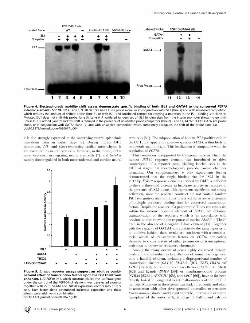
ISL1 and GATA4 each can bind the FGF10-Int1 element invitro
To investigate the specificity of ISL1 binding to its consensus
site within FGF10-Int1, we performed an electrophoretic mobility
shift assay (EMSA, Fig. 4). ISL1 bound robustly to its FGF10-ISL1
site, as well as to a previously identified positive control site [10],
termed Insulin I-ISL1 (Fig. 4, lanes 2 and 7 respectively). The
FGF10-ISL1 binding was specific, since it could be partially
competed off by excess unlabeled probe (Fig. 4, lane 3) but not by
a hundredfold excess of unlabeled mutated probe (Fig. 4, lane 4).
In addition, ISL1 did not bind to a labeled, scrambled FGF10-
ISL1 sequence (Fig. 4, lane 5).
In order to verify the affinity of the nearby, non-canonical
GATA site in FGF10-Int1 for GATA4, we performed another
EMSA, confirming that GATA4 was able to occupy this sequence
(Fig. 4, lane 12). Binding to the 59-TGATTA-39 site was
completely abrogated by the addition of unlabeled FGF10-GATA4
probe (Fig. 4, lane 13).
ISL1 and GATA4 cooperate with TBX20 to activate FGF10via its intronic enhancer
The transcriptional response of murine Nkx2.5 to the combina-
tion of Isl1 and Gata4 in vitro can be potentiated by Tbx20, a
member of a large family of genes whose products share a
common DNA-binding domain, similar to the T (brachyury)
transcription factor [15]. We first determined the ability of ISL1
and/or GATA4 to promote luciferase activity using a reporter
with a minimal promoter containing the human cardiac-
responsive FGF10-Int1 fragment located 39 to the luciferase
sequence, mimicking the endogenous location of this regulatory
element relative to the initiation site for FGF10 transcription. Co-
transfection into mesenchymal 10T1/2 cells of a GATA4 or ISL1
expression construct, together with the FGF10-Int1-luciferase
reporter, indeed resulted in robust activation of luciferase activity
(Fig. 5).
We then tested whether this human FGF10-Int1 element could
drive expression of a luciferase reporter gene in the presence of
ISL1, GATA4, and TBX20 proteins separately as well as in
combination. Despite the presence of only a single, non-
palindromic T-box binding core motif [45] within the intronic
response element (Fig. 1), transfection of TBX20 in addition to
GATA4 and ISL1 expression constructs resulted in additive
activation of FGF10-Int1-luc (Fig. 5).
Transgenic mouse embryos express FGF10-Int1-drivenreporter in cardiac OFT
The strict sequence conservation between humans and mice,
and the ability of transfected murine cells to demonstrate ISL1-
and cofactor-driven activation of a reporter gene containing the
FGF10-Int1 enhancer in vitro, led us to then test the ability of the
element to drive reporter expression when introduced in vivo.
FGF10-Int1 was therefore subcloned into the pTK-nlacZ reporter
plasmid [46] and introduced into mouse blastocysts. 43 embryos
out of 66 injected were recovered at E8.5, 22 of 53 at E9.5, 46 of
94 at E10.5, and 37 of 59 at E11.5. Of these, nine animals had
integrated the transgene, confirmed by PCR, and expressed beta-
galactosidase activity: n = 2 at E8.5, n = 3 at E9.5, n = 1 at E10.5
and n = 3 at E11.5.
Labelled cells were observed in the cardiac outflow tract in two
of the reporter embryos, at E9.5 and E10.5 respectively,
demonstrating the conserved ability of this enhancer to drive
gene transcription in both mouse and human hearts. Expression in
both cases concerned a few dozen cells, which were not observed
in other heart compartments (Fig. 6A–B). Among the positive
embryos, a restricted set of additional tissues were also labelled,
varying in combinations from one embryo to another in an age-
appropriate manner (Table 1). These included the forebrain, the
lens, the three first pharyngeal arches, the pancreatic primordia
(dorsal and ventral; Fig. 6C), a subset of dorsal root ganglia cells,
and motoneurons (Fig. 6D–E). Scattered cells were also positive in
the rostral presomitic mesoderm in both E8.5 embryos. Although
neither cardiac nor pharyngeal arch expression were visible in the
three E11.5 embryos, the tunica media of the internal carotid
Figure 2. In vivo and in vitro binding of ISL1 and GATA4 withinthe first intron of FGF10. A: Results of end-point PCR after ChIP usinganti-ISL1 or non-specific IgG (or no antibody at all) on chromatinderived from human embryonic hearts at Carnegie stages (CS)14-15. B:Analogous results using anti-acetylated histone H4 compared to a non-specific IgG or no antibody at all, and end-point PCR of regions in the 59promoter to human ISL1 and FGF10, demonstrating active availabilityfor transcription. C: FGF10 and ISL1 (and ACTB) were co-expressed atfoot plate stages (Carnegie stages [CS]16-17, i.e. 37–43 days ofgestation) in human hindlimbs as seen by RT-PCR, while only FGF10and ACTB were transcribed in forelimbs. D: ChIP using anti-ISL1 onchromatin derived from the C16–17 hindlimb demonstrates noenrichment of the FGF10-Int1 amplicon as compared to the negativecontrol, although this fragment is amplifiable from the total inputchromatin.doi:10.1371/journal.pone.0030677.g002
Transcriptional Control in Human Heart Development
PLoS ONE | www.plosone.org 4 January 2012 | Volume 7 | Issue 1 | e30677

arteries were positive in one, and the trigeminal and acoustic
ganglia were labelled in another. Overall, the sites of transgenic
labelling are compatible with activation by Isl1, given what is
known about its expression pattern in all of these sites at these
stages of development [7,9,47,48], and thus with its positive
regulation of FGF10 transcription in both the human and murine
cardiac OFT.
Discussion
We have found that within the first intron of the FGF10 gene
there exist highly evolutionarily conserved consensus binding sites
for equally conserved transcription factors of the LIM homeodo-
main, GATA and T box families. These sites are arranged in such
a way as to represent a functional cis-regulatory module, with
physical spacing between the binding sites that is itself also
conserved across species, in particular that between the ISL1 and
GATA cognate sites. We have demonstrated that in the human
embryonic heart, this module is physically occupied by ISL1
during the period corresponding to the establishment of the
cardiac chambers but before septation of the OFT [44]. Binding of
ISL1 to the intronic element of FGF10 then ceases in the cardiac
OFT, but is never observed in the human or mouse hindlimb bud,
for example, where both Isl1 and Fgf10 are expressed shortly
thereafter. This observation shows tissue specificity in the function
of this binding site and is consistent with the ISL1 expression
pattern that we and others [11] have observed in the human
embryonic OFT as well as in the splanchnic mesoderm between
CS13-15, as reported in mouse at equivalent morphological stages
[49]. Despite a great deal of study of tissue-specific enhancers
engaged by Isl1 [10,50] and the control of Fgf10 expression by
transcription factors in the limb [51] and inner ear [26,52], this is
the first report of cis-regulation of FGF10 expression through an
intronic element during cardiac development.
In situ hybridization to GATA4 transcripts in adjacent sections
demonstrated that at CS12, unlike the morphologically equivalent
stage in the mouse [53], no GATA4 expression was observed in the
OFT region. Other subtle differences exist as well between the
mouse and human patterns, notably the lack of ISL1 expression
outside of the pharyngeal endoderm at CS12, when in the mouse,
Figure 3. Expression of ISL1 and GATA4 transcripts in the human heart between 26 and 38 days of gestation. A–H: ISL1 in situ atCarnegie stages (CS)12 (26–28 days post fertilization [dpf]), CS13 (28–31 dpf), CS14 (32–33 dpf) and CS15 (34–36 dpf) respectively. E–H aremagnifications of A–D respectively. I–K show GATA4 expression in adjacent sections to B–D. A: ISL1 is expressed at CS12 in foregut endoderm,splanchnic mesoderm, and early motoneurons. B, F: At CS13, ISL1 is transcribed by mesenchyme around the cardiac OFT and pharyngeal arches. ISL1expression continues in the splanchnic mesoderm between the trachea and OFT, and is visible in dorsal root ganglia, at CS14 (C, G) and CS15 (D, H).I–K: GATA4 is expressed in the endocardium and myocardium of the arterial pole at CS13, CS14 and CS15 (I, J, K respectively). Inset: RT-PCR of ISL1,GATA4, GATA5, GATA6, FGF10 and positive control ACTB mRNAs in embryonic human hearts at stages CS13-16 (to 40 dpf). Abbreviations: drg, dorsalroot ganglia; es, esophagus; fb, forebrain; fg, foregut; ph, pharynx; nt, neural tube; oft, OFT; ra, right atrium; t, trachea. Arrows, motoneurons. Bar:110 mm (A–D, I) and 55 mm (E–H, J, K).doi:10.1371/journal.pone.0030677.g003
Transcriptional Control in Human Heart Development
PLoS ONE | www.plosone.org 5 January 2012 | Volume 7 | Issue 1 | e30677
it is also strongly expressed in the underlying ventral splanchnic
mesoderm from an earlier stage [7]. During murine OFT
maturation, Isl1- and Gata4-expressing cardiac mesenchyme is
also colonized by neural crest cells. However, in the mouse, Isl1 is
never expressed in migrating neural crest cells [7], and Gata4 is
rapidly downregulated in both mesectodermal and cardiac neural
crest cells [54]. The subpopulation of human ISL1-positive cells in
the OFT, that apparently also co-expresses GATA4, is thus likely to
be mesodermal in origin. This localization is compatible with the
regulation of FGF10.
This conclusion is supported by transgenic mice in which the
human FGF10 response element was introduced to drive
transcription of a reporter gene, yielding labeled cells in the
OFT at stages that morphologically precede cardiac chamber
formation. Our complementary in vitro experiments further
demonstrated that the single binding site for ISL1 in the
1047 bp FGF10 response element enriched by ChIP is sufficient
to drive a three-fold increase in luciferase activity in response to
the presence of ISL1 alone. This represents significant and strong
activation, since the reporter construct did not contain tandem
ISL1 recognition sites but rather preserved the in vivo arrangement
of multiple predicted binding sites for conserved transcription
factors. Despite the absence of a palindromic T-box consensus site
within the intronic response element of FGF10, we obtained
transactivation of the reporter, which is in accordance with
previous studies showing the response of murine Nkx2.5 to Tbx20
even in the absence of a cognate T-box element [15]. Together
with the capacity of GATA4 to transactivate the same reporter in
an additive fashion, these results are consistent with a combina-
torial action of transcription factors on FGF10 non-coding
elements to confer a state of either permission or transcriptional
activation to otherwise refractory chromatin.
Among the many dozens of genes highly conserved through
evolution and identified as key effectors of animal cardiogenesis,
only a handful of them, including a disproportional number of
transcription factors (GATA4, NKX2.5, ZIC3, TBX1,TBX20 and
CHD7 [55–60]), but also intracellular effectors (TAB2 [61], MID1
[62]) and ligands (BMP4 [56]) or membrane-bound proteins
(STRA6 [63,64], NOTCH1 [65], and CFC1 [66]), have so far been
directly linked to congenital heart malformations of the OFT in
humans. Mutations in these genes can lead, infrequently and often
in association with other developmental anomalies, to persistent
truncus arteriosus, double outlet right ventricle, interruption or severe
hypoplasia of the aortic arch, tetralogy of Fallot, and valvulo-
Figure 4. Electrophoretic mobility shift assays demonstrate specific binding of both ISL1 and GATA4 to the conserved FGF10intronic element (FGF10-Int1). Lane 1, 9, 10: WT FGF10-ISL1 site probe alone, or in conjunction with ISL1 (lane 2) and with unlabeled competitor,which reduces the amount of shifted probe (lane 3), or with ISL1 and unlabeled competitor carrying a mutation in the ISL1 binding site (lane 4).Mutated ISL1 does not shift this probe (lane 5). Lane 6: A validated tandem set of ISL1 binding sites from the insulin promoter shows no gel shiftunless ISL1 is added (lane 7) and this shift is reduced in the presence of unlabelled probe competitor (lane 8). Lane 11, 14: WT FGF10-GATA site probealone, or in conjunction with GATA4 (lane 12) and with unlabeled competitor, which completely abrogates the shift of the probe (lane 13).doi:10.1371/journal.pone.0030677.g004
Figure 5. In vitro reporter assays support an additive combi-natorial effect of transcription factors upon the FGF10 intronicenhancer. LUC-FGF10-Int1, which construct placed the luciferase geneunder the control of the FGF10-Int1 element, was transfected alone ortogether with ISL1, GATA4 and TBX20 expression vectors into 10T1/2cells. Each factor alone potentiated luciferase expression and theseeffects were additive in combination.doi:10.1371/journal.pone.0030677.g005
Transcriptional Control in Human Heart Development
PLoS ONE | www.plosone.org 6 January 2012 | Volume 7 | Issue 1 | e30677

pathies. However, there is only partial correspondence between
murine and human gene inactivation phenotypes, with many
excellent candidate genes through their function in animal model
cardiac development not having been found to be mutated in their
human counterpart coding sequences.
FGF10 is one of these latter genes, whose cardiac knockout
phenotype in the mouse is itself subtle. Based on the murine
phenotypes of Fgf10 and Fgfr2-IIIb knockouts and their expression
patterns [3,19], we had previously found very similar expression
during normal human embryonic development; however, se-
quencing of both FGF10 and FGFR2-IIIb in human fetuses
exhibiting great vessel defects that resembled those in knockout
mice, among other symptoms, did not demonstrate coding
mutations [37]. The responsible gene turned out to encode a
protein, STRA6, necessary to bring vitamin A into cells, a first step
in transcriptional regulation through retinoic acid receptor binding
[63,64]. Retinoic acid, a vitamin A metabolite, normally favors
Gata4 transcription and limits the spatial expansion of Isl1, Fgf8
and Fgf10 expression in the SHF [67–69], while it promotes Fgf10
transcription in the burgeoning lungs [70]. Coding mutations in
FGF10 lead to phenotypic defects only in the submandibular and
lachrymal glands and lungs [34,35], despite being as present as
Stra6 [71] in many other organ systems. Similarly, heterozygous
missense coding mutations in human FGF8 have been shown to be
associated with non-syndromic cleft lip and palate [72], cause
pleiotropic defects in forebrain and pituitary formation [73], and a
recent case of recessive holoprosencephaly with asymptomatic,
consanguineous parents has been attributed to hypomorphic
alleles of FGF8 [74]; none of these patients presented cardiac
malformations. These observations emphasize the danger of
extrapolating findings about the detailed mechanisms of action
of highly conserved genes across species, and demonstrate the
limits of animal models in understanding human organogenesis.
There is increasing evidence that mutations in non-coding, cis-
regulatory elements, controlling transcript availability at a given
point time or a given tissue, represent an alternative mechanism
leading to human congenital malformations. Such mutations can
take the forms of those found for coding sequences, involving
single nucleotides [75] or small or large chromosomal rearrange-
ments [76]. We have discovered an evolutionarily conserved cis-
regulatory module in the FGF10 gene that is functional during
human cardiac development and that could represent an example
of the types of non-coding sites in which mutations may be
responsible for morphological aberrations. Taken together, our
data reveal unexpected complexity in the transcriptional landscape
controlling human cardiogenesis, highlight evolutionary conserva-
tion as well as species-specific aspects of cardiac signalling
networks, and contribute a strategy to identify additional
candidate genomic regions for study in congenital malformations
of the OFT.
Materials and Methods
Ethics statementHuman embryos were obtained from electively terminated
pregnancies, anonymously donated to research after informed
written consent from donors in concordance with French
legislation (94–654 and 08–400) and with prior approval of the
protocol (to M.V.) from the Necker ethical review committee. All
mice used in this study were housed under specific pathogen-free
conditions at the mouse genetics engineering center (C.I.G.M.) of
the Pasteur Institute, Paris, under authorization number A75-15-
09 from the Paris Departmental Directorate for the Protection of
Populations and handled in accordance with French and
European directives.
Chromatin immunoprecipitationChIP was carried out as previously described, starting from
nuclear isolation [77], using eleven microdissected and flash-
frozen cardiac tubes from human embryos at Carnegie stages (CS)
14–15 [78]. An anti-ISL1 (10 mL, Santa Cruz Sc-23590X) or an
anti-GFP antibody as negative control (10 mL, Abcam ab1218),
were used per 10 mg of sonicated chromatin. Immunoprecipitated
DNA was analysed by end-point PCR (primers, Supplementary
Table S1).
Figure 6. Transgenic mice demonstrate responsiveness of theconserved FGF10 intronic enhancer to endogenous transcrip-tion factors within the developing cardiac OFT and other sites.A 1047 bp enhancer region within the first intron of human FGF10,containing multiple transcription factor binding sites including sitesvalidated for ISL1 and GATA4, was placed ahead of a lacZ reporter geneunder a thymidine kinase-driven promoter. A: Transgenic mouse atembryonic day (E)10.5, in which expression was activated in dispersedcells of the posterior outflow tract (magnified, insets), in a distal/lateralsubdomain of the first two pharyngeal arches, in cells within thetrigeminal, acoustic and dorsal root ganglia, and in the lens (right side).B: Same embryo; frontal view. C: Transgenic mouse at E11.5, dorsal andventral pancreatic primordia. No expression was observed in the limbbuds in any injected embryos. In a different transgenic mouse at E11.5,D: motoneuron columns from inner surface of the lumbar spinal cord,and E: cross-section of spinal cord with a labelled subpopulation of cellsin the dorsal root ganglia.doi:10.1371/journal.pone.0030677.g006
Transcriptional Control in Human Heart Development
PLoS ONE | www.plosone.org 7 January 2012 | Volume 7 | Issue 1 | e30677

Expression studiesISL1 and GATA4 in situ hybridizations were performed using
transverse sections of normal human embryos from CS12 to 15.
Tissue fixation, sectioning, and in situ hybridization were carried
out as previously described [79]. Total RNA was extracted from
pooled whole hearts at individual stages from CS13 to CS16 and
RT-PCR was carried out using the GeneAmp kit (Roche), with
500 ng total RNA input for first strand synthesis (primers,
Supplementary Table S1).
Expression constructs and electrophoretic mobility shiftassays (EMSA)
Human TBX20 and ISL1 expression vectors were generated.
Full-length TBX20 cDNA and a fragment of ISL1 cDNA with the
N-terminal 142 amino acids removed [80] were inserted into the
multiple cloning site of pcDNA3.1C (Invitrogen). Full-length
human GATA4 cDNA was purchased from GenScript
(GN026113). HeLa cells were transfected with these constructs,
and nuclear protein extracts were made using standard protocols.
The LightShift Chemiluminescent EMSA Kit (Pierce) was used as
specified. Primers are listed in Supplementary Table S1.
Transactivation assays and reporter constructsFor the FGF10 reporter construct (LUC-FGF10-Int1), 1047 bp
of the FGF10 first intron (NCBI36/hg18 chromosome
5:44421556–44422602) were subcloned into the BamHI site 39
to luc+ in pGL3 (Promega). Mouse 10T1/2 cells [81] in DMEM/
10% fetal calf serum were transfected with FuGene HD (Roche).
Cells were harvested and lysed 24 h after transfection. Firefly and
Renilla luciferase activities were measured on a Berthold Centro
LB960 using the Dual-Luciferase Reporter assay system (Pro-
mega). Firefly luciferase activity was normalized to the Renilla
luciferase internal control, pRL-CMV (Promega). Experiments
were repeated in triplicate in three independent assays.
TransgenesisThe same 1047 bp FGF10-Int1 fragment as in the transactiva-
tion assays was subcloned into the BamHI site of the pSKT-TK-
nLacZ plasmid [46] and orientation verified by capillary
sequencing with a standard T3 primer. The plasmid was
linearized with SalI for injection at 2 ng/mL into mouse
blastocysts. b-galactosidase-containing cells that had transcribed
the reporter plasmid were stained in whole mount by the catalysis
of the X-gal (5-bromo-4-chloro-3-indolyl b-D-galactopyranoside)
substrate.
Supporting Information
Figure S1 Composite image of embryonic hearts at stages
ranging from the beginning of the fourth to the ninth week of
human gestation (upper left to lower right, Carnegie stages 10–23).
Rostral to top. Congenital heart and great vessel malformations
arise during this time window when molecular signaling between
cardiac progenitors and their environment is impaired.
(TIF)
Table S1 Primer sequences for PCR and EMSA.
(DOC)
Acknowledgments
The authors thank Dr. M. Teboul and the Service d’Orthogenie of the
Broussais Hospital in Paris for the human tissues and Dr. D. Montarras for
the 10T1/2 cells used in this work. Dr F. Langa Vives provided invaluable
assistance in the generation of transgenic mice at the Centre d’Ingenierie
Genetique Murine, Institut Pasteur, Paris. Drs. D. Bonnet, M. Bucking-
ham, C. Fournier-Thibault, and R. Kelly provided invaluable discussion.
Author Contributions
Conceived and designed the experiments: CG EH GN SZ HCE.
Performed the experiments: CG EH PD GN CB HCE. Analyzed the
data: CG PD GN SZ HCE. Contributed reagents/materials/analysis tools:
EH PD GN AM MV SL SZ. Wrote the paper: CG EH SL SZ HCE.
References
1. Hoffman JI, Kaplan S (2002) The incidence of congenital heart disease. J Am
Coll Card 39: 1890–1900.
2. Waldo KL, Kumiski DH, Wallis KT, Stadt Ha, Hutson MR, et al. (2001)
Conotruncal myocardium arises from a secondary heart field. Development 128:
3179–3188.
3. Kelly RG, Brown NA, Buckingham ME (2001) The arterial pole of the mouse
heart forms from Fgf10-expressing cells in pharyngeal mesoderm. Dev Cell 1:
435–440.
4. Brade T, Gessert S, Kuhl M, Pandur P (2007) The amphibian second heart field:
Xenopus islet-1 is required for cardiovascular development. Dev Biol 311: 297–310.
Table 1. Sites of b-galactosidase activity in transgenic mouse embryos.
Age forebrain lens MNs DRGs pancreas PSM PA1 PA2 PA3 OFT
E8.5 2 n/a . n/a n/a + . n/a n/a .
E8.5 + n/a . n/a n/a + . n/a n/a .
E9.5 . n/a . . . 2 + + . .
E9.5 . n/a . . . 2 . + . +
E9.5 . n/a . . + 2 + + . .
E10.5 + + . + . 2 + + + +
E11.5 + + + + + 2 . . + .
E11.5 + + + + . 2 . . . .
E11.5 + + + . 2 . . . .
All sites showed only selective cells positive for enhancer activation. DRGs = dorsal root ganglia; E = embryonic day of gestation; MN = motoneurons; OFT = cardiacoutflow tract; PA = pharyngeal arch; PSM = pre-somitic mesoderm.doi:10.1371/journal.pone.0030677.t001
Transcriptional Control in Human Heart Development
PLoS ONE | www.plosone.org 8 January 2012 | Volume 7 | Issue 1 | e30677

5. Zhou Y, Cashman TJ, Nevis KR, Obregon P, Carney Sa, et al. (2011) Latent
TGF-b binding protein 3 identifies a second heart field in zebrafish. Nature 474:
645–648.
6. Stolfi A, Gainous TB, Young JJ, Mori A, Levine M, et al. (2010) Early chordate
origins of the vertebrate second heart field. Science 329: 565–568.
7. Cai C-L, Liang X, Shi Y, Chu P-H, Pfaff SL, et al. (2003) Isl1 identifies a cardiac
progenitor population that proliferates prior to differentiation and contributes a
majority of cells to the heart. Dev Cell 5: 877–889.
8. Lee SK, Pfaff SL (2003) Synchronization of neurogenesis and motor neuron
specification by direct coupling of bHLH and homeodomain transcription
factors. Neuron 38: 731–745.
9. Ahlgren U, Pfaff SL, Jessell TM, Edlund T, Edlund H (1997) Independent
requirement for ISL1 in formation of pancreatic mesenchyme and islet cells.
Nature 385: 257–260.
10. Dodou E, Verzi MP, Anderson JP, Xu S-M, Black BL (2004) Mef2c is a direct
transcriptional target of ISL1 and GATA factors in the anterior heart field
during mouse embryonic development. Development 131: 3931–3942.
11. Sizarov A, Ya J, de Boer Ba, Lamers WH, Christoffels VM, et al. (2011)
Formation of the building plan of the human heart: morphogenesis, growth, and
differentiation. Circulation 123: 1125–1135.
12. Stennard FA, Costa MW, Elliott DA, Rankin S, Haast SJP, et al. (2003) Cardiac
T-box factor Tbx20 directly interacts with Nkx2-5, GATA4, and GATA5 in
regulation of gene expression in the developing heart. Dev Biol 262: 206–224.
13. Miller SA, Huang AC, Miazgowicz MM, Brassil MM, Weinmann AS (2008)
Coordinated but physically separable interaction with H3K27-demethylase and
H3K4-methyltransferase activities are required for T-box protein-mediated
activation of developmental gene expression. Genes Dev 22: 2980–2993.
14. Lickert H, Takeuchi JK, Von Both I, Walls JR, McAuliffe F, et al. (2004) Baf60c
is essential for function of BAF chromatin remodelling complexes in heart
development. Nature 432: 107–112.
15. Takeuchi JK, Mileikovskaia M, Koshiba-Takeuchi K, Heidt AB, Mori AD, et al.
(2005) Tbx20 dose-dependently regulates transcription factor networks required
for mouse heart and motoneuron development. Development 132: 2463–2474.
16. Riazi AM, Takeuchi JK, Hornberger LK, Zaidi SH, Amini F, et al. (2009)
NKX2-5 regulates the expression of beta-catenin and GATA4 in ventricular
myocytes. PLoS One 4: e5698.
17. Takeuchi JK, Bruneau BG (2009) Directed transdifferentiation of mouse
mesoderm to heart tissue by defined factors. Nature 459: 708–711.
18. Stevens KN, Hakonarson H, Kim CE, Doevendans PA, Koeleman BPC, et al.
(2010) Common variation in ISL1 confers genetic susceptibility for human
congenital heart disease. PLoS ONE 5: e10855.
19. Marguerie A, Bajolle F, Zaffran S, Brown NA, Dickson C, et al. (2006)
Congenital heart defects in Fgfr2-IIIb and Fgf10 mutant mice. Cardiovasc Res
71: 50–60.
20. Vega-Hernandez M, Kovacs A, De Langhe S, Ornitz DM (2011) FGF10/
FGFR2b signaling is essential for cardiac fibroblast development and growth of
the myocardium. Development 138: 3331–3340.
21. Xu H, Morishima M, Wylie JN, Schwartz RJ, Bruneau BG, et al. (2004) Tbx1
has a dual role in the morphogenesis of the cardiac outflow tract. Development
131: 3217–3227.
22. Agarwal P, Wylie JN, Galceran J, Arkhitko O, Li C, et al. (2003) Tbx5 is
essential for forelimb bud initiation following patterning of the limb field in the
mouse embryo. Development 130: 623–633.
23. Min H, Danilenko DM, Scully SA, Bolon B, Ring BD, et al. (1998) Fgf-10 is
required for both limb and lung development and exhibits striking functional
similarity to Drosophila branchless. Genes Dev 12: 3156–3161.
24. Sekine K, Ohuchi H, Fujiwara M, Yamasaki M, Yoshizawa T, et al. (1999)
Fgf10 is essential for limb and lung formation. Nat Genet 21: 138–141.
25. Yang L, Cai C-L, Lin L, Qyang Y, Chung C, et al. (2006) Isl1Cre reveals a
common Bmp pathway in heart and limb development. Development 133:
1575–1585.
26. Ohuchi H, Yasue A, Ono K, Sasaoka S, Tomonari S, et al. (2005) Identification
of cis-element regulating expression of the mouse Fgf10 gene during inner ear
development. Dev Dyn 233: 177–187.
27. Ohuchi H, Hori Y, Yamasaki M, Harada H, Sekine K, et al. (2000) FGF10 acts
as a major ligand for FGF receptor 2 IIIb in mouse multi-organ development.
Biochem Biophys Res Comm 277: 643–649.
28. Fairbanks T (2004) Fibroblast growth factor 10 (Fgf10) invalidation results in
anorectal malformation in mice. J Ped Surg 39: 360–365.
29. Urness LD, Bleyl SB, Wright TJ, Moon AM, Mansour SL (2011) Redundant
and dosage sensitive requirements for Fgf3 and Fgf10 in cardiovascular
development. Dev Biol 356: 383–397.
30. Watanabe Y, Miyagawa-Tomita S, Vincent SD, Kelly RG, Moon AM, et al.
(2010) Role of mesodermal FGF8 and FGF10 overlaps in the development of the
arterial pole of the heart and pharyngeal arch arteries. Circ Res 106: 495–503.
31. Vitelli F, Taddei I, Morishima M, Meyers EN, Lindsay EA, et al. (2002) A
genetic link between Tbx1 and fibroblast growth factor signaling. Development
129: 4605–4611.
32. Liu W, Levi G, Shanske A, Frenz DA (2008) Retinoic acid-induced inner ear
teratogenesis caused by defective Fgf3/Fgf10-dependent Dlx5 signaling. Birth
defects Res Part B 83: 134–144.
33. Herzog W, Sonntag C, von der Hardt S, Roehl HH, Varga ZM, et al. (2004)
Fgf3 signaling from the ventral diencephalon is required for early specification
and subsequent survival of the zebrafish adenohypophysis. Development 131:
3681–3692.
34. Entesarian M, Matsson H, Klar J, Bergendal B, Olson L, et al. (2005) Mutations
in the gene encoding fibroblast growth factor 10 are associated with aplasia of
lacrimal and salivary glands. Nat Genet 37: 125–127.
35. Rohmann E, Brunner HG, Kayserili H, Uyguner O, Nurnberg G, et al. (2006)
Mutations in different components of FGF signaling in LADD syndrome. Nat
Genet 38: 414–417.
36. Klar J, Blomstrand P, Brunmark C, Badhai J, Hakansson HF, et al. (2011)
Fibroblast growth factor 10 haploinsufficiency causes chronic obstructive
pulmonary disease. J Med Genet 48: 705–709. doi:10.1136/jmedgenet-2011-
100166.
37. Martinovic-Bouriel J, Bernabe-Dupont C, Golzio C, Grattagliano-Bessieres B,
Malan V, et al. (2007) Matthew-Wood syndrome: report of two new cases
supporting autosomal recessive inheritance and exclusion of FGF10 and
FGFR2. Am J Med Genet Part A 143: 219–228.
38. Genead R, Danielsson C, Wardell E, Kjaeldgaard A, Westgren M, et al. (2010)
Early first trimester human embryonic cardiac Islet-1 progenitor cells and
cardiomyocytes: Immunohistochemical and electrophysiological characteriza-
tion. Stem Cell Res 4: 69–76.
39. Loots GG, Ovcharenko I (2004) rVISTA 2.0: evolutionary analysis of
transcription factor binding sites. Nucleic Acids Res 32: W217–W221.
40. Ovcharenko I, Nobrega MA, Loots GG, Stubbs L (2004) ECR Browser: A tool
for visualizing and accessing data from comparisons of multiple vertebrate
genomes. Nucleic Acids Res 32: W280–W286.
41. Merika M, Orkin SH (1993) DNA-Binding Specificity of GATA Family
Transcription Factors. Mol Cell Biol 13: 3999–4010.
42. Blanchette M, Bataille AR, Chen X, Poitras C, Laganiere J, et al. (2006)
Genome-wide computational prediction of transcriptional regulatory modules
reveals new insights into human gene expression. Genome Res 16: 656–668.
43. Vettese-Dadey M, Grant PA, Hebbes TR, Crane- Robinson C, Allis CD, et al.
(1996) Acetylation of histone H4 plays a primary role in enhancing transcription
factor binding to nucleosomal DNA in vitro. EMBO J 15: 2508–2518.
44. Moorman A, Webb S, Brown NA, Lamers W, Anderson RH (2003)
Development of the heart: (1) formation of the cardiac chambers and arterial
trunks. Heart 89: 806–814.
45. Conlon FL, Fairclough L, Price BMJ, Casey ES, Smith JC (2001) Determinants
of T box protein specificity. Development 128: 3749–3758.
46. Hadchouel J, Carvajal JJ, Daubas P, Bajard L, Chang T, et al. (2003) Analysis of
a key regulatory region upstream of the Myf5 gene reveals multiple phases of
myogenesis, orchestrated at each site by a combination of elements dispersed
throughout the locus. Development 130: 3415–3426.
47. Pfaff SL, Mendelsohn M, Stewart CL, Edlund T, Jessell TM (1996)
Requirement for LIM homeobox gene Isl1 in motor neuron generation reveals
a motor neuron-dependent step in interneuron differentiation. Cell 84: 309–320.
48. Yuan S, Schoenwolf GC (2000) Islet-1 marks the early heart rudiments and is
asymmetrically expressed during early rotation of the foregut in the chick
embryo. AnatRec 260: 204–207.
49. Snarr BS, O’Neal JL, Chintalapudi MR, Wirrig EE, Phelps AL, et al. (2007) Isl1
expression at the venous pole identifies a novel role for the second heart field in
cardiac development. Circ Res 101: 971–974.
50. Kawakami Y, Marti M, Kawakami H, Itou J, Quach T, et al. (2011) Islet1-
mediated activation of the beta-catenin pathway is necessary for hindlimb
initiation in mice. Development 138: 4465–4473.
51. Sasaki H, Yamaoka T, Ohuchi H, Yasue A, Nohno T, et al. (2002) Identification
of cis-elements regulating expression of Fgf10 during limb development. The
IntJ Dev Biol 46: 963–967.
52. Lillevali K, Haugas M, Matilainen T, Pussinen C, Karis A, et al. (2006) Gata3 is
required for early morphogenesis and Fgf10 expression during otic development.
Mech Dev 123: 415–429.
53. Rojas A, De Val S, Heidt AB, Xu SM, Bristow J, et al. (2005) Gata4 expression
in lateral mesoderm is downstream of BMP4 and is activated directly by
Forkhead and GATA transcription factors through a distal enhancer element.
Development 132: 3405–3417.
54. Tomita Y, Matsumura K, Wakamatsu Y, Matsuzaki Y, Shibuya I, et al. (2005)
Cardiac neural crest cells contribute to the dormant multipotent stem cell in the
mammalian heart. J Cell Biol 170: 1135–1146.
55. Goldmuntz E, Geiger E, Benson DW (2001) NKX2.5 mutations in patients with
tetralogy of Fallot. Circulation 104: 2565–2568.
56. Posch MG, Perrot A, Schmitt K, Mittelhaus S, Esenwein EM, et al. (2008)
Mutations in GATA4, NKX2.5, CRELD1, and BMP4 are infrequently found in
patients with congenital cardiac septal defects. Am J Med Genet A 146A:
251–253.
57. Megarbane A, Salem N, Stephan E, Ashoush R, Lenoir D, et al. (2000) X-linked
transposition of the great arteries and incomplete penetrance among males with
a nonsense mutation in ZIC3. Eur J Hum Genet 8: 704–708.
58. Gong W (2001) Mutation analysis of TBX1 in non-deleted patients with features
of DGS/VCFS or isolated cardiovascular defects. J Med Genet 38: 45e–45.
59. Kirk EP, Sunde M, Costa MW, Rankin SA, Wolstein O, et al. (2007) Mutations
in cardiac T-box factor gene TBX20 are associated with diverse cardiac
pathologies, including defects of septation and valvulogenesis and cardiomyop-
athy. Am J Hum Genet 81: 280–291.
Transcriptional Control in Human Heart Development
PLoS ONE | www.plosone.org 9 January 2012 | Volume 7 | Issue 1 | e30677

60. Vissers LELM, van Ravenswaaij CMA, Admiraal R, Hurst JA, de Vries BBA, et
al. (2004) Mutations in a new member of the chromodomain gene family causeCHARGE syndrome. Nat Genet 36: 955–957.
61. Thienpont B, Zhang L, Postma AV, Breckpot J, Tranchevent L-C, et al. (2010)
Haploinsufficiency of TAB2 causes congenital heart defects in humans.Am J Hum Genet 86: 839–849.
62. Pinson L, Auge J, Audollent S, Mattei G, Etchevers H, et al. (2004) Embryonicexpression of the human MID1 gene and its mutations in Opitz syndrome. J Med
Genet 41: 381–386.
63. Pasutto F, Sticht H, Hammersen G, Gillessen-Kaesbach G, Fitzpatrick DR,et al. (2007) Mutations in STRA6 cause a broad spectrum of malformations
including anophthalmia, congenital heart defects, diaphragmatic hernia,alveolar capillary dysplasia, lung hypoplasia, and mental retardation.
Am J Hum Genet 80: 550–560.64. Golzio C, Martinovic-Bouriel J, Thomas S, Mougou-Zrelli S, Grattagliano-
Bessieres B, et al. (2007) Matthew-Wood syndrome is caused by truncating
mutations in the retinol-binding protein receptor gene STRA6. Am J HumGenet 80: 1179–1187.
65. McBride KL, Riley MF, Zender Ga, Fitzgerald-Butt SM, Towbin Ja, et al.(2008) NOTCH1 mutations in individuals with left ventricular outflow tract
malformations reduce ligand-induced signaling. HumMol Genet 17: 2886–2893.
66. Goldmuntz E, Bamford R, Karkera JD, dela Cruz J, Roessler E, et al. (2002)CFC1 mutations in patients with transposition of the great arteries and double-
outlet right ventricle. Am J Hum Genet 70: 776–780.67. Ryckebusch L, Wang Z, Bertrand N, Lin S-C, Chi X, et al. (2008) Retinoic acid
deficiency alters second heart field formation. Proc Natl Acad Sci U S A 105:2913–2918.
68. Waxman JS, Keegan BR, Roberts RW, Poss KD, Yelon D (2008) Hoxb5b acts
downstream of retinoic acid signaling in the forelimb field to restrict heart fieldpotential in zebrafish. Dev Cell 15: 923–934.
69. Kostetskii I, Jiang Y, Kostetskaia E, Yuan S, Evans T, et al. (1999) Retinoidsignaling required for normal heart development regulates GATA-4 in a
pathway distinct from cardiomyocyte differentiation. Dev Biol 206: 206–218.
70. Desai TJ, Malpel S, Flentke GR, Smith SM, Cardoso WV (2004) Retinoic acidselectively regulates Fgf10 expression and maintains cell identity in the
prospective lung field of the developing foregut. Dev Biol 273: 402–415.
71. Bouillet P, Sapin V, Chazaud C, Messaddeq N, Decimo D, et al. (1997)
Developmental expression pattern of Stra6, a retinoic acid-responsive gene
encoding a new type of membrane protein. Mech Dev 63: 173–186.
72. Riley BM, Mansilla MA, Ma J, Daack-Hirsch S, Maher BS, et al. (2007)
Impaired FGF signaling contributes to cleft lip and palate. Proc Natl Acad
Sci U S A 104: 4512–4517.
73. Arauz RF, Solomon BD, Pineda-Alvarez DE, Gropman AL, Parsons JA,
Roessler E, Muenke M (2010) A hypomorphic allele in the FGF8 gene
contributes to holoprosencephaly and is allelic to gonadotropin-releasing
hormone deficiency in humans. Mol Syndromol 1: 59–66.
74. McCabe MJ, Gaston-Massuet C, Tziaferi V, Gregory LC, Alatzoglou KS, et al.
(2011) Novel FGF8 mutations associated with recessive holoprosencephaly,
craniofacial defects, and hypothalamo-pituitary dysfunction. J Clin Endocrinol
Metab 96: E1709–18.
75. Benko S, Fantes JA, Amiel J, Kleinjan D-J, Thomas S, et al. (2009) Highly
conserved non-coding elements on either side of SOX9 associated with Pierre
Robin sequence. Nat Genet 41: 359–364.
76. VanderMeer JE, Ahituv N (2011) cis-regulatory mutations are a genetic cause of
human limb malformations. Dev Dyn 240: 920–930. doi:10.1002/dvdy.22535.
77. Havis E, Anselme I, Schneider-Maunoury S (2006) Whole embryo chromatin
immunoprecipitation protocol for the in vivo study of zebrafish development.
Biotechniques 40: 34, 36, 38 passim.
78. O’Rahilly R, Muller F (1987) Developmental Stages in Humans: including a
revision of Streeter’s ‘‘Horizons’’ and a survey of the Carnegie collection.
Washington, D.C.: Carnegie Institution of Washington.
79. Delous M, Baala L, Salomon R, Laclef C, Vierkotten J, et al. (2007) The ciliary
gene RPGRIP1L is mutated in cerebello-oculo-renal syndrome (Joubert
syndrome type B) and Meckel syndrome. Nat Genet 39: 875–881.
80. Sanchez-Garcia I, Rabbitts TH (1993) Redox regulation of in vitro DNA-
binding activity by the homeodomain of the Isl-1 protein. J Mol Biol 231:
945–949.
81. Reznikoff CA, Brankow DW, Heidelberger C (1973) Establishment and
characterization of a cloned line of C3H mouse embryo cells sensitive to
postconfluence inhibition of division. Cancer Res 33: 3231–3238.
Transcriptional Control in Human Heart Development
PLoS ONE | www.plosone.org 10 January 2012 | Volume 7 | Issue 1 | e30677


