Embed Size (px)
Citation preview

Int. J. Mol. Sci. 2013, 14, 19385-19398; doi:10.3390/ijms141019385
International Journal of
Molecular Sciences ISSN 1422-0067
www.mdpi.com/journal/ijms
Review
Keratin K15 as a Biomarker of Epidermal Stem Cells
Amrita Bose, Muy-Teck Teh, Ian C. Mackenzie and Ahmad Waseem *
Centre for Clinical and Diagnostic Oral Sciences, Barts and the London School of Medicine and Dentistry,
Queen Mary University of London, Turner Street, London E1 2AD, UK;
E-Mails: [email protected] (A.B.); [email protected] (M.-T.T.);
[email protected] (I.C.M.)
* Author to whom correspondence should be addressed; E-Mail: [email protected];
Tel.: +44-207-882-2387; Fax: +44-207-882-7137.
Received: 21 June 2013; in revised form: 5 September 2013 / Accepted: 10 September 2013 /
Published: 25 September 2013
Abstract: Keratin 15 (K15) is type I keratin protein co-expressed with the K5/K14 pair
present in the basal keratinocytes of all stratified epithelia. Although it is a minor
component of the cytoskeleton with a variable expression pattern, nonetheless its
expression has been reported as a stem cell marker in the bulge of hair follicles.
Conversely, suprabasal expression of K15 has also been reported in both normal and
diseased tissues, which is inconsistent with its role as a stem cell marker. Our recently
published work has given evidence of the molecular pathways that seem to control the
expression of K15 in undifferentiated and differentiated cells. In this article, we have
critically reviewed the published work to establish the reliability of K15 as an epidermal
stem cell marker.
Keywords: transit-amplifying cells; keratinocyte differentiation; basal keratinocytes;
intermediate filaments; pluripotent cells
1. Introduction
What Are Stem Cells?
The hierarchy of embryonic stem cells begins with the totipotent zygote and extends up to the
morula stage of the embryo, in which all cells are capable of generating an entire organism. This is
OPEN ACCESS

Int. J. Mol. Sci. 2013, 14 19386
followed by the pluripotent blastocyst stage, where cells of the inner cell mass have the capacity to
form ectoderm, mesoderm and endoderm and the germ cells. However, in adult tissues, the stem cells
become only multipotent, although remaining capable of undergoing unlimited self-renewal to provide
a source of cells for their surrounding tissues. This enables the organism to repair and/or regenerate
lost tissue and is critical for survival [1].
Two distinct features differentiate a stem cell from a non-stem cell population. First, stem cells
possess an unlimited capacity of self-renewal, which enables them to undergo continuous cell division
throughout the life of an organism. Second, stem cells are capable of undergoing asymmetric cell
division to produce two daughter cells with different differentiation capacities. While one enters an
irreversible differentiation pathway, the other is retained in situ with an unlimited capacity to divide
and thus form the next generation of stem cells. The cell committing to differentiation is termed a
“transit-amplifying” (TA) or “progenitor” cell, characterised by finite self-renewal capacity and
commitment to terminal differentiation following a few cell divisions. This hierarchy is unidirectional
and TA cells do not normally regain stem cell characteristics [2]. Although the presence of ordered
columnar structure and “epidermal proliferative units” has been a widely accepted model for adult
epidermal homeostasis, studies with transgenic mice failed to detect such a pattern and suggested an
alternative pattern in which stem cells form a small quiescent population that is activated only for
regeneration after injury; renewal for epidermal homeostasis is due to a single progenitor population
undergoing random, but balanced, symmetric and asymmetric cell division patterns [3]. More recently,
however, it has been shown that the promoter used to drive marker expression is of particular
importance to experimental studies of epithelia in transgenic mice and that use of a K14 promoter
enables a stem and amplifying pattern to be demonstrated [4]. Studies with similar transgenic mice,
also using a marker driven by K14, indicate that the stem cell population in oral mucosa forms the cells
of origin for oral carcinomas [5].
Within a tissue, stem cells reside within a defined area called the “niche”, which consists of
supporting cells and extracellular factors that are conducive to maintaining stem cell characteristics.
For example, in the human skin, such a niche was first detected in the bulge of hair follicles by using
C8/144B monoclonal antibody. This antibody preferentially immunostained the bulge keratinocytes
without staining other parts of the hair follicle and the protein it cross-reacted with was found to be
K15. This was one of the first studies to correlate K15 expression with epidermal stem cells [6]. It
was also demonstrated that the bulge cells, compared to TA cells, retained DNA precursor label,
bromodeoxyuridine, thereby signifying that they underwent limited cell division [7]. The bulge cells
have been shown to generate all the different epithelial cell lineages, such as both follicular and
interfollicular keratinocytes, sebaceous gland cells and the hair matrix cells and are referred to as
“follicular stem cells” [8,9]. Although several studies have reported the importance of the follicular
stem cells in epidermal repair and maintenance, however, presence of discrete epidermal proliferative
units, consisting of stem and committed TA cells, has recently been identified in the interfollicular
epidermis. These studies have reported that the slow-cycling stem cells located in the interfollicular
compartment are primarily responsible for repair and regeneration of the non-follicular epidermis [4].

Int. J. Mol. Sci. 2013, 14 19387
2. Evolution of Epidermal Stem Cell Research
The expansion of epidermal stem cell research from the 1960s until the present has been
tremendous. It started with the recognition of units of epidermal structure in which the suprabasal cells
and the superficial corneocytes are stacked to form cell columns [10] and then demonstration of
proliferative heterogeneity amongst the basal cells beneath such units [11]. Thereafter, a hierarchy of
proliferative epithelial cells, containing a small number of basal stem cells was suggested [12,13].
During the 1980s–1990s, several authors used label retaining assays and clonal analysis to identify and
establish the hierarchy produced by epidermal stem cells and, finally, defined the bulge of the hair
follicles as the site of epidermal stem cells in humans [14–16]. Advanced molecular biology
techniques during the last decade have given evidence of interfollicular stem cells using lineage tracing
in vivo, have detected the immigration of bone marrow-derived stem cells to the epidermis during
epidermal regeneration, have identified the importance of p63 in stem cell differentiation and initiation
of epithelial stratification, and have derived keratinocytes from induced pluripotent cells and
vice-versa [17].
Epidermal Stem Cell Markers
The etio-pathogenesis of cutaneous neoplasms has been attributed to the resident epidermal stem
cells, as the long life span of these cells makes them a likely target for oncogenic mutations [18].
Hence, it has become an absolute necessity to identify and characterise these cells to gain insight
into epidermal cancer formation, progression and metastasis. Identification of differences between
stem cells and the TA cell population also enable a better understanding of the dynamics of
keratinocyte biology.
Although, substantial research has been done to identify epidermal stem cells, the reliability of stem
cell markers still remains debatable. Several variables, such as species variance between mouse and
humans, type of body tissue, presence or absence of hair, in vivo and in vitro conditions and the
isolation techniques employed to identify the stem cells, have been discussed as the primary reason for
the ambiguity of markers [19]. Of the several epidermal stem cell markers identified, the expression of
β1 integrins has been widely accepted. It has been demonstrated that those human keratinocytes,
both cultured and derived from in vivo foreskin, which adhere most rapidly to type IV collagen or
fibronectin, also express the highest level of β1 integrin on their surface. These cells were found to
divide actively and form larger colonies, a characteristic typical of stem cells in culture [20]. Another
study has reported that stem cells derived from murine or neonatal human foreskin, express high levels
of α6 integrin and low levels of CD71 transferrin receptor. Such cells were found to exhibit a small
blast-like phenotype with high self-renewal capacity and low expression levels of K10, a keratin
expressed in the differentiated suprabasal layers of stratified epithelia [21]. Epidermal stem cells have
also been found to express markers that are typically expressed by stem cells in other tissues. For
example, murine bulge keratinocytes have been found to express CD34, a haematopoietic stem cell
marker, and these CD34+ cells formed large colonies, were slowly cycling with a high label-retaining
capacity, existed in the G0/G1 phase of the cell cycle, expressed high K15 levels, and stained intensely
for α6 integrin [22]. An intestinal stem cell marker, Lgr5, has also been used to identify multipotent

Int. J. Mol. Sci. 2013, 14 19388
stem cells of the mouse hair follicle capable of giving rise to new hair follicles with maintenance of all
cell lineages [23]. Recently cells containing Lgr6, a closely related molecule to Lgr5, have been
proposed to be the markers of the most primitive stem cells present in the epidermis [24]. Cell surface
biomarkers have formed the traditional methods for distinguishing stem cells from the TA and
terminally differentiating keratinocytes. However, a recent study has demonstrated that the innate
biochemical composition of a keratinocytes can be used for the same purpose and that infrared
spectroscopy can be used to detect the vibrational modes of phosphate molecules, PO2−, present within
the keratinocytes. Such vibration modes were found to be different between the stem, TA, and
terminally differentiated cells, with each cell type expressing a distinct DNA conformation [25].
3. K15—A Marker of Stem Cells
Several authors have described K15 as a putative epidermal stem cell marker. One of the first
studies to correlate K15 expression with human epidermal stem cells reported (a) that a cross reacting
monoclonal antibody C8/144B preferentially stained keratinocytes of the hair follicle bulge and
(b) that the protein it cross-reacted with was K15 [6]. These bulge cells demonstrated stem cell features
and were slowly-cycling, had high levels of β1 integrin expression, preferentially proliferated during
anagen phase of hair cycle, which reflects the growth phase wherein the stem cells rapidly divide, and
also expressed K19, a reported cutaneous stem cell marker. On the basis of these observations it was
suggested that both K15 and K19 could be used as markers of stem cells to segregate them from the
differentiated progenitor population. The same study also reported that between the two keratins, K19
was found to be expressed more in the TA cells that had left the stem cell compartment, characterised
by β1 integrin dullness, while those keratinocytes that were K15+ represented a more undifferentiated
state, marked by β1 integrin brightness and label retaining capacity [6]. High expression of K19 has
also been reported to co-localize with increased brightness of α3β1 integrin, a marker of slow cycling
keratinocytes of the bulge. However, the authors also observed that K19 could not label any of those
cells of the interfollicular region that retained [3H]thymidine, thereby questioning the reliability of
using K19 as a marker of all cutaneous stem cells [26].
The observation of Lyle and co-workers [6] has been further suported by several other studies
including specific localisation of K15 in stem cells residing in the bulge [27], preferentially targetting
hair follicle bulge cells by mouse K15 promoter in adult K15/lacZ transgenic mice [28], and
reconstruction of all components of skin epithelium by K15+ cells [29]. Furthermore, expression of
K15 mRNA and protein has been detected in the human anagen bulge which represents the onset of a
new hair follicle growth phase [30]. K15+ cells in the mouse epidermal bulge were shown to coincide
with Lgr5+ cells, which were able to regenerate new hair follicles and maintain all cell lineages of the
follicle [23]. K15+ bulge cells from human skin also stained postively for another epidermal stem cell
biomarker, CD200, and such cells had a much higher colony-forming ability [8]. A recent study of
scalp of patients suffering from androgenetic alopecia,showed that K15 postive cells were smaller in
size and had a much lower rate of proliferation, suggestive of a stem cell phenotype [31].
Int. J. Mol. Sci. 2013, 14 19389
3.1. Functions of Keratin K15
The mitotically active basal layer of stratified epithelia expresses the major keratin pair of K5
and K14 and a minor type I keratin K15, which lacks a natural co-expression partner [27,32–34]. In
normal stratified epithelia, the expression of K5/K14 and K15 is confined to the proliferating basal
keratinocytes and ceases to express when these cells commit to terminal differentiation and begin
their journey towards the surface. This journey is characterised by the expression of K1 and K10
in the suprabasal layer and K2 in the upper spinous layers. Such changes demonstrate the
differentiation-specific expression of keratins [33]. While the expression of K5 and K14 is present
uniformly in all the basal keratinocytes of all stratified epithelia, the expression of K15 differs in
different tissues. For example, in the epidermis, expression of K15, although restricted to the basal
layer, is present in patches. The cells at the deep rete ridges show strong expression of K15, whereas
those overlying dermal papillae show little or no expression [21,27,34,35]. However, in internal
epithelia, such as the oral mucosa, K15 expression is uninterrupted [36]. In the neonatal human skin
and very young epidermis (~1.5 years), its expression is present throughout the basal layer [37],
which may indicate a role for K15 in the developing epidermis. To our knowledge, K15 is the
only basal-specific keratin that has been reported to have a suprabasal expression in normal
esophagus [27,34,38] and in pathological conditions, such as oral lichen planus [39].
Expression studies on developing human embryos have shown that K15 is one of the earliest
stratification-related keratins to be expressed in all types of developing stratified epithelia and, together
with K4. K15, continues to be expressed in adult epidermis as a minor component [38]. As K15 is one
of the basal-specific cytoskeletal proteins, providing structural support to the basal layer may be one of
its primary functions but the discontinuous nature of K15 expression in the adult epidermis indicates
that its presence maybe essential for the structural integrity of the basal layer only in the developing
epidermis and not in adults. Therefore, K15 must have another function in the adult epidermis that is
compatible with its selective expression pattern but, as yet, the K15 function in adult stratified epithelia
is not understood.
That the main function of K5 and K14 is to provide structural support to the basal layer of stratified
epithelia is evident by the fact that mutations in either of this pair leads to the pathogenesis of an
inherited group of blistering disorder, collectively termed epidermolysis bullosa (EB) [40]. Most of the
causative mutations are missense in nature and they are inherited as dominant negative [41]. However,
recessive missense mutations in K5 and K14 have also been reported [41]. In some cases homozygous
nonsense mutations in K14 lead to premature termination and the patients are completely devoid of
K14 protein. These EBS cases are extremely rare and are recessive in nature [42]. EBS patients
completely devoid of K5 have not been reported in humans and this suggests that absence of K5 may
be embryonic lethal. The absence of K14 could be compensated by K15 but as K5 is the only type II
keratin present in the basal layer, its absence cannot be compensated by another keratin. Absence of
K5 in the basal layer would remove all type I keratins by ubiquitinylation and, making the basal
keratinocytes devoid of keratin filaments, would consequently destabilise the cyto-architecture of the
basal layer [43]. No mutation in keratin K15 has ever been associated with EBS, or any other blistering
diseases, suggesting either that K15 mutations do not exist, either because they are embryonic lethal
or perhaps they are yet to be discovered. Given the pattern of K15 expression during embryonic

Int. J. Mol. Sci. 2013, 14 19390
development, together with a possible stem cell function, it is plausible that embryos harbouring a K15
mutation would not survive.
To compensate for loss of K14 function, K15 expression would need be elevated in EBS patients
where K14 is completely absent. While some studies have provided evidence for a compensatory role
of K15 [32,44], others have reported no such compensation [42]. In 1995, Lloyd and co-workers
observed that K14 knockout mice had a relatively unaffected oesophagus but had blistered skin. This
was attributed to high level of K15 expression in the oesophagus [32]. This led to the hypothesis that
K15, if expressed at high levels, will be able to compensate for the absence of K14. Up-regulation of
K15 expression in the epidermis of 4 EBS patients with ablated K14 has been reported and in these
patients K5 was able to polymerise with K15 but the K5/K15 pair could form only 6 nm wispy
proto-filaments instead of mature 10 nm keratin filaments [44]. In an independent study, a similar
upregulation of K15 expression in the epidermis of a patient suffering from natural ablation of both
K14 alleles has also been reported [27]. However, a study of a patient suffering with Köbner form of
EBS, characterized by complete absence of K14 in the epidermis, has reported no compensatory
increase of K15 in the basal cells to form filaments with K5 [42]. Therefore, at present it is uncertain
whether K15 can functionally compensate for the absence of K14 in EBS patients. This aspect is
worthy of further investigation.
As stem cells continue to divide throughout the life of an organism, such cells are at a high risk of
accumulating genetic mutations and could be potential targets of tumour initiation [45,46]. For
example, the bulge stem cells have been implicated in the carcinogenesis of trichoepitheliomas (TE)
and basal cell carcinomas (BCC), as both lesions have a histological similarity with the bulge cells [9].
K15 expression in TEs is much higher at the periphery of the lesion than for BCCs. It was therefore
possible to use K15 expression to distinguish between the two lesions [47]. This is of particular
importance as TE is a benign lesion while BCC is a malignant skin neoplasm, so a correct diagnosis is
essential to form the best treatment plan. In sebaceous tumours a subpopulation of cells which were
less differentiated than other cells, suggesting a stem cell phenotype, expressed high levels of K15 [45].
The study of K15 expression in the two most common epithelial malignancies, BCC and squamous
cell carcinomas (SCC), highlights a role of K15 in influencing the behaviour of these lesions. While
BCC is a locally invasive epidermal malignancy with highest rate of incidence, SCC is the second
most common epithelial malignancy and has a significant propensity to metastasise [18]. Several
studies have reported that BCCs express high levels of K15, whereas SCCs have been reported to
express very low levels [9,18,34]. This expression pattern may indicate a role for K15 in maintaining
the epithelial lineage of keratinocytes in BCC, thereby inhibiting metastasis. Downregulation of K15 in
SCC may have a role in inducing the mesenchymal changes in keratinocytes that make them motile
and therefore metastatic. This hypothesis is further supported by studies reporting downregulation of
K15 in keratinocytes during wound healing [27,48]. Furthermore, during epithelial-mesenchymal
transition (EMT), a developmental process leading to increased cell motility, there is a significant
reduction in expression of cytokeratins, including K15. A role for EMT has also been implied during
oncogenesis, and especially related to local invasion and metastasis [49,50].
When comparing keratin expression profiles of SCC samples from tongue, gingiva, floor of the
mouth, buccal mucosa and palate with tissues obtained from similar regions of normal volunteers, it
was found that while K14 is upregulated in oral SCC, K15 expression was significantly downregulated

Int. J. Mol. Sci. 2013, 14 19391
and was almost absent in dysplastic oral tissues [51]. Another study by Troy and co-workers also
reported that K15 expression was progressively downregulated and ultimately lost in an epidermal
tumorigenesis model that was generated by two stage chemical treatments with DMBA and TPA.
However, in this model K14 was found to be expressed throughout the basal and suprabasal layers as
well as tumour islands infiltrating the underlying dermis [52]. These studies not only emphasise the
importance of K15 expression in diagnosis of epidermal tumours, but also highlight that dysplastic
changes in SCC keratinocytes suppress K15 and induce K14. This perhaps suggests that K14 and K15,
the two type I basal keratins, are either regulated by independent mechanisms or by a common
mechanism that has an inverse effect on their expression.
3.2. K15 Is a Target of Stem Cell Specific Transcription Factor FOXM1
The human Forkhead Box M1 (FOXM1) protein belongs to a winged-helix transcription factor
family of at least 50 unique FOX genes identified in the human genome [53]. Transgenic and knockout
mouse studies have provided valuable information and confirm a pivotal role for FOXM1 in cell
cycle regulation, cell-fate determination, embryonic development, adult tissue homeostasis, organ
regeneration and ageing (reviewed in [54,55]. Emerging evidence has indicated that FOXM1 plays an
important role in maintaining stem cell renewal through pluripotency genes Oct4, Nanog and Sox2 in
mouse [56–58]. A recent mouse model study established a key role for FOXM1 in cell fate
determination and showed that FOXM1 regulated mammary luminal cell fate by modulating the
expression of GATA-3, a key regulator of breast luminal epithelial differentiation [59].
Given a role of K15 as a stem cell marker, it is not surprising that a stem cell-related transcription
factor such as FOXM1 may be an upstream target of K15. Indeed, K15 has been shown to be activated
dose-dependently by FOXM1 in primary normal human keratinocytes whereby ectopic expression of
FOXM1 was found to induce stem cell expansion and produce a hyperplastic phenotype in an
organotypical culture system [60]. A FOXM1 DNA-binding motif is present within the promoter
region of K15 gene and it has been demonstrated, using the chromatin immunoprecipitation method,
that FOXM1 protein indeed binds to the DNA-binding motif within the promoter of K15 gene in
human keratinocytes [61]. This finding is in agreement with the fact that FOXM1 and K15 are
co-expressed in the rete-ridges of epidermis [21,60] and in the outer root sheath including the bulge, a
putative stem cell compartment, of the hair follicle [62]. Furthermore, both K15 and FOXM1 are
upregulated in basal cell carcinomas [9,34,63].
3.3. Is K15 a Reliable Stem Cell Marker?
In spite of substantial evidence in the literature supporting K15 to be a stem cell marker, several
authors have questioned the reliability of correlating its expression with stem cell properties of
keratinocytes. For example, in sheep hair follicles, K15 has been shown to be expressed in the outer
root sheath and absent in the bulge region that is thought to contain the stem cells [64]. Contrary to the
previous studies, a continuous K15 expression in the outer root sheath of human hair follicles, basal
layer of epidermis and in eccrine glands has been reported recently [65]. Furthermore, in the internal
oral and vaginal epithelia, uninterrupted K15 expression throughout the basal layer has been
reported [36] and it is highly unlikely that every K15+ cell is a stem cell. Porter and co-workers have

Int. J. Mol. Sci. 2013, 14 19392
suggested K15 to be a marker of laterally differentiating epidermal keratinocytes in the basal
layer [34]. Other recent studies have suggested that high K15 expression may not always mark a stem
cell subpopulation and that basal keratinocytes expressing high levels of K15 are possibly those
undergoing an abnormal differentiation programme [52]. Furthermore, K15 is reported to be expressed
suprabasally in differentiating keratinocytes of normal human esophagus [32,38] and in oral lichen
planus [39], and in freshly-cut skin sections exposed to thyroid hormone or IFNγ [66], findings
inconsistent with an undifferentiated keratinocyte phenotype, a distinctive feature of stem cells.
In vitro studies on keratinocyte differentiation have reported K15 expression only upon reaching
confluence [34,67], which again is a characteristic feature of differentiation-specific genes. The
expression of K15 has also been reported in non-epithelial tissues, such as the lymphoid tissue,
which again questions the reliability of its expression exclusive to epidermal stem cells [68]. These
studies taken together would question the status of K15 as a genuine “stem cell marker”.
In our recently published work, we have shown that differentiating keratinocytes are also
capable of expressing K15. In vitro experiments conducted to trigger keratinocyte differentiation,
expression of K15 could be induced through loss of cell surface β1-integrin receptors in suspension
culture, increased cell-cell interaction in confluent monolayer cultures, or by direct exposure to
differentiation-inducing chemical, such as phorbol 12-myristate 13-acetate (PMA) (Figure 1). We
reported for the first time that such differentiation-specific induction is mediated by the Protein
Kinase-C pathway (PKC) via AP-1 transcription factor which was capable of triggering expression of
K15 along with other differentiation-specific biomarkers, such as K1, K10, involucrin and cornifin
while downregulating the expression of the other type I basal keratin, K14 and FOXM1B, a cell cycle
regulated transcription factor [61]. Our results further question the reliability of using K15 on its own
as a biomarker for identifying any particular keratinocyte phenotype, and suggest that keratinocytes are
capable of switching the expression of K15 in various ways under different conditions.
4. Conclusions
The proliferating basal layer of normal epidermis contains stem cells, TA cells and some
keratinocytes committed to the differentiation pathway and in the process of migrating upwards.
Expression of K15 in the basal layer has been reported a marker of stem cells and is still being used to
distinguish stem cells from the TA and committed keratinocyte populations. However, several reports
have questioned the reliability of K15 for this purpose. We have scanned the literature for studies
relevant to the expression of K15 as an epidermal stem cell marker and found some reports supporting,
as well as some refuting, a relationship. Although a complete explanation will require further
investigations, through our recently published work on K15, we have provided an explanation for this
contradiction. We propose that two putative mechanisms regulate K15 expression in keratinocytes: one
that drives its expression in the basal layer, mediated primarily by FOXM1, and another that induces
its expression in the suprabasal layers, involving PKC/AP-1 signalling [61]. The over all conclusion of
the review is to highlight that K15 could be expressed in the stem cells as well as in differentiated
cells. Therefore, use of this marker on its own may not provide conclusive information about the stem
cell population in a tissue.
Int. J. Mol. Sci. 2013, 14 19393
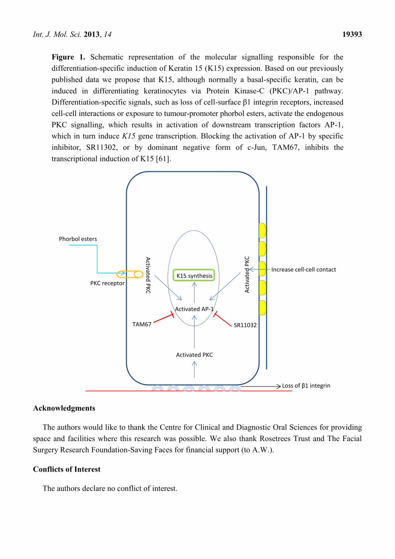
Figure 1. Schematic representation of the molecular signalling responsible for the
differentiation-specific induction of Keratin 15 (K15) expression. Based on our previously
published data we propose that K15, although normally a basal-specific keratin, can be
induced in differentiating keratinocytes via Protein Kinase-C (PKC)/AP-1 pathway.
Differentiation-specific signals, such as loss of cell-surface β1 integrin receptors, increased
cell-cell interactions or exposure to tumour-promoter phorbol esters, activate the endogenous
PKC signalling, which results in activation of downstream transcription factors AP-1,
which in turn induce K15 gene transcription. Blocking the activation of AP-1 by specific
inhibitor, SR11302, or by dominant negative form of c-Jun, TAM67, inhibits the
transcriptional induction of K15 [61].
Activated PKC
Activated AP-1
K15 synthesis
Loss of β1 integrin
Increase cell-cell contact
Act
ivat
ed P
KC
Phorbol esters
PKC receptor
Activated
PK
C
TAM67 SR11032
Acknowledgments
The authors would like to thank the Centre for Clinical and Diagnostic Oral Sciences for providing
space and facilities where this research was possible. We also thank Rosetrees Trust and The Facial
Surgery Research Foundation-Saving Faces for financial support (to A.W.).
Conflicts of Interest
The authors declare no conflict of interest.

Int. J. Mol. Sci. 2013, 14 19394
References
1. Wobus, A.M.; Boheler, K.R. Embryonic stem cells: Prospects for developmental biology and cell
therapy. Physiol. Rev. 2005, 85, 635–678.
2. Hall, P.A.; Watt, F.M. Stem cells: The generation and maintenance of cellular diversity.
Development 1989, 106, 619–633.
3. Clayton, E.; Doupe, D.P.; Klein, A.M.; Winton, D.J.; Simons, B.D.; Jones, P.H. A single type of
progenitor cell maintains normal epidermis. Nature 2007, 446, 185–189.
4. Mascre, G.; Dekoninck, S.; Drogat, B.; Youssef, K.K.; Brohee, S.; Sotiropoulou, P.A.;
Simons, B.D.; Blanpain, C. Distinct contribution of stem and progenitor cells to epidermal
maintenance. Nature 2012, 489, 257–262.
5. Tang, X.H.; Scognamiglio, T.; Gudas, L.J. Basal stem cells contribute to squamous cell
carcinomas in the oral cavity. Carcinogenesis 2013, 34, 1158–1164.
6. Lyle, S.; Christofidou-Solomidou, M.; Liu, Y.; Elder, D.E.; Albelda, S.; Cotsarelis, G.
The C8/144B monoclonal antibody recognizes cytokeratin 15 and defines the location of human
hair follicle stem cells. J. Cell Sci. 1998, 111, 3179–3188.
7. Ma, D.R.; Yang, E.N.; Lee, S.T. A review: The location, molecular characterisation and
multipotency of hair follicle epidermal stem cells. Ann. Acad. Med. Singap. 2004, 33, 784–788.
8. Inoue, K.; Aoi, N.; Sato, T.; Yamauchi, Y.; Suga, H.; Eto, H.; Kato, H.; Araki, J.; Yoshimura, K.
Differential expression of stem-cell-associated markers in human hair follicle epithelial cells.
Lab. Invest. 2009, 89, 844–856.
9. Jih, D.M.; Lyle, S.; Elenitsas, R.; Elder, D.E.; Cotsarelis, G. Cytokeratin 15 expression in
trichoepitheliomas and a subset of basal cell carcinomas suggests they originate from hair follicle
stem cells. J. Cutan. Pathol. 1999, 26, 113–118.
10. Mackenzie, J.C. Ordered structure of the stratum corneum of mammalian skin. Nature 1969, 222,
881–882.
11. Mackenzie, I.C. Relationship between mitosis and the ordered structure of the stratum corneum in
mouse epidermis. Nature 1970, 226, 653–655.
12. Potten, C.S. Epidermal cell production rates. J. Invest. Dermatol. 1975, 65, 488–500.
13. Lajtha, L.G. Stem cell concepts. Nouvelle Revue Francaise D’Hematologie 1979, 21, 59–65.
14. Bickenbach, J.R. Identification and behavior of label-retaining cells in oral mucosa and skin.
J. Dent. Res. 1981, 60, 1611–1120.
15. Bickenbach, J.R.; McCutecheon, J.; Mackenzie, I.C. Rate of loss of tritiated thymidine label in
basal cells in mouse epithelial tissues. Cell Tissue Kinet. 1986, 19, 325–333.
16. Cotsarelis, G.; Sun, T.T.; Lavker, R.M. Label-retaining cells reside in the bulge area of
pilosebaceous unit: Implications for follicular stem cells, hair cycle, and skin carcinogenesis. Cell
1990, 61, 1329–1337.
17. Ghadially, R. 25 years of epidermal stem cell research. J. Invest. Dermatol. 2012, 132, 797–810.
18. Abbas, O.; Bhawan, J. Expression of stem cell markers nestin and cytokeratin 15 and 19 in
cutaneous malignancies. J. Eur. Acad. Dermatol. Venereol. 2011, 25, 311–316.
19. Lavker, R.M.; Sun, T.T. Epidermal stem cells: Properties, markers, and location. Proc. Natl.
Acad. Sci. USA 2000, 97, 13473–13475.

Int. J. Mol. Sci. 2013, 14 19395
20. Watt, F.M. Epidermal stem cells: Markers, patterning and the control of stem cell fate.
Philos. Trans. R. Soc. Lond. Ser. B 1998, 353, 831–837.
21. Webb, A.; Li, A.; Kaur, P. Location and phenotype of human adult keratinocyte stem cells of the
skin. Differentiation 2004, 72, 387–395.
22. Trempus, C.S.; Morris, R.J.; Bortner, C.D.; Cotsarelis, G.; Faircloth, R.S.; Reece, J.M.;
Tennant, R.W. Enrichment for living murine keratinocytes from the hair follicle bulge with the
cell surface marker CD34. J. Invest. Dermatol. 2003, 120, 501–511.
23. Jaks, V.; Barker, N.; Kasper, M.; van Es, J.H.; Snippert, H.J.; Clevers, H.; Toftgard, R.
Lgr5 marks cycling, yet long-lived, hair follicle stem cells. Nat. Genet. 2008, 40, 1291–1299.
24. Snippert, H.J.; Haegebarth, A.; Kasper, M.; Jaks, V.; van Es, J.H.; Barker, N.; van de Wetering, M.;
van den Born, M.; Begthel, H.; Vries, R.G.; et al. Lgr6 marks stem cells in the hair follicle that
generate all cell lineages of the skin. Science 2010, 327, 1385–1389.
25. Patel, II.; Harrison, W.J.; Kerns, J.G.; Filik, J.; Wehbe, K.; Carmichael, P.L.; Scott, A.D.;
Philpott, M.P.; Frogley, M.D.; Cinque, G.; et al. Isolating stem cells in the inter-follicular
epidermis employing synchrotron radiation-based Fourier-transform infrared microspectroscopy
and focal plane array imaging. Anal. Bioanal. Chem. 2012, 404, 1745–1758.
26. Michel, M.; Torok, N.; Godbout, M.J.; Lussier, M.; Gaudreau, P.; Royal, A.; Germain, L.
Keratin 19 as a biochemical marker of skin stem cells in vivo and in vitro: Keratin 19 expressing
cells are differentially localized in function of anatomic sites, and their number varies with donor
age and culture stage. J. Cell Sci. 1996, 109, 1017–1028.
27. Waseem, A.; Dogan, B.; Tidman, N.; Alam, Y.; Purkis, P.; Jackson, S.; Lalli, A.; Machesney, M.;
Leigh, I.M. Keratin 15 expression in stratified epithelia: Downregulation in activated
keratinocytes. J. Invest. Dermatol. 1999, 112, 362–369.
28. Liu, Y.; Lyle, S.; Yang, Z.; Cotsarelis, G. Keratin 15 promoter targets putative epithelial stem
cells in the hair follicle bulge. J. Invest. Dermatol. 2003, 121, 963–968.
29. Morris, R.J.; Liu, Y.; Marles, L.; Yang, Z.; Trempus, C.; Li, S.; Lin, J.S.; Sawicki, J.A.;
Cotsarelis, G. Capturing and profiling adult hair follicle stem cells. Nat. Biotechnol. 2004, 22,
411–417.
30. Ohyama, M.; Terunuma, A.; Tock, C.L.; Radonovich, M.F.; Pise-Masison, C.A.; Hopping, S.B.;
Brady, J.N.; Udey, M.C.; Vogel, J.C. Characterization and isolation of stem cell-enriched human
hair follicle bulge cells. J. Clin. Invest. 2006, 116, 249–260.
31. Garza, L.A.; Yang, C.C.; Zhao, T.; Blatt, H.B.; Lee, M.; He, H.; Stanton, D.C.; Carrasco, L.;
Spiegel, J.H.; Tobias, J.W.; et al. Bald scalp in men with androgenetic alopecia retains hair
follicle stem cells but lacks CD200-rich and CD34-positive hair follicle progenitor cells.
J. Clin. Invest. 2011, 121, 613–622.
32. Lloyd, C.; Yu, Q.C.; Cheng, J.; Turksen, K.; Degenstein, L.; Hutton, E.; Fuchs, E. The basal
keratin network of stratified squamous epithelia: Defining K15 function in the absence of K14.
J. Cell Biol. 1995, 129, 1329–1344.
33. Moll, R.; Divo, M.; Langbein, L. The human keratins: Biology and pathology. Histochem. Cell Biol.
2008, 129, 705–733.

Int. J. Mol. Sci. 2013, 14 19396
34. Porter, R.M.; Lunny, D.P.; Ogden, P.H.; Morley, S.M.; McLean, W.H.; Evans, A.; Harrison, D.L.;
Rugg, E.L.; Lane, E.B. K15 expression implies lateral differentiation within stratified epithelial
basal cells. Lab. Invest. 2000, 80, 1701–1710.
35. Zhan, Q.; Signoretti, S.; Whitaker-Menezes, D.; Friedman, T.M.; Korngold, R.; Murphy, G.F.
Cytokeratin15-positive basal epithelial cells targeted in graft-versus-host disease express a
constitutive antiapoptotic phenotype. J. Invest. Dermatol. 2007, 127, 106–115.
36. Kose, O.; Lalli, A.; Kutulola, A.O.; Odell, E.W.; Waseem, A. Changes in the expression of stem
cell markers in oral lichen planus and hyperkeratotic lesions. J. Oral Sci. 2007, 49, 133–139.
37. Pontiggia, L.; Biedermann, T.; Meuli, M.; Widmer, D.; Bottcher-Haberzeth, S.; Schiestl, C.;
Schneider, J.; Braziulis, E.; Montano, I.; Meuli-Simmen, C.; et al. Markers to evaluate the quality
and self-renewing potential of engineered human skin substitutes in vitro and after transplantation.
J. Invest. Dermatol. 2009, 129, 480–490.
38. Leube, R.E.; Bader, B.L.; Bosch, F.X.; Zimbelmann, R.; Achtstaetter, T.; Franke, W.W.
Molecular characterization and expression of the stratification-related cytokeratins 4 and 15.
J. Cell Biol. 1988, 106, 1249–1261.
39. Bloor, B.K.; Seddon, S.V.; Morgan, P.R. Gene expression of differentiation-specific keratins (K4,
K13, K1 and K10) in oral non-dysplastic keratoses and lichen planus. J. Oral Pathol. Med. 2000,
29, 376–384.
40. Coulombe, P.A.; Kerns, M.L.; Fuchs, E. Epidermolysis bullosa simplex: A paradigm for disorders
of tissue fragility. J. Clin. Invest. 2009, 119, 1784–1793.
41. Rugg, E.L.; Leigh, I.M. The keratins and their disorders. Am. J. Med. Genet. Part C 2004, 131C,
4–11.
42. Rugg, E.L.; McLean, W.H.; Lane, E.B.; Pitera, R.; McMillan, J.R.; Dopping-Hepenstal, P.J.;
Navsaria, H.A.; Leigh, I.M.; Eady, R.A. A functional “knockout” of human keratin 14.
Genes Dev. 1994, 8, 2563–2573.
43. Peters, B.; Kirfel, J.; Bussow, H.; Vidal, M.; Magin, T.M. Complete cytolysis and neonatal
lethality in keratin 5 knockout mice reveal its fundamental role in skin integrity and in
epidermolysis bullosa simplex. Mol. Biol. Cell 2001, 12, 1775–1789.
44. Jonkman, M.F.; Heeres, K.; Pas, H.H.; van Luyn, M.J.; Elema, J.D.; Corden, L.D.; Smith, F.J.;
McLean, W.H.; Ramaekers, F.C.; Burton, M.; et al. Effects of keratin 14 ablation on the clinical
and cellular phenotype in a kindred with recessive epidermolysis bullosa simplex. J. Invest.
Dermatol. 1996, 107, 764–769.
45. Bieniek, R.; Lazar, A.J.; Photopoulos, C.; Lyle, S. Sebaceous tumours contain a subpopulation of
cells expressing the keratin 15 stem cell marker. Br. J. Dermatol. 2007, 156, 378–380.
46. Morris, R.J. Keratinocyte stem cells: Targets for cutaneous carcinogens. J. Clin. Invest. 2000,
106, 3–8.
47. Choi, C.W.; Park, H.S.; Kim, Y.K.; Lee, S.H.; Cho, K.H. Elastic fiber staining and cytokeratin 15
expression pattern in trichoepithelioma and basal cell carcinoma. J. Dermatol. 2008, 35, 499–502.
48. Werner, S.; Munz, B. Suppression of keratin 15 expression by transforming growth factor beta
in vitro and by cutaneous injury in vivo. Exp. Cell Res. 2000, 254, 80–90.
49. Geiger, T.; Sabanay, H.; Kravchenko-Balasha, N.; Geiger, B.; Levitzki, A. Anomalous features of
EMT during keratinocyte transformation. PLoS One 2008, 3, e1574.

Int. J. Mol. Sci. 2013, 14 19397
50. Biddle, A.; Liang, X.; Gammon, L.; Fazil, B.; Harper, L.J.; Emich, H.; Costea, D.E.;
Mackenzie, I.C. Cancer stem cells in squamous cell carcinoma switch between two distinct
phenotypes that are preferentially migratory or proliferative. Cancer Res. 2011, 71, 5317–5326.
51. Sakamoto, K.; Aragaki, T.; Morita, K.; Kawachi, H.; Kayamori, K.; Nakanishi, S.; Omura, K.;
Miki, Y.; Okada, N.; Katsube, K.; et al. Down-regulation of keratin 4 and keratin 13 expression in
oral squamous cell carcinoma and epithelial dysplasia: A clue for histopathogenesis.
Histopathology 2011, 58, 531–542.
52. Troy, T.C.; Arabzadeh, A.; Turksen, K. Re-assessing K15 as an epidermal stem cell marker.
Stem Cell Rev. 2011, 7, 927–934.
53. Jackson, B.C.; Carpenter, C.; Nebert, D.W.; Vasiliou, V. Update of human and mouse forkhead
box (FOX) gene families. Hum. Genomics 2010, 4, 345–352.
54. Myatt, S.S.; Lam, E.W. The emerging roles of forkhead box (Fox) proteins in cancer. Nat. Rev.
Cancer 2007, 7, 847–859.
55. Wierstra, I.; Alves, J. FOXM1, a typical proliferation-associated transcription factor. Biol. Chem.
2007, 388, 1257–1274.
56. Xie, Z.; Tan, G.; Ding, M.; Dong, D.; Chen, T.; Meng, X.; Huang, X.; Tan, Y. Foxm1
transcription factor is required for maintenance of pluripotency of P19 embryonal carcinoma cells.
Nucleic Acids Res. 2010, 38, 8027–8038.
57. Tompkins, D.H.; Besnard, V.; Lange, A.W.; Keiser, A.R.; Wert, S.E.; Bruno, M.D.; Whitsett, J.A.
Sox2 activates cell proliferation and differentiation in the respiratory epithelium. Am. J. Respir.
Cell Mol. Biol. 2011, 45, 101–110.
58. Wang, Z.; Park, H.J.; Carr, J.R.; Chen, Y.J.; Zheng, Y.; Li, J.; Tyner, A.L.; Costa, R.H.; Bagchi, S.;
Raychaudhuri, P. FoxM1 in tumorigenicity of the neuroblastoma cells and renewal of the neural
progenitors. Cancer Res. 2011, 71, 4292–4302.
59. Carr, J.R.; Kiefer, M.M.; Park, H.J.; Li, J.; Wang, Z.; Fontanarosa, J.; DeWaal, D.; Kopanja, D.;
Benevolenskaya, E.V.; Guzman, G.; et al. FoxM1 regulates mammary luminal cell fate.
Cell Rep. 2012, 1, 715–729.
60. Gemenetzidis, E.; Elena-Costea, D.; Parkinson, E.K.; Waseem, A.; Wan, H.; Teh, M.T. Induction
of human epithelial stem/progenitor expansion by FOXM1. Cancer Res. 2010, 70, 9515–9526.
61. Bose, A.; Teh, M.T.; Hutchison, I.L.; Wan, H.; Leigh, I.M.; Waseem, A. Two mechanisms
regulate keratin K15 expression in keratinocytes: Role of PKC/AP-1 and FOXM1 mediated
signalling. PLoS One 2012, 7, e38599.
62. Ghali, L.; Wong, S.T.; Green, J.; Tidman, N.; Quinn, A.G. Gli1 protein is expressed in basal cell
carcinomas, outer root sheath keratinocytes and a subpopulation of mesenchymal cells in normal
human skin. J. Invest. Dermatol. 1999, 113, 595–599.
63. Teh, M.T.; Wong, S.T.; Neill, G.W.; Ghali, L.R.; Philpott, M.P.; Quinn, A.G. FOXM1 is a
downstream target of Gli1 in basal cell carcinomas. Cancer Res. 2002, 62, 4773–4780.
64. Whitbread, L.A.; Powell, B.C. Expression of the intermediate filament keratin gene, K15, in the
basal cell layers of epithelia and the hair follicle. Exp. Cell Res. 1998, 244, 448–459.
65. Abbas, O.; Mahalingam, M. Epidermal stem cells: Practical perspectives and potential uses.
Br. J. Dermatol. 2009, 161, 228–236.
Int. J. Mol. Sci. 2013, 14 19398
66. Radoja, N.; Stojadinovic, O.; Waseem, A.; Tomic-Canic, M.; Milisavljevic, V.; Teebor, S.;
Blumenberg, M. Thyroid hormones and gamma interferon specifically increase K15 keratin gene
transcription. Mol. Cell. Biol. 2004, 24, 3168–3179.
67. Ryle, C.M.; Breitkreutz, D.; Stark, H.J.; Leigh, I.M.; Steinert, P.M.; Roop, D.; Fusenig, N.E.
Density-dependent modulation of synthesis of keratins 1 and 10 in the human keratinocyte line
HACAT and in ras-transfected tumorigenic clones. Differentiation 1989, 40, 42–54.
68. Keratin, type I cytoskeletal 15. Available online: http://www.nextprot.org/db/entry/NX_P19012/
expression#a-ts-line-320 (accessed on 12 September 2013).
© 2013 by the authors; licensee MDPI, Basel, Switzerland. This article is an open access article
distributed under the terms and conditions of the Creative Commons Attribution license
(http://creativecommons.org/licenses/by/3.0/).


