Embed Size (px)
Citation preview

Immune deficiencies, infection, and systemic immune disorders
Large deletions and point mutationsinvolving the dedicator of cytokinesis 8 (DOCK8)in the autosomal-recessive form of hyper-IgE syndrome
Karin R. Engelhardt, Dr,a* Sean McGhee, MD,b* Sabine Winkler, MSc,a Atfa Sassi, PhD,c Cristina Woellner, MSc,a
Gabriela Lopez-Herrera, PhD,a Andrew Chen,b Hong Sook Kim, PhD,b Maria Garcia Lloret, MD,b Ilka Schulze, MD,d
Stephan Ehl, MD,d Jens Thiel, MD,d Dietmar Pfeifer, Dr,e Hendrik Veelken, MD,e Tim Niehues, MD,f Kathrin Siepermann,
MD,f Sebastian Weinspach, MD,g Ismail Reisli, MD,h Sevgi Keles, MD,h Ferah Genel, MD,i Necil Kutuculer, MD,j Yıldız
Camcıoglu, MD,k Ayper Somer, MD,l Elif Karakoc-Aydiner, MD,m Isil Barlan, MD,m Andrew Gennery, MD,n Ayse Metin,
MD, PhD,o Aydan Degerliyurt, MD,o Maria C. Pietrogrande, MD,p Mehdi Yeganeh, MD,q Zeina Baz, MD,r
Salem Al-Tamemi, MD,s Christoph Klein, MD, PhD,t Jennifer M. Puck, MD,u Steven M. Holland, MD,v
Edward R. B. McCabe, MD, PhD,w Bodo Grimbacher, MD,a** and Talal A. Chatila, MD, MScb** London and Newcastle upon
Tyne, UK, Los Angeles and San Francisco, Calif, Tunis, Tunisia, Freiburg, Krefeld, Dusseldorf, and Hannover, Germany, Izmir, Istanbul, and
Ankara, Turkey, Milan, Italy, Tehran, Iran, Beirut, Lebanon, Muscat, Oman, and Bethesda, Md
Background: The genetic etiologies of the hyper-IgE syndromesare diverse. Approximately 60% to 70% of patients with hyper-IgE syndrome have dominant mutations in STAT3, and a singlepatient was reported to have a homozygous TYK2 mutation. Inthe remaining patients with hyper-IgE syndrome, the geneticetiology has not yet been identified.Objectives: We aimed to identify a gene that is mutated ordeleted in autosomal recessive hyper-IgE syndrome.Methods: We performed genome-wide single nucleotidepolymorphism analysis for 9 patients with autosomal-recessive hyper-IgE syndrome to locate copy number variations and homozygoushaplotypes. Homozygosity mapping was performed with 12 patientsfrom 7 additional families. The candidate gene was analyzed bygenomic andcDNA sequencing to identify causative alleles ina total of27 patients with autosomal-recessive hyper-IgE syndrome.Results: Subtelomeric biallelic microdeletions were identifiedin 5 patients at the terminus of chromosome 9p. In all 5
From athe Department of Immunology and Molecular Pathology, Royal Free Hospital
and University College London; bthe Division of Immunology, Allergy and Rheuma-
tology, Department of Pediatrics, David Geffen School of Medicine at the University
of California at Los Angeles; cLaboratoire d’immunologie, vaccinologie et genetique
moleculaire, Institut Pasteur de Tunis, Tunisia; dthe Center of Chronic Immunodefi-
ciency and ethe Department of Hematology/Oncology, University Medical Center
Freiburg; fHELIOS Klinikum Krefeld, Zentrum f€ur Kinder-und Jugendmedizin; gthe
Department of Pediatric Oncology, Hematology and Clinical Immunology, Center
of Child and Adolescent Medicine, Heinrich-Heine-University Dusseldorf; hSelcuk
University, Division of Pediatric Allergy and Immunology, Konya; iBehcet Uz State
Hospital Division of Pediatric Immunology, Izmir; jthe Ege University Faculty of
Medicine, Department of Pediatrics, Izmir; kthe Division of Pediatric Allergy-Immu-
nology and Infectious Diseases, Cerrahpasa Medical Faculty, and lthe Division of In-
fectious Diseases and Immunology, Istanbul Medical Faculty, Istanbul University;mthe Division of Pediatric Allergy and Immunology, Marmara University, Istanbul;nthe Institute of Cellular Medicine, Child Health, University of Newcastle Upon
Tyne; othe Pediatric Immunology Unit, SB Ankara Diskapi Children’s Hospital; pthe
Department of Maternal and Pediatric Sciences, University of Milan, Fondazione
IRCCS Policlinico Milano; qthe Immunology Asthma and Allergy Research Institute,
Children’s Medical Center, Tehran University of Medical Sciences; rthe Department of
Pediatrics, St George Hospital University Medical Center, Beirut; sthe Department of
Pediatrics, Sultan Qaboos University, Muscat; tthe Department of Pediatric Hematol-
ogy/Oncology, Hannover Biomedical Research School; uthe University of California,
San Francisco; vthe Laboratory of Clinical Infectious Diseases, NIAID, NIH,
Bethesda; and wthe Departments of Pediatrics, Human Genetics, and Bioengineering,
and the Center for Society and Genetics, UCLA, Los Angeles.
patients, the deleted interval involved dedicator ofcytokinesis 8 (DOCK8), encoding a protein implicated in theregulation of the actin cytoskeleton. Sequencing of patientswithout large deletions revealed 16 patients from 9 unrelatedfamilies with distinct homozygous mutations in DOCK8causing premature termination, frameshift, splice sitedisruption, and single exon deletions and microdeletions.DOCK8 deficiency was associated with impaired activation ofCD41 and CD81T cells.Conclusion: Autosomal-recessive mutations in DOCK8 areresponsible for many, although not all, cases of autosomal-recessive hyper-IgE syndrome. DOCK8 disruption is associatedwith a phenotype of severe cellular immunodeficiencycharacterized by susceptibility to viral infections, atopiceczema, defective T-cell activation and TH17 cell differentiation,and impaired eosinophil homeostasis and dysregulation of IgE.(J Allergy Clin Immunol 2009;124:1289-302.)
*These authors contributed equally to this work.
**These authors contributed equally to this work.
Supported by National Institutes of Health grants 5R01AI065617 and 1R21AI087627 to
T.C. and by the EU Marie-Curie grant MEXT-CT-2006-042316 and the European
Community’s 7th Framework Programme FP7/2007-2013 grant EURO-PADnet
HEALTH-F2-2008-201549 to B.G.
Disclosure of potential conflict of interest: K. R. Engelhardt, S. Winkler, and G. Lopez-
Herrera are employed on a research grant from the European Union (EU Marie-Curie
grant). S. McGhee is a board member of Madison’s Foundation. E. R. B. McCabe has
received research support from the National Institutes of Health/National Human
Genome Research Institute. B. Grimbacher (EU Marie-Curie grant) has received
research support from the European Union and the Primary Immunodeficiency
Association. The rest of the authors have declared that they have no conflict of interest.
Received for publication September 23, 2009; revised October 22, 2009; accepted for
publication October 26, 2009.
Reprint requests: Bodo Grimbacher, MD, Department of Immunology and Molecular
Pathology, Royal Free Hospital and University College London, Pond Street, Lon-
don NW3 2QG, United Kingdom. E-mail: [email protected]. Talal Chatila,
MD, MSc, Division of Pediatric, Immunology, Allergy and Rheumatology,
MDCC 12-430, Mail Code 175217, David Geffen School of Medicine, University
of California at Los Angeles, 10833 Le Conte Avenue Los Angeles, CA
90095-1752. E-mail: [email protected].
0091-6749/$00.00
Published by Elsevier, Inc on behalf of the American Academy of Allergy, Asthma &
Immunology
doi:10.1016/j.jaci.2009.10.038
1289
Abbreviations used
AR: Autosomal-recessive
CFSE: Carboxyfluorescein succinimidyl ester
CNV: Copy number variation
DHR: Dedicator of cytokinesis homology region
DOCK: Dedicator of cytokinesis
GEF: Guanine nucleotide exchange factor
HIES: Hyper-IgE syndrome
NIH: National Institutes of Health
NK: Natural killer
STAT3: Signal transducer and activator of transcription 3
WASP: Wiskott-Aldrich syndrome protein
J ALLERGY CLIN IMMUNOL
DECEMBER 2009
1290 ENGELHARDT ET AL
Key words: Autosomal recessive hyper-IgE syndrome, human genemutation, DOCK8, primary immunodeficiency, molluscum contagio-sum, recurrent infection, T cells, TH17 cells, eosinophils, IgE regula-tion, copy number variations, genomic deletions
The hyper-IgE syndromes (HIES; also called Job syndrome;OMIM: Online Mendelian Inheritance in Man #147060 and#243700) are rare primary immunodeficiencies (estimated preva-lence <1:1 million) characterized by the clinical triad of recurrentstaphylococcal skin abscesses, recurrent pneumonia, and serumIgE levels >10 times the upper norm. HIESs usually manifestin childhood and have a highly variable expressivity.1,2 Althoughmost cases are sporadic, both autosomal-dominant and autoso-mal-recessive (AR) inheritance have been described. The pre-dominant form is autosomal-dominant HIES caused byheterozygous, dominant-negative mutations in signal transducerand activator of transcription 3 (STAT3).3 In this form of HIES, ex-traimmune manifestations occur, including skeletal abnormalitiessuch as retained primary teeth and a typical facial appearance. Ascoring system for these findings has been developed at the NationalInstitutes of Health (NIH)4 and refined to a STAT3 score by Woell-ner et al.5 In contrast, AR-HIES is characterized by recurrent viraland bacterial infections, extreme eosinophilia, and elevated IgEwithout skeletal or dental abnormalities.6 The genetic causes ofAR-HIES are largely unknown. Minegishi et al7 reported a monoge-netic defect in the cytoplasmatic tyrosine kinase Tyk2 in a single pa-tient with clinical features of AR- HIES who had consanguineousparents. In a follow-up study, however, additional patients withAR-HIES did not have mutations in the TYK2 gene.8 Recently a sec-ond patient with a related phenotype has been found to be deficientin Tyk2 (J.-L. Casanova, personal communication, October, 2009).
Autosomal-recessive HIES has been described predominantlyin consanguineous families from Turkey. We investigated, bygenome-wide homozygosity mapping and copy number analysis,16 patients from 14 families with AR-HIES, defined as a positiveNIH HIES score, and absence of significant skeletal findings.Eleven additional patients from six families were analyzed afterthe candidate gene had been identified.
Homozygosity mapping is a method to localize a disease-associated recessive genotype by searching for homozygous hap-lotypes in consanguineous families; the underlying assumption isthat the recessive mutant allele is identical by descent in affectedsubjects.9 If the founder mutation is relatively recent, the causativemutation is likely to be found within the largest stretches of homo-zygosity. The genotyping required for this approach may be accom-plished by using high-density oligonucleotide arrays, which havethe additional benefit of providing data on copy number at each
single nucleotide polymorphism locus on the array. Copy numbervariations (CNVs) occur as a result of deletions and insertions ofvariable size in the genome.10 CNVs are common and may be pol-ymorphic in the population or arise de novo in individuals.
Our analysis has demonstrated an AR-HIES genomic locus andmade possible detection of mutations in the dedicator of cytoki-nesis (DOCK)–8 as the major defect in AR-HIES.
METHODS
Patients and controlsThe phenotypes of the patients with AR-HIES analyzed are shown in Table I.
Whole blood samples were taken from patients, family members, and healthy
volunteers with informed consent. We analyzed DNA from 20 families sus-
pected of having AR-HIES on the basis of clinical assessment. Criteria for
AR-HIES were defined as elevated IgE, eczema, hypereosinophilia, and signif-
icant infections (particularly with molluscum contagiosum and herpes family
viruses; Fig 1). In three kindreds, there were five deceased affected siblings,
but all parents were unaffected. There were 13 families from Turkey, 2 from
Iran, and 1 each from Lebanon, Oman, Mexico, Italy, and Ireland. All affected
individuals had an NIH HIES score4 > 20. In family AHR019 an elder sibling
died following seizures and developing a coma, both characteristic of AR-HIES.
Clinical data on families ARH011 and ARH015 were previously published by
Renner et al,6 family ARH011 has also been reported by Zhang et al,11 family
ARH010 is family 18 in the report by Grimbacher et al,4 and patients ARH001
to ARH004 and ARH006 to ARH009 were reported by Al Khatib et al.12
MethodsA detailed description of the methods can be found in this article’s Methods
section in the Online Repository at www.jacionline.org, including homozy-
gosity mapping and copy number analysis, PCR and sequence analysis, immu-
noblotting, and proliferation and carboxyfluorescein succinimidyl ester
(CFSE) dilution studies.
RESULTS
Search for copy number variations, microdeletions,
and homozygosityRepresentational oligonucleotide microarray analysis for CNVs
was performed on 8 index patients with AR-HIES previouslyidentified among a cohort of Turkish patients12 and a singleton pa-tient from Italy with HIES (Fig 2, A). In the subtelomeric region ofchromosome 9p, homozygous deletions (copy number 5 0) wereidentified in 4 patients and a large compound heterozygous deletionwas identified in the Italian patient with AR-HIES. In 1 additionalpatient hemizygosity was identified at this locus. For most patientsthe deletion extended from the most terminal p locus to within theDOCK8 gene (Fig 2, B). Homozygous deletions were confirmed byPCR of the affected segments of DOCK8 (see this article’s Fig E1 inthe Online Repository at www.jacionline.org). The parental originof the deletion was investigated and is shown in this article’s Fig E2in the Online Repository at www.jacionline.org.
To obtain additional evidence that the terminal region ofchromosome 9p is associated with AR-HIES, homozygositymapping was performed on the four patients that did not havebiallelic deletions and on further seven affected subjects. Alleleven subjects were identified to have homozygous regions ondistal chromosome 9p (data not shown).
Positional identification of candidate genesTaken together, all 16 affected probands had either deletions or
extended homozygous haplotypes at 9p24.3, a region encoding atleast 4 genes, the largest of which is DOCK8. The other known

J ALLERGY CLIN IMMUNOL
VOLUME 124, NUMBER 6
ENGELHARDT ET AL 1291
genes in this interval include forkhead box D4 (FoxD4), COBWdomain containing 1 (CBWD1), and family with sequence simi-larity 138, member C (FAM138C, a non-coding RNA gene). Inaddition, there are 4 hypothetical genes and 1 open reading framepresent in this region. The majority of the deletions described inour families (ARH001-ARH006) deleted all of these genes aswell as disrupting DOCK8. Because individuals ARH003 andARH005 had only DOCK8 involved in biallelic deletions, we fo-cused on DOCK8 for further analysis.
Mutation detection in DOCK8Analysis of DOCK8 genomic and cDNA sequences in
individuals with no biallelic deletions detectable by CNVanalysis revealed a number of mutations and smaller dele-tions in 16 patients from 9 unrelated families of 15 familiessequenced.
The lack of exonic PCR products in families ARH014,ARH015, and ARH016 suggests deletions including parts of theDOCK8 gene (Fig 3). The single affected individual in familyARH014 showed normal PCR amplification products for exons3 to 10, a shorter PCR product for exon 11, and no amplificationproducts for exons 12, 13, 15, 16, 26, and 46 to 48 (Fig 3, A and B).We conclude that a large homozygous genomic deletion begins inexon 11 and extends beyond the DOCK8 gene. Likewise, patientsfrom families ARH015 and ARH016 show a deletion up to andincluding exon 26 and 25, respectively (Fig 3, C-F), deletingthe first half of the gene.
In family ARH010 (Fig 4, A), we found a homozygous splicedonor site mutation (gta/tta) after exon 25 (Fig 4, B) that leadsto skipping of exon 25 in the DOCK8 cDNA (Fig 4, C). PCR am-plification of exons 22 to 27 from cDNA revealed 2 products,1 normal for wild-type individual ARH010.5 and 1 faster migrat-ing because of a lack of exon 25 sequence for the patients,whereas both products were obtained for heterozygous familymembers (Fig 4, C). Sequence analysis confirmed that exon 25was missing from mRNA transcribed from the mutated allele(Fig 4, C). The mutation perfectly segregated with the phenotypein this family (Fig 4, A), with all affected subjects homozygous,and the mother as well as the healthy sister of the affected girlsheterozygous for the mutation. Family members heterozygousor homozygous for the wild-type allele had no signs of HIES.We analyzed this specific splice site mutation in DNA from 148healthy controls and found no evidence of this sequence variant,indicating that it is not a polymorphism. Exon 25 is made up of150 base pairs; hence its excision leads to an in-frame deletionof 50 amino acids within the DOCK8 protein that are not partof either of the 2 predicted functional domains (UniProtKB/Swiss-Prot: DOCK8_HUMAN, Q8NF50; Fig 5).
Sequence analysis of family ARH011 revealed a homozygouspoint mutation (A/G) at position 133 in exon 12 of DOCK8 thatsegregated with HIES (Fig 4, D), resulting in substitution of argi-nine for lysine at position 473 (K473R; Fig 4, E). However, in thecDNA of patient ARH011.4, we found a 4-bp deletion directly af-ter the point mutation, which was not seen on the genomic level(Fig 4, F). To explain this finding, the point mutation must createan upstream cryptic splice donor site, described as CAG/gc site(http://www.life.umd.edu/labs/mount/RNAinfo/consensus.html),which may be used instead of or in preference over the wild-typesequence CAG/gt at the 39-exon/intron boundary (Fig 4, G) andcauses the 4-bp deletion in the cDNA. The deletion creates aframeshift followed by a premature stop codon (Fig 4, H),
predicted to result in the expression of a truncated protein lackingthe end of the dedicator of cytokinesis homology region (DHR)–1 and the whole DHR-2 domain (Fig 5). The residual full-lengthprotein expression reported in this family by Zhang et al11 sug-gests that the cryptic splice site is used in preference over, ratherthan instead of, the wild-type splice site. We sequenced exon 12 ofDOCK8 in 91 healthy controls and found no evidence that this is apolymorphism.
In the only patient of family ARH012, genomic DNA sequenc-ing revealed a homozygous point mutation (A/T) at position 70in exon 7 of DOCK8 that segregated with HIES (Fig 4, I), result-ing in the premature stop codon TAG at amino acid position 271(Fig 4, J and K). Thus, the mutation will cause a truncated form ofthe DOCK8 protein lacking both DHR domains (Fig 5). We se-quenced exon 7 of DOCK8 in 115 healthy controls and foundno evidence that this is a polymorphism.
In family ARH013, exon 46 of DOCK8 was absent in thecDNA of both patients (Fig 4, M). In contrast with the parents’DNA, it was impossible to amplify exon 46 with specific primersby PCR from the patients’ genomic DNA (Fig 4, N). Sequenceanalysis of the parents’ PCR products showed no mutation withinexon 46 or the flanking splice sites (data not shown). We thereforeconclude that the patients harbor a homozygous genomic deletionof exon 46. The lack of exon 46 with its 107 base pairs will lead toa frameshift and cause a truncated protein (Fig 5).
In patient ARH008.3, a homozygous missense mutationreplaced the canonical G in the exon 17 splice acceptor site to aC (IVS16-1 G/C; Fig 4, P). The mutation segregated with HIES(Fig 4, O). The failure to amplify cDNA beyond exon 11 (data notshown) suggested that only proximal shorter messages were ex-pressed. Longer messages would probably be rendered unstablebecause of exon skipping, leading to out-of-frame sequencesand premature termination, which in turn would trigger nonsensemediated degradation. Consistent with this view, the parentalcDNA sequences were normal, suggesting that abnormal mes-sages directed by the mutant allele were lost.
Sequencing of DOCK8 cDNA in the proband ARH009 revealedskipping of exon 37 (Fig 4, S). This deletion is predicted to result inan in-frame deletion of 53 amino acids. Although genomic se-quences of exon 36 and 38 and their surrounding intronic junctionswere normal in the proband, attempts to amplify exon 37 and its as-sociated splice junctions were unsuccessful (Fig 4, R). Normal ge-nomic exon 37 sequences were amplified from both parents, butcDNA analysis revealed 2 species, 1 normal and 1 faster migratingbecause of a lack of exon 37 sequence. These results are consistentwith homozygous deletion of exon 37 in the proband, inheritedfrom parents heterozygous for the same deletion (Fig 4, Q).
Finally, sequencing of genomic DNA of proband ARH006,who had a heterozygous deletion in the DOCK8 locus, failed toreveal exonic mutations, suggesting the presence of a cryptic mu-tation in the undeleted allele.
DOCK8 deficiency impairs T-cell activationTo establish the functional consequences of DOCK8 mutations,
we first analyzed the expression of DOCK8 protein in PBMCs ofprobands, their family members, and control subjects. Immuno-blot analysis of PBMC lysates of control subjects and familymembers, carried out by using an anti-human DOCK8 antibody,revealed a major immunoreactive band at about 180 kilo Dalton(kDa) (Fig 6, A, arrowheads) and several less intensely staining

TABLE I. Characteristics of patients with AR-HIES with and without detected DOCK8 mutations
ARH001 ARH002 ARH003 ARH004 ARH005 ARH006
Ethnicity Turkish Turkish Turkish Turkish Italian TurkishConsanguinity 1 — 1 — — —
Age (y) 3.8 6,9* 8,2* 8.1 8* 9,8*
Sex Female Male Male Male Male Male
DOCK8 mutation Homozygous
deletion
Homozygous
deletion
Homozygous
deletion
Homozygous
deletion
Compound
heterozygous
deletion
Heterozygous
deletion
HIES score 47 48 42 58 49 61
IgE (IU/mL) 5,000 30,000 4,970 5,000 5,400 1,163
Eosinophil count
(cells/mL) or (%)
10,600 7,400 37,880 1,400 NA 2,100
Upper respiratory
infections
Recurrent upper
respiratory
infections
Recurrent otitis Recurrent otitis Recurrent otitis Sinusitis and otitis Recurrent upper
respiratory
infections
Lower respiratory
infections
1 Pneumonia 2 Pneumonia 2 Pneumonia >5 Pneumonia and
recurrent
bronchitis
>3 Pneumonia >5 Pneumonia
Lung abnormalities — — — Bronchiectasis Bronchiectasis BronchiectasisAbscesses 1 1 1 1 1 —
Viral infections Molluscum
contagiosum,
recurrent HSV:
herpes simplex
virus
Herpetic keratitis — — JC virus-associated
PML
Molluscum
contagiosum,
chronic hepatitis
B virus
Other infections Candidiasis Candidiasis Candidiasis Candidiasis Candidiasis NA
Atopy Eczema Eczema, newborn
rash
Eczema Eczema Eczema Eczema, newborn
rash
CNS — CNS vasculitis 1 — Severe neurological
disease (PML)
Fatal encephalitis
Skeletal/dental
features
Characteristic
facial features,
high palate
Characteristic
facial features
Characteristic
facial features,
high palate
Characteristic
facial features,
high palate
Retained primary
teeth, increased
nasal width
Increased nasal
width, high
palate, retained
primary teeth
Malignancies — — — — — —
Autoimmunity — — — — — —
Other features — — — — — —
Immunologic analysis
WBC, white blood
cells (cells/mL)
29,400 13,700 51,200 11,180 10,300 19,100
ANC, absolute
neutrophil count
(cells/mL)
7,200 3,100 4,096 7,210 10,900
ALC, absolute
lymphocyte
count (cells/mL)
6,800 2,300 4,096 2,158 2,266 5,340
PLT, platelets count
(cells/mL)
596,000 413,000 350,000 49,000 263,000 684,000
IgA (mg/dL) 103 (44-244) 174 (57-282) 215 (70-303) 484 (70-303) 387 468 (62-390)
IgM (mg/dL) 72 (52-297) 46.1 (78-261) 28 (69-387) 29.2 (69-387) 35 31 (54-392)IgG (mg/dL) 1,930 (640-2,010) 1,740 (745-1,804) 1,400 (764-2,314) 2,460 (764-2,134) 1,153 1,190 (842-1,943)
CD3 (%) 69 (43-76) 51 (55-78) 32,5 (55-78) 52,7 (55-78) 29 25 (55-78)CD4 (%) 18 (23-48) 15 (27-53) 14.9 (27-53) 19 (27-53) 12 13 (27-53)
CD8 (%) 33 (14-33) 36 (19-34) 8,4 (19-34) 31 (19-34) 15 12 (19-34)CD19 (%) 18 (10-31) 41.6 (10-31) 20 (10-31) 29 (10-31) 55 8 (10-31)
CD16/CD56 (%) 8 (4-23) 4 (4-26) 13 (4-26) 13 (4-26) NA 54 (4-26)
CMV, Cytomegalovirus; CNS, central nervous system; HSV, herpes simplex virus; HPV, human papilloma virus; NA, not available; PML, progressive multifocal leukoencephalopathy.
*Deceased individuals.
J ALLERGY CLIN IMMUNOL
DECEMBER 2009
1292 ENGELHARDT ET AL

ARH007 ARH008 ARH009 ARH0010.2 ARH0010.4 ARH0010.8 ARH0010.9
Turkish Turkish Turkish Turkish Turkish Turkish Turkish
1 1 1 1 1 1 1
9.3 8 12.9 45.4 40.8 15.9 14.8
Female Female Female Male Male Female Female
None detected Homozygous point
mutation in
splice site
Homozygous single
exon deletion
(37)
Homozygous point
mutation in
splice site
Homozygous point
mutation in
splice site
Homozygous point
mutation in
splice site
Homozygous point
mutation in
splice site
49 65 61 40 52 42 49
400 10,500 19,100 4,074 90,910 74,688 25,987
8,100 5,100 1,000 544 1,330 1,527 932
Recurrent otitis Recurrent otitis,
sinusitis
Recurrent
suppurative otitis
Sinusitis, recurrent
upper respiratory
tract infections
Recurrent otitis
media and
sinusitis
Mild recurrent
upper respiratory
infections
Mild recurrent
upper respiratory
infections
>5 Pneumonia >5 Pneumonia >5 Pneumonia >20 Pneumonia,
recurrent
bronchitis
>20 Pneumonia Pneumonia Frequent
pneumonia
Pneumatoceles — — Bronchiectasis Bronchiectasis NA NA
1 1 1 1 1 1 1
— Recurrent HSV Heck disease
associated with
chronic oral and
vulvar HPV
infection
HSV Molluscum
contagiosum
Recurrent HSV Recurrent HSV,
herpes zoster
Candidiasis Candidiasis,
Listeria
meningitis
Candidiasis Candidiasis Candidiasis,
onychomycosis
Candidiasis Candidiasis, sepsis
Eczema Eczema Eczema Severe eczema,
asthma, multiple
food allergies
Eczema, newborn
rash, asthma
Severe eczema,
asthma, multiple
food dust mite
and latex
allergies
Severe eczema,
asthma, multiple
food dust mite
and latex
allergies
— Brain infarct,
meningitis
— — — — CNS vasculitis
Retained primary
teeth
Characteristic
facial features
Increased nasal
width, high
palate, retained
primary teeth
— — — —
Burkitt lymphoma — Squamous cell
carcinoma
— — — —
— Autoimmune
hemolytic
anemia
— — — — —
— — — Thrombopenia Dupuytren
contraction
Paronychia —
12,200 13,600 8,900 6,800 9,500 10,300 8,400
2,000 7,800 3,750 4,692 6,460 4,635 4,956
5,100 3,200 1,500 952 1,243 3,811 1,596
172,000 393,000 327,000 188,000 282,000 371,000 211,000
428 (62-390) 239 (70-303) 20 (67-433) 165 (40-238) 94 156 162
93 (54-392) 62 (69-387) 26 (47-484) 31 (48-228) 42 59 30
778 (842-1,943) 1,500 (764-2,134) 1,568 (835-2,094) 1,050 (672-1,536) 1,050 2,150 1,440
70 (55-78) 46 (55-78) 54 (52-78) 60 71 (55-83) 79 70
16 (27-53) 32 (27-53) 24 (25-48) 64 (% of CD31) 35 (28-57) 55 (% of CD31) 62 (% of CD31)
49 (19-34) N/A 27 (9-35) 35 (% of CD31) 29 (10-39) 43 (% of CD31) 35 (% of CD31)
20 (10-31) 58 (10-31) 20 (8-24) 30 21 (6-19) 16 25
6 (4-26) NA 15 (6-27) 10 7 (7-31) 5 5
(Continued)
J ALLERGY CLIN IMMUNOL
VOLUME 124, NUMBER 6
ENGELHARDT ET AL 1293

ARH011.3 ARH011.4 ARH011.5 ARH012 ARH013.3 ARH013.4 ARH014
Ethnicity Mexican Mexican Mexican Irish Turkish Turkish Turkish
Consanguinity 1 1 1 1 1 1 1
Age (y) 24 18* 16 9.5 9.6 4.4 16.7
Sex Male Female Male Female Female Female Male
DOCK8
mutation
Homozygous point
mutation,
cryptic splice site
Homozygous point
mutation,
cryptic splice site
Homozygous point
mutation,
cryptic splice site
Homozygous point
mutation,
premature
termination
Homozygous single
exon deletion
(exon 46)
Homozygous single
exon deletion
(exon 46)
Homozygous
deletion
exons 11-48
HIES score 44 54 42 24 41 23 51
IgE (IU/mL) 5,992 39,669 31,348 10,030 11,140 2,830 8,080
Eosinophil count
(cells/mL) or (%)
13,600 4,730 2,610 1,069 290 1,965 18% (2% to 4%)
Upper respiratory
infections
Recurrent upper
respiratory
infections
Otitis media,
sinusitis
Recurrent upper
respiratory
infections
— Chronic otitis
media
Recurrent otitis
media
—
Lower respiratory
infections
Pneumonia Pneumonia Pneumonia Pneumonia 2 Pneumonia,
bronchitis
— >3 Pneumonia,
bronchitis
Lung abnormalities — — — — — — —
Abscesses 1 1 1 — 1 — 1
Viral infections Papilloma virus Molluscum
contagiosum,
HSV, herpes
zoster, papilloma
virus
Molluscum
contagiosum,
recurrent HSV
Severe and
refractory
molluscum
contagiosum
Recurrent HSV,
herpetic keratitis,
CMV meningitis,
papillitis and
retinitis
— Severe and
refractory
molluscum
contagiosum,
HSV
Other Infections Candidiasis,
Tinea cruris
Candidiasis,
Haemophilus
influenza and
cryptococcal
meningitis, sepsis
Candidiasis Pneumococcal
meningitis
— — Candidiasis,
Klebsiella sepsis
Atopy Eczema Eczema, multiple
food and drug
allergies
Eczema, multiple
food and
environmental
allergies
Eczema, asthma Eczema, asthma,
multiple food, and
drug allergies
Eczema Eczema, asthma,
multiple food and
animal hair
allergies
CNS — Meningitis, post-
cryptococcal
lesions in brain
— Meningitis Meningitis — —
Skeletal/dental
features
— — Retained
primary teeth
— Clavicular fracture — Characteristic facial
features, fractures
Malignancies — — — — Burkitt lymphoma — —
Autoimmunity — — — — — — —Other features Osteomyelitis of the
femur
Malabsorption and
diarrhea
— — — Microcytic anemia Retinitis,
osteomyelitis
Immunologic
analysis
WBC white blood
cells (cells/mL)
6,900 19,200 10,300 8,400 5,800 13,100 15,900
ANC absolute
neutrophil count
(cells/mL)
3,531 6,509 3,955 4,180 NA NA 10,176
ALC absolute
lymphocyte count
(cells/mL)
963 2,054 2,225 2,332 4,290 6,943 1,908
PLT platelets count
(cells/mL)
263,000 567,000 370,000 601,000 250,000 420,000 407,000
IgA (mg/dL) 175 630 362 211 (40-2,180) 29 (70-230) 90 (40-180) 151 (67-310)
IgM (mg/dL) 24 53 44 <22 (43-190) 5 (40-150) 49 (40-180) 17 (50-190)
IgG (mg/dL) 1,710 2,530 3,310 2,500 (360-1,520) 1,090 (700-1,400) 1,250 (500-1,300) 1,300 (720-1,560)
CD3 (%) 58 (57-86) 65 (57-86) 59 (57-86) 47 64 (55-78) 63 (43-76) 36.2 (52-78)
CD4 (%) 28 (29-57) 39 (29-57) 32 (29-57) 21 28 (27-53) 31 (23-48) 24 (25-48)
CD8 (%) 39 (25-51) 23 (25-51) 25 (25-51) 24 35 (19-34) 29 (14-33) 5 (9-35)
CD19 (%) 22 (4-16) 30 (4-16) 36 (4-16) 38 27 (10-31) 22 (14-44) 41 (8-24)
CD16/CD56 (%) 20 (5-30) 5 (5-30) 6 (5-30) 13 2 (4-26) 7 (4-23) 19.4 (6-27)
J ALLERGY CLIN IMMUNOL
DECEMBER 2009
1294 ENGELHARDT ET AL
smaller bands. In contrast, these bands were missing in 2 patientswith documented mutations in DOCK8, including 1 with a splicejunction mutation leading to early premature termination
(ARH008) and another with an in-frame exon 37 deletion(ARH009). These results demonstrate a deleterious effect of theseDOCK8 mutations on protein expression. Interestingly, another

ARH015 ARH016.6 ARH016.7 ARH017 ARH018 ARH019 ARH020
Turkish Turkish Turkish Iranian Iranian Omani Lebanese
1 1 1 1 1 1 1
19 6 3 17 18 9.8 23
Female Male Female Female Male Female Female
Deletion up to
exon 26
Deletion up to
exon 25
Deletion up to
exon 25
None detected None detected None detected None detected
40 38 34 49 53 30 36
21,000 16,000 3,000 2,291 >1,000 3,325 3,703
4,500 25% 42% 1,368 5,513 1,400 1,026
Recurrent otitis
media, sinusitis
Recurrent pulmonary
infections
Recurrent pulmonary
infections
Recurrent otitis
media
Recurrent upper
respiratory
infections
Recurrent upper
respiratory
infections
—
>10 Pneumonia,
bronchitis
Pneumonia
(2-3/y)
3-4/y >3 Pneumonia >3 Pneumonia — Pneumonia
— — NA Bronchiectasis Bronchiectasis — Bronchiectasis
1 — — 1 1 — 1
Severe molluscum
contagiosum,
recurrent herpes
zoster, rotavirus
enteritis
JC virus-associated
PML
— HSV Molluscum
contagiosum
Viral skin infections,
oozing ear
infections with
ulcer
Molluscum
contagiosum,
HSV
Candidiasis Candidiasis — — — Severe fungal nail
bed infections,
persistent
mastoiditis
Candidiasis
Eczema, multiple
food allergies
Pustular neonatal
rash, severe
multiple food
allergies
Eczema, severe
multiple food
allergies
Eczema Eczema Eczema Eczema, multiple
food allergies
— Severe neurological
disease (PML)
— — — Headache, diplegia,
chorioform and
dystonic
movements
—
— Coarse facies, deep-
set eyes
— Hyperflexibility,
increased nasal
width
Hyperflexibility,
increased nasal
width
— —
— — — — — — —
— — — — — — —
— — — — — Hypertension,
parenchymal
kidney disease
—
4,100 13,200 16,100 7,200 14,900 9,400 14,700
NA 9,500 5,600 4,104 6,556 4,700 9,408
1,066 1,400 3,000 1,656 1,926 1,900 3,381
NA 423 423,000 216,000 466,000 342,000 474,000
220 8.74 (70-303) 8 (26-296) 153 (70-312) 115 (70-312) 20.67 (8.2- 45.3) 351
127 70.8 (69-387) 70 (71-235) 37 (56-352) <20 (56-352) 2.6 (4.6-30.4) 49
1,450 1,580 (764-2,134) 1,600 (604-1,941) 3,129 (639-1,349) 3,150 (639-1,349) 184 (75-156) 1,928
53 25 42 50.4 (52-78) 63 (52-78) 33 NA
16 11.7 15 20.9 (25-48) 56 (25-48) 11 NA
26 13.6 27 53.9 (9-35) 18 (9-35) 18 NA
22 52 41.4 28 (8-24) 28 (8-24) 7 NA
14 2 5 NA NA 59 NA
J ALLERGY CLIN IMMUNOL
VOLUME 124, NUMBER 6
ENGELHARDT ET AL 1295
proband (ARH007) with no identified DOCK8 mutations also hadabsent DOCK8 immunoreactive bands, consistent with absentprotein expression. This may be indicative of a hitherto
undetected deleterious mutation affecting DOCK8 in the non-exonic regions of the gene or a mutation in another gene that reg-ulates DOCK8 expression and/or stability.

FIG 1. Clinical findings in patients with DOCK8 mutations. A-C, Severe molluscum contagiosum burden of
patient ARH014. H, Molluscum infection of patient ARH012. D-F, Severe dermatitis in patients ARH010.8 and
ARH010.9. G, Severe oral papilloma virus infection of patient ARH009. I (MRI) and J (diffusion scan) docu-
ment the cause of death in patient ARH003, who developed an undefined form of encephalitis.
J ALLERGY CLIN IMMUNOL
DECEMBER 2009
1296 ENGELHARDT ET AL
We next analyzed T-cell proliferative responses in probands withdocumented DOCK8 defects compared with those of controlsubjects. Results revealed a profound defect in anti-CD3 mAb–induced lymphoproliferation in the probands when compared withcontrols as measured by 3H-thymidine incorporation into prolifer-ating cells (Fig 6, B). Similarly, proband T cells that had been loadedwith the chromophore precursor CFSE before stimulation with anti-CD3 1 anti-CD28 mAbs failed to dilute their immunofluorescence
signal. Such a dilution is a marker of cell proliferation and was read-ily detected in similarly treated control T cells (Fig 6, C). The pro-liferative defect affected both the CD41 and CD81 subsets,consistent with a prominent role for DOCK8 in T-cell activation.
Clinical phenotype of DOCK8 deficiencyAll but 2 patients with mutations in DOCK8 had upper respira-
tory tract infections, and all but 3 had recurrent pneumonia on as

FIG 2. A, Representational oligonucleotide microarray analysis data demonstrating copy number abnor-
malities consistent with subtelomeric deletions of 9p in AR-HIES. Individuals ARH001 to ARH004 have ho-
mozygous deletions, ARH005 has a compound heterozygous deletion, and ARH006 has a heterozygous
deletion. The remaining subjects do not have demonstrable deletions. Genome-wide single nucleotide pol-
ymorphism Nsp 250k arrays were used for subjects ARH001 to ARH009. B, Deletions and homozygous in-
tervals and known and predicted genes at the terminus of chromosome 9p for patients with HIES.
C9orf66 is an open reading frame, and FAM138C is a noncoding RNA gene. FOXD4 is a transcription factor,
and CBWD has a cobalamin binding domain and nuclease function. DOCK8 is described in the text.
J ALLERGY CLIN IMMUNOL
VOLUME 124, NUMBER 6
ENGELHARDT ET AL 1297
many as 20 occasions (Table I). Four patients developed bronchi-ectasis. Seventeen of 21 (81%) patients had skin abscesses, and 17of 21 (81%) patients had a severe susceptibility to recurrent andpartially mutilating viral infections mainly caused by herpes sim-plex virus or molluscum contagiosum. Candidiasis was also veryprevalent, with 17 of 21 (81%) patients affected. A very severe
form of atopic dermatitis, often colonized with Staphylococcusaureus was seen in all 21 patients. Seven of 21 patients had(33%) asthma, and 10 of 21 (48%) patients had multiple foodallergies, with 6 patients also having environmental allergies.Cerebral features associated with DOCK8 mutations were cen-tral nervous system vasculitis (2 patients), brain infarction

FIG 3. Exonic deletions in DOCK8. Pedigrees (A, C, E). Squares, males; circles, females. Filled symbols,
patients; slashes, deceased individuals. The lack of PCR products from patients’ DNA compared with control
DNA suggests exonic deletions (B, D, F). cntrl, Control.
J ALLERGY CLIN IMMUNOL
DECEMBER 2009
1298 ENGELHARDT ET AL
(1 patient), meningitis (4 patients), and JC virus-associatedprogressive multifocal leukoencephalopathy (2 patients). Onepatient had Burkitt lymphoma, and 1 patient had autoimmunehemolytic anemia.
DOCK8 deficiency was associated with elevated serum IgE(range, 2830-90,910 IU/mL; median, 20,225 IU/mL; normal< 100 IU/mL) and eosinophilia (range, 290-37,880 cells/mL; me-dian, 5675 cells/mL). The remainder of the differential bloodcount was normal in most of the patients. Total T-cell numberswere within the normal range in 8 of 13 patients (62%) and de-creased in 5 of 13 patients (38%). CD41 T cells were decreasedin 7 of 13 patients (54%), whereas CD81 T cells were decreasedin 3 of 12 patients (25%) and within the normal range in 7 of 12patients (58%). In contrast with the data reported by Zhang et al,11
B cells were within the normal range in 6 of 13 patients (46%) andincreased in 7 of 13 patients (54%). Natural killer (NK) cells werewithin the normal range in most (11/12; 92%) patients. IgG serumlevels were within the normal range in 11 of 13 (85%) of patientswith mutations in DOCK8. Eight of 13 patients (62%) had normalIgA. In contrast, 10 of 13 (77%) of patients had decreased levelsof IgM.
DISCUSSIONIn 20 families with 27 patients with AR-HIES, a total of 21
patients had biallelic deletions or intragenic small mutationsinvolving DOCK8. Four patients harbored large homozygous de-letions, 1 had a large compound heterozygous deletion, 7 had ex-onic deletions, and 9 had homozygous point mutations predictedto impair DOCK8 protein expression or function.
The fact that we were not able to demonstrate exonic mutationsin the remaining 6 AR-HIES families may be a result of difficultiesin detecting e.g. compound heterozygous mutations in a 48-exongene. Moreover, mutations may lie in intronic regions or in theregulatory regions of the gene. We will pursue this by in-depthanalysis of patients’ cDNA and protein expression.
By genomic sequence analysis, Griggs et al13 had mappedDOCK8 to chromosome 9p24. They determined that DOCK8contains 47 exons spanning 190 kb; more recently, however, theEnsembl database (http://www.ensembl.org) provides evidencefor 10 different transcripts and 48 exons. Northern blot analysisdetected DOCK8 expression in human placenta, lung, kidney,and pancreas and to a lesser degree in brain, heart, and skeletalmuscle.14

FIG 4. Mutations in DOCK8. Pedigrees (A, D, I, L, O, Q). Squares, males; circles, females. Filled symbols, patients; slashes, deceased individuals. Point mu-
tations in the splice site (B, P) or within exons (E, J). Stop codon caused by point mutation (K) or frameshift (H, M). Generation of a cryptic splice site leading
to a 4-bp deletion (F, G). Exon skipping shown by cDNA sequencing (C, M, S) and PCR (C, R). Lack of PCR products from genomic DNA suggesting exonic
deletions (N, R).
J ALLERGY CLIN IMMUNOL
VOLUME 124, NUMBER 6
ENGELHARDT ET AL 1299
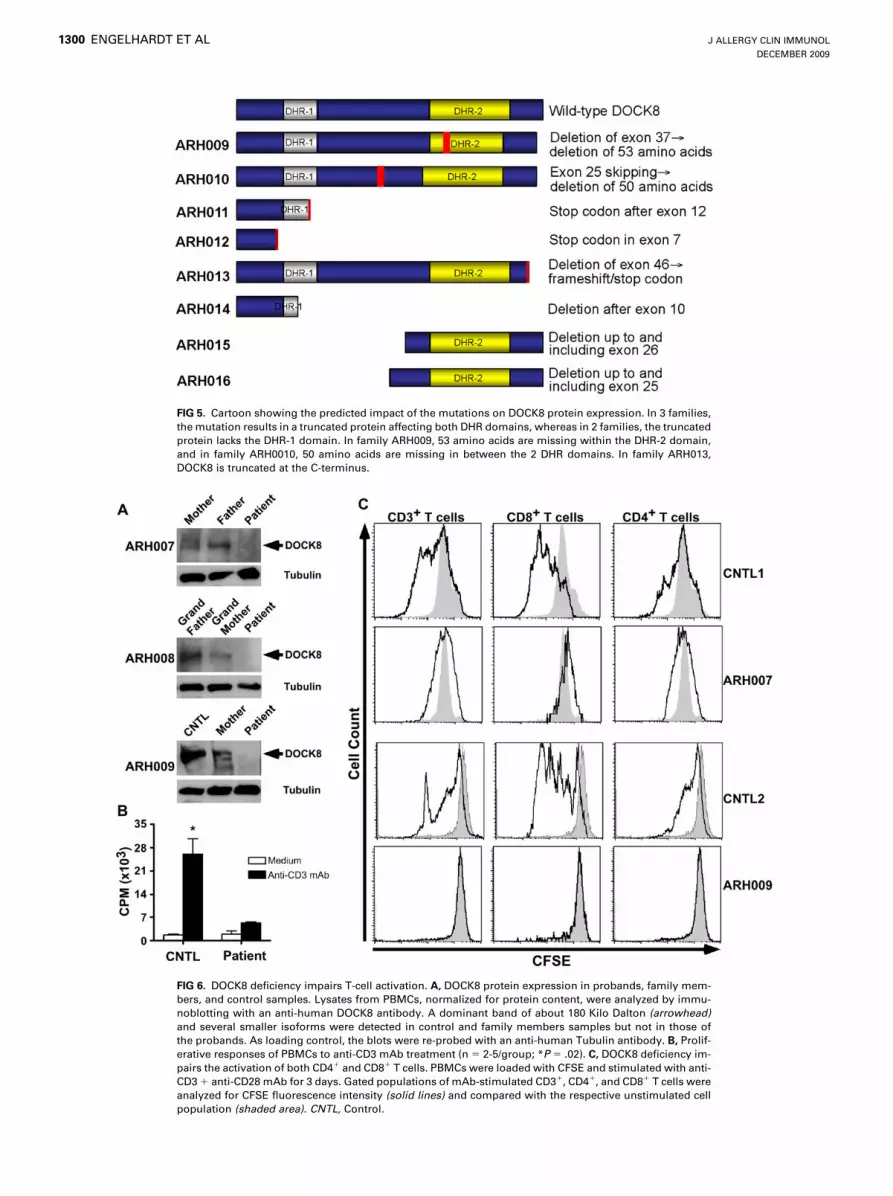
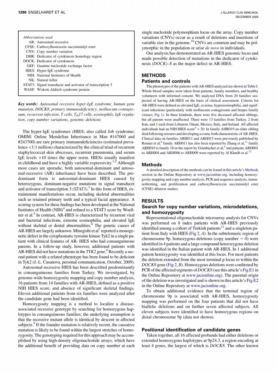
FIG 5. Cartoon showing the predicted impact of the mutations on DOCK8 protein expression. In 3 families,
the mutation results in a truncated protein affecting both DHR domains, whereas in 2 families, the truncated
protein lacks the DHR-1 domain. In family ARH009, 53 amino acids are missing within the DHR-2 domain,
and in family ARH0010, 50 amino acids are missing in between the 2 DHR domains. In family ARH013,
DOCK8 is truncated at the C-terminus.
FIG 6. DOCK8 deficiency impairs T-cell activation. A, DOCK8 protein expression in probands, family mem-
bers, and control samples. Lysates from PBMCs, normalized for protein content, were analyzed by immu-
noblotting with an anti-human DOCK8 antibody. A dominant band of about 180 Kilo Dalton (arrowhead)
and several smaller isoforms were detected in control and family members samples but not in those of
the probands. As loading control, the blots were re-probed with an anti-human Tubulin antibody. B, Prolif-
erative responses of PBMCs to anti-CD3 mAb treatment (n 5 2-5/group; *P 5 .02). C, DOCK8 deficiency im-
pairs the activation of both CD41 and CD81 T cells. PBMCs were loaded with CFSE and stimulated with anti-
CD3 1 anti-CD28 mAb for 3 days. Gated populations of mAb-stimulated CD31, CD41, and CD81 T cells were
analyzed for CFSE fluorescence intensity (solid lines) and compared with the respective unstimulated cell
population (shaded area). CNTL, Control.
J ALLERGY CLIN IMMUNOL
DECEMBER 2009
1300 ENGELHARDT ET AL
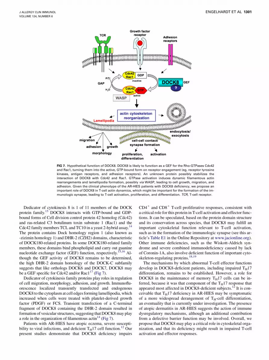
FIG 7. Hypothetical function of DOCK8. DOCK8 is likely to function as a GEF for the Rho-GTPases Cdc42
and Rac1, turning them into the active, GTP-bound form on receptor engagement (eg, receptor tyrosine
kinases, antigen receptors, and adhesion receptors). An unknown protein possibly stabilizes the
interaction of DOCK8 with Cdc42 and Rac1. GTPase activation induces dynamic filamentous actin
rearrangements and lamellipodia formation, possibly via WASP, leading to cell growth, migration, and
adhesion. Given the clinical phenotype of the AR-HIES patients with DOCK8 deficiency, we propose an
important role of DOCK8 in T-cell actin dynamics, which might be important for the formation of the im-
munologic synapse, leading to T-cell activation, proliferation, and differentiation. TCR, T-cell receptor.
J ALLERGY CLIN IMMUNOL
VOLUME 124, NUMBER 6
ENGELHARDT ET AL 1301
Dedicator of cytokinesis 8 is 1 of 11 members of the DOCKprotein family.15 DOCK8 interacts with GTP-bound and GDP-bound forms of Cell division control protein 42 homolog (Cdc42)and ras-related C3 botulinum toxin substrate 1 (Rac1) and theCdc42 family members TCL and TC10 in a yeast 2-hybrid assay.14
The protein contains Dock homology region 1 (also known as-zizimin homology 1) and DHR-2 (CZH2) domains, characteristicof DOCK180-related proteins. In some DOCK180-related familymembers, these domains bind phospholipid and carry out guaninenucleotide exchange factor (GEF) function, respectively.15,16 Al-though the GEF activity of DOCK8 remains to be determined,the high DHR-2 domain homology of the DOCK-C subfamilysuggests that like orthologs DOCK6 and DOCK7, DOCK8 maybe a GEF specific for Cdc42 and/or Rac117 (Fig 7).
Dedicator of cytokinesis family proteins play roles in regulationof cell migration, morphology, adhesion, and growth. Immunoflu-orescence localized transiently transfected and endogenousDOCK8 to the cytoplasm at cell edges forming lamellipodia, whichincreased when cells were treated with platelet-derived growthfactor (PDGF) or FCS. Transient transfection of a C-terminalfragment of DOCK8 containing the DHR-2 domain resulted information of vesicular structures, suggesting that DOCK8 may playa role in the organization of filamentous actin14 (Fig 7).
Patients with AR-HIES have atopic eczema, severe suscepti-bility to viral infections, and deficient TH17 cell function.12 Ourpresent studies demonstrate that DOCK8 deficiency impairs
CD41 and CD81 T-cell proliferative responses, consistent witha critical role for this protein in T-cell activation and effector func-tions. It can be speculated, based on the protein domain structureand its conservation across species, that DOCK8 may fulfill animportant cytoskeletal function relevant to T-cell activation,such as in the formation of the immunologic synapse (see this ar-ticle’s Table E1 in the Online Repository at www.jacionline.org).Other immune deficiencies, such as the Wiskott-Aldrich syn-drome and severe combined immunodeficiency caused by lackof Coronin-1A, also involve deficient function of important cyto-skeleton-regulating proteins.18,19
The mechanisms by which abnormal T-cell effector functionsdevelop in DOCK8-deficient patients, including impaired TH17differentiation, remains to be established. However, a role forDOCK8 in the maintenance of memory TH17 cells can be in-ferred, because it was that component of the TH17 response thatappeared most affected in DOCK8-deficient subjects.12 It is con-ceivable that TH17 deficiency in AR-HIES may be symptomaticof a more widespread derangement of TH-cell differentiation,an eventuality that is currently under investigation. The presenceof atopic dermatitis in AR-HIES suggests the action of immunedysregulatory mechanisms, although an additional contributionfrom a defective barrier function may be involved. Overall, wepropose that DOCK8 may play a critical role in cytoskeletal orga-nization, and that its deficiency might result in impaired T-cellactivation and effector responses.

J ALLERGY CLIN IMMUNOL
DECEMBER 2009
1302 ENGELHARDT ET AL
Two unrelated patients have been previously described withmental retardation and developmental disability associated withheterozygous disruption of DOCK8 by deletion and by a trans-location breakpoint, respectively. Immunologic abnormalitieswere not described in these patients. Mapping of the critical re-gion shared by the 2 patients showed truncation of the longestisoform of DOCK8. However, these patients had single-copydeletions of DOCK8 along with disruptions of other genes,13,20
which may explain why their phenotype differed from those ofour patients.
After the initial submission of our work, Zhang et al11 pub-lished homozygous and compound heterozygous mutations inDOCK8 in a cohort of 12 patients from 8 families. One of theirfamilies (#8 in Zhang et al11) is family ARH011 in our article.They described the phenotype as a ‘‘combined T- B- NK- immu-nodeficiency’’ and presented evidence of decreased T, B, and NK–cell numbers. Our assessment of these published cases would sug-gest that they also have an AR form of HIES, given that the majordiagnostic criteria of elevated IgE, eosinophilia, recurrent pneu-monia, and skin eczema together with a susceptibility to viral in-fections were fulfilled in all but 1 patient. However, in contrastwith the findings by Zhang et al,11 studies on the cohort presentedherein demonstrate a selective decrease in CD41 T-cell numbersin many but not all patients. The CD81 T-cell population is lessaffected, whereas B and NK cells are usually within the normalrange. Furthermore, whereas Zhang et al11 found a selective de-fect in CD81 T-cell activation, our findings are consistent witha more comprehensive T-cell activation defect involving boththe CD41 and CD81 T-cell subsets. Such a global defect inT-cell activation may explain the severe clinical phenotype ofAR-HIES.
Dedicator of cytokinesis 8 deficiency is characterized byrecurrent infections of the upper and lower respiratory tract;susceptibility to severe, recurrent, and mutilating viral infections(especially by molluscum contagiosum and herpes viruses); asevere dermatitis that resembles atopic dermatitis and may oftenbe superinfected; elevated IgE levels; and eosinophilia. Otherclinical features such as asthma, allergies, central nervous systemsymptoms, or autoimmune phenomena are variably associated.Whether there is a genotype-phenotype correlation with regard tothe size of the deletion or whether other deleted genes next toDOCK8 contribute to the phenotype needs to be determined infuture studies. It is remarkable that the typical feature of cyst-forming pneumonia (pneumatoceles), as frequently seen inSTAT3-mutated HIES, is not typical for DOCK8 deficiency andcan be used as a discriminating feature.
Subtelomeric deletions often arise at sites of interspersedrepetitive genomic sequences such as Alu, long interspersedelements, long terminal repeats, and simple tandem re-peats.21,22 The region surrounding DOCK8 is particularlyrich in these sequences, with 264 such sequences in the regionfrom the p terminus to the end of DOCK8 ( National Center forBiotechnology Information Alu database). In particular, thereare Alu-Jb sequences near the breakpoints of our patients asmapped by microarray analysis. Abnormal recombination ortransposable element activity could contribute to the high fre-quency of deletions seen. Careful analysis of the breakpointsof these deletions will be helpful in delineating which func-tions of DOCK8 are affected by the deletions, potentiallyshedding light on the role of DOCK8 in immune and integu-ment function.
We thank Cindy Ng and Anupama Rambhatla for technical support, Jennifer
Birmelin for collecting healthy control samples, Erik Glocker for sequencing
controls, Alejandro A. Schaffer and Michael Gertz for help with homozygosity
mapping, and Othmar Engelhardt for critical reading of the manuscript.
Clinical implications: Homozygous mutations in DOCK8 wereidentified in most patients with AR-HIES. Hence patients witha phenotype of elevated IgE, eosinophilia, and recurrent skinboils, pneumonia, and viral infections (especially molluscumcontagiosum and herpes) should be suspected of having muta-tions in DOCK8.
REFERENCES
1. Grimbacher B, Holland SM, Gallin JI, Greenberg F, Hill SC, Malech HL, et al.
Hyper-IgE syndrome with recurrent infections—an autosomal dominant multisys-
tem disorder. N Engl J Med 1999;340:692-702.
2. Grimbacher B, Holland SM, Puck JM. Hyper-IgE syndromes. Immunol Rev 2005;
203:244-50.
3. Holland SM, DeLeo FR, Elloumi HZ, Hsu AP, Uzel G, Brodsky N, et al. STAT3
mutations in the hyper-IgE syndrome. N Engl J Med 2007;357:1608-19.
4. Grimbacher B, Schaffer AA, Holland SM, Davis J, Gallin JI, Malech HL, et al.
Genetic linkage of hyper-IgE syndrome to chromosome 4. Am J Hum Genet
1999;65:735-44.
5. Woellner C, Gertz EM, Schaffer AA, Lagos M, Perro M, Glocker EO, et al. Mutations
in the signal transducer and activator of transcription 3 (STAT3) and diagnostic guide-
lines for the Hyper-IgE syndrome. J Allergy Clin Immunol 2009. in press.
6. Renner ED, Puck JM, Holland SM, Schmitt M, Weiss M, Frosch M, et al. Auto-
somal recessive hyperimmunoglobulin E syndrome: a distinct disease entity.
J Pediatr 2004;144:93-9.
7. Minegishi Y, Saito M, Morio T, Watanabe K, Agematsu K, Tsuchiya S, et al.
Human tyrosine kinase 2 deficiency reveals its requisite roles in multiple cytokine
signals involved in innate and acquired immunity. Immunity 2006;25:745-55.
8. Woellner C, Schaffer A, Puck JM, Renner ED, Knebel C, Holland SM, et al. The
hyper IgE syndrome and mutations in TYK2. Immunity 2007;26:535.
9. Kristiansson K, Naukkarinen J, Peltonen L. Isolated populations and complex dis-
ease gene identification. Genome Biol 2008;9:109.
10. Iafrate AJ, Feuk L, Rivera MN, Listewnik ML, Donohoe PK, Qi Y, et al. Detection
of large-scale variation in the human genome. Nat Genet 2004;36:949-51.
11. Zhang Q, Davis JC, Lamborn IT, Freeman AF, Jing H, Favreau AJ, et al. Combined
immunodeficiency associated with DOCK8 mutations. N Engl J Med 2009 Sep 23
[Epub ahead of print].
12. Al Khatib S, Keles S, Garcia-Lloret M, Karakoc-Aydiner E, Reisli I, Artac H, et al.
Defects along the T(H)17 differentiation pathway underlie genetically distinct
forms of the hyper IgE syndrome. J Allergy Clin Immunol 2009;124:342-8.
13. Griggs BL, Ladd S, Saul RA, DuPont BR, Srivastava AK. Dedicator of cytokinesis
8 is disrupted in two patients with mental retardation and developmental disabil-
ities. Genomics 2008;91:195-202.
14. Ruusala A, Aspenstrom P. Isolation and characterisation of DOCK8, a member of
the DOCK180-related regulators of cell morphology. FEBS Lett 2004;572:159-66.
15. Cote JF, Vuori K. Identification of an evolutionarily conserved superfamily of
DOCK180-related proteins with guanine nucleotide exchange. J Cell Sci 2002;
115:4901-13.
16. Yang J, Zhang Z, Roe SM, Marshall CJ, Barford D. Activation of Rho GTPases by
DOCK exchange factors is mediated by a nucleotide sensor. Science 2009;325:
1398-402.
17. Miyamoto Y, Yamauchi J, Sanbe A, Tanoue A. Dock6, a Dock-C subfamily gua-
nine nucleotide exchanger, has the dual specificity for Rac1 and Cdc42 and regu-
lates neurite outgrowth. Exp Cell Res 2007;313:791-804.
18. Bouma G, Burns SO, Thrasher AJ. Wiskott-Aldrich syndrome: immunodeficiency
resulting from defective cell migration and impaired immunostimulatory activa-
tion. Immunobiology 2009;214:778-90.
19. Shiow LR, Roadcap DW, Paris K, Watson SR, Grigrova IL, Lebet T, et al. The ac-
tin regulator coronin 1A is mutant in a thymic egress–deficient mouse strain and in
a patient with severe combined immunodeficiency. Nat Immunol 2008;9:1307-15.
20. MacDermot KD, Hulten M. Female with hypohidrotic ectodermal dysplasia and de
novo (X;9) translocation: clinical documentation of the AnLy cell line case. Hum
Genet 1990;84:577-9.
21. Stankiewicz P, Lupski JR. Genome architecture, rearrangements and genomic dis-
orders. Trends Genet 2002;18:74-82.
22. Yatsenko SA, Brundage EK, Roney EK, Cheung SW, Chinault AC, Lupski JR. Mo-
lecular mechanisms for subtelomeric rearrangements associated with the 9q34.3
microdeletion syndrome. Hum Mol Genet 2009;18:1924-36.

REFERENCES
E1. Carvalho B, Speed TP, Irizarry RA. Exploration, normalization, and genotype
calls of high density oligonucleotide SNP array data. Biostatistics 2007;8:485-99.
E2. Purcell S, Neale B, Todd-Brown K, Thomas L, Ferreira MAR, Bender D, et al.
PLINK: a toolset for whole-genome association and population-based linkage
analysis. Am J Hum Genet 2007;81:559-75.
E3. Stanczak CM, Chen Z, Nelson SF, Suchard M, McCabe ER, McGhee S. Repre-
sentational oligonucleotide microarray analysis (ROMA) and comparison of bin-
ning and change-point methods of analysis: application to detection of
del22q11.2 (DiGeorge) syndrome. Hum Mutat 2008;29:176-81.
E4. Olshen AB, Venkatraman ES, Lucito R, Wigler M. Circular binary segmentation for
the analysis of array-based DNA copy number data. Biostatistics 2004;5:557-72.
E5. Venkatraman ES, Olshen AB. A faster circular binary segmentation algorithm for
the analysis of array CGH data. Bioinformatics 2007;15:657-63.
E6. Duret L, Galtier N. Biased gene conversion and the evolution of mammalian
genomic landscapes. Annu Rev Genom Human Genet 2009;10:285-311.
J ALLERGY CLIN IMMUNOL
VOLUME 124, NUMBER 6
ENGELHARDT ET AL 1302.e1
METHODS
Homozygosity mapping and copy number analysisGenomic DNA samples from affected individuals and parents were
amplified and hybridized to Affymetrix 250 k Nsp or 6.0 SNP Mapping
Arrays (Affymetrix, San Jose, Calif) according to the manufacturer’s
standard protocol. Call rates ranged from 88% to 93%. This resulted in 20
affected individuals available for analysis. The arrays were analyzed with the
crlmm package v. 1.0 using R v. 2.9.2.E1 For the purpose of homozygosity
mapping, single nucleotide polymorphisms in which there was a low confi-
dence call (<0.99 by crlmm) were excluded from analysis. Replication anal-
ysis showed that this reduced the genotyping error rate to <1% (data not
shown). After preprocessing, 65,600 SNPs were available for homozygosity
mapping, which was performed by using PLINK v.1.06.E2 Extended
stretches of homozygosity were presumed to be identical by descent in con-
sanguineous matings.
Copy number analysis was performed by comparing signal intensities
from family members to a single standard reference individual as previously
reported.E3 Briefly, the subject and reference arrays were normalized, and the
sums of signal intensities from both alleles were compared after adjusting for
effects of PCR efficiency. The log base 2 ratio of the subject to the reference
was plotted against physical position on the chromosome, and CNV regions
of reduced or expanded copy number were identified by circular binary seg-
mentation.E4,E5 All CNV analysis was performed by using the DNAcopy
package v. 1.16 and R v. 2.9.2. Regions of apparent low copy number were
confirmed by quantitative PCR of coding exons from genes in the CNV
regions.
PCR and sequence analysisGenomic DNA was extracted from whole blood by using a Gentra
Puregene purification kit (Qiagen, Crawley, United Kingdom). For some
patients and family members, total RNA was prepared from blood, and
cDNA was synthesized as previously described. Exons and flanking intron/
exon boundaries from DOCK8 were amplified from genomic DNA by PCR
according to standard protocols with Taq polymerase from PeqLab (Fare-
ham, United Kingdom). Primer sequences are available on request. PCR
products were purified by using shrimp alkaline phosphatase (Promega,
Madison, Mich) and Exonuclease I (Thermo Scientific, Waltham, Mass);
DNA was sequenced with the ABI PRISM BigDye Terminator kit V3.1 (Ap-
plied Biosystems, Foster City, Calif), the 3130xl Applied Biosystems Ge-
netic Analyzer, DNA Sequencing Analysis software, version 5.2 (Applied
Biosystems), and Sequencher, version 4.8 (Gene Codes Corp, Ann Arbor,
Mich).
ImmunoblottingCells were lysed in 1% NP-40 Nonidet P-40, 150 mmol/L NaCl, 25 mM/L
HEPES, pH 7.4, and cell lysis buffer supplemented with protease inhibitors
(Roche, Indianapolis, IN). Whole cell lysates (50 mg/lane) were resolved by
SDS-PAGE and transferred to nitrocellulose filters. The latter were blocked
with 5% nonfat milk, then immunoblotted with polyclonal rabbit anti-
DOCK8 (Sigma-Aldrich, St. Louis, MO). The blots were developed by using
horseradish peroxidase–conjugated secondary antibodies and enzyme-linked
chemiluminescence, and exposed to film.
Proliferation studiesPBMCs were resuspended at 1 3 106 cells/mL in RPMI medium supple-
mented with 10% FCS. The cells were seeded in 96-well plates at 2 3 105
cells/well and were left otherwise untreated or were stimulated with 1 mg/
mL anti-CD3 (OKT3, Ortho Biotech). After 3 days, the cell cultures were
pulsed with 3H-thymidine at 0.4 mCi and analyzed for radioisotype incorpora-
tion by using a b-counter.
CFSE dilution studiesPBMCs were suspended at 107/mL in PBS and loaded with 5 mmol/L CFSE
(Invitrogen) for 15 min in the dark, washed, and resuspended at 2 3 106/mL in
RPMI/10% FCS. CFSE-labeled cells were placed in culture and treated either
with 50 U/mL recombinant human IL-2 (Peprotech) (control cells) or stimulated
with anti-CD3 plus anti-CD28 mAb coated beads (Miltenyi Biotech) for 3 days.
The cells were then harvested and analyzed for CFSE fluorescence by flow
cytometry. T-cell subsets were concurrently identified using phycoerythrin–
anti-CD4 and APC–anti-CD8 antibodies (BD Biosciences, San Jose, California).

FIG E1. PCR identification of homozygous deletions affecting DOCK8 in pa-
tients with AR-HIES with CNV abnormalities at 9p. A, The deletion in
ARH002 ends between E25 and E26, whereas that of ARH003 ends between
E32 and E33. B, The deletion in ARH001 ends between E3 and E4. All 3 de-
letions were found to extend proximally beyond exon 1, but their precise 59�edges have not been established. Note that ARH006, who has a heterozy-
gous deletion at 9p, successfully amplifies the respective exons, as would
be expected given his possession of an undeleted copy of DOCK8. CNTL,
Control.
J ALLERGY CLIN IMMUNOL
DECEMBER 2009
1302.e2 ENGELHARDT ET AL

FIG E2. Heritability of copy loss in 4 families with AR-HIES. ARH004 shows typical mendelian inheritance of
a deletion. ARH006 demonstrates partial repair of the CNV, presumably by gene conversion.E6 ARH001
shows a de novo homozygous deletion. Parentage for ARH001 was confirmed by SNP inheritance. No men-
delian errors were found in 44,645 genotypes surveyed. ARH007 is a patient without a deletion but with
extensive homozygosity at this locus.
J ALLERGY CLIN IMMUNOL
VOLUME 124, NUMBER 6
ENGELHARDT ET AL 1302.e3

TABLE E1. Proteins similar to DOCK8 are found in a variety of euteleosts, suggesting evolutionary conservation in greater than 90% of
vertebrate organisms
Species Symbol Protein DNA d dN/dS dNR/dNC
Pan troglodytes DOCK8 99.1 99.1 0.009 0 0
Canis lupus familiaris DOCK8 94 90.4 0.103 0 0
Mus musculus DOCK8 91.8 87.1 0.141 0 0
Rattus norvegicus DOCK8 92 87.4 0.138 0 0
Gallus gallus DOCK8 82.3 76.3 0.285 0 0
Danio rerio DKEY-91M11.1 68.3 66.9 0.436 0 0
The table gives the percentage protein or DNA homology between Homo sapiens and the listed model organisms, as well as the number of nucleotide substitutions per site (d) and
the ratio of nonsynonymous to synonymous substitutions (dN/dS or dNR/dNC). HomoloGene, http://www.ncbi.nlm.nih.gov/homologene/, accessed 10/15/09.
J ALLERGY CLIN IMMUNOL
DECEMBER 2009
1302.e4 ENGELHARDT ET AL


