Embed Size (px)
Citation preview
PROCEEDINGS OF A WORKSHOP ON
FRESHWATER
LARVALFISHES
Held atKnoxville,TennesseeFebruary 21-22, 1978
EDITORS:ROBERT WALLUS&
CLYDE W. VOIGTLANDER
Tennessee Valley AuthorityDivision of Forestry, Fisheries, and Wildlife Development
Norris, TN 378281979

This document is dedicated to the memory ofDoyne Richard Martin, 1945-1977. Dickwas a fisheries biologist with TVA for three years.
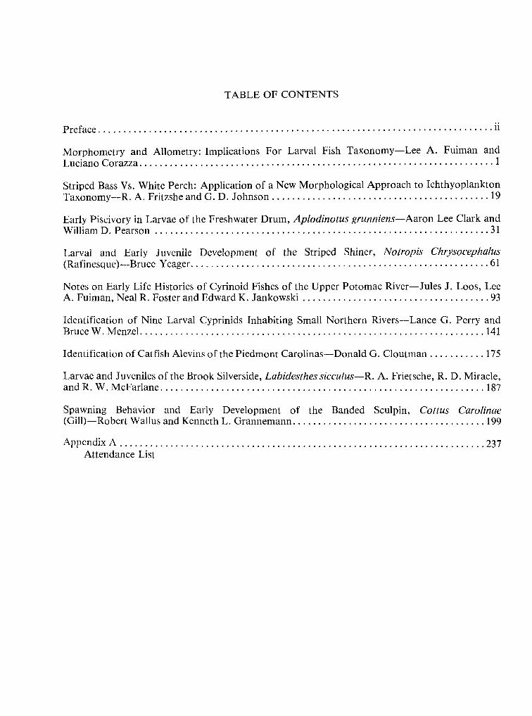
TABLE OF CONTENTS
Preface ii
Morphometry and Allometry: Implications For Larval Fish Taxonomy— Lee A. Fuiman andLuciano Corazza 1
Striped Bass Vs. White Perch: Application of a New Morphological Approach to IchthyoplanktonTaxonomy —
R. A.Fritzshe and G. D.Johnson 19
Early Piscivory inLarvae of the Freshwater Drum, Aplodinotus grunniens— Aaron Lee Clark andWilliamD.Pearson 31
Larval and Early Juvenile Development of the Striped Shiner, Notropis Chrysocephalus(Rafinesque) —Bruce Yeager 61
Notes on Early LifeHistories of Cyrinoid Fishes of the Upper Potomac River—
Jules J. Loos, LeeA.Fuiman, Neal R. Foster and Edward K. Jankowski 93
Identification of Nine Larval Cyprinids Inhabiting Small Northern Rivers —Lance G. Perry andBruce W.Menzel 141
Identification ofCatfish Alevins of the Piedmont Carolinas —Donald G. Cloutman 175
Larvae and Juveniles ofthe Brook Silverside, Labidesthes sicculus—
R. A.Frietsche, R. D.Miracle,and R. W. McFarlane 187
Spawning Behavior and Early Development of the Banded Sculpin, Cottus Carolinae(Gill)—Robert Wallus and Kenneth L.Grannemann 199
Appendix A 237Attendance List
ii
PREFACE
On February 21-22, 1978, the Fisheries and Waterfowl Resources Branch, Division of
Forestry, Fisheries, and Wildlife Development of the Tennessee Valley Authority sponsored and
hosted a freshwater larval fish workshop in Knoxville,Tennessee.
The theme of the workshop was "Current Trends inLarval Fish Taxonomy and Early
Life History Studies." Objectives were to:
1. discuss current problem areas in larval fish taxonomy;
2. discuss current research inother aspects of the early life history of freshwater fishes; and
3. share ideas as well as new laboratory and field techniques and methodologies.
The meeting consisted of one day of formally presented papers and one day ofinfor-mal laboratory workshop.
Fifty-five individuals representing 29 agencies attended the meeting. Universities, con-
sulting companies, private power companies, State and Federal agencies, museums, and research
institutes were represented.
Of the 12 formal presentations made at this workshop, 9 are presented herein.
MORPHOMETRY AND ALLOMETRY:
IMPLICATIONS FOR LARVALFISH TAXONOMY
by
Lee A.Fuiman and Luciano CorazzaDepartment of Natural Resources
Cornell UniversityIthaca, New York 14853
ABSTRACT
Basic principles and problems ofrelative body sizes of fishes are discussed. Allometry
was observed in five species ofsucker (catostomid) larvae. This non-linear relationship ofone body
part to another was common to nearly allspecies and morphometric parameters recorded. Log-log
plots of morphometric data revealed a curvilinear relationship which was satisfactorily approx-
imated bymultiple regression lines. A character was constructed using the allometry data which pro-
vided better discriminating capabilities than the conventional character of percent total length. A
method for graphic identification of species is also discussed.
3
INTRODUCTION
Basic types of characters useful in fish taxonomy are: meristic, morphometric, pigmen-
tary, and specialized (i.e., peculiar to a given taxon). A fifthtype, applicable to larval fishes, is size
or age at a given developmental state and might be termed morphological. Meristic characters are
most reliable in fish taxonomy because enumeration errors are frequently small and statistical
manipulations are not difficult. Unfortunately, larval fishes have few significant countable struc-
tures. Pigmentary characters vary considerably with factors such as age, sex, and substrate color
and are difficult to quantify for statistical treatment. Specialized and morphological characters are
oflimited value for similar reasons. Intaxonomic studies morphometry is typically presented as per-
cent of some standard. This method assumes that there is a constant ratio of a part of the body to
the standard (isometry). This is not a valid assumption for many dimensions of larval fishes.
This investigation is a continuation ofprevious studies of larval fish development. In
descriptions of three species of cyprinid larvae, Fuiman and Loos (1977, 1978) found allometric
trends (i.e., body proportions varied with total length through the entire larval period). Otherworkers (Doan, 1939; Martin, 1949) have noted allometry in fish larvae, but most studies have dealtwith only the later portion of the larval period. The present study is intended to reacquaint tax-
onomists with the principles and problems ofrelative body sizes inthe lightof recent interest in iden-
tification of larval fishes. Specific examples of allometric growth throughout the larval period are
presented with suggestions for construction of useful characters from the data.
METHODS
Materials and data used in this study are part of a descriptive investigation of north-eastern catostomids (Fuiman, 1978). Larvae were reared ina laboratory at 20 C witha 14 hdaylightphotoperiod. Measurements were made to the nearest 0.01 mm with a dissecting microscope and
ocular micrometer. Four body dimensions, standard length (SL), preanal length (PAL),head length
(HL),and eye diameter (ED), were selected as examples for this study. Data were grouped according
to common integer values for total length (TL), e.g. all individuals of a species between 9.00 and
9.99 mm TL were grouped together. Mean values for each group were plotted on log-log coor-
dinates. Points of inflection, indicating a change in growth stanza, were chosen by inspection.
Regression lines were calculated using Bartlett's (1949) method because values of both variables
4
were subject to error [Kidwell and Chase (1967) systematically compared ten methods for fitting
lines to relative growth data and concluded that Bartlett's method was most adequate]. Resulting
equations were used to estimate more accurately the inflection points and tomodel changes inbody
proportions.
RESULTS ANDDISCUSSION
Different types ofplots of the data are used in studying relative growth. These must be
defined at the outset to avoid confusion. Plots may be on linear or logarithmic coordinates. Theproportional plot represents the ratio ofa body part to the standard (TL).This is not tobe confusedwith the plot of body part against the standard. As previously stated, isometric growth is theassumption underlying the present use of morphometric characters. In a case of isometry a linearplot of a body proportion against TL would yield a horizontal line, its intercept being the mean
value of that character for the taxon. This is true only when the body part-TL plot is linear and
passes through the origin. A non-zero intercept yields a curve, on the proportional plot, which is
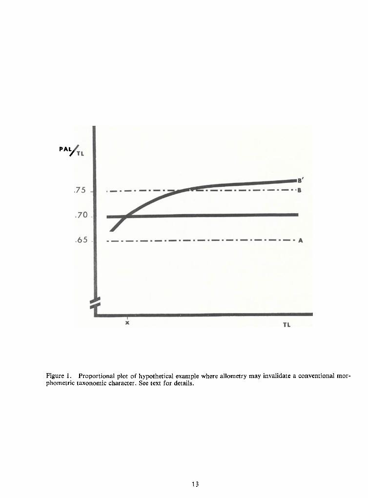
asymptotic to the mean value for the taxon (Marr, 1955). Inallometric growth the proportional plot
is logarithmic. If growth were assumed to be isometric, taxonomic difficulties could arise. For
example, Taxa A and B are routinely distinguished by the following couplet:
This character may have been based on discrete samples representing a small size range
with mean PAL values of 65 and 75 percent, respectively (Figure 1). Alternatively, these values
could have been derived from specimens occupying a growth stanza which approximated isometry
but followed an allometric stanza. Examination of a broader size range in taxon Bmay indicate that
growth is allometric (line B' of Figure 1). Therefore, specimens of taxon B smaller than Xmm TL
would be identified incorrectly according to the proposed couplet.
Allometry is predominant in the early growth (to 25 mm TL) of the five catostomidspecies studied. Huxley (1932) proposed the equation, V
-bXk as a model for relative growth. This
equation is linear in its logarithmic form: log V = k log X + log b, where k is the growth coefficientand b is a constant related to the units ofmeasure. Isometric growth is represented by this equation
greater than 70% TL Breanal length
ess than 70% TL Areanal lengthreanal length ess than 70% TL A
reanal length greater than 70% TL B
5
when k-
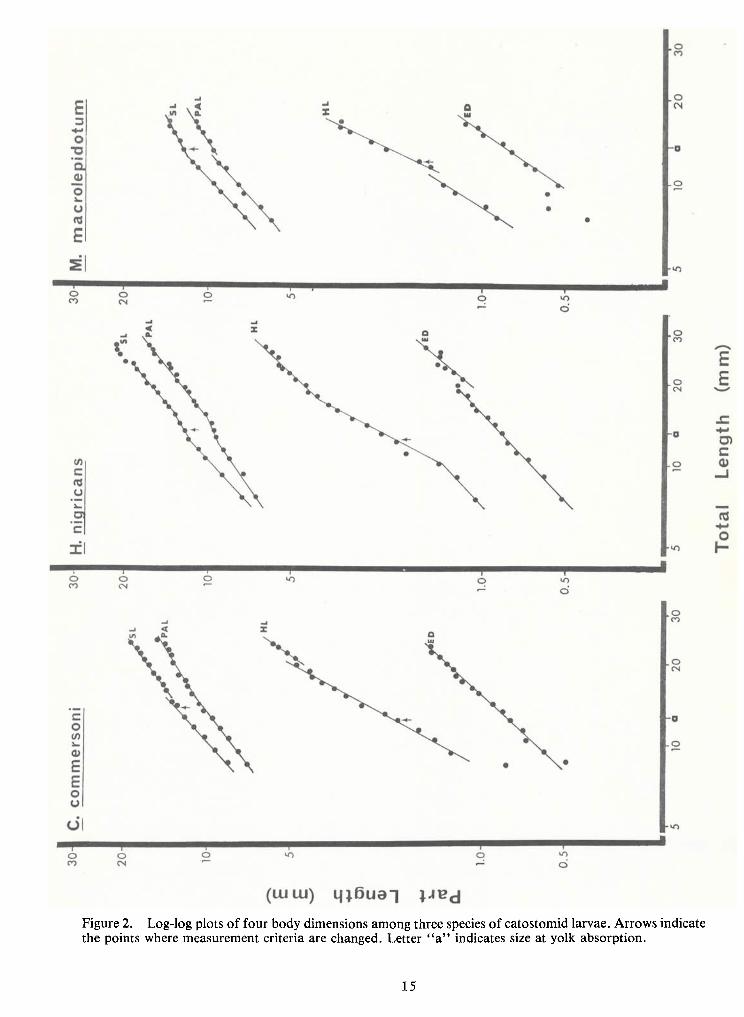
1. Log-log plots of body parts against TL (Figures 2 and 3; Table 1) show allometric
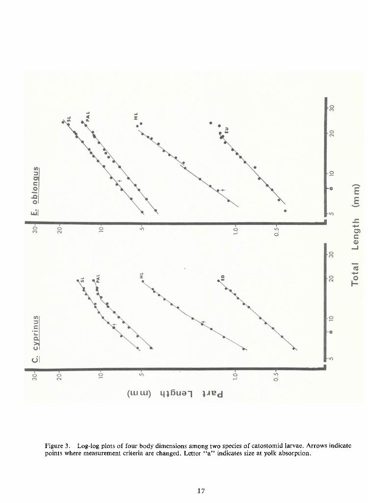
growth innearly all species and body parts. Only SL and PALof Carpiodes cyprinus, the quillback,
and ED of Hypentelium nigricans, northern hogsucker, approximate isometric growth (k-
1.01,
1.01 and 0.97, respectively). Most k values are less than unity, indicating negative allometry (i.e., the
body part grows at a slower rate than TL). Cases ofpositive allometry (k > 1) are restricted to in-
creases in eye diameter and head length.
Multiple growth stanzas, i.e., intervals of growth with different rates of change (k
values), are found in most of these graphs. They become apparent when straight lines are fitted to
log-log plots of relative growth data. There are at least two stanzas for SL, PAL and HL
measurements ineach species except Erimyzon oblongus. Data for H.nigricans may be resolved in-
to three stanzas for these same dimensions and into two stanzas for ED. Eye diameters of the re-
maining four species are represented by single allometric lines.
Martin (1949) explained growth stanzas as being the result ofphysiological changes in
the organism. Change from an endogenous to exogenous food source would seem to be a major
physiological crisis during the larval period. Size at which yolk is typically absorbed is noted by the
letter "a" on the abscissae of Figures 2 and 3. These points do not correspond with changes in
growth stanzas. Discontinuities may be a result of changes inmeasurement criteria (e.g. head length
is measured to the posterior edge of the auditory vesicles until the cleithra ossify). Formation ofhypural elements may affect relative growth plots of SL. These points are noted on their respective
graphs by arrows and apparently do not cause the inflections (with the possible exception of SL inH. nigricans).
Growth stanzas may be an artifact of the linear approximation. Laird (1965) proposed
the use of a Gompertz equation in relative growth studies. The resulting sigmoid curve includes a
time component not found inHuxley's equation. Laird found that the curvilinear (Gompertz) rela-
tionship adequately describes the change in growth rate with time and that the use of multiple
straight lines has no biological significance.
Parr (1949) suggested a method for taxonomic treatment ofmorphometric data whichwas based on body proportion vs. body length plots. Ratio plots of this nature prohibit statistical
treatment and comparisons of the data (Marr, 1955). Complexity of the Gompertz equation (W = -
ae-be-kt) precludes its use in taxonomy. Multiple linear approximations of log-log graphs may
6
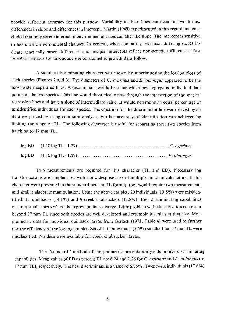
provide sufficient accuracy for this purpose. Variability in these lines can occur in two forms:
differences in slope and differences in intercept. Martin (1949) experimented in this regard and con-
cluded that only severe internal or environmental crises can alter the slope. The intercept is sensitive
to less drastic environmental changes. In general, when comparing two taxa, differing slopes in-
dicate genetically based differences and unequal intercepts reflect non-genetic differences. Two
possible methods for taxonomic use of allometric growth data follow.
A suitable discriminating character was chosen by superimposing the log-log plots ofeach species (Figures 2 and 3). Eye diameters of C. cyprinus and E. oblongus appeared to be the
most widely separated lines. Adiscriminant would be a line which best segregated individual datapoints of the two species. This line would theoretically pass through the intersection of the species'
regression lines and have a slope of intermediate value. Itwould determine an equal percentage ofmisidentified individuals for each species. The equation for the discriminant line was derived by an
iterative procedure using computer analysis. Further accuracy of identification was achieved by
limiting the range of TL.The following character is useful for separating these two species fromhatching to 17 mm TL.
logED (1.10 log TL- 1.27) C. cyprinus
IogED (1.10 logTL-1.27) E. oblongus
Two measurements are required for this character (TL and ED). Necessary log
transformations are simpler now with the widespread use of multiple function calculators. If thischaracter were presented in the standard percent TL form it,too, would require twomeasurements
and similar algebraic manipulation. Using the above couplet, 20 individuals (13.5%) were misiden-
tified: 11 quillbacks (14.1%) and 9 creek chubsuckers (12.8%). Best discriminating capabilities
occur at smaller sizes where the regression lines diverge. Littleproblem withidentification can occur
beyond 17 mm TL since both species are well developed and resemble juveniles at that size. Mor-
phometric data for individual quillback larvae from Gerlach (1973, Table 4) were used to further
test the efficiency of the log-log couplet. Six of 110 individuals (5.5%) smaller than 17 mm TL were
misclassified. No data were available for creek chubsucker larvae.
The "standard" method of morphometric presentation yields poorer discriminating
capabilities. Mean values ofED as percent TLare 6.24 and 7.26 for C. cyprinus and E. oblongus (to
17 mm TL),respectively. The best discriminant is a value of 6.75%. Twenty-six individuals (17.6%)
7
were misidentified: 14 quillbacks (17.9%) and 12 creek chubsuckers (17.1%). In this case the dif-
ference inmisclassified individuals using the two characters is four percent. Other data may give riseto similar characters with even greater efficiency over the conventional method.
A simpler approach is the direct use of logarithmic graphs and regressions. When data
are presented as inFigures 2 and 3, the taxonomist can easily compare his measurements with those
in the graphs. Species determinations can be based on the proximity of a data point to a regression
line. Confidence intervals for the regressions may be included on the graphs and would simplify tax-
onomic decisions.
Examination of the relative growth patterns indifferent species of fishes is important if
morphometric characters are to be used to characterize the species. Allometry often affects relative
dimensions ofbody parts, particularly at early developmental stages. This demands unconventional
treatment ifmorphometry is to be used taxonomically. On the other hand, improper analysis ofmorphometric variation can lead to the conclusion ofallometry from purely isometric data (Marr,
1955). When analyzed properly, morphometry can be a useful tool for the larval fish taxonomist.
9
LITERATURE CITED
Bartlett, M. S. 1949. Fitting a straight line when both variables are subject to error. Biometrics5:207-212.
Doan, K.H. 1939. Growth of bass fry. Copeia 1939(2):81-87.
Fuiman, L.A.1978. Descriptions and comparisons ofnortheastern catostomid fish larvae. Master'sThesis, Cornell University, Ithaca, New York. 110 pp.
Fuiman, L.A.and J. J. Loos. 1977. Identifying characters of the early development of the dacesRhinichthys atratulus and R. cataractae (Osteichthyes: Cyprinidae). Proceedings of theAcademy of Natural Sciences of Philadelphia. 129(2):23-32.
. 1978. Morphological changes during the larval development of the cutlips minnow,Exoglossum maxillingua. Trans. Amer. Fish. Soc. 107: (in press).
Gerlach, J. M. 1973. Early development of the quillback carpsucker Carpiodes cyprinus.Master's Thesis, Millersville State College, Millersville,Pennsylvania. 60 pp.
Huxley, J. S. 1932. Problems of Relative Growth. Methuen and Company, London. 276 pp.
Kidwell, J. F. and H. B. Chase. 1967. Fitting the allometric equation— a comparison of themethods by computer simulation. Growth 31:165-179.
Laird, A. K. 1965. Dynamics of relative growth. Growth 29:249-263.
Marr, J. C. 1955. The use ofmorphometric data in systematic, racial and relative growth studies infishes. Copeia 1955(1):23-31.
Martin, W. R. 1949. The mechanics of environmental control ofbody form in fishes. University ofToronto Studies, Biological Series 58: 91 pp.
Parr, A. E. 1949. An appropriate formula for stating taxonomically significant proportions offishes with reference to growth changes. Copeia 1949(1):47-55.
11
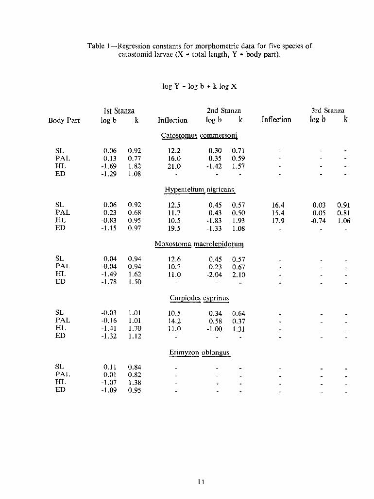
Table I—Regression1—
Regression constants for morphometric data for five species ofcatostomid larvae (X
-total length, V
-body part).
-1.09 0.95ED-1.07 1.38HL0.01 0.82PAL0.11 0.84SL
Erimyzon oblongus
-1.32 1.12ED11.0 -1.00 1.31-1.41 1.70HL14.2 0.58 0.37-0.16 1.01PAL10.5 0.34 0.64-0.03 1.01SL
Carpiodes cyprinus
-1.78 1.50ED11.0 -2.04 2.10-1.49 1.62HL10.7 0.23 0.67-0.04 0.94PAL12.6 0.45 0.570.04 0.94SL
Moxostoma macrolepidotum
19.5 -1.33 1.08-1.15 0.97ED-0.74 1.0617.910.5 -1.83 1.93-0.83 0.95HL0.05 0.8115.411.7 0.43 0.50• 0.23 0.68PAL0.03 0.9116.412.5 0.45 0.570.06 0.92SL
Hypentelium nigricans
-1.29 1.08ED21.0 -1.42 1.57-1.69 1.82HL16.0 0.35 0.590.13 0.77PAL12.2 0.30 0.710.06 0.92SL
Catostomus commersoni
logb kInflectionInflection log b klogb kBody Part3rd Stanza2nd StanzaIst Stanza
log V-
log b + k log Xlog V-
log b + k log X
Ist Stanza 2nd Stanza 3rd StanzaBody Part logb k Inflection log b k Inflection logb k
Catostomus commersoni
SL 0.06 0.92 12.2 0.30 0.71PAL 0.13 0.77 16.0 0.35 0.59HL -1.69 1.82 21.0 -1.42 1.57ED -1.29 1.08
Hypentelium nigricans
SL 0.06 0.92 12.5 0.45 0.57 16.4 0.03 0.91PAL • 0.23 0.68 11.7 0.43 0.50 15.4 0.05 0.81HL -0.83 0.95 10.5 -1.83 1.93 17.9 -0.74 1.06ED -1.15 0.97 19.5 -1.33 1.08
Moxostoma macrolepidotum
SL 0.04 0.94 12.6 0.45 0.57PAL -0.04 0.94 10.7 0.23 0.67HL -1.49 1.62 11.0 -2.04 2.10ED -1.78 1.50
Carpiodes cyprinus
SL -0.03 1.01 10.5 0.34 0.64PAL -0.16 1.01 14.2 0.58 0.37HL -1.41 1.70 11.0 -1.00 1.31ED -1.32 1.12
Erimyzon oblongus
SL 0.11 0.84PAL 0.01 0.82HL -1.07 1.38ED -1.09 0.95
13
PAI/u
Figure 1. Proportional plot of hypothetical example where allometry may invalidate a conventional mor-phometric taxonomic character. See text for details.
15
Figure 2. Log-logplots of four body dimensions among three species of catostomid larvae. Arrows indicatethe points where measurement criteria are changed. Letter "a" indicates size at yolk absorption.
17
Figure 3. Log-log plots of four body dimensions among two species of catostomid larvae. Arrows indicatepoints where measurement criteria are changed. Letter "a" indicates size at yolk absorption.
STRIPED BASS VS. WHITE PERCH: APPLICATION OF A NEW MORPHOLOGICAL
APPROACH TO ICHTHYOPLANKTON TAXONOMY
R. A. FritzscheDepartment of Biology, The University of Mississippi
University, Mississippi
and
G. D. JohnsonDivision of Fishes, National Museum of Natural History
Washington, D.C.
ABSTRACT
A technique has recently been developed to stain cartilage in whole cleared specimens.
This technique permits comparison of fish skeletons in their earliest stages of development (pre-
ossification). Preliminary investigations employing this technique indicate that larval striped bassand white perch exhibit diagnostic differences in the position and shape ofcertain skeletal elements,
particularly the predorsal bones, and that these differences are identifiable at the earliest appearance
of these elements as cartilage. This method should reduce or eliminate the subjectivity now
associated with larval striped bass-white perch identification and thus provide more rapid andpotentially 100 percent accurate identification of each species from about 8 mm TL.
21
INTRODUCTION
The striped bass (Morone saxatilis) is one of the most important sport and commercial
fish found along the Atlantic Coast ofNorth America. Because ofits importance, large amounts of
money and effort are spent on studies ofitspopulation dynamics and lifehistory. Alarge portion of
the research effort is concerned with the particularly vulnerable early life history stages.
Environmental perturbations adversely affecting the eggs and larvae of striped bass could have far
reaching effects on the population. Accurate identification of the eggs and larvae of this species is
therefore, crucial to the study of its biology.
Mansueti (1958, 1964) described the eggs and larvae of white perch (M. Americana) and
striped bass after stripping and fertilizing ripe ova from spawning fish. His studies provided the
basis for the sorting methods now used. However, it has become apparent that specimens between
6.0 and 20.0 mm TL are not unequivocally distinguishable using Mansueti's criteria. Morgan (1975)
used electrophoretic patterns of muscle proteins to distinguish white perch from striped bass. This
method, although very accurate, requires a great deal of time and the specimens are destroyed intheprocess. Sidell et al. (1978) developed a valid method ofbiochemical identification using starch gel
electrophoresis and stains for specific enzyme systems. They conclude, however, that routine elec-
trophoretic analysis of samples may be logistically difficult.
A recently developed cartilage staining technique (Dingerkus and Uhler, 1977) allows
examination of osteological development prior to complete ossification of the endoskeleton.Preliminary investigations with a modification of this technique indicate that larval striped bass and
white perch exhibit diagnostic differences in the position and shape of certain skeletal elements.These differences are identifiable at the earliest appearance of these elements as cartilage.
METHOD
Larval-juvenile series of fieldcollected M.saxatilis andM. Americana were cleared and
stained using modifications of the cartilage technique of Dingerkus and Uhler (1977). Cleared and
stained specimens were then examined for possible species-specific osteological differences. We
began by examining readily identifiable juveniles and then traced characters back through the size
series. As a check we examined a series of striped bass reared from yolk-sac larvae obtained from the
hatchery operated by the Virginia Commission of Game and Inland Fisheries at Brookneal,
Virginia.
22
Staining Technique:
The cartilage staining technique described by Dingerkus and Uhler (1977) was
simplified and modified for more rapid preparation of larval fish samples. The modified method
used for fish is the following:
1. Wash preserved material in two or three changes of distilled H2O for several hours or until no
trace of preservative can be detected.
2. Place directly into a mixture of 10 mg Alcian Blue (BGN or preferably BGS), 80 ml 95 percent
ethyl alcohol, and 20 ml glacial acetic acid for 12 to 24 hours or untilcartilage is well stained. Forvery small larvae this step may take only 12 hours.
3. Transfer through series of approximately 95 percent, 50 percent, 10 percent ethyl alcohol forabout one hour each, or untilspecimen(s) sink. The alcohol dilutions are made by adding distill-
ed water to the sample so they only need be approximate.
4. Transfer to distilled H2O for one hour, or untilspecimen sinks.
5. Place in sodium borate buffered trypsin enzyme solution (Taylor, 1967). Change solution ifit
takes on bluish color. Continue until specimen(s) is cleared and flesh retains no blue color. Thisstep usually takes one to two days.
6. Transfer specimen(s) to 50 percent glycerins {Vidistilled water-1/2glycerine) for sorting and iden-
tification. Transfer to 100 percent glycerine for long-term storage is recommended (several
crystals of thymol should be added to this solution).
By following the above simplified procedure a sample of fish larvae willbe ready forsorting and identification in two to three days. This technique leaves external and internal pigmenta-
tion intact so that pigment patterns remain available for identification purposes.
OBSERVATIONS ANDDISCUSSION
By following the above procedure we discovered that the shape and position of the
predorsal bones were diagnostically different in each of the two species, M. saxatilis and M.
americana. In addition, the compound interhaemal in M. Americana is much larger and more
robust.
23
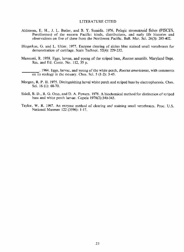
The predorsal bone pattern for M.saxatilis is typically 0/0/0/2 +1/1 + 1and that for
M. Americana is 0/0/0 + 2/1 + 1 (fig. 1) (for explanation of formula used see Ahlstrom et al. 1976).
From these patterns it is evident that the most striking difference is the more anterior position ofthe
first dorsal pterygiophore inM. Americana, i.e., between the second and third neural spines rather
than posterior to the third neural spine as inM.saxatilis.
The shape of the predorsal bones also differs between the species. InM.saxatilis, thepredorsal bones have a strong winglike flange developed posteriorly. Inaddition, the first predorsal
is strongly concave anteriorly (fig. IB).InM.americana, the predorsal bones are more rod-like and
are not strongly bent (fig. 1A).
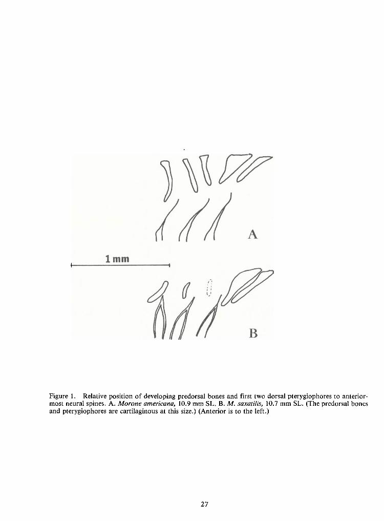
These differences in shape and position can be traced back to the earliest formation of
these elements as cartilage. The first (anterior-most) predorsal is the first of these elements to form,
at about 8.0 mm TL.InM.saxatilis, this first predorsal forms at approximately 45° from the ver-
tical so that the oval-shaped element appears to be "leaning backwards" (fig.2B). The first predor-
sal inM. Americana forms as a vertical rod (fig. 2A). The remaining two predorsals first appear at
about 10 mm TL.
At 9-10 mm TL, the position of the first dorsal pterygiophore relative to the neural
spines is readily apparent. The development of the neural spines preceeds that of the pterygiophores
and predorsal bones.
The compound interhaemal and associated second anal spines are sufficiently well
developed at about 15 mm TL that they can be used together with the predorsal pattern for iden-tification. The white perch has the larger compound interhaemal and second anal spine. Meristiccharacters can also be used at this time since the daring and staining process allows fast and accurate
fin-ray counting. However, there is a slight overlap in the range of fin-ray counts for the two
species.
From these observations we can now say that striped bass and white perch can be easily
identified from the onset of predorsal bone formation (about 8 mm TL). The only stage at whichthese two species cannot now be separated is between about 6.0 and 8.0 mm TL.We have some in-
dication that internal pigment patterns may be useful; however, these investigations are stillpreliminary and no unequivocal results have been obtained.
24
Specimens came from the Potomac River in Maryland and the hatchery in Brookneal,
Virginia. Specimens from other areas willbe examined to reveal possible geographical variation.
Final results willbe published in a more comprehensive study on the developmental osteology of
striped bass and white perch.
Preliminary observations on the white bass, Morone chrysops, indicate that this fish
has the same predorsal pattern as the white perch. Osteological characters may also aid in theseparation oflarvae ofthis species and the yellow bass, M.mississippiensis . The modification oftheDingerkus and Uhler cartilage staining technique may also result in solutions to other larval tax-
onomic problems, e.g., blue back herring, Alosa aestivalis vs. alewife, Alosa pseudoharengus.
ACKNOWLEDGEMENTS
We wish to thank Guido Dingerkus for providing the manuscript describing his car-
tilage staining technique while it was still inpress. Kathy Wood, Chesapeake Biological Laboratory,
supplied the field samples containing white perch and striped bass larvae. David Whitehurst,
Virginia Commission of Game and Inland Fisheries, supplied the laboratory spawned striped bass
larvae. Our former colleagues at the Chesapeake Biological Laboratory, Solomns, Maryland helped
rear striped bass larvae and also contributed much information during discussions of larval iden-
tification problems. This work was supported by contracts #2-72-02 (77 Mod. 2), #T2-72-02 (78
Mod. 3), and #P4B-78-04 with the Power Plant Siting Program, Department ofNatural Resources,
State of Maryland.
25
LITERATURE CITED
Ahlstrom, E. H., J. L. Butler, and B. Y. Sumida. 1976. Pelagic stromateoid fishes (PISCES,Perciformes) of the eastern Pacific: kinds, distributions, and early life histories andobservations on five of these from the Northwest Pacific. Bull. Mar. Sci. 26(3): 285-402.
Dingerkus, G. and L.Uhler. 1977. Enzyme clearing of alcian blue stained small vertebrates fordemonstration of cartilage. Stain Technol. 52(4): 229-232.
Mansueti, R. 1958. Eggs, larvae, and young of the striped bass, Roccus saxatilis. Maryland Dept.Res. and Ed. Contr. No. 112, 35 p.
. 1964. Eggs, larvae, and young of the white perch, Roccus americanus, with commentson its ecology in the estuary. Ches. Sci. 5 (1-2): 3-45.
Morgan, R. P. 11. 1975. Distinguishing larval white perch and striped bass by electrophoresis. Ches.Sci. 16 (1): 68-70.
Sidell, B. D., R. G. Otto, and D. A.Powers. 1978. Abiochemical method for distinction of stripedbass and white perch larvae. Copeia 1978(2): 340-343.
Taylor, W. R. 1967. An enzyme method of clearing and staining small vertebrates. Proc. U.S.National Museum 122 (3596): 1-17.
27
Figure 1. Relative position of developing predorsal bones and first two dorsal pterygiophores to anterior-most neural spines. A.Morone americana, 10.9 mm SL. B.M.saxatilis, 10.7 mm SL. (The predorsal bonesand pterygiophores are cartilaginous at this size.) (Anterior is to the left.)
29
Figure 2. Relative position ofpredorsal bones and first two dorsal pterygiophores to anterior-most neuralspines. A.Morone americana, 38.9 mm SL. B.M.saxatilis, 32.7 mm SL. (Stippled areas indicate cartilage.)(Anterior is to the left.)
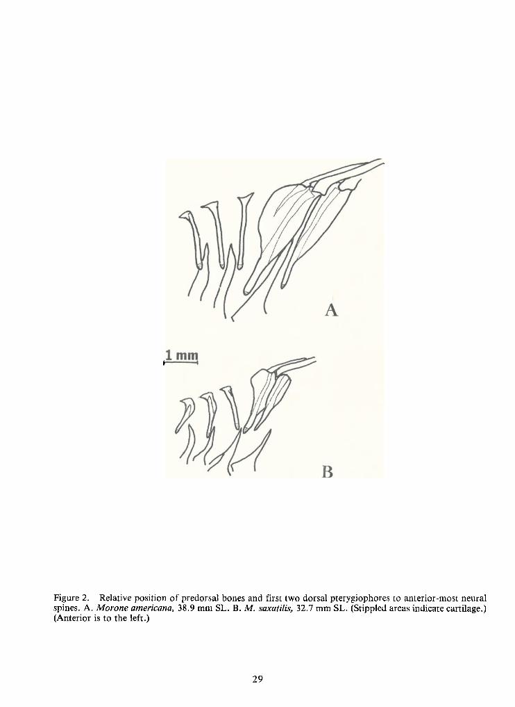
I. £&{T 4/evus/rfer /o($) Deo /?&0CAUTION: EARLY FRESHWATER DRUM
ARE PISCIVOROUS
(But there are errorsin the documentation)
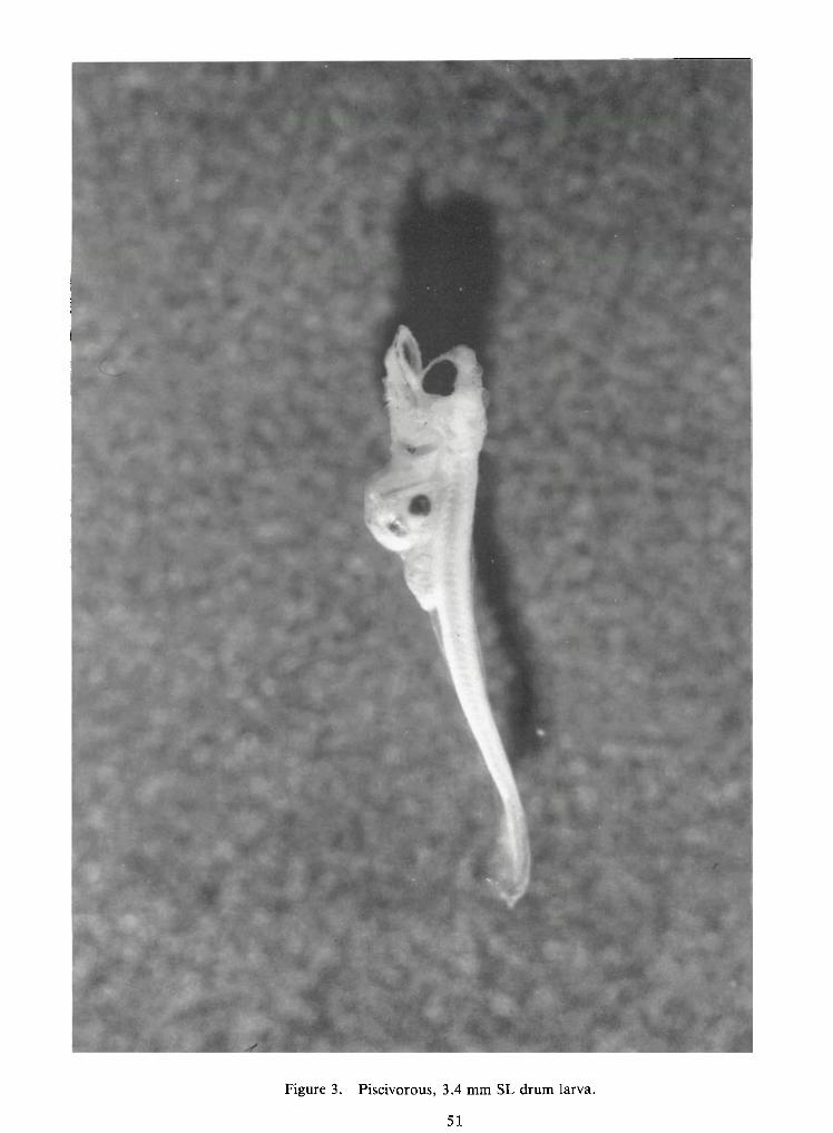
Two serious errors appear in a paper Ipublished with Aaron L. Clark in 1979(Clark, A. L. and W. D. Pearson, 1979.Early piscivory in larvae of the fresh-water drum, Aplodinotus grunniens .Pages 31-59 in: R. Wallus and C. W.Voigtlander, Eds. Proceedings of aworkshop on freshwater larval fishes.Tennessee Valley Authority, Norris,TN). The 3.4 mm SL larval fish which isphotographed in Figure 3 and drawn inFigure 7 appears to have 40-45 myo-meres, and was probably a percid(either a darter, or more likely aStizostedion) . A second error appearsto have been made in measuring thestandard length of this fish, andperhaps others in the data set. Thelarva photographed cannot be located,and may not have been part of the dataset. About 75% of the fish examined arestill available, and all of these havebeen verified as freshwater drum. Thesmallest of the available specimens are4.3 mm SL. An eyepiece micrometercalibration error seems to be the mostlikely explanation for this discrepancyin lengths of the small specimens. Thelargest fish among the remaining speci-mens is 14.6 mm SL, not greatly differ-ent from the 14.7 mm SL maximum report-ed in the paper. Although the specimenphotographed was misidentif ied and thespecific lengths reported cannot berelied upon, especially for the smallerspecimens, the essential conclusions ofthe paper remain valid. Ibring thesetwo errors to the attention of all ELHSmembers and ask that you make thecorrections known to all students and
colleagues. Photocopies of this sheetshould be placed in copies of theProceedings and reprints of the paper.Ithank Darrel Snyder (Larval FishLaboratory, Colorado State University)for notifying me of these errors andfor confirming the identity of six ofthe smallest drum larvae.
William D. PearsonWater Resources LaboratoryUniversity of LouisvilleLouisville, KY 40292
EARLY PISCIVORY IN LARVAEOF THEFRESHWATER DRUM,
APLODINOTUS GRUNNIENS¹
by
Aaron Lee Clarkand
William D. Pearson
Water Resources LaboratoryUniversity of Louisville
Louisville,Kentucky
ABSTRACT
Larval freshwater drum of 3.3-8.1 mm SL from the Ohio, Cumberland, Tennessee,
and Missouri Rivers consumed other larval fishes, mostly cyprinids and clupeids. This widespread
piscivory in drum is accompanied by morphological characteristics which change markedly after8-10 mm SL when the young drum cease feeding on other fishes and begin feeding almost exclusively
on zooplankton. The evolutionary advantages of exploiting an abundant but ephemeral foodresource and the significance of such exploitation are discussed.
iThis work was supported by a grant from the Kentucky Institute for Mining and
Minerals Research.
33
INTRODUCTION
The establishment of strong year-classes in fishes seems to depend on events taking
place in the first year of life, and perhaps in the first few weeks or even days after hatching.
Hjort (1926) used the term''critical period" to refer to this early stage in the life
histories ofherring and cod when the subsequent strength of the year-class was set. The concept of
the critical period has been reviewed byMarr (1956) and May (1974). Hjort suggested that the most
important cause of morality during the critical period might be the lack of suitable food, and
pointed out that the hatching period ofherring inhis studies corresponded with the season ofmax-
imum plankton densities. May (1974) has reviewed the available evidence bearing on the assump-
tions inherent in Hjort's critical period concept. He concludes that for many species of fish starva-
tion and starvation-enhanced mortalities (especially predation) are important causes ofmortality at
the time of yolk sac absorption when feeding on exogenous foods is just beginning. In freshwaterfishes, which are subject to greater variations in the physical environment, factors such as
temperature, wave action, and turbidity may be important as catastrophic causes ofdirect mortality
inlarval fishes (Kramer and Smith 1962). Physical factors may also interact with food-related mor-
talities byprolonging incubation, reducing the swimming and food-gathering abilities of fish larvae
relative to the escapement abilities of planktonic prey, reducing prey densities, and reducing
visibility.
Hunter (1976) offered the opinion that the major causes of larval mortality in fishes are
starvation and predation, and there may be interactions between them. He concluded that, although
it is important to treat the problem of stock and recruitment inholistic fashion, it is unlikely that
any new general models can be formulated, given the present state of knowledge.
Butler (1965) reviewed the lifehistory of the freshwater drum {Aplodinotus grunniens)
inthe upper Mississippi River and concluded that year-class strength inthis very fecund species must
be set during the egg and larval stages, since no adequate source of regulating mortality could be
found in subsequent stages oflife.During a study of the distribution oflarval fishes along a transect
of the Ohio River above Louisville,Kentucky, we discovered an unusual predator-prey relation bet-ween larvae of the freshwater drum and larvae of several other fishes. This relationship has been
overlooked because of its ephemeral appearance in the first week or two of life,and may be impor-
tant in establishing the year-class strength of both the predatory drum and the prey species.
34
MATERIALSAND METHODS
A field sampling program to determine the spatial and temporal distribution of
ichthyoplankton at Ohio River Mile571 in Trimble County, Kentucky, was conducted from March
through August of 1977. Four sampling stations were established along a transect and weekly sur-
face and bottom samples were collected inmidafternoon and at night at each of the stations. Each
sample consisted of a five-minute, upstream tow. Sampling gear consisted of 0.5-m cone-shaped
plankton nets constructed of361 nylon mesh. Adetachable 1-liter plankton bucket was affixed to
the cod end of each net and the nets were mounted on a brass ring with steel cable bridles. A flow
meter was suspended in the center of each brass ring. Tow net samples were preserved in10 percent
formalin and sorted in the laboratory.
Larval drum collected withplankton nets were also obtained from the Tennessee Valley
Authority and the U.S. Fish and Wildlife Service (North Central Reservoir Investigations). Ten-
nessee Valley Authority collections were from Nickajack Reservoir on the Tennessee River (1976),
Barkley Lake on the Cumberland River in southwestern Kentucky (1976), and at Ohio River Mile
946 near Paducah, Kentucky (1975). Larval drum from North Central Reservoir Investigations were
collected from impoundments of the Missouri River (Ft. Peck, 1975; Sakakawea, 1976; Oahe, 1974;
and Sharpe, 1975) in Montana and the Dakotas.
In the laboratory, the followingmeasurements were made to the nearest 0.01 mm using
a Bausch and Lomb dissecting microscope with calibrated ocular micrometer: standard length (SL),
maxilla length, mandible length, head length (the longest distance from the most anterior portion of
the maxillary to the most posterior portion of the opercular flap), and the distance from the anterior
margin of the eye to the upper corner of the opercular opening. These measurements were made on
250 larval drum selected from each of the eight locations. Alllarvae were separated into 2 mm SL
size classes for analysis of food habits.
The alimentary canal from the junction of the esophagus and the stomach to the anus
was removed from 554 larval drum using a pair of finely-sharpened dissecting needles. The percent-
age of the yolk still to be absorbed by the larvae was estimated by comparison withnewly hatched
drum. Allfood items found in the guts were identified and counted and the percent fullness of each
gut was estimated. Larval fish consumed by the larval drum were identified to the family level, con-
dition of the specimen permitting. Ingested invertebrates were categorized as oligochaetes, eubran-
chiopods, cladocerans, copepods, ostracods, amphipods, chironomids, and trichopterans.
35
RESULTS
Larval freshwater drum first appeared in the ichthyoplankton on May 9th and con-
stituted a significant percentage of the fauna from late May untilearly July. The smallest larva ex-
amined was 3.2 mm and the largest was 14.7 mm SL. There were no larvae available in the 3-5 mm
size class from Lakes Sakakawea and Sharpe on the Missouri River, and some larger size classes
were also absent from the samples obtained from six locations (Table 1).
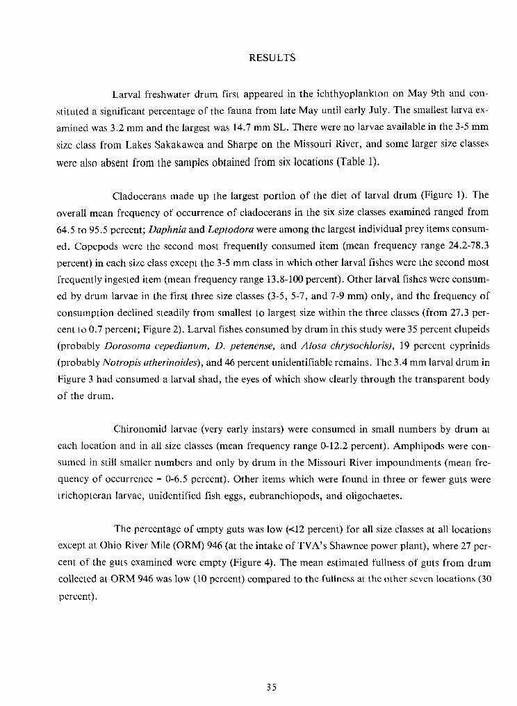
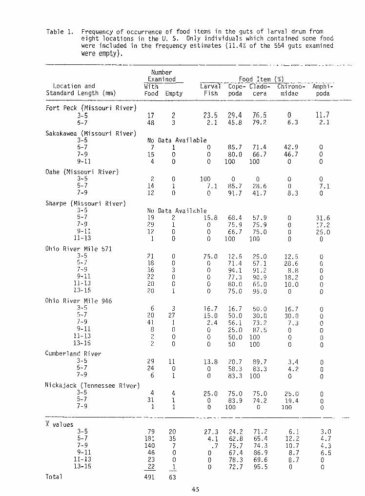
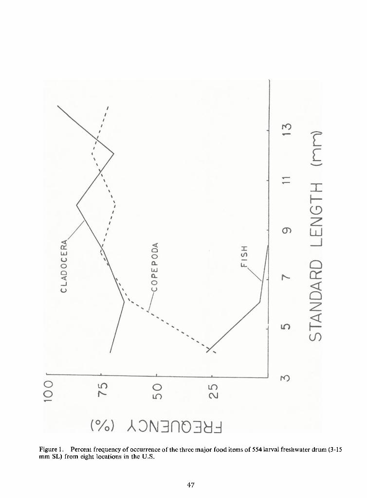
Cladocerans made up the largest portion of the diet of larval drum (Figure 1). The
overall mean frequency of occurrence of cladocerans in the six size classes examined ranged from
64.5 to95.5 percent; Daphnia and Leptodora were among the largest individual prey items consum-
ed. Copepods were the second most frequently consumed item (mean frequency range 24.2-78.3
percent) ineach size class except the 3-5 mm class in which other larval fishes were the second most
frequently ingested item (mean frequency range 13.8-100 percent). Other larval fishes were consum-
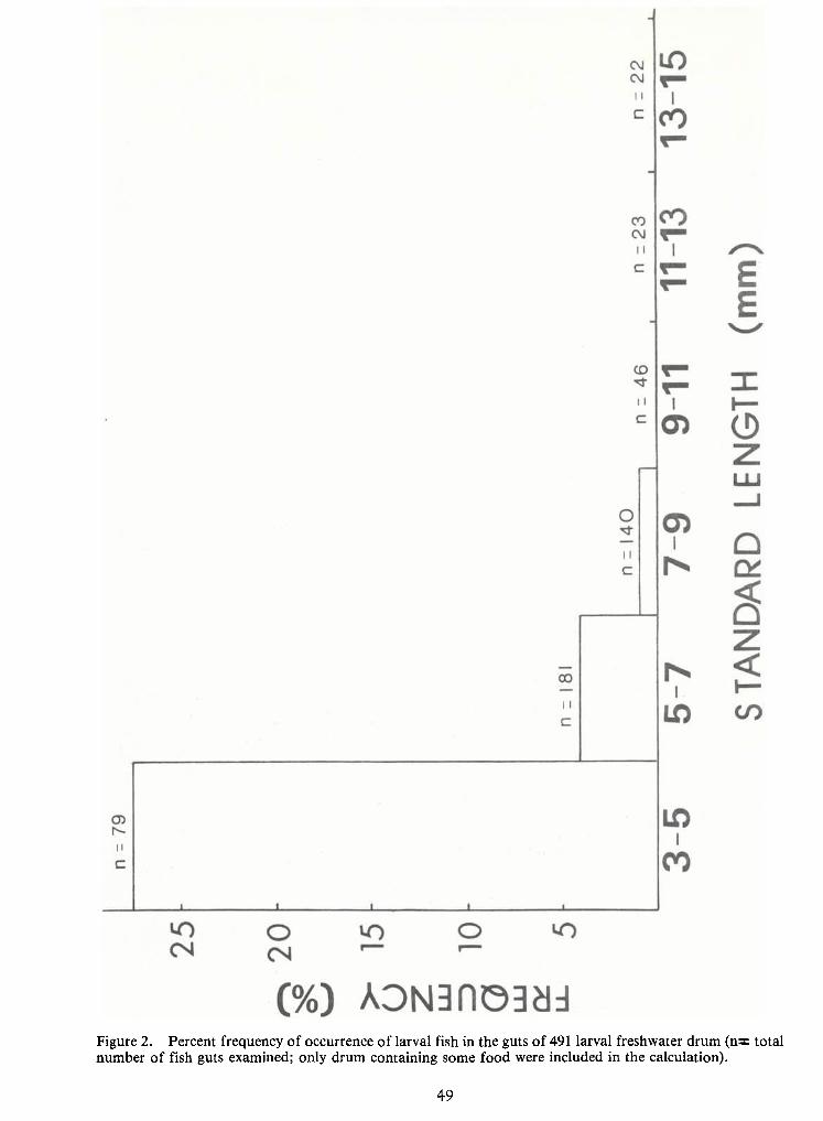
ed by drum larvae in the first three size classes (3-5, 5-7, and 7-9 mm) only, and the frequency ofconsumption declined steadily from smallest to largest size within the three classes (from 27.3 per-
cent to0.7 percent; Figure 2). Larval fishes consumed by drum in this study were 35 percent clupeids
(probably Dorosoma cepedianum, D. petenense, and Alosa chrysochloris) , 19 percent cyprinids
(probably Notropis atherinoides), and 46 percent unidentifiable remains. The 3.4 mm larval drum inFigure 3 had consumed a larval shad, the eyes of which show clearly through the transparent body
of the drum.
Chironomid larvae (very early instars) were consumed in small numbers by drum at
each location and inall size classes (mean frequency range 0-12.2 percent). Amphipods were con-
sumed in still smaller numbers and only by drum in the Missouri River impoundments (mean fre-
quency of occurrence = 0-6.5 percent). Other items which were found in three or fewer guts were
trichopteran larvae, unidentified fish eggs, eubranchiopods, and oligochaetes.
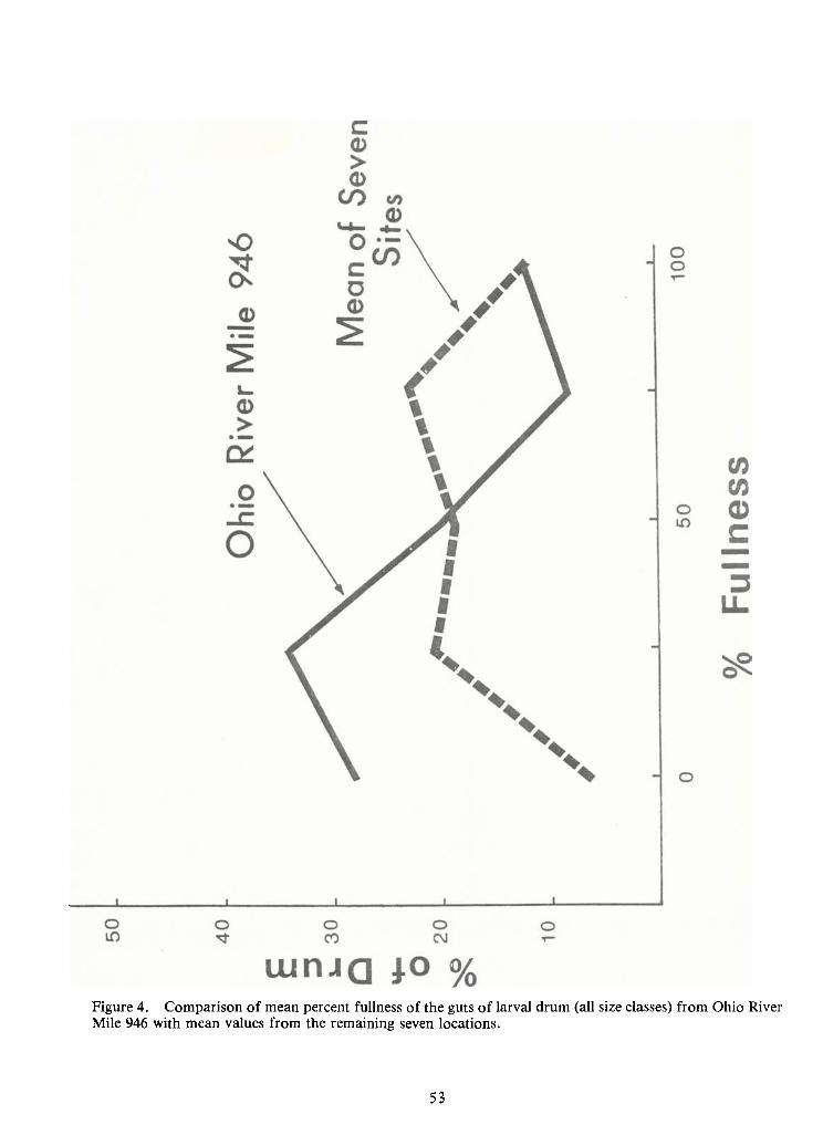
The percentage of empty guts was low (<l2 percent) for all size classes at all locationsexcept at Ohio River Mile (ORM) 946 (at the intake of TVA's Shawnee power plant), where 27 per-
cent of the guts examined were empty (Figure 4). The mean estimated fullness of guts from drum
collected at ORM946 was low (10 percent) compared to the fullness at the other seven locations (30
percent).
36
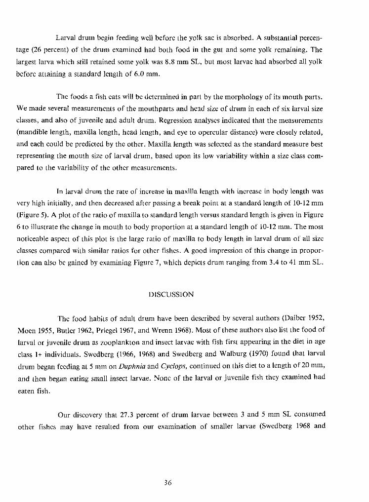
Larval drum begin feeding well before the yolk sac is absorbed. A substantial percen-
tage (26 percent) of the drum examined had both food in the gut and some yolk remaining. The
largest larva which still retained some yolk was 8.8 mm SL, but most larvae had absorbed all yolk
before attaining a standard length of 6.0 mm.
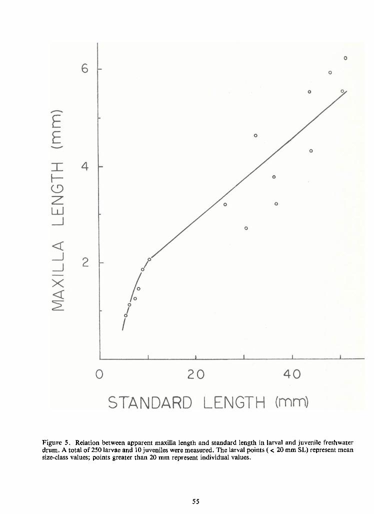
The foods a fish eats willbe determined in part by the morphology ofits mouth parts.
We made several measurements of the mouthparts and head size of drum ineach of six larval size
classes, and also of juvenile and adult drum. Regression analyses indicated that the measurements
(mandible length, maxilla length, head length, and eye to opercular distance) were closely related,
and each could be predicted by the other. Maxilla length was selected as the standard measure bestrepresenting the mouth size of larval drum, based upon its low variability withina size class com-
pared to the variability of the other measurements.
In larval drum the rate of increase in maxilla length with increase inbody length was
very high initially, and then decreased after passing a break point at a standard length of 10-12 mm
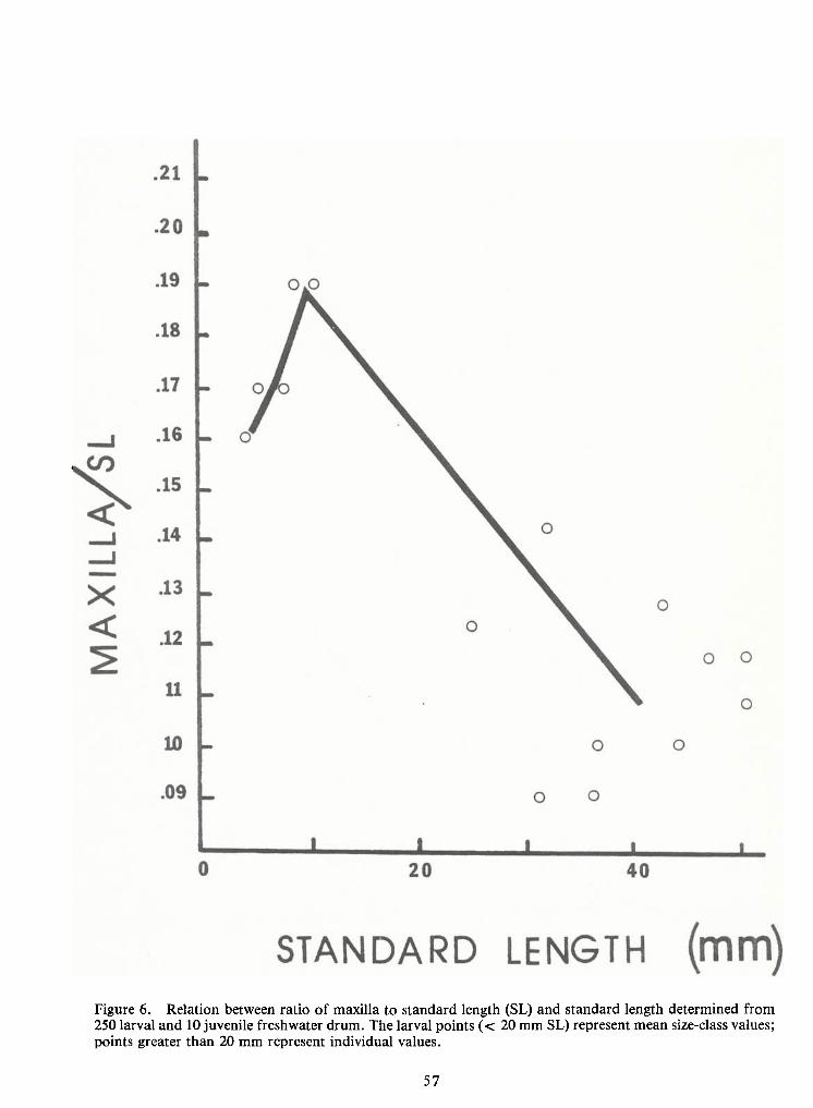
(Figure 5). A plot of the ratio ofmaxilla to standard length versus standard length is given inFigure
6 to illustrate the change in mouth to body proportion at a standard length of 10-12 mm. The most
noticeable aspect of this plot is the large ratio of maxilla to body length in larval drum of all size
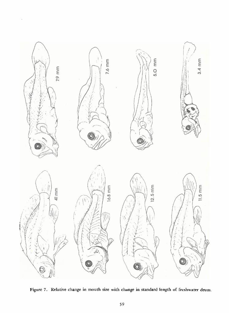
classes compared with similar ratios for other fishes. Agood impression of this change in propor-
tion can also be gained by examining Figure 7, which depicts drum ranging from 3.4 to 41 mm SL.
DISCUSSION
The food habits of adult drum have been described by several authors (Daiber 1952,
Moen 1955, Butler 1962, Priegel 1967, and Wrenn 1968). Most of these authors also list the food of
larval or juvenile drum as zooplankton and insect larvae with fish first appearing in the diet in age
class 1+ individuals. Swedberg (1966, 1968) and Swedberg and Walburg (1970) found that larval
drum began feeding at 5 mm on Daphnia and Cyclops, continued on this diet to a length of 20 mm,
and then began eating small insect larvae. None of the larval or juvenile fish they examined had
eaten fish.
Our discovery that 27.3 percent of drum larvae between 3 and 5 mm SL consumed
other fishes may have resulted from our examination of smaller larvae (Swedberg 1968 and
37
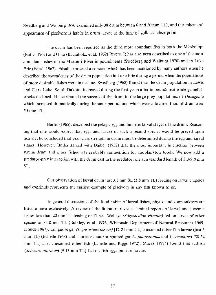
Swedberg and Walburg 1970 examined only 38 drum between 6 and 20 mm TL),and the ephemeral
appearance of piscivorous habits in drum larvae at the time of yolk sac absorption.
The drum has been reported as the third most abundant fish in both the Mississippi
(Butler 1965) and Ohio (Krumholz, et al. 1962) Rivers. Ithas also been described as one of the most
abundant fishes in the Missouri River impoundments (Swedberg and Walburg 1970) and in Lake
Erie (Edsall 1967). Edsall expressed a concern which has been mentioned by many authors when he
described the ascendency of the drum population inLake Erie during a period when the populations
ofmore desirable fishes were in decline. Swedberg (1968) found that the drum population inLewis
and Clark Lake, South Dakota, increased during the first years after impoundment while gamefish
stocks declined. He attributed the success of the drum to the large prey populations ofHexagenia
which increased dramatically during the same period, and which were a favored food of drum over
50 mm TL.
Butler (1965), described the pelagic egg and limnetic larval stages of the drum. Reason-ing that one would expect that eggs and larvae of such a fecund species would be preyed upon
heavily, he concluded that year-class strength indrum must be determined during the egg and larval
stages. However, Butler agreed with Daiber (1952) that the most important interaction betweenyoung drum and other fishes was probably competition for zooplankton foods. We now add a
predator-prey interaction with the drum cast in the predator role at a standard length of3.3-9.0 mm
SL.
Our observation of larval drum just 3.3 mm SL (3.8 mm TL)feeding on larval clupeids
and cyprinids represents the earliest example of piscivory in any fish known to us.
Ingeneral discussions of the food habits of larval fishes, phyto- and zooplanktors are
listed almost exclusively. A review of the literature revealed limited reports of larval and juvenile
fishes less than 20 mm TL feeding on fishes. Walleye (Stizostedion vitreum) fed on larvae ofother
species at 8-10 mm TL (Bulkley, et al. 1976, Wisconsin Department of Natural Resources 1969,
Houde 1967). Longnose gar (Lepisosteus osseus) [17-21 mm TL]consumed other fish larvae (just 5
mm TL) (Echelle 1968) and shortnose and/or spotted gar L.platostomus and L. oculatus) [50-34
mm TL] also consumed other fish (Echelle and Riggs 1972). Marak (1974) found that redfish
(Sebastes morinus) [9-13 mm TL] fed on fish eggs but not larvae.
38
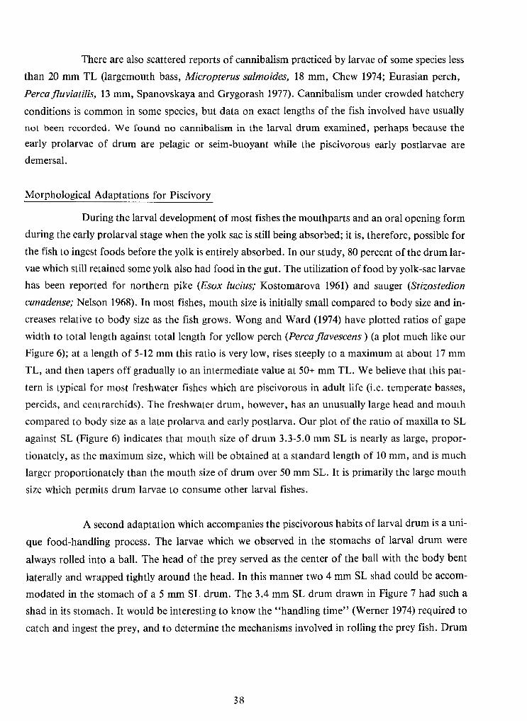
There are also scattered reports ofcannibalism practiced by larvae of some species less
than 20 mm TL (largemouth bass, Micropterus salmoides, 18 mm, Chew 1974; Eurasian perch,
Perca fluviatilis,13 mm, Spanovskaya and Grygorash 1977). Cannibalism under crowded hatchery
conditions is common in some species, but data on exact lengths of the fish involved have usually
not been recorded. We found no cannibalism in the larval drum examined, perhaps because theearly prolarvae of drum are pelagic or seim-buoyant while the piscivorous early postlarvae are
demersal.
Morphological Adaptations for Piscivory
During the larval development ofmost fishes the mouthparts and an oral opening formduring the early prolarval stage when the yolk sac is stillbeing absorbed; it is, therefore, possible forthe fish to ingest foods before the yolk is entirely absorbed. Inour study, 80 percent of the drum lar-
vae which stillretained some yolk also had food inthe gut. The utilization of food by yolk-sac larvae
has been reported for northern pike (Esox lucius; Kostomarova 1961) and sauger (Stizostedion
canadense; Nelson 1968). Inmost fishes, mouth size is initiallysmall compared to body size and in-
creases relative to body size as the fish grows. Wong and Ward (1974) have plotted ratios of gape
width to total length against total length for yellow perch (Perca flavescens ) (a plot much like our
Figure 6); at a length of5-12 mm this ratio is very low, rises steeply to a maximum at about 17 mm
TL, and then tapers offgradually to an intermediate value at 50+ mm TL.We believe that this pat-
tern is typical for most freshwater fishes which are piscivorous in adult life (i.e. temperate basses,
percids, and centrarchids). The freshwater drum, however, has an unusually large head and mouth
compared to body size as a late prolarva and early postlarva. Our plot of the ratio of maxilla to SL
against SL (Figure 6) indicates that mouth size of drum 3.3-5.0 mm SL is nearly as large, propor-
tionately, as the maximum size, which willbe obtained at a standard length of 10 mm, and is much
larger proportionately than the mouth size of drum over 50 mm SL. Itis primarily the large mouth
size which permits drum larvae to consume other larval fishes.
Asecond adaptation which accompanies the piscivorous habits oflarval drum is a uni-
que food-handling process. The larvae which we observed in the stomachs of larval drum were
always rolled into a ball. The head of the prey served as the center of the ball with the body bent
laterally and wrapped tightly around the head. In this manner two 4 mm SL shad could be accom-
modated in the stomach of a 5 mm SL drum. The 3.4 mm SL drum drawn inFigure 7 had such a
shad in its stomach. Itwould be interesting to know the "handling time" (Werner 1974) required to
catch and ingest the prey, and to determine the mechanisms involved inrolling the prey fish. Drum
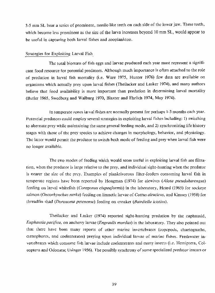
39
3-5 mm SL bear a series ofprominent, needle-like teeth on each side of the lower jaw. These teeth,
which become less prominent as the size of the larva increases beyond 10 mm SL, would appear to
be useful in capturing both larval fishes and zooplankton.
Strategies for Exploiting Larval Fish
The total biomass of fish eggs and larvae produced each year must represent a signifi-
cant food resource for potential predators. Although much importance is often attached to the role
of predation in larval fish mortality (i.e. Ware 1975, Hunter 1976) few data are available on
organisms which actually prey upon larval fishes (Theilacker and Lasker 1974), and many authors
believe that food availability is more important than predation in determining larval mortality
(Butler 1965, Swedberg and Walburg 1970, Blaxter and Ehrlich 1974, May 1974).
Intemperate zones larval fishes are normally present for perhaps 1-3 months each year.
Potential predators could employ several strategies inexploiting larval fishes including: 1) switching
to alternate prey while maintaining the same general feeding mode, and 2) synchronizing lifehistory
stages with those of the prey species to achieve changes in morphology, behavior, and physiology.
The latter would permit the predator to switch both mode of feeding and prey when larval fish were
no longer available.
The two modes of feeding which would seem useful inexploiting larval fish are filtra-
tion, when the predator is large relative to the prey, and individual sight-hunting when the predator
is nearer the size of the prey. Examples of planktivorous filter-feeders consuming larval fish in
temperate regions have been reported by Hoagman (1974) for alewives (Alosa pseudoharengus)
feeding on larval whitefish (Coregonus clupeaformis) in the laboratory, Heard (1965) for sockeye
salmon {Oncorhynchus nerka) feeding on limnetic larvae ofCottus aleuticus, and Kimsey (1958) for
threadfin shad {Dorosoma petenense) feeding on croaker {Bairdiella icistius).
Theilacker and Lasker (1974) reported sight-hunting predation by the euphausid,
Euphausia pacifica, on anchovy larvae (Engraulis mordax) in the laboratory. They also pointed out
that there have been many reports of other marine invertebrates (copepods, chaetognaths,
ctenophores, and coelenterates) preying upon individual larvae of marine fishes. Freshwater in-
vertebrates which consume fish larvae include coelenterates and many insects (i.e. Hemiptera, Col-
eoptera and Odonata; Usinger 1956). The possibly synchrony of some specialized predator instars or
40
developmental stages with the appearance oflarval fishes should be investigated. The only recorded
examples of fishes individually sight-hunting other fish larvae are those previously described, ex-
cluding examples of cannabilism.
We suspect that larval fishes are preyed upon by many organisms, and to a much
greater extent than has been reported. Future studies in temperate regions should be made to ex-
amine the feeding habits of large filterfeeders (i.e. paddlefish, shad, herrings, and buffalo) during
the reproductive season, keeping inmind that larval fishes are fragile and quickly digested. Studies
of the morphology ofmouthparts could provide clues to the existence of larvae capable ofpreying
on other larvae. Studies in tropical regions should be directed toward finding both specialized filter-
feeders and sight-hunters preying on larval fishes throughout the year.
Information from these studies would enable us to identify specific causes of egg and
larval mortalities in fishes and estimate their individual impacts. This could lead to predictive and
realistic models of larval fish population dynamics, and eventually to management strategies forenhancing year-class strength in desirable species.
ACKNOWLEDGEMENTS
The authors wish to thank WilliamR. Nelson and Charles H.Walburg of the U.S. Fish
and Wildlife Service and Robert Wallus of the Tennessee Valley Authority for supplying larvae.
Thanks are also given to Lisa Maliszewski for her drawings and to Randall G. Farmer and B.
Douglas Steele for help incollecting larvae from the Ohio River. This research was made possible by
a grant from the Kentucky Institute for Miningand Minerals Research.
41
LITERATURE CITED
Blaxter, J.H.S. and K.F. Ehrlich. 1974. Changes in behavior during starvation of herring andplaice larvae, p. 575-588. In: J.H.S. Blaxter (cd.) The early life history of fish. Springer-Verlag, New York, N. Y.
Bulkley, R. V., V.L. Spykermann and L.E. Inmon. 1976. Food of the pelagic young of walleyesand five cohabiting fish species inClear Lake, lowa. Trans. Amer. Fish. Soc. 105(l):77-83.
Butler, R. L. 1962. The status of the freshwater drum Aplodinotus grunniens Rafinesque, in thecommercial fishery of the upper Mississippi River. Unpubl. Ph.D. Thesis. Univ. of Minn.178 p.
Butler, R. L.1965. Freshwater drum, Aplodinotus grunniens, in the navigational impoundments ofthe upper Mississippi River. Trans. Amer. Fish. Soc. 94(4):339-349.
Chew, R. L.1974. Early life history of the Florida largemouth bass. Fla. Game &Freshwat. Fish.Comm. Fish. Bull. 7. 76 p.
Daiber, F. C. 1952. The food and feeding relationships of the freshwater drum, Aplodinotusgrunniens, in western Lake Erie. Ohio J. Sci. 52(1):35-46.
Echelle, A. A. 1968. Food habits of young-of-year longnose gar in Lake Texoma, Oklahoma.Southwestern Nat. 13(l):45-50.
Echelle, A.A.and CD. Riggs. 1972. Aspects of the early lifehistory ofgars (Lepisosteus) inLakeTexoma. Trans. Amer. Fish. Soc. 101(1): 106-1 12.
Edsall, T.A.1967. Biology of the freshwater drum in western Lake Erie. Ohio J. Sci. 67(6):321-340.
Heard, W. R. 1965. Limnetic cottid larvae and their utilization as food by juvenile sockeye salmon.Trans. Amer. Fish. Soc. 94(2):191-193.
Hjort, J. 1926. Fluctuations in the year classes of important food fishes. J. Cons. Int.Explor. Mer.1:5-38.
Hoagman, W. J. 1974. Feeding by alewives (Alosa pseudoharengus) on larval lake whitefish(Coregonus clupeaformis) in the laboratory. J. Fish. Res. Board Can. 31(2):229-230.
Houde, E. D.1967. Food ofpelagic young of the walleye, Stizostedion vitreum vitreum, inOneidaLake, New York. Trans. Amer. Fish. Soc. 96(1):17-24.
Hunter, J. R. 1976. Report of a colloquium on larval fish mortality studies and their relation tofishery research, January 1975. NOAA Tech. Retp. NMFS Circ-395, 5 p.
Kimsey, J. B. 1958. Possible effects of introducing threadfin shad (Dorosoma petenense) into theSacramento-San Joaquin Delta. Calif. Inland Fish. Admin. Rept. 58-16, 21 p.
Kostomarova, A.A. 1961. The significance of the stage of mixed feeding for the survival of Esoxlucius L. larvae. Tr. Soveshch. Ikhtiol. Komis. Akad, Nauk SSSR 13:334-347. U. S. Bur.Comm. Fish. Translation, Washington, D. C.
42
Kramer, R. L.and L. L. Smith, Jr. 1962. Formation of year classes in largemouth bass. Trans.Amer. Fish. Soc. 91(1):29-41.
Krumholtz, L.A., J. R. Charles and W. L.Minckley. 1962. The fish population of the Ohio River,p. 49-89. In:ORSANCO, Aquatic-life resources of the Ohio River. Ohio River Valley Wat.San. Comm. Cincinnati, Ohio.
Marak, R. R. 1974. Food and feeding of larval redfish in the Gulf ofMaine, p. 267-275. In: J.H.S.Blaxter (cd.) The early lifehistory of fish. Springer- Verlag, New York, N. Y.
Marr, J. C. 1956. The criticalperiod in the early lifehistory ofmarine fishes. J. Cons. Int. Explor.Mer. 21:160-170.
May, R. C. 1974. Larval mortality in marine fishes and the critical period concept, p. 3-19. In:J.H.S. Blaxter (cd.) The early life history of fish. Springer- Verlag, New York, N.Y.
Moen, T.1955. Food of the freshwater drum, Aplodinotus grunnies Rafinesque, in four DickinsonCounty, lowa, lakes. Proc. lowa Acad., Sci. 62:589-598.
Nelson, W.R. 1968. Reproduction and early lifehistory of sauger, Stizostedion canadense, inLewisand Clark Lake. Trans. Amer. Fish. Soc. 97(2): 159-166.
Priegel, G. R. 1967. Food of the freshwater drum, Aplodinotus grunniens, in Lake Winnebago,Wisconsin. Trans. Amer. Fish. Soc. 96(2) :218-220.
Spanovskaya, V. D. and V.A.Grygorash. 1977. Development and food of age-O Eurasian perch{Perca fluviatilis) in reservoirs near Moscow, USSR. J. Fish. Res. Board Can.34(10):1551-1558.
Swedberg, D. V. 1966. Foods taken by freshwater drum. Prog. Fish-Cult. 28(4): 192.
Swedberg, D. V. 1968. Food and growth of the freshwater drum in Lewis and Clark Lake, SouthDakota. Trans. Amer. Fish. Soc. 97(4):442-447.
Swedberg, D. V.and C. H. Walburg. 1970. Spawning and early lifehistory of the freshwater drumin Lewis and Clark Lake, Missouri River. Trans. Amer. Fish. Soc. 99(3):56C-570.
Theilacker, G. H. and R.Lasker. 1974. Laboratory studies of predation byEuphausid shrimps onfish larvae, p. 287-299. In: J.H.S. Blaxter (cd.) The early lifehistory of fish. Springer- Verlag,New York, N. Y.
Usinger, R. L.1956. Aquatic insects of California. Univ. Calif. Press Berkeley, Calif. 508 pp.
Ware, D. M.1975. Relation between egg size, growth, and natural mortality oflarval fish. J. Fish.Res. Board Can. 32(12):2503-2512.
Werner, E. E. 1974. The fish size, prey size, handling time relation in several sunfishes and someimplications. J. Fish. Res. Board Can. 31:1531-1536.
Wisconsin Department ofNatural Resources. 1969. Walleye fry-plankton relationship inEscanabaLake. Prog. Rept. F-83-R-5. 10 p.

43
Wong, B. and F. J. Ward. 1972. Size selection of Daphnia pulicaria by yellow perch (Percaflavescens) fry inWest Blue Lake, Manitoba. J. Fish. Res. Board Can. 29(12): 1761-1764.
Wrenn, W. B. 1968. Lifehistory aspects of smallmouth buffalo and freshwater drum in WheelerReservoir, Alabama. Proc. 22d Ann. Conf. S. E. Assoc. Game and Fish Comm. 479-495.
45
Table 1. Frequency of occurrence of food items in the guts of larval drum fromeight locations in the U. S. Only individuals which contained some foodwere included in the frequency estimates (11.4% of the 554 guts examinedwere empty).
63491Total
00
8.70
69.695.5
78.372.7
00
0_1_
2322
11-1313-15
4.36.5
10.78.7
74.386.9
75.767.4
.70
70
14046
7-99-11
3.04.7
6.112.2
71.265.4
24.262.8
27.34.1
2035
79181
3-55-7
X values
00O
25.019.4
100
75.074.2
0
75.083.9
100
4 25.01 01 0
4311
3-55-77-9
Nickajack (Tennessee River)0010083.31 067-9
00
3.44.2
89.783.3
20.758.3
11 13.80 0
2924
3-55-7
Cumberland River00100500 0213-150010050.00 0211-130087.525.00 089-1107.373.256.11 2.4417-9030.030.050.027 15.0205-7016.750.016.73 16.763-5
Ohio River Mile 9460095.075.01 02013-15010.065.080.00 02011-13018.290.977.30 0229-1108.891.294.13 0367-9
00
12.528.6
25.057.1
12.571.4
0 75.00 0
2118
3-55-7
Ohio River Mile 571
25.00
00
75.0100
66.7100
0 00 0
121
9-1111-13
17.2075.975.91 0297-931.6057.968.4
Data Available2 15.8
No19
3-55-7
Sharpe (Missouri River)08.341.791.70 0127-97.1028.685.71 7.1145-700000 1002
Oahe (Missouri River)3-5
001001000 049-11046.766.780.00 0157-9042.971.485.7
Data Available1 0
No7
3-55-7
Sakakawea (Missouri River)
11.72.1
06.3
76.579.2
29.445.8
2 23.53 2.1
1748
Fort Peck (Missouri River)3-55-7
EmptyFoodAmphi-
podaChirono-midae
Clado-cera
Cope-poda
LarvalFish
With(mm)
Location andStandard Length
MItemFoodNumber
ExaminedNumber
Examined Food Item MLocation and
Standard Length (mm)With Larval
FishCope-poda
Clado-cera
Chirono-midae
Amphi-podaFood Empty
Fort Peck (Missouri River)3-55-7
1748
2 23.53 2.1
29.445.8
76.579.2
06.3
11.72.1
Sakakawea (Missouri River)3-55-7
No7
Data Available1 0 85.7 71.4 42.9 0
7-9 15 0 0 80.0 66.7 46.7 09-11 4 0 0 100 100 0 0
Oahe (Missouri River)3-5 2 0 100 0 0 0 05-7 14 1 7.1 85.7 28.6 0 7.17-9 12 0 0 91.7 41.7 8.3 0
Sharpe (Missouri River)3-55-7
No19
Data Available2 15.8 68.4 57.9 0 31.6
7-9 29 1 0 75.9 75.9 0 17.29-11
11-13121
0 00 0
66.7100
75.0100
00
25.00
Ohio River Mile 5713-55-7
2118
0 75.00 0
12.571.4
25.057.1
12.528.6
00
7-9 36 3 0 94.1 91.2 8.8 09-11 22 0 0 77.3 90.9 18.2 0
11-13 20 0 0 80.0 65.0 10.0 013-15 20 1 0 75.0 95.0 0 0
Ohio River Mile 9463-5 6 3 16.7 16.7 50.0 16.7 05-7 20 27 15.0 50.0 30.0 30.0 07-9 41 1 2.4 56.1 73.2 7.3 09-11 8 0 0 25.0 87.5 0 0
11-13 2 0 0 50.0 100 0 013-15 2 0 0 50 100 0 0
Cumberland River3-55-7
2924
11 13.80 0
20.758.3
89.783.3
3.44.2
00
7-9 6 1 0 83.3 100 0 0Nickajack (Tennessee River)
3-55-77-9
4311
4 25.01 01 0
75.083.9
100
75.074.2
0
25.019.4
100
00O
X values3-55-7
79181
2035
27.34.1
24.262.8
71.265.4
6.112.2
3.04.7
7-99-11
14046
70
.70
75.767.4
74.386.9
10.78.7
4.36.5
11-1313-15
2322
0_1_
00
78.372.7
69.695.5
8.70
00
Total 491 63
47
Figure 1. Percent frequency ofoccurrence of the three major food items of 554 larval freshwater drum (3-15mm SL) from eight locations in the U.S.
49
Figure 2. Percent frequency of occurrence of larval fish in the guts of 491 larval freshwater drum (n= totalnumber of fish guts examined; only drum containing some food were included in the calculation).
51
Figure 3. Piscivorous, 3.4 mm SL drum larva.
53
Figure 4. Comparison of mean percent fullness of the guts of larval drum (all size classes) from Ohio RiverMile 946 with mean values from the remaining seven locations.
55
Figure 5. Relation between apparent maxilla length and standard length in larval and juvenile freshwaterdrum. A total of250 larvae and 10 juveniles were measured. The larval points (< 20 mm SL) represent meansize-class values; points greater than 20 mm represent individual values.
57
Figure 6. Relation between ratio of maxilla to standard length (SL) and standard length determined from250 larval and 10 juvenile freshwater drum. The larval points (< 20 mm SL) represent mean size-class values;points greater than 20 mm represent individual values.
59
Figure 7. Relative change in mouth size with change in standard length of freshwater drum
LARVALAND EARLY JUVENILE DEVELOPMENT OF THE STRIPED SHINER,
NOTROPIS CHRYSOCEPHALUS (RAFINESQUE)
byBruce Yeager
Division of Forestry, Fisheries, and Wildlife Development
Tennessee Valley AuthorityNorris, Tennessee 37828
ABSTRACT
Larval phases of the striped shiner, Notropis chrysocephalus, are described from
meristic and morphometric data as wellas morphological changes. Myomere counts, morphometric
ratios, pigmentation, and finray counts are useful for identification.
Gravid striped shiners were collected from Hinds Creek, Anderson County, Tennessee.
Specimens were reared from artifically fertilized eggs into the juvenile phase.
Fertilized eggs have a mean diameter of2.2 mm and are demersal and adhesive. Larvae
hatch at a mean size of 5.9 mm TL.Newly hatched larvae are unpigmented. By three days post-
hatching (7.2 mm TL), larvae have a characteristic pigmentation pattern. Early larvae have 26 to 28
preanal and 12 to 14 postanal myomeres. Morphological changes associated with yolk sac absorp-
tion (7.9 mm TL)include: upward deflection of the notochord, fillingof the gas bladder, and inci-
pient median finray development. Successive fin ray development is: caudal, dorsal, anal, pelvics
and pectorals respectively. Coiling of the gut begins at 12 mm TLand attains the characteristic adultshape at 13.5 mm TL.Squamation develops between 17 and 23 mm TL.
63
INTRODUCTION
The striped shiner, Notropis chrysocephalus, ranges throughout the east central United
States from the Coosa River system of Alabama and Georgia and the lower Mississippi River
drainage of Alabama, Mississippi, Tennessee, Arkansas, and Oklahoma, northward to the lower
Great Lakes from Wisconsin to New York (Gilbert 1964). As juveniles and adults, striped shiners
are the most commonly collected member of the Luxilus subgenus in eastern Tennessee streams.
Striped shiner larvae are abundant in stream drift net samples. This study provides means for iden-
tification of larval and early juvenile striped shiners at various stages of development.
METHODS
On May 4, 1976, ripe adult striped shiners were collected from Hinds Creek, a tributary
of the Clinch River, Melton HillReservoir, in Anderson County, Tennessee. Gravid females were
seined from a gravel bottom rifflehead in50 cm of water. Ripe males were seined from a deep run in
80 cm of water over bedrock. Stream temperature was 13 C.
The eggs were field stripedand allowed to water-harden one hour before transport. In
the laboratory, the eggs were maintained in a once-through flow incubator system utilizing spring
water at 13 to 15 C. Treatment with 0.5 mg l-1 malachite green was used on alternate days to
reduce fungal infection.
Within six hours after hatching, larvae were transferred to a 114 1 aquarium maintain-
ed at 13 C for three days. The temperature was then allowed to rise to room temperature and
thereafter fluctuated between 18 and 22 C. A zooplankton mixture containing Filinia, Hexarthra,
Cyclops and Daphnia was offered the third day after hatching. Newlyhatched brine shrimp were fed
to the larvae from the fifthday after hatching.
Samples of 2-10 specimens were collected in the following sequence: every 8 hours forthe first 5 days, every 24 hours for the next 7 days, every 4 days for the next 24 days, and every 9
days for the last 27 days. Samples of larvae were preserved in 10 percent Formalin and later transfer-red to buffered 5 percent Formalin.
64
Meristic and morphometric data were obtained using a steromicroscope equipped with
an ocular micrometer and polarizers. Characters examined included: total length, urostyle length,
preanal length, postanal length, snout length, head length, greatest body depth, length to dorsal fin
origin, length to anal fin origin, number of preanal and postanal myomeres, and numbers of fin
rays.
Head length was defined as the distance from the tip of the snout to the posterior
margin of the auditory vesicle or opercle (when developed). Yolk sac depth was measured at the
maximum point. Other measurements were obtained following Trautman (1957). The method ofcounting preanal and postanal myomeres was that of Hogue et al. (1976).
Mean total lengths were tabulated by age. Meristic and morphometric information
other than total length was tabulated bymillimeter class size for identification. Static morphometric
and meristic data are presented in tabular form, while the description of development follows the
dynamic approach (Berry and Richards 1973).
Development of bone structure was studied after specimens were stained withAlizarin
Red S and cleared in a KOH-glycerol solution (Granneman and Kay, MS). Scale development was
observed by staining specimens in a solution of0.2-0.3 ft*-'1 Methylene Blue in distilled water.
Drawings were made with the aid of a camera lucida. Terminology is that of Hubbs
(1943), with reference to both Balinsky (1948) and Snyder (1976).
Allspecimens are catalogued as TV737, DS-11 in the Larval Fish Identification and In-
formation Center, Tennessee Valley Authority, Norris, Tennessee.
65
RESULTS
Meristic and morphometric data for prolarvae and postlarvae appear in Table I.Mor-
phometric ratios appear in Table 11.
Eggs and Hatching
Fertilized eggs averaged 2.2 mm indiameter (range 2.0-2.3 mm) and were demersal and
adhesive. Eggs hatched between 152 and 160 hours after fertilization, yielding 752 larvae of which
260 were used for this study.
Prolarval Phase (Figures 1and 2)
Larvae at hatching averaged 5.9 mm TL (range 5.6-6.0 mm TL).
The prolarval phase is equivalent to Balinsky's stages 25 to 32 and roughly corresponds
to Snyder's protolarval phase. The notochord, however, does not deflect upwards until the postlar-
val phase.
Athatching the head is curved over the club-shaped yolk sac. Byone day after hatching
the head turns up inline with the body axis. Gills are not developed at hatching; gillarches are ap-
parent as three or four tissue folds at two days of age. Otoliths appear as unossified refracting
spheres.
Myomeres ofearly prolarvae range from 26 to 28 for preanal and 12 to 14 for postanal
with modes of 27 and 13 respectively. The determination ofcompletion ofmyoseptae on early pro-
larvae is difficult.
Opercular flaps are present on 7.2 mm larvae and cover the first three gill arches. Atthis size, larvae have a more streamlined appearance due to yolk sac absorption. The mouth has
become well formed and subterminal.
Findevelopment on prolarvae is slow. Athatching the median finfold ispresent dorsal-ly from the ninth myomere, extending around the caudal finand ventrally to the forming anus. Asthe yolk sac is absorbed, the preanal median finfold becomes apparent.
66
Pectoral fin buds are present at hatching as opaque thickenings on the dorso-lateralarea of the yolk sac. By two days after hatching, the pectoral fins extend beyond the edge of the yolk
sac as viewed from the dorsal perspective. Pectoral fins are paddle-shaped on prolarvae greater than
7.2 mm TL.No ray nor hypural complex development is evident during the prolarval phase.
Except for the eyes, early prolarvae are unpigmented. Within a day after hatching, a
few large melanophores are widely dispersed over the yolk sac. By three days (7.2 mm TL) several
melanophores on either side of the heart area form a "V"ventrally. Adouble row ofmelanophores
extends from the forming anus to the caudal finbase. The head is moderately pigmented and a dou-
ble row ofmelanophores from the occiput to the caudal finis present. Two or three chromatophores
are present on the finfold in the peduncle area and one to three are found on the caudal fin. The
opercular region, otoliths, and dorsum of the incipient air bladder are also pigmented. A midlateralrow of melanophores extends from the incipient air bladder to the caudal fin. Large stellatechromatophores are present on the lateral and ventral portions of the yolk sac.
By 7.7 mm TL,the caudal pigmentation extends to the tip of the urostyle, outlining itand on some specimens making the dorsal and ventral pigment lines continuous. Snout pigmenta-
tion is moderate and the premaxilla is outlined with pigment.
On later prolarvae a diffuse caudal spot, slightly more prominent on early postlarvae,
is situated on the lower caudal fin.
Five days after hatching the remaining yolk material was rapidly assimilated ina period
of 16 hours. The prolarval phase was completed at about five and one-half days at an average size of7.8 mm. Larvae larger than 7.9 mm retained no yolk material.
Postlarval Phase (Figures 3 and 4)
Postlarval stages correspond toBalinsky's stages 33 through 41, except that the median
finfold has completely disappeared by the juvenile phase.
During the early postlarval phase (7.8 mm TL), corresponding to Snyder's mesolarval
phase, fiveor six caudal rays are evident, the notochord has deflected slightly upward, the air blad-
der has filled,the opercle covers four gillarches, and the dorsal finposition is evident as an opaque
67
area along the median finfold. The apex formed by the differentiating dorsal finis situated over the
19th and 20th myomeres. Ventrally the finfold extends forward to the fifthmyomere.
Between 8.2 and 8.6 mm TL, the anal finposition becomes apparent as an opaque area
on the ventral finfold. The hypural complex is present by 8.5 mm TLand five or six dorsal rays and
five anal rays are apparent. The caudal finbecomes truncated and finally bilobed by 9.0 mm TL,at
which size it has the adult complement of 19 rays.
By 8.8 mm the opercle extends beyond the posterior edge of the auditory vesicle and is
thereafter used in determining head length measurement.
Pelvic fin buds become apparent between 11.3 and 12.2 mm TL. This stage roughly
precedes the beginning of Snyder's metalarval phase, which begins between 12.2 and 13.6 mm TL
when the adult complement of dorsal and anal rays is attained. Pectoral and pelvic ray development
is complete by 17.2 mm.
Successive fin ray development is caudal, dorsal, anal, pelvics, and pectorals. The last
vestige of the median finfold between the anus and the pelvic fins is absorbed by 19 mm TL.
The midgut begins to bend at 12-13 mm TLand as the gut lengthens a large loop forms
ventrally. The gut shape then remains unchanged into the juvenile phase (to 30 mm TL). The mouth
remains somewhat subterminal throughout postlarval development, gradually moving to a terminalposition by the juvenile phase.
Ventro-lateral squamation appears on the caudal peduncle at 17 mm TL.Scale develop-
ment proceeds anteriorly, most rapidly below the mid-lateral line. The last areas to form scales are
the predorsal fin area and the ventral foregut. Squamation was complete by 23 mm TL.
Postlarvae have from 26 to 27 preanal myomeres, the mode being 26 and 12 to 14
postanal myomeres, the mode being 12. Myomeres were obscured by melanophores on larvae
greater than 19.0 mm TL.
Early postlarval (8 mm TL) pigmentation resembles late prolarval pigmentation. A
double row ofmelanophores extends from the head to the caudal fin.The head is heavily pigmented
and the premaxilla is outlined by melanophores.
68
On older postlarvae the dorsal edge of the gut is pigmented from the heavily pigmented
dorsum of the air bladder to the anus. An internal row ofmelanophores extends forward from the
dorsum of the air bladder to the base of the skull. The midlateral line,arching slightly anteriorly, ex-
tends from the back of the eye to the caudal finbase. The heaviest portion of the midlateral line is
over the region extending from the air bladder to the peduncle. The urostyle is still outlined and
lower caudal fin pigmentation has increased over that seen in the prolarvae.
Some specimens have one to three large chromatophores on the forming opercle. The
nares are outlined by 8.1 mm TL.The sides of the air bladder are pigmented, the upper and lower
rims of the mouth are well pigmented, and the dorsal finhas a few chromatophores by 8.9 mm TL.
A definite "V" pattern of pigmentation is evident in the branchiostegal region. The
foregut has a characteristic trident-shaped pigment pattern with the outside lines of melanophores
extending dorsally and posteriorly to fuse with the pigmentation on the dorsum of the air bladder
and gut.
The middle fork fades immediately below the air bladder. By 12 mm TL this ventral
midline may extend the length of the gut.
Late postlarvae still have a double row of melanophores dorsally with increased
pigmentation within the rows. Internally, the gill arches become pigmented by 12.2 mm. The
midlateral line is now heaviest in the peduncle area. The head and opercle are heavily pigmented.
The caudal fin rays are moderately pigmented and the dorsal rays are pigmented in the proximal
half.
Juvenile Phase (Figure 5)
Specimens having the fulladult complement of fin rays were considered juveniles.
Virtually alltransitions occurred between 17 and 19 mm TL;the smallest size at which a
specimen became a juvenile was 14 mm TL.Attainment of the juvenile phase was a function of size
rather than age. A 17.2 mm TL specimen 27 days old was juvenile; a 15 mm TL specimen 35 days
old was not.
69
On juveniles (19 mm TL) the ventral postanal double row ofmelanophores is present
only from the anus along both sides of the anal fin.Thereafter it fuses to form a single diffuse line to
the caudal finbase. The visceral area is characterized by increasing pigmentation.
REMARKS
Descriptions of larval cyprinids are scarce and often cursory. Specimens collected in
areas other than the Tennessee River Valley often exhibit geographical variation which limits their
utilityas practical taxonomic aids in identifying specimens from this area.
Larval cyprinid descriptions in the literature include Fish (1932), Battle (1940), Taber(1969), Fuiman and Loos (1977), Snyder et al. (1977), and Granneman and Kay (MS). Moore(1944), Harrington (1947), and Reed (1958) present some descriptive material on larval notropids.
Fish (1932) presents a cursory description of TV. cornutus chrysocephalus from Lake
Erie collections. However, the preanal myomere count given (21) is much too low, probably due to
inability to detect myomeres. Her line drawing of a 13.2 mm specimen does closely resemble TV.
chrysocephalus and the proportional relationships of body parts is consistent with those found in
the present study for specimens of similar size.
The larval development of the majority ofcyprinid species in the Tennessee Valleyhas
not been described. Differentiation of TV. chrysocephalus from notropid larvae other than TV.
spilopterus and TV. atherinoides is thus impractical at this time.
The best characters for separating TV. chrysocephalus from either of these species are
the preanal myomere counts and pigmentation. TV. chrysocephalus has 26 to 27 preanal myomeres
whereas TV. spilopterus has 22 to 24 and TV. atherinoides 23 to 24. As a postlarva, TV. chrysocephalus
also has the characteristic trident-shaped melanophore pattern ventrally on the foregut, a pattern
lacking in both of the other species.
Morphometric data are presented inTable I.Morphometric ratios (Table II)chosen to
best represent visual cues are presented by size interval. For all size classes an average ratio of two
body parts possibly would be misleading for use in separating similar notropid species showing dif-
ferential growth.
70
The relationships ofpreanal length, postanal length, and head length to total length are
essentially linear (Figure 6). The inflection point associated with greatest body depth is due to ab-
sorption of yolk sac material between 5.9 and 7.9 mm TL and subsequent general body growth.
Larval growth, reflected by increase in total length (Figure 7) is rapid. During the pro-
larval phase total length increases 33 percent infive days. Larvae more than double in size during the
postlarval phase of about 35 days. Rapid growth continues into the early juvenile phase.
The development of Notropis chrysocephalus may be summarized as follows:
5.6-5.9 mm (hatching) Pectoral finbuds are present; the club-shaped yolk sac extends the length
of gut; the larvae are unpigmented except for the eyes.
7.2 mm The head is moderately pigmented and a double row of melanophores extends from the
occiput to the caudal fin. Ventrally there is a "V" for melanophores in the isthmusregion and a double row of melanophores from the anal position to the caudal fin.
7.7 mm The urostyle is outlined to the tip.
7.8 mm The remaining yolk sac is rapidly absorbed.
7.9 mm Five or six hypural rays are evident, the notochord has deflected upwards, the air bladder
has filled, and the incipient dorsal fin is present.
8.2 mm The anal finposition is evident.
8.5 mm The hypural complex is present.
9.0 mm The caudal fin has become bilobed and the adult complement of caudal rays is present.
11.3 mm The pelvic fin buds are apparent.
13.0 mm The characteristic trident-shaped pigmentation pattern is present ventrally.

71
17.2 mm Pectoral and pelvic finray development is complete; squamation begins.
19.0 mm The last vestige of the median finfold is absorbed.
23.0 mm Squamation is completed.
73
LITERATURE CITED
Balinsky, B. I.1948. On the development of specific characters in cyprinid fishes. J. Zool.118:335-344.
Battle, H. I.1940. The embryology and larval development of the goldfish (Carassius auratus L.)
from Lake Erie. Ohio J. Sci. 40(2):82-89.
Berry, F. H. and W. J. Richards. 1973. Characters useful to the study oflarval fishes. Pages 48-65.A.L.Pacheco, cd. Proc. ofa workshop on egg, larval, and juvenile stages of fishin Atlanticestuaries. Middle Atl.Coastal Fisheries Center, Tech. Publ. No. 1, July 1973, 338 pp.
Fish, M.P. 1932. Contributions to early lifehistories of 62 species of fishes from Lake Erie and itstributary waters. U.S. Bur. of Fish. Bull. 47(10): 293-398.
Fuiman, L.A.and J. J. Loos. 1977. Identifying characters of the early development of the dacesRhinichthys atratulus and R. cataractae (Osteichthyes: Cyprinidae). Proc. Acad. Nat. Sci.129(2):23-32.
Gilbert, C. R. 1964. The American cyprinid fishes of the subgenus Luxilus (genus Notropis). Bull,
of the Fla. State Museum, Vol. 8, No. 2:95-194.
Granneman, K.L. and L.K.Kay. Larval development of the stoneroller Campostoma anomalum(Rafinesque). Unpublished manuscript.
Harrington, R. W. 1947. The early lifehistory of the bridled shiner, Notropis bifrenatus (Cope).Copeia 1947(2):97-102.
Hogue, J. J., Jr., R. Wallus, and L.K.Kay. 1976. Preliminary guide to the identification oflarvalfishes in the Tennessee River. Tennessee Valley Authority Tech. Note 819.
Hubbs, C. L. 1943. Terminology of early stages of fishes. Copeia 1943(4):260.
Moore, G. A.1944. Notes on the early life history of Notropis girardi. Copeia 1944(4): 209-2 14.
Reed, R. J. 1958. The early life history of two cyprinids Notropis rubellus and Campostomaanomalum Pullum. Copeia 1958(4):325-327.
Snyder, D. E. 1976. Terminologies for intervals of larval fish development. Pages 41-60. J.Boreman, cd. Great Lakes fish egg and larvae identification: proc. of a workshop. Nat.Power Plant Team, U.S. Fish and Wildl. Svc, 220 pp.
Snyder, D. E., M. B. Snyder, and S. C. Douglas. 1977. Identification of golden shiner,Notemigonus crysoleucas; spotfin shiner, Notropis spilopterus; and fathead minnow(Pimephales promelas) larvae. J. Fish. Res. Board Can. 23:1397-1409.
Taber, C. A.1969. The distribution and identification oflarval fishes in the Buncombe Creek armof Lake Texoma with observations on spawning habits and relative abundance. Ph.D. thesisUniv. of Okla., 120 pp.
Trautman, M.B. 1957. The fishes of Ohio. The Ohio State Univ. Press, 683 pp.
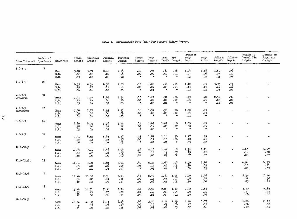
75
Table 1.
Morphometric Data (mm.) For Striped Shiner Larvae,
8.25 .32.14
6.06 .26.12
1.73 .19.08
2.46 .27.12
1.19 .06 .03
2.22 .09.04
3.20 .15.07
.66.05.02
6.28 .28.13
8.23 .26.12
12.30 .48.22
14.51 .31.14
MeanS.D.S.E.5
14.0-14.9
7.85 .22.08
5.79 .17.06
1.63 .19.07
2.40 .29.10
1.10 .05.02
2.03 •09.03
2.94 .18.06
.61.04 .02
5.58 .29.10
7.88 .17.06
11.51 .23.08
13.46 .33.12
Mean S.D.S.E.8
13.0-13.9
7.20 .30 .11
5.34 .12•05
1.46 .16.06
2.20 .26 .10
1.05 .07 .03
1.89 .03 .01
2.72 .15.06
.45.08.03
5.13 .46.17
7.31 .24.09
10.68.12 .08
12.44.31.12
MeanS.D. S.E.7
12.0-12.9
6.75 .11.03
4.94 .10 .03
1.16 .09.03
1.79 .16.04
.96.03
1.64 .08.02
2.33 .12.03
.42.03
4.51 .26.07
6.80 .16.04
9-70 .22.01
11.31 •25.07
Mean S.D.S.E.
13
11.0-11.9 .
6.47 .20.07
4.65 .14.05
1.11 .21.07
1.72• 19.07
.88.05.02
1.51 .19.07
2.16 .15.05
.38.04.01
3.96 .15.05
6.48 .25.09
9.21 .27 .09
10.44 .33.12
Mean S.D.S.E.8
10iO-10»9
•79.11.02
1.27 .18.03
.76.06
1.18 .10 .02
1.82 .11.02
.23.05 *
3.57 .15.03
5.84 .24.04
8.52 .23.04
9.41 .25.06
Mean S.D.S.E.
359.0-9.9
.69 .05
1.09 .08.68.07 *
1.07.06
1.63 .14.02
• 19.03
3-22 .12.02
5.38•
19.02
8.02.20.02
8.60 .28.03
Mean S.D.S.E.
658.0-8.9
.63.03
1.00.Ok.01
.58.02 *
.98.03
1.39.03 *
.16.03
2.93 .09.03
4.93 .06.02
7.47 .08.02
7.86 .09.02
Mean S.D.S.E.
137.0-7.9Postlarva
.37.25.05
2.58 .83.15
.70.05
• 95.Ok *
.56.02.06 .01
1.22 .09.02
.15.03
2.72 .16.03
4.69 .16.03
7.07 .2k.Ok
7.4l .22.05
Mean S.D.S.E.
307.0-7.9Prolarva
•74.16.03
3.37 .18.04
.93.15 .03
1.10.13.03
.5k.03
.5k.03
1.05 .07.01
.12.02
2.23 .21 .05
4.32 .11.02
6.34• 27.06
6.55 .29.06
Mean S.D.S.E.
226.0-6.9
.10.04
3.51 .07.03
1.18 .06.02
1.24 .07.03
.48.01.80.02
•95.02
.10.02
1.74.Ok.02
4.15 .07•03
5.71 .08•03
5.89 .08•03
MeanS.D. S.E.75.6-5.9
Length toAnal FinOrigin
Length toDorsal FinOriginYolksac DepthYolksac LengthBodyWidth
Greatest Body DepthEye LengthHead DepthHead LengthSnout- LengthPostanal LengthPreanal LengthUrostyleLengthTotal LengthStatisticNumber ofSpecimensSize IntervalSize Interval Number ofSpecimens Statistic Total Length UrostyleLength Preanal Length Postanal Length Snout- Length Head Length Head Depth Eye LengthGreatest Body Depth
BodyWidth Yolksac Length Yolksac Depth
Length toDorsal FinOrigin
Length toAnal FinOrigin5.6-5.9 7 MeanS.D. S.E.
5.89 .08•03
5.71 .08•03
4.15 .07•03
1.74.Ok.02
.10.02
•95.02
.80.02
.48.01
1.24 .07.03
1.18 .06.02
3.51 .07.03
.10.046.0-6.9 22 Mean S.D.S.E.
6.55 .29.06
6.34• 27.06
4.32 .11.02
2.23 .21 .05
.12.02
1.05 .07.01
.5k.03
.5k.03
1.10.13.03
.93.15 .03
3.37 .18.04
•74.16.037.0-7.9Prolarva 30 Mean S.D.S.E.
7.4l .22.05
7.07 .2k.Ok
4.69 .16.03
2.72 .16.03
.15.03
1.22 .09.02
.06 .01
.56.02 • 95.Ok *
.70.05
2.58 .83.15
.37.25.057.0-7.9Postlarva 13 Mean S.D.S.E.
7.86 .09.02
7.47 .08.02
4.93 .06.02
2.93 .09.03
.16.03
1.39.03 *
.98.03
.58.02 *
1.00.Ok.01
.63.038.0-8.9 65 Mean S.D.S.E.
8.60 .28.03
8.02.20.02
5.38•
19.02
3-22 .12.02
• 19.03
1.63 .14.02
1.07.06
.68.07 *
1.09 .08
.69 .059.0-9.9 35 Mean S.D.S.E.
9.41 .25.06
8.52 .23.04
5.84 .24.04
3.57 .15.03
.23.05 *
1.82 .11.02
1.18 .10 .02
.76.06
1.27 .18.03
•79.11.0210iO-10»9 8 Mean S.D.S.E.
10.44 .33.12
9.21 .27 .09
6.48 .25.09
3.96 .15.05
.38.04.01
2.16 .15.05
1.51 .19.07
.88.05.02
1.72• 19.07
1.11 .21.07
4.65 .14.05
6.47 .20.0711.0-11.9 . 13 Mean S.D.S.E.
11.31 •25.07
9-70 .22.01
6.80 .16.04
4.51 .26.07
.42.03
2.33 .12.03
1.64 .08.02
.96.03
1.79 .16.04
1.16 .09.03
4.94 .10 .03
6.75 .11.0312.0-12.9 7 MeanS.D. S.E.
12.44.31.12
10.68.12 .08
7.31 .24.09
5.13 .46.17
.45.08.03
2.72 .15.06
1.89 .03 .01
1.05 .07 .03
2.20 .26 .10
1.46 .16.06
5.34 .12•05
7.20 .30 .1113.0-13.9 8 Mean S.D.S.E.
13.46 .33.12
11.51 .23.08
7.88 .17.06
5.58 .29.10
.61.04 .02
2.94 .18.06
2.03 •09.03
1.10 .05.02
2.40 .29.10
1.63 .19.07
5.79 .17.06
7.85 .22.0814.0-14.9 5 MeanS.D.S.E.
14.51 .31.14
12.30 .48.22
8.23 .26.12
6.28 .28.13
.66.05.02
3.20 .15.07
2.22 .09.04
1.19 .06 .03
2.46 .27.12
1.73 .19.08
6.06 .26.12
8.25 .32.14
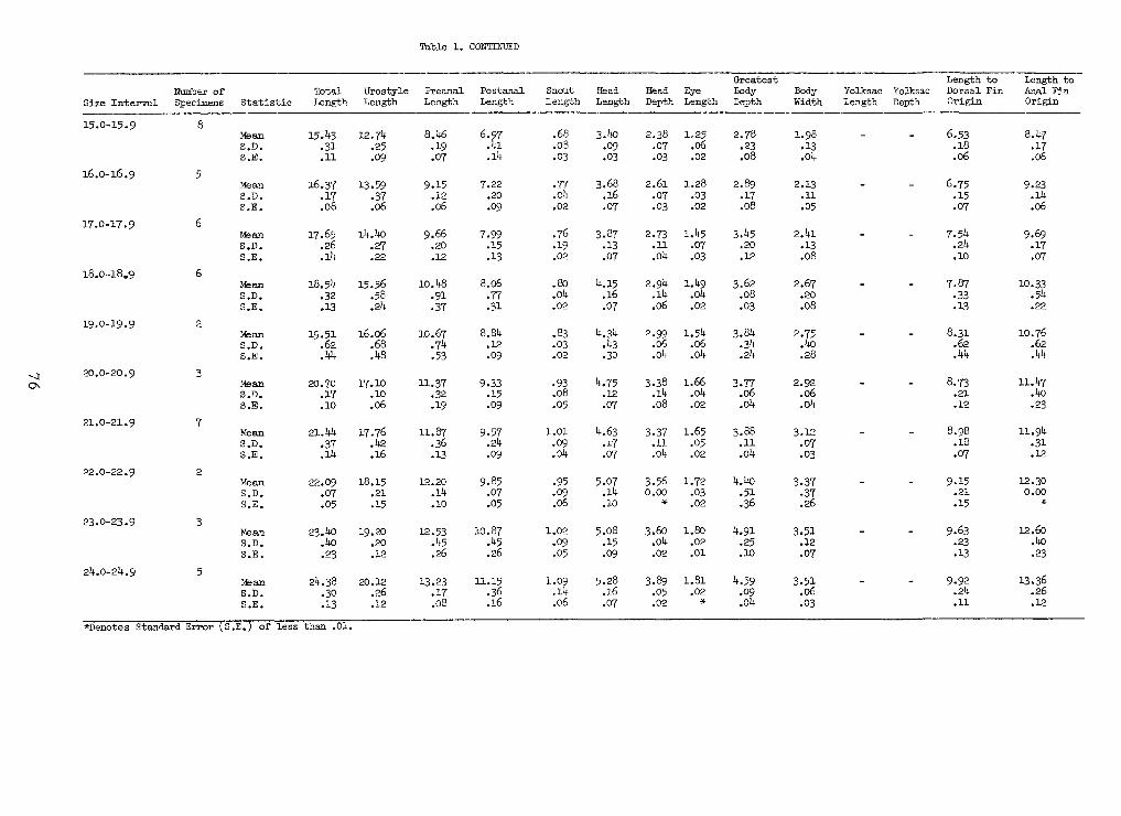
76
Table 1.CONTINUED
of less than .01.S.E.Standard Error
13.36.26.12
9-92 .24.11
3.51 .06 .03
4.59 .09.04
1.81.02
3.89 .05.02
5.28 .16 .07
1.09 .14.06
11.15.36.16
13.23 .17.08
20.12 .26.12
24.38• 30.13
Ms anS.D.S.E.
524.0-24.9
12.60.40.23
9.63 .23.13
3.51 .12.07
4.91 .25 .10
1.80 .02.01
3.60 .04 .02
5.08•
15.09
1.02• 09.05
10.87 .45 .26
12.53 .45 .26
19.20.20.12
23-40 .40.23
Ms anS.D.S.E.
323.0-23.9
12.30 0.009.15 .21 .15
3.37 .37.26
4.40 .51 .36
1.72.03.02
3.560.00
5.07 .14.10
•95.09.06
9.85 .07.05
12.20.14.10
18.15 .21.15
22.09 .07.05
MeanS.D.S.E.222.0-22.9
11.94.31.12
8.98 .18• 07
3.12 .07.03
3.88 .11.04
1.65.05.02
3-37 .11.04
4.63 .17 .07
1.01 .09.04
9.57 .24•09
11.87 .36.13
17.76.42 .16
21.44 .37.14
MeanS.D. S.E.721.0-21.9
11.47 .40 .23
8.73 .21 .12
2.92 .06.04
3.77 .06.04
1.66 .04.02
3.38 .14 .08
4.75 .12.07
• 93.08.05
9-33 .15•09
11.37• 32.19
17.10.10.06
20.70 .17.10
Mean S.D.S.E.320.0-20.9
10.76.62.44
8.31 .62.44
2.75 .40.28
3.84 .34.24
1.54 .06 .04
2.99 .06.04
4.34 .43• 30
.83.03.02
8.84.12.09
10.67 .74.53
16.06 .68.48
19.51 .62.44
MeanS.D. S.E.2
19.0-19.9
10.33 .54 .22
7.87 .33.13
2.67 .20.08
3.62 .08• 03
1.49 .04.02
2.94 .14.06
4.15 .16.07
.80.04.02
8.06 .77.31
10.48.91.37
15.56.58.24
18.54.32.13
Mean S.D.S.E.6
18.0-18.9 9.69 .17 .07
7.54 .24.10
2.4l .13.08
3.45 .20.12
1.45 .07.03
2.73 .11.04
3.87 .13.07
.76.19.02
7.99 .15 .13
9.66 .20.12
14.40.27.22
17.65 .26.14
MeanS.D.S.E.
6
17.0-17.9 9.23 .14 .06
6.75 .15.07
2.13 .11.05
2.89 .17.08
1.28•03.02
2.61 .07• 03
3.68 .16.07
.77.04.02
7.22 .20.09
9.15 .12.06
13.59 .37.06
16.37 .17.08
MeanS.D.S.E.5
16.0-16.9
8.47 .17.06
6.53 .18.06
1.98.13.ok
2.78 .23 .08
1.25.06.02
2.38 .07.03
3.1)0 •09.03
.68.03.03
6.97 .41
8.1(6 .19.07
12.7^.25.09
15.43 .31.11
Ms anS.D.S.E.
8
15.0-15.9
Length toAnal Fin.Origin
Length toDorsal FinOriginYolksac DepthYolksac LengthBodyWidth
Greatest Body DepthEye LengthHead DepthHead LengthSnout LengthPostanal LengthPreanal LengthUrostyleLengthTotal LengthStatisticNumber ofSpecimensSize IntervalSize Interval Number ofSpecimens Statistic Total Length UrostyleLength Preanal Length Postanal Length Snout Length Head Length Head Depth Eye LengthGreatest Body Depth BodyWidth Yolksac Length Yolksac Depth
Length toDorsal FinOrigin
Length toAnal Fin.Origin15.0-15.9 8
Ms anS.D.S.E.
15.43 .31.11
12.7^.25.09
8.1(6 .19.07
6.97 .41
.68.03.03
3.1)0 •09.03
2.38 .07.03
1.25.06.02
2.78 .23 .08
1.98.13.ok
6.53 .18.06
8.47 .17.0616.0-16.9 5 MeanS.D.S.E.
16.37 .17.08
13.59 .37.06
9.15 .12.06
7.22 .20.09
.77.04.02
3.68 .16.07
2.61 .07• 03
1.28•03.02
2.89 .17.08
2.13 .11.05
6.75 .15.07
9.23 .14 .0617.0-17.9 6 MeanS.D.S.E.
17.65 .26.14
14.40.27.22
9.66 .20.12
7.99 .15 .13
.76.19.02
3.87 .13.07
2.73 .11.041.45 .07.03
3.45 .20.12
2.4l .13.08
7.54 .24.10
9.69 .17 .0718.0-18.9 6 Mean S.D.S.E.
18.54.32.13
15.56.58.24
10.48.91.37
8.06 .77.31
.80.04.02
4.15 .16.07
2.94 .14.06
1.49 .04.02
3.62 .08• 03
2.67 .20.08
7.87 .33.13
10.33 .54 .2219.0-19.9 2 MeanS.D. S.E.
19.51 .62.44
16.06 .68.48
10.67 .74.53
8.84.12.09
.83.03.02
4.34 .43• 30
2.99 .06.04
1.54 .06 .04
3.84 .34.24
2.75 .40.28
8.31 .62.44
10.76.62.4420.0-20.9 3 Mean S.D.S.E.
20.70 .17.10
17.10.10.06
11.37• 32.19
9-33 .15•09
• 93.08.05
4.75 .12.07
3.38 .14 .08
1.66 .04.02
3.77 .06.04
2.92 .06.04
8.73 .21 .12
11.47 .40 .2321.0-21.9 7 MeanS.D. S.E.
21.44 .37.14
17.76.42 .16
11.87 .36.13
9.57 .24•09
1.01 .09.04
4.63 .17 .07
3-37 .11.04
1.65.05.02
3.88 .11.04
3.12 .07.03
8.98 .18• 07
11.94.31.1222.0-22.9 2 MeanS.D.S.E.
22.09 .07.05
18.15 .21.15
12.20.14.10
9.85 .07.05
•95.09.06
5.07 .14.10
3.560.001.72
.03.02
4.40 .51 .36
3.37 .37.26
9.15 .21 .15
12.30 0.0023.0-23.9 3
Ms anS.D.S.E.
23-40 .40.23
19.20.20.12
12.53 .45 .26
10.87 .45 .26
1.02• 09.05
5.08•
15.09
3.60 .04 .02
1.80 .02.01
4.91 .25 .10
3.51 .12.07
9.63 .23.13
12.60.40.2324.0-24.9 5
Ms anS.D.S.E.
24.38• 30.13
20.12 .26.12
13.23 .17.08
11.15.36.16
1.09 .14.06
5.28 .16 .07
3.89 .05.02
1.81.02
4.59 .09.04
3.51 .06 .03
9-92 .24.11
13.36.26.12Standard Error S.E. of less than .01.
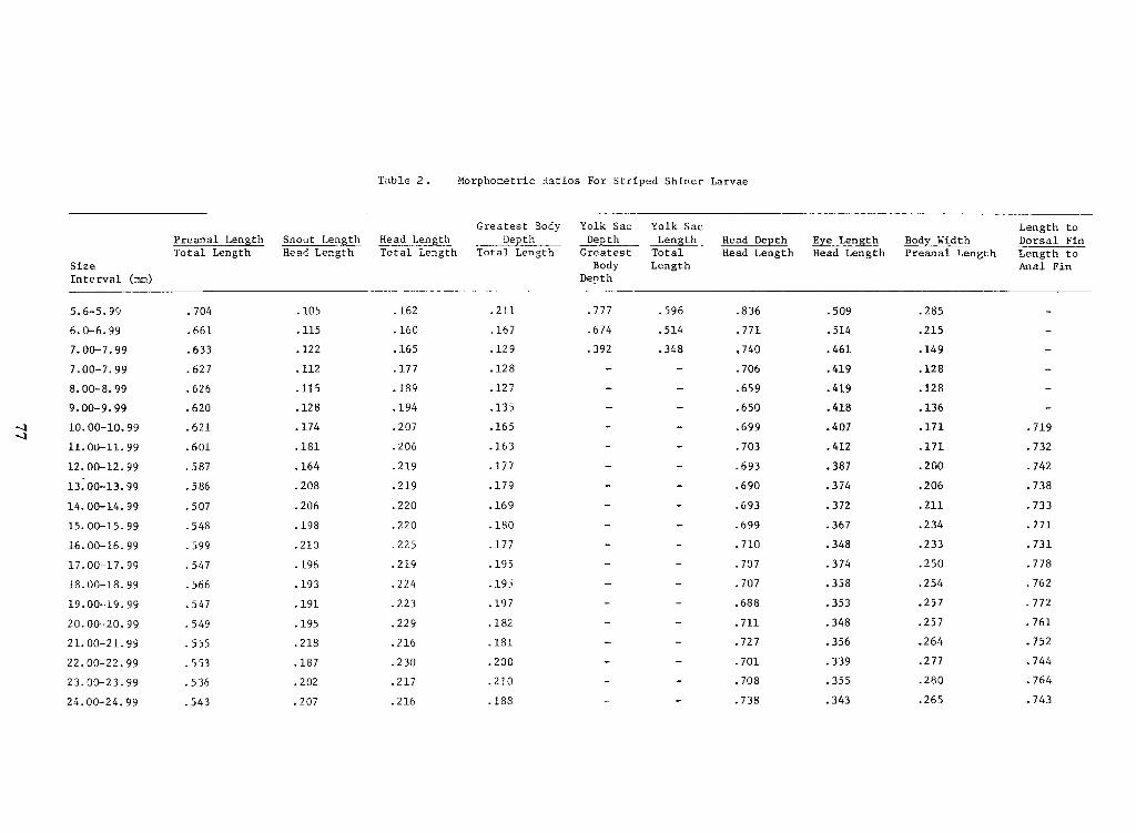
77
Table 2 . Morphometric Ratios For Striped Shiner Larvae
.743.265.343.738.188.216.207.54324.00-24.99 .764.280.355.708.210.217.202.53623.00-23.99 .744.277.339.701.200.230.187.55322.00-22.99 .752.264.356.727.181.216.218.55521.00-21.99 .761.257.348.711.182.229.195.54920.00-20.99 .772.257.353.688.197.223.191.54719.00-19.99 .762.254.358.707.195.224.193.56618.00-18.99 .778.250• .374.707.195.219.196.54717.00-17.99 .731.233.348.710.177.225.210.59916.00-16.99 .771.234.367.699.180.220.198.54815.00-15.99 .733.211.372.693.169.220.206.50714.00-14.99 .738.206.374.690.179.219.208.58613.00-13.99 .742.200.387.693.177.219.164.58712.00-12.99 .732.171.412.703.163.206.181.60111.00-11.99 .719.171.407.699.165.207.174.62110.00-10.99 .136.418,650,135.194.128.6209.00-9.99 .128.419.659.127.189.115.6268.00-8.99 .128.419.706.128.177.112.6277.00-7.99 .149.461,740.348.392.129.165.122.6337.00-7.99 .215.514.771.514.674.167.160.115.6616.0-6.99 .285.509.836.596.777.211.162.105.7045.6-5.99
DepthInterval (mm)
Size Total LengthTotal LengthHead LengthTotal Length Dorsal FinLength toAnal Fin
Bod Body WidthPreanal Lengththc LenEye LengthHead LengththHead DeHead DepthHead LengthLengthTotal LengthLengthDepthGreatest BodyDepthDepththHead LenthSnout LenthP re anal Len
Length toYolk SacYolk SacYolk SacYolk SacGreatest BodyGreatest Body Yolk SacYolk Sac Yolk SacYolk Sac Length to
P re anal Len th Snout Len th Head Len th Depth DepthDepthGreatest Body
LengthLengthTotal Length
Head DepthHead LengthHead De th Eye LengthHead Lengthc Len th Body WidthPreanal LengthBod Dorsal FinLength toAnal Fin
Total Length Head Length Total Length Total LengthSizeInterval (mm) Depth5.6-5.99 .704 .105 .162 .211 .777 .596 .836 .509 .2856.0-6.99 .661 .115 .160 .167 .674 .514 .771 .514 .2157.00-7.99 .633 .122 .165 .129 .392 .348 ,740 .461 .1497.00-7.99 .627 .112 .177 .128 .706 .419 .1288.00-8.99 .626 .115 .189 .127 .659 .419 .1289.00-9.99 .620 .128 .194 ,135 ,650 .418 .13610.00-10.99 .621 .174 .207 .165 .699 .407 .171 .71911.00-11.99 .601 .181 .206 .163 .703 .412 .171 .73212.00-12.99 .587 .164 .219 .177 .693 .387 .200 .74213.00-13.99 .586 .208 .219 .179 .690 .374 .206 .73814.00-14.99 .507 .206 .220 .169 .693 .372 .211 .73315.00-15.99 .548 .198 .220 .180 .699 .367 .234 .77116.00-16.99 .599 .210 .225 .177 .710 .348 .233 .73117.00-17.99 .547 .196 .219 .195 .707 • .374 .250 .77818.00-18.99 .566 .193 .224 .195 .707 .358 .254 .76219.00-19.99 .547 .191 .223 .197 .688 .353 .257 .77220.00-20.99 .549 .195 .229 .182 .711 .348 .257 .76121.00-21.99 .555 .218 .216 .181 .727 .356 .264 .75222.00-22.99 .553 .187 .230 .200 .701 .339 .277 .74423.00-23.99 .536 .202 .217 .210 .708 .355 .280 .76424.00-24.99 .543 .207 .216 .188 .738 .343 .265 .743
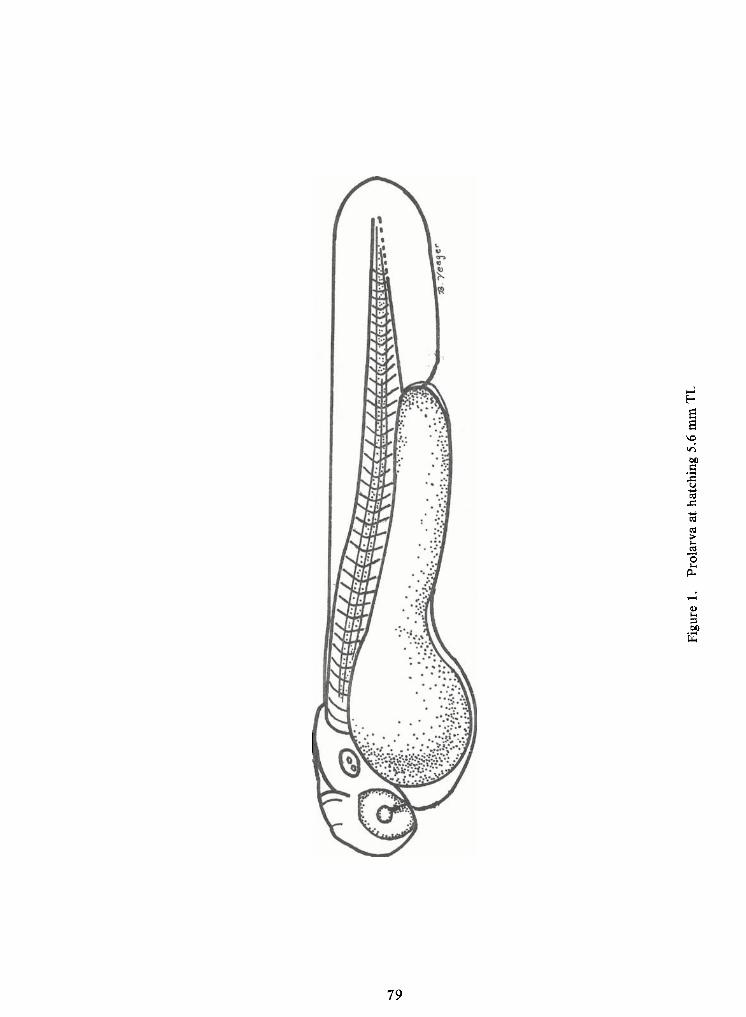
79
Figure1.
Prolarvaat
hatching5.6mmTL
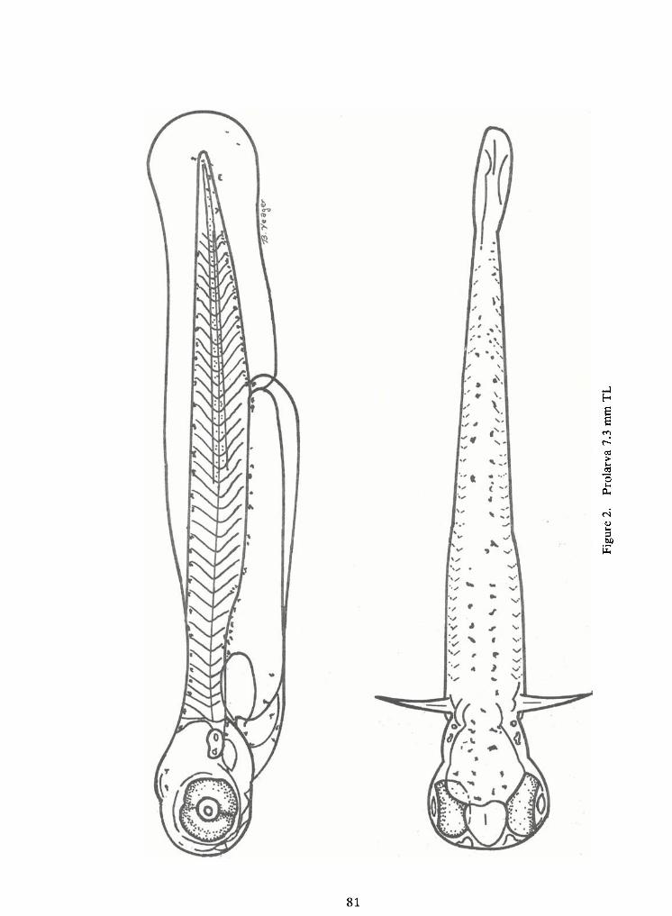
81
Figure2.
Prolarva7.3mmTL
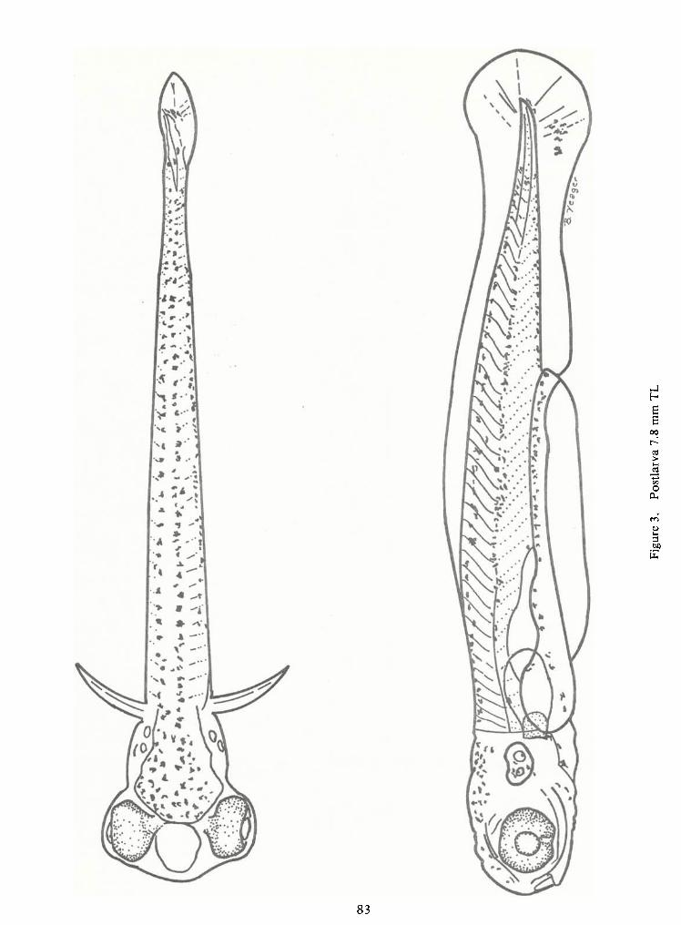
83
Figure3.
Postlarva7.8mmTL
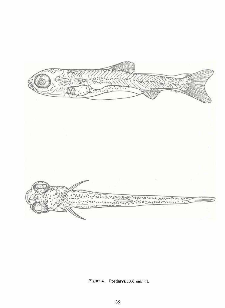
85
Figure 4. Postlarva 13.0 mm TL
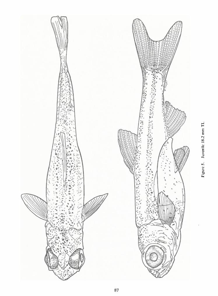
87
Figure5.
Juvenile18.2
mmTL
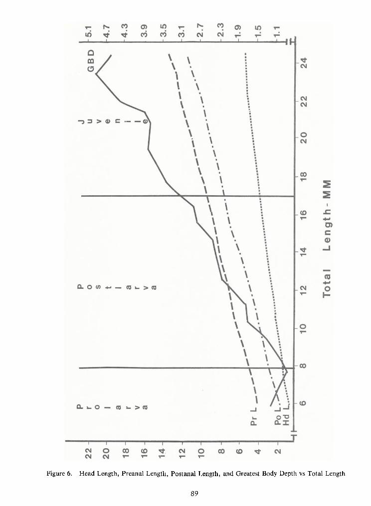
89
Figure 6. Head Length, Preanal Length, Postanal Length, and Greatest Body Depth vs Total Length
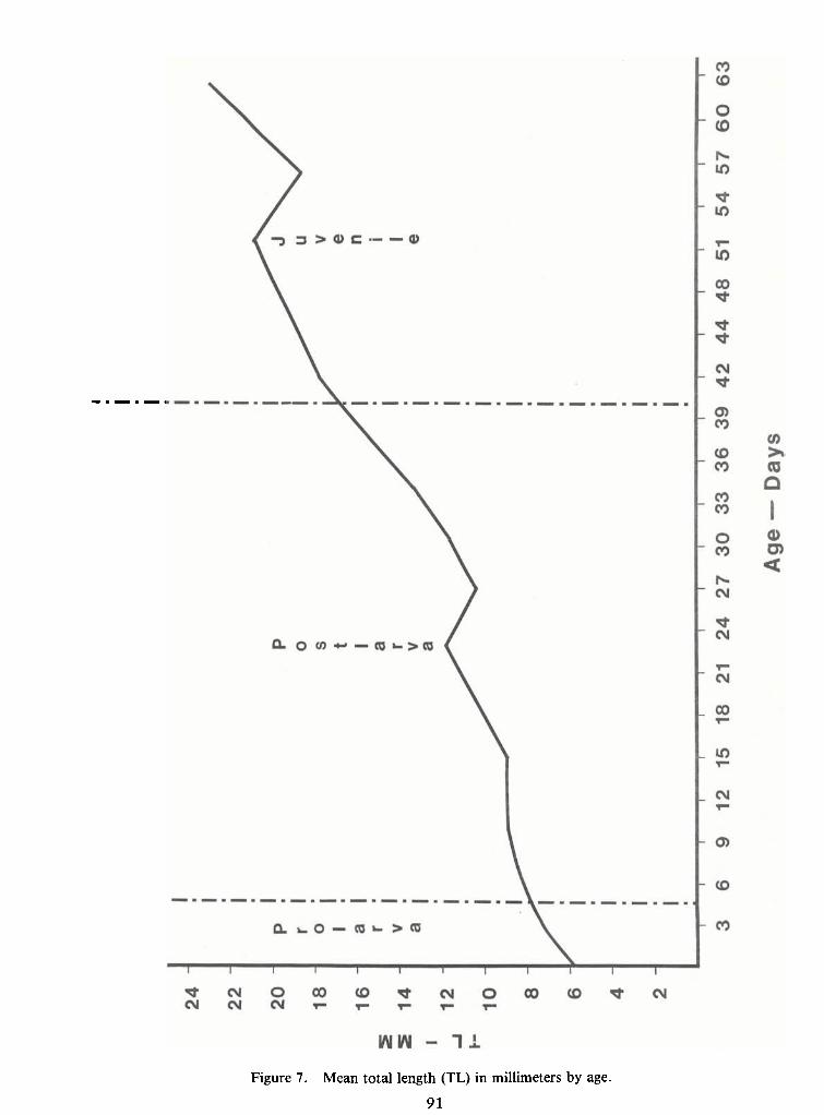
91
Figure 7. Mean total length (TL) in millimeters by age.
NOTES ON
EARLYLIFE HISTORIES OF CYPRINOID FISHES
OF THE UPPER POTOMAC RIVER
by
Jules J. Loos, Lee A.Fuiman,Neal R. Foster and Edward K. Jankowski
Academy of Natural Sciences of PhiladelphiaDivision of Limnology and Ecology
19th and the ParkwayPhiladelphia, Pa. 19103
ABSTRACT
Eggs of cyprinoid fishes in the upper Potomac River were collected from natural
spawning sites, then reared through the larval period to identifiable size. It was found (as expected)
that groups of species that utilize similar spawning sites had incommon certain early developmental
characteristics. Rearing is usually necessary to ensure correct identifications within such groups.
While collecting and identifying eggs, the authors discovered or verified severalreproductive associations: spawning of Notropis procne in the nests of Lepomis auritus, spawning
of Notropis amoenus over completed nests of Nocomis micropogon, and a strong tendency forRhinichthys cataractae to use spawning areas of Catostomus commersoni.
95
INTRODUCTION
The study of the early life history of fishes is ofboth academic interest and practical
importance for fisheries management and evaluation ofman's environmental impacts. While much
data is at hand for North American game and commercial species, little information is available
concerning numerous smaller fishes, especially in the suborder cyprinoidei (Scott and Crossman
1973). This taxon exhibits an extraordinary range of reproductive habits (Breeder and Rosen 1966)
and detailed studies have elucidated many points ofbiological interest (Balon 1975, Kryzhanovsky
1948, Nakamura 1969).
The purpose of this paper is to present notes on the early life history of cyprinoid
species collected from Frederick and Montgomery Counties, Maryland during environmental
studies in the vicinity of the Dickerson Generating Station located on the Potomac River. Twenty-
six of the 37 cyprinoids reported from the Potomac River drainage by Jenkins et al. (1972) and
Davis and Enamait (1977) are considered in this study, with emphasis placed on the relationship ofspawning site to egg and larval traits. Additional information was derived from collections made at
other locations inPennsylvania, Maryland, and Virginia. Although breeding behavior and spawn-
ing seasonality are summarized elsewhere (Breeder and Rosen 1966 and Carlander 1969), eggs and
larvae of many cyprinoids collected were not previously described.
METHODS
The Academy ofNatural Sciences ofPhiladelphia (ANSP) conducted twelve surveys ofjuvenile and adult fish fauna (in conjunction with comprehensive surveys ofother aquatic life)inthe
Potomac River, Frederick and Montgomery Counties, Maryland, from 1956 to 1974 at stationscircled in Fig. 1. Fishes were collected with rotenone and/or a 20-foot bag seine as described by
Cairns and Kaesler (1971). In addition, a list of the cyprinoid fishes (Table 1) was compiled fromspecimens deposited in the Ichthyology Department of ANSP.
Reproductive studies were carried out primarily between 2 May and 14 July 1973.
Observations and collections were made at 15 Potomac River areas indicated by triangles in Fig. 1.
Similar supplementary collections and observations were made in local Potomac tributaries at the
following sites now shown inFig. 1:
96
Catoctin Cr. at the crossing of Maryland Route 464, Frederick County, Maryland
Monacacy R. at LilyPons, Frederick County, Maryland
Unnamed tributary approximately 4 km south of the confluence of the Monacacy and
Potomac Rivers, just above junction with Chesapeake and Ohio Canal, Mont-
gomery County, Maryland
Identification of larvae was facilitated byrearing eggs or larvae ofquestionable identity
to a stage where identification was possible or by comparison with developmental reference series.
Much of the reference series material was reared from eggs collected at other locations. In most
cases naturally fertilized eggs or larvae were collected and taken back to a field laboratory or to
ANSP. Eggs or larvae were placed in shallow plastic containers (89 x 152 x 304 mm) in aeratedstream water between 16 to 20 C. Newly fertilized eggs generally hatched withina week. Larvae were
fed daily with Artemia nauplii and finely ground commercial fish food. Allmaterial, preserved infive percent Formalin buffered with borax, is deposited in the collection of the Ichthyology Depart-
ment of ANSP.
We use the reproductive guild terminology ofBalon (1975) and the developmental ter-
minology of Snyder (1976). Hatching is used as the boundary between the embryonic and larval
periods rather than complete yolk absorption and feeding.
Myomeres were counted with the aid ofPolaroid filters placed above and below the lar-
vae and used with a microscope equipped with a substage lamp. Myomeres between the most
anterior myoseptum and a vertical line at the posterior margin of the anus were termed preanal
myomeres. We follow Siefert (1969) in including allmyomeres transected by this line in the preanal
count. Myomeres in metalarvae were difficult to count due to opacity. Consequently, counts are
given only for protolarvae and mesolarvae.
Internal melanophores, useful in identification of cyprinoid fishes (Balinsky 1948), were made visi-
ble by clearing larvae in glycerine for a few minutes. Larvae which became dehydrated by this pro-
cedure were completely reconstituted when replaced in dilute Formalin.
The presence of cement glands was another character useful in identification. The abili-
tyofa larva to adhere to the walls ofthe rearing chamber was considered evidence of the presence of
cement glands. Illustrations ofNotropis species are not included because these are being published
in an earlier workshop in this series (Loos and Fuiman 1978).
97
ANNOTATED LIST OF SPECIES
Campostoma anomalum (Stoneroller, Fig. 2 A-C). Stoneroller eggs, adhesive during
water hardening, are reported to be 2.4 mm indiameter (Reed 1958). They are found in irregular pit-
like nests constructed by the breeding male or in nests of other species such as creek chubs (Miller
1962) or river chubs (Reighard 1943).
Newly hatched larvae have a large yolk mass that is bulbous anteriorly and tubular
posteriorly. Hatching length is approximately 5.7 mm TL(Reed 1958), but absence of the tubular
posterior portion of the yolk inhis illustration suggests that his specimens were not developing nor-
mally. The retina is unpigmented or light tan at this stage. The head is deflected over the yolk andpectorals are not prominent. These characteristics suggest early hatching larvae. Balon (1975) in-
dicates that protolarval characteristics of brood hiding lithophils may facilitate respiration.
Hogue, et al. (1976) report 27 to 28 preanal myomeres and a mesolarval pigment pat-
tern similar to that we have observed for other nest-building minnows as well as Rhinichthys in the
same phase. Inmetalarvae, the distinct caudal spot separated from the lateral body stripe and the
subterminal mouth readily distinguishes stonerollers from fallfish and creek chubs. The intestinedoes not become greatly elongated until the juvenile period (Kraatz 1924).
Chrosomus oreas (Mountain Redbelly Dace, Fig. 4 D-F)—This dace, while not found
in the study area, occurs in the Potomac drainage. Itis a common breeding associate ofNocomis
leptocephalus. Mesolarvae are characterized by diagonal rows ofexternal body melanophores align-
ed along the myosepta.
Clinostomus funduloides (Redside Dace, (Fig. 89 D-F)—This breeding associate of Semotilus
atromaculatus is found in the Potomac drainage but not the study area. Its eggs are smaller than
those ofS. atromaculatus (1.8 to 2 mm in diameter) and distinctly more yellow; newly hatched lar-vae have the characteristics of other lithophils at the same stage. Hogue et al. (1976) indicate 22-25
preanal myomeres and a pigment pattern different than that expressed by our specimens. Theirs
from the Tennessee drainage had a single midventral row ofmelanophores extending to the anus;
our specimens from the Delaware drainage had a dark patch ofmelanophores on the breast and a
poorly developed midventral row.
98
Cyrpinus carpio and Carassius auratus (Carp and Goldfish, Fig. 2 D-F)—Eggs are at-
tached to aquatic plants or flooded vegetation (Richardson 1913). Along the Potomac River expos-
ed roots of trees are common spawning sites.
Eggs of both species were reared; eggs of the carp were slightly larger than those ofgoldfish (1.8 mm versus 1.5 mm). Hatching size for carp is also slightly greater (5.2 mm versus 4.7
mm TL).
Athatching, the eyes ofboth are pigmented and the head is not deflected over the yolk.
These traits indicate that larvae are relatively advanced at hatching. This and the presence of cement
glands, which allow the larvae to adhere to a substrate, are characters Balon (1975) considers typical
of the open substrate phytophilous reproductive guild. In the mesolarval phase the carp develops a
prominent dark spot at the base of the caudal fin;the goldfish, which lacks this spot, develops a line
of melanophores along the horizontal myoseptum.
Adense band ofmelanophores on the ventral aspect ofpreanal and postanal myomeres
and a proportionately larger preanal length are useful for differentiating protolarval and mesolarvalcarp and goldfish from native minnows. Both species lack a lateral band or line of body
melanophores common to mesolarvae and metalarvae of other minnows. Metalarval carp andgoldfish have ten or more incipient dorsal rays; other minnows of the area have fewer.
Ericymba buccata (Silver jaw Minnow)—The presence of silver jaw minnows in the area was con-
firmed by rearing one larval specimen collected just above area three in early May (exact date and
temperature data lost) and two others from Catoctin Creek in July (24 C). Reproductive lifehistory
information is presented by Hoyt (1971) and Wallace (1973).
Exoglossum maxillingua (Cutlips Minnow, Fig. 3 A-Q—Adults are rare in the study area and no
larvae were collected. Fuiman and Loos (inpress) describe larval development and note that eggs
may be laid in nests of Nocomis micropogon. Van Duzer (1939) gave a detailed account of its
reproductive habits including nest building, spawning association with common shiners, and a brief
description ofits eggs.
Hybognathus regius (Silvery Minnow)—The silvery minnow was rarely collected inthe
study area. No larvae were identified, but their apparent similarity to larvae of the spottail shiner
99
may have caused misidentifications. Raney (1939) described breeding behavior and illustrated larval
specimens ofH. regius. These and additional illustrations and descriptions were presented by Man-
sueti and Hardy (1967).
Nocomis micropogon (River Chub, Fig. 3 D-F)—lts nests (often used by other species)
were described by Cope (1868), Greely (1929), Raney (1940b), Reighard (1943), Lachner (1952) and
others.
Eggs, adhesive during water hardening, are approximately two mm in diameter (Fish
1932). They are relatively clear, with yellowish yolk, and can be distinguished from those of
Rhinichthys cataractae, which sometimes uses Nocomis nests, because the latter are more opaque.
They are similar to eggs of breeding associates, Notropis rubellus, N. cornutus, Exoglossum max-
illigua and Campostoma anomalum. When onlyNocomis and N.rubellus eggs are present, they can
be distinguished by the smaller size of the shiner eggs. Ifeggs of the others are present, accurate
identification is not feasible with available data.
Newly hatched, unpigmented protolarvae of all nest builders and their associates are
similar. They do differ in size, but overlapping measurements usually make identification imprac-
tical without rearing. Larvae are robust and approximately 7 mm TLat hatching. They develop two
dorsal body rows of melanophores that are strongly divergent just posterior to the head in the
mesolarval phase. They are similar toNocomis leptocephalus (Fig. 4 A-C) larvae reported from the
drainage system (Jenkins et al., 1972) but not from the study area.
Notemigonus crysoleucas (Golden Shiner, Fig. 4 A-E)—Eggs were found attached to
small pebbles ina tributary ofPiscataway Cr. (Potomac drainage) and were hatched and larvae were
reared to confirm identification.
Golden shiners reportedly spawn over vegetation and centrarchid nests (Kramer and
Smith 1960, Johnson C. S. Wang, personal communication). Snyder et al. (1977) present a detaileddescription of egg and larval characteristics of this species. No other cyprinoid from the study area
had this combination of developmental characters. Larvae which we reared conform to theirdescriptions except that our specimens hatched at approximately 4.2 mm TLwith darkly pigmented
eyes. An earlier statement by Loos (1974) that mesolarval N. crysoleucas may lack the midventral
abdominal stripe was apparently incorrect.
100
Larvae apparently possess cement glands for they adhered to the walls of the rearing
chamber. These glands, found on the head, are characteristic of species assigned byBalon (1975) to
the open substrate phytophilous reproductive guild.
Notropis amoenus (Comely Shiner) —Specimens of this Notropis species are illustratedby Loos and Fuiman (1978)
—No eggs or larvae were identified from the study area; however, we
reared eggs from the Susquehanna drainage of south central Pennsylvania. These were similar to
Notropis rubellus and closely corresponded to descriptions and illustrations of Notropis
atherinoides, the emerald shiner (Flittner 1964).
The delicate nonadhesive egg capsule of these species is not unlike that of striped bass.The colorless egg is approximately 2.7 to 3 mm in diameter. The perivitelline space width, larger
than for any other Potomac River cyprinoid, is nearly equal to the diameter of the yolk.
The newly hatched larvae (approximately 5.5 mm TL) are also similar to N.
atherinoides specimens illustrated by Flittner (1964), except that they are more advanced, i.e., eyes
are darkly pigmented; the head is not deflected over the yolk, and the pectorals are developed. A
midventral row of abdominal melanophores is present; N. atherinoides is not pigmented at thisstage.
Mesolarvae and metalarvae are similar to Notropis rubellus in the same phases, in that
both have a single midventral row of abdominal melanophores and melanophores on the tip of a
projecting lower jaw. Both lack the concentration ofmelanophores on the dorsal uro style found inNotemigonus crysoleucas at the same phase of development.
Notropis cornutus (Common Shiner) —Eggs are found in hollows cleared by breeding
males or in nests ofriver chubs, fallfish,cutlips, minnows, creek chubs or stonerollers (Kendall and
Goldsborough 1908, Raney 1940 a).
Eggs, approximately 1.7 to 1.8 mm in diameter (Raney 1940a), are intermediate bet-
ween those of the rosyface shiner and river chub. Egg capsules are adhesive during water hardening
and relatively clear. Newlyhatched larvae are approximately 5 mm TL.Mesolarvae are characteriz-
ed by two rows of dorsal body melanophores, a concentration of melanophores on the dorsal
urostyle, and generally by variously developed midventral and ventro-lateral rows of abdominal
melanophores.
101
Notropis hudsonius (Spottail Shiner)— This species dominated larval collections in late
May and early June. Midsummer spawning, as reported by Griswold (1963), does not occur in the
study area.
Inthe Potomac River and elsewhere their eggs were found in shallow riffles attached to
the upper surface of fine gravel or sand (Wright and Allen1913, Greely 1930). Eggs were also found
in patches of Cladophora (Wells and House 1974).
Balon (1975) tentatively placed N. hudsonius in the open substrate psammophilous
reproductive guild. Our observations support his classification, i.e., eggs are small (approximately
1.3 mm indiameter); protolarvae lack cement glands; and they are phototropic after the swim blad-
der fills.
Newly hatched protolarvae (approximately 4.3 mm TL) have poorly pigmented eyes
and dense ventral pigmentation similar to that ofNotropis bifrenatus (Harrington 1947, Mansuetiand Hardy 1967). In this stage, the former can be distinguished by the absence of cement glands. In
the mesolarval phase (Lippson and Moran 1974), N.hudsonius is characterized by two dorsal rows
ofbody melanophores and scattered melanophores on ventrum (sometimes arranged in three vague
rows—
Darrel Snyder, personal communication). The caudal spot is sometimes well developed inthe
metalarval phase.
Notropis procne (Swallowtail Shiner)—
We have found swallowtail shiner eggs at-
tached to pebbles in the nests of Lepomis auritus (redbreast sunfish). They also spawn in riffleareas over fine gravel and sand (Raney 1947b). No larvae were identified from our collections in
the study reach.
Notropis rubellus (Rosyface Shiner)—
Eggs are found in nests of other cyprinids
(Pfeiffer 1955, Hankinson 1932b, Lachner 1952, Pflieger 1975); river chub nests are used in the
study area.
Eggs and larvae are most similar to those ofNotropis ardens, which are found in thePotomac River drainage, but not in the study area. Raney (1947a) considers N. ardens a southern
ecological replacement of N. rubellus. The eggs and newly hatched larvae of these two species
are similar in size. The exact sizes are unknown because eggs were mixed with those of other
102
species, but they are among the smallest eggs in the nest (1.5 to 1.7 mm in diameter). Newly
hatched larvae are approximately 5 mm TL.Reed (1958) reported that N. rubellus eggs were 1.5mm in diameter and newly hatched larvae were 5.1 mm TL.
Mesolarvae of the two species are similar in pigmentation, morphology, and
behavior (Loos and Fuiman 1978). Compared with other minnow larvae from the study area,
rosyface shiner larvae were similar to those of the golden and common shiners. The mesolarva
of the common shiner has a concentration of melanophores dorsally on the urostyle. This con-
centration is absent in the rosyface shiner. N. rubellus also lacks ventro-lateral abdominal rows
of melanophores often found in common shiner mesolarvae. The golden shiner protolarvae have
cement glands; the rosyface shiner protolarvae do not. (See Notropis amoneus for comparisons
with that specis.)
Notropis spilopterus (Spotfin Shiner)—
Adhesive eggs (1.2 to 1.5 mm in diameter,
Snyder et al. 1977) are generally attached to wood in moderate current. Clumps of eggs are fre-quently found in cracks in branches or boards or under bark (Hankinson 1930, Stone 1940,
Pflieger 1965). No other minnow in the study area has clumped eggs laid in this manner.
Larvae (approximately 4mm TL at hatching) have been described in detail by Snyder
et al. (1977). They can be identified in the mesolarval phase on the bases of dorso-ventral com-
pression of the head, absence of a midventral abdominal stripe, and pale dorsal pigmentation.
Eggs and larvae of this species are similar to those of the satinfin shiner (Notropis analostanus)
(Loos and Fuiman 1978), another species of the Cyprinella subgenus found in the Potomac River
near Washington, D.C. (Lippson and Moran, 1974).
Mesolarval satinfin shiners often lack the "V"-shaped pattern ofmelanophores on top
of the head and the elongate breast melanophores described by Loos (1974). Protolarvae and
mesolarvae are indistinguishable from those of the spotfin shiner. Metalarvae can readily be
distinguished because the satinfin shiner typically has nine anal rays while the spotfin shiner
typically has eight.
Pimephales notatus (Bluntnose Minnow, Fig. 6 A-D)—Adhesive eggs (1.6 mm in
diameter, Hubbs and Cooper 1936) are laid on the underside of flat objects (Westman 1938). Em-
bryonic and protolarval development has been described (Fish 1932) and a complete developmental
series of our specimens is available at ANSP.
103
Mesolarvae and metalarvae are superficially similar to spottail shiners inthe same stage
of development. The metalarvae of both generally have an intensification ofpigment in the caudal
area. The bluntnose minnow is characterized by chevron-shaped markings along the notochord that
become visible when the specimen is cleared in glycerin.
Rhinichthys atratulus (Blacknose Dace, Fig. 6 C-F)—Adhesive eggs are approximately
2.3 mm in diameter. We have found eggs in gravel riffles and in pockets of gravel in streams overbedrock, often near those of the longnose dace and white sucker. Bartnik (1970) reports spawning
sites ofthe daces, though sometimes nearly adjacent, are separate; the western blacknose dace (R. a.
melegris) spawn in slower water over finer substrata than the longnose dace. Elsewhere, R. atratulus
spawns over nests of Semotilus corporalis (Reed, 1971) and Nocomis biguttatus (Hankinson,
1932 a). We have not found eggs of the Potomac or Susquehanna populations in Nocomis
micropogon nests, although Rhinichthys cataractae often spawns there.
Fuiman and Loos (1977) described the larvae that hatch at 6 mm TL. A subterminalmouth, numerous scattered dorsal body melanophores, and a mode of 24 preanal myomeres are
characteristics developed by the mesolarval phase. The mouth of the mesolarva is more terminal
than that ofR. cataractae in the same phase. Mesolarvae are difficult to distinguish from those ofCampostoma anomalum and Exoglossum maxillingua. The subterminal mouth and frenum andposition of dorsal-fin origin posterior to base of pelvic finbuds are the only characters needed to
distinguish the metalarvae of this species from those of other minnows in the area.
Rhinichthys cataractae (Longnose Dace, Fig. 7 A-D)—The adhesive, relatively opaque
eggs are 2.5 mm indiameter. Fuiman and Loos (1977) report that larvae hatch at approximately 5.5
mm TL and lack pigment and that mesolarvae have a pigment pattern similar to R. atratulus,
modally 25 preanal myomeres, and an inferior mouth. Their long noses are unmistakable in themetalarval phase.
Semotilus atromaculatus (Creek Chub, Fig. 8 A-C)—This species' nest and breeding
habits were described by Reighard (1910). Eggs are 2.0 to 2.1 in diameter, and newly hatchedlarvae are 5.3 mm TL.
Mesolarvae are similar to those of the fallfish, but have a concentrated area of pig-
ment on the pectoral fin near its base which is not present in mesolarval fallfish. Both species
104
have more preanal myomeres (usually 28-29) than other minnows in the area. A metalarvalspecimen was described and illustrated by Fish (1932). She indicated fewer preanal myomeres,
but her counting techniques differed from ours.
Semotilus corporalis (Fallfish, Fig. 9 A-Q—Reed reported that the adhesive eggs
are 2.7 mm in diameter and that the larvae are approximately 6.5 mm TLat hatching; thus being
larger than eggs and newly hatched larvae of other minnows in the area. Reed's descriptions
and illustrations (1971) as well as our observations indicate that fallfish larvae are generally
larger than those of the blacknose and longnose dace at any given stage of development.
Carpiodes cyprinus (Quillback Carpsucker, Fig. 9 D-F)—
Juveniles have been col-
lected in tributaries of the Potomac River below Washington, D.C., but none have been collectedin the study area. We have found eggs attached to the upper surface of gravel and organic mat-
ter in shallow water. These eggs are approximately 2.1 mm indiameter.
Development of C. cyprinus larvae was described by Gerlack (1973). Newly hatched
larvae have darkly pigmented eyes. In white suckers, northern hogsuckers, and shorthead and
golden redhorses the larvae are larger at any given stage and the snout-to-vent length is about0.8 times the TL, while in quillback carpsucker this proportion is much less. C. cyprinus lacks thepale dorsal head stripe of the creek chubsucker.
Erimyzon oblongus (Creek Chubsucker, Fig. 10 A-C)—Adhesive eggs are found ingravel. They are approximately 1.8 mm in diameter and the newly hatched larvae are approx-
imately 6.5 mm TL.Both eggs and larvae are considerably smaller than those of white suckers,
hogsuckers, and redhorses. Larvae illustrated by Carnes (1958) and reprinted by Mansueti and
Hardy (1967) appear deformed. Mesolarvae and metalarvae are characterized by a pale stripe
on top of the head extending posterior to the eye.
Moxostoma macrolepidotum (Shorthead Redhorse) and M. erythrurum (Golden
Redhorse)— Descriptions of larval series of the shorthead redhorse are available (Furiman 1978)
while those of the golden redhorse are forthcoming (Fuiman and Witman, ms.).
Hypentelium nigricans (Hogsucker, Fig. 10 D-E)—This sucker spawns in rapids
(Carlander 1969). Raney and Lachner (1946) reported that nonadhesive eggs were dislodged from
spawning sites.
105
The development of the larvae of this species has been described by Fuiman (1978), and
Fish (1932) described and illustrated a young juvenile.
Catostomus commersoni (White Sucker, Fig. 11 A-D)—Adhesive eggs are deposited in
gravel riffles where protolarvae remain after hatching for an undetermined period. Detailed em-
bryological descriptions of the stages and rates of development at several temperatures were made
by Long and Ballard (1976). Eggs and larvae (Steward 1927, Mansueti and Hardy 1967) are least
superficially similar to those of the redhorses and hogsucker. Their presence in the study area was
documented by collecting larvae and rearing them to an identifiable size.
COMPARISONS AMONG CYPRINOID GROUPS THATUTILIZEDIFFERENT SPAWNING SITES
At the present time, it is often impossible to identify eggs based on morphological
characters without hatching and culturing them at least through the protolarval phase. However,
one can usually determine immediately if a specimen belongs to a group of species with similarreproductive habits, thereby at least narrowing the range ofchoices. Insome cases there is only one
representative ina given mode. The following list summarizes information on reproductive modes
of species from the study area. Some species willappear inmore than one group; the primary modeof a given species is indicated by an asterisk.!
Group: 1 Eggs laid over abandoned redds (or nests) built by other species; nonadhesive eggs
are not buried but sift into chinks between pebbles: Notropis amoenus.*
2 Eggs attached to aquatic plants or roots: Carassius auratus,*
Cyprinus carpio,*
Notemigonus crysoleucas* (Copper, 1935) and Notropis hudsonius (Wells and
House, 1974).2
1These groupings are not proposed as a substitute for Balon's system but are used as aconvenient way to summarize our data and to serve as an interim system until the species can beplaced with certainty into Balon's scheme based on morphological and ecological characteristics.
2A report of Rhinichthys atratulus spawning on aquatic plants (Wright and Allen,1913) needs verification.
106
3 Eggs laid on the upper surface of gravel or small pebbles or in sand: Notemigonus
crysoleucas, Notropis hudsonius, *Notropis procne (Raney, 1947b) and Erimyzon
oblongus.*
Occasionally, carp eggs have been found on this substrate.)
4 Eggs laid in centrarchid nests: Notropis cornutus (Latta, 1963), Notropis procne,*
Notemigonus crysoleucas Kramer and Smith, 1960; Pflieger, 1975).
5 Eggs laidin nests built by male principally by picking up pebbles inmouth; nests of
the following types described by Raney (1940b):
pit-and-ridge: Semotilus atromaculatus*
pit-mound: Nocomis micropogon*; Exoglossum maxillingua*
pit: Campostoma anomalum*mound: Semotilus corporalis*
6 Eggs buried in nests constructed by one of more species in group 5: Notropis cor-
nutus,*
Notropis rubellus, *Rhinichthys cataractae, and Rhinichthys atratulus
(Reed, 1971).
7 Constructs spawning site in nests of other species in group 5: Campostoma
anomalum (Reighard, 1943) and Exoglossum maxillingua.
8 Eggs laidin depressions cleared out by sweeping action of body and fins sometimes
by a group of males (Raney, 1940 a): Notropis cornutus.
9 Eggs, buried in gravel by vibrations during spawning: Rhinichthys cataractae,*
Rhinichthys atratulus, *Catostomus commersoni, *
Hypentelium nigricans,*
Mox-
ostoma macrolepidotum* (Jenkins, 1970); Rhinichthys species often lay eggs in
spawning areas of Catostomus.3
10 Eggs hidden in crevices: Notropis spilopterus.*
11 Eggs laid on underside of flat objects that form roofs over excavated hollows:
Pimephales notatus.*
Eggs and newly hatched larvae of species in each group are united by certain
developmental characters summarized in Table 2. The adaptive significance of such traits in
ißeed (1971) considered eggs of C. commersoni from S. corporalis nests a "drift"product.
107
cyprinoids and other fishes is discussed by Balon (1975), Kryzhanovski (1949, as translated 1974),
Nakamura (1963, English summary) and Norman (1963).
Egg size is related to adult body size and spawning substrate. Species that spawn
primarily over vegetation (group 2) and fine gravel (group 3) have smaller ratios of egg diameter to
adult total length than those that spawn over coarse gravel (group 9) or those whose eggs are buried
in cyprinid nests (groups 5-8) (Fig. 12). Also, larvae in groups 2 and 3 are usually comparatively
more advanced at hatching than those in groups 5-9 (Table 2).
Besides Potomac River species in group 4, several other cyprinoids have been found
with centrachid breeding associations: Notgropis umbratilis with Lempomis cyanellus (Hunter and
Hasler, 1965; Snelson and Pflieger, 1975); Notropis lutrensis withLepomis humilis and L.cyanellus
(Pflieger, 1975); Notropis maculatus and Erimyzon sucetta with Micropterus salmoides (Chew,
1974; Carr, 1942, respectively).
Group 5 consists of species considered closely related by Jenkins and Lachner (1971).
Eggs and newly hatched larvae in this group are similar. Species identity of the male which has con-
structed the nest can be determined in many cases using nest characteristics described by Raney
(1904b).
ACKNOWLEDGEMENTS
We are indebted to Peter Benziger, Vice President, Generating, and Steven Guilland
and David Bailey, Environmental Affairs,Potomac Electric Power Company, for their help and forpermission to publish these results. We also thank the staff and management ofPotomac ElectricPower Company for their assistance and support. Clyde Goulden also allocated ANSP funds to
help cover costs. Appreciation is also expressed to Robert Jenkins, Robert Denoncourt, DarrellSnyder, Donald Phoenix and Don Martin for their comments and criticisms.
Barbara Lathrop gave us specimens of Carpiodes cyprinus and Kurt Steinwascherhelped us collect and gave us specimens of Chrosomus oreas. They also provided us with informa-
tion for identification of fish larvae. Darrell Snyder also helped us identify certain specimens and
facilitated work on this project.
109
LITERATURE CITED
Balinsky, B.I.1948. On the development of specific characters incyprinid fishes. Pro. Zool. Soc.(London) 118(2):335:344.
Balon, E. K.1975. Reproductive guilds of fishes: a proposal and definition. J. Fish. Res. BoardCan. 32(6):821-864.
Bartnik, V.C. 1970. Reproduction isolation between two synpatric dace, Rhinichthys atratulus andR. cataractae in Manitoba. J. Fish. Res. Board Can. 27(12):2125-2141.
Bragenski, R. Y. 1960. Early development of the carp. p. 129-49. In:C. G. Krevanovski. Works onthe early development of bony fishes (in Russian). Stud. A.N. Stevertsova Inst. Anim.Morphol., Soviet Acad. Sci., No. 28.
Breder, C. M., Jr. and D. E. Rosen. 1966. Modes of reproduction in fishes. The Natural HistoryPress, Garden City, N.Y.941 pp.
Cairns, J., Jr. and R. L.Kaesler. 1971. Cluster analysis of fish in a portion of the upper PotomacRiver. Trans. Am. Fish. Soc. 100(4): 750-756.
Carlander, K. D. 1969. Handbook of freshwater fishery biology. Vol. 1: Life history data onfreshwater fishes of the United States and Canada, exclusive of the Perciformes. The lowaState Univ. Press. Ames, lowa. 752 + vipp.
Carnes, W. C. 1958. Contributions to the biology of the eastern creek chubsucker, Erimyzonoblongus oblongus (Mitchill).MS Thesis. North Carolina State College. 69 + viiipp.
Carr, M.H. 1942. The breeding habits, embryology and larval development of the large-mouthedblack bass in Florida. Proc. New England Zool. Club. 20:43-77.
Chew, R. L.1974. Early lifehistory of the Florida large-mouth bass. Florida Game and Fresh WaterFish Comm. Fishing Bull. No. 7. 76 pp.
Cooper, G. P. 1935. Some results of forage fish investigations inMichigan. Trans. Am. Fish. Soc.65:132-142.
Cope, E. D. 1868. On the distribution of the freshwater fishes in the Allegheny region of southwestVirginia. Jour. Acad. Nat. Sci. Phila. 2(6):207-247.
Davis, R. M.and E. Enamait. 1977, Upper Potomac River investigations, Federal AidProject No.F-29-R-2. Job Progress Rept., Maryland Dept. Nat. Res., Fish. Admin. 33 pp.
Ecological Analysts. 1974. Power Plant site evaluation. Aquatic biology, final report. Dickersonsite. Ecol. Anal. 75 pp.
Fish, M.P. 1932. Contributions to the early life histories of sixty-two species of fishes from LakeErie and its tributary waters. U.S. Bur. Fish. Bull. 47(10):293-398.
Flittner, G. A.1964. Morphometry and life history of the emerald shiner, Notropis atherinoidesRafinesque. Ph.D. Thesis. Univ. of Michigan, Ann Arbor. 239 pp.
110
Fuiman, L. A. 1978. Descriptions and comparisons of northeastern catostomid fish larvae. MSThesis. Cornell University. 110 pp.
and J. J. Loos. 1977. Identifying characters of the early development of the daces,Rhinichthys atratulus and R. cataractae (Osteichthyes: cyprinidae). Proc. Acad. Nat. Sci.Phila. 129(2):23-32.
. Inpress. Morphological changes during the larval development of the cutlips minnow,Exoglossum maxillingua. Trans. Am. Fish. Soc.
Gerlack, J. R. 1973. Early development of the quillback carpsucker, Carpiodes cyprinus. MSThesis. Millersville State Coll., Millersville,Pa.
Greeley, J. R. 1929. Fishes of the Erie-Niagara watershed. In: A biological survey of the Erie-Niagara system. Suppl. 18th Ann. Rept. N.Y. Cons. Dept. 1928: 150-188.
1930. Fishes of the Lake Champlain watershed. In: A biological survey of theChamplain watershed. Suppl. 19th Ann. Rept. N. Y.Cons. Dept. 1929:44-87.
Griswold, B. L.1963. Food and growth of spottail shiners and other forage fishes of Clear Lake,lowa. Proc. lowa Acad. Sci. 70:215-223.
Hankinson, T.L. 1930. Breeding behavior of the silverfin minnow, Notropis whipplei spilopterus(Cope). Copeia 1930(3):73-74.
, 1932 a. Observations on the breeding behavior and habits of fishes in southernMichigan. Papers Mich. Acad. Sci. Arts, and Letters. 15:411-425.
. 1932b. Report on the investigations of the fish of the Galien River, Berrien County,Michigan. Occ. Pap. Mus. Zool. Univ. Mich. 89:1-14.
Harrington, R. W., Jr. 1947. The early life history of the bridled shiner, Notropis bifrenatus(Cope. Copeia 1947(2):97-102.
Hogue, J. J., Jr., R. Walrus, and L.K.Kay. 1976. Preliminary guide to the identification oflarvalfishes in the Tennessee River. Term. Valley Auth., Tech. Note 819. 67 pp.
Hoyt, R. D.1971. The reproductive biology of the silverjaw minnow, Ericymba buccata Cope, inKentucky. Trans. Am. Fish. Soc. 1OO(3):51O-519.
Hubbs, C. L.1943. Terminology of early stages of fishes. Copeia 1943(4):260
and G. P. Cooper. 1936. Minnows of Michigan. Cranbrook Inst., Sci., Bull. No. 8.95 pp.
Hunter, J. R. and A. D. Hasler. 1965. Spawning association of the redfin shiner, Notropisumbratilis, and the green sunfish, Lepomis cyanellus. Copeia 1965(3):265-285.
Jenkins, R. E. 1970. Systematic studies of the catostomid fish tribe Moxostomatini. Ph.D. Thesis,Cornell Univ., Ithaca, N. Y.818 pp.
Michigan. Papers Mich. Acad. Sci. Arts, and Letters. 15:411-425.
Champlain watershed. Suppl. 19th Ann. Rept. N. Y.Cons. Dept. 1929:44-87.
111
and E. A.Lachner. 1971. Criteria for analysis and interpretation of the American fishgenera Nocomis Girard and Hypopsis Agassiz. Smithsonian Contr. to Zool. 90:1-15.
,E. A. Lachner, and F. J. Schwartz. 1972. Fishes of the central Appalachian drainages:
their distribution and dispersal. In: The distributional history of the biota of the southernAppalachians. Part III:Vertebrates. Research Div.Monogr. 4, Vir.Polytechnic Inst. andState Univ. 43-117.
Kendall, W. C. and E.L.Goldsborough. 1908. The fishes of the Connecticut lakes and neighboring
waters withnotes on the plankton environment. U.S. Bur. Fish. Doc. No. 633. 77 pp.
Kraatz, W. C. 1924. The intestine of the minnow Campostoma anomalum (Rafinesque), withspecial reference to the development of its coiling. Ohio J. Sci. 24(6):265 -298.
Kramer, R. H. and L.L. Smith, Jr. 1960. Utilization of nests of largemouth bass, Micropterussalmoides, by golden shiners, Notemigonus crysoleucas. Copeia 1960(l):73-74.
Kryzhanovsky, S. G. 1948. Eco-morphological principles of development among carps, loaches andcatfishes. Tr. Inst. Morph. Zhiv. Severtsova. 1:5-322 (Part 11, Ecological groups of fishesand patterns of their distribution, pp. 237-331, translated from Russian by Fish. Res. BoardCan. Transl. Serv. No. 2945, 1974).
Lachner, E. A. 1952. Studies of the biology of the cyprinid fishes of the chub genus Nocomis ofnortheastern United States. Am. Mid. Nat. 48 (2):43 3 -466.
Latta, W. C. 1963. The life history of the smallmouth bass, Micropterus d. dolomieui atWaugoshance Point, Lake Michigan. Inst. Fish. Res. Bull. No. 5. 56 pp.
Lippson, A. J. and R. L.Moran. 1974. Manual for identification of early developmental stages offishes of the Potomac River estuary. Md. Power Plant Siting Prog. Misc. Publ. No. 13. 282pp.
Loos, J. J. 1974. Key to several common species ofCyprinidae of the Potomac Estuary, pp. 16-11.In:A.J. Lippson and R. L.Moran. 1974. Manual for identification ofearly developmentalstages of fishes of the Potomac River estuary. Md. Power Plant Siting Prog. Misc. Publ. No.13.
Loos, J. J. and L. A. Fuiman. 1978. Subordinate taxa of the genus Notropis: A preliminarycomparative survey of their developmental traits. Proceedings Freshwater Larval FishWorkshop, I.Southeastern Electric Exchange.
Long, W. L.and W. W. Ballard. 1976. Normal embryonic stages of the white sucker, Catostomuscommersoni. Copeia 1976(2): 342-351.
Mansueti, A.J. and J. D.Hardy, Jr. 1967. Development of fishes of the Chesapeake Bay Region,an atlas of egg, larval, and juvenile stages. Part 1. Nat. Resour. Inst., Univ. ofMd. 202 pp.
Miller, R. J. 1962. Reproductive behavior of the stoneroller minnow, Campostoma anomalumpullum. Copeia 1962(2):407-417.
Nakamura, M.1969. Studies on the lifehistory of cyprinid fishes of Japan. Res. Inst. Nat. Resour.Special Publ. 4:ii-455 (13 page English summary; figure captions withEnglish translations).
,E. A. Lachner, and F. J. Schwartz. 1972. Fishes of the central Appalachian drainages:
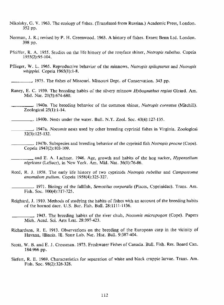
112
Nikolsky, G. V. 1963. The ecology of fishes. (Translated from Russian.) Academic Press, London.352 pp.
Norman, J. R.; revised by P. H. Greenwood. 1963. Ahistory of fishes. Ernest Benn Ltd.London398 pp.
Pfeiffer, R. A.1955. Studies on the life history of the rosyface shiner, Notropis rubellus. Copeia1955(2):95-104.
Pflieger, W. L.1965. Reproductive behavior of the minnows, Notropis spilopterus and Notropiswhipplei. Copeia 1965(1): 1-8.
1975. The fishes of Missouri. Missouri Dept. of Conservation. 343 pp.
Raney, E. C. 1939. The breeding habits of the silvery minnow Hybognathus regius Girard. Am.Mid. Nat. 21(3):674-680.
1940a. The breeding behavior of the common shiner, Notropis cornutus (Mitchill).Zoological 25(1):1-14.
1940b. Nests under the water. Bull. N.Y.Zool. Soc. 43(4): 127-135
. 1947 a. Nocomis nests used by other breeding cyprinid fishes in Virginia. Zoological32(3): 125-132.
1947b. Subspecies and breeding behavior of the cyprinid fish Notropis procne (Cope).Copeia 1947(2): 103-109.
and E. A. Lachner. 1946. Age, growth and habits of the hog sucker, Hypenteliumnigricans (LeSuer), inNew York. Am. Mid.Nat. 36(1):76-86.
Reed, R. J. 1958. The early life history of two cyprinids Notropis rubellus and Campostomaanomalum pullum. Copeia 1958(4): 325-327.
1971. Biology of the fallfish, Semotilus corporalis (Pisces, Cyprinidae). Trans. Am.Fish. Soc. 100(4):717-725.
Reighard, J. 1910. Methods of studying the habits of fishes with an account of the breeding habitsof the horned dace. U.S. Bur. Fish. Bull. 28:1111-1136.
, 1943. The breeding habits of the river chub, Nocomis micropogon (Cope). PapersMich. Acad. Sci. Arts Lett. 28:397-423.
Richardson, R. E. 1913. Observations on the breeding of the European carp in the vicinity ofHavana, Illinois. 111. State Lab. Nat. Hist. Bull. 9:387-404.
Scott, W. B. and E. J. Crossman. 1973. Freshwater Fishes ofCanada. Bull.Fish. Res. Board Can.184:966 pp.
Siefert, R. E. 1969. Characteristics for separation of white and black crappie larvae. Trans. Am.Fish. Soc. 98(2):326-328.
Mich. Acad. Sci. Arts Lett. 28:397-423.
Zoological 25(1): 1-14.
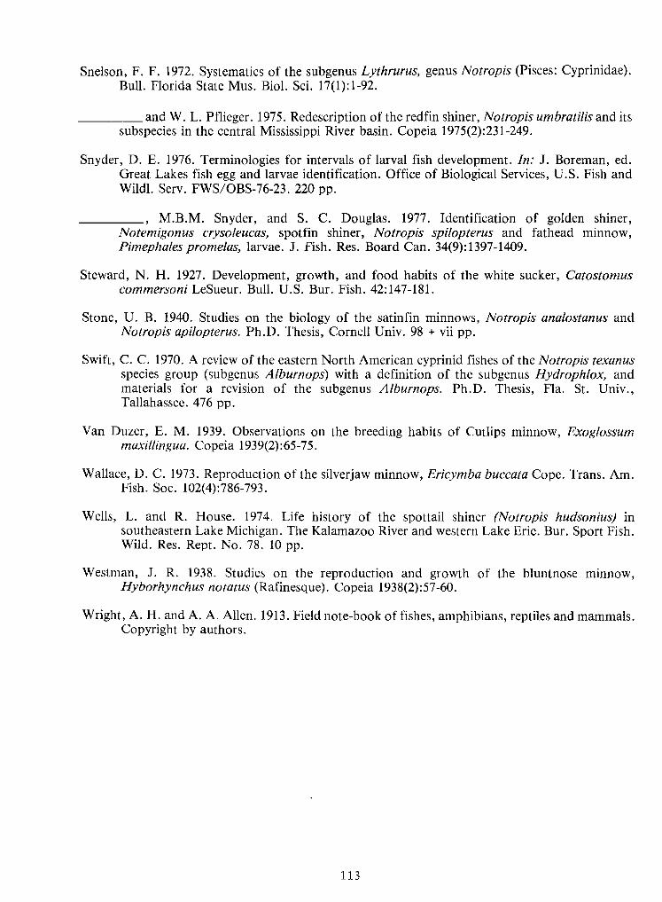
113
Snelson, F. F. 1972. Systematics of the subgenus Lythrurus, genus Notropis (Pisces: Cyprinidae).Bull. Florida State Mus. Biol. Sci. 17(1): 1-92.
and W. L.Pflieger. 1975. Redescription of the redfin shiner, Notropis umbratilis and itssubspecies in the central Mississippi River basin. Copeia 1975(2):23 1-249.
Snyder, D. E. 1976. Terminologies for intervals of larval fish development. In: J. Boreman, cd.Great Lakes fish egg and larvae identification. Office of Biological Services, U.S. Fish andWildl. Serv. FWS/OBS-76-23. 220 pp.
, M.B.M. Snyder, and S. C. Douglas. 1977. Identification of golden shiner,Notemigonus crysoleucas, spotfin shiner, Notropis spilopterus and fathead minnow,Pimephales promelas, larvae. J. Fish. Res. Board Can. 34(9): 1397-1409.
Steward, N. H. 1927. Development, growth, and food habits of the white sucker, Catostomuscommersoni LeSueur. Bull. U.S. Bur. Fish. 42:147-181.
Stone, U. B. 1940. Studies on the biology of the satinfin minnows, Notropis analostanus andNotropis apilopterus. Ph.D. Thesis, Cornell Univ. 98 + viipp.
Swift, C. C. 1970. A review of the eastern North American cyprinid fishes of the Notropis texanusspecies group (subgenus Alburnops) with a definition of the subgenus Hydrophlox, andmaterials for a revision of the subgenus Alburnops. Ph.D. Thesis, Fla. St. Univ.,Tallahassee. 476 pp.
Van Duzer, E. M. 1939. Observations on the breeding habits of Cutlips minnow, Exoglossummaxillingua. Copeia 1939(2):65-75.
Wallace, D. C. 1973. Reproduction of the silverjaw minnow, Ericymba buccata Cope. Trans. Am.Fish. Soc. 102(4):786-793.
Wells, L. and R. House. 1974. Life history of the spottail shiner (Notropis hudsonius) insoutheastern Lake Michigan. The Kalamazoo River and western Lake Erie. Bur. Sport Fish.Wild. Res. Rept. No. 78. 10 pp.
Westman, J. R. 1938. Studies on the reproduction and growth of the bluntnose minnow,Hyborhynchus notatus (Rafinesque). Copeia 1938(2):57-60.
Wright, A.H.and A.A.Allen.1913. Field note-book of fishes, amphibians, reptiles and mammals.Copyright by authors.
, M.B.M. Snyder, and S. C. Douglas. 1977. Identification of golden shiner,
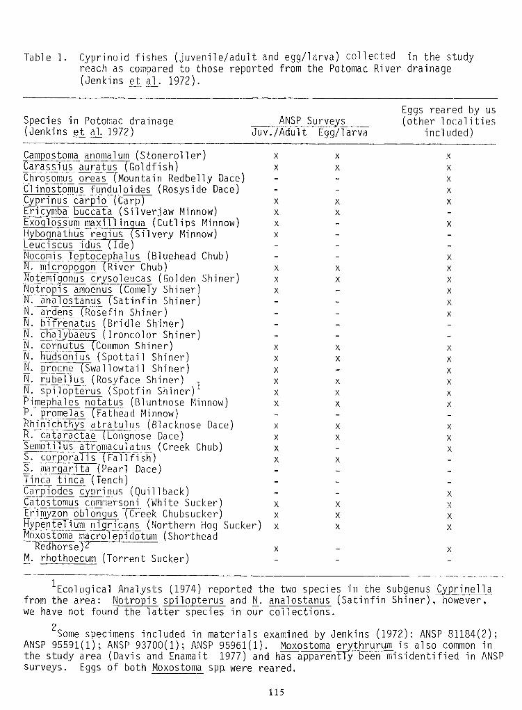
115
Table 1. Cyprinoid fishes (juvenile/adult and egg/larva) collected in the studyreach as compared to those reported from the Potomac River drainage(Jenkins et al_. 1972).
Ecological Analysts (1974) reported the two species in the subgenus Cyprinellafrom the area: Notropis spilopterus and N. analostanus (Satinfin Shiner), however,we have not found the latter species in our collections.
2Some specimens included in materials examined by Jenkins (1972): ANSP 81184(2);
ANSP 95591(1); ANSP 93700(1); ANSP 95961(1). Moxostoma erythrurum is also common inthe study area (Davis and Enamait 1977) and has apparently been misidentified in ANSPsurveys. Eggs of both Moxostoma spp. were reared.
M_. rhothoecum (Torrent Sucker)XXRedhorse)2
X
XX
X
XX
X
X
X
X
us
c
obionzon
iodes
Erim
CarCarpiodes cyprinus (Quillback)Catostomus commersoni (White Sucker)Erimyzon oblongus (Creek Chubsucker)Hypentelium nigricans (Northern Hog Sucker)Moxostoma macrole pidotum (Shorthead
XXS. corporal is (FaiifiTh)i." margarita (Pearl Dace)Tinea tinea (Tench)
cataractaeR.
X
X
X
X
X
X
X
X
X
X
X
X
X
X
X
X
X
X
X
X
X
X
X
X
xxx
xxxxxx
xxX
ontocele
microN.NocomisNocomis leptocephalus (Bluehead Chub)N_. micropogon (River Chub)Notemigonus crysoleucas (Golden Shiner)Notropis amoenus (homely Shiner)N. analostanus (Satinfin Shiner)N_. ardens (Rose fin Shiner)N_. bifrenatus (Bridle Shiner)Hm chalybaeus (Ironcolor Shiner)_N. cornutus (Common Shiner)N_. hudsonius (Spottail Shiner)N. procne (Swallowtail Shiner)N.- rubell us (Rosy face Shiner) 1H.- spilopterus (Spot fin Shiner)Pimephales notatus (Bluntnose Minnow)P. promeias (Fathead Minnow)Rhinichthys atratulus (Blacknose Dace)R_. cataractae~(Lonqnose Dace)Semotilus atrpmacuiatus (Creek Chub)
ExoEric
X
X
XX
xxxx
iocarrinusCyprinus carpio (Carp)Ericymba buccata (Silverjaw Minnow)Exoglossum maxil lingua (Cutlips Minnow)Hybognathus regius (Silvery Minnow)Leuciscus idus (Ide)
X
X
oreasChrosomusChrosomus oreas TMountain Redbelly Dace)Clinostomus funduloides (Rosyside Dace)
auratusarassiusX
X
X
Xxx
Campostoma anomalum (Stonerol ler)Carassius auratus (Goldfish)
Egg/larvaJuv. /Adult
Eggs reared by us(other localities
included)SurveANSPin Potomac drainage
§ial. 1972)Species(JenkinsSpecies(Jenkins
in Potomac drainage§ial. 1972)
ANSP SurveEggs reared by us(other localities
included)Juv. /Adult Egg/larva
Campostoma anomalum (Stonerol ler)Carassius auratus (Goldfish)
xx
X
XX
Xarassius auratusChrosomus oreas TMountain Redbelly Dace)Clinostomus funduloides (Rosyside Dace)Chrosomus oreas X
XCyprinus carpio (Carp)Ericymba buccata (Silverjaw Minnow)Exoglossum maxil lingua (Cutlips Minnow)Hybognathus regius (Silvery Minnow)Leuciscus idus (Ide)
rinus car io xxxx
XX
X
X
EricExo
Nocomis leptocephalus (Bluehead Chub)N_. micropogon (River Chub)Notemigonus crysoleucas (Golden Shiner)Notropis amoenus (homely Shiner)N. analostanus (Satinfin Shiner)N_. ardens (Rose fin Shiner)N_. bifrenatus (Bridle Shiner)Hm chalybaeus (Ironcolor Shiner)_N. cornutus (Common Shiner)N_. hudsonius (Spottail Shiner)N. procne (Swallowtail Shiner)N.- rubell us (Rosy face Shiner) 1H.- spilopterus (Spot fin Shiner)Pimephales notatus (Bluntnose Minnow)P. promeias (Fathead Minnow)Rhinichthys atratulus (Blacknose Dace)R_. cataractae~(Lonqnose Dace)Semotilus atrpmacuiatus (Creek Chub)
NocomisN. micro
le toceon x
xx
xxxxxx
xxX
X
X
X
X
X
X
X
X
X
X
X
X
X
X
X
X
X
X
X
X
X
X
X
X
R. cataractae
S. corporal is (FaiifiTh)i." margarita (Pearl Dace)Tinea tinea (Tench)
X X
Carpiodes cyprinus (Quillback)Catostomus commersoni (White Sucker)Erimyzon oblongus (Creek Chubsucker)Hypentelium nigricans (Northern Hog Sucker)Moxostoma macrole pidotum (Shorthead
Car
Erim
iodes
zon obion
c
usX
X
X
XX
X
X
XX
X
Redhorse)2 X XM_. rhothoecum (Torrent Sucker)
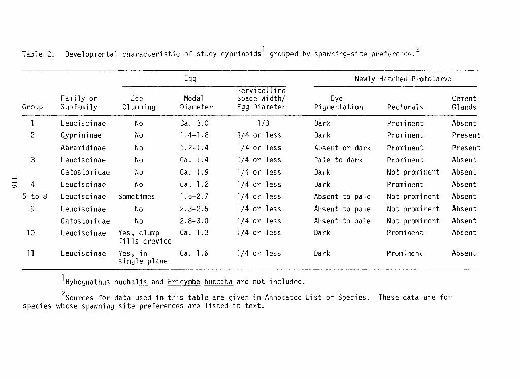
116
1 2Table 2.
Developmental characteristic of study cyprinoids grouped by spawning-site preference.
are listed in text.whose spawning site preferencesspecies foraredataTheseSpecies.ofListAnnotatedtable are given inSources for data used in this
included.buccata are notHybognathus nuchalis and Ericymba
AbsentProminentDarkor less1/4Ca. 1.6Yes , i nsingle plane
Leuciscinae11
AbsentProminentDarkor less1/4Ca. 1.3Yes, clumpfills creviceLeuciscinae10
AbsentNot prominentto paleAbsentor less1/42.8-3.0No
Catostomidae AbsentNot prominentto paleAbsentor less1/42.3-2.5No
Leuciscinae9
AbsentNot prominentto paleAbsentor less1/41.5-2.7SometimesLeuciscinae8to5
AbsentProminentDarkor less1/4Ca. 1.2No
Leuciscinae4
AbsentNot prominentDarkor less1/4Ca. 1.9NoCatostomidae AbsentProminenti
darkPale toor less1/4Ca. 1.4No
Leuciscinae3
PresentProminentor darkAbsent i
or less1/41.2-1.4NoAbramidinae PresentProminentDarkor less1/41.4-1.8NoCyprininae2
AbsentProminentDark1/3Ca. 3.0No
Leuciscinae1
GlandsPectoralsPigmentationClumpingSubfamilyGroup
CementEyeSpace Width/EggDiameterDiameter ModalEggFamily or
Pervitelline ProtolarvaHatchedNewlyEggEgg Newly Hatched ProtolarvaPervitellineFamily or Egg ModalDiameter Space Width/EggDiameter Eye CementGroup Subfamily Clumping Pigmentation Pectorals Glands
1
Leuciscinae No Ca. 3.0 1/3 Dark Prominent Absent
2 Cyprininae No
1.4-1.8 1/4 or less Dark Prominent PresentAbramidinae No
1.2-1.4 1/4 or less Absent ior dark Prominent Present
3
Leuciscinae No Ca. 1.4 1/4 or less Pale to i
dark Prominent AbsentCatostomidae No Ca. 1.9 1/4 or less Dark Not prominent Absent
4
Leuciscinae No Ca. 1.2 1/4 or less Dark Prominent Absent
5 to 8
Leuciscinae Sometimes 1.5-2.7 1/4 or less Absent to pale Not prominent Absent
9
Leuciscinae No 2.3-2.5 1/4 or less Absent to pale Not prominent AbsentCatostomidae No 2.8-3.0 1/4 or less Absent to pale Not prominent Absent
10 Leuciscinae Yes, clumpfills crevice Ca. 1.3 1/4 or less Dark Prominent Absent
11
Leuciscinae Yes , i nsingle plane Ca. 1.6 1/4 or less Dark Prominent AbsentHybognathus nuchalis and Ericymba buccata are not included.Sources for data used in this table are given in Annotated List of Species. These data are forspecies whose spawning site preferences are listed in text.
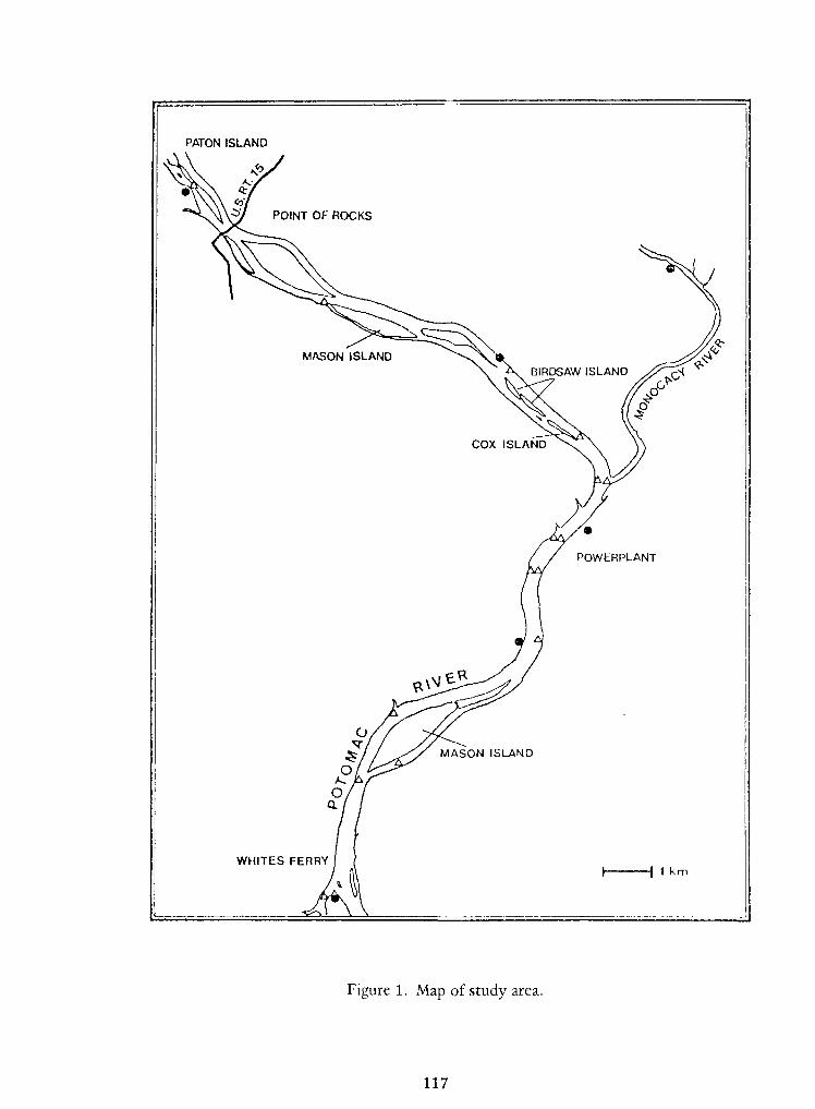
117
Figure 1. Map of study area.
1 kmWHITES FERRY I }
MASON ISLAND
POWERPLANT
COX ISLAND
A. BIRMASON ISLAND
BIRDSAW ISLAND
POINT OF ROCKS
PATON ISLANDPATON ISLAND
POINT OF ROCKS
BIRDSAW ISLANDMASON ISLAND
A. BIR
COX ISLAND
POWERPLANT
MASON ISLAND
WHITES FERRY I }1 km
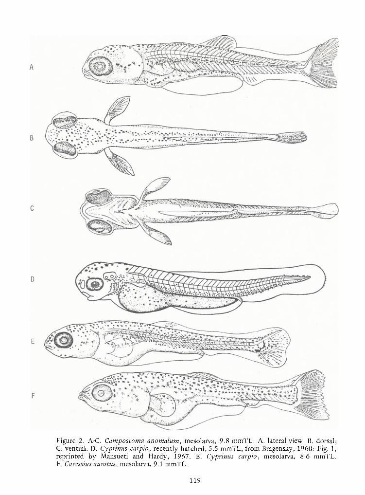
119
Figure 2. A-C. Campostoma anomalum, mesolarva, 9.8 mmTL: A. lateral view; B. dorsal;C. ventral. D. Cyprinus carpio, recently hatched, 5.5 mmTL, from Bragensky, I960: Fig. 1,reprinted by Mansueti and Hardy, 1967. E. Cyprinus carpio, mesolarva, 8.6 mmTL.F. Carassius auratus, mesolarva, 9.1 mmTL.
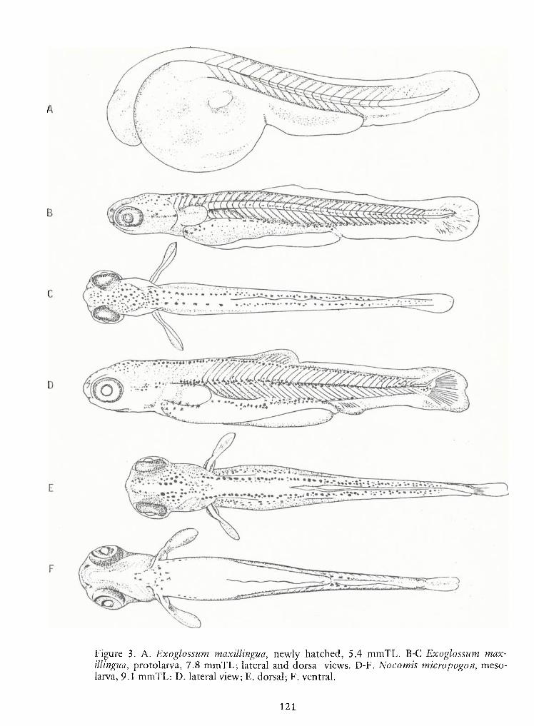
121
Figure 3. A. Exoglossum maxillingua, newly hatched, 5.4 mmTL. B-C Exoglossum max-illingua, protolarva, 7.8 mmTL; lateral and dorsa views. D-F. Nocomis micropogon, meso-larva, 9.1 mmTL: D. lateral view;E. dorsal; F. ventral.
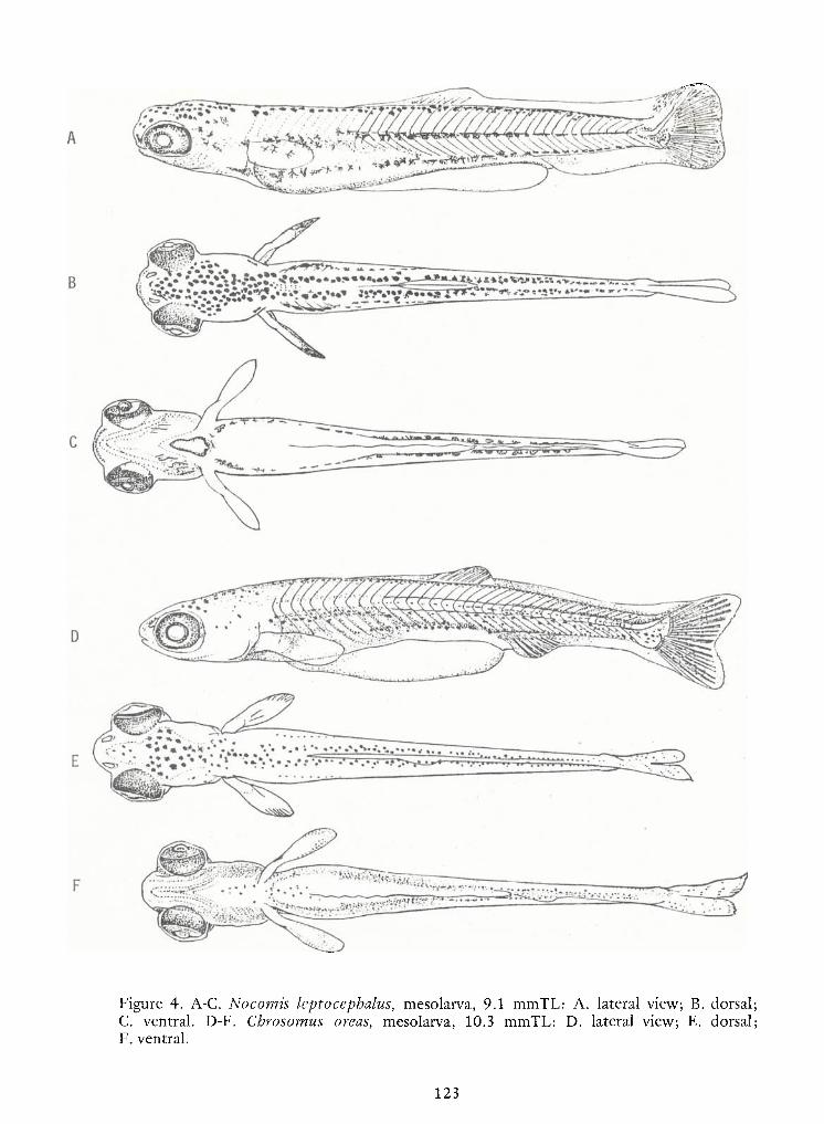
5
1
123
Figure 4. A-C. Nocomis leptocephalus, mesolarva, 9.1 mmTL: A. lateral view; B. dorsalC. ventral. D-F. Cbrosomus areas, mesolarva, 10.3 mmTL: D. lateral view; E. dorsalF. ventral.
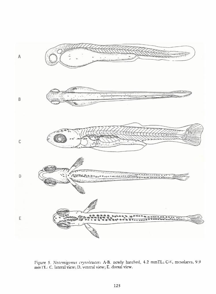
125
Figure 5. Notemigonus crysoleucas: A-B. newly hatched, 4.2 mmTL; C-E, mesolarva, 9.9mmTL: C. lateral view;D.ventral view;E. dorsal view.
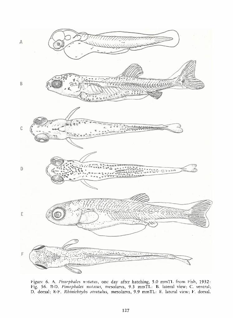
127
Figure 6. A. Pimephales notatus, one day after hatching, 5.0 mmTL from Fish, 1932:Fig. 56. B-D. Pimephales notatus, mesolarva, 9.3 mmTL: B. lateral view; C. ventral;D. dorsal; E-F. Rhinichtyhs atratulus, mesolarva, 9.9 mmTL: E. lateral view; F. dorsal.
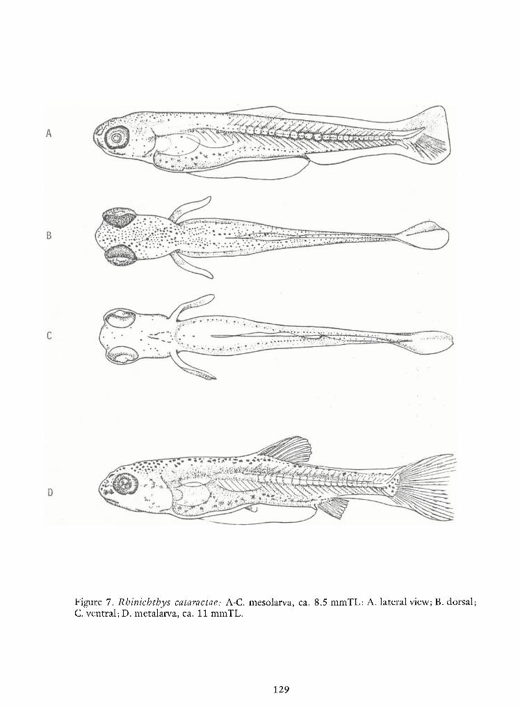
129
Figure 7. Rhinichthys cataractae: A-C. mesolarva, ca. 8.5 mmTL; A. lateral view;B. dorsal;C. ventral; D. metalarva, ca. 11 mmTL.
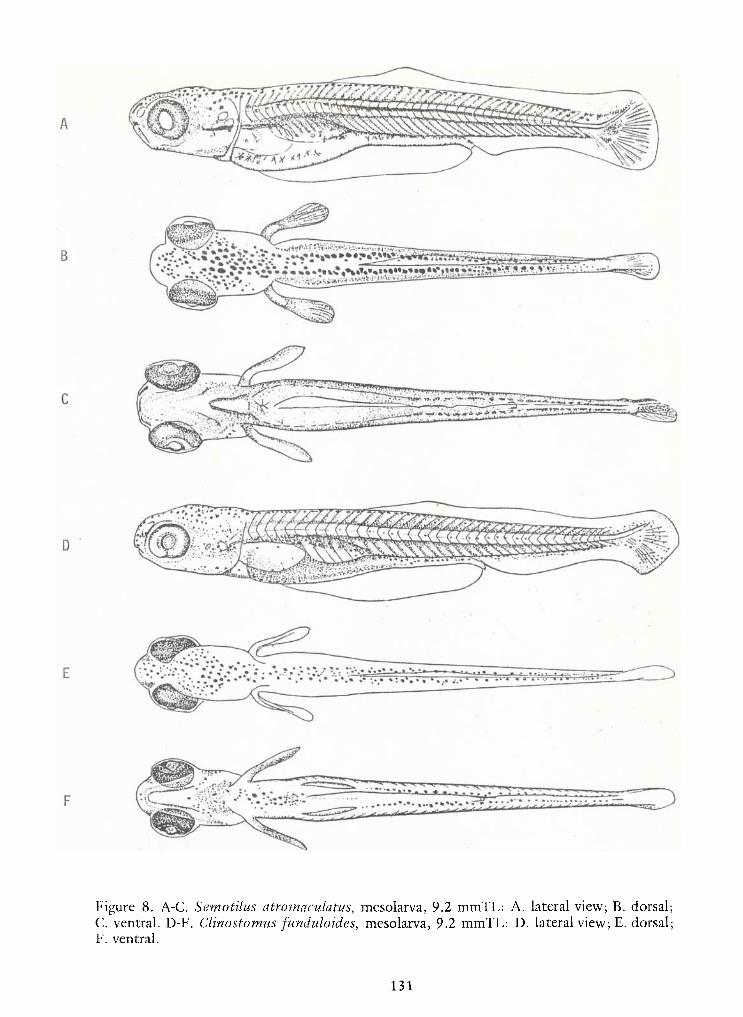
131
Figure 8. A-C. Semotilus atromaculatus, mesolarva, 9.2 mmTL: A. lateral view; B. dorsal;C. ventral. D-F. Clinostomus funduloides, mesolarva, 9.2 mmTL: D. lateral view;E. dorsal;F. ventral.
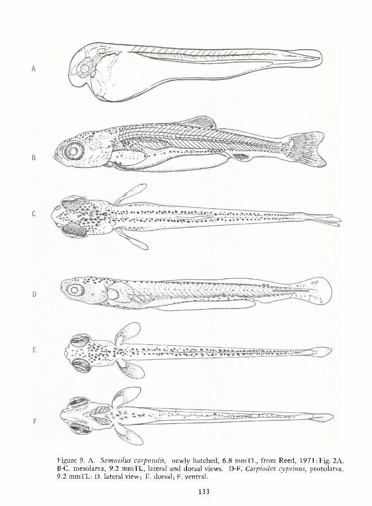
133
Figure 9. A. Semotilus corporalis, newly hatched, 6.8 mmTL, from Reed, 1971: Fig. 2A.B-C. mesolarva, 9.2 mmTL, lateral and dorsal views. D-F. Carpiodes cyprinus, protolarva,9.2 mmTL: D. lateral view; E. dorsal; F. ventral.
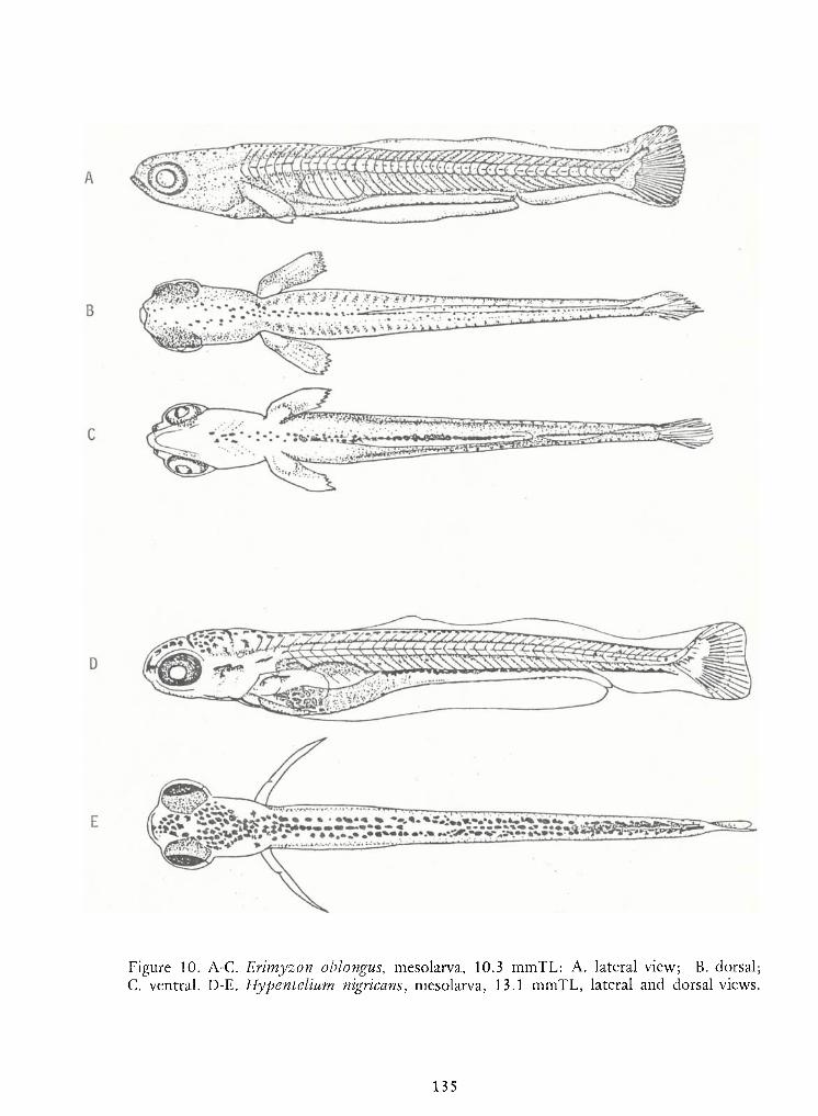
i
135
Figure 10. A-C. Erimyzon oblongus, mesolarva, 10.3 mmTL: A. lateral view; B. dorsalC. ventral. D-E. Hypentelium nigricans, mesolarva, 13.1 mmTL, lateral and dorsal views

137
Figure 11. A. Catostomus commersoni, newly hatched, 8.0 mmTL, from Steward, 1926,Fig. 20 reprinted by Mansueti and Hardy, 1967. B-D. Catostomus commersoni, mesolarva,14.3 mmTL: B. lateral view; C. Dorsal; D. ventral.
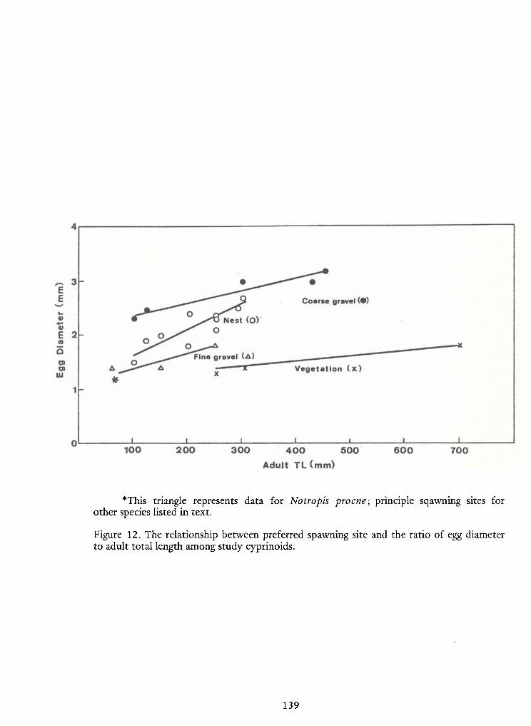
139
*This triangle represents data for Notropis procne; principle sqawning sites forother species listed in text.
Figure 12. The relationship between preferred spawning site and the ratio of egg diameterto adult total length among study cyprinoids.
IDENTIFICATIONOF NINELARVALCYPRINIDS INHABITING
SMALLNORTHERN RIVERS*
Lance G. Perry and Bruce W. Menzel
Department of Animal Ecologylowa State University
Ames, lowa 50011
ABSTRACT
Taxonomic descriptions are given for larvae ofnine species of cyprinids: Campostoma
anomalum, Hybognathus hankinsoni, Notropis cornutus, N. dorsalis, N. lutrensis, N. stramineus,
Pimephales notatus, P. promelas, and Semotilus atromaculatus. Study material was collected in theupper Skunk River, Story County, lowa. Identification criteria emphasize characters that are useful
for distinguishing between morphologically similar species.
"\u25a0Journal Paper No. J-9181 of the lowa Agriculture and Home Economics Experiment Station,
Ames, lowa. Project No. 2234.
143
INTRODUCTION
Species identification of North American cyprinid larvae presently is hindered by a
paucity of published descriptions and keys and by the general morphological, ecological, and
reproductive similarities ofmany species. Identification ofundescribed larvae may be accomplished
by comparison with series of reference specimens produced from controlled matings; however, few
such reference collections are available. Alternatively, identification may be made by field collection
of sequential size series of conspecific larvae. Characters of greatest diagonostic utilityare initially
determined through examination ofa life stage that can be positively identified. Recognition of pro-
gressively smaller-sized conspecifics then proceeds according to the presence of shared
characteristics. For distinguishing between morphologically similar forms, characters must be
selected that are species specific and relatively invariable during larval development. Complete
developmental series of larvae of all species occurring in the study area should be available for com-
parative purposes.
In this study, extensive field collection of larvae resulted in identification of ninespecies of the genera Campostoma, Hybognathus, Notropis, Pimephales, and Semotilus. Larvae ofseveral of these species are described here for the first time. Allof the taxa discussed range widely
through the north-central United States and the fish are common inhabitants of streams and small
rivers.
METHODS
Specimens were obtained from the upper Skunk River between Story City and Cam-
bridge, Story County, central lowa. Collections were made with stationary drift nets and dip nets
from April 25 to July 11, 1977. Identification of each species was based upon a combination of
characters including melanophore distribution, lengths at selected developmental stages, myomere
and finray counts, and several morphometric measurements. Available literature descriptions were
utilized where applicable. The presence ofidentifiable juveniles in the collections and knowledge ofthe local adult populations also aided in species determinations. Allspecimens were field preserved
in 5-10 percent Formalin. Observations and measurements were made with a stereoscopic
microscope equipped with an ocular micrometer. The characters of greatest diagnostic value were
determined empirically with the aid ofguidelines inBerry and Richards (1973) and Lippson (1976a).
Total length (TL), standard length (SL), and snout-vent length measurements were made according
144
to the methods ofMansueti and Hardy (1967). The predorsal length measurement was made from
the tip of the snout to the originof the dorsal finfold (when present) or to the dorsal finorigin. The
method of Siefert (1969) was utilized to distinguish preanal and postanal myomeres. Principal anal
rays were counted in standard fashion (Hubbs and Lagler 1964). Photography was accomplished
with a Leitz macro-dia utilizing a circular reflected-light system and Kodak Plus-X 35-mm film.
Common and scientific names of fishes follow Bailey et al. (1970).
The use of the descriptive terms larva, protolarva, mesolarva, metalarva, and juvenile
are in accordance with the definitions of Snyder (1976). The term stage is defined as "a just observ-ed, immediate moment of development" (Balon 1975). Interspecific differences were apparent inthe relative length at which comparable developmental stages were reached. Accordingly, the
lengths (TLmm) of each species of initial caudal fin ray formation (i.e., beginning of mesolarvalphase of development) and initial pelvic fin bud appearance were recorded for comparative pur-
poses.
RESULTS AND DISCUSSION
The features that are useful for identifying cyprinid larvae are summarized inLippson
and Moran (1974) and Lippson (1976b) as follows. The vent (anus) position generally is posterior to
the midpoint of the body. The yolk sac is typically spherical anteriorly and more cylindrical near the
vent. Although pigmentation is variable, several series ofmelanophores are usually evident: dorsally
on the head and body, laterally along the horizontal septum, on the ventral surface anterior to thevent, and ventrally on the myomeres behind the vent. The latter series is also continuous internally
above the body cavity. As development progresses, a single dorsal fin and a two-chambered gas
bladder appear. The anal finorigin is usually below or just behind the insertion of the dorsal fin,
and the pelvic fins develop below or slightly in advance of the dorsal fin origin.
The descriptions of key characters given immediately below, plus data presented in
Table 1 and Fig. 1, allow for initial separation of the nine species into four groups on the basis of
morphological similarities. Each group is subsequently treated inmore detail for species identifica-
tion. The descriptions follow a "dynamic" approach (Berry and Richards 1973) and are com-
parative; i.e., the distinguishing features of each species are described as they develop. Although
there is frequent reference to melanophore distributions, newly hatched individuals may lack pig-
ment entirely. As a result, caution should be exercised when using these descriptions for identifying
early protolarvae.
145
Group Descriptions:
Group I. Preanal Myomeres usually 27-28. Midventral surface between origin ofpreanal fin-
fold and vent unpigmented or only a few scattered melanophores present.
Semotilus atromaculatus—
creek chub
Campostoma anomalum—stoneroller
Group 11. Preanal myomeres usually 25-26. Aprominent midventral series of melanophores
present between heart region and vent.
Notropis cornutus—
common shiner
Hybognathus hankinsoni—
brassy minnow
Group 111. Preanal myomeres usually 24-25. Yolk usually persists in size range 5.0—
6.5 mm
TL.Distinct linear series or aggregation of melanophores on each side of midlineimmediately behind heart region. Midventral surface from behind heart region to
origin of preanal finfold usually unpigmented. Scattered melanophores ventrally
below intestine.
Pimephales promelas—
fathead minnowPimephales notatus
—bluntnose minnow
Group IV. Preanal myomeres usually 21-22. Yolkcompletely assimilated by 5.0 mm TL.Entiremidventral surface between heart region and vent with scattered melanophores or
unpigmented.
Notropis dorsalis —bigmouth shinerNotropis stramineus —sand shiner
Notropis lutrensis— red shiner
Species Descriptions:
Group I. Semotilus atromaculatus—
creek chub (Plates 1-4)
Campostoma anomalum —stoneroller (Plates 5-8)
146
The only published description ofthe larval creek chub is that ofa 14 mm TL specimen
by Fish (1932). Creek chub larvae are characterized by a high number ofpreanal myomeres, usually
28 (Table 1). Protolarvae are large, ranging 7.0-8.5 mm TL.A substantial amount ofyolk may per-
sist early in the mesolarval phase. Various stages in the larval development of the stoneroller are
described inFish (1932) and Hogue et al. (1976), and are included in the key ofMay and Gasaway
(1967). Length at hatching has been recorded as 6.3-6.9 mm (Hogue et al. 1976). The usual preanal
myomere count for this species is 27 (Table 1).
Although larvae of the creek chub and stoneroller are morphologically sinilar, several
characters serve to separate them. Stoneroller larvae develop more precociously, usually acquiring
caudal fin rays and pelvic fin buds at a smaller size (Fig. 1). In creek chub larvae, the "ventro-
visceral" pigmentation (Balinsky 1948) branches anterior to the gas bladder and extends anteriorly
under each auditory vesicle. Adense, continuous line ofinternal pigment is thus evident above the
body cavity from immediately behind the eye to the vent (Plate 1). On stoneroller larvae, the internalmelanophores above the intestine and anterior to the gas bladder are sparse and scattered, and the
ventro-visceral pigment line appears discontinuous and less dense than that of creek chub larvae.
Protolarval creek chubs have scattered melanophores on the ventral surface of the yolk sac im-
mediately behind the heart region. As the yolk is absorbed, midventral pigment is confined to the
region anterior to the preanal finfold. The midventral surface between the origin of the preanal fin-
fold and the vent remains unpigmented throughout larval development (Plates 2,4). Protolarval
stonerollers have scattered melanophores on the ventral surface below the intestine. The midventralsurface anterior to the preanal finfold usually remains unpigmented, although a few melanophores
may occur in this region (Plates 6,8).
Early inthe mesolarval phase, creek chubs acquire a concentration ofmelanophores on
the ventral surface of the operculum (Plate 4). The ventral opercular surface remains unpigmented
on mesolarval stonerollers (Plate 8). As metalarvae, the ratio ofpredorsal length to TL is greater in
creek chubs relative to stonerollers (Table 1). Juvenile creek chubs have a large, terminal mouth.
The tip of the upper jaw is on a level with the lower margin of the pupil, and the maxillary extends
posteriorly to a point below the eye. A small, subterminal mouth is characteristic of juvenile
stonerollers. The upper jaw is entirely below the eye, and the maxillary is short, its posterior margin
not extending under the eye.
Group 11. Notropis cornutus—
common shiner (Plates 9-12)
Hybognathus hankinsoni— brassy minnow (Plates 13-16)
147
The description of newly hatched and 2-day old larvae of Notropis cornutus
chrysocephalus by Fish (1932) includes several features that are inconsistent with the distinguishing
characteristics of cyprinid larvae as given by Lippson and Moran (1974) and Lippson (1976b).
Specifically, an apparent midbody location ofthe vent and the presence ofan oilglobule in the yolk
sac are not typical features of the family. The preanal and postanal myomere counts (14 and 19,
respectively) additionally conflict with the findings of the present study. Accordingly, certain por-
tions of that description are unreliable for identification. There isno other published description of
common shiner larvae. Itis uncertain whether the chrysocephalus form of the common shiner group
represents a distinct species or a subspecies ofNotropis cornutus (Gilbert 1961, Miller1968, Menzel
1976). Our study material represents Notropis cornutus cornutus inthe nomenclatural arrangement
recommended by Menzel (1976). The larval development of the brassy minnow has not been
described previously.
Common shiner larvae are characterized by the presence of three diverging, ventral
lines ofmelanophores which emanate from a common origin immediately behind the heart region.
One line extends along the midventral surface to the vent. The others extend obliquely across the
body cavity (Plates 10, 12). In contrast, brassy minnow larvae possess only a single line ofmelanophores anterior to the vent (Plates 14, 16). Anadditional feature of this species is the acquisi-
tion of a prominent melanophore on the pectoral fin base early in the mesolarval phase (Plate 16).
The pectoral fin base of the common shiner remains unpigmented during larval development.
Late in the metalarval phase, common shiners acquire a longitudinal band ofmelanophores on the midlateral body surface. Inbrassy minnow metalarvae, surface melanophores
on the epaxial myomeres are evenly distributed so that no lateral pigment band is evident. The
relatively smaller sizes at which brassy minnow larvae acquire caudal fin rays and pelvic fin buds
also are diagnostic for distinguishing between these two species (Fig. 1).
Group 111. Pimephales promelas— fathead minnow (Plates 17-20)
Pimephales notatus— bluntnose minnow (Plates 21-24)
Characters useful for identifying fathead minnow larvae are given in Fish (1932),
Hogue et al. (1976), and Snyder et al. (1977). Hatching length has been recorded as 4 mm TL
(Hogue et al. 1976, Snyder et al. 1977). Fish (1932) described the larval development of the blunt-
nose minnow from hatching (4.6 mm TL) to the "young adult" stage (17.75 mm TL). Hogue et al.
148
(1976) provided a photograph of a 6 mm TL specimen, which they indicated may be a larvalPimephales. Separating species of Pimephales as protolarvae is difficult,but two features were
useful for distinguishing bluntnose and fathead minnows among our collections. Fathead minnow
protolarvae of5.0-5.5 mm TLacquire heavy pigmentation dorsally on the head and body (Plate 18),
while pigment on the dorsal surface of bluntnose minnow protolarvae is restricted to a few late-developing melanophores in the occipital region and nape (Plate 22). The eye ofbluntnose minnowprotolarvae is distinctly flattened dorso-ventrally, whereas that ofthe protolarval fathead minnow is
rounder (Plates 17, 21).
As the yolk is absorbed and the position of the mouth becomes established, bluntnoseminnow larvae can be distinguished by the conspicuously decurved snout and subterminal mouth,
the upper jaw placed well below the center of the eye (Plates 21, 23). In contrast, the snout of the
fathead minnow larvae is not noticeably decurved, the mouth is terminal, and the tip of the upper
jaw is level with the center of the eye (Plates 17, 19). Late in the metalarval phase, bluntnose min-
nows acquire a concentration ofmelanophores at the base of the dorsal finand a dark spot at the
base of the caudal fin. These characters are not evident on fathead minnow metalarvae.
Group IV. Notropis dorsalis— bigmouth shiner (Plates 25-28)
Notropis stramineus—sand shiner (Plates 29-32)
Notropis lutrensis—
red shiner (Plates 33-36)
Larval features of the bigmouth shiner have not been recorded previously. Fish (1932)
described the larval development ofNotropis deliciosus stramineus (Cope) from 5 mm TLto 28.6
mm TL.Descriptions and illustrations ofred shiner larvae are provided inSaksena (1962) and Taber(1969). Hatching lengths for the bigmouth shiner and sand shiner evidently are less than 3.5 mmTL,
as indicated by the frequent occurrence ofprotolarvae in our collections ranging from 3.5-4.0 mm
TL.The eye and entire body of early protolarvae of these species may lack pigment, thus making
their separation tentative at this stage (see Plate 29, specimen A). The smallest red shiner observed(4.3 mm TL) had a small cylindrical yolk sac, pigmented eye, and a series ofmelanophores on the
lateral surface of the yolk (Plate 33). Larvae of all three species usually complete assimilation of
yolk material by 5.0 mm TL.
Bigmouth shiner protolarvae (greater than ca. 3.8 mm TL) have melanophores scat-
tered on the dorsal surface of the head and body. After yolk absorption, the entire dorsal surface is
149
densely pigmented, and scattered melanophores are present ventrally between the heart region and
vent (Plate 26). On sand shiner protolarvae (less than ca. 5.0 mm TL), dorsal pigmentation is
restricted to the occipital region. After yolk absorption, two irregular rows ofmelanophores appear
on the dorsal surface of the body (Plate 30). This dorsal pigment remains less dense than that ofthebigmouth shiner; however, the melanophores are consistently smaller and more widely spaced along
the body axis. Pigment on the ventral surface of sand shiner larvae is essentially similar to that ofthebigmouth shiner.
Early in the mesolarval phase, bigmouth shiners acquire a prominent melanophore in
the nasal pit. The nasal pits of sand shiner larvae remain unpigmented until the late metalarvalphase. As metalarvae, sand shiners acquire a concentration of melanophores along the base of the
dorsal fin, and a dusky lateral band becomes evident during the juvenile period. Both characters are
lacking on bigmouth shiners of comparable development.
Red shiner protolarvae have pigment in the occipital region, and a few melanophores
appear dorsally on the body late in the phase. A short, linear series of melanophores is present on
each side of the midline immediately behind the heart region. Aprominent series also is present on
the lateral surface ofthe intestine from immediately behind the gas bladder to the vent. The midven-
tral surface between the heart region and the vent usually remains unpigmented during larval
development (Plates 34, 36). Incontrast, melanophores are scattered over the entire ventral region
of bigmouth and sand shiner larvae (Plates 26, 28, 30, 32).
ACKNOWLEDGMENTS
This study was funded by the lowa Agriculture and Home Economics Experiment Sta-tion, the lowa State University Graduate College, and the lowa Academy of Science. Some collect-ing equipment was provided by the lowa Cooperative Fishery Research Unit. Photographic
assistance was given by Harry T. Homer, Jr., and Clark Miller.
151
LITERATURE CITED
Bailey, R. M., J. E. Fitch, E. S. Herald, E. A.Lachner, C. C. Lindsey, C.R. Robbins, and W. G.Scott. 1970. A list of common and scientific names of fishes from the United States andCanada. 3rd cd. Am. Fish. Soc. Spec. Publ. 6. 150 pp.
Balinsky, B.I.1948. On the development of specific characters incyprinid fishes. Trans. Zoo. Soc.London 118(2):335-344.
Balon, E. K. 1975. Terminology of intervals in fish development. J. Fish. Res. Board Can32(9): 1663-1670.
Berry, F.H.and W. J. Richards. 1973. Characters useful to the study oflarval fishes, Pages 48-65 inA.L.Pacheco, cd. Proceedings of a workshop on egg, larval, and juvenile stages of fish inAtlantic coast estuaries. U.S. Dep. Commer., Natl. Mar. Fish. Serv., Middle Atlantic CoastFisheries Center, Tech. Publ. 1. 338 pp.
Fish, M.P. 1932. Contributions to the early lifehistories of sixty-two species of fishes from LakeErie and its tributary waters. Bull.U.S. Bur. Fish. 47:293-398.
Gilbert, C. R. 1961. Hybridization versus intergradation: an inquiry into the relationship of twocyprinid fishes. Copeia 1962(2):181-192.
Hogue, J. J., Jr., R. Wallus, and L.K.Kay. 1976. Preliminary guide to the identification of larvalfishes in the Tennessee River. Term. Val. Auth. Tech. Note 819. 66 pp.
Hubbs, C. L. and K.F. Lagler. 1964. Fishes of the Great Lakes region. Univ.Mich. Press, AnnArbor, Mich. 213 pp.
Lippson, A.J. (Chairperson— Working group no. 3). 1976 a. Variability incharacteristics appearingin keys regionwide and nationwide, Pages 97-101 in J. Boreman, cd. Great Lakes fish eggand larvae identification: Proceedings of a workshop. U.S. Fish. Wildl. Serv., Off. Biol.Serv., Natl. Power Plant Team Publ. FWS/OBS-76/23. 220 pp.
Lippson, A.J. 1976b. Distinguishing family characteristics among Great Lakes fish larvae, Pages208-216 in J. Boreman cd. Great Lakes fish egg and larvae identification: Proceedings of aworkshop. U.S. Fish Wildl. Serv., Off. Biol. Serv., Natl. Power Plant Team Publ.FWS/OBS-76/23. 220 pp.
Lippson, A.J. and R. L.Moran. 1974. Manual for identification of early developmental stages offishes of the Potomac River estuary. Environ. Tech. Center, Martin Marietta Corp.,Baltimore, Md., Publ. PPSP-MP-13. 282 pp.
Mansueti, A.J. and J. D.Hardy, Jr. 1967. Development of fishes of the Chesapeake Bay region; anatlas of egg, larval, and juvenile stages. Natl. Res, Institute, Univ. Maryland. 202 pp.
May, E. B. and C. R. Gasaway. 1967. Apreliminary key to the identification of larval fishes ofOklahoma, withparticular reference to Canton reservoir, including a selected bibliography.Okla. Fish. Res. Lab. Bull. 5. 42 pp.
152
Menzel, B. W. 1976. Biochemical systematics and evolutionary genetics of the common shinerspecies group. Biochem. Syst. Ecol. 4:281-293.
Miller,R. J. 1968. Speciation in the common shiner: an alternative view. Copeia 1968(3):539-544.
Saksena, V. P. 1962. The posthatching stages of the red shiner, Notropis lutrensis. Copeia1962(3):539-544.
Siefert, R. E. 1969. Characteristics for separation of white and black crappie larvae. Trans. Am.Fish. Soc. 98(2):326-328.
Snyder, D. E. 1976. Terminologies for intervals of larval fish development, Pages 41-58 in J.Boreman cd. Great Lakes fish egg and larvae identification: Proceedings of a workshop.U.S. Fish. Wildl. Serv., Off.Biol. Serv., Natl. Power Plant Team Publ. FWS/OBS-76/23.220 pp.
Snyder, D. E., M.B.M. Snyder, and S. C. Douglas. 1977. Identification of golden shiner,Notemigonus crysoleucas, spotfin shiner, Notropis spilopterus, and fathead minnow,Pimephales promelas, larvae. J. Fish. Res. Board Can. 34(9): 1397-1409.
Taber, C. A.1969. The distribution and identification of larval fishes in the Buncombe Creek armof Lake Texoma with observations on spawning habits and relative abundance. Ph.D.dissertation, Univ. of Oklahoma, Norman. 128 pp.
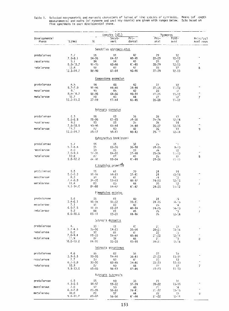
Table 1. Selected morphometric and meristic characters of larvae of nine species of cyprinids. Means (of lengthmeasurements) and modes (of myomere and anal ray counts) are given with ranges below. Data based onfive specimens in each developmental phase.
9
1413-15
1413-15
1312-14
2120-22
2121-22
2221-22
3837-39
4039-41
4441-44
95 6094-97 59-62
92 6089-95 58-63
86 5785-88 56-58
4.94.3-5.5
7.06.3-7.810.0
9.4-10.7
protolarvae
mesolarvae
metalarvae
Notropis lutrensis
7
1313-14
1312-13
1211-13
2222-23
2322-23
2221-23
3836-41
4139-44
4442-46
95 6293-96 59-64
93 6390-96 62-65
87 6085-88 56-63
4.63.9-5.5
7.76.7-9.810.9
9.9-13.0
protolarvae
mesolarvae
metalarvae
Notropis stramineus
8
1313-14
1312-14
1312-14
2120-21
2221-22
2120-21
4138-44
4140-44
4443-44
95 \u25a0 619b-96 59-63
92 6189-95 59-62
87 5886-39 56-59
4.13.7-4.5
8.27.0-9.411.9
10.2-13.2
protolarvae
mesolarvae
metalarvae
Notropis dorsal is
7
1514-15
1414-15
1312-14
2424-25
2524-25
2b25
4039-41
4240-44
4544-46
95 6195-96 58-62
94 6391-95 62-64
88 6185-91 60-61
5.65.4-6.1
7.16.5-7.5
9.28.6-10.6
protolarvae
mesolarvae
metalarvae
Pimephales notatus
7
1413-15
1312-15
1211-12
242424
24-2525
24-25
3937-41
4140-42
4441-47
95 6195-96 59-63
94 6194-95 53-63
86 6084-88 59-62
5.55.0-6.3
8.37.4-8.911.4
9.1-14.0
protolarvae
mesolarvae
metalarvae
Pimephales promeluV
8
119-1312
11-1312
11-13
2524-25
2625-26
2524-26
3836-40
3937-40
4341-44
95 6595 63-6693 6b
87-96 64-6687 62
34-88 59-64
5.75.1-6.6
8.07.0-9.410.8
9.8-12.2
protolarvae
mesolarvae
metalarvae
Hybognathus hankinsoni
))
1312-14
1412-15
1312-14
2625-26
2626-27
2625-^7
\u25a0 3634-38
3836-40
4240-43
96 6395-96 61-65
93 6390-95 63-64
87 6085-87 bB-61
6.35.6-6.8
9.27.6-10.913.7
12.6-14.7
protolarvae
mesolarvae
metalarvae
Notropis cornutus
7
1211-12
1211-12
1211-12
2727-28
2827-28
2726-28
4239-44
4240-44
4443-45
94 6691-95 64-68
93 6588-95 64-66
88 6337-89 61-64
6.55.7-7.0
9.18.0-10.712.7
12.2-13.2
protolarvae
mesol arvae
metalarvae
Campostoma anomalum
8
1212-13
1212-13
1212-13
2928-29
2828-29
2327-29
4240-45
4341-48
4746-48
95 6594-96 64-67
94 6491-95 62-66
88 6388-90 61-64
7.77.0-8.1
9.18.0-10.713.6
12.3-14.7
protolarvae
mesol arvae
metal arvae
Semotilus atromaculatus
analSi.TL(mm)Principalanal rays
Post-analPre-Pre-
dorsalSnout-vent
DevelopmentalPhase
MyomeresmithsLen
153
Len iths m MyomeresDevelopmental
PhaseSnout-vent
Pre-dorsal
Pre-anal
Post- Principalanal raysTL(mm) Si. anal
Semotilus atromaculatus
protolarvae
mesol arvae
metal arvae
7.77.0-8.1
9.18.0-10.713.6
12.3-14.7
95 6594-96 64-67
94 6491-95 62-66
88 6388-90 61-64
4240-45
4341-48
4746-48
2928-29
2828-29
2327-29
1212-13
1212-13
1212-13
8
Campostoma anomalum
protolarvae
mesol arvae
metalarvae
6.55.7-7.0
9.18.0-10.712.7
12.2-13.2
94 6691-95 64-68
93 6588-95 64-66
88 6337-89 61-64
4239-44
4240-44
4443-45
2727-28
2827-28
2726-28
1211-12
1211-12
1211-12
7
Notropis cornutus
protolarvae
mesolarvae
metalarvae
6.35.6-6.8
9.27.6-10.913.7
12.6-14.7
96 6395-96 61-65
93 6390-95 63-64
87 6085-87 bB-61
\u25a0 3634-38
3836-40
4240-43
2625-26
2626-27
2625-^7
1312-14
1412-15
1312-14
))
Hybognathus hankinsoni
protolarvae
mesolarvae
metalarvae
5.75.1-6.6
8.07.0-9.410.8
9.8-12.2
95 6595 63-6693 6b
87-96 64-6687 62
34-88 59-64
3836-40
3937-40
4341-44
2524-25
2625-26
2524-26
119-1312
11-1312
11-138
Pimephales promeluV
protolarvae
mesolarvae
metalarvae
5.55.0-6.3
8.37.4-8.911.4
9.1-14.0
95 6195-96 59-63
94 6194-95 53-63
86 6084-88 59-62
3937-41
4140-42
4441-47
242424
24-2525
24-25
1413-15
1312-15
1211-12
7
Pimephales notatus
protolarvae
mesolarvae
metalarvae
5.65.4-6.1
7.16.5-7.5
9.28.6-10.6
95 6195-96 58-62
94 6391-95 62-64
88 6185-91 60-61
4039-41
4240-44
4544-46
2424-25
2524-25
2b25
1514-15
1414-15
1312-14
7
Notropis dorsal is
protolarvae
mesolarvae
metalarvae
4.13.7-4.5
8.27.0-9.411.9
10.2-13.2
95 \u25a0 619b-96 59-63
92 6189-95 59-62
87 5886-39 56-59
4138-44
4140-44
4443-44
2120-21
2221-22
2120-21
1313-14
1312-14
1312-14
8
Notropis stramineus
protolarvae
mesolarvae
metalarvae
4.63.9-5.5
7.76.7-9.810.9
9.9-13.0
95 6293-96 59-64
93 6390-96 62-65
87 6085-88 56-63
3836-41
4139-44
4442-46
2222-23
2322-23
2221-23
1313-14
1312-13
1211-13
7
Notropis lutrensis
protolarvae
mesolarvae
metalarvae
4.94.3-5.5
7.06.3-7.810.0
9.4-10.7
95 6094-97 59-62
92 6089-95 58-63
86 5785-88 56-58
3837-39
4039-41
4441-44
2120-22
2121-22
2221-22
1413-15
1413-15
1312-14
9
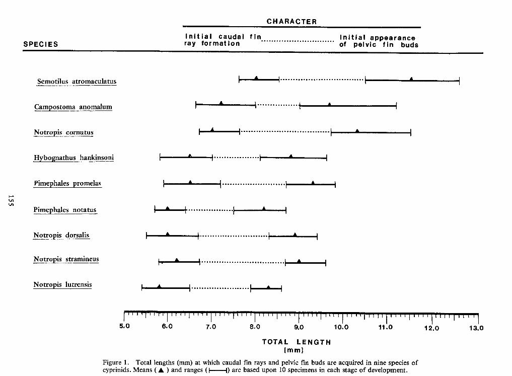
155
Figure 1 . Total lengths (mm) at which caudal fin rays and pelvic fin buds are acquired in nine species ofcyprinids. Means ( A ) and ranges (|(| 1)
are based upon 10 specimens in each stage of development.
(mm]TOTAL LENGTH 13.012.011.010.09.08.07.06.05.0
islutrensisNotro
is stramineusNotrois dorsalisNotro
'hales notatusPime
romelashalesPimebo hankinsoniHvbognatus
cornutusNotropisostoma anomalumCam
Semotilus atromaculatusSPECIES Initial appearance
of pelvic fin budsfinInitial caudalray formationCHARACTERCHARACTERInitial caudalray formation fin Initial appearance
of pelvic fin budsSPECIESSemotilus atromaculatus
Cam ostoma anomalumNotropis cornutus
Hvbognatus hankinsoniboPime hales romelas
Pime 'hales notatusNotro is dorsalis
Notro is stramineusNotro is
lutrensis
5.0 6.0 7.0 8.0 9.0 10.0 11.0 12.0 13.0TOTAL LENGTH(mm]
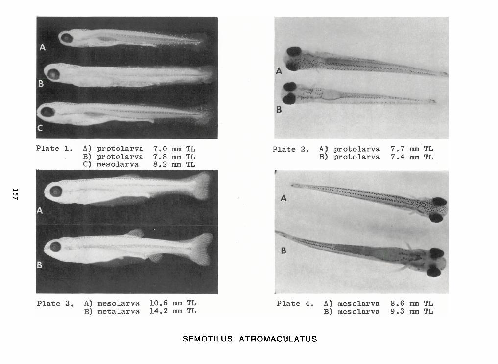
157
SEMOTILUS ATROMACULATUS
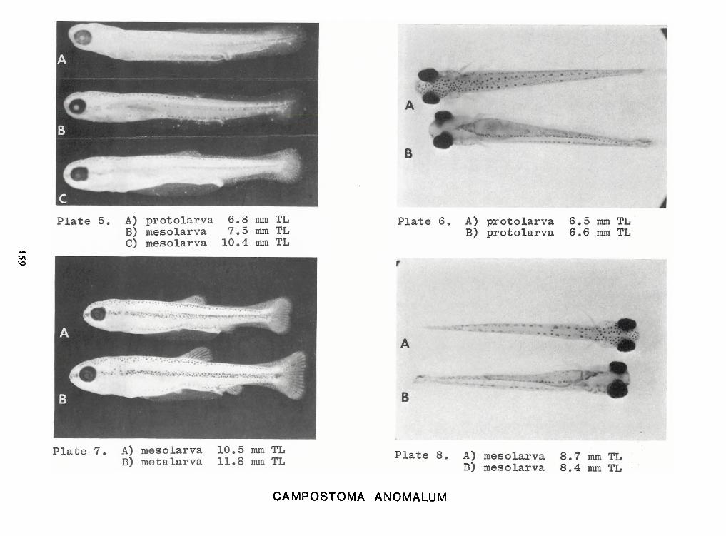
159
CAMPOSTOMA ANOMALUM
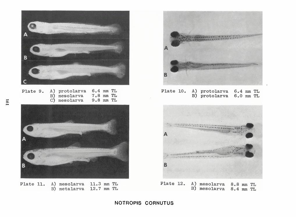
161
NOTROPIS CORNUTUS
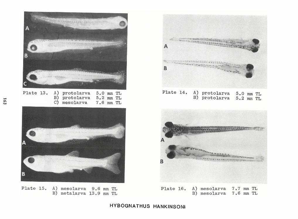
163
HYBOGNATHUS HANKINSONI
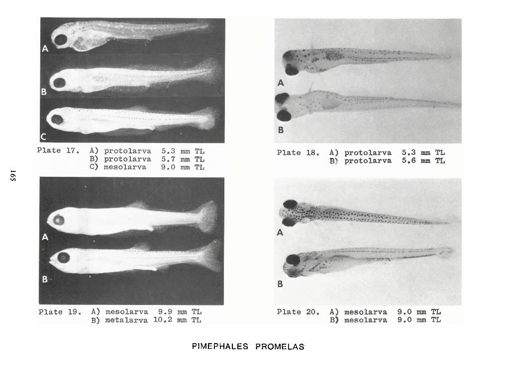
165
PIMEPHALES PROMELAS
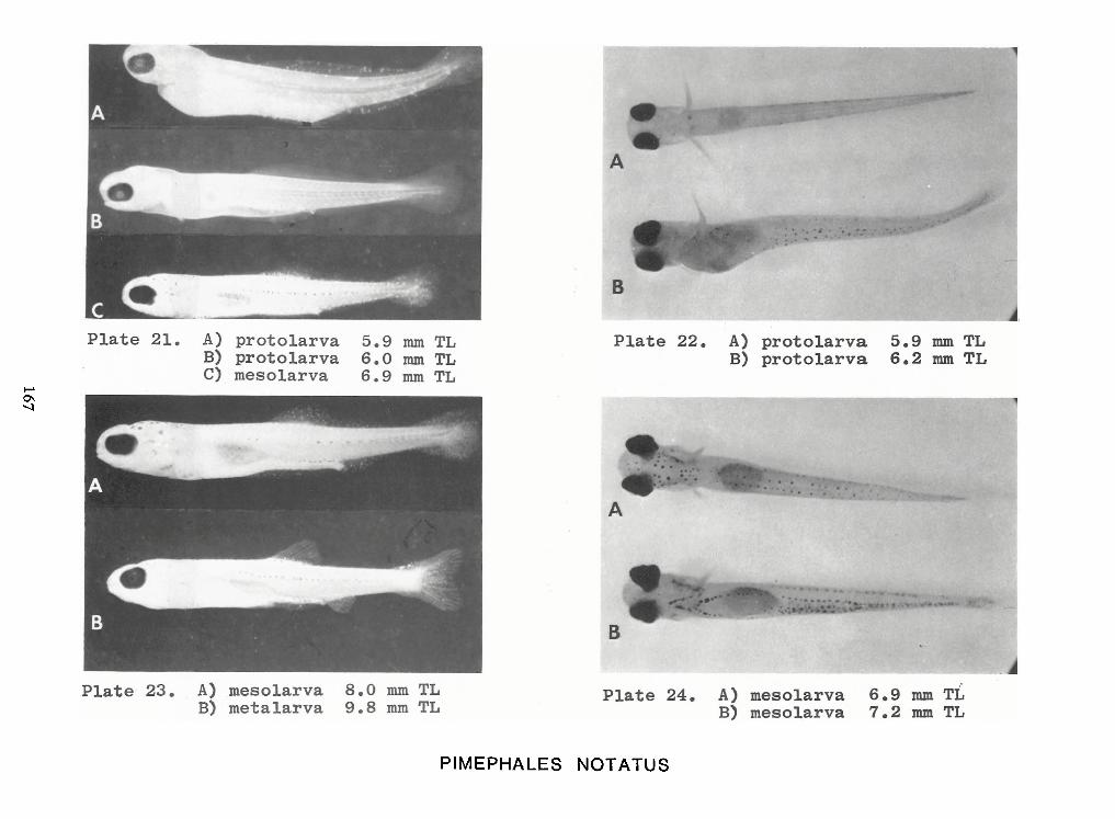
167
PIMEPHALES NOTATUS
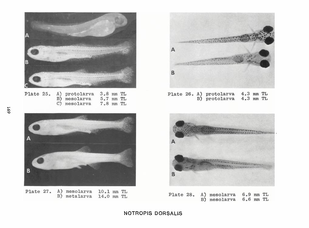
169
NOTROPIS DORSALIS
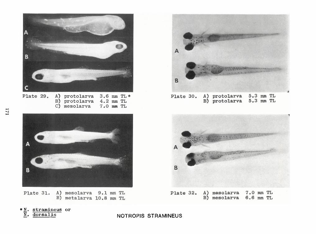
171
NOTROPIS STRAMINEUS*N . stramineus orN.
dorsalis
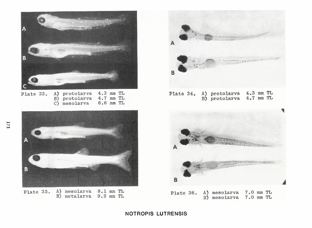
173
NOTROPIS LUTRENSIS
IDENTIFICATIONOF CATFISH ALEVINS
OF THE PIEDMONT CAROLINAS
by
Donald G. Cloutman
Duke Power Company Environmental LabRoute 4, Box 531
Huntersville, North Carolina 28078
ABSTRACT
A key to the alevins of catfishes (excluding madtoms) of the Piedmont Province of
North and South Carolina is provided. Characters most useful for identification include anal ray
counts, pelvic ray counts, caudal fin morphology, pectoral spine morphology, and pigmentation.
177
INTRODUCTION
Eleven species of catfishes are known to occur in the Piedmont Province ofNorth and
South Carolina. Six of these species are native: snail bullhead, Ictalurus brunneus (Jordan); whitecatfish, /. catus (LI.naeus); yellow bullhead, /. natalis I.eSueur); brown bullhead, /. nebulosuI.and Evermann); and margined madtom, N.insignis (Richardson). Four species are introduced: bluecatfish, /. furcatusI .LeSueur); black bullhead, /. melas (RI.inesque); channel catfish, /.punctatuI.lly
common except for the yellow bullhead, which is characteristically a Coastal Plain inhabitant, and
the orangefin madtom, which is rare and localized in the upper Dan River drainage. Most of the in-
troduced species are rare, except for the channel catfish, which is common in several rivers and
reservoirs.
Larval development in many fishes is commonly divisible into prolarval (sac fry) and
postlarval stages. However, in ictalurids there is no true postlarval stage because the fish resemble
adults when the yolk sac disappears. Early juveniles just after the prolarval stage, are called alevins(Lagler et al. 1977). Even though alevins have some adult features, certain problems in identificationhave been encountered. Morphology and pigmentation may differ between alevins and adults.
Pigmentation may be affected by water or light conditions and is also masked by stains such as rose
bengal, which are used to facilitate picking and sorting of fish eggs and larvae from samples. Conse-quently, reliable diagnostic characters need to be established for identification of alevins.
The purpose ofthis paper is to describe characters which can be used to identify catfish
alevins from the Piedmont Carolinas. Most specimens used in this study were from the Piedmont
Province ofNorth and South Carolina, but other specimens were also used toobtain supplementary
data or to fillinformation gaps. Itis hoped that the characters presented here willalso be useful in
other regions. A key is provided to facilitate identifications.
Madtoms are excluded from this paper because of lack of material. They cannot be
separated from bullheads unless development of the adipose finis complete.
178
MATERIALSAND METHODS
Materials Examined
Number of specimens examined followed by range of total lengths (mm) are given inparentheses.
Ictalurus brunneus: Broad R., Cherokee Co., S.C. (30, 14-54); Dan R., Rockingham Co., N.C.
(16, 31-105).
Ictalurus catus: Broad R., Cherokee Co., S.C. (38, 12-67); Lake Wylie, Catawba R. dr., GastonCo., N.C. (3, 16-24); Lake Norman, Catawba R. dr., Lincoln Co., N.C. (21, 16-29); Yadkin R.,
Davie-Davidson Co. border, N.C. (2, 15-16).
Ictalurus furcatus: Tennessee R., Tennessee-Mississippi-Alabama border (4, 16-18).
Ictalurus melas: Trib. of Long Crk., Yadkin R. dr., Stanley Co., N.C. (15, 36-40); Belews Lake,
Dan R.dr., Forsyth Co., N.C. (128, 30-57); LittleBear Reservoir, Bear Crk. dr., Franklin Co., Ala.
(50, 32-41); Cow Crk., Arkansas R. dr., Crawford Co., KS. (2, 49-55).
Ictalurus natalis: Back Crk., Pee Dee R. dr., Cabarrus Co., N.C. (1, 57); Lynches R., ChesterfieldCo., S.C. (2, 56-61); Lake Robinson, Pee Dee R. dr., Chesterfield Co., S.C. (1, 17); Gapeway
Swamp Canal, Pee Dee R. dr., Columbus Co., N.C. (7, 29-45); Dan R., Rockingham Co., N.C. (1,
74); Holston R., Hawkins Co., Term. (29, 14-30).
Ictalurus nebulosus: Broad R., Cherokee Co., S.C. (158, 12-17); Lake Norman, Catawba R. dr.,
Lincoln Co., N.C. (40, 16-58).
Ictalurus platycephalus: Broad R., Cherokee Co., S.C. (4, 17-19); Lake Norman, Catawba R.dr
Lincoln Co., N.C. (12, 18-38); Lynches R., Chesterfield Co., S.C. (3, 36-50).
Ictalurus punctatus: Broad R., Cherokee Co., S.C. (10, 14-17); Lake Wylie, Catawba R. dr.,
Gaston Co., N.C. (108, 13-31); Yadkin River, Davie-Davidson Co. border, N.C. (34, 13-19); Dan
R., Rockingham Co., N.C. (1, 29).
Pylodictis olivaris: Yadkin R., Davie-Davidson Co. border, N.C. (1, 15); Kentucky Lake, HardinCo., TN (3,15-21).
179
Methods
The total length of each specimen was measured to the nearest millimeter. The anal
and pelvic rays were counted, including the smallest anterior rudiments; the last two rays (dou-
ble rays with a single base) were counted as one. Pectoral spines were excised from the fish,
and the flesh covering each spine was removed by soaking in enzyme detergent. Ray counts and
observations of pectoral spine morphology were made with a dissecting microscope using
polarized light. Drawings of pectoral spines were made with the aid of a camera lucida or
microprojector.
RESULTS AND DISCUSSION
The most useful characters for identification of alevins 15 mm or longer include anal
ray counts, pelvic ray counts, caudal fin morphology, pectoral spine morphology, and pigmenta-
tion (Table 1, Fig. 1). With smaller specimens, identification may be difficult or impossible
because fins and spines may not be sufficiently developed to be useful (e.g., a channel catfish 14
mm or less in length may have a rounded rather than a forked tail and the anal rays may not
have developed enough for an accurate count). Fortunately, most specimens taken by conven-
tional trawls are 15 mm or larger. Data on distribution, habitat requirements, and reproductive
habits of adults from the collection area may also provide insight for identification of alevins.
Further descriptions and identification aids can be found in Armstrong (1962), Fish(1932), Hogue et al. (1976), Lippson and Moran (1974), and Mansueti and Hardy (1967).
Key to Species of Catfish Alevins
1. a. Anal finrays usually 32-34 (30-36); Caudal findeeply forked Ictalurus furcatus
b. Anal finrays less than 30; Caudal finmay or may not be forked 2
2. a. Distal barb of pectoral spine hook-shaped (Fig. IF, G); Caudal fin forked to emarginate3
b. Pectoral spine barbs not distinctly hooked; Caudal finrounded, truncate, or emarginate4
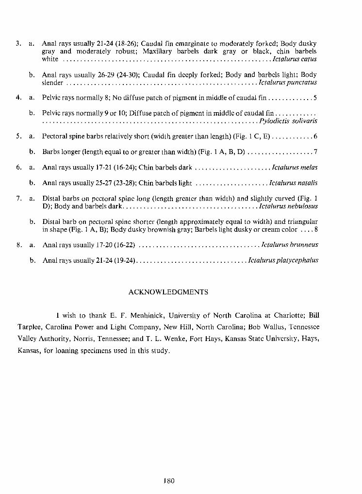
180
3. a. Anal rays usually 21-24 (18-26); Caudal fin emarginate to moderately forked; Body duskygray and moderately robust; Maxillary barbels dark gray or black, chin barbelswhite Ictalurus catus
b. Anal rays usually 26-29 (24-30); Caudal fin deeply forked; Body and barbels light; Bodyslender Ictalurus punctatus
4. a. Pelvic rays normally 8; No diffuse patch ofpigment inmiddle ofcaudal fin 5
b. Pelvic rays normally 9or 10; Diffuse patch ofpigment inmiddle ofcaudal finPylodictis solivaris
5. a. Pectoral spine barbs relatively short (width greater than length) (Fig. IC,E) 6
b. Barbs longer (length equal toor greater than width) (Fig. IA,B,D) 7
6. a. Anal rays usually 17-21 (16-24); Chin barbels dark Ictalurus melas
b. Anal rays usually 25-27 (23-28); Chin barbels light Ictalurus natalis
7. a. Distal barbs on pectoral spine long (length greater than width) and slightly curved (Fig. 1D);Body and barbels dark Ictalurus nebulosus
b. Distal barb on pectoral spine shorter (length approximately equal to width) and triangularinshape (Fig. 1 A,B);Body dusky brownish gray; Barbels light dusky or cream color .... 8
8. a. Anal rays usually 17-20 (16-22) Ictalurus brunneus
b. Anal rays usually 21-24 (19-24) Ictalurus platycephalus
ACKNOWLEDGMENTS
Iwish to thank E. F. Menhinick, University of North Carolina at Charlotte; Bill
Tarplee, Carolina Power and Light Company, New Hill,North Carolina; Bob Wallus, Tennessee
Valley Authority, Norris, Tennessee; and T.L.Wenke, Fort Hays, Kansas State University, Hays,
Kansas, for loaning specimens used in this study.
181
LITERATURE CITED
Armstrong, P. B. 1962. Stages in the development of Ictalurus nebulosus. Syracuse Univ. Press,Syracuse, N.Y.8 p.
Fish, M.P. 1932. Contributions to the early lifehistories of sixty-two species of fishes from LakeErie and its tributary waters. Bull. U.S. Bur. Fish. 47(10):293-398.
Hogue, J. J., Jr., R. Wallus, and L.K.Kay. 1976. Preliminary guide to the identification oflarvalfishes in the Tennessee River. Tennessee Valley Authority, Norris, Term. Tech. Note 819.66 p.
Lagler, K.F., J. E. Bardach, R. R. Miller,and D.R. May Passino. 1977. Ichthyology. 2d cd. JohnWiley and Sons, N.Y. 506 p.
Lippson, A.J. and R. L.Moran, 1974. Manual for identification ofearly developmental stages offishes of the Potomac River estuary. Power Plant Siting Prog. Maryland Dept. Nat. Res.282 p.
Mansueti, A. J. and J. D. Hardy, Jr. 1967. Development of fishes of the Chesapeake Bay region;an atlas of egg, larval, and juvenile stages. Natural Resources Inst., Univ. Maryland.202 p.
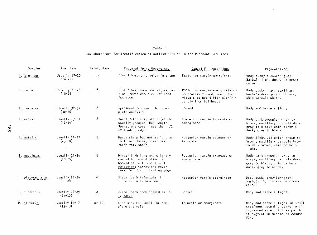
183
Table 1
Key characters foridentification of catfish alevins in the Piedmont Carol mas
fin.of pigment in middle of caudal
Body and barbels light in smalspecimens becoming darker withincreased size; diffuse patch
Truncate or emarginateSpecimens too small for com-plete analysis9 or 10Usually 14-17(13-18)P_. ol i var i s
Body and barbels lightForkedDistal barb hook-shaped as in
I . catus8Usual ly 26-29(24-30)J_. punctatus color.Body dusky brown Fsh-g ray ;barbels light dusky or creamPosterior margin emarginateDistal barb triangular inshape as in I . brunneus8Usually 21-24(19-24)J_. Pla tycepha 1 us
gray to black; chin barbelsdusky gray to black.hooked as in I . catus or I .punctatus ;
serrations coverless than 1/2 of leading edge
Body dark brownish gray toblack; maxillary barbels dark
Posterior margin truncate oremarg mateDistal barb long and slightlycurved but not distinctly8Usual ly 21-24(20-25)l_.
nebulosus light.1
ight.Body light yellowish brown tobrown; maxillary barbels brown
to dark brown; chin barbels
Posterior margin rounded ortruncateBarbs sharp but not as long asin I. nebu losus , sometimesrel at i yel y short .
8Usually 24-27(23-28)_!_•natal is
Body dark brownish gray toblack; maxillary barbels darkgray to black; chin barbelsdusky gray to black.
Posterior margin truncate oremarginateBarbs relatively short (widthusually greater than length).Serrations cover less than 1/2
of leading edge.
8Usually 17-21(16-24)J_. me las
Body and barbels lightForkedSpecimens too small for com-plete ana Iys1
ys i s
8Usual ly 32-34(30-36)J_. f urcatus cantly from bullheads Body dusky gray; maxillarybarbels dark gray or black,chin barbels white.
Posterior margin emarginate tomoderately forked; small indi-viduals do not differ signifi-
Distal barb hook-shaped; serra-tions cover about 2/3 of lead-ing edge
8Usual ly 21-24(18-26)J_. catus
col orBody dusky brownish-gray;Barbels light dusky or creamPosterior margin emarginateDistal barb triangular in shape8Usually 17-20(16-22)I . brunneus P igmentat i onholo 1
Caudal Fin Morphologyholome MorS
PectoralPectoral Spine MorphologyPelvic RaysAnal RaysSpeciesSpecies Anal Rays Pelvic Rays Pectoral Spine MorphologyPectoral S me Mor holo Caudal Fin Morphology holo 1
P igmentat i on
I . brunneus Usually 17-20(16-22) 8 Distal barb triangular in shape Posterior margin emarginate Body dusky brownish-gray;Barbels light dusky or creamcol or
J_. catus Usual ly 21-24(18-26) 8 Distal barb hook-shaped; serra-tions cover about 2/3 of lead-ing edge
Posterior margin emarginate tomoderately forked; small indi-viduals do not differ signifi-
Body dusky gray; maxillarybarbels dark gray or black,chin barbels white.cantly from bullheads
J_. f urcatus Usual ly 32-34(30-36) 8 Specimens too small for com-plete ana Iys1
ys i s
Forked Body and barbels light
J_. me las Usually 17-21(16-24) 8 Barbs relatively short (widthusually greater than length).Serrations cover less than 1/2
of leading edge.
Posterior margin truncate oremarginate Body dark brownish gray toblack; maxillary barbels darkgray to black; chin barbelsdusky gray to black.
_!_•natal is Usually 24-27(23-28) 8 Barbs sharp but not as long asin I
. nebu losus , sometimesrel at i yel y short .
Posterior margin rounded ortruncate Body light yellowish brown tobrown; maxillary barbels brown
to dark brown; chin barbels
light.1
ight.
l_.
nebulosus Usual ly 21-24(20-25) 8 Distal barb long and slightlycurved but not distinctly Posterior margin truncate oremarg mate Body dark brownish gray toblack; maxillary barbels darkhooked as in I . catus or I .punctatus ;
serrations coverless than 1/2 of leading edge
gray to black; chin barbelsdusky gray to black.
J_. Pla tycepha 1 us Usually 21-24(19-24) 8 Distal barb triangular inshape as in I . brunneus Posterior margin emarginate Body dusky brown Fsh-g ray ;barbels light dusky or creamcolor.
J_. punctatus Usual ly 26-29(24-30) 8 Distal barb hook-shaped as in
I . catus Forked Body and barbels light
P_. ol i var i s
Usually 14-17(13-18) 9 or 10 Specimens too small for com-plete analysis Truncate or emarginate Body and barbels light in smalspecimens becoming darker withincreased size; diffuse patch
of pigment in middle of caudalfin.
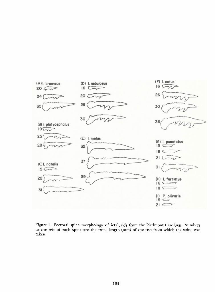
185
Figure 1. Pectoral spine morphology of ictalurids from the Piedmont Carolinas. Numbersto the left of each spine are the total length (mm) of the fish from which the spine wastaken.

LARVAEAND JUVENILES OF THEBROOK SILVERSIDE,
Labidesthes sicculusl
by
R. A.Frietsche, R. D.Miracle, and R. W. McFarlane
Savannah River LaboratoryE. I.duPonte de Nemours and Company
Aiken, SC 29801
ABSTRACT
Protolarvae have 6 to 7 preanal and 29 to 31 postanal myomeres. Migration of the anus
and pelvic buds during the metalarval phase results in 14 to 15 preanal and 24 to 25 postanal
myomeres and causes a relative increase in prepelvic body length. Pectoral fins in protolarvae ap-
pear before finfold differentiation. Median finray development inmesolarvae appears sequentially
in the caudal, anal, and soft dorsal fins. The spinous dorsal and pelvic fins differentiate inmetalar-vae. Protolarval pigmentation consists ofnumerous large melanophores on the dorsal surface of the
head and on the ventral surface of the breast, and a single row ofmelanophores midventrally and
midlaterally. Juvenile fish lack the fully formed beak-like snout of adults.
1. The information contained in this article was developed during the course of work under
Contract No. AT(O7-2)-l with the U.S. Department of Energy.
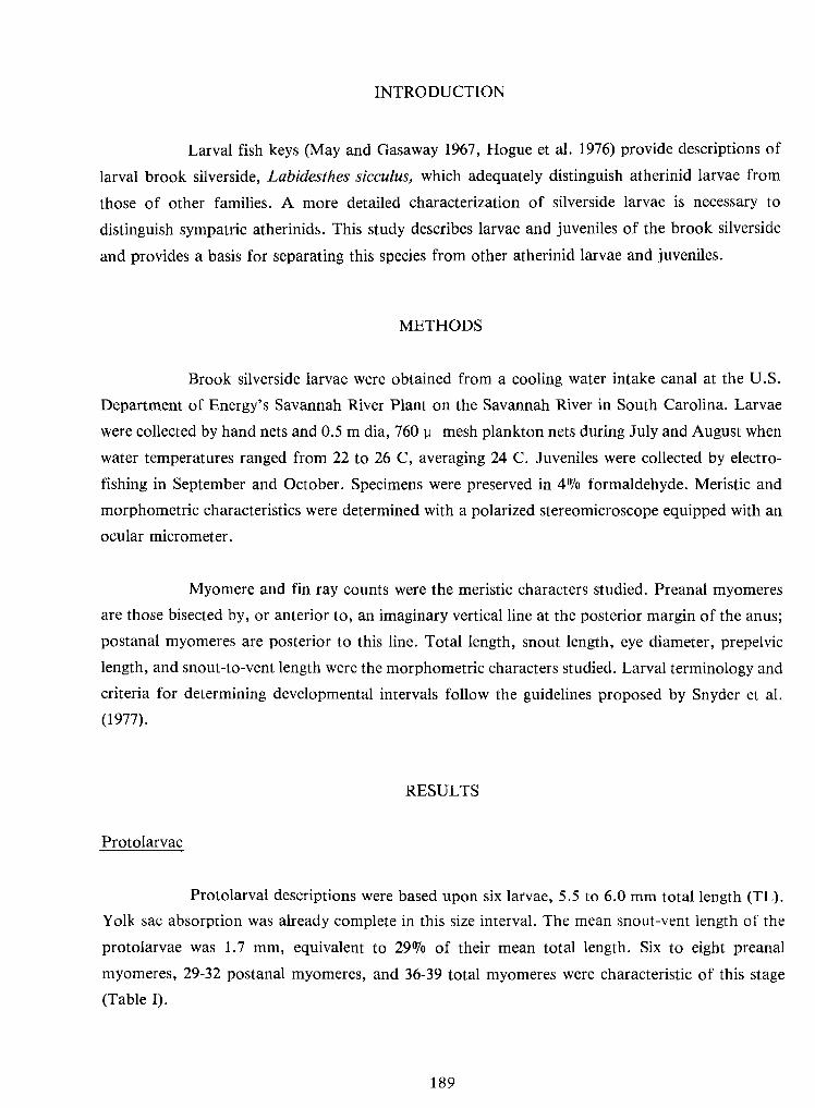
189
INTRODUCTION
Larval fish keys (May and Gasaway 1967, Hogue et al. 1976) provide descriptions of
larval brook silverside, Labidesthes sicculus, which adequately distinguish atherinid larvae from
those of other families. A more detailed characterization of silverside larvae is necessary to
distinguish sympatric atherinids. This study describes larvae and juveniles of the brook silverside
and provides a basis for separating this species from other atherinid larvae and juveniles.
METHODS
Brook silverside larvae were obtained from a cooling water intake canal at the U.S.
Department of Energy's Savannah River Plant on the Savannah River in South Carolina. Larvae
were collected by hand nets and 0.5 m dia, 760 y mesh plankton nets during July and August whenwater temperatures ranged from 22 to 26 C, averaging 24 C. Juveniles were collected by electro-fishing in September and October. Specimens were preserved in 4% formaldehyde. Meristic andmorphometric characteristics were determined witha polarized stereomicroscope equipped with an
ocular micrometer.
Myomere and fin ray counts were the meristic characters studied. Preanal myomeres
are those bisected by, or anterior to, an imaginary vertical line at the posterior margin of the anus;
postanal myomeres are posterior to this line. Total length, snout length, eye diameter, prepelvic
length, and snout-to-vent length were the morphometric characters studied. Larval terminology and
criteria for determining developmental intervals follow the guidelines proposed by Snyder et al.
(1977).
RESULTS
Protolarvae
Protolarval descriptions were based upon six larvae, 5.5 to 6.0 mm total length (TL).
Yolk sac absorption was already complete in this size interval. The mean snout-vent length of the
protolarvae was 1.7 mm, equivalent to 29% of their mean total length. Six to eight preanal
myomeres, 29-32 postanal myomeres, and 36-39 total myomeres were characteristic of this stage
(Table I).
190
The median finfold originated dorsally at the seventh or eighth myomere and extended
to the posterior margin of the anus. The pectoral fins were paddle-shaped but exhibited no finray
development. Gillrakers and filaments were present and the opercular flap covered the gillarches.
The eyes were well developed, pigmented, and elliptical in shape, and the auditory vesicles were visi-
ble posterior to the eyes (Figure lb).
Dorsal pigmentation consisted ofnumerous large melanophores in the tissue overlying
the midbrain and hindbrain, withsmaller melanophores extending onto the anterior one-quarter of
the body along the middorsal line (Figure la). Melanophores were present at the base of the medianfinfold on the caudal peduncle. Two small melanophores were present at the nares.
Ventral pigmentation consisted of 4 to 6 large melanophores in the tissue covering the
breast and abdomen. Melanophores formed a single line ofpigment from the anus to the caudal fin.Three to four small melanophores were present on the anterior portion of the mandible and the dor-sal surface of the swim bladder was heavily pigmented (Figure lc).
Lateral pigmentation consisted of scattered melanophores midlaterally, small
melanophores surrounding the caudal end of the notochord, and melanophores ventral to the
auditory vesicle between the eye and pectoral fin (Figure lb).
Mesolarvae
Caudal findifferentiation was initiated inmesolarvae of 6.2 mm TL.Dorsal flexion of
the notochord occurred on mesolarvae at 8.1 mm TL.Fin rays appeared in the anal and soft dorsal
fins of mesolarvae at 8.3 and 8.6 mm TL, respectively (Figure Id). Myomere counts remained as
they were in the protolarvae.
The small melanophores surrounding the notochord moved with it as it flexed. Three
parallel rows ofmelanophores appeared ventrally: one row on each side of the midventral line from
the anus to the caudal fin, and one row at the base of the anal finand the remaining finfold(Figure
le). Other regions of pigmentation remained basically as they were in protolarvae.
Metalarvae
Metalarval development was characterized by anal and pelvic bud migration between
11-15 mm TL.This migration altered the proportional relationship ofpreanal myomeres (13 to 15)
191
to postanal myomeres (24 to 25) while the total number ofmyomeres remained constant (37 to 39).
Simultaneously, the mean snout-vent length of the 18 metalarvae studied increased to 36% of theirtotal length (Table 1).
By 9.4 mm TL11-12 rays were present in the pectoral fin, 4-5 spines had appeared in
the spinous dorsal, and pelvic buds were present. Subsequently, 5 to 6 fin rays developed in the
pelvic fins during migration. Metalarvae 15 mm TLhad the characteristic sickle-shaped anal finand
22 to 25 anal rays and one spine (Figure If)- The last remnant of the finfold,between the anus and
anal fin,persisted until the metalarvae reached a total length of 16.5 mm. Ten to 11 soft dorsal fin
rays developed.
The large melanophores on the dorsal surface of the head and ventral surface of the
breast fused during the metalarval phase. Scattered dorsal melanophores formed a double row ofpigment from the soft dorsal fin to the caudal fin. Pigmentation increased on the lips and adjacent
tissues. Melanophores occurred on the branchiostegal membranes, and midlaterally a solid black
line developed.
Juveniles
Development of the beak-like snout increased the ratio of snout length to eye diameterfrom 0.6 to 1.0 in juveniles. The eye assumed a round shape and a silvery choroid coat.
The development of small melanophores increased the pigmentation of the head, dor-sum, and ventrum. Increased pigmentation along the midlateral line produced a silvery lateral stripe
of adult proportions.
DISCUSSION
Larval development of the brook silverside resembles the larval descriptions of
estuarine atherinids provided by Kuntz (1916), Kuntz and Radcliffe (1917), and Lippson and Moran(1974). The overlapping geographical ranges of the tidewater silverside, Menidia beryllina, and the
brook silverside provide the potential for the larvae of the two species tooccur sympatrically in tidalfresh waters. Larval separation can be based on size differences at hatching; the tidewater silverside
hatches at 3.0 mm, (Hildebrand 1922) and the brook silverside hatches at 4.0 mm (Nelson 1968).
192
The tidewater silverside has fewer anal rays (15 to 18) than does the brook silverside (22 to 25). In
addition, the origin ofthe spinous dorsal finis anterior to the origin of the anal finon the tidewater
silverside but directly above the anal fin origin on the brook silverside.
The Mississippi silverside, Menidia audens, and the brook silverside occur sympatrical-
lyin the Mississippi River drainage. The Mississippi silverside has fewer anal rays (15-20) than does
the brook silverside and the originof the spinous dorsal finis anterior to the anal finorigin. Brook
silverside larvae and juveniles lack the fully formed, beak-like snout characteristic of the adults and
they have a snout length less than their eye diameter (Table I) as do the Mississippi silversides. The
beak-like snout of adult brook silversides distinguishes them from Mississippi silversides; however,
the larvae and juveniles of these two species cannot be separated by this late-developing character.
193
LITERATURE CITED
Hildebrand, S. F. 1922. Notes on habits and development of eggs and larvae of silversides,Menidia audens and Menidia beryllina. Bull.U.S. Bur. Fish. 38: 113-120.
Hogue, J. J., Jr., R. Wallus, and L.K. Kay. 1976. Preliminary guide to the identification of larvalfishes in the Tennessee River. Tennessee Valley Authority Tech. Note 819, Norris, TN.
Kuntz, A. 1916. Notes on the embryology and larval development of five species of teleosteanfishes. Bull. U.S. Bur. Fish. 34 (1914): 409-430.
Kuntz, A., and L.Radcliffe, 1917. Notes on the embryology and larval development of twelveteleostean fishes. Bull. U.S. Bur. Fish. 35 (1915-1916): 87-134.
Lippson, A. J., and R. L. Moran. 1974. Manual for the identification of early developmentalstages of fishes of the Potomac River estuary. Power Plant Siting Program of the MarylandDepartment of Natural Resources (PPSP-MP-13), 282 pp.
May, E. 8., and C. R. Gasaway. 1967. Apreliminary key to the identification of larval fishes ofOklahoma, with particular reference to Canton Reservoir, including a selected bibliography.Okla. Fish. Res. Lab., Bull. No. 5: 42 pp.
Nelson, J. S. 1968. Life history of the brook silverside, Labidesthes sicculus, in Crooked Lake,Indiana. Trans. Am. Fish. Soc, 97 (3): 293-296.
Snyder, D. E., M.B.M. Snyder, and S. C. Douglas. 1977. Identification of Golden Shiner,Notemigonus crysoleucas, Spotfin Shiner, Notropis spilopterus, and Fathead Minnow,Pimephales promelas, larvae. J. Fish. Res. Board Can., 34 (9): 1397-1409.
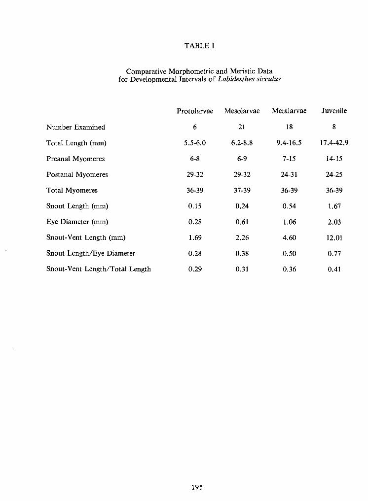
195
TABLEI
Comparative Morphometric and Meristic Datafor Developmental Intervals ofLabidesthes sicculus
0.410.360.310.29Snout-Vent Length/Total Length
0.770.500.380.28Snout Length/Eye Diameter
12.014.602.261.69Snout-Vent Length (mm)
2.031.060.610.28Eye Diameter (mm)
1.670.540.240.15Snout Length (mm)
36-3936-3937-3936-39Total Myomeres
24-2524-3129-3229-32Postanal Myomeres
14-157-156-96-8Preanal Myomeres
17.4-42.99.4-16.56.2-8.85.5-6.0Total Length (mm)
818216Number Examined
JuvenileMetalarvaeMesolarvaeProtolarvaeProtolarvae Mesolarvae Metalarvae Juvenile
Number Examined 6 21 18 8
Total Length (mm) 5.5-6.0 6.2-8.8 9.4-16.5 17.4-42.9
Preanal Myomeres 6-8 6-9 7-15 14-15
Postanal Myomeres 29-32 29-32 24-31 24-25
Total Myomeres 36-39 37-39 36-39 36-39
Snout Length (mm) 0.15 0.24 0.54 1.67
Eye Diameter (mm) 0.28 0.61 1.06 2.03
Snout-Vent Length (mm) 1.69 2.26 4.60 12.01
Snout Length/Eye Diameter 0.28 0.38 0.50 0.77
Snout-Vent Length/Total Length 0.29 0.31 0.36 0.41
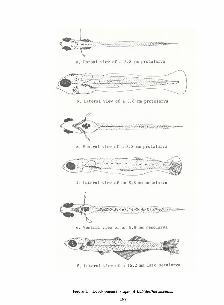
197
Figure 1. Developmental stages of Labidesthes sicculus,
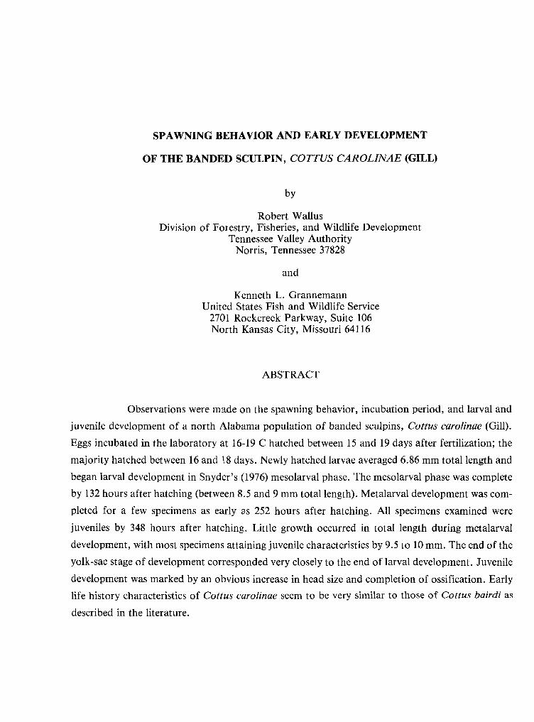
SPAWNING BEHAVIOR ANDEARLYDEVELOPMENT
OF THE BANDED SCULPIN, COTTUS CAROLINAE (GILL)
by
Robert WallusDivision of Forestry, Fisheries, and WildlifeDevelopment
Tennessee Valley AuthorityNorris, Tennessee 37828
and
Kenneth L. GrannemannUnited States Fish and Wildlife Service
2701 Rockcreek Parkway, Suite 106North Kansas City, Missouri 64116
ABSTRACT
Observations were made on the spawning behavior, incubation period, and larval and
juvenile development of a north Alabama population of banded sculpins, Cottus carolinae (Gill).
Eggs incubated in the laboratory at 16-19 Chatched between 15 and 19 days after fertilization; themajority hatched between 16 and 18 days. Newly hatched larvae averaged 6.86 mm total length andbegan larval development in Snyder's (1976) mesolarval phase. The mesolarval phase was complete
by 132 hours after hatching (between 8.5 and 9 mm total length). Metalarval development was com-
pleted for a few specimens as early as 252 hours after hatching. Allspecimens examined were
juveniles by 348 hours after hatching. Little growth occurred in total length during metalarvaldevelopment, with most specimens attaining juvenile characteristics by 9.5 to 10 mm. The end of theyolk-sac stage of development corresponded very closely to the end of larval development. Juveniledevelopment was marked by an obvious increase inhead size and completion of ossification. Early
life history characteristics of Cottus carolinae seem to be very similar to those of Cottus bairdi as
described in the literature.
201
INTRODUCTION
The banded sculpin, Cottus carolinae (Gill), is widely distributed in the southeastern
United States. It occurs in upland spring-fed streams from eastern Kansas and Oklahoma to West
Virginia; north to Indiana and Illinois;and south to Mississippi, Alabama, and Georgia (Moore
1968).
Spawning habits and early ecological aspects of the life histories of the cottids have
been well documented (Bailey 1952, Craddock 1965, Craig and Wells 1976, Foltz 1976, Goto 1975,
Heard 1965, Ikusemiju 1975, Jones 1972, Ludwig and Lange 1975, Northcote 1954, Petrosky and
Waters 1975, Savage 1963, Sheldon 1968, Simon and Brown 1943, Smith 1922, and Zarbock 1952).
The systematics of adult cottids have also been well documented (Abe 1976, Bailey and Bond 1963,
McAllister and Aniskowicz 1976, Robins 1961, Robins and Miller 1957, Williams 1968, and
Williams and Robins 1970), and larval development of several species in the genus Myoxocephalus
has been described (Khan and Faber 1973). There is, however, little descriptive detail in the
literature concerning post-hatching development of species of the genus Cottus.
This study was initiated to document aspects of the spawning behavior and early lifehistory of the banded sculpin, Cottus carolinae, and to describe in detail its larval development.
METHODS
During January and February 1975-1976, observations were made on the spawning ac-
tivities ofa population ofbanded sculpin inBuffler Spring on Cypress Creek, a tributary ofthe Ten-
nessee River (Pickwick Reservoir, Lauderdale County, Alabama). During the two years of study,
several clusters of fertilized eggs were collected and returned to TVA's laboratory inMuscle Shoals,
Alabama, where notes were made on egg development and larval behavior.
Eggs were removed from nests and immediately transported to the laboratory for in-cubation in MacDonald hatching jars. During incubation, a continuous flow of filtered,
dechlorinated, aerated water ranging in temperature from 16 to 19 C was provided. On hatching,
larvae were transferred to flow-through aquaria (30 x 50 x 15 cm). Water temperatures in the
aquaria ranged from 15 to 19 C.
202
Larvae were fed brine shrimp three times daily for a week after the onset of feeding.
From that time forward they were fed mixed zooplankton, primarily Daphnia spp.
Larvae, from hatching through juvenile stages, were preserved for descriptive pur-
poses. Specimens were initially preserved in 10 percent Formalin and were later transferred to a
solution of 5 percent Formalin buffered to approximately pH 7.5. Aminimum of five specimenswas preserved daily for description of larval development. A total of 225 larval and juvenile
specimens was examined in the study.
Specimens were examined with a stereo-microscope equipped with an ocularmicrometer and polarizers. Terminology used for developmental phases follows Snyder (1976).
Morphometric and meristic characters examined (Figure 1) include: total, standard, preanal,
postanal, and head length; orbit diameter; greatest body depth; and numbers of preanal
myomeres, postanal myomeres, vertebrae, and fin rays. Standard length measurements were
made to the posterior tip of the notochord until complete development of the hypural complex.
Head length was the distance from the tip of the snout to the posterior margin of the auditory
vesicle, initially, then to the posterior margin of the operculum when it formed. Some specimens
were stained with Alizarin Red S and cleared in a KOH-glycerol solution to facilitate vertebralcounts. Fin rays were counted using techniques described by Robins and Miller (1957) and adultcomplements were based on Moore (1968).
Illustrations for each developmental phase ,vere drawn with the aid of a camera
lucida. Each drawing is a composite, blending characteristics from several specimens of the size
indicated in an effort to portray an idealized specimen.
The dynamic approach is utilized for the presentation of descriptive data. Meristic
and morphometric data are tabulated by age and size intervals.
RESULTS
Spawning Behavior
The spawning habits of this population of Cottus carolinae are similar to those of its
relatives C. meridionalis (Smith 1922), C. bairdi (Adams and Hankinson 1928, Harm 1927), C.
203
bairdi punctulatus (Bailey 1952), C. bairdi semiscaber (Zarbock 1952), C. beldingi (Jones 1972),
and C. semiscaber (Simon and Brown 1943). Spawning took place in a small spring-fed stream
(Buffler Spring) 1 to 2 meters wide and less than 0.3 meter deep. Simon and Brown (1943) found
that the velocity of water seemed to make no difference in the choice of "nest rocks" for Cottus
semiscaber. Water velocity was likewise not a determining factor for selection of nest sites for
this population of Cottus carolinae, as nests were also observed in both slow and rapid currents
of Cox Creek, a somewhat larger stream in the Cypress Creek system. Bottom substrate in both
streams was primarily sand, gravel, and rubble. Clusters of eggs were found in crevices on the
undersurfaces of rocks and logs.
Spawning males were larger and more darkly pigmented than females. The adults were
often captured in nesting crevices on overturning rocks or logs. Single males, spawning pairs lying
side by side, and males apparently guarding or attending the eggs were captured, but single females
were not found inthe crevices. Apparently the male establishes a territory beneath a rock or log, the
female enters the nest, spawning occurs, a cluster ofadhesive eggs is deposited on the underside ofthe rock or log, the female departs, and the male remains to guard eggs until hatching occurs.
A paternal brooding habit is commonplace in the cottids. Simon and Brown (1943)
observed that rarely were developing eggs of Cottus semiscaber found unaccompanied by a male
fish, and that females, when found innests during the spawning season, were usually accompanied
by the male. Harm (1927) stated that the male Cottus bairdi guards the nest while the eggs were in-
cubating. Bailey (1952) preferred tocall the male Cottus bairdipunctulatus an attendant rather than
a guardian; Smith (1922) also observed a paternal brooding habit with Cottus meridionalis.
Occasionally, males were found guarding more than one cluster of eggs. Thisphenomenon was observed byBailey (1952) for Cottus bairdi punctulatus and by Simon and Brown(1943) for Cottus semiscaber. Generally, however, male Cottus carolinae in this investigation were
observed guarding only one cluster of eggs.
Eggs and Hatching
Eggs were laid inround or oval clusters and were so adhesive that a complete cluster
could be dislodged from a rock without separating the eggs from each other. No actual counts were
made, but it is estimated that each cluster contained from 100 to 300 eggs.
204
On January 27, 1975, one cluster of eggs was found on the underside of a railroad tie
lying inabout 0.2 meter of water (14 C). The eggs were removed from the nest and transported to
the laboratory for incubation. When the eggs were first discovered they were dull yellow in color,
but as development progressed, they turned salmon and became continuously darker as they ap-
proached hatching. Eggs were "eyed" and embryos could be seen occasionally moving within the
chorion, beginning on the eighth day after spawning. Hatching occurred from 15 to 19 days afterspawning with the majority hatching between 16 and 18 days after spawning.
Larvae used for the descriptions inthis paper were reared from eggs collected inBuffler
Spring on February 12, 1976. Thirty-four eggs from this collection ranged from 2.6-3.3 mm in
diameter, witha mean diameter of3.05 mm. Several clusters were hatched. The resulting larvae usedfor description were the progeny ofa single population but not of a single breeding pair.
DEVELOPMENT
Morphometric data obtained from larvae and juveniles are presented by post-hatching
age and by size intervals in Tables 1and 2. Meristic data are presented bypost-hatching age and by
size intervals in Tables 3 and 4.
Mesolarval Development
Banded sculpins are precocious at hatching and exhibit characteristics that exclude
Snyder's (1976) protolarval phase from the description of their development. Athatching the caudal
fin had six to nine hypochordal rays and the urostyle had already begun its upward turn. These
characteristics result in the larval period of development beginning in Snyder's mesolarval phase.
Dorsal and lateral line drawings of recently hatched and 2.5-day-old Cottus carolinae are shown in
Figures 2 and 3. Average size at hatching was 6.86 mm total length. The mesolarval phase was com-
pleted on some specimens by 132 hours after hatching between 8.5 and 9.0 mm total length.
By the time hatching occurred, banded sculpin larvae were well developed. The eyes
were large with heavy pigmentation and nares were present. Auditory vesicles were well developed
although the otoliths were not yet visible. Mouth parts were formed or nearly so. Gillarches with
bud-like filaments were present.
205
At hatching, a large, round, yellowish-orange yolk sac was present with an oilglobule
located anteriorly. Apatch ofoff-colored tissue was observed dorso-laterally on the left side slightly
anterior to the middle of the yolk sac (Figure 2). This tissue, probably generative innature, was visi-
ble throughout the mesolarval phase and into the metalarval phase untilobscured by heavy dorso-
lateral pigmentation. We did not determine the developmental function or significance, ifany, of
the tissue. A fairly rapid decrease inyolk mass occurred during the first 36 hours as evidenced by a
reduction in greatest depth measurements (Table 1). Little change in the mass occurred, however,
during the remainder of the mesolarval phase (Figure 4).
The opercular flap had begun to form at hatching and nearly covered the gillarches. By
36 hours the opercular flap extended to the pectoral finbase and branchiostegals appeared in the
opercular membranes.
Myosepta were well developed at hatching withmesolarvae having from 15-17 preanal
and from 14-17 postanal myomeres (Table 3). By 36 hours, myomeres had developed a piscine shape
("w"configuration).
Fin development was well advanced at hatching. Pectoral fins were large and well
formed with11-12 incipient rays. Fourteen to 16 pectoral rays were visible by the end of the mesolar-val phase. The median finfold was continuous from the first or second myomere from the occiput to
the posterior margin of the anus, and median fin differentiation had begun at hatching. The caudalfin was slightly bilobed and possessed six tonine incipient rays inits lower lobe. Incipient soft dorsaland anal finrays were visible on close examination of some specimens at hatching. By36 hours thecaudal finhad lost itsbilobed appearance and was rounded. Mesolarval development ofmedian finswas rapid, with adult complements of soft dorsal, anal, and caudal rays having developed by 132
hours. Pelvic buds first appeared between 8.5 and 9.0 mm TLand were present on all specimens by
108 hours.
The urostyle showed a slight dorsal deflection at hatching (Figure 1), but by 36 hours
was well upturned and hypural development was visible. The hypural complex was very nearly
developed by the end of the mesolarval phase.
Although the eyes were darkly pigmented at hatching, very little body pigmentation
was present. That present was limited to one or more stellate erythrophores on the yolk sac near the
206
anus. By 36 hours a few stellate erythrophores were present between the eyes and scattered over the
forebrain and midbrain. By 60 hours, inaddition to this dorsal pigment, varying degrees ofpigmen-
tation were present (Figure 3) as: a band ofpigment at the anterior base ofthe pectoral finextending
ventrally to the gular region from either side; concentrations of stellate erythrophores dorso-
laterally on the torso in the region of the occiput extending ventrally onto the yolk and proceeding
posteriorly on the yolk to the anal region; a few stellate erythrophores present laterally on the caudalpeduncle; and littleor no ventral pigmentation. By the end of the mesolarval phase, more advanced
specimens showed nares outlined withpigment, as well as more profuse pigmentation on the headand yolk and laterally on the body. Apatch of stellate erythrophores was present ventrally in the
gular region and bars of such pigment were beginning to form behind the eyes. No ventral pigment
was present on the yolk sac at the end of mesolarval phase.
Larval activity during the first few days after hatching was limited. Larvae clustered in
aquaria corners and in crevices until three or four days after hatching when they began to scatter
randomly across the bottoms.
Metalarval Development
The metalarval phase ofdevelopment for Cottus carolinae began between 8.5 and 9.0
mm TL.Figure 5 shows dorsal and lateral views ofa typical metalarva. Figure 6 shows a specimen at
completion of metalarval development.
Yolk material diminished gradually during metalarval development as evidenced by a
gradual reduction in greatest depth (Figure 4). In many specimens the yolk material was absorbed
more rapidly anteriorly with the shape of the yolkmass becoming oval by 132 to 156 hours after hat-
ching. The end of the yolk-sac phase of development corresponded closely to the end ofmetalarval
development. Yolkmaterial was almost gone on several specimens by 252 hours after hatching and
by 348 hours (14.5 days) specimens lacked yolk material.
Metalarvae had 15-16 preanal and 15-17 postanal myomeres. Myomeres were obscured
by pigmentation by 252 hours after hatching. No counts were taken from specimens greater than 10
mm TL.
Fin development was rapid during the metalarval phase. The adult complement of
spinous dorsal rays and pectoral rays was present on most specimens soon after the onset ofmetalar-
207
val development. The full complement of pelvic rays was present (marking the end ofmetalarval
development) as early as 252 hours after hatching. The smallest specimen seen withan adult comple-
ment of finrays inallfins was 9.3 mm TL.Most specimens, however, achieved this state ofdevelop-
ment at total lengths beween 9.5 and 10.0 mm. By two weeks post-hatching, all specimens were
juveniles.
By the beginning of the metalarval phase, pigmentation had increased to the degree
that the first indication of adult banding could be seen. By132 hours concentrations ofpigment ap-
peared as bands at the posterior margin of the yolk sac and near the middle of the caudal peduncle.
Dorso-lateral pigmentation continued to intensify on the yolk,converging to the spinous dorsal fin,
until the most anterior band was visible. By this time (156 hours), two dorso-lateral concentrations
of postanal pigment as well as an aggregation of pigment in the hypural region were obvious. Thispattern of pigment development continued until,by approximately 9.0 mm TL, four dorso-lateralbands of pigment were present. By 180 hours, at approximately 9.4 mm TL, the posterior barsbehind the eyes ran through the eyes onto the snout, the head was heavily pigmented dorsally and
laterally, and specimens exhibited the adult pigment pattern of four to five dorso-lateral bands.
Ossification first became evident in metalarvae 228 hours after hatching. Initial
calcification was observed in the cleithrum, pharyngeal arch, branchiostegals, operculum, and
mouth parts (premaxilla, maxilla, and dentary). At 276 hours post-hatching, considerable braincase
ossification had occurred; the anterior attachment for the branchiostegals, the ceratohyal, had
begun to ossify; and teeth were visible on the premaxilla and dentary. The vertebral column hadbegun to ossify with the neural and hemal spines heavily stained. At this time preopercular arma-
ment was obvious with three prominent spines visible. At 300 hours vertebral ossification was com-
plete enough to facilitate counts on stained specimens (Table 3) and four preopercular spines were
obvious. The ossification process seemed to slow for the remainder of the metalarval phase.
Metalarval activity in the aquaria was limited to short quick dashes associated withfeeding. This suggests that larvae of Cottus carolinae, like several other larval cottids (Heard 1965,
Sheldon 1968, and Goto 1975), are benthic innature. The larvae began feeding on freshly hatchedbrine shrimp soon after becoming metalarvae (five to seven days after hatching) and well beforetheir yolk material was used up. Attempts to feed frozen brine shrimp, tubificid worms, and com-
mercial fish food were unsuccessful. Observations indicated that larvae fed by sight and would only
ingest moving organisms. Nutrition appeared to be good throughout the metalarval phase.
208
Juvenile Development
Figure 7is a line drawing of the lateral view ofa 20.0 mm TL juvenile Cottus carolinae.
Few significant changes were noted in the external morphology of the banded sculpin
during juvenile development. One rather obvious change, however, was an increase in head size.This was expressed to some degree by head length (Table 2); however, length did not adequately
show the overall growth that occurred.
Pigmentation patterns continued to develop toward that of the adult with further
concentration of pigment in the areas of the band and the appearance of another bar of pigment
near the eye which extended from the eye posterio-ventrally across the operculum. Adult pig-
ment patterns seemed to be completed by about 420 hours (17.5 days) after hatching.
Prickles were not apparent untilapproximately 11 mm TL. By approximately 20 mm
TL they appeared as two parallel patches that began below the origin of the spinous dorsal fin
(the lower patch at a level with the dorsal-most three or four pectoral rays) and extended its
length. A few scattered prickles were also present posteriorly along the base of the soft dorsalfin.
Preopercular armament continued to develop during the juvenile phase with the
dorsal-most spine becoming prominent and pointing posteriorly with a slight hook to the dor-
sum. The second spine was pointed posterioventrally, and the third was pointed ventrally and
was considerably smaller than the dorsal two. The fourth spine, mentioned earlier, was not
noticeable by 20 mm TL.
Poor staining techniques or a reduction in the rate of ossification process precluded
detailed descriptions of bone development during the juvenile phase. This could be related to
poor nutrition, which became apparent by about 2.5 weeks. At this time (396 hours), growth rate
as expressed by mean TL (Table 1) began a steady decline. This trend continued until, by about
three weeks, larvae became emaciated. However, vertebral ossification was complete enough
to obtain counts for some juveniles ranging in age from 14.5 days (348 hours) to 7 weeks
(Table 3).
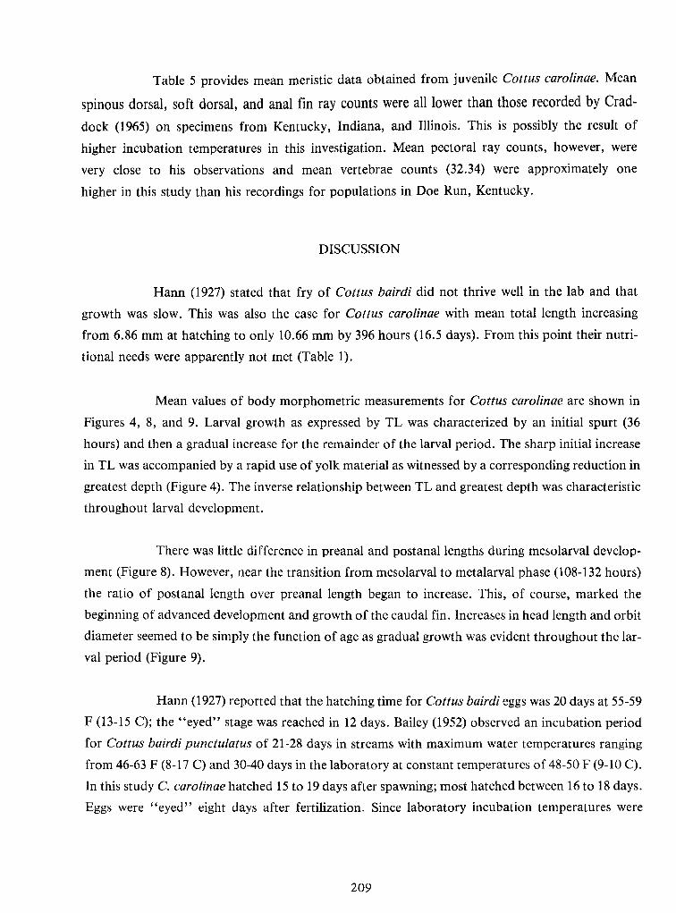
209
Table 5 provides mean meristic data obtained from juvenile Cottus carolinae. Mean
spinous dorsal, soft dorsal, and anal fin ray counts were all lower than those recorded by Crad-
dock (1965) on specimens from Kentucky, Indiana, and Illinois. This is possibly the result of
higher incubation temperatures in this investigation. Mean pectoral ray counts, however, were
very close to his observations and mean vertebrae counts (32.34) were approximately one
higher in this study than his recordings for populations inDoe Run, Kentucky.
DISCUSSION
Harm (1927) stated that fry of Cottus bairdi did not thrive well in the lab and that
growth was slow. This was also the case for Cottus carolinae with mean total length increasing
from 6.86 mm at hatching to only 10.66 mm by 396 hours (16.5 days). From this point their nutri-
tional needs were apparently not met (Table 1).
Mean values of body morphometric measurements for Cottus carolinae are shown inFigures 4, 8, and 9. Larval growth as expressed by TL was characterized by an initial spurt (36
hours) and then a gradual increase for the remainder of the larval period. The sharp initialincrease
inTLwas accompanied by a rapid use of yolk material as witnessed by a corresponding reduction ingreatest depth (Figure 4). The inverse relationship between TLand greatest depth was characteristicthroughout larval development.
There was littledifference inpreanal and postanal lengths during mesolarval develop-
ment (Figure 8). However, near the transition from mesolarval tometalarval phase (108-132 hours)
the ratio of postanal length over preanal length began to increase. This, of course, marked thebeginning ofadvanced development and growth of the caudal fin. Increases inhead length and orbitdiameter seemed tobe simply the function of age as gradual growth was evident throughout the lar-
val period (Figure 9).
Harm (1927) reported that the hatching time for Cottus bairdi eggs was 20 days at 55-59
F (13-15 C); the "eyed" stage was reached in 12 days. Bailey (1952) observed an incubation period
for Cottus bairdi punctulatus of 21-28 days in streams withmaximum water temperatures ranging
from 46-63 F (8-17 C) and 30-40 days in the laboratory at constant temperatures of 48-50 F (9-10 C).
Inthis study C. carolinae hatched 15 to 19 days after spawning; most hatched between 16 to 18 days.
Eggs were "eyed" eight days after fertilization. Since laboratory incubation temperatures were
210
higher (16-19 C) than those reported by either Harm or Bailey, it is reasonable to assume that the
rate of egg development is probably similar for C. carolinae and C. bairdi in natural conditions,
especially since Craddock (1965) indicated developmental rates similar to those of C. Bairdi for
banded sculpins studied inKentucky.
Harm (1927) gives the hatching size of C. bairdi as about 6.4 mm in length. Mean
hatching size for C. carolinae was 6.86 mm TL.Bailey (1952) reported that it took 14 days for C.
bairdi punctulatus to completely absorb their yolk-sacs at 51-56 F (11-13 C). These specimens had
attained total lengths of9.0-9.9 mm (x = 9.5). At 14,5 days (348 hours) mean total length was 10.05
mm for C. carolinae with a range of 9.5-10.3. Complete yolk absorption was first apparent on
specimens at approximately 9.5 mm TLand had occurred on all specimens by 14.5 days (348 hours).
These observations suggest that taxonomic differentiation of these two species may be as difficult
with larval forms as it sometimes is for the adults.
211
LITERATURE CITED
Adams, C. C. and Hankinson, T. L.1928. The ecology and economics of Oneida Lake Fish. Bui.New York State Col. Forestry, Roosevelt Wildlife Annals 1:235-548.
Abe, Syuiti. 1976. A cytotaxonomical study in some freshwater cottoid fishes (Cottidae, Pisces).Cytologia 41:323-329.
Bailey, Jack E. 1952. Life history and ecology of the sculpin, Cottus bairdi punctulatus insouthwestern Montana. Copeia 4:243-255.
Bailey, Reeve M.and Carl E. Bond. 1963. Four new species of freshwater sculpins, genus Cottus,from western North America. Occ. Pap. Mus. Zoo. Univ. Michigan, 634:1-27.
Craddock, J. E. 1965. The biology of the banded sculpin, Cottus carolinae, in Doe Run, MeadeCounty, Kentucky. Ph.D. Thesis, unpubl. Univ. Louisville. 175 p.
Craig, P. C. and J. Wells. 1976. Life history notes for a population of slimy sculpin {Cottuscognatus) in an Alaskan stream. J. Fish. Res. Board Can. 33:1639-1643.
Foltz, Jeffery W. 1976. Fecundity of the slimy sculpin, Cottus cognatus, inLake Michigan. Copeia.4:802-805.
Goto, Akira. 1975. Ecological and morphological divergence of the freshwater sculpin, Cottusnozawae Snyder - I.Spawning behavior and process of the development in the post-
hatching stage. Bui. Fisheries Faculty, Hokkaido Univ. Japan. 26(l):31-37.
Harm, Harry W. 1927. The history of the germ cells of Cottus bairdi Girard. J. Morph. Physiol.43(2):427-480.
Heard, William R. 1965. Limnetic cottid larvae and their utilization as food by juvenile sockeyesalmon. Trans. Am. Fish. Soc. 94(2): 191-193.
Ikusemiju, Kola. 1975. Aspects of the ecology and life history of the sculpin, Cottus aleuticus(Gilbert), in Lake Washington. J. of Fish. Biol. 7:235-245.
Jones, Albert C. 1972. Contributions to the life history of the piute sculpin in Sagehen Creek,California. Calif. Fish and Game 58(4):28 5-290.
Khan, N. Y.and D. J. Faber. 1973. A comparison of the larvae of the deepwater and fourhornsculpin, Myoxocephalus quadricornis L. from North America. I. Morphologicaldevelopment, pp. 703-712. IN:J.H.S. Blaxter, ed;, The Early LifeHistory ofFish. Springer-Verlag, Germany.
Ludwig, Gerald M. and Eugene L.Lange. 1975. The relationship of length, age, and age-lengthin teraction to the fecundity of the northern mottled sculpin, Cottus b. bairdi. Trans. Am.Fish. Soc. 104(l):64-67.
McAllister,Don E. and Jadwiga Aniskowicz. 1976. Vertebral number in North American sculpinsof the Myoxocephalus quadricornis-comples. J. Fish. Res. Board Can. 33(12):2792-2799.
Fish. Soc. 104(l):64-67.in teraction to the fecundity of the northern mottled sculpin, Cottus b. bairdi. Trans. Am.
212
Moore, George A. 1968. Fishes, pp. 31-210. IN: Blair, W. F. et al., Vertebrates of the UnitedStates. McGraw-Hill Book Company, New York.
Northcote, Thomas G. 1954. Observation on the comparative ecology of two species of fish, Cottusasper and Cottus rhotheus, in British Columbia. Copeia 1:25-28.
Petrosky, Charles E. and Thomas F. Waters. 1975. Annual production by the slimy sculpinpopulation in a small Minnesota trout stream. Trans. Am. Fish. Soc. 2:237-244.
Robins, C. Richard. 1961. Two new cottid fishes from the freshwaters of eastern United States.Copeia 3:305-315.
Robins, C. Richard and Robert Rush Miller. 1957. Classification, variation, and distribution of thesculpins, genus Cottus, inhabiting Pacific slope waters in California and southern Oregonwith a key to the species. Calif. Fish and Game 43(3):213-233.
Savage, Thomas. 1963. Reproductive behavior of the mottled sculpin, Cottus bairdi Girard. Copeia2:317-325.
Sheldon, Andrew L. 1968. Drift, growth, and mortality of juvenile sculpins in Sagehen Creek,California. Trans. Am. Fish. Soc. 97(4):495-496.
Simon, James R. and Robert C. Brown. 1943. Observations on the spawning of the sculpin, Cottussemiscaber. Copeia 1:41-43.
Smith, Bertran G. 1922. Notes on the nesting habits of Cottus. Mich. Acad. Sci. Papers 2:222-224.
Snyder, Darrel E. 1976. Terminologies for intervals of larval fish development. IN: J. Great LakesFish Egg and Larvae Identification. U.S. Fish Wildlife Service, OBS National Power PlantTeam, FWS/OBS-67/23:41-58.
Williams, James D. 1968. Anew species of sculpin, Cottus pygmaeus, from a spring inthe AlabamaRiver basin. Copeia 2:334-342.
Williams, James D. and C. Richard Robins. 1970. Variation in populations of the fish Cottuscarolinae in the Alabama River system withdescriptions ofa new species from below the fallline. Am. Midi.Nat. 83(2):368-381.
Zarbock, William M. 1952. Life history of the Utah sculpin, Cottus bairdi semiscaber (Cope), inLogan River, Utah. Trans. Am. Fish. Soc. 81:249-259.
McGraw-Hill Book Company, New York.
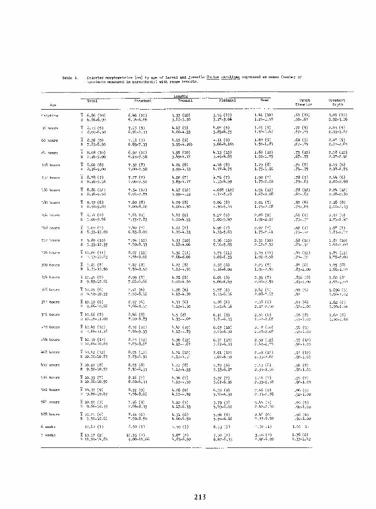
213
Table .1. Selected raorphometrics (mm) by age of larval and juvenile Cottus carollnae expressed as means (number of
specimens measured inparenthesis) with range beneath.
1.38 (2)
1.33-1. U23.U6 (2)2.92-U.OO
7.50 (2)6.67-8.33
5.67 (2)U.83-6
10.33 (2)9.00-11.66
X 13.17 (2)R 11.50-1U.83
7 weeks
1.00 (1)2.58(1)6.33 (1)U.50 (1)8.50 (1)10.83 (1)6 weeks
.96 (6)
.92-1.002.1+6 (6)2.33-2.50
5.9U (6)5.50-6.16
U.31 (6)U.OO-U.50
B.ll+ (6)7.50-8.50
X 10.25 (6)R 9.50-10.66
588 hours
.95 (5)
.92-1.002.U5 (5)2.U2-2.50
5.79 (5)5.U9-6.00
U.20 (5)U.17-U.33
7.96 (5)7.66-8.33
X 10.00 (5)R 9.66-10.33
56U hours
•96 (9).92-1.00
2.U6 (9)2.33-2.58
6.09 (9)5.U9-6.50
U.28 (9)U.17-U.50
8.22 (9)7.66-8.66
x 10.37 (9)R 9.66-10.83
SUO hours
•95 (7).92-1.00
2.U6 (7)2.33-2.58
5.97 (7)5.67-6.16
U.36 (7)U.17-U.50
8.16 (7)8.00-8.33
X 10.33 (7)R 10.00-10.50
516 hours
.96 (8)
.92-1.002.U3 (8)2. 33-2.50
5.83 (8)5.33-6.17
U.27 (8)U.17-U.33
8.03 (8)7.50-8.33
X 10.10 (8)R 9.50-10.50
U92 hours
.92 (12)
.92-1.002.U9 (12)2.33-2.67
5.91 (12)5.67-6.50
U.21 (12)U.17-U.3U
8.05 (12)7.83-8.50
X 10.12 (12)R 10.00-10.83
U6B hours
•97 (12).92-1.00
2.50 (12)2.U2-2.75
6.02 (12)5.67-6.33
U.36 (12)U.17-U.67
8.23 (12)7.83-8.66
X 10.39 (12)R 10.00-10.83
UUU hours
•96 (9).92-1.00
2.U8 (10)2.25-2.67
6.03 (10)5.U9-6.50
U.Us (10)U.17-U.83
8.35 (10)7.66-9.33 .x 10.1+5 (10)
R 9.66-11.16U-20 hours
1.62 (8)1.50-1.66
.98 (8)
.92-1.002.5U (8)2.U2-2.67
6.16 (8)5.83-6.33
U.5(8)U.33-U.67
8.U6 (8)8.00-8.83
X 10.66 (8)R 10.16-11.00
396 hours
1.62 (6)1.50-1.66
.93 (6)
.92-1.002.38 (6)2.17-2.50 \u25a0
5.86 (6)5.U9-6.16
U.33 (6)U.17-U.50
8.07 (6)7.66-8.50
X 10.19 (6)R 9.66-10.66
372 hours
.1.696 (5)1.58-1.92
•92 (5).92
2.U2 (6)2.08-2.67
5.78 (6)5.33-6.16
U.28 (6)U.17-U.50
7.91 (6)7.50-8.16
X 10.05 (6)R 9.50-10.33
3UB hours
1.80 (8)1.66-1.92
.\u25a0896 (8)
.83-1.002.3U (8)2.08-2.50
6.0U (8)5.66-6.U9
U.21 (8)U.OO-U.50
8.08 (8)7.66-8.66
X 10.25 (8)R 9.83-10.66
32U hours
1.75 (8)1.66-1.92
.87 (8)
.83-1.002.25 (8)1.92-2.50
5.58 (8)5.16-6.00
U.27 (8)U.17-U.50
7.87 (8)7.50-8.50
X 9.85 (8)X 9-33-10.50
300 hours
1.89 (11)1.75-2.00
.89 (H)
.79- -922.39 (11)1.92-2.58
5.75 (11)5.00-6.33
U.32 (11)7.66-8.66
8.07 (11)7.66-8.66
X 10.06 (11)R 9.33-10.83
276 hours
1.87 (10)1.67-2.08
.88 (10)
.83-.922.32 (10)2.17-2 .50 .5.56 (10)
5.16-5.83U.33 (10)U.17-U.66
7.9U (10)7.50-8.33
x 9.89 (10)R 9.33-10.33
252 hours
1.98 (7)1.83-2.17
.82 (7)
.75-.922.07 (7)1.75-2.U1
U.92 (7)U.33-5. 83
U.17 (7)U.OO-U.33
7.U0 (7)6.83-8.00
x 9.09 (7)R 8.33-10.00
228 hours
2.17 (9)1.75-2.U2
.85 (9)
.75-.922.08 (9)1.92-2.17
5.27 (9)5.00-5.U9
U.17 (9)U.OO-U.33
7.61 (9)7.33-7.83
x 9.1*1+ (q)R 9.00-9.66
20U hours
2.18 (8)2.00-2.33
.82 (8)
.75-.832.01 (8)1.75-2.08
5.06 (8)U.50-5. 33
U.29 (8)U.OO-U.50
7.60 (8)7.00-8.00
X 9.35 (8)R 8.50-9.83
180 hours
2.2U (12)2.08-2.50
.78 (12)
.67-.831.91 (12)1.67-2.08
U.698 (12)U.17-5
U.17 (12)3.99-U.33
7.3U (12)6.91-7.83
X 8.86 (12)R 8.16-9.58
156 hours
2.UU (6)2.00-2.58
.78 (7)
.75-.83-1.90 (7)1.75-2.00
U.76 (7)U.33-U.99
U.02 (7)3.83-U.17
7.27 (7)7.00-7.50
x 8.78 (7)R 8.16-9.16
132 hours
2.U9 (8)2.17-2.75
.7U (8)
.71-.751.79 (8)1.75-1.96
U.U6 (8)U.17-U.75
U.21 (8)3.99-U.33
7.32 (8)7.00-7.58
X 8.66 (8)R 8.16-9.00
108 hours
2.58 (10)2.17-2.92
.73 (10)
.67-.751.68 (10)1.50-1.75
U.13 (10)3.U9-U.83
3.95 (10)3.U9-U.17
6.92 (10)6.25-7.58
X 8.08 (10)n 7.16-9.00
8U hours
2.U7 (9)2.17-2.83
.69 (9)
.67-.751.69 (9)1.50-1.83
U.ll (9)3.66-U.165
U.15 (9)3.99-U.165
7.11 (9)6.83-7.33
x 8.26 (9)R 7.83-8.50
60 hours
2.U5 (5)2.33-2.67
.70 (5).67-.75
1.65 (5)1.58-1.67
k.07 (5)3.83-U.33
U.17 (5)U.OO-U.33
7.15 (5)6.91-7.33
X 8.23 (5)R 8.00-8.50
36 hours
3.03 (10)2.92-3.08
.63 (10)
.50-. 67I.UI(10)1.25-1.58
3.53 (10)3.17-3.66
3.33 (10)3.17-3.50
6.U6 (10)6.16-6.66
X- 6.86 (10)R 6.66-6.99
Hatching
A,,.
GreatestDepth
OrbitDiameter
HeadPoatanalPreanalStandardTotal
!>:);,!>:);,
Total Standard Preanal Poatanal Head OrbitDiameter
GreatestDepthA,,.
Hatching X- 6.86 (10)R 6.66-6.99
6.U6 (10)6.16-6.66
3.33 (10)3.17-3.50
3.53 (10)3.17-3.66
I.UI(10)1.25-1.58
.63 (10)
.50-. 673.03 (10)2.92-3.08
36 hours X 8.23 (5)R 8.00-8.50
7.15 (5)6.91-7.33
U.17 (5)U.OO-U.33
k.07 (5)3.83-U.33
1.65 (5)1.58-1.67
.70 (5).67-.75
2.U5 (5)2.33-2.67
60 hours x 8.26 (9)R 7.83-8.50
7.11 (9)6.83-7.33
U.15 (9)3.99-U.165
U.ll (9)3.66-U.165
1.69 (9)1.50-1.83
.69 (9)
.67-.752.U7 (9)2.17-2.83
8U hours X 8.08 (10)n 7.16-9.00
6.92 (10)6.25-7.58
3.95 (10)3.U9-U.17
U.13 (10)3.U9-U.83
1.68 (10)1.50-1.75
.73 (10)
.67-.752.58 (10)2.17-2.92
108 hours X 8.66 (8)R 8.16-9.00
7.32 (8)7.00-7.58
U.21 (8)3.99-U.33
U.U6 (8)U.17-U.75
1.79 (8)1.75-1.96
.7U (8)
.71-.752.U9 (8)2.17-2.75
132 hours x 8.78 (7)R 8.16-9.16
7.27 (7)7.00-7.50
U.02 (7)3.83-U.17
U.76 (7)U.33-U.99
1.90 (7)1.75-2.00
.78 (7)
.75-.83-2.UU (6)2.00-2.58
156 hours X 8.86 (12)R 8.16-9.58
7.3U (12)6.91-7.83
U.17 (12)3.99-U.33
U.698 (12)U.17-5
1.91 (12)1.67-2.08
.78 (12)
.67-.832.2U (12)2.08-2.50
180 hours X 9.35 (8)R 8.50-9.83
7.60 (8)7.00-8.00
U.29 (8)U.OO-U.50
5.06 (8)U.50-5. 33
2.01 (8)1.75-2.08
.82 (8)
.75-.832.18 (8)2.00-2.33
20U hours x 9.1*1+ (q)R 9.00-9.66
7.61 (9)7.33-7.83
U.17 (9)U.OO-U.33
5.27 (9)5.00-5.U9
2.08 (9)1.92-2.17
.85 (9)
.75-.922.17 (9)1.75-2.U2
228 hours x 9.09 (7)R 8.33-10.00
7.U0 (7)6.83-8.00
U.17 (7)U.OO-U.33
U.92 (7)U.33-5. 83
2.07 (7)1.75-2.U1
.82 (7)
.75-.921.98 (7)1.83-2.17
252 hours x 9.89 (10)R 9.33-10.33
7.9U (10)7.50-8.33
U.33 (10)U.17-U.66
5.56 (10)5.16-5.83
2.32 (10)2.17-2 .50 . .88 (10)
.83-.921.87 (10)1.67-2.08
276 hours X 10.06 (11)R 9.33-10.83
8.07 (11)7.66-8.66
U.32 (11)7.66-8.66
5.75 (11)5.00-6.33
2.39 (11)1.92-2.58
.89 (H)
.79- -921.89 (11)1.75-2.00
300 hours X 9.85 (8)X 9-33-10.50
7.87 (8)7.50-8.50
U.27 (8)U.17-U.50
5.58 (8)5.16-6.00
2.25 (8)1.92-2.50
.87 (8)
.83-1.001.75 (8)1.66-1.92
32U hours X 10.25 (8)R 9.83-10.66
8.08 (8)7.66-8.66
U.21 (8)U.OO-U.50
6.0U (8)5.66-6.U9
2.3U (8)2.08-2.50
.\u25a0896 (8)
.83-1.001.80 (8)1.66-1.92
3UB hours X 10.05 (6)R 9.50-10.33
7.91 (6)7.50-8.16
U.28 (6)U.17-U.50
5.78 (6)5.33-6.16
2.U2 (6)2.08-2.67
•92 (5).92
.1.696 (5)1.58-1.92
372 hours X 10.19 (6)R 9.66-10.66
8.07 (6)7.66-8.50
U.33 (6)U.17-U.50
5.86 (6)5.U9-6.16
2.38 (6)2.17-2.50 \u25a0
.93 (6)
.92-1.001.62 (6)1.50-1.66
396 hours X 10.66 (8)R 10.16-11.00
8.U6 (8)8.00-8.83
U.5(8)U.33-U.67
6.16 (8)5.83-6.33
2.5U (8)2.U2-2.67
.98 (8)
.92-1.001.62 (8)1.50-1.66
U-20 hours x 10.1+5 (10)R 9.66-11.16
8.35 (10)7.66-9.33 . U.Us (10)
U.17-U.836.03 (10)5.U9-6.50
2.U8 (10)2.25-2.67
•96 (9).92-1.00
UUU hours X 10.39 (12)R 10.00-10.83
8.23 (12)7.83-8.66
U.36 (12)U.17-U.67
6.02 (12)5.67-6.33
2.50 (12)2.U2-2.75
•97 (12).92-1.00
U6B hours X 10.12 (12)R 10.00-10.83
8.05 (12)7.83-8.50
U.21 (12)U.17-U.3U
5.91 (12)5.67-6.50
2.U9 (12)2.33-2.67
.92 (12)
.92-1.00
U92 hours X 10.10 (8)R 9.50-10.50
8.03 (8)7.50-8.33
U.27 (8)U.17-U.33
5.83 (8)5.33-6.17
2.U3 (8)2. 33-2.50
.96 (8)
.92-1.00
516 hours X 10.33 (7)R 10.00-10.50
8.16 (7)8.00-8.33
U.36 (7)U.17-U.50
5.97 (7)5.67-6.16
2.U6 (7)2.33-2.58
•95 (7).92-1.00
SUO hours x 10.37 (9)R 9.66-10.83
8.22 (9)7.66-8.66
U.28 (9)U.17-U.50
6.09 (9)5.U9-6.50
2.U6 (9)2.33-2.58
•96 (9).92-1.00
56U hours X 10.00 (5)R 9.66-10.33
7.96 (5)7.66-8.33
U.20 (5)U.17-U.33
5.79 (5)5.U9-6.00
2.U5 (5)2.U2-2.50
.95 (5)
.92-1.00
588 hours X 10.25 (6)R 9.50-10.66
B.ll+ (6)7.50-8.50
U.31 (6)U.OO-U.50
5.9U (6)5.50-6.16
2.1+6 (6)2.33-2.50
.96 (6)
.92-1.00
6 weeks 10.83 (1) 8.50 (1) U.50 (1) 6.33 (1) 2.58(1) 1.00 (1)
7 weeks X 13.17 (2)R 11.50-1U.83
10.33 (2)9.00-11.66
5.67 (2)U.83-6
7.50 (2)6.67-8.33
3.U6 (2)2.92-U.OO
1.38 (2)
1.33-1. U2
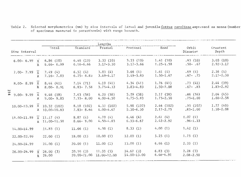
Table 2. Selected morphometrics (mm) by size intervals of larval and juvenile Cottus carolinae expressed as means (number
of specimens measured in parenthesis) with range beneath.
2.08-2.506.66-6.9114.00-15.0011.00-12.0020.00-21.0026.00R
2.28 (3)6.83 (3)
14.67 (3)
11.33 (3)20.50 (3)26.00 (3)X26.00-26.99 2.33 (1)6.66 (1)
13.00 (1)11.00 (1)20.00 (1)24.00 (1)24.00-24.99 1.75 (1)5.25 (1)
12.00 (1)
10.00 (1)
18.00 (1)22.00 (1)22.00-22.99 1.42 (1)4.00 (1)8.33 (1)6.50 (1)
11.66 (1)
14.83 (1)
14.00-14.99 .96-1.332.42-2.926.33-6.674.50-4.838.66- 9.0011.00-11.50R
1.07 (4)2.61 (4)6.46 (4)4.70 (4)8.87 (4)11.17 (4)X
11.00-11.99 1.50-2.08.83-1.002.17-2.755.50-6.504.00-4.677.83- 8.6610.00-10.83R
1.77 (40).95 (102)2.46 (102)5.98 (102)4.33 (102)8.18 (102)10.32 (102)X
10.00-10.99 2.04 (45)1.66-2.58.86 (56).75-1.002.17 (58)1.75-2.505.29 (58)4.75-5.834.20 (58)4.00-4.507.63 (58)7.33- 8.009.48 (58)9.00- 9.83XR
9.00- 9.99
1.83-2.92.67- .83
1.50-2.083.83-4.833.75-4.336.83- 7.588.00- 8.91R
2.44 (39).73 (41)1.76 (41)4.34 (41)4.10 (41)7.14 (71)8.44 (41)X8.00- 8.99
2.36 (4)2.17-2.50.69 (4).67- .75
1.61 (4)1.50-1.673.66 (4)3.49-3.833.83 (4)3.49-4.176.52 (4)6.25- 6.837.49 (4)7.16- 7.83XR
7.00- 7.99
2.92-3.17.50- .67
1.25-1.583.17-3.663.17-3.506.16-6.666.66- 6.99R
3.03 (10).63 (10)1.41 (10)3.53 (10)3.33 (10)6.46 (10)6.86 (10)X6.00- 6.99
DepthDiameterIntervalSize
GreatestOrbitHeadPostanalPreanalStandardTotal thsLen
214
Len thsTotal Standard Preanal Postanal Head Orbit GreatestSize Interval Diameter Depth
6.00- 6.99 X 6.86 (10) 6.46 (10) 3.33 (10) 3.53 (10) 1.41 (10) .63 (10) 3.03 (10)
R 6.66- 6.99 6.16-6.66 3.17-3.50 3.17-3.66 1.25-1.58 .50- .67 2.92-3.177.00- 7.99 XR
7.49 (4)7.16- 7.83 6.52 (4)6.25- 6.833.83 (4)3.49-4.17 3.66 (4)3.49-3.83 1.61 (4)1.50-1.67 .69 (4).67- .75
2.36 (4)2.17-2.508.00- 8.99 X 8.44 (41) 7.14 (71) 4.10 (41) 4.34 (41) 1.76 (41) .73 (41) 2.44 (39)
R 8.00- 8.91 6.83- 7.58 3.75-4.33 3.83-4.83 1.50-2.08 .67- .83
1.83-2.92
9.00- 9.99 XR
9.48 (58)9.00- 9.83
7.63 (58)7.33- 8.00 4.20 (58)4.00-4.50 5.29 (58)4.75-5.83 2.17 (58)1.75-2.50 .86 (56).75-1.00 2.04 (45)1.66-2.5810.00-10.99 X 10.32 (102) 8.18 (102) 4.33 (102) 5.98 (102) 2.46 (102) .95 (102) 1.77 (40)
R
10.00-10.83 7.83- 8.66 4.00-4.67 5.50-6.50 2.17-2.75 .83-1.00 1.50-2.0811.00-11.99X
11.17 (4) 8.87 (4) 4.70 (4) 6.46 (4) 2.61 (4)
1.07 (4)
R
11.00-11.50 8.66- 9.00 4.50-4.83 6.33-6.67 2.42-2.92 .96-1.3314.00-14.99 14.83 (1)
11.66 (1) 6.50 (1) 8.33 (1) 4.00 (1)
1.42 (1)22.00-22.99 22.00 (1)
18.00 (1)
10.00 (1)
12.00 (1) 5.25 (1) 1.75 (1)24.00-24.99 24.00 (1) 20.00 (1)
11.00 (1)
13.00 (1) 6.66 (1) 2.33 (1)26.00-26.99 X 26.00 (3) 20.50 (3)
11.33 (3)
14.67 (3) 6.83 (3) 2.28 (3)
R 26.00 20.00-21.00 11.00-12.00 14.00-15.00 6.66-6.91 2.08-2.50
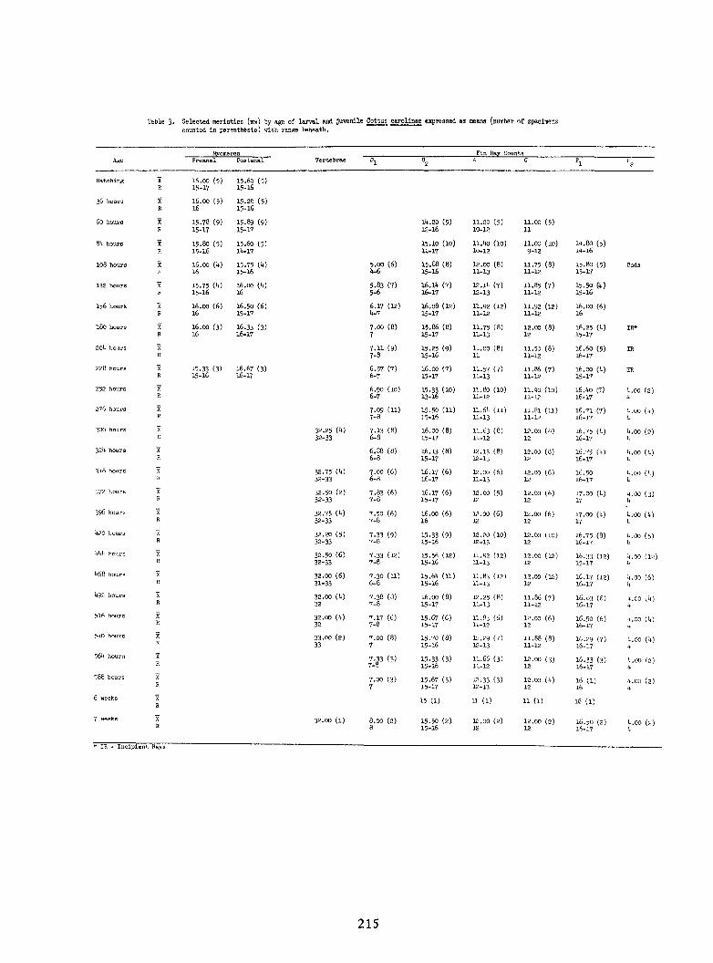
215
Table 3. Selected meristics (mm) by age of larval and Juvenile Cottus carolinae expressed as means (number of .specimenscounted inparenthesis) with range beneath.
IR-
Incipient Rays
U.OO (2)1*
16.50 (2)16-17
12.00 (2)12
12.00 (2)12
15.50 (2).15-16
8.00 (2)8
32.00 (!)\u25a0XR
7 weeks
16 (1)11 (1)11 (1)15 (1)XR
6 weeks
It.oo (2)It
16 (1)16
12.00 (It)12
12.33 (3)12-13
15.67 (3)15-17
7.00 (3)7
XR
588 hours
u.OO (2)It
16.33 (3)16-17
12.00.(3)12
11.66 (3)11-12
15.33 (3)15-16
7.33 (3)7-8
XR
56U hours
It.00 (It)It
16.29 (7)16-17
11.88 (8)11-12 \u25a0
12.29 (7)12-13
15.70 (8)15-16
. 7.00 (8)7
33.00 (2)33
XR
s^o hours
It.00 (U)It
16.50 (6)16-17
12.00 (6)12
11.83 (6)11-12
15.67 (6)15-17
7.17 (6)7-8
32.00 (1*)32
XR
516 hours
U.00 (U)It
16.83 (6)16-17
11.86 (7)11-12
12.25 (8)11-13
16.00 (8)15-17
7.38 (8)7-8
32.00 (it)32
XR
U92 hours
U.00 (6)It
16.17 (12)16-17
12.00 (12)12
11.83 (12)11-13
15.6U (11)15-16
7.30 (11)6-8
32.00 (6)31-33
XR
I*6B hours
It.00 (12)I*
16.33 (12)15-17
12.00 (12)12
11.92 (12)11-13
15.56 (12)15-16
7.33 (12)7-8
32.50 (6)32-33
XR
hkk hours
't.OO (5)ll
16.75 (8)16-17
12.00 (10)12
12.20 (10)12-13
15.33 (9)15-16
7.33 (9)7-8
32.20 (5)32-33
XR
U2O hours
It.oo (It)It
17.00 (It)17
12.00 (6)12
12.00 (6)12
16.00 (6)16
7.50 (6)7-8
32.75 CO32-33
XR
396 hours .
It.oo (3)It
17.00 (It)17
12.00 (6)12
12.00 (5)12
16.17 (6)15-17
7.83 (6)7-8
32.50 (2)32-33
XR
372 hours
It.oo (It)v .16.50
16-1712.00 (6)12
12.00 (6)11-13
16.17 (6)16-17
7.00 (6)6-8
32.75 (I*)32-33
XR
3*tB hours
It.00 (It)It
16.75 d)16-17
12.00 (8)12
12.13 (8)12-13
16.13 (8)15-17
6.88 (8)6-8
X.R
32't hours
't.OO (2)It
16.75 Ct)
16-1712.00 (8)12
11.63 (8)11-12
16.00 (8)15-17
7.13 (8)6-8
32.25 (I*)32-33
XR
300 hours
(U)It
16.71 (7)16-17
11.81 (11)11-12
11.6i. (11)11-13
15.50 (11)]5-l6
7.09 (11)7-8
XR
276 hours
U.OO (2)16.it0 (5)16-17
11.90 (10)11-12
11.80 (10)11-12
15.33 (10)13-16
6.90 (10)6-7
XR
252 hours
IR16.00 (it)15-17
11.86 (7)11-12
11.57 (7)11-13
16.00 (7)15-17
6.57 (7)6-7
16.67 (3)16-17
15.33 (3)15-16
XR
228 hours
IR16.60 (5)16-17
11.50 (8)11-12
11.00 (8)11
15.25 (9)15-16
7.11 (9)7-8
XR
20l* hours
IR*16.25 (U). 15-17
12.00 (8)12
11.75 (8)11-13
15.86 (8)15-17
7.00 (8)7
16.33 (3)16-17
16.00 (3)16
XR
180 hours
16.00 (6)16
11.92 (12)11-12
11.92 (12)11-12
16.08 (12)15-17
6.17 (12)U-7
16.50 (6)15-17
16.00 (6)16
XR
156 hours
15.50 (It)15-16
11.85 (7)11-12
12.1U (7)12-13
16.11* (7)16-17
5.83 (7)5-6
16.00 (it)16
15.75 (U)15-16
XR
132 hours
Buds15.80 (5)15-17
11.75' (8).11-12
12.00 (8)11-13
15.88 (8)15-16
5.00 (6)U-6
15.75 W15-16
16.00 (i*)16.
XR
108 hours
ih.BQ (5)lit-16
11.00 (10)9-12
11.1)0 (10)10-12
15.10 (10)11-17
15.60 (5)lit-17
15.80 (5)15-16
XR
8U hours
11.00 (5)11
11.20 (5)10-12
1U.20 (5)12-16
15.89 (9)15-17
15.78 (9)15-17
XR
60 hours
15.20 (5)15-16
16.00 (5)16
XR
36 hours
15.60 (5)15-16
16.00 (5)15-17
XR
Hatching
P2P2P1D2DlVertebraePI.tanalPreanalAgeCountsFin Ray•<!:;•<!:; Fin Ray Counts
Age Preanal PI.tanal Vertebrae Dl D2 P1 P2P2
Hatching XR
16.00 (5)15-17
15.60 (5)15-16
36 hours XR
16.00 (5)16
15.20 (5)15-16
60 hours XR
15.78 (9)15-17
15.89 (9)15-17
1U.20 (5)12-16
11.20 (5)10-12
11.00 (5)11
8U hours XR
15.80 (5)15-16
15.60 (5)lit-17
15.10 (10)11-17
11.1)0 (10)10-12
11.00 (10)9-12
ih.BQ (5)lit-16
108 hours XR
16.00 (i*)16.
15.75 W15-16
5.00 (6)U-6
15.88 (8)15-16
12.00 (8)11-13
11.75' (8).11-12
15.80 (5)15-17
Buds
132 hours XR
15.75 (U)15-16
16.00 (it)16
5.83 (7)5-6
16.11* (7)16-17
12.1U (7)12-13
11.85 (7)11-12
15.50 (It)15-16
156 hours XR
16.00 (6)16
16.50 (6)15-17
6.17 (12)U-7
16.08 (12)15-17
11.92 (12)11-12
11.92 (12)11-12
16.00 (6)16
180 hours XR
16.00 (3)16
16.33 (3)16-17
7.00 (8)7
15.86 (8)15-17
11.75 (8)11-13
12.00 (8)12
16.25 (U). 15-17
IR*
20l* hours XR
7.11 (9)7-8
15.25 (9)15-16
11.00 (8)11
11.50 (8)11-12
16.60 (5)16-17
IR
228 hours XR
15.33 (3)15-16
16.67 (3)16-17
6.57 (7)6-7
16.00 (7)15-17
11.57 (7)11-13
11.86 (7)11-12
16.00 (it)15-17
IR
252 hours XR 6.90 (10)
6-715.33 (10)13-16
11.80 (10)11-12
11.90 (10)11-12
16.it0 (5)16-17
U.OO (2)
276 hours XR
7.09 (11)7-8
15.50 (11)]5-l6
11.6i. (11)11-13
11.81 (11)11-12
16.71 (7)16-17
(U)It
300 hours XR
32.25 (I*)32-33
7.13 (8)6-8
16.00 (8)15-17
11.63 (8)11-12
12.00 (8)12
16.75 Ct)
16-17't.OO (2)It
32't hours X.R
6.88 (8)6-8
16.13 (8)15-17
12.13 (8)12-13
12.00 (8)12
16.75 d)16-17
It.00 (It)It
3*tB hours XR
32.75 (I*)32-33
7.00 (6)6-8
16.17 (6)16-17
12.00 (6)11-13
12.00 (6)12
16.5016-17
It.oo (It)v .
372 hours XR
32.50 (2)32-33
7.83 (6)7-8
16.17 (6)15-17
12.00 (5)12
12.00 (6)12
17.00 (It)17
It.oo (3)It
396 hours . XR
32.75 CO32-33
7.50 (6)7-8
16.00 (6)16
12.00 (6)12
12.00 (6)12
17.00 (It)17
It.oo (It)It
U2O hours XR
32.20 (5)32-33
7.33 (9)7-8
15.33 (9)15-16
12.20 (10)12-13
12.00 (10)12
16.75 (8)16-17
't.OO (5)ll
hkk hours XR
32.50 (6)32-33
7.33 (12)7-8
15.56 (12)15-16
11.92 (12)11-13
12.00 (12)12
16.33 (12)15-17
It.00 (12)I*
I*6B hours XR
32.00 (6)31-33
7.30 (11)6-8
15.6U (11)15-16
11.83 (12)11-13
12.00 (12)12
16.17 (12)16-17
U.00 (6)It
U92 hours XR
32.00 (it)32
7.38 (8)7-8
16.00 (8)15-17
12.25 (8)11-13
11.86 (7)11-12
16.83 (6)16-17
U.00 (U)It
516 hours XR
32.00 (1*)32
7.17 (6)7-8
15.67 (6)15-17
11.83 (6)11-12
12.00 (6)12
16.50 (6)16-17
It.00 (U)It
s^o hours XR
33.00 (2)33
. 7.00 (8)7
15.70 (8)15-16
12.29 (7)12-13
11.88 (8)11-12 \u25a0
16.29 (7)16-17
It.00 (It)It
56U hours XR
7.33 (3)7-8
15.33 (3)15-16
11.66 (3)11-12
12.00.(3)12
16.33 (3)16-17
u.OO (2)It
588 hours XR
7.00 (3)7
15.67 (3)15-17
12.33 (3)12-13
12.00 (It)12
16 (1)16
It.oo (2)It
6 weeks XR
15 (1) 11 (1) 11 (1) 16 (1)
7 weeks XR
32.00 (!)\u25a0 8.00 (2)8 15.50 (2).
15-1612.00 (2)12
12.00 (2)12
16.50 (2)16-17
U.OO (2)1*
IR-
Incipient Rays
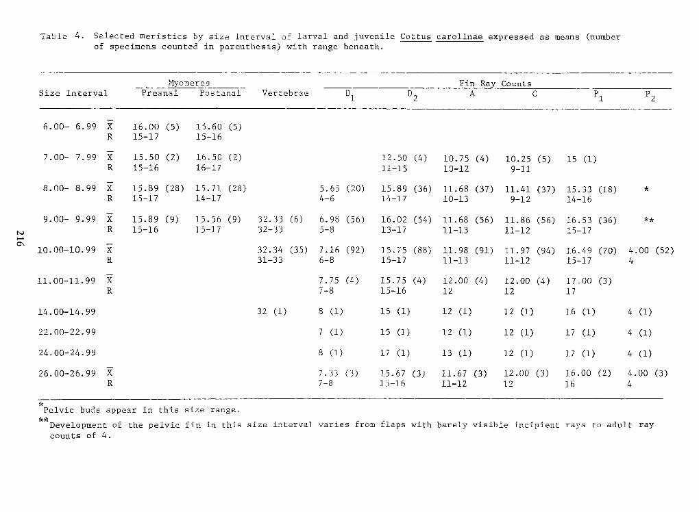
216
Table 4.
Selected meristics by size interval oflarval and juvenile Cottus carolinae expressed as means (number
of specimens counted in parenthesis) with range beneath.
counts of 4.
rayadulttoraysincipientbarely visibleflaps withfromvariesintervalsizein thisfinDevelopment of the pelvic
range.size
*Pelvic buds appear in this
4161211-1215-167-8R
(3)4.00(2)
16.00(3)
12.00(3)
11.67(3)
15.67(3)7.33X26.00-26.99 4 (1)17 (1)12 (1)13 (1)17 (1)8 (1)
24.00-24.99 4 (1)17 (1)12 (1)12 (1)15 (1)7 (1)22.00-22.99 4 (!)16 (1)12 (1)12 (1)15 CD8.(1)32 (1)
14.00-14.99 17121215-167-8R
(3)
17.00(4)
12.00(4)
12.00(4)
15.75(4)7.75X
11.00-11.99 415-1711-1211-1315-176-831-33R
(52)4.00(70)16.49(94)11.97(91)11.98(88)15.75(92)7.16(35)32.34X
10.00-10.99 15-1711-1211-1313-175-832-3315-1715-16R
**(36)16.53(56)11.86(56)11.68(54)16.02(56)6.98(6)32.33<9)
15.56(9)
15.89X9.00- 9.99
14-169-1210-1314-174-614-1715-17R
*(18)15.33(37)11.41(37)11.68(36)15.89(20)5.65(28)15.71(28)15.89X8.00- 8.99
9-1110-1211-1516-1715-16R
15 (1)(5)
10.25(4)
10.75(4)
12.50(2)
16.50(2)
15.50X7.00- 7.99
15-1615-17R
(5)
15.60(5)
16.00X6.00- 6.99
p2 p
2
P1-i
:_: _VertebraePostanalPreanalIntervalSize CountsRaFinMyomeresMyomeres Fin Ra CountsSize Interval Preanal Postanal Vertebrae :_: _ -i
P1
p2 p
2
6.00- 6.99 X 16.00 (5)
15.60 (5)R 15-17 15-167.00- 7.99 X 15.50 (2)
16.50 (2)
12.50 (4)
10.75 (4)
10.25 (5) 15 (1)R 15-16 16-17 11-15 10-12 9-118.00- 8.99 X 15.89 (28) 15.71 (28) 5.65 (20) 15.89 (36) 11.68 (37) 11.41 (37) 15.33 (18) *
R 15-17 14-17 4-6 14-17 10-13 9-12 14-169.00- 9.99 X 15.89 (9)
15.56 <9) 32.33 (6) 6.98 (56) 16.02 (54) 11.68 (56) 11.86 (56) 16.53 (36) **
R 15-16 15-17 32-33 5-8 13-17 11-13 11-12 15-1710.00-10.99 X 32.34 (35) 7.16 (92) 15.75 (88) 11.98 (91) 11.97 (94) 16.49 (70) 4.00 (52)R 31-33 6-8 15-17 11-13 11-12 15-17 411.00-11.99 X 7.75 (4)
15.75 (4)
12.00 (4)
12.00 (4)
17.00 (3)
R 7-8 15-16 12 12 1714.00-14.99 32 (1) 8.(1) 15 CD 12 (1) 12 (1) 16 (1) 4 (!)22.00-22.99 7 (1) 15 (1) 12 (1) 12 (1) 17 (1) 4 (1)24.00-24.99 8 (1) 17 (1) 13 (1) 12 (1) 17 (1) 4 (1)26.00-26.99 X 7.33 (3)
15.67 (3)
11.67 (3)
12.00 (3)
16.00 (2) 4.00 (3)R 7-8 15-16 11-12 12 16 4*Pelvic buds appear in this size range.Development of the pelvic fin in this size interval varies from flaps with barely visible incipient rays to adult raycounts of 4.
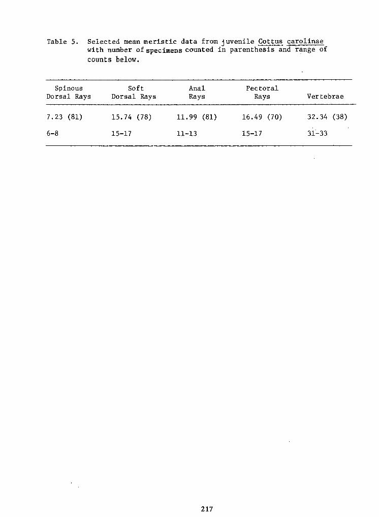
217
Table 5. Selected mean meristic data from juvenile Cottus carolinaewith number of specimens counted in parenthesis and range of
counts below.
31-3315-1711-1315-176-8
(38)32.34(70)16.49(81)11.99(78)15.74(81)7.23
VertebraeRaysRaysDorsal RaysDorsal RaysPectoralAnalSoftSpinousSpinous Soft Anal Pectoral
Dorsal Rays Dorsal Rays Rays Rays Vertebrae
7.23 (81) 15.74 (78) 11.99 (81) 16.49 (70) 32.34 (38)
6-8 15-17 11-13 15-17 31-33
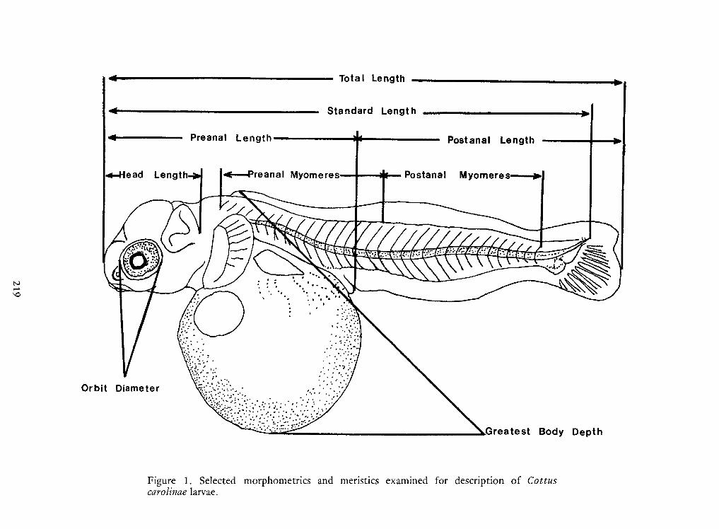
219
Figure 1.
Selected morphometrics and meristics examined for description of Cottuscarolinae larvae.
DepthBody.Greatest
DiameterOrbit
->
H,-. >v-.•—ATTi"- Wi'7TT;-»y?- JpL~-•/•*i
l^-
Length Myomeres \u25a0
>PostanalMyomeres< Preanal
'os tana! LengthI
Length —I
Length — 'os tana! Length
< Preanal Myomeres Postanal Myomeres \u25a0
>Length
l^-
•/•*i L~- Jp?- »y ;- '7TT Wii"- TT .•—A >v-H,-.
->
Orbit Diameter
.Greatest Body Depth
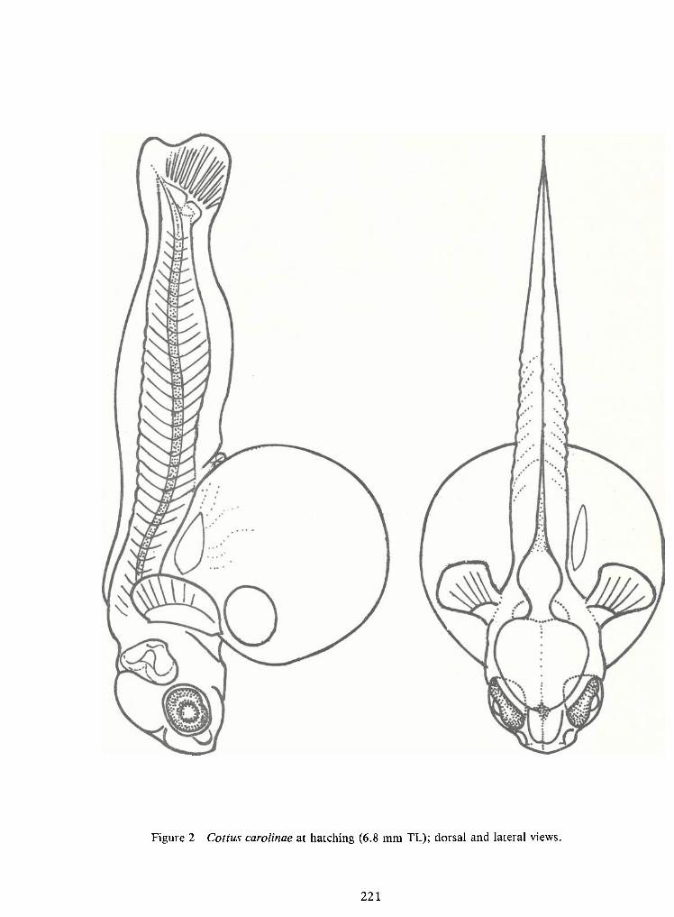
221
Figure 2 Cottus carolinae at hatching (6.8 mm TL); dorsal and lateral views.
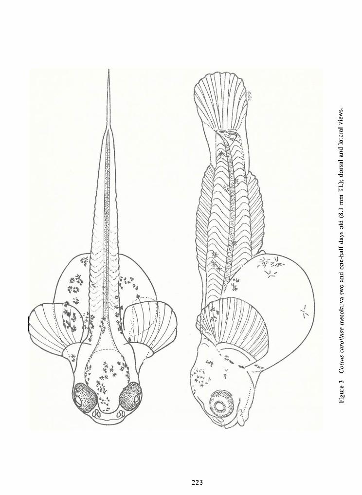
223
Figure3
Cottuscarolinae
mesolarvatwoand
one-halfdaysold
(8.1mmTL);dorsal
andlateral
views.
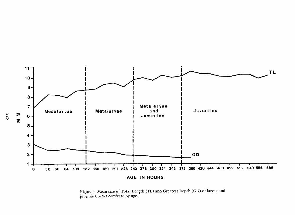
225
Figure 4 Mean size of Total Length (TL) and Greatest Depth (GD) oflarvae andjuvenile Cottus carolinae by age.
HOURSIN !AGE
588540 564516492468420 444396372348300 324276228 252204180156132108846036
12
34
5
2
JuvenilesGD
Metalarvae andJuvenilesMerta larvaeI
Mesolarvae6
78
9
2
TL
i
I
I
101111
10 I
I
i TL
2
98
76
MesolarvaeI
Merta larvae Metalarvae andJuveniles JuvenilesGD
2
54
32
1
36 60 84 108 132 156 180 204 228 252 276 300 324 348 372 396 420 444 468 492 516 540 564 588
AGE IN !HOURS
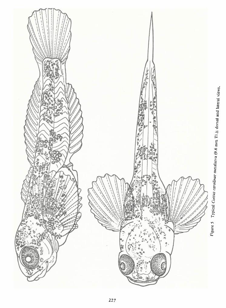
227
*™5Typicala*.
meta,arva
*.«mmTL);dorsa,and
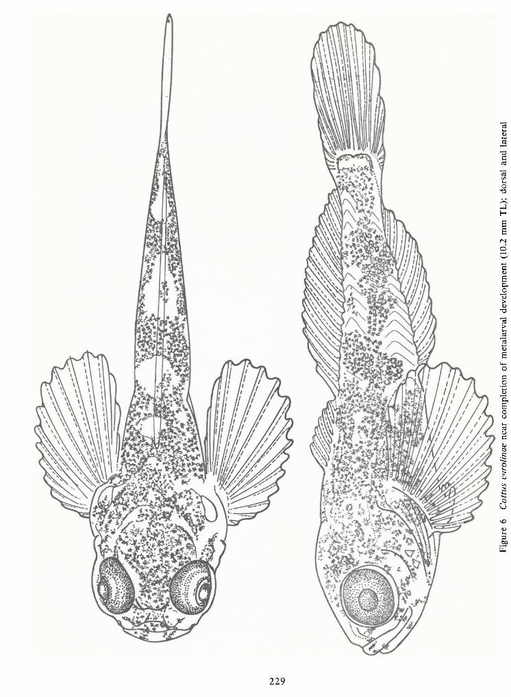
229
Figure6
Cottuscarolinaenearcompletionof
metalarvaldevelopment(10.2mmTL);dorsal
andlateral
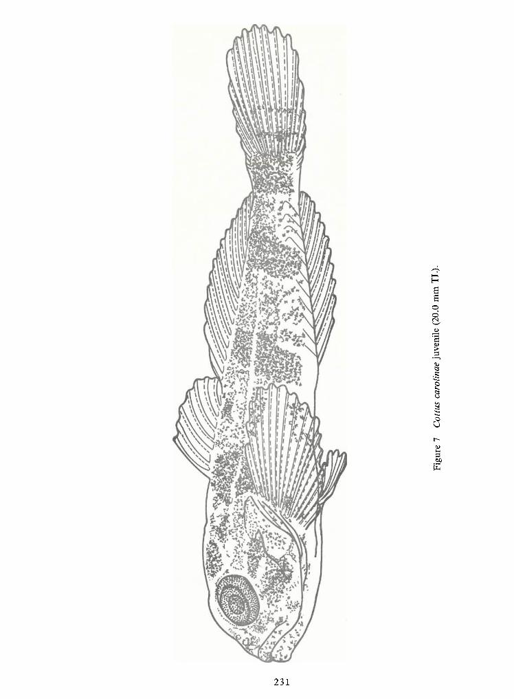
231
Figure7
Cottuscarolinaejuvenile
(20.0mmTL).
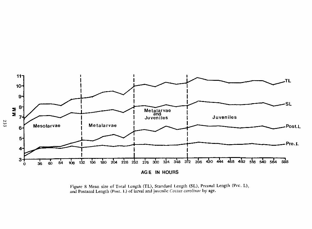
233
Figure 8 Mean size of Total Length (TL), Standard Length (SL), Preanal Length (Pre. L),and Postanal Length (Post. L) of larval and juvenile Cottus carolinae by age.
IN HOURSAGE
5492468444 <
>0"3E"IS
fe-ll300 324 348276252228I 2041801561321088460360
3-
I4-
•Pre.J.Ii
5^6-
MetalarvaeMesolarvae \u25a0PostL
JuvenilesII
Metalarvae andJuvenilesIII
I7
m
i
;
8m
•SL
5I
II
9-
TL10-
IIi
11"11"
Ii
I
10-
TL
9-
II
5I
•SLm;
8 i
m7 I
III
Metalarvae andJuveniles II
Juveniles \u25a0PostLMesolarvae Metalarvae6-5^ Ii •Pre.J.4- I
3-0 36 60 84 108 132 156 180 I 204 228 252 276 300 324 348 ll
fe- IS E" "3 >0 444 <468 492 5
AGE IN HOURS
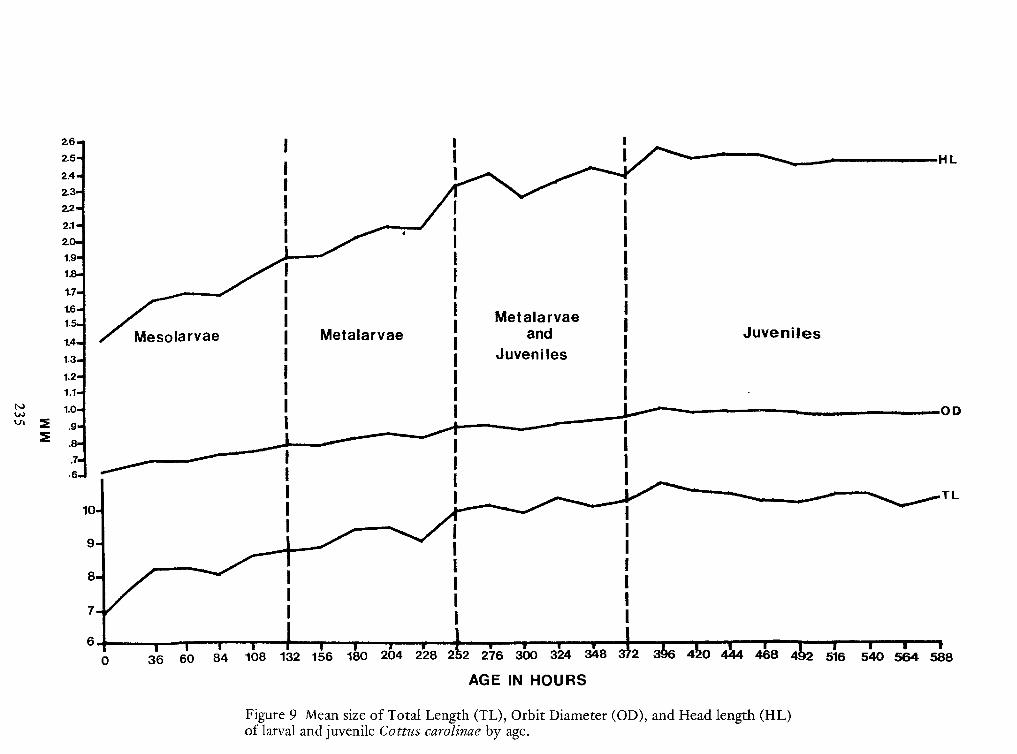
235
Figure 9 Mean size of Total Length (TL), Orbit Diameter (OD), and Head length (HL)oflarval and juvenile Cottus carolinae by age.
AGE IN HOURS 588564540516(2
468444to96 42372 3«52 276 300 324 348 \2*22820418015613210884603606.
II
II
II7-
II
I
III
IIIi
8-
\u25a0
iII9-
\II
i
III
10-
I
IIII
.6-.7-
I
II
.8-
5
II
I
.9-2
1.0-
II1.1-
II1.2-
!
JuvenilesI1.3-
IanaI
rvaestaiarMeac>larv<Meso14-
II1.5-
II1.6-
II1.7-
Ii1-8-
II1.9-
II2.0-
II2.1-
I22-
JuvenilesI
I
Metalarvae andJuvenilesII
/j
Metalarvae1
}Jr
Mesolarvae2.3-
2.4-
iIIIII
2.5-2.6-2.6-2.5-
I IIII
i
2.4-2.3-
MesolarvaeJr
1}
Metalarvae/jII
Metalarvae andJuvenilesI
I
Juveniles
22- I
2.1- I I
2.0- I I
1.9- I I
1-8- i I
1.7- I I
1.6- I I
1.5- I I
14- Meso >larv< ac Me staiar rvae I
ana I
1.3- I
Juveniles !
1.2- I I
1.1- I I
1.0-2 .9-
I
II
5
.8-
II
I
.7-.6- II II
I
10-
III
i\II9-
IIi
\u25a0
8-
iIII
III
III
7-
II
II
II
6.0 36 60 84 108 132 156 180 204 228 2* 52 276 300 324 348 \
372 3« 96 42 to 444 468 (2
516 540 564 588AGE IN HOURS

APPENDIX A
239
ATTENDANCE LIST
James M. BakerDivision of FF&WDTVANorris, TN 37828(615)494-7173
John A.BakerUniversity of So. Miss.Southern Station 1750Hattiesburg, MS 39401(601) 582-5310
B.H. BauerUniversity of TennesseeKnoxville, TN 37916
Mike BentonEnwright Assoc.P. O. Drawer 1180Summerville, SC 29843
Skip Bergmann111. Nat. Hist. SurveyWilmington, IL 60481(815) 476-7236
Charles BiggarLa. Dept. Wildlife and Fish.P. O. Box 4004Monroe, LA 71203(318) 343-4045
BillBolinVEPCOGrayland AvenueRichmond, VA 23219(804) 771-4200
Johnny BuchananDivision ofFF&WDTVANorris, TN 37828(615) 494-7173
Frank J. BulowDept. of BiologyTTUCookeville, TN 38501(615) 528-3147
Gary L.BurtonDivision of FF&WDTVANorris, TN 37828(615) 494-7173
Glenn CadaOak Ridge Nat. Lab.Oak Ridge, TN 37830(615) 494-7173
Aaron L.ClarkWater Resources LabUniversity of LouisvilleLouisville,KY 40208(501) 588-5932
Don CloutmanDuke Power Co.Route 3, P.O. Box 90Charlotte, NC 28078(704) 875-1381
John Van ConnerForest and Wildlife Mgt.LSU237 Ag. CenterBaton Rouge, LA 70803(504) 642-8129
A.Carter CookeVEPCOGrayland AvenueRichmond, VA 23219(804) 771-4200
Kevin D. CurryPurdue Univ.W. Lafayette, IN 47901(317)494-8616
John DorrGr. Lk. Res. Div. UM3114 Inst. of Sci. and Tech,
Ann Arbor, MI 48109(313) 763-5467
240
Melvin DouglassDivision of FF&WDTVANorris, TN 37828(615) 494-7173
Christopher C. EllisAppliedBiology5891 New Peachtree St.Atlanta, GA 30340(404) 292-8031
D. A.EtnierUniv. of TennesseeKnoxville,TN 37916
Richard A. FrietscheDupont Co.Savannah River Lab.Aiken, SC 29801FTS 239-3651
Ronald A.FritzscheDept. of Biol., UMUniversity, MS 38677(601) 232-7203
Lee A.FuimanDept. ofNat. Res.Fernon HallCornell Univ.Ithaca, NY 14853(607)256-5469
Lee GraserDivision of FF&WDTVANorris, TN 37828(615) 494-7173
Gordon HallDivision of FF&WDTVANorris, TN 37828(615) 494-7173
LarryHiltonDivision of FF&WDTVANorris, TN 37828(615) 494-7173
Jeff HogueDivision of FF&WDTVANorris, TN 37828(615) 494-7173
Robert D.HoytWestern Kentucky Univ.Bowling Green, KY 42101(502) 745-5481
David JudeGr. Lk. Res. Div.UM3114 Inst. of Sci. and Tech,
Ann Arbor, MI 48109(313) 764-2424
Larry KayDivision of FF&WDOS WHA, TVAMuscle Shoals, AL 35660(205) 383-4631
Ron J. KernehanIchthyological Assn., Inc.Rt. 1, P.O. Box 120Middletown, DE 19709(302) 368-0778
Vincent R. KranzNUS Corp.Box 73Bayport, MN 55003(612) 439-6708
Star LakavageDivision of FF&WDTVANorris, TN 37828(615) 494-7173
Barbara F. LathropIchthyological Assn.P. O. Box 223Etters, PA 17319(717) 938-3585
Jules J. LoosANSPNineteenth and Parkway St,Philadelphia, PA 19103(215)299-1072
241
John E. McCalebRMCPottstown, PA 19464(215) 326-9662
Larry W. McDonaldDivision of FF&WDOS WHA, TVAMuscle Shoals, AL 35660(205) 383-4631
Gerald McGowanHarris E&E CenterApex, NC 27502(919) 362-8663
Gary OvermannWestern Kentucky Univ.Bowling Green, KY 42101(502) 745-5481
WilliamD. PearsonWater Res. LabUniv. of LouisvilleLouisville, KY 40208(502) 588-6731
Lance G. Perrylowa State Univ.Ames, lowa 50011(515) 294-6148
Eric D. PrinceU.S. Fish and Wildlife
ServiceSERIClemson, SC 29631
David RupleUniv. of So. Miss.So. Station 1750Hattiesburg, MS 39401(601)266-4110
Harold SchrammUSAE/WESVicksburg, MS 39180(601)636-3111
Ed ScottDivision of FF&WDTVANorris, TN 37828(615) 494-7173
Pat SmithDivision of FF&WDOS WHA, TVAMuscle Shoals, AL 35660(205) 383-4631
Ted Storck111. Nat. His. SurveySullivan, IL 61951(217) 782-4498
Linda TorbettDivision of FF&WDTVANorris, TN 37828(615) 494-7173
Mike Van Den AvyleTN Coop Fishery UnitTTUCookeville, TN 38501(615) 528-4130
Robert WallusDivision of FF&WDTVANorris, TN 37828(615) 494-7173 494-9800
Roger WhitakerDivision of FF&WDTVANorris, TN 37828(615) 494-7173
John W. WiltzGA Power Co.791 Dekalb Ind. WayAtlantic, GA 30307(404) 522-6060 Ext. 3448


