Embed Size (px)
Citation preview

Pergamon Geochimica et Cosmochimica Acta, Vol. 59. No. 19. 3997-4007. 1995 pp.
Copyright 0 1995 Elsevier Saence Ltd
F’rinted in the USA. All rights reserved
0016-7037/95 $9.50 + 00
0016.7037( 95)00294-4
Laser ablation ICP-MS elemental analysis of individual fluid inclusions: An evaluation study*
T. J. SHEPHERD and S. R. CHENERY
British Geological Survey, Kingsley Dunham Centre, Keyworth, Nottingham NGl2 SGG, United Kingdom
(Received November 15, 1995; accepted in revised form June 2 1. 1995 )
Abstract-Details are given of the elemental analysis of single fluid inclusions using a UV laser ablation microprobe interfaced to an inductively coupled plasma mass spectrometer. The UV laser, a frequency quadrupled Nd:YAG operating at 266 nm, allows higher spatial resolution ( <2 pm) than can be achieved using near-IR or visible wavelengths. Tests have been carried out on lo- 100 pm diameter aqueous (liquid + vapour) inclusions in fluorite, quartz, and halite up to 60 pm beneath the surface. A key feature of the system is a novel high temperature ablation cell which substantially improves the efficiency and reproduc- ibility of fluid release. Calibration was carried out using a dual gas flow system that allowed use of standard solutions and NIST glasses for tuning the instrument and for obtaining relative sensitivity factors. As an alternative to synthetic fluid inclusions, a new calibration approach is described involving the encapsulation of microdroplets of standard solutions in hydrophobic epoxy resinsfluid inclusion analogues. To illustrate the scope and performance of the instrument, data are reported for Ba, Ca, Cs, Cu, K, Mg, Mn, Na, Pb, Rb, Sr, and Zn in saline aqueous inclusions associated with evaporite and low temperature base metal deposits. Element detection limits vary according to the mass of material released for analysis and are thus related to the volume and composition of each inclusion. Precision is estimated to be better than 30%.
1. INTRODUCTION
This paper reports progress in the development of a UV laser ablation microprobe-inductively coupled plasma-mass spec- trometer (LAMP-ICP-MS) for the multi-element analysis of single fluid inclusions. It details the performance and appli- cation of a W laser microprobe for the ablation of inclusions in different minerals, new ICP-MS calibration procedures us- ing a dual gas flow system, and the use of a novel thermal ablation cell for improving the efficiency and reproducibility of ablation.
The importance of fluid inclusions as a geological record of the fluid inventory of the earth’s crust is now widely ac- cepted. Less accepted, however, is their reliability in provid- ing unequivocal information on the chemistry of the fluids. Such uncertainties are fully justified since the quantitative analysis of inclusion fluids presents major technical difficul- ties. As well as being extremely small, typically <30 pm, it is rare to find material that contains only one generation of inclusion. While multiple populations of inclusion offer an exciting insight into changes in fluid activity, their existence complicates bulk sample methods of analysis and adds to the uncertainty of data interpretation. To avoid such problems, considerable effort has been invested in the development of single fluid inclusion techniques. Laser Raman spectroscopy for example is an acknowledged method for analysing poly- atomic volatiles and has been extensively applied to nonaque- ous fluid inclusions (Dubessy et al., 1989). Routine interla- boratory calibration and between-sample calibration proce- dures have still to be demonstrated (Pasteris et al., 1988) but
* Presented at the fifth biennial Pan-American Conference on Re- search on Fluid Inclusions (PACROFI V) held May 19-21, 1994, at the lnstituto de lnvestigaciones Electricas in Cuemavaca, Morelos, Mexico.
in combination with microthermometry more information is obtained than by either technique alone.
Nonvolatile species, namely the dissociated electrolytes, have proved more difficult to analyse. The lack of suitable techniques for the quantitative elemental analysis of single inclusions constitutes a serious obstacle to the broader appli- cation of fluid inclusions and presents a formidable analytical challenge. Most instrumental techniques with high spatial res- olution ( <30 pm) and high sensitivity (ppm detection) are designed for surface analysis. Electron probe microanalysis (EPMA) (Ayora et al., 1993), proton-induced x-ray analysis (PIXE) (Ryan et al., 1991), and synchrotron x-ray analysis (Rankin et al., 1992) have the requisite specifications (multi- element capability and a spot size of less than 5 pm) but are subject to severe fluorescence absorption corrections when applied to microsamples that are located well below the sur- face. Depending upon the size and type of inclusion, and the chemistry of the host mineral, encouraging results have been obtained for each of the above techniques. Procedures for overcoming surface limitations have also been tried, including the thermal rupture of inclusions close to the surface and anal- ysis of the precipitated salts by SEM/EDA (Haynes et al., 1988). Difficulties were encountered though in producing mi- cro-scale homogeneity for mixed salt calibration standards and the technique is unsuitable for multiple populations of fracture-controlled inclusions. Another technique affording ppm detection capability is secondary ion mass spectrometry (SIMS ) This can perform both elemental and isotopic anal- yses on extremely small samples but is likewise poorly suited to liquid samples (Diamond et al., 1990). Earlier SIMS stud- ies by Nabu and Sato ( 1981) using frozen liquid inclusions achieved more reproducible results but encountered other problems related to the variable ionization of ice-salt hydrate mixtures. A further constraint is the very slow rate of ion thinning; up to 1 hour may be required to ablate inclusions
3997

3998 T. J. Shepherd and S. R. Chenery
FIG. 1. Schematic layout of the dual gas flow sample introduction system and laser microprobe used for LAMP-ICP-MS analysis.
located lo-20 pm below the surface of the sample. Consid- erably more development work is needed before the full chemical and isotope potential of SIMS can be exploited.
A promising technique for single inclusion elemental anal- ysis, without recourse to synchrotron or reactor facilities, is laser ablation-ICP spectroscopy (Horn and Tye, 1989; Che- nery and Rankin, 1989; Boiron et al., 1991; Ramsey et al., 1992; Wilkinson et al., 1994). This technique has the advan- tage in allowing the fluid to be sampled and isolated from the host matrix prior to analysis. Using a Q-switched ruby laser (694 nm) linked to an ICP-atomic emission spectrometer (ICP-AES), Wilkinson and coworkers (1994) were able to ablate large (30-60 pm), moderate to high salinity ( >20 wt% NaCl equivalent) aqueous inclusions in quartz to obtain information on the chemistry of magmatic/hydrothermal flu- ids. While representing a bench-mark in inclusion analysis, it highlighted certain laser ablation limitations experienced by earlier workers (Horn and Tye, 1989):
2)
3)
1) Many minerals, especially quartz, are transparent to visi- ble or near-IR light. This results in inefficient energy cou- pling and the formation of large ablation craters ( + 30 pm) caused by thermomechanical shock. Since quartz is one of the principal minerals used for fluid inclusion studies, con- ventional near-IR Nd:YAG ( 1064 nm) and visible ruby lasers (694 nm) do not provide the most appropriate ab- lation microprobes. Due to the large size of the ablation craters, the material released for analysis from inclusions less than 30 pm in diameter includes a significant proportion of the host min- eral. Matrix contamination is potentially very high for host minerals such as fluorite and calcite which are known to contain elevated concentrations of trace elements. ICP-AES working detection limits are too high to deter- mine the minor and trace element constituents of most in- clusions less than 50 pm in diameter (i.e., species with a fluid concentration < 1000 ppm).
By interfacing a UV laser microprobe (LAMP) to an ICP- mass spectrometer (ICP-MS), the present research sought to overcome some these problems. In doing so, the work sought to improve the efficiency of ablation and to assess the best methods for ICP-MS calibration and measurement.
TABLE 1. Operating conditions of the UV laser ablation microprobe.
Laser Specuon SL803 Nd:YAG Wavelength 266 nm (frequency quadrupled from 1064 nm) Maximum energy 70 ml Mode Q-switched, TEM, Pulse length 10 ns Laser repetition rate 10 Hz
Microscope Leiu Aristomet Laser focusing objective x25 and x36
2. INSTRUMENTATION
The instrumentation and method of analysis is broadly based on that used for the conventional laser ablation ICP- MS elemental analysis of solids ( Moenke-Blankenburg, 1989; Imai, 1990; Perkins et al., 1991; Williams and Jarvis, 1993). For the analysis of single fluid inclusions, three new features have been incorporated: a UV laser microprobe, a thermal ablation cell, and a dual gas flow system for calibra- tion and measurement. A schematic layout of the instrumen- tation is shown in Fig. 1.
2.1. UV Laser Microprobe
The laser ablation microprobe developed for single inclu- sion analysis (UK Patent) comprises a Spectron laser oper- ating in the far-UV (266 nm) linked to a Leitz Aristomet microscope. The focussing objectives are 266 nm overcoated, UV optimized ~36, and ~25 reflecting lenses. Due to the improved absorption of UV over IR light by silicates, car- bonates, halides, sulphates, sulphides, and oxides, this com- bination enables ablation of a wide range of minerals. The spot size is -2 pm giving a very much improved spatial res- olution compared to equivalent Nd:YAG and ruby lasers. The laser was used in Q-switched mode with a repetition rate of 10 Hz. Further specifications and operating conditions are given in Table 1.
2.2. Thermal Ablation Cell
The thermal ablation cell is similar in design to that de- scribed by Thompson et al. ( 1989) but incorporates an inner
TABLE 2. Operating conditions of the ICP-mass spectrometer.
Specrmmcter VG Plaslr!aQuad 2+ Forward power IlooW Gas now t-dtes
Coolant 13 l.min”
Auxiliary 0.8 l.min-’
Injector
Thermal ablation cell 0.6 l.min-’
Nebulizer 0.4 l.min”
Data acquisition softwan VGTRA Data acquisition mode Peak jumping Points per peak 3 Dwell time per peak 5 ms sweeps per time unit 8 Tii unit 1-2 s

Multi-element analysis of single fluid inclusions 3999
tb)
FIG. 2. Photomicrograph showing the high spatial resolution of the laser beam and the selective manner in which the inclusion contents are extracted for analysis. (a) before drilling; (b-c) after drilling. Note lack of damage to adjacent inclusions. Figure (a) shows the laser entry hole at the surface of the sample. Scale bar = 50 pm. Two phase (liquid + vapour) inclusions in quartz, Carrock Fell Mine, Cumbria, U.K. Ablation at 140°C.
cell comprising a programmable, temperature-controlled metal block. This allows ablation at elevated temperatures. The sample rests on the upper surface of the metal block and is heated by conduction from below. A small diameter axial window within the block allows viewing in transmitted and reflected light. The outer walls of the cell are fabricated from perspex which arbitrarily limits the present version to a max- imum of 350°C. [ N.B. With very minor modification the up- per working limit of the cell could be increased to 6OO”C.] Ablation of the sample is facilitated via an upper window of
optical quality silica built into the perspex top of the thermal cell; the sample to lens distance being within the focal dis- tance of the objective lens. Illumination is provided by a long working distance sub-stage condenser unit that focuses light at the level of the mineral sample. During ablation, the thermal cell is securely fixed to the Leitz microscope- stage and the ablated material transported to the ICP-MS instrument by a continuous flow of argon through the cell (Fig. 1).
2.3. ICP-Mass Spectrometer
For analysis of the ablated material, a VG Plasmaquad 2+ ICP-mass spectrometer was used in peak jumping acquisition mode where only selected masses are analysed (Table 2). Data were acquired using time-resolved software to permit identification and quantification of peak and background re- sponses at each selected mass. In this way the signal due to the fluid pulse can be clearly distinguished from the back- ground, matrix reference signal (see Fig. 4). The choice of elements (Ba, Ca, Cs, Cu, K, Mg, Mn, Na, Pb, Rb, Sr, and Zn) was based on the general chemical composition of oil- field waters and related formational brines (White, 1965; Land and Macpherson, 1992). but simplified to exclude ele- ments with known ICP-MS polyatomic interferences (e.g., sulphur). Copper, lead, and zinc were included in the element menu because of interest by economic geologists in the metal content of saline waters and their role in the genesis of sedi- ment-hosted base metal deposits (Sverjensky, 1984).
3. METHODOLOGY
3.1. Laser Ablation Procedure
Ablation is initiated by focussing onto the surface of the sample to create a shallow pilot hole (2-5 pm) using the minimum power necessary for laser-mineral interaction. This
FIG. 3. Back scatter SEM photomicrograph showing the entry point of the laser beam into a brine inclusion and its liquid contents pre- cipitated on the surface as a corona of chloride salts due to capillary action. Ablation at 22°C. On entering the inclusion, the ablation was stopped to avoid totally volatilising the aqueous fluid. Note the vari- ation in luminosity of the salt crystals indicating variation in atomic weight. Two phase (liquid + vapour) brine inclusion in quartz, Car- rock Fell Mine, Cumbria, U.K.

4000 T. J. Shepherd and S. R. Chenery
Pb
Zn
FIG. 4. Time resolved ICP-MS spectrum for Na, K, Zn, and Pb for a discrete pulse of fluid released from a 30 pm diameter aqueous inclusion in quartz, Carrock Fell Mine, Cumbria, U.K. Ablation at 140°C.
power depends upon the specific mineral species; quartz > carbonate > fluorite > halite. As the rate of ablation grad- ually decreases due to beam defocussing, the power is slowly increased whilst continually adjusting the focus to achieve a steady rate of drilling. The main problem encountered is ther- mal fracturing of the host mineral. Excessive thermal fractur- ing of the matrix ahead of the beam can cause premature re- lease and loss of the inclusion fluid by leakage along micro- cracks. As drilling proceeds, the diameter of the hole at the surface increases in accordance with the numerical aperture of the lens, giving the hole an inverted, tightly conical ge- ometry. However, the base of the hole maintains its overall minimum diameter and progress can be monitored by occa- sionally focussing at the point of ablation. In this manner the
’ . l
2. l .* .
. . . . . .
. .
.
FIG. 5. Photomicrograph showing distribution of fluid inclusion analogues in epoxy resin. Scale bar = 200 pm.
beam can be tracked to the point of entry into the inclusion. For an inclusion in quartz at a depth of 60 pm, ablation may take 3-4 mins. Figure 2 demonstrates the high spatial reso- lution capability of the UV laser microprobe which can be positioned to within + l-2 pm of an inclusion.
The rate and manner of liquid release are determined by the volume and internal vapour pressure of the inclusion. Ex- periments at room temperature using large monophase, liquid inclusions in halite (4 X 10’ pm’) within 20 pm of the surface showed that by controlling the laser energy, continuous sam- pling (vaporization) of the liquid phase could be maintained over a period of lo-45 sets. Extended liquid release is illus- trated in Fig. 7. By contrast, C02-rich inclusions in quartz behave totally differently. On breakthrough, the high internal gas causes explosive release of the liquid phase giving a sharp and broadly symmetric ICP-MS response. A similar response (Fig. 4) is observed for the ablation of aqueous inclusions at elevated temperatures (see below).
For many two phase (liquid + vapour) inclusions however, fluid release at room temperature is extended over l-2 sec- onds and the resultant time-resolved ICP-MS traces are itreg- ular and strongly skewed. Moreover, ablation is often accom- panied by the precipitation of salts at the surface (Fig. 3) due to capillary attraction of the liquid. For small inclusions ( <20
TABLE 3. Typical detection limits and selected a.m.“. for Cu and Zn in 20- 106,m synthetic fluid inclusions in halite.
Detection Limits ppm
Isotope selected for analysis (a.m.u.)
63cu+ 65cu+ 64zn+ 66zn+
I 8 24 2s
Multi-element analysis of single fluid inclusions 4001
pm diameter) signal intensity is weak and close to or below background. As the depth of the inclusion beneath the surface increases, the problem becomes more noticeable.
To improve the efficiency of fluid release, use has been made of a high temperature. ablation cell. As the temperature of the cell increases, so does the internal vapour pressure of the inclusion. At 200°C for example, a saline inclusion may be overpressured by several atmospheres. On laser break- through, an overpressured inclusion ruptures instantaneously and its contents energetically and efficiently expelled. High temperature ablation minimises the poor reproducibility and unpredictable ablation responses noted by previous workers (Ramsey et al., 1992; Rankin et al., 1992). It also improves the ablation of deeper inclusions. As a guide-line, the cell temperature should be set close to the liquid-vapour homog- enization temperature (Th) for the inclusion. For COz-rich fluids, an ablation cell temperature of 100°C is usually suffi- cient since the internal vapour pressure of H20-CO2 inclusion increases rapidly with temperature.
3.2. ICP-MS Calibration and Analysis
In conventional ICP-MS analysis, aqueous solutions of the sample and standard material are continuously nebulized into the ICP. By comparison, laser ablation of solids and inclusions is a 100% dry process. Because of the strongly contrasting plasma conditions (wet vs. dry), this is likely to lead to differ- ences in ionization efficiency and hence conventional ICP-MS aqueous nebulization was not considered totally appropriate for the calibration of fluid inclusions. Ablation of NIST glasses appears to offer better matching conditions though the range of ablation particle sizes probably differs appreciably from that produced by the explosive volatilization of a fluid inclusion. To avoid introducing potential calibration errors due to con- trasting ICP conditions or ablation particle sizes, a dual gas flow calibration procedure ( Moenke-Blankenburg, 1992; Che- nery and Cook, 1993; Querol and Chenery, 1995) was adopted in which the flow from the ablation cell is mixed with the carrier
gas from the nebulizer in a glass mixer positioned between the nebulizer and the torch. By adjusting the argon flow rates, si- multaneous introduction of laser ablated material and nebulized aqueous solutions can be analysed under controlled, identical bulk plasma conditions. This offers a better strategy for inclu- sion analysis since differences between dry and wet plasma conditions are minimised, thereby allowing use of standard SO-
lutions or NIST glasses for tuning the ICP-MS and for obtain- ing relative, element sensitivity factors. The latter are used for calibrating the fluid inclusion responses. By comparing the ab- lation responses for NIST glasses for different argon flow rates through the thermal ablation cell and nebulizer unit, the best overall conditions for inclusion analysis (Table 2 ) were estab- lished.
4. CALIBRATION STANDARDS
Although NIST glasses and aqueous solutions under dual flow conditions provide an improved method of calibration, the ideal calibration standards are synthetic fluid inclusions. Methods for their preparation are well documented (Bodnar and Sterner, 1987) and large inclusions in quartz can be syn- thesized from high temperature solutions ( >SOO”C) contain- ing 2,3, or 4 alkali or alkaline earth salts. Quartz is the perfect matrix since it is one of the principal inclusion-bearing min- erals and has a low geochemical background. However, dif- ficulties exist in carrying out experimental runs with metallic salts whose behaviour and solubility at high temperatures are unknown. At lower temperatures, inclusion formation in quartz is less predictable and generally results in small or flattened inclusions. Lacking facilities for the preparation of large numbers of synthetic fluid inclusions in quartz required for destructive testing, a different approach was investigated. This involved encapsulation of standard solutions (Spex In- dustries Inc., New Jersey, USA) in epoxy resins to fotm fluid inclusion analogues. Latterly, work has begun on the use of synthetic fluid inclusions in halite which are easier to prepare than those in quartz. Their main disadvantage is that they cannot be used for the determination of sodium..
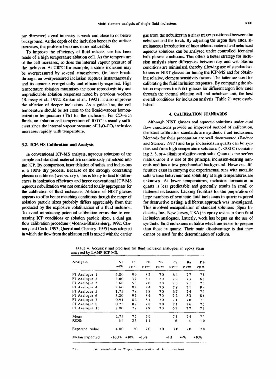
TABLE 4. Accuracy and precision for fluid inclusion analogues in epoxy resin analysed by LAMP-ICP-MS.
Analysis Na Cu Rb *Sr Cs Ba Pb wt% ppm ppm ppm ppm ppm ppm
FI Analogue 1 6.80 99 82 70 64 77 78 FI Analogue 2 2.60 37 61 70 72 73 69 FI Analogue 3 3.60 58 70 70 73 71 71 FI Analogue 4 2.60 82 94 70 78 71 94 Fl Analogue 5 1.75 78 78 70 67 74 73 Fl Analogue 6 3.20 97 84 70 72 83 86 FI Analogue 7 0.91 82 81 70 71 76 73 Fl Analogue 8 0.28 82 78 70 71 76 73 FI Analogue 10 3.00 78 79 70 67 77 73
Mean 2.75 77 79 71 75 77 RsLY?h 64 23 I 1 6 4 10
Expected value 4.00 70 70 70 70 70 70
Mean/Expected -160% +10% +13% +l% +7% +lO%
‘Sr data normalwd 10 1Oppm (concentralion of Sr in solution)

T. J. Shepherd and S. R. Chenery
TABLE 5. LAMP-ICP-MS analyses for single fluid inclusions in halite, Wilkesley Halite, Triassic Mercia Mudstone Group, Cheshire, U.K. (see text for details).
Fluid Mg/Sr KlSr CalSr Mn/Sr Cu/Sr Rb/Sr Cs/Sr Ba/Sr PblSr Inclusion No
x100 x100 x100 x100 x100
Basal Unit HAL 2a HAL 2b HAL 2c HAL 2f
HAL 5a HAL 5b HAL 5c HAL 5d HAL 5e HAL 51 HAL 59
RSE
Upper Unit HAL 3a HAL 3b HAL 3c HAL 3d
HAL lb HAL Id HAL le HAL 11 HAL lg HAL lh HAL li HAL lj HAL lk HAL 4a HAL 4b HAL 4c HAL 4d HAL 4e HAL 41 HAL 4g HAL 4h HAL 4i
am
23 3.1 22 35 4.7 29 31 4.2 26 ma 0.6 24
6 0.9 29 ma 5.6 30 ma 1.2 25 46 1.5 32 42 1.2 20
136 14 43 41 4.2 35 46 3.6 29 67 100.0 21
66 23 20 ma 11 23 56 16 18
145 35 36
42 13 13 56 11 14 56 16 15 69 25 15 26 7 12
202 62 34 71 17 10 67 74 30 97 67 37 49 27 26 39 25 29 35 11 26 50 35 27 34 21 40 69 54 34 90 33 33 65 41 30 61 36 29 73 30 25 55 63 36
4.7 2.5 4.3 1.1
n.a n.a n.a “.a
3.5 2.1 0.9 2.5 1 .o 2.4 1.6 2.4
54.0
n.a 1.1 n.a n.a “.a n.a “.a
45 40 36 90
ma 2.6 0.6 n.a
60 33 31 “.a 46 “.a 61 ma 36 ma 90 2.4 43 6.3 25 4.6 30 3.2 32 3.1 22 4.4 26 19 31 5.7 27 6.3 23 9.3 21 1.7 20 ma 23 2.4 39 7.0 51 120
0.6
1.0 2.2 0.7
0.2 1.6 0.4 0.3 0.3 ma “.a 0.6
86.0
5.2 5.1 5.5 13
3.8 4.5 7.4 10
5.5 20 33 16 37
5.2 5.3 3.7 9.1 5.4 16
7.7 2.1 10
10.6 66
0.4 0.3 1.0 0.3
ma 0.4 0.1 0.1 ma ma ma 0.4
75.0
0.4 0.2 0.7 2.4
1.4 0.3 0.9 1.3 0.5 ma “.a
3.7 11
0.6 0.2 0.4 1.0 0.7 ma 0.6 ma 1 .l 1.5
166
0.3 1.1 0.6 0.6 0.4 1.0 0.6 0.3
0.3 0.2 1.6 0.3 0.7 ma 1.1 0.2 0.9 ma 1.5 ma 2.1 0.4 1 .o 0.5
60.0 60.0
6.2 1.6 6.5 1.3 8.5 1.7 0.3 3.7
6.6 2.0 6.3 0.7 5.5 1.2 6.1 3.2 5.7 1.5 2.2 5.9 4.6 23 1.6 6.9 3.0 16 5.4 1.1 2.3 1.3 4.9 0.6 3.0 5.4 2.9 1.6 1.6 6.5 0.3 0.9 “.a 2.2 0.9 6.6 4.1 4.4 61 130
4.1. Fluid Inclusion Analogues
Initial experiments using ordinary epoxy resin mixes proved unsuccessful due to the reaction of water with the amine-based hardening agent. Satisfactory results were finally achieved using a liquid bisphenol A-epichlorohydrin epoxide resin of medium viscosity (Shell Chemicals UK Ltd, Chester, UK) and a tris-Zethyl hexoate curing agent (Anchor Chem- icals UK Ltd, Manchester, UK). By varying gel times and curing rates, conditions were found for producing a high pro- portion of < 100 pm droplets distributed throughout the resin (Fig. 5). The fluid inclusion analogue samples are prepared as resin wafers enclosed between 100 pm thick glass cover slips as used for geological thin sections. After mixing the epoxy resin and curing agent according to the manufacturer’s instructions, the mix is allowed to set for 2 hours until it has the consistency of thick syrup. [ N.B. The curing agent is haz- ardous to health and must be handled with care.] One side of each of the cover slips is then coated with resin and quickly
heated to 60-70°C to reduce the viscosity. Whilst still fluid, l-2 drops of standard solution are placed on one of the resin coated cover slips and the second cover slip fixed in place to form a resin wafer. If the resin viscosity is correct, the solution is dispersed throughout the resin as numerous microdroplets. The resin is then allowed to cure overnight. Being strongly hydrophobic, the resin does not always permit liquid droplet formation and a certain amount trial and error is needed. Fur- ther details can be obtained from the authors. For the laser ablation studies a NaCl solution containing 40,000 ppm Na and 70 ppm each of Ba, Cs, Pb, Rb, Sr, and Zn was encap- sulated in resin. Though this method provides an easy means of generating an abundance of liquid droplets for analysis (with few restrictions concerning the chemistry of the encap- sulated solutions) there are certain limitations. Two problems were encountered: ( 1) the resins contain trace metal impuri- ties, and (2) there is progressive loss of water from the drop lets over a period of several weeks. A further disadvantage is their low thermal stability; the epoxy resin cannot be ablated

Multi-element analysis of single fluid inclusions 4003
at elevated temperatures ( > 100°C). Despite these problems, fluid inclusion analogues provide a convenient and practical alternative to synthetic fluid inclusions.
4.2. Synthetic Fluid Inclusions in Halite
Towards the end of this evaluation study, the fluid inclusion analogues were supplemented by a small number of synthetic fluid inclusions in halite (courtesy of A. Moissette) to esti- mate approximate detection limits for Cu and Zn for fluid inclusions in natural halite. The laboratory grown halites were prepared according to the method described by Pironon ( 1990) and contained large numbers of small to medium size aqueous inclusions (<lo0 pm in diameter) distributed throughout the salt crystals in well defined growth zones. The synthetic fluid inclusions corresponded to NaCl-saturated so- lutions containing 1000 ppm of Cu and Zn.
5. NATURAL FLUID INCLUSIONS
To demonstrate the application of LAMP-ICP-MS to nat- ural fluid inclusions, five samples of halite, six samples of fluorite and one sample of quartz were analysed. The halite samples are from the Cheshire Basin (U.K) ; a Permo-Triassic basin of approximately 5000 km’ areal extent comprising a 4.5 km thick sequence of first cycle red bed elastic sediments overlain by variegated argillaceous mudstones containing massive evaporite beds (Whittacker, 1985). The dominant saline mineral is halite, together with subordinate anhydrite but no potash minerals. Samples were taken from the Basal and Upper Units of the Wilkesley Halite; the uppermost evap- orite horizon of the Mercia Mudstone Group. The fluid inclu- sions are generally well developed throughout and adopt a typical cuboid morphology. The large examples, up to 100 pm in size, are associated with areas of clear or weakly banded chevron halite and presented perfect targets for abla- tion. The fluorite and quartz samples are representative of different styles of hydrothermal lead-zinc-fluorite-barite rnin- eralization in the UK (North Pennines, SW England and Lake District ore fields), where previous fluid inclusion studies (Alderton, 1978; Shepherd et al., 1982; Ball et al., 1985; Shepherd and Scrivener, 1987) had confirmed the presence of low temperature ( 120-18O”C), high salinity fluid inclu- sions (>24 wt% TDS) with estimated Na/Ca ratios < 2; values considered typical for MVT deposits (Roedder, 1967; Haynes and Kesler, 1987). The inclusions analysed varied from IO-60 pm in diameter.
Sample preparation for ablation analysis varied according to the nature and mineralogy of the material. For fluorite and quartz, normal 200 pm thick, doubly polished fluid inclusion wafers were used; for halite, thicker mineral slices were used to facilitate handling.
6. RESULTS
All samples (nebulized standard solutions, NIST glasses, fluid inclusion analogues, synthetic fluid inclusions in halite, and naturally occurring aqueous inclusions in halite, fluorite, and quartz) were analysed under identical dual gas flow con- ditions. Liquid inclusions in halite were ablated at room tem- perature, those in fluorite and quartz at 140°C. After correc-
(a) 125
.
0 20 40 60 60
K/Sr
(W ‘0 r
. . .
.
l
.
2 - . . 0..
l . l .* 4.0 .
09 8.
0 20 40 60 60
K/Sr
(c) 25 1 2o t .
0 20 40 60 60
KlSr
FIG. 6. Element ratio covariations for aqueous fluid inclusions in halite, Wilkesley Halite, Triassic Mercia Mudstone Group, Cheshire. U.K. (a) Mg/Sr-WSr (b) Pb/Sr-WSr (c) Rb/Sr-IUSr.
tion for blank and isotopic abundances, the raw ICP-MS re- sponses were converted into relative element abundances using relative sensitivity factors obtained by nebulizing stan- dard solutions. An important advantage of ICP-MS compared to other spectroscopic techniques is that the relative sensitiv- ities for nearly all elements, with the exception of the halogens and metalloids, are within a factor of 3. Polyatomic interfer- ences (e.g., *‘NamAr+ on a.m.u. “Cu + ) normally expected during conventional ICP-MS analysis of saline waters were not observed, presumably due to the small mass of material injected into the plasma.
For fluid inclusions (synthetic and natural ) , the lower lim- its of detection, expressed as ppm concentrations in the inclu-

4004 T. J. Shepherd and S. R. Chenery
sion fluid, are governed by the absolute mass of material re- leased for analysis. At constant composition the minimum detectable concentration is, therefore, related to the volume of the inclusion. In practice the volume of inclusions is very difficult to calculate and thus, for purposes of comparison and presentation, the data are presented as element weight ratios. Normally elements are ratioed to Na, either measured or es- timated from microthermometric salinity determinations. However, because of the analytical problems encountered for Na (see below), ratios have been calculated relative to Sr.
Detection limits for the naturally occurring fluid inclusions were calculated according to theoretical variances from Pois- sonian counting statistics for integrated background count rates less than lo4 and on a fixed variance of 9% for back- ground count rates greater than 104. For Cu and Zn in the synthetic fluid inclusions (20- 100 pm in diameter), this cor- responds to lower limits of detection of 5-25 ppm (Table 3 ) . It is noteworthy that no significant polyatomic interferences were observed for either 6’Cu + and “‘Cu + or 65Zn + and 66Zn + . The authors wish to emphasis the provisional nature of the data for Cu and Zn and that lower limits of detection, ex- pressed as ppm concentrations in the inclusion fluid, for single fluid inclusions are only relevant and meaningful in the con- text of the mass of analyte vaporised/ionised. Further studies are in progress to generate working calibration curves that take into account inclusion size and salinity.
7. DISCUSSION
7.1. Fluid Inclusion Analogues and Synthetic Fluid Inchlsions in Halite
Table 4 details nine analyses of individual fluid inclusion analogues in epoxy resin. For Rb, Cs, Ba, Cu, and Pb, accu- racies (found/expected) are better than 15% and precisions (RSDs) range from 5-23%. The accuracy (160%) and pre- cision (64%) for Na are considerably worse. This is due pri- marily to the poor signal to background (S/B ) ratio for so- dium, and work is in progress to identify and reduce the so- dium background. As referred to above, due to high background concentrations of zinc in the resins, the zinc re- sults are very variable and have been excluded from Table 3. Whether this is specific to a batch of resin or is a more general contamination problem is not yet known. Overall, the fluid inclusion analogues performed well and provide realistic es- timates of the accuracy and precision to be expected for nat- ural inclusions of similar size.
An interesting feature of the time resolved traces for the fluid inclusion analogues is the presence of significant signals for Cl and Br (Fig. 7). ICP-MS is not normally considered appropriate for the analysis of the halogens due to their high first ionization potentials (Date and Gray, 1989). However, the traces clearly indicate the potential for the direct LAMP- ICP-MS measurement of Cl/Br ratios in encapsulated drop- lets of saline solution and, by analogy, saline fluid inclusions. Follow-up work has since confirmed this possibility (Shep- herd et al., 1995); the high concentration of chlorine in the inclusions offsetting the low degree of ionization. This reali- zation opens up the opportunity for tracing the evolution of palaeobrines using conservative element ratios (Hovorka et al., 1993).
7.2. Natural Fluid Inclusions in Halite, Fluorite, and
Except for inclusions < 10 pm in diameter, ICP-MS re- sponses were obtained for most of the major cation species.
7.2.1. Halite
Three aspects of the halite data (Table 5) merit discussion. Firstly, the estimated concentrations of the major elements (Mg, K, and Ca) assuming an average Sr concentration of 1000 ppm are within the ranges reported for fluid inclusions in halite (Bein et al., 1991; Rosin, 1993). Secondly, the tech- nique provides data for a wide range of minor and trace ele- ments for comparison with present-day evaporite brines (Fi- scher and Kreitler, 1987; Land and Macpherson, 1992). Thirdly, the data indicate a high degree of geochemical dis- crimination between different lithostratigraphic units of the Triassic halite deposits, Cheshire Basin, UK (i.e., Upper and Basal Units). The strongly correlated distributions for Mg/ Sr-K/Sr, -Rb/Sr, and -Pb/Sr (Fig. 6) suggest an advanced stage of seawater evaporation and provide, indirectly, a mea- sure of applicability of LAMP-ICP-MS to the analysis of sin- gle inclusions. Much of the scatter shown in Fig. 6 and the high RSDs given in Table 5 are probably due to natural within-sample variation and should not be regarded as a mea-
Chlorine mass 35CI+
INTENSITY I FSD 5X10’ cps
Bromine mass *lb+
A A FSD 1.5XlO’cps
Sodium mess 23Na+
lb i0 i0
TIME 6)
FIG. 7. Time resolved ICP-MS spectrum for Cl, Br, and Na for a 100 pm diameter fluid inclusion analogue (Cl& wt ratio - 500) showing the potential for obtaining direct measurements of CVBr ratios in saline solutions by LAMP-ICP-MS. Ablation at 22°C.

Multi-element analysis of single fluid inclusions 4005
sure of analytical precision. More detailed discussion and in- studies are needed to verify the mineralogical origin of the terpretation of the data are outside the scope of this paper. metals.
7.2.2. Fluorite and Quartz 8. CONCLUSIONS AND FUTURE DEVELOPMENTS
Thrtx important conclusions may be drawn from the results shown in Table 6. Firstly, the technique is clearly applicable to the analysis of brine inclusions in fluorite and quartz. Some of the elements determined, notably the transition metals, have hitherto been considered difficult or impossible to ana- lyse in inclusion fluids. Secondly, although the calculated RSDs are generally higher than those for the fluid inclusion analogues, contrasting ratios for Na/Sr, KISr, Mg/Sr, Cs/Sr, and Pb/Sr are indicative of geologically distinctive fluid events within and between ore fields. Thirdly, based on dif- ferences in inclusion morphology and estimated temperatures of entrapment, chemical differences may be observed between different generations of inclusions within the same sample. This latter observation is extremely important for ore deposit modelling because it provides the possibility for resolving fine scale temporal variation in fluid chemistry during a single mineralizing event.
When ablating fluorite and quartz samples from sulphide- rich veins, significant signal spikes were often observed for Cu, Pb, and Zn prior to inclusion rupture. This is taken to indicate the presence of submicroscopic sulphide impurities in the host mineral and supports our assertion that the analyses reported here refer specifically to the inclusion fluid. In con- trast, data acquired using an IR or ruby laser would be subject to much greater uncertainty, since the ablated material in- cludes a significant component of the host matrix. Further
The results confirm conclusively that ICP-MS when used in conjunction with a UV laser microprobe and thermal ab- lation cell has the spatial resolution and sensitivity required for the multi-element analysis of single fluid inclusions. Use of a UV laser successfully eliminates many of the ablation problems encountered using an IR or ruby laser and extends the technique to quartz, fluorite, and other inclusion-bearing minerals that absorb at 266 nm. Thermal fracturing is reduced and the small diameter of the beam dramatically minimises the matrix contribution to the analyte signal. The optics of the BGS laser microprobe permit precision drilling and ablation of inclusions up to 60 micron beneath the sample surface without damaging adjacent inclusions. The technique thus provides the potential for discriminating between different populations of inclusion within the same sample. Using a ther- mal ablation cell, fluid release, and signal to background ratios are significantly improved at elevated temperatures due to the higher internal vapour pressures. On laser breakthrough, the fluid is explosively and efficiently released simulating that observed for gas-rich inclusions. However, although this im- proves the ablation process, minimum detection limits are strictly related to the mass of analyte ion&d in the ICP and the sensitivity of the instrument. The present study did not seek to establish the optimum conditions for single inclusion analysis but data for fluid inclusion analogues, a practical al- ternative to synthetic fluid inclusions, indicate that for typical
TABLE 6. LAMP-ICP-MS analyses for single Ruid inclusions in fluorite and quartz for MVT-type Pb-Zn-F-Ba vein deposits, U.K. (see text for details).
Na/Sr K/Sr Mg/Sr BalSr Rb/Sr CslSr MnJSr Pb/Sr Zn/Sr Cu/Sr
North Pennines
R31fl a.m. 14.6(14)
RSD% 48 R29tl
B.ln. 25.9(7) RSD% 45
NPn-3ltl a.m. 13.4(5)
RSD% 31
SW England
SW82-90 35.40)
Rt& 69 SW824fl
a.m. 31.q3j RSD% 28
SW82-6fl 39.3(10)
R& 65
Lake District
CFn-lo9q a.nl. 8.3(3)
4.9( 14) 45
0.29(11) 41
6.9(7) 0.34(6) 17 53
3.1(3)
12.7(5) 21
24.5(3)
13.8(10) 28
0.14(5) 29
0.58(4)
0.16(3)
0.47(9) 32
O.ll(4) 0.04(4) 0.01(l) 0.37(4) 0.02(6) 0.03( 10) 25 60
0.03(5) O.lN2) 33
0.75(2)
0.14(l) 0.10(l) 0.14(5) 0.43(l) 0.08(4) 0.04(4) 28
0.32(l) 0.80(l) 4.1(3) l.](3) 1.0 0.16(3)
O.ll(6) 0.11(3) 0.05(5) 0.03( 1) 75
0.02(3)

4006 T. J. Shepherd and S. R. Chenery
20-100 micron diameter inclusions, the RSDs for many el- ements (Na excluded) are better that 30%.
Though ICP-MS is generally considered a multi-element technique, there is no immediate solution to the problem of polyatomic interferences. Major interferences at a.m.u. 32,34, and 75 preclude determination of sulphur and arsenic; two of the most important elements involved in the transport and deposition of metals in high temperature hydrothermal fluids. These disadvantages are more than compensated for, how- ever, by the speed of measurement and wide range of elements amenable for analysis, especially the high field strength ele- ments. Of significant importance is the detection of Cl and Br in the fluid inclusion analogues. This observation was quite unexpected and further work is in progress to verify the direct measurement of Cl/Br ratios in natural fluid inclusions by ICP-MS. If proven, it should then be possible to normalise all elements to chlorine, thereby affording general application to chloride-dominant palaeofluids of diverse origin, pending ac- curate measurement of inclusion volumes and subsequent de- termination of absolute element concentrations. A more rig- orous evaluation of calibration procedures including compar- ative testing of NIST reference glasses, synthetic fluid inclusion, and aqueous solutions is currently in progress.
Acknowledgmenrs-The laser ablation microprobe was &signed by C. Flint, Birkbeck College, University of London and is protected by U.K. Patent Number 9106337.0, Serial 2254444,7/ 10192. Publica- tion is by permission of the Director, British Geological Survey (NERC). The authors wish to thank Joaquin Ruiz (Univ. Arizona, USA), Andy Rankin (Univ. Kingston, UK), and an anonymous ref- eree for their useful advice and comments which did much to improve the paper for publication.
Edirorial handling: D. A. Vanko
REFERENCES
Alderton D. H. M. ( 1978) Fluid inclusion data for lead-zinc ores from SW England. Trans. Insr. Min. hferall. 87, B132-B 135.
Ayora C., Garcia-Veigas J., and Pueyo J-J. ( 1993) X-ray microanal- ysis of fluid inclusions and its application to the geochemical mod- elling of evaporite basins. Geochim. Cosmochim. Acra 58,43-55.
Ball T. K., Fortey N. J., and Shepherd T. J. ( 1985) Mineralisation at the Carrock Fell Tungsten Mines, N. England: Paragenetic, fluid inclusion and geochemical study. Mineral. Deposira 20, 57-65.
Bein A., Hovorka S. D.. Fisher R. S., and Rcedcler E. ( 1991) Fluid inclusions in bedded Permian halite, Palo Duro Basin, Texas: ev- idence for modification of Seawater in evaporite brine-pools and subsequent early diagenesis. /. Sedimenr. Perrol. 61, I - 14.
Bodnar R. J. and Sterner S. M. ( 1987) Synthetic fluid inclusions. In Hydrorhermal Experimenral Techniques (ed. G. C. Ulmer and H. L. Barnes), pp. 423-457. Wiley.
Boiron M. C., Dubessy J., Andre N., Briand A., Lacour J. L., Mau- chien P.. and Mermet J. M. ( 1991) Analysis of mono-atomic ions in individual fluid inclusions by laser-produced plasma emission spectroscopy. Geochem. Cosmochim. Acra 55,917-923.
Chenery S. and Cook J. M. ( 1993) Determination of rare earth ele- ments in single mineral grains by laser ablation microprobe-in- ductively coupled plasma mass spectrometry-preliminary study. J. Anal. Arom. Spec. 8,299-303.
Chenery S. and Rankin A. H. ( 1989) The use of a laser ablation microprobe attached to an ICP spectrometer for the elemental anal- ysis of inclusions. ECROFI X Prog. Absrr., p2 1.
Date A. R. and Gray A. L. ( 1989) Applicarions of Inducrively Cou- pled Plasma Mass Specrromerry. Blackie.
Diamond L. W., Marshall D. D., Jackman J. A., and Skippen G. B. ( 1990) Elemental analysis of individual fluid inclusions in min-
erals by Secondary Ion Mass Spectmmetry (SIMS): Application to cation ratios of fluid inclusions in an Archaean mesotbennal gold-quartz vein. Geochim. Cosmochim. Acra 54,545-552.
Dubessy J., Poty B., and Ramboz C. (1989) Advances in C-O-H-N- S fluid geochemistry based on micro-Raman spectrometric analy- sis of fluid inclusions. Eur. J. Mineral. 1,517-534.
Fischer R. S. and Kreitler C. W. ( 1987) Geochemistry and hydro- dynamics of deep-basin brines, Palo Duro Basin, Texas, USA. Appl. Geochem. 2,459-476.
Haynes F. M. and Kesler S. E. ( 1987) Chemical evolution of brines during Mississippi Valley-type mineralization: evidence from East Tennessee and Pine Point. Econ. Geol. 82,53-7 I.
Haynes F. M., Sterner S. M.. and Bodnar R. J. ( 1988) Synthetic fluid inclusions in natural quartz. IV. Chemical analyses of fluid inclu- sions by SEMIEDA: evaluation of method. Geochim. Cosmochim. Acra 52,969-977.
Hovorka S. D., Knauth L. P., Fisher R. S., and Gao G. ( 1993) Marine to nonmarine facies transition in Permian evaporites of the Palo Duro Basin, Texas. Bull. Geol. Sot. Amer. 105, 1119- 1134.
Horn E. E. and Tye C. T. ( 1989) Analysis of fluid inclusions in minerals by VG laser ablation ICP-MS. PACROFI Prog. Absrr. 2, 32 (abstr.)
Imai N. ( 1990) Quantitative analysis of original and powdered rocks and mineral inclusions by laser ablation inductively coupled plasma mass spectrometry. Anal. Chim. Acra 235,381-384.
Land L. S. and Macpherson G. L. ( 1992) Origin of saline waters, Cenozoic section, Gulf of Mexico sedimentary basin. Amer. Assoc. Perrol. Geol. Bull. 76, 1344- 1362.
Moenke-Blankenburg L. ( 1989) Laser Micro Analysis. Wiley Inter- sci.
Moenke-Blankenburg L., Schumann T., Gunther D., Kuss H-M., and Paul M. ( 1992) Quantitative analysis of glass using inductively coupled plasma atomic emission spectroscopy and mass spectrom- etry. laser micro-analysis inductively coupled plasma atomic emis- sion spectroscopy, and laser ablation inductively coupled plasma mass spectrometty. J. AMY. Ar. Specrrom. 7,251.
Nabu M. and Sato T. ( 1981) The analysis of fluid inclusions in the microgram range with an ion microanalyser. Bull. Mineral. 104, 827-833.
Pasteris J. D., Wopenka B., and Seitz J. C. (1988) Practical aspects of quantitative laser Raman microprobe spectroscopy for the study of fluid inclusions. Geochim. Cosmochim. Acra 52.979-988.
Perkins W. T., Fuge R.. and Pearce N. J. G. ( 1991) Quantitative analysis of trace elements in carbonates using laser ablation in- ductively coupled plasma mass spectrometry. J. Anal. Ar. Spec- rrom. 6,445~448.
Querol X. and Chenery S. R. ( 1995) The determination of trace el- ement affinities in coal by laser ablation microprobe-inductively coupled plasma-mass spectrometry. In European Coal Geology (ed. M. K. G. Whateley and D. A. Spears), pp. 147-155. J. Geol. Sot. Spec. Publ. 82
Ramsey M. H., Coles B. J., and Wilkinson J. J. ( 1992) Single fluid inclusion analysis by laser ablation inductively coupled plasma atomic emission spectrometry: quantification and validation. .I. Anal. Arom. Spec. 7,587-593.
Rankin A. H., Ramsey M. H., Coles B., Van Langevelde F., and Thomas C. R. ( 1992) The composition of hypersaline, iron-rich granitic fluids based on laser-ICP and Synchrotron-XRF micro- probe analysis of individual inclusions in topaz, Mole granite, east- em Australia. Geochim. Cosmochim. Acra 56,67-79.
Roedder E. ( 1967) Environment of depostion of stratiform (Missis- sippi Valley type) ore deposits, from studies of fluid inclusions. Econ. Geol. Mon. 3, 349-362.
Rosin C. ( 1993 ) Applications du couplage Torche a Plasma spectro- metrie de masse a la recherche d’elements trous en eau portable et dam les saumures. These d’universite & Nancy.
Ryan C. G., Cousens D. R., Heinrich C. A., Griffin W. L., Sie S. H., and Memagh T. P. ( 1991) Quantitative PIXE microanalysis of fluid inclusions based on a layered yield model. Nucl. Znsrr. Merh- ods Phys. Res. BS4,292-297.
Shepherd T. J. and Sctivener R. C. (1987) Role of basinal brines in the genesis of polymetallic vein deposits, Kit Hill-Gunnislake area, SW England. Ussher Sot. Proc. 6,491-497.

Multi-element analysis of single fluid inclusions 4007
Shepherd T. J., Darbyshire D. P. F., and Moore G. R. ( 1982) Rare earth element and isotopic geochemistry of the North Pennine ore deposits. Bull. du B.R.G.M., Spec. Issue 4, 371-377.
Shepherd T. J., Chenery S. R., and Moissette A. ( 1995) Optimization of Laser ablation-ICP-MS for the chemical analysis of fluid inclu- sions in evaporite minerals. Terra Absrr. EUG 8, 344.
Svejensky D. A. ( 1984) Oil field brines as ore forming solutions. Econ. Geol. 79, 23-37.
Thompson M., Chenery S. R., and Brett L. ( 1989) Calibration studies in laser ablation microprobe-inductively coupled plasma-atomic emission spectrometry. J. Anal. At. Specrrom. 4, 1 1 - 16.
Wilkinson J. J., Rankin A. H., Mulshaw S. C., Nolan J., and Ramsey M. H. ( 1994) Laser ablation-ICP-AES for the deter-
mination of metals ih fluid inclusions: An application to the study of magmatic ore fluids. Geochim. Cosmochim. Acta 58, 1133- 1146.
Williams J. G. and Jarvis K. E. ( 1993) Preliminary assessment of laser ablation inductively coupled plasma mass spectrometry for quantitative multi-element determination in silicates. J. Anal. Ar. Specrrom. 8, 25-34.
White D. E. ( 1965) Saline waters in sedimentary rocks. Fluids in Subsurface Environments-a Symposium. Amer. Assoc. Pet. Geol. Mem. 4,342-366.
Whittacker A. ( 1985) Ados of onshore Sedimentary Basins in En- gland and Wales: Post Carboniferous Tectonics and Srradgraphy. Blackie.


