Embed Size (px)
Citation preview

Dermatol Clin
Current Therapy
Laser Treatment of Vascular Lesions
Chrys Delling Schmults, MD
Department of Dermatology, University of Pennsylvania Medical Center, 2 Rhoads Pavilion, 3400 Spruce Street,
Philadelphia, PA 19104, USA
Lasers and other light sources have been devel-
oped that remove or improve many vascular lesions
that were previously untreatable. Port-wine stains are
the most notable example. Many port-wine stains are
unresectable, but now can be removed in their en-
tirety noninvasively via laser therapy. Vascular lasers
and light sources represent a major advance in der-
matology for cosmetic and noncosmetic applications.
This article reviews the common vascular conditions
amenable to laser therapy and the approaches and
devices used.
Guiding principles in laser therapy
The theory of selective photothermolysis was
developed by Anderson and Parish [1]. In simple
terms, it postulates that a laser can work by heating to
the point of destruction a specific targeted structure
within the skin. The targeted structure is destroyed
without destroying surrounding tissue because the
wavelength of laser light used is absorbed preferen-
tially by the targeted structure and not by surrounding
tissue. In the case of vascular lesions, the targeted
structure is hemoglobin within blood vessels. The
hemoglobin is heated, which also heats and destroys
the endothelial cells of the blood vessel walls. In an
ideal treatment, the vessel is damaged to the point
that blood no longer can course through it. Hemo-
globin has three wavelengths of light at which it
absorbs a maximal amount of energy: 418 nm,
542 nm, and 577 nm. Vascular lasers have been de-
veloped to use these absorption peaks to heat and
destroy blood vessels selectively.
0733-8635/05/$ – see front matter D 2005 Elsevier Inc. All rights
doi:10.1016/j.det.2005.05.023
E-mail address: [email protected]
A second important principle in laser therapy is
that of thermal relaxation time. Simply stated, thermal
relaxation time is a measurement of the amount of
time it takes for a structure to be heated to the point
that heat escapes from it to adjacent structures. The
larger the structure, the longer the thermal relaxation
time because it takes longer to heat a larger object. In
laser therapy, the goal is to heat the target structure
maximally, but to stop energy input before the heat
begins to escape and damage adjacent structures.
The next important concept is that of pulse
duration and the need to match pulse duration to
the thermal relaxation time of the target. The pulse
duration is the amount of time over which a given
dose of light is administered to the skin. For example,
q-switched lasers have short pulse durations in the
nanosecond range. If 5 Joules of light energy is ad-
ministered with a q-switched laser, that 5-J dose is
given very rapidly over nanoseconds. In contrast,
vascular lasers have much longer pulse durations in
the millisecond range. With a vascular laser, the same
5-J dose of light may be administered over 1 ms,
40 ms, or 1000 ms; much more slowly than with a
q-switched laser.
For a given structure in the skin, the amount of
energy absorbed and the subsequent heating and
destruction that occur vary greatly between the two
previous scenarios. In the first example, when the light
dose is given quickly (nanoseconds), only very small
structures (with subsequently short thermal relaxation
times) are heated significantly. The larger structures
did not have a chance to heat up because the energy
was given so rapidly. Q-switched nanosecond lasers
are used to heat and destroy small structures within
the skin, such as particles of tattoo pigment or mela-
nin. Conversely, lasers with longer pulse durations
administer a given dose of light relatively slowly.
23 (2005) 745 – 755
reserved.
derm.theclinics.com

schmults746
They heat and destroy larger structures with longer
thermal relaxation times. Blood vessels, which are
much larger than particles of tattoo pigment, are
heated and destroyed by lasers with pulse durations
in the millisecond range (termed ‘‘long-pulsed la-
sers’’). It follows that larger vessels require longer
pulse durations than small vessels for maximal de-
struction and clinical improvement; this is generally
borne out in clinical practice, although multiple other
factors come into play as discussed below.
Mismatch of pulse duration and thermal relaxation
time can result in adverse effects. Using a pulse
duration that is too short (brief) for the targeted
structure leads to underheating of that structure and
poor efficacy. It also may lead to destruction of smaller
unintended targets with shorter thermal relaxation
times. Use of pulse durations that are too long can
lead to overheating of the target structure. Heat may
escape into adjacent structures causing damage.
Finally, another important principle is that longer
wavelengths of light penetrate more deeply into the
skin. Deeper lesions require a longer wavelength for
efficacy. Conversely, more superficial lesions respond
to shorter wavelengths.
Two other important terms in laser therapy are
‘‘fluence’’ and ‘‘spot size.’’ The fluence is the energy
delivered to a given area of skin. It is measured in
units of J/cm2. An increase in fluence is an increase in
energy emitted by the laser. Increasing the fluence
can increase efficacy, but also can increase the risk
of scarring if safe parameters are not observed. Spot
size is the size of the laser beam administered to
the skin surface. In most cases, it is circular, although
rectangular spot sizes also are available. Use of a
5-mm spot size means that a circular beam of light
measuring 5 mm in diameter is administered to the
skin. In general, moving to a larger spot size requires
a decrease in energy (a lower fluence) and vice versa.
Larger spot sizes also tend to have more scatter (and
slightly less efficacy) at the periphery. This difference
is usually not noticeable clinically, especially with
spot sizes 10 mm and smaller.
In practice, the interaction between laser light and
tissue is complicated. Perfectly selective photother-
molysis is not yet possible. Multiple structures within
the skin absorb laser light to various degrees. If too
much energy is absorbed by nontarget structures,
blistering, scarring, and dyspigmentation can result.
Absorption by epidermal melanin is particularly
problematic, especially in patients with Fitzpatrick
skin types IV through VI. In these patients, pigmen-
tary alterations (hyperpigmentation and hypopigmen-
tation) resulting from damage to melanocytes are
common. Epidermal melanin absorption also can lead
to blistering and to injury of nearby dermal structures,
resulting in scar formation. Absorption of light by
melanin decreases with longer wavelengths. In the
case of vascular lasers, longer wavelength lasers still
have a high incidence of melanin-related adverse
effects (see treatment tip #3 later).
In addition to the direct effects of laser energy on
the skin, there are secondary effects of laser injury,
such as the inflammatory response. Inflammation and
the subsequent changes it produces may play a large
role in the ultimate clinical improvement seen with
laser therapy. This inflammatory response may help
to explain why vascular lesions usually do not dis-
appear immediately with laser treatments, but rather
fade gradually in the days and weeks after treatment.
Brief history of vascular laser development
Port-wine stains were the initial vascular lesions
extensively studied in laser therapy. Argon 488-nm
and 514-nm and continuous wave dye 577-nm and
585-nm lasers were used initially. They were asso-
ciated with a relatively high risk of scarring and
pigmentary change [2,3]. Ablative lasers, such as the
10,600-nm carbon dioxide and 2940-nm erbium:
yttrium aluminum garnet (Er:YAG) lasers, also were
used [4]. These did not target the vasculature, but rather
obliterated the lesion as the laser energy was absorbed
primarily by water ubiquitous in the skin. Because
destruction of a portion of the dermis was necessary for
lesion removal, some form of scarring was the norm.
Copper vapor 510-nm and 578-nm lasers represented
an improvement over the previous lasers above [5].
However, the pulsed dye laser (PDL) largely replaced
other lasers in the treatment of port-wine stains.
The PDL with wavelengths ranging from 585 to
600 nm was perfected by Anderson and his group in
the 1980s [1,6]. It has become the treatment of choice
for port-wine stains. Subsequently the KTP 532-nm
laser was found to be useful in the treatment of fine
telangiectasias, and intense pulsed light (IPL) systems
are effective for diffuse facial erythema. The newer
long-pulsed 532-nm KTPs, PDLs, 755-nm alex-
andrite, diode (with wavelengths 800–900 nm),
and 1064-nm neodynium:yttrium aluminum garnet
(Nd:YAG) lasers may be helpful in the treatment of
leg varicosities and other vascular lesions. Currently
a wide array of lasers and light sources are available
to the clinician for treatment of vascular lesions, and
new devices are being developed constantly.
Many different types of vascular lesions have been
shown to resolve or substantially improve with laser
therapy, including port-wine stains, hemangiomas,

Fig. 1. Port-wine stain in a child before (A) and after (B) treatment with pulsed dye laser with near-total resolution. (Courtesy of
David J. Goldberg, MD, Hackensack, NJ Westwood, NJ, and New York, NY.)
vascular lesion laser treatment 747
diffuse facial erythema, facial telangiectasia, sclero-
therapy-induced matted telangiectasia, spider telan-
giectasias of the legs, cherry angiomas, spider
angiomas, pyogenic granuloma, and vascular malfor-
mations arising from CREST syndrome and Osler-
Weber-Rendu disease. These lesions are discussed
subsequently. Other skin conditions, such as psoriasis,
verrucae, and scars, sometimes improve with treat-
ment by vascular lasers. However, because these le-
sions are not primarily vascular in nature, effects on
the vasculature only partially explain the improve-
ments attained. These lesions are not discussed in
this article.
Port wine stains
PDLs remain the mainstay of therapy for port-
wine stains. Currently, several devices are commer-
cially available. These lasers have wavelengths
ranging from 585 to 600 nm. The early PDLs had
fixed pulse durations of 0.45 ms. Newer models have
Fig. 2. A thicker port-wine stain in an adult before treatment (A)
clearance after several pulsed dye laser treatment sessions. (Courtes
and New York, NY.)
been developed with longer pulse widths. These
longer pulse durations better match the 1- to 10-ms
thermal relaxation times of port-wine stain vessels.
Longer wavelengths of 595 nm and 600 nm penetrate
more deeply, enhancing effectiveness. However,
these wavelengths are farther from the hemoglobin
absorption peak of 577 nm, so higher fluences are
required. These higher fluences can cause damage to
epidermal melanin. Subsequently, cooling devices
have been employed to cool and protect the epidermis
while heat is generated in the dermal vessels below.
Multiple spot sizes often are available on a single
laser via interchangeable hand pieces with 3-, 5-, 7-,
and 10-mm round spot sizes commonly seen. Some
devices also have rectangular spot sizes that can be
used to trace out individual vessels.
The aforementioned modifications have resulted
in PDLs with better safety and efficacy in the
treatment of port-wine stains. However, complete
removal is still not possible in all patients. Overall,
approximately 50%–60% of patients have a 75%
lightening. A 50% or greater improvement is seen
and after (B) treatment with improvement, but incomplete
y of David J. Goldberg, MD, Hackensack, NJ Westwood, NJ,

Fig. 3. Port-wine stain on the hand of a child before (A) and after (B) several treatments with a pulsed dye laser, showing
marked improvement, but incomplete resolution. (Courtesy of David J. Goldberg, MD, Hackensack, NJ, Westwood, NJ, and
New York, NY.)
schmults748
in 70%–80% [7]. Thick lesions remain particularly
problematic, so treatment should be done in child-
hood if possible before thickening occurs (Figs. 1
and 2). Large lesions (> 20 cm) and lesions of the
legs and hands also respond less well (Fig. 3). In most
patients, multiple treatments (� 4–10) are required.
Optimal treatment parameters are difficult to define
from the literature because controlled comparative
studies are few, and new lasers are constantly
replacing older models. Although often equivalent,
the 600-nm wavelength has been shown to improve
clearance in some patients (50%) compared with
585-nm wavelength. A higher fluence must be used,
however, to compensate for the lower hemoglobin
absorption. The higher fluence may result in more
dyspigmentation [8].
The main side effect of PDLs is purpura, which
generally lasts 5–14 days. Although newer PDLs
may generate less purpura, many practitioners believe
that treatment is not effective for port-wine stains
without purpura. A purpuric response is the chief
clinical marker used during treatment. The lowest
fluence that produces purpura should be used.
Lasers other than the PDL have been shown to be
effective in the treatment of port-wine stains. The
long-pulsed 1064-nm Nd:YAG laser has been shown
to have efficacy equivalent to the PDL, but with an
increased risk of scarring in one study [9]. In a study
comparing IPL systems with PDLs, most patients
had superior improvement with the PDL, but a subset
(6 of 32) had better clearing after IPL therapy [10].
The 532-nm KTP laser has been used to treat port-
wine stains refractory to PDL treatment. Adverse
effects were increased, however, because higher flu-
ences are needed, and the shorter wavelength results
in more epidermal absorption of energy [11]. The
long-pulsed 1064-nm Nd:YAG laser also seems to
be effective in port-wine stains resistant to PDL. No
adverse effects were found in this study [12].
Hemangiomas (strawberry nevus)
Hemangiomas appear early in infancy, have a
growth period of 6–9 months, and usually sponta-
neously involute during early childhood. Because
they resolve on their own, treatment generally is
reserved for problematic lesions, such as lesions with
a risk of visual or airway obstruction, ocular
malformation, bleeding, ulceration, or rapid growth.
In these scenarios, a laser may halt growth of the
lesion, but cannot remove it. Intralesional or occa-
sionally oral steroids are generally superior to laser
therapy in slowing the growth of rapidly proliferating
hemangiomas. Laser therapy is useful, however,
when steroid therapy is contraindicated or in the case
of bleeding or ulcerated hemangioma. One to two
PDL treatments generally lead to healing of the
ulcerated area, with 5–6 J/cm2 generally being
effective [13,14]. Some clinicians also treat early
hemangiomas to ‘‘nip them in the bud.’’ Studies have
not been done to determine if early treatment alters
the natural course of hemangiomas. Lasers also can
improve the telangiectasia that often remains after a
hemangioma has regressed.
Spider and varicose veins
Sclerotherapy remains the gold standard of non-
invasive treatment for spider varicosities of the legs.
Progress is rapidly being made in laser technology,
however. Laser therapy currently is indicated for
matted telangiectasias and other vessels that are too
vascular lesion laser treatment 749
small to accommodate a sclerotherapy needle. It also
can be used in patients who cannot tolerate sclero-
therapy because of needle phobia or allergy to
sclerosing agents.
Compared with port-wine stains, spider veins
often require longer wavelengths to enhance pene-
tration to deep vessels. Spider and varicose veins also
require longer pulse widths of 20–50 ms or longer
because vessels are larger (0.1–5 mm) than vessels
comprising port-wine stains. Hyperpigmentation is
common even in light-skinned individuals; this
usually resolves with time.
Long-pulsed PDLs have been shown to clear
vessels with diameters less than 0.5 mm completely.
For vessels 0.5–1 mm, improvement, but not clear-
ance, is achieved [15]. Long-pulsed 532-nm KTP and
532-nm Nd:YAG lasers also have been shown to
improve, and sometimes clear, lesions [16,17]. The
532-nm Nd:YAG had a high incidence of adverse
effects with 10% scarring and 94% hyperpigmenta-
tion rates. The long-pulsed 755-nm alexandrite also
improved, but did not clear vessels after a single
treatment. More than one third of patients had
hyperpigmentation [18]. Diode lasers use a small
hemoglobin absorption peak at 915 nm in treating
vascular lesions. The longer wavelength penetrates
more deeply to treat deeper and generally larger
vessels. A 940-nm diode laser had no effect on small
vessels, but was found to improve larger spider veins
of 0.8–1.4 mm in most (88%) patients [19].
In a comparative study, a long-pulsed 1064-nm
Nd:YAG laser was found to be superior to long-
pulsed 755-nm alexandrite and 810-nm diode lasers
for spider veins [20]. Initially the long-pulsed
1064-nm Nd:YAG laser was found to be inferior to
sclerotherapy [21]. However, a more recent study has
shown them to be equivalent [22]. Diode lasers also
are improving, with 6 of 35 patients having complete
clearance 6 months after two treatments with a long-
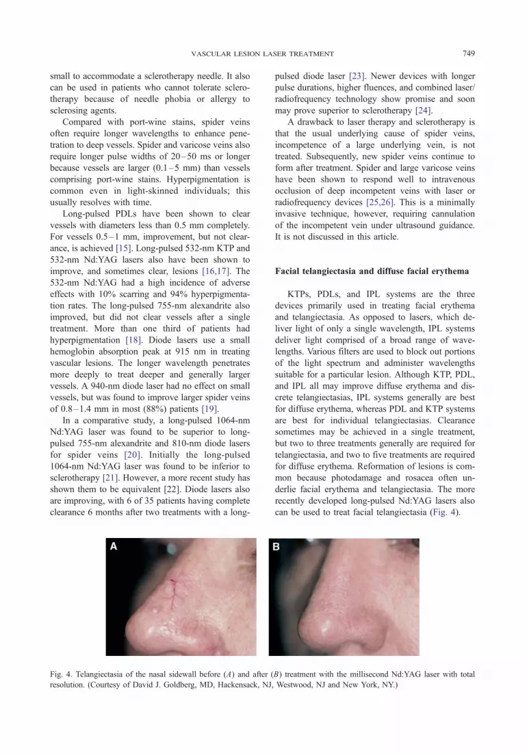
Fig. 4. Telangiectasia of the nasal sidewall before (A) and after (
resolution. (Courtesy of David J. Goldberg, MD, Hackensack, NJ,
pulsed diode laser [23]. Newer devices with longer
pulse durations, higher fluences, and combined laser/
radiofrequency technology show promise and soon
may prove superior to sclerotherapy [24].
A drawback to laser therapy and sclerotherapy is
that the usual underlying cause of spider veins,
incompetence of a large underlying vein, is not
treated. Subsequently, new spider veins continue to
form after treatment. Spider and large varicose veins
have been shown to respond well to intravenous
occlusion of deep incompetent veins with laser or
radiofrequency devices [25,26]. This is a minimally
invasive technique, however, requiring cannulation
of the incompetent vein under ultrasound guidance.
It is not discussed in this article.
Facial telangiectasia and diffuse facial erythema
KTPs, PDLs, and IPL systems are the three
devices primarily used in treating facial erythema
and telangiectasia. As opposed to lasers, which de-
liver light of only a single wavelength, IPL systems
deliver light comprised of a broad range of wave-
lengths. Various filters are used to block out portions
of the light spectrum and administer wavelengths
suitable for a particular lesion. Although KTP, PDL,
and IPL all may improve diffuse erythema and dis-
crete telangiectasias, IPL systems generally are best
for diffuse erythema, whereas PDL and KTP systems
are best for individual telangiectasias. Clearance
sometimes may be achieved in a single treatment,
but two to three treatments generally are required for
telangiectasia, and two to five treatments are required
for diffuse erythema. Reformation of lesions is com-
mon because photodamage and rosacea often un-
derlie facial erythema and telangiectasia. The more
recently developed long-pulsed Nd:YAG lasers also
can be used to treat facial telangiectasia (Fig. 4).
B) treatment with the millisecond Nd:YAG laser with total
Westwood, NJ and New York, NY.)

Fig. 5. Spider angioma on the cheek of a child before (A) and after (B) treatment with the pulsed dye laser with resolution of the
lesion and hyperpigmentation, which resolved spontaneously. (Courtesy of David J. Goldberg, MD, Hackensack, NJ, Westwood,
NJ, and New York, NY.)
schmults750
KTP lasers are equipped with small spot sizes of
less than 1 mm and are ideally suited for tracing out
individual telangiectasias. In a study comparing four
different KTP lasers, all were found to have equal
efficacy [27]. Although no adverse effects occurred
in this study, there is a risk of blistering with KTP
lasers if the operator does not keep the laser tip mov-
ing during treatment. This risk may be minimized
if epidermal cooling is employed [28]. There is no
purpura as with PDL treatment.
PDLs can be used to clear individual telangiecta-
sias and diffuse erythema. PDLs have a circular spot
size, and treatment areas should not overlap by more
than 30%. PDLs generally do not allow for clearance
of diffuse erythema in a single treatment. Between
treatments, patients may have a meshwork of clear
areas separated by areas of erythema. Patients should
be made aware of this in advance. Although newer
PDLs generate less purpura, it seems that purpura is
still required for optimal lesion clearance in many
cases [29].
IPL systems are ideal for treating diffuse ery-
thema. IPL systems have large rectangular spot sizes,
and overlap of treatment areas is less problematic
compared with PDLs. It is easier to clear large areas
of diffuse erythema in fewer treatments.
Fig. 6. Pyogenic granuloma on the lip before (A) and after (B) tr
(Courtesy of David J. Goldberg, MD, Hackensack, NJ, Westwood
Miscellaneous lesions
Pediatric spider angiomas and pyogenic granulomas
Spider angiomas usually occur on the face in
children. If they are of cosmetic concern to the child,
they usually can be cleared in one to two treatments
with either the PDL [30] or the KTP laser (Fig. 5).
Pyogenic granuloma is another common childhood
lesion amenable to laser treatment. Although they
generally resolve spontaneously, these lesions may
create cosmetic concern and often bleed profusely,
making rapid resolution desirable. Pyogenic granulo-
mas are easily treated in one to two treatments,
particularly with the PDL (Fig. 6).
Syndromes and other conditions with vascular
anomalies
Many diseases and syndromes have cutaneous
vascular lesions as a feature [31]. Lasers currently
have a limited role in the treatment of more severe
disorders, such as Klippel-Trenaunay-Weber or Maf-
fucci syndrome, in which malformation of large,
dilated, or deep vessels is involved. Disorders with
more superficial lesions, such as the lesions seen in
eatment with the pulsed dye laser with complete resolution.
, NJ, and New York, NY.)

Fig. 7. Vascular malformations of the lip in a patient with Osler-Weber-Rendu disease before (A) and after (B) treatment with the
pulsed dye laser. Total resolution is achieved briefly, but new lesions continue to form. This patient is retreated every 6–8 weeks.
Fig. 8. Hyperpigmentation after pulsed dye laser therapy.
This should resolve over months to years. Improvement
may be hastened by topical hydroquinone treatment or
q-switched Nd:YAG therapy by an experienced practitioner.
(Courtesy of David J. Goldberg, MD, Hackensack, NJ,
Westwood, NJ, and New York, NY.)
vascular lesion laser treatment 751
hereditary hemorrhagic telangiectasia (also known as
‘‘Osler-Weber-Rendu disease’’) or CREST syndrome,
are amenable to laser therapy. PDL and KTP lasers
may be used to treat these lesions, which may be of
cosmetic concern or cause difficulty due to bleeding
in the case of hereditary hemorrhagic telangiectasia.
Complete resolution is not always possible, and
patients are likely to continue to develop new lesions.
Control rather than cure is achieved (Fig. 7).
Patients with multiple telangiectasias of the
mouth, lips, palms, or soles should be asked about
a history of nosebleeds (epistaxis) and a family
history of similar lesions. If the patient reports such a
history, he or she may have hereditary hemorrhagic
telangiectasia and should be referred to a pulmonol-
ogist, gastroenterologist, and possibly a neurologist to
rule out vascular anomalies in other organs. Pulmonic
arteriovenous fistulas are particularly life-threatening.
Sturge-Weber syndrome is important to consider
in patients with port-wine stains in the V1 (ophthal-
mic) distribution of the trigeminal nerve. MRI should
be done at birth in such patients and repeated within a
few years if normal. An electroencephalogram should
be obtained if abnormalities are present on MRI, and
the patient should be seen by a neurologist. Glaucoma
may develop in patients with periocular port-wine
stains of either V1 or 2 (maxillary) distributions.
These patients should be followed by an ophthal-
mologist. Port-wine stains associated with these dis-
orders may respond well to PDL treatment. Treatment
should be performed as early as possible.
Cobb syndrome should be considered in patients
with vascular malformations overlying the spinal
cord. MRI should be performed to rule out vascular
malformation within the spinal cord. The patient
should be followed by a neurologist for early de-
tection of symptoms even if MRI is negative.
Finally, a wide array of systemic medical con-
ditions, including such varied diseases as lupus,
sarcoidosis, lymphoma, and tuberculosis, can present
with erythematous or telangiectatic cutaneous lesions.
It is important to keep the presentations of these
diseases in mind when seeing patients with cutaneous
vascular lesions. The wide array of diseases with
similar presentations underscores the need for der-
matologists to be involved in the care of patients
seeking cutaneous laser therapy.
Complications
Hyperpigmentation is a common adverse effect of
vascular laser therapy (Fig. 8). It occurs in approxi-
mately 10%–30% of patients and occurs unpredict-
ably. Patients should be warned in advance of this
possibility. It is most common in darker skin types
and in patients with tans. However, hyperpigmenta-
tion can occur in light-skinned patients as well, even

Fig. 9. Severe burn (A) leading to scarring (B) of the dorsal foot after an intense pulsed light treatment. (Courtesy of David J.
Goldberg, MD, Hackensack, NJ, Westwood, NJ, and New York, NY.)
schmults752
when treatment is performed under correct parame-
ters. It usually resolves with time, but may take
months to years to fade completely. Fading may
be expedited by use of hydroquinone-containing
creams. Sun avoidance is paramount during the
resolution phase.
Hypopigmentation also occurs in 2%–3% of
patients. It often improves with time, but some cases
may be permanent. Atrophic scarring occurs in 1%–
5% of patients. If mild, hypopigmentation may
improve with time to the point that it is no longer
visible. Hypertrophic scarring is rare, occurring in
less than 1% of patients when appropriate treatment
parameters are used. Dermatitis and ulceration have
been reported. Keloid formation has been reported in
association with isotretinoin therapy after PDL treat-
ment [32], but not after diode laser therapy [33].
Burns generally occur as a result of overtreatment
with an excessive fluence. Care must be taken and
lower fluences used on the neck, periocular area,
upper lip, and bony prominences, where the risk of
burning is increased (Fig. 9).
Treatment tips
1. Take an appropriate history, noting medica-
tions such as nonsteroidal anti-inflammatory
drugs and warfarin, which may cause in-
creased purpura, and isotretinoin, which may
increase risk of scarring. Many practitioners
wait for 1 year after isotretinoin therapy has
been completed before performing laser or
other elective surgical procedures.
2. Perform a physical examination as needed to
ensure that the lesion you are treating is be-
nign and does not reflect an underlying
medical condition. If there is any question as
to the diagnosis, perform a biopsy before any
treatment. Examine closely for tan lines and
inquire about use of bronzers. Treatment
should be delayed until the patient’s skin has
returned to its natural color.
3. Note the patient’s Fitzpatrick skin type and
adjust the treatment accordingly. Particular
care must be taken when treating patients with
Fitzpatrick skin types IV and V. In darker
skinned individuals, melanin absorbs much of
the light output from vascular lasers. This
absorption decreases the effectiveness of vas-
cular laser therapy because less light reaches
the target vessels in the dermis. A higher
fluence is needed to increase the energy to
the deeper vessels. However, these higher
fluences can cause severe epidermal damage,
including blistering, scarring, and dyspigmen-
tation. In a study of Asian patients, cryogen
cooling was found to protect the epidermis,
allowing for adequate fluences to treat port-
wine stains effectively with minimal adverse
effect [34]. Safe and effective treatment of
vascular lesions patients with type VI skin
remains elusive, even in the most experi-
enced hands.
4. Set realistic patient expectations. Ensure that
patients know approximately how many treat-
ments will be required and whether or not
total resolution is likely. Particularly for
thicker port-wine stains, spider veins, and hem-
angiomas, it is important to stress that im-
provement, rather than clearance, is the goal.
If recurrence is likely, as with spider veins,
and facial erythema and telangiectasia, this
too should be explained. Take standardized
vascular lesion laser treatment 753
photographs of patients before, during, and
after treatment to chronicle the effects of
treatment. Discuss common adverse effects,
such as dyspigmentation, and rarer ones, such
as scarring.
5. Know thy laser. Different devices have differ-
ent settings, so it is paramount to consult the
operation manual for the device you are using
to establish a safe and appropriate range of
laser settings for the lesion you are treating. It
also is helpful to obtain advice from col-
leagues who have experience with the particu-
lar laser device you plan to use. Use of too
high a fluence can lead to burns and scarring.
You also must know how to administer a
treatment safely. There are often different dis-
tance adapters for different laser hand pieces.
These adapters set the focal point for the laser
light by determining how far the laser tip is
held from the patient during treatment. If
they are not used correctly, and the hand piece
is not held at the correct distance from the
patient, the actual fluence delivered to the skin
can be altered, resulting in an ineffective
treatment or in burns and scars. Many lasers
have adjustable cooling mechanisms or re-
quire cooling gels (eg, as with IPL systems) to
protect the epidermis and decrease adverse
effects. You must be familiar with the proper
use of the cooling system for your laser.
Finally, almost all lasers require eye protec-
tion for the patient and any other personnel in
the treatment room (Fig. 10). The type of
protection differs depending on the device
and the wavelength of light emitted. Failure
to use proper eye protection can result in
retinal damage. Corneal shields must be used
if working over or near the orbit.
6. Perform test spots before treating the entire
area to establish optimal treatment parameters.
These tests should be done in the least ob-
vious location. For each new test spot, in-
crease the fluence at small increments over a
safe range. Evaluate in 1 month, then perform
treatment to the entire area using the lowest
fluence that produced clearing of the lesion.
This approach also can be used to determine
optimal pulse durations.
7. Consider anesthetic requirements. Most adult
patients tolerate laser treatment of vascular
lesions well without anesthesia if coolants are
used appropriately. However, pain may vary
by patient and by the lesion being treated.
Port-wine stains and spider veins are the most
painful lesions to treat. Topical anesthetics,
such as EMLA cream, are vasoconstrictors
and may make treatment more difficult to ad-
minister and potentially less effective. Many
children have difficulty tolerating laser treat-
ment. If the treatment area is large, conscious
sedation or general anesthesia may be consid-
ered. An appropriately trained person other
than the laser operator should administer and
monitor this anesthesia.
8. Postoperative care: After treatment, advise
patients to apply cool compresses as necessary
for mild discomfort, apply petrolatum or
Aquaphor to any areas of crusting, avoid the
sun during healing, use sunblock thereafter,
and avoid makeup to the area until healed. An
exception is PDL-induced purpura, which can
be covered by makeup. Green-tinted makeup
works best and can be purchased at depart-
ment stores.
9. Treatment of erythema and telangiectasia with
the PDL: Begin with the lowest fluence that
causes faint purpura. Purpura may be tran-
sient, with a brief purpuric flash indicating
adequate treatment in some cases. At subse-
quent treatments, you may increase by 0.5 J/
cm2 to maintain efficacy. For most lesions, the
maximum fluence is 9–11 J/cm2. Lower
fluences are often sufficient for clearance.
On delicate areas, such as the neck, eyelids,
and upper lip, fluences should be kept lower.
A maximum of 6–8 J/cm2 is usually more
appropriate in these areas. The maximal
energy depends on the laser and lesion in
question. Treatment should be stopped when a
therapeutic plateau has been reached. Con-
tinuing therapy at energy levels higher than
those mentioned should be done cautiously
and only with experience because the risk of
scarring is elevated at higher fluences.
10. Treatment of port-wine stains with the PDL: If
the lesion was treated in the past with laser or
x-ray therapy, examine closely for evidence
of scarring and pigmentary change. This may
become more obvious as the port-wine stain is
removed. Warn patients about purpura and its
usual duration of 5–14 days. Use test spots,
evaluate at 1 month, and begin with the lowest
fluence that resulted in clearance of the lesion.
Repeat treatments every 6–8 weeks. Purpura
and light crusting can be expected. Blisters
or erosions indicate overtreatment, usually
with too high a fluence. Most lesion clearance
occurs in 4–10 treatments. However, addi-

schmults754
tional benefits can occur with continued
therapy [35].
11. Using the KTP laser: There is more room for
operator-dependent error with KTP lasers
compared with PDLs. The laser tip must be
moved constantly during treatment. If the
laser is fired repetitively in the same area,
overtreatment occurs, as indicated by blister-
ing, erosion, and potentially scarring. When
using the KTP, trace the vessels with the laser
light moving at a rate that produces transient
disappearance of the vessels without epider-
mal blanching. Using a headlamp and loops
may aid in visualizing this therapeutic end
point. Erythema is common for the first
24 hours after treatment.
Tips for entering laser practice
The dermatologist should decide which types of
vascular lesions he or she wishes to treat. It is
important to consider this initially because no single
laser can treat all vascular lesions with equal efficacy.
If one’s primary interest is diffuse facial erythema,
an IPL system might be chosen. If one sees pediatric
patients with port-wine stains, pyogenic granulomas,
and spider angiomas and adults with facial telangi-
ectasia, the PDL is likely the best choice. As the
dermatologist refines his or her expertise in laser
therapy, he or she may purchase or rent additional
lasers to treat an increasing number of lesions. The
temptation to treat all lesions with a single device
should be avoided, and it should be recognized that
some lesions may be suboptimally treated with the
Fig. 10. Eye protection is a must for all persons present
in the room during laser therapy. (Courtesy of David J.
Goldberg, MD, Hackensack, NJ, Westwood, NJ, and New
York, NY.)
device on hand. Referrals to other physicians should
be made as appropriate in these cases.
Acknowledgments
The author thanks Dr. David J. Goldberg for
the kind use of his photographs and Dr. Mussar-
rat Hussain for his generous assistance with back-
ground material.
References
[1] Anderson RR, Parish JA. Microvasculature can be
selectively damaged using dye lasers: a basic theory
and experimental evidence in human skin. Lasers Surg
Med 1981;1:263–76.
[2] Cosman B. Clinical experience in the laser therapy of
port wine stains. Lasers Surg Med 1980;1:133–52.
[3] Lanigan SW, Cartwright P, Cotterill JA. Continuous
wave dye laser therapy of port wine stains. Br J
Dermatol 1989;121:345–52.
[4] Lanigan SW, Cotterill JA. The treatment of port wine
stains with the carbon dioxide laser. Br J Dermatol
1990;123:229–35.
[5] Sheehan-Dare RA, Cotterill JA. Copper vapour laser
treatment of port wine stains: clinical evaluation and
comparison with conventional argon laser therapy. Br J
Dermatol 1993;128:546–9.
[6] Dierickx CC, Casparian JM, Venugopalan V, et al.
Thermal relaxation of port-wine stain vessels probed in
vivo: the need for 1–10-millisecond laser pulse treat-
ment. J Invest Dermatol 1995;105:709–14.
[7] Kelly KM, Nanda VS, Nelson JS. Treatment of port-
wine stain birthmarks using the 1.5-msec pulsed dye
laser at high fluences in conjunction with cryogen
spray cooling. Dermatol Surg 2002;28:309–13.
[8] Edstrom DW, Ros AM. The treatment of port-wine
stains with the pulsed dye laser at 600 nm. Br J
Dermatol 1997;136:360–3.
[9] Lorenz S, Scherer K, Wimmershoff MB, et al. Variable
pulse frequency-doubled Nd:YAG laser versus flash-
lamp-pumped pulsed dye laser in the treatment of port
wine stains. Acta Derm Venereol 2003;83:210–3.
[10] Strempel H, Klein G. Laser therapy without laser:
a controlled clinical trial comparing the flashlamp-
pumped dye laser with the Photderm high-energy gas
discharge lamp. Lasers Surg Med 1996;11:185–7.
[11] Chowdhury MM, Harris S, Lanigan SW. Potassium
titanyl phosphate laser treatment of resistant port-wine
stains. Br J Dermatol 2001;144:814–7.
[12] Woo WK, Jasim ZF, Handley JM. Evaluating the
efficacy of treatment of resistant port-wine stains with
variable-pulse 595-nm pulsed dye and 532-nm Nd:YAG
lasers. Dermatol Surg 2004;30(2 Pt 1):158–62.

vascular lesion laser treatment 755
[13] Garden JM, Bakus AD, Paller AS. Treatment of cuta-
neous hemangiomas by the flashlamp-pumped pulsed
dye laser: prospective analysis. J Pediatr 1992;120(4 Pt 1):
555–60.
[14] Ashinoff R, Geronemus RG. Capillary hemangiomas
and treatment with the flash lamp-pumped pulsed dye
laser. Arch Dermatol 1991;127:202–5.
[15] Reichert D. Evaluation of the long-pulse dye laser for
the treatment of leg telangiectasias. Dermatol Surg
1998;24:737–40.
[16] Fournier N, Brisot D, Mordon S. Treatment of leg
telangiectasias with a 532-nm KTP laser in multipulse
mode. Dermatol Surg 2002;28:564–71.
[17] McMeekin TO. Treatment of spider veins of the leg
using a long-pulsed Nd:YAG laser (Versapulse) at
532 nm. J Cutan Laser Ther 1999;1:179–80.
[18] Kauvar AN, Lou WW. Pulsed alexandrite laser for the
treatment of leg telangiectasia and reticular veins. Arch
Dermatol 2000;136:1371–5.
[19] Passeron T, Olivier V, Duteil L, et al. The new
940-nanometer diode laser: an effective treatment for
leg venulectasia. J Am Acad Dermatol 2003;48:768–74.
[20] Eremia S, Li C, Umar SH. A side-by-side comparative
study of 1064 nm Nd:YAG, 810 nm diode and 755 nm
alexandrite lasers for treatment of 0.3–3 mm leg veins.
Dermatol Surg 2002;28:224–30.
[21] Lupton JR, Alster TS, Romero P. Clinical comparison
of sclerotherapy versus long-pulsed Nd:YAG laser
treatment for lower extremity telangiectases. Dermatol
Surg 2002;28:694–7.
[22] Levy JL, Elbahr C, Jouve E, et al. Comparison and
sequential study of long pulsed Nd:YAG 1,064 nm
laser and sclerotherapy in leg telangiectasias treatment.
Lasers Surg Med 2004;34:273–6.
[23] Wollina U, Konrad H, Schmidt WD, et al. Response
of spider leg veins to pulsed diode laser (810 nm): A
clinical, histological and remission spectroscopy study.
J Cosmet Laser Ther 2003;5:154–62.
[24] Chess C. Prospective study on combination diode laser
and radiofrequency energies (ELOS) for the treatment
of leg veins. J Cosmet Laser Ther 2004;6:86–90.
[25] Weiss RA, Weiss MA. Controlled radiofrequency
endovenous occlusion using a unique radiofrequency
catheter under duplex guidance to eliminate saphenous
varicose vein reflux: a 2-year follow-up. Dermatol
Surg 2002;28:38–42.
[26] Navarro L, Min RJ, Bone C. Endovenous laser: a new
minimally invasive method of treatment for varicose
veins—preliminary observations using an 810 nm di-
ode laser. Dermatol Surg 2001;27:117–22.
[27] Goldberg DJ, Meine JG. A comparison of four
frequency-doubled Nd:YAG (532 nm) laser systems
for treatment of facial telangiectases. Dermatol Surg
1999;25:463–7.
[28] Kauvar AN, Frew KE, Friedman PM, et al. Cooling
gel improves pulsed KTP laser treatment of facial
telangiectasia. Lasers Surg Med 2002;30:149–53.
[29] Alam M, Dover JS, Arndt KA. Treatment of facial
telangiectasia with variable-pulse high-fluence pulsed-
dye laser: comparison of efficacy with fluences im-
mediately above and below the purpura threshold.
Dermatol Surg 2003;29:681–5.
[30] Geronemus RG. Treatment of spider telangiectases in
children using the flashlamp-pumped pulsed dye laser.
Pediatr Dermatol 1991;8:61–3.
[31] Paller A, Hirschhorn K, Willner J. Disorders of vas-
cularization. In: Spitz JL, editor. Genodermatoses:
a full-color clinical guide to genetic skin disorders.
Baltimore7 Williams & Willkins; 1996. p. 88–116.
[32] Bernstein LJ, Geronemus RG. Keloid formation with
585-nm pulsed dye laser during isotretinoin treatment.
Arch Dermatol 1997;133:111–2.
[33] Khatri KA. Diode laser hair removal in patients under-
going isotretinoin therapy. Dermatol Surg 2004;30:
1205–7.
[34] Chang CJ, Nelson JS. Cryogen spray cooling and
higher fluence pulsed dye laser treatment improve port-
wine stain clearance while minimizing epidermal
damage. Dermatol Surg 1999;25:767–72.
[35] Kauvar AN, Geronemus RG. Repetitive pulsed dye
laser treatments improve persistent port wine stains.
Dermatol Surg 1995;21:182–8.


