Embed Size (px)
Citation preview

O
O
Loo
BS
AA
KMAAEC
1
iupncmt
1
ARTICLE IN PRESSG ModelTSR-958; No. of Pages 6
Orthopaedics & Traumatology: Surgery & Research xxx (2014) xxx–xxx
Available online at
ScienceDirectwww.sciencedirect.com
riginal article
ateral meniscus allograft transplantation: Clinical and anatomicutcomes after arthroscopic implantation with tibial tunnels versuspen implantation without tunnels
. Faivre ∗, P. Boisrenoult , G. Lonjon , N. Pujol , P. Beaufilservice d’orthopédie traumatologie, centre hospitalier de Versailles, 78150 Le Chesnay, France
a r t i c l e i n f o
rticle history:ccepted 3 January 2014
eywords:eniscal
llograftrthroscopyxtrusionartilage coverage
a b s t r a c t
Meniscus allograft transplantation (MAT) is used to treat patients with knee pain after total or subtotalmeniscectomy. The graft can be inserted during open or arthroscopic surgery. The objectives are anatomichorn positioning and strong fixation to the bone and capsule of an appropriately sized graft.Hypothesis: Arthroscopic MAT with trans-tibial bone fixation ensures better mid-term functional out-comes and limits allograft extrusion.Patients and methods: We conducted a retrospective single-centre study of 23 consecutive patients whounderwent MAT between 2001 and 2010. Among them, 11 had open surgery and anchoring of the hornswithout tunnels and 12 had arthroscopically-assisted surgery with bony fixation of the horns throughtrans-tibial tunnels. The two groups were comparable at baseline. Mean follow-up was 66.1 months.Post-operative outcomes were assessed using the IKDC score and KOOS, standard radiographs of bothknees, and either magnetic resonance imaging or computed arthrotomography. We measured joint spacenarrowing, meniscal extrusion in the sagittal and coronal planes; and the degree of cartilage coverage bythe graft using an index developed for this study.Results: The overall failure rate was 17.4% (4/23, two cases each of complete and partial graft removal).Joint space narrowing increased by 28% versus the pre-operative value (P = 0.009). IKDC and KOOS valueswere not significantly different between the two groups. Absolute meniscus extrusion was greater in thearthroscopy group (4 mm vs. 3 mm, P = 0.03).
Discussion: Osteoarthritis of the transplanted compartment is unavoidable. Open surgery is associatedwith less meniscal extrusion. The clinical outcomes are independent from the technique used. Otherfactors require investigation, including graft rehabilitation, quality peripheral suturing, and intermeniscalligament reconstruction.Level of evidence: IV, retrospective study.© 2014 Published by Elsevier Masson SAS.
. Introduction
Patients with a history of meniscectomy are at considerablyncreased risk for knee osteoarthritis compared to the general pop-lation [1] and develop the disease 10 to 20 years earlier than doatients with primary knee osteoarthritis. Total meniscectomy hasow been virtually superseded by partial meniscectomy, menis-us repair, or surgical abstention with the goal of sparing the
Please cite this article in press as: Faivre B, et al. Lateral meniscafter arthroscopic implantation with tibial tunnels versus open imphttp://dx.doi.org/10.1016/j.otsr.2014.01.007
eniscus [2]. Nevertheless, large irreparable lesions may requireotal or subtotal meniscectomy. These procedures carry a risk of
∗ Corresponding author.E-mail address: [email protected] (B. Faivre).
http://dx.doi.org/10.1016/j.otsr.2014.01.007877-0568/© 2014 Published by Elsevier Masson SAS.
subsequent functional impairments and early knee osteoarthritis,most notably at the lateral compartment.
Meniscus allograft transplantation (MAT) may constitute atreatment option in young patients who have knee pain aftermeniscectomy but have not yet developed advanced osteoarthri-tis. Studies of MAT have shown good pain relief and functionalimprovements in the short, medium and long terms [3–5]. Theanatomic objectives of MAT are to obtain anatomic horn position-ing and strong fixation to the bone and capsule of a properly sizedmeniscus graft; meeting these objectives increases the likelihood ofrestoring joint homeostasis, thereby ensuring good knee function
us allograft transplantation: Clinical and anatomic outcomeslantation without tunnels. Orthop Traumatol Surg Res (2014),
in the long term.MAT was initially performed during open surgery, and graft fixa-
tion was confined to the periphery [6,7]. Techniques involving bonefixation of the horns using plugs inserted into tibial tunnels or a

IN PRESSG ModelO
2 ology: Surgery & Research xxx (2014) xxx–xxx
bdtit
etaOto
2
i2M
apg
2
oOstt(ew
2
auatpspw
TCs
ndp
Fig. 1. The lateral collateral ligament/popliteus complex has been displaced, thegraft is being introduced, and the PDS 0 sutures will be stitched to the capsule.
ARTICLETSR-958; No. of Pages 6
B. Faivre et al. / Orthopaedics & Traumat
ony bridge [8] with no tibial tunnel (lateral grafts [9,10]) wereeveloped. Arthroscopic techniques [11] were then devised on theheoretical grounds that soft-tissue lesions, scarring, and the risk ofnfection would be minimised; cartilage assessment improved andhe meniscus horns positioned with greater accuracy.
Here, we compared two groups of patients who underwent lat-ral MAT. One group was managed by open surgery with fixationo the capsule but not to the bone and the other by arthroscopy-ssisted surgery with trans-tibial and peripheral graft fixation.ur working hypothesis was that the arthroscopic technique with
rans-tibial bony fixation produced better medium-term functionalutcomes and minimised allograft extrusion.
. Patients and methods
Between 2001 and 2010, two surgeons performed lateral MATn 23 patients at the Versailles Hospital, Le Chesnay, France. Of the3 patients, 11 underwent open MAT and 12 arthroscopy-assistedAT (Table 1). Both groups were composed of consecutive patients.At baseline, the two groups were comparable for all study vari-
bles except a history of varus osteotomy, which was noted for fouratients in the open-surgery group versus none in the arthroscopyroup.
.1. Open surgery technique
A 3- to 4-cm lateral approach was used. The femoral attachmentsf the lateral collateral ligament and popliteus were identified.steotomy of these attachments was achieved by removing a bone
lice about 1 cm in thickness. PDS 0 sutures were inserted throughhe graft at 3-mm intervals. The graft was then positioned intohe compartment and the sutures used for fixation to the capsuleFig. 1). At the end of the procedure, the popliteus/lateral collat-ral ligament complex was reattached using a 3.5-mm screw and aasher (Fig. 2).
.2. Arthroscopy-assisted technique
After freshening of the meniscal wall, two guide wires wereimed at the tibial insertion sites of the anterior and posterior horns,sing a ligamentoplasty aiming system. The tunnels were then cre-ted using 5-mm cannulated bits. Sutures were run through theunnels and left for later use. A PDS loop was inserted through the
Please cite this article in press as: Faivre B, et al. Lateral meniscafter arthroscopic implantation with tibial tunnels versus open imphttp://dx.doi.org/10.1016/j.otsr.2014.01.007
opliteus. Both horns of the graft were tied using high-strengthuture material, and a PDS 0 suture was inserted through theopliteal hiatus. After extension of the lateral approach, the graftas introduced by pulling on the sutures through the posterior
able 1omparison of the groups managed with open surgery and arthroscopy-assistedurgery for lateral meniscus allograft transplantation: baseline data and follow-up.
Open surgery Arthroscopy P value
n 11 12Age, years 26.7 28 0.62F/M 4/7 2/10 0.54Time since meniscectomy, years 10.1 8.9 0.45Cartilage lesions 0/1/2 3 1 0.57Cartilage lesions 3/4 8 11 0.57Joint space narrowing 24% 21% 0.96History of osteotomy 4 0History of ligamentoplasty 1 2 0.59Mean follow-up, monthsa 32 43 0.13Lost to follow-up 1 (18 months) 3 (24 months) 0.59
: number of patients; cartilage damage 0/1/2, numbers of patients without cartilageamage and with stage 1 or 2 cartilage damage; cartilage damage 3/4, numbers ofatients with stage 3 or 4 cartilage damage.a At first re-evaluation in the open-surgery group.
Fig. 2. At the end of the procedure, the osteotomy performed to detach the lateralcollateral ligament/popliteus complex is re-implanted using a 3.5-mm screw.
horn and popliteus then secured using Fast-Fix (Smith & Nephew)at the posterior and middle graft segments. The anterior segmentwas not sutured to the capsule (Fig. 3). At the end of the procedure,both horns were sutured to the anterior aspect of the tibia usingbuttons.
2.3. Post-operative care
Patients in both groups wore a long-leg splint maintaining theknee in extension for one month. Flexion was not to exceed 90◦
during the next month and was subsequently unrestricted. Weightbearing was eliminated for six months then resumed gradually.
2.4. Post-operative assessments
Patients in the open-surgery group were re-evaluated in 2007,after a mean follow-up of 31.8 months. Patients in both groups wereseen in 2012, after mean follow-up of 63.3 months (range, 22–122)overall, 73.3 months in the open-surgery group, and 43.4 months inthe arthroscopy group. The two groups were compared using thedata collected in the open-surgery group at the first re-evaluation,
us allograft transplantation: Clinical and anatomic outcomeslantation without tunnels. Orthop Traumatol Surg Res (2014),
as the follow-up durations were then similar in the two groups.Patients were assessed using the subjective and objective IKDC
scores and the KOOS. Radiographs of both knees were obtained inthe antero-posterior, lateral, and schuss views. Magnetic resonance

ARTICLE IN PRESSG ModelOTSR-958; No. of Pages 6
B. Faivre et al. / Orthopaedics & Traumatology: Surgery & Research xxx (2014) xxx–xxx 3
Fig. 3. Final arthroscopic appearance of an allograft. Fast-Fix was used for graftfixation to the residual meniscus wall and to the capsule.
Fe(
ieftmaeos(mtstP
Fig. 5. Measurement of anterior segment extrusion (3 mm) (normal meniscus).
ig. 4. Measurement of absolute meniscus extrusion (1.6 mm), relative meniscusxtrusion (1.6/[1.6 + 5.9]), and the cartilage coverage index in the coronal plane5.9/33.3) (normal meniscus).
maging (MRI) or computed arthrotomography was performed tovaluate the graft. The coronal view through the centre of the tibio-emoral compartment was used to assess graft extrusion [12] ashe value in millimetres of meniscus overhang beyond the lateral
argin of the tibial plateau, without taking any osteophytes intoccount. On this same view, we computed the ratio of meniscusxtrusion over total coronal graft size to obtain the percentagef extruded meniscus [13,14] (relative extrusion) (Fig. 4). On theagittal view through the centre of the tibio-femoral compartmentidentified by counting the number of views through that compart-
ent then selecting the middle view), we measured extrusion of
Please cite this article in press as: Faivre B, et al. Lateral meniscafter arthroscopic implantation with tibial tunnels versus open imphttp://dx.doi.org/10.1016/j.otsr.2014.01.007
he anterior and posterior segments [15]. Anterior segment extru-ion was the distance in millimetres between the anterior margin ofhe anterior segment and the anterior margin of the tibial cartilage.osterior segment extrusion was the distance from the posterior
Fig. 6. Measurement of posterior segment extrusion (4.2 mm) (normal meniscus).
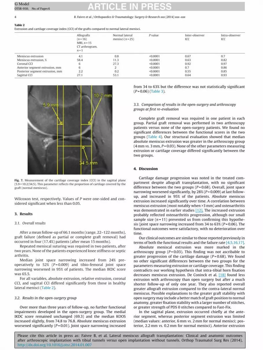
margin of the posterior segment and the posterior margin of thetibial cartilage (Figs. 5 and 6). These same sagittal and coronal viewswere used to measure the cartilage coverage index (CCI) as the per-centage of tibial cartilage covered by the graft, which we computedby determining the ratio of tibial cartilage covered by the graft overthe total tibial cartilage (Figs. 4 and 7). To our knowledge, the CCIhas not been used previously.
To validate our measurements, we determined the same MRIextrusion parameters in the coronal and sagittal planes for the lat-eral meniscus of each of 25 young healthy patients. Inter-observerand intra-observer variability was assessed by having two seniorsurgeons obtain the measurements. All allograft MRI measure-ments were performed by the same senior surgeon, who had norole in any of the surgical procedures. Table 2 reports the intra-classcoefficients (ICCs) for inter-observer and intra-observer variabil-ity. The interval between the two measurements used to assessintra-observer variability was three months.
2.5. Statistics
Descriptive statistics were computed for all the study data.Continuous variables were described as mean ± SD or median(interquartile range) depending on their distribution. Categoricalvariables were described as number (%).
us allograft transplantation: Clinical and anatomic outcomeslantation without tunnels. Orthop Traumatol Surg Res (2014),
Between-group comparisons of mean values were performedusing Student’s t test when the underlying assumptions were metand Fisher’s nonparametric test otherwise. Similarly, to comparepercentages, we chose the chi-square test and non-parametric
ARTICLE IN PRESSG ModelOTSR-958; No. of Pages 6
4 B. Faivre et al. / Orthopaedics & Traumatology: Surgery & Research xxx (2014) xxx–xxx
Table 2Extrusion and cartilage coverage index (CCI) of the grafts compared to normal lateral menisci.
Allografts(n = 16)MRI, n = 15CT arthrogram,n = 1
Normal lateralmenisci (n = 25)
P value Inter-observerICC
Intra-observerICC
Meniscus extrusion 4.1 0.8 <0.0001 0.67 0.7Meniscus extrusion, % 58.4 11.3 <0.0001 0.63 0.82Coronal CCI 6 27.3 <0.0001 0.92 0.97Anterior segment extrusion, mm 6 2
Posterior segment extrusion, mm 2.2 0.2
Sagittal CCI 27.1 53.1
F(g
Ws
3
3
go
ta
onw
Cl
3
iIiw
ig. 7. Measurement of the cartilage coverage index (CCI) in the sagittal plane5.9 + 10.2/34.5). This parameter reflects the proportion of cartilage covered by theraft (normal meniscus).
ilcoxon test, respectively. Values of P were one-sided and con-idered significant when less than 0.05.
. Results
.1. Overall results
After a mean follow-up of 66.1 months (range, 22–122 months),raft failure (defined as partial or complete graft removal) hadccurred in four (17.4%) patients (after mean 15 months).
Repeated meniscal suturing was required in two patients, afterwo years. None of the patients experienced knee stiffness or septicrthritis.
Median joint space narrowing increased from 24% pre-peratively to 52% (P = 0.009) and tibio-femoral joint spacearrowing worsened in 95% of patients. The median IKDC scoreas 65.5.
For all variables, absolute extrusion, relative extrusion, coronalCI, and sagittal CCI differed significantly from those in healthy
ateral menisci (Table 2).
.2. Results in the open-surgery group
Over more than three years of follow-up, no further functional
Please cite this article in press as: Faivre B, et al. Lateral meniscafter arthroscopic implantation with tibial tunnels versus open imphttp://dx.doi.org/10.1016/j.otsr.2014.01.007
mpairments developed in the open-surgery group. The medialKDC score remained unchanged (69.3) and the median KOOSncreased slightly, from 74.8 to 76.8. Absolute meniscus extrusion
orsened significantly (P = 0.01). Joint space narrowing increased
<0.0001 0.7 0.86<0.0001 0.55 0.85<0.0001 0.64 0.93
from 34 to 63% but the difference was not statistically significant(P = 0.06) (Table 3).
3.3. Comparison of results in the open-surgery and arthroscopygroups at first re-evaluation
Complete graft removal was required in one patient in eachgroup. Partial graft removal was performed in two arthroscopypatients versus none of the open-surgery patients. We found nosignificant differences between the functional scores in the twogroups (Table 4). Our structural evaluation showed that medianabsolute meniscus extrusion was greater in the arthroscopy group(4 mm vs. 3 mm, P = 0.03). None of the other parameters measuringextrusion or cartilage coverage differed significantly between thetwo groups.
4. Discussion
Cartilage damage progression was noted in the treated com-partment despite allograft transplantation, with no significantdifference between the two groups (P = 0.68). Overall, joint spacenarrowing worsened significantly, by 28% (P = 0.009) at last follow-up, and increased in 95% of the patients. Absolute meniscusextrusion increased significantly over time. A correlation betweenmeniscus extrusion (most notably when >3 mm) and osteoarthritiswas demonstrated in earlier studies [12]. The increased extrusionprobably reflected osteoarthritis progression, although our smallsample size (n = 11) prevented us from confirming this hypothe-sis (joint space narrowing increased from 34 to 63% (P = 0.06). Thefunctional outcomes were satisfactory, with no deterioration overtime.
Our clinical outcomes are similar to those reported previously interms of both the functional results and the failure rate [4,5,16,17].
Absolute meniscal extrusion was more marked in thearthroscopy group (P = 0.03). This finding was not ascribable togreater progression of the cartilage damage (P = 0.68). We foundno other significant differences between the two groups for theparameters measuring extrusion or cartilage coverage. This findingcontradicts our working hypothesis that intra-tibial horn fixationdecreases meniscus extrusion. De Coninck et al. [18] found lessextrusion with arthroscopy than open surgery but after a muchshorter follow-up of only one year. They also reported overallgreater allograft extrusion compared to the contra-lateral normalmeniscus. Possible explanations to the greater graft stability withopen surgery may include a better match of graft position to normalanatomy, greater fixation stability with a larger number of stitches,or greater strength of PDS 0 stitches compared to Fast-Fix.
us allograft transplantation: Clinical and anatomic outcomeslantation without tunnels. Orthop Traumatol Surg Res (2014),
In the sagittal plane, extrusion occurred chiefly at the ante-rior segment, whereas posterior segment extrusion was limited(median values: anterior, 6 mm vs. 2 mm for normal menisci; pos-terior, 2.2 mm vs. 0.2 mm for normal menisci). Anterior extrusion

ARTICLE IN PRESSG ModelOTSR-958; No. of Pages 6
B. Faivre et al. / Orthopaedics & Traumatology: Surgery & Research xxx (2014) xxx–xxx 5
Table 3Outcomes in the open-surgery group at the first and second re-evaluations.
First re-evaluation Second re-evaluation P value
Follow-up, months 31.8 77.4Survival rate, % 91 91Partial graft removal rate, % 0 9Meniscus suture rate, % 9 18Objective IKDC 1A, 3B, 1C, 1D, 7B, 2DSubjective IKDC 69.5 69.5 1KOOS symptoms 80.4 76.8 0.63KOOS pain 84.7 88.9 1KOOS activities of daily living 94 97 0.72KOOS sports and recreation function 52.5 60 0.55KOOS quality of life 62.5 61.7 0.76Joint space narrowing, % 34 63 0.06Meniscus extrusion, mm 3 4.3 0.01Meniscus extrusion, % 63 60.6 0.67% cartilage coverage, coronal 8.4 5 0.12Anterior segment extrusion, mm 3 7.4 0.08Posterior segment extrusion, mm −1.3 1.4 0.83% cartilage coverage, sagittal 25.9 26.3 0.49
Table 4Comparison of the outcomes in the open surgery and arthroscopy groups.
Open surgery Arthroscopy P value
n 11 12Follow-up, months 31.8 43.4Survival rate, % 91 92 1Meniscus lesion rate, % 18.1 25 0.64Meniscus suture rate, % 9 0Partial graft removal rate, % 0 16.6Joint space narrowing, % 34 55 0.68Objective IKDC 1A, 3B, 1C, 1D, 7B, 2DSubjective IKDC 69.5 47.1 0.37KOOS symptoms 80.4 50 0.63KOOS pain 84.7 64 1KOOS activities of daily living 94 69 0.72KOOS sports and recreation function 52.5 30 0.55KOOS quality of life 62.5 31.3 0.76Meniscus extrusion, mm 3 4 0.03Meniscus extrusion, % 63 52 0.09% cartilage coverage, coronal 8.4 9 0.93
rv
soabteatfnori
uitGaWt
Anterior segment extrusion, mm 3
Posterior segment extrusion, mm −1.3
% cartilage coverage, sagittal 25.9
esulted in decreased cartilage coverage (27.1% with the allograftss. 53.1% for normal menisci).
Despite the smaller degree of absolute extrusion in the open-urgery group, we found no proof that open surgery was superiorver arthroscopy in terms of the clinical outcomes. Koh et al. [19]nd Verdonk et al. [20] underlined the absence of any correlationetween meniscus extrusion and clinical outcomes. Furthermore,he clinical outcomes cannot be ascribed solely to the degree ofxtrusion, as graft recovery and pre-operative cartilage status arelso major factors. Nevertheless, all the functional scores were bet-er in the open-surgery group than in the arthroscopy group and,or some of these scores, the difference approached statistical sig-ificance (P = 0.06). The open-surgery technique is clearly superiorver the arthroscopy technique and that our small sample size andesulting limited statistical power prevented us from demonstrat-ng this point.
Extrusion occurs almost consistently regardless of the techniquesed. Graft extrusion always exceeds the normal extrusion seen
n healthy knees and also exceeds the 3-mm cut-off consideredo indicate an osteoarthritic process. In a study of native knees,ale et al. [12] found a correlation between meniscus extrusion
Please cite this article in press as: Faivre B, et al. Lateral meniscafter arthroscopic implantation with tibial tunnels versus open imphttp://dx.doi.org/10.1016/j.otsr.2014.01.007
nd tibio-femoral joint space narrowing. Bennett and Buckland-right [21] showed that tibio-femoral narrowing was due initially
o cartilage wear and meniscus extrusion. Thus, allograft extrusion
3.3 0.792.35 0.0928 0.56
and the resulting cartilage exposure are probably ascribable to theprogression of joint space narrowing over time.
In our study, there is no evidence that trans-tibial horn fix-ation produced better outcomes. Wajsfisz et al. [22] establishedthe anatomic feasibility of arthroscopically-assisted lateral MAT inwhich horn fixation is achieved without tunnels, thereby sparingthe bone stock. Abat et al. [23] found no evidence that a tech-nique involving bone plugs was superior over absence of boneplugs.
Other factors may affect the development of meniscus graftextrusion. Excessive graft size has been incriminated [15]. Janget al. [24–26] showed that decreasing graft size by 5% to ensurepre-tensioning of the allograft was associated with diminishedextrusion. The lateral meniscus is not normally attached to theanterior capsule. Excessively tight stitches to secure the lateralgraft to the anterior capsule may pull the graft forward, explainingthe development of anterior extrusion contrasting with the limiteddegree of posterior extrusion. We no longer suture the anterior seg-ment. The absence of inter-meniscal reconstruction may contributeto the anterior extrusion. The role for this ligament is unclear [27].It does not connect the horns but instead extends from the mid-
us allograft transplantation: Clinical and anatomic outcomeslantation without tunnels. Orthop Traumatol Surg Res (2014),
dle of the anterior segment of the lateral meniscus to the anteriorsegment of the medial meniscus, thus closing the peripheral menis-cal belt and possibly contributing to pre-tensioning of the anterior

ING ModelO
6 ology:
li
stCsrc(t
racfirm
5
riimo
ae
etiri
D
N
r
R
[
[
[
[
[
[
[
[
[
[
[
[
[
[
[
[
[
ARTICLETSR-958; No. of Pages 6
B. Faivre et al. / Orthopaedics & Traumat
ateral meniscus. Reconstruction of this ligament is not performedn any of the available techniques.
Strengths of our study include the single-centre design with twourgeons and the accurate MRI evaluation of meniscus extrusion inhe coronal and sagittal planes. We introduced a new criterion, theCI, to evaluate the percentage of the graft that actually fulfils thehock-absorbing role of the meniscus. To improve measurementeproducibility, we deliberately measured all the parameters at theentre of the joint space, in contradistinction to previous studiesmeasurement of maximal extrusion as opposed to extrusion athe centre of the joint space).
The main limitations of our study are the small sample size andetrospective data collection. The pre-operative scores were notvailable and we were consequently unable to assess the degree oflinical improvement. The presence in the open-surgery group ofour (36%) patients with a history of varus osteotomy versus nonen the arthroscopy group may have biased the interpretation of theesults. We were unable to obtain dynamic MRI scans to assess graftobility during knee flexion.
. Conclusion
The clinical outcomes in our patients are similar to thoseeported previously in both groups. Thus, horn fixation throughntra-tibial tunnels during arthroscopy-assisted surgery did notmprove the results. The outcomes were not influenced by the
edium-term cartilage status. Joint space narrowing increasedver time.
Absolute and relative graft extrusion occurred at the anteriornd middle segments, resulting in exposure of the cartilage. Thisxtrusion was less marked with the open-surgery technique.
Our findings fail to confirm our working hypothesis. Meniscusxtrusion is not solely dependent on horn fixation, regardless ofhe technique used. Other factors that deserve further evaluationnclude the quality of peripheral suturing, whether or not the ante-ior segment of the lateral graft is sutured, and the role for thenter-meniscal ligament.
isclosure of interest
P. Beaufils: occasional educational consultant for Smith &ephew and Zimmer company.
B. Faivre, P. Boisrenoult, G. Lonjon, N. Pujol have no interest inelation with this article.
eferences
[1] Roos H, Lauren M, Adalberth T, Roos EM, Jonsson K, Lohmander LS. Kneeosteoarthritis after meniscectomy: prevalence of radiographic changes after21 years, compared with matched controls. Arthritis Rheum 1998;41:687–93.
[2] Beaufils P, Hulet C, Dhénain M, Nizard R, Nourissat G, Pujol N. Clinical practiceguidelines for the management of meniscal lesions and isolated lesions of the
Please cite this article in press as: Faivre B, et al. Lateral meniscafter arthroscopic implantation with tibial tunnels versus open imphttp://dx.doi.org/10.1016/j.otsr.2014.01.007
anterior cuciate ligament of the knee in adults. Orthop Traumatol Surg Res2009;95:437–42.
[3] Verdonk PC, Verstraete KL, Almqvist KF, et al. Meniscal allograft transplan-tation: long term clinical results with radiological and magnetic resonanceimaging correlations. Knee Surg Sports Traumatol Arthrosc 2006;14:694–706.
[
PRESS Surgery & Research xxx (2014) xxx–xxx
[4] Stollsteimer GT, Shelton WR, Dukes A, Bomboy AL. Meniscal allograft trans-plantation: a 1- to 5-year follow-up of 22 patients. Arthroscopy 2000;16:343–7.
[5] Verdonk PC, Demurie A, Almqvist KF, Veys EM, Verbruggen G, Verdonk R.Transplantation of viable meniscal allograft. Survivorship analysis and clinicaloutcome of one hundred cases. J Bone Joint Surg Am 2005;87:715–24.
[6] Garrett JC, Steensen RN. Meniscal transplantation in the human knee: a pre-liminary report. Arthroscopy 1991;7:57–62.
[7] Milachowski KA, Weismeier K, Wirth CJ. Homologous meniscus transplanta-tion, experimental and clinical results. Int Orthop 1989;13:1–11.
[8] Shelton WR, Dukes AD. Meniscus replacement with bone anchors: a surgicaltechnique. Arthroscopy 1994;10:324–7.
[9] Rueff D, Nyland J, Kocabey Y, Chang HC, Caborn DN. Self-reported patient out-comes at a minimum of five years after allograft anterior cruciate ligamentreconstruction with or without medial meniscus transplantation: an age, sex,and activity level matched comparison in patients aged approximately 50 years.Arthroscopy 2006;22:1053–62.
10] Zhang H, Liu X, Wei Y, Hong L, Geng WS, Wang XS, et al. Meniscal allografttransplantation in isolated and combined surgery. Knee Surg Sports TraumatolArthrosc 2012;20:281–9.
11] Boisrenoult P, Pujol N. Arthroscopic technique with bone plugs. In: Beaufils P,Verdonk R, editors. The meniscus. Berlin, Heidelberg: Springer; 2010. p. 343–8.
12] Gale DR, Chaisson CE, Totterman SMS, Schwartz RK, Gale ME, Felson D. Menis-cal subluxation: association with osteoarthritis and joint space narrowing.Osteoarthritis Cartilage 1999;7:526–32.
13] Lee DH, Kim TH, Lee SH, Kim CW, Kim JM, Bin SI. Evaluation of meniscus allo-graft transplantation with serial magnetic resonance imaging during the firstpostoperative year: focus on graft extrusion. Arthroscopy 2008;24:1115–21.
14] Puig L, Monllau JC, Corrales M, Pelfort X, Melendo E, Caceres E. Factors affectingmeniscal extrusion: correlation with MRI, clinical, and arthroscopic findings.Knee Surg Sports Traumatol Arthrosc 2006;14:394–8.
15] Lee BS, Chung JW, Kim JM, Kim KA, Bin SI. Width is a more important predictorin graft extrusion than length using plain radiographic sizing in lateral meniscaltransplantation. Knee Surg Sports Traumatol Arthrosc 2012;20:179–86.
16] Rath E, Richmond J, Yassir W, Albright J, Gundogan F. Meniscal allograft trans-plantation: two- to eight-year results. Am J Sports Med 2001;29:410–4.
17] Roumazeille T, Klouche S, Rousselin B, Bongiorno V, Graveleau N, Bil-lot N, et al. Arthroscopic meniscal allograft transplantation with twotibia tunnels without bone plugs: evaluation of healing on MR arthrogra-phy and functional outcomes. Knee Surg Sports Traumatol Arthrosc 2013,http://dx.doi.org/10.1007/s00167-013-2476-1 [Epub ahead of print (PMID:23508524)].
18] De Coninck T, Huysse W, Verdonk R, Verdonk P. Open versus arthroscopicmeniscus allograft transplantation: magnetic resonance imaging study ofmeniscal radial displacement. Arthroscopy 2013;29:514–21.
19] Koh YG, Moon HK, Kim YC, Park YS, Jo SB, Kwon SK. Comparison of medial andlateral meniscal transplantation with regard to extrusion of the allograft, andits correlation with clinical outcome. J bone Joint Surg Br 2012;94:190–3.
20] Verdonk P, Depaepe Y, Desmyter S, et al. Normal and transplanted lateral kneemenisci: evaluation of extrusion using magnetic resonance imaging and ultra-sound. Knee Surg Sports Traumatol Arthrosc 2004;12:411–9.
21] Bennett LD, Buckland-Wright JC. Meniscal and articular cartilage changesin knee osteoarthritis: a cross-sectional double-contrast macro-radiographicstudy. Rheumatology 2002;41:917–23.
22] Wajsfisz A, et al. A new arthroscopic technique for lateral meniscal allo-graft transplantation: cadaver feasibility study. Orthop Traumatol Surg Res2013;734:1–6.
23] Abat F, Gelber PE, Erquicia JI, Tey M, Gonzalez-Lucena G, Monllau JC. Prospectivecomparative study between two different fixation techniques in meniscal allo-graft transplantation. Knee Surg Sports Traumatol Arthrosc 2013;21:1516–22.
24] Pollard ME, Kang Q, Berg EE. Radiographic sizing for meniscal transplantation.Arthroscopy 1995;11:684–7.
25] Jang SH, Kim JG, Ha JG, Shim JC. Reducing the size of the meniscal allograftdecreases the percentage of extrusion after meniscal allograft transplantation.Arthroscopy 2011;27:914–22.
26] Berhouet J, et al. Meniscus matching: evaluation of direct anatomical, indirect
us allograft transplantation: Clinical and anatomic outcomeslantation without tunnels. Orthop Traumatol Surg Res (2014),
radiographic, and photographic methods in 10 cadaver knees. Orthop Trauma-tol Surg Res 2013;731:1–7.
27] Masouros SD, McDermott ID, Amis AA, Bull AM. Biomechanics of the meniscus-meniscal ligament construct of the knee. Knee Surg Sports Traumatol Arthosc2008;16:1121–32.


