Embed Size (px)
Citation preview

Hindawi Publishing CorporationInternational Journal of PeptidesVolume 2011, Article ID 969818, 11 pagesdoi:10.1155/2011/969818
Research Article
Leptin and Fasting Regulate Rat Gastric Glucose-RegulatedProtein 58
Susana B. Bravo,1 Jorge E. Caminos,1, 2, 3 Carmen R. Gonzalez,1, 2 Marıa J. Vazquez,1, 2
Marıa F. Garces,3 Libia A. Cepeda,3 Marıa E.R. Garcıa-Rendueles,1
Antonio Iglesias-Gamarra,3 Consuelo Gomez-Dıaz,4 Miguel Lopez,1, 2
Justo P. Castano,2, 5 Carlos Dieguez,1, 2 and Ruben Nogueiras1, 2
1 Department of Physiology, School of Medicine, Instituto de Investigaciones Sanitarias (IDIS), University of Santiago de Compostela,15782 Santiago de Compostela, Spain
2 CIBER Physiopathology of Obesity and Nitrition (CIBERobn), S. Francisco s/n, 15782 Santiago de Compostela, Spain3 Department of Physiology and Internal Medicine, School of Medicine, National University of Colombia, 11001000 Bogota, Colombia4 Central Service of Proteomic Unity for Research (SCAI), University of Cordoba, 14071 Cordoba, Spain5 Department of Cell Biology, Physiology, and Immunology, University of Cordoba and Maimonides Institute of Biomedical Research(IMIBIC), 14014 Cordoba, Spain
Correspondence should be addressed to Jorge E. Caminos, [email protected] Ruben Nogueiras, [email protected]
Received 23 June 2011; Revised 19 August 2011; Accepted 19 August 2011
Academic Editor: Jean-Marie Zajac
Copyright © 2011 Susana B. Bravo et al. This is an open access article distributed under the Creative Commons AttributionLicense, which permits unrestricted use, distribution, and reproduction in any medium, provided the original work is properlycited.
The stomach secretes a wide range of peptides with essential metabolic functions, and thereby plays an important role in theregulation of energy homeostasis. Disulfide isomerase glucose-regulated protein 58 (GRp58) is a molecular chaperone memberof the endoplasmic reticulum (ER) stress signaling pathway, which is a marker for human gastric cancer. Since GRp58 seemsto be regulated by a phosphorylation/dephosphorylation pattern shift, we used the 2DE gel methodology and peptide massfingerprinting-protein identification by means of MALDI-TOF mass spectrometry. We show that gastric mucosa GRp58 isdephosphorylated by fasting, and this effect is blunted when fasted rats are treated with leptin. Furthermore, we assessed thegene expression of GRp58 under different physiological settings known to be associated with energy homeostasis (fasting, leptintreatment and leptin deficiency). We found that intraperitoneal administration of leptin increases whereas leptin deficiencydecreases GRp58 mRNA levels. However, GRp58 expression remains unchanged after fasting, indicating that leptin actions onGRp58 are no direct sensitivity to fasting. Dissection of the molecular pathways mediating the interactions between ER stress-related factors and nutrient availability, as well as their target genes, may open a new avenue for the study of obesity and othermetabolic disorders.
1. Introduction
The stomach is a central metabolic crossroad wherein nu-merous signals converge and depart to control nutrient in-take, appetite, and general metabolic homeostasis [1]. Thus,data gleaned over the last few years have shown that thestomach plays a key role in the regulation of food intakethrough the secretion of peptides such as ghrelin [2]. In ad-dition, gastric emptying through the regulation of gastric
distension also influence the rate of intestinal exposure ofnutrients thereby influencing the secretion of a vast array ofgastrointestinal hormones which in turn also control foodintake and/or energy expenditure. Therefore, the stomachhas emerged as a key organ in the regulation of bodyweight homeostasis, and gastric surgery is at the front lineof treatment of patients with morbid obesity. However, thereis a general lack of knowledge on the adaptation process thattake part in the stomach in relation to energy balance. We felt

2 International Journal of Peptides
the critical question pending was to identify the subcellularevents that take place in this tissue and its relationship toalterations in energy balance.
Disulfide isomerase glucose-regulated protein 58 (alsoknown as, GRp58, ERp57, ER-60, ERp60, ERp61) is a molec-ular chaperone member of the ER-stress signaling pathway.Specifically, GRp58 is a 58 kDa thiol-disulfide oxidoreductaseprotein, physiologically involved in the folding catalystsmodification of disulfide bonds in glycoprotein [3, 4], andhas been previously identified in the stomach as a markerfor human gastric cancer [5]. Moreover, it is also highlyexpressed in liver, placenta, and lung, and weakly expressedin other tissues [4]. In vitro and in vivo studies have shownthat GRp58 protein acts on glycosylated substrates throughthe ER resident lectins-calnexin and the calreticulin systemto catalyze the isomerization or exchange of disulfide bonds[6, 7]. GRp58 substrates share common properties: they areglycoprotein, heavily disulfide bonded, and likely to formnonnative disulfide bonds [6, 7]. Most of these substratescontain abundant domains of cysteine residues, whichusually promote formation of the disulfide bonds duringglycoprotein folding [6–8]. In line with the interrelationamong metabolism, ER-stress and chaperone function, it hasbeen shown that GRp58 expression is induced by differentstress conditions, such as glucose deprivation or hypoxia[9, 10]. Furthermore, hepatic GRp58 was phosphorylatedafter 12-h/24-h fasting and after leptin treatment [11], andinteracts with the JAK-STAT signaling pathway implicated inthe intracellular signal transduction of leptin [12].
Although GRp58 is a marker for human gastric cancer[5], few studies have addressed the regulation and potentialrole of ER-stress, and in particular that of GRp58, in gastricfunction. We aimed to investigate the involvement of GRp58in energy homeostasis. Our results demonstrate that GRp58is expressed in rat gastric and reveal that gastric GRp58mRNA expression and dephosphorylation are increased afterleptin treatment. Therefore, our results suggest that gastricGRp58 might play an important role in the regulation ofenergy homeostasis.
2. Methods and Procedures
2.1. Animals. Adult male Sprague Dawley rats (250–300 g,8–10 weeks old) were bred in the animalario General USC;University of Santiago de Compostela, Spain, and maleleptin-deficient mice (8 weeks old) were purchased fromCharles River, Barcelona, Spain. Animals were housed inopen cages under conditions of controlled illumination (12-hour light/dark schedule), humidity, and temperature. Theanimals were sacrificed by decapitation in a room separatefrom other experimental animals in the afternoon (16:00-17:00 h). The gastric mucosa was then collected and frozenat −80◦C until analysis. All experiments were conducted inaccordance with the European Union Laws on protectionregarding laboratory animals after previous approval by theEthics Committee of the University of Santiago de Com-postela, Spain (Permit number: PGIDIT06PXIB208063PR).
2.1.1. Experimental Setting 1: Effects of Fasting. Animalgroups (n = 8 per group) were deprived of food for 48 hwhile the control group was fed ad libitum [13]. All animalshad free access to tap water. Gastric mucosa was rapidly dis-sected and stored at−80◦C until proteomic and mRNA anal-yses were performed.
2.1.2. Experimental Setting 2: Effects of Leptin Treatment andLeptin Deficiency. We next investigated whether leptin affectsgastric mucosa GRp58 mRNA expression and modificationsin the protein phosphorylation pattern shift. Rats and micewere assigned to one of the following groups (n = 8 pergroup): (a) i.p. vehicle fed ad libitum; (b) i.p. leptin fedad libitum; (c) i.p. vehicle after 48 h fasting and (d) i.p.leptin after 48 h fasting. On the other hand, leptin-deficientanimals were distributed in three groups: (a) i.p. vehicle fedad libitum, (b) i.p. vehicle after 48h-fasting and (c) i.p. leptinafter 48h fasting. Animals were treated with recombinantleptin (L-4146, Sigma-Aldrich) at a dose of 0.8 μg/kg of bodyweight every 12 h for 3 days (intraperitoneal injection) asdescribed elsewhere [13].
For the proteomic setting, we treated rats with leptin atshort term: 6 h and 24 h before the sacrifice with a singleintraperitoneal injection (0,8 μg/kg of body weight in avolume of 0.25 mL). We again had the same 4 groups (n = 4per group). The reason we carried out this short-term ex-periment for proteomic approaches is that the pattern shiftof phosphorylation/dephosphorylation is commonly fasterthan changes at transcriptional level.
2.2. Western Blot Analysis. Western blot analysis from 1D and2D gels were performed as described elsewhere [14]. Briefly,gastric mucosa proteins were resolved by polyacrylamide gelelectrophoresis according to the method described belowand transferred to nitrocellulose membrane (Hybond C-Super, Amersham Pharmacia Biotech). Membranes wereincubated with primary antibodies against Erp57 (H-220;sc-28823. Santa Cruz Biotechnology, Santa Cruz, CA, USA)and alpha-tubulin (Sigma; Poole, UK). The membranes afterincubation with respective secondary antibodies were probedwith HRP-conjugated secondary antibodies and the signalwas developed with chemiluminescence reagents (Tropix,Bedford; MA, USA) and exposed to X-ray film. All theexperiments were repeated four times and a representativeresult is shown.
2.3. Two-Dimensional Electrophoresis. 2DE analyses wereperformed using four samples per group. Gastric mucosa waslysed at 4◦C using a tissue homogenizator (Omni Tis-sueMaster-125 Homogenizers). Lysates were prepared in lysisbuffer (7 M urea, 2 M thiourea, 4% CHAPS, 5 mM mag-nesium acetate, 20 mMTris HCl pH 8.5, 0.2% ampholytesw/v, and bromophenol trace). Samples were centrifuged at15000 g under 4◦C during 15 min to remove the debris asdescribed previously [15].
First dimension isoelectric focusing was performed withthe horizontal Multiphor IPGphor III system (GE Health-care, formerly Amersham Biosciences); using 11 cm IPG

International Journal of Peptides 3
strips (pH 4–7, GE Healthcare). The sample (50 μg) wasdissolved by soaking in 200 μL of rehydration buffer (7 MUrea, 2 M thiourea, 4% CHAPS and bromophenol trace)during 8 hours. Then, it was loaded onto an 11 cm-lin-ear range with pH 4–7 immobilized gradient strip (4 gelsper experimental group and each one originated from dif-ferent animals) and isoelectric focusing was performed asrecommended by the manufacturer, ramping the voltagesubsequently applied to reach 6000 Vht. The strips contain-ing focused proteins were thawed, equilibrated, and reducedin equilibration buffer (6 M urea, 50 mM M Tris-Cl pH8.8, 2% SDS, 30% glycerol, and 2% (w/v) DTT) during15 min, then alkylated in equilibration buffer (6 M urea,50 mM Tris-Cl pH 8.8, 30% glycerol, 2% SDS, 2.5% (w/v)iodoacetamide. Equilibrated strips were transferred to a SDS-PAGE gel (10%), secured in place by molten agarose and sub-jected to electrophoresis (Mini-Protean Tetra Cell; Bio-RadLaboratories). Afterwards, electrophoresis gels were stainedusing PlusOne Silver Staining Kit, Protein (GE Healthcare),as recommended by the manufacturer protocols.
2.4. MALDI-TOF/TOF Mass Spectrometry and Protein Iden-tification. Spots were excised automatically in a ProPicstation (Genomic Solutions, UK) and digested with modifiedporcine trypsin (sequencing grade; Promega), by using aProGest digestion station (Genomic Solutions, U.K.). Thegel specimens were destained twice over 30 min at 37◦Cwith 200 mM ammonium bicarbonate/40% acetonitrile. Gelpieces were then subjected to three consecutive dehydrata-tion/rehydratation cycles with pure acetonitrile and 25 mMammonium bicarbonate in 50% acetonitrile, respectively,and finally dehydrated for 5 min with pure acetonitrile anddried out over 4 h at room temperature. Then, 20 μL trypsin,at a concentration of 12.5 ng/μL in 25 mM ammonium bi-carbonate was added to the dry gel pieces and the digestionproceded at 37◦C for 12 h. Peptides were extracted from gelplugs by adding 1 μL of 10% (v/v) trifluoracetic acid (TFA)and incubating for 15 min. Then, they were desalted andconcentrated by using μC-18 ZipTip columns (Millipore)in a ProMS station (Genomic Solutions, U.K.) and directlyloaded onto the MALDI plate using α-cyano hydroxycin-namic acid as the matrix.
Mass analysis of peptides of each sample were per-formed with a MALDI-TOF/TOF (4700 Proteomics Ana-lyzer, Applied Biosystems, USA) in automatic mode with thefollowing setting: for the MS data, m/z range 800 to 4000with an accelerating voltage of 20 kV and delayed extraction,peak density of maximum 50 peaks per 200 Da, minimal S/Nratio of 10 and maximum peak at 65. Spectra were internallycalibrated with peptides from trypsin autolysis (M + H+ =842.509, M + H+ = 2211.104).
For the MS/MS data, mass range was set between 60 Daand 10 Da below each precursor mass, with a minimum S/Nratio of 5, a maximum number of peak set at 65 and peakdensity of maximum 50 peaks per 200 Da. Proteins wereassigned identification by peptide mass fingerprinting andconfirmed by MS/MS analysis. Mascot 2.0 search engine(Matrix Science, U.K.) was used for protein identification
running on GPS software (Applied Biosystems) over theNational Center for Biotechnology Information (NCBI) pro-tein database (updated monthly).
Search setting allowed one missed cleavage with the se-lected trypsin enzyme, an MS/MS fragment tolerance of0.2 Da and a precursor mass tolerance of 100 ppm. Proteinswith a statistically significant (P < 0.05) were positively as-signed identification after considering Mr and pI values.
2.5. RNA Isolation and GRp58 mRNA Expression Analysisby RT-PCR. Total RNA from gastric mucosa was extractedusing Trizol (Invitrogen, Inc.) according to the manufac-turer’s instructions. RNA was reverse transcribed and theresulting cDNA was synthesized from 2 ug total RNA byrandom priming RT. The resulting cDNA was subjectedto PCR amplification as described at [13, 16] using senseand antisense primers specific for the rat GRp58 and thehousekeeping gene hypoxanthine guanine phosphoribosyl-transferase (HPRT) mRNAs (Table 1).
2.6. Real-Time Semiquantitative RT-PCR. For real-time sem-iquantitative analysis we used the Applied Biosystems 7500Real-Time PCR System (Applied Biosystems, CA, USA), aspreviously described [13–15]. Rat GRp58 and 18S ribosomalrRNA relative gene expression quantification was made usingspecific primers and TaqMan fluorescent probes for thesetarget genes, and the sequences are described in Table 1.
2.7. Statistical Analysis. The results are shown as the means± SEM (standard error of the mean) and analyzed by usingGraphPad Instat 3.05 (GraphPad Software, Inc.) software.Statistical significance was determined by Student’s t-testwhen two groups were compared. In the experimentsconstituted by three groups the data were analyzed by one-way ANOVA followed by a post hoc multiple comparison test(Bonferroni test). P < 0.05 was considered significant.
3. Results
3.1. Rat Gastric GRp58 mRNA Expression. Expression of themRNA encoding GRp58 gene (Figure 1) was evaluated byRT-PCR and compared to housekeeping gene HPRT. Thisanalysis demonstrates the expression of the message ofthe gene in male rat liver and gastric mucosa whereas itsexpression was not found in tissues such as pancreas andbrain (Figure 1).
3.2. Rat Gastric GRp58 Protein Levels. After discovering thatGRp58 is expressed in the gastric mucosa by means ofRT-PCR, we employed Western blot analysis to determinewhether the message is translated into protein. Using aspecific primary antibody for GRp58, we detected a majorband in protein extracts of rat gastric, and observed thatprotein levels of GRp58 were decreased after 24 h and 48 hof fasting (Figure 2). Moreover, in the gastric mucosa ofrats fasted during 48 h, we found that the band had a lowermolecular weight, likely suggesting a dephosphorylation(Figure 2).

4 International Journal of Peptides
Table 1: PCR primers and probe sequence for rat GRp58 and 18S ribosomal rRNA.
Primer/probe Sequence Genebank accession number
GRP58FW 5′-GGACCAGCTTCAGTTCCTCTCA-3′
NM 017319.1GRP58RV 5′-TGCTGGCTGCTTTTAGGAACTC-3′
GRP58Pb FAM 5′-ATGCCTCGGTGGTGGGCTTTTTCA- 3′TAMRA
18SFW 5′-CGGCTACCACATCCAAGGAA-3′
M11188.118SRV 5′-GCTGGAATTACCGCGGCT-3′
18SPb FAM 5′-GACGGCAAGTCTGGTGCCAGCA-3′TAMRA
HPRT FW 5′-CAGTCCCAGCGTCGTGATTA-3′NM 012583
HPRT RV 5′-AGCAAGTCTTTCAGTCCTGTC-3′
HPRT GRp58
Live
r
Gas
tric
mu
cosa
Pan
crea
sB
rain
RT
-P
CR
-
Live
r
Gas
tric
mu
cosa
Pan
crea
sB
rain
RT
-
PC
R-
Figure 1: Expression of GRp58 gene in rat gastric. RepresentativeRT-PCR assay of the expression levels of GRp58 mRNA in liverand gastric mucosa whereas pancreas and brain failed to show anyamplification. HPRT was used as a housekeeping gene.
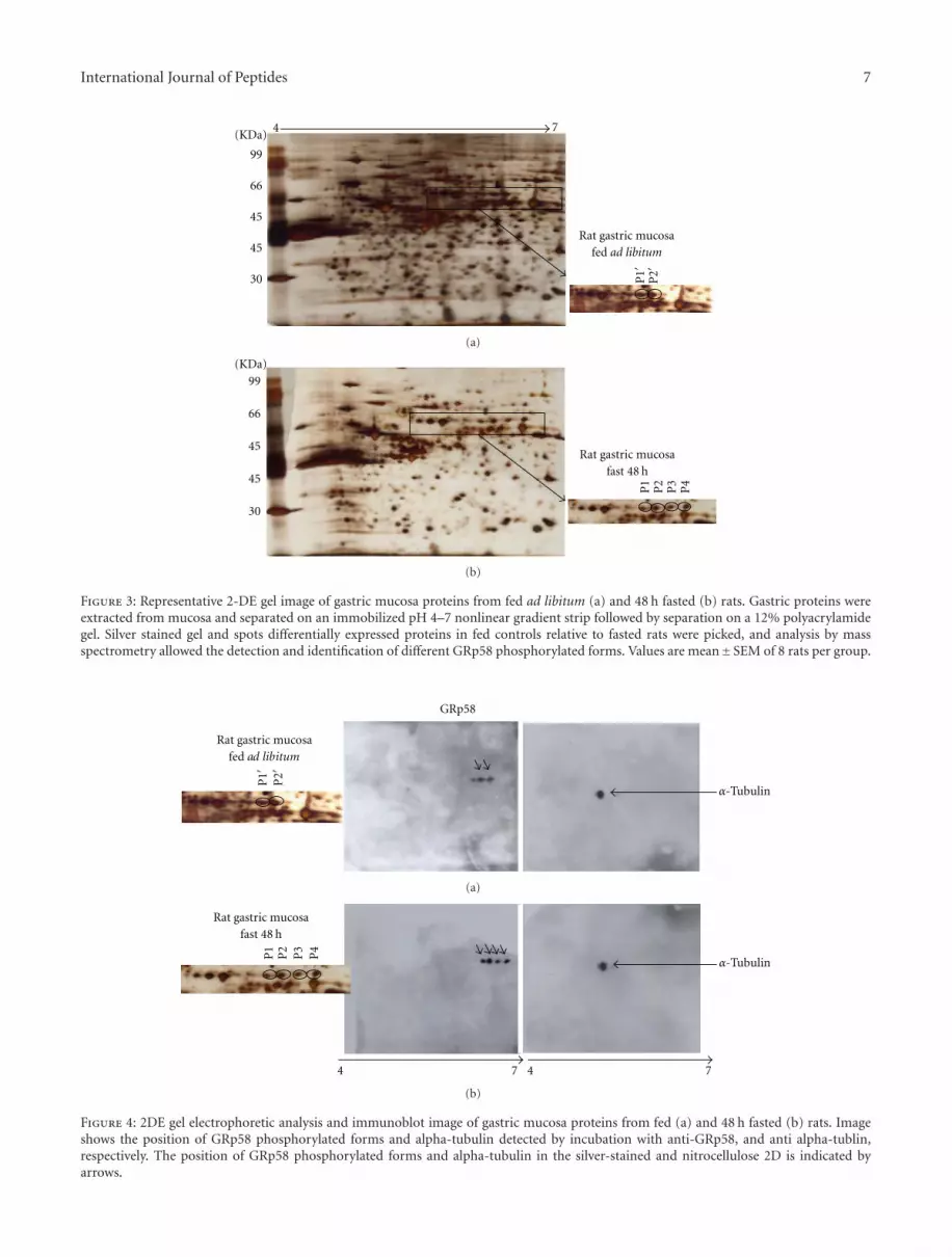
3.3. Proteomic Identification and Molecular Analysis. To testthe hypothesis that GRp58 was dephosphorylated duringfasting, we performed proteomic analysis. Representative 2-DE gel images of rat gastric tissue with differential expressedprotein in fed state (Figure 3(a)) compared to fasted state(Figure 3(b)). Differentially expressed protein spots (markedwith letters) were picked, digested with trypsin, and analyzedby peptide mass fingerprinting as previously described(Tables 2 and 3) [17, 18]. The peptide mass fingerprintswere acquired by mass spectrometry using a matrix-assistedlaser desorption/ionization time-of-flight (MALDI-TOF).All analyzed spots correspond to multiple phosphorylatedforms of GRp58 protein, which were identified by thecomparison of peptide mass fingerprint data generatedby MALDI-TOF mass spectrometry against theoreticallydigested Homo sapiens, Swiss-Prot and TrEMBL database,and by the use of Mascot software. Among the identified pro-tein spots, a 4-protein series with identical molecular weightof approximately 58 kDa and different isoelectric pointswere identified as GRp58. These differences reflect differentdegrees of phosphorylation as previously described [19–21].The identified phosphorylated forms of GRp58 protein inrat gastric along with referred data are listed in Table 3.These phosphorylation profile shifts were identified through a2-DE Western blot analysis using an anti-GRp58 antibody,
Table 2: Cutoff score value for protein: 71 (score is −10 ∗ Log(P),where P is the probability that the observed match is a randomevent. Protein scores greater than 71 are significant (P < 0.05)).The software used for peak-picking was 4000 Series Explorer (TM)RAC Software, version 3.5.3 (Applied Biosystems/MDS SCIEX,Concord, Ontario, Canada). Parameters and thresholds used forpeak-picking: (a) intensity or S/N threshold: S/N = 10, local noisewindow width (m/z) = 250, min peak width at full width halfmax (bins) = 2.9; (b) means of calibrating each spectrum: internalcalibration with peptides from trypsin autolysis (M + H+ = 842.509,M + H+ = 2211.104); (c) resolution: 12000 for the mass 842.51and 18000 for the mass 2211. Search parameters: (a) MASCOT(Matrix Science, London, UK) software, VERSION 2.0; (b) enzymespecificity: trypsin; (c) missed cleavages permitted: 1; (d) fixedmodification (s): carbamidomethyl (C); (e) variable modifications:oxidation (M), Phospho (ST), Phospho (Y); (f) mass tolerancefor precursor ions: ±100 ppm; (g) mass tolerance for fragmentions: ±0.2 Da; (h) name of database searched and release version:NCBInr 20080628 (6655203 sequences); (i) species restriction:Mammalia (mammals) (689751 sequences); (j) acceptance criteria:cut-off score value for protein: 71 (score is -10∗Log(P), where P isthe probability that the observed match is a random event. Proteinscores greater than 71 are significant (P < 0.05)).
Spot Noproteinscore
proteinscore 1%
Coverage%
Number ofpeptides
identified
P1′ 170 100 60 13
P2′ 149 100 50 12
P1 282 100 90 24
P2 439 100 95 24
P3 288 100 64 15
P4 237 100 55 12
recognizing four immunoreactive protein spots (Figures 4(a)and 4(b)) that showed a basic shifted signal in the profile ofGRp58.
3.4. Fasting Induces GRp58 Dephosphorylation Whereas LeptinBlunts Fasting-Induced Dephosphorylation of Gastric MucosaGRp58. Additionally, using 2DE, we found that 48 h offasting dephosphorylated gastric mucosa GRp58 in com-parison with rats fed ad libitum (Figure 5). When 48 hfasted-rats were treated with leptin for 6 hours or 24 h, we

International Journal of Peptides 5
Table 3: Identification of GRp58 protein in the gastric mucosa of the rat. Rat gastric proteins were separated by 2-DE and identified bymeans of MALDI-TOF. The protein identified represents the spots shown in Figures 1(a) and 1(b): protein disulfide-isomerase A3 precursor(Disulfide isomerase ER-60; ERp60; 58 kDa microsomal protein; p58; ERp57; HIP-70; Q-2; 35 petides), the spots labelled as P1′ and P2′ arethe same spots as P1 and P2 but in different gels.
Protein MW: 57043.9 Protein PI: 5.88 Accession No: gi|1352384|Observed Mr (expt) Mr (calc) ± da ± ppm Start-end Sequence Spot number
823.4785 823.4559 0.0226 27 190–196 IVAYTEK p1′,p1
866.4574 866.4617 −0.0043 5 387–393 YKELGEK p1′,p1
877.4915 877.489275 0.0025 3 268–275 LNFAVASR p1′,p1
995.5628 995.5632 −0.0004 0 102–111 QAGPASVPLR p1′,p1
997.5101 997.459 −0.0511 −51 153–161 DASVVGFFR p2
1084.5674 1084.5688 0.0014 1 95–104 YGVSGYPTLK p2′,p1,p2,P3
1123.6582 1123.6589 0.0007 1 101–111 KQAGPASVPLR p1′,p1
1125.547 1125.5436 0.0034 3 243–251 NTKGSNYWR p1
1172.5404 1172.4702 −0.0702 −60 336–344 FVMQEEFSR p1,p2
1179.5865 1179.6239 0.0374 32 174–183 AASNLRDNYR p2′,p1,p2
1188.5354 1188.5305 −0.0049 −4 344–336 FVMQEEFSR p2′,p1,p2,P3
1191.6005 1191.6084 0.0079 7 63–73 LAPEYEAAATR p1′,p2′,p1,p2,P3,P4
1236.5127 1236.5029 −0.0098 −8 108–119 DGEEAGAYDGPR p2′,p1,p2,P3
1244.6633 1244.6276 −0.0357 −29 184–194 FAHTNVESLVK p1,P3
1341.6837 1341.683 −0.0007 −1 449–460 GFPTIYFSPANK p2′,p1,p2,P3,P4
1347.7043 1347.7015 0.0028 2 33–44 RLAPEYEAAATR p1
1373.6736 1373.5839 −0.0897 −65 352–362 FLQEYFDGNLK p2
1394.6587 1394.5729 −0.0858 −62 162–173 DLFSDGHSEFLK p1,p2
1396.6954 1396.6727 −0.0227 −16 367–379 SEPIPETNEGPVK p2′,p1,p2,P3,P4
1397.5784 1397.7063 0.1279 92 83–94 VDCTANTNTCNK p1′,p2′,p2,P3,P4
1397.7059 1397.7063 0.0004 0 472–482 ELNDFISYLQR p1′,p2′,p1,P3,P4
1469.7787 1469.691 −0.0877 −60 449–461 GFPTIYFSPANKK p1,p2,P4
1472.6838 1472.6184 −0.0654 −44 336–347 FVMQEEFSRDGK p2
1488.6787 1488.6698 −0.0089 −6 336–347 FVMQEEFSRDGK P3
1529.7747 1529.775 0.0003 0 352–363 FLQEYFDGNLKR p1′,p2′,p1,p2,P3,P4
1593.8483 1593.8551 0.0068 4 483–496 EATNPPIIQEEKPK p2′,p1,p2,P3,P4
1607.7476 1607.764 0.0164 10 259–271 DLLTAYYDVDYEK p1′,p2′,p2,P3,P4
1636.7523 1636.6552 −0.0971 −59 434–448 MDATANDVPSPYEVK p1,p2,P4
1652.7472 1652.6615 −0.0857 −52 434–448 MDATANDVPSPYEVK p2
1652.7662 1652.7704 0.0042 3 105–119 IFRDGEEAGAYDGPR p1′,p2′,p1,p2,P3,P4
1744.8864 1744.7698 −0.1166 −67 131–146 QAGPASVPLRTEDEFK p2
1746.9286 1746.8311 −0.0975 −56 289–304 TFLDAGHKLNFAVASR p1′
1800.9377 1800.9132 −0.0245 −14 364–379 YLKSEPIPETNEGPVK p1′,p2′,p1,p2,P3,P4
1950.9331 1950.7816 −0.1515 −78 259–274 DLLTAYYDVDYEKNTK p2
2463.1279 2462.9673 −0.1606 −65 83–104 VDCTANTNTCNKYGVSGYPTLK p2
observed that the fasting-induced GRp58 dephosphorylationwas blunted (Figure 5). Therefore, our results indicate thatthe dephosphorylation of gastric mucosa GRp58 inducedby fasting, a hypoleptinemic state, is likely mediated bycirculating leptin levels.
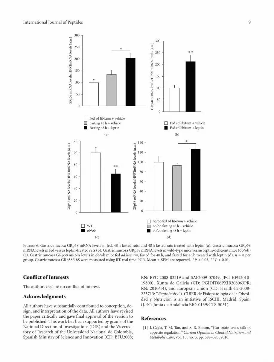
3.5. Gastric Mucosa GRp58 mRNA Levels Are Induced byLeptin. We then measured the mRNA expression of gastricmucosa GRp58 in the same experimental paradigm de-scribed above, meaning, fed ad libitum, fasted, and fasted
leptin-treated rats. We failed to detect any significant changein the gene expression of GRp58 of fasted rats when com-pared to feed ad libitum rats (Figure 6(a)). However, wefound that leptin treatment was able to trigger gastric mu-cosa GRp58 mRNA levels in fasted rats in comparisonwith vehicle-treated rats (Figure 6(a)). Similar results weredetected in fed ad libitum rats, which showed a clearupregulation in GRp58 gene expression after the leptintreatment (Figure 6(b)). According to pharmacological data,the endogenous lack of leptin downregulated gastric mucosa

6 International Journal of Peptides
Fed 24 h 48 h
GRp58
Tubulin
Fast
(a)
∗ ∗
∗ ∗ ∗
∗
0
20
40
60
80
100
120
140
160
Pro
tein
leve
ls(G
Rp5
8/tu
bulin
)
Fed ad libitumFasting 24 hFasting 48 h
(b)
Figure 2: Representative Western blot analysis (a) and quantifi-cation (b) of rat gastric GRp58 protein levels in fasted rats. Fiftymicrograms of total proteins were loaded on a 10% SDS-PAGEgel. To confirm equal loading, the same blot was stripped off
and incubated with monoclonal beta-tubulin antibody. Values aremean± SEM of 8 rats per group.
GRp58 gene expression when compared to wild-type mice(Figure 6(c)). Finally, when leptin-deficient mice were fastedfor 48 h we failed to detect changes in GRp58 mRNA levels,but when those fasted mice were treated with exogenousleptin, an increase in gastric mucosa GRp58 gene expressionwas found (Figure 6(d)). Overall, our findings show thatleptin is an important mediator of GRp58 gene expressionin the gastric mucosa.
4. Discussion
The endoplasmic reticulum (ER) is a multifunctional or-ganelle that plays a critical role in multiple cellular processesincluding, among others, protein and lipid biosynthesis, ster-oid production, calcium homeostasis, and carbohydrate me-tabolism [6]. In addition, the ER contains a large numberof calcium-dependent molecular chaperones and foldingenzymes, which are physiologically responsible for severalcotranslational and posttranslational modifications [22, 23].Alterations in energy metabolism including glucose/energydeprivation, oxidative injury, hypoglycemia, hypoxia, andhigh-fat diet impair ER homeostasis. More specifically,nutritional excess induces ER stress in subcutaneous adiposetissue of obese human subjects [24]. Additionally, ER stresshas been related to the development of atherosclerosis anddiabetes [24–27]. For instance, obese humans and rodentsdevelop ER stress in liver and adipose tissues, leading toinsulin resistance whereas weight-loss decreases ER stressand improves insulin sensitivity, suggesting a correlationbetween ER stress and the metabolic syndrome [27, 28].
By using proteomics analysis, we found evidence of aGRp58 phosphorylation/dephosphorylation pattern shift inthe rat gastric under changes in nutritional status. Morespecifically, using the 2DE gel methodology and peptide massfingerprinting-protein identification by means of MALDI-TOF mass spectrometry, we show that gastric mucosa GRp58is dephosphorylated by fasting, and this effect is bluntedwhen fasted rats are treated with leptin. GRp58, a well-known stress protein, has been previously identified in thestomach as a marker for human gastric cancer [5]. Fur-thermore, previous works have demonstrated the presenceof leptin and leptin receptor in the stomach, suggestingthat gastric mucosa cells may be targets for leptin [29, 30].GRp58 and signal transducer and activator of transcription3 (STAT3), an essential signaling molecule mediating thebiological actions of leptin [31], have been localized in boththe cytoplasm and nucleus [32, 33]. In this regard, somestudies have shown that GRp58 may be implicated in thetranscriptional control of STAT3 [12, 32]. Notwithstandingthe evidence mentioned above, there was a general lack ofknowledge regarding a potential role of gastric GRp58 inmetabolism. Concurring with a role of gastric GRp58 onenergy balance, we demonstrate that leptin induces, whereasleptin-deficiency reduces, gastric mucosa GRp58 mRNAexpression, suggesting that leptin modulates both GRp58phosphorylation and expression. Since these effects seem tobe nutritional, it is plausible to hypothesize that other tissuesthat are target of leptin action in nutritional context like theintestine might be also involved in the regulation of GRp58.For instance, leptin is expressed and secreted by inflamedcolonic epithelial cells [34], it may act as a growth factorin colon tissue [35]. Moreover, leptin receptors have beenlocated in the rat colonic epithelium [36]. Thus, the role ofleptin on GRp58 throughout the entire gastrointestinal tractdeserves further investigation.
Our findings obtained in rat gastric differ from data ob-tained in the rat liver, where it was found that serine 150of GRp58 was phosphorylated by both fasting and leptin
International Journal of Peptides 7
P1′
P2′30
45
45
66
99
(KDa)74
Rat gastric mucosafed ad libitum
(a)
Rat gastric mucosafast 48 h
P1
P2
P3
P4
30
45
45
66
99
(KDa)
(b)
Figure 3: Representative 2-DE gel image of gastric mucosa proteins from fed ad libitum (a) and 48 h fasted (b) rats. Gastric proteins wereextracted from mucosa and separated on an immobilized pH 4–7 nonlinear gradient strip followed by separation on a 12% polyacrylamidegel. Silver stained gel and spots differentially expressed proteins in fed controls relative to fasted rats were picked, and analysis by massspectrometry allowed the detection and identification of different GRp58 phosphorylated forms. Values are mean±SEM of 8 rats per group.
α-Tubulin
GRp58
Rat gastric mucosafed ad libitum
P1′
P2′
(a)
α-Tubulin
Rat gastric mucosafast 48 h
74 74
P2
P3
P4
P1
(b)
Figure 4: 2DE gel electrophoretic analysis and immunoblot image of gastric mucosa proteins from fed (a) and 48 h fasted (b) rats. Imageshows the position of GRp58 phosphorylated forms and alpha-tubulin detected by incubation with anti-GRp58, and anti alpha-tublin,respectively. The position of GRp58 phosphorylated forms and alpha-tubulin in the silver-stained and nitrocellulose 2D is indicated byarrows.

8 International Journal of Peptides
48 h fasted rats
6 h leptin-treated rats 24 h leptin-treated rats
74 pH74 pH
ad libitum rats
P1
P2
P3
P4
P1
P2
P3
P4
P1
P2
P3
P4
P1′
P2′
Figure 5: 2-DE gel image of GRp58 phosphorylation pattern of rat gastric from fed, 48 h fasted rats, and 48 h fasted rats treated with leptinand sacrificed after 6 h and 24 h of leptin administration. GRp58 protein acidic isoelectric points decreased in 2DE gels in fasted and leptintreated rats compared to vehicle-treated control fed rats. Gastric mucosa proteins were extracted and separated on an immobilized pH 4–7 non-linear gradient strip followed by separation on a 12% polyacrylamide gel. Red arrows show the different phosphorilation patternobserved in different treatments.
[11]. Thereby, those results were indicating that nutritional-induced changes in liver GRp58 phosphorylation were notmediated by circulating leptin levels. Our current datademonstrating that leptin blocks the fasting-induced de-phosphorylation of gastric GRp58 suggest that changes inthe activity of this protein are modulated by nutritionalstatus. Importantly, leptin seems to be responsible for thosenutritional-induced changes in gastric GRp58 phosphoryla-tion. Moreover, the different results obtained in the patternof GRp58 phosphorylation in the liver [11] and the stomachsuggests that GRp58 is regulated by nutritional status ina tissue-specific manner. Since we have detected multiplephosphorylated forms of GRp58 protein in the gastricmucosa (Figure 2), we hypothesize that these phosphorylatedforms could contribute to the tissue-specific regulation ofGRp58 in response to fasting and leptin treatment.
It is important to point out several issues that deservefurther attention. First, the functional role of phosphory-lation/dephosphorylation of GRp58 caused by physiologi-cal and physiopathological conditions is largely unknown.Second, the sites of phosphorylation/dephosphorylation ofGrp58 are different in each tissue [11, 37], so it might bepossible that the GRp58 phosphorylated forms that wedetected in the gastric mucosa also have different phosphory-lation sites. Third, the factors that induce these phosphoryla-tion/dephosphorylation shifts in a tissue-specific manner aremostly unknown. All these issues seem relevant to elucidatethe precise role and mechanisms mediating GRp58 actions inthe organism.
In addition to the actions of nutritional status on the pat-tern of gastric mucosa GRp58 phosphorylation, we have alsoassessed the regulation of GRp58 gene expression. Our find-ings indicate that leptin increases GRp58 mRNA expression
whereas leptin deficiency decreases the levels of this protein.Since leptin lowers blood glucose levels and leptin-deficientmice are hyperglycemic, our results are in agreement withprevious in vitro results indicating that GRp58 expression isinduced by glucose deprivation [38].
Although GRp58 mRNA expression remained un-changed after fasting, we found that its protein levels were de-creased after 24 h and 48 h of fasting. These results suggestthat fasting modulates GRp58 at post-transcriptional levels.Therefore, it is likely that the actions of leptin on GRp58are dependent of nutritional status. On the other hand, theingestion of specific components of the diet like fructoseincreases the secretion of gastric leptin [39], and this mightalso affect GRp58 gastric levels.
Taken together, our findings obtained on the dephos-phorylation and total mRNA expression of GRp58 indicatethat leptin regulates the phosphorylation and expression ofgastric GRp58 in rats. It also seems reasonable to hypothesizethat at least some of the actions of leptin at the gastric levelmight be mediated by GRp58 [40].
In conclusion, our results demonstrate that GRp58 phos-phorylation responds rapidly to changes in dietary energyand leptin treatment, and thereby support that GRp58 canplay an important physiological role in the signaling path-ways related to energy balance in the stomach. More pre-cisely, our findings indicate that (a) fasting dephosphorylatesgastric mucosa GRp58, (b) leptin blunts fasting-inducedGRp58 dephosphorylation, and (c) leptin stimulates gastricmucosa mRNA expression. Dissection of the molecular path-ways mediating the interactions between ER stress-relatedfactors and nutrient availability, as well as their target genesmay open a new avenue for the study of obesity and othermetabolic disorders.
International Journal of Peptides 9
0
50
100
150
200
250
300
∗
GR
p58
mR
NA
leve
ls/H
PR
TmR
NA
leve
ls(a
.u.)
Fed ad libitum + vehicle
Fasting 48 h + leptinFasting 48 h + vehicle
(a)
0
50
100
150
200
250
300
GR
p58
mR
NA
leve
ls/H
PR
TmR
NA
leve
ls(a
.u.)
∗∗
Fed ad libitum + vehicleFed ad libitum + leptin
(b)
GR
p58
mR
NA
leve
ls/H
PR
TmR
NA
leve
ls(a
.u.)
∗∗
0
20
60
40
80
100
120
WTob/ob
(c)
∗
0
20
60
40
80
100
120
140
ob/ob fed ad libitum + vehicleob/ob fasting 48 h + vehicleob/ob fasting 48 h + leptin
GR
p58
mR
NA
leve
ls/H
PR
TmR
NA
leve
ls(a
.u.)
(d)
Figure 6: Gastric mucosa GRp58 mRNA levels in fed, 48 h fasted rats, and 48 h fasted rats treated with leptin (a). Gastric mucosa GRp58mRNA levels in fed versus leptin-treated rats (b). Gastric mucosa GRp58 mRNA levels in wild-type mice versus leptin-deficient mice (ob/ob)(c). Gastric mucosa GRp58 mRNA levels in ob/ob mice fed ad libitum, fasted for 48 h, and fasted for 48 h treated with leptin (d). n = 8 pergroup. Gastric mucosa GRp58/18S were measured using RT-real time PCR. Mean ± SEM are reported. ∗P < 0.05, ∗∗P < 0.01.
Conflict of Interests
The authors declare no conflict of interest.
Acknowledgments
All authors have substantially contributed to conception, de-sign, and interpretation of the data. All authors have revisedthe paper critically and gave final approval of the version tobe published. This work has been supported by grants of theNational Direction of Investigations (DIB) and the Vicerrec-tory of Research of the Universidad Nacional de Colombia,Spanish Ministry of Science and Innovation (CD: BFU2008;
RN: RYC-2008-02219 and SAF2009-07049, JPC: BFU2010-19300), Xunta de Galicia (CD: PGIDIT06PXIB208063PR;RN: 2010/14), and European Union (CD: Health-F2-2008-223713: “Reprobesity”). CIBER de Fisiopatologıa de la Obesi-dad y Nutricion is an initiative of ISCIII, Madrid, Spain.(J.P.C: Junta de Andalucıa BIO-0139/CTS-5051).
References
[1] J. Cegla, T. M. Tan, and S. R. Bloom, “Gut-brain cross-talk inappetite regulation,” Current Opinion in Clinical Nutrition andMetabolic Care, vol. 13, no. 5, pp. 588–593, 2010.

10 International Journal of Peptides
[2] M. Kojima, H. Hosoda, Y. Date, M. Nakazato, H. Matsuo, andK. Kangawa, “Ghrelin is a growth-hormone-releasing acylatedpeptide from stomach,” Nature, vol. 402, no. 6762, pp. 656–660, 1999.
[3] M. Michalak, J. Groenendyk, E. Szabo, L. I. Gold, and M. Opas,“Calreticulin, a multi-process calcium-buffering chaperone ofthe endoplasmic reticulum,” Biochemical Journal, vol. 417, no.3, pp. 651–666, 2009.
[4] P. Koivunen, N. Horelli-Kuitunen, T. Helaakoski et al., “Struc-tures of the human gene for the protein disulfide isomerase-related polypeptide ERp60 and a processed gene and assign-ment of these genes to 15q15 and 1q21,” Genomics, vol. 42, no.3, pp. 397–404, 1997.
[5] C. M. Leys, S. Nomura, B. J. LaFleur et al., “Expression andprognostic significance of prothymosin-α and ERp57 inhuman gastric cancer,” Surgery, vol. 141, no. 1, pp. 41–50,2007.
[6] F. Hatahet and L. W. Ruddock, “Substrate recognition by theprotein disulfide isomerases,” FEBS Journal, vol. 274, no. 20,pp. 5223–5234, 2007.
[7] L. Ellgaard and L. W. Ruddock, “The human protein disul-phide isomerase family: substrate interactions and functionalproperties,” EMBO Reports, vol. 6, no. 1, pp. 28–32, 2005.
[8] P. Maattanen, G. Kozlov, K. Gehring, and D. Y. Thomas,“ERp57 and PDI: multifunctional protein disulfide isomeraseswith similar domain architectures but differing substrate-partner associations,” Biochemistry and Cell Biology, vol. 84,no. 6, pp. 881–889, 2006.
[9] L. Cicchihitti, M. Di Michele, A. Urbani et al., “Compar-ative proteomic analysis of paclitaxel sensitive A2780 epi-thelial ovarian cancer cell line and its resistant counterpartA2780TC1 by 2D-DIGE: the role of ERp57,” Journal ofProteome Research, vol. 8, no. 4, pp. 1902–1912, 2009.
[10] S. Laudi, W. Steudel, K. Jonscher et al., “Comparison of lungproteome profiles in two rodent models of pulmonary arterialhypertension,” Proteomics, vol. 7, no. 14, pp. 2469–2478, 2007.
[11] K. Kita, N. Okumura, T. Takao et al., “Evidence forphosphorylation of rat liver glucose-regulated protein 58,GRP58/ERp57/ER-60, induced by fasting and leptin,” FEBSLetters, vol. 580, no. 1, pp. 199–205, 2006.
[12] G. G. Guo, K. Patel, V. Kumar et al., “Association of the chaper-one glucose-regulated protein 58 (GRP58/ER-60/ERp57) withStat3 in cytosol and plasma membrane complexes,” Journal ofInterferon and Cytokine Research, vol. 22, no. 5, pp. 555–563,2002.
[13] C. R. Gonzalez, M. J. Vazquez, M. Lopez, and C. Dieguez, “In-fluence of chronic undernutrition and leptin on GOAT mRNAlevels in rat stomach mucosa,” Journal of Molecular Endocrinol-ogy, vol. 41, no. 6, pp. 415–421, 2008.
[14] J. E. Caminos, S. B. Bravo, M. E. Garcia-Rendueles et al., “Ex-pression of neuropeptide W in rat stomach mucosa: regulationby nutritional status, glucocorticoids and thyroid hormones,”Regulatory Peptides, vol. 146, no. 1–3, pp. 106–111, 2008.
[15] A. Paradela, S. B. Bravo, M. Henriquez, G. Riquelme, F. Ga-vilanes, and J. P. Albar, “Proteomic analysis of apical microvil-lous membranes of syncytiotrophoblast cells reveals a highdegree of similarity with lipid rafts,” Journal of Proteome Re-search, vol. 4, no. 6, pp. 2435–2441, 2005.
[16] C. R. Gonzalez, J. E. Caminos, R. Gallego et al., “Adiponectinreceptor 2 is regulated by nutritional status, leptin and preg-nancy in a tissue-specific manner,” Physiology and Behavior,vol. 99, no. 1, pp. 91–99, 2010.
[17] V. Goeb, M. Thomas-L’Otellier, R. Daveau et al., “Candidateautoantigens identified by mass spectrometry in early rheuma-toid arthritis are chaperones and citrullinated glycolyticenzymes,” Arthritis Research and Therapy, vol. 11, no. 2, articleR38, 2009.
[18] T. C. Lai, H. C. Chou, Y. W. Chen et al., “Secretomic and prot-eomic analysis of potential breast cancer markers by two-dimensional differential gel electrophoresis,” Journal of Pro-teome Research, vol. 9, no. 3, pp. 1302–1322, 2010.
[19] Y. Tokutomi, N. Araki, K. Kataoka, E. Yamamoto, and S.Kim-Mitsuyama, “Oxidation of Prx2 and phosphorylationof GRP58 by angiotensin II in human coronary smoothmuscle cells identified by 2D-DIGE analysis,” Biochemical andBiophysical Research Communications, vol. 364, no. 4, pp. 822–830, 2007.
[20] M. Obeid, “ERP57 membrane translocation dictates the im-munogenicity of tumor cell death by controlling the mem-brane translocation of calreticulin,” Journal of Immunology,vol. 181, no. 4, pp. 2533–2543, 2008.
[21] T. Panaretakis, N. Joza, N. Modjtahedi et al., “The co-trans-location of ERp57 and calreticulin determines the immuno-genicity of cell death,” Cell Death and Differentiation, vol. 15,no. 9, pp. 1499–1509, 2008.
[22] J. Mandl, T. Meszaros, G. Banhegyi, L. Hunyady, and M. Csala,“Endoplasmic reticulum: nutrient sensor in physiology andpathology,” Trends in Endocrinology and Metabolism, vol. 20,no. 4, pp. 194–201, 2009.
[23] A. R. English, N. Zurek, and G. K. Voeltz, “Peripheral ERstructure and function,” Current Opinion in Cell Biology, vol.21, no. 4, pp. 596–602, 2009.
[24] G. Boden, X. Duan, C. Homko et al., “Increase in endoplasmicreticulum stress-related proteins and genes in adipose tissueof obese, insulin-resistant individuals,” Diabetes, vol. 57, no. 9,pp. 2438–2444, 2008.
[25] N. Naidoo, “ER and aging-Protein folding and the ER stressresponse,” Ageing Research Reviews, vol. 8, no. 3, pp. 150–159,2009.
[26] S. G. Fonseca, M. Burcin, J. Gromada, and F. Urano, “Endo-plasmic reticulum stress in β-cells and development of dia-betes,” Current Opinion in Pharmacology, vol. 9, no. 6, pp. 763–770, 2009.
[27] M. F. Gregor, L. Yang, E. Fabbrini et al., “Endoplasmic ret-iculum stress is reduced in tissues of obese subjects afterweight loss,” Diabetes, vol. 58, no. 3, pp. 693–700, 2009.
[28] G. S. Hotamisligil, “Endoplasmic reticulum stress and the in-flammatory basis of metabolic disease,” Cell, vol. 140, no. 6,pp. 900–917, 2010.
[29] A. Bado, S. Levasseur, S. Attoub et al., “The stomach is a sourceof leptin,” Nature, vol. 394, no. 6695, pp. 790–793, 1998.
[30] I. Sobhani, A. Bado, C. Vissuzaine et al., “Leptin secretion andleptin receptor in the human stomach,” Gut, vol. 47, no. 2, pp.178–183, 2000.
[31] E. C. Villanueva and M. G. Myers Jr., “Leptin receptorsignaling and the regulation of mammalian physiology,”International Journal of Obesity, vol. 32, supplement 7, pp. S8–S12, 2008.
[32] M. I. Ndubuisi, G. G. Guo, V. A. Fried, J. D. Etlinger, and P. B.Sehgal, “Cellular physiology of STAT3: where’s the cytoplasmicmonomer?” Journal of Biological Chemistry, vol. 274, no. 36,pp. 25499–25509, 1999.
[33] M. Eufemi, S. Coppari, F. Altieri, C. Grillo, A. Ferraro, andC. Turano, “ERp57 is present in STAT3-DNA complexes,”Biochemical and Biophysical Research Communications, vol.323, no. 4, pp. 1306–1312, 2004.
International Journal of Peptides 11
[34] N. K. Saxena, M. A. Titus, X. Ding et al., “Leptin as a novelprofibrogenic cytokine in hepatic stellate cells: mitogenesisand inhibition of apoptosis mediated by extracellular regu-lated kinase (Erk) and Akt phosphorylation,” FASEB Journal,vol. 18, no. 13, pp. 1612–1614, 2004.
[35] T. Aparicio, L. Kotelevets, A. Tsocas et al., “Leptin stimulatesthe proliferation of human colon cancer cells in vitro but doesnot promote the growth of colon cancer xenografts in nudemice or intestinal tumorigenesis in ApcMin/+ mice,” Gut, vol.54, no. 8, pp. 1136–1145, 2005.
[36] J. E. Drew, A. J. Farquharson, S. Padidar et al., “Insulin, leptin,and adiponectin receptors in colon: regulation relative todiffering body adiposity independent of diet and in responseto dimethylhydrazine,” American Journal of Physiology, vol.293, no. 4, pp. G682–G691, 2007.
[37] A. Donella-Deana, P. James, W. Staudenmann et al., “Isolationfrom spleen of a 57-kDa protein substrate of the tyrosinekinase Lyn: identification as a protein related to proteindisulfide-isomerase and localisation of the phosphorylationsites,” European Journal of Biochemistry, vol. 235, no. 1-2, pp.18–25, 1996.
[38] M. Flores-Diaz, J. C. Higuita, I. Florin et al., “A cellular UDP-glucose deficiency causes overexpression of glucose/oxygen-regulated proteins independent of the endoplasmic reticulumstress elements,” Journal of Biological Chemistry, vol. 279, no.21, pp. 21724–21731, 2004.
[39] M. H. Vickers, Z. E. Clayton, C. Yap, and D. M. Sloboda,“Maternal fructose intake during pregnancy and lactationalters placental growth and leads to sex-specific changes infetal and neonatal endocrine function,” Endocrinology, vol.152, no. 4, pp. 1378–1387, 2011.
[40] S. Guilmeau, M. Buyse, and A. Bado, “Gastric leptin: a newmanager of gastrointestinal function,” Current Opinion inPharmacology, vol. 4, no. 6, pp. 561–566, 2004.


