Embed Size (px)
Citation preview
HAL Id: tel-00366311https://tel.archives-ouvertes.fr/tel-00366311
Submitted on 6 Mar 2009
HAL is a multi-disciplinary open accessarchive for the deposit and dissemination of sci-entific research documents, whether they are pub-lished or not. The documents may come fromteaching and research institutions in France orabroad, or from public or private research centers.
L’archive ouverte pluridisciplinaire HAL, estdestinée au dépôt et à la diffusion de documentsscientifiques de niveau recherche, publiés ou non,émanant des établissements d’enseignement et derecherche français ou étrangers, des laboratoirespublics ou privés.
Les hormones thyroïdiennes, leurs récepteurs etl’évolution de la métamorphose chez les Chordés.
Mathilde Paris
To cite this version:Mathilde Paris. Les hormones thyroïdiennes, leurs récepteurs et l’évolution de la métamorphose chezles Chordés.. Biochimie [q-bio.BM]. Ecole normale supérieure de lyon - ENS LYON, 2008. Français.�tel-00366311�
THÈSE
Présentée
devant L’ECOLE NORMALE SUPÉRIEURE DE LYON
pour l’obtention
du DIPLÔME DE DOCTORAT
soutenue le 18 décembre 2008
par
M AT H I L D E PA R I S
T H Y RO I D H O R M O N E S , T H E I R R E C E P TO R S
A N D T H E E VO L U T I O N O F M E TA M O R P H O S I S
I N C H O R DAT E S
Jury: Vincent LAUDET Directeur de thèse
Béatrice DESVERGNE Rapportrice
Detlev ARENDT Rapporteur
Philippe JANVIER Examinateur
Nicholas HOLLAND Examinateur
Frédéric FLAMANT Examinateur
Hector ESCRIVA Membre invité

T H Y R O I D H O R M O N E S , T H E I R R E C E P T O R S
A N D T H E E V O L U T I O N O F M E TA M O R P H O S I S
I N C H O R D AT E S
mathilde paris
Doctorate of Life Science
18 December 2008

A B S T R A C T
In an attempt to understand how the regulation of development evolves, particular
attention has been put on transcription factors, which regulate gene expression
during development. Among transcription factors, nuclear hormone receptors
(NRs) have a peculiar status linked to their ligand-dependent activity. However,
their role in the evolution of development is still poorly understood. In this
context, I studied during my thesis the evolution of the estrogen receptor (ER)
and of the thyroid hormone receptor (TR) in chordates (including vertebrates,
urochordates like sea squirt and cephalochordates like amphioxus), by focusing on
the amphioxus species Branchiostoma floridae.
Combined molecular and bioinformatic approaches allowed me to show that the
amphioxus ER does not bind estrogen, suggesting that estrogen binding appeared
in the vertebrate lineage. In parallel, a large part of my thesis was dedicated to
understand how metamorphosis evolved in the chordate lineage, as a paradigm of
the evolution of a developmental process. Although most chordates metamorphose,
the morphological changes during larva-to-adult transitions vary extensively from
one species to another. Does the molecular determinism of metamorphosis in
this group reflect this morphological diversity? In the well-studied vertebrates,
metamorphosis is triggered by thyroid hormones (THs) binding to their receptor
TR, member of the NR superfamily. In order to get better insight into the evolution
of the molecular determinism of metamorphosis in chordates, I focused on the most
basal chordate amphioxus. Combined biochemical and phylogenetic approaches
allowed me to establish that amphioxus produces various THs through metabolic
pathways homologous to vertebrate ones. Then I showed that TH-dependent
TR activation is essential for metamorphosis induction in amphioxus, like in
vertebrates, with the slight difference that the active TH is not T3, the classical
vertebrate TH, but possibly its derivative TRIAC. Consequently the homology
of metamorphosis in chordates is revealed by the conservation of its triggering
mechanism. This suggests that the evolution of metamorphosis in chordates is
marked by the conservation of the couple TH/TR whereas other parts of the
regulatory network may change to underlie the morphological diversity observed
nowadays.
3
R É S U M É
Un des principaux objectifs de ma thèse a été de comprendre comment les pro-
cessus du développement évoluent. Dans ce contexte, les facteurs de transcription
sont importants car ils régulent l’expression génique au cours du développement.
Parmi les facteurs de transcription, les récepteurs nucléaires (RNs) ont un statut
particulier car leur activité est régulée par un ligand. Le rôle qu’ils jouent dans
l’évolution du développement et plus généralement leur évolution (leur fonction,
leur capacité à lier un ligand...) sont mal compris. Durant ma thèse, je me suis
intéressée à l’évolution des RNs chez les chordés (comprenant les vertébrés, les
urochordés tels que les tuniciés et les céphalochordés tels que l’amphioxus) en
étudiant deux RNs particuliers : le récepteurs aux oestrogènes (ER) et le récepteur
aux hormones thyroïdiennes (TR). Les études que j’ai menées ont été réalisées en
particulier chez l’amphioxus Branchiostoma floridae.
J’ai montré que l’ER d’amphioxus ne lie pas les oestrogènes, suggérant que
cette capacité est apparue chez les vertébrés. La majeure partie de ma thèse a
été dédiée à l’étude de l’évolution de la métamorphose chez les chordés. Bien
que la plupart des chordés métamorphosent, les modifications morphologiques
caractéristiques du passage d’une forme larvaire à une forme juvénile sont très
variables d’une espèce à l’autre. Cette variabilité morphologique se reflète-telle
dans le déterminisme moléculaire de la métamorphose ? Ce dernier est encore mal
connu en dehors des modèles vertébrés classiques chez qui la métamorphose est
induite par la fixation des hormones thyroïdiennes (HTs) sur TR. Afin de combler
ce manque, je me suis intéressée au protochordé amphioxus. Par des approches
bioinformatiques et biochimiques, j’ai établi que l’amphioxus produit des HTs
par une voie métabolique homologue à celle des vertébrés. J’ai alors montré que
les HTs régulent la métamorphose de l’amphioxus, en modulant l’activité de TR,
comme chez les vertébrés. Une différence notable est la nature de l’HT active :
T3 chez les vertébrés contre TRIAC, un dérivé de T3, chez l’amphioxus. J’ai ainsi
proposé que l’homologie de la métamorphose chez les chordés est soutenue par la
conservation du déterminisme moléculaire de ce processus du développement (le
couple HT/TR) alors que les autre parties de la voie de régulation ont été moins
conservées au cours de l’évolution et expliquent la diversité morphologique que
l’on observe de nos jours.
4
P U B L I C AT I O N S
Holland N. D., Paris M., and Koop D. (2009) The club-shaped gland of am-
phioxus: export of secretion to the pharynx in pre-metamorphic larvae and apopto-
sis during metamorphosis. Acta Zoologica 89, doi: 10.1111/j.1463–6395.(2008)00379.x.
Tocchini-Valentini G. D., Rochel N., Escriva H., Germain P., Peluso-Iltis C.,
Paris M., Sanglier-Cianferani S., Dorsselaer A. V., Moras D., and Laudet V. (2008)
Structural and functional insights into the ligand binding domain of a non-
duplicated RXR from the invertebrate chordate amphioxus. J Biol Chem 284, 1938-
1948.
Paris M., Brunet F., Markov G., Schubert M., and Laudet V. (2008) The amphioxus
genome enlightens the evolution of the thyroid hormone signaling pathway. Dev
Genes Evol 218, 667-680.
Paris M., and Laudet V. (2008) The history of a developmental stage: Metamor-
phosis in chordates. Genesis 46, 657-672.
Schubert M., Brunet F., Paris M., Bertrand S., Benoit G., and Laudet V. (2008)
Nuclear hormone receptor signaling in amphioxus. Dev Genes Evol 218, 651-665.
Paris M., Pettersson K., Schubert M., Bertrand S., Pongratz I., Escriva H., and
Laudet V. (2008) An amphioxus orthologue of the estrogen receptor that does not
bind estradiol: insights into estrogen receptor evolution. BMC Evol Biol, 8, 219.
Paris M., Escriva H., Schubert M., Brunet F., Brtko J., Ciesielski F., Roecklin D.,
Vivat-Hannah V., Jamin E. L., Cravedi J. P., Scanlan T. S., Renaud J. P., Holland
N. D., and Laudet V. (2008). Amphioxus postembryonic development reveals the
homology of chordate metamorphosis. Curr Biol, 18, 825–30.
Markov G. V., Paris M., Bertrand S., and Laudet V. (2008) The evolution of the
ligand / receptor couple: a long road from comparative endocrinology to compara-
5
tive genomics. Molecular and Cellular Endocrinology, 293, 5–16.
Pagès M., Calvignac S., Klein C., Paris M., Hugues S., and Hänni C. (2008)
Combined analysis of fourteen nuclear genes refines the Ursidae phylogeny. Mol
Phylogenet Evol, 47, 73-83.
Fuentes M., Benito E., Bertrand S., Paris M., Mignardot A., Godoy L., Jimenez-
Delgado S., Oliveri D., Candiani S., Hirsinger E., D’Aniello S., Pascual-Anaya J.,
Maeso I., Pestarino M., Vernier P., Nicolas J. F., Schubert M., Laudet V., Geneviere A.
M., Albalat R., Garcia Fernandez J., Holland N. D., and Escriva H. (2007) Insights
into spawning behavior and development of the european amphioxus (Branchios-
toma lanceolatum). J Exp Zool B Mol Dev Evol, 308, 484-493.
Bertrand S., Camasses A., Paris M., Holland N.D., and Escriva H. (2006) Phylo-
genetic analysis of Amphioxus genes of the proprotein convertase family, including
aPC6C, a marker of epithelial fusions during embryology. Int J Biol Sci 2, 125-132.
Hassani Z., François J.-C., Alfama G., Dubois G. M., Paris M., Giovannangeli
C., and Demeneix B. A. (2007) A hybrid CMV-H1 construct improves efficiency of
PEI-delivered shRNA in the mouse brain. Nucleic Acids Res, 35, e65.
Brunet F. G., Crollius H. R., Paris M., Aury J. M., Gibert P., Jaillon O., Laudet
V., and Robinson-Rechavi M. (2006) Gene loss and evolutionary rates following
whole-genome duplication in teleost fishes. Mol Biol Evol, 23, 1808–16.
6
R E M E R C I E M E N T S
Je voudrais tout d’abord remercier les membres de mon jury de thèse Béatrice Desvergne,
Detlev Arendt, Philippe Janvier, Nicholas Holland et Frédéric Flamant pour avoir accepté
de juger mon travail, pour leur indulgence et leurs commentaires précieux.
Je voudrais remercier Vincent Laudet de m’avoir accueillie dans son laboratoire (pendant
5 ans et demi tout de même). On n’a pas assez discuté, on n’a pas toujours été d’accord
mais on a toujours fini par un bon compromis. Je rends hommage à son ouverture d’esprit
et à sa porte toujours ouverte. Vincent m’a donné un passionnant sujet de thèse, qui a
métamorphosé ma vision du développement tardif chez les chordés (non pas que j’en avais
une avant la thèse...). Zoupette à fleurs te remercie, chef.
Je voudrais remercier Hector Escriva pour m’avoir si bien encadrée pendant mon DEA
et ma première année de thèse. Il était disponible et attentif juste comme il faut et est
resté très concerné par mon travail malgré son départ de Lyon. Sa profonde gentillesse
s’accompagne d’un esprit vif et d’une efficacité impressionnante. Une grande partie de ce
que je sais maintenant vient de lui. Alors un respectueux merci à un brillant chercheur.
Je voudrais également chaudement remercier Linda Holland de m’avoir aidée à travailler
sur ce charmant petit ver qu’est l’amphioxus. J’ai été accueillie à Tampa par Linda comme
si j’étais de sa propre équipe, j’ai pu partager la joie de la pêche avec les requins et les raies
(aïe), le visionnage des Simpson et les nuits blanches avec son équipe. Mille merci pour
cette nouvelle variété d’algues Isochrysis qui m’a été d’une utilité que seuls les éleveurs
d’amphioxus peuvent apprécier. Je lui suis extrêmement reconnaissante pour tout ce qu’elle
a fait pour moi. Je voudrais remercier une fois encore Nick pour m’avoir tant apporté. Sa
passion pour la zoologie toujours si vive, son humour et sa disponibilité ont fait de lui un
mentor pour moi, et je le salue respectueusement. C’est aussi grâce à lui que j’ai pu oublier
dans l’alcool de Rachelle mon angoisse à l’idée d’aller chercher des larves d’amphioxus à la
tombée de la nuit dans le garde-manger des requins affamés. Une fausse note toutefois : la
conduite de Nick au volant, un chouïa trop napolitaine au goût de mon cardiologue.
Je tiens également à remercier le plus évolutionniste des spécialistes de la leucémie
promyélocytaire aigüe (et malheureusement pas l’inverse), Gérard B. Il m’a beaucoup
appris, non seulement parce qu’il s’y connaît un max sur les récepteurs nucléaires, mais
aussi, et surtout, parce qu’il a empoisonné mes petits poumons pendant plus de 3 ans avec
ses Lucky Strike. Il a subi en contre-partie mes jérémiades sur TR qui ne lie pas T3, TR
qui lie TRIAC, TR qui n’est pas bien produit en bactéries, TR TR TR... il a été cette oreille
7
attentive et amicale dont j’avais tant besoin. En plus, il a un goût sûr en matière de comté
("c’t’excellent"). Bref un poteau, en plus d’un mentor, qui me manquera beaucoup.
Un grand merci également à Sophie, pour sa passion de la science (une chercheuse
comme il en faudrait d’avantage dans les labos), pour son sourire si lumineux, pour son
intérêt et son aide enthousiastes. Grâce à elle, je ne crible plus les clones comme avant. Je
voudrais également remercier Marie S. Elle a été une voisine charmante, toujours souriante,
et toujours d’une intelligence redoutable. Il faut par contre faire attention avec Marie parce
que si on a un problème et qu’on lui en parle, elle passera le temps qu’il faut pour vous
le résoudre. A ne pas abuser si l’on veut pouvoir se regarder dans un miroir. Un petit
clin d’oeil allumeur à Gérard T, c’est ma façon à moi de le remercier de sa bougritude.
Toutes ces années, il a été d’un réconfortant graveleux. Je tire mon chapeau à son expertise
biochimique, qui m’a bien aidée ainsi qu’à sa dextérité inégalable dans l’art du jeu de mots.
Merci aussi à Pascale qui fait de si bons gâteaux au chocolat et qui m’a fait découvrir les
toutes petites souris (Nannomys pour les intimes).
Je voudrais également remercier Florent pour sa bonne humeur quasi permanente, c’est
un charmant camarade de paillasse. Alors si tu es polonaise, pas catholique intégriste, que
tu as moins de 25 ans, que tu aimes le pâté et les lamproies (mais pas pour les mêmes
raisons), et que les vidéos équestres ne te font pas peur, je connais un brillant thésard qui
cherche une étudiante à encadrer de près. Je remercie également son chef Michael, qui a
bien du mérite, et qui m’a fait profiter de ses talents d’écriture. On a échangé quelques
discussions des plus constructives, quelques bonnes engueulades aussi, et quelques bières.
Il y a également tous les autres membres de l’équipe : la douce et chocolatée Mme
Christelle Gobet Betoulle, l’enthousiaste et geakesque Alexa, Anne et ses petits mots doux,
Laure qui aime tant les poissons (surtout leurs épitaphes), Maria qui perturbe la sexualité
du zebrafish, Charles qui travaille à des heures indues, Jasmine qui travaille trop, Maline
qui se lève trop tôt le matin, Marie T qui apprécie les ambassades...
Je souhaite ne pas oublier les passés au milieu de cet océan de présents. Alors merci à
Stéphanie pour sa gentillesse et sa disponibilité, je lui tire mon chapeau d’avoir fait une
thèse, un post-doc et 2 enfants. Je félicite aussi Yann qui a su mener de front 2 carrières
(chercheur et stadier). Merci aussi au si discret Shin-Ichi, à la joyeuse Lamia, à François
pour son aide du début, à Juliette qui m’a marquée par sa générosité, à Arnaud Martin
qui vole vers d’autres horizons évolutifs, à Arnaud Chaumot qui travaillait dans plus de 3
dimensions, à Fred Brunet qui a fait des progrès en braille grâce à moi et à Raquel pour son
encouragement au nudisme. Je reste admirative de l’amour d’Ingrid pour l’enseignement-
recherche. Je souhaite à Ferdi de s’éclater en post-doc, même s’il y verra moins de poubelles
crâmer qu’à Endoume. J’espère qu’il ne perdra jamais sa "roots" attitude version evo-devo.
Un grand merci à Fred Flamant d’avoir aiguisé mes fantasmes laborantins et pour
l’intérêt qu’il a porté à mon travail. Merci aux autres thésards d’avoir souffert avec moi et
8
d’avoir entre autres ouvert mes yeux sur les pigeons mognons. Merci à Barbara Demeneix
pour son envie de soniquer des amphioxus ainsi qu’aux membres de l’ENS pour leur aide.
Je n’aurais également pas pu réaliser cette thèse sans le soutien de ma famille et en
particulier de mes parents. Ils ont toujours été derrière moi bien qu’ils aient désapprouvé
certains de mes choix (ma mère trouvant que je gaspillais les cônes qu’elle m’a vue jeter
après chaque utilisation : "quel gâchis". Mon père quelque peu déçu de me voir opter pour
la biologie plutôt que pour un hypothétique chapeau). Merci aussi à ma famille d’adoption
pour ses encouragements et ses présents vaudous. Merci aussi à mes amis qui ont toujours
eu autant de mal à se rappeler le nom de la bestiole sur laquelle je travaillais (j’espère que
Samia me pardonnera un jour de ne pas être devenue un vrai docteur).
Enfin, je remercie Bastien pour son soutien de roc et de choc. Pendant toutes ces années,
il s’est toujours ardemment inquiété de savoir si ma PCR avait marché, si j’avais reçu mes
échantillons si précieux, si j’avais lu cet article si intéressant, si j’avais pu discuter avec ce
chercheur que je voulais voir mais n’osais aborder. Il n’est pas plus petit que Shane, fait la
vaisselle mieux que moi et est tellement brillant. C’est mon héros.
Je voudrais terminer ces (longs) remerciements par un rapide portrait sonore du lab-
oratoire. Parce les souvenirs de ces quelques années passées dans l’équipe de Zoologie
Moléculaire passent aussi par les oreilles.
Ambiance au labo: sonnerie du timer que quelqu’un a oublié sur sa paillasse, centri
bien (ou mal) équilibrée, pas d’une personne pressée (un maître de conf’ qui a cours ? Une
manip oubliée ? Un séminaire qui commence ?), aspiration de la hotte, porte de la chambre
froide que l’on ouvre, puis que l’on ferme, cri de désespoir d’un étudiant qui vient de perdre
ses embryons d’in situ, discussion animée sur l’avenir de la recherche. Pour couronner le
tout, certaines personnes ont le mauvais goût d’aimer écouter de la musique : Flamenco
pour Stéphanie pendant la rédaction de sa thèse, jazz pour Charles quand il travaille la
nuit, Muse pour Juliette se préparant au concert du groupe, Joe Dassin pour Florent (et
pourtant il n’a pas de raison !), Scorpions d’Alexa (?????), musique de la petite souris qui
croit aux miracles pour Gérard T, chanson réclamant à Benoît de se retourner...
Ensuite il y a certains sons que chacun fait: le rire de Vincent, les expressions teintées
d’accent espagnol d’Hector ("un tipeu", "c’pas mal"), les énervements de Michael ("ça
m’énerfe", "la pétite"), l’accent du sud d’Arnaud, le rire crystallin de Sophie, les promesses
(non tenues) de fessée de la part de Gérard T, la roulette de souris ou les touches de clavier
de Marie S, la canette de soda que Maria boit dans l’après-midi, le bruit de pas genre "force
tranquille" de Gérard B, le tripotage de mèches de cheveux de Christelle (je sais, ça fait
un bruit limité...), les bracelets d’Ingrid quand elle gigote les bras d’excitation, le silence
éloquent de François qui n’en pense pas moins, le "hâaäà" de Florent, si difficile à imiter
sur support papier, les barbarismes jurassiens de Laure...
9

Ce sont mes petites madeleines à moi.
10
A C R O N Y M S
AR Androgen receptor
CAR Constitutive androstane receptor
CDS Coding DNA sequence
CRE Cis-regulatory element
CRF Corticotropin releasing factor
DNA Deoxyribonucleic acid
DBD DNA binding domain
ER Estrogen hormone receptor
ERR Estrogen-related receptor
FXR Farnesoid X receptor
GR Glucocorticoid receptor
GRN Genetic regulatory network
IOD Deiodinase
IOP Iodopanoic acid
LBD Ligand binding domain
LXR Liver X receptor
MR Mineralocorticoid receptor
NR Nuclear hormone receptor
PERT Thyroid peroxidase
PPAR Peroxysome proliferator-activated receptor
PR Progesterone receptor
PXR Pregnene X receptor
RAR Retinoic acid receptor
11
12 acronyms
ROR Retinoic acid orphan receptor
RNA Ribonucleic acid
RXR Retinoid X receptor
T3 Triiodo-l-thyronine
T3AM 3-triiodothyronamine
T4 l-thyroxine
TAAR Trace amine-associated receptor
TETRAC Tetraiodothyroacetic acid
TF Transcription factor
TG Thyroglobulin
TR Thyroid hormone receptor
TRIAC Triiodothyroactic acid
USP Ultraspiracle

C O N T E N T S
13

L I S T O F F I G U R E S
L I S T O F TA B L E S
14
P R E FA C E
as time goes by
In this preface, intended for a lay audience (and especially my family), I will intro-
duce my PhD work and briefly situate it in a general context, in order to explain
why I chose to work on this subject. More precisely, I will first try to broadly
present evolutionary biology (or at least why it interests me), then I will present
the field of evolution of development, and I will finally introduce metamorphosis
in the context of evolutionary developmental biology. The last paragraph of the
preface describes the content of this manuscript.
One of the main aims of my PhD has been to discuss how metamorphosis
evolved in the chordate lineage. There are two questions that one may ask: why
metamorphosis and why evolution? Let’s answer the second one first.
Although the famous quote of Theodosius Dobzhansky ?: "Nothing in biology
makes sense except in the light of evolution" may sound commonplace for some people,
it does nonetheless well summarize my motivation to study evolution. Indeed, in
the attempt to find mechanistic models that explain biological diversity, the time
parameter is of special importance since it explains why distantly related species
are more different than closely related species, the way cousins look less alike
than brothers and sisters. More generally, as all living organisms share a common
ancestry, going from close relatedness, like humans and chimpanzees, to deep
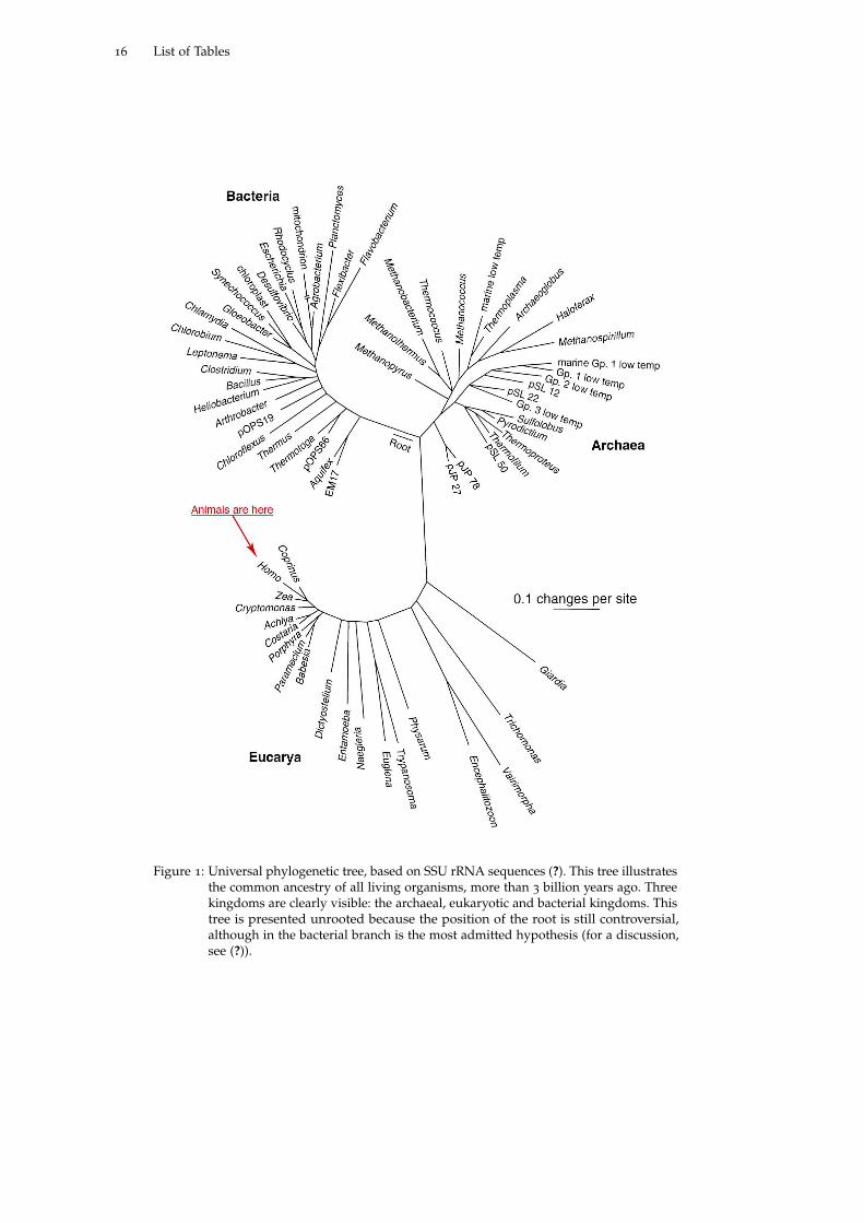
ancestry, forming a tree of life (Figure ??), and as all living organisms differ at
some point, then they necessarily evolved through time from this common ancestor
to extant biodiversity. Consequently many characteristics of any organism find
an explanation in the evolutionary pathway that has been taken from its origins
to current days. For instance, why do so many people get backaches and more
generally why is our skeleton not that well suited for straight position? The recent
origins of mankind from quadrupedal ancestors well explain the resulting bad
back that evolution tinkered from an horizontal position (?). In other words,
There is not a single question in biology that can be answered adequately
without a consideration of evolution (?).
15
16 List of Tables
Figure 1: Universal phylogenetic tree, based on SSU rRNA sequences (?). This tree illustratesthe common ancestry of all living organisms, more than 3 billion years ago. Threekingdoms are clearly visible: the archaeal, eukaryotic and bacterial kingdoms. Thistree is presented unrooted because the position of the root is still controversial,although in the bacterial branch is the most admitted hypothesis (for a discussion,see (?)).
Preface 17
Evolution is implicit in any subfield of biology. For instance, medicine posits
that all organisms of the same species have a similar functioning, and a physician
who uses an animal model to test a particular drug implicitly accepts the common
ancestry of the animal model with human, evolution from this common ancestor
and more specifically inherited similarity (this is homology, see section ??) of the
processes that are scrutinized. Otherwise it would be at the very least immoderate
to consider that an efficient medicine in a mouse may be of any potential interest
for a human being. In other words, people count on conserved physiological
and molecular biological processes between species from a common ancestor to
extrapolate rules applicable to humans from their animal model. Evolution makes
these similarities decrease with time, explaining why mice are less faithful stand-ins
for us than chimpanzees.
While evolutionary concepts are underlying most of biology, the discipline of
evolutionary biology is all about understanding the processes by which the living
world has changed since the origin of life (?). It is this mechanistic approach that
gives an explanation to "how" the biodiversity that we have today arose. And to
understand the evolution of the shapes of organisms, it is important to study the
evolution of development.
evo-devo, a subfield of evolutionary biology
Because evolution unifies all living forms, it can be investigated from virtually
all biological areas of studies. Historically, evolution has been associated with
systematics (in which species are classified following relevant criteria, see section
??), paleontology (to study the evolution of forms from the fossil record) and
population genetics (to study the dynamics of the evolution of mutations in
populations of organisms). Although a joint interest in development and evolution
dates back to the nineteenth century, it got lost in the first half of the twentieth
century, with the building of the modern synthesis. Molecular biology brought
evolution and development back together twenty years ago as a new edge of
evolutionary biology, called evo-devo, in which evolution is scrutinized in the light
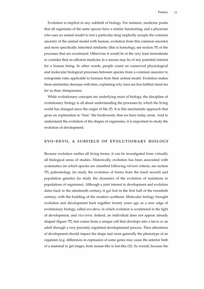
of development, and vice-versa. Indeed, an individual does not appear already
shaped (figure ??), but comes from a unique cell that develops into a larva or an
adult through a very precisely regulated developmental process. Then alterations
of development should impact the shape and more generally the phenotype of an
organism (e.g. differences in expression of some genes may cause the anterior limb
of a mammal to get longer, from mouse-like to bat-like (?)). So overall, because the
18 List of Tables
phenotype is the result of a developmental process, evolution of development is
central to understanding the diversity of forms. To cite Hans Spemann (?):
"We no longer believe, that we first can establish the phylogenetic relations
between animals in order to subsequently derive developmental laws. Rather
we begin to realize, that we first have to determine these laws, before we can
understand or even establish the morphological series that we use to classify
organisms."
Functional approaches are more and more chosen to study issues related to the
evolution of development, to unveil genetic mechanisms underlying the evolution
of species. This makes evo-devo a very powerful tool to understand the “what”
and “how” of multicellular life (?).
Figure 2: Reproduction of a homonculus by Nicolas Hart-soecker (1695). It illustrates the "spermid the-ory" of the origin of babies. According to thistheory, individuals contain in their gametes pre-formed "miniatures" of their descendants, likeRussian dolls. The picture represents a "littleman" already preformed in the head of a sper-matozoid, that simply needs to be deposited inthe mother’s uterus to further grow. This the-ory implies that offsprings come from only thefather, that there is no fertilization and no sub-sequent development.
why studying the evolution of metamorphosis?
During my PhD, I have studied the evolution of metamorphosis in chordates (a
group containing vertebrates, see section ??). In this study I define metamorphosis
as the larva-to-juvenile transformation like tadpole-to-frog or caterpillar-to-butterfly
transitions. More precisely it is a post-embryonic developmental stage allowing
a larva to become a juvenile, and which is characterized by drastic ecological,
morphological, metabolic and behavioral modifications.
Metamorphosis will be considered in this manuscript in two different and com-
plementary ways: (i) for itself and (ii) as a paradigm for developmental processes.
(i) It is a spectacular developmental process that whets imagination: there is some
sort of magic in the abrupt transformation of a tadpole into a frog, or of a maggot
into a fly, and indeed this developmental process does trigger interest among
Preface 19
children (the macroscopic scale at which metamorphosis occurs compared to the
microscopic scale of most other developmental processes may help). Moreover
metamorphosis is so widespread among animals (see chapter ??) that the evolution
of this dramatic event of larva-to-juvenile transition is especially intriguing, consid-
ering the theoretical strong obstacles to such a non-linear developmental strategy.
In addition, and as will be detailed in later chapters, metamorphosis is hormonally
regulated (thyroid hormones in the case of amphibians), which makes it an ideal
model to study the role of endocrine systems during development. Overall, how
the tadpole-to-frog transformation evolved has remained unclear and therefore
deserves more insight.
(ii) Metamorphosis is highly variable among chordates: it is thus difficult to
decipher whether it appeared several times independently during chordate evolu-
tion or was present in the chordate ancestor (chordate metamorphoses would be
homologous) and subsequently diverged widely between species. Metamorphosis
can then be studied as an illustration of difficulties in determining homology and
more generally it can be considered as an example used to decipher the general
rules that constrain the evolution of developmental processes. As explained in the
previous section, the mechanisms through which development evolves and impacts
species phenotypes are getting increasing attention from evolutionary biologists.
My work is a small contribution to this field of evolutionary biology.
During my PhD I have studied the evolution of the genetic network regulating
metamorphosis in chordates. This work led me to propose a model in which the
evolution of metamorphosis at the phenotypic level is underlain by the evolution of
the corresponding genetic network. Before presenting my thesis from the above two
axes, I will first present the evolutionary context of my work (chapter ??). Then I will
briefly introduce the notion of metamorphosis in both broad and developmental
senses (chapter ??), with a special focus on chordates and amphioxus. I will then
describe the regulatory network of metamorphosis in chordates (and especially
amphibians) with an emphasis on the thyroid hormone receptor (chapters ?? and
??) (pieces of these chapters were modified from ?).
After this introduction, I will present my PhD work in the form of articles
recently published: the main part of my work was dedicated to understanding
metamorphosis in amphioxus and especially its molecular determinism. For this
purpose, I have sought into the thyroid hormone signaling pathway in amphioxus
(chapter ??), and its role in the regulation of metamorphosis (chapter ??). To com-
plete this work, unpublished additional experiments were done and are presented
in chapter ??. These articles allowed me to propose a model of the evolution of
metamorphosis in chordates, based on alterations of its molecular determinism

20 List of Tables
(this model is discussed in chapter ??, and uses modified parts of (?)). Contempora-
neously I have also studied the evolution of the estrogen receptor in amphioxus, a
gene closely related to the thyroid hormone receptor (chapters ?? and ??. In chapter
??, the evolution of NRs will also be discussed).

Part I
I N T R O D U C T I O N

1H O M O L O G Y A N D C H O R D AT EP H Y L O G E N Y
1.1 descent with modification and homology
As briefly stated in the preface, all living organisms share a common ancestry, 3.8
billion year ago. Then the evolutionist’s job is to understand how the subsequent
3.8 billion years of evolution gave rise to the extant diversity from the common
ancestor. How to determine what criteria to look for in a given set of species?
1.1.1 "All true classification is genealogical"
During evolution, characteristics of organisms change, sometimes leaving a more
or less easy-to-follow track of their common ancestry (or even no track at all).
One of the most famous examples is the vertebrate anterior limb: the human
arm, the chicken wing, the dolphin flipper, or the amphibian anterior leg (figure
??), evolved from the same structure in their common ancestor, to become a
grasping, flying, swimming or running organ. Even before evolutionary theories
were available, the morphological similitude between body parts had already been
noticed by the French naturalist Geoffroy Saint-Hilaire and reformulated by the
morphologist Richard Owen who proposed that such structures are homologous, i.e.
they corresponded to:
the same organ in different animals under every variety of form and function
(?, p379).
Charles Darwin added the evolutionary (temporal) dimension to the notion of
homology (?, p413):
[...] I believe that something more is included [in our classification, than
mere resemblance]; and that propinquity of descent,—the only known cause
of the similarity of organic beings,—is the bond, hidden as it is by various
degrees of modification, which is partially revealed to us by our classifications.
So two characters are considered homologous if they are:
derived from the same or equivalent feature of their nearest common ancestor
(?).
23
24 homology and chordate phylogeny
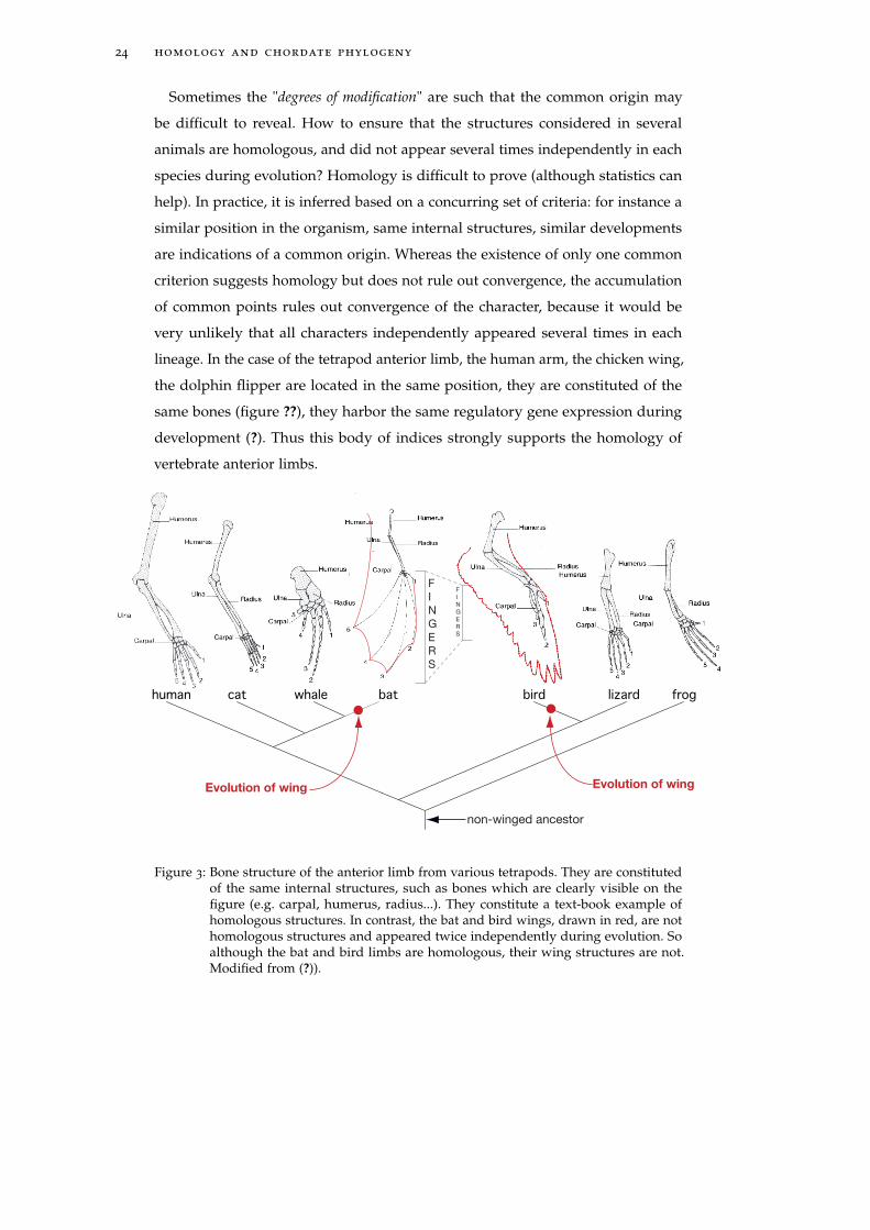
Sometimes the "degrees of modification" are such that the common origin may
be difficult to reveal. How to ensure that the structures considered in several
animals are homologous, and did not appear several times independently in each
species during evolution? Homology is difficult to prove (although statistics can
help). In practice, it is inferred based on a concurring set of criteria: for instance a
similar position in the organism, same internal structures, similar developments
are indications of a common origin. Whereas the existence of only one common
criterion suggests homology but does not rule out convergence, the accumulation
of common points rules out convergence of the character, because it would be
very unlikely that all characters independently appeared several times in each
lineage. In the case of the tetrapod anterior limb, the human arm, the chicken wing,
the dolphin flipper are located in the same position, they are constituted of the
same bones (figure ??), they harbor the same regulatory gene expression during
development (?). Thus this body of indices strongly supports the homology of
vertebrate anterior limbs.
human cat whale bat lizard frog
Evolution of wingEvolution of wing
F
I
N
G
E
R
S
F
I
N
G
E
R
S
bird
non-winged ancestor
Figure 3: Bone structure of the anterior limb from various tetrapods. They are constitutedof the same internal structures, such as bones which are clearly visible on thefigure (e.g. carpal, humerus, radius...). They constitute a text-book example ofhomologous structures. In contrast, the bat and bird wings, drawn in red, are nothomologous structures and appeared twice independently during evolution. Soalthough the bat and bird limbs are homologous, their wing structures are not.Modified from (?)).
1.1 descent with modification and homology 25
1.1.2 Convergence
When comparing a character in different species using a large body of criteria (like
position, shape, internal structure, development...), one should be able to detect
cases of convergence, for which similar and functionnally redundant structures ap-
peared independently during evolution. If we go back to the example of vertebrate
anterior limbs, the bird and bat wings are not considered as homologous. Indeed
the flying surface in bird consists of feathers extending all along the arm whereas
the flight surface in bats consists of a membrane stretched between the bones of
the elongated fingers and the arm (figure ??, highlighted in red). Accordingly, the
300 million year old reptile-like common ancestor from birds and mammals was
most probably non-winged (which is confirmed by fossil record (?)).
1.1.3 Development and homology
Although homology was originally established for morphological structures, dur-
ing my PhD, I have studied the homology of developmental processes. Homologous
structures are not directly inherited but are recreated anew in each generation from
a single cell, through a series of steps during the development of the organism.
Then one would guess that the developments of these homologous structures as
well as their genetic regulation are homologous as well. This is most often the case.
Sometimes, though, the function of some genes involved in the regulation of a
developmental process may not have been conserved whereas the genetic network
has been. For instance, all insects are organized following an anterio-posterior
(A-P) polarity (with the head on one end and the abdomen on the other). However,
the Drosophila bicoid gene, which is at the top of the genetic hierarchy controlling
anterior-posterior (A-P) patterning, is present only in some flies, replacing the func-
tion of Orthodenticle in other insects, whereas downstream parts of the regulatory
network are homologous.
Segmentation (during which the early embryo is divided into a repeating series
of segmental structures along the A-P axis) is another example of homologous
developmental processes regulated by non-homologous genes. Indeed, although
segmentation is ancestral in the insect lineage, the segmental function of the genes
fushi tarazu (ftz) and even-skipped (eve) appeared in some dipterans (like Drosophila),
the genes did not have this function in the ancestral insects (as illustrated for
Schistocerca) (??).
Not only can the development of homologous morphological characters be
regulated by non-homologous genes, but conversely homologous genes may have
26 homology and chordate phylogeny
different functions during development. For instance ftz mentioned above, is a
homeobox gene (the protein contains a homeobox that allows it to bind DNA
on specific sequences). As said before, it regulates segmentation in Drosophila
melanogaster. However, this gene is thought to have displayed a homeotic activity in
the arthropod ancestor, i.e. it could specify regional identity during development.
During insect development, it gained its role in segmentation but also lost its
homeotic activity (?).
Overall, the homology of development, developmental genes and morphology
is not straightforward (??). Some have proposed that homology can be better
understood by considering the evolution of the whole network regulating a de-
velopmental process, rather than considering isolated genes. Then, all parts of
the developmental regulatory network do not encounter the same evolutionary
dynamics and alterations preferentially affect a subset of a network. From the
above example, genes involved in early A-P patterning (bicoid, ftz or eve) are more
prone to evolutionary changes than genes involved in later A-P patterning, like
engrailed which harbors a more conserved expression pattern in all arthropods (?).
Similarly, I have investigated during my thesis whether some parts of the molecu-
lar determinism of metamorphosis in chordates were better conserved than others,
this way revealing a homology of metamorphoses. I focused on the amphioxus
and my results are given in chapters ?? and ??. In chapter ??, a model of chordate
metamorphosis is proposed, based on the differential evolution of parts of the
genetic network regulating metamorphosis, with conserved and more rapidly
evolving modules (?).
1.2 amphioxus and chordates in the phyloge-netic context of my phd
Chordate phylogeny has significantly changed during my thesis. Before summa-
rizing these changes and their impact on my work, I will first briefly introduce
phylogenetic concepts and techniques.
1.2.1 Phylogenetic reconstructions
The essence of phylogenetic reconstruction is as follows: "relevant" characters
are compared between different species; if two species look especially alike (if
they share a similar character repertoire), they are probably “close cousins” that
diverged quite recently. In contrast, if the "likeness" is less pronounced, then the
1.2 amphioxus and chordates in the phylogenetic context of my phd 27
species are probably more distantly related. Several parameters come into play:
the finding of "relevant" characters and a way of measuring "likeness".
Evolutionary documents
Characters are considered as "relevant" if they are homologous (see section ?? for
further details), i.e. they were inherited from a common ancestor. This criterion is
essential in phylogenetic reconstructions that infer the evolutionary path taken by
characters from a common ancestry.
Historically, morphological traits (like the number and position of appendages,
their position...) were first compared and used to build phylogenies. At least some
of them have the advantage of being found in fossils of extinct species that can
be compared to extant ones. However, their number is rather limited and the ho-
mology of morphological characters may be difficult to assess. Nowadays, another
biological material is available and greatly informative regarding phylogeny: the
genome. This source of evolutionary information presents several advantages:
• genes are directly and vertically inherited from parents to offspring (except
in the case of horizontal gene transfer, frequent in prokaryotes but rare in
Eukaryotes)
• genes and genomes are very rich in information (in some species, up to
billions of sites can be considered as characters to approach some questions).
• Several kinds of characters can be used, depending on the question that is
asked:
– the genetic alphabet (A, T, C, G) of nucleic acid
– the amino acid alphabet of proteins
– the gene content of a genome
– the genomic structure (e.g. gene order, exon-intron structure, gaps)
• In our "post-genomic" era, high throughput collection of this genomic mate-
rial is becoming fast and affordable.
• Accurate models of gene, genome and protein evolution are available.
All these reasons make DNA a valuable evolutionary document (?).
Models of phylogenetic recontructions
What about the way of measuring "likeness"? When using morphological char-
acters, the comparative algorithm is usually based on parsimonious reasoning: a
phylogenetic tree is constructed that minimizes the number of evolutionary events.
28 homology and chordate phylogeny
This “parsimonious” tree is considered to be the closest to the real one. This is
the reasonning that was used for the inference of the wing-status of the tetrapod
ancestor in figure ??. There are also other more realistic phylogenetic reconstruc-
tion algorithms, based on statistical models of character evolution (especially for
molecular markers). For instance, likelihood and bayesian methods (??) infer the
evolution of sequences by estimating the most likely tree or a distribution of the
most likely trees of protein or DNA sequences.These methods take into account the
properties of the molecular marker (for instance, some site substitutions have more
drastic consequences than others) and are thus more accurate than parsimonious
methods (?). These phylogenetic methods also allow one to infer the sequence
of ancestral sequences, this way "resurrecting" sequences, that otherwise are not
available to extant evolutionary biologists (?).
In order to study the evolution of genes involved in thyroid hormone signaling
pathway in chordates (chapters ?? and ??) and the evolution of the estrogen hor-
mone receptors (chapter ??), I have used maximum likelihood methods. Moreover,
I have also discussed the relevance of ancestral reconstructions, in the particular
case of the evolutionary study of steroid hormone receptors ( chapter ??).
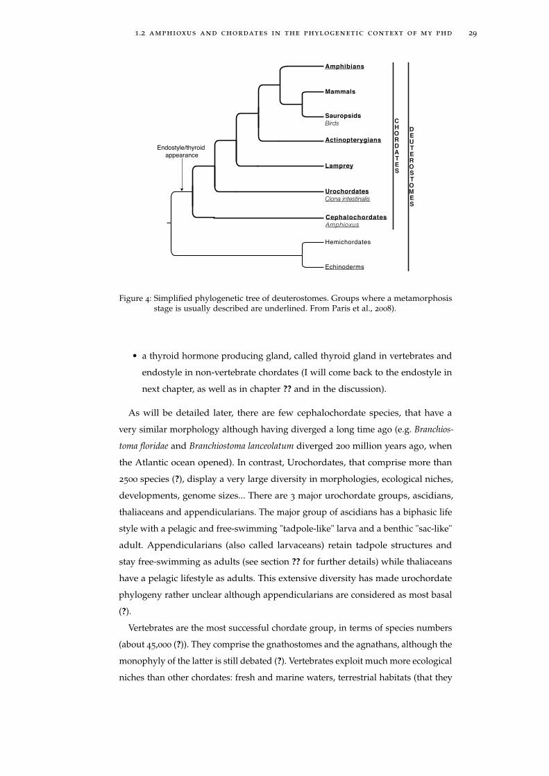
1.2.2 Chordate phylogeny
Chordate characteristics
The chordate lineage comprises cephalochordates (such as the amphioxus Bran-
chiostoma lanceolatum), urochordates (such as the tunicate Ciona intestinalis) and
vertebrates. The vertebrate group comprises notably agnathans (like lamprey and
hagfish), actinopterygian fishes (like Danio rerio), mammals (like us), sauropsids
(including birds) and amphibians (including anurans like Xenopus laevis) (figure
??).
Chordates are united by several characters:
• a notochord, at least at one stage of their development (hence their name). It is
a mesendoderm derivative that stretches along the body between the gut and
the nerve tube. The notochord persists for the entire life in cephalochordates
and only during larval period in some urochordates.
• a hollow dorsal neural tube.
• a postanal tail
• segmented muscles, at least at one stage.
1.2 amphioxus and chordates in the phylogenetic context of my phd 29
Actinopterygians
Amphibians
Hemichordates
Sauropsids
Birds
Urochordates
Ciona intestinalis
Cephalochordates
Amphioxus
Lamprey
Mammals
Echinoderms
DEUTEROSTOMES
CHORDATES
Endostyle/thyroid
appearance
Figure 4: Simplified phylogenetic tree of deuterostomes. Groups where a metamorphosisstage is usually described are underlined. From Paris et al., 2008).
• a thyroid hormone producing gland, called thyroid gland in vertebrates and
endostyle in non-vertebrate chordates (I will come back to the endostyle in
next chapter, as well as in chapter ?? and in the discussion).
As will be detailed later, there are few cephalochordate species, that have a
very similar morphology although having diverged a long time ago (e.g. Branchios-
toma floridae and Branchiostoma lanceolatum diverged 200 million years ago, when
the Atlantic ocean opened). In contrast, Urochordates, that comprise more than
2500 species (?), display a very large diversity in morphologies, ecological niches,
developments, genome sizes... There are 3 major urochordate groups, ascidians,
thaliaceans and appendicularians. The major group of ascidians has a biphasic life
style with a pelagic and free-swimming "tadpole-like" larva and a benthic "sac-like"
adult. Appendicularians (also called larvaceans) retain tadpole structures and
stay free-swimming as adults (see section ?? for further details) while thaliaceans
have a pelagic lifestyle as adults. This extensive diversity has made urochordate
phylogeny rather unclear although appendicularians are considered as most basal
(?).
Vertebrates are the most successful chordate group, in terms of species numbers
(about 45,000 (?)). They comprise the gnathostomes and the agnathans, although the
monophyly of the latter is still debated (?). Vertebrates exploit much more ecological
niches than other chordates: fresh and marine waters, terrestrial habitats (that they
30 homology and chordate phylogeny
are the only animals to have colonized, with insects), air, all in a wide range of
temperatures, latitudes, etc. Accordingly, many adaptations to specific ecological
niches have evolved (from wings to amphibious life-style through regulation
of internal temperature, etc.). What contributed to the success of Vertebrates is
thought to be neural crest cells, at the origin of head structures (e.g. advanced
sense organs) that enabled vertebrates to become more active predators (?) (see
next section).
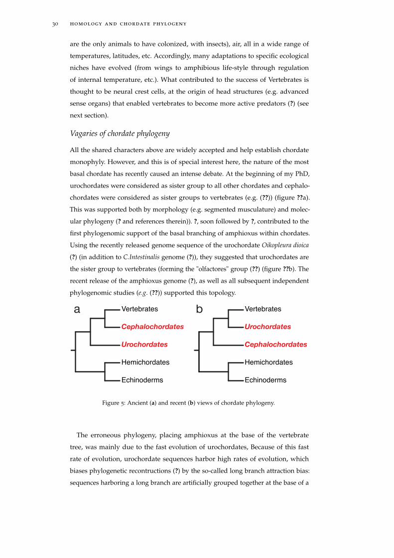
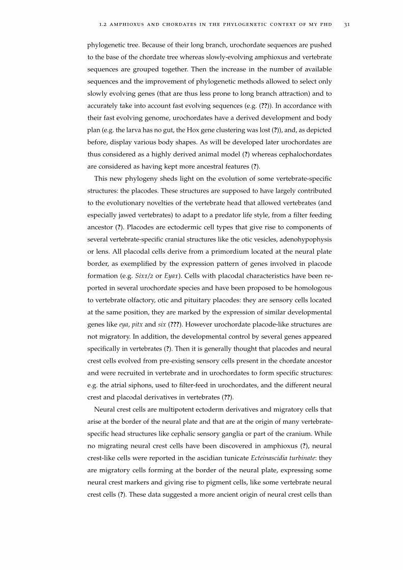
Vagaries of chordate phylogeny
All the shared characters above are widely accepted and help establish chordate
monophyly. However, and this is of special interest here, the nature of the most
basal chordate has recently caused an intense debate. At the beginning of my PhD,
urochordates were considered as sister group to all other chordates and cephalo-
chordates were considered as sister groups to vertebrates (e.g. (??)) (figure ??a).
This was supported both by morphology (e.g. segmented musculature) and molec-
ular phylogeny (? and references therein)). ?, soon followed by ?, contributed to the
first phylogenomic support of the basal branching of amphioxus within chordates.
Using the recently released genome sequence of the urochordate Oikopleura dioica
(?) (in addition to C.Intestinalis genome (?)), they suggested that urochordates are
the sister group to vertebrates (forming the "olfactores" group (??) (figure ??b). The
recent release of the amphioxus genome (?), as well as all subsequent independent
phylogenomic studies (e.g. (??)) supported this topology.
Echinoderms
Hemichordates
Cephalochordates
Urochordates
Vertebrates
Echinoderms
Hemichordates
Urochordates
Cephalochordates
Vertebratesa b
Figure 5: Ancient (a) and recent (b) views of chordate phylogeny.
The erroneous phylogeny, placing amphioxus at the base of the vertebrate
tree, was mainly due to the fast evolution of urochordates, Because of this fast
rate of evolution, urochordate sequences harbor high rates of evolution, which
biases phylogenetic recontructions (?) by the so-called long branch attraction bias:
sequences harboring a long branch are artificially grouped together at the base of a
1.2 amphioxus and chordates in the phylogenetic context of my phd 31
phylogenetic tree. Because of their long branch, urochordate sequences are pushed
to the base of the chordate tree whereas slowly-evolving amphioxus and vertebrate
sequences are grouped together. Then the increase in the number of available
sequences and the improvement of phylogenetic methods allowed to select only
slowly evolving genes (that are thus less prone to long branch attraction) and to
accurately take into account fast evolving sequences (e.g. (??)). In accordance with
their fast evolving genome, urochordates have a derived development and body
plan (e.g. the larva has no gut, the Hox gene clustering was lost (?)), and, as depicted
before, display various body shapes. As will be developed later urochordates are
thus considered as a highly derived animal model (?) whereas cephalochordates
are considered as having kept more ancestral features (?).
This new phylogeny sheds light on the evolution of some vertebrate-specific
structures: the placodes. These structures are supposed to have largely contributed
to the evolutionary novelties of the vertebrate head that allowed vertebrates (and
especially jawed vertebrates) to adapt to a predator life style, from a filter feeding
ancestor (?). Placodes are ectodermic cell types that give rise to components of
several vertebrate-specific cranial structures like the otic vesicles, adenohypophysis
or lens. All placodal cells derive from a primordium located at the neural plate
border, as exemplified by the expression pattern of genes involved in placode
formation (e.g. Six1/2 or Eya1). Cells with placodal characteristics have been re-
ported in several urochordate species and have been proposed to be homologous
to vertebrate olfactory, otic and pituitary placodes: they are sensory cells located
at the same position, they are marked by the expression of similar developmental
genes like eya, pitx and six (???). However urochordate placode-like structures are
not migratory. In addition, the developmental control by several genes appeared
specifically in vertebrates (?). Then it is generally thought that placodes and neural
crest cells evolved from pre-existing sensory cells present in the chordate ancestor
and were recruited in vertebrate and in urochordates to form specific structures:
e.g. the atrial siphons, used to filter-feed in urochordates, and the different neural
crest and placodal derivatives in vertebrates (??).
Neural crest cells are multipotent ectoderm derivatives and migratory cells that
arise at the border of the neural plate and that are at the origin of many vertebrate-
specific head structures like cephalic sensory ganglia or part of the cranium. While
no migrating neural crest cells have been discovered in amphioxus (?), neural
crest-like cells were reported in the ascidian tunicate Ecteinascidia turbinate: they
are migratory cells forming at the border of the neural plate, expressing some
neural crest markers and giving rise to pigment cells, like some vertebrate neural
crest cells (?). These data suggested a more ancient origin of neural crest cells than
32 homology and chordate phylogeny
previously thought, at the origin of urochordates and vertebrates. However, these
cells are not considered as neural crest cells per se because they do not display
the full multipotency of vertebrate structures and because they do not express
the complete genetic regulatory network (?). Rather, it is thought that neural crest
cells did not suddenly appear in the vertebrate ancestor but rather evolved form
preexisting "rudiment" structures present in the chordate ancestor, and that can be
found in non-vertebrate chordates, that gained a sub-set of neural crest properties
(like migration) in the common ancestor of vertebrates and urochordates (?), and
was further elaborated during vertebrate evolution by gene co-option (?).
Whole genome duplications at the base of the vertebrate clade
It was proposed decades ago that whole genome duplications appeared at the base
of the vertebrate clade (?). Duplication would be a generous source of genetic ma-
terial “ready to use”, i.e. complete functional genes with their open reading frame,
regulatory regions and biochemical context. One copy of a duplicated gene could
keep the function of the original gene when the other one, freed from any selection
pressure linked to the original gene function, could experience another fate by
rapidly accumulating mutations and maybe acquire new functions (?). In contrast,
it seems more difficult for an already functional single-copy gene to acquire a new
function without altering its present one. To quote Ohno "natural selection merely
modified, while redundancy created" and he imagined that gene duplications played
an important role in major evolutionary transitions whereas classical Darwinian
gradualism was restricted to microevolutionary events. It comes as no surprise that
such a model induced a lot of debates and that many authors tried to challenge it,
especially when complete genome sequence provided for the first time the data to
tackle this issue.
Known as “2R” for “2 Rounds of whole genome duplications”, Ohno’s hy-
pothesis postulates that 2 events of genome duplication occurred at the origin of
Vertebrates. Ohno’s theory was ignored by many biologists for two decades until
genome sequencing and the appearance of new computational methods allowed
confirming this hypothesis (??). The exact dating of the duplication events during
vertebrate evolution is still unclear: did the two duplications occur before lamprey
split (?), or one before and one after (?)? The upcoming release of the lamprey
genome should help solve this issue. One central question is still to know if gene
duplications were instrumental in the appearance of the new morphological traits
of vertebrates. The split of amphioxus and tunicates before genome duplication
surely make both of them interesting models to study the effect of duplication in
the evolution of gene function (?).
1.3 paleontological context of chordate origins 33
1.2.3 Deuterostome phylogeny
Together with Xenoturbellida (like Xenoturbella bocki) and the ambulacraria, com-
posed of the echinoderms (like the sea urchin Strongylocentrotys purpuratus) and
the hemichordates (like Saccoglossus kowalevskii), chordates constitute the deuteros-
tomes group (?) (figure ??). Synapomorphic developmental features are for instance
the enterocoelous formation of the body cavity or the secondary emergence of
the mouth, after the anus. However, the chordate mouth was also proposed to
have either independently appeared or to have shifted (?) or to have independently
evolved (??).
The monophyly of echinoderms and hemichordates has important consequences
for our understanding of deuterostome evolution. Indeed, hemichordates share
some characteristics (like gill slits) with chordates (hence their name). Consequently
the structures, previously thought to be derived in hemichordates and chordates,
are ancestral in deuterostomes and were secondarily lost in echinoderms. In addi-
tion, the inferred deuterostome ancestor is more "amphioxus-like" than previously
expected.
The deuterostome monophyly has recently been challenged by a phylogenetic
study that proposed that ambulacraria and Xenoturbellida were more closely
related to protostomes than to chordates (??). Although these results were not
statistically supported and need further confirmation, they may lead to interesting
conclusions regarding the evolution of bilaterians because they place chordates at
the base of bilaterians (with amphioxus at the base of chordates). This would then
suggest that the characters common to chordates and hemichordates are basal in
bilaterians, which would lead to the conclusion that the very bilaterian ancestor is
more "amphioxus-like" than previously expected. Although this hypothesis is very
elusive, it once more contradicts that the rampant gradualist view of the evolution
of chordates and vertebrates.
1.3 paleontological context of chordate ori-gins
Pikaia gracilens (figure ??a) may be the most famous proposed fossil of a basal
chordate. It is an elongated, amphioxus-like animal, like most fossils of early
chordates, and the most important discovery of the Burgess shales according to ?.
It used to be the oldest chordate fossil (about 505 million years old, in the middle
Cambrian) (?). Although it possesses important chordate characteristics (e.g. a
notochord), it is considered to belong to a stem chordate group (i.e. an extinct
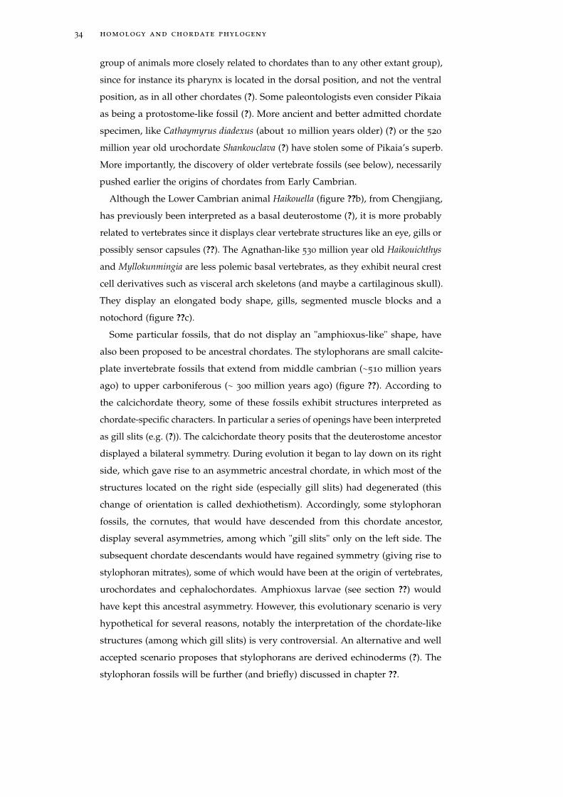
34 homology and chordate phylogeny
group of animals more closely related to chordates than to any other extant group),
since for instance its pharynx is located in the dorsal position, and not the ventral
position, as in all other chordates (?). Some paleontologists even consider Pikaia
as being a protostome-like fossil (?). More ancient and better admitted chordate
specimen, like Cathaymyrus diadexus (about 10 million years older) (?) or the 520
million year old urochordate Shankouclava (?) have stolen some of Pikaia’s superb.
More importantly, the discovery of older vertebrate fossils (see below), necessarily
pushed earlier the origins of chordates from Early Cambrian.
Although the Lower Cambrian animal Haikouella (figure ??b), from Chengjiang,
has previously been interpreted as a basal deuterostome (?), it is more probably
related to vertebrates since it displays clear vertebrate structures like an eye, gills or
possibly sensor capsules (??). The Agnathan-like 530 million year old Haikouichthys
and Myllokunmingia are less polemic basal vertebrates, as they exhibit neural crest
cell derivatives such as visceral arch skeletons (and maybe a cartilaginous skull).
They display an elongated body shape, gills, segmented muscle blocks and a
notochord (figure ??c).
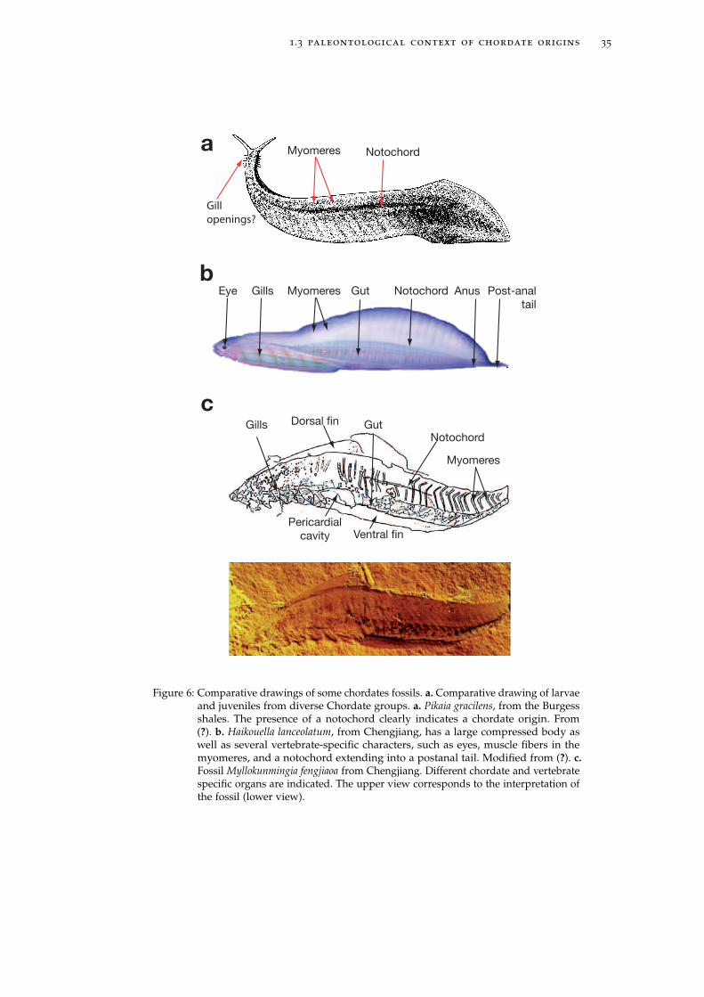
Some particular fossils, that do not display an "amphioxus-like" shape, have
also been proposed to be ancestral chordates. The stylophorans are small calcite-
plate invertebrate fossils that extend from middle cambrian (∼510 million years
ago) to upper carboniferous (∼ 300 million years ago) (figure ??). According to
the calcichordate theory, some of these fossils exhibit structures interpreted as
chordate-specific characters. In particular a series of openings have been interpreted
as gill slits (e.g. (?)). The calcichordate theory posits that the deuterostome ancestor
displayed a bilateral symmetry. During evolution it began to lay down on its right
side, which gave rise to an asymmetric ancestral chordate, in which most of the
structures located on the right side (especially gill slits) had degenerated (this
change of orientation is called dexhiothetism). Accordingly, some stylophoran
fossils, the cornutes, that would have descended from this chordate ancestor,
display several asymmetries, among which "gill slits" only on the left side. The
subsequent chordate descendants would have regained symmetry (giving rise to
stylophoran mitrates), some of which would have been at the origin of vertebrates,
urochordates and cephalochordates. Amphioxus larvae (see section ??) would
have kept this ancestral asymmetry. However, this evolutionary scenario is very
hypothetical for several reasons, notably the interpretation of the chordate-like
structures (among which gill slits) is very controversial. An alternative and well
accepted scenario proposes that stylophorans are derived echinoderms (?). The
stylophoran fossils will be further (and briefly) discussed in chapter ??.
1.3 paleontological context of chordate origins 35
Eye GutGills Myomeres
Myomeres
Post-anal
tail
Notochord
Notochord
Anus
GutGills
Myomeres
Notochord
Dorsal fin
Ventral finPericardial
cavity
a
b
c
Gill
openings?
Figure 6: Comparative drawings of some chordates fossils. a. Comparative drawing of larvaeand juveniles from diverse Chordate groups. a. Pikaia gracilens, from the Burgessshales. The presence of a notochord clearly indicates a chordate origin. From(?). b. Haikouella lanceolatum, from Chengjiang, has a large compressed body aswell as several vertebrate-specific characters, such as eyes, muscle fibers in themyomeres, and a notochord extending into a postanal tail. Modified from (?). c.Fossil Myllokunmingia fengjiaoa from Chengjiang. Different chordate and vertebratespecific organs are indicated. The upper view corresponds to the interpretation ofthe fossil (lower view).
36 homology and chordate phylogeny
Figure 7: Reconstruction of the cor-nute Ceracystis perneri in adorsal view, as interpretedby R.P.S. Jefferies. Modi-fied from (?).
1.4 the animal model amphioxus
A large part of my PhD was dedicated to understanding amphioxus post-embryonic
development. As explained in section ??, amphioxus is now considered to branch
at the base of the chordate tree (figure ??).
Introduction to the amphioxus
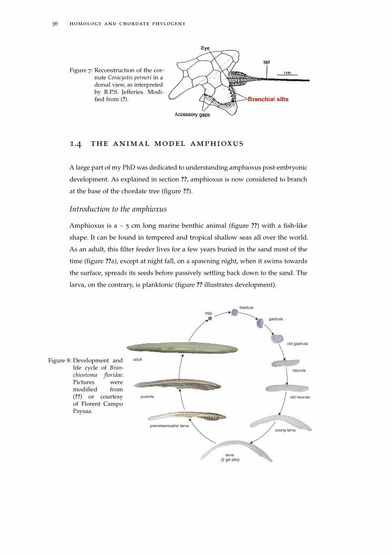
Amphioxus is a ∼ 5 cm long marine benthic animal (figure ??) with a fish-like
shape. It can be found in tempered and tropical shallow seas all over the world.
As an adult, this filter feeder lives for a few years buried in the sand most of the
time (figure ??a), except at night fall, on a spawning night, when it swims towards
the surface, spreads its seeds before passively settling back down to the sand. The
larva, on the contrary, is planktonic (figure ?? illustrates development).
Figure 8: Development andlife cycle of Bran-chiostoma floridae.Pictures weremodified from(??) or courtesyof Florent CampoPaysaa.
blastula
egg
gastrula
old gastrula
neurula
old neurula
young larva
larva
(2 gill slits)
premetamorphic larva
juvenile
adult
1.4 the animal model amphioxus 37
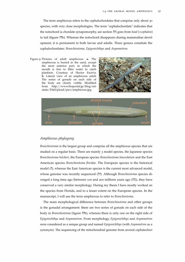
The term amphioxus refers to the cephalochordates that comprise only about 30
species, with very close morphologies. The term "cephalochordate" indicates that
the notochord (a chordate synapomorphy, see section ??) goes from head (=cephalon)
to tail (figure ??b). Whereas the notochord disappears during mammalian devel-
opment, it is permanent in both larvae and adults. Three genera consitute the
cephalochordates: Branchiostoma, Epigonichthys and Asymmetron.
Figure 9: Pictures of adult amphioxus. a. Theamphioxus is buried in the sand, exceptthe most anterior part, in which themouth is free to filter water to catchplankton. Courtesy of Hector Escriva.b. Lateral view of an amphioxus adult.The series of gonads on each side ofthe body are clearly visible. Modifiedfrom http://www.bioportal.jp/blog/mt-static/FileUpload/pics/amphioxus.jpg.
Amphioxus phylogeny
Branchiostoma is the largest group and comprise all the amphioxus species that are
studied on a regular basis. There are mainly 3 model species, the Japanese species
Branchiostoma belcheri, the European species Branchiostoma lanceolatum and the East
American species Branchiostoma floridae. The European species is the historical
model (?), whereas the East American species is the current most advanced model,
whose genome was recently sequenced (??). Although Branchiostoma species di-
verged a long time ago (between 100 and 200 millions years ago (??)), they have
conserved a very similar morphology. During my thesis I have mostly worked on
the species from Florida, and to a lesser extent on the European species. In the
manuscript, I will use the term amphioxus to refer to Branchiostoma.
The main morphological difference between Branchiostoma and other groups
is the gonadal arrangement: there are two series of gonads on each side of the
body in Branchiostoma (figure ??b), whereas there is only one on the right side of
Epigonichthys and Asymmetron. From morphology, Epigonichthys and Asymmetron
were considered as a unique group and named Epigonichthys (with Asymmetron as a
synonym). The sequencing of the mitochondrial genome from several cephalochor-
38 homology and chordate phylogeny
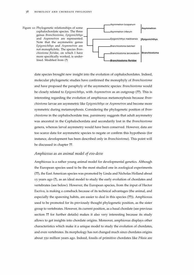
Figure 10: Phylogenetic relationships of somecephalochordate species. The threegenus Branchiostoma, Epigonichthysand Asymmetron are represented.Note that the asymmetric genusEpigonichthys and Asymmetron arenot monophyletic. The species Bran-chiostoma floridae, on which I havemore specifically worked, is under-lined. Modified from (?) Branchiostoma floridae
Branchiostoma lanceolatum
Branchiostoma belcheri
Epigonichthys maldivensis
Asymmetron inferum
Asymmetron lucayanum
Asymmetron
Epigonichthys
Branchiostoma
date species brought new insight into the evolution of cephalochordates. Indeed,
molecular phylogenetic studies have confirmed the monophyly of Branchiostoma
and have proposed the paraphyly of the asymmetric species: Branchiostoma would
be closely related to Epigonichthys, with Asymmetron as an outgroup (??). This is
interesting regarding the evolution of amphioxus metamorphosis because Bran-
chiostoma larvae are asymmetric like Epigonichthys or Asymmetron and become more
symmetric during metamorphosis. Considering the phylogenetic position of Bran-
chiostoma in the cephalochordate tree, parsimony suggests that adult asymmetry
was ancestral in the Cephalochordata and secondarily lost in the Branchiostoma
genera, whereas larval asymmetry would have been conserved. However, data are
too scarce data for asymmetric species to negate or confirm this hypothesis (for
instance, development has been described only in Branchiostoma). This point will
be discussed in chapter ??.
Amphioxus as an animal model of evo-devo
Amphioxus is a rather young animal model for developmental genetics. Although
the European species used to be the most studied one in zoological experiments
(??), the East American species was promoted by Linda and Nicholas Holland about
15 years ago (?), as an ideal model to study the early evolution of chordates and
vertebrates (see below). However, the European species, from the input of Hector
Escriva, is making a comeback because of its technical advantages (the animal, and
especially the spawning habits, are easier to deal in this species (??)). Amphioxus
used to be promoted for its previously thought phylogenetic position, as the sister
group to vertebrates. However, its current position, as a basal chordate (see previous
section ?? for further details) makes it also very interesting because its study
allows to get insights into chordate origins. Moreover, amphioxus displays other
characteristics which make it a unique model to study the evolution of chordates,
and even vertebrates. Its morphology has not changed much since chordates origins
about 550 million years ago. Indeed, fossils of primitive chordates like Pikaia are

1.4 the animal model amphioxus 39
"amphioxus-like" (see section ?? for further details) and amphioxus genome has
a slow evolving rate (?). In contrast and as explained earlier, the vertebrate sister-
group urochordates are morphologically and developmentally much derived (even
within the urochordate subphylum, the numerous genera of Urochordates display
extensive morphological and developmental differences). So all things considered,
this special phylogenetic situation makes amphioxus interesting in two aspects: it
is basal and slowly evolving (especially morphologically, see section ??) (?). This
makes amphioxus a relevant model to study the evolution of chordates (??).
Notably, the current phylogenetic position of amphioxus supposes that many
conserved features that it shares with vertebrates are ancestral within chordates
and that the chordate ancestor was more "vertebrate-like" than previously ex-
pected. This will be discussed in this manuscript in the context of the evolution of
metamorphosis.

2I N T R O D U C T I O N T O M E TA M O R P H O S I S
The inaugural use of the Greek word “metamorphosis” is attributed to the Latin
Ovide (?). It is generally associated with the notion of shape change (“meta”
meaning change and “morphe” form) of an individual. In biology it refers to
the larva-to-juvenile transformation like tadpole-to-frog or caterpillar-to-butterfly
transitions (see the following section ??) whereas the lay audience has applied the
term to fantasy or litterature.
2.1 metamorphosis for the lay audience
Although metamorphosis refers to a transformation, the drastic changes that occur
at metamorphosis and also the sudden and seemingly magical origin of the modifi-
cations make it more than a simple transformation: the association between the pre-
and post-metamorphic forms is not straightforward and the extensiveness of the
modifications renders mechanisms underlying these changes difficult to conceive.
Then supernatural causes are more appealing, but also more convenient because
they shrug off the explanation problem to focus on the result of the metamorphosis:
two different organisms in one. This concept has also been extensively used for less
realistic transformations, sometimes physical, sometimes metaphorical, some of
which are briefly introduced below.
The very evocative potential of metamorphosis was amply illustrated in countless
films and books. Horror or Sci Fi movies have overused the concept to feed
audiences with the inexhaustible stories of gentle people transforming into horrible
monsters or ugly monsters transforming into even more atrocious other monsters.
The notion of metamorphosis has also been illustrated less "literally". Often below
the surface of morphological alterations, it can also be a metaphor for inexplicable
and important changes that reunite two apparently separate parts of the same
individual. Among the plethora of examples, one of the most famous films dealing
with metamorphosis, called The fly directed by David Cronenberg, tells the story
of a man slowly transforming into a fly. The film is as much about slimy and
disgusting flesh exhibition as about internal duality of a man, transfigured into
his physical appearance. Among books, The Strange case of Dr Jekyll and Mr. Hyde
by R.L. Stevenson again uses the metaphor of metamorphosis to discuss the
41
42 introduction to metamorphosis
conflict between two irreconcilable parts of a person. Similarly, Kafka centers
his novel "Die Verwandlung" ("The Metamorphosis") around the metamorphosis of
a typical average man called Gregor into a giant cockroach. First physical, the
transformations slowly reaches his mind and Gregor gradually loses his humanity
(especially his dignity) until death. A notable difference with Stevenson’s novel
is the irreversibility of the metamorphosis: Gregor never goes back to the way
he used to be, like metamorphosed animals never "re-metamorphose" to their
larval form. Kafka’s Metamorphosis is not about internal conflict of a person, but
rather about how a person reacts to the changes in his relatives’ look. Overall each
time, metamorphosis does not "distort" a person, but rather "develops" the true
and hidden personality, which is hard to accept for both him/her and the others.
This notion of “transformation”, this denial of permanence is central in the book
Metamorphosis by Ovid, in which a series of independent short stories tell the
story of different event more or less dealing with metamorphosis. This notion may
remind of one of the ecological advantages of animal metamorphosis, that allows
one individual to colonize different niches by adapting each time to their particular
environmental characteristics (e.g. water for an amphibian tadpole and earth for
adult frog).
Even in the geological vocabulary, metamorphosis is used to designate important
changes in the rock structure, due to gigantic forces (the term metamorphism
is rather used). This notion of super-powerful force that cannot be controled
is always underlying when talking about metamorphosis. Even in biology, as
will be discussed in chapter ??, genetic parameters "program" the occurrence of
metamorphosis (it has to happen), as well as external conditions influence the onset
and unfolding of metamorphosis (see chapter ??), like the moon triggers werewolf
transformation.
2.2 metamorphosis for a biologist
2.2.1 Definition
In biology, metamorphosis has a more restricted definition, based on developmental
considerations. Although there are still debates about the exact definition of
metamorphosis (?), a consensus embraces the notion of drastic morphological
changes associated with an ecological transition. In the following chapters, I will
define metamorphosis as a short post-embryonic developmental stage allowing
a larva to become a juvenile/adult and during which important morphological,
ecological and metabolic modifications occur. This definition will be applied to
2.2 metamorphosis for a biologist 43
animals (figure ??). Plant development won’t be discussed here. Metamorphosis
is not applicable to unicellular organisms like Bacteria (and even multicellular
Bacteria) or Archaea, for which there is no development.
A direct corollary to the occurrence of a metamorphosis stage during develop-
ment is the existence of larval and juvenile/adult stages. Between the juvenile and
the adult occurs sexual maturation (for instance in amphibians, amphioxus, sea
urchin, oyster). The latter, during which morphological changes can also occur,
should not be confused with metamorphosis because it is not triggered by the
same molecular mechanism (see chapter ??) and most often does not involve an
ecological transformation. However, in some species and especially in insects, the
post-metamorphic individual is already mature (thus there is no juvenile stage).
2.2.2 Metamorphosis is widespread among metazoans.
If one were to pick up any animal and guess whether it metamorphoses during
its life time, one should bet on the affirmative answer. Indeed, most species
metamorphose, including many insects, mollusks, annelids, cnidarians, sea urchins,
some fishes, frogs or sea squirts (of course this list is not exhaustive). The plankton
is basically filled with small aquatic larvae from non-insect invertebrates (??). The
diversity of larval forms can compare to the diversity of names given to the different
larvae: e.g. planula larva in some cnidarians, pluteus larva in some echinoderms,
caterpillar in some insects, maggot in others, trochophor larva in some mollusks
and annelids, tadpole in some amphibians and urochordates, ammocoete in some
lampreys, nauplius larva in some crustaceans...
This diversity in metamorphoses extends from full metamorphosis (with exten-
sive remodeling) to no metamorphosis. Indeed, some animals (like us) directly
develop into an individual that looks like the adult and apparently do not go
through the morphological, ecological and metabolic modifications characteristic
of metamorphosis. Actually, there are many ecological strategies among animals,
with a morphological remodeling more or less pronounced, ecological shifts more
or less drastic, feeding or non feeding larvae... depending on each species situ-
ation (see following section). Some species also undergo several morphological
and ecological metamorphic transitions (like insects or crustaceans), although the
last transition is usually the most dramatic one, in addition to leading to settle-
ment before transformation into the pre-reproductive form (juvenile). Figure ??
summarizes some typical life cycles.
As can be seen on figure ??, life cycles of metazoans are very diverse. They
can be rather "simple" as it is the case for the amphioxus B.floridae or the sea
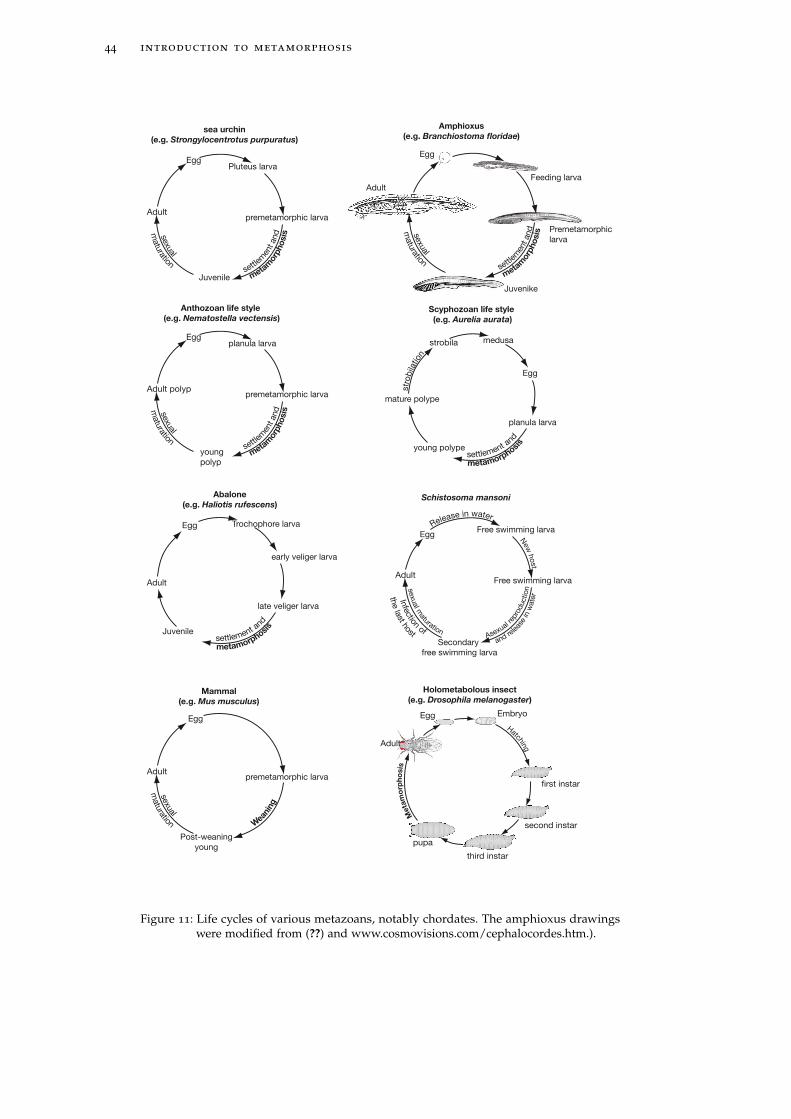
44 introduction to metamorphosis
sea urchin
(e.g. Strongylocentrotus purpuratus)
Egg
Adult
Pluteus larva
premetamorphic larva
Juvenile settl
emen
t and
meta
morp
ho
sis
sexual
matu
ration
Scyphozoan life style
(e.g. Aurelia aurata)
Egg
strobila
planula larva
medusa
young polype settlement and
metamorphosi
s
str
obila
tion
mature polype
Abalone
(e.g. Haliotis rufescens)
early veliger larva
Egg
late veliger larva
Trochophore larva
Juvenile settlement and
metamorphosi
s
Adult
Anthozoan life style
(e.g. Nematostella vectensis)
Egg
Adult polyp
planula larva
premetamorphic larva
young
polyp
settlem
ent and
m
eta
morp
ho
sis
sexual
matu
ration
Mammal
(e.g. Mus musculus)
Egg
Adultpremetamorphic larva
Post-weaning
young
Wean
ing
sexual
matu
ration
Schistosoma mansoni
Egg
Adult
Free swimming larva
Free swimming larva
Secondary
free swimming larva
Asexual r
epro
ductio
n
.....and re
le
ase
in w
ate
r
sexual m
aturation
Infe
ction of
New
host
the la
st host
Release in water
Holometabolous insect
(e.g. Drosophila melanogaster)
Egg Embryo
first instar
second instar
third instar
HatchingAdult
pupa
Meta
mo
rph
osis
settl
emen
t and
meta
morp
ho
sis
sexual
matu
ration
Egg
Feeding larva
Premetamorphic
larva
Adult
Amphioxus
(e.g. Branchiostoma floridae)
Juvenike
Figure 11: Life cycles of various metazoans, notably chordates. The amphioxus drawingswere modified from (??) and www.cosmovisions.com/cephalocordes.htm.).
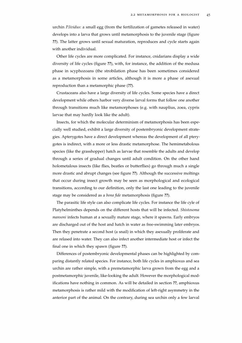
2.2 metamorphosis for a biologist 45
urchin P.lividus: a small egg (from the fertilization of gametes released in water)
develops into a larva that grows until metamorphosis to the juvenile stage (figure
??). The latter grows until sexual maturation, reproduces and cycle starts again
with another individual.
Other life cycles are more complicated. For instance, cnidarians display a wide
diversity of life cycles (figure ??), with, for instance, the addition of the medusa
phase in scyphozoans (the strobilation phase has been sometimes considered
as a metamorphosis in some articles, although it is more a phase of asexual
reproduction than a metamorphic phase (??).
Crustaceans also have a large diversity of life cycles. Some species have a direct
development while others harbor very diverse larval forms that follow one another
through transitions much like metamorphoses (e.g. with nauplius, zoea, cypris
larvae that may hardly look like the adult).
Insects, for which the molecular determinism of metamorphosis has been espe-
cially well studied, exhibit a large diversity of postembryonic development strate-
gies. Apterygotes have a direct development whereas the development of all ptery-
gotes is indirect, with a more or less drastic metamorphose. The hemimetabolous
species (like the grasshopper) hatch as larvae that resemble the adults and develop
through a series of gradual changes until adult condition. On the other hand
holometalous insects (like flies, beatles or butterflies) go through much a single
more drastic and abrupt changes (see figure ??). Although the successive moltings
that occur during insect growth may be seen as morphological and ecological
transitions, according to our definition, only the last one leading to the juvenile
stage may be considered as a bona fide metamorphosis (figure ??).
The parasitic life style can also complicate life cycles. For instance the life cyle of
Platyhelminthes depends on the different hosts that will be infected. Shistosoma
mansoni infects human at a sexually mature stage, where it spawns. Early embryos
are discharged out of the host and hatch in water as free-swimming later embryos.
Then they penetrate a second host (a snail) in which they asexually proliferate and
are relased into water. They can also infect another intermediate host or infect the
final one in which they spawn (figure ??).
Differences of postembryonic developmental phases can be highlighted by com-
paring distantly related species. For instance, both life cycles in amphioxus and sea
urchin are rather simple, with a premetamorphic larva grown from the egg and a
postmetamorphic juvenile, like-looking the adult. However the morphological mod-
ifications have nothing in common. As will be detailed in section ??, amphioxus
metamorphosis is rather mild with the modification of left-right asymmetry in the
anterior part of the animal. On the contrary, during sea urchin only a few larval
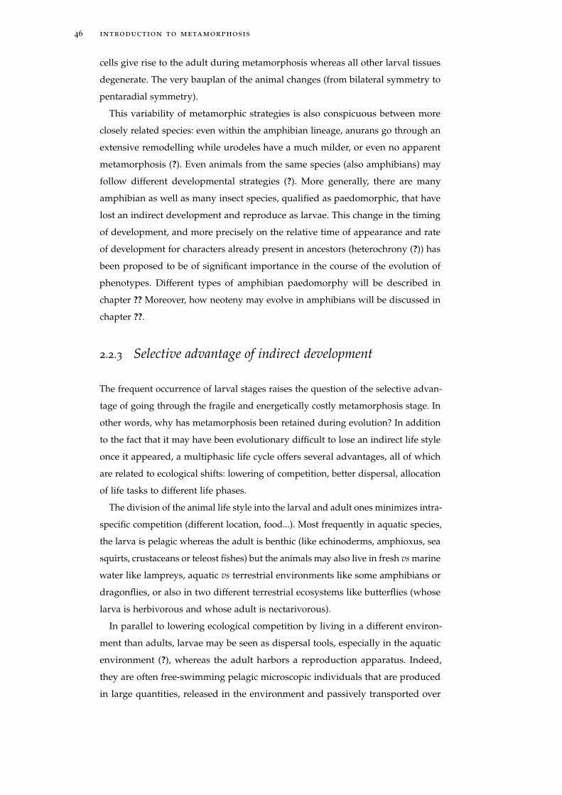
46 introduction to metamorphosis
cells give rise to the adult during metamorphosis whereas all other larval tissues
degenerate. The very bauplan of the animal changes (from bilateral symmetry to
pentaradial symmetry).
This variability of metamorphic strategies is also conspicuous between more
closely related species: even within the amphibian lineage, anurans go through an
extensive remodelling while urodeles have a much milder, or even no apparent
metamorphosis (?). Even animals from the same species (also amphibians) may
follow different developmental strategies (?). More generally, there are many
amphibian as well as many insect species, qualified as paedomorphic, that have
lost an indirect development and reproduce as larvae. This change in the timing
of development, and more precisely on the relative time of appearance and rate
of development for characters already present in ancestors (heterochrony (?)) has
been proposed to be of significant importance in the course of the evolution of
phenotypes. Different types of amphibian paedomorphy will be described in
chapter ?? Moreover, how neoteny may evolve in amphibians will be discussed in
chapter ??.
2.2.3 Selective advantage of indirect development
The frequent occurrence of larval stages raises the question of the selective advan-
tage of going through the fragile and energetically costly metamorphosis stage. In
other words, why has metamorphosis been retained during evolution? In addition
to the fact that it may have been evolutionary difficult to lose an indirect life style
once it appeared, a multiphasic life cycle offers several advantages, all of which
are related to ecological shifts: lowering of competition, better dispersal, allocation
of life tasks to different life phases.
The division of the animal life style into the larval and adult ones minimizes intra-
specific competition (different location, food...). Most frequently in aquatic species,
the larva is pelagic whereas the adult is benthic (like echinoderms, amphioxus, sea
squirts, crustaceans or teleost fishes) but the animals may also live in fresh vs marine
water like lampreys, aquatic vs terrestrial environments like some amphibians or
dragonflies, or also in two different terrestrial ecosystems like butterflies (whose
larva is herbivorous and whose adult is nectarivorous).
In parallel to lowering ecological competition by living in a different environ-
ment than adults, larvae may be seen as dispersal tools, especially in the aquatic
environment (?), whereas the adult harbors a reproduction apparatus. Indeed,
they are often free-swimming pelagic microscopic individuals that are produced
in large quantities, released in the environment and passively transported over
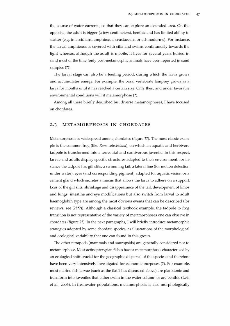
2.3 metamorphosis in chordates 47
the course of water currents, so that they can explore an extended area. On the
opposite, the adult is bigger (a few centimeters), benthic and has limited ability to
scatter (e.g. in ascidians, amphioxus, crustaceans or echinoderms). For instance,
the larval amphioxus is covered with cilia and swims continuously towards the
light whereas, although the adult is mobile, it lives for several years buried in
sand most of the time (only post-metamorphic animals have been reported in sand
samples (?)).
The larval stage can also be a feeding period, during which the larva grows
and accumulates energy. For example, the basal vertebrate lamprey grows as a
larva for months until it has reached a certain size. Only then, and under favorable
environmental conditions will it metamorphose (?).
Among all these briefly described but diverse metamorphoses, I have focused
on chordates.
2.3 metamorphosis in chordates
Metamorphosis is widespread among chordates (figure ??). The most classic exam-
ple is the common frog (like Rana catesbeiana), on which an aquatic and herbivore
tadpole is transformed into a terrestrial and carnivorous juvenile. In this respect,
larvae and adults display specific structures adapted to their environment: for in-
stance the tadpole has gill slits, a swimming tail, a lateral line (for motion detection
under water), eyes (and corresponding pigment) adapted for aquatic vision or a
cement gland which secretes a mucus that allows the larva to adhere on a support.
Loss of the gill slits, shrinkage and disappearance of the tail, development of limbs
and lungs, intestine and eye modifications but also switch from larval to adult
haemoglobin type are among the most obvious events that can be described (for
reviews, see (????)). Although a classical textbook example, the tadpole to frog
transition is not representative of the variety of metamorphoses one can observe in
chordates (figure ??). In the next paragraphs, I will briefly introduce metamorphic
strategies adopted by some chordate species, as illustrations of the morphological
and ecological variability that one can found in this group.
The other tetrapods (mammals and sauropsids) are generally considered not to
metamorphose. Most actinopterygian fishes have a metamorphosis characterized by
an ecological shift crucial for the geographic dispersal of the species and therefore
have been very intensively investigated for economic purposes (?). For example,
most marine fish larvae (such as the flatfishes discussed above) are planktonic and
transform into juveniles that either swim in the water column or are benthic (Leis
et al., 2006). In freshwater populations, metamorphosis is also morphologically
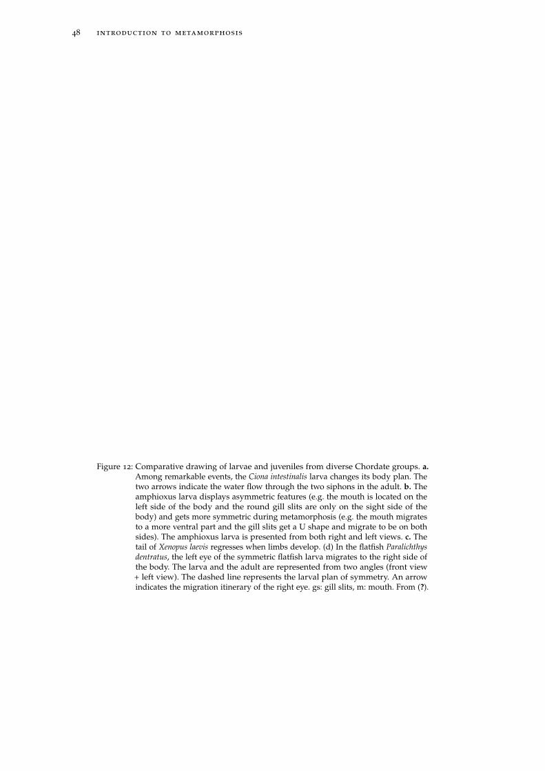
48 introduction to metamorphosis
Larva Juvenile
Ciona
intestinalis
a
amphioxus
(Branchiostoma
lanceolatum)
b
Xenopus
laevis
c
flatfish
(Paralichthys
dentratus)
d
eye
mouth
gs m
eye
Left viewFront view
m
eyes
Left viewFront view
gs
Right view
mLeft view
pharynx
Figure 12: Comparative drawing of larvae and juveniles from diverse Chordate groups. a.Among remarkable events, the Ciona intestinalis larva changes its body plan. Thetwo arrows indicate the water flow through the two siphons in the adult. b. Theamphioxus larva displays asymmetric features (e.g. the mouth is located on theleft side of the body and the round gill slits are only on the sight side of thebody) and gets more symmetric during metamorphosis (e.g. the mouth migratesto a more ventral part and the gill slits get a U shape and migrate to be on bothsides). The amphioxus larva is presented from both right and left views. c. Thetail of Xenopus laevis regresses when limbs develop. (d) In the flatfish Paralichthysdentratus, the left eye of the symmetric flatfish larva migrates to the right side ofthe body. The larva and the adult are represented from two angles (front view+ left view). The dashed line represents the larval plan of symmetry. An arrowindicates the migration itinerary of the right eye. gs: gill slits, m: mouth. From (?).
2.3 metamorphosis in chordates 49
obvious, as seen in the alteration of the zebrafish fins and gut (?), or salmonid
smoltification (?).
The larva from the basal vertebrate lamprey dwells in the silt of freshwater
streams and filter feeds on primary detritus. Larval period lasts several years
after which lampreys metamorphose into adults, some of which are (sometimes
parasitic) predators. During metamorphosis, several structures are remodeled: the
eyes and the gut epithelium are modified, the larval kidney is replaced with a
juvenile one, the mouth develops into a specific sucker apparatus and the endostyle
is profoundly remodeled into a thyroid gland (?). The members of the sister group
hagfish apparently have a direct development, although their post-embryonic
development remains to be fully studied (?).
Outside vertebrates, dramatic morphological remodeling occurs in some uro-
chordates where the very bauplan of the organism is modified between larvae
and adults, like in ascidians (figure ??). Ascidians are the most studied and the
largest taxonomic group in urochordates, with Ciona intestinalis as a genomic and
developmental model system. The larva is pelagic and “tadpole-like” with a typical
chordate body plan (a tail -“uro”- with a notochord, a dorsal nerve tube, striated
bilateral segmented musculature), whereas the adult is sessile with a different body
plan. Ciona intestinalis, for instance, has a “sac-like” body with no tail, an hypertro-
phied pharynx and two siphons allowing water flux (figure ??). This remodeling
is so drastic that some zoologists, including Linnaeus, did not relate the adult to
the larva and classified the former as a mollusk. Notably, other ascidian species
undergo a slightly less dramatic transition as they do not lose their tails during
metamorphosis (?). The other two classes of tunicates, Larvacea and Thaliacea, are
both planktonic in the adult stage. In addition, in Larvaceans, the adult keeps a
tadpole-like body plan and can thus be viewed as paedomorphic. In Thaliacea,
the larva does not settle before rapidly metamorphosing into a planktonic sac-like
adult (?).
Amphioxus metamorphosis
Amphioxus (and more precisely Branchiostoma) was described at metamorphosis by
? in very precise detail more than a century ago. The animal goes into an ecological
transition from pelagos to benthos. Accordingly, the larval body is covered with
cilia that are used for locomotion and are replaced by muscular swimming at
metamorphosis (??). However, the most conspicuous change is the transition from
an asymmetric larva to a more symmetric juvenile. Indeed the larval pharynx is
highly asymmetric: the mouth is on the left side whereas the single gill slit row is
located on the right side (figure ??a). During metamorphosis, the animal will get
50 introduction to metamorphosis
more symmetric: in parallel to the appearance of two metapleural folds on each
side of the body, that will recover the pharynx and gill slits in the juvenile, a second
gill slit appears above the first row (figure ??b). Soon after, all initially round gills
slits get a U shape (figure ??b) and the first gill slit row migrates towards the left
side of the pharynx while the mouth migrates in opposite direction towards a more
ventral and anterior position (figures ??b and c. A close-up of the pharynx from
the figure ??b is given figure ??). Cirris develop around the mouth that migrates
from the left side to an antero-ventral part (figure ??) (??). The gut also changes,
with the appearance of a protuberance just posterior to the pharynx (figure ??c),
called gut diverticulum (?). All these morphological modifications have been taken
into account during the experiments I conducted and participated to, and that are
reported in chapters ??, ?? and in the article reported in the annexe (page ??).
This asymmetric organization of gill slit development has been proposed to
support the calcichordate theory (see section ??), according to which the ances-
tral chordate (that would have looked like cornute fossils) was asymmetric and
contained gill slits only on one side of the body. Amphioxus larvae would then
have kept the ancestral asymmetry. During evolution, symmetry would have been
regained, the way amphioxus larvae become symmetric during metamorphosis
(e.g. (?), discussed in (?)). The cornute fossils are briefly discussed in chapter ??.
Morphologically less conspicuously, the organ endostyle also undergoes pro-
found remodeling from an anterior position and a rather compact shape (figure ??a)
to a ventral position and an elongated shape (figure ??c). Moreover, an amphioxus-
specific organ, called the club-shaped gland, located posteriorly to the endostyle,
disappears. The fate of this organ has been debated for decades (does it really
disappear? Is it reincorporated into surrounding tissues?) and will be discussed
in page ?? (article included in the annexe). There are probably also metabolic
and physiological modifications (for instance, is endostyle activity changing dur-
ing its metamorphosis?) but they have not been looked for yet. At the end of
metamorphosis, a much more symmetric juvenile starts its benthic life buried in
sand.
Some other amphioxus species from the genus Asymmetron and Epygonichthys (?)
stay asymmetric even as adults. As mentioned at the end of the previous section,
the post-embryonic development of these species is still unresolved yet, although
some modifications of the symmetry may occur between larval adult stages (for
instance, the adult mouth is in the midline).
During my PhD, I have sought into the regulatory mechanisms underlying the
evolution of metamorphosis in chordates. Metamorphosis has been molecularly
well characterized only in amphibians and more precisely in anurans, and to a
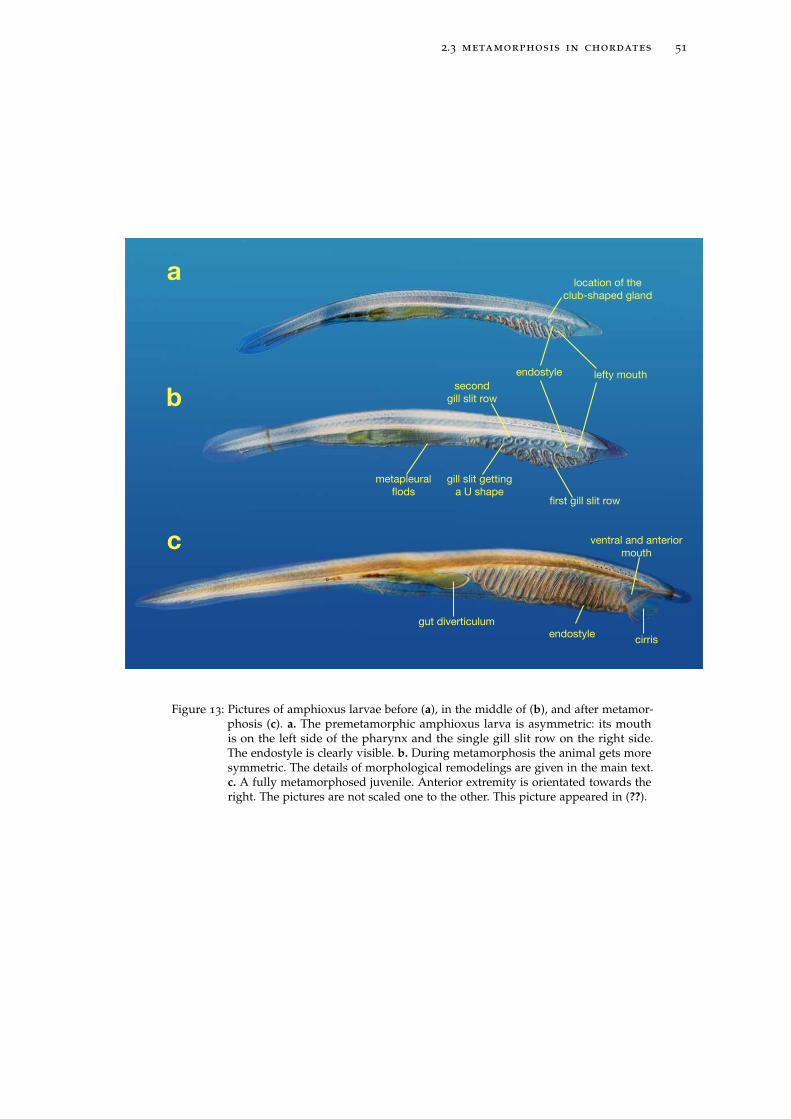
2.3 metamorphosis in chordates 51
endostyle
endostyle
a
b
c
location of the
club-shaped gland
metapleural
flods
gut diverticulum
cirris
ventral and anterior
mouth
gill slit getting
a U shape
second
gill slit row
first gill slit row
lefty mouth
Figure 13: Pictures of amphioxus larvae before (a), in the middle of (b), and after metamor-phosis (c). a. The premetamorphic amphioxus larva is asymmetric: its mouthis on the left side of the pharynx and the single gill slit row on the right side.The endostyle is clearly visible. b. During metamorphosis the animal gets moresymmetric. The details of morphological remodelings are given in the main text.c. A fully metamorphosed juvenile. Anterior extremity is orientated towards theright. The pictures are not scaled one to the other. This picture appeared in (??).
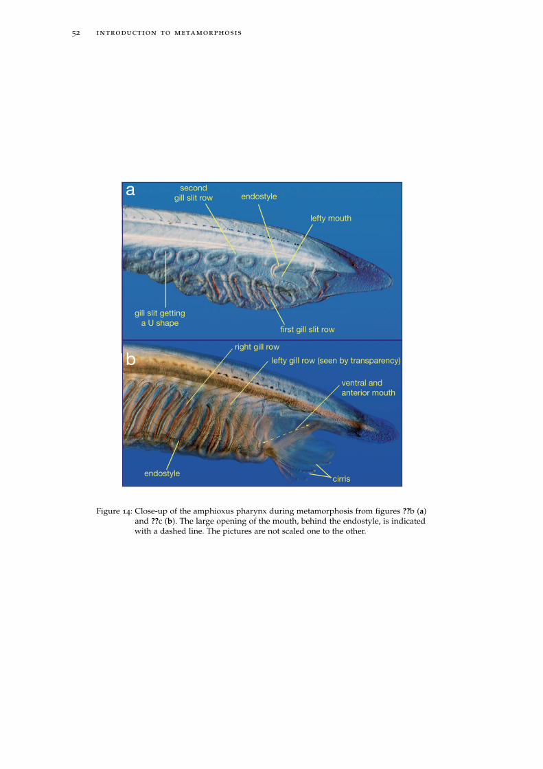
52 introduction to metamorphosis
endostyle
gill slit getting
a U shape
second
gill slit row
first gill slit row
cirris
lefty mouth
right gill row
lefty gill row (seen by transparency)
endostyle
ventral and
anterior mouth
a
b
Figure 14: Close-up of the amphioxus pharynx during metamorphosis from figures ??b (a)and ??c (b). The large opening of the mouth, behind the endostyle, is indicatedwith a dashed line. The pictures are not scaled one to the other.

2.3 metamorphosis in chordates 53
lesser extent in teleost fishes. The knowledge about the molecular determinism of
metamorphosis will be briefly presented in the next chapter of this manuscript.

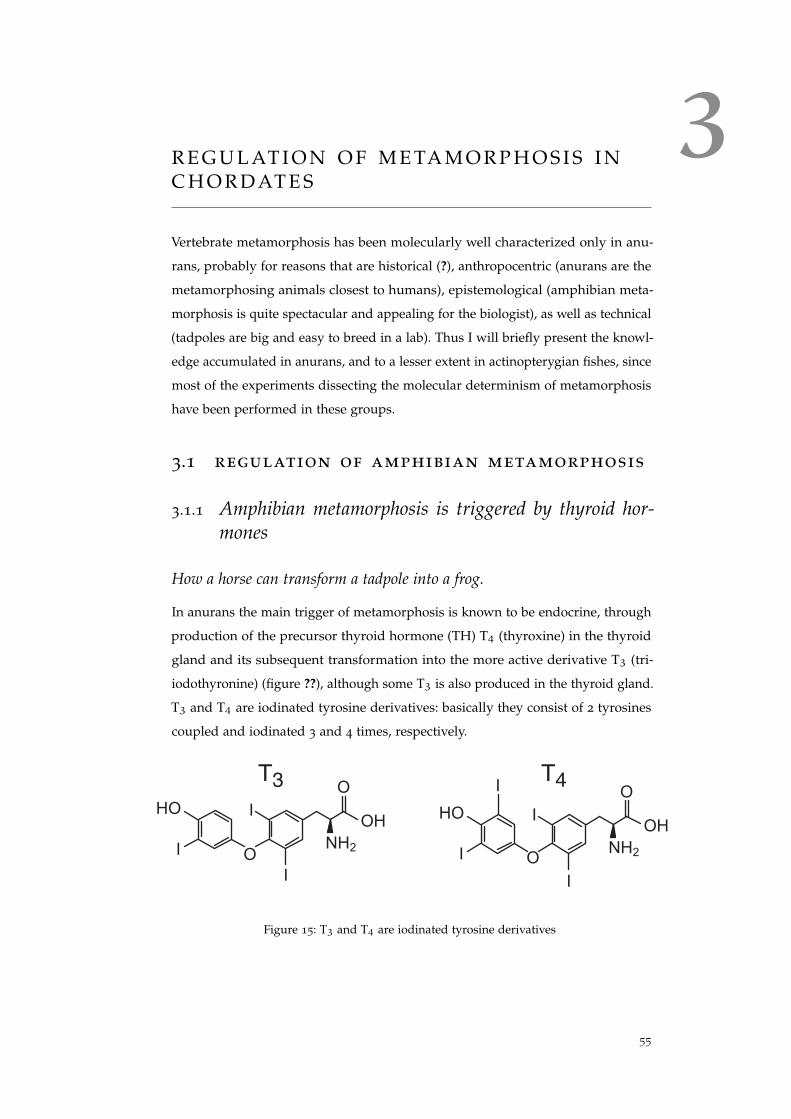
3R E G U L AT I O N O F M E TA M O R P H O S I S I NC H O R D AT E S
Vertebrate metamorphosis has been molecularly well characterized only in anu-
rans, probably for reasons that are historical (?), anthropocentric (anurans are the
metamorphosing animals closest to humans), epistemological (amphibian meta-
morphosis is quite spectacular and appealing for the biologist), as well as technical
(tadpoles are big and easy to breed in a lab). Thus I will briefly present the knowl-
edge accumulated in anurans, and to a lesser extent in actinopterygian fishes, since
most of the experiments dissecting the molecular determinism of metamorphosis
have been performed in these groups.
3.1 regulation of amphibian metamorphosis
3.1.1 Amphibian metamorphosis is triggered by thyroid hor-mones
How a horse can transform a tadpole into a frog.
In anurans the main trigger of metamorphosis is known to be endocrine, through
production of the precursor thyroid hormone (TH) T4 (thyroxine) in the thyroid
gland and its subsequent transformation into the more active derivative T3 (tri-
iodothyronine) (figure ??), although some T3 is also produced in the thyroid gland.
T3 and T4 are iodinated tyrosine derivatives: basically they consist of 2 tyrosines
coupled and iodinated 3 and 4 times, respectively.
T4
I
I
HO
O
I
NH2
OH
O
IT3
I
I
HO
O
I
NH2
OH
O
Figure 15: T3 and T4 are iodinated tyrosine derivatives
55
56 regulation of metamorphosis in chordates
The discovery that THs induce metamorphosis was done by J.F. Gudernatsch
almost a century ago. He was studying "the influence of various organic extracts
on the development of fish (Belone, Gobius etc.) and amphibian (Rana) eggs" (?).
Although his experiments on fish did not give conclusive results (because boats
could not deliver fresh eggs fast enough), he was more satisfied with the results
obtained from frogs. Indeed, as he was feeding tadpoles of Rana temporaria and
Rana esculenta with pieces of horse, calf, rabbit, pigs or even cats, he noticed that
tadpoles fed with thyroid extracts metamorphosed earlier and gave rise to "fully
developed frogs, though dwarfs in size". So exogenous thyroid extracts prematurely
induced metamorphosis (?). But was this effect of any biological significance or
artificial? In other words, is the active compound of horse thyroid necessary for
frog metamorphosis?
The answer came a few years after Gudernatch’s discovery, when metamorpho-
sis was reported to be inhibited upon thyroid removal (?) and recovered upon
subsequent feeding of the larvae with thyroid preparations. These experiments
showed that thyroid gland activity is necessary and sufficient to induce frog meta-
morphosis. Chemical inhibition of TH production by various compounds has
also demonstrated its efficiency in preventing metamorphosis and creating giant
tadpoles (e.g. (?)).
The active molecule present in the thyroid was isolated at the end of the same
decade (?) from hog thyroid extracts (figure ??) and identified as an "iodine-
containing compound", which was originally called thyroxin (T4). Much later its
more active but less abundant derivative T3 was identified (???).
Tissue-specific metamorphic response to TH induction
Making a tadpole into a frog requires an extensive remodelling mostly related to
the water-to-land ecological shift: from loss of the tail to development of limbs,
through repositionning of the eyes, remodeling of the intestine, keratinization of
the skin, modifications of the central nervous signal, ossification of the previously
cartilaginous skull, adaptation of the visual system to fit new light dispersion
properties. Then, the range of the molecular response to TH signal is as wide as
de novo morphogenesis, apoptosis, switching of the hemoglobin subtype, of the
visual pigment (from porphyropsin to thodopsin), production of keratin...
Consequently, the response to the hormone is local, tissue-specific and tissue-
autonomous. For instance a limited and localized application of TH to a tadpole
will induce only the surrouding tisues to undergo metamorphosis (?). Moreover
exposition of exogenous T3 to isolated larval tissues will induce the resorption
of the tail and the morphogenesis of the hind limb bud. Convincingly, if a larval
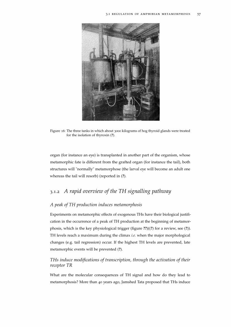
3.1 regulation of amphibian metamorphosis 57
Figure 16: The three tanks in which about 3000 kilograms of hog thyroid glands were treatedfor the isolation of thyroxin (?).
organ (for instance an eye) is transplanted in another part of the organism, whose
metamorphic fate is different from the grafted organ (for instance the tail), both
structures will "normally" metamorphose (the larval eye will become an adult one
whereas the tail will resorb) (reported in (?).
3.1.2 A rapid overview of the TH signalling pathway
A peak of TH production induces metamorphosis
Experiments on metamorphic effects of exogenous THs have their biological justifi-
cation in the occurrence of a peak of TH production at the beginning of metamor-
phosis, which is the key physiological trigger (figure ??)((?) for a review, see (?)).
TH levels reach a maximum during the climax i.e. when the major morphological
changes (e.g. tail regression) occur. If the highest TH levels are prevented, late
metamorphic events will be prevented (?).
THs induce modifications of transcription, through the activation of theirreceptor TR
What are the molecular consequences of TH signal and how do they lead to
metamorphosis? More than 40 years ago, Jamshed Tata proposed that THs induce
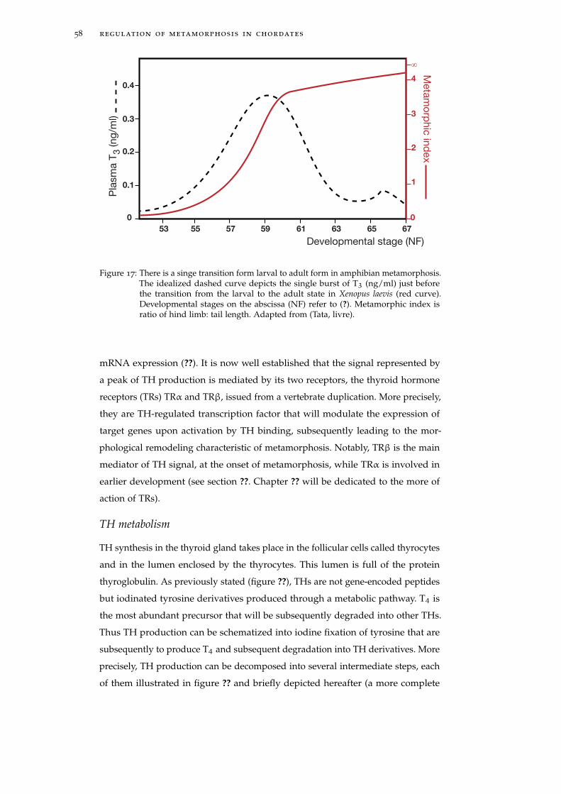
58 regulation of metamorphosis in chordates
53
0
0.1
0.2
0.3
0.4
55 57 59 61 63 65 67
0
1
2
3
4
∞
Pla
sm
a T
3 (ng
/ml)
Developmental stage (NF)
Meta
mo
rphic
ind
ex
Figure 17: There is a singe transition form larval to adult form in amphibian metamorphosis.The idealized dashed curve depicts the single burst of T3 (ng/ml) just beforethe transition from the larval to the adult state in Xenopus laevis (red curve).Developmental stages on the abscissa (NF) refer to (?). Metamorphic index isratio of hind limb: tail length. Adapted from (Tata, livre).
mRNA expression (??). It is now well established that the signal represented by
a peak of TH production is mediated by its two receptors, the thyroid hormone
receptors (TRs) TRα and TRβ, issued from a vertebrate duplication. More precisely,
they are TH-regulated transcription factor that will modulate the expression of
target genes upon activation by TH binding, subsequently leading to the mor-
phological remodeling characteristic of metamorphosis. Notably, TRβ is the main
mediator of TH signal, at the onset of metamorphosis, while TRα is involved in
earlier development (see section ??. Chapter ?? will be dedicated to the more of
action of TRs).
TH metabolism
TH synthesis in the thyroid gland takes place in the follicular cells called thyrocytes
and in the lumen enclosed by the thyrocytes. This lumen is full of the protein
thyroglobulin. As previously stated (figure ??), THs are not gene-encoded peptides
but iodinated tyrosine derivatives produced through a metabolic pathway. T4 is
the most abundant precursor that will be subsequently degraded into other THs.
Thus TH production can be schematized into iodine fixation of tyrosine that are
subsequently to produce T4 and subsequent degradation into TH derivatives. More
precisely, TH production can be decomposed into several intermediate steps, each
of them illustrated in figure ?? and briefly depicted hereafter (a more complete

3.1 regulation of amphibian metamorphosis 59
description of TH signalling pathway is given in chapter ??): (1) Iodine first needs
to be concentrated by the specific transmembrane Sodium/Iodine Symporter (SIS),
(2) then it is added to tyrosines from the matrix protein thyroglobulin by the
Thyroid Peroxidase (TPO) to form monoiodotyrosines or diiodotyrosines (if one
or two iodines have been added to a tyrosine, respectively). (3) The iodinated
tyrosines are coupled by the TPO to form a TH (mostly T4 but also T3). (4)
The newly synthesized TH is then separated from the matrix protein, (5) and
transported in the blood by various carriers such as TBG (thyroid binding globulin)
or TTR (transthyretin). (6) In peripheral tissues, T4 is deiodinated into the more
active form T3. (7) Both THs can be converted into less active forms, that will be
subsequently degraded. (8) There are also alternative TH pathways, like sulfatation
and glucuronidation, mostly resulting in production of inactive T3 derivatives,
targeted for degradation. (9) However, some alternative active forms are also
produced, like the deaminated form of T3, TRIAC, which is a very potent, but
very unstable TH derivative (see (?) (figures ?? and ??). (10) Finally, within the cell,
upon thyroactive compound binding, TR will modulate the expression of target
genes. The direct synthesis of T3 in the thyroid gland, along with the peripheral
deiodination of T4, is an important source of active hormone since cellular uptake
is faster for T3 than for T4 (?).
Extracellular
Iodide
Intracellular
Iodide
SIS (1)
TTR
Inactive
Reverse-T3
T3T4Thyroglobuline
-T4
Hydrolase (4)
Thyroglobuline
T3S
T3G
Peroxidase
(2,3)
Deaminase (9) Deaminase (9)
Deiodinase (7)
Deiodinase (6)
Inactive
T2 Degradation
Transport (5)
Deiodinase (6)
Sulfatation or
Glucoronidation (8)Deiodinase (7)
Deiodinase (7)
TR RXR
TRE Target gene
TRIACTETRAC
Binding on TR (10)
Binding on TR (10)
Sulfate or glucoronide
Figure 18: Thyroid hormone signaling pathway. The different steps from iodine concentra-tion to TH binding to TR are indicated. Numbers located between brackets referto the main text. From (?).
3.1.3 Diversity of TH derivatives
THs and other iodinated tyrosine derivatives were discovered as thyroid gland
products (hence their name). They have been detected in the plasma of all verte-
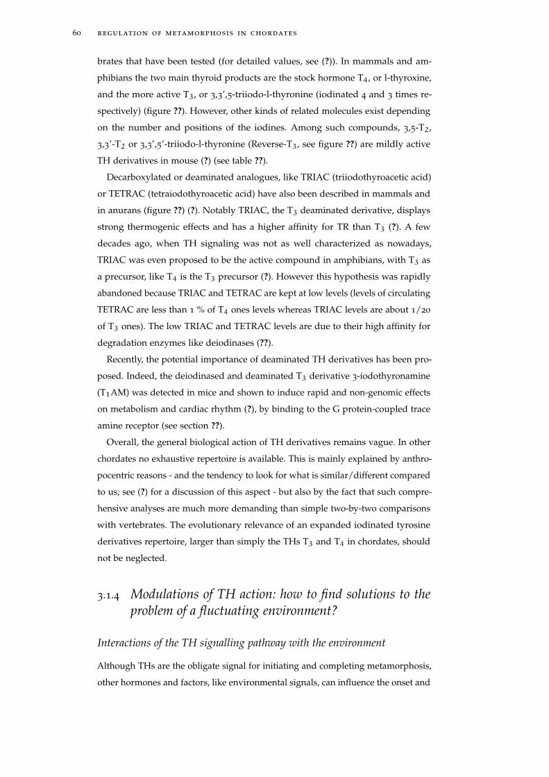
60 regulation of metamorphosis in chordates
brates that have been tested (for detailed values, see (?)). In mammals and am-
phibians the two main thyroid products are the stock hormone T4, or l-thyroxine,
and the more active T3, or 3,3’,5-triiodo-l-thyronine (iodinated 4 and 3 times re-
spectively) (figure ??). However, other kinds of related molecules exist depending
on the number and positions of the iodines. Among such compounds, 3,5-T2,
3,3’-T2 or 3,3’,5’-triiodo-l-thyronine (Reverse-T3, see figure ??) are mildly active
TH derivatives in mouse (?) (see table ??).
Decarboxylated or deaminated analogues, like TRIAC (triiodothyroacetic acid)
or TETRAC (tetraiodothyroacetic acid) have also been described in mammals and
in anurans (figure ??) (?). Notably TRIAC, the T3 deaminated derivative, displays
strong thermogenic effects and has a higher affinity for TR than T3 (?). A few
decades ago, when TH signaling was not as well characterized as nowadays,
TRIAC was even proposed to be the active compound in amphibians, with T3 as
a precursor, like T4 is the T3 precursor (?). However this hypothesis was rapidly
abandoned because TRIAC and TETRAC are kept at low levels (levels of circulating
TETRAC are less than 1 % of T4 ones levels whereas TRIAC levels are about 1/20
of T3 ones). The low TRIAC and TETRAC levels are due to their high affinity for
degradation enzymes like deiodinases (??).
Recently, the potential importance of deaminated TH derivatives has been pro-
posed. Indeed, the deiodinased and deaminated T3 derivative 3-iodothyronamine
(T1AM) was detected in mice and shown to induce rapid and non-genomic effects
on metabolism and cardiac rhythm (?), by binding to the G protein-coupled trace
amine receptor (see section ??).
Overall, the general biological action of TH derivatives remains vague. In other
chordates no exhaustive repertoire is available. This is mainly explained by anthro-
pocentric reasons - and the tendency to look for what is similar/different compared
to us; see (?) for a discussion of this aspect - but also by the fact that such compre-
hensive analyses are much more demanding than simple two-by-two comparisons
with vertebrates. The evolutionary relevance of an expanded iodinated tyrosine
derivatives repertoire, larger than simply the THs T3 and T4 in chordates, should
not be neglected.
3.1.4 Modulations of TH action: how to find solutions to theproblem of a fluctuating environment?
Interactions of the TH signalling pathway with the environment
Although THs are the obligate signal for initiating and completing metamorphosis,
other hormones and factors, like environmental signals, can influence the onset and
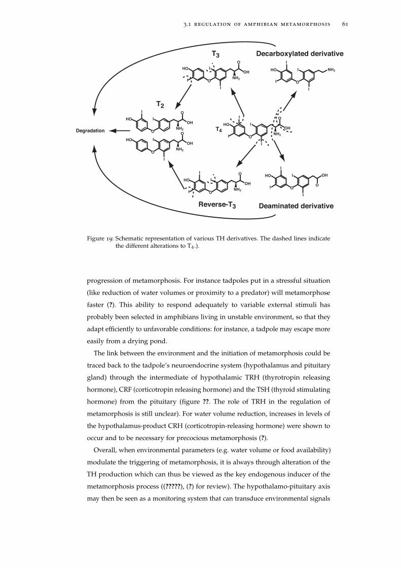
3.1 regulation of amphibian metamorphosis 61
T4
T2
I
I
HO
O
I
NH2
OH
O
I
IHO
ONH2
OH
O
I
IHO
O
I
NH2
OH
O
Deaminated derivative
I
I
I
HO
O
I
OH
O
Decarboxylated derivative
I
I
I
HO
O
I
NH2
T3
I
I
HO
O
I
NH2
OH
O
Reverse-T3
I
I
HO
ONH2
OH
O
I
Degradation
Figure 19: Schematic representation of various TH derivatives. The dashed lines indicatethe different alterations to T4.).
progression of metamorphosis. For instance tadpoles put in a stressful situation
(like reduction of water volumes or proximity to a predator) will metamorphose
faster (?). This ability to respond adequately to variable external stimuli has
probably been selected in amphibians living in unstable environment, so that they
adapt efficiently to unfavorable conditions: for instance, a tadpole may escape more
easily from a drying pond.
The link between the environment and the initiation of metamorphosis could be
traced back to the tadpole’s neuroendocrine system (hypothalamus and pituitary
gland) through the intermediate of hypothalamic TRH (thyrotropin releasing
hormone), CRF (corticotropin releasing hormone) and the TSH (thyroid stimulating
hormone) from the pituitary (figure ??. The role of TRH in the regulation of
metamorphosis is still unclear). For water volume reduction, increases in levels of
the hypothalamus-product CRH (corticotropin-releasing hormone) were shown to
occur and to be necessary for precocious metamorphosis (?).
Overall, when environmental parameters (e.g. water volume or food availability)
modulate the triggering of metamorphosis, it is always through alteration of the
TH production which can thus be viewed as the key endogenous inducer of the
metamorphosis process ((?????), (?) for review). The hypothalamo-pituitary axis
may then be seen as a monitoring system that can transduce environmental signals
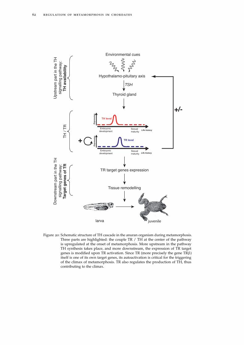
62 regulation of metamorphosis in chordates
Exp
ressio
n
Embryonic
developmentSexual
maturity
TH level
Life history
Environmental cues
Hypothalamo-pituitary axis
TR target genes expression
Tissue remodelling
larva juvenile
Thyroid gland
+/-
+
Exp
ressio
n
Embryonic
developmentSexual
maturity
TR level
Life history
TSH
TH
/ T
R
Upstr
eam
part
in t
he T
H s
ignalli
ng p
ath
way:
TH
avail
ab
ilit
y
Dow
nstr
eam
part
in t
he T
H s
ignalli
ng p
ath
way:
Targ
et
gen
es o
f T
R
Figure 20: Schematic structure of TH cascade in the anuran organism during metamorphosis.Three parts are highlighted: the couple TR / TH at the center of the pathwayis upregulated at the onset of metamorphosis. More upstream in the pathwayTH synthesis takes place, and more downstream, the expression of TR targetgenes is modified upon TR activation. Since TR (more precisely the gene TRβ)itself is one of its own target genes, its autoactivation is critical for the triggeringof the climax of metamorphosis. TR also regulates the production of TH, thuscontributing to the climax.
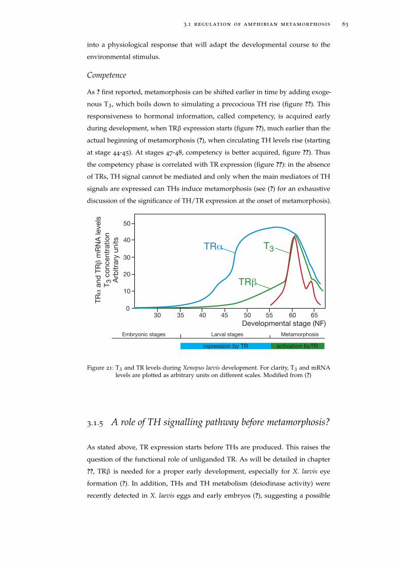
3.1 regulation of amphibian metamorphosis 63
into a physiological response that will adapt the developmental course to the
environmental stimulus.
Competence
As ? first reported, metamorphosis can be shifted earlier in time by adding exoge-
nous T3, which boils down to simulating a precocious TH rise (figure ??). This
responsiveness to hormonal information, called competency, is acquired early
during development, when TRβ expression starts (figure ??), much earlier than the
actual beginning of metamorphosis (?), when circulating TH levels rise (starting
at stage 44-45). At stages 47-48, competency is better acquired, figure ??). Thus
the competency phase is correlated with TR expression (figure ??): in the absence
of TRs, TH signal cannot be mediated and only when the main mediators of TH
signals are expressed can THs induce metamorphosis (see (?) for an exhaustive
discussion of the significance of TH/TR expression at the onset of metamorphosis).
30
10
0
20
30
40
35 40 45 50 55 60 65
50
TRα
and
TRβ
mR
NA
levels
T3 c
oncentr
atio
n
Arb
itra
ry u
nits
Developmental stage (NF)
TRα
TRβ
T3
Embryonic stages Larval stages Metamorphosis
repression by TR activation byTR
Figure 21: T3 and TR levels during Xenopus laevis development. For clarity, T3 and mRNAlevels are plotted as arbitrary units on different scales. Modified from (?)
3.1.5 A role of TH signalling pathway before metamorphosis?
As stated above, TR expression starts before THs are produced. This raises the
question of the functional role of unliganded TR. As will be detailed in chapter
??, TRβ is needed for a proper early development, especially for X. laevis eye
formation (?). In addition, THs and TH metabolism (deiodinase activity) were
recently detected in X. laevis eggs and early embryos (?), suggesting a possible
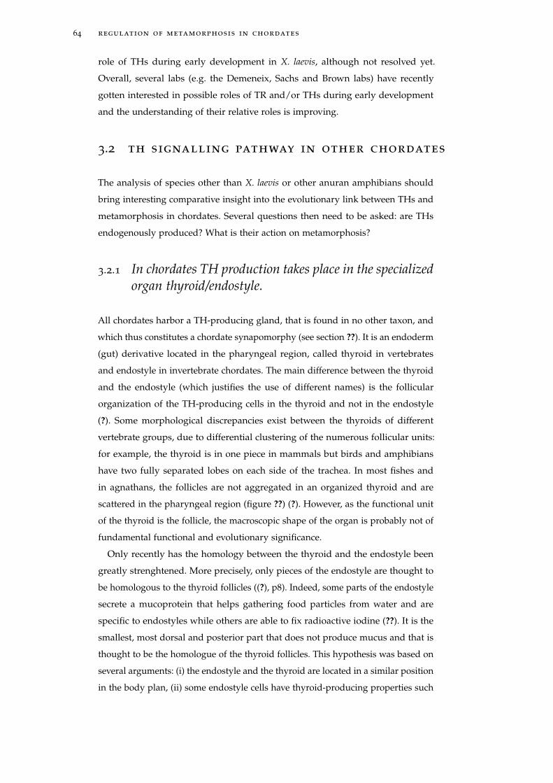
64 regulation of metamorphosis in chordates
role of THs during early development in X. laevis, although not resolved yet.
Overall, several labs (e.g. the Demeneix, Sachs and Brown labs) have recently
gotten interested in possible roles of TR and/or THs during early development
and the understanding of their relative roles is improving.
3.2 th signalling pathway in other chordates
The analysis of species other than X. laevis or other anuran amphibians should
bring interesting comparative insight into the evolutionary link between THs and
metamorphosis in chordates. Several questions then need to be asked: are THs
endogenously produced? What is their action on metamorphosis?
3.2.1 In chordates TH production takes place in the specializedorgan thyroid/endostyle.
All chordates harbor a TH-producing gland, that is found in no other taxon, and
which thus constitutes a chordate synapomorphy (see section ??). It is an endoderm
(gut) derivative located in the pharyngeal region, called thyroid in vertebrates
and endostyle in invertebrate chordates. The main difference between the thyroid
and the endostyle (which justifies the use of different names) is the follicular
organization of the TH-producing cells in the thyroid and not in the endostyle
(?). Some morphological discrepancies exist between the thyroids of different
vertebrate groups, due to differential clustering of the numerous follicular units:
for example, the thyroid is in one piece in mammals but birds and amphibians
have two fully separated lobes on each side of the trachea. In most fishes and
in agnathans, the follicles are not aggregated in an organized thyroid and are
scattered in the pharyngeal region (figure ??) (?). However, as the functional unit
of the thyroid is the follicle, the macroscopic shape of the organ is probably not of
fundamental functional and evolutionary significance.
Only recently has the homology between the thyroid and the endostyle been
greatly strenghtened. More precisely, only pieces of the endostyle are thought to
be homologous to the thyroid follicles ((?), p8). Indeed, some parts of the endostyle
secrete a mucoprotein that helps gathering food particles from water and are
specific to endostyles while others are able to fix radioactive iodine (??). It is the
smallest, most dorsal and posterior part that does not produce mucus and that is
thought to be the homologue of the thyroid follicles. This hypothesis was based on
several arguments: (i) the endostyle and the thyroid are located in a similar position
in the body plan, (ii) some endostyle cells have thyroid-producing properties such
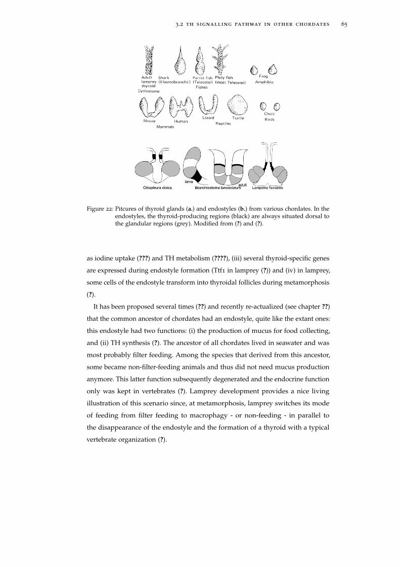
3.2 th signalling pathway in other chordates 65
a
b
Figure 22: Pitcures of thyroid glands (a.) and endostyles (b.) from various chordates. In theendostyles, the thyroid-producing regions (black) are always situated dorsal tothe glandular regions (grey). Modified from (?) and (?).
as iodine uptake (???) and TH metabolism (????), (iii) several thyroid-specific genes
are expressed during endostyle formation (Ttf1 in lamprey (?)) and (iv) in lamprey,
some cells of the endostyle transform into thyroidal follicles during metamorphosis
(?).
It has been proposed several times (??) and recently re-actualized (see chapter ??)
that the common ancestor of chordates had an endostyle, quite like the extant ones:
this endostyle had two functions: (i) the production of mucus for food collecting,
and (ii) TH synthesis (?). The ancestor of all chordates lived in seawater and was
most probably filter feeding. Among the species that derived from this ancestor,
some became non-filter-feeding animals and thus did not need mucus production
anymore. This latter function subsequently degenerated and the endocrine function
only was kept in vertebrates (?). Lamprey development provides a nice living
illustration of this scenario since, at metamorphosis, lamprey switches its mode
of feeding from filter feeding to macrophagy - or non-feeding - in parallel to
the disappearance of the endostyle and the formation of a thyroid with a typical
vertebrate organization (?).
66 regulation of metamorphosis in chordates
3.2.2 TH metabolism in other chordates
TH synthesis in other chordates
In mammals and fishes, TH metabolism is very similar to the one in amphibians (for
a review, see (?)). The "thyroidal" picture in other chordates has been investigated
much less thoroughly.
The search for TH derivatives in a wide variety of chordates was intense during
the 50’s and 60’s and significantly calmed down afterwards (?????). These experi-
ments were facilitated by the fact that TH synthesis is dependent on exogenous
iodide, which has the great advantage of being very specific to THs. Consequently
the detection of iradio-labelled odine characterizes and localizes TH synthesis.
THs were found in all chordate animals that were searched for them. T3 and
T4 (as well as their precursors MIT and DIT) were detected in amphioxus (??),
Ciona intestinalis (??), lampreys (?), sharks (?) and lungfishes (reported in (?)). TH
metabolism was also found in all chordates examined: peroxidase-dependent iodine
fixation (??), peroxidase activity (???), deiodinase activity (???), and a thyroglobulin-
like protein were reported in lamprey, hagfish and amphioxus (???). More generally,
the evolution of the TH signaling pathway within chordates will be discussed in
chapter ??. It is important to keep in mind that TH metabolism is not limited to
T3 and T4 in mammals or amphibians: other TH derivatives are produced, like
deiodinated, decarboxylated or deaminated derivatives, as well as sulfated and
glucuronidated conjugates (?) (figures ?? and ??). Similarly, it is most probable that
other chordates have a richer iodinated tyrosine derivatives repertoire than just T3
and T4. As already mentioned in section ??, this variability can be of significant
evolutionary importance and should not be dismissed.
TH and TH derivative transport in chordates
After synthesis, T4 needs to reach target organs from the production site (the
thyroid gland). The tissue- and cellular-specific expressions of the genes involved
in TH transport as well as deiodinases can influence the timing and localization of
TH action and therefore modulate the tissue-specific response to TH. Generally in
vertebrates, TH plasma transport is similar, although the relative importance of the
different carriers varies between species - for example TBG is the main T4 carrier
in human instead of TTR in rodents) (?). In lamprey, one TTR was recently cloned
from two species, P. marinus and L. appendix (?), and its expression was found to
be correlated with metamorphosis. In amphioxus and in C.intestinalis, virtually
nothing is known about TH transport.
3.3 ths and metamorphosis outside anuran amphibians 67
Intracellular transporters contribute to the fine regulation of TH availability to
each cell. Unfortunately, due to the relative youth of this field, virtually nothing
is known about cellular transport outside mammals. Moreover the molecules are
apparently not specific to THs and can also transport other amino-acid derivatives.
Inside the cell, free TH levels are also controlled. For instance, CTHBP (Cytosolic
Thyroid Hormone Binding Protein) is a cytosolic protein that binds T3 and thus is
likely to modulate the availability of T3 to TR in the nucleus (?).
3.3 ths and metamorphosis outside anuran am-phibians
3.3.1 Urodeles and amphibians paedomorphosis
Studies on amphibian metamorphosis, as described in the previous sections, have
been done on anuran amphibians like Xenopus laevis. However, not all amphibian
species have a similar post-embryonic development. For instance, urodeles are a
group of amphibians in which some species, called paedomorphic, are sexually
mature at the larval stage. There are several sorts of paedomorphosis. Some
species, called facultative paedomorphs, like Ambystoma tigrinum, may facultatively
metamorphose in nature and may be induced to metamorphose by exogenous THs
or even stressful conditions. The defects in the natural triggering of metamorphosis
have been attributed, not to TR mutations, but to problems in TSH production
e.g. (?). Other paedomorphs that never metamorphose in nature can be induced
to do so under TH stimulation, again suggesting problems in TH production.
On the contrary, permanent paedomorphs (like Necturus) cannot be induced
to metamorphose, even after TH stimulation. A scenario for the evolution of
paedomorphosis is detailed in the discussion (see chapter ??).
3.3.2 Metamorphosis in fishes is triggered by THs
After amphibians, actinopterygian fishes have been best studied for the role of
TH in the regulation of metamorphosis. More precisely, the molecular pathway
controlling metamorphosis has been mainly elucidated in flatfishes (pleuronec-
tiformes) such as flounders and turbot in which metamorphosis is spectacular,
as in anurans. In these fishes a symmetrical planktonic larva transforms into an
asymmetric benthic adult which has the two eyes on the same side of the body
((?), p442) (figure ??). The data obtained from these species took advantage of the
knowledge accumulated in anurans as a starting point and confirmed the central
68 regulation of metamorphosis in chordates
role of THs and TRα in the early regulation of metamorphosis (???). A schematic
structure of the TH signaling pathway involved in metamorphosis can be drawn
from those studies in which the TH / TR couple appears to be the key component
triggering metamorphosis in anurans and flat fishes (figure ??). Upstream TH
metabolism regulates TH production and availability, therefore TR activity; more
downstream target genes whose expression is regulated by the coupled TH/TR
control the morphological remodeling leading to metamorphosis (figure ??).
The role of THs in post-embryonic development has also been documented
in other groups. In zebrafish, THs were shown to be important for the larva-
to-juvenile transition: exogenous THs induce differentiation of the pectoral fin
precociously, in contrast the inhibition of TH synthesis blocks it (?). Smoltification
in salmonid fishes corresponds to physiological, morphological, metabolic and
behavioral modifications prior to migration from freshwater to sea water. It is
considered to correspond to metamorphosis: it is a period of high T4 and T3
levels (??) involving a morphological, physiological and behavioral switch from a
freshwater-adapted form to a salt-water-adapted form. At least some of the former
modifications were shown to be TH-sensitive (??).
3.3.3 TH in mammal development
A noticeable difference with amphibians is the apparent lack of metamorphosis in
mammals. Nonetheless, THs play central roles during mammalian development.
Deficiency in TH production after birth causes severe developmental problems and
especially mental retardation in humans. For instance, cretinism is due to problems
in TH production (and the huge goiter in cretins’ necks is caused by the swelling
of the thyroid gland). Similarly, people living in remote mountains, used to have
iodine supply deficiencies and mental development problems.
In mouse by suppressing THs or TR, it was shown that the alteration of the TH
signalling pathway leads to several defects in eye, bone and intestine developments
(for a review, see ?). For instance mice exhibited delayed maturation of bone and
intestine (?).
THs were shown to be especially important during weaning in mouse. Suppres-
sion of the thyroid gland (by the mutation of the pax8 gene, involved in thyroid
follicles formation (?)), causes severe malformation and death at weaning. Indeed,
several vital morphological and metabolic modifications occur during that period.
It is an ecological shift that is characterized by modifications like neural matura-
tion, intestine remodeling or chondrogenesis that are reminiscent of similar events
during amphibian metamorphosis. These modifications occur in correlation with
3.3 ths and metamorphosis outside anuran amphibians 69
a peak of TH production (both T3 and T4) (?) and were shown to be under the
control of TH and TR (???).
3.3.4 TH and metamorphosis in other chordates
Metamorphosis in lamprey
In lamprey, although several factors like temperature and growth advancement
(indicated by length and weight, for instance) modulate metamorphosis (?), it is
now well established that TH endogenous synthesis needs to be tightly regulated
for the proper metamorphosis. Interestingly, the role of TH displays a remarkable
difference compared to amphibians and teleost fishes: several reports clearly show
that it is a drop, instead of an increase, in circulating TH that ends the larval
period. This was shown by several means: circulating TH levels were shown to
drop at the onset of metamorphosis, exogenous THs blocked metamorphosis
and the inhibition of TH signaling with chemical compounds induced premature
metamorphosis (???????). However, the molecular pathway that mediates TH action
has been poorly investigated. T3 binding was demonstrated in lamprey nucleus
(?). There are two TRs in lamprey, from a lamprey-specific duplication (?), whose
function was unknown before my PhD. In chapter ??, I will present the molecular
characterization that I have done for these receptors, in which I show that both
receptors are functional (they bind TR DNA response elements, bind T3 and
activate transcription upon T3 binding), suggesting interesting differences in the
molecular events that regulate metamorphosis.
Metamorphosis in urochordates
Another interesting case of divergence of the TH signaling pathway is provided by
urochordates. Metamorphosis of Ciona intestinalis can be induced by exogenous
THs and inhibited by goitrogens known to block TH synthesis in vertebrates
(??). However, the molecular pathways triggered by THs remain elusive since the
only Ciona intestinalis TR does not bind T3 (?). Chapter ?? will describe some
of the work that I have done on this issue. The urochordates are particularly
interesting regarding the evolution of metamorphosis because of the diversity of
their post-embryonic developmental strategies. The elucidation of the role of THs
and iodinated tyrosine derivatives in general in late development in other species,
and in particular in larvaceans should generate interesting data for comparative
analysis.
70 regulation of metamorphosis in chordates
THs and metamorphosis in amphioxus
Chapter ?? presented the animal model amphioxus and chapter ?? described
amphioxus metamorphosis, on which most of my work was focused.
Although amphioxus metamorphosis has been known for more than a century
(?), its molecular basis has remained quite elusive until now. To my knowledge, the
only attempt to decipher whether THs are involved in amphioxus metamorphosis
was performed by ?: the author treated premetamorphic larvae of Branchiostoma
lanceolatum collected from plankton with T3, T4 and a TH synthesis inhibitor.
He noticed a slight acceleration of metamorphosis by THs and no effect by the
inhibitor. However, as the author confessed himself, few and rather old animals
were used for the experiment: "All the larvae were at a late stage of development, mostly
with 12-14 gill slits". Although "not one was metamorphosing", the larvae were already
advanced in their larval life and were about to metamorphose (starting when the
larva have about 12-13 gills, H.Escriva, personnal observation). As hormonal action
occurs during an extended period of time, I conducted experiments with younger
larvae to elucidate the possible role of THs during amphioxus metamorphosis
(chapters ?? and ??).
As explained in the next chapter, TR is the mediator of TH signal. In order to
better understand the evolution of TH signaling pathway in chordates, I focused
on TR action in amphioxus. The following chapter will introduce the molecular
basis of TR action.
4T R I S A T H - D E P E N D E N TT R A N S C R I P T I O N FA C T O R
Nuclear hormone receptors (NRs) constitute a rather large family of ligand-
dependent transcription factors (figure ??). Two properties of NRs make them
of particular interest: (i) they play a central role in various physiological and
pathophysiological processes as diverse as reproduction, development, and the
control of homeostasis (???) and (ii) their activity is regulated upon ligand binding,
and thereby may be manipulated with synthetic compounds, for therapeutical
purposes. Because NRs are both intracellular receptors of non-protein compounds
and ligand-dependent transcription factors, they have been a burning subject in
molecular biology. Moreover, their dependency on ligand makes them convenient
to study fundamental mechanisms of transcription. There are 48 NRs in human
and 33 in amphioxus (?). Among NRs, TRs belong to the subfamily NR1 (boxed
in orange in figure ??) that regroups some of the best studied NRs (e.g. RAR, TR,
PPAR).
This chapter does not intend to be exhaustive on TR molecular mechanism and
will focus on notions that are relevant for the understanding of the experiments
reported in this manuscript.
4.1 tr structure / function
TRs are nuclear proteins and ligand-dependent transcription factors. In the absence
of a ligand, the receptor can bind DNA, interacts specifically with response ele-
ments in the regulatory regions of target genes, and actively inhibits transcription.
Inhibition results because receptor conformation favors interactions with core-
pressors that recruit deacetylases able to block transcription (figure ??). Ligand
binding induces a conformational changes that results in the recruitment of coacti-
vators which in turn alter chromatin structure and influence the recruitment of the
transcription machinery (figure ??) (?). This recruitment leads to activation of the
expression of target genes, subsequently triggering specific biological processes
such as metamorphosis (?). There are two TRs in vertebrates, TRα and TRβ that
play different role in TH signaling and that were issued form a vertebrate specific
71
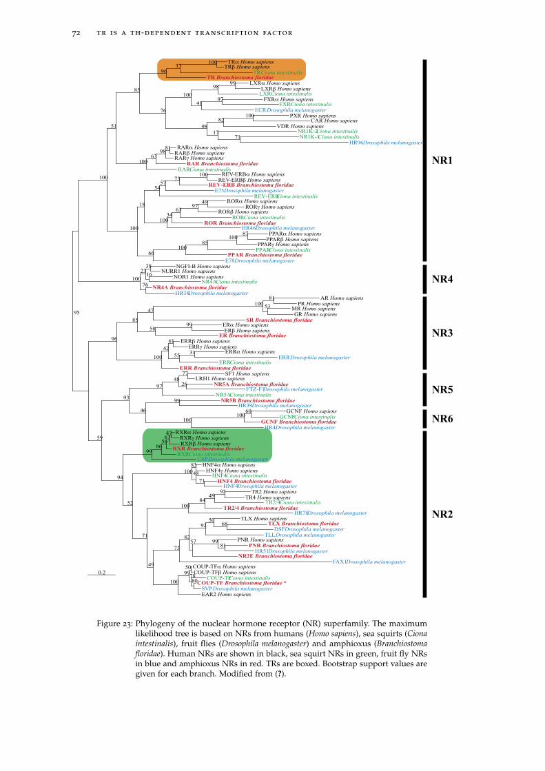
72 tr is a th-dependent transcription factor
EAR2 Homo sapiensSVP Drosophila melanogaster
COUP-TF Branchiostoma floridae *COUP-TFCiona intestinalis
80
COUP-TFβ Homo sapiensCOUP-TFα Homo sapiens
78
50
99
FAX1 Drosophila melanogasterNR2E Branchiostoma floridae
HR51Drosophila melanogasterPNR Branchiostoma floridae
PNR Homo sapiens 81
99
TLL Drosophila melanogasterDSFDrosophila melanogaster
TLX Branchiostoma floridaeTLX Homo sapiens
68 50
57
92
82
100
73
HR78Drosophila melanogasterTR2/4 Branchiostoma floridae
TR2/4Ciona intestinalisTR4 Homo sapiens
TR2 Homo sapiens 92 49
84
49
100
HNF4Drosophila melanogasterHNF4 Branchiostoma floridae
HNF4Ciona intestinalis 71
HNF4γ Homo sapiensHNF4α Homo sapiens
48
83
71
100
USPDrosophila melanogasterRXRCiona intestinalis
RXR Branchiostoma floridaeRXRβ Homo sapiensRXRγ Homo sapiens
RXRα Homo sapiens 47 67
56
86
52
99
HR4Drosophila melanogasterGCNF Branchiostoma floridae
GCNFCiona intestinalisGCNF Homo sapiens 60
100
HR39Drosophila melanogasterNR5B Branchiostoma floridae
100
99
NR5A Ciona intestinalisFTZ-F1Drosophila melanogaster
NR5A Branchiostoma floridaeLRH1 Homo sapiens
SF1 Homo sapiens
26
77
48
46
97
94
93
ERR Branchiostoma floridaeERRCiona intestinalis
ERRDrosophila melanogasterERRα Homo sapiens 31
ERRγ Homo sapiensERRβ Homo sapiens
55
61
42
ER Branchiostoma floridaeERβ Homo sapiens
ERα Homo sapiens 99 SR Branchiostoma floridae
GR Homo sapiensMR Homo sapiens
PR Homo sapiensAR Homo sapiens
53
81
100
58
47
100
85
59
96
HR38Drosophila melanogaster NR4A Branchiostoma floridae
NR4A Ciona intestinalisNOR1 Homo sapiens
NURR1 Homo sapiensNGFI-B Homo sapiens
16
38
76
23
E78Drosophila melanogasterPPAR Branchiostoma floridae
PPAR Ciona intestinalisPPARγ Homo sapiens
PPARβ Homo sapiensPPARα Homo sapiens 87
100
85
100
HR46Drosophila melanogasterROR Branchiostoma floridae
RORCiona intestinalisRORβ Homo sapiens
RORγ Homo sapiensRORα Homo sapiens 49
97 63
34
REV-ERB Ciona intestinalisE75Drosophila melanogaster
REV-ERB Branchiostoma floridaeREV-ERBβ Homo sapiens
REV-ERBα Homo sapiens 100 73
57
100
54
66
18
RARCiona intestinalisRAR Branchiostoma floridae
RARγ Homo sapiensRARβ Homo sapiensRARα Homo sapiens 81
98
63
HR96Drosophila melanogaster NR1K-1Ciona intestinalis
NR1K-2Ciona intestinalis 71
VDR Homo sapiensCAR Homo sapiens
PXR Homo sapiens 100
17
82
ECRDrosophila melanogaster FXRCiona intestinalis
FXRα Homo sapiens 97
LXRCiona intestinalisLXRβ Homo sapiens
LXRα Homo sapiens 99
41
98
98
100
TR Branchiostoma floridaeTRCiona intestinalis
TRβ Homo sapiensTRα Homo sapiens 100
37
76
96
100
85
100
51
100
100
95
NR2
NR6
NR5
NR3
NR4
NR1
0.2
Figure 23: Phylogeny of the nuclear hormone receptor (NR) superfamily. The maximumlikelihood tree is based on NRs from humans (Homo sapiens), sea squirts (Cionaintestinalis), fruit flies (Drosophila melanogaster) and amphioxus (Branchiostomafloridae). Human NRs are shown in black, sea squirt NRs in green, fruit fly NRsin blue and amphioxus NRs in red. TRs are boxed. Bootstrap support values aregiven for each branch. Modified from (?).
4.1 tr structure / function 73
duplication (there have been independent supplementary duplications in fishes
and in X.laevis (?), see section ?? for further details)
Like all NRs, TR displays a modular organization in functional domains (figure
??): the DNA binding domain (DBD), the ligand binding domain (LBD), the N-
terminal A/B domain, the hinge between the DBD and the LBD and the C-terminal
F-domain. Only the DBD and the LBD are highly conserved between NRs, even
within TRs (?) and will focus the attention of this chapter.
4.1.1 A/B domain
The A/B domain, at the N-terminal end, contains an activation function (AF-1)
in TRα1 and TRβ1 isoforms (?). This domain is dependent on cellular context
(ref cf Germain) and can also be submitted to post-translational modifications,
like phosphorylation, that can alter transcriptional properties (e.g. DNA binding
properties, AF-1-dependent activation) (?). The AF-1 is highly variable in both
length and sequence. This domain displays a especially fast rate of substitution,
potentially reflecting a minor evolutionary importance and has been the subject of
virtually no evolutionary study.
4.1.2 C domain: DNA binding domain
TRs recognize specific sequences (figure ??a), called thyroid hormone response
elements (TREs). The consensus TRE is composed of two (AGGTCA) motifs in
different configurations, the consensus binding site being constituted of 2 motifs
in the same orientation and separated by 4 nucleotides (DR4, figure ??a). There
are however numerous alternative sites. For instance, an imperfect DR4 in the
promoter of TR in Xenopus laevis (xDR4 in figure ??a), is responsible for TR
expression autoregulation (figure ?? in the previous chapter) (?). TRs may also
bind inverted palindromes of the (AGGTCA) motif (figure ??a). Whether all TREs
contain (AGGTCA) motifs is still to be determined On the other hand, many
potential sequences are not bona fide TREs, as exemplified in the genome-wide
determination of binding sites of the estrogen receptor, another NR (?).
The DNA binding domain (DBD) is a highly conserved short 66-residue long
core. 3D structures of the TR DBD show the same modular organization as other
NRs (?):
• Two cysteine-rich zinc fingers (figure ??b) interact with DNA on TREs (figure
??c).
74 tr is a th-dependent transcription factor
NCOR/SMRT
HDAC
RXR
Repressionof
Transcription
TR
Repression
P
Me Ac
Ac Ac
P
P
MeAc
Ac Ac Ac
Ac
Ac
Me
SWI/SNF
HistonePhosphorylation,
Acetylation,Methylation
NucleosomeTranslocation
CoactivatorComplex
ChromatinRemodelingComplex
CBP, P300
SRC1
HMT
Kinase
HAT
RXR TR
PGC1
De-Repression
TranscriptionMachinery
Initiationof
Transcription
Basal
P
Me Ac
Ac Ac
P
MeAc
Ac Ac
TAFs
TBP
TFIIETFIIB
TFIIA
RNA Pol.II
TFIIHTFIIF
MediatorComplex
RXR TR
MED1/
TRAP220
Activation
ba
Figure 24: Molecular basis of NR-dependent transcriptional regulation. In absence of aligand, NRs are bound to DNA to response elements located in the promoterregions of target genes and are associated with a complex containing histonedeacetylases (HDACs) and corepressors (NCOR/SMRT) that repress transcription(upper panel). Binding of a ligand induces a conformational change in the LBDresulting in dissociation of corepressors and recruitment of coactivators (NCOA,CBP and P300 and PGC1) that are associated with histone acetyltransferases(HATs), histone methyltransferases (HMTs), kinases and an ATP-dependent chro-matin remodeling complex (SWI/SNF), which together decompact and unfoldthe condensed chromatin (middle panel). Finally, the coactivators dissociate fromthe NRs and the mediator complex (including MED1/TRAP220) is assembled onthe DNA locus. This mediator complex recruits transcription factors of the basaltranscription machinery and RNA polymerase II (RNA Pol.II) to the promoterresulting in initiation of transcription (lower panel). Courtesy of Gérard Benoît,adapted from (?).
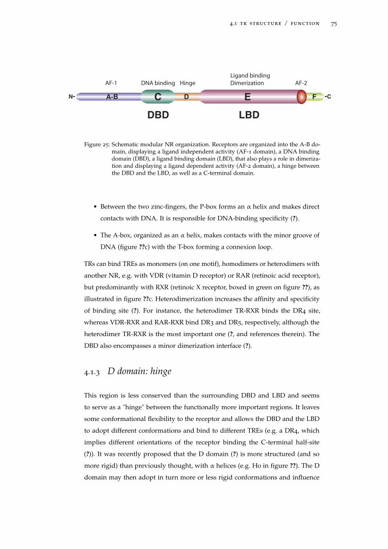
4.1 tr structure / function 75
A-B C D E FN- -C
DBD LBD
Ligand binding
DimerizationAF-1 HingeDNA binding AF-2
Figure 25: Schematic modular NR organization. Receptors are organized into the A-B do-main, displaying a ligand independent activity (AF-1 domain), a DNA bindingdomain (DBD), a ligand binding domain (LBD), that also plays a role in dimeriza-tion and displaying a ligand dependent activity (AF-2 domain), a hinge betweenthe DBD and the LBD, as well as a C-terminal domain.
• Between the two zinc-fingers, the P-box forms an α helix and makes direct
contacts with DNA. It is responsible for DNA-binding specificity (?).
• The A-box, organized as an α helix, makes contacts with the minor groove of
DNA (figure ??c) with the T-box forming a connexion loop.
TRs can bind TREs as monomers (on one motif), homodimers or heterodimers with
another NR, e.g. with VDR (vitamin D receptor) or RAR (retinoic acid receptor),
but predominantly with RXR (retinoic X receptor, boxed in green on figure ??), as
illustrated in figure ??c. Heterodimerization increases the affinity and specificity
of binding site (?). For instance, the heterodimer TR-RXR binds the DR4 site,
whereas VDR-RXR and RAR-RXR bind DR3 and DR5, respectively, although the
heterodimer TR-RXR is the most important one (?, and references therein). The
DBD also encompasses a minor dimerization interface (?).
4.1.3 D domain: hinge
This region is less conserved than the surrounding DBD and LBD and seems
to serve as a "hinge" between the functionally more important regions. It leaves
some conformational flexibility to the receptor and allows the DBD and the LBD
to adopt different conformations and bind to different TREs (e.g. a DR4, which
implies different orientations of the receptor binding the C-terminal half-site
(?)). It was recently proposed that the D domain (?) is more structured (and so
more rigid) than previously thought, with α helices (e.g. H0 in figure ??). The D
domain may then adopt in turn more or less rigid conformations and influence
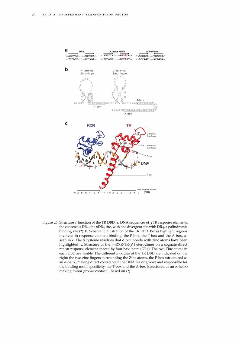
76 tr is a th-dependent transcription factor
TRRXR
DNA
Zn
Zn
Zn
Zn
T
T
T
T
T C
C
C
C
A
A
A
A
GG
G
G
G
TTTTT CCCC AA AA GGGG G
A-box
DNA response element
(DR4)
C-terminal
Zinc finger
N-terminal
Zinc finger
P-box
5'-AGGTCAnnnnAGGTCA-3
'
3'-TCCAGTnnnnTCCAGT-5
'
DR4
5'-AGGTCAnnnnAGGACA-3
'
3'-TCCAGTnnnnTCCTGT-5
'
X.laevis xDR4
5'-AGGTCAnnnTGACCT-3
'
3'-TCCAGTnnnACTGGA-5
'
palindrome
RKKCIYVGMATDLVLDDSKRL
APKHGISKQLEERRRKERNEEILKRK
K G F F R R T I Q K N L H P S YDKDELS
G
G
Y
Zn
YH
R
G
C
C C
C CC C
T
T
T
I
A
K
D
C
C
V E
V
Y
Zn
TR
E
V
V
I
G
E
Q
K
K
N
Q
K
D
P-box
T-box
A-box
N-terminal
Zinc-f inger
C-terminal
Zinc-f inger
a
b
c
Figure 26: Structure / function of the TR DBD. a. DNA sequences of 3 TR response elements:the consensus DR4, the xDR4 site, with one divergent site with DR4, a palindromicbinding site (?). b. Schematic illustration of the TR DBD. Boxes highlight regionsinvolved in response element binding: the P-box, the T-box and the A-box, asseen in c. The 8 cysteine residues that direct bonds with zinc atoms have beenhighlighted. c. Structure of the 5’-RXR-TR-3’ heterodimer on a cognate directrepeat response element spaced by four base pairs (DR4). The two Zinc atoms ineach DBD are visible. The different modules of the TR DBD are indicated on theright: the two zinc fingers surrounding the Zinc atoms, the P-box (structured asan α-helix) making direct contact with the DNA major groove and responsible forthe binding motif specificity, the T-box and the A-box (structured as an α-helix)making minor groove contact. . Based on (?).
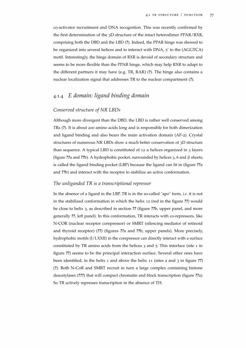
4.1 tr structure / function 77
co-activator recruitment and DNA recognition. This was recently confirmed by
the first determination of the 3D structure of the intact heterodimer PPAR/RXR,
comprising both the DBD and the LBD (?). Indeed, the PPAR hinge was showed to
be organized into several helices and to interact with DNA, 5’ to the (AGGTCA)
motif. Interestingly, the hinge domain of RXR is devoid of secondary structure and
seems to be more flexible than the PPAR hinge, which may help RXR to adapt to
the different partners it may have (e.g. TR, RAR) (?). The hinge also contains a
nuclear localization signal that addresses TR to the nuclear compartment (?).
4.1.4 E domain: ligand binding domain
Conserved structure of NR LBDs
Although more divergent than the DBD, the LBD is rather well conserved among
TRs (?). It is about 200 amino acids long and is responsible for both dimerization
and ligand binding and also bears the main activation domain (AF-2). Crystal
structures of numerous NR LBDs show a much better conservation of 3D structure
than sequence. A typical LBD is constituted of 12 α helices organized in 3 layers
(figure ??a and ??b). A hydrophobic pocket, surrounded by helices 3, 6 and β sheets,
is called the ligand binding pocket (LBP) because the ligand can fit in (figure ??a
and ??b) and interact with the receptor to stabilize an active conformation.
The unliganded TR is a transcriptional repressor
In the absence of a ligand in the LBP, TR is in the so-called "apo" form, i.e. it is not
in the stabilized conformation in which the helix 12 (red in the figure ??) would
be close to helix 3, as described in section ?? (figure ??b, upper panel, and more
generally ??, left panel). In this conformation, TR interacts with co-repressors, like
N-COR (nuclear receptor corepressor) or SMRT (silencing mediator of retinoid
and thyroid receptor) (??) (figures ??a and ??b, upper panels). More precisely,
hydrophobic motifs (I/LXXII) in the corepressor can directly interact with a surface
constituted by TR amino acids from the helices 3 and 5. This interface (site 1 in
figure ??) seems to be the principal interaction surface. Several other ones have
been identified, in the helix 1 and above the helix 11 (sites 2 and 3 in figure ??)
(?). Both N-CoR and SMRT recruit in turn a large complex containing histone
deacetylases (???) that will compact chromatin and block transcription (figure ??a).
So TR actively represses transcription in the absence of TH.
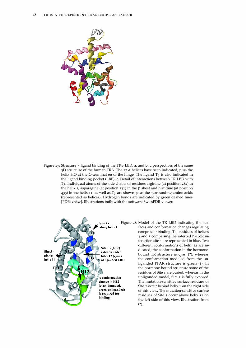
78 tr is a th-dependent transcription factor
H0
H1
H2
H3
H4
H5H6
H7
H8
H9
H10
H11
H12
H0
H1
H2
H3
H4
H5
H6
H7
H8
H9
H10
H11
H12
T3
H3
R282
H11
H435
β-sheet
R331
T3
a b
c
Figure 27: Structure / ligand binding of the TRβ LBD. a. and b. 2 perspectives of the same3D structure of the human TRβ. The 12 α helices have been indicated, plus thehelix HO at the C-terminal en of the hinge. The ligand T3 is also indicated inthe ligand binding pocket (LBP). c. Detail of interactions between TR LBD withT3. Individual atoms of the side chains of residues arginine (at position 282) inthe helix 3, asparagine (at position 331) in the β sheet and histidine (at position435) in the helix 11, as well as T3 are shown, plus the surrounding amino acids(represented as helices). Hydrogen bonds are indicated by green dashed lines.[PDB: 2h6w]. Illustrations built with the software SwissPDB-viewer.
Figure 28: Model of the TR LBD indicating the sur-faces and conformation changes regulatingcorepressor binding. The residues of helices3 and 5 comprising the inferred N-CoR in-teraction site 1 are represented in blue. Twodifferent conformations of helix 12 are in-dicated; the conformation in the hormone-bound TR structure is cyan (?), whereasthe conformation modeled from the un-liganded PPAR structure is green (?). Inthe hormone-bound structure some of theresidues of Site 1 are buried, whereas in theunliganded model, Site 1 is fully exposed.The mutation-sensitive surface residues ofSite 2 occur behind helix 1 on the right sideof this view. The mutation-sensitive surfaceresidues of Site 3 occur above helix 11 onthe left side of this view. Illustration from(?).
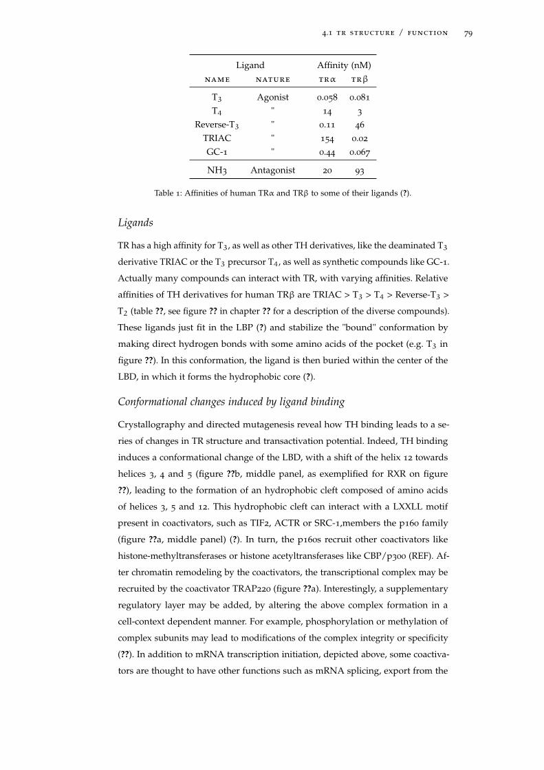
4.1 tr structure / function 79
Ligand Affinity (nM)name nature trα trβ
T3 Agonist 0.058 0.081
T4 " 14 3
Reverse-T3 " 0.11 46
TRIAC " 154 0.02
GC-1 " 0.44 0.067
NH3 Antagonist 20 93
Table 1: Affinities of human TRα and TRβ to some of their ligands (?).
Ligands
TR has a high affinity for T3, as well as other TH derivatives, like the deaminated T3
derivative TRIAC or the T3 precursor T4, as well as synthetic compounds like GC-1.
Actually many compounds can interact with TR, with varying affinities. Relative
affinities of TH derivatives for human TRβ are TRIAC > T3 > T4 > Reverse-T3 >
T2 (table ??, see figure ?? in chapter ?? for a description of the diverse compounds).
These ligands just fit in the LBP (?) and stabilize the "bound" conformation by
making direct hydrogen bonds with some amino acids of the pocket (e.g. T3 in
figure ??). In this conformation, the ligand is then buried within the center of the
LBD, in which it forms the hydrophobic core (?).
Conformational changes induced by ligand binding
Crystallography and directed mutagenesis reveal how TH binding leads to a se-
ries of changes in TR structure and transactivation potential. Indeed, TH binding
induces a conformational change of the LBD, with a shift of the helix 12 towards
helices 3, 4 and 5 (figure ??b, middle panel, as exemplified for RXR on figure
??), leading to the formation of an hydrophobic cleft composed of amino acids
of helices 3, 5 and 12. This hydrophobic cleft can interact with a LXXLL motif
present in coactivators, such as TIF2, ACTR or SRC-1,members the p160 family
(figure ??a, middle panel) (?). In turn, the p160s recruit other coactivators like
histone-methyltransferases or histone acetyltransferases like CBP/p300 (REF). Af-
ter chromatin remodeling by the coactivators, the transcriptional complex may be
recruited by the coactivator TRAP220 (figure ??a). Interestingly, a supplementary
regulatory layer may be added, by altering the above complex formation in a
cell-context dependent manner. For example, phosphorylation or methylation of
complex subunits may lead to modifications of the complex integrity or specificity
(??). In addition to mRNA transcription initiation, depicted above, some coactiva-
tors are thought to have other functions such as mRNA splicing, export from the
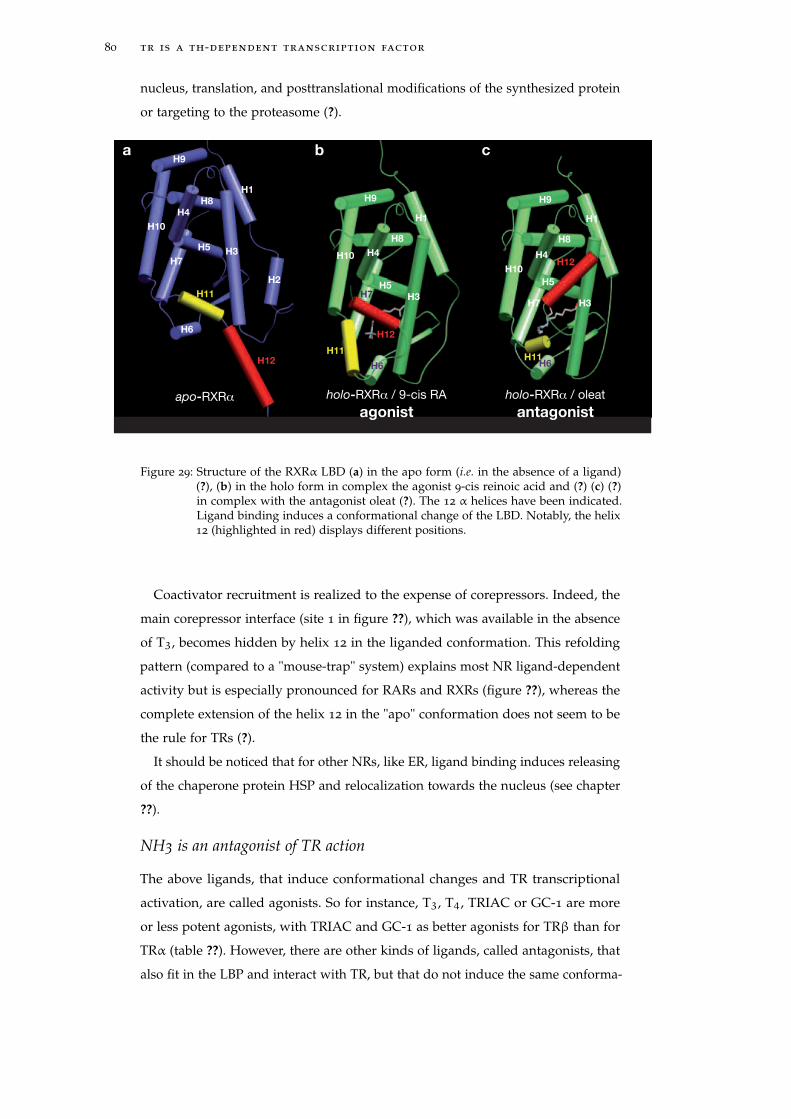
80 tr is a th-dependent transcription factor
nucleus, translation, and posttranslational modifications of the synthesized protein
or targeting to the proteasome (?).
H2
H3
H4
H5
H7
H10
H8
H1
H9
H12
H6
H11H3
H4
H5
H7
H10
H8
H1
H9
H12
H6H11
H3
H4
H5H7
H10
H8
H1
H9
H12
H6
H11
apo-RXRα holo-RXRα / oleat
antagonist
holo-RXRα / 9-cis RA
agonist
a b c
Figure 29: Structure of the RXRα LBD (a) in the apo form (i.e. in the absence of a ligand)(?), (b) in the holo form in complex the agonist 9-cis reinoic acid and (?) (c) (?)in complex with the antagonist oleat (?). The 12 α helices have been indicated.Ligand binding induces a conformational change of the LBD. Notably, the helix12 (highlighted in red) displays different positions.
Coactivator recruitment is realized to the expense of corepressors. Indeed, the
main corepressor interface (site 1 in figure ??), which was available in the absence
of T3, becomes hidden by helix 12 in the liganded conformation. This refolding
pattern (compared to a "mouse-trap" system) explains most NR ligand-dependent
activity but is especially pronounced for RARs and RXRs (figure ??), whereas the
complete extension of the helix 12 in the "apo" conformation does not seem to be
the rule for TRs (?).
It should be noticed that for other NRs, like ER, ligand binding induces releasing
of the chaperone protein HSP and relocalization towards the nucleus (see chapter
??).
NH3 is an antagonist of TR action
The above ligands, that induce conformational changes and TR transcriptional
activation, are called agonists. So for instance, T3, T4, TRIAC or GC-1 are more
or less potent agonists, with TRIAC and GC-1 as better agonists for TRβ than for
TRα (table ??). However, there are other kinds of ligands, called antagonists, that
also fit in the LBP and interact with TR, but that do not induce the same conforma-
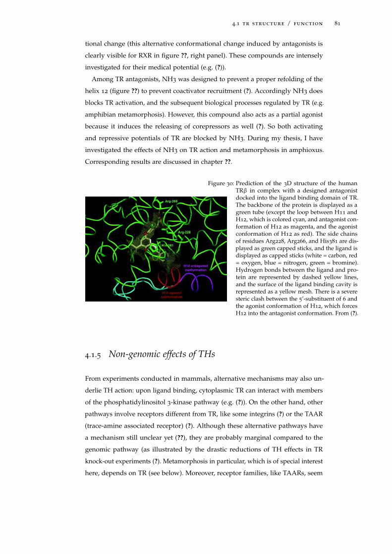
4.1 tr structure / function 81
tional change (this alternative conformational change induced by antagonists is
clearly visible for RXR in figure ??, right panel). These compounds are intensely
investigated for their medical potential (e.g. (?)).
Among TR antagonists, NH3 was designed to prevent a proper refolding of the
helix 12 (figure ??) to prevent coactivator recruitment (?). Accordingly NH3 does
blocks TR activation, and the subsequent biological processes regulated by TR (e.g.
amphibian metamorphosis). However, this compound also acts as a partial agonist
because it induces the releasing of corepressors as well (?). So both activating
and repressive potentials of TR are blocked by NH3. During my thesis, I have
investigated the effects of NH3 on TR action and metamorphosis in amphioxus.
Corresponding results are discussed in chapter ??.
Figure 30: Prediction of the 3D structure of the humanTRβ in complex with a designed antagonistdocked into the ligand binding domain of TR.The backbone of the protein is displayed as agreen tube (except the loop between H11 andH12, which is colored cyan, and antagonist con-formation of H12 as magenta, and the agonistconformation of H12 as red). The side chainsof residues Arg228, Arg266, and His381 are dis-played as green capped sticks, and the ligand isdisplayed as capped sticks (white = carbon, red= oxygen, blue = nitrogen, green = bromine).Hydrogen bonds between the ligand and pro-tein are represented by dashed yellow lines,and the surface of the ligand binding cavity isrepresented as a yellow mesh. There is a severesteric clash between the 5’-substituent of 6 andthe agonist conformation of H12, which forcesH12 into the antagonist conformation. From (?).
4.1.5 Non-genomic effects of THs
From experiments conducted in mammals, alternative mechanisms may also un-
derlie TH action: upon ligand binding, cytoplasmic TR can interact with members
of the phosphatidylinositol 3-kinase pathway (e.g. (?)). On the other hand, other
pathways involve receptors different from TR, like some integrins (?) or the TAAR
(trace-amine associated receptor) (?). Although these alternative pathways have
a mechanism still unclear yet (??), they are probably marginal compared to the
genomic pathway (as illustrated by the drastic reductions of TH effects in TR
knock-out experiments (?). Metamorphosis in particular, which is of special interest
here, depends on TR (see below). Moreover, receptor families, like TAARs, seem
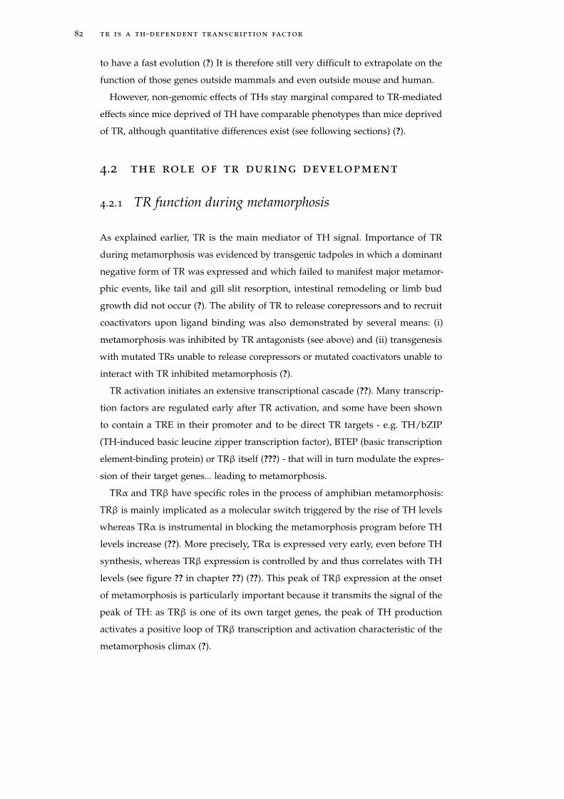
82 tr is a th-dependent transcription factor
to have a fast evolution (?) It is therefore still very difficult to extrapolate on the
function of those genes outside mammals and even outside mouse and human.
However, non-genomic effects of THs stay marginal compared to TR-mediated
effects since mice deprived of TH have comparable phenotypes than mice deprived
of TR, although quantitative differences exist (see following sections) (?).
4.2 the role of tr during development
4.2.1 TR function during metamorphosis
As explained earlier, TR is the main mediator of TH signal. Importance of TR
during metamorphosis was evidenced by transgenic tadpoles in which a dominant
negative form of TR was expressed and which failed to manifest major metamor-
phic events, like tail and gill slit resorption, intestinal remodeling or limb bud
growth did not occur (?). The ability of TR to release corepressors and to recruit
coactivators upon ligand binding was also demonstrated by several means: (i)
metamorphosis was inhibited by TR antagonists (see above) and (ii) transgenesis
with mutated TRs unable to release corepressors or mutated coactivators unable to
interact with TR inhibited metamorphosis (?).
TR activation initiates an extensive transcriptional cascade (??). Many transcrip-
tion factors are regulated early after TR activation, and some have been shown
to contain a TRE in their promoter and to be direct TR targets - e.g. TH/bZIP
(TH-induced basic leucine zipper transcription factor), BTEP (basic transcription
element-binding protein) or TRβ itself (???) - that will in turn modulate the expres-
sion of their target genes... leading to metamorphosis.
TRα and TRβ have specific roles in the process of amphibian metamorphosis:
TRβ is mainly implicated as a molecular switch triggered by the rise of TH levels
whereas TRα is instrumental in blocking the metamorphosis program before TH
levels increase (??). More precisely, TRα is expressed very early, even before TH
synthesis, whereas TRβ expression is controlled by and thus correlates with TH
levels (see figure ?? in chapter ??) (??). This peak of TRβ expression at the onset
of metamorphosis is particularly important because it transmits the signal of the
peak of TH: as TRβ is one of its own target genes, the peak of TH production
activates a positive loop of TRβ transcription and activation characteristic of the
metamorphosis climax (?).
4.2 the role of tr during development 83
4.2.2 TR and early development
In amphibians
Attention was long concentrated on the role of TR during metamorphosis because
TR was thought to have no major roles when virtually no TH is produced, as during
early development (figure ?? in section ??). Recently however, TR was shown to
be important during amphibian premetamorphic development by inhibiting the
expression of specific target genes. Accordingly, chromatin immunoprecipitation
(ChIP) experiments showed that TR binds DNA on the promoter of target genes
in premetamorphic tadpole larvae (?), when TH levels are undetectable, and is
in a repressive state (?). A possible role of TR may then be to actively repress the
initiation of metamorphosis. Indeed, in transgenic tadpoles expressing a dominant
negative form of the corepressor N-COR, TR was not repressing gene expression,
leading to an increase in the expression of its target genes as well as a faster
development (?). However, TR inhibitory action during early development has more
important effects than just preventing metamorphosis. The reciprocal transgenesis
of mutated TRs unable to recruit N-COR showed a more drastic phenotype with
embryonic lethality for 75 % of the tadpoles and with string defects in cranial and
especially eye development for the remaining 25 % (?), demonstrating the function
of TR before metamorphosis, for instance as a regulator of eye formation.
In mammals
TR activation mechanism, as TH-dependent transcription factor is very similar in
mammals and in amphibians. As in amphibians, a role during development has
been well documented, mostly through the use of transgenic mice. Comparative ex-
periments were done between mice devoid of TRα, TRβ (e.g. (??)) and/or TH (mice
KO for the gene Pax8 do not develop a thyroid gland, (?)), with complementary
results. From these experiments, TRα seems to mainly regulate early development,
before weaning (about 3 weeks after birth). More precisely, the post-natal and
pre-weaning period is characterized by important morphological and metabolic
modifications like neural maturation, intestine remodeling, or chondrogenesis.
These modifications occur in correlation with a peak of TH production (both T3
and T4) (?), and were shown to be under the control of TH and TRαfl (???).
The importance of TR action prior to weaning was exemplified by the severe
developmental damages caused by both suppression of TH production or knock-in
of a dominant negative TRα (?), leading in both cases to death at weaning. On the
other hand, mice devoid of TRα suffered from much less severe defects and did not
die at weaning. This effect was attributed to the transcriptional inhibitory role of
84 tr is a th-dependent transcription factor
TRα before TH levels increase: not lifting the transcriptional repression exerted by
TRα at the appropriate developmental moment (weaning), is more harmful than
lifting TRα transcriptional inhibition during early development (?). Accordingly,
the suppression of both TH and TRα causes a less severe phenotype (?).
4.3 tr phylogeny
TRs were originally described in chicken (?) and soon thereafter in mammals (??)
and in other gnathostomes (e.g. (?)). As mentioned above, there are two TRs in
gnathostomes (TRα and TRβ) from a vertebrate-specific genome duplication (??)
(actually there are two TRα and two TRβ in Xenopus laevis from a recent species-
specific genome duplication (?) but the duplicates are functionally redundant).
TRs were long thought to be vertebrate-specific, which turned out to be erroneous.
Instead TRs have been found in both deuterostomes and protostomes: there are
two TRs in lamprey from a lamprey-specific duplication (?), there is one TR
in Ciona intestinalis (?), in amphioxus (?) (see chapter ?? for further details), in
the echinoderm Strongylocentrotus purpuratus (?). TRs were also found in the
platyhelminths Schistosoma mansoni, S. japonium and Schmidtea mediterranea, in a
mollusc Lottia gigantean, in an arthropod Daphnia pulex (?) as well as in the annelid
Capitella sp.. In contrast, TR was lost in ecdysozoans (no TR has been found in
Ecdysozoan species whose genome has been sequenced so far) (?).
For a long time, attention was focused on vertebrate TRs, which all showed
similar properties. Indeed, all vertebrate (and even gnathostome) TRs characterized
so far display the same modular organization, the same 3D structure and the
same activation process (excluding isoform-specific peculiarities, like mouse TRα2).
The only exception, so far, is the TR from Ciona intestinalis, in which a 200-amino
acid long insertion in the C-terminal end may be at the origin of T3 binding
impairments (?).
As will be considered in the discussion of this manuscript (see chapter ?? in
the discussion), the ancient origin of TRs in bilaterians raises the question of the
ancestral function of this NR, especially in species where no clear TH signaling
has been described.
4.4 nr evolution
The evolution of TR ligand binding can be considered in the perspective of the
evolution of NR evolution. Indeed, NRs display various ligand binding properties
that seem difficult to reconcile. Several NRs like TR are high-affinity ligand-
4.4 nr evolution 85
dependant transcription factors whereas others like PPAR are low affinity receptors
(?). However many other (about half of human NRs) are called orphan because
they either have an unknown ligand or they do not have any and their activity is
regulated by alternative mechanisms (?). Moreover NRs closely related bind very
different ligands (e.g. TR binds a tyrosine derivative whereas RAR binds a vitamin
A derivative).
This disparity in ligand binding properties raises the question of the evolution of
NR ligand binding ability evolved (????). What was the ligand binding properties
of NR ancestors? How did it evolve? These questions raise more general ones about
the evolution of endocrine systems. In the chapter ?? and the general discussion
(chapter ??), I will go further into this debate, to which I contributed additional
insights on the evolution of the estrogen receptor.

5O R G A N I S AT I O N O F T H E M A N U S C R I P T
My thesis work focused on the evolution of a late developmental process named
metamorphosis. In this respect I have explored to what extent the evolution of the
thyroid hormone signaling pathway may underlie the evolution of metamorphosis
in chordates. The work I am presenting will highlight metamorphosis as a triptych
in which endocrinology (TH availability), transcriptomics (TR regulation of gene
expression) and morphology (remodeling characteristic of metamorphosis) are
intricately linked. During my thesis I have investigated through the scope of these
different aspects whether chordate metamorphoses were homologous and how
they evolved from a common ancestor to reach extant diversity. I have also studied
the evolution of the nuclear receptor by focusing on the estrogen receptor. In the
rest of this manuscript, I will follow the following organization:
• In chapter ?? (?), I have studied whether amphioxus is able to produce THs in
the first place and through what metabolic pathway. Using an in silico strategy,
I searched in the amphioxus Branchiostoma floridae genome for orthologous
sequences to vertebrate genes encoding proteins involved in TH metabolism.
• In chapter ??, I showed that the T3 derivative TRIAC induces metamorphosis
in amphioxus through a molecular mechanism probably homologous to the
vertebrate one. Both In vivo and in vitro experiments allowed me to show
that TR function during metamorphosis was similar in amphioxus and in
vertebrates, suggesting an ancestral role of TR as the mediator of TH signal
(?).
• The previous work on TH amphioxus thyroid signaling pathway during
metamorphosis was complemented by unpublished experiments described
in chapter ??. First, an in vivo analysis of TH metabolism in amphioxus was
realized in collaboration with J.P. Cravedi (Toulouse) and H. Escriva. It al-
lowed us to demonstrated that TRIAC is a natural TH product in amphioxus
(at least in adults). Second, I also completed the analysis of the importance
of THs during metamorphosis by studying the effects of TH synthesis inhibi-
tion on amphioxus metamorphosis. Finally, in collaboration with the AliX
company, the 3D structure of the amphioxus TR in complex with TRIAC was
obtained and allowed us to understand the structural basis of TR activity in
amphioxus.
87
88 organisation of the manuscript
• A last article, described in chapter ??, was dedicated to the understanding of
the evolution of the estrogen receptor, a transcription factor closely related to
thyroid hormone recepors (?).
• The final part of this manuscript (chapters ?? and ??) discuss the evolution
of NR ligand binding ability, in the light of my thesis work (chapter ??). In
chapter ??, I will more thoroughly discuss the evolution of metamorphosis
in chordates: the conservation of the molecular determinism of chordate
metamorphosis as opposed to the intensive variability in tissue remodeling
made us propose a model for the evolution of metamorphosis in amphioxus,
according to which TH-regulated metamorphosis in chordates is ancestral. I
will particularly discuss how my results relate to the current debate on the
evolution of developmental regulation.

Part II
A RT I C L E S

6T H E A M P H I O X U S G E N O M E E N L I G H T E N ST H E E V O L U T I O N O F T H E T H Y R O I DH O R M O N E S I G N A L I N G PAT H WAY
Before discussing whether THs regulate metamorphosis in amphioxus in chapter ??,
a first issue relates to the origin and evolution of TH signaling itself: is amphioxus
able to produce THs? If yes, how?
TH signaling roughly comprises three modules: TH production (and degrada-
tion), TH action through TR binding, and TH target genes whose expression is
regulated upon TR activation. An important feature of TH signaling is that THs
are not proteins encoded by genes but instead are tyrosine derivatives produced
through a metabolic pathway (described in section ??). Although TH production
has been previously demonstrated in vivo in amphioxus, it is not clear whether
this production is homologous to the vertebrate one, and thus is ancestral in the
chordate lineage.
The following article deals with this issue, namely the evolution of the TH
signaling pathway in chordate. More precisely it is a phylogenetic study of the
different members of the TH signaling pathway, utilizing species whose genomes
have been sequenced. Notably I will focus on amphioxus, whose genome was
recently sequenced (??). The evolution of the genes encoding the different proteins
of the TH signaling pathway is studied using phylgenetic reconstructions made
from maximum likelihood models of sequence evolution. This article will propose
that THs are produced in amphioxus through a signaling pathway homologous to
the vertebrate one, suggesting an ancestral origin of TH action.
This article was published in the journal Development, Genetics and Evolution,
in the special edition about the annotation of the amphioxus genome.
Please notice that I am not presenting this article and the following one (chap-
ter ??) in the same chronologic order as the publication order (this article was
published after the one described in chapter ??, which it refers to). I chose this
non-chronological order because I it was more logical to first present the study of
TH production first and the one on TH effects in vivo second.
91
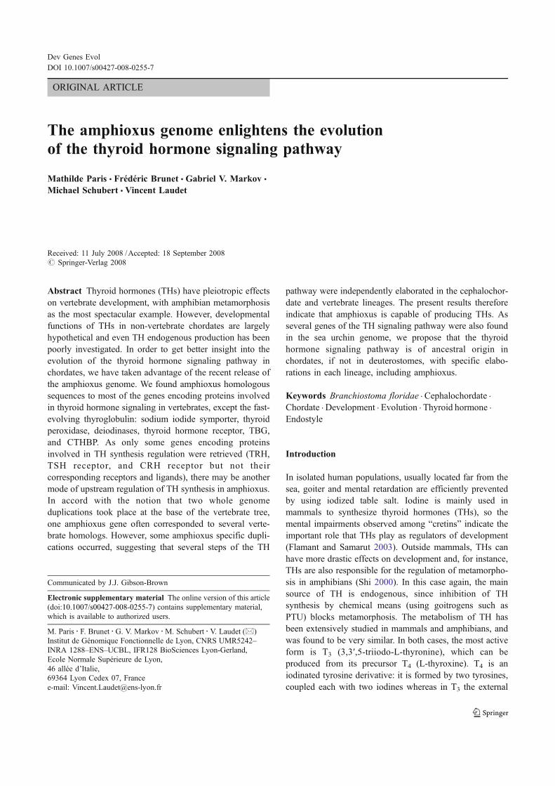
ORIGINAL ARTICLE
The amphioxus genome enlightens the evolution
of the thyroid hormone signaling pathway
Mathilde Paris & Frédéric Brunet & Gabriel V. Markov &
Michael Schubert & Vincent Laudet
Received: 11 July 2008 /Accepted: 18 September 2008# Springer-Verlag 2008
Abstract Thyroid hormones (THs) have pleiotropic effectson vertebrate development, with amphibian metamorphosisas the most spectacular example. However, developmentalfunctions of THs in non-vertebrate chordates are largelyhypothetical and even TH endogenous production has beenpoorly investigated. In order to get better insight into theevolution of the thyroid hormone signaling pathway inchordates, we have taken advantage of the recent release ofthe amphioxus genome. We found amphioxus homologoussequences to most of the genes encoding proteins involvedin thyroid hormone signaling in vertebrates, except the fast-evolving thyroglobulin: sodium iodide symporter, thyroidperoxidase, deiodinases, thyroid hormone receptor, TBG,and CTHBP. As only some genes encoding proteinsinvolved in TH synthesis regulation were retrieved (TRH,TSH receptor, and CRH receptor but not theircorresponding receptors and ligands), there may be anothermode of upstream regulation of TH synthesis in amphioxus.In accord with the notion that two whole genomeduplications took place at the base of the vertebrate tree,one amphioxus gene often corresponded to several verte-brate homologs. However, some amphioxus specific dupli-cations occurred, suggesting that several steps of the TH
pathway were independently elaborated in the cephalochor-date and vertebrate lineages. The present results thereforeindicate that amphioxus is capable of producing THs. Asseveral genes of the TH signaling pathway were also foundin the sea urchin genome, we propose that the thyroidhormone signaling pathway is of ancestral origin inchordates, if not in deuterostomes, with specific elabo-rations in each lineage, including amphioxus.
Keywords Branchiostoma floridae . Cephalochordate .
Chordate . Development . Evolution . Thyroid hormone .
Endostyle
Introduction
In isolated human populations, usually located far from thesea, goiter and mental retardation are efficiently preventedby using iodized table salt. Iodine is mainly used inmammals to synthesize thyroid hormones (THs), so themental impairments observed among “cretins” indicate theimportant role that THs play as regulators of development(Flamant and Samarut 2003). Outside mammals, THs canhave more drastic effects on development and, for instance,THs are also responsible for the regulation of metamorpho-sis in amphibians (Shi 2000). In this case again, the mainsource of TH is endogenous, since inhibition of THsynthesis by chemical means (using goitrogens such asPTU) blocks metamorphosis. The metabolism of TH hasbeen extensively studied in mammals and amphibians, andwas found to be very similar. In both cases, the most activeform is T3 (3,3′,5-triiodo-L-thyronine), which can beproduced from its precursor T4 (L-thyroxine). T4 is aniodinated tyrosine derivative: it is formed by two tyrosines,coupled each with two iodines whereas in T3 the external
Dev Genes Evol
DOI 10.1007/s00427-008-0255-7
Communicated by J.J. Gibson-Brown
Electronic supplementary material The online version of this article(doi:10.1007/s00427-008-0255-7) contains supplementary material,which is available to authorized users.
M. Paris : F. Brunet :G. V. Markov :M. Schubert :V. Laudet (*)Institut de Génomique Fonctionnelle de Lyon, CNRS UMR5242–INRA 1288–ENS–UCBL, IFR128 BioSciences Lyon-Gerland,Ecole Normale Supérieure de Lyon,46 allée d’Italie,69364 Lyon Cedex 07, Francee-mail: [email protected]
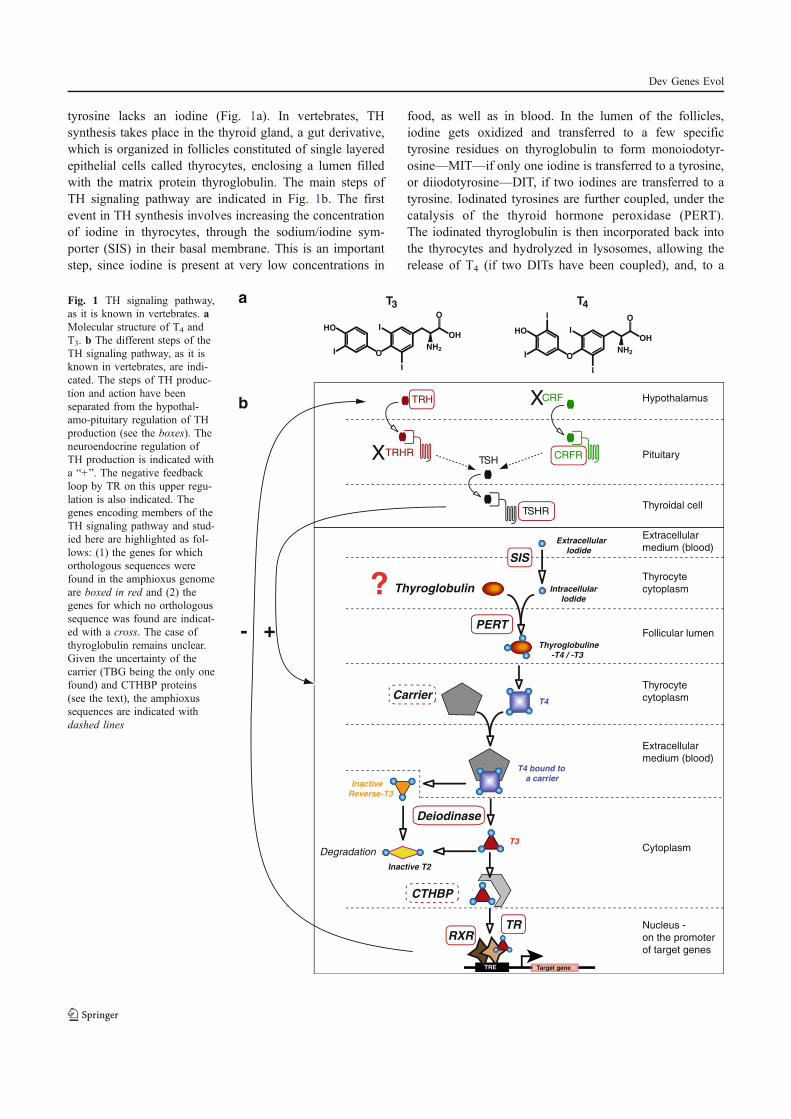
tyrosine lacks an iodine (Fig. 1a). In vertebrates, THsynthesis takes place in the thyroid gland, a gut derivative,which is organized in follicles constituted of single layeredepithelial cells called thyrocytes, enclosing a lumen filledwith the matrix protein thyroglobulin. The main steps ofTH signaling pathway are indicated in Fig. 1b. The firstevent in TH synthesis involves increasing the concentrationof iodine in thyrocytes, through the sodium/iodine sym-porter (SIS) in their basal membrane. This is an importantstep, since iodine is present at very low concentrations in
food, as well as in blood. In the lumen of the follicles,iodine gets oxidized and transferred to a few specifictyrosine residues on thyroglobulin to form monoiodotyr-osine—MIT—if only one iodine is transferred to a tyrosine,or diiodotyrosine—DIT, if two iodines are transferred to atyrosine. Iodinated tyrosines are further coupled, under thecatalysis of the thyroid hormone peroxidase (PERT).The iodinated thyroglobulin is then incorporated back intothe thyrocytes and hydrolyzed in lysosomes, allowing therelease of T4 (if two DITs have been coupled), and, to a
Extracellular
Iodide
Intracellular
Iodide
SIS
Inactive
Reverse-T3
T3
T4
T4 bound to
a carrier
Thyroglobuline
-T4 / -T3
Inactive T2
TRRXR
Target gene
T4
I
I
HO
O
I
NH2
OH
O
I
T3
I
I
HO
O
I
NH2
OH
O
Extracellular
medium (blood)
Thyroidal cell
Pituitary
Hypothalamus
Thyrocyte
cytoplasm
Follicular lumen
Thyrocyte
cytoplasm
Extracellular
medium (blood)
Cytoplasm
Nucleus -
on the promoter
of target genes
Thyroglobulin
PERT
Carrier
?
Deiodinase
Degradation
a
b
CTHBP
TSHR
TSHTRHR
TRH
CRFR
CRF
+-
X
X
TRE
Fig. 1 TH signaling pathway,as it is known in vertebrates. aMolecular structure of T4 andT3. b The different steps of theTH signaling pathway, as it isknown in vertebrates, are indi-cated. The steps of TH produc-tion and action have beenseparated from the hypothal-amo-pituitary regulation of THproduction (see the boxes). Theneuroendocrine regulation ofTH production is indicated witha “+”. The negative feedbackloop by TR on this upper regu-lation is also indicated. Thegenes encoding members of theTH signaling pathway and stud-ied here are highlighted as fol-lows: (1) the genes for whichorthologous sequences werefound in the amphioxus genomeare boxed in red and (2) thegenes for which no orthologoussequence was found are indicat-ed with a cross. The case ofthyroglobulin remains unclear.Given the uncertainty of thecarrier (TBG being the only onefound) and CTHBP proteins(see the text), the amphioxussequences are indicated withdashed lines
Dev Genes Evol
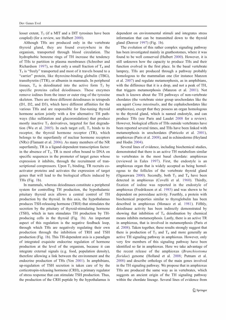
lesser extent, T3 (if a MIT and a DIT tyrosines have beencoupled) (for a review, see Hulbert 2000).
Although THs are produced only in the vertebratethyroid gland, they are found everywhere in theorganism, transported through blood circulation. Thehydrophobic benzene rings of TH increase the tendencyof THs to partition in plasma membranes (Schreiber andRichardson 1997), so that only a small fraction of T4 andT3 is “freely” transported and most of it travels bound to a“carrier” protein, like thyroxine-binding globulin (TBG),transthyretin (TTR), or albumin in mammals. In peripheraltissues, T4 is deiodinated into the active form T3 byspecific proteins called deiodinases. These enzymesremove iodines from the inner or outer ring of the tyrosineskeleton. There are three different deiodinases in tetrapods(D1, D2, and D3), which have different affinities for thevarious THs and are responsible for fine-tuning thyroidhormone action jointly with a few alternative TH path-ways (like sulfatation and glucoronidation) that producemostly inactive T3 derivatives, targeted for fast degrada-tion (Wu et al. 2005). In each target cell, T3 binds to itsreceptor, the thyroid hormone receptor (TR), whichbelongs to the superfamily of nuclear hormone receptors(NRs) (Flamant et al. 2006). As many members of the NRsuperfamily, TR is a ligand-dependent transcription factor:in the absence of T3, TR is most often bound to DNA onspecific sequences in the promoter of target genes whoseexpression it inhibits, through the recruitment of tran-scriptional corepressors. Upon T3 binding, TR recruits co-activator proteins and activates the expression of targetgenes that will lead to the biological effects induced byTHs (Fig. 1b).
In mammals, whereas deiodinases constitute a peripheralsystem for controlling TH production, the hypothalamicpituitary thyroid axis allows a central control of THproduction by the thyroid. In this axis, the hypothalamusproduces TSH-releasing hormone (TRH) that stimulates thesecretion by the pituitary of thyroid-stimulating hormone(TSH), which in turn stimulates TH production by TH-producing cells in the thyroid (Fig. 1b). An importantaspect of this regulation is the negative feedback loop,through which THs are negatively regulating their ownproduction through the inhibition of TRH and TSHproduction (Fig. 1b). This TH-dependent axis is a paradigmof integrated exquisite endocrine regulation of hormoneproduction at the level of the organism, because it canintegrate external signals (e.g. food, population density),therefore allowing a link between the environment and theendocrine production of THs (Yen 2001). In amphibians,up-regulation of TSH secretion is taken care of by thecorticotropin-releasing hormone (CRH), a primary regulatorof stress response that can stimulate TSH production. Thus,the production of the CRH peptide by the hypothalamus is
dependent on environmental stimuli and integrates stressinformation that can be transmitted down to the thyroidgland (Denver 1997) (Fig. 1b).
The evolution of this rather complex signaling pathwayhas been investigated mainly in gnathostomes, where it wasfound to be well conserved (Hulbert 2000). However, it isstill unknown how the capacity to produce THs and theirfunction evolved in the first place. In the basal vertebratelamprey, THs are produced through a pathway probablyhomologous to the mammalian one (for instance Manzonet al. 2007) and regulate metamorphosis, as in amphibians,with the difference that it is a drop, and not a peak of TH,that triggers metamorphosis (Manzon et al. 2001). Notmuch is known about the TH pathways of non-vertebratechordates (the vertebrate sister group urochordates like thesea squirt Ciona intestinalis, and the cephalochordates likeamphioxus), except that they possess an organ homologousto the thyroid gland, which is named endostyle, and canproduce THs (see Paris and Laudet 2008 for a review).However, biological effects of THs outside vertebrates havebeen reported several times, and THs have been linked withmetamorphosis in urochordates (Patricolo et al. 2001),amphioxus (Paris et al. 2008), and in echinoderms (Heylandand Hodin 2004).
Several lines of evidence, including biochemical studies,demonstrated that there is an active TH metabolism similarto vertebrates in the most basal chordate: amphioxus(reviewed in Eales 1997). First, the endostyle is anamphioxus organ that is widely accepted as being homol-ogous to the follicles of the vertebrate thyroid gland(Ogasawara 2000). Secondly, both T3 and T4 have beendetected in amphioxus (Covelli et al. 1960). Thirdly,fixation of iodine was reported in the endostyle ofamphioxus (Fredriksson et al. 1985) and was shown to bedependent on peroxidase activity. Fourthly, a protein withbiochemical properties similar to thyroglobulin has beendescribed in amphioxus (Monaco et al. 1981). Fifthly,deiodinase activity has been indirectly demonstrated byshowing that inhibition of T4 deiodination by chemicalmeans inhibits metamorphosis. Lastly, there is an active TRin amphioxus, that is involved in metamorphosis (Paris etal. 2008). Taken together, these results strongly suggest thatthere is production of T3 and T4 and more generally anactive TH signaling pathway in amphioxus. However, onlyvery few members of this signaling pathway have beenidentified so far in amphioxus. Here we take advantage ofthe recent release of the amphioxus (Branchiostomafloridae) genome (Holland et al. 2008; Putnam et al.2008) and describe orthologs of the main genes involvedin the TH signaling pathway. We propose that in amphioxusTHs are produced the same way as in vertebrates, whichsuggests an ancient origin of the TH signaling pathwaywithin the chordate lineage. Several lines of evidence from
Dev Genes Evol
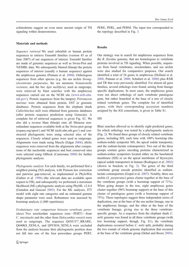
echinoderms suggest an even more ancient origin of THsignaling within deuterostomes.
Materials and methods
Sequence retrieval We used zebrafish or human proteinsequences to retrieve Ensembl families (version 45 as ofJune 2007) of our sequences of interest. Ensembl familiesare made of genomic sequences as well as Swiss-Prot andTrEMBL data. We subsequently blasted (blastp) the proteinsequences of interest (usually a human sequence) againstthe amphioxus genome (Putnam et al. 2008). Orthologoussequences from other species (e.g. the sea urchin Strong-
ylocentrotus purpuratus, the sea anemone Nematostella
vectensis, and the bee Apis mellifera), used as outgroups,were retrieved by blast searches with the amphioxussequences carried out on the NCBI site (www.ncbi.nlm.nih.gov/). Protein sequences from the lamprey Petromyzon
marinus were obtained from protein, EST or genomicdatabases. Protein sequences from the elephant sharkCallorhinchus milii were obtained from genomic databases(after protein sequence prediction using Genscan). Acomplete list of retrieved sequences is given Fig. S2. Wealso did a reverse blast (blastp) onto all Metazoa non-redundant sequences available both at the Swiss-prot group(expasy.org/sprot/) and NCBI (ncbi.nlm.nih.gov/) and con-structed phylogenetic trees using selected sites of thesequences. Closely related genes were used as outgroups.Alignments were made using Muscle (Edgar 2004), allelicsequences were removed from the alignments after compar-isons of the nucleotide sequences and best conserved siteswere selected using GBlock (Castresana 2000) for furtherphylogenetic analysis.
Phylogenetic analysis For each family, we performed first aneighbor-joining (NJ) analysis, with Poisson law correctionand pairwise gap-removal, as implemented in PhyloWin(Galtier et al. 1996) (the relevant data are available uponrequest to FB), and subsequently we performed a maximumlikelihood (ML) phylogenetic analysis using PhyML v2.4.4(Guindon and Gascuel 2003). For the ML analyses, JTTmodel with eight rate categories and an estimated gammashape parameter were used. Robustness was assessed bybootstrap analysis (1,000 repetitions).
Evolutionary rate comparison between vertebrate perox-
idases Two urochordate sequences (one—PERT1—fromC. intestinalis and the other from Halocynthia roretzi) wereused as outgroups. The sequence Q6NUY7_BRARE,Q640K8_XENLA, and Q9YH34_XENLA were excludedfrom the analysis because their phylogenetic position doesnot fall into one of the four peroxidase groups PERT,
PERE, PERL, and PERM. The input tree corresponded tothe topology described in Fig. 3.
Results
Our strategy was to search for amphioxus sequences fromthe B. floridae genome, that are homologous to vertebrateproteins involved in TH signaling. When possible, sequen-ces from basal vertebrates, urochordates, and sea urchinwere also studied for comparative purposes. Our searchidentified a total of 24 genes in amphioxus (Holland et al.2008; Putnam et al. 2008; Schubert et al. 2008) plus RXRand TR that were previously identified. For almost all genefamilies, several orthologs were found, arising from lineagespecific duplications. In most cases, the amphioxus geneswere not direct orthologs of each vertebrate paralogousgene, but rather branched at the base of a tree of closelyrelated vertebrate genes. The complete list of identifiedgenes, with their corresponding accession numbersassigned by the JGI consortium, is given in Table S1.
SIS
Blast searches allowed us to identify eight predicted genes,for which orthology was tested by a phylogenetic analysis(Fig. 2). We found three groups of closely related vertebrategenes, including SIS, each time with 100% bootstrap (thesodium-iodide symporter SIS, the apical iodide transporter,and the sodium-lactate cotransporter). Two out of the threegroups contain genes encoding proteins characterized assodium-iodine symporters located either on the basolateralmembrane (SIS) or on the apical membrane of thyrocytes(apical iodide transporter) in human (Rodriguez et al. 2002)(shown in brackets in Fig. 2). The genes of the thirdvertebrate group encode proteins identified as sodium-lactate cotransporters (Gopal et al. 2007). Notably, three seaurchin (S. purpuratus) genes cluster together at the base ofthe vertebrate groups (with a bootstrap support of 71%).When going deeper in the tree, eight amphioxus genescluster together (96% bootstrap support) at the base of thiscluster of paralogous genes (with a bootstrap support of75%). These topologies suggest three independent series ofduplication, one at the base of the sea urchin lineage, one inthe amphioxus lineage, and the other at the base of thevertebrate lineage, giving rise to the three vertebrate-specific groups. As a sequence from the elephant shark C.
milii genome was found in all three vertebrate groups (withlow bootstrap support, though, Fig. S1), the vertebrateduplications occurred before C. milii split, probably duringthe two rounds of whole genome duplication that occurredat the base of the vertebrate group (Dehal and Boore 2005).
Dev Genes Evol
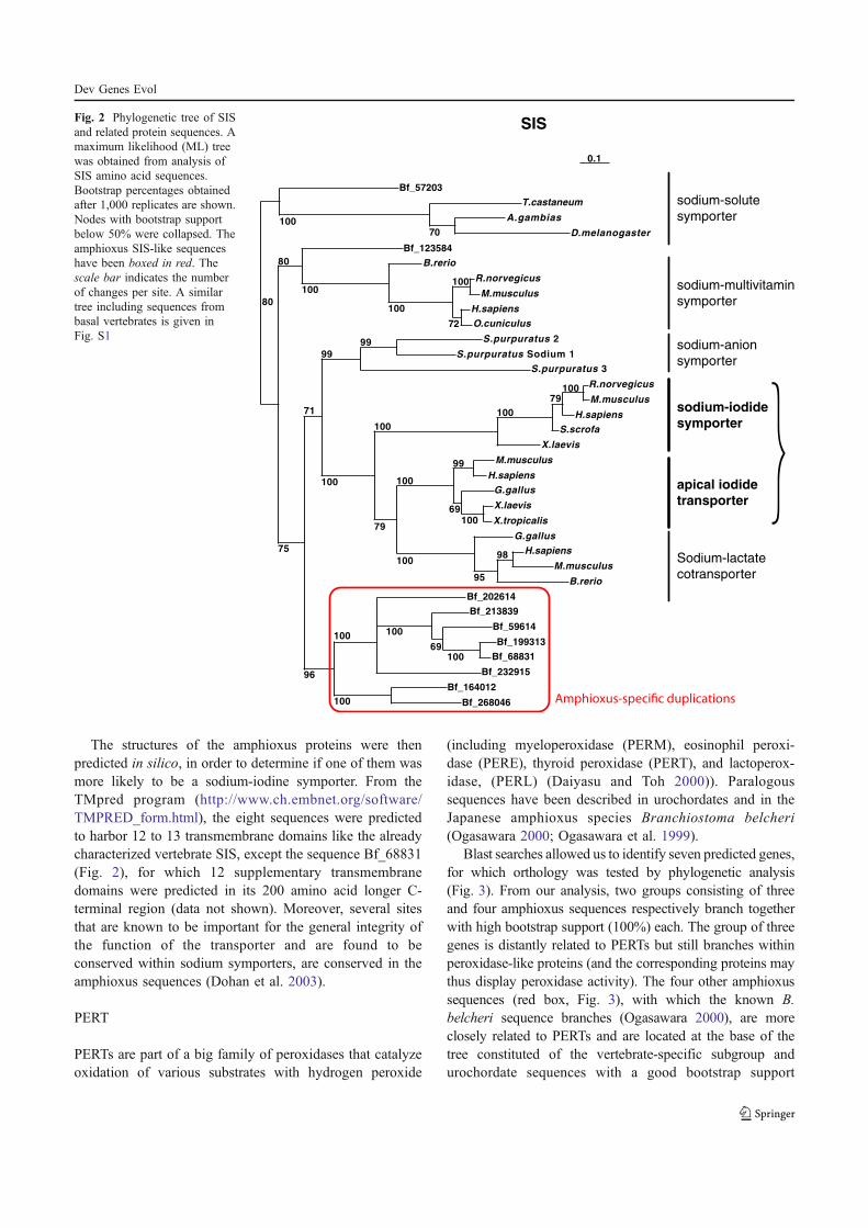
The structures of the amphioxus proteins were thenpredicted in silico, in order to determine if one of them wasmore likely to be a sodium-iodine symporter. From theTMpred program (http://www.ch.embnet.org/software/TMPRED_form.html), the eight sequences were predictedto harbor 12 to 13 transmembrane domains like the alreadycharacterized vertebrate SIS, except the sequence Bf_68831(Fig. 2), for which 12 supplementary transmembranedomains were predicted in its 200 amino acid longer C-terminal region (data not shown). Moreover, several sitesthat are known to be important for the general integrity ofthe function of the transporter and are found to beconserved within sodium symporters, are conserved in theamphioxus sequences (Dohan et al. 2003).
PERT
PERTs are part of a big family of peroxidases that catalyzeoxidation of various substrates with hydrogen peroxide
(including myeloperoxidase (PERM), eosinophil peroxi-dase (PERE), thyroid peroxidase (PERT), and lactoperox-idase, (PERL) (Daiyasu and Toh 2000)). Paralogoussequences have been described in urochordates and in theJapanese amphioxus species Branchiostoma belcheri
(Ogasawara 2000; Ogasawara et al. 1999).Blast searches allowed us to identify seven predicted genes,
for which orthology was tested by phylogenetic analysis(Fig. 3). From our analysis, two groups consisting of threeand four amphioxus sequences respectively branch togetherwith high bootstrap support (100%) each. The group of threegenes is distantly related to PERTs but still branches withinperoxidase-like proteins (and the corresponding proteins maythus display peroxidase activity). The four other amphioxussequences (red box, Fig. 3), with which the known B.
belcheri sequence branches (Ogasawara 2000), are moreclosely related to PERTs and are located at the base of thetree constituted of the vertebrate-specific subgroup andurochordate sequences with a good bootstrap support
Bf_268046
Bf_164012
Bf_232915
Bf_68831
Bf_199313
Bf_59614
100
Bf_213839
69
Bf_202614
100
100
100
B.rerio
M.musculus
H.sapiens98
G.gallus
95
X.tropicalis
X.laevis
G.gallus
100
H.sapiens
M.musculus
69
99
100
100
X.laevis
S.scrofa
H.sapiens
M.musculus
R.norvegicus10079
100
79
100
S.purpuratus 3
S.purpuratus Sodium 1
S.purpuratus 299
100
99
96
71
O.cuniculus
H.sapiens
M.musculus
R.norvegicus
72
100
B.rerio
100
Bf_123584
100
75
80
D.melanogaster
A.gambias
T.castaneum
70
Bf_57203
100
80
0.1
sodium-iodide
symporter
apical iodide
transporter
Sodium-lactate
cotransporter
sodium-multivitamin
symporter
sodium-solute
symporter
sodium-anion
symporter
SISFig. 2 Phylogenetic tree of SISand related protein sequences. Amaximum likelihood (ML) treewas obtained from analysis ofSIS amino acid sequences.Bootstrap percentages obtainedafter 1,000 replicates are shown.Nodes with bootstrap supportbelow 50% were collapsed. Theamphioxus SIS-like sequenceshave been boxed in red. Thescale bar indicates the numberof changes per site. A similartree including sequences frombasal vertebrates is given inFig. S1
Dev Genes Evol
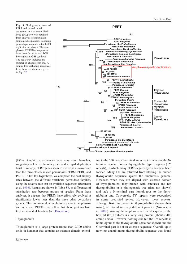
(88%). Amphioxus sequences have very short branches,suggesting a low evolutionary rate and a rapid duplicationburst. Similarly, PERT genes seem to evolve at a slower ratethan the three closely related peroxidases PERM, PERL, andPERE. To test this hypothesis, we compared the evolutionaryrates between the different vertebrate peroxidase families,using the relative-rate test on available sequences (Robinsonet al. 1998). Results are shown in Table S3, as differences ofsubstitution rate between groups of species. From theseanalyses, it appears that PERTs have effectively evolved atsignificantly lower rates than the three other peroxidasegroups. This common slow evolutionary rate in amphioxusand vertebrate PERTs may reflect that these proteins havekept an ancestral function (see Discussion).
Thyroglobulin
Thyroglobulin is a large protein (more than 2,700 aminoacids in humans) that contains an esterase domain extend-
ing to the 500 most C-terminal amino acids, whereas the N-terminal domain houses thyroglobulin type I repeats (TYrepeats), in which many PERT-targeted tyrosines have beenlocated. Many hits are retrieved from blasting the humanthyroglobulin sequence against the amphioxus genome.However, when they are aligned with esterase domainof thyroglobulins, they branch with esterases and notthyroglobulins in a phylogenetic tree (data not shown)and lack a N-terminal part homologous to the thyro-globulin one. Conversely, TY repeats were recognizedin some predicted genes. However, these repeats,although first discovered in thyroglobulins (hence theirname), are found in many different proteins (Novinec etal. 2006). Among the amphioxus retrieved sequences, thebest hit (Bf_123169) is a very long protein (about 2,400amino acids). However, nothing else but the TY repeats ishomologous to the thyroglobulin (data not shown) and theC-terminal part is not an esterase sequence. Overall, up tonow, no unambiguous thyroglobulin sequence was found
Chorion peroxidase D.melanogaster
Peroxidase A.aegypti
Salivary peroxidase A.albimanus
100
Peroxidase-like E.scolopes
Bf_99381
Bf_125849
Bf_125550
99
99
Peroxidase1 X.laevis
Peroxidase2 X.laevis
PERL-like C.dromedarius
PERL B.taurusPERL H.sapiens
83
PERL M.musculus
PERL M.auratus
65
91
PERM H.sapiens
PERM M.musculusPERE H.sapiens
PERE M.musculus
100
100
100
95
100
85
Peroxidase B.rerio
100
PERT R.norvegicus
PERT M.musculusPERT H.sapiens
100
PERT S.scrofa
PERT C.familiaris
66
54
95
100
Peroxidase H.roretzi
PERT1 C.intestinalis
PERT2 C.intestinalis
65
100
Peroxidase B.belcheri
Bf_67515
Bf_67517
56
Bf_123696
79
Bf_226591
92
99
100
Peroxidasin-like H.sapiens
Peroxidasin M.musculus
Peroxidasin homolog H.sapiens
100
Peroxidasin X.tropicalis
58
Peroxidasin-homolog L.variegatus Peroxidasin-homolog S.purpuratus
100
100
Peroxidase-like A.californica
58
88
72
100
100
100
100
96
98
Peroxidase N.tabacum
Peroxidase-like P.alcaligenes
PGH1 H.sapiensPGH2 H.sapiens
100
100
100
0.2
PERT
ThyroidPeroxidase
MyeloidPeroxidase
EosinophilPeroxidase
Lacto-Peroxidase
Peroxidasin
Fig. 3 Phylogenetic tree ofPERT and related proteinsequences. A maximum likeli-hood (ML) tree was obtainedfrom analysis of peroxidaseamino acid sequences. Bootstrappercentages obtained after 1,000replicates are shown. The am-phioxus PERT-like sequenceshave been boxed in red. PGH:Prostaglandin G/H synthase.The scale bar indicates thenumber of changes per site. Asimilar tree including sequencesfrom basal vertebrates is givenin Fig. S2
Dev Genes Evol
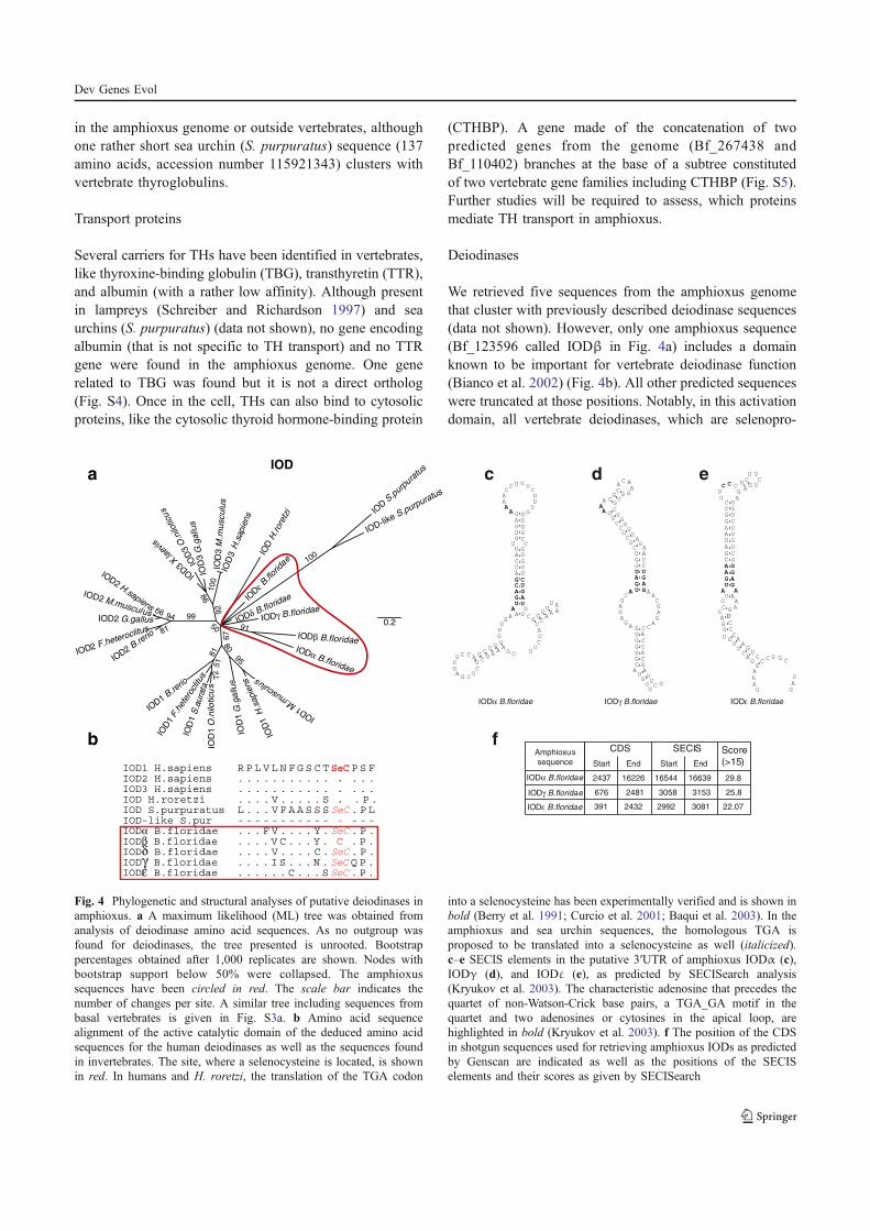
in the amphioxus genome or outside vertebrates, althoughone rather short sea urchin (S. purpuratus) sequence (137amino acids, accession number 115921343) clusters withvertebrate thyroglobulins.
Transport proteins
Several carriers for THs have been identified in vertebrates,like thyroxine-binding globulin (TBG), transthyretin (TTR),and albumin (with a rather low affinity). Although presentin lampreys (Schreiber and Richardson 1997) and seaurchins (S. purpuratus) (data not shown), no gene encodingalbumin (that is not specific to TH transport) and no TTRgene were found in the amphioxus genome. One generelated to TBG was found but it is not a direct ortholog(Fig. S4). Once in the cell, THs can also bind to cytosolicproteins, like the cytosolic thyroid hormone-binding protein
(CTHBP). A gene made of the concatenation of twopredicted genes from the genome (Bf_267438 andBf_110402) branches at the base of a subtree constitutedof two vertebrate gene families including CTHBP (Fig. S5).Further studies will be required to assess, which proteinsmediate TH transport in amphioxus.
Deiodinases
We retrieved five sequences from the amphioxus genomethat cluster with previously described deiodinase sequences(data not shown). However, only one amphioxus sequence(Bf_123596 called IODβ in Fig. 4a) includes a domainknown to be important for vertebrate deiodinase function(Bianco et al. 2002) (Fig. 4b). All other predicted sequenceswere truncated at those positions. Notably, in this activationdomain, all vertebrate deiodinases, which are selenopro-
0.2
IOD
3 G
.gallu
s
IODβ B.floridae
IOD
1S
.aura
ta
IOD-lik
e S.purpuratus
IODδ B.floridae
IOD
S.pur
pura
tus
IOD3
X.laev
is
IOD
H.rore
tzi
IOD
εB.fl
oridae
IOD
3M
.musculu
s
IOD
1 F.h
eter
oclit
us
IOD
3H
.sapie
ns
IOD
3O
.nilo
ticus
IODγ B.floridae
IODα B.floridaeIO
D2 B.re
rio
IOD2 F.heteroclitus
IOD1
B.rerio
6699
86
95
100
92
80
72
51
67
50
100
91
94
81
81
IOD2 G.gallus
IOD2 M.musculus
IOD2 H.sapiens
IOD
1 G
.gallu
s
IOD1 M
.musc
ulus
IOD
1H
.sapie
ns
IOD
1O
.nilo
ticu
s
IOD1 H.sapiens R P L V L N F G S C T SeC P S FIOD2 H.sapiens . . . . . . . . . . . . . . .IOD3 H.sapiens . . . . . . . . . . . . . . .IOD H.roretzi . . . . V . . . . . S . . P .IOD S.purpuratus L . . . V F A A S S S SeC . P LIOD-like S.pur - - - - - - - - - - - - - - -IODα B.floridae . . . F V . . . . Y . SeC . P .IODβ B.floridae . . . . V C . . . Y . C . P .IODδ B.floridae . . . . V . . . . C . SeC . P .IODγ B.floridae . . . . I S . . . N . SeC Q P .IODε B.floridae . . . . . . C . . . S SeC . P .
b
aIOD
IODα B.floridae
IODε B.floridae
CDS
Start
Amphioxus
sequence End Start End
SECIS Score
(>15)
2437 16226 16544 16639 29.8
391 2432 2992 3081 22.07
f
IODγ B.floridae 676 2481 3058 3153 25.8
c
IODα B.floridae
A
A
A
A
A
A
A
A
A
AA A
A
AA
A
AC
C
CC
C
C
C
C
C
CC
C
C
C
C
C
C
C
CC
C
GG
G
G
G
G
G
G
G
G
G
G
G
G
G
G
G
GG
U
U
U U
U
U
U
A
AA
A
A
U
U
U
U
UUU
U
U
U
GG
U
U
U
U
UU
U
U
U
U
U
U
UU
U
d
IODγ B.floridae
A
A
A
A A
A
A
A
AA
A
A
A
A
A
AA
A
AA
A
AC
C
C
C
C
C
C
C
C
C
C
C
C
CC
C
G
G
G
G
G
G
G
G
G
G
GG
GGG
G
G
G
G G
G
G
G
CG
C
C
C
G
G
G
GG
G
U
U
U
U
U
U
U
U
AU
U U
U
e
IODε B.floridae
C
C
G
C
C
C CC
C
GG
G
G
C
C
C
C
C
CCC
C
C
C
C
G
G
G
G
GG
G
G
G
G
GGG
G
G
G
G
G
A
U
U
U
U
U
U U
U
A
A
A
G
G
A
AA AA
A
A
AA
A
A
A
A
A
A
A
A
U
A U
U
U
UU
U
U
U
U
UU
Fig. 4 Phylogenetic and structural analyses of putative deiodinases inamphioxus. a A maximum likelihood (ML) tree was obtained fromanalysis of deiodinase amino acid sequences. As no outgroup wasfound for deiodinases, the tree presented is unrooted. Bootstrappercentages obtained after 1,000 replicates are shown. Nodes withbootstrap support below 50% were collapsed. The amphioxussequences have been circled in red. The scale bar indicates thenumber of changes per site. A similar tree including sequences frombasal vertebrates is given in Fig. S3a. b Amino acid sequencealignment of the active catalytic domain of the deduced amino acidsequences for the human deiodinases as well as the sequences foundin invertebrates. The site, where a selenocysteine is located, is shownin red. In humans and H. roretzi, the translation of the TGA codon
into a selenocysteine has been experimentally verified and is shown inbold (Berry et al. 1991; Curcio et al. 2001; Baqui et al. 2003). In theamphioxus and sea urchin sequences, the homologous TGA isproposed to be translated into a selenocysteine as well (italicized).c–e SECIS elements in the putative 3′UTR of amphioxus IODα (c),IODγ (d), and IODε (e), as predicted by SECISearch analysis(Kryukov et al. 2003). The characteristic adenosine that precedes thequartet of non-Watson-Crick base pairs, a TGA_GA motif in thequartet and two adenosines or cytosines in the apical loop, arehighlighted in bold (Kryukov et al. 2003). f The position of the CDSin shotgun sequences used for retrieving amphioxus IODs as predictedby Genscan are indicated as well as the positions of the SECISelements and their scores as given by SECISearch
Dev Genes Evol
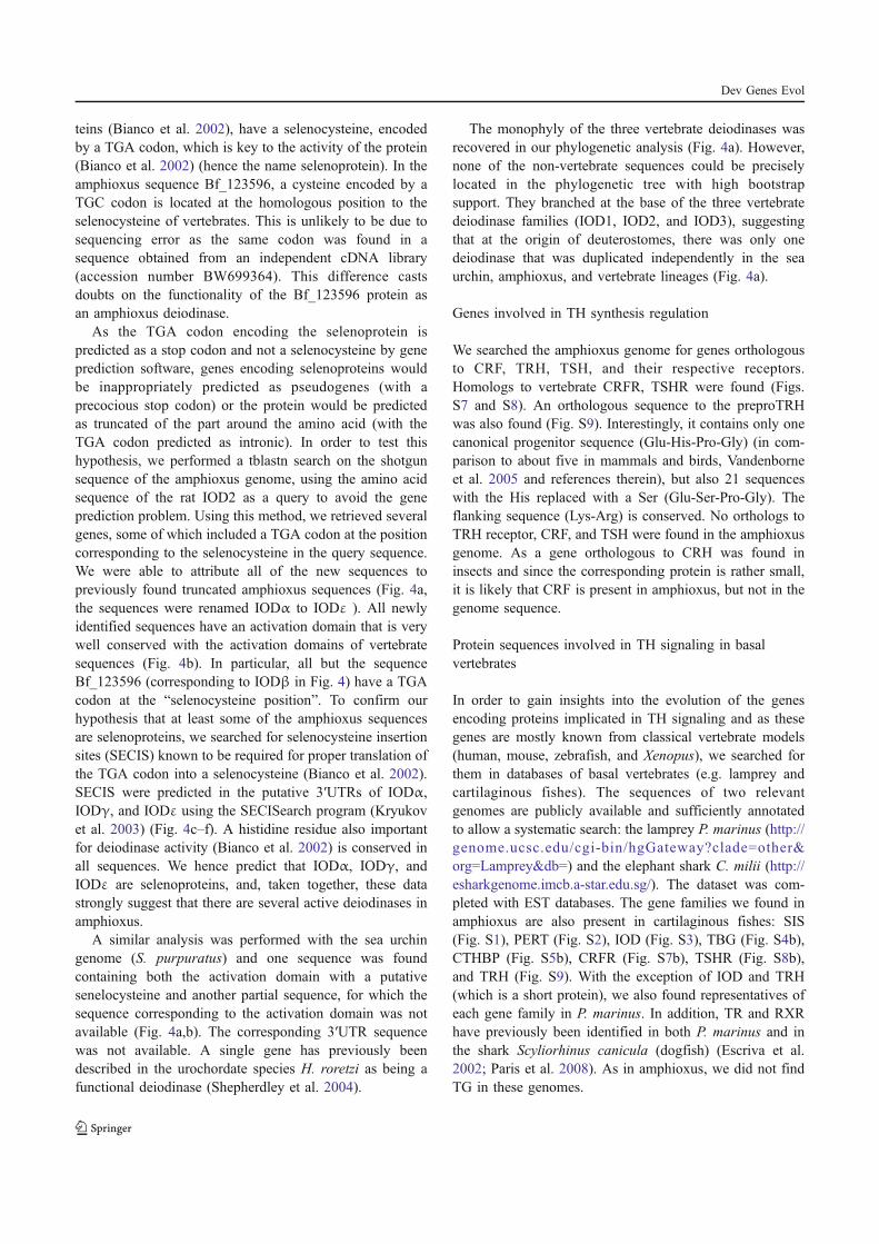
teins (Bianco et al. 2002), have a selenocysteine, encodedby a TGA codon, which is key to the activity of the protein(Bianco et al. 2002) (hence the name selenoprotein). In theamphioxus sequence Bf_123596, a cysteine encoded by aTGC codon is located at the homologous position to theselenocysteine of vertebrates. This is unlikely to be due tosequencing error as the same codon was found in asequence obtained from an independent cDNA library(accession number BW699364). This difference castsdoubts on the functionality of the Bf_123596 protein asan amphioxus deiodinase.
As the TGA codon encoding the selenoprotein ispredicted as a stop codon and not a selenocysteine by geneprediction software, genes encoding selenoproteins wouldbe inappropriately predicted as pseudogenes (with aprecocious stop codon) or the protein would be predictedas truncated of the part around the amino acid (with theTGA codon predicted as intronic). In order to test thishypothesis, we performed a tblastn search on the shotgunsequence of the amphioxus genome, using the amino acidsequence of the rat IOD2 as a query to avoid the geneprediction problem. Using this method, we retrieved severalgenes, some of which included a TGA codon at the positioncorresponding to the selenocysteine in the query sequence.We were able to attribute all of the new sequences topreviously found truncated amphioxus sequences (Fig. 4a,the sequences were renamed IODα to IODε ). All newlyidentified sequences have an activation domain that is verywell conserved with the activation domains of vertebratesequences (Fig. 4b). In particular, all but the sequenceBf_123596 (corresponding to IODβ in Fig. 4) have a TGAcodon at the “selenocysteine position”. To confirm ourhypothesis that at least some of the amphioxus sequencesare selenoproteins, we searched for selenocysteine insertionsites (SECIS) known to be required for proper translation ofthe TGA codon into a selenocysteine (Bianco et al. 2002).SECIS were predicted in the putative 3′UTRs of IODα,IODγ, and IODε using the SECISearch program (Kryukovet al. 2003) (Fig. 4c–f). A histidine residue also importantfor deiodinase activity (Bianco et al. 2002) is conserved inall sequences. We hence predict that IODα, IODγ, andIODε are selenoproteins, and, taken together, these datastrongly suggest that there are several active deiodinases inamphioxus.
A similar analysis was performed with the sea urchingenome (S. purpuratus) and one sequence was foundcontaining both the activation domain with a putativesenelocysteine and another partial sequence, for which thesequence corresponding to the activation domain was notavailable (Fig. 4a,b). The corresponding 3′UTR sequencewas not available. A single gene has previously beendescribed in the urochordate species H. roretzi as being afunctional deiodinase (Shepherdley et al. 2004).
The monophyly of the three vertebrate deiodinases wasrecovered in our phylogenetic analysis (Fig. 4a). However,none of the non-vertebrate sequences could be preciselylocated in the phylogenetic tree with high bootstrapsupport. They branched at the base of the three vertebratedeiodinase families (IOD1, IOD2, and IOD3), suggestingthat at the origin of deuterostomes, there was only onedeiodinase that was duplicated independently in the seaurchin, amphioxus, and vertebrate lineages (Fig. 4a).
Genes involved in TH synthesis regulation
We searched the amphioxus genome for genes orthologousto CRF, TRH, TSH, and their respective receptors.Homologs to vertebrate CRFR, TSHR were found (Figs.S7 and S8). An orthologous sequence to the preproTRHwas also found (Fig. S9). Interestingly, it contains only onecanonical progenitor sequence (Glu-His-Pro-Gly) (in com-parison to about five in mammals and birds, Vandenborneet al. 2005 and references therein), but also 21 sequenceswith the His replaced with a Ser (Glu-Ser-Pro-Gly). Theflanking sequence (Lys-Arg) is conserved. No orthologs toTRH receptor, CRF, and TSH were found in the amphioxusgenome. As a gene orthologous to CRH was found ininsects and since the corresponding protein is rather small,it is likely that CRF is present in amphioxus, but not in thegenome sequence.
Protein sequences involved in TH signaling in basalvertebrates
In order to gain insights into the evolution of the genesencoding proteins implicated in TH signaling and as thesegenes are mostly known from classical vertebrate models(human, mouse, zebrafish, and Xenopus), we searched forthem in databases of basal vertebrates (e.g. lamprey andcartilaginous fishes). The sequences of two relevantgenomes are publicly available and sufficiently annotatedto allow a systematic search: the lamprey P. marinus (http://genome.ucsc.edu/cgi-bin/hgGateway?clade=other&org=Lamprey&db=) and the elephant shark C. milii (http://esharkgenome.imcb.a-star.edu.sg/). The dataset was com-pleted with EST databases. The gene families we found inamphioxus are also present in cartilaginous fishes: SIS(Fig. S1), PERT (Fig. S2), IOD (Fig. S3), TBG (Fig. S4b),CTHBP (Fig. S5b), CRFR (Fig. S7b), TSHR (Fig. S8b),and TRH (Fig. S9). With the exception of IOD and TRH(which is a short protein), we also found representatives ofeach gene family in P. marinus. In addition, TR and RXRhave previously been identified in both P. marinus and inthe shark Scyliorhinus canicula (dogfish) (Escriva et al.2002; Paris et al. 2008). As in amphioxus, we did not findTG in these genomes.
Dev Genes Evol
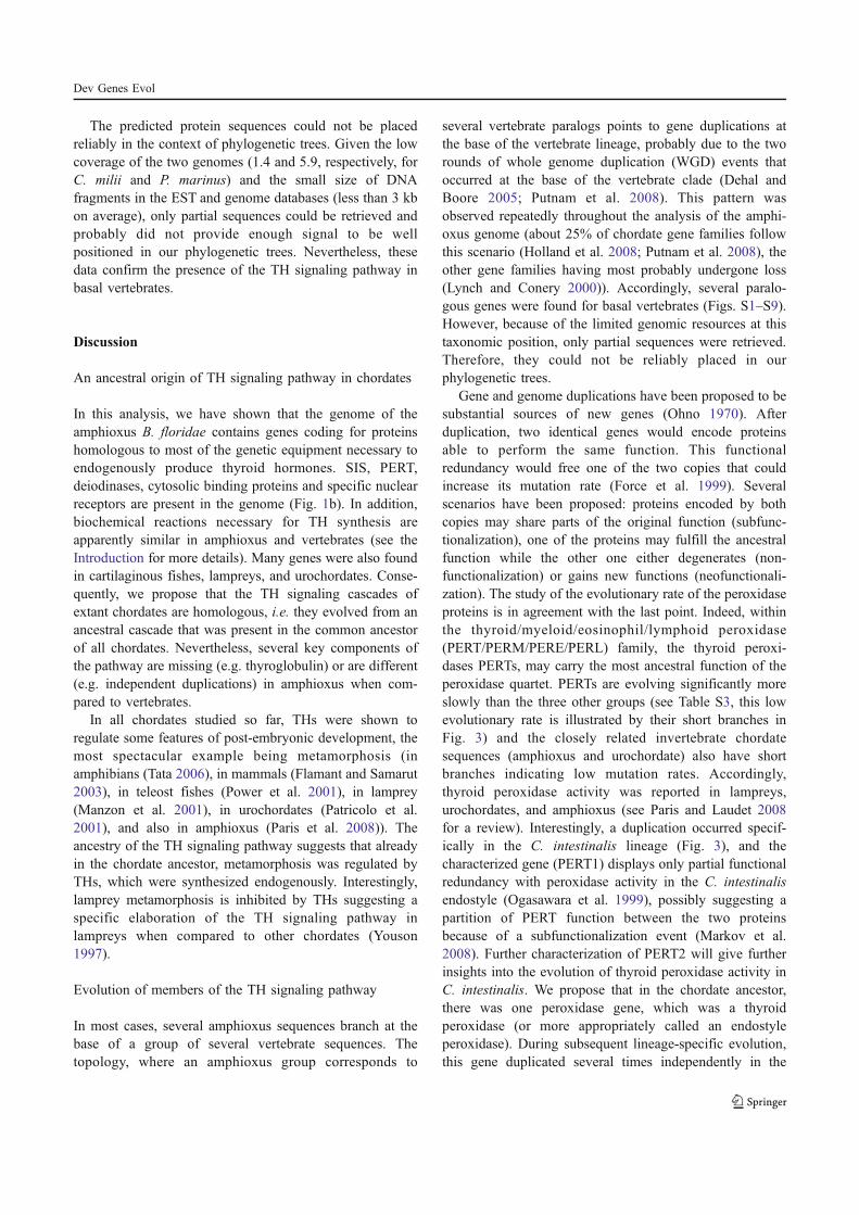
The predicted protein sequences could not be placedreliably in the context of phylogenetic trees. Given the lowcoverage of the two genomes (1.4 and 5.9, respectively, forC. milii and P. marinus) and the small size of DNAfragments in the EST and genome databases (less than 3 kbon average), only partial sequences could be retrieved andprobably did not provide enough signal to be wellpositioned in our phylogenetic trees. Nevertheless, thesedata confirm the presence of the TH signaling pathway inbasal vertebrates.
Discussion
An ancestral origin of TH signaling pathway in chordates
In this analysis, we have shown that the genome of theamphioxus B. floridae contains genes coding for proteinshomologous to most of the genetic equipment necessary toendogenously produce thyroid hormones. SIS, PERT,deiodinases, cytosolic binding proteins and specific nuclearreceptors are present in the genome (Fig. 1b). In addition,biochemical reactions necessary for TH synthesis areapparently similar in amphioxus and vertebrates (see theIntroduction for more details). Many genes were also foundin cartilaginous fishes, lampreys, and urochordates. Conse-quently, we propose that the TH signaling cascades ofextant chordates are homologous, i.e. they evolved from anancestral cascade that was present in the common ancestorof all chordates. Nevertheless, several key components ofthe pathway are missing (e.g. thyroglobulin) or are different(e.g. independent duplications) in amphioxus when com-pared to vertebrates.
In all chordates studied so far, THs were shown toregulate some features of post-embryonic development, themost spectacular example being metamorphosis (inamphibians (Tata 2006), in mammals (Flamant and Samarut2003), in teleost fishes (Power et al. 2001), in lamprey(Manzon et al. 2001), in urochordates (Patricolo et al.2001), and also in amphioxus (Paris et al. 2008)). Theancestry of the TH signaling pathway suggests that alreadyin the chordate ancestor, metamorphosis was regulated byTHs, which were synthesized endogenously. Interestingly,lamprey metamorphosis is inhibited by THs suggesting aspecific elaboration of the TH signaling pathway inlampreys when compared to other chordates (Youson1997).
Evolution of members of the TH signaling pathway
In most cases, several amphioxus sequences branch at thebase of a group of several vertebrate sequences. Thetopology, where an amphioxus group corresponds to
several vertebrate paralogs points to gene duplications atthe base of the vertebrate lineage, probably due to the tworounds of whole genome duplication (WGD) events thatoccurred at the base of the vertebrate clade (Dehal andBoore 2005; Putnam et al. 2008). This pattern wasobserved repeatedly throughout the analysis of the amphi-oxus genome (about 25% of chordate gene families followthis scenario (Holland et al. 2008; Putnam et al. 2008), theother gene families having most probably undergone loss(Lynch and Conery 2000)). Accordingly, several paralo-gous genes were found for basal vertebrates (Figs. S1–S9).However, because of the limited genomic resources at thistaxonomic position, only partial sequences were retrieved.Therefore, they could not be reliably placed in ourphylogenetic trees.
Gene and genome duplications have been proposed to besubstantial sources of new genes (Ohno 1970). Afterduplication, two identical genes would encode proteinsable to perform the same function. This functionalredundancy would free one of the two copies that couldincrease its mutation rate (Force et al. 1999). Severalscenarios have been proposed: proteins encoded by bothcopies may share parts of the original function (subfunc-tionalization), one of the proteins may fulfill the ancestralfunction while the other one either degenerates (non-functionalization) or gains new functions (neofunctionali-zation). The study of the evolutionary rate of the peroxidaseproteins is in agreement with the last point. Indeed, withinthe thyroid/myeloid/eosinophil/lymphoid peroxidase(PERT/PERM/PERE/PERL) family, the thyroid peroxi-dases PERTs, may carry the most ancestral function of theperoxidase quartet. PERTs are evolving significantly moreslowly than the three other groups (see Table S3, this lowevolutionary rate is illustrated by their short branches inFig. 3) and the closely related invertebrate chordatesequences (amphioxus and urochordate) also have shortbranches indicating low mutation rates. Accordingly,thyroid peroxidase activity was reported in lampreys,urochordates, and amphioxus (see Paris and Laudet 2008for a review). Interestingly, a duplication occurred specif-ically in the C. intestinalis lineage (Fig. 3), and thecharacterized gene (PERT1) displays only partial functionalredundancy with peroxidase activity in the C. intestinalis
endostyle (Ogasawara et al. 1999), possibly suggesting apartition of PERT function between the two proteinsbecause of a subfunctionalization event (Markov et al.2008). Further characterization of PERT2 will give furtherinsights into the evolution of thyroid peroxidase activity inC. intestinalis. We propose that in the chordate ancestor,there was one peroxidase gene, which was a thyroidperoxidase (or more appropriately called an endostyleperoxidase). During subsequent lineage-specific evolution,this gene duplicated several times independently in the
Dev Genes Evol
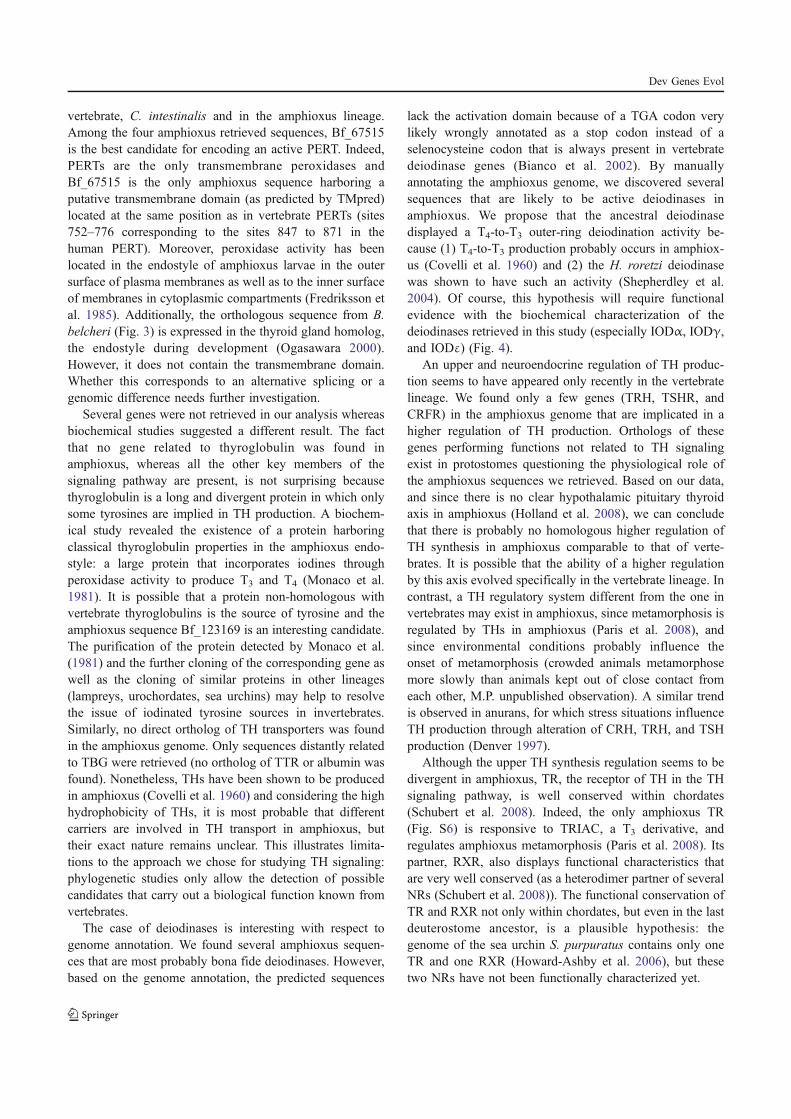
vertebrate, C. intestinalis and in the amphioxus lineage.Among the four amphioxus retrieved sequences, Bf_67515is the best candidate for encoding an active PERT. Indeed,PERTs are the only transmembrane peroxidases andBf_67515 is the only amphioxus sequence harboring aputative transmembrane domain (as predicted by TMpred)located at the same position as in vertebrate PERTs (sites752–776 corresponding to the sites 847 to 871 in thehuman PERT). Moreover, peroxidase activity has beenlocated in the endostyle of amphioxus larvae in the outersurface of plasma membranes as well as to the inner surfaceof membranes in cytoplasmic compartments (Fredriksson etal. 1985). Additionally, the orthologous sequence from B.
belcheri (Fig. 3) is expressed in the thyroid gland homolog,the endostyle during development (Ogasawara 2000).However, it does not contain the transmembrane domain.Whether this corresponds to an alternative splicing or agenomic difference needs further investigation.
Several genes were not retrieved in our analysis whereasbiochemical studies suggested a different result. The factthat no gene related to thyroglobulin was found inamphioxus, whereas all the other key members of thesignaling pathway are present, is not surprising becausethyroglobulin is a long and divergent protein in which onlysome tyrosines are implied in TH production. A biochem-ical study revealed the existence of a protein harboringclassical thyroglobulin properties in the amphioxus endo-style: a large protein that incorporates iodines throughperoxidase activity to produce T3 and T4 (Monaco et al.1981). It is possible that a protein non-homologous withvertebrate thyroglobulins is the source of tyrosine and theamphioxus sequence Bf_123169 is an interesting candidate.The purification of the protein detected by Monaco et al.(1981) and the further cloning of the corresponding gene aswell as the cloning of similar proteins in other lineages(lampreys, urochordates, sea urchins) may help to resolvethe issue of iodinated tyrosine sources in invertebrates.Similarly, no direct ortholog of TH transporters was foundin the amphioxus genome. Only sequences distantly relatedto TBG were retrieved (no ortholog of TTR or albumin wasfound). Nonetheless, THs have been shown to be producedin amphioxus (Covelli et al. 1960) and considering the highhydrophobicity of THs, it is most probable that differentcarriers are involved in TH transport in amphioxus, buttheir exact nature remains unclear. This illustrates limita-tions to the approach we chose for studying TH signaling:phylogenetic studies only allow the detection of possiblecandidates that carry out a biological function known fromvertebrates.
The case of deiodinases is interesting with respect togenome annotation. We found several amphioxus sequen-ces that are most probably bona fide deiodinases. However,based on the genome annotation, the predicted sequences
lack the activation domain because of a TGA codon verylikely wrongly annotated as a stop codon instead of aselenocysteine codon that is always present in vertebratedeiodinase genes (Bianco et al. 2002). By manuallyannotating the amphioxus genome, we discovered severalsequences that are likely to be active deiodinases inamphioxus. We propose that the ancestral deiodinasedisplayed a T4-to-T3 outer-ring deiodination activity be-cause (1) T4-to-T3 production probably occurs in amphiox-us (Covelli et al. 1960) and (2) the H. roretzi deiodinasewas shown to have such an activity (Shepherdley et al.2004). Of course, this hypothesis will require functionalevidence with the biochemical characterization of thedeiodinases retrieved in this study (especially IODα, IODγ,and IODε) (Fig. 4).
An upper and neuroendocrine regulation of TH produc-tion seems to have appeared only recently in the vertebratelineage. We found only a few genes (TRH, TSHR, andCRFR) in the amphioxus genome that are implicated in ahigher regulation of TH production. Orthologs of thesegenes performing functions not related to TH signalingexist in protostomes questioning the physiological role ofthe amphioxus sequences we retrieved. Based on our data,and since there is no clear hypothalamic pituitary thyroidaxis in amphioxus (Holland et al. 2008), we can concludethat there is probably no homologous higher regulation ofTH synthesis in amphioxus comparable to that of verte-brates. It is possible that the ability of a higher regulationby this axis evolved specifically in the vertebrate lineage. Incontrast, a TH regulatory system different from the one invertebrates may exist in amphioxus, since metamorphosis isregulated by THs in amphioxus (Paris et al. 2008), andsince environmental conditions probably influence theonset of metamorphosis (crowded animals metamorphosemore slowly than animals kept out of close contact fromeach other, M.P. unpublished observation). A similar trendis observed in anurans, for which stress situations influenceTH production through alteration of CRH, TRH, and TSHproduction (Denver 1997).
Although the upper TH synthesis regulation seems to bedivergent in amphioxus, TR, the receptor of TH in the THsignaling pathway, is well conserved within chordates(Schubert et al. 2008). Indeed, the only amphioxus TR(Fig. S6) is responsive to TRIAC, a T3 derivative, andregulates amphioxus metamorphosis (Paris et al. 2008). Itspartner, RXR, also displays functional characteristics thatare very well conserved (as a heterodimer partner of severalNRs (Schubert et al. 2008)). The functional conservation ofTR and RXR not only within chordates, but even in the lastdeuterostome ancestor, is a plausible hypothesis: thegenome of the sea urchin S. purpuratus contains only oneTR and one RXR (Howard-Ashby et al. 2006), but thesetwo NRs have not been functionally characterized yet.
Dev Genes Evol
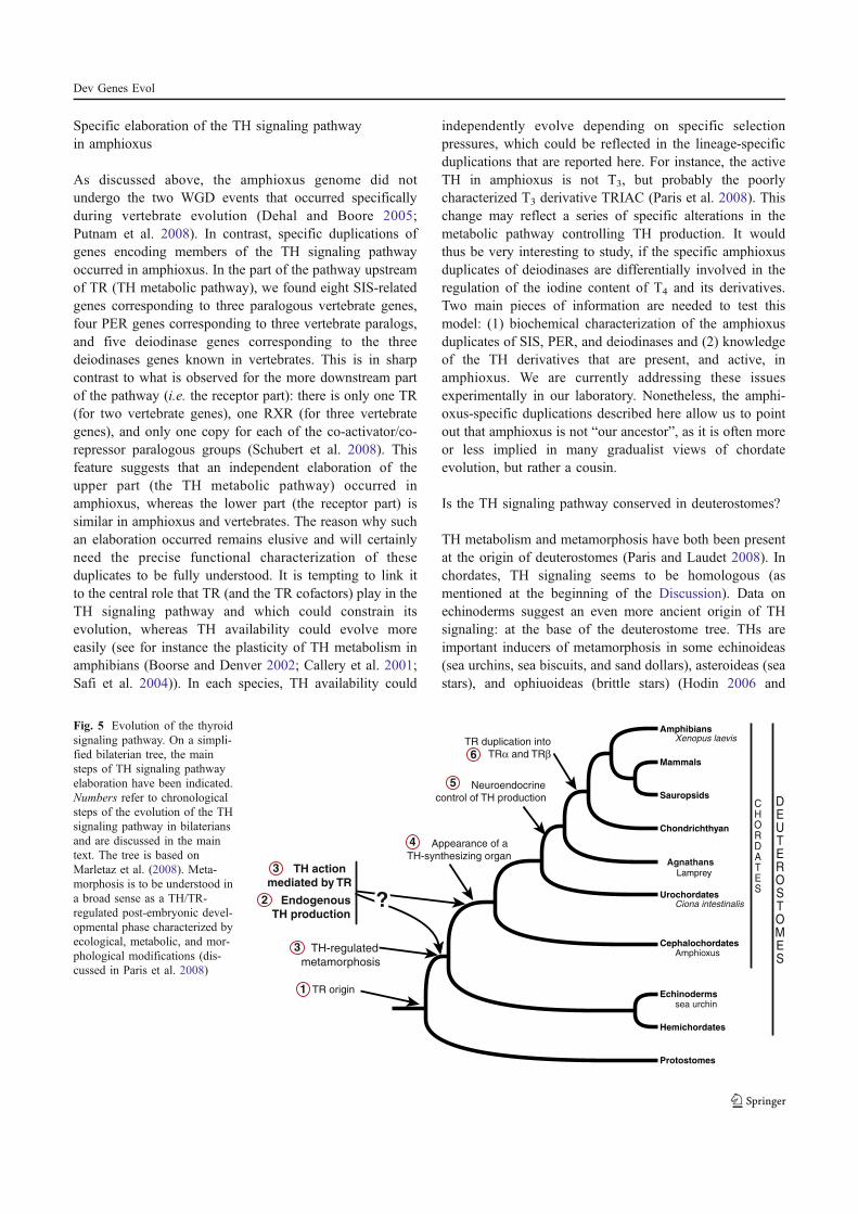
Specific elaboration of the TH signaling pathwayin amphioxus
As discussed above, the amphioxus genome did notundergo the two WGD events that occurred specificallyduring vertebrate evolution (Dehal and Boore 2005;Putnam et al. 2008). In contrast, specific duplications ofgenes encoding members of the TH signaling pathwayoccurred in amphioxus. In the part of the pathway upstreamof TR (TH metabolic pathway), we found eight SIS-relatedgenes corresponding to three paralogous vertebrate genes,four PER genes corresponding to three vertebrate paralogs,and five deiodinase genes corresponding to the threedeiodinases genes known in vertebrates. This is in sharpcontrast to what is observed for the more downstream partof the pathway (i.e. the receptor part): there is only one TR(for two vertebrate genes), one RXR (for three vertebrategenes), and only one copy for each of the co-activator/co-repressor paralogous groups (Schubert et al. 2008). Thisfeature suggests that an independent elaboration of theupper part (the TH metabolic pathway) occurred inamphioxus, whereas the lower part (the receptor part) issimilar in amphioxus and vertebrates. The reason why suchan elaboration occurred remains elusive and will certainlyneed the precise functional characterization of theseduplicates to be fully understood. It is tempting to link itto the central role that TR (and the TR cofactors) play in theTH signaling pathway and which could constrain itsevolution, whereas TH availability could evolve moreeasily (see for instance the plasticity of TH metabolism inamphibians (Boorse and Denver 2002; Callery et al. 2001;Safi et al. 2004)). In each species, TH availability could
independently evolve depending on specific selectionpressures, which could be reflected in the lineage-specificduplications that are reported here. For instance, the activeTH in amphioxus is not T3, but probably the poorlycharacterized T3 derivative TRIAC (Paris et al. 2008). Thischange may reflect a series of specific alterations in themetabolic pathway controlling TH production. It wouldthus be very interesting to study, if the specific amphioxusduplicates of deiodinases are differentially involved in theregulation of the iodine content of T4 and its derivatives.Two main pieces of information are needed to test thismodel: (1) biochemical characterization of the amphioxusduplicates of SIS, PER, and deiodinases and (2) knowledgeof the TH derivatives that are present, and active, inamphioxus. We are currently addressing these issuesexperimentally in our laboratory. Nonetheless, the amphi-oxus-specific duplications described here allow us to pointout that amphioxus is not “our ancestor”, as it is often moreor less implied in many gradualist views of chordateevolution, but rather a cousin.
Is the TH signaling pathway conserved in deuterostomes?
TH metabolism and metamorphosis have both been presentat the origin of deuterostomes (Paris and Laudet 2008). Inchordates, TH signaling seems to be homologous (asmentioned at the beginning of the Discussion). Data onechinoderms suggest an even more ancient origin of THsignaling: at the base of the deuterostome tree. THs areimportant inducers of metamorphosis in some echinoideas(sea urchins, sea biscuits, and sand dollars), asteroideas (seastars), and ophiuoideas (brittle stars) (Hodin 2006 and
CHORDATES
DEUTEROSTOMES
TR duplication into
TRα and TRβ
Appearance of a
TH-synthesizing organ
Neuroendocrine
control of TH production
TH-regulated
metamorphosis
TH action
mediated by TR
Endogenous
TH production
TR origin
Lamprey
Echinoderms
Urochordates
Hemichordates
Sauropsids
Amphibians
Mammals
Cephalochordates
Protostomes
Chondrichthyan
?
Agnathans
Ciona intestinalis
Amphioxus
Xenopus laevis
sea urchin
1
2
4
5
6
3
3
Fig. 5 Evolution of the thyroidsignaling pathway. On a simpli-fied bilaterian tree, the mainsteps of TH signaling pathwayelaboration have been indicated.Numbers refer to chronologicalsteps of the evolution of the THsignaling pathway in bilateriansand are discussed in the maintext. The tree is based onMarletaz et al. (2008). Meta-morphosis is to be understood ina broad sense as a TH/TR-regulated post-embryonic devel-opmental phase characterized byecological, metabolic, and mor-phological modifications (dis-cussed in Paris et al. 2008)
Dev Genes Evol
references therein). Moreover, peroxidase activity-depen-dent T4 production was also demonstrated (Heyland et al.2006). We found in the sea urchin genome many genesorthologous to vertebrate TH signaling members. Conse-quently, it is very probable that the deuterostome ancestorwas capable of endogenous TH production. However, asechinoderms do not have a recognizable endostyle/thyroid,it will be interesting to localize TH production.
There are still many deuterostome groups that have beenneglected in terms of developmental and genomic studies.This is, for example, the case for hemichordates: almostnothing is known about TH signaling in this sister group ofechinoderms (Fig. 5), although iodine was detected in thepharyngeal area (same body part as the endostyle/thyroid)(Ruppert 2005) supporting the notion of an ancestral originof TH production in deuterostomes. Other deuterostometaxa that are poorly studied include sea stars, sea cucumber,crinoids as well as divergent urochordates, such aslarvaceans and thaliaceans (Brusca and Brusca 2003).
The existence of a TH signaling pathway outsidedeuterostomes still remains elusive. Indeed, in protostomesor cnidarians, TH metabolism has been poorly studied(Eales 1997), few reports have investigated TH effects onmetamorphosis, and, although TRs were cloned in severalprotostome species, they have not been molecularlycharacterized (Paris and Laudet 2008). We propose thefollowing scenario regarding the evolution of the THsignaling pathway (Fig. 5): (1) appearance of a TR geneat the base of the bilaterians, whose function remainselusive; (2) the chordate, and likely even the deuterostome,ancestor acquired the ability to endogenously produce THs(TH production has been reported in several deuterostomesand the main members of the TH signaling pathway likeSIS, PERT, IODs, TR, and some carrier proteins have beendescribed in vertebrates, amphioxus, and, to a lesser extent,in echinoderms); (3) this process can probably be correlatedwith the ability to metamorphose under TH/TR regulation;(4) more complete and localized internalization of THsynthesis in a specialized organ with the evolution of adual-function endostyle in chordates and its subsequentspecialization into a thyroid within the vertebrate lineage;(5) in the vertebrate lineage, there is the appearance of aneuroendocrine control of TH synthesis using peptidehormones, such as TRH and TSH, that are not found inthe amphioxus genome; (6) further elaboration of thepathway occurred in gnathostomes with the duplication ofthe ancestral TR into two genes, TRα and TRβ, allowing afurther refinement of the pathway (there has also been alamprey-specific TR duplication (Escriva et al. 2002)).According to this scenario, the downstream part of thepathway, namely the genes regulated by TH and TR, arederived in each species giving rise to the extraordinarydiversity of morphological, physiological, and ecological
rearrangements observed during metamorphosis (Paris andLaudet 2008).
Conclusions
From our study and previous data, we propose that theamphioxus TH signaling pathway is homologous to thevertebrate TH signaling pathway implying an ancient originof TH metabolism. However, biochemical investigation onthe proteins encoded by the genes described here should becarried out in the future. Outside chordates, much scarcerdata are available. In some echinoderms, TH productionand biological actions by TH are similar to what has beenobserved in chordates. Further work on echinoderms willbe required to address questions such as where are THsproduced and whether TR is involved in TH action inechinoderms. In order to better understand the evolution ofthe TH signaling pathway and its link to development andespecially to metamorphosis, data on a wider range ofanimals should be obtained. Thus, even if a continuouseffort should be maintained to keep improving ourunderstanding of the TH signaling pathway in chordates,the sister group of chordates, the Ambulacraria (regroupingthe echinoderms, the hemichordates, and xenoturbella(Marletaz et al. 2008)) and the protostomes are a “thyroidaldesert” that may be worth our attention.
Acknowledgements We would like to thank Maria Theodosiou forcritical reading of the manuscript. This work was supported by fundsfrom the ANR, CNRS, and the MENRT. This study was furthersupported by CRESCENDO, a European Union Integrated Project ofFP6, and by CASCADE, a Network of Excellence of FP6.
References
Baqui M, Botero D, Gereben B, Curcio C, Harney JW, Salvatore D,Sorimachi K, Larsen PR, Bianco AC (2003) Human type 3iodothyronine selenodeiodinase is located in the plasma mem-brane and undergoes rapid internalization to endosomes. J BiolChem 278:1206–1211
Berry MJ, Banu L, Larsen PR (1991) Type I iodothyronine deiodinaseis a selenocysteine-containing enzyme. Nature 349:438–440
Bianco AC, Salvatore D, Gereben B, Berry MJ, Larsen PR (2002)Biochemistry, cellular and molecular biology, and physiologicalroles of the iodothyronine selenodeiodinases. Endocr Rev 23:38–89
Boorse GC, Denver RJ (2002) Acceleration of Ambystoma tigrinummetamorphosis by corticotropin-releasing hormone. J Exp Zool293:94–98
Brusca RC, Brusca GJ (2003) Invertebrates. Sinauer Associates,Sunderland
Callery EM, Fang H, Elinson RP (2001) Frogs without polliwogs:evolution of anuran direct development. Bioessays 23:233–241
Castresana J (2000) Selection of conserved blocks from multiplealignments for their use in phylogenetic analysis. Mol Biol Evol17:540–552
Dev Genes Evol
Covelli I, Salvatore G, Sena L, Roche J (1960) Sur la formationd’hormones thyroidiennes et de leurs précurseurs par Branchios-toma lanceolatum. C R Soc Biol Paris 154:1165–1169
Curcio C, Baqui MM, Salvatore D, Rihn BH, Mohr S, Harney JW,Larsen PR, Bianco AC (2001) The human type 2 iodothyroninedeiodinase is a selenoprotein highly expressed in a mesotheliomacell line. J Biol Chem 276:30183–30187
Daiyasu H, Toh H (2000) Molecular evolution of the myeloperoxidasefamily. J Mol Evol 51:433–445
Dehal P, Boore JL (2005) Two rounds of whole genome duplication inthe ancestral vertebrate. PLoS Biol 3:e314
Denver RJ (1997) Environmental stress as a developmental cue:corticotropin-releasing hormone is a proximate mediator ofadaptive phenotypic plasticity in amphibian metamorphosis.Horm Behav 31:169–179
Dohan O, De la Vieja A, Paroder V, Riedel C, Artani M, Reed M,Ginter CS, Carrasco N (2003) The sodium/iodide symporter(NIS): characterization, regulation, and medical significance.Endocr Rev 24:48–77
Eales JG (1997) Iodine metabolism and thyroid-related functions inorganisms lacking thyroid follicles: are thyroid hormones alsovitamins? Proc Soc Exp Biol Med 214:302–317
Edgar RC (2004) MUSCLE: multiple sequence alignment with highaccuracy and high throughput. Nucleic Acids Res 32:1792–1797
Escriva H, Manzon L, Youson J, Laudet V (2002) Analysis oflamprey and hagfish genes reveals a complex history of geneduplications during early vertebrate evolution. Mol Biol Evol19:1440–1450
Flamant F, Samarut J (2003) Thyroid hormone receptors: lessons fromknockout and knock-in mutant mice. Trends Endocrinol Metab14:85–90
Flamant F, Baxter JD, Forrest D, Refetoff S, Samuels H, Scanlan TS,Vennstrom B, Samarut J (2006) International Union of Pharma-cology. LIX. The pharmacology and classification of the nuclearreceptor superfamily: thyroid hormone receptors. Pharmacol Rev58:705–711
Force A, Lynch M, Pickett FB, Amores A, Yan YL, Postlethwait J(1999) Preservation of duplicate genes by complementary,degenerative mutations. Genetics 151:1531–1545
Fredriksson G, Öfverholm T, Ericson LE (1985) Electron-microscopicstudies of iodine-binding and peroxidase activity in the endostyleof the larval amphioxus (Branchiostoma lanceolatum). CellTissue Res 241:257–266
Galtier N, Gouy M, Gautier C (1996) SEAVIEW and PHYLO_WIN:two graphic tools for sequence alignment and molecularphylogeny. Comput Appl Biosci 12:543–548
Gopal E, Umapathy NS, Martin PM, Ananth S, Gnana-Prakasam JP,Becker H, Wagner CA, Ganapathy V, Prasad PD (2007) Cloningand functional characterization of human SMCT2 (SLC5A12)and expression pattern of the transporter in kidney. BiochimBiophys Acta 1768:2690–2697
Guindon S, Gascuel O (2003) A simple, fast, and accurate algorithmto estimate large phylogenies by maximum likelihood. Syst Biol52:696–704
Heyland A, Hodin J (2004) Heterochronic developmental shift causedby thyroid hormone in larval sand dollars and its implications forphenotypic plasticity and the evolution of nonfeeding develop-ment. Evolution Int J Org Evolution 58:524–538
Heyland A, Reitzel AM, Price DA, Moroz LL (2006) Endogenousthyroid hormone synthesis in facultative planktotrophic larvae ofthe sand dollar Clypeaster rosaceus: implications for theevolutionary loss of larval feeding. Evol Dev 8:568–579
Hodin J (2006) Expanding networks: Signaling components in and ahypothesis for the evolution of metamorphosis. Integr Comp Biol46:719–742
Holland LZ, Albalat R, Azumi K, Benito-Gutierrez E, Blow MJ,Bronner-Fraser M, Brunet F, Butts T, Candiani S, Dishaw LJ,Ferrier DE, Garcia-Fernandez J, Gibson-Brown JJ, Gissi C,Godzik A, Hallbook F, Hirose D, Hosomichi K, Ikuta T, Inoko H,Kasahara M, Kasamatsu J, Kawashima T, Kimura A, KobayashiM, Kozmik Z, Kubokawa K, Laudet V, Litman GW, McHardyAC, Meulemans D, Nonaka M, Olinski RP, Pancer Z, PennacchioLA, Pestarino M, Rast JP, Rigoutsos I, Robinson-Rechavi M,Roch G, Saiga H, Sasakura Y, Satake M, Satou Y, Schubert M,Sherwood N, Shiina T, Takatori N, Tello J, Vopalensky P, WadaS, Xu A, Ye Y, Yoshida K, Yoshizaki F, Yu JK, Zhang Q, ZmasekCM, de Jong PJ, Osoegawa K, Putnam NH, Rokhsar DS, SatohN, Holland PW (2008) The amphioxus genome illuminatesvertebrate origins and cephalochordate biology. Genome Res18:1100–1111
Howard-Ashby M, Materna SC, Brown CT, Chen L, Cameron RA,Davidson EH (2006) Gene families encoding transcription factorsexpressed in early development of Strongylocentrotus purpur-atus. Dev Biol 300:90–107
Hulbert AJ (2000) Thyroid hormones and their effects: a newperspective. Biol Rev Camb Philos Soc 75:519–631
Kryukov GV, Castellano S, Novoselov SV, Lobanov AV, Zehtab O,Guigo R, Gladyshev VN (2003) Characterization of mammalianselenoproteomes. Science 300:1439–1443
Lynch M, Conery JS (2000) The evolutionary fate and consequencesof duplicate genes. Science 290:1151–1155
Manzon RG, Holmes JA, Youson JH (2001) Variable effects ofgoitrogens in inducing precocious metamorphosis in sealampreys (Petromyzon marinus). J Exp Zool 289:290–303
Manzon RG, Neuls TM, Manzon LA (2007) Molecular cloning, tissuedistribution, and developmental expression of lamprey trans-thyretins. Gen Comp Endocrinol 151:55–65
Markov GV, Paris M, Bertrand S, Laudet V (2008) The evolution ofthe ligand/receptor couple: a long road from comparativeendocrinology to comparative genomics. Mol Cell Endocrinol293:5–16
Marletaz F, Gilles A, Caubit X, Perez Y, Dossat C, Samain S, GyapayG, Wincker P, Le Parco Y (2008) Chaetognath transcriptomereveals ancestral and unique features among bilaterians. GenomeBiol 9:R94
Monaco F, Dominici R, Andreoli M, Pirro RD, Roche J (1981)Thyroid hormone formation in thyroglobulin synthesized in theamphioxus (Branchiostoma lanceolatum Pallas). Comp BiochemPhysiol B 70:341–343
Novinec M, Kordis D, Turk V, Lenarcic B (2006) Diversity andevolution of the thyroglobulin type-1 domain superfamily. MolBiol Evol 23:744–755
Ogasawara M (2000) Overlapping expression of amphioxus homologsof the thyroid transcription factor-1 gene and thyroid peroxidasegene in the endostyle: insight into evolution of the thyroid gland.Dev Genes Evol 210:231–242
Ogasawara M, Di Lauro R, Satoh N (1999) Ascidian homologs ofmammalian thyroid peroxidase genes are expressed in thethyroid-equivalent region of the endostyle. J Exp Zool285:158–169
Ohno S (1970) Evolution by gene duplication. Springer, BerlinParis M, Escriva H, Schubert M, Brunet F, Brtko J, Ciesielski F,
Roecklin D, Vivat-Hannah V, Jamin EL, Cravedi JP, Scanlan TS,Renaud JP, Holland ND, Laudet V (2008) Amphioxus postem-bryonic development reveals the homology of chordate meta-morphosis. Curr Biol 18:825–830
Paris M, Laudet V (2008) The history of developmental stages:metamorphosis in chordates. Genesis, in press
Patricolo E, Cammarata M, D'Agati P (2001) Presence of thyroidhormones in ascidian larvae and their involvement in metamor-phosis. J Exp Zool 290:426–430
Dev Genes Evol
Power DM, Llewellyn L, Faustino M, Nowell MA, Bjornsson BT,Einarsdottir IE, Canario AV, Sweeney GE (2001) Thyroidhormones in growth and development of fish. Comp BiochemPhysiol C Toxicol Pharmacol 130:447–459
Putnam NH, Butts T, Ferrier DE, Furlong RF, Hellsten U, KawashimaT, Robinson-Rechavi M, Shoguchi E, Terry A, Yu JK, Benito-Gutierrez EL, Dubchak I, Garcia-Fernandez J, Gibson-Brown JJ,Grigoriev IV, Horton AC, de Jong PJ, Jurka J, Kapitonov VV,Kohara Y, Kuroki Y, Lindquist E, Lucas S, Osoegawa K,Pennacchio LA, Salamov AA, Satou Y, Sauka-Spengler T,Schmutz J, Shin IT, Toyoda A, Bronner-Fraser M, Fujiyama A,Holland LZ, Holland PW, Satoh N, Rokhsar DS (2008) Theamphioxus genome and the evolution of the chordate karyotype.Nature 453:1064–1071
Robinson M, Gouy M, Gautier C, Mouchiroud D (1998) Sensitivity ofthe relative-rate test to taxonomic sampling. Mol Biol Evol15:1091–1098
Rodriguez AM, Perron B, Lacroix L, Caillou B, Leblanc G,Schlumberger M, Bidart JM, Pourcher T (2002) Identificationand characterization of a putative human iodide transporterlocated at the apical membrane of thyrocytes. J Clin EndocrinolMetab 87:3500–3503
Ruppert EE (2005) Key character uniting hemichordates andchordates: homologies or homoplasies. Can J Zool 83:8–23
Safi R, Bertrand S, Marchand O, Duffraisse M, de Luze A, VanackerJM, Maraninchi M, Margotat A, Demeneix B, Laudet V (2004)The axolotl (Ambystoma mexicanum), a neotenic amphibian,
expresses functional thyroid hormone receptors. Endocrinology145:760–772
Schreiber G, Richardson SJ (1997) The evolution of gene expression,structure and function of transthyretin. Comp Biochem Physiol BBiochem Mol Biol 116:137–160
Schubert M, Brunet F, Paris M, Bertrand S, Benoit G, Laudet V(2008) Nuclear hormone receptor signaling in amphioxus. DevGenes Evol, in press
Shepherdley CA, Klootwijk W, Makabe KW, Visser TJ, KuiperGGJM (2004) An ascidian homolog of vertebrate iodothyroninedeiodinases. Endocrinology 145:1255–1268
Shi Y-B (2000) Amphibian metamorphosis: from morphology tomolecular biology. Wiley, New York
Tata JR (2006) Amphibian metamorphosis as a model for thedevelopmental actions of thyroid hormone. Mol Cell Endocrinol246:10–20
Vandenborne K, Roelens SA, Darras VM, Kuhn ER, Van der GeytenS (2005) Cloning and hypothalamic distribution of the chickenthyrotropin-releasing hormone precursor cDNA. J Endocrinol186:387–396
Wu SY, Green WL, Huang WS, Hays MT, Chopra IJ (2005)Alternate pathways of thyroid hormone metabolism. Thyroid15:943–958
Yen PM (2001) Physiological and molecular basis of thyroid hormoneaction. Physiol Rev 81:1097–1142
Youson JH (1997) Is lamprey metamorphosis regulated by thyroidhormones? Amer Zool 37:441–460
Dev Genes Evol

7A M P H I O X U S P O S T- E M B RY O N I CD E V E L O P M E N T R E V E A L S T H EH O M O L O G Y O F C H O R D AT EM E TA M O R P H O S I S
As explained in chapter ?? in the introduction, metamorphoses display a wide
variability among chordates, from undetectable, as in mammals, to an extensive
tissue remodeling, like in tunicates. This variability suggests that metamorphosis
appeared several times independently during evolution. However, the molecular
determinism of metamorphosis was well established only in some amphibians, and
to a lesser extent in teleost fishes, where THs were shown to be the main players
(see chapter ?? in the introduction).
In order to gain better insight into the evolution of metamorphosis in chordates,
we sought into the molecular determinism of this post-embryonic developmental
stage, by focusing on the amphioxus animal model. Indeed, as explained in chapter
??, amphioxus both branches at the base of the chordate tree and has evolved
relatively slowly since then. It is thus a favorable model to digging into the origins
of TH effects on metamorphosis in chordates. However, very few experiments had
been conducted on amphioxus before, because of the unavailability of larvae at a
late developmental stage. This article reports the first large scale study of the role
of THs during amphioxus metamorphosis.
The work described in this section illustrates the difficulty in determining
homology for anciently derived or fast evolving developmental processes. Here the
morphological component of the evolution of metamorphosis may be underlain by
its molecular determinism, where some parts of the regulatory pathway are better
conserved than others. See the general discussion (chapter ??) for further details.
This article was published in Current Biology in June
2008. Please notice that supplemental data are pre-
sented after the main article to help understand the
main article.
107
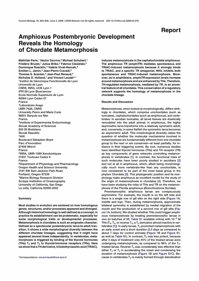
Current Biology 18, 825–830, June 3, 2008 ª2008 Elsevier Ltd All rights reserved DOI 10.1016/j.cub.2008.04.078
ReportAmphioxus Postembryonic DevelopmentReveals the Homologyof Chordate Metamorphosis
Mathilde Paris,1 Hector Escriva,2 Michael Schubert,1
Frederic Brunet,1 Julius Brtko,3 Fabrice Ciesielski,4
Dominique Roecklin,4 Valerie Vivat-Hannah,4
Emilien L. Jamin,5 Jean-Pierre Cravedi,5
Thomas S. Scanlan,6 Jean-Paul Renaud,4
Nicholas D. Holland,7 and Vincent Laudet1,*1Institut de Genomique Fonctionnelle de Lyon
Universite de Lyon
CNRS, INRA, UCB Lyon 1
IFR128 Lyon Biosciences
Ecole Normale Superieure de Lyon
69364 Lyon Cedex 07
France2Laboratoire Arago
UMR-7628, CNRS
University Pierre and Marie Curie
66651 Banyuls sur Mer
France3Institute of Experimental Endocrinology
Slovak Academy of Sciences
833 06 Bratislava
Slovak Republic4AliX
Boulevard Sebastien Brant
Parc d’Innovation
67400 Illkirch
France5INRA, UMR-1089 Xenobiotiques
31931 Toulouse Cedex 9
France6Department of Physiology and Pharmacology
Oregon Health and Science University
3181 SW Sam Jackson Park Road
Portland, Oregon 972397Marine Biology Research Division
Scripps Institution of Oceanography
University of California, San Diego
La Jolla, California 92093-0202
Summary
Most studies in evolution are centered on how homologous
genes, structures, and/or processes appeared and diverged.
Although historical homology iswell defined as a concept, in
practice its establishment can be problematic, especially for
some morphological traits or developmental processes.
Metamorphosis in chordates is such an enigmatic character.
Defined as a spectacular postembryonic larva-to-adult tran-
sition, it shows a wide morphological diversity between the
different chordate lineages, suggesting that it might have
appeared several times independently. In vertebrates, meta-
morphosis is triggered by binding of the thyroid hormones
(THs) T4 and T3 to thyroid-hormone receptors (TRs). Here
weshow that aTHderivative, triiodothyroacetic acid (TRIAC),
inducesmetamorphosis in the cephalochordate amphioxus.
The amphioxus TR (amphiTR) mediates spontaneous and
TRIAC-induced metamorphosis because it strongly binds
to TRIAC, and a specific TR antagonist, NH3, inhibits both
spontaneous and TRIAC-induced metamorphosis. More-
over, as in amphibians, amphiTR expression levels increase
aroundmetamorphosis and are enhancedby THs. Therefore,
TH-regulated metamorphosis, mediated by TR, is an ances-
tral feature of all chordates. This conservation of a regulatory
network supports the homology of metamorphosis in the
chordate lineage.
Results and Discussion
Metamorphosis, when looked at morphologically, differs strik-
ingly in chordates, which comprise urochordates (such as
tunicates), cephalochordates (such as amphioxus), and verte-
brates: In ascidian tunicates, all larval tissues are drastically
remodeled into the adult animal; in amphioxus, the highly
asymmetric larva transforms into a relatively symmetric adult;
and, conversely, in some flatfish the symmetric larva becomes
an asymmetric adult. This morphological diversity raises the
question of whether the molecular mechanisms involved in
metamorphosis are fundamentally different from one chordate
group to the next or are conserved—at least partially, for in-
stance in their triggering events. By now, numerous studies
have identified thyroid hormones (THs) and TH receptor (TR)
as key components of gene networks controlling metamor-
phosis in vertebrates [1]. In contrast, the functional roles of
such molecules have been poorly studied in ascidians [2]
and not at all in amphioxus, which, albeit being morphologi-
cally much more vertebrate-like than any urochordate, is
now considered to be part of the most basal group in the
phylum Chordata [3]. This phylogenetic position and its mor-
phology make amphioxus an excellent model for the study of
the origin of metamorphosis in chordates [4]. Therefore, we
have been studying the roles of THs and TR on the metamor-
phosis of the Florida amphioxus (Branchiostoma floridae).
Premetamorphic amphioxus larvae are conspicuously
asymmetric: For example, the mouth is on the left side and
there is a single row of gill slits on the right side (Figure 1A,
middle and top). Then, during metamorphosis, approximate
bilateral symmetry is established by medial migration of the
mouth and the production of a second row of gill slits (Fig-
ure 1A, bottom). We studied whether THs could trigger amphi-
oxus metamorphosis by treating premetamorphic larvae in
vivo (in batches of 50, Table S1 available online) with 1028 M
THs (T3, T4, or reverse T3, a T4 derivative almost inactive in ver-
tebrates [1]). In early larvae, T3 promoted metamorphosis with
an early onset and a short duration (2.5 days as compared to
about 7 days for control animals) (Figure 1B and Figure S1,
as well as Table S1). In contrast, T4 was less potent, because
after 3 days of treatment only 45% of the treated larvae were
undergoing metamorphosis, as compared to 98% of the T3-
treated larvae. Reverse T3 was considerably less effective than
either T3 or T4 in accelerating the onset and condensing the
duration of metamorphosis (Figure 1B and Figure S1C). Be-
cause in vertebrates T3 is mainly formed through deiodination*Correspondence: [email protected]
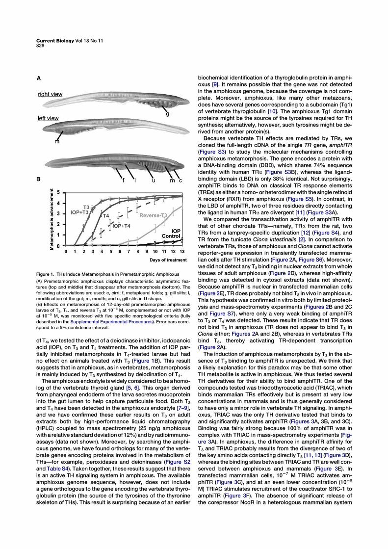
of T4, we tested the effect of a deiodinase inhibitor, iodopanoic
acid (IOP), on T3 and T4 treatments. The addition of IOP par-
tially inhibited metamorphosis in T4-treated larvae but had
no effect on animals treated with T3 (Figure 1B). This result
suggests that in amphioxus, as in vertebrates, metamorphosis
is mainly induced by T3 synthesized by deiodination of T4.
Theamphioxus endostyle iswidely considered tobeahomo-
log of the vertebrate thyroid gland [5, 6]. This organ derived
from pharyngeal endoderm of the larva secretes mucoprotein
into the gut lumen to help capture particulate food. Both T3
and T4 have been detected in the amphioxus endostyle [7–9],
and we have confirmed these earlier results on T3 on adult
extracts both by high-performance liquid chromatography
(HPLC) coupled to mass spectrometry (25 ng/g amphioxus
with a relative standarddeviationof 12%)andby radioimmuno-
assays (data not shown). Moreover, by searching the amphi-
oxus genome, we have found orthologs for many of the verte-
brate genes encoding proteins involved in the metabolism of
THs—for example, peroxidases and deioninases (Figure S2
and Table S4). Taken together, these results suggest that there
is an active TH signaling system in amphioxus. The available
amphioxus genome sequence, however, does not include
a gene orthologous to the gene encoding the vertebrate thyro-
globulin protein (the source of the tyrosines of the thyronine
skeleton of THs). This result is surprising because of an earlier
biochemical identification of a thyroglobulin protein in amphi-
oxus [9]. It remains possible that the gene was not detected
in the amphioxus genome, because the coverage is not com-
plete. Moreover, amphioxus, like many other metazoans,
does have several genes corresponding to a subdomain (Tg1)
of vertebrate thyroglobulin [10]. The amphioxus Tg1 domain
proteins might be the source of the tyrosines required for TH
synthesis; alternatively, however, such tyrosines might be de-
rived from another protein(s).
Because vertebrate TH effects are mediated by TRs, we
cloned the full-length cDNA of the single TR gene, amphiTR
(Figure S3) to study the molecular mechanisms controlling
amphioxus metamorphosis. The gene encodes a protein with
a DNA-binding domain (DBD), which shares 74% sequence
identity with human TRa (Figure S3B), whereas the ligand-
binding domain (LBD) is only 38% identical. Not surprisingly,
amphiTR binds to DNA on classical TR response elements
(TREs) as either a homo- or heterodimerwith the single retinoid
X receptor (RXR) from amphioxus (Figure S5). In contrast, in
the LBD of amphiTR, two of three residues directly contacting
the ligand in human TRa are divergent [11] (Figure S3A).
We compared the transactivation activity of amphiTR with
that of other chordate TRs—namely, TRa from the rat, two
TRs from a lamprey-specific duplication [12] (Figure S4), and
TR from the tunicate Ciona intestinalis [2]. In comparison to
vertebrate TRs, those of amphioxus andCiona cannot activate
reporter-gene expression in transiently transfected mamma-
lian cells after TH stimulation (Figure 2A, Figure S6). Moreover,
we did not detect any T3 binding in nuclear extracts fromwhole
tissues of adult amphioxus (Figure 2D), whereas high-affinity
binding was detected in cytosol extracts (data not shown).
Because amphiTR is nuclear in transfected mammalian cells
(Figure 2E), TR does probably not bind T3 in vivo in amphioxus.
This hypothesis was confirmed in vitro both by limited proteol-
ysis and mass-spectrometry experiments (Figures 2B and 2C
and Figure S7), where only a very weak binding of amphiTR
to T3 or T4 was detected. These results indicate that TR does
not bind T3 in amphioxus (TR does not appear to bind T3 in
Ciona either; Figures 2A and 2B), whereas in vertebrates TRs
bind T3, thereby activating TR-dependent transcription
(Figure 2A).
The induction of amphioxus metamorphosis by T3 in the ab-
sence of T3 binding to amphiTR is unexpected. We think that
a likely explanation for this paradox may be that some other
TH metabolite is active in amphioxus. We thus tested several
TH derivatives for their ability to bind amphiTR. One of the
compounds tested was triiodothyroacetic acid (TRIAC), which
binds mammalian TRs effectively but is present at very low
concentrations in mammals and is thus generally considered
to have only a minor role in vertebrate TH signaling. In amphi-
oxus, TRIAC was the only TH derivative tested that binds to
and significantly activates amphiTR (Figures 3A, 3B, and 3C).
Binding was fairly strong because 100% of amphiTR was in
complex with TRIAC in mass-spectrometry experiments (Fig-
ure 3A). In amphioxus, the difference in amphiTR affinity for
T3 and TRIAC probably results from the divergence of two of
the key amino acids contacting directly T3 [11, 13] (Figure 3D),
whereas the binding sites between TRIAC and TR are well con-
served between amphioxus and mammals (Figure 3E). In
transfected mammalian cells, 1027 M TRIAC activates am-
phiTR (Figure 3C), and at an even lower concentration (1028
M) TRIAC stimulates recruitment of the coactivator SRC-1 to
amphiTR (Figure 3F). The absence of significant release of
the corepressor NcoR in a heterologous mammalian system
Figure 1. THs Induce Metamorphosis in Premetamorphic Amphioxus
(A) Premetamorphic amphioxus displays characteristic asymmetric fea-
tures (top and middle) that disappear after metamorphosis (bottom). The
following abbreviations are used: c, cirri; f, metapleural folds; g, gill slits; l,
modification of the gut; m, mouth; and u, gill slits in U shape.
(B) Effects on metamorphosis of 12-day-old premetamorphic amphioxus
larvae of T3, T4, and reverse T3 at 1028 M, complemented or not with IOP
at 1025 M, was monitored with five specific morphological criteria (fully
described in the Supplemental Experimental Procedures). Error bars corre-
spond to a 5% confidence interval.
Current Biology Vol 18 No 11826
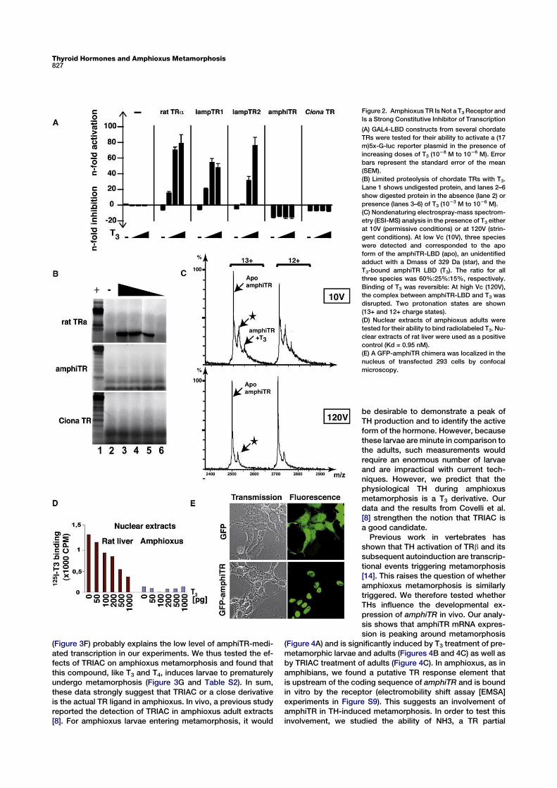
(Figure 3F) probably explains the low level of amphiTR-medi-
ated transcription in our experiments. We thus tested the ef-
fects of TRIAC on amphioxus metamorphosis and found that
this compound, like T3 and T4, induces larvae to prematurely
undergo metamorphosis (Figure 3G and Table S2). In sum,
these data strongly suggest that TRIAC or a close derivative
is the actual TR ligand in amphioxus. In vivo, a previous study
reported the detection of TRIAC in amphioxus adult extracts
[8]. For amphioxus larvae entering metamorphosis, it would
Figure 2. Amphioxus TR Is Not a T3Receptor and
Is a Strong Constitutive Inhibitor of Transcription
(A) GAL4-LBD constructs from several chordate
TRs were tested for their ability to activate a (17
m)5x-G-luc reporter plasmid in the presence of
increasing doses of T3 (1028 M to 1026 M). Error
bars represent the standard error of the mean
(SEM).
(B) Limited proteolysis of chordate TRs with T3.
Lane 1 shows undigested protein, and lanes 2–6
show digested protein in the absence (lane 2) or
presence (lanes 3–6) of T3 (1023 M to 1026 M).
(C) Nondenaturing electrospray-mass spectrom-
etry (ESI-MS) analysis in the presence of T3 either
at 10V (permissive conditions) or at 120V (strin-
gent conditions). At low Vc (10V), three species
were detected and corresponded to the apo
form of the amphiTR-LBD (apo), an unidentified
adduct with a Dmass of 329 Da (star), and the
T3-bound amphiTR LBD (T3). The ratio for all
three species was 60%:25%:15%, respectively.
Binding of T3 was reversible: At high Vc (120V),
the complex between amphiTR-LBD and T3 was
disrupted. Two protonation states are shown
(13+ and 12+ charge states).
(D) Nuclear extracts of amphioxus adults were
tested for their ability to bind radiolabeled T3. Nu-
clear extracts of rat liver were used as a positive
control (Kd = 0.95 nM).
(E) A GFP-amphiTR chimera was localized in the
nucleus of transfected 293 cells by confocal
microscopy.
be desirable to demonstrate a peak of
TH production and to identify the active
form of the hormone. However, because
these larvae areminute in comparison to
the adults, such measurements would
require an enormous number of larvae
and are impractical with current tech-
niques. However, we predict that the
physiological TH during amphioxus
metamorphosis is a T3 derivative. Our
data and the results from Covelli et al.
[8] strengthen the notion that TRIAC is
a good candidate.
Previous work in vertebrates has
shown that TH activation of TRb and its
subsequent autoinduction are transcrip-
tional events triggering metamorphosis
[14]. This raises the question of whether
amphioxus metamorphosis is similarly
triggered. We therefore tested whether
THs influence the developmental ex-
pression of amphiTR in vivo. Our analy-
sis shows that amphiTR mRNA expres-
sion is peaking around metamorphosis
(Figure 4A) and is significantly induced by T3 treatment of pre-
metamorphic larvae and adults (Figures 4B and 4C) as well as
by TRIAC treatment of adults (Figure 4C). In amphioxus, as in
amphibians, we found a putative TR response element that
is upstream of the coding sequence of amphiTR and is bound
in vitro by the receptor (electromobility shift assay [EMSA]
experiments in Figure S9). This suggests an involvement of
amphiTR in TH-induced metamorphosis. In order to test this
involvement, we studied the ability of NH3, a TR partial
Thyroid Hormones and Amphioxus Metamorphosis827
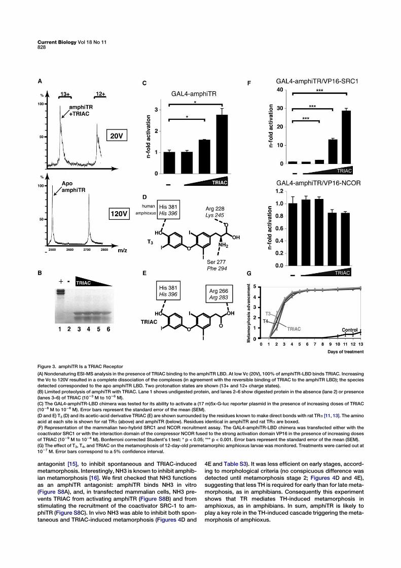
antagonist [15], to inhibit spontaneous and TRIAC-induced
metamorphosis. Interestingly, NH3 is known to inhibit amphib-
ian metamorphosis [16]. We first checked that NH3 functions
as an amphiTR antagonist: amphiTR binds NH3 in vitro
(Figure S8A), and, in transfected mammalian cells, NH3 pre-
vents TRIAC from activating amphiTR (Figure S8B) and from
stimulating the recruitment of the coactivator SRC-1 to am-
phiTR (Figure S8C). In vivo NH3 was able to inhibit both spon-
taneous and TRIAC-induced metamorphosis (Figures 4D and
4E and Table S3). It was less efficient on early stages, accord-
ing to morphological criteria (no conspicuous difference was
detected until metamorphosis stage 2; Figures 4D and 4E),
suggesting that less TH is required for early than for late meta-
morphosis, as in amphibians. Consequently this experiment
shows that TR mediates TH-induced metamorphosis in
amphioxus, as in amphibians. In sum, amphiTR is likely to
play a key role in the TH-induced cascade triggering the meta-
morphosis of amphioxus.
Figure 3. amphiTR Is a TRIAC Receptor
(A) Nondenaturing ESI-MS analysis in the presence of TRIAC binding to the amphiTR LBD. At low Vc (20V), 100% of amphiTR-LBD binds TRIAC. Increasing
the Vc to 120V resulted in a complete dissociation of the complexes (in agreement with the reversible binding of TRIAC to the amphiTR LBD); the species
detected corresponded to the apo amphiTR LBD. Two protonation states are shown (13+ and 12+ charge states).
(B) Limited proteolysis of amphiTR with TRIAC. Lane 1 shows undigested protein, and lanes 2–6 show digested protein in the absence (lane 2) or presence
(lanes 3–6) of TRIAC (1023 M to 1026 M).
(C) The GAL4-amphiTR-LBD chimera was tested for its ability to activate a (17 m)5x-G-luc reporter plasmid in the presence of increasing doses of TRIAC
(1028 M to 1026 M). Error bars represent the standard error of the mean (SEM).
(D and E) T3 (D) and its acetic-acid derivative TRIAC (E) are shown surrounded by the residues known to make direct bonds with rat TRa [11, 13]. The amino
acid at each site is shown for rat TRa (above) and amphiTR (below). Residues identical in amphiTR and rat TRa are boxed.
(F) Representation of the mammalian two-hybrid SRC1 and NCOR recruitment assay. The GAL4-amphiTR-LBD chimera was transfected either with the
coactivator SRC1 or with the interaction domain of the corepressor NCOR fused to the strong activation domain VP16 in the presence of increasing doses
of TRIAC (1029 M to 1026 M). Bonferroni corrected Student’s t test: * p < 0.05; *** p < 0.001. Error bars represent the standard error of the mean (SEM).
(G) The effect of T3, T4, and TRIAC on the metamorphosis of 12-day-old premetamorphic amphioxus larvae was monitored. Treatments were carried out at
1027 M. Error bars correspond to a 5% confidence interval.
Current Biology Vol 18 No 11828
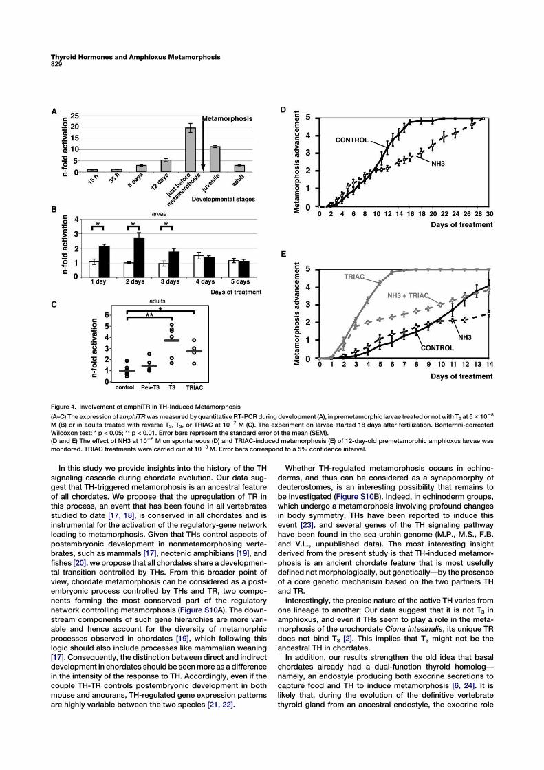
In this study we provide insights into the history of the TH
signaling cascade during chordate evolution. Our data sug-
gest that TH-triggered metamorphosis is an ancestral feature
of all chordates. We propose that the upregulation of TR in
this process, an event that has been found in all vertebrates
studied to date [17, 18], is conserved in all chordates and is
instrumental for the activation of the regulatory-gene network
leading to metamorphosis. Given that THs control aspects of
postembryonic development in nonmetamorphosing verte-
brates, such as mammals [17], neotenic amphibians [19], and
fishes [20], we propose that all chordates share a developmen-
tal transition controlled by THs. From this broader point of
view, chordate metamorphosis can be considered as a post-
embryonic process controlled by THs and TR, two compo-
nents forming the most conserved part of the regulatory
network controlling metamorphosis (Figure S10A). The down-
stream components of such gene hierarchies are more vari-
able and hence account for the diversity of metamorphic
processes observed in chordates [19], which following this
logic should also include processes like mammalian weaning
[17]. Consequently, the distinction between direct and indirect
development in chordates should be seenmore as a difference
in the intensity of the response to TH. Accordingly, even if the
couple TH-TR controls postembryonic development in both
mouse and anourans, TH-regulated gene expression patterns
are highly variable between the two species [21, 22].
Whether TH-regulated metamorphosis occurs in echino-
derms, and thus can be considered as a synapomorphy of
deuterostomes, is an interesting possibility that remains to
be investigated (Figure S10B). Indeed, in echinoderm groups,
which undergo a metamorphosis involving profound changes
in body symmetry, THs have been reported to induce this
event [23], and several genes of the TH signaling pathway
have been found in the sea urchin genome (M.P., M.S., F.B.
and V.L., unpublished data). The most interesting insight
derived from the present study is that TH-induced metamor-
phosis is an ancient chordate feature that is most usefully
defined not morphologically, but genetically—by the presence
of a core genetic mechanism based on the two partners TH
and TR.
Interestingly, the precise nature of the active TH varies from
one lineage to another: Our data suggest that it is not T3 in
amphioxus, and even if THs seem to play a role in the meta-
morphosis of the urochordate Ciona intesinalis, its unique TR
does not bind T3 [2]. This implies that T3 might not be the
ancestral TH in chordates.
In addition, our results strengthen the old idea that basal
chordates already had a dual-function thyroid homolog—
namely, an endostyle producing both exocrine secretions to
capture food and TH to induce metamorphosis [6, 24]. It is
likely that, during the evolution of the definitive vertebrate
thyroid gland from an ancestral endostyle, the exocrine role
Figure 4. Involvement of amphiTR in TH-Induced Metamorphosis
(A–C) The expression of amphiTRwasmeasured by quantitative RT-PCRduring development (A), in premetamorphic larvae treated or not with T3 at 53 1028
M (B) or in adults treated with reverse T3, T3, or TRIAC at 1027 M (C). The experiment on larvae started 18 days after fertilization. Bonferrini-corrected
Wilcoxon test: * p < 0.05; ** p < 0.01. Error bars represent the standard error of the mean (SEM).
(D and E) The effect of NH3 at 1026 M on spontaneous (D) and TRIAC-induced metamorphosis (E) of 12-day-old premetamorphic amphioxus larvae was
monitored. TRIAC treatments were carried out at 1028 M. Error bars correspond to a 5% confidence interval.
Thyroid Hormones and Amphioxus Metamorphosis829
dwindled and disappeared while the endocrine production of
THs became the major and finally the only function.
In cephalochordates, TH-dependent regulation of the transi-
tion from a highly asymmetric larva to a relatively symmetric
juvenile in the genus Branchiostoma raises the question of
how THs affect metamorphosis in the related genus Epigo-
nichthys, which remains conspicuously asymmetric as an
adult. Whether this is an instance of neoteny or a different
type of metamorphosis remains to be determined. In addition,
several enigmatic fossils, including thewidely discussed stylo-
phorans [5, 25], also show asymmetric features. Our conclu-
sion that TH-induced metamorphosis is an ancient feature
invites the re-evaluation of these fossils, considering that
some could be premetamorphic or neotenic.
Supplemental Data
Experimental Procedures, ten figures, and four tables are available at http://
www.current-biology.com/cgi/content/full/18/11/825/DC1/.
Acknowledgments
We thank John Lawrence for generously providing laboratory space at the
University of South Florida.Wewould also like to thankGerard Benoıt, Fran-
cois Bonneton, Bastien Boussau, Barbara Demeineix, Frederic Flamant,
and two anonymous reviewers for reading the manuscript and for fruitful
discussion, Anne Chabadel for help with the confocal microscope, Georges
Delous for technical helpwith THmeasurements of amphioxus extracts, and
Maria Danihelova for technical assistance. This work was supported by the
Centre National de la Recherche Scientifique, the Ecole Normale Superieure
de Lyon, the Association pour la Recherche sur le Cancer (ARC), and the
Ministere de l’Education Nationale, de la Recherche et de la Technologie
(MENRT), by CRESCENDO, a European Union Integrated Project of FP6,
and also by grants of the Network of Excellence (NoE) Project EC, FOOD-
CT-2004-506319 (CASCADE) and VEGA No. 2/0022/08.
Received: October 29, 2007
Revised: April 17, 2008
Accepted: April 25, 2008
Published online: May 29, 2008
References
1. Flamant, F., Baxter, J.D., Forrest, D., Refetoff, S., Samuels, H., Scanlan,
T.S., Vennstrom, B., and Samarut, J. (2006). International Union of Phar-
macology. LIX. The pharmacology and classification of the nuclear
receptor superfamily: Thyroid hormone receptors. Pharmacol. Rev.
58, 705–711.
2. Carosa, E., Fanelli, A., Ulisse, S., Di Lauro, R., Rall, J.E., and Jannini, E.A.
(1998). Ciona intestinalis nuclear receptor 1: A member of steroid/
thyroid hormone receptor family. Proc. Natl. Acad. Sci. USA 95,
11152–11157.
3. Delsuc, F., Brinkmann, H., Chourrout, D., and Philippe, H. (2006).
Tunicates and not cephalochordates are the closest living relatives of
vertebrates. Nature 439, 965–968.
4. Schubert, M., Escriva, H., Xavier-Neto, J., and Laudet, V. (2006). Amphi-
oxus and tunicates as evolutionary model systems. Trends Ecol. Evol.
21, 269–277.
5. Gee, H. (1996). Before the Backbone: Views on the Origin of the Verte-
brates (London: Chapman & Hall).
6. Ogasawara, M. (2000). Overlapping expression of amphioxus homologs
of the thyroid transcription factor-1 gene and thyroid peroxidase gene in
the endostyle: Insight into evolution of the thyroid gland. Dev. Genes
Evol. 210, 231–242.
7. Tong, W., Kerkof, P., and Chaikoff, I.L. (1962). Identification of labeled
thyroxine and triiodothyronine in amphioxus treated with 131-I.
Biochim. Biophys. Acta 56, 326–331.
8. Covelli, I., Salvatore, G., Sena, L., and Roche, J. (1960). Sur la formation
d’hormones thyroidiennes et de leurs precurseurs par Branchiostoma
lanceolatum. C. R. Soc. Biol. Paris 154, 1165–1169.
9. Monaco, F., Dominici, R., Andreoli, M., De Pirro, R., andRoche, J. (1981).
Thyroid hormone formation in thyroglobulin synthesized in the
amphioxus (Branchiostoma lanceolatum Pallas). Comp. Biochem.
Physiol. B 70, 341–343.
10. Novinec, M., Kordis, D., Turk, V., and Lenarcic, B. (2006). Diversity and
evolution of the thyroglobulin type-1 domain superfamily. Mol. Biol.
Evol. 23, 744–755.
11. Wagner, R.L., Apriletti, J.W., McGrath, M.E., West, B.L., Baxter, J.D.,
and Fletterick, R.J. (1995). A structural role for hormone in the thyroid
hormone receptor. Nature 378, 690–697.
12. Escriva, H., Manzon, L., Youson, J., and Laudet, V. (2002). Analysis of
lamprey and hagfish genes reveals a complex history of gene duplica-
tions during early vertebrate evolution. Mol. Biol. Evol. 19, 1440–1450.
13. Wagner, R.L., Huber, B.R., Shiau, A.K., Kelly, A., Cunha Lima, S.T.,
Scanlan, T.S., Apriletti, J.W., Baxter, J.D., West, B.L., and Fletterick,
R.J. (2001). Hormone selectivity in thyroid hormone receptors. Mol.
Endocrinol. 15, 398–410.
14. Tata, J.R. (2006). Amphibianmetamorphosis as amodel for the develop-
mental actions of thyroid hormone. Mol. Cell. Endocrinol. 246, 10–20.
15. Nguyen, N.H., Apriletti, J.W., Cunha Lima, S.T., Webb, P., Baxter, J.D.,
and Scanlan, T.S. (2002). Rational design and synthesis of a novel
thyroid hormone antagonist that blocks coactivator recruitment.
J. Med. Chem. 45, 3310–3320.
16. Lim, W., Nguyen, N.H., Yang, H.Y., Scanlan, T.S., and Furlow, J.D.
(2002). A thyroid hormone antagonist that inhibits thyroid hormone
action in vivo. J. Biol. Chem. 277, 35664–35670.
17. Fraichard, A., Chassande, O., Plateroti, M., Roux, J.P., Trouillas, J., De-
hay, C., Legrand, C., Gauthier, K., Kedinger, M., Malaval, L., et al. (1997).
The T3R alpha gene encoding a thyroid hormone receptor is essential
for post-natal development and thyroid hormone production. EMBO
J. 16, 4412–4420.
18. Marchand, O., Duffraisse, M., Triqueneaux, G., Safi, R., and Laudet, V.
(2004). Molecular cloning and developmental expression patterns of
thyroid hormone receptors and T3 target genes in the turbot (Scophtal-
musmaximus) during post-embryonic development. Gen. Comp. Endo-
crinol. 135, 345–357.
19. Safi, R., Bertrand, S., Marchand, O., Duffraisse, M., de Luze, A.,
Vanacker, J.M., Maraninchi, M.,Margotat, A., Demeneix, B., and Laudet,
V. (2004). The axolotl (Ambystoma mexicanum), a neotenic amphibian,
expresses functional thyroid hormone receptors. Endocrinology 145,
760–772.
20. Brown, D.D. (1997). The role of thyroid hormone in zebrafish and axolotl
development. Proc. Natl. Acad. Sci. USA 94, 13011–13016.
21. Yen, P.M., Feng, X., Flamant, F., Chen, Y., Walker, R.L., Weiss, R.E.,
Chassande, O., Samarut, J., Refetoff, S., and Meltzer, P.S. (2003).
Effects of ligand and thyroid hormone receptor isoforms on hepatic
gene expression profiles of thyroid hormone receptor knockout mice.
EMBO Rep. 4, 581–587.
22. Das, B., Cai, L., Carter, M.G., Piao, Y.L., Sharov, A.A., Ko, M.S., and
Brown, D.D. (2006). Gene expression changes at metamorphosis
induced by thyroid hormone in Xenopus laevis tadpoles. Dev. Biol.
291, 342–355.
23. Heyland, A., Reitzel, A.M., and Hodin, J. (2004). Thyroid hormones
determine developmental mode in sand dollars (Echinodermata:
Echinoidea). Evol. Dev. 6, 382–392.
24. Sembrat, K. (1953). Effect of the endostyle of the lancelet (Branchios-
toma lanceolatum Pall.) on the metamorphosis of axolotl. Zool. Pol. 6,
3–19.
25. Ruta, M. (1999). Brief review of the stylophoran debate. Evol. Dev. 1,
123–135.
Current Biology Vol 18 No 11830
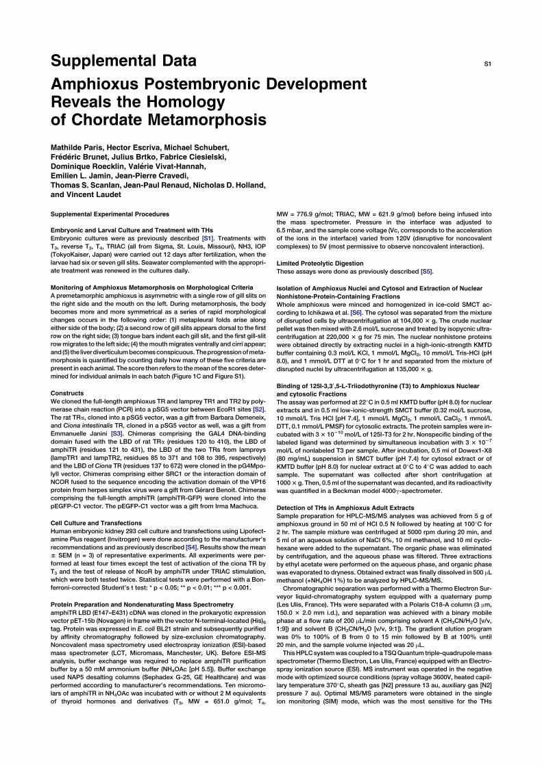
Supplemental Data S1
Amphioxus Postembryonic DevelopmentReveals the Homologyof Chordate Metamorphosis
Mathilde Paris, Hector Escriva, Michael Schubert,
Frederic Brunet, Julius Brtko, Fabrice Ciesielski,
Dominique Roecklin, Valerie Vivat-Hannah,
Emilien L. Jamin, Jean-Pierre Cravedi,
Thomas S. Scanlan, Jean-Paul Renaud, Nicholas D. Holland,
and Vincent Laudet
Supplemental Experimental Procedures
Embryonic and Larval Culture and Treatment with THs
Embryonic cultures were as previously described [S1]. Treatments with
T3, reverse T3, T4, TRIAC (all from Sigma, St. Louis, Missouri), NH3, IOP
(TokyoKaiser, Japan) were carried out 12 days after fertilization, when the
larvae had six or seven gill slits. Seawater complemented with the appropri-
ate treatment was renewed in the cultures daily.
Monitoring of Amphioxus Metamorphosis on Morphological Criteria
A premetamorphic amphioxus is asymmetric with a single row of gill slits on
the right side and the mouth on the left. During metamorphosis, the body
becomes more and more symmetrical as a series of rapid morphological
changes occurs in the following order: (1) metapleural folds arise along
either side of the body; (2) a second row of gill slits appears dorsal to the first
row on the right side; (3) tongue bars indent each gill slit, and the first gill-slit
rowmigrates to the left side; (4) themouthmigrates ventrally and cirri appear;
and (5) the liverdiverticulumbecomesconspicuous.Theprogressionofmeta-
morphosis is quantified by counting daily howmany of these five criteria are
present in each animal. The score then refers to themeanof the scores deter-
mined for individual animals in each batch (Figure 1C and Figure S1).
Constructs
We cloned the full-length amphioxus TR and lamprey TR1 and TR2 by poly-
merase chain reaction (PCR) into a pSG5 vector between EcoR1 sites [S2].
The rat TRa, cloned into a pSG5 vector, was a gift from Barbara Demeneix,
and Ciona intestinalis TR, cloned in a pSG5 vector as well, was a gift from
Emmanuelle Janini [S3]. Chimeras comprising the GAL4 DNA-binding
domain fused with the LBD of rat TRa (residues 120 to 410), the LBD of
amphiTR (residues 121 to 431), the LBD of the two TRs from lampreys
(lampTR1 and lampTR2, residues 85 to 371 and 108 to 395, respectively)
and the LBD of Ciona TR (residues 137 to 672) were cloned in the pG4Mpo-
lyII vector. Chimeras comprising either SRC1 or the interaction domain of
NCOR fused to the sequence encoding the activation domain of the VP16
protein from herpes simplex virus were a gift from Gerard Benoit. Chimeras
comprising the full-length amphiTR (amphiTR-GFP) were cloned into the
pEGFP-C1 vector. The pEGFP-C1 vector was a gift from Irma Machuca.
Cell Culture and Transfections
Human embryonic kidney 293 cell culture and transfections using Lipofect-
amine Plus reagent (Invitrogen) were done according to the manufacturer’s
recommendations and as previously described [S4]. Results show themean
6 SEM (n = 3) of representative experiments. All experiments were per-
formed at least four times except the test of activation of the ciona TR by
T3 and the test of release of NcoR by amphiTR under TRIAC stimulation,
which were both tested twice. Statistical tests were performed with a Bon-
ferroni-corrected Student’s t test: * p < 0.05; ** p < 0.01; *** p < 0.001.
Protein Preparation and Nondenaturating Mass Spectrometry
amphiTR LBD (E147–E431) cDNA was cloned in the prokaryotic expression
vector pET-15b (Novagen) in framewith the vector N-terminal-located (His)6tag. Protein was expressed in E. coli BL21 strain and subsequently purified
by affinity chromatography followed by size-exclusion chromatography.
Noncovalent mass spectrometry used electrospray ionization (ESI)-based
mass spectrometer (LCT, Micromass, Manchester, UK). Before ESI-MS
analysis, buffer exchange was required to replace amphiTR purification
buffer by a 50 mM ammonium buffer (NH4OAc [pH 5.5]). Buffer exchange
used NAP5 desalting columns (Sephadex G-25, GE Healthcare) and was
performed according to manufacturer’s recommendations. Ten micromo-
lars of amphiTR in NH4OAc was incubated with or without 2 M equivalents
of thyroid hormones and derivatives (T3, MW = 651.0 g/mol; T4,
MW = 776.9 g/mol; TRIAC, MW = 621.9 g/mol) before being infused into
the mass spectrometer. Pressure in the interface was adjusted to
6.5 mbar, and the sample cone voltage (Vc, corresponds to the acceleration
of the ions in the interface) varied from 120V (disruptive for noncovalent
complexes) to 5V (most permissive to observe noncovalent interaction).
Limited Proteolytic Digestion
These assays were done as previously described [S5].
Isolation of Amphioxus Nuclei and Cytosol and Extraction of Nuclear
Nonhistone-Protein-Containing Fractions
Whole amphioxus were minced and homogenized in ice-cold SMCT ac-
cording to Ichikawa et al. [S6]. The cytosol was separated from the mixture
of disrupted cells by ultracentrifugation at 104,000 3 g. The crude nuclear
pellet was thenmixed with 2.6mol/L sucrose and treated by isopycnic ultra-
centrifugation at 220,000 3 g for 75 min. The nuclear nonhistone proteins
were obtained directly by extracting nuclei in a high-ionic-strength KMTD
buffer containing 0.3 mol/L KCl, 1 mmol/L MgCl2, 10 mmol/L Tris-HCl (pH
8.0), and 1 mmol/L DTT at 0�C for 1 hr and separated from the mixture of
disrupted nuclei by ultracentrifugation at 135,000 3 g.
Binding of 125I-3,30,5-L-Triiodothyronine (T3) to Amphioxus Nuclear
and cytosolic Fractions
The assay was performed at 22�C in 0.5 ml KMTD buffer (pH 8.0) for nuclear
extracts and in 0.5 ml low-ionic-strength SMCT buffer (0.32 mol/L sucrose,
10 mmol/L Tris HCl [pH 7.4], 1 mmol/L MgCl2, 1 mmol/L CaCl2, 1 mmol/L
DTT, 0.1 mmol/L PMSF) for cytosolic extracts. The protein samples were in-
cubated with 33 10210 mol/L of 125I-T3 for 2 hr. Nonspecific binding of the
labeled ligand was determined by simultaneous incubation with 3 3 1027
mol/L of nonlabeled T3 per sample. After incubation, 0.5 ml of Dowex1-X8
(80 mg/mL) suspension in SMCT buffer (pH 7.4) for cytosol extract or of
KMTD buffer (pH 8.0) for nuclear extract at 0�C to 4�C was added to each
sample. The supernatant was collected after short centrifugation at
10003 g. Then, 0.5ml of the supernatant was decanted, and its radioactivity
was quantified in a Beckman model 4000g-spectrometer.
Detection of THs in Amphioxus Adult Extracts
Sample preparation for HPLC-MS/MS analyses was achieved from 5 g of
amphioxus ground in 50 ml of HCl 0.5 N followed by heating at 100�C for
2 hr. The sample mixture was centrifuged at 5000 rpm during 20 min, and
5 ml of an aqueous solution of NaCl 6%, 10 ml methanol, and 10 ml cyclo-
hexane were added to the supernatant. The organic phase was eliminated
by centrifugation, and the aqueous phase was filtered. Three extractions
by ethyl acetate were performed on the aqueous phase, and organic phase
was evaporated to dryness. Obtained extract was finally dissolved in 500 mL
methanol (+NH4OH 1%) to be analyzed by HPLC-MS/MS.
Chromatographic separation was performed with a Thermo Electron Sur-
veyor liquid-chromatography system equipped with a quaternary pump
(Les Ulis, France). THs were separated with a Polaris C18-A column (3 mm,
150.0 3 2.0 mm i.d.), and separation was achieved with a binary mobile
phase at a flow rate of 200 mL/min comprising solvent A (CH3CN/H2O [v/v,
1:9]) and solvent B (CH3CN/H2O [v/v, 9:1]). The gradient elution program
was 0% to 100% of B from 0 to 15 min followed by B at 100% until
20 min, and the sample volume injected was 20 mL.
ThisHPLCsystemwascoupled toaTSQQuantum triple-quadrupolemass
spectrometer (Thermo Electron, Les Ulis, France) equipped with an Electro-
spray ionization source (ESI). MS instrument was operated in the negative
mode with optimized source conditions (spray voltage 3600V, heated capil-
lary temperature 370�C, sheath gas [N2] pressure 13 au, auxiliary gas [N2]
pressure 7 au). Optimal MS/MS parameters were obtained in the single
ion monitoring (SIM) mode, which was the most sensitive for the THs
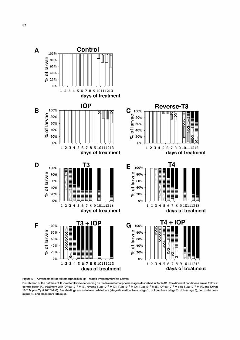
Figure S1. Advancement of Metamorphosis in TH-Treated Premetamorphic Larvae
Distribution of the batches of TH-treated larvae depending on the five metamorphosis stages described in Table S1. The different conditions are as follows:
control batch (A), treatment with IOP at 1025 M (B), reverse T3 at 1028 M (C), T3 at 10
28 M (D), T4 at 1028 M (E), IOP at 1025 Mplus T3 at 10
28 M (F), and IOP at
1025 M plus T4 at 1028 M (G). Bar shadings are as follows: white bars (stage 0), vertical lines (stage 1), oblique lines (stage 2), dots (stage 3), horizontal lines
(stage 4), and black bars (stage 5).
S2
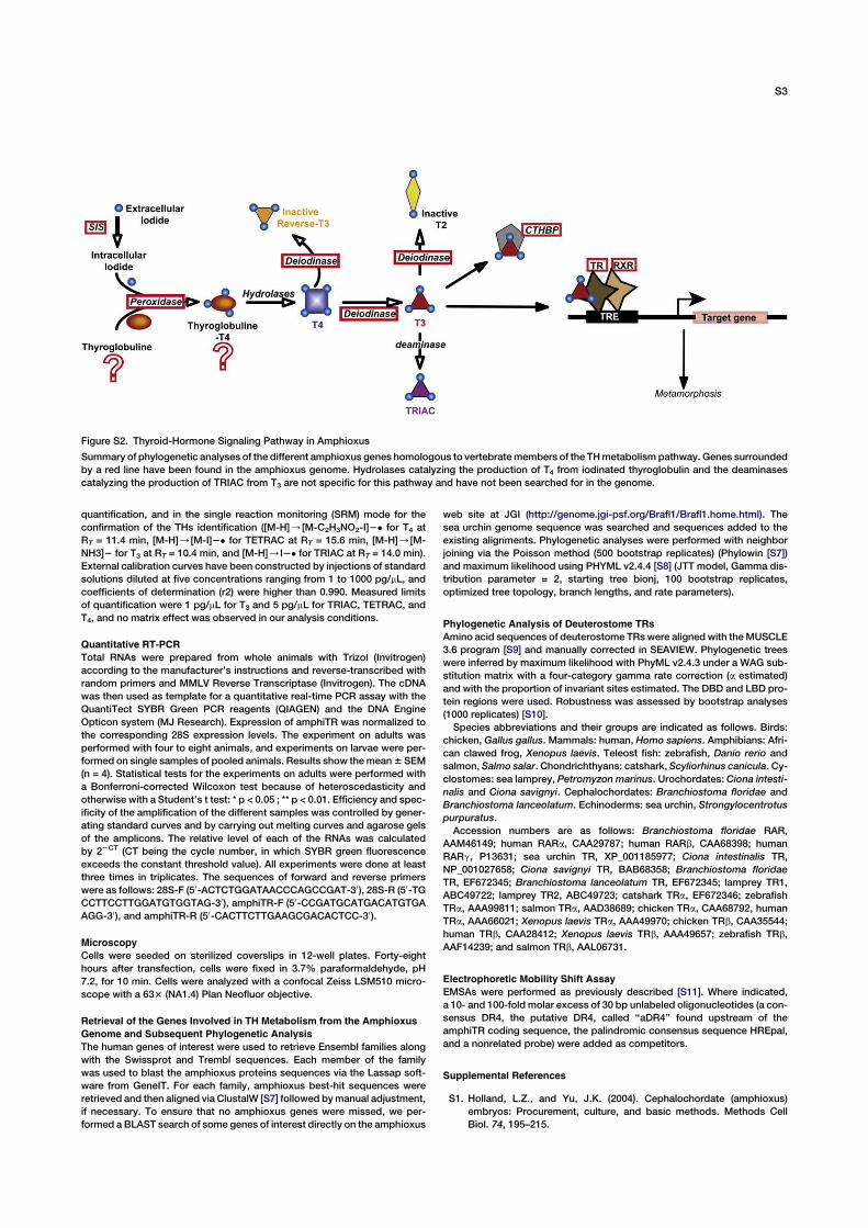
quantification, and in the single reaction monitoring (SRM) mode for the
confirmation of the THs identification ([M-H]/[M-C2H3NO2-I]2� for T4 at
RT = 11.4 min, [M-H]/[M-I]2� for TETRAC at RT = 15.6 min, [M-H]/[M-
NH3]2 for T3 at RT = 10.4 min, and [M-H]/I2� for TRIAC at RT = 14.0 min).
External calibration curves have been constructed by injections of standard
solutions diluted at five concentrations ranging from 1 to 1000 pg/mL, and
coefficients of determination (r2) were higher than 0.990. Measured limits
of quantification were 1 pg/mL for T3 and 5 pg/mL for TRIAC, TETRAC, and
T4, and no matrix effect was observed in our analysis conditions.
Quantitative RT-PCR
Total RNAs were prepared from whole animals with Trizol (Invitrogen)
according to the manufacturer’s instructions and reverse-transcribed with
random primers and MMLV Reverse Transcriptase (Invitrogen). The cDNA
was then used as template for a quantitative real-time PCR assay with the
QuantiTect SYBR Green PCR reagents (QIAGEN) and the DNA Engine
Opticon system (MJ Research). Expression of amphiTR was normalized to
the corresponding 28S expression levels. The experiment on adults was
performed with four to eight animals, and experiments on larvae were per-
formed on single samples of pooled animals. Results show themean6 SEM
(n = 4). Statistical tests for the experiments on adults were performed with
a Bonferroni-corrected Wilcoxon test because of heteroscedasticity and
otherwise with a Student’s t test: * p < 0.05 ; ** p < 0.01. Efficiency and spec-
ificity of the amplification of the different samples was controlled by gener-
ating standard curves and by carrying out melting curves and agarose gels
of the amplicons. The relative level of each of the RNAs was calculated
by 22CT (CT being the cycle number, in which SYBR green fluorescence
exceeds the constant threshold value). All experiments were done at least
three times in triplicates. The sequences of forward and reverse primers
were as follows: 28S-F (50-ACTCTGGATAACCCAGCCGAT-30), 28S-R (50-TG
CCTTCCTTGGATGTGGTAG-30), amphiTR-F (50-CCGATGCATGACATGTGA
AGG-30), and amphiTR-R (50-CACTTCTTGAAGCGACACTCC-30).
Microscopy
Cells were seeded on sterilized coverslips in 12-well plates. Forty-eight
hours after transfection, cells were fixed in 3.7% paraformaldehyde, pH
7.2, for 10 min. Cells were analyzed with a confocal Zeiss LSM510 micro-
scope with a 633 (NA1.4) Plan Neofluor objective.
Retrieval of the Genes Involved in TH Metabolism from the Amphioxus
Genome and Subsequent Phylogenetic Analysis
The human genes of interest were used to retrieve Ensembl families along
with the Swissprot and Trembl sequences. Each member of the family
was used to blast the amphioxus proteins sequences via the Lassap soft-
ware from GeneIT. For each family, amphioxus best-hit sequences were
retrieved and then aligned via ClustalW [S7] followed bymanual adjustment,
if necessary. To ensure that no amphioxus genes were missed, we per-
formed a BLAST search of some genes of interest directly on the amphioxus
web site at JGI (http://genome.jgi-psf.org/Brafl1/Brafl1.home.html). The
sea urchin genome sequence was searched and sequences added to the
existing alignments. Phylogenetic analyses were performed with neighbor
joining via the Poisson method (500 bootstrap replicates) (Phylowin [S7])
and maximum likelihood using PHYML v2.4.4 [S8] (JTT model, Gamma dis-
tribution parameter = 2, starting tree bionj, 100 bootstrap replicates,
optimized tree topology, branch lengths, and rate parameters).
Phylogenetic Analysis of Deuterostome TRs
Amino acid sequences of deuterostome TRswere aligned with theMUSCLE
3.6 program [S9] and manually corrected in SEAVIEW. Phylogenetic trees
were inferred by maximum likelihood with PhyML v2.4.3 under a WAG sub-
stitution matrix with a four-category gamma rate correction (a estimated)
and with the proportion of invariant sites estimated. The DBD and LBD pro-
tein regions were used. Robustness was assessed by bootstrap analyses
(1000 replicates) [S10].
Species abbreviations and their groups are indicated as follows. Birds:
chicken, Gallus gallus. Mammals: human, Homo sapiens. Amphibians: Afri-
can clawed frog, Xenopus laevis. Teleost fish: zebrafish, Danio rerio and
salmon, Salmo salar. Chondrichthyans: catshark, Scyliorhinus canicula. Cy-
clostomes: sea lamprey, Petromyzon marinus. Urochordates: Ciona intesti-
nalis and Ciona savignyi. Cephalochordates: Branchiostoma floridae and
Branchiostoma lanceolatum. Echinoderms: sea urchin, Strongylocentrotus
purpuratus.
Accession numbers are as follows: Branchiostoma floridae RAR,
AAM46149; human RARa, CAA29787; human RARb, CAA68398; human
RARg, P13631; sea urchin TR, XP_001185977; Ciona intestinalis TR,
NP_001027658; Ciona savignyi TR, BAB68358; Branchiostoma floridae
TR, EF672345; Branchiostoma lanceolatum TR, EF672345; lamprey TR1,
ABC49722; lamprey TR2, ABC49723; catshark TRa, EF672346; zebrafish
TRa, AAA99811; salmon TRa, AAD38689; chicken TRa, CAA68792, human
TRa, AAA66021; Xenopus laevis TRa, AAA49970; chicken TRb, CAA35544;
human TRb, CAA28412; Xenopus laevis TRb, AAA49657; zebrafish TRb,
AAF14239; and salmon TRb, AAL06731.
Electrophoretic Mobility Shift Assay
EMSAs were performed as previously described [S11]. Where indicated,
a 10- and 100-fold molar excess of 30 bp unlabeled oligonucleotides (a con-
sensus DR4, the putative DR4, called ‘‘aDR4’’ found upstream of the
amphiTR coding sequence, the palindromic consensus sequence HREpal,
and a nonrelated probe) were added as competitors.
Supplemental References
S1. Holland, L.Z., and Yu, J.K. (2004). Cephalochordate (amphioxus)
embryos: Procurement, culture, and basic methods. Methods Cell
Biol. 74, 195–215.
Figure S2. Thyroid-Hormone Signaling Pathway in Amphioxus
Summary of phylogenetic analyses of the different amphioxus genes homologous to vertebratemembers of the THmetabolism pathway. Genes surrounded
by a red line have been found in the amphioxus genome. Hydrolases catalyzing the production of T4 from iodinated thyroglobulin and the deaminases
catalyzing the production of TRIAC from T3 are not specific for this pathway and have not been searched for in the genome.
S3
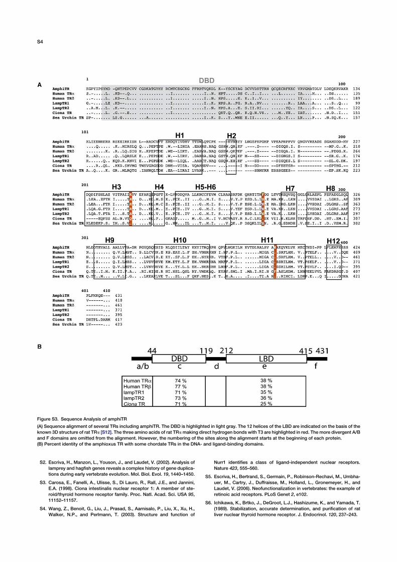
S2. Escriva, H., Manzon, L., Youson, J., and Laudet, V. (2002). Analysis of
lamprey and hagfish genes reveals a complex history of gene duplica-
tions during early vertebrate evolution. Mol. Biol. Evol. 19, 1440–1450.
S3. Carosa, E., Fanelli, A., Ulisse, S., Di Lauro, R., Rall, J.E., and Jannini,
E.A. (1998). Ciona intestinalis nuclear receptor 1: A member of ste-
roid/thyroid hormone receptor family. Proc. Natl. Acad. Sci. USA 95,
11152–11157.
S4. Wang, Z., Benoit, G., Liu, J., Prasad, S., Aarnisalo, P., Liu, X., Xu, H.,
Walker, N.P., and Perlmann, T. (2003). Structure and function of
Nurr1 identifies a class of ligand-independent nuclear receptors.
Nature 423, 555–560.
S5. Escriva, H., Bertrand, S., Germain, P., Robinson-Rechavi, M., Umbha-
uer, M., Cartry, J., Duffraisse, M., Holland, L., Gronemeyer, H., and
Laudet, V. (2006). Neofunctionalization in vertebrates: the example of
retinoic acid receptors. PLoS Genet 2, e102.
S6. Ichikawa, K., Brtko, J., DeGroot, L.J., Hashizume, K., and Yamada, T.
(1989). Stabilization, accurate determination, and purification of rat
liver nuclear thyroid hormone receptor. J. Endocrinol. 120, 237–243.
Figure S3. Sequence Analysis of amphiTR
(A) Sequence alignment of several TRs including amphiTR. The DBD is highlighted in light gray. The 12 helices of the LBD are indicated on the basis of the
known 3D structure of rat TRa [S12]. The three amino acids of rat TRamaking direct hydrogen bonds with T3 are highlighted in red. The more divergent A/B
and F domains are omitted from the alignment. However, the numbering of the sites along the alignment starts at the beginning of each protein.
(B) Percent identity of the amphioxus TR with some chordate TRs in the DNA- and ligand-binding domains.
S4
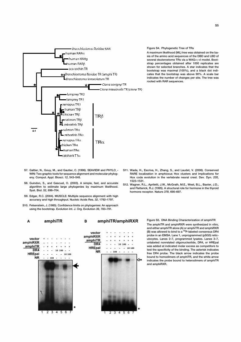
S7. Galtier, N., Gouy, M., and Gautier, C. (1996). SEAVIEW and PHYLO_-
WIN: Two graphic tools for sequence alignment andmolecular phylog-
eny. Comput. Appl. Biosci. 12, 543–548.
S8. Guindon, S., and Gascuel, O. (2003). A simple, fast, and accurate
algorithm to estimate large phylogenies by maximum likelihood.
Syst. Biol. 52, 696–704.
S9. Edgar, R.C. (2004). MUSCLE: Multiple sequence alignment with high
accuracy and high throughput. Nucleic Acids Res. 32, 1792–1797.
S10. Felsenstein, J. (1985). Confidence limits on phylogenies: An approach
using the bootstrap. Evolution Int. J. Org. Evolution 39, 783–791.
S11. Wada, H., Escriva, H., Zhang, S., and Laudet, V. (2006). Conserved
RARE localization in amphioxus Hox clusters and implications for
Hox code evolution in the vertebrate neural crest. Dev. Dyn. 235,
1522–1531.
S12. Wagner, R.L., Apriletti, J.W., McGrath, M.E., West, B.L., Baxter, J.D.,
and Fletterick, R.J. (1995). A structural role for hormone in the thyroid
hormone receptor. Nature 378, 690–697.
Figure S4. Phylogenetic Tree of TRs
A maximum likelihood (ML) tree was obtained on the ba-
sis of the amino acid sequences of the DBD and LBD of
several deuterostome TRs via a WAG+g+I model. Boot-
strap percentages obtained after 1000 replicates are
shown for selected branches. A star indicates that the
bootstrap was maximal (100%), and a black dot indi-
cates that the bootstrap was above 90%. A scale bar
indicates the number of changes per site. The tree was
rooted with RAR sequences.
Figure S5. DNA Binding Characterization of amphiTR
The amphiTR and amphiRXR were synthesized in vitro,
and either amphiTR alone (A) or amphiTR and amphiRXR
(B) was allowed to bind to a 32P-labeled consensus DR4
probe in an EMSA. Lane 1, unprogrammed (pSG5) retic-
ulocytes. Lanes 2–7, programmed lysates. Lanes 3–7,
unlabeled nonrelated oligonucleotide, DR4, or HREpal
was added at indicated molar excess as competitors to
test the specificity of the binding. The asterisk indicates
free DR4 probe. The black arrow indicates the probe
bound to homodimers of amphiTR, and the white arrow
indicates the probe bound to heterodimers of amphiTR
and amphiRXR.
S5
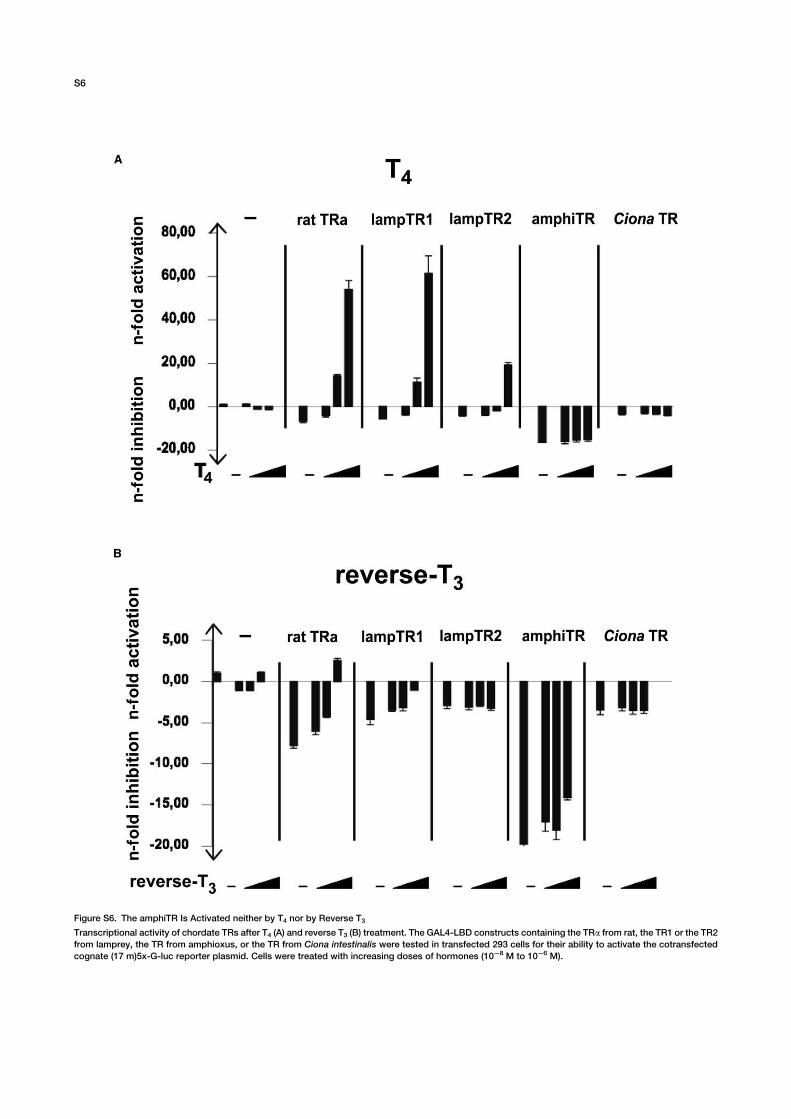
Figure S6. The amphiTR Is Activated neither by T4 nor by Reverse T3
Transcriptional activity of chordate TRs after T4 (A) and reverse T3 (B) treatment. The GAL4-LBD constructs containing the TRa from rat, the TR1 or the TR2
from lamprey, the TR from amphioxus, or the TR from Ciona intestinalis were tested in transfected 293 cells for their ability to activate the cotransfected
cognate (17 m)5x-G-luc reporter plasmid. Cells were treated with increasing doses of hormones (1028 M to 1026 M).
S6
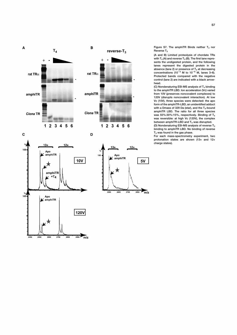
Figure S7. The amphiTR Binds neither T4 nor
Reverse T3
(A and B) Limited proteolysis of chordate TRs
with T4 (A) and reverse T3 (B). The first lane repre-
sents the undigested protein, and the following
lanes represent the digested protein in the
absence (lane 2) or presence of T4 at decreasing
concentrations (1023 M to 1026 M, lanes 3–6).
Protected bands compared with the negative
control (lane 2) are indicated with a black arrow-
head.
(C) Nondenaturing ESI-MS analysis of T4 binding
to the amphiTR LBD. Ion acceleration (Vc) varied
from 10V (preserves noncovalent complexes) to
120V (disrupts noncovalent interaction). At low
Vc (10V), three species were detected: the apo
form of the amphiTR-LBD, an unidentified adduct
with a Dmass of 329 Da (star), and the T4-bound
amphiTR LBD. The ratio for all three species
was 55%:30%:15%, respectively. Binding of T4was reversible: at high Vc (120V), the complex
between amphiTR-LBD and T4 was disrupted.
(D) Nondenaturing ESI-MS analysis of reverse T3binding to amphiTR LBD. No binding of reverse
T3 was found in the gas phase.
For each mass-spectrometry experiment, two
protonation states are shown (13+ and 12+
charge states).
S7
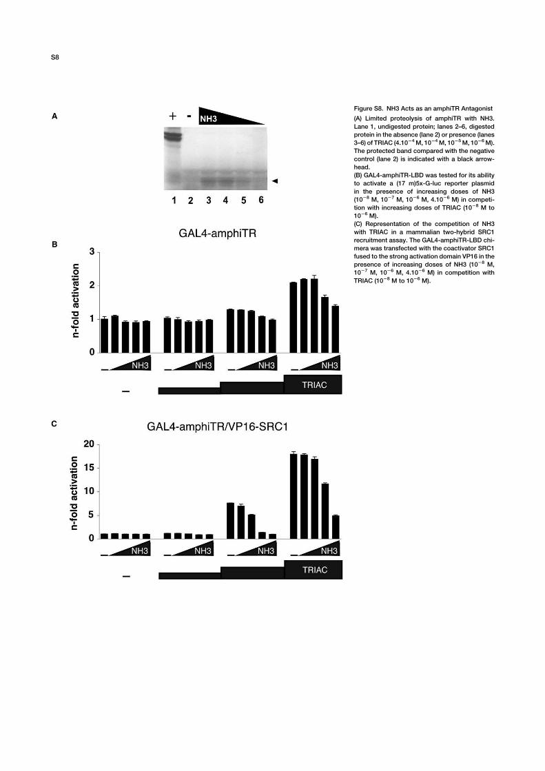
Figure S8. NH3 Acts as an amphiTR Antagonist
(A) Limited proteolysis of amphiTR with NH3.
Lane 1, undigested protein; lanes 2–6, digested
protein in the absence (lane 2) or presence (lanes
3–6) of TRIAC (4.1024M, 1024M, 1025M, 1026M).
The protected band compared with the negative
control (lane 2) is indicated with a black arrow-
head.
(B) GAL4-amphiTR-LBD was tested for its ability
to activate a (17 m)5x-G-luc reporter plasmid
in the presence of increasing doses of NH3
(1028 M, 1027 M, 1026 M, 4.1026 M) in competi-
tion with increasing doses of TRIAC (1028 M to
1026 M).
(C) Representation of the competition of NH3
with TRIAC in a mammalian two-hybrid SRC1
recruitment assay. The GAL4-amphiTR-LBD chi-
mera was transfected with the coactivator SRC1
fused to the strong activation domain VP16 in the
presence of increasing doses of NH3 (1028 M,
1027 M, 1026 M, 4.1026 M) in competition with
TRIAC (1028 M to 1026 M).
S8
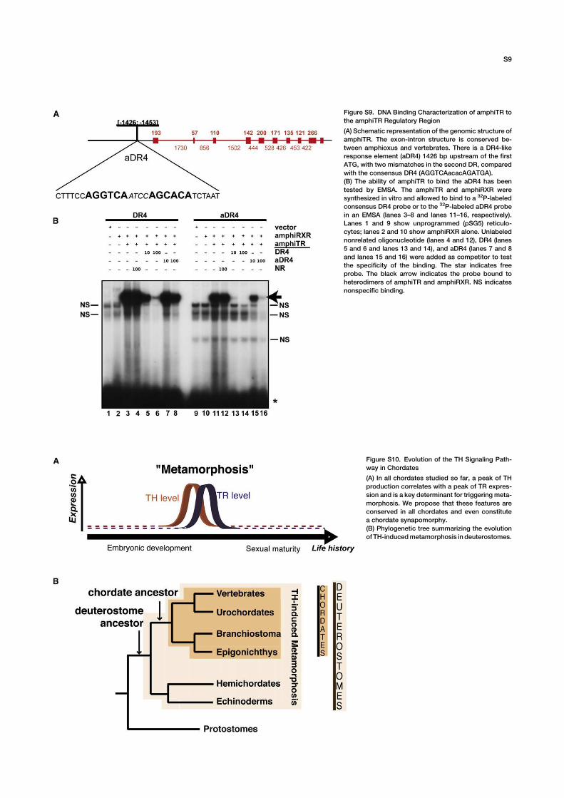
Figure S9. DNA Binding Characterization of amphiTR to
the amphiTR Regulatory Region
(A) Schematic representation of the genomic structure of
amphiTR. The exon-intron structure is conserved be-
tween amphioxus and vertebrates. There is a DR4-like
response element (aDR4) 1426 bp upstream of the first
ATG, with two mismatches in the second DR, compared
with the consensus DR4 (AGGTCAacacAGATGA).
(B) The ability of amphiTR to bind the aDR4 has been
tested by EMSA. The amphiTR and amphiRXR were
synthesized in vitro and allowed to bind to a 32P-labeled
consensus DR4 probe or to the 32P-labeled aDR4 probe
in an EMSA (lanes 3–8 and lanes 11–16, respectively).
Lanes 1 and 9 show unprogrammed (pSG5) reticulo-
cytes; lanes 2 and 10 show amphiRXR alone. Unlabeled
nonrelated oligonucleotide (lanes 4 and 12), DR4 (lanes
5 and 6 and lanes 13 and 14), and aDR4 (lanes 7 and 8
and lanes 15 and 16) were added as competitor to test
the specificity of the binding. The star indicates free
probe. The black arrow indicates the probe bound to
heterodimers of amphiTR and amphiRXR. NS indicates
nonspecific binding.
Figure S10. Evolution of the TH Signaling Path-
way in Chordates
(A) In all chordates studied so far, a peak of TH
production correlates with a peak of TR expres-
sion and is a key determinant for triggering meta-
morphosis. We propose that these features are
conserved in all chordates and even constitute
a chordate synapomorphy.
(B) Phylogenetic tree summarizing the evolution
of TH-inducedmetamorphosis in deuterostomes.
S9
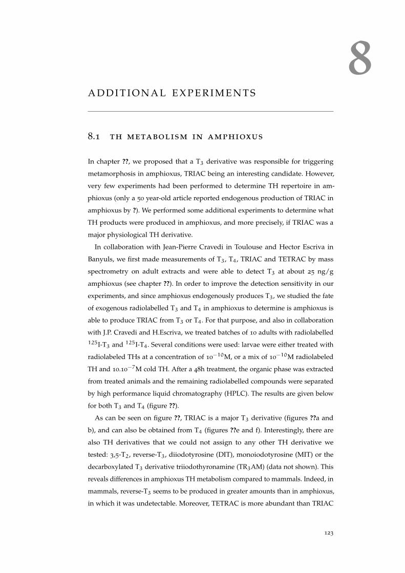
8A D D I T I O N A L E X P E R I M E N T S
8.1 th metabolism in amphioxus
In chapter ??, we proposed that a T3 derivative was responsible for triggering
metamorphosis in amphioxus, TRIAC being an interesting candidate. However,
very few experiments had been performed to determine TH repertoire in am-
phioxus (only a 50 year-old article reported endogenous production of TRIAC in
amphioxus by ?). We performed some additional experiments to determine what
TH products were produced in amphioxus, and more precisely, if TRIAC was a
major physiological TH derivative.
In collaboration with Jean-Pierre Cravedi in Toulouse and Hector Escriva in
Banyuls, we first made measurements of T3, T4, TRIAC and TETRAC by mass
spectrometry on adult extracts and were able to detect T3 at about 25 ng/g
amphioxus (see chapter ??). In order to improve the detection sensitivity in our
experiments, and since amphioxus endogenously produces T3, we studied the fate
of exogenous radiolabelled T3 and T4 in amphioxus to determine is amphioxus is
able to produce TRIAC from T3 or T4. For that purpose, and also in collaboration
with J.P. Cravedi and H.Escriva, we treated batches of 10 adults with radiolabelled125I-T3 and 125I-T4. Several conditions were used: larvae were either treated with
radiolabeled THs at a concentration of 10−10M, or a mix of 10−10M radiolabeled
TH and 10.10−7M cold TH. After a 48h treatment, the organic phase was extracted
from treated animals and the remaining radiolabelled compounds were separated
by high performance liquid chromatography (HPLC). The results are given below
for both T3 and T4 (figure ??).
As can be seen on figure ??, TRIAC is a major T3 derivative (figures ??a and
b), and can also be obtained from T4 (figures ??e and f). Interestingly, there are
also TH derivatives that we could not assign to any other TH derivative we
tested: 3,5-T2, reverse-T3, diiodotyrosine (DIT), monoiodotyrosine (MIT) or the
decarboxylated T3 derivative triiodothyronamine (TR3AM) (data not shown). This
reveals differences in amphioxus TH metabolism compared to mammals. Indeed, in
mammals, reverse-T3 seems to be produced in greater amounts than in amphioxus,
in which it was undetectable. Moreover, TETRAC is more abundant than TRIAC
123
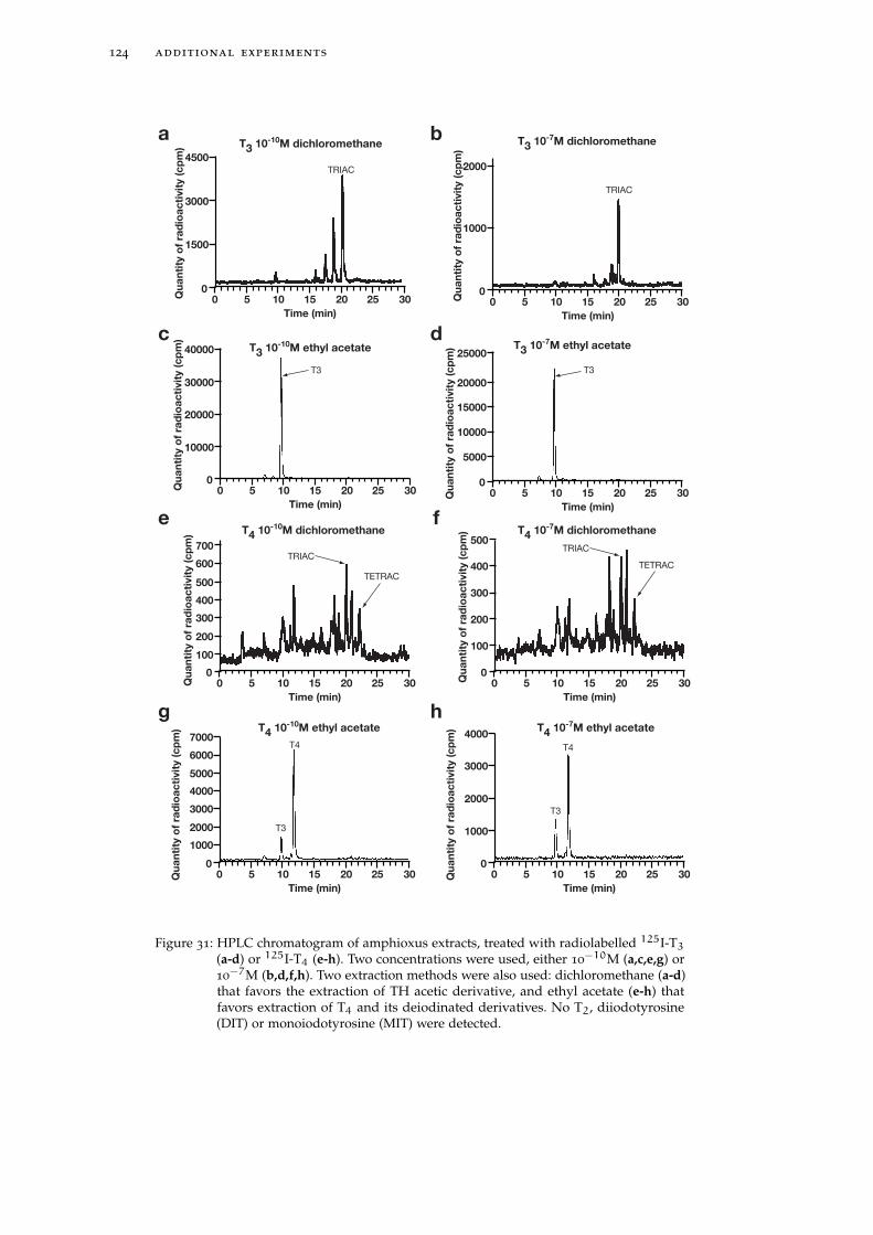
124 additional experiments
00
100
200
300
500
400
5 10
Time (min)
Qu
an
tity
of
rad
ioa
cti
vit
y (
cp
m)
15 20 25 30
TETRAC
TRIAC
T4 10-7M dichloromethaneT4 10-10M dichloromethane
00
1000
2000
3000
4000
5 10
Time (min)
Qu
an
tity
of
rad
ioa
cti
vit
y (
cp
m)
15 20 25 30
T4
T3
T4 10-7M ethyl acetate
Time (min)
00
100
200
300
500
400
700
600
5 10Qu
an
tity
of
rad
ioa
cti
vit
y (
cp
m)
15 20 25 30
TETRAC
TRIAC
00
1000
2000
3000
4000
5000
6000
7000
5 10
Time (min)
Qu
an
tity
of
rad
ioa
cti
vit
y (
cp
m)
15 20 25 30
T4
T3
T4 10-10M ethyl acetate
e f
g h
00
1000
2000
5 10
Time (min)
Qu
an
tity
of
rad
ioa
cti
vit
y (
cp
m)
15 20 25 30
TRIAC
T3 10-7M dichloromethaneT3 10-10M dichloromethane
T3 10-10M ethyl acetate
TRIAC
00
1500
3000
4500
5 10
Time (min)
Qu
an
tity
of
rad
ioa
cti
vit
y (
cp
m)
15 20 25 30
T3 10-7M ethyl acetate
T3
00
10000
20000
30000
40000
5 10
Time (min)
Qu
an
tity
of
rad
ioa
cti
vit
y (
cp
m)
15 20 25 30 00
5000
10000
15000
20000
25000
5 10
Time (min)
Qu
an
tity
of
rad
ioa
cti
vit
y (
cp
m)
15 20 25 30
T3
a b
c d
Figure 31: HPLC chromatogram of amphioxus extracts, treated with radiolabelled 125I-T3
(a-d) or 125I-T4 (e-h). Two concentrations were used, either 10−10M (a,c,e,g) or10−7M (b,d,f,h). Two extraction methods were also used: dichloromethane (a-d)that favors the extraction of TH acetic derivative, and ethyl acetate (e-h) thatfavors extraction of T4 and its deiodinated derivatives. No T2, diiodotyrosine(DIT) or monoiodotyrosine (MIT) were detected.
8.1 th metabolism in amphioxus 125
(?), whereas the situation seems to be reverse in amphioxus. The other peaks
could not be identified. This experiment also suggests that there are active outer-
ring deiodinases in amphioxus, since T3 and TRIAC can be produced from T4
(figures ??e-h). Indeed, T4 to T3 transformation (prior to TRIAC formation) is
done by removing one iodine from the outer ring (figure ??) of T4 by deiodinases.
As inner ring and outer ring deiodinations are two distinct enzymatic reactions
(that may be taken care of by different deiodinases), we can then conclude that
amphioxus possesses deiodinases that carry out outer-ring deiodination, which
is not surprising as we proposed fron the phylogenetic study of the TH signaling
pathway (see chapter ??), and as in urochordates (?).
From this experiment, we can conclude that (i) TRIAC is an important deriva-
tive of amphioxus TH metabolism, which strengthens its possible role during
amphioxus metamorphosis (see chater ??), and (ii) although very close, amphioxus
TH metabolism probably displays differences with vertebrates, which we antici-
pated from our genomic analysis of the TH signaling pathway (chapter ??).
Several important experiments remain to be done. Our conclusions on amphioxus
TH metabolism pertain to adult, not on larval metabolism: we have confirmed that
amphioxus is capable of producing TRIAC but we still do not know whether it does
so at larval stage. More generally, we have not brought to light any clue about TH
production in general during amphioxus metamorphosis: is there a peak of TH
production at the onset of metamorphosis, that accompanies TR expression, as we
proposed in chapter ??? This question will however be difficult to answer. We tried
to detect TH metabolism in larvae using a similar protocol as described above for
adults. However, we could not collect enough biological material and the results
were not conclusive. It will be technically very challenging to collect enough larvae
and do such an experiment. An alternative will be to study the expression of the
genes involved in TH signaling pathway during development and determine if a
peak of expression occurs at metamorphosis (as for deiodinases in amphibians (?)).
This should allow us to confirm or reject our model described in chapter ?? and
discussed in the final discussion (??), according to which a peak of TH production
and TR expression are the triggering events of metamorphosis in amphioxus.
Alternatively and in order to determine whether a peak of TH production
triggers metamorphosis in amphioxus, we studied whether blocking the peak of
TH production inhibits amphioxus metamorphosis. This experiment is described
in the next section.
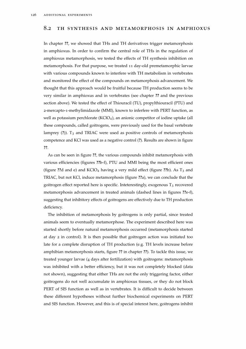
126 additional experiments
8.2 th synthesis and metamorphosis in amphioxus
In chapter ??, we showed that THs and TH derivatives trigger metamorphosis
in amphioxus. In order to confirm the central role of THs in the regulation of
amphioxus metamorphosis, we tested the effects of TH synthesis inhibition on
metamorphosis. For that purpose, we treated 11 day-old premetamorphic larvae
with various compounds known to interfere with TH metabolism in vertebrates
and monitored the effect of the compounds on metamorphosis advancement. We
thought that this approach would be fruitful because TH production seems to be
very similar in amphioxus and in vertebrates (see chapter ?? and the previous
section above). We tested the effect of Thiouracil (TU), propylthiouracil (PTU) and
2-mercapto-1-methylimidazole (MMI), known to inferfere with PERT function, as
well as potassium perchlorate (KClO4), an anionic competitor of iodine uptake (all
these compounds, called goitrogens, were previously used for the basal vertebrate
lamprey (?)). T3 and TRIAC were used as positive controls of metamorphosis
competence and KCl was used as a negative control (?). Results are shown in figure
??.
As can be seen in figure ??, the various compounds inhibit metamorphosis with
various efficiencies (figures ??b-f), PTU and MMI being the most efficient ones
(figure ??d and e) and KClO4 having a very mild effect (figure ??b). As T3 and
TRIAC, but not KCl, induce metamorphosis (figure ??a), we can conclude that the
goitrogen effect reported here is specific. Inteterestingly, exogenous T3 recovered
metamorphosis advancement in treated animals (dashed lines in figures ??e-f),
suggesting that inhibitory effects of goitrogens are effectively due to TH production
deficiency.
The inhibition of metamorphosis by goitrogens is only partial, since treated
animals seem to eventually metamorphose. The experiment described here was
started shortly before natural metamorphosis occurred (metamorphosis started
at day 2 in control). It is then possible that goitrogen action was initiated too
late for a complete disruption of TH production (e.g. TH levels increase before
amphibian metamorphosis starts, figure ?? in chapter ??). To tackle this issue, we
treated younger larvae (4 days after fertilization) with goitrogens: metamorphosis
was inhibited with a better efficiency, but it was not completely blocked (data
not shown), suggesting that either THs are not the only triggering factor, either
goitrogens do not well accumulate in amphioxus tissues, or they do not block
PERT of SIS function as well as in vertebrates. It is difficult to decide between
these different hypotheses without further biochemical experiments on PERT
and SIS function. However, and this is of special interest here, goitrogens inhibit
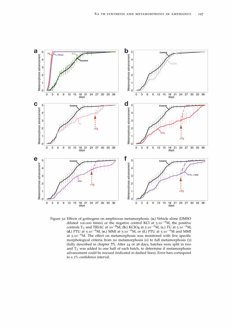
8.2 th synthesis and metamorphosis in amphioxus 127
?
? ?
?
?
?
?
?
?
?
?
?
?
?
?
?
?
?
?
?? ?
?
0
1
2
3
4
5
days
Meta
morp
hosis
advancem
ent
0 3 6 9 12 15 18 21 24 27 30 33 36
TRIACT3
?
? ?
?
?
?
?
?
?
?
?
?
?
?
?
?
?
?
?
?? ?
?
Control
KCl
?
? ?
?
?
?
?
?
?
?
?
?
?
?
?
?
?
?
?
?? ?
?
0
1
2
3
4
5
days
Meta
morp
hosis
advancem
ent
0 3 6 9 12 15 18 21 24 27 30 33 36
Control
KClO4
?
? ?
?
?
?
?
?
?
?
?
?
?
?
?
?
?
?
?
?? ?
?
0
1
2
3
4
5
days
Meta
morp
hosis
advancem
ent
0 3 6 9 12 15 18 21 24 27 30 33 36
Control
PTU + MMI
?
? ?
?
?
?
?
?
?
?
?
?
?
?
?
?
?
?
?
?? ?
?
0
1
2
3
4
5
days
Meta
morp
hosis
advancem
ent
0 3 6 9 12 15 18 21 24 27 30 33 36
Control
MMI
?
? ?
?
?
?
?
?
?
?
?
?
?
?
?
?
?
?
?
?? ?
?
0
1
2
3
4
5
days
Meta
morp
hosis
advancem
ent
0 3 6 9 12 15 18 21 24 27 30 33 36
Control
PTU
?
? ?
?
?
?
?
?
?
?
?
?
?
?
?
?
?
?
?
?? ?
?
0
1
2
3
4
5
days
Meta
morp
hosis
advancem
ent
0 3 6 9 12 15 18 21 24 27 30 33 36
Control
TU
+T3
+T3
+T3
+T3
a
c
e
b
d
f
Figure 32: Effects of goitrogens on amphioxus metamorphosis. (a.) Vehicle alone (DMSOdiluted 100.000 times) or the negative control KCl at 5.10−4M, the positivecontrols T3 and TRIAC at 10−8M, (b.) KClO4 at 5.10−4M, (c.) TU at 5.10−4M,(d.) PTU at 5.10−4M, (e.) MMI at 5.10−4M, or (f.) PTU at 5.10−4M and MMIat 5.10−4M. The effect on metamorphosis was monitored with five specificmorphological criteria from no metamorphosis (0) to full metamorphosis (5)(fully described in chapter ??). After 24 or 26 days, batches were split in twoand T3 was added to one half of each batch, to determine if metamorphosisadvancement could be rescued (indicated in dashed lines). Error bars correspondto a 5% confidence interval.
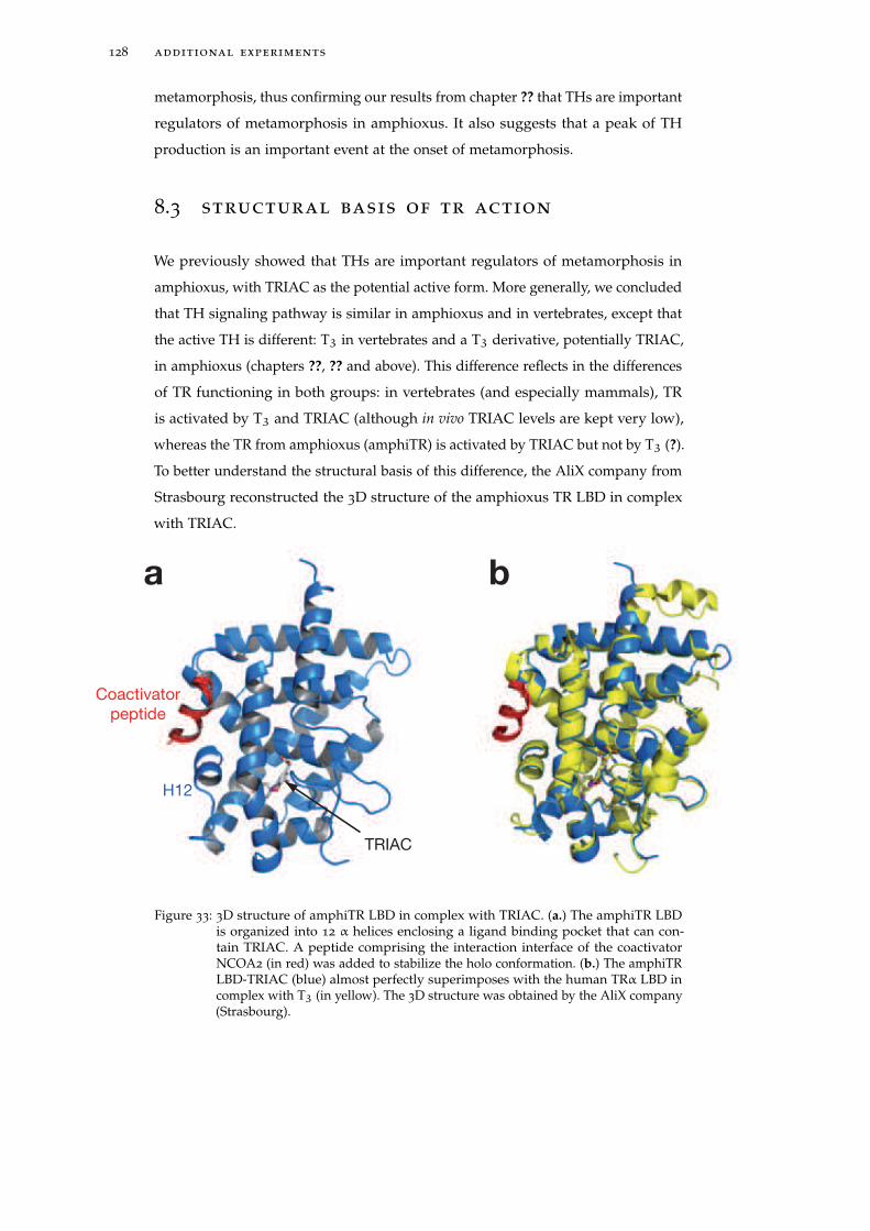
128 additional experiments
metamorphosis, thus confirming our results from chapter ?? that THs are important
regulators of metamorphosis in amphioxus. It also suggests that a peak of TH
production is an important event at the onset of metamorphosis.
8.3 structural basis of tr action
We previously showed that THs are important regulators of metamorphosis in
amphioxus, with TRIAC as the potential active form. More generally, we concluded
that TH signaling pathway is similar in amphioxus and in vertebrates, except that
the active TH is different: T3 in vertebrates and a T3 derivative, potentially TRIAC,
in amphioxus (chapters ??, ?? and above). This difference reflects in the differences
of TR functioning in both groups: in vertebrates (and especially mammals), TR
is activated by T3 and TRIAC (although in vivo TRIAC levels are kept very low),
whereas the TR from amphioxus (amphiTR) is activated by TRIAC but not by T3 (?).
To better understand the structural basis of this difference, the AliX company from
Strasbourg reconstructed the 3D structure of the amphioxus TR LBD in complex
with TRIAC.
a b
H12
TRIAC
Coactivator
peptide
Figure 33: 3D structure of amphiTR LBD in complex with TRIAC. (a.) The amphiTR LBDis organized into 12 α helices enclosing a ligand binding pocket that can con-tain TRIAC. A peptide comprising the interaction interface of the coactivatorNCOA2 (in red) was added to stabilize the holo conformation. (b.) The amphiTRLBD-TRIAC (blue) almost perfectly superimposes with the human TRα LBD incomplex with T3 (in yellow). The 3D structure was obtained by the AliX company(Strasbourg).
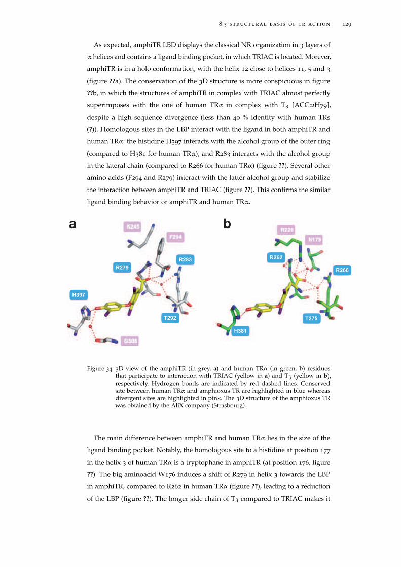
8.3 structural basis of tr action 129
As expected, amphiTR LBD displays the classical NR organization in 3 layers of
α helices and contains a ligand binding pocket, in which TRIAC is located. Morever,
amphiTR is in a holo conformation, with the helix 12 close to helices 11, 5 and 3
(figure ??a). The conservation of the 3D structure is more conspicuous in figure
??b, in which the structures of amphiTR in complex with TRIAC almost perfectly
superimposes with the one of human TRα in complex with T3 [ACC:2H79],
despite a high sequence divergence (less than 40 % identity with human TRs
(?)). Homologous sites in the LBP interact with the ligand in both amphiTR and
human TRα: the histidine H397 interacts with the alcohol group of the outer ring
(compared to H381 for human TRα), and R283 interacts with the alcohol group
in the lateral chain (compared to R266 for human TRα) (figure ??). Several other
amino acids (F294 and R279) interact with the latter alcohol group and stabilize
the interaction between amphiTR and TRIAC (figure ??). This confirms the similar
ligand binding behavior or amphiTR and human TRα.
a b
H381
H397
R279
R283
T292 T275
R266
R262
Figure 34: 3D view of the amphiTR (in grey, a) and human TRα (in green, b) residuesthat participate to interaction with TRIAC (yellow in a) and T3 (yellow in b),respectively. Hydrogen bonds are indicated by red dashed lines. Conservedsite between human TRα and amphioxus TR are highlighted in blue whereasdivergent sites are highlighted in pink. The 3D structure of the amphioxus TRwas obtained by the AliX company (Strasbourg).
The main difference between amphiTR and human TRα lies in the size of the
ligand binding pocket. Notably, the homologous site to a histidine at position 177
in the helix 3 of human TRα is a tryptophane in amphiTR (at position 176, figure
??). The big aminoacid W176 induces a shift of R279 in helix 3 towards the LBP
in amphiTR, compared to R262 in human TRα (figure ??), leading to a reduction
of the LBP (figure ??). The longer side chain of T3 compared to TRIAC makes it
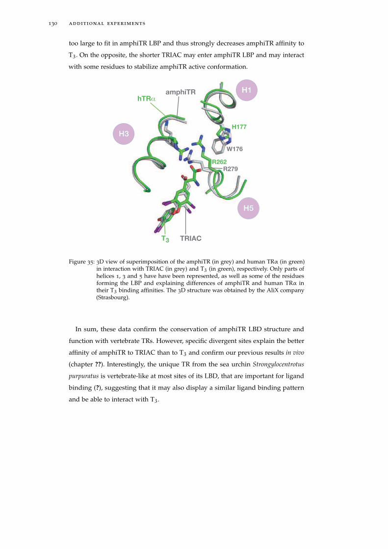
130 additional experiments
too large to fit in amphiTR LBP and thus strongly decreases amphiTR affinity to
T3. On the opposite, the shorter TRIAC may enter amphiTR LBP and may interact
with some residues to stabilize amphiTR active conformation.
H3
H1
H5
hTRα
T3
H177
R262
amphiTR
W176
R279
TRIAC
Figure 35: 3D view of superimposition of the amphiTR (in grey) and human TRα (in green)in interaction with TRIAC (in grey) and T3 (in green), respectively. Only parts ofhelices 1, 3 and 5 have have been represented, as well as some of the residuesforming the LBP and explaining differences of amphiTR and human TRα intheir T3 binding affinities. The 3D structure was obtained by the AliX company(Strasbourg).
In sum, these data confirm the conservation of amphiTR LBD structure and
function with vertebrate TRs. However, specific divergent sites explain the better
affinity of amphiTR to TRIAC than to T3 and confirm our previous results in vivo
(chapter ??). Interestingly, the unique TR from the sea urchin Strongylocentrotus
purpuratus is vertebrate-like at most sites of its LBD, that are important for ligand
binding (?), suggesting that it may also display a similar ligand binding pattern
and be able to interact with T3.
9A N A M P H I O X U S O RT H O L O G U E O F T H EE S T R O G E N R E C E P T O R T H AT D O E S N O TB I N D E S T R A D I O L : I N S I G H T S I N T O E RE V O L U T I O N
Nuclear hormone receptors have their activity regulated upon ligand binding,
a phenomenon that allows them to interact with the environment and also to
regulate transcription. How this dual function arose during evolution deserves
some insight. There is little doubt that all NRs are homologous (?) and evolved
from a common ancestor to the diversity encountered now (?). If one considers NR
modular organization (as depicted in chapter ??), two domains, the DBD and the
LBD are of particular interest.
The evolution of DNA binding ability is rather straightforward: almost all
NRs have very close DNA binding properties. Indeed all DBDs interact with DNA
through zinc-fingers and bind very similar combinations of more or less degenerate
(AGGTCA) motifs (see section ??). Differences lie in binding site specificity, whose
alteration may occur through point mutations (which are rather limited considering
the similarities between specificities between different NRs). It was proposed that
this zinc-finger DBD, that is specific to NRs, may be homologous with the DNA
binding elements from LIM and GATA proteins, that are found in both metazoans
and yeast (??). Moreover, NRs typically bind DNA as dimers, which confers
most of DNA binding specificity. Then dimerization is probably ancestral and its
evolution may be of greater importance than the evolution the the DBD regarding
the evolution of DNA binding ability. Interestingly, some NRs are able to interact
with several other members of the family (e.g. TR can interact with VDR or RXR,
or itself). This conservative evolution suggests that the ancestral NR was a zinc-
finger transcription factor that bound DNA as a dimer (as a homodimer or a
heterodimer) on motifs around the canonical (AGGTCA) one. Moreover, as most
NRs are transcription factors, the ancestral NRs were most probably transcription
factors.
In contrast, the evolution of ligand binding ability and more generally of tran-
scriptional activation, is more elusive and has been discussed in the past decade
(e.g. (?????)). LBDs function in diverse ways, so it is difficut to elaborate upon the
evolution of ligand binding. First, and as mentioned in the introduction (section
??), a large portion of NRs are still orphan, meaning that no known ligand has
131
132 evolution of er binding
been identified so far. Some NRs may have a ligand in vivo that has not been
identified yet (despite extensive search, like for ERR (?)). However the activity of
others may be regulated by ligand-independent mechanisms. For instance Nurr1
has no ligand binding pocket (some may talk about a "filled" pocket), and its
activity is regulated by post-translational modifications like phosphorylation (?).
Other orphan receptors, such as some USPs (the insect RXR orthologue), do have a
pocket but a USP-specific loop interacts with the LBP and prevents ligand binding
(?). Second, ligand-activated receptors show important ligand binding differences.
Some NRs, like TR, display a high affinity and a highly selectivity for their ligand.
Accordingly, they have a small and constrained ligand binding pocket. Alterna-
tively, other NRs like LXR have larger pockets combined to a wider repertoire of
low affinity ligands (?). Notably, the NR PXR binds to a large diversity of foreign
compounds and is involved in detoxification (?). A last category of NRs have a
non-exchangeable ligand (referred to as constitutive, e.g. RORα and cholesterol).
Consequently transcriptional activity the receptor is modulated by other means
(e.g. expression, post-translational modifications...) (?). How did this diversity
evolve, and what were the ligand binding properties of the ancestral receptors?
The above diversity of ligand binding profiles should be put in a phylogenetic
perspective. Orphan receptors, high-affinity and low-affinity NRs are not grouped
together but instead are scattered along the NR tree (figure ??). Moreover, the
chemical nature of ligands does not fit NR phylogeny either. Indeed, closely
related NRs may bind chemically very different ligands (e.g. TR and RAR) whereas
distantly related NRs may bind similar ligands (ecdysteroid receptors and other
sex steroid receptors, figure ??). Overall, considering NR phylogeny does not help
to understand the distribution of NR ligand binding characteristics.
How this ligand binding situation evolved is then far from clear. Several hy-
potheses have been presented. One proposes that the ancestral receptor was a
high affinity receptor that subsequently evolved new binding affinities after gene
duplication and divergence (??). In contrast, ligand binding may also have been
secondarily acquired from an orphan ancestor (??) that subsequently gained the
ability to be regulated upon ligand binding.
The following article is in accord with previous works on the evolution of NR
ligand binding. More precisely, it deals with the evolution of estrogen binding in
the NR3 subfamily (see figure ??, as well as figure ?? in chapter ??). The article
is divided into two parts. First, we used a comparative approach and focused on
the lamprey and amphioxus estrogen receptors (ER): we characterized their DNA
and ligand binding as well as their transactivation abilities to show that lamprey
ER behaves as a "classical" vertebrate ER whereas the amphioxus ER is not an
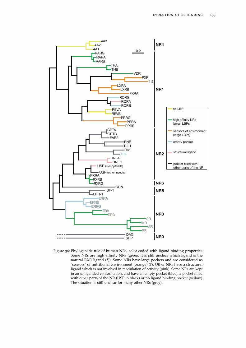
evolution of er binding 133
PRAR
MRGR
ERBERA
ERRGERRB
ERRALRH-1
SF-1GCN
RXRGRXRB
RXRAUSP (other insects)
USP (mecopterida)
HNFGHNFA
TR4TR2TLL1PNR
EAR2CPTBCPTA
PPRBPPRA
PPRGREVBREVA
RORBRORA
RORGFXRA
LXRBLXRA
1I3PXR
VDRTHBTHA
RARBRARA
RARG4A14A2
4A3
SHPDAX
NR1
NR0
NR3
NR2
NR5
NR6
NR4
0.2
high affinity NRs
(small LBPs)
no LBP
empty pocket
sensors of environment
(large LBPs)
pocket filled with
other parts of the NR
structural ligand
Figure 36: Phylogenetic tree of human NRs, color-coded with ligand binding properties.Some NRs are high affinity NRs (green, it is still unclear which ligand is thenatural RXR ligand (?)). Some NRs have large pockets and are considered as"sensors" of nutritional environment (orange) (?). Other NRs have a structuralligand which is not involved in modulation of activity (pink). Some NRs are keptin an unliganded conformation, and have an empty pocket (blue), a pocket filledwith other parts of the NR (USP in black) or no ligand binding pocket (yellow).The situation is still unclear for many other NRs (grey).

134 evolution of er binding
estrogen receptor. In a second time, we used computational methods to discuss
the pros and cons of an approach different from comparative endocrinology:
instead of comparing ERs from extant species to understand the evolution of ER
ligand binding ability, one may predict in silico ancestral sequences, "resurrect" and
molecularly characterize them, to infer biochemical properties of the real ancestral
ERs.
This article was published in BMC Evolutionary Biology.
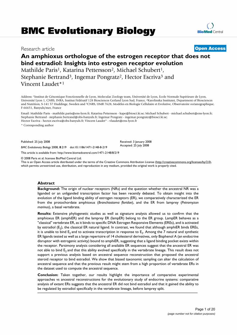
BioMed Central
Page 1 of 20
(page number not for citation purposes)
BMC Evolutionary Biology
Open AccessResearch article
An amphioxus orthologue of the estrogen receptor that does not bind estradiol: Insights into estrogen receptor evolutionMathilde Paris1, Katarina Pettersson2, Michael Schubert1, Stephanie Bertrand3, Ingemar Pongratz2, Hector Escriva3 and Vincent Laudet*1
Address: 1Institut de Génomique Fonctionnelle de Lyon, Molecular Zoology team, Université de Lyon, Ecole Normale Supérieure de Lyon, Université Lyon 1, CNRS, INRA, Institut Fédératif 128 Biosciences Gerland Lyon Sud, France, 2Karolinska Institutet, Department of Biosciences and Nutrition, S-141 57 Huddinge, Sweden and 3CNRS, UMR 7628, Modèles en Biologie Cellulaire et Evolutive, Observatoire océanographique, F-66651, Banyuls/mer, France
Email: Mathilde Paris - [email protected]; Katarina Pettersson - [email protected]; Michael Schubert - [email protected]; Stephanie Bertrand - [email protected]; Ingemar Pongratz - [email protected]; Hector Escriva - [email protected]; Vincent Laudet* - [email protected]
* Corresponding author
Abstract
Background: The origin of nuclear receptors (NRs) and the question whether the ancestral NR was a
liganded or an unliganded transcription factor has been recently debated. To obtain insight into the
evolution of the ligand binding ability of estrogen receptors (ER), we comparatively characterized the ER
from the protochordate amphioxus (Branchiostoma floridae), and the ER from lamprey (Petromyzon
marinus), a basal vertebrate.
Results: Extensive phylogenetic studies as well as signature analysis allowed us to confirm that the
amphioxus ER (amphiER) and the lamprey ER (lampER) belong to the ER group. LampER behaves as a
"classical" vertebrate ER, as it binds to specific DNA Estrogen Responsive Elements (EREs), and is activated
by estradiol (E2), the classical ER natural ligand. In contrast, we found that although amphiER binds EREs,
it is unable to bind E2 and to activate transcription in response to E2. Among the 7 natural and synthetic
ER ligands tested as well as a large repertoire of 14 cholesterol derivatives, only Bisphenol A (an endocrine
disruptor with estrogenic activity) bound to amphiER, suggesting that a ligand binding pocket exists within
the receptor. Parsimony analysis considering all available ER sequences suggest that the ancestral ER was
not able to bind E2 and that this ability evolved specifically in the vertebrate lineage. This result does not
support a previous analysis based on ancestral sequence reconstruction that proposed the ancestral
steroid receptor to bind estradiol. We show that biased taxonomic sampling can alter the calculation of
ancestral sequence and that the previous result might stem from a high proportion of vertebrate ERs in
the dataset used to compute the ancestral sequence.
Conclusion: Taken together, our results highlight the importance of comparative experimental
approaches vs ancestral reconstructions for the evolutionary study of endocrine systems: comparative
analysis of extant ERs suggests that the ancestral ER did not bind estradiol and that it gained the ability to
be regulated by estradiol specifically in the vertebrate lineage, before lamprey split.
Published: 25 July 2008
BMC Evolutionary Biology 2008, 8:219 doi:10.1186/1471-2148-8-219
Received: 3 January 2008Accepted: 25 July 2008
This article is available from: http://www.biomedcentral.com/1471-2148/8/219
© 2008 Paris et al; licensee BioMed Central Ltd. This is an Open Access article distributed under the terms of the Creative Commons Attribution License (http://creativecommons.org/licenses/by/2.0), which permits unrestricted use, distribution, and reproduction in any medium, provided the original work is properly cited.
BMC Evolutionary Biology 2008, 8:219 http://www.biomedcentral.com/1471-2148/8/219
Page 2 of 20
(page number not for citation purposes)
BackgroundHormone signaling is a very important feature in metazo-ans, allowing communication between cells or organswithin the organism. Two components of these signalingsystems are of particular importance, the hormone and itsreceptor. The nuclear hormone receptor (NR) superfamilyincludes ligand dependent transcription factors that playa central role in various physiological processes as diverseas reproduction, development, and control of homeosta-sis [1,2]. They share a common structural organizationand exhibit a highly conserved DNA binding domain(DBD) and a moderately conserved ligand-bindingdomain (LBD). Some members of this superfamily are lig-anded receptors (24 among the 48 genes encoding NRs inthe human genome) but many lack identified ligand andare therefore called "orphan" [3]. Some orphan receptorsare 'true' orphans in the sense that they do not possess abona fide ligand-binding pocket (LBP), like the membersof the NR4 subfamily (for instance, NURR1, DHR38 orNGFI-B. For review, see [4]), and are regulated by othermechanisms [4]. Alternatively, the crystal structures ofseveral orphan receptors such as HNF4 were found tohave a phospholipid constitutively bound to a large lig-and binding pocket [5,6]. The functional and evolution-ary implications of these constitutive ligands remaindiscussed. Other orphan nuclear receptors have a ligandbinding pocket and thus have the potential to bind com-pounds. It is still not known whether those receptors havenatural ligands, still to be discovered. Undoubtedly, theexistence of such orphan receptors with physiological ordevelopmental activities constitutes both a major chal-lenge for understanding nuclear receptor evolution and apotential opportunity for pharmacology [1].
The existence of orphan and liganded members in the NRfamily raises the question of the evolution regarding theirligand binding ability. Whether the ancestral NR was lig-anded or orphan and more generally how NR ligand bind-ing ability evolved has been recently debated [7-14]. Ingeneral, it is still unclear if there is a correlation betweenthe evolution of the hormone repertoire and NRs. Moreo-ver the mechanisms underlying this coevolution are ofparticular interest [7,12,15-19].
Among the scenarios of NR evolution that have been pro-posed, one suggests that the ancestral NR was a ligand-independent transcription factor which acquired the abil-ity to be regulated by ligands several times during evolu-tion [7,18-20]. This hypothesis was based on theobservation that compounds of similar chemical naturebind to divergent NRs and on the contrary compounds ofvery different nature bind to closely related receptors. Forinstance, orphan receptors are found in all families ofNRs, and steroid receptors are not monophyletic but arelocated in two different subfamilies within the NR super-
family: the ecdysteroid as well as the sex steroid receptors.Interestingly, the evolution of sex steroid hormone recep-tors has also been used as an argument for an alternativehypothesis, the ligand exploitation model [8,11] (for analternative view, see [21,22]). Phylogenetic trees showthat sex steroid hormone receptors are grouped with ERRsas the NR3 subfamily, following the official nomenclature[23]. They contain receptors that bind estradiol (ERs), thatform the NR3A group as well as mineralocorticoids(MRs), glucocorticoids (GRs), progesterone (PRs), andandrogen (ARs) that form the NR3C group. All known lig-ands in this subfamily can be seen as variations aroundthe archetypical sterol skeleton. Consequently, Thorntonet al. suggested in the ligand exploitation model that theancestral steroid receptor was a high affinity estradiolreceptor [8,11] and the other steroid receptors that origi-nated later on, experienced, following gene duplication,shifts in their binding affinities to eventually bind to theirextant ligand. The model in fact suggests that the newlyduplicated receptors (here NR3C) exploit as ligandschemical species that serve as intermediary compounds inthe "ancestral ligand" synthesis pathway (here the estra-diol synthesis pathway) [8]. According to this view,orphan receptors, like ERRs, secondarily lost the ability tohave their activity regulated by a ligand and becameorphan. Interestingly, within the NR3 family, two receptorsubfamilies, ERRs and ERs, appear to be ancient since theyare found in a wide variety of metazoans including deuter-ostomes and protostomes, whereas, up to now, MRs, GRs,PRs and ARs have been found only in vertebrates. Theonly non-vertebrate ERs that have been described so farwere from mollusks and were shown to be unable to bindestradiol [11,12,24-26]. Since the ligand exploitationmodel implies an ancestral estradiol-binding ER and sinceall liganded ER found so far come from vertebrates, and toimprove taxonomic sampling, the ER orthologues fromthe basal vertebrate lamprey and the invertebrate chordateamphioxus were characterized here. Indeed, lamprey andamphioxus are located at key positions in the chordatephylum [27-30]. Moreover, amphioxus (Brachiostoma flor-idae) is much less derived than urochordates in its mor-phology as well as in its genome organization [30].Indeed, amphioxus and vertebrates share a similar generalbody plan whereas urochordate morphology is morederived. For instance, during metamorphosis of some uro-chordates, the tadpole-like larva transforms into an adultthat looks so different that it was first considered as a mol-lusk [31]. Moreover the urochordate genome is fast evolv-ing [27], with for instance the loss of the clustering of thehox genes [32]. There is no ER in the sequenced genomeof Ciona intestinalis [33] or in the sea urchin [34], one ERwas previously cloned in lamprey [8], only one ER wasfound in the amphioxus genome [35]. These reasonsmake lamprey and amphioxus excellent models to studythe evolution of estrogen signaling pathway at the origin
BMC Evolutionary Biology 2008, 8:219 http://www.biomedcentral.com/1471-2148/8/219
Page 3 of 20
(page number not for citation purposes)
of vertebrates. In this study, we cloned the unique ER fromamphioxus (amphiER) and characterized it, as well as thepreviously cloned but uncharacterized lamprey ER (lam-pER). AmphiER is an orphan receptor, showing no affinityto the estrogen hormone estradiol, when in contrast, thelamprey ER behaves as a "classical" vertebrate ER. As noER from invertebrates studied so far binds estradiol, wepropose that the ancestral ER (and the ancestral steroidreceptor) was not a receptor for estradiol and gained lateron during evolution the ability to bind the hormone.
ResultsCloning of the ER from amphioxus (amphiER)
Using degenerate primers designed to match motifs in themost conserved part of vertebrate ERs in the DNA bindingdomain, a single gene fragment from total RNA of anadult Branchiostoma floridae was amplified, cloned andsequenced. Rapid amplification of cDNA ends (RACE)was utilized to obtain the full-length cDNA. From thissequence, a new set of specific primers were designed andused to amplify the full length open reading frame of thisgene. The obtained cDNA [GenBank: ACF16007] is 2118bp long and encodes a 705 aa long putative protein (Fig-ure 1) that harbors the classical features of an ER with the5 main functional domains (Figure 2A), among which ahighly conserved DNA binding domain (DBD) and a lessconserved ligand binding domain (LBD). The DBD sharesan 82% sequence identity with the human ERα one (83%with human ERβ) and much less with the other NR3receptors (<62%). The same pattern is observed for theLBD, although this domain is less conserved since itexhibits only 34% amino acid identity with human ERα(35% for human ERβ) and about 20% with other steroidreceptors (Figure 2A). The three other domains, namelythe A/B region in the N-terminal part, the hinge betweenDBD and LBD, and the short C-terminal end of the pro-tein, are more divergent, which is a general pattern forNRs [2] (Figure 2A). The recent release of the amphioxusgenome confirmed the presence of a single ER gene [35].In contrast the previously described lamprey ER is moresimilar to the human ERα with its DBD sharing a 93%sequence identity (93% for human ERβ) and its LBD shar-ing 55% sequence identity (56% for human ERβ) [8].
Phylogenetic analysis of ERs
The orthology relationships of the amphioxus and lam-prey ER sequences were studied in a phylogenetic analysisof the NR3 family using an exhaustive dataset comprising69 members of the NR3 subfamily as well as sequences ofRXRs as an outgroup. The dataset included the 6 currentlyknown mollusk ER sequences (from Nucella lapillus,Crassostrea gigas, Marisa cornuarietis, Thais clavigera, Octo-pus vulgaris, Aplysia californica), as well as the 2 NR3sequences previously known from amphioxus (1 ERR[GenBank: AAU88062] and 1 NR3C [JGI: 201600],
retrieved from a previous work [36] or from the completegenome sequence [35]). In the resulting phylogenetic tree,the sequence of lamprey ER branches within the ER cladewith a high bootstrap support (95%), at the expectedposition before the split of vertebrate ERα and ERβ (Figure2B, and for a tree presenting all sequences, see Additionalfile 1A), as previously shown [8]. The sequence of amph-iER branches within the ERs and is located at the base ofthe vertebrate estrogen receptor group, before the split ofERα and ERβ but after the split of the mollusk ERs (boot-strap value of 81%, Figure 2B). However its precise posi-tion within the ER group is poorly supported (bootstrapvalue of 42%).
Such low bootstrap supports reveal either the weakness ofthe phylogenetic signal contained in ER proteins, or thepresence of two incompatible signals in the data, one sup-porting the observed position of amphiER within ERs,and the other supporting another position. Whereas theweakness of the signal is not testable, the long branchleading to amphiER in the tree suggests that sites that haveundergone a large number of substitutions may accountfor one of the two signals. Such sites may be saturated tothe point that phylogenetic methods are not able to cor-rectly recover their evolution, a situation leading to thelong branch attraction artifact [37]. It is therefore impor-tant to correctly characterize sites that support the ER posi-tion of amphiER: if only fast-evolving sites support thishypothesis, it is probably due to long branch attraction,and an alternative branching should be favored. Alterna-tively, if slowly-evolving sites support this position, onecan confidently identify amphiER as a bona fide ER. Tocharacterize sites with respect to their evolutionary ratesand the amphiER position they favor, both site likeli-hoods and site evolutionary rates were computed for allpossible positions of amphiER.
First, AmphiER was pruned from the tree shown in Figure2B, and then re-grafted in all 149 remaining branches.This yielded 149 topologies, for which site likelihoodsand site evolutionary rates could be computed usingPhyML-aLRT. This allowed us to obtain an evolutionaryrate per site averaged over all possible positions of amph-iER, and therefore independent from the precise positionof amphiER in the tree. Additionally, as likelihoods werecomputed for each of the 149 positions, these positionscould be compared according to the Approximately Unbi-ased test (AU test, implemented in Consel [38]). Out of allthe 149 resulting trees, 26 could not be distinguished withthe AU test and had a likelihood significantly better thanall the other ones (p-value > 0,05). Of these 26 topologies,all but three place amphiER within the ER clade ("ER"trees). The remaining topologies ("alter-ER" trees) placeamphiER either at the base of the NR3C clade (comprisingthe ARs, PRs, MRs and GRs), within the NR3C or at the
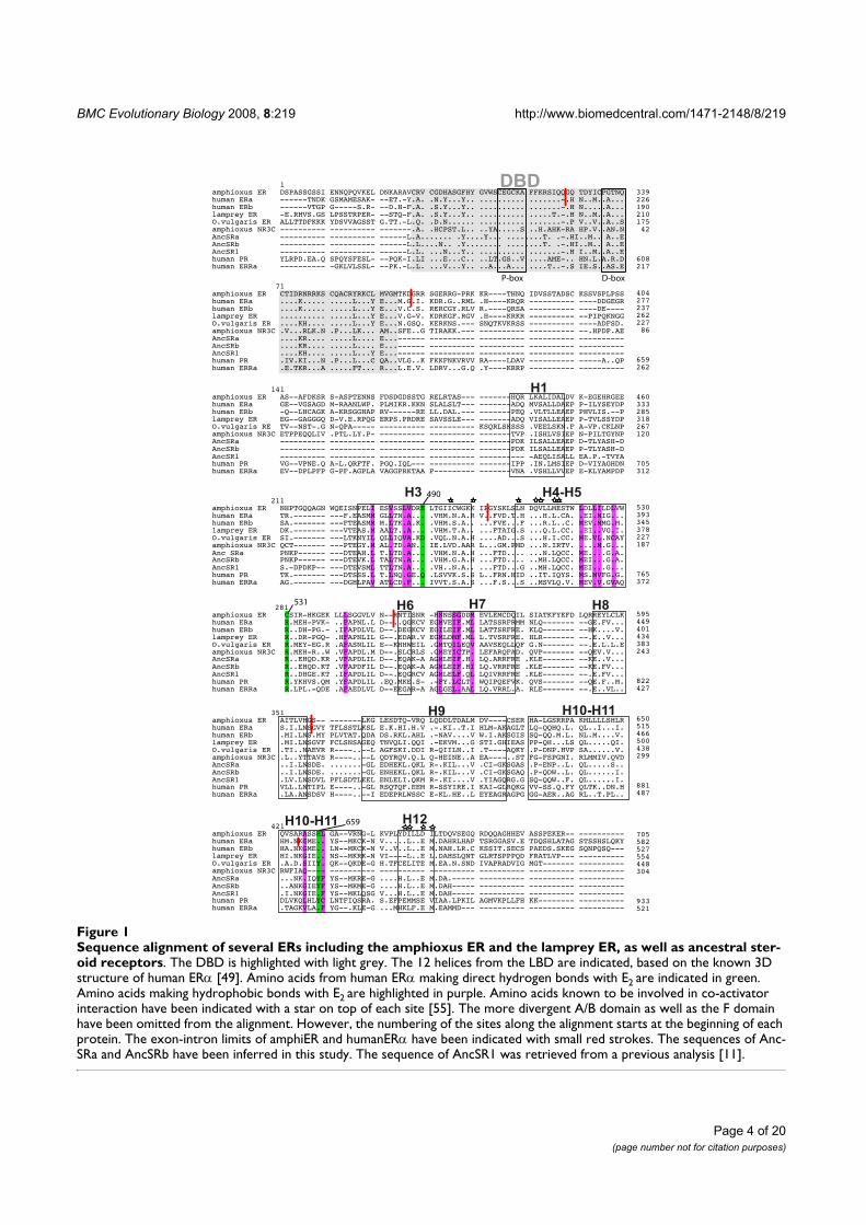
BMC Evolutionary Biology 2008, 8:219 http://www.biomedcentral.com/1471-2148/8/219
Page 4 of 20
(page number not for citation purposes)
Sequence alignment of several ERs including the amphioxus ER and the lamprey ER, as well as ancestral steroid receptorsFigure 1Sequence alignment of several ERs including the amphioxus ER and the lamprey ER, as well as ancestral ster-oid receptors. The DBD is highlighted with light grey. The 12 helices from the LBD are indicated, based on the known 3D structure of human ERα [49]. Amino acids from human ERα making direct hydrogen bonds with E2 are indicated in green. Amino acids making hydrophobic bonds with E2 are highlighted in purple. Amino acids known to be involved in co-activator interaction have been indicated with a star on top of each site [55]. The more divergent A/B domain as well as the F domain have been omitted from the alignment. However, the numbering of the sites along the alignment starts at the beginning of each protein. The exon-intron limits of amphiER and humanERα have been indicated with small red strokes. The sequences of Anc-SRa and AncSRb have been inferred in this study. The sequence of AncSR1 was retrieved from a previous analysis [11].
DBD
H1
H10-H11 H12
H9 H10-H11
H7 H8H6
339226190210175 42
608217
404277237262227 86
659262
460333285318267120
705312
530393345378227187
765372
595449401434383243
822427
650515466500438299
881487
705582527554448304
933521
H3 H4-H5
1amphioxus ER DSPASSGSSI ENNQPQVKEL DNKARAVCRV CGDHASGFHY GVWSCEGCKA FFKRSIQQGQ TDYICPGTNQhuman ERa ------TNDK GSMAMESAK- --ET.-Y.A. .N.Y...Y.. .......... .......-.H N..M..A...human ERb ------VTGP G-----S.R- --D.H-F.A. .S.Y...Y.. .......... .......-.H N.....A...lamprey ER -E.RHVS.GS LPSSTRPER- --STQ-F.A. .S.Y...Y.. .......... .....T.-.H N..M..A...O.vulgaris ER ALLTTDFKKK YDSVVAGSST G.TT.-L.Q. .D.N...... .......... .......-.P V..V..A..Samphioxus NR3C ---------- ---------- -------.A. .HCPST.L.. ..YA.....S ..H.AHK-RA HP.V..AN.N
human PR YLRPD.EA.Q SPQYSFESL- --PQK-I.LI ...E...C.. ..LT.GS..V ....AME-.. HN.L.A.R.Dhuman ERRa ---------- -GKLVLSSL- --PK.-L.L. ...V...Y.. ..A...A... ....T..-.S IE.S..AS.E
71amphioxus ER CTIDRNRRKS CQACRYRKCL MVGMTKDGRR SGERRG-PRK KR----THNQ IDVSSTADSC KSSVSPLPSShuman ERa ....K..... .....L...Y E...M.G.I. KDR.G..RML .H----KRQR ---------- ----DDGEGRhuman ERb ....K..... .....L...Y E...V.C.S. RERCGY.RLV R.----QRSA ---------- ----DE----lamprey ER .......... .....L...Y E...V.G-V. KDRKGF.RGV .H----KRKR ---------- --PIPQKNGGO.vulgaris ER ....KH.... .....L...Y E...N.GSQ. KERKNS.--- SNQTKVKRSS ---------- ----ADFSD.amphioxus NR3C .V...RLK.N .P...LK... AM..SFE..G TIRAKK.--- ---------- ---------- --.HPDP.AE
human PR .IV.KI...N .P...L...C QA..VLG..K FKKFNKVRVV RA----LDAV ---------- -----A..QPhuman ERRa .E.TKR...A .....FT... R...L.E.V. LDRV...G.Q .Y----KRRP ---------- ----------
141amphioxus ER AS--AFDKSR S-ASPTENNS FDSDGDSSTG RELRTAS--- -------HQR LKALIDALDV K-EGEHRGEEhuman ERa GE--VGSAGD M-RAANLWP. PLMIKR.KKN SLALSLT--- -------ADQ MVSALLDAEP P-ILYSEYDPhuman ERb -Q--LHCAGK A-KRSGGHAP RV------RE LL.DAL.--- -------PEQ .VLTLLEAEP PHVLIS.--Plamprey ER EG--GAGGGQ D-V.E.RPQG ERPS.PRDRE SAVSSLE--- -------ADQ VISALLEAEP P-TVLSSYDPO.vulgaris RE TV--NST-.G N-QPA----- ---------- ---------- KSQRLSKSSS .VEELSKN.F A-VP.CKLNPamphioxus NR3C ETPPEQQLIV .PTL.LY.P- ---------- ---------- -------TVP .ISHLVSIEP N-PILTGYNP
human PR VG--VPNE.Q A-L.QRFTF. PGQ.IQL--- ---------- -------IPP .IN.LMSIEP D-VIYAGHDNhuman ERRa EV--DPLPFP G-PF.AGPLA VAGGPRKTAA P--------- -------VNA .VSHLLVVEP E-KLYAMPDP
211amphioxus ER NHPTGQQAGN WQEISNPELI ESVSSLVDRE LTGIICWGKK IPGYSKLSLN DQVLLMESTW LDLLILDLVWhuman ERa TR.------- ---F.EASMM GLLTN.A... .VHM.N.A.R V..FVD.T.H ...H.L.CA. .EI.MIG...human ERb SA.------- ---FTEASMM M.LTK.A.K. .VHM.S.A.. ...FVE...F ...R.L..C. MEV.MMG.M.lamprey ER DK.------- ---VTEAS.M AALT..A... .VHM.T.A.. ...FTAIG.S ...Q.L.CC. .EI..VG.I.O.vulgaris ER SI.------- ---LTKNYIL QLLIQVA.KD .VQL.N.A.H ....AD...S ...H.I.CC. ME.VL.NCAYamphioxus NR3C QCT------- ---PTEGY.M AL.TD.AN.. IE.LVD.AAR L...GM.PMD ...N.IRTV. ....M.G...
human PR TK.------- ---DTSSS.L T.LNQ.GE.Q .LSVVK.S.S L..FRN.HID ..IT.IQYS. MS.MVFG.G.human ERRa AG.------- ---DGHLPAV ATLCD.F... IVVT.S.A.S ...F.S...S ..MSVLQ.V. MEV.V.GVAQ
281amphioxus ER CSIR-HKGEK LLLSGGVLV N--RNTISNR -RNNSSGDDM EVLEMCDQIL SIATKFYEFD LQRREYLCLKhuman ERa R.MEH-PVK- ..FAPNL.L D--..QGKCV EGMVEIF.ML LATSSRFRMM NLQ------- --GE.FV...human ERb R..DH-PG.- .IFAPDLVL D--.DEGKCV EGILEIF.ML LATTSRFRE. KLQ------- --HK....V.lamprey ER R..DR-PGQ- .HFAPNLIL G--.EDAR.V EGMLDMF.ML L.TVSRFRE. HLR------- --.E..V...O.vulgaris ER R.MEY-EG.R .AFASNLIL E--KHHWEIL .GMTQILEQV AAVSEQLLQF G.N------- --.E.L.L.Eamphioxus NR3C R.MEH-R..W .VFAPDL.M D--.SLCRLS .GMEYICTP. LEFARQFAD. QVP------- --QEV.V...
human PR R.YKHVS.QM .YFAPDLIL .EQ.MKE.S- .-FY.LCLT. WQIPQEFVK. QVS------- --QE.F..M.human ERRa R.LPL.-QDE .AFAEDLVL D--EEGAR-A AGLGEL.AAL LQ.VRRL.A. RLE------- --.E..VL..
490
531
659
P-box D-box
AncSRa ---------- ---------- ---------- ---------- -------PDK ILSALLEAEP D-TLYASH-DAncSRb ---------- ---------- ---------- ---------- -------PDK ILSALLEAEP P-TLYASH-DAncSR1 ---------- ---------- ---------- ---------- ---------- -AEQLISALL EA.P.-TVYA
Anc SRa PNKP------ ---DTEAH.L T.LTD.A... .VHM.N.A.H ...FTD.... ...N.LQCC. ME....G.A.AncSRb PNKP------ ---DTEVK.L TALTN.A... .VHM.G.A.H ...FTD.... ..MH.LQCC. MEI...G.A.AncSR1 S.-DPDKP-- ---DTEVSML TTLTN.A... .VH..N.A.. ...FTD...G ..MH.LQCC. MEI...G...
AncSRa R..EHQD.KR .VFAPDLIL D--.EQAK-A AGMLEIF.H. LQ.ARRFRE .KLE------- --KE..V...AncSRb R..EHQD.KT .VFAPDFIL D--.EQAK-A AGMLEIF.HI LQ.VRRFRE .KLE------- --KE.FV...AncSR1 R..DHGE.KT .IFAPDLIL D--.EQGRCV AGMLELF.QL LQIVRRFRE .KLE------- --.E.FV...
AncSRa ..I.LNSDE. .......-GL EDHEKL.QKL R-.KIL...V .CI-GKSGAS .P-ENP..L. QL.....S..AncSRb ..I.LNSDE. .......-GL ENHEKL.QKL R-.KIL...V .CI-GKSGAQ .P-QDW..L. QL......I.AncSR1 .LV.LNSDVL PFLSDTLEEL ENLELI.QKM R-.KI....V .YIAGQRG.G SQ-QQW..F. QL......I.
AncSRa ...NK.IQYF YS--MKRE-G ....H.L..E M.DA.----- ---------- ---------- ----------AncSRb ..ANKGIEYF YS--MKME-G ....H.L..E M.DAH----- ---------- ---------- ----------AncSR1 .I.NKGIE.F YS--MKLQSG V...H.L..E M.DAH----- ---------- ---------- ----------
AncSRa ---------- ---------- ------L.A....... .Y....Y... ........T. .-.HI..M.. A..EAncSRb ---------- ---------- ------L.L....N.. .Y........ ........T. .-.HI..M.. A..EAncSR1 ---------- ---------- ------L.L. ...N...Y.. .......... .......-.H I..M..A..E
AncSR1 ....KH.... .....L...Y E...------ ---------- ---------- ---------- ----------AncSRb ....KR.... .....L.... E...------ ---------- ---------- ---------- ----------AncSRa ....KR.... .....L.... E...------ ---------- ---------- ---------- ----------
351amphioxus ER AITLVHGS-- -------LKG LESDTQ-VRQ LQDDLTDALM DV----CSER HA-LGSRRPA KMLLLLSHLRhuman ERa S.I.LNSGVY TFLSSTLKSL E.K.HI.H.V .-.KI..T.I HLM-AKAGLT LQ-QQHQ.L. QL..I...I.human ERb .MI.LNS.MY PLVTAT.QDA DS.RKL.AHL .-NAV....V W.I.AKSGIS SQ-QQ.M.L. NL.M....V.lamprey ER .MI.LNSGVF FCLSNSAGEQ TNVQLI.QQI .-EKVM...G STI.GHIEAS PP-QH...LS QL.....QI.O.vulgaris ER .TI..NAEVR R----..--L AGFSKI.DDI R-QIILN..I .T----AQKY .P-DNP.HVP SA......V.amphioxus NR3C .L..YTTAVS R----..--L QDYRQV.Q.L Q-HEINE..A EA----..ST FG-FSPGNI. RLMMIV.QVD
human PR VLL.LNTIPL E----..-GL RSQTQF.EEM R-SSYIRE.I KAI-GLRQKG VV-SS.Q.FY QLTK..DN.Hhuman ERRa .LA.ANSDSV H----..--I EDEPRLWSSC E-KL.HE..L EYEAGRAGPG GG-AER..AG RL..T.PL..
421amphioxus ER QVSARASSHL GA--VRNG-L KVPLYDILLD ILTDQVSEGQ RDQQAGHHEV ASSPEKER-- ----------human ERa HM.NKGME.. YS--MKCK-N V.....L..E M.DAHRLHAP TSRGGASV.E TDQSHLATAG STSSHSLQKYhuman ERb HA.NKGME.. LN--MKCK-N V..V..L..E M.NAH.LR.C KSSIT.SECS PAEDS.SKEG SQNPQSQ---lamprey ER HI.NKGIE.. NS--MKRK-N VI----L..E L.DAHSLQNT GLRTSPPPQD FRATLVP--- ----------O.vulgaris ER .A.D.SIIY. QK--QKDE-G H.TFCELITE M.EA.N.SND IVAPRADVIG MGT------- ----------amphioxus NR3C RWFIAQ---- ---------- ---------- ---------- ---------- ---------- ----------
human PR DLVKQLHLYC LNTFIQSRA. S.EFPEMMSE VIAA.LPKIL AGMVKPLLFH KK-------- ----------human ERRa .TAGKVLA.F YG--.KLE-G ...MHKLF.E M.EAMMD--- ---------- ---------- ----------
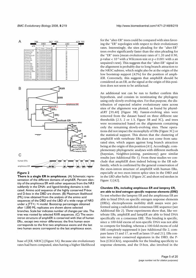
BMC Evolutionary Biology 2008, 8:219 http://www.biomedcentral.com/1471-2148/8/219
Page 5 of 20
(page number not for citation purposes)
base of (ER, NR3C) (Figure 3A). Because site evolutionaryrates had been computed, sites having a higher likelihood
for the "alter-ER" trees could be compared with sites favor-ing the "ER" topologies with respect to their evolutionaryrates. Interestingly, the sites pleading for the "alter-ER"trees evolve significantly faster than the sites pleading forthe "ER" trees (mean evolutionary rates of 1.20 and 0.90,p-value < 10-5 with a Wilcoxon-test or p < 0.001 with a anunpaired t-test). This suggests that the "alter-ER" signal inthe alignment is probably due to long branch attraction tothe NR3C subtree, which might also be at the origin of thelow bootstrap support (42%) for the position of amph-iER. Conversely, this suggests that amphiER should beconsidered as an ER, as the signal at the origin of this posi-tion does not seem to be artifactual.
An additional test can be run to further confirm thishypothesis, and consists in reestimating the phylogenyusing only slowly-evolving sites. For that purpose, the dis-tribution of expected relative evolutionary rates acrosssites of the alignment was plotted, as found by phyml-aLRT [39,40] (Figure 3B). Fastest-evolving sites wereremoved from the dataset based on three different ratethresholds (2.5, 2 or 1.5, Figure 3B and 3C), and treeswere reconstructed based on the alignments containingonly the remaining slowly-evolving sites. These opera-tions did not impact the monophyly of ERs (Figure 3C) orthe statistical support. This shows that the clustering ofamphiER with vertebrate ERs does not come from satu-rated sites, which argues against long branch attractionbeing at the origin of this position [41]. Accordingly, com-plementary phylogenetic analyses with different methods(bayesian, Neighbor-joining, parsimony) gave similarresults (see Additional file 1). From these studies we con-clude that amphiER does indeed belong to the ER sub-family, which is confirmed by the general conservation ofthe exon-intron structure of amphiER with human ERα,especially at two exon-intron splice sites in the DBD andin the LBD after helix 3 (Figure 2C and short red strokes inFigure 1) [42].
Chordate ERs, including amphioxus ER and lamprey ER,
are able to bind estrogen specific response elements (ERE)
To test whether the lamprey ER and the amphioxus ER areable to bind DNA on specific estrogen response elements(EREs), electrophoresis mobility shift assays were per-formed using a radiolabeled consensus ERE sequence (seeAdditional file 2). These experiments show that, like ver-tebrate ERs, amphiER and lampER are able to bind DNAspecifically on a consensus ERE. This binding is specific,since a 100-fold excess of non-specific DNA was not ableto compete for binding, whereas a 100-fold excess of coldERE completely suppressed it (see Additional file 2, com-pare lanes 15 and 17, as well as lanes 19 and 21). ERs con-tain two major conserved signatures in the DBD, the P-box (CEGCKA), responsible for the binding specificity toresponse elements, and the D-box, also involved in the
There is a single ER in amphioxusFigure 2There is a single ER in amphioxus. (A) Schematic repre-sentation of the different domains of amphiER. Percent iden-tity of the amphioxus ER with other sequences from the NR3 subfamily in the DNA- and ligand-binding domains is indi-cated. Amino acid sequence of the highly conserved P-box and D-box in the DBD are shown. (B) Maximum likelihood (ML) tree obtained from the analysis of the amino acid sequences of the DBD and the LBD of a wide range of NR3 under a JTT+γ +i model. Bootstrap percentages obtained after 1,000 ML replicates are shown above selected branches. Scale bar indicates number of changes per site. The tree was rooted by selected RXR sequences. (C) The exon-intron structure of amphiER is conserved with that of human ERα, except two minor differences: the first human exon corresponds to the first two amphioxus exons and the last two human exons correspond to the last amphioxus exon.
DBD LBD
436296 363 678 705
a/b d ec f
Amphioxus ER
Lamprey ER
Aplysia ER
Crassostrea ER
Marisa ER
Nucella ER
Octopus ER
Thais ER
Human ERa
Human ERb
Human ERRa
Human ERRb
Human ERRg
Amphioxus ERR
Human AR
Human PR
Human GR
Human MR
Amphioxus NR3C
CEGCKA
CEGCKA
CEGCKA
CEGCKA
CEGCKA
CEGCKA
CEGCKA
CEGCKA
CEGCKA
CEGCKA
CEACKA
CEACKA
CEACKA
CEACKA
CGSCKV
CGSCKV
CGSCKV
CGSCKV
CEGCKS
PGTNQ
PATNQ
PATNT
PATNT
PATNN
PATNN
PATNS
PATNN
PATNQ
PATNQ
PASNE
PASNE
PASNE
PATNE
ASRND
AGRND
AGRND
AGRND
PANNN
P-box% %D-box
---
82
77
77
77
77
77
77
82
83
68
71
68
73
54
58
62
61
54
---
33
27
31
28
30
26
30
34
35
22
27
27
26
20
19
19
19
35
shark MR
hagfish NR3C
hagfish CRlamprey CR
lamprey NR3C
0.3
MR
GR
AR
PR
7 5
5 1
7 6
7 9
6 68 1
7 6
1 0 0
1 0 0
1 0 0
9 9
9 9
9 7
1 0 0
1 0 0
1 0 0
9 5
9 5
1 0 0
RXR
ERR
mollusk ER
ER
ER
amphioxus ERlamprey ER
amphioxus NR3C
A
B
C
human ER
amphioxus ERexon 1 exon 2 exon 3 exon 4 exon 5 exon 6 exon 7 exon 8
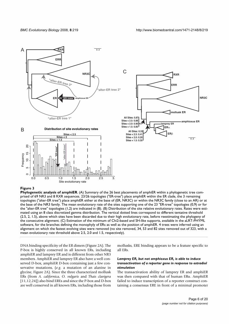
BMC Evolutionary Biology 2008, 8:219 http://www.biomedcentral.com/1471-2148/8/219
Page 6 of 20
(page number not for citation purposes)
DNA binding specificity of the ER dimers (Figure 2A). TheP-box is highly conserved in all known ERs, includingamphiER and lamprey ER and is different from other NR3members. AmphiER and lamprey ER also have a well con-served D-box, amphiER D-box containing just a few con-servative mutations, (e.g. a mutation of an alanine inglycine, Figure 2A). Since the three characterized molluskERs (from A. californica, O. vulgaris and Thais clavigera[11,12,24]) also bind EREs and since the P-box and D-boxare well conserved in all known ERs, including those from
mollusks, ERE binding appears to be a feature specific toall ERs.
Lamprey ER, but not amphioxus ER, is able to induce
transactivation of a reporter gene in response to estradiol
stimulation
The transactivation ability of lamprey ER and amphiERwas then compared with that of human ERα. AmphiERfailed to induce transcription of a reporter construct con-taining a consensus ERE in front of a minimal promoter
Phylogenetic analysis of amphiERFigure 3Phylogenetic analysis of amphiER. (A) Summary of the 26 best placements of amphiER within a phylogenetic tree com-prised of 69 NR3 and 8 RXR sequences. 23/26 topologies ("ER-tree") place amphiER within the ER clade, the 3 remaining topologies ("alter-ER tree") place amphiER either at the base of (ER, NR3C) or within the NR3C family (close to an AR) or at the base of the NR3 family. The mean evolutionary rate of the sites supporting one of the 23 "ER-tree" topologies (0,9) or for the "alter-ER tree" topologies (1,2) are indicated in (B). (B) Distribution of the site relative evolutionary rates. Rates were esti-mated using an 8 class discretized gamma distribution. The vertical dotted lines correspond to different tentative threshold (2.5, 2, 1.5), above which sites have been discarded due to their high evolutionary rate, before reestimating the phylogeny of the consecutive alignment. (C) Estimation of the minimum of Chi2-based and SH-like supports, available in the aLRT-PHYML software, for the branches defining the monophyly of ERs as well as the position of amphiER. 4 trees were inferred using an alignment on which the fastest evolving sites were removed (no site removed, 34, 53 and 82 sites removed out of 323, with a mean evolutionary rate threshold above 2.5, 2.0 and 1.5, respectively).
Distribution of site evolutionary rates
Site evolutionary rate
Fre
qu
en
cy
0.0 0.5 1.0 1.5 2.0 2.5 3.
05
10
15
20
Sites < 2.5Sites < 2
Sites < 1.5
"a
lte
r-E
R t
re
es"
"E
R t
re
es"
0.3RXR
ERR
ER amphioxus ER
NR3C
23 "ER trees"
"alter-ER tree 1"
"alter-ER tree 3"
"alter-ER tree 2"
0.3
All Sites: 0.87
Sites < 2.5: 0.88
Sites < 2.0: 0.90
Sites < 1.5: 0.85
All Sites: 0.33
Sites < 2.5: 0.41
Sites < 2.0: 0.20
Sites < 1.5: 0.35
RXR
ERR
ER
ER
amphioxus ERlamprey ER
NR3C
mollusk ER
A
B
C
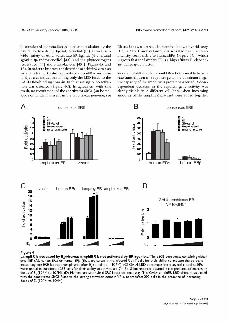
BMC Evolutionary Biology 2008, 8:219 http://www.biomedcentral.com/1471-2148/8/219
Page 7 of 20
(page number not for citation purposes)
in transfected mammalian cells after stimulation by thenatural vertebrate ER ligand, estradiol (E2) as well as awide variety of other vertebrate ER ligands (the naturalagonist 3β-androstenediol [43], and the phytoestrogensresveratrol [44] and enterolactone [45]) (Figure 4A and4B). In order to improve the detection sensitivity, was alsotested the transactivation capacity of amphiER in responseto E2 as a construct containing only the LBD fused to theGAL4 DNA-binding domain. In this case again, no activa-tion was detected (Figure 4C). In agreement with thisresult, no recruitment of the coactivator SRC1 (an homo-logue of which is present in the amphioxus genome, see
Discussion) was detected in mammalian two-hybrid assay(Figure 4D). However lampER is activated by E2, with anintensity comparable to humanERα (Figure 4C), whichsuggests that the lamprey ER is a high affinity E2-depend-ant transcription factor.
Since amphiER is able to bind DNA but is unable to acti-vate transcription of a reporter gene, the dominant nega-tive capacity of the amphioxus protein was tested. A dose-dependent decrease in the reporter gene activity wasclearly visible in 2 different cell lines when increasingamounts of the amphiER plasmid were added together
LampER is activated by E2 whereas amphiER is not activated by ER agonistsFigure 4LampER is activated by E2 whereas amphiER is not activated by ER agonists. The pSG5 constructs containing either amphiER (A), human ERα or human ERβ (B), were tested in transfected Cos 7 cells for their ability to activate the co-trans-fected cognate ERE-luc reporter plasmid after E2 stimulation (10-6M). (C) GAL4-LBD constructs from several chordate ERs were tested in transfectec 293 cells for their ability to activate a (17m)5x-G-luc reporter plasmid in the presence of increasing doses of E2 (10-9M to 10-6M). (D) Mammalian two-hybrid SRC1 recruitment assay. The GAL4-amphiER-LBD chimera was used with the coactivator SRC1 fused to the strong activation domain VP16 to transfect 293 cells in the presence of increasing doses of E2 (10-9M to 10-6M).
Fo
ld a
ctiva
tio
n
0
2
4
6
8
10
12
14
16
18
20
E2
vector human ER lamprey ER amphioxus ER
GAL4-amphioxus ER
VP16-SRC1
Fo
ld a
ctiva
tio
n
E2
0
1
2
0
100
200
300
400
500
600
700
800
0
0.2
0.4
0.6
0.8
1
1.2
1.4
1.6 -
E2
3b-Adiol
Resveratrol
Enterolactone
-
E2
3b-Adiol
Resveratrol
Enterolactone
Fo
ld a
ctiva
ion
Fo
ld a
ctiva
tio
n
amphioxus ER vector human ER
consensus ERE consensus ERE
human ER
A
C
B
D
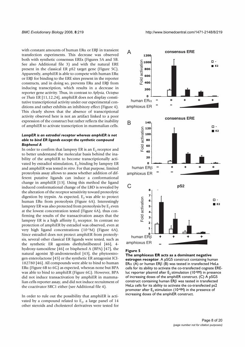
BMC Evolutionary Biology 2008, 8:219 http://www.biomedcentral.com/1471-2148/8/219
Page 8 of 20
(page number not for citation purposes)
with constant amounts of human ERα or ERβ in transienttransfection experiments. This decrease was observedboth with synthetic consensus EREs (Figures 5A and 5B.See also Additional file 3) and with the natural EREpresent in the classical ER pS2 target gene (Figure 5C).Apparently, amphiER is able to compete with human ERαor ERβ for binding to the ERE sites present in the reporterconstructs, and in doing so, prevents ERα and ERβ frominducing transcription, which results in a decrease inreporter gene activity. Thus, in contrast to Aplysia, Octopusor Thais ER [11,12,24], amphiER does not display consti-tutive transcriptional activity under our experimental con-ditions and rather exhibits an inhibitory effect (Figure 4).This clearly shows that the absence of transcriptionalactivity observed here is not an artifact linked to a poorexpression of the construct but rather reflects the inabilityof amphiER to activate transcription in mammalian cells.
LampER is an estradiol receptor whereas amphiER is not
able to bind ER ligands except the synthetic compound
Bisphenol A
In order to confirm that lamprey ER is an E2 receptor andto better understand the molecular basis behind the ina-bility of the amphiER to become transcriptionally acti-vated by estradiol stimulation, E2 binding by lamprey ERand amphiER was tested in vitro. For that purpose, limitedproteolysis assay allows to assess whether addition of dif-ferent putative ligands can induce a conformationalchange in amphiER [13]. Using this method the ligandinduced conformational change of the LBD is revealed bythe alteration of the receptor sensitivity toward proteolyticdigestion by trypsin. As expected, E2 was able to protecthuman ERα from proteolysis (Figure 6A). Interestinglylamprey ER was also protected from proteolysis by E2 evenat the lowest concentration tested (Figure 6A), thus con-firming the results of the transactivation assays that thelamprey ER is a high affinity E2 receptor. In contrast noprotection of amphiER by estradiol was observed, even atvery high ligand concentrations (10-3M) (Figure 6A).Since estradiol does not protect amphiER from proteoly-sis, several other classical ER ligands were tested, such asthe synthetic ER agonists diethylstilbestrol [46], 4-hydroxy-tamoxifene [46] or bisphenol A (BPA) [47], thenatural agonist 3β-androstenediol [43], the phytoestro-gen enterolactone [45] or the synthetic ER antagonist ICI-182780 [46]. All compounds were able to bind to humanERα (Figure 6B to 6G) as expected, whereas none but BPAwas able to bind to amphiER (Figure 6G). However, BPAdid not induce transactivation by amphiER in mamma-lian cells reporter assay, and did not induce recruitment ofthe coactivator SRC1 either (see Additional file 4).
In order to rule out the possibility that amphiER is acti-vated by a compound related to E2, a large panel of 14other steroids and cholesterol derivatives were tested for
The amphioxus ER acts as a dominant negative estrogen receptorFigure 5The amphioxus ER acts as a dominant negative estrogen receptor. A pSG5 construct containing human ERα (A) or human ERβ (B) was tested in transfected HeLa cells for its ability to activate the co-transfected cognate ERE-luc reporter plasmid after E2 stimulation (10-6M) in presence of increasing doses of the amphiER construct. (C) A pSG5 construct containing human ERβ was tested in transfected HeLa cells for its ability to activate the co-transfected ps2 promoter after E2 stimulation (10-6M) in the presence of increasing doses of the amphiER construct.
0
200
400
600
800
1000
1200
-
E2
Fold
activation
human ER
amphioxus ER
-
E2
Fold
activation
human ER
amphioxus ER
-
E2
Fold
activation
human ER
amphioxus ER
0
20
40
60
80
100
120
140
0
1
2
3
4
5
6
7
8
consensus ERE
pS2
consensus ERE
A
B
C
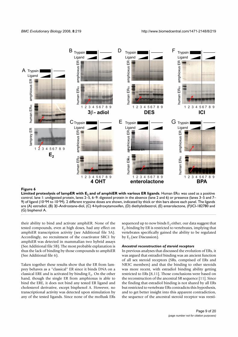
BMC Evolutionary Biology 2008, 8:219 http://www.biomedcentral.com/1471-2148/8/219
Page 9 of 20
(page number not for citation purposes)
their ability to bind and activate amphiER. None of thetested compounds, even at high doses, had any effect onamphiER transcription activity (see Additional file 5A).Accordingly, no recruitment of the coactivator SRC1 byamphiER was detected in mammalian two hybrid assays(See Additional file 5B). The most probable explanation isthus the lack of binding by those compounds to amphiER(See Additional file 6).
Taken together these results show that the ER from lam-prey behaves as a "classical" ER since it binds DNA on aclassical ERE and is activated by binding E2. On the otherhand, though the single ER from amphioxus is able tobind the ERE, it does not bind any tested ER ligand andcholesterol derivative, except bisphenol A. However, notranscriptional activity was detected upon stimulation byany of the tested ligands. Since none of the mollusk ERs
sequenced up to now binds E2 either, our data suggest thatE2-binding by ER is restricted to vertebrates, implying thatvertebrates specifically gained the ability to be regulatedby E2 (see Discussion).
Ancestral reconstruction of steroid receptors
In previous analyses that discussed the evolution of ERs, itwas argued that estradiol binding was an ancient functionof all sex steroid receptors (SRs, comprised of ERs andNR3C members) and that the binding to other steroidswas more recent, with estradiol binding ability gettingrestricted to ERs [8,11]. Those conclusions were based onthe reconstruction of the ancestral SR sequence [11]. Sincethe finding that estradiol binding is not shared by all ERsbut restricted to vertebrate ERs contradicts this hypothesis,and to get better insight into this apparent contradiction,the sequence of the ancestral steroid receptor was reesti-
Limited proteolysis of lampER with E2 and of amphiER with various ER ligandsFigure 6Limited proteolysis of lampER with E2 and of amphiER with various ER ligands. Human ERα was used as a positive control. lane 1: undigested protein, lanes 2–5, 6–9: digested protein in the absence (lane 2 and 6) or presence (lanes 3–5 and 7–9) of ligand (10-3M to 10-5M). 2 different trypsine doses are shown, indicated by thick or thin bars above each panel. The ligands are (A) estradiol, (B) 3β-Androstane-diol, (C) 4-hydroxytamoxifen, (D) diethylstibestrol, (E) enterolactone, (F)ICI-182780 and (G) bisphenol A.
E2
4 OHT enterolactone
DES ICI3 - adiol
B
Trypsin
Ligand
hu
ma
n E
Rla
mp
rey E
Ra
mp
hio
xu
s E
R
hu
ma
n E
Ra
mp
hio
xu
s E
R
hu
ma
n E
Ra
mp
hio
xu
s E
R
hu
ma
n E
Ra
mp
hio
xu
s E
R
hu
ma
n E
Ra
mp
hio
xu
s E
R
hu
ma
n E
Ra
mp
hio
xu
s E
R
hu
ma
n E
Ra
mp
hio
xu
s E
R
Amphioxus ER
a
A
BPA
C
D
E
FTrypsin
Ligand
Trypsin
Ligand
Trypsin
Ligand
Trypsin
Ligand
Trypsin
Ligand
Trypsin
Ligand
G
1 2 3 4 5 6 7 8 9
1 2 3 4 5 6 7 8 9
1 2 3 4 5 6 7 8 9
1 2 3 4 5 6 7 8 9
1 2 3 4 5 6 7 8 9
1 2 3 4 5 6 7 8 9
1 2 3 4 5 6 7 8 9
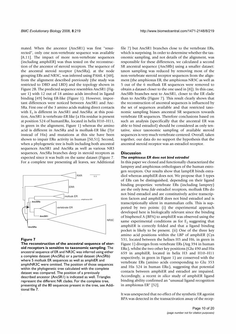
BMC Evolutionary Biology 2008, 8:219 http://www.biomedcentral.com/1471-2148/8/219
Page 10 of 20
(page number not for citation purposes)
mated. When the ancestor (AncSR1) was first "resur-rected", only one non-vertebrate sequence was available[8,11]. The impact of more non-vertebrate sequences(including amphiER) was thus tested on the reconstruc-tion of the ancestor of steroid receptors. The sequence ofthe ancestral steroid receptor (AncSRa), at the nodegrouping ERs and NR3C, was inferred using PAML 4 [48],from the alignment described previously (the study wasrestricted to DBD and LBD) and the topology shown inFigure 2B. The predicted sequence resembles AncSR1 (Fig-ure 1) with 12 out of 18 amino acids involved in ligandbinding [49] being ER-like (Figure 1). However, impor-tant differences were noticed between AncSR1 and Anc-SRa. First one of the 3 amino acids making direct contactswith E2 is different in AncSR1 and AncSRa: at this posi-tion, AncSR1 is vertebrate-ER like (a His residue is presentat position 524 of humanERα, located in helix H10–H11,in green in the alignment, Figure 1) whereas the aminoacid is different in AncSRa and is mollusk-ER like (Tyrinstead of His) and mutations at this site have beenshown to impair ERα activity in human [50,51]. Second,when a phylogenetic tree is built including both ancestralsequences AncSR1 and AncSRa as well as various NR3sequences, AncSRa branches deep in steroid receptors asexpected since it was built on the same dataset (Figure 7.For a complete tree presenting all leaves, see Additional
file 7) but AncSR1 branches close to the vertebrate ERs,which is surprising. In order to determine whether the tax-onomic sampling, and not details of the alignment, wasresponsible for these differences, we calculated a secondSR ancestral sequence (AncSRb) using a smaller dataset:taxon sampling was reduced by removing most of thenon-vertebrate steroid receptor sequences from the align-ment (the amphioxus ER, the amphioxus NR3C as well as5 out of the 6 mollusk ER sequences were removed toobtain a dataset closer to the one used in [8]). In this case,AncSRb branches next to AncSR1, closer to the ER cladethan to AncSRa (Figure 7). This result clearly shows thatthe reconstruction of ancestral sequences is influenced bythe set of sequences available and that restricted taxo-nomic sampling biases ancestral SR sequences towardsvertebrate ER sequences. Therefore conclusions based onsuch an analysis (specifically that the ancestral ER wasable to bind estradiol) should be considered as only ten-tative, since taxonomic sampling of available steroidsequences is very much vertebrate-centered. Overall, takentogether, our data do no support the hypothesis that theancestral steroid receptor was an estradiol receptor.
DiscussionThe amphioxus ER does not bind estradiol
In this paper we cloned and functionally characterized thelamprey and amphioxus orthologues of the human estro-gen receptors. Our results show that lampER binds estra-diol whereas amphiER does not. We propose that 3 typesof ERs can be distinguished, depending on their ligandbinding properties: vertebrate ERs (including lamprey)are the only bona fide estradiol receptors, mollusk ERs donot bind estradiol and are constitutively active transcrip-tion factors and amphiER does not bind estradiol and istranscriptionally silent in mammalian cells. This is sup-ported by two points: (i) the experimental approachdeveloped here is biologically relevant since the bindingof bisphenol A (BPA) to amphiER was observed using thesame experimental conditions as for E2 suggesting thatamphiER is correctly folded and that a ligand bindingpocket is likely to be present. (ii) One of the three keyamino acid positions within the LBP of amphiER (Cys531, located between the helixes H5 and H6, in green inFigure 1) diverges from vertebrate ERs (Arg 394 in humanERα), whilst the two other key positions (Glu 490 and His659 in amphiER, located in helix H3 and H10–H11respectively, in green in Figure 1) are conserved with thevertebrate ERs (amino acids corresponding to Glu 353and His 524 in human ERα), suggesting that potentialcontacts between amphiER and estradiol are impaired.Accordingly, a recent in silico study of amphiER ligandbinding ability confirmed an "unusual ligand recognitionin amphioxus ER" [52].
It was unexpected that no effect of the synthetic ER agonistBPA was detected in the transactivation assay of the recep-
The reconstruction of the ancestral sequence of steroid receptors is sensitive to taxonomic samplingFigure 7The reconstruction of the ancestral sequence of ster-oid receptors is sensitive to taxonomic sampling. The ancestral sequence of ER and NR3C was inferred using either a complete dataset (AncSRa) or a partial dataset (AncSRb) where 5 mollusk ER sequences as well as amphiER and amphiNR3C were omitted. The position of those sequences within the phylogenetic tree calculated with the complete dataset was compared. The position of a previously described ancestor (AncSR1) is indicated as well. Triangles represent the different NR clades. For the complete tree, presenting all the 80 sequences present in the tree, see Addi-tional file 7.
0.3RXR
ERR
NR3C
mollusk ER
vertebrate ER
amphioxus ER
AncSRa
AncSRb
AncSR1
BMC Evolutionary Biology 2008, 8:219 http://www.biomedcentral.com/1471-2148/8/219
Page 11 of 20
(page number not for citation purposes)
tor in mammalian cells since BPA induces a conforma-tional change of amphiER. This apparent absence ofcoactivator recruitment (see Additional file 4B) resultingin no transcriptional activity in response to BPA can beinterpreted in several ways: (i) because of the differentgeometry of the ligand-binding pocket in amphiER, BPAbehaves as an antiestrogen (partial agonist or even a par-tial antagonist) and blocks the transcriptional activationproperties of amphiER, for instance by inducing a confor-mational change that does not allow coactivator recruit-ment (like human ERα and 4-raloxifen, [53]) or byexcluding amphiER from nucleus (like ICI-182,780 withhuman ERα, [54]). (ii) Alternatively, the coactivator inter-face of amphiER does not fit with mammalian coactiva-tors, resulting in artifactual loss of activation. However,the conservation of the amino acids involved in co-activa-tor interaction, compared to human ERα does not supportthis hypothesis (sites indicated with a star in Figure 1, asdescribed in [55]). Among the divergent sites, at a posi-tion implicated in the charge clamp necessary for coacti-vator contact, amphiER contains an aspartate (D677)instead of a glutamate in humanERα (E542). Importantly,the divergence (E->D) is conservative and preserves thenegative charge of the amino acid, which is important forinteraction with the lysine from helix 3 (conserved inamphiER) to form this charge clamp [56]. In addition, aunique orthologue of the p160 family of coactivators wasfound in the amphioxus genome [35] and its overall con-servation with its 3 human ohnologues (genes that havebeen duplicated during the two rounds of whole genomeduplications in the chordate lineage [57]) is good. (iii)Interaction between mammalian chaperones like HSP90and amphiER is impaired, leading to improper binding tothe hormone [58]. Taking these results into account, itwill be interesting to test the effect of BPA on the subcel-lular localization of amphiER and to study if other relatedcompounds are able to bind and/or activate amphiER. Inaddition, it will be important, when cell cultures fromamphioxus are available, to check the activity of amphiERin a monospecific transient transfection assay. It shouldbe remembered that some orphan receptors such as ERRsare thought to have no natural ligands even if they are ableto bind synthetic compounds [59]. More generally, theprecise status of amphiER in terms of ligand bindingremains an open question. It is nevertheless clear, and thisis an important issue for the current evolutionary debate,that amphiER is not able to bind estradiol.
Is there any receptor for estradiol in amphioxus?
The observation that amphiER does not bind E2 is indeeda surprising observation since E2 was detected in amphi-oxus by RIA, the hormonal production being correlatedwith breeding season [15]. Several aspects of steroidmetabolism were described in amphioxus [60] and thehomologues of many enzymes necessary for estradiol syn-
thesis in mammals were cloned from amphioxus ovaries[15,61]. Of particular interest is the report of an aromatasegene (CYP19) in amphioxus, which suggests that the cru-cial step in estradiol synthesis is indeed possible inamphioxus. These experimental data were recently con-firmed by the analysis of the complete amphioxusgenome sequence [35]. It may be that, in amphioxus, theactive sex hormone is an E2-derivative [62] or another sexhormone, like in the case of androgens in lamprey [63],and this derivative is still to be discovered. In a similarway, we recently demonstrated that the amphioxus TRorthologue does not bind T3 or T4, the classical thyroidhormones, but deaminated derivatives TRIAC and TET-RAC, which are able to induce amphioxus metamorphosis[64].
A second possibility is that E2 itself has a central role in sexmaturation in amphioxus, and that the functional estro-gen receptor in amphioxus is different from amphiER.Several candidates are possible. First, there is another ster-oid receptor in amphioxus (amphiNR3C in Figures 1 and2) [35] that exhibits several ER-like features. Its P- and D-boxes are closer to ERs than to vertebrate NR3C (Figure2A). The sequence identity of its LBD with human ERα(37%) and with NR3C members (35%) are similar. More-over, most of the amino acids involved in ligand bindingare more ER-like than AR-, PR- or MR-like (Figure 1).However it is the only NR3C receptor (orthologous to AR,PR, MR and GR) found in the amphioxus genome. Thus ifamphiNR3C plays the role of an estradiol receptor, thissuggests an absence of a "classical" steroid receptor able tobind testosterone, progesterone or corticoids. Alterna-tively, a non-nuclear receptor could mediate E2 action inamphioxus. Indeed, several non-genomic effects of estra-diol were reported in mammals involving GPCRs (forreviews see [65-67]). For instance, very recently, a highaffinity receptor for the steroid androstenedione linked tothe membrane, was described in lamprey [68] and aGPCR with high affinity for progestines was isolated fromsea trout [69].
Implications for the evolution of ERs
The absence of E2 binding by the amphioxus estrogenreceptor has interesting consequences for the evolution ofSRs and ERs. Indeed, only the well characterized gnathos-tome ERs and the lamprey ER (studied here) have beenshown to mediate E2 action. Outside vertebrates, all theERs studied so far (in mollusks and amphioxus) do notbind E2 [11,12,24-26]. Parsimony implies that the func-tion of estradiol in the bilaterian ancestor was not medi-ated by ER and that ER had another function. Only laterduring evolution, in the vertebrate lineage, ER would thenhave gained the ability to be activated by E2 and to medi-ate the hormonal action of this compound (Figure 8). Thealternative scenario (ancestral E2 binding and independ-
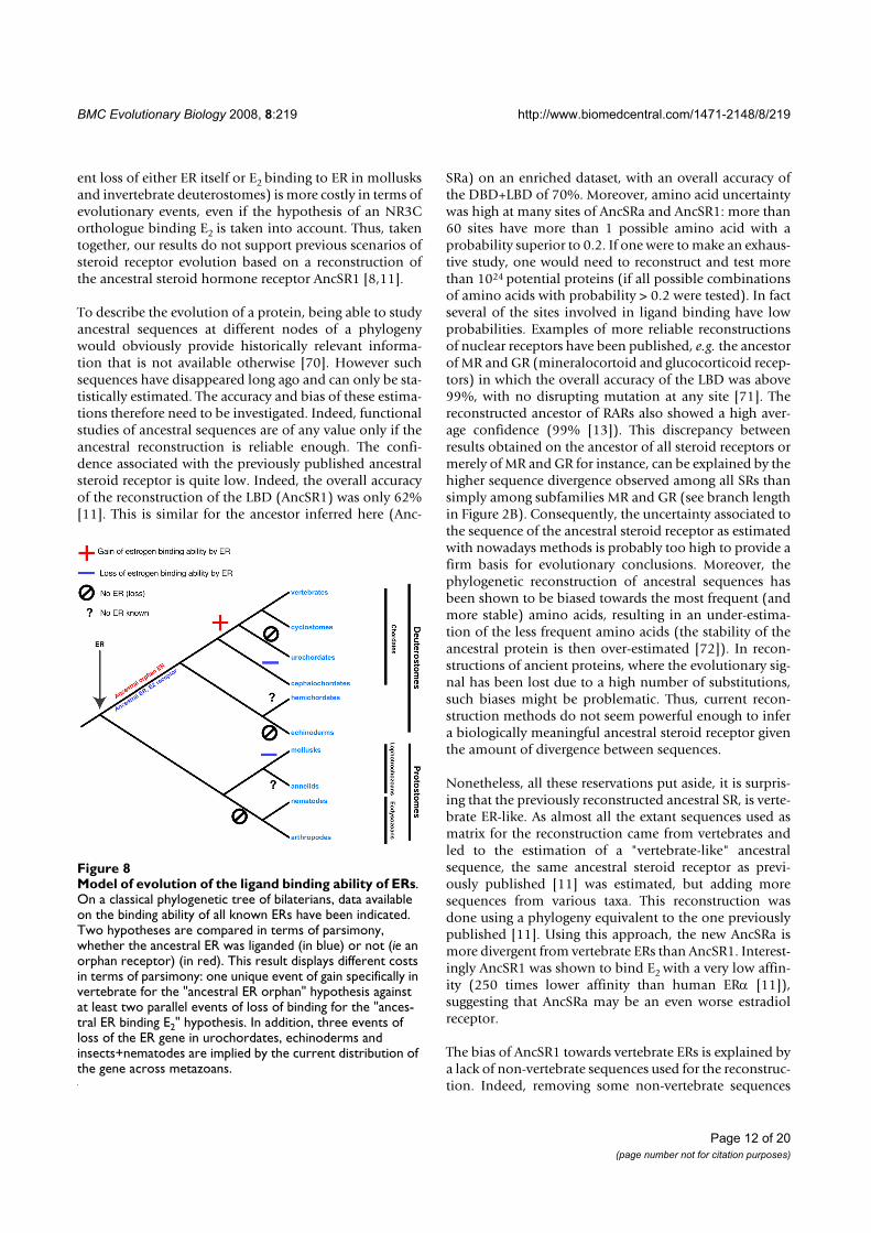
BMC Evolutionary Biology 2008, 8:219 http://www.biomedcentral.com/1471-2148/8/219
Page 12 of 20
(page number not for citation purposes)
ent loss of either ER itself or E2 binding to ER in mollusksand invertebrate deuterostomes) is more costly in terms ofevolutionary events, even if the hypothesis of an NR3Corthologue binding E2 is taken into account. Thus, takentogether, our results do not support previous scenarios ofsteroid receptor evolution based on a reconstruction ofthe ancestral steroid hormone receptor AncSR1 [8,11].
To describe the evolution of a protein, being able to studyancestral sequences at different nodes of a phylogenywould obviously provide historically relevant informa-tion that is not available otherwise [70]. However suchsequences have disappeared long ago and can only be sta-tistically estimated. The accuracy and bias of these estima-tions therefore need to be investigated. Indeed, functionalstudies of ancestral sequences are of any value only if theancestral reconstruction is reliable enough. The confi-dence associated with the previously published ancestralsteroid receptor is quite low. Indeed, the overall accuracyof the reconstruction of the LBD (AncSR1) was only 62%[11]. This is similar for the ancestor inferred here (Anc-
SRa) on an enriched dataset, with an overall accuracy ofthe DBD+LBD of 70%. Moreover, amino acid uncertaintywas high at many sites of AncSRa and AncSR1: more than60 sites have more than 1 possible amino acid with aprobability superior to 0.2. If one were to make an exhaus-tive study, one would need to reconstruct and test morethan 1024 potential proteins (if all possible combinationsof amino acids with probability > 0.2 were tested). In factseveral of the sites involved in ligand binding have lowprobabilities. Examples of more reliable reconstructionsof nuclear receptors have been published, e.g. the ancestorof MR and GR (mineralocortoid and glucocorticoid recep-tors) in which the overall accuracy of the LBD was above99%, with no disrupting mutation at any site [71]. Thereconstructed ancestor of RARs also showed a high aver-age confidence (99% [13]). This discrepancy betweenresults obtained on the ancestor of all steroid receptors ormerely of MR and GR for instance, can be explained by thehigher sequence divergence observed among all SRs thansimply among subfamilies MR and GR (see branch lengthin Figure 2B). Consequently, the uncertainty associated tothe sequence of the ancestral steroid receptor as estimatedwith nowadays methods is probably too high to provide afirm basis for evolutionary conclusions. Moreover, thephylogenetic reconstruction of ancestral sequences hasbeen shown to be biased towards the most frequent (andmore stable) amino acids, resulting in an under-estima-tion of the less frequent amino acids (the stability of theancestral protein is then over-estimated [72]). In recon-structions of ancient proteins, where the evolutionary sig-nal has been lost due to a high number of substitutions,such biases might be problematic. Thus, current recon-struction methods do not seem powerful enough to infera biologically meaningful ancestral steroid receptor giventhe amount of divergence between sequences.
Nonetheless, all these reservations put aside, it is surpris-ing that the previously reconstructed ancestral SR, is verte-brate ER-like. As almost all the extant sequences used asmatrix for the reconstruction came from vertebrates andled to the estimation of a "vertebrate-like" ancestralsequence, the same ancestral steroid receptor as previ-ously published [11] was estimated, but adding moresequences from various taxa. This reconstruction wasdone using a phylogeny equivalent to the one previouslypublished [11]. Using this approach, the new AncSRa ismore divergent from vertebrate ERs than AncSR1. Interest-ingly AncSR1 was shown to bind E2 with a very low affin-ity (250 times lower affinity than human ERα [11]),suggesting that AncSRa may be an even worse estradiolreceptor.
The bias of AncSR1 towards vertebrate ERs is explained bya lack of non-vertebrate sequences used for the reconstruc-tion. Indeed, removing some non-vertebrate sequences
Model of evolution of the ligand binding ability of ERsFigure 8Model of evolution of the ligand binding ability of ERs. On a classical phylogenetic tree of bilaterians, data available on the binding ability of all known ERs have been indicated. Two hypotheses are compared in terms of parsimony, whether the ancestral ER was liganded (in blue) or not (ie an orphan receptor) (in red). This result displays different costs in terms of parsimony: one unique event of gain specifically in vertebrate for the "ancestral ER orphan" hypothesis against at least two parallel events of loss of binding for the "ances-tral ER binding E2" hypothesis. In addition, three events of loss of the ER gene in urochordates, echinoderms and insects+nematodes are implied by the current distribution of the gene across metazoans.
BMC Evolutionary Biology 2008, 8:219 http://www.biomedcentral.com/1471-2148/8/219
Page 13 of 20
(page number not for citation purposes)
from our dataset leads to an estimation of an ancestralsteroid receptor that is more "vertebrate ER"-like (AncSRbin Figure 7). The clustering of AncSRb with the ER cladeand the exclusion of AncSRa from the ER clade were sup-ported with good statistical values (minimum of Chi2-based and SH-like supports of 0.83 and 0.89, respec-tively). Those data show that AncSR1 reconstruction wasprobably sensitive to the vertebrate bias in the data set(Figure 7). Overall, we suggest that analysis based onancestral reconstructions should be taken as tentative,especially in case of low statistical confidence and limitedtaxonomic sampling. In case of the ancestral steroid recep-tor, even if exhaustive taxonomic sampling is necessary,phylogenetic signal is weak and the resulting confidence isquite low. Thus we think that even if the ancestralsequence built here is biologically more relevant than pre-viously calculated ones (because of better taxonomic sam-pling), it remains quite uncertain. Consequentlyconclusions regarding the ancestral steroid receptorshould be based mostly on comparative characterizationof extant receptors. In that case, all the data based oninvertebrate ER receptors (from mollusks and amphioxus)support an ancestor of steroid receptors that was not ableto bind estradiol. This conclusion will obviously requirethe functional characterization of ERs from other proto-stome phyla in order to carefully check if this observationis general. Thus, available data converge towards a re-eval-uation of the ancestral status of estrogen receptors.
Sequence conservation reflects functional constraints:
ligand binding ability is more recent than DNA-binding
ability
From our and previous studies, only vertebrate ERs areable to bind and activate transcription under estradiolstimulation [11,12,24-26]. The LBD of amphiER is moredivergent from its vertebrate counterparts (ca. 34% aminoacid identities) than the LBD of other liganded amphi-oxus nuclear receptors such as amphiRAR (ca. 58%),which has been shown to bind the same ligand as its ver-tebrate homologue [7,13]. This suggests that a conservedfunctional feature (e.g. binding to the same ligand) isreflected in the sequence conservation of the LBD.
The same observation can be done concerning the DBDsince all ERs, including amphiER, have a highly conservedDBD and are able to bind EREs. Thus, for this domainalso, a conservation of the function is reflected insequence conservation.
Accordingly with this notion, the LBD of invertebrate ERsis highly divergent but their DNA binding domain, as wellas other functionally important domains not directlylinked to ligand binding such as the dimerization inter-face, or the amino acids responsible for interaction withthe co-activators [55] are well conserved. This is true foramphiER as well as mollusk ERs. This strongly suggests
that amphiER is a bona fide NR regulating ERE-containinggenes in an E2-independent manner. Post-translationalmodifications such as phosphorylation or the presence/absence of other receptor-interacting proteins such astranscriptional coactivators have been shown to regulateunliganded nuclear receptors [73]. Whether one of thesemechanisms acts to regulate the activity of invertebrateERs or if those receptors have unknown ligands still to beidentified remains to be explored. Anyway our observa-tions strongly suggest that for ERs, the DNA binding func-tion of the receptor as well as its interaction with co-regulators have been conserved due to selective pressure.Interestingly, when studying the AncSRa, the P- and D-boxes in the DBD are ER/ERR-like (Figure 1), suggestingthat ER/ERR DNA binding ability is ancestral, in accord-ance with the fact that these are the only receptors of theNR3 family found in invertebrates. This difference in theselection pressure between DBD and LBD has been pro-posed to be a general evolutionary pattern for the wholeNR family [7]. The plasticity of the ligand binding abilityof NRs was recently illustrated in the case of RXR-USPwhere the ability of the receptor to be regulated by a lig-and was suggested to have been subject to several succes-sive episodes of gain and loss during evolution [74].
Evolution of endocrine systems: refinement of the ligand
exploitation model
The ligand exploitation model hypothesizes how newhormones and new receptors appear during evolution. Itsuggests that the ancestral ligand is the last metabolite ofa synthesis pathway [8]. According to this model, theancestral steroid ligand was estradiol (and the ancestral SRbound estradiol). During evolution, other steroid recep-tors appeared by duplication of the ancestral ER andgained the ability to bind other steroids, intermediate inthe synthesis pathway (like testosterone or progesterone).
Our findings on the evolution of ERs do not support theligand exploitation model, since our data strongly suggestthat the ancestral ER did not bind estradiol. However, asestradiol has been detected in deuterostomes as well asprotostomes (for instance in vertebrates, amphioxus,echinoderms, mollusks, for review, see [75]), steroid sign-aling may have been already present in bilaterian ances-tor. However, up to now, the ancestral steroid moleculeremains to be determined. If estradiol is an ancient hor-mone, it then probably bound another receptor and lateron ER gained the ability to recognize it, as did other ster-oid receptors for their extant ligand. Thus the evolution ofsteroid system intermingles two distinct processes, theevolution of the receptor on one hand, and the evolutionof the ligand on the other.
The receptor can evolve by point mutations and change itsaffinity for a ligand towards another. This idea was con-vincingly exemplified in the case of corticoid receptors
BMC Evolutionary Biology 2008, 8:219 http://www.biomedcentral.com/1471-2148/8/219
Page 14 of 20
(page number not for citation purposes)
(the ancestor of MR and GR) for which it was recentlydemonstrated that ability of the ancestral vertebrate corti-coid receptor to bind gnathostome-specific hormonealdosterone (a MR ligand) was a by-product of its abilityto bind the ancestral ligand 11-deoxycorticosterone(DOC) [71]. GR gained the ability to bind cortisol only inthe gnathostome lineage, in parallel to endogenous syn-thesis of the hormone [16,71]. This detailed study showsthat a receptor binding a given ligand can acquire affinityfor compounds present in the cell that are structurallyclose to its natural ligand: this refines the ligand exploita-tion model, since new ligands are not necessarily precur-sors of ancient ligands, simply compounds present in thecell and structurally close to the ancestral ligand. Similarconclusions were drawn previously in the case of RAR evo-lution [13]. It has to be emphasized that the pool of avail-able compounds is also subject to evolutionary changes inparallel. For instance, the spatiotemporal production ofestradiol is variable in the different vertebrate groups(reviewed in [76]). Glucocorticoids differ in mouse (corti-sol) and in human (corticosterone), with both hormonesbeing GR ligands [77]. There are several androgens in tel-eost fishes, with 11-ketotestosterone being teleost-specific[78]. As there are 2 androgen receptors (ARs) in teleostfishes, from a whole genome duplication [79], the studyof the ligand-binding ability of those ARs is a potentiallyinteresting case for the evolution of endocrine systems. Ashighlighted by Bridgham et al. (2006), lamprey does notproduce cortisol [71]. In accordance, their genomes donot contain the sequence corresponding to the enzymeresponsible for cortisol synthesis (11b-hydroxylase) andin general classical steroids except estradiol are rarelyfound in lamprey. This suggests that the steroids actuallyfound in lamprey are different from the ones found inmammals (reviewed in [80]). Those cases exemplify thelargely underestimated diversity of endocrine systems:except for lamprey and some teleost fishes, the hormonalpool of animals remains largely unknown. As proposedfor the study of steroid receptors, a comparative approachshould be applied to determine the metabolism of ster-oids in poorly studied animals. Indeed, the hormonalpool of such animals is usually evaluated from the pres-ence/absence of putative orthologues of mammalianenzymes. As the enzymatic machinery involved in hormo-nal metabolism has a very labile activity (reviewed in[81,82]), equating orthology with functional identitymight be unreliable.
The evolution of steroid receptors can be replaced in themore general context of ligand-nuclear receptor co-evolu-tion. The evolution of the NR1H subfamily, that includesreceptors for other steroidal compounds, like the majortranscriptional regulator of bile salt synthesis farnesoid ×receptor (FXR), the pregnane × receptor (PXR), the vita-
min D receptor (VDR) or liver × receptor (LXR)/ecdysonereceptor (Ecr), has been extensively studied and is not inline with the ligand exploitation model [83-85]. Forinstance, comparative functional studies of FXRs from var-ious chordate species showed that the vertebrate FXRsbind "late" cholesterol derivatives (from a complex syn-thesis pathway) but are thought to have evolved from anancestral FXR that bound early cholesterol derivatives(from a simpler synthesis pathway) [83].
In other cases, the evolution of ligand binding is more"chaotic" with close orthologs having a selective ligandbinding ability that varies extensively (vertebrate VDRs arevery well conserved when PXRs have the widest ligandrepertoire of all NRs) [83].
These complex histories are probably linked to specificfunction of some of those NRs, considered as xenotoxiccompounds "sensors". This tight relationship with theunstable environment probably makes receptors like FXRand especially PXR more prone to fast evolution [86]. Yetthey illustrate the impressive variety of scenarios of NRevolution.
ConclusionIn this article, we demonstrated that vertebrate ERs(including lamprey ER) are estradiol receptors whilst non-vertebrate ER (including amphioxus ER) are not. The mostparsimonous scenario proposes that the ancestral ER wasnot able to bind estradiol and that it had another func-tion. It later gained the ability to be regulated by estradiol,specifically in the vertebrate lineage. However, additionalcritical data remains to be discovered in poorly studiedtaxa [62]. To fully understand the evolution of steroid sig-naling pathway, a larger number of taxa need to be tar-geted for detailed comparative studies. More precisely,ERs and other steroid receptors should be cloned fromwidely distributed taxa, especially in protostomes.Enzymes involved in steroidogenesis should also becloned and characterized, to understand the evolution ofsteroid availability. In order to avoid the blinders of a"vertebrate-centered" view, it is of particular importanceto establish the steroid hormone repertoire of an enlargedanimal panel, including more protostomes. The descrip-tion of various endocrine systems will certainly be rele-vant to the early evolution of hormone signaling.
MethodsCloning of amphiER
An initial piece of amphiER was obtained by degeneratePCR on different RT reactions from total RNA extractedeither from developing B. floridae embryos and larvae (at13 h–15 h, 28 h, 36 h, 48 h or 3 d–4 d of development)or from B. floridae adults. The oligonucleotides used were
BMC Evolutionary Biology 2008, 8:219 http://www.biomedcentral.com/1471-2148/8/219
Page 15 of 20
(page number not for citation purposes)
as follows: forward primer 5'-TGYGARGGITGYAARGCIT-TYTT-3' and reverse primer 5'-GTRCAYTSRTTIGTIGCIG-GRCA-3'.
The touchdown PCR program used was as follows:
5' 94 degrees
5× (30" 94 degrees, 1' 55 degrees, 1' 72 degrees)
5× the same cycle, but at 50 degrees annealing tempera-ture
5× the same cycle, but at 45 degrees annealing tempera-ture
5× the same cycle, but at 40 degrees annealing tempera-ture
25× the same cycle, but at 37 degrees annealing tempera-ture
7' 72 degrees
All degenerate PCRs irrespective of the RT reaction tem-plate used yielded a 83 bp fragment of amphiER. The frag-ment was sequenced on both strands and used for thedesign of oligonucleotides for 5' and 3' RACE experimentswith the Invitrogen GeneRacer Kit. The template for theRACE experiments was pooled total RNA from 13 h–15 hB. floridae embryos and from B. floridae adults. In addi-tion to the oligonucleotides provided by the kit, for the 3'RACE, the following primers were used:
3' RACE, 1st PCR: 5'-AACGGAGCATTCAGCAAGGTC-3'
3' RACE, 2nd PCR: 5'-GCATTCAGCAAGGTCAGACAG-3'
5' RACE, 1st strand cDNA synthesis: 5'-ATGTAATCT-GTCTGACCTTGC-3'
5' RACE, 1st PCR: 5'-CTGTCTGACCTTGCTGAATGC-3'
5' RACE, 2nd PCR: 5'-TCTGACCTTGCTGAATGCTCC-3'
The protocols for the 1st and 2nd round of PCR experi-ments are given in the Invitrogen GeneRacer Kit. The 3'and 5' RACE products were subsequently sequenced onboth strands and used for the design of oligonucleotidesfor the full-length cloning of amphiER: forward primer 5'-CGGCGAAGCGAAGAAGATCGAG-3' and reverse primer5'-CTTAACCGATACTAACGGAACAG-3'. The full-lengthamphiER was obtained by PCR on pooled RT reactionsfrom total RNA extracted from B. floridae 13 h–15 h
embryos, 3 d–4 d larvae and B. floridae adults. The PCRprotocol used was as follows:
10' 94 degrees
5× (30" 94 degrees, 30" 55 degrees, 2' 72 degrees)
35× the same cycle, but at 50 degrees annealing tempera-ture
10' 72 degrees
The full-length amphiER clone resulting from this PCR is2279 bp long, was cloned into the pCR2.1 vector (Invitro-gen) and subsequently sequenced on both strands.
Plasmid constructs and reagents
Full length amphiER were amplified by polymerase chainreaction (PCR) and the obtained fragments were insertedinto a pSG5 vector between EcoR1 sites. Lamprey ER wasa generous gift from JW Thornton. Human pSG5-ERα andpSG5-ERβ and the 3xERE-Luc luciferase reporter constructhave been described previously [87]. The pS2-Luc reporterconstruct encompasses an 1100 bp estrogen-responsiveregion of the human pS2 promoter inserted into the pGL3basic vector (Promega). Chimeras comprising the GAL4DNA-binding domain fused with the LBD of the humanERα (residues 251 to 595), the LBD of amphiER (residues364 to 705), the LBD of lampER (residues 234 to 554)have been cloned in the pG4MpolyII vector. 17β-estra-diol, genistein, 3β-androstenediol, resveratrol, choles-terol, cholic acid, chenodeoxycholic acid, 22®-hydroxycholesterol, 20-Hydroxyecdysone, pregnenolone,trans-Dehydroandrosterone (DHEA), corticosterone, pro-gesterone, 4-androstene-3,17-dione, estrone, testoster-one, 5α-androstan-17β-ol-3-one and 1a,25-Dihydroxyvitamin D3 (calcitriol) were purchased fromSigma. Enterolactone was a generous gift from Dr SariMäkelä [88].
Phylogenetic analysis of NR3
Protein sequences of NR3 family members were obtainedfrom GenBank by BLAST search using Homo sapiens ERα asa query. Eight additional sequences from the closelyrelated RXR group were also obtained to serve as outgroupsequences. For accession numbers of the sequences used,see Additional file 8.
The retrieved sequences were aligned using the muscle 3.6program [89] and the resulting alignment was manuallycorrected with SEAVIEW [90]. Phylogenetic tree was calcu-lated by maximum likelihood as implemented in PhyMLversion 2.4.3 under a JTT substitution matrix plus a eight-category gamma rate correction (α estimated) and withthe proportion of invariant sites estimated. Both the DBD
BMC Evolutionary Biology 2008, 8:219 http://www.biomedcentral.com/1471-2148/8/219
Page 16 of 20
(page number not for citation purposes)
and the LBD were used. Robustness was assessed by boot-strap analysis (1,000 repetitions) [91].
The Bayesian inference was done using the programMrBayes 3.1.2 [92]. Two simultaneous independent runswere performed. For each run, one chain was sampledevery 100 generations for 1,000,000 generations after theburn-in cycles, until the average SD of split frequencieswas <0.01; additionally, the potential scale reduction fac-tors of the parameters were close to or equal to 1, whichindicates that the runs had most probably converged. Theneighbour-joining (Poisson correction) and maximumparsimony trees were done with Phylo_win [90].
Likelihood-based tests of alternative topologies placing
amphiER at all possible positions in the tree
The 149 trees were built by reconnecting amphiER fromthe maximum likelihood tree, into the 149 possible posi-tions. The branch length and the different parameters ofthe obtained trees were re-estimated using PhyML. Likeli-hood-based tests of the 149 alternative topologies werecalculated using CONSEL: site-wise log-likelihood values,available as output of PhyML, were used to calculate theP-values of the different positions according to the AU testwith the software R.
Ancestral sequence reconstruction
The aminoacid sequence of the ancestral AncSRa and Anc-SRb was inferred only for the most conserved part of thealignment, i.e. the DBD and LBD (defined as in Figure 1).The ancestral sequences were reconstructed by maximumlikelihood as implemented in PAML [48], under the JTTsubstitution model and a gamma distribution with 8 cat-egories of rates across sites, using the tree described inAdditional file 1A for AncSRa and a the same topologytruncated of the mollusk ER sequences and the amphi-oxus NR3 sequences, after reestimation of the branchlengths using phyml (JTT+γ) for AncSRb.
Electrophoretic Mobility Shift Assay (EMSA)
EMSAs were performed as previously described [93].Where indicated, a 10- and 100-fold molar excess of 30-bp unlabeled oligonucleotides (a consensus ERE and anon-related probe) were added as competitors. Thesequence of the probe containing the consensus ERE is 5'-CGGGCCGAGGTCACAGTGACCTCGGCCCGT-3' andthe sequence of the non-related probe is 5'-CTAGTC-CTAGGTCTAGAGAATTCA-3'.
Cell culture and transfections
Human embryonic kidney 293 cell culture and transfec-tions using Lipofectamine Plus reagent (Invitrogen) weredone according to the manufacturer's recommendationsand as previously described [94]. Briefly, 200 ng of thechimeras comprising the GAL4 DNA-binding domain
fused with the LBD of either human ERα, lampER orAmphiER (or LBD for the control) were co-transfectedtogether with 100 ng of reporter plasmid and 10 ng of a β-galactosidase expression vector, included as a control fortransfection efficiency. For the mammalian double hybridassays, the GAL4-amphiER-LBD chimera was transfectedwith 200 ng of the coactivator SRC1 fused to the strongactivation domain VP16. Three to five hours post-transfec-tion, serum and hormones (as indicated in the figures)were added to the cells which were incubated for an addi-tional 48 hours before harvest and luciferase and β-galac-tosidase activities were determined. Results show themean ± s.e.m. (n = 3) of representative experiments.Human HeLa cervical cancer cells and CV-1 green monkeykidney cells were routinely maintained in Dulbecco'smodified eagle's medium (Invitrogen), supplementedwith 10% fetal bovine seum, 1% v/v L-glutamine and 1%v/v penicillin/streptomycin. Cells were seeded in 12 or 24well plates one day prior to transfection. Transient trans-fections were carried out using the Lipofectamine Plus rea-gent according to instructions of the manufacturer(Invitrogen) in culture media devoid of serum, phenol-red and antibiotics. Briefly, 1 ng of ERα, ERβ or AmphiERexpression vectors were co-transfected together with 100ng 3xERE-Luc (or 200 ng pS2-Luc where indicated) and 20ng of a β-galactosidase expression vector, included as acontrol for transfection efficiency. In the co-expressionexperiments, AmphiER was co-transfected together withERα or ERβ in ratios of 0.5:1, 1:1 and 1:5, respectively.Three hours post-transfection, serum and hormones (asindicated in the figures) were added to the cells whichwere thereafter incubated for an additional 48 hoursbefore harvest and luciferase and β-galactosidase activitieswere determined. Figures represent results from at leastthree independent experiments performed in duplicates.Data is presented as mean +/- SD of fold induction of rel-ative luciferase values corrected against β-galactosidaseactivity, where activity obtained from transfected reporterplasmid alone and treated with vehicle, was arbitrarily setto 1.
Limited proteolytic digestion
These assays were done as previously described [13].
Authors' contributionsMP, HE, MS, SB and VL contributed to the conception anddesign of the study. MS cloned amphiER. MP performedthe EMSA, limited proteolysis experiments, part of thetransactivaton assays and the bioinformatics study. KPand IP performed the rest of the transactivation assays. MPand VL wrote the manuscript.
BMC Evolutionary Biology 2008, 8:219 http://www.biomedcentral.com/1471-2148/8/219
Page 17 of 20
(page number not for citation purposes)
Additional material
AcknowledgementsWe thank Bastien Boussau for help with phylogenetic analysis and for com-
ments on the manuscript. We are grateful to Joseph Thornton for the gift
of the lamprey ER clone, Gerard Benoit and Gabriel Markov for critical
reading of the manuscript. This work was supported by the EU funded
CASCADE network of excellence, CNRS, UCB Lyon 1, ENS de Lyon and
MENRT.
Additional file 1
Phylogenetic analysis of NR3 sequences using several methods. Phylo-
genetical trees of an alignment comprising 69 NR3 sequences as well as
RXR sequences were inferred using the maximum likelihood method
(ML) (A), Bayesian analysis (B), neighbour-joining method (C) and
maximum parsimony method (MP) (D) based on an elision alignment of
the DBD and LBD of 77 NR3 and RXRs (accession numbers are given in
Additional file 8). Labels above each branch show percentages of bootstrap
values after 1000 replicates (A), posterior probabilities (B), percentages
of bootstrap values after 500 replicates (C) or 100 replicates (D). The
fastest evolving sites (with an evolutionary rape above 2, as indicated in
the Figure 3A) were removed from the alignment before computing phyl-
ogeny by maximum parsimony, to preserve the branching of mollusk ERs
within the ER clade. In (A) nodes with bootstrap values below 50% are
presented as polytomies, as in the Figure 2B.
Click here for file
[http://www.biomedcentral.com/content/supplementary/1471-
2148-8-219-S1.pdf]
Additional file 2
DNA binding characterization of chordate ERs. Various chordate mem-
bers of the NR3 family, namely human ERα, human ERβ, mouse ERRα,
amphiER and lamprey ER, were synthesized in vitro and allowed to bind
to a 32P-labeled consensus ERE probe in an EMSA. Lane 1, empty vector
(pSG5) reticulocytes lysates. Lanes 2–5, human ERα. Lanes 6–9, human
ERβ. Lanes 10–13, mouse ERRα. Lanes 14–17, amphiER. Lanes 18–21,
lamprey ER. Lanes 3–5, 7–9, 11–13, 15–17, 19–21, unlabeled non-spe-
cific oligonucleotide (NS) or ERE were added at indicated molar excess as
competitors to test the specificity of the binding. The arrows indicated the
gel shift induced by amphiER binding the ERE probe. The asterisk indi-
cates free ERE probe.
Click here for file
[http://www.biomedcentral.com/content/supplementary/1471-
2148-8-219-S2.pdf]
Additional file 3
The amphioxus ER acts as a dominant negative estrogen receptor in
CV1 cells. A pSG5 construct containing human ERα (A) or human ERβ
(B) was tested in transfected CV1 cells for its ability to activate the co-
transfected cognate ERE-luc reporter plasmid after E2, genistein or β-
Androstane-diol stimulation (10-6M) in presence of increasing doses of
the amphiER construct.
Click here for file
[http://www.biomedcentral.com/content/supplementary/1471-
2148-8-219-S3.pdf]
Additional file 4
The amphioxus ER is not activated by BPA. (A) GAL4-LBD constructs
from several chordate ERs were tested in transfected 293 cells for their
ability to activate a (17 m)5x-G-luc reporter plasmid in the presence of
increasing doses of BPA (10-9M to 10-6M). (B) Representation of the
mammalian two-hybrid SRC1 recruitment assay. The GAL4-amphiER-
LBD chimera was used with the coactivator SRC1 fused to the strong acti-
vation domain VP16 to transfect 293 cells in the presence of increasing
doses of BPA (10-9M to 10-6M).
Click here for file
[http://www.biomedcentral.com/content/supplementary/1471-
2148-8-219-S4.pdf]
Additional file 5
amphiER is not activated by cholesterol derivatives. (A) The GAL4-
amphiER-LBD chimera was tested in transfected 293 cells for its ability
to activate a (17 m)5x-G-luc reporter plasmid in the presence of various
cholesterol derivatives at a high concentration (1 μM) (black). The empty
vector (white) was used as a negative control and the GAL4-humanERα-
LBD in the presence of E2 was used as a positive control (B) Representa-
tion of the mammalian two-hybrid SRC1 recruitment assay. The GAL4-
amphiER-LBD chimera was used with the coactivator SRC1 fused to the
strong activation domain VP16 to transfect 293 cells in the presence of
various cholesterol derivatives at 1 μM. The empty vector (white) was used
as a negative control.
Click here for file
[http://www.biomedcentral.com/content/supplementary/1471-
2148-8-219-S5.pdf]
Additional file 6
Limited proteolysis of amphiER with various cholesterol derivatives.
lane 1: undigested protein, lanes 2–4, 5–7: digested protein in the absence
(lane 2 and 5) or presence (lanes 3–4 and 6–7) of ligand (10-3M and
10-4M). 2 different trypsine doses are shown, indicated by thick or thin
bars above each panel. The ligands are cholic acid (A), Chenodeoxycholic
acid (B), 22R-OH-cholesterol (C), cholesterol (D), 4-androstene-3,17-
dione (E), DHEA (F), corticosterone (G), progesterone (H), pregne-
nolone (I), estrone (J), testosterone (K), 5α-androstane-dione (L), 20-
hydroxyecdysone (M) and calcitriol (N).
Click here for file
[http://www.biomedcentral.com/content/supplementary/1471-
2148-8-219-S6.pdf]
Additional file 7
Phylogenetic tree of NR3 sequences as well as ancestral sequences.
Complete tree corresponding to the simplified one presented in the figure
7. The ancestral sequence of ER and NR3C was inferred using either a
complete dataset (AncSRa) or a partial dataset (AncSRb) where 5 mol-
lusk ER sequences as well as amphiER and amphiNR3C were omitted.
The position of those sequences within the phylogenetic tree calculated
with the complete dataset was compared. The position of a previously
described ancestor (AncSR1) is indicated as well. Minimum of Chi2-
based and SH-like supports are shown for each branch.
Click here for file
[http://www.biomedcentral.com/content/supplementary/1471-
2148-8-219-S7.pdf]
Additional file 8
Accession number of sequences used for phylogenetic analyses. AR:
androgen receptor; ER: estrogen receptor; ERR: estrogen related receptor;
GR: glucocorticoid receptor; MR: mineralocorticoid receptor; PR: proges-
terone receptor; RXR: retinoid × receptor.
Click here for file
[http://www.biomedcentral.com/content/supplementary/1471-
2148-8-219-S8.pdf]
BMC Evolutionary Biology 2008, 8:219 http://www.biomedcentral.com/1471-2148/8/219
Page 18 of 20
(page number not for citation purposes)
References1. Gronemeyer H, Gustafsson JA, Laudet V: Principles for modula-
tion of the nuclear receptor superfamily. Nat Rev Drug Discov2004, 3:950-964.
2. Laudet V, Gronemeyer H: The Nuclear Receptor FacsBook.London: Academic Press;; 2005.
3. Germain P, Staels B, Dacquet C, Spedding M, Laudet V: Overview ofnomenclature of nuclear receptors. Pharmacological reviews2006, 58:685-704.
4. Benoit G, Cooney A, Giguere V, Ingraham H, Lazar M, Muscat G, Per-lmann T, Renaud JP, Schwabe J, Sladek F, Tsai MJ, Laudet V: Interna-tional Union of Pharmacology. LXVI. Orphan nuclearreceptors. Pharmacological reviews 2006, 58:798-836.
5. Dhe-Paganon S, Duda K, Iwamoto M, Chi YI, Shoelson SE: Crystalstructure of the HNF4 alpha ligand binding domain in com-plex with endogenous fatty acid ligand. The Journal of biologicalchemistry 2002, 277:37973-37976.
6. Wisely GB, Miller AB, Davis RG, Thornquest AD Jr, Johnson R,Spitzer T, Sefler A, Shearer B, Moore JT, Miller AB, Willson TM, Wil-liams SP: Hepatocyte nuclear factor 4 is a transcription factorthat constitutively binds fatty acids. Structure 2002,10:1225-1234.
7. Escriva H, Delaunay F, Laudet V: Ligand binding and nuclearreceptor evolution. Bioessays 2000, 22:717-727.
8. Thornton JW: Evolution of vertebrate steroid receptors froman ancestral estrogen receptor by ligand exploitation andserial genome expansions. Proceedings of the National Academy ofSciences of the United States of America 2001, 98:5671-5676.
9. Escriva H, Bertrand S, Laudet V: The evolution of the nuclearreceptor superfamily. Essays in biochemistry 2004, 40:11-26.
10. Krylova IN, Sablin EP, Moore J, Xu RX, Waitt GM, MacKay JA, Juzum-iene D, Bynum JM, Madauss K, Montana V, Lebedeva L, Suzawa M,Williams JD, Williams SP, Guy RK, Thornton JW, Fletterick RJ, Will-son TM, Ingraham HA: Structural analyses reveal phosphatidylinositols as ligands for the NR5 orphan receptors SF-1 andLRH-1. Cell 2005, 120:343-355.
11. Thornton JW, Need E, Crews D: Resurrecting the ancestralsteroid receptor: ancient origin of estrogen signaling. Science2003, 301(5640):1714-1717.
12. Keay J, Bridgham JT, Thornton JW: The Octopus vulgaris estrogenreceptor is a constitutive transcriptional activator: evolu-tionary and functional implications. Endocrinology 2006,147:3861-3869.
13. Escriva H, Bertrand S, Germain P, Robinson-Rechavi M, Umbhauer M,Cartry J, Duffraisse M, Holland L, Gronemeyer H, Laudet V: Neo-functionalization in vertebrates: the example of retinoic acidreceptors. PLoS genetics 2006, 2:e102.
14. Baker ME: Evolution of adrenal and sex steroid action in ver-tebrates: a ligand-based mechanism for complexity. Bioessays2003, 25:396-400.
15. Mizuta T, Kubokawa K: Presence of sex steroids and cyto-chrome P450 (CYP) genes in amphioxus. Endocrinology 2007.
16. Ortlund EA, Bridgham JT, Redinbo MR, Thornton JW: Crystalstructure of an ancient protein: evolution by conformationalepistasis. Science 2007, 317(5844):1544-1548.
17. Fox JE, Bridgham JT, Bovee TF, Thornton JW: An evolvable oestro-gen receptor activity sensor: development of a modular sys-tem for integrating multiple genes into the yeast genome.Yeast (Chichester, England) 2007, 24:379-390.
18. Escriva H, Safi R, Hanni C, Langlois MC, Saumitou-Laprade P, StehelinD, Capron A, Pierce R, Laudet V: Ligand binding was acquiredduring evolution of nuclear receptors. Proceedings of the NationalAcademy of Sciences of the United States of America 1997,94:6803-6808.
19. Laudet V: Evolution of the nuclear receptor superfamily: earlydiversification from an ancestral orphan receptor. Journal ofmolecular endocrinology 1997, 19:207-226.
20. Baker ME, Chandsawangbhuwana C: Analysis of 3D models ofoctopus estrogen receptor with estradiol: evidence for stericclashes that prevent estrogen binding. Biochem Biophys Res Com-mun 2007, 361:782-788.
21. Baker ME: Amphioxus, a primitive chordate, is on steroids:evidence for sex steroids and steroidogenic enzymes. Endo-crinology 2007, 148:3551-3553.
22. Baker ME: Recent insights into the origins of adrenal and sexsteroid receptors. Journal of molecular endocrinology 2002,28:149-152.
23. committee NRN: A unified nomenclature system for thenuclear receptor superfamily. Cell 1999, 97:161-163.
24. Kajiwara M, Kuraku S, Kurokawa T, Kato K, Toda S, Hirose H, Taka-hashi S, Shibata Y, Iguchi T, Matsumoto T, Miyata T, Miura T, Taka-hashi Y: Tissue preferential expression of estrogen receptorgene in the marine snail, Thais clavigera. Gen Comp Endocrinol2006, 148:315-326.
25. Matsumoto T, Nakamura AM, Mori K, Akiyama I, Hirose H, TakahashiY: Oyster estrogen receptor: cDNA cloning and immunolo-calization. Gen Comp Endocrinol 2007, 151:195-201.
26. Bannister R, Beresford N, May D, Routledge EJ, Jobling S, Rand-Weaver M: Novel estrogen receptor-related Transcripts inMarisa cornuarietis; a freshwater snail with reported sensi-tivity to estrogenic chemicals. Environmental science & technology2007, 41:2643-2650.
27. Delsuc F, Brinkmann H, Chourrout D, Philippe H: Tunicates andnot cephalochordates are the closest living relatives of ver-tebrates. Nature 2006, 439:965-968.
28. Marletaz F, Holland LZ, Laudet V, Schubert M: Retinoic acid sign-aling and the evolution of chordates. International journal of bio-logical sciences 2006, 2:38-47.
29. Blair JE, Hedges SB: Molecular phylogeny and divergence timesof deuterostome animals. Mol Biol Evol 2005, 22:2275-2284.
30. Schubert M, Escriva H, Xavier-Neto J, Laudet V: Amphioxus andtunicates as evolutionary model systems. Trends in ecology &evolution (Personal edition) 2006, 21:269-277.
31. Holland LZ, Gibson-Brown JJ: The Ciona intestinalis genome:when the constraints are off. Bioessays 2003, 25:529-532.
32. Ikuta T, Saiga H: Organization of Hox genes in ascidians:present, past, and future. Dev Dyn 2005, 233:382-389.
33. Dehal P, Satou Y, Campbell RK, Chapman J, Degnan B, De Tomaso A,Davidson B, Di Gregorio A, Gelpke M, Goodstein DM, Harafuji N,Hastings KE, Ho I, Hotta K, Huang W, Kawashima T, Lemaire P, Mar-tinez D, Meinertzhagen IA, Necula S, Nonaka M, Putnam N, Rash S,Saiga H, Satake M, Terry A, Yamada L, Wang HG, Awazu S, Azumi K,et al.: The draft genome of Ciona intestinalis: insights intochordate and vertebrate origins. Science 2002, 298:2157-2167.
34. Howard-Ashby M, Materna SC, Brown CT, Chen L, Cameron RA,Davidson EH: Gene families encoding transcription factorsexpressed in early development of Strongylocentrotus purpu-ratus. Developmental biology 2006, 300:90-107.
35. Holland LZ, Albalat R, Azumi K, Benito-Gutiérrez E, Blow MJ, Bron-ner-Fraser M, Brunet F, Butts T, Candiani S, Dishaw LJ, Ferrier DE,Garcia-Fernàndez J, Gibson-Brown JJ, Gissi C, Godzik A, Hallböök F,Hirose D, Hosomichi K, Ikuta T, Inoko H, Kasahara M, Kasamatsu J,Kawashima T, Kimura A, Kobayashi M, Kozmik Z, Kubokawa K, Lau-det V, Litman GW, Mchardy AC, Meulemans D, Nonaka M, OlinskiRP, Pancer Z, Pennacchio LA, Rast JP, Rigoutsos I, Robinson-RechaviM, Roch G, Saiga H, Sasakura Y, Satake M, Satou Y, Schubert M, Sher-wood N, Shiina T, Takatori N, Tello J, Vopalensky P, Wada S, Xu A,Ye Y, Yoshida K, Yoshizaki F, Yu JK, Zhang Q, Zmasek CM, de JongPJ, Osoegawa K, Putnam NH, Rokhsar DS, Satoh N, Holland PW:The amphioxus genome illuminates vertebrate origins andcephalochordate biology. Genome Res 2008, 18:1100-1111.
36. Bardet PL, Schubert M, Horard B, Holland LZ, Laudet V, Holland ND,Vanacker JM: Expression of estrogen-receptor related recep-tors in amphioxus and zebrafish: implications for the evolu-tion of posterior brain segmentation at the invertebrate-to-vertebrate transition. Evolution & development 2005, 7:223-233.
37. Felsenstein J: Cases in which parsimony or compatibility meth-ods will be positively misleading. Systematic zoology 1978,27:401-410.
38. Shimodaira H, Hasegawa M: CONSEL: for assessing the confi-dence of phylogenetic tree selection. Bioinformatics (Oxford, Eng-land) 2001, 17:1246-1247.
39. Anisimova M, Gascuel O: Approximate likelihood-ratio test forbranches: A fast, accurate, and powerful alternative. System-atic biology 2006, 55:539-552.
40. Guindon S, Gascuel O: A simple, fast, and accurate algorithmto estimate large phylogenies by maximum likelihood. Sys-tematic biology 2003, 52:696-704.
BMC Evolutionary Biology 2008, 8:219 http://www.biomedcentral.com/1471-2148/8/219
Page 19 of 20
(page number not for citation purposes)
41. Lartillot N, Brinkmann H, Philippe H: Suppression of long-branchattraction artefacts in the animal phylogeny using a site-het-erogeneous model. BMC evolutionary biology 2007, 7(Suppl 1):S4.
42. Zhang Z, Burch PE, Cooney AJ, Lanz RB, Pereira FA, Wu J, Gibbs RA,Weinstock G, Wheeler DA: Genomic analysis of the nuclearreceptor family: new insights into structure, regulation, andevolution from the rat genome. Genome research 2004,14:580-590.
43. Kuiper GG, Carlsson B, Grandien K, Enmark E, Haggblad J, Nilsson S,Gustafsson JA: Comparison of the ligand binding specificityand transcript tissue distribution of estrogen receptors alphaand beta. Endocrinology 1997, 138:863-870.
44. Gehm BD, McAndrews JM, Chien PY, Jameson JL: Resveratrol, apolyphenolic compound found in grapes and wine, is an ago-nist for the estrogen receptor. Proceedings of the National Acad-emy of Sciences of the United States of America 1997, 94:14138-14143.
45. Wang LQ: Mammalian phytoestrogens: enterodiol and enter-olactone. Journal of chromatography 2002, 777:289-309.
46. Dahlman-Wright K, Cavailles V, Fuqua SA, Jordan VC, Katzenellenbo-gen JA, Korach KS, Maggi A, Muramatsu M, Parker MG, GustafssonJA: International Union of Pharmacology. LXIV. Estrogenreceptors. Pharmacological reviews 2006, 58:773-781.
47. Wetherill YB, Akingbemi BT, Kanno J, McLachlan JA, Nadal A, Sonn-enschein C, Watson CS, Zoeller RT, Belcher SM: In vitro molecu-lar mechanisms of bisphenol A action. Reprod Toxicol 2007,24:178-198.
48. Yang Z: PAML 4: phylogenetic analysis by maximum likeli-hood. Mol Biol Evol 2007, 24:1586-1591.
49. Tanenbaum DM, Wang Y, Williams SP, Sigler PB: Crystallographiccomparison of the estrogen and progesterone receptor's lig-and binding domains. Proceedings of the National Academy of Sci-ences of the United States of America 1998, 95:5998-6003.
50. Ekena K, Weis KE, Katzenellenbogen JA, Katzenellenbogen BS: Iden-tification of amino acids in the hormone binding domain ofthe human estrogen receptor important in estrogen binding.The Journal of biological chemistry 1996, 271:20053-20059.
51. Aliau S, Mattras H, Richard E, Bonnafous JC, Borgna JL: Differentialinteractions of estrogens and antiestrogens at the 17 beta-hydroxyl or counterpart hydroxyl with histidine 524 of thehuman estrogen receptor alpha. Biochemistry 2002,41:7979-7988.
52. Baker ME, Chandsawangbhuwana C: Motif analysis of amphioxus,lamprey and invertebrate estrogen receptors: toward a bet-ter understanding of estrogen receptor evolution. BiochemBiophys Res Commun 2008, 371:724-728.
53. Brzozowski AM, Pike AC, Dauter Z, Hubbard RE, Bonn T, EngstromO, Ohman L, Greene GL, Gustafsson JA, Carlquist M: Molecularbasis of agonism and antagonism in the oestrogen receptor.Nature 1997, 389:753-758.
54. Pick H, Jankevics H, Vogel H: Distribution plasticity of thehuman estrogen receptor alpha in live cells: distinct imagingof consecutively expressed receptors. Journal of molecular biology2007, 374:1213-1223.
55. Shiau AK, Barstad D, Loria PM, Cheng L, Kushner PJ, Agard DA,Greene GL: The structural basis of estrogen receptor/coacti-vator recognition and the antagonism of this interaction bytamoxifen. Cell 1998, 95:927-937.
56. Nolte RT, Wisely GB, Westin S, Cobb JE, Lambert MH, Kurokawa R,Rosenfeld MG, Willson TM, Glass CK, Milburn MV: Ligand bindingand co-activator assembly of the peroxisome proliferator-activated receptor-gamma. Nature 1998, 395:137-143.
57. Ohno S: Evolution by gene duplication. Berlin, New York,:Springer-Verlag;; 1970.
58. Pratt WB, Toft DO: Steroid receptor interactions with heatshock protein and immunophilin chaperones. Endocrine reviews1997, 18:306-360.
59. Greschik H, Wurtz JM, Sanglier S, Bourguet W, van Dorsselaer A,Moras D, Renaud JP: Structural and functional evidence for lig-and-independent transcriptional activation by the estrogen-related receptor 3. Molecular cell 2002, 9:303-313.
60. Callard GV, Pudney JA, Kendall SL, Reinboth R: In vitro conversionof androgen to estrogen in amphioxus gonadal tissues. GenComp Endocrinol 1984, 56:53-58.
61. Castro LF, Santos MM, Reis-Henriques MA: The genomic environ-ment around the Aromatase gene: evolutionary insights.BMC evolutionary biology 2005, 5:43.
62. Baker ME: Co-evolution of steroidogenic and steroid-inacti-vating enzymes and adrenal and sex steroid receptors. MolCell Endocrinol 2004, 215:55-62.
63. Bryan MB, Young BA, Close DA, Semeyn J, Robinson TC, Bayer J, LiW: Comparison of synthesis of 15 alpha-hydroxylated ster-oids in males of four North American lamprey species. GenComp Endocrinol 2006, 146:149-156.
64. Paris M, Escriva H, Schubert M, Brunet F, Brtko J, Ciesielski F, Roeck-lin D, Vivat-Hannah V, Jamin EL, Cravedi J-P, Scanlan TS, Renaud J-P,Holland ND, Laudet V: Amphioxus Postembryonic Develop-ment Reveals the Homology of Chordate Metamorphosis.Current Biology 2008, 18:825-830.
65. Losel R, Wehling M: Nongenomic actions of steroid hormones.Nature reviews 2003, 4:46-56.
66. Wehling M, Losel R: Non-genomic steroid hormone effects:membrane or intracellular receptors? J Steroid Biochem Mol Biol2006, 102:180-183.
67. Losel RM, Falkenstein E, Feuring M, Schultz A, Tillmann HC, Rossol-Haseroth K, Wehling M: Nongenomic steroid action: contro-versies, questions, and answers. Physiological reviews 2003,83:965-1016.
68. Bryan MB, Scott AP, Li W: The sea lamprey (Petromyzon mari-nus) has a receptor for androstenedione. Biology of reproduction2007, 77:688-696.
69. Thomas P, Pang Y, Dong J, Groenen P, Kelder J, de Vlieg J, Zhu Y,Tubbs C: Steroid and G protein binding characteristics of theseatrout and human progestin membrane receptor alphasubtypes and their evolutionary origins. Endocrinology 2007,148:705-718.
70. Thornton JW: Resurrecting ancient genes: experimental anal-ysis of extinct molecules. Nat Rev Genet 2004, 5:366-375.
71. Bridgham JT, Carroll SM, Thornton JW: Evolution of hormone-receptor complexity by molecular exploitation. Science 2006,312(5770):97-101.
72. Williams PD, Pollock DD, Blackburne BP, Goldstein RA: Assessingthe accuracy of ancestral protein reconstruction methods.PLoS computational biology 2006, 2:e69.
73. Volakakis N, Malewicz M, Kadkhodai B, Perlmann T, Benoit G: Char-acterization of the Nurr1 ligand-binding domain co-activatorinteraction surface. Journal of molecular endocrinology 2006,37:317-326.
74. Iwema T, Billas IM, Beck Y, Bonneton F, Nierengarten H, Chaumot A,Richards G, Laudet V, Moras D: Structural and functional char-acterization of a novel type of ligand-independent RXR-USPreceptor. The EMBO journal 2007, 26:3770-3782.
75. Lafont R, Mathieu M: Steroids in aquatic invertebrates. Ecotoxi-cology (London, England) 2007, 16:109-130.
76. Lange IG, Hartel A, Meyer HH: Evolution of oestrogen functionsin vertebrates. J Steroid Biochem Mol Biol 2002, 83:219-226.
77. Lu NZ, Wardell SE, Burnstein KL, Defranco D, Fuller PJ, Giguere V,Hochberg RB, McKay L, Renoir JM, Weigel NL, Wilson EM, McDon-nell DP, Cidlowski JA: International Union of Pharmacology.LXV. The pharmacology and classification of the nuclearreceptor superfamily: glucocorticoid, mineralocorticoid,progesterone, and androgen receptors. Pharmacological reviews2006, 58:782-797.
78. Olsson PE, Berg AH, von Hofsten J, Grahn B, Hellqvist A, Larsson A,Karlsson J, Modig C, Borg B, Thomas P: Molecular cloning andcharacterization of a nuclear androgen receptor activatedby 11-ketotestosterone. Reprod Biol Endocrinol 2005, 3:37.
79. Jaillon O, Aury JM, Brunet F, Petit JL, Stange-Thomann N, Mauceli E,Bouneau L, Fischer C, Ozouf-Costaz C, Bernot A, Nicaud S, Jaffe D,Fisher S, Lutfalla G, Dossat C, Segurens B, Dasilva C, Salanoubat M,Levy M, Boudet N, Castellano S, Anthouard V, Jubin C, Castelli V,Katinka M, Vacherie B, Biemont C, Skalli Z, Cattolico L, Poulain J, etal.: Genome duplication in the teleost fish Tetraodon nigro-viridis reveals the early vertebrate proto-karyotype. Nature2004, 431:946-957.
80. Bryan MB, Scott AP, Li W: Sex steroids and their receptors inlampreys. Steroids 2008, 73:1-12.
81. Markov G, Paris M, Bertrand S, Laudet V: The evolution of the lig-and/receptor couple: A long road from comparative endo-crinology to comparative genomics. Molecular and CellularEndocrinology 2008 in press.
Publish with BioMed Central and every scientist can read your work free of charge
"BioMed Central will be the most significant development for
disseminating the results of biomedical research in our lifetime."
Sir Paul Nurse, Cancer Research UK
Your research papers will be:
available free of charge to the entire biomedical community
peer reviewed and published immediately upon acceptance
cited in PubMed and archived on PubMed Central
yours — you keep the copyright
Submit your manuscript here:
http://www.biomedcentral.com/info/publishing_adv.asp
BioMedcentral
BMC Evolutionary Biology 2008, 8:219 http://www.biomedcentral.com/1471-2148/8/219
Page 20 of 20
(page number not for citation purposes)
82. Markov G, Lecointre G, Demeneix B, Laudet V: The «street lightsyndrome», or how protein taxonomy can bias experimentalmanipulations. Bioessays 2008, 30:349-357.
83. Reschly EJ, Ai N, Ekins S, Welsh WJ, Hagey LR, Hofmann AF, Kra-sowski MD: Evolution of the bile salt nuclear receptor FXR invertebrates. Journal of lipid research 2008.
84. Reschly EJ, Ai N, Welsh WJ, Ekins S, Hagey LR, Krasowski MD: Lig-and specificity and evolution of liver × receptors. J Steroid Bio-chem Mol Biol 2008.
85. Reschly EJ, Bainy AC, Mattos JJ, Hagey LR, Bahary N, Mada SR, Ou J,Venkataramanan R, Krasowski MD: Functional evolution of thevitamin D and pregnane × receptors. BMC evolutionary biology2007, 7:222.
86. Iyer M, Reschly EJ, Krasowski MD: Functional evolution of thepregnane × receptor. Expert opinion on drug metabolism & toxicology2006, 2:381-397.
87. Delaunay F, Pettersson K, Tujague M, Gustafsson JA: Functional dif-ferences between the amino-terminal domains of estrogenreceptors alpha and beta. Molecular pharmacology 2000,58:584-590.
88. Penttinen P, Jaehrling J, Damdimopoulos AE, Inzunza J, Lemmen JG,Saag P van der, Pettersson K, Gauglitz G, Makela S, Pongratz I: Diet-derived polyphenol metabolite enterolactone is a tissue-spe-cific estrogen receptor activator. Endocrinology 2007,148:4875-4886.
89. Edgar RC: MUSCLE: multiple sequence alignment with highaccuracy and high throughput. Nucleic acids research 2004,32:1792-1797.
90. Galtier N, Gouy M, Gautier C: SEAVIEW and PHYLO_WIN:two graphic tools for sequence alignment and molecularphylogeny. Comput Appl Biosci 1996, 12:543-548.
91. Felsenstein J: Confidence limits on phylogenies: An approachusing the bootstrap. Evolution 1985, 39:783-791.
92. Swofford DL, Waddell PJ, Huelsenbeck JP, Foster PG, Lewis PO, Rog-ers JS: Bias in phylogenetic estimation and its relevance to thechoice between parsimony and likelihood methods. System-atic biology 2001, 50:525-539.
93. Wada H, Escriva H, Zhang S, Laudet V: Conserved RARE localiza-tion in amphioxus Hox clusters and implications for Hoxcode evolution in the vertebrate neural crest. Dev Dyn 2006,235:1522-1531.
94. Wang Z, Benoit G, Liu J, Prasad S, Aarnisalo P, Liu X, Xu H, WalkerNP, Perlmann T: Structure and function of Nurr1 identifies aclass of ligand-independent nuclear receptors. Nature 2003,423:555-560.

Part III
D I S C U S S I O N


10E V O L U T I O N O F N R L I G A N D B I N D I N GA B I L I T Y
During my thesis I focused on the role of the TH signaling pathway in the evolution
of metamorphosis in chordates. Chapters ??, ?? and ?? were dedicated to this
subject, whereas chapter ?? focused on the evolution of NRs in general and ERs
in particular. In the present chapter, I will discuss the evolution of NRs and, in a
second time, I will discuss the evolution of chordates in light of my thesis work
(chapter ??).
In human, a thyroid hormone receptor usually refers to TR, as an estradiol
receptor usually refers to ER. However, this assessment is not relevant in species
distantly related to mammals. As explained in chapter ??, amphioxus ER does
not bind estradiol and therefore is not an estradiol receptor. Consequently, the
evolution of the gene and the function performed by the gene are not identical.
For instance, TRs were present in the ancestral bilaterian. However, TH binding
by TR has been demonstrated only in chordates (chapter ??). Since the discussion
below, which concerns the evolution of NRs takes into account these two aspects
(the gene and its function), it is important to keep the distinction in mind.
10.1 evolution of thyroid hormone receptors
Thyroid hormone receptors (TRs) have been found in all major groups of bilate-
rians. However, their function has been well studied only in chordates, where it
was shown to be well conserved throughout chordate evolution. Indeed, we have
shown that all TRs (except that of Ciona intestinalis) are TH-dependent transcrip-
tion factors. Accordingly, the 3D structure of TRs has also been well conserved
(compare the 3D structures of the amphioxus TR and the human TRα, on figure
??b in chapter ??). Consequently, TR ligand binding has been quite stable since
chordate origins. Whether this statement may be generalized to all deuterostomes
and even bilaterians, whose genome contains an uncharacterized TR, will need
further investigation (see section ?? below). Interestingly, properties of coactivator
recruitment have been well conserved (chapters ?? and ??) whereas corepressor
releasing seems to show some discrepencies (chapter ??). Whether this peculiarity
can be applied generally to TRs is still unclear.
157
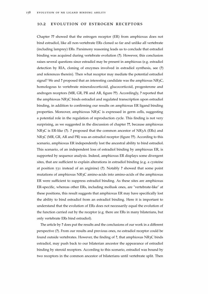
158 evolution of nr ligand binding ability
10.2 evolution of estrogen receptors
Chapter ?? showed that the estrogen receptor (ER) from amphioxus does not
bind estradiol, like all non-vertebrate ERs cloned so far and unlike all vertebrate
(including lamprey) ERs. Parsimony reasoning leads us to conclude that estradiol
binding was acquired during vertebrate evolution (?). However, this conclusion
raises several questions since estradiol may be present in amphioxus (e.g. estradiol
detection by RIA, cloning of enzymes involved in estradiol synthesis, see (?)
and references therein). Then what receptor may mediate the potential estradiol
signal? We and ? proposed that an interesting candidate was the amphioxus NR3C,
homologous to vertebrate mineralocorticoid, glucocorticoid, progesterone and
androgen receptors (MR, GR, PR and AR, figure ??). Accordingly, ? reported that
the amphioxus NR3C binds estradiol and regulated transcription upon estradiol
binding, in addition to confirming our results on amphioxus ER ligand binding
properties. Moreover, amphioxus NR3C is expressed in germ cells, suggesting
a potential role in the regulation of reproduction cycle. This finding is not very
surprising, as we suggested in the discussion of chapter ??, because amphioxus
NR3C is ER-like (?). ? proposed that the common ancestor of NR3A (ERs) and
NR3C (MR, GR, AR and PR) was an estradiol receptor (figure ??). According to this
scenario, amphioxus ER independently lost the ancestral ability to bind estradiol.
This scenario, of an independent loss of estradiol binding by amphioxus ER, is
supported by sequence analysis. Indeed, amphioxus ER displays some divergent
sites, that are sufficient to explain alterations in estradiol binding (e.g. a cysteine
at position 531 instead of an arginine) (?). Notably ? showed that some point
mutations of amphioxus NR3C amino-acids into amino-acids of the amphioxus
ER were sufficient to suppress estradiol binding. As these sites are amphioxus
ER-specific, whereas other ERs, including mollusk ones, are "vertebrate-like" at
these positions, this result suggests that amphioxus ER may have specifically lost
the ability to bind estradiol from an estradiol binding. Here it is important to
understand that the evolution of ERs does not necessarily equal the evolution of
the function carried out by the receptor (e.g. there are ERs in many bilaterians, but
only vertebrate ERs bind estradiol).
The article by ? does put the results and the conclusions of our work in a different
perspective (?). From our results and previous ones, no estradiol receptor could be
found outside vertebrates. However, the finding of ?, that amphioxus NR3C binds
estradiol, may push back to our bilaterian ancestor the appearance of estradiol
binding by steroid receptors. According to this scenario, estradiol was bound by
two receptors in the common ancestor of bilaterians until vertebrate split. Then
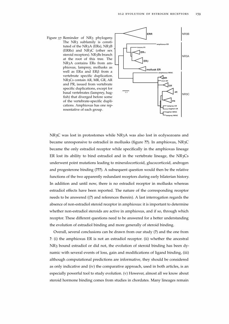
10.2 evolution of estrogen receptors 159
Figure 37: Reminder of NR3 phylogeny.The NR3 subfamily is consti-tuted of the NR3A (ERs), NR3B(ERRs) and NR3C (other sexsteroid receptors). NR3Bs branchat the root of this tree. TheNR3A contains ERs from am-phioxus, lamprey, mollusks aswell as ERα and ERβ from avertebrate specific duplication.NR3Cs contain AR, MR, GR, ARand PR, issued from vertebratespecific duplications, except forbasal vertebrates (lamprey, hag-fish) that diverged before someof the vertebrate-specific dupli-cations. Amphioxus has one rep-resentative of each group.
shark MR
hagfish NR3C
hagfish CR
lamprey CR
lamprey NR3C
0.3
MR
GR
AR
PR
ERR
mollusk ER
ERβ
ERα
amphioxus ER
lamprey ER
amphioxus NR3C
NR3B
NR3A
NR3C
NR3C was lost in protostomes while NR3A was also lost in ecdysozoans and
became unresponsive to estradiol in mollusks (figure ??). In amphioxus, NR3C
became the only estradiol receptor while specifically in the amphioxus lineage
ER lost its ability to bind estradiol and in the vertebrate lineage, the NR3Cs
underwent point mutations leading to mineralocorticoid, glucocorticoid, androgen
and progesterone binding (???). A subsequent question would then be the relative
functions of the two apparently redundant receptors during early bilaterian history.
In addition and until now, there is no estradiol receptor in mollusks whereas
estradiol effects have been reported. The nature of the corresponding receptor
needs to be answered ((?) and references therein). A last interrogation regards the
absence of non-estradiol steroid receptor in amphioxus: it is important to determine
whether non-estradiol steroids are active in amphioxus, and if so, through which
receptor. These different questions need to be answered for a better understanding
the evolution of estradiol binding and more generally of steroid binding.
Overall, several conclusions can be drawn from our study (?) and the one from
?: (i) the amphioxus ER is not an estradiol receptor. (ii) whether the ancestral
NR3 bound estradiol or did not, the evolution of steroid binding has been dy-
namic with several events of loss, gain and modifications of ligand binding, (iii)
although computational predictions are informative, they should be considered
as only indicative and (iv) the comparative approach, used in both articles, is an
especially powerful tool to study evolution. (v) However, almost all we know about
steroid hormone binding comes from studies in chordates. Many lineages remain
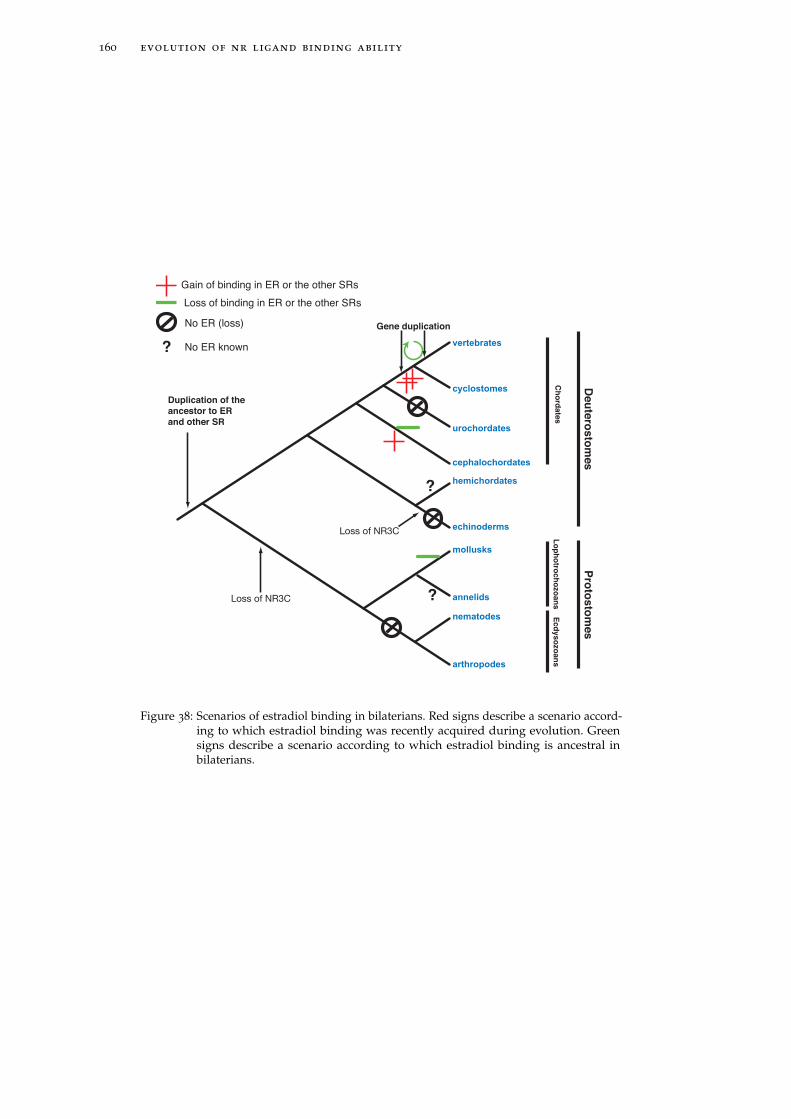
160 evolution of nr ligand binding ability
vertebrates
cyclostomes
urochordates
cephalochordates
hemichordates
nematodes
annelids
mollusks
arthropodes
echinoderms
Duplication of the
ancestor to ER
and other SR
Gene duplication
?
?
?
Loss of binding in ER or the other SRs
No ER (loss)
No ER known
Gain of binding in ER or the other SRs
Deu
tero
sto
mes
Ch
ord
ate
sL
op
ho
troch
ozo
an
sE
cd
yso
zo
an
s
Pro
tosto
mes
Loss of NR3C
Loss of NR3C
Figure 38: Scenarios of estradiol binding in bilaterians. Red signs describe a scenario accord-ing to which estradiol binding was recently acquired during evolution. Greensigns describe a scenario according to which estradiol binding is ancestral inbilaterians.
10.3 evolution of nr ligand binding 161
to be studied, especially in protostomes. (vi) Finally, and related to the former
statement, steroidogenesis should be much more thoroughly investigated, from
a non-anthropocentric point of view, in order to better grasp steroid hormone
signaling pathway outside vertebrates, and thus to better understand its evolution
(?).
10.3 evolution of nr ligand binding
The evolution of NRs has been seen as modular, with the appearance of ligand
binding as a control switch of NR transcriptional activity. However, it is still
unclear how this ability appeared and evolved. Although TR ligand binding has
been investigated with a rather small taxonomic sampling, it seems to have been
fairly well conserved, since all chordate TRs tested so far (to the exception of the
Ciona intestinalis TR) are TH-dependent transcription factors. The only difference
is the exact nature of the ligand: T3 in vertebrates and a T3 derivative, possibly
TRIAC, in amphioxus. This difference is conservative since vertebrate TRs do
bind TRIAC as well. Similarly, although ERs experienced several events of gain
and loss of ligand binding ability, the evolution of ligand binding in the NR3
family in general was confined to refinements of affinities to steroid hormones
(figure ??) and members of the NR4 subfamily appear to lack a ligand binding
pocket (in yellow on the figure ??). However, this conservative evolution cannot be
generalized to all families. For instance, some NR1 members are high affinity or
low affinity receptors of very different ligands (TR, RAR, VDR, PPAR, Rev-erb),
while others have constitutive ligands (RORα) or are still orphan (RORγ) (figure ??).
In the NR2 subfamily, our lab has contributed to bring to light the understanding
of RXR/USP evolution. Indeed, the ancestral RXR (in the common ancestor of
bilaterians and cnidarians) is supposed to display similar properties as the human
RXR for 2 reasons: first, mollusk USP (?), and cnidarian USP (?) bind 9cis-retinoic
acid and activate transcription upon ligand binding; second sequence analysis
revealed that LBPs from all RXRs (except arthropod ones) have experienced a
conserved evolution (??). In the arthropod lineage, RXR-USP became orphan and
in mecopterida (including insects and lepidopterans), USP ligand affinity changed
to phospholipid (?). Notably, in all arthropods, USP is found in an antagonist
conformation. Overall this RXR-USP example illustrates the plasticity of NR ligand
binding evolution. At the scale of the whole family, It is still very obscure how this
diversity in ligand binding arose.
Although it is not unlikely (although not clear yet) that the ancestral NR3 was
a steroid receptor (??), the nature of the ancestor of NR1, of NR2... or of all NRs
162 evolution of nr ligand binding ability
is much more elusive. The structure of different LBDs revealed a common 3D
structure but different strategies of activation, i.e. a similar active conformation
but different activation processes (see the introduction of chapter ?? for further
details). This diversity suggests that NR ligand binding evolution has necessarily
been non-linear and is difficult to assess. Indeed, ligand binding properties are
fast evolving (for instance human LRH-1 is a ligand-dependent transcription factor
whereas mouse LRH-1 activity is believed to be ligand-independent with a large
empty pocket (?)). As ? highlighted, NRs are evolutionary plastic, which may have
blurred the signal of the ancestral state to a point where it becomes difficult to
unearth.
Although ancestral reconstructions should be considered as only indicative (see
chapter ??), what may these approaches teach us about the evolution of NRs? In
order to try and estimate the ancestral NR state, it is possible to map on an NR
phylogenetic tree the characteristics of each NR and to use statistics to infer the
ancestral state. I have thus inferred the 2 following characteristics of ancestral NRs:
(a) the size of the ligand binding pocket as well as (b) ligand binding properties.
The different states that were tested were (i) specific and high affinity endocrine of
paracrine receptors (like TR or ER) ; (ii) poorly specific and low-affinity nutritional
sensors (like PPAR or LXR) ; (iii) receptors with structural ligands (like some USP),
(iv) receptors with an empty pocket (like ERR); or (v) no pocket (like Nurr1) (?).
This experiment was done with the software Mesquite (?)
When one follows this procedure, the ancestral NR is estimated to have (a) a
rather small pocket (∼ 420 Å3, figure ??), like high affinity endocrine or paracrine
NRs. In addition, using a model where all transitions between the different tested
ligand binding properties are equiprobable, (b) 3 states are considered as signifi-
cantly better than the other ones: "endocrine or paracrine", "no pocket" and "sensor"
states were all likely (P-values of 0.67, 0.11 and 0.14, respectively).
The experiments of reconstruction of ancestral LBP volumes and ligand binding
properties gave similar results: the ancestral NR is inferred to be a high affinity
receptor. Indeed, endocrine or paracrine receptors (that are high affinity receptors)
usually have a small LBP that just fits the ligand (e.g. the volume of the RARγ
LBP is 412 Å3 (?) whereas the volume of its ligand 9-cis retinoic acid is 291 Å3).
Following this hypothesis, the nature of the ancestral ligands remains an enigma
that will be difficult to assess. A complementary approach may be to estimate the
gene-content of the ancestral organism, to determine which metabolic pathway
was present, that may reflect the endogenous ligand. However, one should keep
in mind that the evolutionary origin of a ligand may have been exogenous (like a
vitamin) and its endogenous synthesis subsequently acquired during evolution (?).
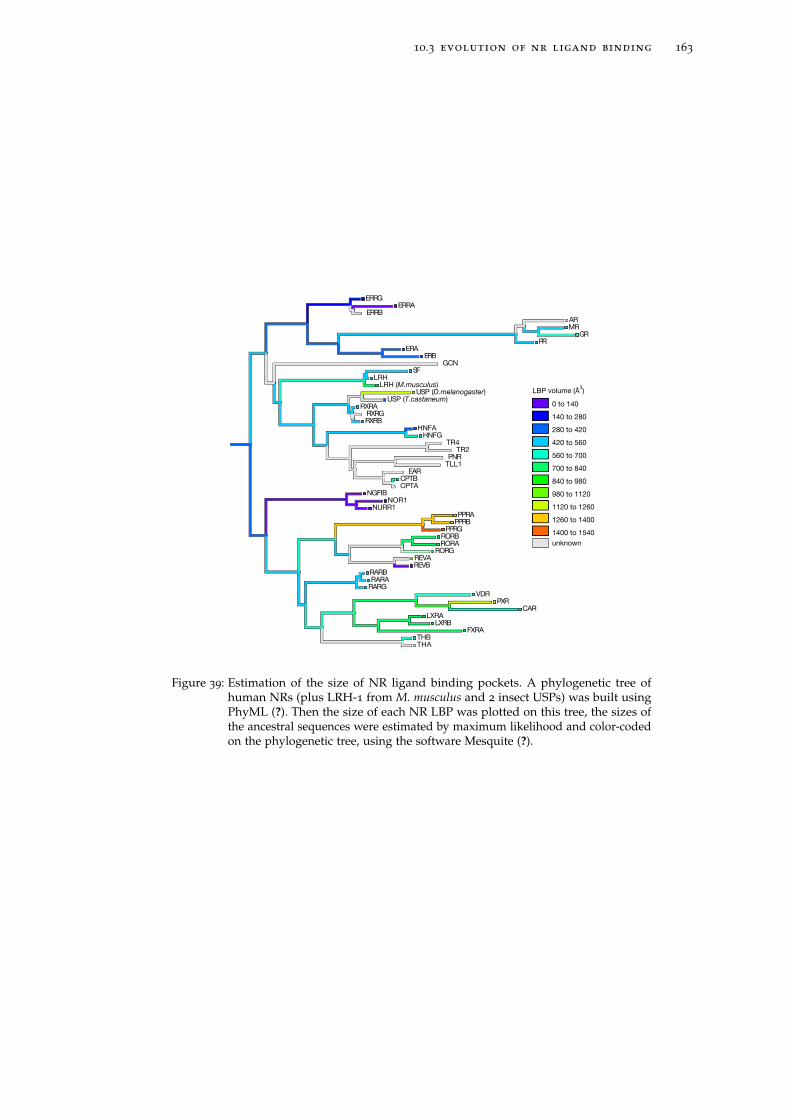
10.3 evolution of nr ligand binding 163
ERRGERRA
ERRBARMR
GRPR
ERAERB
GCNSF
LRHLRH (M.musculus)
USP (D.melanogaster)USP (T.castaneum)
RXRARXRGRXRB
HNFAHNFG
TR4TR2
PNRTLL1
EARCPTBCPTA
NGFIBNOR1
NURR1PPRA
PPRBPPRG
RORBRORA
RORGREVAREVB
RARBRARA
RARGVDR
PXRCAR
LXRALXRB
FXRATHBTHA
0 to 140
140 to 280
280 to 420
420 to 560
560 to 700
700 to 840
840 to 980
980 to 1120
1120 to 1260
LBP volume (Å3)
1260 to 1400
1400 to 1540
unknown
Figure 39: Estimation of the size of NR ligand binding pockets. A phylogenetic tree ofhuman NRs (plus LRH-1 from M. musculus and 2 insect USPs) was built usingPhyML (?). Then the size of each NR LBP was plotted on this tree, the sizes ofthe ancestral sequences were estimated by maximum likelihood and color-codedon the phylogenetic tree, using the software Mesquite (?).
164 evolution of nr ligand binding ability
One of the possible ancestral states inferred from our analysis, an orphan receptor
with an empty pocket is in agreement with our estimation of the ancestral LBP
size, since some NRs with a small pocket are transcriptionally active in the absence
of a ligand (e.g. ERR). This scenario of an ancestral orphan receptor, that was a
ligand-independent transcription factor has previously been proposed (??). This
hypothesis was based on the observation that NR phylogeny does not reflect the
distribution of ligands in the tree (e.g. steroid receptors are paraphyletic) and
the size of the LBP. This hypothesis also allows tan easier interpretation of the
apparently complex evolution of ligand binding since an ancestral small LBP could
have adapted to various evolutionary scenarios without no loss of function. Indeed
the ancestral NR could have evolved from a state of no binding to independent
grains of ligand binding by parallel point mutations.
The third hypothesis of an ancestral "sensor" receptor is appealing for several
reasons. First, the NR family appeared in the ancestral metazoan (?), which was
most probably a multicellular animal with few cellular types and displaying
a limited level of integration. Thus an endocrine or paracrine function seems
rather useless in such conditions. Intuitively, the ancestral NR may have served
as an environmental "sensor", that could have interacted with many compounds
(like extant "sensors" (?)) and would thus have helped reacting in an appropriate
way to external conditions (abundant food? toxic compounds? etc...). Indeed,
such a transcription factor which activity is regulated upon metabolic or foreign
compounds is found in humans (e.g. the NRs PPAR and PXR) but also in yeast (e.g.
Oaf1 and Pdr1p). In humans PPAR has a large LBP (figure ??) that binds products
of fatty acid metabolism (?) and PXR, also with a large LBP, binds a wide range of
foreign compounds and is involved in detoxification (?). In yeast, Oaf1 is known
to induce fatty acid metabolism. Although its proposed homology with NRs is
doubtful, the protein was predicted to have a NR LBP-like 3D structure, it was also
shown to interact with oleate and to activate transcription upon ligand-binding
(?). Another study highlighted the analogous function of the human sensor PXR
and the yeast TF Pdr1p: they both bind structurally diverse compounds with low
affinity (µM order) and regulate gene activation upon ligand binding (?). Although
there is no clear evidence that the above yeast TFs and NRs are homologous, these
examples illustrate the biological role that NRs may have played in our ancient
animal ancestor. Second, The hypothesis of an ancestral "sensor" explains the
existence of a LBP in so many NRs and provides an evolutionary framework to
the evolution of the many activation strategies that appeared in the NR family.
Indeed, if the ancestral NR had a large LBP in which many compounds may
have fit in it, then it could have secondarily easily adapted to one specific ligand,
10.4 conclusions on the evolution of nrs 165
like for endocrine NRs, or lose its dependancy to ligand binding, like in NR4.
This hypothesis of an ancestral role of NRs in the regulation of xenobiotic action
has been previously proposed (?) and is supported by a phylogenetic analysis of
genes involved in steroidogenesis recently performed in our lab, that suggests that
extant NR ligands are the evolutionary end-product of metabolic pathway linked
to xenobiotics degradation (environmental chemicals) (?). However, although this
hypothesis is attractive, it is still very speculative. Moreover, an ancestral "sensor"
NR would probably have a large LBP (like LXR), which is not supported by the
estimation of the ancestral LBP volume (figure ??).
Overall, the above experiment seems to favor a ligand-binding state, to the
opposite of ligand-independent state. However, the resolution is still very poor,
since only 2 states out of 5 were rejected with the model used. It is possible that a
model where all transitions are equiprobable is not realistic. On the other hand,
the taxonomic sampling used here may be biased. Indeed human NRs were almost
exclusively used because almost all functional assays were done on them. It will
be very interesting in the next future to broaden our knowledge of NR ligand
binding ability (especially in NR2) but most importantly to perform functional
studies on NRs from other species. In this respect, NRs have been reported in all
bilaterians (e.g. TR (?)), and in other metazoan groups like cnidarians (e.g. RXR (?)),
placozoans (e.g. ERR (?)) or sponges (e.g. RXR (?)). In these species that diverged
from human a long time ago, NRs may have evolved totally different characteristics
that should be informative regarding ancestral NR states. Consequently, a larger
taxonomic sampling is absolutely necessary to better understand the evolution of
NRs.
10.4 conclusions on the evolution of nrs
Overall, the evolution of NR may be seen as follows: the ancestral NRs were
transcription factors that bind DNA on combinations of (AGGTCA) motifs. How
their transcriptional activity was originally regulated is still elusive. Was it ligand-
binding dependent? Independent? As discussed above, more work needs to be
done. What can be concluded here, is that the subsequent evolution was chaotic,
with several episodes of gain, loss and alterations of ligand binding.
Although the evolution of ligand binding has focused the attention of evolution-
ary biologists, other NR features may be worth digging into, since the mode of
action of NRs is better and better understood. For instance, how does the target
gene repertoire evolve? A better understanding of how NR cis-regulatory sequences
evolve should contribute to shed light on the evolution of NR function and more

166 evolution of nr ligand binding ability
generally of the regulatory network they are involved in. Such large-scale studies of
cis-regulation are now technically possible and are appearing in the litterature (?),
some of which focus on the evolutionary component of cis-regulation (?). Another
issue of NR evolution may relate to the evolution of cofactors interaction and more
generally of NR-protein interaction, which has been overlooked, except for few
studies (e.g. (?)). These issues should help us understand the evolution of NR
function in a broader context than the biochemical properties of the receptor alone
and maybe illustrate the evolution of transcription factors in general.
11E V O L U T I O N O F M E TA M O R P H O S I S I NC H O R D AT E S
As described in the introduction, because chordate metamorphoses are morpho-
logically very diverse, they were proposed by several authors to have appeared
independently several times in the chordate lineage (????). Recent data, including
the work I have done during my PhD, have nevertheless shown that, in chordates
like amphibians and actinopterygians, THs and other thyroactive compounds
are playing a key role in the control of metamorphosis. The conservation of the
molecular determinism of metamorphosis suggests a unique and ancient origin
for this key post-embryonic developmental process in the chordate lineage. More
precisely, we proposed that the chordate ancestor had an indirect development and
metamorphosed under iodothyronine derivative stimulation (?). This hypothesis
unifies all the postembryonic developmental stages regulated by THs and their
derivatives since it implies that they are all homologous. This model also pro-
vides an evolutionary framework through which metamorphosis has diversified
within chordates. We speculate that this hypothesis can even be extended to the
deuterostome super-phylum, although key data are still missing to unambiguously
demonstrate that TH-regulated metamorphosis is shared by all deuterostomes.
In the next sections, I will explain our model and give some examples of evolution
of metamorphosis from alterations of TH signaling pathway (the following text
was modified from (?)).
11.1 the evolution of metamorphosis is governed
by the evolution of the th signaling path-way.
Our hypothesis that metamorphosis is an ancestral feature of chordates is sup-
ported by the conservation of its molecular determinism. Indeed THs, other iodi-
nated tyrosine derivatives and their receptor TR have been systematically involved
in metamorphosis in all chordates in which their role has been tested (as detailed
in chapter ?? of the introduction, and in chapters ??, ?? and ?? of this manuscript),
even in animals located at very distant branches of the chordate phylogenetic tree
(amphibians, actinopterygian fishes and amphioxus). Then parsimony reasoning
167
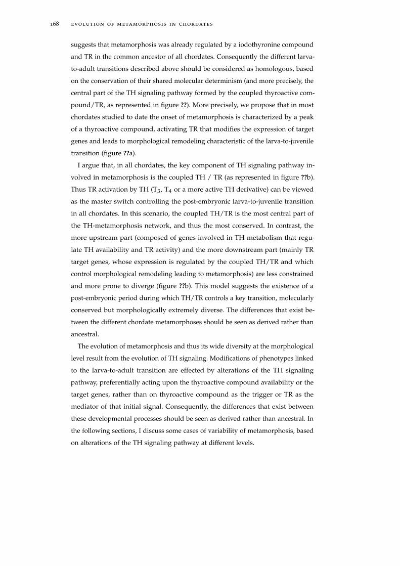
168 evolution of metamorphosis in chordates
suggests that metamorphosis was already regulated by a iodothyronine compound
and TR in the common ancestor of all chordates. Consequently the different larva-
to-adult transitions described above should be considered as homologous, based
on the conservation of their shared molecular determinism (and more precisely, the
central part of the TH signaling pathway formed by the coupled thyroactive com-
pound/TR, as represented in figure ??). More precisely, we propose that in most
chordates studied to date the onset of metamorphosis is characterized by a peak
of a thyroactive compound, activating TR that modifies the expression of target
genes and leads to morphological remodeling characteristic of the larva-to-juvenile
transition (figure ??a).
I argue that, in all chordates, the key component of TH signaling pathway in-
volved in metamorphosis is the coupled TH / TR (as represented in figure ??b).
Thus TR activation by TH (T3, T4 or a more active TH derivative) can be viewed
as the master switch controlling the post-embryonic larva-to-juvenile transition
in all chordates. In this scenario, the coupled TH/TR is the most central part of
the TH-metamorphosis network, and thus the most conserved. In contrast, the
more upstream part (composed of genes involved in TH metabolism that regu-
late TH availability and TR activity) and the more downstream part (mainly TR
target genes, whose expression is regulated by the coupled TH/TR and which
control morphological remodeling leading to metamorphosis) are less constrained
and more prone to diverge (figure ??b). This model suggests the existence of a
post-embryonic period during which TH/TR controls a key transition, molecularly
conserved but morphologically extremely diverse. The differences that exist be-
tween the different chordate metamorphoses should be seen as derived rather than
ancestral.
The evolution of metamorphosis and thus its wide diversity at the morphological
level result from the evolution of TH signaling. Modifications of phenotypes linked
to the larva-to-adult transition are effected by alterations of the TH signaling
pathway, preferentially acting upon the thyroactive compound availability or the
target genes, rather than on thyroactive compound as the trigger or TR as the
mediator of that initial signal. Consequently, the differences that exist between
these developmental processes should be seen as derived rather than ancestral. In
the following sections, I discuss some cases of variability of metamorphosis, based
on alterations of the TH signaling pathway at different levels.
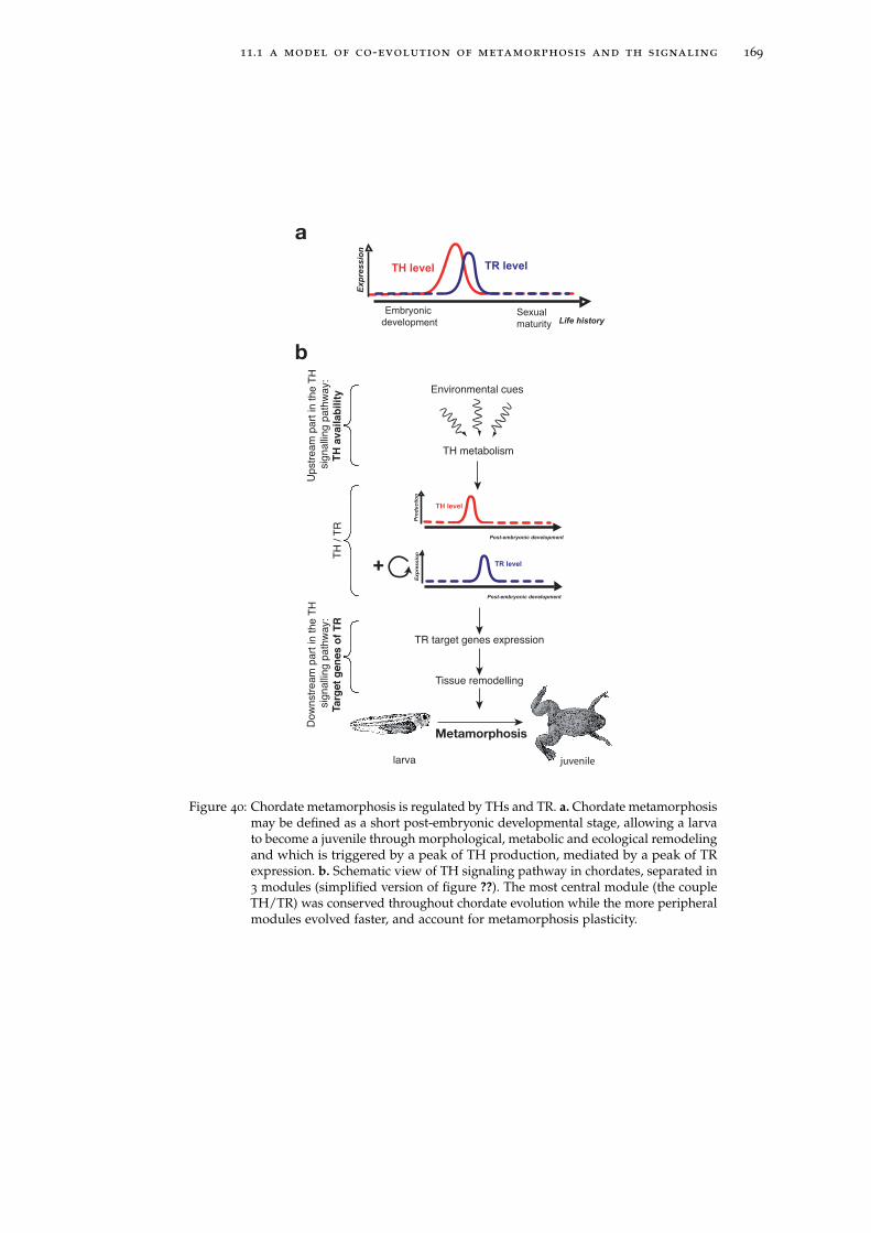
11.1 a model of co-evolution of metamorphosis and th signaling 169
Pro
du
cti
on
TH level
Post-embryonic development
Exp
ressio
n
Environmental cues
TH metabolism
TR target genes expression
Tissue remodelling
larva juvenile
+ TR level
TH
/ T
R
Upstr
eam
part
in t
he T
H s
ignalli
ng p
ath
way:
TH
avail
ab
ilit
y
Dow
nstr
eam
part
in t
he T
H s
ignalli
ng p
ath
way:
Targ
et
gen
es o
f T
R
Exp
ressio
n
Embryonic
developmentSexual
maturity
TH level TR level
Life history
Post-embryonic development
a
b
Metamorphosis
Figure 40: Chordate metamorphosis is regulated by THs and TR. a. Chordate metamorphosismay be defined as a short post-embryonic developmental stage, allowing a larvato become a juvenile through morphological, metabolic and ecological remodelingand which is triggered by a peak of TH production, mediated by a peak of TRexpression. b. Schematic view of TH signaling pathway in chordates, separated in3 modules (simplified version of figure ??). The most central module (the coupleTH/TR) was conserved throughout chordate evolution while the more peripheralmodules evolved faster, and account for metamorphosis plasticity.
170 evolution of metamorphosis in chordates
11.2 chordate metamorphosis and alterations
of th signaling: some examples
11.2.1 Heterochrony and evolution of metamorphosis in am-phibians: alteration of the TH signaling pathway.
In amphibians, metamorphosis can be spectacular, as seen in the anuran Xeno-
pus laevis. Still, amphibians exhibit a wide range of variations in the intensity
of the morphological remodeling during metamorphosis. The modifications can
be relatively inconspicuous, as for urodeles and apodes, or even reduced to the
point of vanishing, as in neotenic amphibians. On the other hand the larval stage
can be suppressed, as in direct developing frogs, where little frogs directly hatch.
However, as will be exemplified, in all cases there is a TH sensitive period of
morphological remodeling. TH signaling was studied in the entirely terrestrial
and direct developing frog Eleutherodactylus coqui in an attempt to understand the
molecular basis of the apparent suppression of the larval period. It was shown that
E. coqui does metamorphose under TH stimulation but the main difference with
“classic” amphibian metamorphosis is the very early triggering of this metamor-
phosis, as it occurs inside the egg (?). More precisely, during late developmental
stages, there is a TH-dependent period of morphological remodeling (including
cartilage remodeling, tail regression or limb elongation) correlated with a peak
of TR? expression (?). Consequently, E. coqui metamorphosis is rather similar to
the “classical anuran”, except that the timing of TH production and TR expression
have probably been shifted earlier during development. It is also possible that the
TR target genes have diverged as well, since the morphological remodeling is not
as drastic as in anurans. E. coqui development is a clear example of heterochrony
(species-specific variation in the timing of the development, leading to changes in
the phenotype of the species).
Alternative changes of TH signaling lead to metamorphoses that are very mild
and make adults look too much like the larvae to be described as metamorphic.
These animals are called paedomorphic, because the adults have retained larval
traits, and constitute another case of heterochrony. As described in the introduction,
there are several cases of paedomorphosis: in facultative paedomorphic species, like
the Mexican axolotl (Ambystoma mexicanum), the tadpole responds to exogenous
TH by metamorphosing, but does not spontaneously metamorphose (?). In fact
the axolotl is a spectacular example of a series of species of the genus Ambystoma
in which several types of unusual post-embryonic life history strategy occur (?).
Facultative neoteny has been attributed especially to low levels of circulating
11.2 chordate metamorphosis and alterations of th signaling: some examples 171
THs due to weak activation of the thyroid axis by the pituitary gland, when the
rest of TH signaling pathway is “normally” functional (??). Obligatory neotenic
amphibians (such as the perennibranchiates Urodeles Necturus or Proteus), long
thought to be insensitive to THs, were in fact shown to respond to exogenous TH
but with limited amplitude: for instance expression of the typical metamorphosis
genes TR? and stromelysin are induced by TH (?). Rather, the lack of response to
TH is probably caused by the loss of TH regulation by some target genes such as
collagenase 3.
These examples of “alternative” metamorphoses illustrate the important plas-
ticity of the metamorphosis process in different amphibians. Each time, TR was
shown to be functional (??). Rather, the genetic variations thought to be responsible
for phenotypic differences rely on the production of TH (an early production in E.
coqui, or a default of TH production in the axolotl) or on the target genes (as in the
Necturus) or both. These extreme examples show that metamorphosis triggered by
TH is always present in amphibians, even if in some cases it is weakened or shifted
during late development stages.
11.2.2 Evolution of metamorphosis in other vertebrates: whydirect development is not so direct in chordates.
Under the hypothesis of an ancestral metamorphosis in the chordate lineage, then
descending species should have divergent variants of this developmental stage.
According to our definition of metamorphosis as a post-embryonic developmental
stage characterized by a thyroactive compound-dependent morphological remod-
eling, groups that were previously thought to have a direct development may in
fact be seen as animals with indirect development and a mild metamorphosis.
Indeed, similarly to neotenic amphibians having a mild metamorphosis, mammals
metamorphose. In these lineages, the metamorphosis stage does not coincide with
birth since the latter is not marked by an intense TH signaling. Sexual maturity
does not qualify as metamorphosis either since it is regulated by steroids and is
often asynchronous with metamorphosis. On the contrary, mammalian weaning
gathers all characteristics of metamorphosis as defined above as a period of tissue
remodeling governed by TH (?). It is an ecological shift that is characterized by
modifications like neural maturation, intestine remodeling or chondrogenesis that
are reminiscent of similar events during amphibian metamorphosis. These modifi-
cations occur in correlation with a peak of TH production (both T3 and T4) (?) and
were shown to be under the control of TH and TR (???). Consequently, weaning
should be viewed as homologous to classical metamorphosis even if the precise
172 evolution of metamorphosis in chordates
role of THs in controlling this event and the regulatory hierarchy controlled by
these hormones should be better analyzed. In addition weaning is poorly studied
outside classical mammalian models (e.g. mouse, rat, human) and it should be
analyzed in a wider taxonomic sampling.
As discussed above, in actinopterygians, and especially in flat fishes, THs are
important regulators of metamorphosis (?), the best known case being flounder,
for which T4 and T3 were reported to induce precocious metamorphosis (?). But
the role of THs in post-embryonic development has also been documented in other
groups. In zebrafish, THs were shown to be important for the larva-to-juvenile
transition: exogenous THs induce differentiation of the pectoral fin precociously,
in contrast the inhibition of TH synthesis blocks it (?). Smoltification in salmonid
fishes is considered to correspond to metamorphosis: it is a period of high T4
and T3 levels (??) involving a morphological, physiological and behavioral switch
from a freshwater-adapted form to a salt-water-adapted form. At least some of
the former modifications were shown to be TH-sensitive (??). The larval to adult
transition is extremely important in terms of recruitment in many fish species
that are living close to the substratum, such as coral fishes. Although a direct
role of thyroid hormones in regulating metamorphosis has not yet been clearly
demonstrated, in some species like the grouper, existing evidence clearly points in
this direction (?).
In lamprey, although it is now well established that TH endogenous synthesis
needs to be tightly regulated for proper metamorphosis, the role of TH displays
a remarkable difference compared to other chordates. Strikingly, several reports
clearly show that it is a drop, instead of an increase, in circulating TH that ends the
larval period (???????). Among other factors, temperature and growth advancement
(indicated by length and weight, for instance) are important parameters: lampreys
enter metamorphosis when they have reached a certain size (?). Interestingly even
if the inversion of TH action is unique to lampreys, TR is still likely to play a role
in this TH-mediated action since nuclear TH binding was reported (?) and the TRs
from lamprey are activated by TH (?). Given that it is a drop of TH levels that
triggers metamorphosis, TR’s role in controlling metamorphosis must be different
in lamprey than in other vertebrates. It is possible that in lamprey, TR induces
the expression of genes that inhibit metamorphosis. According to this scenario,
when TH levels drops, TR will stop inducing the expression of these inhibitors,
then triggering metamorphosis. However, many other hypotheses can be proposed,
since in vivo functional experiments are still to be completed. The use of specific
TR agonists and antagonists (???) could help deciphering the specific role of TR in
this inverted situation.
11.2 chordate metamorphosis and alterations of th signaling: some examples 173
Another interesting case of divergence on the TH signaling pathway is provided
by urochordates. Metamorphosis of Ciona intestinalis can be induced by exogenous
THs and inhibited by goitrogens known to block TH synthesis in vertebrates
(??). However, the molecular pathways triggered by THs remain elusive since the
only TR of Ciona intestinalis (?) does not bind any iodinated tyrosine derivative
tested (??). The urochordates are particularly interesting regarding the evolution of
metamorphosis because of the diversity of their post-embryonic developmental
strategies. The elucidation of the role of THs and iodinated tyrosine derivatives in
general in late development in other species, and in particular in larvaceans should
bring interesting data for comparative analysis.
11.2.3 TH signaling pathway and development outside chor-dates
The role of THs in metamorphosis might even be extended to all deuterostomes. In-
deed, most echinoderms metamorphose, although their life history is quite variable
from one species to another (?). Interestingly, effects of TH on the metamorphosis
of echinoderm larvae have now been reported in several families, including species
with feeding and nonfeeding development: Echinoidea (sea urchins, sea biscuits,
and sand dollars) asteroids (sea stars) and ophiuoidea (brittle stars) ((?) and refer-
ences therein). Moreover, outside chordates, echinoderms are the only animals that
display clear endogenous TH metabolism: T4 production was demonstrated in sea
urchin and sea biscuits (??). Interestingly orthologs of many proteins involved in
mammal TH metabolism were found in the sea urchin genome (??). This suggests
an even more ancestral origin of TH endogenous synthesis in deuterostomes. One
TR was found in the genome of the sea urchin Strongylocentrotus purpuratus (?). It is
not as divergent as the TR from Ciona intestinalis, making it a potential TH receptor.
It will be interesting to test in vitro if this receptor can bind any iodinated tyrosine
derivative and regulate transcription of TRE-containing genes in response to this
hormone. As THs seem to play a similar role in echinoderm metamorphosis as in
chordates, the involvement of TR in this process would be of particular interest.
TR genes were recently described in some protostomes (mollusks, flatworms
and crustaceans) (?), implying that the gene was present in the common ancestor
of all bilaterians and was secondarily lost in insects and nematodes. It will be
very interesting to decipher the function of these genes. The encoded proteins are
apparently able to bind similar DNA sequences and to heterodimerize with the
classical nuclear receptor partner RXR (?) but are they active transcription factors?
More particularly are they responsive to THs? Attempts to determine if THs had
174 evolution of metamorphosis in chordates
any effects on protostome development are scant and relatively preliminary. One
study reported that THs accelerated metamorphosis in the mollusk abalone (?)
and another one in a crustacean (a prawn (?)). In contrast THs did not seem to
have any effect on the crustacean Daphnia (?) in accordance with the observation
that no endogenous TH is present in this species (?). Consequently, hypotheses on
the roles of THs or other derivatives and TR in protostomes can only be tentative:
a potential responsiveness to THs would imply a very ancient function of TR as
the TH receptor or, on the other hand, the establishment of an alternative function
would imply an evolutionary shift. In any case, it is necessary to better characterize
the TH signaling pathway in protostomes: are they able to produce iodinated
tyrosine derivatives, and if they do so, how? Are the TRs functional? What are the
molecular events triggered by iodinated tyrosine derivatives? To what extent do
they regulate metamorphosis? Those questions need to be addressed, in order to
fully understand the evolution of TH signaling in bilaterian and its role in late
development.
11.3 plasticity of th signaling and plasticity
of metamorphosis
11.3.1 The evolution of metamorphosis is tightly linked to en-vironmental conditions.
We proposed a new definition of metamorphosis highlighting the role of THs
and TH derivatives as the triggering element and TR as the direct mediator of
TH signal in chordates. This definition is based on the hypothesis of an ancestral
origin of TH and thyroactive compound-induced metamorphosis in chordates.
From this definition, all chordates metamorphose under thyroactive compound
stimulation. We postulate that the impressive diversity of metamorphic phenotypes
is constrained by the divergence of the TH signaling in the chordate groups.
Interestingly metamorphosis is intrinsically linked to the environment, since it
allows an animals to change its ecological niche. Alterations of the TH signaling
pathway may then allow a species to adapt to a constantly changing environment
(see chapter ??). The fast changing environment may constrain a fast adapting TH
signaling and a fast evolving metamorphosis.
An elegant example of the fast evolution of amphibian metamorphosis has
recently been demonstrated by ?, who used geographical data to clarify the appar-
ently complex evolution of metamorphosis in marsupial frogs. Marsupial frogs
are a group of amphibians in which females possess a pouch on their back, where
11.3 plasticity of th signaling and plasticity of metamorphosis 175
they carry their eggs. In some frogs, these eggs hatch directly into froglets, in
others, they hatch into tadpoles. Direct-developping and biphasic frogs do not
seem to constitute monophyletic groups, as shown by a phylogenetic tree based
on the analysis of several loci. Notably, a group of biphasic frogs seems to be
nested inside a clade of direct-developping frogs. Two hypotheses could explain
this phylogenetic distribution: either (a) direct development has appeared indepen-
dently many times, or (b) a tadpole phase has reappeared from direct-developing
ancestors. This second hypothesis seems a priori unlikely, as many examples of
transitions from biphasic to direct development are known, and almost no example
shows the other kind of transition. The authors reconstructed the evolution of
frog development along the phylogeny using two different probabilistic models of
character evolution. A simple model of evolution provides support for hypothesis
(b), but a more complex model supports hypothesis (a). This second model is
significantly more likely, so this would appear the case is closed, if geographical
data had nothing more to say. In fact, analysing the geographical distribution of
marsupial frogs, the authors found that biphasic frogs live at a higher altitude than
direct-developing ones. They then used a probabilistic model to infer the altitudes
of various marsupial frog ancestors. They found that ancestors of the biphasic
group of frogs lived at an altitude more consistent with a direct-developing lifestyle
than with a biphasic lifestyle, in agreement with hypothesis (b).
Overall, this study finely illustrates the adaptability of metamorphosis to envi-
ronmental changes. It would be interesting to study the evolution of TH signaling
in the different above species, as differences of TH signaling between species
may reflect the heterochrony. It was proposed that amphibian paedomorphy is an
adaptation of amphibian post-embryonic development to specific environmental
conditions, in which the aquatic niche is more stable than the terrestrial one. As
Garstang wrote in a poem (?):
"But when a lake’s attractive, nicely aired, and full of food
They climb to youth perpetual, and rear a tadpole brood[...]
They think aquatic life is bliss, terrestrial is curse"
More prosaically, species that live in a favorable aquatic environment and whose
larval stage is extended will be have a selective advantage compared to other
species that do not benefit as much from the favorable environment. Larval prolon-
gation may reach sexual maturity, causing animals to reproduce without metamor-
phosing (?). This sort of paedomorphy, called neoteny, may appear rapidly through
alterations of endocrine regulation by the TH signaling pathway (see section ??)
and thus allows to give a rapid answer to specific environmental conditions.
176 evolution of metamorphosis in chordates
11.3.2 Modulation of TH availability
The plasticity of the TH pathway allows evolution of the regulation of metamor-
phosis within the chordates, as proposed in the above example. The action of
thyroactive compounds, mediated by TR, is dependent on the availability of the
compound to each cell, as well as on the list of potential target genes. In am-
phibians and mammals, this control is observed at several levels: global TH level
is regulated by the hypothalamo-pituitary axis while the local levels are tightly
regulated by membrane transporters (?) and deiodinases (?). The situation is po-
tentially similar in all chordates where some orthologous genes to TH transporters
have been isolated (like the C.intestinalis orthologous gene to Oatp14, involved in
transport of T3 through the blood-brain barrier (??)). Of course, the expression
pattern of TR is a central parameter, but the role of its partners (coactivators and
corepressors) should not be neglected. A crucial aspect in the diversification of
the metamorphosis pattern observed in vertebrates is the plasticity of the TH-
regulated gene network. In Xenopus it has been shown that THs control a specific
gene regulatory hierarchy through TR activation. It is likely that, as observed in
obligate paedomorphic species, this gene network is modified in different species,
resulting in a distinct outcome in terms of metamorphosis. This importance of the
gene regulatory cascade controlled by a relatively invariant master regulator is
reminiscent of the situation observed in other cases such as eye development (with
pax6 being the master gene (?)) or antero-posterior development of arthropods
(with Hox genes being the key selector genes (?) and more generally (?)). Indeed,
TH-regulated gene expression pattern is highly variable from one species to an-
other, as shown in X. laevis and the mouse (??). It should be interesting to compare
gene expression in different vertebrate groups. One of the only common pathways
is the TH-induced positive upregulation of one of the TR genes occurring during
amphibian, fish or amphioxus metamorphosis (??) and mammalian weaning (TRα,
(??)) in concert with a peak of TH production (?). The TR gene activated in these
various groups is not the same though: TRβ is the key target in X. laevis whereas
in actinopterygians and mammals TRα appears to be more important.
11.4 perspectives for the study of the evolu-tion of chordate metamorphosis
One should keep in mind that the model of metamorphosis evolution presented
in this chapter is based on data accumulated in a few model species that repre-
sent a very restricted taxonomic sampling of vertebrates. Weaning in mammals,
11.4 perspectives for the study of the evolution of metamorphosis 177
metamorphosis in urochordates or larval to adult transitions in actinopterygian
fishes (not to mention the post-embryonic development in sauropsids (that include
birds, lizards, snakes and turtles, figure ?? in chapter ??) are known from work on
very few model systems. It will be very important in the future to study the role
of THs and their derivatives and TR in the coordination of this neglected period
of the vertebrate post-embryonic development. We nevertheless believe that the
model proposed in this paper offers a coherent view of chordate post-embryonic
development in general and of chordate metamorphosis in particular.
general conclusions about the evolution of
tr and chordate metamorphosis
During my PhD, I sought the role of the transcription factor TR in the evolution of
metamorphosis in chordates. I would like to take the opportunity in this conclusion
to place my work in the more general context of the role of developmental gene
regulation in the evolution of phenotypes.
Evo-devo studies have focused on the role of transcription factors (TFs) in the
evolution of phenotypes. As they are master regulators of early development,
they are of particular interest in such investigations, as these proteins are key to
the establishment of body axes and body shape. They are at the core of a gene
regulatory network that governs early development, so that any alteration of their
function should have massive consequences on development and on body shape
(?). There has been an intense debate recently about which component of the devel-
opmental regulatory network are most important in evolution: are modifications of
coding fractions of the genome (and more precisely in TF sequences) responsible
for the differences between species? Are alterations in non-coding fractions of the
genome responsible for the spatio-temporal expression of genes more prone to
underlie evolution (e.g. by altering the TF expression pattern or the list of its target
genes), while TFs have been kept fairly conserved throughout animal evolution?
The discovery that most animals share a common set of developmental regulatory
genes has drawn considerable attention on the cis-regulatory sequences as raw
material for evolutionary changes (?). However, as illustrated on the figure ??, the
molecular mechanisms through which the evolution of TFs underlies the evolution
of body shape is still sharply debated (e.g. (?????), see (?) for a recent summary).
More generally, the evolution of TFs is then to be considered in the context of
the evolution of the genetic regulatory network (GRNs) TFs are involved in, and
which govern development. It was proposed that alterations of GRNs underlie the
evolution of morphology (??). Following this model, how may GRNs evolve?
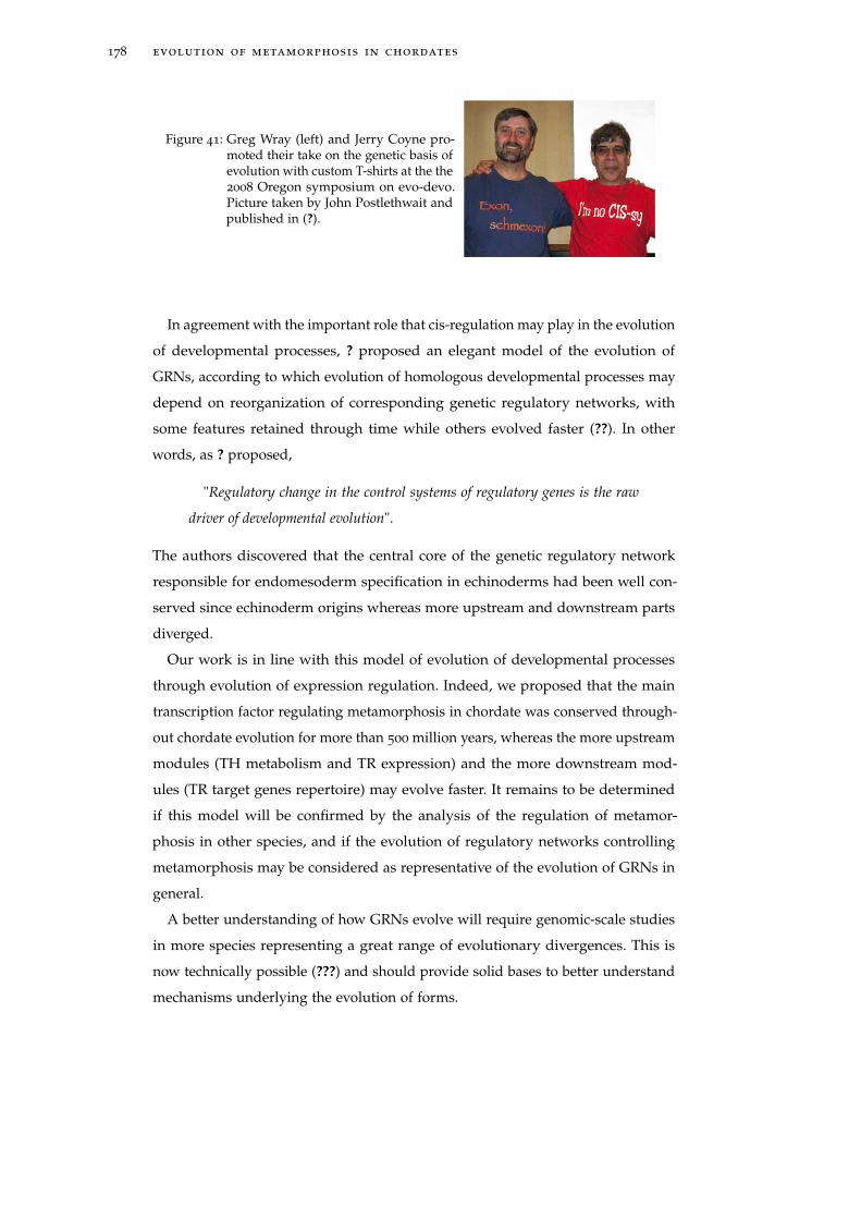
178 evolution of metamorphosis in chordates
Figure 41: Greg Wray (left) and Jerry Coyne pro-moted their take on the genetic basis ofevolution with custom T-shirts at the the2008 Oregon symposium on evo-devo.Picture taken by John Postlethwait andpublished in (?).
In agreement with the important role that cis-regulation may play in the evolution
of developmental processes, ? proposed an elegant model of the evolution of
GRNs, according to which evolution of homologous developmental processes may
depend on reorganization of corresponding genetic regulatory networks, with
some features retained through time while others evolved faster (??). In other
words, as ? proposed,
"Regulatory change in the control systems of regulatory genes is the raw
driver of developmental evolution".
The authors discovered that the central core of the genetic regulatory network
responsible for endomesoderm specification in echinoderms had been well con-
served since echinoderm origins whereas more upstream and downstream parts
diverged.
Our work is in line with this model of evolution of developmental processes
through evolution of expression regulation. Indeed, we proposed that the main
transcription factor regulating metamorphosis in chordate was conserved through-
out chordate evolution for more than 500 million years, whereas the more upstream
modules (TH metabolism and TR expression) and the more downstream mod-
ules (TR target genes repertoire) may evolve faster. It remains to be determined
if this model will be confirmed by the analysis of the regulation of metamor-
phosis in other species, and if the evolution of regulatory networks controlling
metamorphosis may be considered as representative of the evolution of GRNs in
general.
A better understanding of how GRNs evolve will require genomic-scale studies
in more species representing a great range of evolutionary divergences. This is
now technically possible (???) and should provide solid bases to better understand
mechanisms underlying the evolution of forms.

Part IV
B I B L I O G R A P H Y

B I B L I O G R A P H Y
Allen B. M. 1916. The results of extirpation of the anterior lobe of the hypophysis
and of the thyoid of Rana pipiens larvae. Science, 44(1143), 755–758.
Apriletti J. W., Ribeiro R. C., Wagner R. L., Feng W., Webb P., Kushner P. J., West
B. L., Nilsson S., Scanlan T. S., Fletterick R. J., & Baxter J. D. 1998. Molecular
and structural biology of thyroid hormone receptors. Clin Exp Pharmacol Physiol
Suppl, 25, S2–11.
Arendt D., & Nübler-Jung K. 1994. Inversion of dorsoventral axis? Nature, 371(6492),
26.
Baker K. D., Shewchuk L. M., Kozlova T., Makishima M., Hassell A., Wisely B.,
Caravella J. A., Lambert M. H., Reinking J. L., Krause H., Thummel C. S., Willson
T. M., & Mangelsdorf D. J. 2003. The Drosophila orphan nuclear receptor DHR38
mediates an atypical ecdysteroid signaling pathways. Cell, 113(6), 731–42.
Baker M. E. 2005. Xenobiotics and the Evolution of Multicellular Animals: Emer-
gence and Diversification of Ligand-Activated Transcription Factors. Integr Comp
Biol, 45(1), 172–178.
Baker M. E. 2008. Trichoplax, the simplest known animal, contains an estrogen-
related receptor but no estrogen receptor: Implications for estrogen receptor
evolution. Biochem Biophys Res Commun, 375(4), 623–627.
Barnett P., Tabak H. F., & Hettema E. H. 2000. Nuclear receptors arose from pre-
existing protein modules during evolution. Trends Biochem Sci, 25(5), 227–228.
Barrington E. J., & Thorpe A. 1963. Comparative observations on iodine binding
by Saccoglossus horsti Brambell and Goodhart, and by the tunic of Ciona intestinalis
(L). Gen Comp Endocrinol, 3, 166–75.
Barrington E. J., & Thorpe A. 1965. The Identification of Monoiodotyrosine,
Diiodotyrosine and Thyroxine in Extracts of the Endostyle of the Ascidian,
Ciona Intestinalis L. Proc R Soc Lond B Biol Sci, 163, 136–49.
Barrington E. J., & Thorpe A. 1968. Histochemical and biochemical aspects of
iodine binding in the tunic of the ascidian, Dendrodoa grossularia (van Beneden).
Proc R Soc Lond B Biol Sci, 171(22), 91–109.
181
182 bibliography
Bassham S., & Postlethwait J. H. 2005. The evolutionary history of placodes: a
molecular genetic investigation of the larvacean urochordate Oikopleura dioica.
Development, 132(19), 4259–4272.
Bassham S., Cañestro C., & Postlethwait J. H. 2008. Evolution of developmental
roles of Pax2/5/8 paralogs after independent duplication in urochordate and
vertebrate lineages. BMC Biol, 6, 35.
Bastien J., & Rochette-Egly C. 2004. Nuclear retinoid receptors and the transcription
of retinoid-target genes. Gene, 328(Mar), 1–16.
Baxter J. D., Goede P., Apriletti J. W., West B. L., Feng W., Mellstrom K., Fletterick
R. J., Wagner R. L., Kushner P. J., Ribeiro R. C., Webb P., Scanlan T. S., & Nilsson
S. 2002. Structure-based design and synthesis of a thyroid hormone receptor
(TR) antagonist. Endocrinology, 143(2), 517–24.
Benoit G., Cooney A., Giguere V., Ingraham H., Lazar M., Muscat G., Perlmann T.,
Renaud J. P., Schwabe J., Sladek F., Tsai M. J., & Laudet V. 2006. International
Union of Pharmacology. LXVI. Orphan nuclear receptors. Pharmacol Rev, 58(4),
798–836.
Benoit G., Malewicz M., & Perlmann T. 2004. Digging deep into the pockets of
orphan nuclear receptors: insights from structural studies. Trends Cell Biol, 14(7),
369–376.
Bertrand S., Brunet F. G., Escriva H., Parmentier G., Laudet V., & Robinson-Rechavi
M. 2004. Evolutionary genomics of nuclear receptors: from twenty-five ancestral
genes to derived endocrine systems. Mol Biol Evol, 21(10), 1923–37.
Bishop C. D., & Brandhorst B. P. 2003. On nitric oxide signaling, metamorphosis,
and the evolution of biphasic life cycles. Evol Dev, 5(5), 542–50.
Bishop C. D., Erezyilmaz D. F., Flatt T., Georgiou C. D., Hadfield M. G., Heyland
A., Hodin J., Jacobs M. W., Maslakova S. A., Pires A., Reitzel A. M., Santagata S.,
Tanaka K., & Youson J. H. 2006. What is metamorphosis? Integr Comp Biol, 46(6),
655–661.
Blair J. E., & Hedges S. B. 2005. Molecular phylogeny and divergence times of
deuterostome animals. Mol Biol Evol, 22(11), 2275–84.
Boelen A., Wiersinga W. M., & Fliers E. 2008. Fasting-Induced Changes in the
Hypothalamus-Pituitary-Thyroid Axis. Thyroid, 18, 123–129.
bibliography 183
Boeuf G., & Gaignon J. 1989. Effects of rearing conditions on growth and thyroid
hormones during smolting of atlantic salmon, Salmo salar L. Aquaculture, 82,
29–38.
Bone Q. 2001. Chordata (Chordates). Encyclopedia of life science.
Bonneton F., Zelus D., Iwema T., Robinson-Rechavi M., & Laudet V. 2003. Rapid
divergence of the ecdysone receptor in Diptera and Lepidoptera suggests coevo-
lution between ECR and USP-RXR. Mol Biol Evol, 20(4), 541–53.
Bonneton F., Brunet F. G., Kathirithamby J., & Laudet V. 2006. The rapid divergence
of the ecdysone receptor is a synapomorphy for Mecopterida that clarifies the
Strepsiptera problem. Insect Mol Biol, 15(3), 351–62.
Boorse G. C., & Denver R. J. 2002. Acceleration of Ambystoma tigrinum metamor-
phosis by corticotropin-releasing hormone. J Exp Zool, 293(1), 94–8.
Boorse G. C., & Denver R. J. 2003. Endocrine Mechanisms Underlying Plasticity in
Metamorphic Timing in Spadefoot Toads. Integr Comp Biol, 43(5), 646–657.
Bourguet W., Ruff M., Chambon P., Gronemeyer H., & Moras D. 1995a. Crystal
structure of the ligand-binding domain of the human nuclear receptor RXR-alpha.
Nature, 375(6530), 377–382.
Bourguet W., Ruff M., Bonnier D., Granger F., Boeglin M., Chambon P., Moras D.,
& Gronemeyer H. 1995b. Purification, functional characterization, and crystal-
lization of the ligand binding domain of the retinoid X receptor. Protein Expr
Purif, 6(5), 604–608.
Bourguet W., Vivat V., Wurtz J. M., Chambon P., Gronemeyer H., & Moras D. 2000.
Crystal structure of a heterodimeric complex of RAR and RXR ligand-binding
domains. Mol Cell, 5(2), 289–298.
Bourlat S. J., Juliusdottir T., Lowe C. J., Freeman R., Aronowicz J., Kirschner
M., Lander E. S., Thorndyke M., Nakano H., Kohn A. B., Heyland A., Moroz
L. L., Copley R. R., & Telford M. J. 2006. Deuterostome phylogeny reveals
monophyletic chordates and the new phylum Xenoturbellida. Nature, 444(7115),
85–8.
Bouton D., Escriva H., de Mendonca R. L., Glineur C., Bertin B., Noel C., Robinson-
Rechavi M., de Groot A., Cornette J., Laudet V., & Pierce R. J. 2005. A conserved
retinoid X receptor (RXR) from the mollusk Biomphalaria glabrata transactivates
transcription in the presence of retinoidsbroo. J Mol Endocrinol, 34(2), 567–82.
184 bibliography
Braverman L. E., & Utiger R. D. 1996. The thyroid. A Fundamental and Clinical Text.
Philaderphia, New York: Lippincott-Raven.
Bridgham J. T., Carroll S. M., & Thornton J. W. 2006. Evolution of hormone-receptor
complexity by molecular exploitation. Science, 312(5770), 97–101.
Bridgham J. T., Brown J. E., Rodríguez-Marí A., Catchen J. M., & Thornton J. W.
2008. Evolution of a new function by degenerative mutation in cephalochordate
steroid receptors. PLoS Genet, 4(9), e1000191.
Bringhurst R. 2002. The Elements of Typographic Style. Version 2.5. Point Roberts,
WA, USA: Hartley & Marks, Publishers.
Brooks A. R., Sweeney G., & Old R. W. 1989. Structure and functional expression of
a cloned Xenopus thyroid hormone receptor. Nucleic Acids Res, 17(22), 9395–9405.
Brown D. D. 1997. The role of thyroid hormone in zebrafish and axolotl develop-
ment. Proc Natl Acad Sci U S A, 94(24), 13011–6.
Brown D. D. 2005. The role of deiodinases in amphibian metamorphosis. Thyroid,
15(8), 815–21.
Brown D. D., & Cai L. 2007. Amphibian metamorphosis. Dev Biol, 306(1), 20–33.
Buchholz D. R., Hsia S. C., Fu L., & Shi Y. B. 2003. A dominant-negative thyroid
hormone receptor blocks amphibian metamorphosis by retaining corepressors at
target genes. Mol Cell Biol, 23(19), 6750–8.
Buchholz D. R., Paul B. D., Fu L., & Shi Y. B. 2006. Molecular and developmental
analyses of thyroid hormone receptor function in Xenopus laevis, the African
clawed frogs. Gen Comp Endocrinol, 145(1), 1–19.
Butler A. B., & Saidel W. M. 2000. Defining sameness: historical, biological, and
generative homology. BioEssays, 22(9), 846–853.
Cai L., & Brown D. D. 2004. Expression of type II iodothyronine deiodinase marks
the time that a tissue responds to thyroid hormone-induced metamorphosis in
Xenopus laevis. Dev Biol, 266(1), 87–95.
Callery E. M., & Elinson R. P. 2000a. Opercular development and ontogenetic
re-organization in a direct-developing frog. Dev Genes Evol, 210(7), 377–81.
Callery E. M., & Elinson R. P. 2000b. Thyroid hormone-dependent metamorphosis
in a direct developing frog. Proc Natl Acad Sci U S A, 97(6), 2615–20.
bibliography 185
Canestro C., Albalat R., Hjelmqvist L., Godoy L., Jornvall H., & Gonzalez-Duarte
R. 2002. Ascidian and amphioxus Adh genes correlate functional and molecular
features of the ADH family expansion during vertebrate evolution. J Mol Evol,
54(1), 81–9.
Carosa E., Fanelli A., Ulisse S., Di Lauro R., Rall J. E., & Jannini E. A. 1998. Ciona
intestinalis nuclear receptor 1: a member of steroid/thyroid hormone receptor
family. Proc Natl Acad Sci U S A, 95(19), 11152–7.
Carroll S. B. 2005. Evolution at two levels: on genes and form. PLoS Biol, 3(7), e245.
Carroll S. B. 2008. Evo-devo and an expanding evolutionary synthesis: a genetic
theory of morphological evolution. Cell, 134(1), 25–36.
Carroll S. B., Grenier J. K., & Weatherbee S. D. 2005. From DNA to diversity : molecular
genetics and the evolution of animal design. 2nd edn. Malden, MA: Blackwell.
Carroll S. M., Bridgham J. T., & Thornton J. W. 2008. Evolution of hormone
signaling in elasmobranchs by exploitation of promiscuous receptors. Mol Biol
Evol, Sep.
Chambon J. P., Soule J., Pomies P., Fort P., Sahuquet A., Alexandre D., Mangeat
P. H., & Baghdiguian S. 2002. Tail regression in Ciona intestinalis (Prochordate)
involves a Caspase-dependent apoptosis event associated with ERK activation.
Development, 129(13), 3105–14.
Chambon J. P., Nakayama A., Takamura K., McDougall A., & Satoh N. 2007. ERK-
and JNK-signalling regulate gene networks that stimulate metamorphosis and
apoptosis in tail tissues of ascidian tadpoles. Development, 134(6), 1203–19.
Chandra V., Huang P., Hamuro Y., Raghuram S., Wang Y., Burris T. P., & Rastinejad
F. 2008. Structure of the intact PPAR-gamma-RXR-alpha nuclear receptor complex
on DNA. Nature, Oct.
Chen J. D., & Evans R. M. 1995. A transcriptional co-repressor that interacts with
nuclear hormone receptors. Nature, 377(6548), 454–457.
Chen J.-Y., Huang D.-Y., Peng Q.-Q., Chi H.-M., Wang X.-Q., & Feng M. 2003. The
first tunicate from the Early Cambrian of South China. Proc Natl Acad Sci U S A,
100(14), 8314–8318.
Chiellini G., Apriletti J. W., Yoshihara H. A., Baxter J. D., Ribeiro R. C., & Scanlan
T. S. 1998. A high-affinity subtype-selective agonist ligand for the thyroid
hormone receptor. Chem Biol, 5(6), 299–306.
186 bibliography
Christiaen L., Jaszczyszyn Y., Kerfant M., Kano S., Thermes V., & Joly J.-S. 2007.
Evolutionary modification of mouth position in deuterostomes. Semin Cell Dev
Biol, 18(4), 502–511.
Clarke N. D., & Berg J. M. 1998. Zinc fingers in Caenorhabditis elegans: finding
families and probing pathways. Science, 282(5396), 2018–2022.
Covelli I., Salvatore G., Sena L., & Roche J. 1960. Sur la formation d’hormones
thyroidiennes et de leurs précurseurs par Branchiostoma lanceolatum. C R Soc Biol
Paris, 154, 1165–9.
Cowen R. K., Paris C. B., & Srinivasan A. 2006. Scaling of Connectivity in Marine
Populations. Science, 311(5760), 522–527.
Cretekos C. J., Wang Y., Green E. D., Martin J. F., Rasweiler J. J., & Behringer R. R.
2008. Regulatory divergence modifies limb length between mammals. Genes Dev,
22(2), 141–151.
Darwin C. 1859. On the origin of species by means of natural selection, or the preservation
of favoured races in the struggle for life. London: John Murray.
Das B., Cai L., Carter M. G., Piao Y. L., Sharov A. A., Ko M. S., & Brown D. D.
2006. Gene expression changes at metamorphosis induced by thyroid hormone
in Xenopus laevis tadpoles. Dev Biol, 291(2), 342–55.
Davidson E. H., & Erwin D. H. 2006. Gene regulatory networks and the evolution
of animal body plans. Science, 311(5762), 796–800.
Davis P. J., Davis F. B., & Cody V. 2005. Membrane receptors mediating thyroid
hormone action. Trends Endocrinol Metab, 16(9), 429–435.
Dawes R., Dawson I., Falciani F., Tear G., & Akam M. 1994. Dax, a locust Hox gene
related to fushi-tarazu but showing no pair-rule expression. Development, 120(6),
1561–1572.
de Jesus E. G. T., Toledo J. D., & Simpas M. S. 1998. Thyroid Hormones Pro-
mote Early Metamorphosis in Grouper (Epinephelus coioides) Larvae. Gen Comp
Endocrinol, 112(1), 10–16.
Debuire B., Henry C., Bernissa M., Biserte G., Claverie J. M., Saule S., Martin P.,
& Stehelin D. 1984. Sequencing the erbA gene of avian erythroblastosis virus
reveals a new type of oncogene. Science, 224(4656), 1456–1459.
Dehal P., & Boore J. L. 2005. Two rounds of whole genome duplication in the
ancestral vertebrate. PLoS Biol, 3(10), e314.
bibliography 187
Dehal P., Satou Y., Campbell R. K., Chapman J., Degnan B., De Tomaso A., Davidson
B., Di Gregorio A., Gelpke M., Goodstein D. M., Harafuji N., Hastings K. E., Ho
I., Hotta K., Huang W., Kawashima T., Lemaire P., Martinez D., Meinertzhagen
I. A., Necula S., Nonaka M., Putnam N., Rash S., Saiga H., Satake M., Terry A.,
Yamada L., Wang H. G., Awazu S., Azumi K., Boore J., Branno M., Chin-Bow
S., DeSantis R., Doyle S., Francino P., Keys D. N., Haga S., Hayashi H., Hino K.,
Imai K. S., Inaba K., Kano S., Kobayashi K., Kobayashi M., Lee B. I., Makabe
K. W., Manohar C., Matassi G., Medina M., Mochizuki Y., Mount S., Morishita T.,
Miura S., Nakayama A., Nishizaka S., Nomoto H., Ohta F., Oishi K., Rigoutsos I.,
Sano M., Sasaki A., Sasakura Y., Shoguchi E., Shin-i T., Spagnuolo A., Stainier D.,
Suzuki M. M., Tassy O., Takatori N., Tokuoka M., Yagi K., Yoshizaki F., Wada S.,
Zhang C., Hyatt P. D., Larimer F., Detter C., Doggett N., Glavina T., Hawkins
T., Richardson P., Lucas S., Kohara Y., Levine M., Satoh N., & Rokhsar D. S.
2002. The draft genome of Ciona intestinalis: insights into chordate and vertebrate
origins. Science, 298(5601), 2157–67.
Delsuc F., Brinkmann H., Chourrout D., & Philippe H. 2006. Tunicates and not
cephalochordates are the closest living relatives of vertebrates. Nature, 439(7079),
965–8.
Delsuc F., Tsagkogeorga G., Lartillot N., & Philippe H. 2008. Additional molecular
support for the new chordate phylogeny. Genesis, 46(11), 592–604.
Denver R. J. 1997. Environmental stress as a developmental cue: corticotropin-
releasing hormone is a proximate mediator of adaptive phenotypic plasticity in
amphibian metamorphosis. Horm Behav, 31(2), 169–79.
Denver R. J. 1998. The molecular basis of thyroid hormone-dependent central
nervous system remodeling during amphibian metamorphosis. Comp Biochem
Physiol C Pharmacol Toxicol Endocrinol, 119(3), 219–28.
Denver R. J. 2008. Chordate metamorphosis: ancient control by iodothyronines.
Curr Biol, 18(13), R567–9.
Desvergne B. 1994. How do thyroid hormone receptors bind to structurally diverse
response elements? Mol Cell Endocrinol, 100(1-2), 125–131.
Dobzhansky T. 1964. Biology, Molecular And Organismic. Am Zool, 4(Nov.),
443–52.
Dominguez P., Jacobson A. G., & Jefferies R. P. S. 2002. Paired gill slits in a fossil
with a calcite skeleton. Nature, 417(6891), 841–844.
188 bibliography
Donoghue P. C. J., Graham A., & Kelsh R. N. 2008. The origin and evolution of the
neural crest. BioEssays, 30(6), 530–541.
Dubois A., & Drogi P. 2008. Métamorphose.
Eales J. G. 1997. Iodine metabolism and thyroid-related functions in organisms
lacking thyroid follicles: are thyroid hormones also vitamins? Proc Soc Exp Biol
Med, 214(4), 302–17.
Egea P. F., Mitschler A., Rochel N., Ruff M., Chambon P., & Moras D. 2000. Crystal
structure of the human RXRalpha ligand-binding domain bound to its natural
ligand: 9-cis retinoic acid. EMBO J, 19(11), 2592–2601.
Escriva H., Safi R., Hanni C., Langlois M. C., Saumitou-Laprade P., Stehelin D.,
Capron A., Pierce R., & Laudet V. 1997. Ligand binding was acquired during
evolution of nuclear receptors. Proc Natl Acad Sci U S A, 94(13), 6803–8.
Escriva H., Delaunay F., & Laudet V. 2000. Ligand binding and nuclear receptor
evolution. BioEssays, 22(8), 717–27.
Escriva H., Manzon L., Youson J., & Laudet V. 2002a. Analysis of lamprey and
hagfish genes reveals a complex history of gene duplications during early
vertebrate evolution. Mol Biol Evol, 19(9), 1440–50.
Escriva H., Holland N. D., Gronemeyer H., Laudet V., & Holland L. Z. 2002b.
The retinoic acid signaling pathway regulates anterior/posterior patterning in
the nerve cord and pharynx of amphioxus, a chordate lacking neural crest.
Development, 129(12), 2905–16.
Escriva H., Bertrand S., Germain P., Robinson-Rechavi M., Umbhauer M., Cartry
J., Duffraisse M., Holland L., Gronemeyer H., & Laudet V. 2006. Neofunctional-
ization in vertebrates: the example of retinoic acid receptors. PLoS Genet, 2(7),
e102.
Everts M. E., Verhoeven F. A., Bezstarosti K., Moerings E. P., Hennemann G., Visser
T. J., & Lamers J. M. 1996. Uptake of thyroid hormones in neonatal rat cardiac
myocytes. Endocrinology, 137(10), 4235–42.
Felsenstein J. 1978. Cases in which parsimony or compatibility methods will be
positively misleading. Syst zool, 27, 401–10.
Flamant F., & Samarut J. 2003. Thyroid hormone receptors: lessons from knockout
and knock-in mutant mice. Trends Endocrinol Metab, 14(2), 85–90.
bibliography 189
Flamant F., Poguet A. L., Plateroti M., Chassande O., Gauthier K., Streichenberger
N., Mansouri A., & Samarut J. 2002. Congenital hypothyroid Pax8(-/-) mutant
mice can be rescued by inactivating the TRalpha genes. Mol Endocrinol, 16(1),
24–32.
Flamant F., Baxter J. D., Forrest D., Refetoff S., Samuels H., Scanlan T. S., Vennstrom
B., & Samarut J. 2006. International Union of Pharmacology. LIX. The pharma-
cology and classification of the nuclear receptor superfamily: thyroid hormone
receptors. Pharmacol Rev, 58(4), 705–11.
Force A., Lynch M., Pickett F. B., Amores A., Yan Y. L., & Postlethwait J. 1999.
Preservation of duplicate genes by complementary, degenerative mutations.
Genetics, 151(4), 1531–45.
Forrest D., Erway L. C., Ng L., Altschuler R., & Curran T. 1996. Thyroid hormone
receptor beta is essential for development of auditory function. Nat Genet, 13(3),
354–357.
Fraichard A., Chassande O., Plateroti M., Roux J. P., Trouillas J., Dehay C., Legrand
C., Gauthier K., Kedinger M., Malaval L., Rousset B., & Samarut J. 1997. The
T3R alpha gene encoding a thyroid hormone receptor is essential for post-natal
development and thyroid hormone production. EMBO J, 16(14), 4412–20.
Fredriksson G. 1988. Thyroid-like systems in endostyles: a study of morphology, function
and evolution in "primitive" chordates. Ph.D. thesis, University of Stockholm.
Fredriksson G., Ofverholm T., & Ericson L. E. 1985. Electron-microscopic studies of
iodine-binding and peroxidase activity in the endostyle of the larval amphioxus
(Branchiostoma lanceolatum). Cell Tiss Res, 241(2), 257–266.
Friesema E. C., Jansen J., Heuer H., Trajkovic M., Bauer K., & Visser T. J. 2006.
Mechanisms of disease: psychomotor retardation and high T3 levels caused by
mutations in monocarboxylate transporter 8. Nat Clin Pract Endocrinol Metab,
2(9), 512–23.
Fuentes M., Schubert M., Dalfo D., Candiani S., Benito E., Gardenyes J., Godoy
L., Moret F., Illas M., Patten I., Permanyer J., Oliveri D., Boeuf G., Falcon J.,
Pestarino M., Fernandez J. G., Albalat R., Laudet V., Vernier P., & Escriva H.
2004. Preliminary observations on the spawning conditions of the European
amphioxus (Branchiostoma lanceolatum) in captivity. J Exp Zool B Mol Dev Evol,
302(4), 384–91.
Fuentes M., Benito E., Bertrand S., Paris M., Mignardot A., Godoy L., Jimenez-
Delgado S., Oliveri D., Candiani S., Hirsinger E., D’Aniello S., Pascual-Anaya
190 bibliography
J., Maeso I., Pestarino M., Vernier P., Nicolas J. F., Schubert M., Laudet V.,
Geneviere A. M., Albalat R., Garcia Fernandez J., Holland N. D., & Escriva
H. 2007. Insights into spawning behavior and development of the european
amphioxus (Branchiostoma lanceolatum). J Exp Zool B Mol Dev Evol.
Fukazawa H., Hirai H., Hori H., Roberts R. D., Nukaya H., Ishida I., & Tsuji K.
2001. Induction of abalone larval metamorphosis by thyroid hormones. Fisheries
Sciences, 67, 985–988.
Furlow J. D., & Brown D. D. 1999. In vitro and in vivo analysis of the regula-
tion of a transcription factor gene by thyroid hormone during Xenopus laevis
metamorphosis. Mol Endocrinol, 13(12), 2076–89.
Furlow J. D., & Kanamori A. 2002. The transcription factor basic transcription
element-binding protein 1 is a direct thyroid hormone response gene in the frog
Xenopus laevis. Endocrinology, 143(9), 3295–3305.
Gans C., & Northcutt R. G. 1983. Neural Crest and the Origin of Vertebrates: A
New Head. Science, 220(4594), 268–273.
Garstang W. 1951. Larval forms and other zoological verses. Chicago Press.
Gauthier K., Chassande O., Plateroti M., Roux J. P., Legrand C., Pain B., Rousset
B., Weiss R., Trouillas J., & Samarut J. 1999. Different functions for the thyroid
hormone receptors TRalpha and TRbeta in the control of thyroid hormone
production and post-natal development. EMBO J, 18(3), 623–31.
Gee H. 1996. Before the backbone : views on the origin of the vertebrates. London:
Chapman & Hall.
Germain P., Staels B., Dacquet C., Spedding M., & Laudet V. 2006a. Overview of
nomenclature of nuclear receptors. Pharmacol Rev, 58(4), 685–704.
Germain P., Chambon P., Eichele G., Evans R. M., Lazar M. A., Leid M., Lera A.
R. D., Lotan R., Mangelsdorf D. J., & Gronemeyer H. 2006b. International Union
of Pharmacology. LXIII. Retinoid X receptors. Pharmacol Rev, 58(4), 760–772.
Gilbert L. I., Tata J. R., & Atkinson B. G. 1996. Metamorphosis : postembryonic
reprogramming of gene expression in amphibian and insect cells. Cell biology. San
Diego, Calif.: Academic Press.
Glass C. K. 1994. Differential recognition of target genes by nuclear receptor
monomers, dimers, and heterodimers. Endocr Rev, 15(3), 391–407.
Gould S. J. 1977. Ontogeny and phylogeny.
bibliography 191
Gould S. J. 1990. Wonderful Life: The Burgess Shale and the Nature of History. WW
Norton & Company, London.
Grasse P.-P. 1948. Traité de zoologie. Tome XI: Échinodermes-Stomodés-Prochordés.
Libraires de l’académie de médecine.
Gronemeyer H., Gustafsson J. A., & Laudet V. 2004. Principles for modulation of
the nuclear receptor superfamily. Nat Rev Drug Discov, 3(11), 950–64.
Gross J., & Pitt-Rivers R. 1953a. 3:5:3‚l-Triiodothyronine. 1. Isolation from thyroid
gland and synthesis. J Biochem, 53, 645–652.
Gross J., & Pitt-Rivers R. 1953b. 3:5:3‚l-Triiodothyronine. 2. Physiological activity. J
Biochem, 53, 652–657.
Gudernatsch J. F. 1912. Feeding Experiments on Tadpoles. Wilhem Roux Arch
Entwicklungsmech Org, 35, 457–84.
Guindon S., & Gascuel O. 2003. A simple, fast, and accurate algorithm to estimate
large phylogenies by maximum likelihood. Syst Biol, 52(5), 696–704.
Hadfield M. G. 2000. Why and how marine-invertebrate larvae metamorphose so
fast. Semin Cell Dev Biol, 11(6), 437–43.
Hadj-Sahraoui N., Seugnet I., Ghorbel M. T., & Demeneix B. 2000. Hypothyroidism
prolongs mitotic activity in the post-natal mouse brain. Neuroscience Letters,
280(2), 79–82.
Hashiguchi Y., & Nishida M. 2007. Evolution of trace amine associated receptor
(TAAR) gene family in vertebrates: lineage-specific expansions and degradations
of a second class of vertebrate chemosensory receptors expressed in the olfactory
epithelium. Mol Biol Evol, 24(9), 2099–2107.
Hauenschild A., Ringrose L., Altmutter C., Paro R., & Rehmsmeier M. 2008. Evolu-
tionary Plasticity of Polycomb/Trithorax Response Elements in Drosophila Species.
PLoS Biology, 6(10), e261.
Havis E., Le Mevel S., Morvan Dubois G., Shi D. L., Scanlan T. S., Demeneix B. A., &
Sachs L. M. 2006. Unliganded thyroid hormone receptor is essential for Xenopus
laevis eye developments. EMBO J, 25(20), 4943–51.
Hennemann G. 1986. Thyroid hormone metabolism. Marcel Dekker, Inc.
Heyland A., Hodin J., & Reitzel A. M. 2005. Hormone signaling in evolution and
development: a non-model system approach. BioEssays, 27(1), 64–75.
192 bibliography
Heyland A., Reitzel A. M., Price D. A., & Moroz L. L. 2006a. Endogenous thy-
roid hormone synthesis in facultative planktotrophic larvae of the sand dollar
Clypeaster rosaceus: implications for the evolutionary loss of larval feeding. Evol
Dev, 8(6), 568–79.
Heyland A., Price D. A., Bodnarova-Buganova M., & Moroz L. L. 2006b. Thyroid
hormone metabolism and peroxidase function in two non-chordate animals. J
Exp Zool B Mol Dev Evol, 306(6), 551–66.
Hinchliffe J. R. 2002. Developmental basis of limb evolution. Int J Dev Biol, 46(7),
835–845.
Hinman V. F., & Davidson E. H. 2007. Evolutionary plasticity of developmental gene
regulatory network architecture. Proc Natl Acad Sci U S A, 104(49), 19404–19409.
Hinman V. F., Nguyen A. T., Cameron R. A., & Davidson E. H. 2003. Developmental
gene regulatory network architecture across 500 million years of echinoderm
evolution. Proc Natl Acad Sci U S A, 100(23), 13356–13361.
Hiroi Y., Kim H.-H., Ying H., Furuya F., Huang Z., Simoncini T., Noma K., Ueki
K., Nguyen N.-H., Scanlan T. S., Moskowitz M. A., Cheng S.-Y., & Liao J. K.
2006. Rapid nongenomic actions of thyroid hormone. Proc Natl Acad Sci, 103(38),
14104–14109.
Hodin J. 2006. Expanding networks: Signaling components in and a hypothesis for
the evolution of metamorphosis. Integr Comp Biol, 46(6), 719–742.
Hoekstra H. E., & Coyne J. A. 2007. The locus of evolution: evo devo and the
genetics of adaptation. Evolution, 61(5), 995–1016.
Holland L. Z., & Gibson-Brown J. J. 2003. The Ciona intestinalis genome: when the
constraints are off. BioEssays, 25(6), 529–32.
Holland L. Z., & Holland N. D. 1998. Developmental gene expression in amphioxus:
New insights into the evolutionary origin of vertebrate brain regions, neural
crest, and rostrocaudal segmentation. Am Zool, 38(4), 647–658.
Holland L. Z., & Holland N. D. 2001. Evolution of neural crest and placodes:
amphioxus as a model for the ancestral vertebrate? J Anat, 199(Pt 1-2), 85–98.
Holland L. Z., Albalat R., Azumi K., Benito-Gutierrez E., Blow M. J., Bronner-Fraser
M., Brunet F., Butts T., Candiani S., Dishaw L. J., Ferrier D. E., Garcia-Fernandez
J., Gibson-Brown J. J., Gissi C., Godzik A., Hallbook F., Hirose D., Hosomichi
K., Ikuta T., Inoko H., Kasahara M., Kasamatsu J., Kawashima T., Kimura A.,
Kobayashi M., Kozmik Z., Kubokawa K., Laudet V., Litman G. W., McHardy
bibliography 193
A. C., Meulemans D., Nonaka M., Olinski R. P., Pancer Z., Pennacchio L. A.,
Pestarino M., Rast J. P., Rigoutsos I., Robinson-Rechavi M., Roch G., Saiga H.,
Sasakura Y., Satake M., Satou Y., Schubert M., Sherwood N., Shiina T., Takatori
N., Tello J., Vopalensky P., Wada S., Xu A., Ye Y., Yoshida K., Yoshizaki F., Yu J. K.,
Zhang Q., Zmasek C. M., de Jong P. J., Osoegawa K., Putnam N. H., Rokhsar
D. S., Satoh N., & Holland P. W. 2008. The amphioxus genome illuminates
vertebrate origins and cephalochordate biology. Genome Res, 18(7), 1100–11.
Holland N. D. 2005. Chordates. Curr Biol, 15(22), R911–R914.
Holland P. W., Holland L. Z., Williams N. A., & Holland N. D. 1992. An amphioxus
homeobox gene: sequence conservation, spatial expression during development
and insights into vertebrate evolution. Development, 116(3), 653–61.
Horard B., & Vanacker J.-M. 2003. Estrogen receptor-related receptors: orphan
receptors desperately seeking a ligand. J Mol Endocrinol, 31(3), 349–357.
Howard-Ashby M., Materna S. C., Brown C. T., Chen L., Cameron R. A., & Davidson
E. H. 2006. Gene families encoding transcription factors expressed in early
development of Strongylocentrotus purpuratus. Dev Biol, 300(1), 90–107.
Huang E. Y., Zhang J., Miska E. A., Guenther M. G., Kouzarides T., & Lazar M. A.
2000. Nuclear receptor corepressors partner with class II histone deacetylases in
a Sin3-independent repression pathway. Genes Dev, 14(1), 45–54.
Hulbert A. J. 2000. Thyroid hormones and their effects: a new perspective. Biol Rev
Camb Philos Soc, 75(4), 519–631.
Hörlein A. J., Näär A. M., Heinzel T., Torchia J., Gloss B., Kurokawa R., Ryan A.,
Kamei Y., Söderström M., & Glass C. K. 1995. Ligand-independent repression
by the thyroid hormone receptor mediated by a nuclear receptor co-repressor.
Nature, 377(6548), 397–404.
Inui Y., & Miwa S. 1985. Thyroid hormone induces metamorphosis of flounder
larvae. Gen Comp Endocrinol, 60(3), 450–4.
Ito Y., Okada R., Mochida H., Hayashi H., Yamamoto K., & Kikuyama S. 2004.
Molecular cloning of bullfrog corticotropin-releasing factor (CRF): effect of
homologous CRF on the release of TSH from pituitary cells in vitro. Gen Comp
Endocrinol, 138(3), 218–27.
Iwema T., Billas I. M., Beck Y., Bonneton F., Nierengarten H., Chaumot A., Richards
G., Laudet V., & Moras D. 2007. Structural and functional characterization of a
novel type of ligand-independent RXR-USP receptor. EMBO J, 26(16), 3770–82.
194 bibliography
Jacob F. 1977. Evolution and tinkering. Science, 196(4295), 1161–1166.
Janvier P. 1996. Early Vertebratess. Oxford Science Publications.
Janvier P. 2003. Vertebrate characters and the Cambrian vertebrates. CRPaléontol, 2,
523–531.
Jefferies R. 1971. Some comments on the origin of chordates. J Paleontol, 45,
910–912.
Jeffery W. R., Swalla B. J., Ewing N., & Kusakabe T. 1999. Evolution of the ascidian
anural larva: evidence from embryos and molecules. Mol Biol Evol, 16(5), 646–54.
Jeffery W. R., Strickler A. G., & Yamamoto Y. 2004. Migratory neural crest-like cells
form body pigmentation in a urochordate embryo. Nature, 431(7009), 696–699.
Jenner R. A., & Wills M. A. 2007. The choice of model organisms in evo-devo. Nat
Rev Genet, 8(4), 311–319.
Jeyakumar M., Webb P., Baxter J. D., Scanlan T. S., & Katzenellenbogen J. A. 2008.
Quantification of ligand-regulated nuclear receptor corepressor and coactivator
binding, key interactions determining ligand potency and efficacy for the thyroid
hormone receptor. Biochemistry, 47(28), 7465–7476.
Jorgensen J. M. 1998. The biology of hagfishes. 1st edn. London ; New York: Chapman
& Hall.
Joss J. M. 2006. Lungfish evolution and development. Gen Comp Endocrinol, 148(3),
285–9.
Kallen J. A., Schlaeppi J.-M., Bitsch F., Geisse S., Geiser M., Delhon I., & Fournier B.
2002. X-ray structure of the hRORalpha LBD at 1.63 A: structural and functional
data that cholesterol or a cholesterol derivative is the natural ligand of RORalpha.
Structure, 10(12), 1697–1707.
Kao H. Y., Ordentlich P., Koyano-Nakagawa N., Tang Z., Downes M., Kintner C. R.,
Evans R. M., & Kadesch T. 1998. A histone deacetylase corepressor complex
regulates the Notch signal transduction pathway. Genes Dev, 12(15), 2269–2277.
Kashian D. R., & Dodson S. 2004. Effects of vertebrate hormones on development
and sew determination in Daphnia Magna. Env Toxicol Chem, 23, 1282–1288.
Kawahara A., Baker B. S., & Tata J. R. 1991. Developmental and regional expression
of thyroid hormone receptor genes during Xenopus metamorphosis. Development,
112(4), 933–43.
bibliography 195
Keay J., Bridgham J. T., & Thornton J. W. 2006. The Octopus vulgaris estrogen
receptor is a constitutive transcriptional activator: evolutionary and functional
implications. Endocrinology, 147(8), 3861–9.
Kendall E. 1919. Isolation of the iodine compound which occurs in the thyroid. J
Biol chem, 39, 125–147.
Kluge B., Renault N., & Rohr K. B. 2005. Anatomical and molecular reinvestigation
of lamprey endostyle development provides new insight into thyroid gland
evolution. Dev Genes Evol, 215(1), 32–40.
Kobayashi H., Tsuneki K., Akiyoshi H., Kobayashi Y., Nozaki M., & Ouji M. 1983.
Histochemical distribution of peroxidase in ascidians with special reference to
the endostyle and the branchial sac. Gen Comp Endocrinol, 50(2), 172–87.
Koehler K., Gordon S., Brandt P., Carlsson B., Bäcksbro-Saeidi A., Apelqvist T.,
Agback P., Grover G. J., Nelson W., Grynfarb M., Färnegårdh M., Rehnmark S.,
& Malm J. 2006. Thyroid receptor ligands. 6. A high affinity "direct antagonist"
selective for the thyroid hormone receptor. J Med Chem, 49(23), 6635–6637.
Koehrle J., Auf’mkolk M., Rokos H., Hesch R. D., & Cody V. 1986. Rat liver
iodothyronine monodeiodinase. Evaluation of the iodothyronine ligand-binding
site. J Biol Chem, 261(25), 11613–11622.
Kon T., Nohara M., Yamanoue Y., Fujiwara Y., Nishida M., & Nishikawa T. 2007.
Phylogenetic position of a whale-fall lancelet (Cephalochordata) inferred from
whole mitochondrial genome sequences. BMC Evol Biol, 7, 127.
Kostrouch Z., Kostrouchova M., Love W., Jannini E., Piatigorsky J., & Rall J. E. 1998.
Retinoic acid X receptor in the diploblast, Tripedalia cystophora. Proc Natl Acad Sci
U S A, 95(23), 13442–7.
Kozmik Z. 2008. The role of Pax genes in eye evolution. Brain Res Bull, 75(2-4),
335–9.
Kroiher M. 2000. Morphological chimeras of larvae and adults in a hydrozoan–
insights into the control of pattern formation and morphogenesis. Int J Dev Biol,
44(8), 861–866.
Krylova I. N., Sablin E. P., Moore J., Xu R. X., Waitt G. M., MacKay J. A., Juzumiene
D., Bynum J. M., Madauss K., Montana V., Lebedeva L., Suzawa M., Williams
J. D., Williams S. P., Guy R. K., Thornton J. W., Fletterick R. J., Willson T. M.,
& Ingraham H. A. 2005. Structural analyses reveal phosphatidyl inositols as
ligands for the NR5 orphan receptors SF-1 and LRH-1. Cell, 120(3), 343–55.
196 bibliography
Kuraku S., Meyer A., & Kuratani S. 2008. Timing of genome duplications relative
to the origin of the vertebrates: did cyclostomes diverge before, or after? Mol
Biol Evol, Oct.
Lartillot N., & Philippe H. 2008. Improvement of molecular phylogenetic inference
and the phylogeny of Bilateria. Philos Trans R Soc Lond B Biol Sci, 363(1496),
1463–1472.
Laudet V. 1997. Evolution of the nuclear receptor superfamily: early diversification
from an ancestral orphan receptor. J Mol Endocrinol, 19(3), 207–26.
Laudet V., & Gronemeyer H. 2002. The Nuclear Receptor FacsBook. London: Academic
Press.
Lee Y., & Mahdavi V. 1993. The D domain of the thyroid hormone receptor alpha 1
specifies positive and negative transcriptional regulation functions. J Biol Chem,
268(3), 2021–2028.
Lee Y.-H., Coonrod S. A., Kraus W. L., Jelinek M. A., & Stallcup M. R. 2005.
Regulation of coactivator complex assembly and function by protein arginine
methylation and demethylimination. Proc Natl Acad Sci U S A, 102(10), 3611–
3616.
Leloup J., & Buscaglia M. 1977. La triiodothyronine: hormone de la metamorphose
des amphibians. Comptes rendus de l’académie des sciences, 284, 2261–2263.
Leloup J., & Lebel J. M. 1993. Triiodothyroine is necessary for the action of growth
hormone in acclimatation to seawater of brown (Salmo trutta) and rainbow trout
(Oncorhynchus mykiss). Fish Physiology and Biochemistry, 11, 165–173.
Levine M., & Tjian R. 2003. Transcription regulation and animal diversity. Nature,
424(6945), 147–51.
Li X. Y., MacArthur S., Bourgon R., Nix D., Pollard D. A., Iyer V. N., Hechmer
A., Simirenko L., Stapleton M., Luengo Hendriks C. L., Chu H. C., Ogawa N.,
Inwood W., Sementchenko V., Beaton A., Weiszmann R., Celniker S. E., Knowles
D. W., Gingeras T., Speed T. P., Eisen M. B., & Biggin M. D. 2008. Transcription
factors bind thousands of active and inactive regions in the Drosophila blastoderm.
PLoS Biol, 6(2), e27.
Lim W., Nguyen N. H., Yang H. Y., Scanlan T. S., & Furlow J. D. 2002. A thyroid
hormone antagonist that inhibits thyroid hormone action in vivo. J Biol Chem,
277(38), 35664–70.
bibliography 197
Lin C.-Y., Vega V. B., Thomsen J. S., Zhang T., Kong S. L., Xie M., Chiu K. P.,
Lipovich L., Barnett D. H., Stossi F., Yeo A., George J., Kuznetsov V. A., Lee Y. K.,
Charn T. H., Palanisamy N., Miller L. D., Cheung E., Katzenellenbogen B. S.,
Ruan Y., Bourque G., Wei C.-L., & Liu E. T. 2007. Whole-genome cartography of
estrogen receptor alpha binding sites. PLoS Genet, 3(6), e87.
Lintlop S. P., & Youson J. H. 1983. Binding of triiodothyronine to hepatocyte nuclei
from sea lampreys, Petromyzon marinus L., at various stages of the life cycle. Gen
Comp Endocrinol, 49(3), 428–36.
Lonard D. M., & O’Malley B. W. 2006. The expanding cosmos of nuclear receptor
coactivators. Cell, 125(3), 411–414.
Löhr U., Yussa M., & Pick L. 2001. Drosophila fushi tarazu. a gene on the border
of homeotic function. Curr Biol, 11(18), 1403–1412.
Maddison W., & Maddison D. 2008. Mesquite: a modular system for evolutionary
analysis, v.2.5.
Mallatt J., Chen J., & Holland N. D. 2003. Comment on "A new species of yun-
nanozoan with implications for deuterostome evolution". Science, 300(5624), 1372;
author reply 1372.
Mangin L. 2008. La métamorphose, un phénomène partagé. Pour la science, 369,
18.
Mansouri A., Chowdhury K., & Gruss P. 1998. Follicular cells of the thyroid gland
require Pax8 gene functions. Nat Genet, 19(1), 87–90.
Manzon R. G., & Youson J. H. 1997. The effects of exogenous thyroxine (T4) or
triiodothyronine (T3), in the presence and absence of potassium perchlorate, on
the incidence of metamorphosis and on serum T4 and T3 concentrations in larval
sea lampreys (Petromyzon marinus L.). Gen Comp Endocrinol, 106(2), 211–20.
Manzon R. G., & Youson J. H. 1999. Temperature and KClO(4)-induced meta-
morphosis in the sea lamprey (Petromyzon marinus). Comp Biochem Physiol C
Pharmacol Toxicol Endocrinol, 124(3), 253–7.
Manzon R. G., & Youson J. H. 2002. KClO(4) inhibits thyroidal activity in the larval
lamprey endostyle in vitro. Gen Comp Endocrinol, 128(3), 214–23.
Manzon R. G., Eales J. G., & Youson J. H. 1998. Blocking of KC1O4-induced meta-
morphosis in premetamorphic sea lampreys by exogenous thyroid hormones
(TH); effects of KC1O4 and TH on serum TH concentrations and intestinal
thyroxine outer-ring deiodination. Gen Comp Endocrinol, 112(1), 54–62.
198 bibliography
Manzon R. G., Holmes J. A., & Youson J. H. 2001. Variable effects of goitrogens in
inducing precocious metamorphosis in sea lampreys (Petromyzon marinus). J Exp
Zool, 289(5), 290–303.
Manzon R. G., Neuls T. M., & Manzon L. A. 2007. Molecular cloning, tissue
distribution, and developmental expression of lamprey transthyretins. Gen Comp
Endocrinol, 151(1), 55–65.
Marcellini S., & Simpson P. 2006. Two or four bristles: functional evolution of an
enhancer of scute in Drosophilidae. PLoS Biol, 4(12), e386.
Marchand O., Duffraisse M., Triqueneaux G., Safi R., & Laudet V. 2004. Molecular
cloning and developmental expression patterns of thyroid hormone receptors
and T3 target genes in the turbot (Scophtalmus maximus) during post-embryonic
development. Gen Comp Endocrinol, 135(3), 345–57.
Marimuthu A., Feng W., Tagami T., Nguyen H., Jameson J. L., Fletterick R. J., Baxter
J. D., & West B. L. 2002. TR surfaces and conformations required to bind nuclear
receptor corepressor. Mol Endocrinol, 16(2), 271–286.
Markov G., Tavares R., Dauphin-Villemant C., Baker M., B. D., & V. L. submitted.
Independent elaborations of steroid hormone signaling pathways in Metazoans.
Proc Natl AcadSci USA.
Markov G. V., Paris M., Bertrand S., & Laudet V. 2008a. The evolution of the ligand
/ receptor couple: a long road from comparative endocrinology to comparative
genomics. Mol Cell Endocrinol, 293, 5–16.
Markov G. V., Lecointre G., Demeneix B., & Laudet V. 2008b. The "street light
syndrome", or how protein taxonomy can bias experimental manipulations.
BioEssays, 30(4), 349–357.
Marletaz F., Gilles A., Caubit X., Perez Y., Dossat C., Samain S., Gyapay G., Wincker
P., & Le Parco Y. 2008. Chaetognath transcriptome reveals ancestral and unique
features among bilaterians. Genome Biol, 9(6), R94.
Mayr E. 2001. What evolution is. New York: Basic Books.
Mazet F., Hutt J. A., Milloz J., Millard J., Graham A., & Shimeld S. M. 2005. Molec-
ular evidence from Ciona intestinalis for the evolutionary origin of vertebrate
sensory placodes. Dev Biol, 282(2), 494–508.
McComb D. M., Gelsleichter J., Manire C. A., Brinn R., & Brown C. L. 2005.
Comparative thyroid hormone concentration in maternal serum and yolk of the
bibliography 199
bonnethead shark (Sphyrna tiburo) from two sites along the coast of Florida. Gen
Comp Endocrinol, 144(2), 167–73.
Meulemans D., & Bronner-Fraser M. 2007. The amphioxus SoxB family: implica-
tions for the evolution of vertebrate placodes. Int J Biol Sci, 3(6), 356–364.
Michalik L., Auwerx J., Berger J. P., Chatterjee V. K., Glass C. K., Gonzalez F. J.,
Grimaldi P. A., Kadowaki T., Lazar M. A., O’Rahilly S., Palmer C. N. A., Plutzky
J., Reddy J. K., Spiegelman B. M., Staels B., & Wahli W. 2006. International Union
of Pharmacology. LXI. Peroxisome proliferator-activated receptors. Pharmacol
Rev, 58(4), 726–741.
Miwa S., & Inui Y. 1985. Effects of l-thyroxine and ovine growth hormone on
smoltification of amago salmon (Oncorhynchus rhodurus). Gen Comp Endocrinol,
58(3), 436–442.
Mochizuki K., Yagi E., Sakaguchi N., Mochizuki H., Takabe S., Kuranuki S., Suzuki
T., Shimada M., & Goda T. 2007. The critical period for thyroid hormone
responsiveness through thyroid hormone receptor isoform alpha in the postnatal
small intestine. Biochim Biophys Acta, 1770(4), 609–16.
Monaco F., Andreoli M., La Posta A., & Roche J. 1978. Thyroglobulin biosynthesis
in a larval (ammocoete) and adult freshwater lamprey (Lampetra planeri Bl.).
Comp Biochem Physiol B, 60(1), 87–91.
Monaco F., Dominici R., Andreoli M., PirroR. D., & Roche J. 1981. Thyroid hor-
mone formation in thyroglobulin synthesized in the amphioxus (Branchiostoma
lanceolatum Pallas). Comp Biochem Physiol B, 70, 341–3.
Moore D. D., Kato S., Xie W., Mangelsdorf D. J., Schmidt D. R., Xiao R., & Kliewer
S. A. 2006. International Union of Pharmacology. LXII. The NR1H and NR1I
receptors: constitutive androstane receptor, pregnene X receptor, farnesoid X
receptor alpha, farnesoid X receptor beta, liver X receptor alpha, liver X receptor
beta, and vitamin D receptor. Pharmacol Rev, 58(4), 742–759.
Moreno M., Kaptein E., Goglia F., & Visser T. J. 1994. Rapid glucuronidation of tri-
and tetraiodothyroacetic acid to ester glucuronides in human liver and to ether
glucuronides in rat liver. Endocrinology, 135(3), 1004–9.
Morris S. C. 1979. The Burgess shale (Middle cambrian) fauna. Ann Rev Ecol Syst,
10, 327–349.
Morvan Dubois G., Sebillot A., Kuiper G. G., Verhoelst C. H., Darras V. M., Visser
T. J., & Demeneix B. A. 2006. Deiodinase activity is present in Xenopus laevis
during early embryogenesis. Endocrinology, 147(10), 4941–9.
200 bibliography
Morvan-Dubois G., Demeneix B. A., & Sachs L. M. 2008. Xenopus laevis as a model
for studying thyroid hormone signalling: From development to metamorphosis.
Mol Cell Endocrinol, 293(1-2), 71–79.
Nascimento A. S., Dias S. M. G., Nunes F. M., Aparício R., Ambrosio A. L. B.,
Bleicher L., Figueira A. C. M., Santos M. A. M., de Oliveira Neto M., Fischer H.,
Togashi M., Craievich A. F., Garratt R. C., Baxter J. D., Webb P., & Polikarpov I.
2006. Structural rearrangements in the thyroid hormone receptor hinge domain
and their putative role in the receptor function. J Mol Biol, 360(3), 586–598.
Nelson J. S. 2006. Fishes of the world. 4th edn. Hoboken, N.J.: John Wiley.
Newman R. A. 1998. Ecological constraints on amphibian metamorphosis: interac-
tions of temperature and larval density with responses to changing food level.
Oecologia, 115(1), 9–16.
Nguyen N. H., Apriletti J. W., Cunha Lima S. T., Webb P., Baxter J. D., & Scanlan
T. S. 2002. Rational design and synthesis of a novel thyroid hormone antagonist
that blocks coactivator recruitment. J Med Chem, 45(15), 3310–20.
Nielsen C. 1998. Origin and evolution of animal life cycles. Biol Rev Camb Philos
Soc, 73(2), 125–155.
Nieuwkoop P. D., & Faber F. 1994. Normal Table of Xenopus laevis. Garland, New
York.
Nohara M., Nishida M., Miya M., & Nishikawa T. 2005. Evolution of the mitochon-
drial genome in cephalochordata as inferred from complete nucleotide sequences
from two epigonichthys species. J Mol Evol, 60(4), 526–537.
Nolte R. T., Wisely G. B., Westin S., Cobb J. E., Lambert M. H., Kurokawa R.,
Rosenfeld M. G., Willson T. M., Glass C. K., & Milburn M. V. 1998. Ligand binding
and co-activator assembly of the peroxisome proliferator-activated receptor-
gamma. Nature, 395(6698), 137–43.
Nomura T., Khan M. M., Kaul S. C., Dong H. D., Wadhwa R., Colmenares C.,
Kohno I., & Ishii S. 1999. Ski is a component of the histone deacetylase complex
required for transcriptional repression by Mad and thyroid hormone receptor.
Genes Dev, 13(4), 412–423.
Nübler-Jung K., & Arendt D. 1996. Enteropneusts and chordate evolution. Curr
Biol, 6(4), 352–353.
bibliography 201
Ogasawara M. 2000. Overlapping expression of amphioxus homologs of the thyroid
transcription factor-1 gene and thyroid peroxidase gene in the endostyle: insight
into evolution of the thyroid glands. Dev Genes Evol, 210(5), 231–42.
Ohno S. 1970. Evolution by gene duplication. Berlin, New York,: Springer-Verlag.
Onate S. A., Tsai S. Y., Tsai M. J., & O’Malley B. W. 1995. Sequence and character-
ization of a coactivator for the steroid hormone receptor superfamily. Science,
270(5240), 1354–1357.
Ortlund E. A., Bridgham J. T., Redinbo M. R., & Thornton J. W. 2007. Crystal
structure of an ancient protein: evolution by conformational epistasis. Science,
317(5844), 1544–8.
Owen R. 1843. Lectures on the comparative anatomy and physiology of the invertebrate
animals. Longman, Brown, Green, and Longmans.
Pace N. R. 1997. A molecular view of microbial diversity and the biosphere. Science,
276(5313), 734–740.
Paris M., Pettersson K., Schubert M., Bertrand S., Pongratz I., Escriva H., & Laudet
V. 2008a. An amphioxus orthologue of the estrogen receptor that does not bind
estradiol: insights into estrogen receptor evolution. BMC Evol Biol, 8(1), 219.
Paris M., Escriva H., Schubert M., Brunet F., Brtko J., Ciesielski F., Roecklin D.,
Vivat-Hannah V., Jamin E. L., Cravedi J. P., Scanlan T. S., Renaud J. P., Holland
N. D., & Laudet V. 2008b. Amphioxus postembryonic development reveals the
homology of chordate metamorphosis. Curr Biol, 18(11), 825–30.
Paris M., & Laudet V. 2008. The history of a developmental stage: Metamorphosis
in chordates. Genesis, 46, 657–672.
Paris M., Brunet F., Markov G., Schubert M., & Laudet V. 2008c. The amphioxus
genome enlightens the evolution of the thyroid hormone signaling pathway. Dev
Genes Evol, Nov.
Patel N. H., Ball E. E., & Goodman C. S. 1992. Changing role of even-skipped
during the evolution of insect pattern formation. Nature, 357(6376), 339–342.
Patricolo E., Ortolani G., & Cascio A. 1981. The effect of l-thyroxine on the
metamorphosis of Ascidia malaca. Cell Tissue Cell Res, 214(2), 289–301.
Patricolo E., Cammarata M., & D’Agati P. 2001. Presence of thyroid hormones
in ascidian larvae and their involvement in metamorphosis. J Exp Zool, 290(4),
426–30.
202 bibliography
Pearson J. C., Lemons D., & McGinnis W. 2005. Modulating Hox gene functions
during animal body patterning. Nat Rev Genet, 6(12), 893–904.
Pennisi E. 2008. Evolutionary biology. Deciphering the genetics of evolution. Science,
321(5890), 760–3.
Phelps C., Gburcik V., Suslova E., Dudek P., Forafonov F., Bot N., MacLean M.,
Fagan R. J., & Picard D. 2006. Fungi and animals may share a common ancestor
to nuclear receptors. Proc Natl Acad Sci U S A, 103(18), 7077–7081.
Philippe H., Zhou Y., Brinkmann H., Rodrigue N., & Delsuc F. 2005a. Heterotachy
and long-branch attraction in phylogenetics. BMC Evol Biol, 5, 50.
Philippe H., Lartillot N., & Brinkmann H. 2005b. Multigene analyses of bilate-
rian animals corroborate the monophyly of Ecdysozoa, Lophotrochozoa, and
Protostomia. Mol Biol Evol, 22(5), 1246–1253.
Plateroti M., Chassande O., Fraichard A., Gauthier K., Freund J. N., Samarut J.,
& Kedinger M. 1999. Involvement of T3Ralpha- and beta-receptor subtypes
in mediation of T3 functions during postnatal murine intestinal development.
Gastroenterology, 116(6), 1367–78.
Power D. M., Llewellyn L., Faustino M., Nowell M. A., Bjornsson B. T., Einarsdottir
I. E., Canario A. V., & Sweeney G. E. 2001. Thyroid hormones in growth and
development of fish. Comp Biochem Physiol C Toxicol Pharmacol, 130(4), 447–59.
Prud’homme B., Gompel N., & Carroll S. B. 2007. Emerging principles of regulatory
evolution. Proc Natl Acad Sci U S A, 104 Suppl 1, 8605–12.
Putnam N. H., Butts T., Ferrier D. E., Furlong R. F., Hellsten U., Kawashima T.,
Robinson-Rechavi M., Shoguchi E., Terry A., Yu J. K., Benito-Gutierrez E. L.,
Dubchak I., Garcia-Fernandez J., Gibson-Brown J. J., Grigoriev I. V., Horton A. C.,
de Jong P. J., Jurka J., Kapitonov V. V., Kohara Y., Kuroki Y., Lindquist E., Lucas
S., Osoegawa K., Pennacchio L. A., Salamov A. A., Satou Y., Sauka-Spengler T.,
Schmutz J., Shin I. T., Toyoda A., Bronner-Fraser M., Fujiyama A., Holland L. Z.,
Holland P. W., Satoh N., & Rokhsar D. S. 2008. The amphioxus genome and the
evolution of the chordate karyotype. Nature, 453(7198), 1064–71.
Ranjan M., Wong J., & Shi Y. B. 1994. Transcriptional repression of Xenopus TR beta
gene is mediated by a thyroid hormone response element located near the start
site. J Biol Chem, 269(40), 24699–24705.
Rastinejad F., Perlmann T., Evans R. M., & Sigler P. B. 1995. Structural determinants
of nuclear receptor assembly on DNA direct repeats. Nature, 375(6528), 203–211.
bibliography 203
Reisz R. R. 1997. The origin and early evolutionary history of amniotes. Trends in
Ecology & Evolution, 12(6), 218–222.
Renaud J. P., Rochel N., Ruff M., Vivat V., Chambon P., Gronemeyer H., & Moras
D. 1995. Crystal structure of the RAR-gamma ligand-binding domain bound to
all-trans retinoic acid. Nature, 378(6558), 681–689.
Ribeiro R. C., Apriletti J. W., Yen P. M., Chin W. W., & Baxter J. D. 1994. Het-
erodimerization and deoxyribonucleic acid-binding properties of a retinoid X
receptor-related factor. Endocrinology, 135(5), 2076–2085.
Roche J., Lissitzky S., & Michel R. 1952a. Sur la présence de triiodothyronine dans
la thyroglobuline. C R Acad Scie, 234, 1228–1230.
Roche J., Lissitzky S., & Michel R. 1952b. Sur la triiodothyronine, produit interme-
diaire de la transformation de la diiodothyronine en thyroxine. C R Acad Scie,
234, 997–998.
Ronquist F., & Huelsenbeck J. P. 2003. MrBayes 3: Bayesian phylogenetic inference
under mixed models. Bioinformatics, 19(12), 1572–1574.
Roustaian P., & Gaik L. A. 2006. Effect of thyroxine immersion on larval sur-
vival, growth and postlarvae production of freshwater prawn, Macrobrachium
rosenbergii (de Man). Aquaculture Research, 37, 1378–1380.
Ruta M. 1999. Brief review of the stylophoran debate. Evol Dev, 1(2), 123–35.
Sachs L. M., & Shi Y. B. 2000. Targeted chromatin binding and histone acetylation
in vivo by thyroid hormone receptor during amphibian development. Proc Natl
Acad Sci U S A, 97(24), 13138–13143.
Sachs L. M., Jones P. L., Havis E., Rouse N., Demeneix B. A., & Shi Y. B. 2002.
Nuclear receptor corepressor recruitment by unliganded thyroid hormone recep-
tor in gene repression during Xenopus laevis development. Mol Cell Biol, 22(24),
8527–38.
Safi R., Bertrand S., Marchand O., Duffraisse M., de Luze A., Vanacker J. M.,
Maraninchi M., Margotat A., Demeneix B., & Laudet V. 2004. The axolotl
(Ambystoma mexicanum), a neotenic amphibian, expresses functional thyroid
hormone receptors. Endocrinology, 145(2), 760–72.
Safi R., Vlaeminck-Guillem V., Duffraisse M., Seugnet I., Plateroti M., Margotat
A., Duterque-Coquillaud M., Crespi E. J., Denver R. J., Demeneix B., & Laudet
V. 2006. Pedomorphosis revisited: thyroid hormone receptors are functional in
Necturus maculosus. Evol Dev, 8(3), 284–92.
204 bibliography
Sap J., Muñoz A., Damm K., Goldberg Y., Ghysdael J., Leutz A., Beug H., &
Vennström B. 1986. The c-erb-A protein is a high-affinity receptor for thyroid
hormone. Nature, 324(6098), 635–640.
Sato Y., Buchholz D. R., Paul B. D., & Shi Y. B. 2007. A role of unliganded thyroid
hormone receptor in postembryonic development in Xenopus laevis. Mech Dev,
124(6), 476–488.
Satoh N. 1994. Developmental biology of ascidians. Cambridge University Press,
Cambridge, UK.
Sauka-Spengler T., & Bronner-Fraser M. 2008. Evolution of the neural crest viewed
from a gene regulatory perspective. Genesis, 46(11), 673–682.
Sauka-Spengler T., Meulemans D., Jones M., & Bronner-Fraser M. 2007. Ancient
evolutionary origin of the neural crest gene regulatory network. Dev Cell, 13(3),
405–420.
Scanlan T. S., Suchland K. L., Hart M. E., Chiellini G., Huang Y., Kruzich P. J.,
Frascarelli S., Crossley D. A., Bunzow J. R., Ronca-Testoni S., Lin E. T., Hatton
D., Zucchi R., & Grandy D. K. 2004. 3-Iodothyronamine is an endogenous and
rapid-acting derivative of thyroid hormone. Nat Med, 10(6), 638–42.
Schlosser G. 2005. Evolutionary origins of vertebrate placodes: insights from
developmental studies and from comparisons with other deuterostomes. J Exp
Zoolog B Mol Dev Evol, 304(4), 347–99.
Schlosser G. 2008. Do vertebrate neural crest and cranial placodes have a common
evolutionary origin? BioEsays, 30(7), 659–672.
Schubert M., Escriva H., Xavier-Neto J., & Laudet V. 2006. Amphioxus and tunicates
as evolutionary model systems. Trends Ecol Evol, 21(5), 269–77.
Schubert M., Brunet F., Paris M., Bertrand S., Benoit G., & Laudet V. 2008. Nuclear
hormone receptor signaling in amphioxus. Dev Genes Evol, Sep.
Schueler P. A., Schwartz H. L., Strait K. A., Mariash C. N., & Oppenheimer J. H.
1990. Binding of 3,5,3’-triiodothyronine (T3) and its analogs to the in vitro
translational products of c-erbA protooncogenes: differences in the affinity of
the alpha- and beta-forms for the acetic acid analog and failure of the human
testis and kidney alpha-2 products to bind T3. Mol Endocrinol, 4(2), 227–234.
Sea Urchin Genome Sequencing C., Sodergren E., Weinstock G. M., Davidson
E. H., Cameron R. A., Gibbs R. A., Angerer R. C., Angerer L. M., Arnone M. I.,
Burgess D. R., Burke R. D., Coffman J. A., Dean M., Elphick M. R., Ettensohn

bibliography 205
C. A., Foltz K. R., Hamdoun A., Hynes R. O., Klein W. H., Marzluff W., McClay
D. R., Morris R. L., Mushegian A., Rast J. P., Smith L. C., Thorndyke M. C.,
Vacquier V. D., Wessel G. M., Wray G., Zhang L., Elsik C. G., Ermolaeva O.,
Hlavina W., Hofmann G., Kitts P., Landrum M. J., Mackey A. J., Maglott D.,
Panopoulou G., Poustka A. J., Pruitt K., Sapojnikov V., Song X., Souvorov A.,
Solovyev V., Wei Z., Whittaker C. A., Worley K., Durbin K. J., Shen Y., Fedrigo
O., Garfield D., Haygood R., Primus A., Satija R., Severson T., Gonzalez-Garay
M. L., Jackson A. R., Milosavljevic A., Tong M., Killian C. E., Livingston B. T.,
Wilt F. H., Adams N., Belle R., Carbonneau S., Cheung R., Cormier P., Cosson B.,
Croce J., Fernandez-Guerra A., Geneviere A.-M., Goel M., Kelkar H., Morales
J., Mulner-Lorillon O., Robertson A. J., Goldstone J. V., Cole B., Epel D., Gold
B., Hahn M. E., Howard-Ashby M., Scally M., Stegeman J. J., Allgood E. L.,
Cool J., Judkins K. M., McCafferty S. S., Musante A. M., Obar R. A., Rawson
A. P., Rossetti B. J., Gibbons I. R., Hoffman M. P., Leone A., Istrail S., Materna
S. C., Samanta M. P., Stolc V., et al. . 2006. The Genome of the Sea Urchin
Strongylocentrotus purpuratus. Science, 314(5801), 941–952.
Sembrat K. 1953. Effect of the endostyle of the lancelet (Branchiostoma lanceolatum
Pall.) on the metamorphosis of axolotls. Zool Polon, 6, 3–19.
Seo H. C., Kube M., Edvardsen R. B., Jensen M. F., Beck A., Spriet E., Gorsky G.,
Thompson E. M., Lehrach H., Reinhardt R., & Chourrout D. 2001. Miniature
genome in the marine chordate Oikopleura dioica. Science, 294(5551), 2506.
Seo H. C., Edvardsen R. B., Maeland A. D., Bjordal M., Jensen M. F., Hansen A.,
Flaat M., Weissenbach J., Lehrach H., Wincker P., Reinhardt R., & Chourrout
D. 2004. Hox cluster disintegration with persistent anteroposterior order of
expression in Oikopleura dioica. Nature, 431(7004), 67–71.
Shepherdley C. A., Klootwijk W., Makabe K. W., Visser T. J., & Kuiper G. G.
J. M. 2004. An Ascidian Homolog of Vertebrate Iodothyronine Deiodinases.
Endocrinology, 145(3), 1255–1268.
Shi Y.-B. 2000. Amphibian metamorphosis : from morphology to molecular biology. New
York: John Wiley.
Shu D.-G., Morris S. C., & Zhang X.-L. 1996. A Pikaia-like chordate from the Lower
Cambrian of China. Nature, 384(6605), 157–158.
Shu D., Morris S. C., Zhang Z. F., Liu J. N., Han J., Chen L., Zhang X. L., Yasui K.,
& Li Y. 2003. A new species of yunnanozoan with implications for deuterostome
evolution. Science, 299(5611), 1380–1384.
206 bibliography
Sly B. J., Snoke M. S., & Raff R. A. 2003. Who came first–larvae or adults? origins
of bilaterian metazoan larvae. Int J Dev Biol, 47(7-8), 623–32.
Somorjai I., Bertrand S., Camasses A., Haguenauer A., & Escriva H. 2008. Evidence
for stasis and not genetic piracy in developmental expression patterns of Bran-
chiostoma lanceolatum and Branchiostoma floridae, two amphioxus species that have
evolved independently over the course of 200 Myr. Dev Genes Evol, Oct.
Spangenberg D. B. 1971. Thyroxine induced metamorphosis in Aurelia. J Exp Zool,
178(2), 183–94.
Spemann H. 1915. Zur Geschichte und Kritik des Begriffs der Homologies. Wilhelm
Engelmann.
Stokes M. 1997. Larval locomotion of the lancelet. J Exp Biol, 200(11), 1661–1680.
Stokes M. D. 1996. Larval settlement, post-settlement growth and secondary
production of the Florida lancelet (= amphioxus) Branchiostoma floridae. Mar Ecol
Prog Ser, 130, 71–84.
Stokes M. D., & Holland N. D. 1995. Ciliary Hovering in Larval Lancelets (=Am-
phioxus). Biol Bull, 188(3), 231–233.
Strickberger, Monroe W. 2000. Evolution. Jones & Bartlett Publishers, Sudbury.
Sutija M., Longhurst T. J., & Joss J. M. 2003. Deiodinase type II and tissue specific
mRNA alternative splicing in the Australian lungfish, Neoceratodus forsteri. Gen
Comp Endocrinol, 132(3), 409–17.
Sutija M., Longhurst T. J., & Joss J. M. 2004. Deiodinase type III in the Australian
lungfish, Neoceratodus forsteri. Gen Comp Endocrinol, 136(2), 152–61.
Swalla B. J., Cameron C. B., Corley L. S., & Garey J. R. 2000. Urochordates are
monophyletic within the deuterostomes. Syst Biol, 49(1), 52–64.
Tata J. R. 1966. Requirement for RNA and protein synthesis for induced regression
of the tadpole tail in organ culture. Dev Biol, 13(1), 77–94.
Tata J. R. 1968. Early metamorphic competence of Xenopus larvae. Dev Biol, 18(5),
415–40.
Tata J. R. 1993. Gene expression during metamorphosis: an ideal model for post-
embryonic development. BioEssays, 15(4), 239–48.
Tata J. R. 2000. Autoinduction of nuclear hormone receptors during metamorphosis
and its significance. Insect Biochem Mol Biol, 30(8-9), 645–51.
bibliography 207
Tata J. R. 2006. Amphibian metamorphosis as a model for the developmental
actions of thyroid hormone. Mol Cell Endocrinol, 246(1-2), 10–20.
Tata J. 1998. Hormonal signaling and postembryonic development. Springer-Verlag,
Berlin Heidelberg New York.
Tata J., & Widnell C. 1966. Ribonucleic acid synthesis during the early action of
thyroid hormones. Biochem J, 98, 604–620.
Thakur J. K., Arthanari H., Yang F., Pan S.-J., Fan X., Breger J., Frueh D. P., Gulshan
K., Li D. K., Mylonakis E., Struhl K., Moye-Rowley W. S., Cormack B. P., Wagner
G., & Näär A. M. 2008. A nuclear receptor-like pathway regulating multidrug
resistance in fungi. Nature, 452(7187), 604–609.
Thomas I. M. 1956. The accumulation of radioactive iodine by amphioxus. J Mar
Biol Ass UK, 35, 203–10.
Thornton J. M. 2001a. From genome to function. Science, 292(5524), 2095–7.
Thornton J. W. 2001b. Evolution of vertebrate steroid receptors from an ancestral
estrogen receptor by ligand exploitation and serial genome expansions. Proc Natl
Acad Sci U S A, 98(10), 5671–6.
Thornton J. W. 2004. Resurrecting ancient genes: experimental analysis of extinct
molecules. Nat Rev Genet, 5(5), 366–75.
Thornton J. W., Need E., & Crews D. 2003. Resurrecting the ancestral steroid
receptor: ancient origin of estrogen signaling. Science, 301(5640), 1714–7.
Tinnikov A., Nordström K., Thorén P., Kindblom J. M., Malin S., Rozell B., Adams
M., Rajanayagam O., Pettersson S., Ohlsson C., Chatterjee K., & Vennström
B. 2002. Retardation of post-natal development caused by a negatively acting
thyroid hormone receptor alpha1. EMBO J, 21(19), 5079–5087.
Tomura H., Lazar J., Phyillaier M., & Nikodem V. M. 1995. The N-terminal region
(A/B) of rat thyroid hormone receptors alpha 1, beta 1, but not beta 2 contains a
strong thyroid hormone-dependent transactivation function. Proc Natl Acad Sci
U S A, 92(12), 5600–5604.
Tong W., & Chaikoff I. L. 1961. 131I utlization by the aquarium snail and the
cockroach. Biochim Biophys Acta, 48, 347–351.
Tong W., Kerkof P., & Chaikoff I. L. 1962. Identification of labeled thyroxine and
triiodothyronine in amphioxus treated with 131-I. Biochim Biophys Acta, 56,
326–31.
208 bibliography
Tsuneki K., Kobayashi H., & Ouji M. 1983. Histochemical distribution of peroxidase
in amphioxus and cyclostomes with special reference to the endostyle. Gen Comp
Endocrinol, 50(2), 188–200.
Tzagarakis-Foster C., & Privalsky M. L. 1998. Phosphorylation of thyroid hormone
receptors by protein kinase A regulates DNA recognition by specific inhibition
of receptor monomer binding. J Biol Chem, 273(18), 10926–10932.
Umesono K., & Evans R. M. 1989. Determinants of target gene specificity for
steroid/thyroid hormone receptors. Cell, 57(7), 1139–1146.
Varsamos S., Nebel C., & Charmantier G. 2005. Ontogeny of osmoregulation in
postembryonic fish: a review. Comp Biochem Physiol A Mol Integr Physiol, 141(4),
401–29.
Voss S. R., & Shaffer H. B. 1997. Adaptive evolution via a major gene effect:
Paedomorphosis in the Mexican†axolotl. Proc Natl Acad Sci USA, 94(25),
14185–14189.
Wagner G. P. 1989. The biological homology concept. Ann Rev Ecol Syst, 20, 51–59.
Wagner G. P. 2007. The developmental genetics of homology. Nat Rev Genet, 8(6),
473–9.
Wagner R. L., Apriletti J. W., McGrath M. E., West B. L., Baxter J. D., & Fletterick
R. J. 1995. A structural role for hormone in the thyroid hormone receptor. Nature,
378(6558), 690–7.
Wang Z., & Brown D. D. 1993. Thyroid hormone-induced gene expression program
for amphibian tail resorption. J Biol Chem, 268(22), 16270–8.
Wang Z., Benoit G., Liu J., Prasad S., Aarnisalo P., Liu X., Xu H., Walker N. P., &
Perlmann T. 2003. Structure and function of Nurr1 identifies a class of ligand-
independent nuclear receptors. Nature, 423(6939), 555–60.
Weinberger C., Thompson C. C., Ong E. S., Lebo R., Gruol D. J., & Evans R. M.
1986. The c-erb-A gene encodes a thyroid hormone receptor. Nature, 324(6098),
641–646.
Weirich R. T., Schwartz H. L., & Oppenheimer J. H. 1987. An analysis of the
interrelationship of nuclear and plasma triiodothyronine in the sea lamprey, lake
trout, and rat: evolutionary considerations. Endocrinology, 120(2), 664–677.
Wickstead J. H. 1967. Branchiostoma lanceolatum larvae: some experiments on the
effects of thiouracil on metamorphosis. J Mar Biol Ass UK, 47, 49–59.
bibliography 209
Wiens J. J., Kuczynski C. A., Duellman W. E., & Reeder T. W. 2007. Loss and
re-evolution of complex life cycles in marsupial frogs: does ancestral trait recon-
struction mislead? Evolution, 61(8), 1886–1899.
Willey A. 1894. Amphioxus and the ancestry of the vertebrates. New York, London,:
Macmillan.
Winchell C. J., Sullivan J., Cameron C. B., Swalla B. J., & Mallatt J. 2002. Evaluating
Hypotheses of Deuterostome Phylogeny and Chordate Evolution with New LSU
and SSU Ribosomal DNA Data. Mol Biol Evol, 19(5), 762–776.
Wray G. A. 2007. The evolutionary significance of cis-regulatory mutations. Nat
Rev Genet, 8(3), 206–16.
Wu S. Y., Green W. L., Huang W. S., Hays M. T., & Chopra I. J. 2005. Alternate
pathways of thyroid hormone metabolism. Thyroid, 15(8), 943–58.
Wu W., Niles E. G., & LoVerde P. T. 2007. Thyroid hormone receptor orthologues
from invertebrate species with emphasis on Schistosoma mansoni. BMC Evol Biol,
7, 150.
Yamano K., & Miwa S. 1998. Differential gene expression of thyroid hormone
receptor alpha and beta in fish development. Gen Comp Endocrinol, 109(1), 75–85.
Yen P. M., Feng X., Flamant F., Chen Y., Walker R. L., Weiss R. E., Chassande O.,
Samarut J., Refetoff S., & Meltzer P. S. 2003. Effects of ligand and thyroid hor-
mone receptor isoforms on hepatic gene expression profiles of thyroid hormone
receptor knockout mice. EMBO Rep, 4(6), 581–7.
Young G., Bjornsson B. T., Prunet P., Lin R. J., & Bern H. A. 1989. Smoltification
and seawater adaptation in Coho salmon (Oncorhyncus kisutch): plasma prolactin,
growth hormone, thyroid hormone, and cortisol. Gen Comp endocrinol, 74, 335–
345.
Youson J. H., Manzon R. G., Peck B. J., & Holmes J. A. 1997. Effects of exoge-
nous thyroxine (T4) and triiodothyronine (T3) on spontaneous metamorphosis
and serum T4 and T3 levels in immediately premetamorphic sea lampreys,
Petromyzon marinus. J Exp Zool, 279(2), 145–55.
Youson J. H. 1997. Is Lamprey Metamorphosis Regulated by Thyroid Hormones?
Amer. Zool., 37(6), 441–460.
Zeitlinger J., Zinzen R. P., Stark A., Kellis M., Zhang H., Young R. A., & Levine M.
2007. Whole-genome ChIP-chip analysis of Dorsal, Twist, and Snail suggests

210 bibliography
integration of diverse patterning processes in the Drosophila embryo. Genes Dev,
21(4), 385–390.
Zhaxybayeva O., Lapierre P., & Gogarten J. P. 2005. Ancient gene duplications and
the root(s) of the tree of life. Protoplasma, 227(1), 53–64.
Zuckerkandl E., & Pauling L. 1965. Molecules as documents of evolutionary history.
J Theor Biol, 8(2), 357–366.

Part V
A P P E N D I X

A D D I T I O N A L A RT I C L E S
In addition to the articles described in the main chapters of this manuscript, I
participated in other studies. A brief description of the different subjects as well as
my contribution to each article are described below. The articles are given in the
following pages.
• I wrote with Vincent Laudet a review that discusses the evolution of meta-
morphosis in light of the evolution of its molecular determinism. Most of
this review was included after modifications in the introduction (chapters ??
and ??) and the discussion (chapter ??). This article was published in Genesis
(page ??)
• I participated in a study focusing on morphological aspects of amphioxus
metamorphosis (page ??). In this study, the fate of the club-shaped gland
during metamorphosis was scrutinized in an attempt to understand whether
it disappears or is, at least partly, reincorporated into other adjacent organs. I
contributed to the amphioxus culture, as well as metamorphosis monitoring.
This article was published in Acta Zoologica (page ??).
• I participated in the phylogenetic study of the NR superfamily. In this study,
published in the Development, Genes and Evolution special edition on the
amphioxus genome, the complete set of amphioxus NRs as well as their
corepressors, were searched in the amphioxus genome, and their sequence
evolution was studied through phylogenetic analyses. I contributed to the
study of the NR3 subfamily (page ??).
• I participated in the writing of a review that discusses pros and cons of
current approaches used in comparative endocrinology to study the evolution
of endocrine systems and that pleads for a multi-species approach. This article
was published in Molecular and cellular endocrinology (page ??).
• I participated in a collective study of the reproductive behavior of the am-
phioxus species Branchiostoma floridae that enables to deepen our knowledge
of inducible spawning in amphioxus. This article was published in Journal of
experimental zoology (part B) (page ??).
• I participated in a phylogenetic study of Ursidae using genes involved in
the thyroid hormone signaling pathway. I was involved in the choice of
213
214 bibliography
the markers and I participated in the phylogenetic studies. This article was
published in Molecular Phylogenetics and Evolution (page ??).
• I participated in the improvement of a method of shRNA injection in mouse
brain. This technique was applied to the TR gene. My contribution was
technical, as I participated in some experiments of cell transfection and
measurements of mRNA expression by quantitative RT-PCR. This article was
published in Nucleic Acid Research (page ??)
• I participated in the study of the convertase family in amphioxus by contribut-
ing to the phylogenetic analysis of this family. This article was published in
International Journal of Biological Sciences (page ??).
• I participated in a structural analysis of the amphioxus RXR. My contribution
consisted on making a phylogenetic analysis of RXRs in a large sample of
metazoans. This article was published in Journal of Biological Chemistry
(page ??).
• I participated in an analysis of the fate of duplicate genes in fishes after the
fish-specific genome duplication. This article was published in Molecular
Biology and Evolution (page ??).
REVIEW
The History of a Developmental Stage: Metamorphosis inChordates
Mathilde Paris and Vincent Laudet*Institut de Genomique Fonctionnelle de Lyon, Universite de Lyon, CNRS, INRA, UCB Lyon 1, IFR128 Lyon Biosciences,Ecole Normale Superieure de Lyon, 69364 Lyon Cedex 07, France
Received 15 April 2008; Revised 1 September 2008; Accepted 3 September 2008
Summary: Metamorphosis displays a striking diversity inchordates, a deuterostome phylum that comprises ver-tebrates, urochordates (tunicates), and cephalochor-dates (amphioxus). In anuran amphibians, the tadpoleloses its tail, develops limbs, and undergoes profoundchanges at the behavioral, physiological, biochemical,and ecological levels. In ascidian tunicates, the tail islost and the head tissues are drastically remodeled intothe adult animal, whereas in amphioxus, the highlyasymmetric larva transforms into a relatively symmetricadult. This wide diversity has led to the proposal thatmetamorphosis evolved several times independently inthe different chordate lineages during evolution. How-ever, the molecular mechanisms involved in metamor-phosis are largely unknown outside amphibians and tel-eost fishes, in which metamorphosis is regulated by thethyroid hormones (TH) T3 and T4 binding to their recep-tors (thyroid hormone receptors). In this review, we com-pare metamorphosis in chordates and then propose aunifying definition of the larva-to-adult transition, basedon the conservation of the role of THs and some of theirderivatives as the main regulators of metamorphosis.According to this definition, all chordates (if not, alldeuterostomes) have a homologous metamorphosisstage during their postembryonic development. The in-tensity and the nature of the morphological remodelingvaries extensively among taxa, from drastic remodelinglike in some ascidians or amphibians to more subtleevents, as in mammals. genesis 0:1–16, 2008. VVC 2008
Wiley-Liss, Inc.
Key words: thyroid hormones; metamorphosis; chordates;endostyle; evolution
Animal metamorphosis, the possibility for an organismto transform its form during life, has fascinated biologistsfor years. In developmental biology, it generally refers toa postembryonic developmental stage of an animal thatis characterized by intense changes at the morphologi-cal, physiological, biochemical, behavioral, and ecologi-cal levels (Bishop et al., 2006). More precisely, it can beviewed as a transition from a larva to a juvenile that ismorphologically distinct (‘‘meta’’ meaning ‘‘change’’ and
‘‘morph’’ meaning ‘‘form’’ in Greek) and exploits a differ-ent ecological environment. It should be distinguishedfrom the transition to sexual maturity in many species,since the latter is not triggered by the same molecularmechanism and most often does not involve an ecologi-cal transformation.
When talking about metamorphosis, the caterpillar-to-butterfly and tadpole-to-frog transitions immediatelycome to mind. However, most metazoan phyla meta-morphose: for example, in cnidarians, flat worms ormollusks, although the morphological remodelingvaries quite extensively between lineages (Brusca andBrusca, 2003). The study of this spectacular transfor-mation in insects as well as in vertebrates has shownthe important role that hormones play during develop-ment (Gilbert et al., 1996). In this review, we will dis-cuss the notion of metamorphosis in the chordate phy-lum, which comprises cephalochordates (such as theamphioxus Branchiostoma), urochordates (such asthe tunicate Ciona intestinalis), and vertebrates (Fig.1). Within vertebrates, we will mostly present theagnathan lamprey, actinopterygian fishes, mammals,sauropsids (including birds), and amphibians in whichmetamorphosis has been studied to various extents(Fig. 1).
Vertebrate metamorphosis has been molecularly wellcharacterized only in anurans, probably for historical(Gudernatsch, 1912), anthropocentric (anurans are themetamorphosing animals closest to humans), epistemo-logical (amphibian metamorphosis is quite spectacularand appealing for the biologist), as well as technical rea-sons (tadpoles are big and easy to breed in a lab). First,we will briefly present the knowledge accumulated in
* Correspondence to: Vincent Laudet, Institut de Genomique Fonction-
nelle de Lyon, Universite de Lyon, CNRS, INRA, UCB Lyon 1, IFR128 Lyon
Biosciences, Ecole Normale Superieure de Lyon, 69364 Lyon Cedex 07,
France. E-mail: [email protected] or [email protected]
Contract grant sponsors: MENRT, CNRSPublished online in
Wiley InterScience (www.interscience.wiley.com).
DOI: 10.1002/dvg.20443
' 2008 Wiley-Liss, Inc. genesis 0:1–16 (2008)
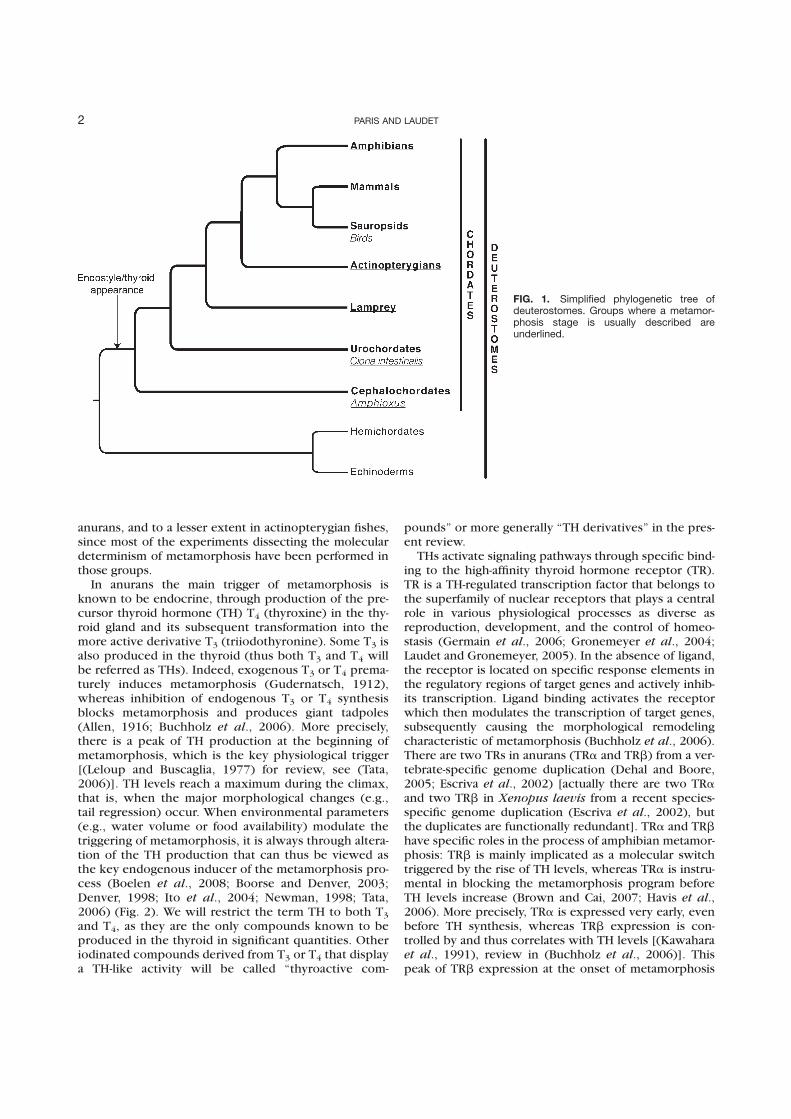
anurans, and to a lesser extent in actinopterygian fishes,since most of the experiments dissecting the moleculardeterminism of metamorphosis have been performed inthose groups.
In anurans the main trigger of metamorphosis isknown to be endocrine, through production of the pre-cursor thyroid hormone (TH) T4 (thyroxine) in the thy-roid gland and its subsequent transformation into themore active derivative T3 (triiodothyronine). Some T3 isalso produced in the thyroid (thus both T3 and T4 willbe referred as THs). Indeed, exogenous T3 or T4 prema-turely induces metamorphosis (Gudernatsch, 1912),whereas inhibition of endogenous T3 or T4 synthesisblocks metamorphosis and produces giant tadpoles(Allen, 1916; Buchholz et al., 2006). More precisely,there is a peak of TH production at the beginning ofmetamorphosis, which is the key physiological trigger[(Leloup and Buscaglia, 1977) for review, see (Tata,2006)]. TH levels reach a maximum during the climax,that is, when the major morphological changes (e.g.,tail regression) occur. When environmental parameters(e.g., water volume or food availability) modulate thetriggering of metamorphosis, it is always through altera-tion of the TH production that can thus be viewed asthe key endogenous inducer of the metamorphosis pro-cess (Boelen et al., 2008; Boorse and Denver, 2003;Denver, 1998; Ito et al., 2004; Newman, 1998; Tata,2006) (Fig. 2). We will restrict the term TH to both T3
and T4, as they are the only compounds known to beproduced in the thyroid in significant quantities. Otheriodinated compounds derived from T3 or T4 that displaya TH-like activity will be called ‘‘thyroactive com-
pounds’’ or more generally ‘‘TH derivatives’’ in the pres-ent review.
THs activate signaling pathways through specific bind-ing to the high-affinity thyroid hormone receptor (TR).TR is a TH-regulated transcription factor that belongs tothe superfamily of nuclear receptors that plays a centralrole in various physiological processes as diverse asreproduction, development, and the control of homeo-stasis (Germain et al., 2006; Gronemeyer et al., 2004;Laudet and Gronemeyer, 2005). In the absence of ligand,the receptor is located on specific response elements inthe regulatory regions of target genes and actively inhib-its transcription. Ligand binding activates the receptorwhich then modulates the transcription of target genes,subsequently causing the morphological remodelingcharacteristic of metamorphosis (Buchholz et al., 2006).There are two TRs in anurans (TRa and TRb) from a ver-tebrate-specific genome duplication (Dehal and Boore,2005; Escriva et al., 2002) [actually there are two TRaand two TRb in Xenopus laevis from a recent species-specific genome duplication (Escriva et al., 2002), butthe duplicates are functionally redundant]. TRa and TRbhave specific roles in the process of amphibian metamor-phosis: TRb is mainly implicated as a molecular switchtriggered by the rise of TH levels, whereas TRa is instru-mental in blocking the metamorphosis program beforeTH levels increase (Brown and Cai, 2007; Havis et al.,2006). More precisely, TRa is expressed very early, evenbefore TH synthesis, whereas TRb expression is con-trolled by and thus correlates with TH levels [(Kawaharaet al., 1991), review in (Buchholz et al., 2006)]. Thispeak of TRb expression at the onset of metamorphosis
FIG. 1. Simplified phylogenetic tree ofdeuterostomes. Groups where a metamor-phosis stage is usually described areunderlined.
2 PARIS AND LAUDET
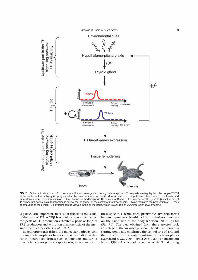
is particularly important, because it transmits the signalof the peak of TH: as TRb is one of its own target genes,the peak of TH production activates a positive loop ofTRb production and activation characteristic of the met-amorphosis climax (Tata et al., 1993).
In actinopterygian fishes, the molecular pathway con-trolling metamorphosis has been mainly studied in flat-fishes (pleuronectiformes) such as flounders and turbotin which metamorphosis is spectacular, as in anurans. In
these species, a symmetrical planktonic larva transformsinto an asymmetric benthic adult that harbors two eyeson the same side of the body [(Nelson, 2006), p442](Fig. 3d). The data obtained from these species tookadvantage of the knowledge accumulated in anurans as astarting point, and confirmed the central role of THs andtheir receptor in the early regulation of metamorphosis(Marchand et al., 2004; Power et al., 2001; Yamano andMiwa, 1998). A schematic structure of the TH signaling
FIG. 2. Schematic structure of TH cascade in the anuran organism during metamorphosis. Three parts are highlighted: the couple TR/THat the center of the pathway is upregulated at the onset of metamorphosis. More upstream in the pathway takes place TH synthesis, andmore downstream, the expression of TR target genes is modified upon TR activation. Since TR (more precisely the gene TRb) itself is one ofits own target genes, its autoactivation is critical for the trigger of the climax of metamorphosis. TR also regulates the production of TH, thuscontributing to the climax. [Color figure can be viewed in the online issue, which is available at www.interscience.wiley.com.]
3METAMORPHOSIS IN CHORDATES
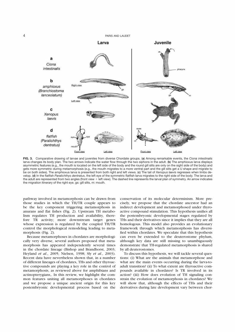
pathway involved in metamorphosis can be drawn fromthose studies in which the TH/TR couple appears tobe the key component triggering metamorphosis inanurans and flat fishes (Fig. 2). Upstream TH metabo-lism regulates TH production and availability, there-fore TR activity; more downstream target geneswhose expression is regulated by the coupled TH/TRcontrol the morphological remodeling leading to meta-morphosis (Fig. 2).
Because metamorphoses in chordates are morphologi-cally very diverse, several authors proposed that meta-morphosis has appeared independently several timesin the chordate lineage (Bishop and Brandhorst, 2003;Heyland et al., 2005; Nielsen, 1998; Sly et al., 2003).Recent data have nevertheless shown that, in a numberof different lineages of chordates, THs and other thyroac-tive compounds are playing a key role in the control ofmetamorphosis, as reviewed above for amphibians andactinopterygians,. In this review, we highlight the com-mon features uniting all metamorphoses in chordatesand we propose a unique ancient origin for this keypostembryonic developmental process based on the
conservation of its molecular determinism. More pre-cisely, we propose that the chordate ancestor had anindirect development and metamorphosed under thyro-active compound stimulation. This hypothesis unifies allthe postembryonic developmental stages regulated byTHs and their derivatives since it implies that they are allhomologous. This model also provides an evolutionaryframework through which metamorphosis has diversi-fied within chordates. We speculate that this hypothesiscan even be extended to the deuterostome phylum,although key data are still missing to unambiguouslydemonstrate that TH-regulated metamorphosis is sharedby all deuterostomes.
To discuss this hypothesis, we will tackle several ques-tions: (i) What are the animals that metamorphose andwhat are the main events occurring during the larva-to-adult transition? (ii) To what extent are thyroactive com-pounds available in chordates? Is TR involved in itsaction? (iii) How does evolution of TH signaling con-strain the evolution of metamorphosis in chordates? Wewill show that, although the effects of THs and theirderivatives during late development vary between chor-
FIG. 3. Comparative drawing of larvae and juveniles from diverse Chordate groups. (a) Among remarkable events, the Ciona intestinalislarva changes its body plan. The two arrows indicate the water flow through the two siphons in the adult. (b) The amphioxus larva displaysasymmetric features (e.g., the mouth is located on the left side of the body and the round gill slits are only on the sight side of the body) andgets more symmetric during metamorphosis (e.g., the mouth migrates to a more ventral part and the gill slits get a U shape and migrate tobe on both sides). The amphioxus larva is presented from both right and left views. (c) The tail of Xenopus laevis regresses when limbs de-velop. (d) In the flatfish Paralichthys dentratus, the left eye of the symmetric flatfish larva migrates to the right side of the body. The larva andthe adult are represented from two angles (front view1 left view). The dashed line represents the larval plan of symmetry. An arrow indicatesthe migration itinerary of the right eye. gs: gill slits, m: mouth.
4 PARIS AND LAUDET
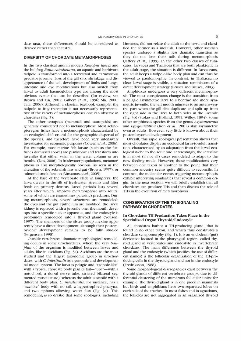
date taxa, these differences should be considered asderived rather than ancestral.
DIVERSITY OF CHORDATE METAMORPHOSES
In the two classical anuran models Xenopus laevis andthe bullfrog Rana catesbeiana, an aquatic and herbivoretadpole is transformed into a terrestrial and carnivorouspredator juvenile. Loss of the gill slits, shrinkage and dis-appearance of the tail, development of limbs and lungs,intestine and eye modifications but also switch fromlarval to adult haemoglobin type are among the mostobvious events that can be described (for review, seeBrown and Cai, 2007; Gilbert et al., 1996; Shi, 2000;Tata, 2006). Although a classical textbook example, thetadpole to frog transition is not necessarily representa-tive of the variety of metamorphoses one can observe inchordates (Fig. 3).
The other tetrapods (mammals and sauropsids) aregenerally considered not to metamorphose. Most actino-pterygian fishes have a metamorphosis characterized byan ecological shift crucial for the geographic dispersal ofthe species, and therefore have been very intensivelyinvestigated for economic purposes (Cowen et al., 2006).For example, most marine fish larvae (such as the flat-fishes discussed above) are planktonic and transform intojuveniles that either swim in the water column or arebenthic (Leis, 2006). In freshwater populations, metamor-phosis is also morphologically obvious, as seen in thealteration of the zebrafish fins and gut (Brown, 1997), orsalmonid smoltification (Varsamos et al., 2005).
At the base of the vertebrate clade in lampreys, thelarva dwells in the silt of freshwater streams and filterfeeds on primary detritus. Larval periods lasts severalyears after which lampreys metamorphose into adults,some of which are (sometimes parasitic) predators. Dur-ing metamorphosis, several structures are remodeled:the eyes and the gut epithelium are modified, the larvalkidney is replaced with a juvenile one, the mouth devel-ops into a specific sucker apparatus, and the endostyle isprofoundly remodeled into a thyroid gland (Youson,1997). The members of the sister group myxine appa-rently have a direct development, although their postem-bryonic development remains to be fully studied(Jørgensen, 1998).
Outside vertebrates, dramatic morphological remodel-ing occurs in some urochordates, where the very bau-plan of the organism is modified between larvae andadults, like in ascidians (Fig. 3a). Ascidians are the moststudied and the largest taxonomic group in urochor-dates, with C. intestinalis as a genomic and developmen-tal model system. The larva is pelagic and ‘‘tadpole-like’’with a typical chordate body plan (a tail—‘‘uro’’—with anotochord, a dorsal nerve tube, striated bilateral seg-mented musculature), whereas the adult is sessile with adifferent body plan. C. intestinalis, for instance, has a‘‘sac-like’’ body with no tail, a hypertrophied pharynx,and two siphons allowing water flux (Fig. 3a). Thisremodeling is so drastic that some zoologists, including
Linnaeus, did not relate the adult to the larva and classi-fied the former as a mollusk. However, other ascidianspecies undergo a slightly less dramatic transition asthey do not lose their tails during metamorphosis(Jeffery et al., 1999). In the other two classes of tuni-cates, Larvacea and Thaliacea that are both planktonic inthe adult stage, the situation is different. In Larvaceans,the adult keeps a tadpole-like body plan and can thus beviewed as paedomorphic. In contrast, in Thaliacea noclear larval stage is visible, a situation reminiscent of adirect development strategy (Brusca and Brusca, 2003).
Amphioxus undergoes a very different metamorpho-sis. The most conspicuous change is the transition froma pelagic asymmetric larva to a benthic and more sym-metric juvenile: the left mouth migrates to an antero-ven-tral part when the gill slits duplicate and split up fromthe right side in the larva to both sides in the juvenile(Fig. 3b) (Stokes and Holland, 1995; Willey, 1894). Someother amphioxus species from the genus Asymmetronsand Epygonichthys (Kon et al., 2007) stay asymmetriceven as adults. However, very little is known about theirpostembryonic development.
Overall, this rapid zoological presentation shows thatmost chordates display an ecological larva-to-adult transi-tion, characterized by an adaptation from the larval eco-logical niche to the adult one. Interestingly, the intestineis in most (if not all) cases remodeled to adapt to thenew feeding mode. However, these modifications varybetween one taxon to another to the point that theircommon ancestry seems quite difficult to reconcile. Incontrast, the molecular events triggering metamorphosisexhibit interesting similarities that reveal a common ori-gin. In the next section, we will briefly establish that allchordates can produce THs and then discuss the role ofTHs in the evolution of metamorphosis.
CONSERVATION OF THE TH SIGNALINGPATHWAY IN CHORDATES
In Chordates TH Production Takes Place in theSpecialized Organ Thyroid/Endostyle
All chordates harbor a TH-producing gland, that isfound in no other taxon, and which thus constitutes achordate synapomorphy (Fig. 1). It is an endoderm (gut)derivative located in the pharyngeal region, called thy-roid gland in vertebrates and endostyle in invertebratechordates. The main difference between the thyroidgland and the endostyle (which justifies the use of differ-ent names) is the follicular organization of the TH-pro-ducing cells in the thyroid gland and not in the endostyle(Fredriksson, 1988).
Some morphological discrepancies exist between thethyroid glands of different vertebrate groups, due to dif-ferential clustering of the numerous follicular units: forexample, the thyroid gland is in one piece in mammalsbut birds and amphibians have two separated lobes oneach side of the trachea. In most fishes and in agnathans,the follicles are not aggregated in an organized thyroid
5METAMORPHOSIS IN CHORDATES
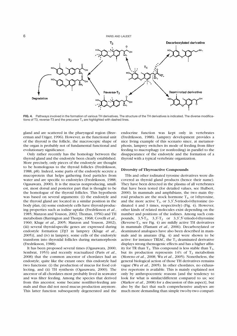
gland and are scattered in the pharyngeal region (Brav-erman and Utiger, 1996). However, as the functional unitof the thyroid is the follicle, the macroscopic shape ofthe organ is probably not of fundamental functional andevolutionary significance.
Only rather recently has the homology between thethyroid gland and the endostyle been clearly established.More precisely, only pieces of the endostyle are thoughtto be homologous to the thyroid follicles (Fredriksson,1988, p8). Indeed, some parts of the endostyle secrete amucoprotein that helps gathering food particles fromwater and are specific to endostyles (Fredriksson, 1988;Ogasawara, 2000). It is the mucus nonproducing, small-est, most dorsal and posterior part that is thought to bethe homologue of the thyroid follicles. This hypothesiswas based on several arguments: (i) the endostyle andthe thyroid gland are located in a similar position in thebody plan, (ii) some endostyle cells have thyroid-produc-ing properties such as iodine uptake (Fredriksson et al.,1985; Manzon and Youson, 2002; Thomas, 1956) and THmetabolism (Barrington and Thorpe, 1968; Covelli et al.,1960; Kluge et al., 2005; Manzon and Youson, 2002),(iii) several thyroid-specific genes are expressed duringendostyle formation [Ttf1 in lamprey (Kluge et al.,2005)], and (iv) in lamprey, some cells of the endostyletransform into thyroidal follicles during metamorphosis(Fredriksson, 1988).
It has been proposed several times (Ogasawara, 2000;Sembrat, 1953) and recently reactualized (Paris et al.,2008) that the common ancestor of chordates had anendostyle, quite like the extant ones: this endostyle hadtwo functions: (i) the production of mucus for food col-lecting, and (ii) TH synthesis (Ogasawara, 2000). Theancestor of all chordates most probably lived in seawaterand was filter feeding. Among the species that derivedfrom this ancestor, some became nonfilter-feeding ani-mals and thus did not need mucus production anymore.This latter function subsequently degenerated and the
endocrine function was kept only in vertebrates(Fredriksson, 1988). Lamprey development provides anice living example of this scenario since, at metamor-phosis, lamprey switches its mode of feeding from filterfeeding to macrophagy (or nonfeeding) in parallel to thedisappearance of the endostyle and the formation of athyroid with a typical vertebrate organization.
Diversity of Thyroactive Compounds
THs and other iodinated tyrosine derivatives were dis-covered as thyroid gland products (hence their name).They have been detected in the plasma of all vertebratesthat have been tested (for detailed values, see Hulbert,2000). In mammals and amphibians, the two main thy-roid products are the stock hormone T4, or l-thyroxine,and the more active T3, or 3,3
0,5-triiodo-l-thyronine (io-dinated 4 and 3 times, respectively) (Fig. 4). However,other kinds of related molecules exist depending on thenumber and positions of the iodines. Among such com-pounds, 3,5-T2, 3,30-T2 or 3,30,50-triiodo-l-thyronine(Reverse-T3, see Fig. 4) are mildly active TH derivativesin mammals (Flamant et al., 2006). Decarboxylated ordeaminated analogues have also been described in mam-mals and in anurans (Fig. 4) and were shown to beactive: for instance TRIAC, the T3 deaminated derivativedisplays strong themogenic effects and has a higher affin-ity for TR than T3. This compound is less stable than T3,but its production represents 14% of T3 metabolism(Moreno et al., 2008; Wu et al., 2005). Nonetheless, thegeneral biological action of those TH derivatives remainsvague (Wu et al., 2005). In other chordates, no exhaus-tive repertoire is available. This is mainly explained notonly by anthropocentric reasons [and the tendency tolook for what is similar/different compared to us; see(Markov et al., 2008) for a discussion of this aspect], butalso by the fact that such comprehensive analyses aremuch more demanding than simple two-by-two compari-
FIG. 4. Pathways involved in the formation of various TH derivatives. The structure of the TH derivatives is indicated. The diverse modifica-tions of T3, reverse-T3 and the precursor T4 are highlighted with dashed lines.
6 PARIS AND LAUDET
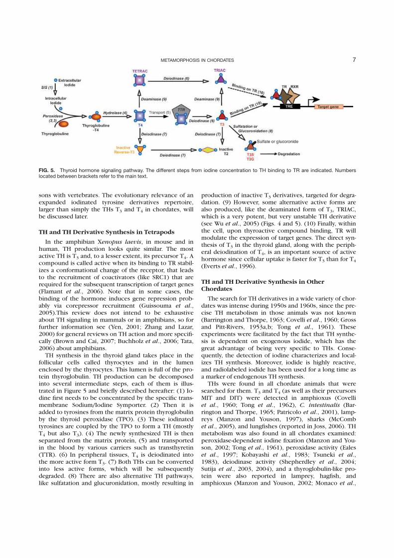
sons with vertebrates. The evolutionary relevance of anexpanded iodinated tyrosine derivatives repertoire,larger than simply the THs T3 and T4 in chordates, willbe discussed later.
TH and TH Derivative Synthesis in Tetrapods
In the amphibian Xenopus laevis, in mouse and inhuman, TH production looks quite similar. The mostactive TH is T3 and, to a lesser extent, its precursor T4. Acompound is called active when its binding to TR stabil-izes a conformational change of the receptor, that leadsto the recruitment of coactivators (like SRC1) that arerequired for the subsequent transcription of target genes(Flamant et al., 2006). Note that in some cases, thebinding of the hormone induces gene repression prob-ably via corepressor recruitment (Guissouma et al.,2005).This review does not intend to be exhaustiveabout TH signaling in mammals or in amphibians, so forfurther information see (Yen, 2001; Zhang and Lazar,2000) for general reviews on TH action and more specifi-cally (Brown and Cai, 2007; Buchholz et al., 2006; Tata,2006) about amphibians.
TH synthesis in the thyroid gland takes place in thefollicular cells called thyrocytes and in the lumenenclosed by the thyrocytes. This lumen is full of the pro-tein thyroglobulin. TH production can be decomposedinto several intermediate steps, each of them is illus-trated in Figure 5 and briefly described hereafter: (1) Io-dine first needs to be concentrated by the specific trans-membrane Sodium/Iodine Symporter. (2) Then it isadded to tyrosines from the matrix protein thyroglobulinby the thyroid peroxidase (TPO). (3) These iodinatedtyrosines are coupled by the TPO to form a TH (mostlyT4 but also T3). (4) The newly synthesized TH is thenseparated from the matrix protein, (5) and transportedin the blood by various carriers such as transthyretin(TTR). (6) In peripheral tissues, T4 is deiodinated intothe more active form T3. (7) Both THs can be convertedinto less active forms, which will be subsequentlydegraded. (8) There are also alternative TH pathways,like sulfatation and glucuronidation, mostly resulting in
production of inactive T3 derivatives, targeted for degra-dation. (9) However, some alternative active forms arealso produced, like the deaminated form of T3, TRIAC,which is a very potent, but very unstable TH derivative(see Wu et al., 2005) (Figs. 4 and 5). (10) Finally, withinthe cell, upon thyroactive compound binding, TR willmodulate the expression of target genes. The direct syn-thesis of T3 in the thyroid gland, along with the periph-eral deiodination of T4, is an important source of activehormone since cellular uptake is faster for T3 than for T4(Everts et al., 1996).
TH and TH Derivative Synthesis in OtherChordates
The search for TH derivatives in a wide variety of chor-dates was intense during 1950s and 1960s, since the pre-cise TH metabolism in those animals was not known(Barrington and Thorpe, 1963; Covelli et al., 1960; Grossand Pitt-Rivers, 1953a,b; Tong et al., 1961). Theseexperiments were facilitated by the fact that TH synthe-sis is dependent on exogenous iodide, which has thegreat advantage of being very specific to THs. Conse-quently, the detection of iodine characterizes and local-izes TH synthesis. Moreover, iodide is highly reactive,and radiolabeled iodide has been used for a long time asa marker of endogenous TH synthesis.
THs were found in all chordate animals that weresearched for them. T3 and T4 (as well as their precursorsMIT and DIT) were detected in amphioxus (Covelliet al., 1960; Tong et al., 1962), C. intestinalis (Bar-rington and Thorpe, 1965; Patricolo et al., 2001), lamp-reys (Manzon and Youson, 1997), sharks (McCombet al., 2005), and lungfishes (reported in Joss, 2006). THmetabolism was also found in all chordates examined:peroxidase-dependent iodine fixation (Manzon and You-son, 2002; Tong et al., 1961), peroxidase activity (Ealeset al., 1997; Kobayashi et al., 1983; Tsuneki et al.,1983), deiodinase activity (Shepherdley et al., 2004;Sutija et al., 2003, 2004), and a thyroglobulin-like pro-tein were also reported in lamprey, hagfish, andamphioxus (Manzon and Youson, 2002; Monaco et al.,
FIG. 5. Thyroid hormone signaling pathway. The different steps from iodine concentration to TH binding to TR are indicated. Numberslocated between brackets refer to the main text.
7METAMORPHOSIS IN CHORDATES
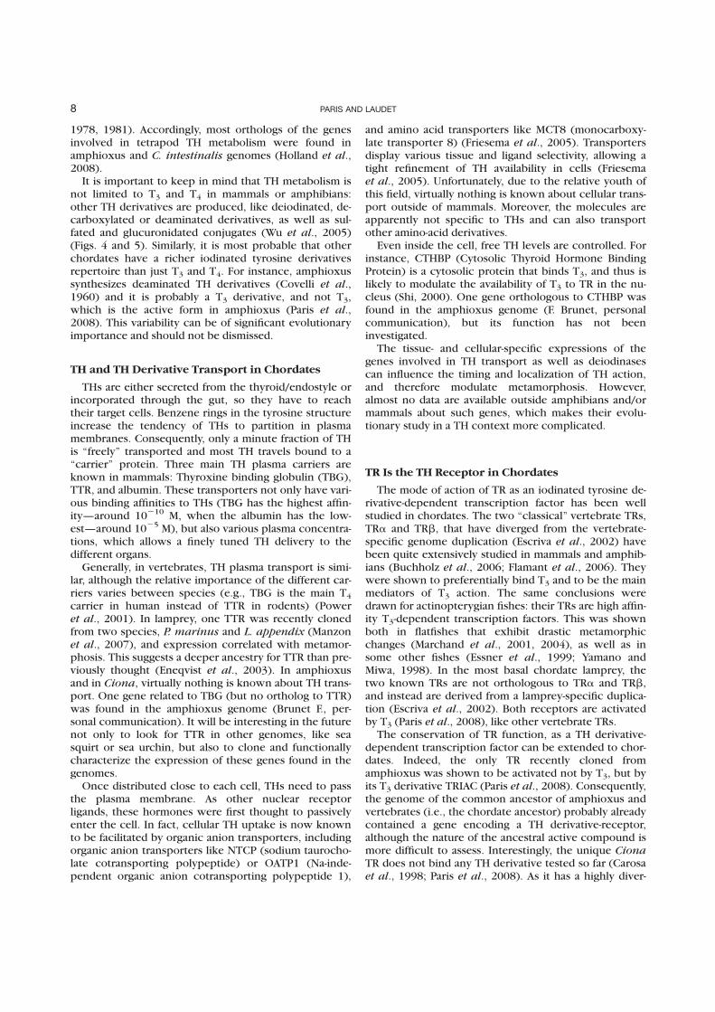
1978, 1981). Accordingly, most orthologs of the genesinvolved in tetrapod TH metabolism were found inamphioxus and C. intestinalis genomes (Holland et al.,2008).
It is important to keep in mind that TH metabolism isnot limited to T3 and T4 in mammals or amphibians:other TH derivatives are produced, like deiodinated, de-carboxylated or deaminated derivatives, as well as sul-fated and glucuronidated conjugates (Wu et al., 2005)(Figs. 4 and 5). Similarly, it is most probable that otherchordates have a richer iodinated tyrosine derivativesrepertoire than just T3 and T4. For instance, amphioxussynthesizes deaminated TH derivatives (Covelli et al.,1960) and it is probably a T3 derivative, and not T3,which is the active form in amphioxus (Paris et al.,2008). This variability can be of significant evolutionaryimportance and should not be dismissed.
TH and TH Derivative Transport in Chordates
THs are either secreted from the thyroid/endostyle orincorporated through the gut, so they have to reachtheir target cells. Benzene rings in the tyrosine structureincrease the tendency of THs to partition in plasmamembranes. Consequently, only a minute fraction of THis ‘‘freely’’ transported and most TH travels bound to a‘‘carrier’’ protein. Three main TH plasma carriers areknown in mammals: Thyroxine binding globulin (TBG),TTR, and albumin. These transporters not only have vari-ous binding affinities to THs (TBG has the highest affin-ity—around 10210 M, when the albumin has the low-est—around 1025 M), but also various plasma concentra-tions, which allows a finely tuned TH delivery to thedifferent organs.
Generally, in vertebrates, TH plasma transport is simi-lar, although the relative importance of the different car-riers varies between species (e.g., TBG is the main T4
carrier in human instead of TTR in rodents) (Poweret al., 2001). In lamprey, one TTR was recently clonedfrom two species, P. marinus and L. appendix (Manzonet al., 2007), and expression correlated with metamor-phosis. This suggests a deeper ancestry for TTR than pre-viously thought (Eneqvist et al., 2003). In amphioxusand in Ciona, virtually nothing is known about TH trans-port. One gene related to TBG (but no ortholog to TTR)was found in the amphioxus genome (Brunet F., per-sonal communication). It will be interesting in the futurenot only to look for TTR in other genomes, like seasquirt or sea urchin, but also to clone and functionallycharacterize the expression of these genes found in thegenomes.
Once distributed close to each cell, THs need to passthe plasma membrane. As other nuclear receptorligands, these hormones were first thought to passivelyenter the cell. In fact, cellular TH uptake is now knownto be facilitated by organic anion transporters, includingorganic anion transporters like NTCP (sodium taurocho-late cotransporting polypeptide) or OATP1 (Na-inde-pendent organic anion cotransporting polypeptide 1),
and amino acid transporters like MCT8 (monocarboxy-late transporter 8) (Friesema et al., 2005). Transportersdisplay various tissue and ligand selectivity, allowing atight refinement of TH availability in cells (Friesemaet al., 2005). Unfortunately, due to the relative youth ofthis field, virtually nothing is known about cellular trans-port outside of mammals. Moreover, the molecules areapparently not specific to THs and can also transportother amino-acid derivatives.
Even inside the cell, free TH levels are controlled. Forinstance, CTHBP (Cytosolic Thyroid Hormone BindingProtein) is a cytosolic protein that binds T3, and thus islikely to modulate the availability of T3 to TR in the nu-cleus (Shi, 2000). One gene orthologous to CTHBP wasfound in the amphioxus genome (F. Brunet, personalcommunication), but its function has not beeninvestigated.
The tissue- and cellular-specific expressions of thegenes involved in TH transport as well as deiodinasescan influence the timing and localization of TH action,and therefore modulate metamorphosis. However,almost no data are available outside amphibians and/ormammals about such genes, which makes their evolu-tionary study in a TH context more complicated.
TR Is the TH Receptor in Chordates
The mode of action of TR as an iodinated tyrosine de-rivative-dependent transcription factor has been wellstudied in chordates. The two ‘‘classical’’ vertebrate TRs,TRa and TRb, that have diverged from the vertebrate-specific genome duplication (Escriva et al., 2002) havebeen quite extensively studied in mammals and amphib-ians (Buchholz et al., 2006; Flamant et al., 2006). Theywere shown to preferentially bind T3 and to be the mainmediators of T3 action. The same conclusions weredrawn for actinopterygian fishes: their TRs are high affin-ity T3-dependent transcription factors. This was shownboth in flatfishes that exhibit drastic metamorphicchanges (Marchand et al., 2001, 2004), as well as insome other fishes (Essner et al., 1999; Yamano andMiwa, 1998). In the most basal chordate lamprey, thetwo known TRs are not orthologous to TRa and TRb,and instead are derived from a lamprey-specific duplica-tion (Escriva et al., 2002). Both receptors are activatedby T3 (Paris et al., 2008), like other vertebrate TRs.
The conservation of TR function, as a TH derivative-dependent transcription factor can be extended to chor-dates. Indeed, the only TR recently cloned fromamphioxus was shown to be activated not by T3, but byits T3 derivative TRIAC (Paris et al., 2008). Consequently,the genome of the common ancestor of amphioxus andvertebrates (i.e., the chordate ancestor) probably alreadycontained a gene encoding a TH derivative-receptor,although the nature of the ancestral active compound ismore difficult to assess. Interestingly, the unique CionaTR does not bind any TH derivative tested so far (Carosaet al., 1998; Paris et al., 2008). As it has a highly diver-
8 PARIS AND LAUDET
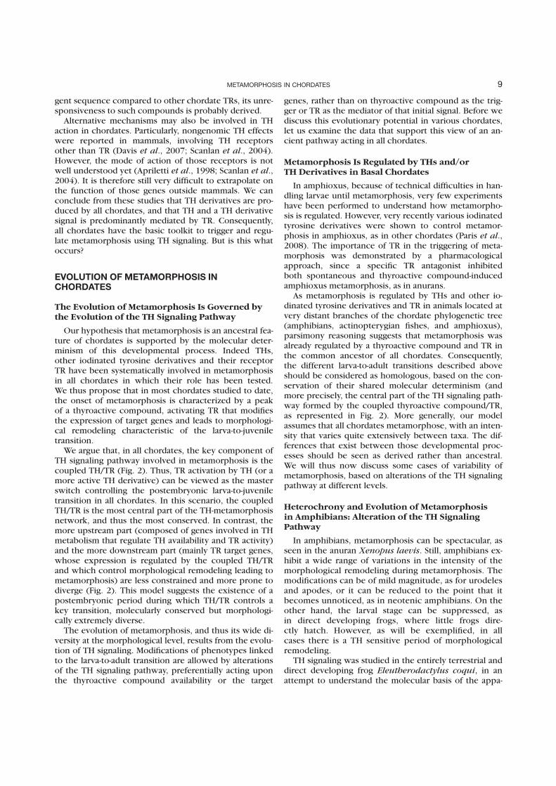
gent sequence compared to other chordate TRs, its unre-sponsiveness to such compounds is probably derived.
Alternative mechanisms may also be involved in THaction in chordates. Particularly, nongenomic TH effectswere reported in mammals, involving TH receptorsother than TR (Davis et al., 2007; Scanlan et al., 2004).However, the mode of action of those receptors is notwell understood yet (Apriletti et al., 1998; Scanlan et al.,2004). It is therefore still very difficult to extrapolate onthe function of those genes outside mammals. We canconclude from these studies that TH derivatives are pro-duced by all chordates, and that TH and a TH derivativesignal is predominantly mediated by TR. Consequently,all chordates have the basic toolkit to trigger and regu-late metamorphosis using TH signaling. But is this whatoccurs?
EVOLUTION OF METAMORPHOSIS INCHORDATES
The Evolution of Metamorphosis Is Governed bythe Evolution of the TH Signaling Pathway
Our hypothesis that metamorphosis is an ancestral fea-ture of chordates is supported by the molecular deter-minism of this developmental process. Indeed THs,other iodinated tyrosine derivatives and their receptorTR have been systematically involved in metamorphosisin all chordates in which their role has been tested.We thus propose that in most chordates studied to date,the onset of metamorphosis is characterized by a peakof a thyroactive compound, activating TR that modifiesthe expression of target genes and leads to morphologi-cal remodeling characteristic of the larva-to-juveniletransition.
We argue that, in all chordates, the key component ofTH signaling pathway involved in metamorphosis is thecoupled TH/TR (Fig. 2). Thus, TR activation by TH (or amore active TH derivative) can be viewed as the masterswitch controlling the postembryonic larva-to-juveniletransition in all chordates. In this scenario, the coupledTH/TR is the most central part of the TH-metamorphosisnetwork, and thus the most conserved. In contrast, themore upstream part (composed of genes involved in THmetabolism that regulate TH availability and TR activity)and the more downstream part (mainly TR target genes,whose expression is regulated by the coupled TH/TRand which control morphological remodeling leading tometamorphosis) are less constrained and more prone todiverge (Fig. 2). This model suggests the existence of apostembryonic period during which TH/TR controls akey transition, molecularly conserved but morphologi-cally extremely diverse.
The evolution of metamorphosis, and thus its wide di-versity at the morphological level, results from the evolu-tion of TH signaling. Modifications of phenotypes linkedto the larva-to-adult transition are allowed by alterationsof the TH signaling pathway, preferentially acting uponthe thyroactive compound availability or the target
genes, rather than on thyroactive compound as the trig-ger or TR as the mediator of that initial signal. Before wediscuss this evolutionary potential in various chordates,let us examine the data that support this view of an an-cient pathway acting in all chordates.
Metamorphosis Is Regulated by THs and/orTH Derivatives in Basal Chordates
In amphioxus, because of technical difficulties in han-dling larvae until metamorphosis, very few experimentshave been performed to understand how metamorpho-sis is regulated. However, very recently various iodinatedtyrosine derivatives were shown to control metamor-phosis in amphioxus, as in other chordates (Paris et al.,2008). The importance of TR in the triggering of meta-morphosis was demonstrated by a pharmacologicalapproach, since a specific TR antagonist inhibitedboth spontaneous and thyroactive compound-inducedamphioxus metamorphosis, as in anurans.
As metamorphosis is regulated by THs and other io-dinated tyrosine derivatives and TR in animals located atvery distant branches of the chordate phylogenetic tree(amphibians, actinopterygian fishes, and amphioxus),parsimony reasoning suggests that metamorphosis wasalready regulated by a thyroactive compound and TR inthe common ancestor of all chordates. Consequently,the different larva-to-adult transitions described aboveshould be considered as homologous, based on the con-servation of their shared molecular determinism (andmore precisely, the central part of the TH signaling path-way formed by the coupled thyroactive compound/TR,as represented in Fig. 2). More generally, our modelassumes that all chordates metamorphose, with an inten-sity that varies quite extensively between taxa. The dif-ferences that exist between those developmental proc-esses should be seen as derived rather than ancestral.We will thus now discuss some cases of variability ofmetamorphosis, based on alterations of the TH signalingpathway at different levels.
Heterochrony and Evolution of Metamorphosisin Amphibians: Alteration of the TH SignalingPathway
In amphibians, metamorphosis can be spectacular, asseen in the anuran Xenopus laevis. Still, amphibians ex-hibit a wide range of variations in the intensity of themorphological remodeling during metamorphosis. Themodifications can be of mild magnitude, as for urodelesand apodes, or it can be reduced to the point that itbecomes unnoticed, as in neotenic amphibians. On theother hand, the larval stage can be suppressed, asin direct developing frogs, where little frogs dire-ctly hatch. However, as will be exemplified, in allcases there is a TH sensitive period of morphologicalremodeling.
TH signaling was studied in the entirely terrestrial anddirect developing frog Eleutherodactylus coqui, in anattempt to understand the molecular basis of the appa-
9METAMORPHOSIS IN CHORDATES
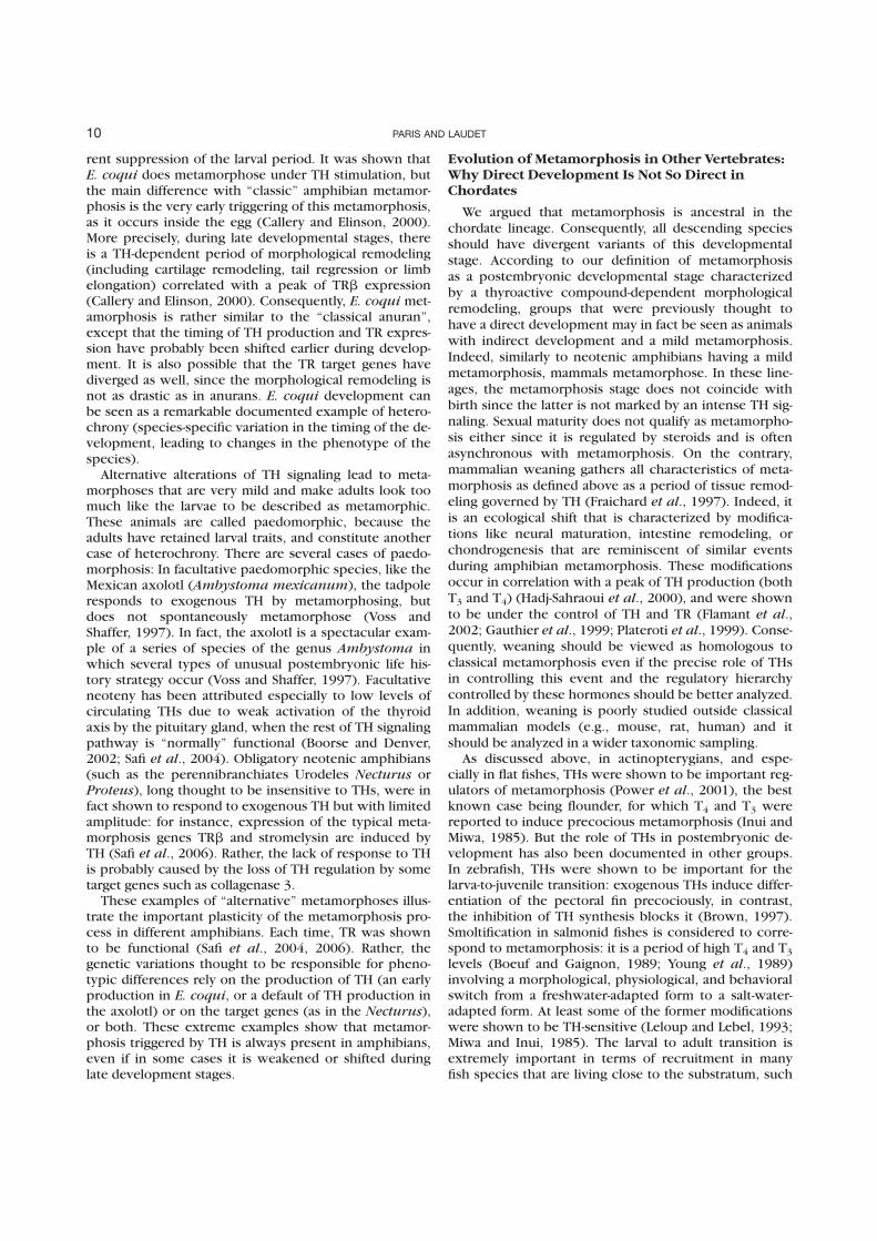
rent suppression of the larval period. It was shown thatE. coqui does metamorphose under TH stimulation, butthe main difference with ‘‘classic’’ amphibian metamor-phosis is the very early triggering of this metamorphosis,as it occurs inside the egg (Callery and Elinson, 2000).More precisely, during late developmental stages, thereis a TH-dependent period of morphological remodeling(including cartilage remodeling, tail regression or limbelongation) correlated with a peak of TRb expression(Callery and Elinson, 2000). Consequently, E. coqui met-amorphosis is rather similar to the ‘‘classical anuran’’,except that the timing of TH production and TR expres-sion have probably been shifted earlier during develop-ment. It is also possible that the TR target genes havediverged as well, since the morphological remodeling isnot as drastic as in anurans. E. coqui development canbe seen as a remarkable documented example of hetero-chrony (species-specific variation in the timing of the de-velopment, leading to changes in the phenotype of thespecies).
Alternative alterations of TH signaling lead to meta-morphoses that are very mild and make adults look toomuch like the larvae to be described as metamorphic.These animals are called paedomorphic, because theadults have retained larval traits, and constitute anothercase of heterochrony. There are several cases of paedo-morphosis: In facultative paedomorphic species, like theMexican axolotl (Ambystoma mexicanum), the tadpoleresponds to exogenous TH by metamorphosing, butdoes not spontaneously metamorphose (Voss andShaffer, 1997). In fact, the axolotl is a spectacular exam-ple of a series of species of the genus Ambystoma inwhich several types of unusual postembryonic life his-tory strategy occur (Voss and Shaffer, 1997). Facultativeneoteny has been attributed especially to low levels ofcirculating THs due to weak activation of the thyroidaxis by the pituitary gland, when the rest of TH signalingpathway is ‘‘normally’’ functional (Boorse and Denver,2002; Safi et al., 2004). Obligatory neotenic amphibians(such as the perennibranchiates Urodeles Necturus orProteus), long thought to be insensitive to THs, were infact shown to respond to exogenous TH but with limitedamplitude: for instance, expression of the typical meta-morphosis genes TRb and stromelysin are induced byTH (Safi et al., 2006). Rather, the lack of response to THis probably caused by the loss of TH regulation by sometarget genes such as collagenase 3.
These examples of ‘‘alternative’’ metamorphoses illus-trate the important plasticity of the metamorphosis pro-cess in different amphibians. Each time, TR was shownto be functional (Safi et al., 2004, 2006). Rather, thegenetic variations thought to be responsible for pheno-typic differences rely on the production of TH (an earlyproduction in E. coqui, or a default of TH production inthe axolotl) or on the target genes (as in the Necturus),or both. These extreme examples show that metamor-phosis triggered by TH is always present in amphibians,even if in some cases it is weakened or shifted duringlate development stages.
Evolution of Metamorphosis in Other Vertebrates:Why Direct Development Is Not So Direct inChordates
We argued that metamorphosis is ancestral in thechordate lineage. Consequently, all descending speciesshould have divergent variants of this developmentalstage. According to our definition of metamorphosisas a postembryonic developmental stage characterizedby a thyroactive compound-dependent morphologicalremodeling, groups that were previously thought tohave a direct development may in fact be seen as animalswith indirect development and a mild metamorphosis.Indeed, similarly to neotenic amphibians having a mildmetamorphosis, mammals metamorphose. In these line-ages, the metamorphosis stage does not coincide withbirth since the latter is not marked by an intense TH sig-naling. Sexual maturity does not qualify as metamorpho-sis either since it is regulated by steroids and is oftenasynchronous with metamorphosis. On the contrary,mammalian weaning gathers all characteristics of meta-morphosis as defined above as a period of tissue remod-eling governed by TH (Fraichard et al., 1997). Indeed, itis an ecological shift that is characterized by modifica-tions like neural maturation, intestine remodeling, orchondrogenesis that are reminiscent of similar eventsduring amphibian metamorphosis. These modificationsoccur in correlation with a peak of TH production (bothT3 and T4) (Hadj-Sahraoui et al., 2000), and were shownto be under the control of TH and TR (Flamant et al.,2002; Gauthier et al., 1999; Plateroti et al., 1999). Conse-quently, weaning should be viewed as homologous toclassical metamorphosis even if the precise role of THsin controlling this event and the regulatory hierarchycontrolled by these hormones should be better analyzed.In addition, weaning is poorly studied outside classicalmammalian models (e.g., mouse, rat, human) and itshould be analyzed in a wider taxonomic sampling.
As discussed above, in actinopterygians, and espe-cially in flat fishes, THs were shown to be important reg-ulators of metamorphosis (Power et al., 2001), the bestknown case being flounder, for which T4 and T3 werereported to induce precocious metamorphosis (Inui andMiwa, 1985). But the role of THs in postembryonic de-velopment has also been documented in other groups.In zebrafish, THs were shown to be important for thelarva-to-juvenile transition: exogenous THs induce differ-entiation of the pectoral fin precociously, in contrast,the inhibition of TH synthesis blocks it (Brown, 1997).Smoltification in salmonid fishes is considered to corre-spond to metamorphosis: it is a period of high T4 and T3levels (Boeuf and Gaignon, 1989; Young et al., 1989)involving a morphological, physiological, and behavioralswitch from a freshwater-adapted form to a salt-water-adapted form. At least some of the former modificationswere shown to be TH-sensitive (Leloup and Lebel, 1993;Miwa and Inui, 1985). The larval to adult transition isextremely important in terms of recruitment in manyfish species that are living close to the substratum, such
10 PARIS AND LAUDET
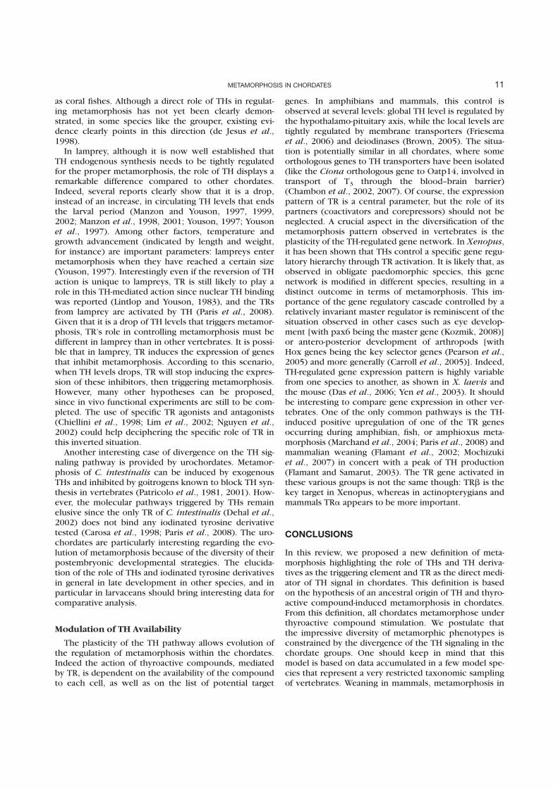
as coral fishes. Although a direct role of THs in regulat-ing metamorphosis has not yet been clearly demon-strated, in some species like the grouper, existing evi-dence clearly points in this direction (de Jesus et al.,1998).
In lamprey, although it is now well established thatTH endogenous synthesis needs to be tightly regulatedfor the proper metamorphosis, the role of TH displays aremarkable difference compared to other chordates.Indeed, several reports clearly show that it is a drop,instead of an increase, in circulating TH levels that endsthe larval period (Manzon and Youson, 1997, 1999,2002; Manzon et al., 1998, 2001; Youson, 1997; Yousonet al., 1997). Among other factors, temperature andgrowth advancement (indicated by length and weight,for instance) are important parameters: lampreys entermetamorphosis when they have reached a certain size(Youson, 1997). Interestingly even if the reversion of THaction is unique to lampreys, TR is still likely to play arole in this TH-mediated action since nuclear TH bindingwas reported (Lintlop and Youson, 1983), and the TRsfrom lamprey are activated by TH (Paris et al., 2008).Given that it is a drop of TH levels that triggers metamor-phosis, TR’s role in controlling metamorphosis must bedifferent in lamprey than in other vertebrates. It is possi-ble that in lamprey, TR induces the expression of genesthat inhibit metamorphosis. According to this scenario,when TH levels drops, TR will stop inducing the expres-sion of these inhibitors, then triggering metamorphosis.However, many other hypotheses can be proposed,since in vivo functional experiments are still to be com-pleted. The use of specific TR agonists and antagonists(Chiellini et al., 1998; Lim et al., 2002; Nguyen et al.,2002) could help deciphering the specific role of TR inthis inverted situation.
Another interesting case of divergence on the TH sig-naling pathway is provided by urochordates. Metamor-phosis of C. intestinalis can be induced by exogenousTHs and inhibited by goitrogens known to block TH syn-thesis in vertebrates (Patricolo et al., 1981, 2001). How-ever, the molecular pathways triggered by THs remainelusive since the only TR of C. intestinalis (Dehal et al.,2002) does not bind any iodinated tyrosine derivativetested (Carosa et al., 1998; Paris et al., 2008). The uro-chordates are particularly interesting regarding the evo-lution of metamorphosis because of the diversity of theirpostembryonic developmental strategies. The elucida-tion of the role of THs and iodinated tyrosine derivativesin general in late development in other species, and inparticular in larvaceans should bring interesting data forcomparative analysis.
Modulation of TH Availability
The plasticity of the TH pathway allows evolution ofthe regulation of metamorphosis within the chordates.Indeed the action of thyroactive compounds, mediatedby TR, is dependent on the availability of the compoundto each cell, as well as on the list of potential target
genes. In amphibians and mammals, this control isobserved at several levels: global TH level is regulated bythe hypothalamo-pituitary axis, while the local levels aretightly regulated by membrane transporters (Friesemaet al., 2006) and deiodinases (Brown, 2005). The situa-tion is potentially similar in all chordates, where someorthologous genes to TH transporters have been isolated(like the Ciona orthologous gene to Oatp14, involved intransport of T3 through the blood–brain barrier)(Chambon et al., 2002, 2007). Of course, the expressionpattern of TR is a central parameter, but the role of itspartners (coactivators and corepressors) should not beneglected. A crucial aspect in the diversification of themetamorphosis pattern observed in vertebrates is theplasticity of the TH-regulated gene network. In Xenopus,it has been shown that THs control a specific gene regu-latory hierarchy through TR activation. It is likely that, asobserved in obligate paedomorphic species, this genenetwork is modified in different species, resulting in adistinct outcome in terms of metamorphosis. This im-portance of the gene regulatory cascade controlled by arelatively invariant master regulator is reminiscent of thesituation observed in other cases such as eye develop-ment [with pax6 being the master gene (Kozmik, 2008)]or antero-posterior development of arthropods [withHox genes being the key selector genes (Pearson et al.,2005) and more generally (Carroll et al., 2005)]. Indeed,TH-regulated gene expression pattern is highly variablefrom one species to another, as shown in X. laevis andthe mouse (Das et al., 2006; Yen et al., 2003). It shouldbe interesting to compare gene expression in other ver-tebrates. One of the only common pathways is the TH-induced positive upregulation of one of the TR genesoccurring during amphibian, fish, or amphioxus meta-morphosis (Marchand et al., 2004; Paris et al., 2008) andmammalian weaning (Flamant et al., 2002; Mochizukiet al., 2007) in concert with a peak of TH production(Flamant and Samarut, 2003). The TR gene activated inthese various groups is not the same though: TRb is thekey target in Xenopus, whereas in actinopterygians andmammals TRa appears to be more important.
CONCLUSIONS
In this review, we proposed a new definition of meta-morphosis highlighting the role of THs and TH deriva-tives as the triggering element and TR as the direct medi-ator of TH signal in chordates. This definition is basedon the hypothesis of an ancestral origin of TH and thyro-active compound-induced metamorphosis in chordates.From this definition, all chordates metamorphose underthyroactive compound stimulation. We postulate thatthe impressive diversity of metamorphic phenotypes isconstrained by the divergence of the TH signaling in thechordate groups. One should keep in mind that thismodel is based on data accumulated in a few model spe-cies that represent a very restricted taxonomic samplingof vertebrates. Weaning in mammals, metamorphosis in
11METAMORPHOSIS IN CHORDATES
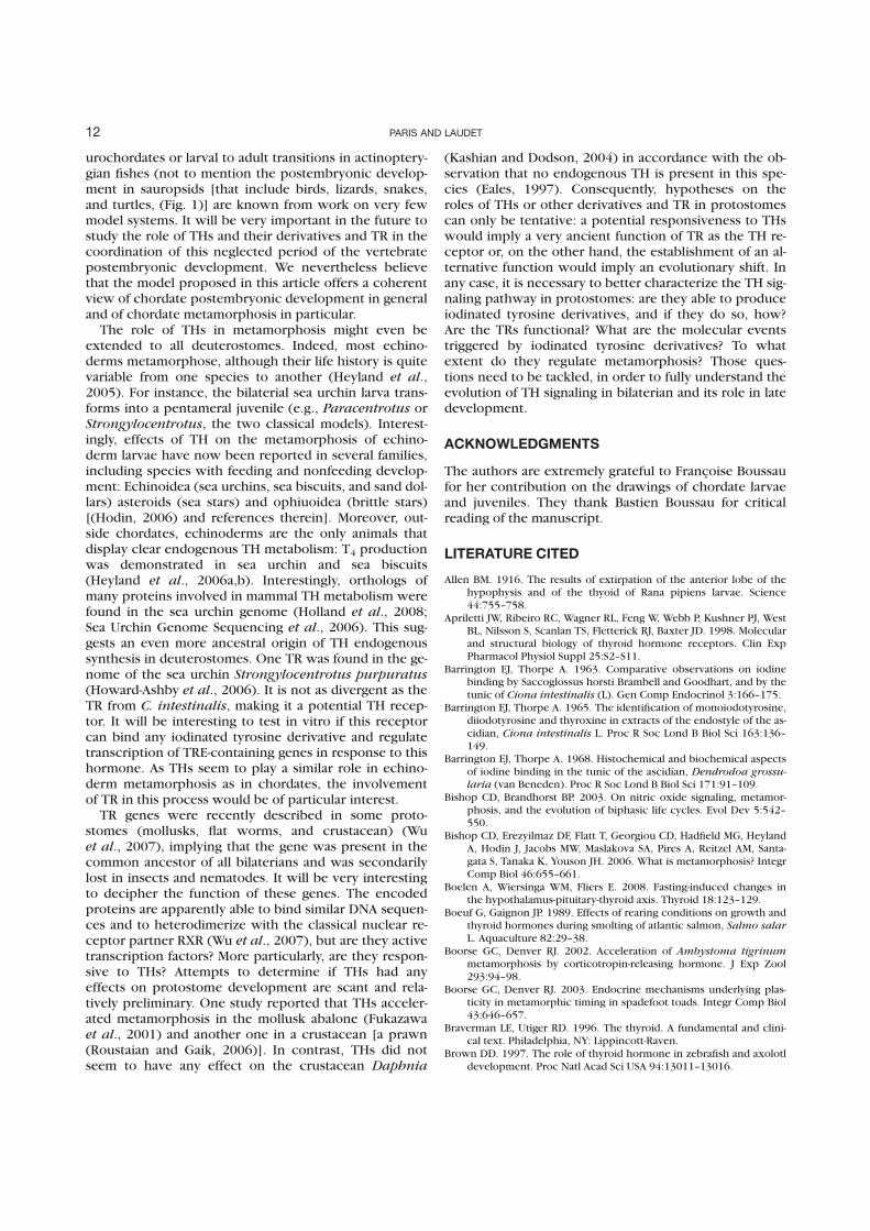
urochordates or larval to adult transitions in actinoptery-gian fishes (not to mention the postembryonic develop-ment in sauropsids [that include birds, lizards, snakes,and turtles, (Fig. 1)] are known from work on very fewmodel systems. It will be very important in the future tostudy the role of THs and their derivatives and TR in thecoordination of this neglected period of the vertebratepostembryonic development. We nevertheless believethat the model proposed in this article offers a coherentview of chordate postembryonic development in generaland of chordate metamorphosis in particular.
The role of THs in metamorphosis might even beextended to all deuterostomes. Indeed, most echino-derms metamorphose, although their life history is quitevariable from one species to another (Heyland et al.,2005). For instance, the bilaterial sea urchin larva trans-forms into a pentameral juvenile (e.g., Paracentrotus orStrongylocentrotus, the two classical models). Interest-ingly, effects of TH on the metamorphosis of echino-derm larvae have now been reported in several families,including species with feeding and nonfeeding develop-ment: Echinoidea (sea urchins, sea biscuits, and sand dol-lars) asteroids (sea stars) and ophiuoidea (brittle stars)[(Hodin, 2006) and references therein]. Moreover, out-side chordates, echinoderms are the only animals thatdisplay clear endogenous TH metabolism: T4 productionwas demonstrated in sea urchin and sea biscuits(Heyland et al., 2006a,b). Interestingly, orthologs ofmany proteins involved in mammal TH metabolism werefound in the sea urchin genome (Holland et al., 2008;Sea Urchin Genome Sequencing et al., 2006). This sug-gests an even more ancestral origin of TH endogenoussynthesis in deuterostomes. One TR was found in the ge-nome of the sea urchin Strongylocentrotus purpuratus(Howard-Ashby et al., 2006). It is not as divergent as theTR from C. intestinalis, making it a potential TH recep-tor. It will be interesting to test in vitro if this receptorcan bind any iodinated tyrosine derivative and regulatetranscription of TRE-containing genes in response to thishormone. As THs seem to play a similar role in echino-derm metamorphosis as in chordates, the involvementof TR in this process would be of particular interest.
TR genes were recently described in some proto-stomes (mollusks, flat worms, and crustacean) (Wuet al., 2007), implying that the gene was present in thecommon ancestor of all bilaterians and was secondarilylost in insects and nematodes. It will be very interestingto decipher the function of these genes. The encodedproteins are apparently able to bind similar DNA sequen-ces and to heterodimerize with the classical nuclear re-ceptor partner RXR (Wu et al., 2007), but are they activetranscription factors? More particularly, are they respon-sive to THs? Attempts to determine if THs had anyeffects on protostome development are scant and rela-tively preliminary. One study reported that THs acceler-ated metamorphosis in the mollusk abalone (Fukazawaet al., 2001) and another one in a crustacean [a prawn(Roustaian and Gaik, 2006)]. In contrast, THs did notseem to have any effect on the crustacean Daphnia
(Kashian and Dodson, 2004) in accordance with the ob-servation that no endogenous TH is present in this spe-cies (Eales, 1997). Consequently, hypotheses on theroles of THs or other derivatives and TR in protostomescan only be tentative: a potential responsiveness to THswould imply a very ancient function of TR as the TH re-ceptor or, on the other hand, the establishment of an al-ternative function would imply an evolutionary shift. Inany case, it is necessary to better characterize the TH sig-naling pathway in protostomes: are they able to produceiodinated tyrosine derivatives, and if they do so, how?Are the TRs functional? What are the molecular eventstriggered by iodinated tyrosine derivatives? To whatextent do they regulate metamorphosis? Those ques-tions need to be tackled, in order to fully understand theevolution of TH signaling in bilaterian and its role in latedevelopment.
ACKNOWLEDGMENTS
The authors are extremely grateful to Francoise Boussaufor her contribution on the drawings of chordate larvaeand juveniles. They thank Bastien Boussau for criticalreading of the manuscript.
LITERATURE CITED
Allen BM. 1916. The results of extirpation of the anterior lobe of thehypophysis and of the thyoid of Rana pipiens larvae. Science44:755–758.
Apriletti JW, Ribeiro RC, Wagner RL, Feng W, Webb P, Kushner PJ, WestBL, Nilsson S, Scanlan TS, Fletterick RJ, Baxter JD. 1998. Molecularand structural biology of thyroid hormone receptors. Clin ExpPharmacol Physiol Suppl 25:S2–S11.
Barrington EJ, Thorpe A. 1963. Comparative observations on iodinebinding by Saccoglossus horsti Brambell and Goodhart, and by thetunic of Ciona intestinalis (L). Gen Comp Endocrinol 3:166–175.
Barrington EJ, Thorpe A. 1965. The identification of monoiodotyrosine,diiodotyrosine and thyroxine in extracts of the endostyle of the as-cidian, Ciona intestinalis L. Proc R Soc Lond B Biol Sci 163:136–149.
Barrington EJ, Thorpe A. 1968. Histochemical and biochemical aspectsof iodine binding in the tunic of the ascidian, Dendrodoa grossu-laria (van Beneden). Proc R Soc Lond B Biol Sci 171:91–109.
Bishop CD, Brandhorst BP. 2003. On nitric oxide signaling, metamor-phosis, and the evolution of biphasic life cycles. Evol Dev 5:542–550.
Bishop CD, Erezyilmaz DF, Flatt T, Georgiou CD, Hadfield MG, HeylandA, Hodin J, Jacobs MW, Maslakova SA, Pires A, Reitzel AM, Santa-gata S, Tanaka K, Youson JH. 2006. What is metamorphosis? IntegrComp Biol 46:655–661.
Boelen A, Wiersinga WM, Fliers E. 2008. Fasting-induced changes inthe hypothalamus-pituitary-thyroid axis. Thyroid 18:123–129.
Boeuf G, Gaignon JP. 1989. Effects of rearing conditions on growth andthyroid hormones during smolting of atlantic salmon, Salmo salarL. Aquaculture 82:29–38.
Boorse GC, Denver RJ. 2002. Acceleration of Ambystoma tigrinummetamorphosis by corticotropin-releasing hormone. J Exp Zool293:94–98.
Boorse GC, Denver RJ. 2003. Endocrine mechanisms underlying plas-ticity in metamorphic timing in spadefoot toads. Integr Comp Biol43:646–657.
Braverman LE, Utiger RD. 1996. The thyroid. A fundamental and clini-cal text. Philadelphia, NY: Lippincott-Raven.
Brown DD. 1997. The role of thyroid hormone in zebrafish and axolotldevelopment. Proc Natl Acad Sci USA 94:13011–13016.
12 PARIS AND LAUDET
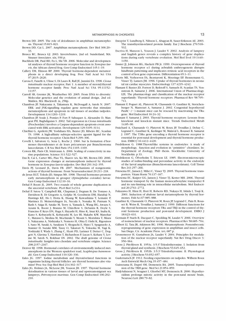
Brown DD. 2005. The role of deiodinases in amphibian metamorpho-sis. Thyroid 15:815–821.
Brown DD, Cai L. 2007. Amphibian metamorphosis. Dev Biol 306:20–33.
Brusca RC, Brusca GJ. 2003. Invertebrates, 2nd ed. Sunderland, MA:Sinauer Associates. xix, 936 p.
Buchholz DR, Paul BD, Fu L, Shi YB. 2006. Molecular and developmen-tal analyses of thyroid hormone receptor function in Xenopus lae-vis, the African clawed frog. Gen Comp Endocrinol 145:1–19.
Callery EM, Elinson RP. 2000. Thyroid hormone-dependent metamor-phosis in a direct developing frog. Proc Natl Acad Sci USA97:2615–2620.
Carosa E, Fanelli A, Ulisse S, Di Lauro R, Rall JE, Jannini EA. 1998. Cionaintestinalis nuclear receptor, Part 1: A member of steroid/thyroidhormone receptor family. Proc Natl Acad Sci USA 95:11152–11157.
Carroll SB, Grenier JK, Weatherbee SD. 2005. From DNA to diversity:Molecular genetics and the evolution of animal design, 2nd ed.Malden, MA: Blackwell. ix, 258p.
Chambon JP, Nakayama A, Takamura K, McDougall A, Satoh N. 2007.ERK- and JNK-signalling regulate gene networks that stimulatemetamorphosis and apoptosis in tail tissues of ascidian tadpoles.Development 134:1203–1219.
Chambon JP, Soule J, Pomies P, Fort P, Sahuquet A, Alexandre D, Man-geat PH, Baghdiguian S. 2002. Tail regression in Ciona intestinalis(Prochordate) involves a Caspase-dependent apoptosis event asso-ciated with ERK activation. Development 129:3105–3114.
Chiellini G, Apriletti JW, Yoshihara HA, Baxter JD, Ribeiro RC, ScanlanTS. 1998. A high-affinity subtype-selective agonist ligand for thethyroid hormone receptor. Chem Biol 5:299–306.
Covelli I, Salvatore G, Sena L, Roche J. 1960. Sur la formation d’hor-mones thyroidiennes et de leurs precurseurs par Branchiostomalanceolatum. C R Soc Biol Paris 154:1165–1169.
Cowen RK, Paris CB, Srinivasan A. 2006. Scaling of connectivity in ma-rine populations. Science 311:522–527.
Das B, Cai L, Carter MG, Piao YL, Sharov AA, Ko MS, Brown DD. 2006.Gene expression changes at metamorphosis induced by thyroidhormone in Xenopus laevis tadpoles. Dev Biol 291:342–355.
Davis PJ, Leonard JL, Davis FB. 2007. Mechanisms of nongenomicactions of thyroid hormone. Front Neuroendocrinol 29:211–218.
de Jesus EGT, Toledo JD, Simpas MS. 1998. Thyroid hormones promoteearly metamorphosis in grouper (Epinephelus coioides) larvae.Gen Comp Endocrinol 112:10–16.
Dehal P, Boore JL. 2005. Two rounds of whole genome duplication inthe ancestral vertebrate. PLoS Biol 3:e314.
Dehal P, Satou Y, Campbell RK, Chapman J, Degnan B, De Tomaso A,Davidson B, Di Gregorio A, Gelpke M, Goodstein DM, Harafuji N,Hastings KE, Ho I, Hotta K, Huang W, Kawashima T, Lemaire P,Martinez D, Meinertzhagen IA, Necula S, Nonaka M, Putnam N,Rash S, Saiga H, Satake M, Terry A, Yamada L, Wang HG, Awazu S,Azumi K, Boore J, Branno M, Chin-Bow S, DeSantis R, Doyle S,Francino P, Keys DN, Haga S, Hayashi H, Hino K, Imai KS, Inaba K,Kano S, Kobayashi K, Kobayashi M, Lee BI, Makabe KW, ManoharC, Matassi G, Medina M, Mochizuki Y, Mount S, Morishita T, MiuraS, Nakayama A, Nishizaka S, Nomoto H, Ohta F, Oishi K, RigoutsosI, Sano M, Sasaki A, Sasakura Y, Shoguchi E, Shin-i T, Spagnuolo A,Stainier D, Suzuki MM, Tassy O, Takatori N, Tokuoka M, Yagi K,Yoshizaki F, Wada S, Zhang C, Hyatt PD, Larimer F, Detter C, Dog-gett N, Glavina T, Hawkins T, Richardson P, Lucas S, Kohara Y, Lev-ine M, Satoh N, Rokhsar DS. 2002. The draft genome of Cionaintestinalis: Insights into chordate and vertebrate origins. Science298:2157–2167.
Denver RJ. 1998. Hormonal correlates of environmentally induced met-amorphosis in the Western spadefoot toad, Scaphiopus hammon-dii. Gen Comp Endocrinol 110:326–336.
Eales JG. 1997. Iodine metabolism and thyroid-related functions inorganisms lacking thyroid follicles: are thyroid hormones also vita-mins? Proc Soc Exp Biol Med 214:302–317.
Eales JG, Holmes JA, McLeese JM, Youson JH. 1997. Thyroid hormonedeiodination in various tissues of larval and upstream-migrant sealampreys, Petromyzon marinus. Gen Comp Endocrinol 106:202–210.
Eneqvist T, Lundberg E, Nilsson L, Abagyan R, Sauer-Eriksson AE. 2003.The transthyretin-related protein family. Eur J Biochem 270:518–532.
Escriva H, Manzon L, Youson J, Laudet V. 2002. Analysis of lampreyand hagfish genes reveals a complex history of gene duplica-tions during early vertebrate evolution. Mol Biol Evol 19:1440–1450.
Essner JJ, Johnson RG, Hackett PB Jr. 1999. Overexpression of thyroidhormone receptor a1 during zebrafish embryogenesis disruptshindbrain patterning and implicates retinoic acid receptors in thecontrol of hox gene expression. Differentiation 65:1–11.
Everts ME, Verhoeven FA, Bezstarosti K, Moerings EP, Hennemann G,Visser TJ, Lamers JM. 1996. Uptake of thyroid hormones in neona-tal rat cardiac myocytes. Endocrinology 137:4235–4242.
Flamant F, Baxter JD, Forrest D, Refetoff S, Samuels H, Scanlan TS, Ven-nstrom B, Samarut J. 2006. International Union of Pharmacology.LIX. The pharmacology and classification of the nuclear receptorsuperfamily: Thyroid hormone receptors. Pharmacol Rev 58:705–711.
Flamant F, Poguet AL, Plateroti M, Chassande O, Gauthier K, Streichen-berger N, Mansouri A, Samarut J. 2002. Congenital hypothyroidPax8(2/2) mutant mice can be rescued by inactivating the TRagene. Mol Endocrinol 16:24–32.
Flamant F, Samarut J. 2003. Thyroid hormone receptors: Lessons fromknockout and knock-in mutant mice. Trends Endocrinol Metab14:85–90.
Fraichard A, Chassande O, Plateroti M, Roux JP, Trouillas J, Dehay C,Legrand C, Gauthier K, Kedinger M, Malaval L, Rousset B, SamarutJ. 1997. The T3Ra gene encoding a thyroid hormone receptor isessential for post-natal development and thyroid hormone produc-tion. EMBO J 16:4412–4420.
Fredriksson G. 1988.Thyroid-like systems in endostyles: A study ofmorphology, function and evolution in ‘‘primitive’’ chordates. In:Department of Zoology, PhD thesis. Stockholm: University ofStockholm. p 33.
Fredriksson G, Ofverholm T, Ericson LE. 1985. Electron-microscopicstudies of iodine-binding and peroxidase activity in the endostyleof the larval amphioxus (Branchiostoma lanceolatum). Cell TissueRes 241:257–266.
Friesema EC, Jansen J, Milici C, Visser TJ. 2005. Thyroid hormone trans-porters. Vitam Horm 70:137–167.
Friesema EC, Kuiper GG, Jansen J, Visser TJ, Kester MH. 2006. Thyroidhormone transport by the human monocarboxylate transporter 8and its rate-limiting role in intracellular metabolism. Mol Endocri-nol 20:2761–2772.
Fukazawa H, Hirai H, Hori H, Roberts RD, Nukaya H, Ishida I, Tsuji K.2001. Induction of abalone larval metamorphosis by thyroid hor-mones. Fish Sci 67:985–988.
Gauthier K, Chassande O, Plateroti M, Roux JP, Legrand C, Pain B, Rous-set B, Weiss R, Trouillas J, Samarut J. 1999. Different functions forthe thyroid hormone receptors TRa and TRb in the control of thy-roid hormone production and post-natal development. EMBO J18:623–631.
Germain P, Staels B, Dacquet C, Spedding M, Laudet V. 2006. Overviewof nomenclature of nuclear receptors. Pharmacol Rev 58:685–704.
Gilbert LI, Tata JR, Atkinson BG. 1996. Metamorphosis: Postembryonicreprogramming of gene expression in amphibian and insect cells.San Diego, CA: Academic Press. xvi, 687 p.
Gronemeyer H, Gustafsson JA, Laudet V. 2004. Principles for modula-tion of the nuclear receptor superfamily. Nat Rev Drug Discov 3:950–964.
Gross J, Pitt-Rivers R. 1953a. 3:5:30 -Triiodothyronine. I. Isolation fromthyroid gland and synthesis. J Biochem 53:645–652.
Gross J, Pitt-Rivers R. 1953b. 3:5:30 -Triiodothyronine. II. Physiologicalactivity. J Biochem 53:652–657.
Gudernatsch JF. 1912. Feeding experiments on tadpoles. Wilhem RouxArch Entwickl Mech Org 35:457–484.
Guissouma H, Dupre SM, Demeneix BA. 2005. Transcriptional repres-sion of the TRH gene. Med Sci 21:854–859.
Hadj-Sahraoui N, Seugnet I, Ghorbel MT, Demeneix B. 2000. Hypothyr-oidism prolongs mitotic activity in the post-natal mouse brain.Neurosci Lett 280:79–82.
13METAMORPHOSIS IN CHORDATES
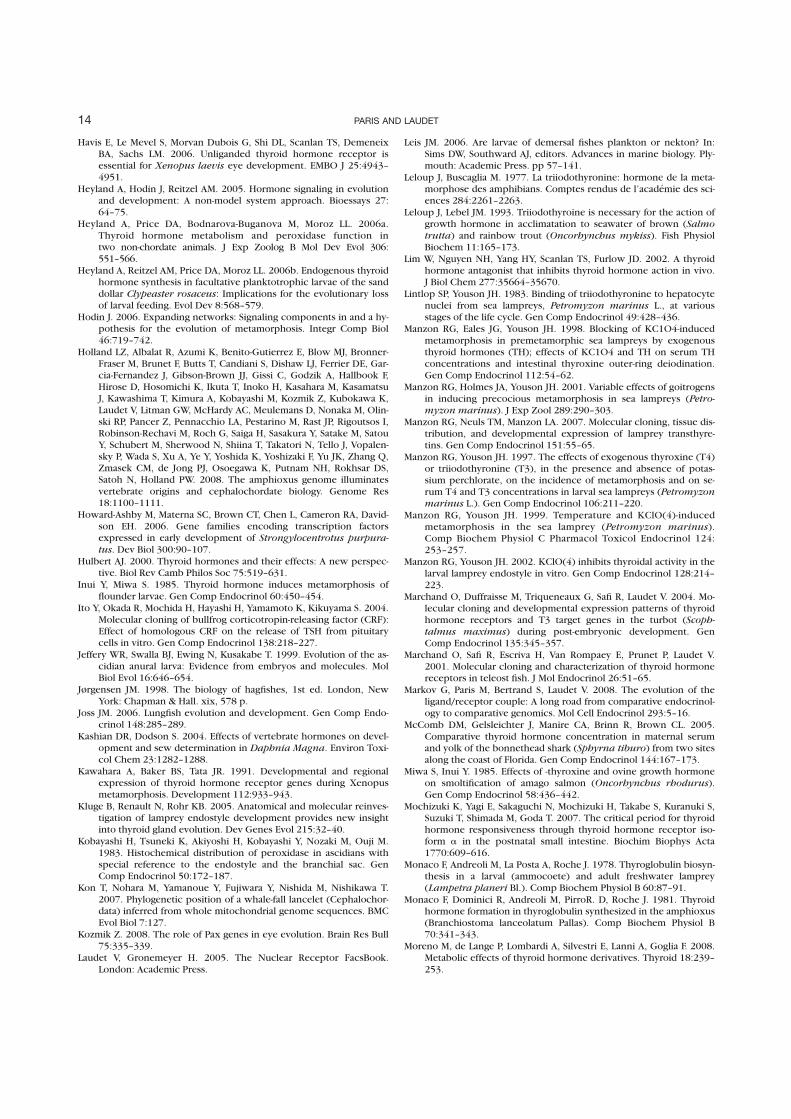
Havis E, Le Mevel S, Morvan Dubois G, Shi DL, Scanlan TS, DemeneixBA, Sachs LM. 2006. Unliganded thyroid hormone receptor isessential for Xenopus laevis eye development. EMBO J 25:4943–4951.
Heyland A, Hodin J, Reitzel AM. 2005. Hormone signaling in evolutionand development: A non-model system approach. Bioessays 27:64–75.
Heyland A, Price DA, Bodnarova-Buganova M, Moroz LL. 2006a.Thyroid hormone metabolism and peroxidase function intwo non-chordate animals. J Exp Zoolog B Mol Dev Evol 306:551–566.
Heyland A, Reitzel AM, Price DA, Moroz LL. 2006b. Endogenous thyroidhormone synthesis in facultative planktotrophic larvae of the sanddollar Clypeaster rosaceus: Implications for the evolutionary lossof larval feeding. Evol Dev 8:568–579.
Hodin J. 2006. Expanding networks: Signaling components in and a hy-pothesis for the evolution of metamorphosis. Integr Comp Biol46:719–742.
Holland LZ, Albalat R, Azumi K, Benito-Gutierrez E, Blow MJ, Bronner-Fraser M, Brunet F, Butts T, Candiani S, Dishaw LJ, Ferrier DE, Gar-cia-Fernandez J, Gibson-Brown JJ, Gissi C, Godzik A, Hallbook F,Hirose D, Hosomichi K, Ikuta T, Inoko H, Kasahara M, KasamatsuJ, Kawashima T, Kimura A, Kobayashi M, Kozmik Z, Kubokawa K,Laudet V, Litman GW, McHardy AC, Meulemans D, Nonaka M, Olin-ski RP, Pancer Z, Pennacchio LA, Pestarino M, Rast JP, Rigoutsos I,Robinson-Rechavi M, Roch G, Saiga H, Sasakura Y, Satake M, SatouY, Schubert M, Sherwood N, Shiina T, Takatori N, Tello J, Vopalen-sky P, Wada S, Xu A, Ye Y, Yoshida K, Yoshizaki F, Yu JK, Zhang Q,Zmasek CM, de Jong PJ, Osoegawa K, Putnam NH, Rokhsar DS,Satoh N, Holland PW. 2008. The amphioxus genome illuminatesvertebrate origins and cephalochordate biology. Genome Res18:1100–1111.
Howard-Ashby M, Materna SC, Brown CT, Chen L, Cameron RA, David-son EH. 2006. Gene families encoding transcription factorsexpressed in early development of Strongylocentrotus purpura-
tus. Dev Biol 300:90–107.Hulbert AJ. 2000. Thyroid hormones and their effects: A new perspec-
tive. Biol Rev Camb Philos Soc 75:519–631.Inui Y, Miwa S. 1985. Thyroid hormone induces metamorphosis of
flounder larvae. Gen Comp Endocrinol 60:450–454.Ito Y, Okada R, Mochida H, Hayashi H, Yamamoto K, Kikuyama S. 2004.
Molecular cloning of bullfrog corticotropin-releasing factor (CRF):Effect of homologous CRF on the release of TSH from pituitarycells in vitro. Gen Comp Endocrinol 138:218–227.
Jeffery WR, Swalla BJ, Ewing N, Kusakabe T. 1999. Evolution of the as-cidian anural larva: Evidence from embryos and molecules. MolBiol Evol 16:646–654.
Jørgensen JM. 1998. The biology of hagfishes, 1st ed. London, NewYork: Chapman & Hall. xix, 578 p.
Joss JM. 2006. Lungfish evolution and development. Gen Comp Endo-crinol 148:285–289.
Kashian DR, Dodson S. 2004. Effects of vertebrate hormones on devel-opment and sew determination in Daphnia Magna. Environ Toxi-col Chem 23:1282–1288.
Kawahara A, Baker BS, Tata JR. 1991. Developmental and regionalexpression of thyroid hormone receptor genes during Xenopusmetamorphosis. Development 112:933–943.
Kluge B, Renault N, Rohr KB. 2005. Anatomical and molecular reinves-tigation of lamprey endostyle development provides new insightinto thyroid gland evolution. Dev Genes Evol 215:32–40.
Kobayashi H, Tsuneki K, Akiyoshi H, Kobayashi Y, Nozaki M, Ouji M.1983. Histochemical distribution of peroxidase in ascidians withspecial reference to the endostyle and the branchial sac. GenComp Endocrinol 50:172–187.
Kon T, Nohara M, Yamanoue Y, Fujiwara Y, Nishida M, Nishikawa T.2007. Phylogenetic position of a whale-fall lancelet (Cephalochor-data) inferred from whole mitochondrial genome sequences. BMCEvol Biol 7:127.
Kozmik Z. 2008. The role of Pax genes in eye evolution. Brain Res Bull75:335–339.
Laudet V, Gronemeyer H. 2005. The Nuclear Receptor FacsBook.London: Academic Press.
Leis JM. 2006. Are larvae of demersal fishes plankton or nekton? In:Sims DW, Southward AJ, editors. Advances in marine biology. Ply-mouth: Academic Press. pp 57–141.
Leloup J, Buscaglia M. 1977. La triiodothyronine: hormone de la meta-morphose des amphibians. Comptes rendus de l’academie des sci-ences 284:2261–2263.
Leloup J, Lebel JM. 1993. Triiodothyroine is necessary for the action ofgrowth hormone in acclimatation to seawater of brown (Salmo
trutta) and rainbow trout (Oncorhynchus mykiss). Fish PhysiolBiochem 11:165–173.
Lim W, Nguyen NH, Yang HY, Scanlan TS, Furlow JD. 2002. A thyroidhormone antagonist that inhibits thyroid hormone action in vivo.J Biol Chem 277:35664–35670.
Lintlop SP, Youson JH. 1983. Binding of triiodothyronine to hepatocytenuclei from sea lampreys, Petromyzon marinus L., at variousstages of the life cycle. Gen Comp Endocrinol 49:428–436.
Manzon RG, Eales JG, Youson JH. 1998. Blocking of KC1O4-inducedmetamorphosis in premetamorphic sea lampreys by exogenousthyroid hormones (TH); effects of KC1O4 and TH on serum THconcentrations and intestinal thyroxine outer-ring deiodination.Gen Comp Endocrinol 112:54–62.
Manzon RG, Holmes JA, Youson JH. 2001. Variable effects of goitrogensin inducing precocious metamorphosis in sea lampreys (Petro-myzon marinus). J Exp Zool 289:290–303.
Manzon RG, Neuls TM, Manzon LA. 2007. Molecular cloning, tissue dis-tribution, and developmental expression of lamprey transthyre-tins. Gen Comp Endocrinol 151:55–65.
Manzon RG, Youson JH. 1997. The effects of exogenous thyroxine (T4)or triiodothyronine (T3), in the presence and absence of potas-sium perchlorate, on the incidence of metamorphosis and on se-rum T4 and T3 concentrations in larval sea lampreys (Petromyzonmarinus L.). Gen Comp Endocrinol 106:211–220.
Manzon RG, Youson JH. 1999. Temperature and KClO(4)-inducedmetamorphosis in the sea lamprey (Petromyzon marinus).Comp Biochem Physiol C Pharmacol Toxicol Endocrinol 124:253–257.
Manzon RG, Youson JH. 2002. KClO(4) inhibits thyroidal activity in thelarval lamprey endostyle in vitro. Gen Comp Endocrinol 128:214–223.
Marchand O, Duffraisse M, Triqueneaux G, Safi R, Laudet V. 2004. Mo-lecular cloning and developmental expression patterns of thyroidhormone receptors and T3 target genes in the turbot (Scoph-talmus maximus) during post-embryonic development. GenComp Endocrinol 135:345–357.
Marchand O, Safi R, Escriva H, Van Rompaey E, Prunet P, Laudet V.2001. Molecular cloning and characterization of thyroid hormonereceptors in teleost fish. J Mol Endocrinol 26:51–65.
Markov G, Paris M, Bertrand S, Laudet V. 2008. The evolution of theligand/receptor couple: A long road from comparative endocrinol-ogy to comparative genomics. Mol Cell Endocrinol 293:5–16.
McComb DM, Gelsleichter J, Manire CA, Brinn R, Brown CL. 2005.Comparative thyroid hormone concentration in maternal serumand yolk of the bonnethead shark (Sphyrna tiburo) from two sitesalong the coast of Florida. Gen Comp Endocrinol 144:167–173.
Miwa S, Inui Y. 1985. Effects of -thyroxine and ovine growth hormoneon smoltification of amago salmon (Oncorhynchus rhodurus).Gen Comp Endocrinol 58:436–442.
Mochizuki K, Yagi E, Sakaguchi N, Mochizuki H, Takabe S, Kuranuki S,Suzuki T, Shimada M, Goda T. 2007. The critical period for thyroidhormone responsiveness through thyroid hormone receptor iso-form a in the postnatal small intestine. Biochim Biophys Acta1770:609–616.
Monaco F, Andreoli M, La Posta A, Roche J. 1978. Thyroglobulin biosyn-thesis in a larval (ammocoete) and adult freshwater lamprey(Lampetra planeri Bl.). Comp Biochem Physiol B 60:87–91.
Monaco F, Dominici R, Andreoli M, PirroR. D, Roche J. 1981. Thyroidhormone formation in thyroglobulin synthesized in the amphioxus(Branchiostoma lanceolatum Pallas). Comp Biochem Physiol B70:341–343.
Moreno M, de Lange P, Lombardi A, Silvestri E, Lanni A, Goglia F. 2008.Metabolic effects of thyroid hormone derivatives. Thyroid 18:239–253.
14 PARIS AND LAUDET
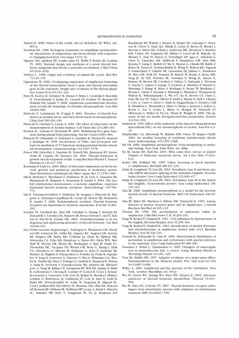
Nelson JS. 2006. Fishes of the world, 4th ed. Hoboken, NJ: Wiley. xix,601 p.
Newman RA. 1998. Ecological constraints on amphibian metamorpho-sis: Interactions of temperature and larval density with responsesto changing food level. Oecologia 115:9–16.
Nguyen NH, Apriletti JW, Cunha Lima ST, Webb P, Baxter JD, ScanlanTS. 2002. Rational design and synthesis of a novel thyroid hor-mone antagonist that blocks coactivator recruitment. J Med Chem45:3310–3320.
Nielsen C. 1998. Origin and evolution of animal life cycles. Biol Rev73:125–155.
Ogasawara M. 2000. Overlapping expression of amphioxus homologsof the thyroid transcription factor-1 gene and thyroid peroxidasegene in the endostyle: Insight into evolution of the thyroid gland.Dev Genes Evol 210:231–242.
Paris M, Escriva H, Schubert M, Brunet F, Brtko J, Ciesielski F, RoecklinD, Vivat-Hannah V, Jamin EL, Cravedi J-P, Scanlan TS, Renaud J-P,Holland ND, Laudet V. 2008. Amphioxus postembryonic develop-ment reveals the homology of chordate metamorphosis. Curr Biol18:825–830.
Patricolo E, Cammarata M, D’Agati P. 2001. Presence of thyroid hor-mones in ascidian larvae and their involvement in metamorphosis.J Exp Zool 290:426–430.
Patricolo E, Ortolani G, Cascio A. 1981. The effect of l-thyroxine on themetamorphosis of Ascidia malaca. Cell Tissue Res 214:289–301.
Pearson JC, Lemons D, McGinnis W. 2005. Modulating Hox gene func-tions during animal body patterning. Nat Rev Genet 6:893–904.
Plateroti M, Chassande O, Fraichard A, Gauthier K, Freund JN, SamarutJ, Kedinger M. 1999. Involvement of T3Ra- and b-receptor sub-types in mediation of T3 functions during postnatal murine intesti-nal development. Gastroenterology 116:1367–1378.
Power DM, Llewellyn L, Faustino M, Nowell MA, Bjornsson BT, Einars-dottir IE, Canario AV, Sweeney GE. 2001. Thyroid hormones ingrowth and development of fish. Comp Biochem Physiol C ToxicolPharmacol 130:447–459.
Roustaian P, Gaik LA. 2006. Effect of thyroxine immersion on larval sur-vival, growth and postlarvae production of freshwater prawn,Macrobrachium rosenbergii (de Man). Aquac Res 37:1378–1380.
Safi R, Bertrand S, Marchand O, Duffraisse M, de Luze A, Vanacker JM,Maraninchi M, Margotat A, Demeneix B, Laudet V. 2004. The axo-lotl (Ambystoma mexicanum), a neotenic amphibian, expressesfunctional thyroid hormone receptors. Endocrinology 145:760–772.
Safi R, Vlaeminck-Guillem V, Duffraisse M, Seugnet I, Plateroti M, Mar-gotat A, Duterque-Coquillaud M, Crespi EJ, Denver RJ, DemeneixB, Laudet V. 2006. Pedomorphosis revisited: Thyroid hormonereceptors are functional in Necturus maculosus. Evol Dev 8:284–292.
Scanlan TS, Suchland KL, Hart ME, Chiellini G, Huang Y, Kruzich PJ,Frascarelli S, Crossley DA, Bunzow JR, Ronca-Testoni S, Lin ET, Hat-ton D, Zucchi R, Grandy DK. 2004. 3-Iodothyronamine is an en-dogenous and rapid-acting derivative of thyroid hormone. Nat Med10:638–642.
Sea Urchin Genome Sequencing C, Sodergren E, Weinstock GM, David-son EH, Cameron RA, Gibbs RA, Angerer RC, Angerer LM, ArnoneMI, Burgess DR, Burke RD, Coffman JA, Dean M, Elphick MR,Ettensohn CA, Foltz KR, Hamdoun A, Hynes RO, Klein WH, Mar-zluff W, McClay DR, Morris RL, Mushegian A, Rast JP, Smith LC,Thorndyke MC, Vacquier VD, Wessel GM, Wray G, Zhang L, ElsikCG, Ermolaeva O, Hlavina W, Hofmann G, Kitts P, Landrum MJ,Mackey AJ, Maglott D, Panopoulou G, Poustka AJ, Pruitt K, Sapojni-kov V, Song X, Souvorov A, Solovyev V, Wei Z, Whittaker CA, Wor-ley K, Durbin KJ, Shen Y, Fedrigo O, Garfield D, Haygood R, PrimusA, Satija R, Severson T, Gonzalez-Garay ML, Jackson AR, Milosavl-jevic A, Tong M, Killian CE, Livingston BT, Wilt FH, Adams N, BelleR, Carbonneau S, Cheung R, Cormier P, Cosson B, Croce J, Fernan-dez-Guerra A, Geneviere A-M, Goel M, Kelkar H, Morales J, Mulner-Lorillon O, Robertson AJ, Goldstone JV, Cole B, Epel D, Gold B,Hahn ME, Howard-Ashby M, Scally M, Stegeman JJ, Allgood EL,Cool J, Judkins KM, McCafferty SS, Musante AM, Obar RA, RawsonAP, Rossetti BJ, Gibbons IR, Hoffman MP, Leone A, Istrail S, MaternaSC, Samanta MP, Stolc V, Tongprasit W, Tu Q, Bergeron K-F,
Brandhorst BP, Whittle J, Berney K, Bottjer DJ, Calestani C, Peter-son K, Chow E, Yuan QA, Elhaik E, Graur D, Reese JT, Bosdet I,Heesun S, Marra MA, Schein J, Anderson MK, Brockton V, BuckleyKM, Cohen AH, Fugmann SD, Hibino T, Loza-Coll M, Majeske AJ,Messier C, Nair SV, Pancer Z, Terwilliger DP, Agca C, Arboleda E,Chen N, Churcher AM, Hallbook F, Humphrey GW, Idris MM,Kiyama T, Liang S, Mellott D, Mu X, Murray G, Olinski RP, Raible F,Rowe M, Taylor JS, Tessmar-Raible K, Wang D, Wilson KH, YaguchiS, Gaasterland T, Galindo BE, Gunaratne HJ, Juliano C, KinukawaM, Moy GW, Neill AT, Nomura M, Raisch M, Reade A, Roux MM,Song JL, Su Y-H, Townley IK, Voronina E, Wong JL, Amore G,Branno M, Brown ER, Cavalieri V, Duboc V, Duloquin L, FlytzanisC, Gache C, Lapraz F, Lepage T, Locascio A, Martinez P, Matassi G,Matranga V, Range R, Rizzo F, Rottinger E, Beane W, Bradham C,Byrum C, Glenn T, Hussain S, Manning G, Miranda E, Thomason R,Walton K, Wikramanayke A, Wu S-Y, Xu R, Brown CT, Chen L,Gray RF, Lee PY, Nam J, Oliveri P, Smith J, Muzny D, Bell S, ChackoJ, Cree A, Curry S, Davis C, Dinh H, Dugan-Rocha S, Fowler J, GillR, Hamilton C, Hernandez J, Hines S, Hume J, Jackson L, Jolivet A,Kovar C, Lee S, Lewis L, Miner G, Morgan M, Nazareth LV,Okwuonu G, Parker D, Pu L-L, Thorn R, Wright R. 2006. The ge-nome of the sea urchin Strongylocentrotus purpuratus. Science314:941–952.
Sembrat K. 1953. Effect of the endostyle of the lancelet (Branchiostomalanceolatum Pall.) on the metamorphosis of axolotl. Zool Pol 6:3–19.
Shepherdley CA, Klootwijk W, Makabe KW, Visser TJ, Kuiper GGJM.2004. An ascidian homolog of vertebrate iodothyronine deiodi-nases. Endocrinology 145:1255–1268.
Shi Y-B. 2000. Amphibian metamorphosis: From morphology to molec-ular biology. New York: John Wiley. xiv, 288p.
Sly BJ, Snoke MS, Raff RA. 2003. Who came first—larvae or adults?Origins of bilaterian metazoan larvae. Int J Dev Biol 47:623–632.
Stokes MD, Holland ND. 1995. Ciliary hovering in larval lancelets(5Amphioxus). Biol Bull 188:231–233.
Sutija M, Longhurst TJ, Joss JM. 2003. Deiodinase type II and tissue spe-cific mRNA alternative splicing in the Australian lungfish, Neocera-todus forsteri. Gen Comp Endocrinol 132:409–417.
Sutija M, Longhurst TJ, Joss JM. 2004. Deiodinase type III in the Austra-lian lungfish, Neoceratodus forsteri. Gen Comp Endocrinol 136:152–161.
Tata JR. 2006. Amphibian metamorphosis as a model for the develop-mental actions of thyroid hormone. Mol Cell Endocrinol 246:10–20.
Tata JR, Baker BS, Machuca I, Rabelo EM, Yamauchi K. 1993. Autoin-duction of nuclear receptor genes and its significance. J SteroidBiochem Mol Biol 46:105–119.
Thomas IM. 1956. The accumulation of radioactive iodine byamphioxus. J Mar Biol Assoc U K 35:203–210.
Tong W, Kerkof P, Chaikoff IL. 1961. 131I utilization by thyroid tissue ofthe hagfish. Biochim Biophys Acta 52:299–304.
Tong W, Kerkof P, Chaikoff IL. 1962. Identification of labeled thyroxineand triiodothyronine in amphioxus treated with 131-I. BiochimBiophys Acta 56:326–331.
Tsuneki K, Kobayashi H, Ouji M. 1983. Histochemical distribution ofperoxidase in amphioxus and cyclostomes with special referenceto the endostyle. Gen Comp Endocrinol 50:188–200.
Varsamos S, Nebel C, Charmantier G. 2005. Ontogeny of osmoregula-tion in postembryonic fish: A review. Comp Biochem Physiol AMol Integr Physiol 141:401–429.
Voss SR, Shaffer HB. 1997. Adaptive evolution via a major gene effect:Paedomorphosis in the Mexican axolotl. Proc Natl Acad Sci USA94:14185–14189.
Willey A. 1894. Amphioxus and the ancestry of the vertebrates. NewYork, London: Macmillan. xiv, 316 p.
Wu SY, Green WL, Huang WS, Hays MT, Chopra IJ. 2005. Alternatepathways of thyroid hormone metabolism. Thyroid 15:943–958.
Wu W, Niles EG, LoVerde PT. 2007. Thyroid hormone receptor ortho-logues from invertebrate species with emphasis on Schistosoma
mansoni. BMC Evol Biol 7:150.
15METAMORPHOSIS IN CHORDATES

Yamano K, Miwa S. 1998. Differential gene expression of thyroid hor-mone receptor a and b in fish development. Gen Comp Endocri-nol 109:75–85.
Yen PM. 2001. Physiological and molecular basis of thyroid hormoneaction. Physiol Rev 81:1097–1142.
Yen PM, Feng X, Flamant F, Chen Y, Walker RL, Weiss RE, Chassande O,Samarut J, Refetoff S, Meltzer PS. 2003. Effects of ligand and thyroidhormone receptor isoforms on hepatic gene expression profiles ofthyroid hormone receptor knockout mice. EMBO Rep 4:581–587.
Young G, Bjornsson BT, Prunet P, Lin RJ, Bern HA. 1989. Smoltificationand seawater adaptation in Coho salmon (Oncorhyncus kisutch):
Plasma prolactin, growth hormone, thyroid hormone, and cortisol.Gen Comp Endocrinol 74:335–345.
Youson JH. 1997. Is lamprey metamorphosis regulated by thyroid hor-mones? Am Zool 37:441–460.
Youson JH, Manzon RG, Peck BJ, Holmes JA. 1997. Effects of exogenousthyroxine (T4) and triiodothyronine (T3) on spontaneous meta-morphosis and serum T4 and T3 levels in immediately premeta-morphic sea lampreys, Petromyzon marinus. J Exp Zool 279:145–155.
Zhang J, Lazar MA. 2000. The mechanism of action of thyroid hor-mones. Annu Rev Physiol 62:439.
16 PARIS AND LAUDET
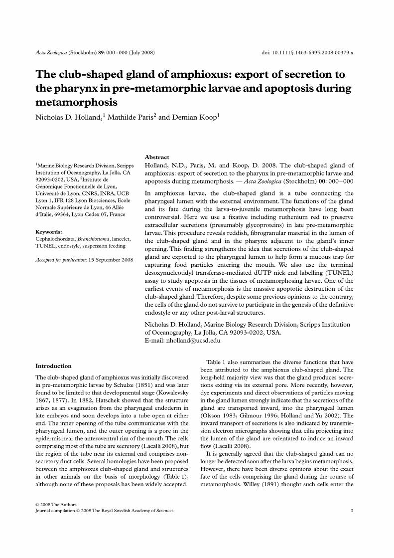
Acta Zoologica
(Stockholm)
89
: 000–000 (July 2008) doi: 10.1111/j.1463-6395.2008.00379.x
© 2008 The Authors
Journal compilation © 2008 The Royal Swedish Academy of Sciences
Abstract
Holland, N.D., Paris, M. and Koop, D. 2008. The club-shaped gland of
amphioxus: export of secretion to the pharynx in pre-metamorphic larvae and
apoptosis during metamorphosis. —
Acta Zoologica
(Stockholm)
00
: 000–000
In amphioxus larvae, the club-shaped gland is a tube connecting the
pharyngeal lumen with the external environment. The functions of the gland
and its fate during the larva-to-juvenile metamorphosis have long been
controversial. Here we use a fixative including ruthenium red to preserve
extracellular secretions (presumably glycoproteins) in late pre-metamorphic
larvae. This procedure reveals reddish, fibrogranular material in the lumen of
the club-shaped gland and in the pharynx adjacent to the gland’s inner
opening. This finding strengthens the idea that secretions of the club-shaped
gland are exported to the pharyngeal lumen to help form a mucous trap for
capturing food particles entering the mouth. We also use the terminal
desoxynucleotidyl transferase-mediated dUTP nick end labelling (TUNEL)
assay to study apoptosis in the tissues of metamorphosing larvae. One of the
earliest events of metamorphosis is the massive apoptotic destruction of the
club-shaped gland. Therefore, despite some previous opinions to the contrary,
the cells of the gland do not survive to participate in the genesis of the definitive
endostyle or any other post-larval structures.
Nicholas D. Holland, Marine Biology Research Division, Scripps Institution
of Oceanography, La Jolla, CA 92093-0202, USA.
E-mail: [email protected]
Blackwell Publishing Ltd
The club-shaped gland of amphioxus: export of secretion to
the pharynx in pre-metamorphic larvae and apoptosis during
metamorphosis
Nicholas D. Holland,
1
Mathilde Paris
2
and Demian Koop
1
1
Marine Biology Research Division, Scripps
Institution of Oceanography, La Jolla, CA
92093-0202, USA,
2
Institute de
Génomique Fonctionnelle de Lyon,
Université de Lyon, CNRS, INRA, UCB
Lyon 1, IFR 128 Lyon Biosciences, Ecole
Normale Supérieure de Lyon, 46 Allée
d’Italie, 69364, Lyon Cedex 07, France
Keywords:
Cephalochordata,
Branchiostoma
, lancelet,
TUNEL, endostyle, suspension feeding
Accepted for publication:
15 September 2008
Introduction
The club-shaped gland of amphioxus was initially discovered
in pre-metamorphic larvae by Schulze (1851) and was later
found to be limited to that developmental stage (Kowalevsky
1867, 1877). In 1882, Hatschek showed that the structure
arises as an evagination from the pharyngeal endoderm in
late embryos and soon develops into a tube open at either
end. The inner opening of the tube communicates with the
pharyngeal lumen, and the outer opening is a pore in the
epidermis near the anteroventral rim of the mouth. The cells
comprising most of the tube are secretory (Lacalli 2008), but
the region of the tube near its external end comprises non-
secretory duct cells. Several homologies have been proposed
between the amphioxus club-shaped gland and structures
in other animals on the basis of morphology (Table 1),
although none of these proposals has been widely accepted.
Table 1 also summarizes the diverse functions that have
been attributed to the amphioxus club-shaped gland. The
long-held majority view was that the gland produces secre-
tions exiting via its external pore. More recently, however,
dye experiments and direct observations of particles moving
in the gland lumen strongly indicate that the secretions of the
gland are transported inward, into the pharyngeal lumen
(Olsson 1983; Gilmour 1996; Holland and Yu 2002). The
inward transport of secretions is also indicated by transmis-
sion electron micrographs showing that cilia projecting into
the lumen of the gland are orientated to induce an inward
flow (Lacalli 2008).
It is generally agreed that the club-shaped gland can no
longer be detected soon after the larva begins metamorphosis.
However, there have been diverse opinions about the exact
fate of the cells comprising the gland during the course of
metamorphosis. Willey (1891) thought such cells enter the
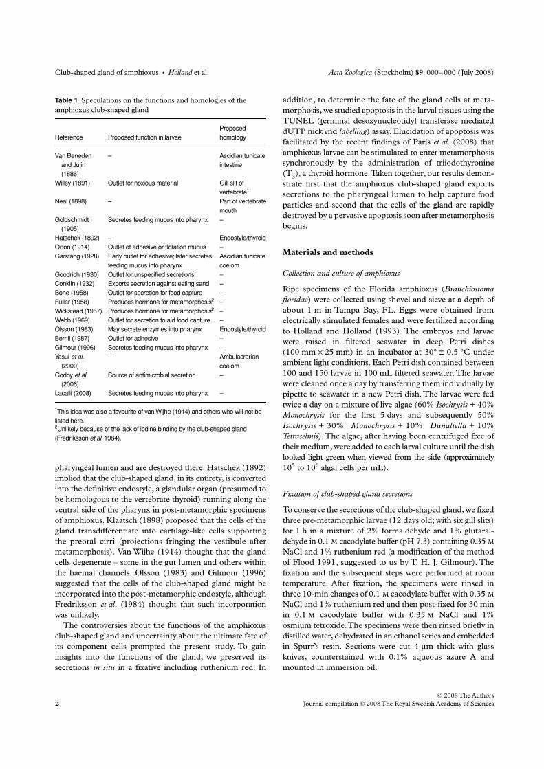
Club-shaped gland of amphioxus
•
Holland
et al.
Acta Zoologica
(Stockholm)
89
: 000–000 (July 2008)
© 2008 The Authors
Journal compilation © 2008 The Royal Swedish Academy of Sciences
pharyngeal lumen and are destroyed there. Hatschek (1892)
implied that the club-shaped gland, in its entirety, is converted
into the definitive endostyle, a glandular organ (presumed to
be homologous to the vertebrate thyroid) running along the
ventral side of the pharynx in post-metamorphic specimens
of amphioxus. Klaatsch (1898) proposed that the cells of the
gland transdifferentiate into cartilage-like cells supporting
the preoral cirri (projections fringing the vestibule after
metamorphosis). Van Wijhe (1914) thought that the gland
cells degenerate – some in the gut lumen and others within
the haemal channels. Olsson (1983) and Gilmour (1996)
suggested that the cells of the club-shaped gland might be
incorporated into the post-metamorphic endostyle, although
Fredriksson
et
al
. (1984) thought that such incorporation
was unlikely.
The controversies about the functions of the amphioxus
club-shaped gland and uncertainty about the ultimate fate of
its component cells prompted the present study. To gain
insights into the functions of the gland, we preserved its
secretions
in situ
in a fixative including ruthenium red. In
addition, to determine the fate of the gland cells at meta-
morphosis, we studied apoptosis in the larval tissues using the
TUNEL (terminal desoxynucleotidyl transferase mediated
dUTP nick
e
nd
labelling
) assay. Elucidation of apoptosis was
facilitated by the recent findings of Paris
et
al
. (2008) that
amphioxus larvae can be stimulated to enter metamorphosis
synchronously by the administration of triiodothyronine
(T
3
), a thyroid hormone. Taken together, our results demon-
strate first that the amphioxus club-shaped gland exports
secretions to the pharyngeal lumen to help capture food
particles and second that the cells of the gland are rapidly
destroyed by a pervasive apoptosis soon after metamorphosis
begins.
Materials and methods
Collection and culture of amphioxus
Ripe specimens of the Florida amphioxus (
Branchiostoma
floridae
) were collected using shovel and sieve at a depth of
about 1 m in Tampa Bay, FL. Eggs were obtained from
electrically stimulated females and were fertilized according
to Holland and Holland (1993). The embryos and larvae
were raised in filtered seawater in deep Petri dishes
(100 mm
×
25 mm) in an incubator at 30
°
± 0.5
°
C under
ambient light conditions. Each Petri dish contained between
100 and 150 larvae in 100 mL filtered seawater. The larvae
were cleaned once a day by transferring them individually by
pipette to seawater in a new Petri dish. The larvae were fed
twice a day on a mixture of live algae (60%
Isochrysis
+ 40%
Monochrysis
for the first 5 days and subsequently 50%
Isochrysis
+ 30%
Monochrysis
+ 10%
Dunaliella
+ 10%
Tetraselmis
). The algae, after having been centrifuged free of
their medium, were added to each larval culture until the dish
looked light green when viewed from the side (approximately
10
5
to 10
6
algal cells per mL).
Fixation of club-shaped gland secretions
To conserve the secretions of the club-shaped gland, we fixed
three pre-metamorphic larvae (12 days old; with six gill slits)
for 1 h in a mixture of 2% formaldehyde and 1% glutaral-
dehyde in 0.1
cacodylate buffer (pH 7.3) containing 0.35
NaCl and 1% ruthenium red (a modification of the method
of Flood 1991, suggested to us by T. H. J. Gilmour). The
fixation and the subsequent steps were performed at room
temperature. After fixation, the specimens were rinsed in
three 10-min changes of 0.1
cacodylate buffer with 0.35
NaCl and 1% ruthenium red and then post-fixed for 30 min
in 0.1
cacodylate buffer with 0.35
NaCl and 1%
osmium tetroxide. The specimens were then rinsed briefly in
distilled water, dehydrated in an ethanol series and embedded
in Spurr’s resin. Sections were cut 4-
µ
m thick with glass
knives, counterstained with 0.1% aqueous azure A and
mounted in immersion oil.
Table 1 Speculations on the functions and homologies of the
amphioxus club-shaped gland
Reference Proposed function in larvae
Proposed
homology
Van Beneden
and Julin
(1886)
– Ascidian tunicate
intestine
Willey (1891) Outlet for noxious material Gill slit of
vertebrate1
Neal (1898) – Part of vertebrate
mouth
Goldschmidt
(1905)
Secretes feeding mucus into pharynx –
Hatschek (1892) – Endostyle/thyroid
Orton (1914) Outlet of adhesive or flotation mucus –
Garstang (1928) Early outlet for adhesive; later secretes
feeding mucus into pharynx
Ascidian tunicate
coelom
Goodrich (1930) Outlet for unspecified secretions –
Conklin (1932) Exports secretion against eating sand –
Bone (1958) Outlet for secretion for food capture –
Fuller (1958) Produces hormone for metamorphosis2 –
Wickstead (1967) Produces hormone for metamorphosis2 –
Webb (1969) Outlet for secretion to aid food capture –
Olsson (1983) May secrete enzymes into pharynx Endostyle/thyroid
Berrill (1987) Outlet for adhesive –
Gilmour (1996) Secretes feeding mucus into pharynx –
Yasui et al.
(2000)
– Ambulacrarian
coelom
Godoy et al.
(2006)
Source of antimicrobial secretion –
Lacalli (2008) Secretes feeding mucus into pharynx –
1This idea was also a favourite of van Wijhe (1914) and others who will not be
listed here.2Unlikely because of the lack of iodine binding by the club-shaped gland
(Fredriksson et al. 1984).
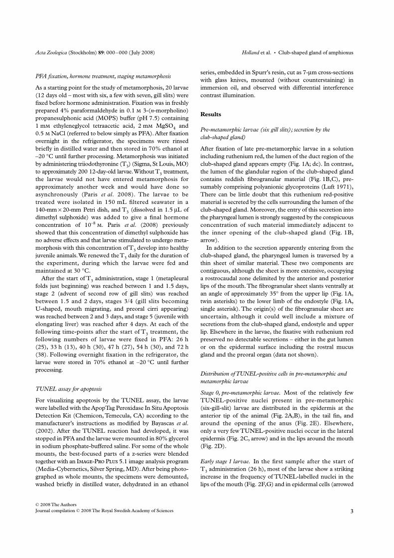
Acta Zoologica
(Stockholm)
89
: 000–000 (July 2008)
Holland
et al.
•
Club-shaped gland of amphioxus
© 2008 The Authors
Journal compilation © 2008 The Royal Swedish Academy of Sciences
PFA fixation, hormone treatment, staging metamorphosis
As a starting point for the study of metamorphosis, 20 larvae
(12 days old – most with six, a few with seven, gill slits) were
fixed before hormone administration. Fixation was in freshly
prepared 4% paraformaldehyde in 0.1
3-(
n
-morpholino)
propanesulphonic acid (MOPS) buffer (pH 7.5) containing
1 m
ethyleneglycol tetraacetic acid, 2 m
MgSO
4
and
0.5
NaCl (referred to below simply as PFA). After fixation
overnight in the refrigerator, the specimens were rinsed
briefly in distilled water and then stored in 70% ethanol at
–20
°
C until further processing. Metamorphosis was initiated
by administering triiodothyronine (T
3
) (Sigma, St Louis, MO)
to approximately 200 12-day-old larvae. Without T
3
treatment,
the larvae would not have entered metamorphosis for
approximately another week and would have done so
asynchronously (Paris
et
al
. 2008). The larvae to be
treated were isolated in 150 mL filtered seawater in a
140-mm
×
20-mm Petri dish, and T
3
(dissolved in 1.5
µ
L of
dimethyl sulphoxide) was added to give a final hormone
concentration of 10
–8
. Paris
et
al
. (2008) previously
showed that this concentration of dimethyl sulphoxide has
no adverse effects and that larvae stimulated to undergo meta-
morphosis with this concentration of T
3
develop into healthy
juvenile animals. We renewed the T
3
daily for the duration of
the experiment, during which the larvae were fed and
maintained at 30
°
C.
After the start of T
3
administration, stage 1 (metapleural
folds just beginning) was reached between 1 and 1.5 days,
stage 2 (advent of second row of gill slits) was reached
between 1.5 and 2 days, stages 3/4 (gill slits becoming
U-shaped, mouth migrating, and preoral cirri appearing)
was reached between 2 and 3 days, and stage 5 (juvenile with
elongating liver) was reached after 4 days. At each of the
following time-points after the start of T
3
treatment, the
following numbers of larvae were fixed in PFA: 26 h
(25), 33 h (13), 40 h (30), 47 h (27), 54 h (30), and 72 h
(38). Following overnight fixation in the refrigerator, the
larvae were stored in 70% ethanol at –20
°
C until further
processing.
TUNEL assay for apoptosis
For visualizing apoptosis by the TUNEL assay, the larvae
were labelled with the ApopTag Peroxidase In Situ Apoptosis
Detection Kit (Chemicon, Temecula, CA) according to the
manufacturer’s instructions as modified by Bayascas
et
al
.
(2002). After the TUNEL reaction had developed, it was
stopped in PFA and the larvae were mounted in 80% glycerol
in sodium phosphate-buffered saline. For some of the whole
mounts, the best-focused parts of a z-series were blended
together with an I
-
P
P
5.1 image analysis program
(Media-Cybernetics, Silver Spring, MD). After being photo-
graphed as whole mounts, the specimens were demounted,
washed briefly in distilled water, dehydrated in an ethanol
series, embedded in Spurr’s resin, cut as 7-
µ
m cross-sections
with glass knives, mounted (without counterstaining) in
immersion oil, and observed with differential interference
contrast illumination.
Results
Pre-metamorphic larvae (six gill slits); secretion by the
club-shaped gland)
After fixation of late pre-metamorphic larvae in a solution
including ruthenium red, the lumen of the duct region of the
club-shaped gland appears empty (Fig. 1A; dc). In contrast,
the lumen of the glandular region of the club-shaped gland
contains reddish fibrogranular material (Fig. 1B,C), pre-
sumably comprising polyanionic glycoproteins (Luft 1971),
There can be little doubt that this ruthenium red-positive
material is secreted by the cells surrounding the lumen of the
club-shaped gland. Moreover, the entry of this secretion into
the pharyngeal lumen is strongly suggested by the conspicuous
concentration of such material immediately adjacent to
the inner opening of the club-shaped gland (Fig. 1B,
arrow).
In addition to the secretion apparently entering from the
club-shaped gland, the pharyngeal lumen is traversed by a
thin sheet of similar material. These two components are
contiguous, although the sheet is more extensive, occupying
a rostrocaudal zone delimited by the anterior and posterior
lips of the mouth. The fibrogranular sheet slants ventrally at
an angle of approximately 35
°
from the upper lip (Fig. 1A,
twin asterisks) to the lower limb of the endostyle (Fig. 1A,
single asterisk). The origin(s) of the fibrogranular sheet are
uncertain, although it could well include a mixture of
secretions from the club-shaped gland, endostyle and upper
lip. Elsewhere in the larvae, the fixative with ruthenium red
preserved no detectable secretions – either in the gut lumen
or on the epidermal surface including the rostral mucus
gland and the preoral organ (data not shown).
Distribution of TUNEL-positive cells in pre-metamorphic and
metamorphic larvae
Stage 0, pre-metamorphic larvae.
Most of the relatively few
TUNEL-positive nuclei present in pre-metamorphic
(six-gill-slit) larvae are distributed in the epidermis at the
anterior tip of the animal (Fig. 2A,B), in the tail fin, and
around the opening of the anus (Fig. 2E). Elsewhere,
only a very few TUNEL-positive nuclei occur in the lateral
epidermis (Fig. 2C, arrow) and in the lips around the mouth
(Fig. 2D).
Early stage 1 larvae.
In the first sample after the start of
T
3
administration (26 h), most of the larvae show a striking
increase in the frequency of TUNEL-labelled nuclei in the
lips of the mouth (Fig. 2F,G) and in epidermal cells (arrowed
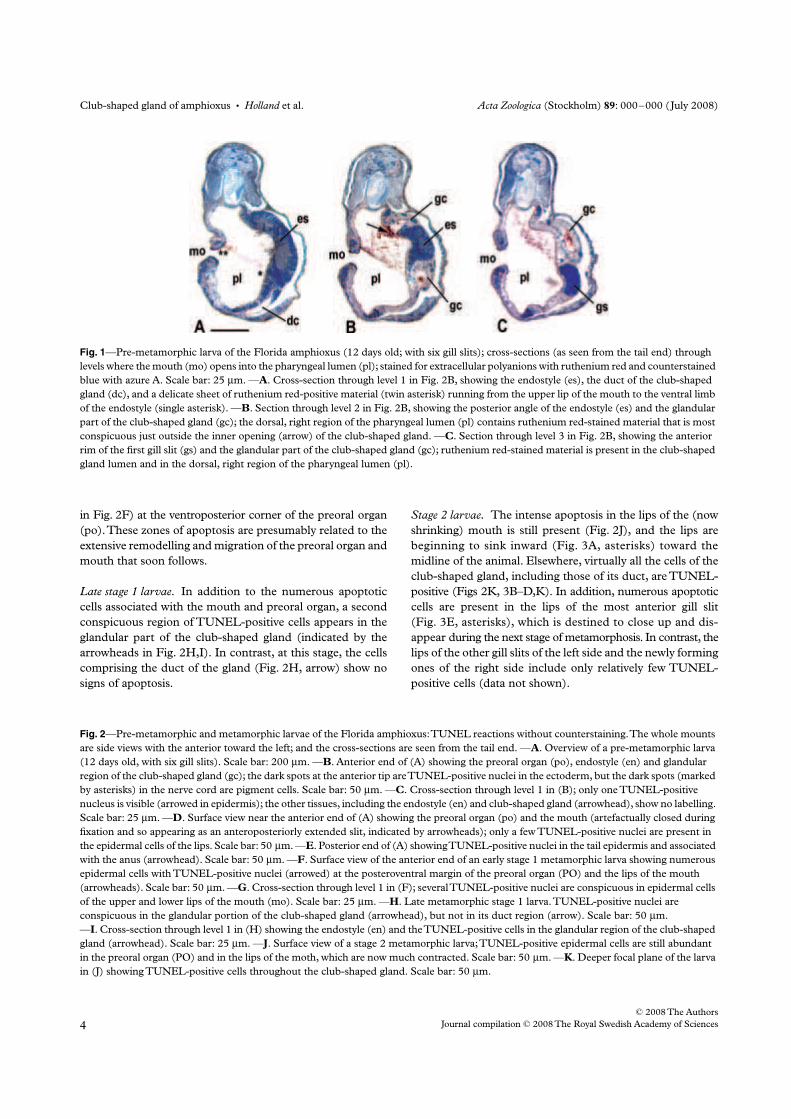
Club-shaped gland of amphioxus
•
Holland
et al.
Acta Zoologica
(Stockholm)
89
: 000–000 (July 2008)
© 2008 The Authors
Journal compilation © 2008 The Royal Swedish Academy of Sciences
in Fig. 2F) at the ventroposterior corner of the preoral organ
(po). These zones of apoptosis are presumably related to the
extensive remodelling and migration of the preoral organ and
mouth that soon follows.
Late stage 1 larvae.
In addition to the numerous apoptotic
cells associated with the mouth and preoral organ, a second
conspicuous region of TUNEL-positive cells appears in the
glandular part of the club-shaped gland (indicated by the
arrowheads in Fig. 2H,I). In contrast, at this stage, the cells
comprising the duct of the gland (Fig. 2H, arrow) show no
signs of apoptosis.
Stage 2 larvae.
The intense apoptosis in the lips of the (now
shrinking) mouth is still present (Fig. 2J), and the lips are
beginning to sink inward (Fig. 3A, asterisks) toward the
midline of the animal. Elsewhere, virtually all the cells of the
club-shaped gland, including those of its duct, are TUNEL-
positive (Figs 2K, 3B–D,K). In addition, numerous apoptotic
cells are present in the lips of the most anterior gill slit
(Fig. 3E, asterisks), which is destined to close up and dis-
appear during the next stage of metamorphosis. In contrast, the
lips of the other gill slits of the left side and the newly forming
ones of the right side include only relatively few TUNEL-
positive cells (data not shown).
Fig. 1—Pre-metamorphic larva of the Florida amphioxus (12 days old; with six gill slits); cross-sections (as seen from the tail end) through
levels where the mouth (mo) opens into the pharyngeal lumen (pl); stained for extracellular polyanions with ruthenium red and counterstained
blue with azure A. Scale bar: 25 µm. —A. Cross-section through level 1 in Fig. 2B, showing the endostyle (es), the duct of the club-shaped
gland (dc), and a delicate sheet of ruthenium red-positive material (twin asterisk) running from the upper lip of the mouth to the ventral limb
of the endostyle (single asterisk). —B. Section through level 2 in Fig. 2B, showing the posterior angle of the endostyle (es) and the glandular
part of the club-shaped gland (gc); the dorsal, right region of the pharyngeal lumen (pl) contains ruthenium red-stained material that is most
conspicuous just outside the inner opening (arrow) of the club-shaped gland. —C. Section through level 3 in Fig. 2B, showing the anterior
rim of the first gill slit (gs) and the glandular part of the club-shaped gland (gc); ruthenium red-stained material is present in the club-shaped
gland lumen and in the dorsal, right region of the pharyngeal lumen (pl).
Fig. 2
—Pre-metamorphic and metamorphic larvae of the Florida amphioxus: TUNEL reactions without counterstaining. The whole mounts
are side views with the anterior toward the left; and the cross-sections are seen from the tail end. —
A
. Overview of a pre-metamorphic larva
(12 days old, with six gill slits). Scale bar: 200
µ
m. —
B
. Anterior end of (A) showing the preoral organ (po), endostyle (en) and glandular
region of the club-shaped gland (gc); the dark spots at the anterior tip are TUNEL-positive nuclei in the ectoderm, but the dark spots (marked
by asterisks) in the nerve cord are pigment cells. Scale bar: 50
µ
m. —
C
. Cross-section through level 1 in (B); only one TUNEL-positive
nucleus is visible (arrowed in epidermis); the other tissues, including the endostyle (en) and club-shaped gland (arrowhead), show no labelling.
Scale bar: 25
µ
m. —
D
. Surface view near the anterior end of (A) showing the preoral organ (po) and the mouth (artefactually closed during
fixation and so appearing as an anteroposteriorly extended slit, indicated by arrowheads); only a few TUNEL-positive nuclei are present in
the epidermal cells of the lips. Scale bar: 50
µ
m. —
E
. Posterior end of (A) showing TUNEL-positive nuclei in the tail epidermis and associated
with the anus (arrowhead). Scale bar: 50
µ
m. —
F
. Surface view of the anterior end of an early stage 1 metamorphic larva showing numerous
epidermal cells with TUNEL-positive nuclei (arrowed) at the posteroventral margin of the preoral organ (PO) and the lips of the mouth
(arrowheads). Scale bar: 50
µ
m. —G. Cross-section through level 1 in (F); several TUNEL-positive nuclei are conspicuous in epidermal cells
of the upper and lower lips of the mouth (mo). Scale bar: 25 µm. —H. Late metamorphic stage 1 larva. TUNEL-positive nuclei are
conspicuous in the glandular portion of the club-shaped gland (arrowhead), but not in its duct region (arrow). Scale bar: 50 µm.
—I. Cross-section through level 1 in (H) showing the endostyle (en) and the TUNEL-positive cells in the glandular region of the club-shaped
gland (arrowhead). Scale bar: 25 µm. —J. Surface view of a stage 2 metamorphic larva; TUNEL-positive epidermal cells are still abundant
in the preoral organ (PO) and in the lips of the moth, which are now much contracted. Scale bar: 50 µm. —K. Deeper focal plane of the larva
in (J) showing TUNEL-positive cells throughout the club-shaped gland. Scale bar: 50 µm.
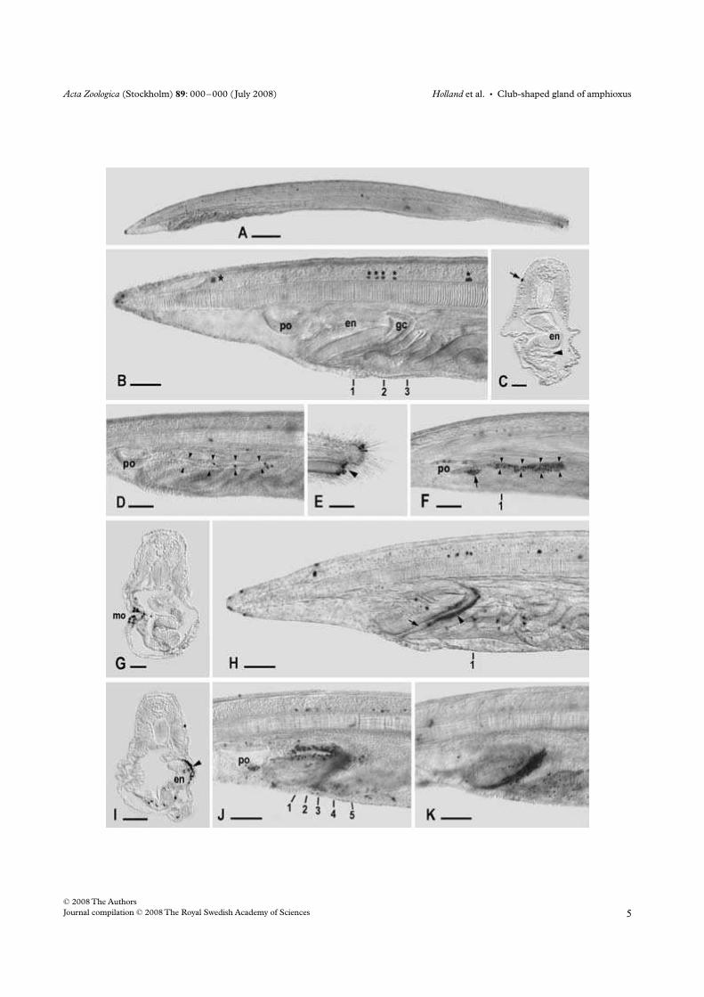
Acta Zoologica (Stockholm) 89: 000–000 (July 2008) Holland et al. • Club-shaped gland of amphioxus
© 2008 The Authors
Journal compilation © 2008 The Royal Swedish Academy of Sciences
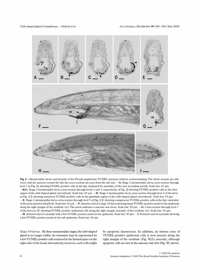
Club-shaped gland of amphioxus • Holland et al. Acta Zoologica (Stockholm) 89: 000–000 (July 2008)
© 2008 The Authors
Journal compilation © 2008 The Royal Swedish Academy of Sciences
Stage 3/4 larvae. By these metamorphic stages, the club-shaped
gland is no longer visible. Its remnants may be represented by
a few TUNEL-positive cells scattered in the haemal space on the
right side of the head; alternatively, however, such cells might
be apoptotic haemocytes. In addition, an intense zone of
TUNEL-positive epidermal cells is now present along the
right margin of the vestibule (Fig. 3F,G, asterisk), although
apoptotic cells are rare in the nascent oral cirri (Fig. 3F, arrow).
Fig. 3—Metamorphic larvae and juvenile of the Florida amphioxus: TUNEL reactions without counterstaining. The whole mounts are side
views with the anterior toward the left; the cross-sections are seen from the tail end. —A. Stage 2 metamorphic larva; cross-section through
level 1 in Fig. 2J, showing TUNEL-positive cells in the lips (indicated by asterisks) of the now in-sunken mouth. Scale bar: 25 µm.
—B,C. Stage 2 metamorphic larva; cross-section through levels 2 and 3, respectively, of Fig. 2J showing TUNEL-positive cells in the duct
region of the club-shaped gland (arrowhead). Scale bar: 25 µm. —D. Stage 2 metamorphic larva; cross-section through level 4 of the larva
in Fig. 2(J) showing numerous TUNEL-positive cells in the glandular region of the club-shaped gland (arrowhead). Scale bar: 25 µm.
—E. Stage 2 metamorphic larva; cross-section through level 5 of Fig. 2(J) showing conspicuous TUNEL-positive cells in the lips (asterisks)
of the most anterior left gill slit. Scale bar: 25 µm. —F. Anterior end of a stage 3/4 larva showing many TUNEL-positive nuclei in the epidermis
along the right margin of the vestibule (ve). The arrow indicates a nascent oral cirrus. Scale bar: 50 µm. —G. Cross-section through level 1
of the larva in (F) showing TUNEL-positive epidermal cells along the right margin (asterisk) of the vestibule (ve). Scale bar: 25 µm.
—H. Anterior end of a juvenile with a few TUNEL-positive nuclei in the epidermis. Scale bar: 50 µm. —I. Posterior end of a juvenile showing
a few TUNEL-positive nuclei in the tail epidermis. Scale bar: 50 µm.
Acta Zoologica (Stockholm) 89: 000–000 (July 2008) Holland et al. • Club-shaped gland of amphioxus
© 2008 The Authors
Journal compilation © 2008 The Royal Swedish Academy of Sciences
Stage 5 ( juvenile). By the beginning of the juvenile stage,
no conspicuous zones of TUNEL-positive cells remain in
any of the tissues. As in the late pre-metamorphic larvae,
most of the apoptotic cells are scattered in the epider-
mis at the anterior tip (Fig. 3H) and tail (Fig. 3I) of the
animal.
Discussion
The function(s) of the club-shaped gland
In the past, many students of amphioxus believed that the
club-shaped gland emits secretions (with a variety of
proposed functions) from its outer opening (Table 1). In
contrast, the results of the present study support the minority
view that the gland’s secretions exit its inner opening into the
pharyngeal lumen (Goldschmidt 1905; Garstang 1928;
Olsson 1983; Gilmour 1996; Holland and Yu 2002; Lacalli
2008). The best insights on how the club-shaped gland’s
secretions function within the pharynx of amphioxus larvae
have come from Gilmour (1996), who videotaped the paths
of particles in the gut lumen and preserved the presumed
mucus trap with ruthenium red-containing fixatives. He
concluded that the endostyle and the club-shaped gland
cooperate to produce a thin vertical curtain of mucus (orientated
mid-sagittally in the pharyngeal lumen) to capture ingested
particles and then transport them toward the more posterior
gut regions. Subsequently, Lacalli (2008) elaborated on this
scheme by proposing that the mucus sheet is produced
predominantly by the endostyle and is transported posteriorly
by the ciliated peripharyngeal bands. Our present results are
in general agreement with those of Gilmour (1996) and
Lacalli (2008), although we differ from them in finding that
the particle-trapping surface of the mucus may be orientated
not vertically near the midline of the pharynx, but at a 35°
angle slanting down from the upper lip to the lower limb of
the endostyle on the opposite wall of the pharynx. Moreover,
in our preparations, the edges of the slanted sheet are not
closely associated with the peripharyngeal bands. Certainly,
in the fixed larvae studied to date, some features of the
mucus trap might be distorted as the result of the shrinkage,
extraction and displacement that extracellular materials tend
to suffer during histological fixation. A satisfactory under-
standing of the structure of the particle trap and its detailed
functions will require further work.
Apoptosis in metamorphic larvae of amphioxus: fate of the
club-shaped gland
Bayascas et al. (2002), who previously studied apoptosis in
amphioxus larvae (in very early ones that were only 2 days
old), described an abundance of TUNEL-positive cells in the
ectoderm and endoderm, especially along the left anterior
side, where the mouth is opening, and in the differentiating
tail. We repeated and confirmed the results of Bayascas et al.
(2002) on early larvae (our unpublished data). In contrast,
our study of later (12-day-old) pre-metamorphic larvae
demonstrates little apoptosis in any of their tissues. Soon
after the start of metamorphosis, however, numerous epidermal
cells around the mouth rim and near the preoral organ
become TUNEL-positive. The apoptosis of cells at the
mouth rim is evidently related to the shrinkage of the mouth
and its anteromedial migration to become the small aperture
surrounded by the velum of the juvenile.
Following the initial burst of apoptosis associated with the
mouth, virtually all the cells of the glandular portion of the
club-shaped gland become TUNEL-positive, and, a few
hours later, the massive apoptosis spreads to the cells
comprising the duct region. By the time the gill slits of the
metamorphic larvae are assuming a U-shape, the apoptotic
club-shaped gland appears to disintegrate into scattered
TUNEL-positive cells that evidently disperse in the haemal
channels and soon disappear, as proposed by van Wijhe
(1914). There is no evidence that such cells enter the pharyngeal
lumen as suggested by Willey (1891). Moreover, our results
conclusively show that the amphioxus club-shaped gland
does not transdifferentiate into cartilage-like cells (Klaatsch
1898) and is not converted into any components of the
post-metamorphic endostyle (Hatschek 1892; Olsson 1983;
Gilmour 1996).
In sum, the club-shaped gland of amphioxus is present
throughout the larval stage and appears to supply a com-
ponent of the mucus that traps food particles in the pharynx.
It is likely that the endostyle also produces some of the
feeding mucus during the larval stage in addition to its likely
fixation of iodine, presumably for the synthesis of thyroid
hormones (Fredriksson et al. 1984). Following the apoptotic
destruction of the club-shaped gland during metamorphosis,
the endostyle of juvenile and adult amphioxus presumably
becomes the major (or even exclusive) source of mucus for
capturing food particles in the pharyngeal lumen. In
addition, the post-larval endostyle continues to be the major
site for iodination (Ericson et al. 1985) possibly because of
the continued synthesis of thyroid hormones influencing
tissue growth and maintenance in the juvenile and adult
amphioxus.
Even with the new information presented here, the homology
between the club-shaped gland of amphioxus and structures
in other animals remains enigmatic. None of the morphology-
based homologies listed in Table 1 has been widely accepted
(the comparisons with the chordate mouth/foregut or a
chordate gill slit remain the best of an inconclusive lot).
Finally, a recent review of developmental genetic data
(Jackman et al. 2004) gave no clear insights into homologies
between the club-shaped gland and structures in other
animals, and the authors suggested that the amphioxus
club-shaped gland might be an evolutionary novelty that has
arisen within the cephalochordate lineage.
Club-shaped gland of amphioxus • Holland et al. Acta Zoologica (Stockholm) 89: 000–000 (July 2008)
© 2008 The Authors
Journal compilation © 2008 The Royal Swedish Academy of Sciences
Acknowledgements
We are grateful to Linda Holland for her criticism of the
manuscript, to John Lawrence and Susan Bell of the Biology
Department of the University of South Florida for their
generous provision of laboratory space, and to Tom Gilmour
for his technical advice. This work was supported in part by
NSF grant IOB-04-16292.
References
Bayascas, J. R., Yuste, V. J., Benito, E., Garcia-Fernàndez, J. and
Comella, J. X. 2002. Isolation of AmphiCASP-3/7, an ancestral
caspase from amphioxus (Branchiostoma floridae). Evolutionary
considerations for vertebrate caspases. – Cell Death and Differentiation
9: 1078–1089.
Berrill, N. J. 1987. Early chordate evolution. Part 2. Amphioxus and
ascidians. To settle or not to settle. – International Journal of
Invertebrate Reproduction and Development 11: 15–28.
Bone, Q. 1958. Observations upon the living larva of amphioxus. –
Pubblicazioni della Stazione Zoologica di Napoli 30: 458–471.
Conklin, E. G. 1932. The embryology of amphioxus. – Journal of
Morphology 54: 69–152.
Ericson, L. E., Fredriksson, G. and Overholm, T. 1985. Ultrastructural
localization of the iodination centre in the endostyle of the adult
amphioxus (Branchiostoma lanceolatum). – Cell and Tissue Research
241: 267–273.
Flood, P. R. 1991. Architecture of, and water circulation and flow
rate in, the house of the planktonic tunicate Oikopleura
labradoriensis. – Marine Biology 111: 95–111.
Fredriksson, G., Ericson, L. E. and Olsson, R. 1984. Iodine binding
in the endostyle of larval Branchiostoma lanceolatum
(Cephalochordata). – General and Comparative Endocrinology 56:
177–184.
Fuller, A. S. 1958. Studies on some Cephalochordata, with special
reference to the amphioxides problem. PhD Dissertation, Univer-
sity of London.
Garstang, W. 1928. The morphology of the Tunicata, and its bearings
on the phylogeny of the Chordata. – Quarterly Journal of Micro-
scopical Science 72: 51–187.
Gilmour, T. H. J. 1996. Feeding methods of cephalochordate
larvae. – Israel Journal of Zoology 42(Suppl.): 87–95.
Godoy, L., Gonzàlez-Duarte, R. and Albalat, R. 2006. S-nitroso-
glutathinone reductase activity of amphioxus ADH3: insights into
the nitric oxide metabolism. – International Journal of Biological
Sciences 2: 117–124.
Goldschmidt, R. 1905. Amphioxides. – Wissenschaftliche Ergebnisse
der Deutschen Tiefsee-Expedition auf dem Dampfer ‘Valdivia’ 12: 1–
93 + pl. I–X.
Goodrich, E. S. 1930. The development of the club-shaped gland in
amphioxus. – Quarterly Journal of Microscopical Science 74: 155–164.
Hatschek, B. 1882. Studien über Entwicklung des Amphioxus. –
Arbeiten aus den Zoologischen Institute der Universität Wien und der
Zoologischen Station in Triest 4: 1–88 + I–IX.
Hatschek, B. 1892. Die Metamerie des Amphioxus und des
Ammocoetes. – Verhandlungen der Anatomische Gesellschaft 6:
136–161.
Holland, N. D. and Holland, L. Z. 1993. Embryos and larvae of
invertebrate deuterostomes. In Stern, C. D. and Holland, P. W. H.
(Eds): Essential Developmental Biology: a Practical Approach, pp. 21–
32. IRL Press, Oxford.
Holland, N. D. and Yu, J. K. 2002. Epidermal receptor development
and sensory pathways in vitally stained amphioxus (Branchiostoma
floridae). – Acta Zoologica (Stockholm) 83: 309–319.
Jackman, W. R., Mougey, J. M., Panopoulou, G. D. and Kimmel, C.
B. 2004. crabp and maf highlight the novelty of the amphioxus
club-shaped gland. – Acta Zoologica (Stockholm) 85: 91–99.
Klaatsch, H. 1898. Ueber den Bau und die Entwickelung des
Tentakelapparates des Amphioxus. – Anatomischer Anzeiger 14:
184–195.
Kowalevsky, A. 1867. Entwickelungsgeschichte des Amphioxus
lanceolatus. – Mémoires de l’Académie Impériale des Sciences de St.-
Pétersbourg (Série VII) 11(4): 1–7 + pl. I–III.
Kowalevsky, A. 1877. Weitere Studien über die Entwickelungsges-
chichte des Amphioxus lancelolatus, nebst einem Beitrage zur
Homologie des Nervensystems der Würmer und Wirbelthiere. –
Archiv für Microskopische Anatomie 13: 181–204 + pl. XV–XVI.
Lacalli, T. 2008. Mucus secretion and transport in amphioxus larvae:
organization and ultrastructure of the food trapping system, and
implications for head evolution. – Acta Zoologica Stockholm 89:
219–230.
Luft, J. H. 1971. Ruthenium red and violet. I. Chemistry, purification,
methods of use for electron microscopy and mechanism of
action. – Anatomical Record 171: 347–368.
Neal, H. V. 1898. The segmentation of the nervous system in Squalus
acanthias. A contribution to the morphology of the vertebrate
head. – Bulletin of the Museum of Comparative Zoology Harvard 31:
145–294 + pl. I–IX.
Olsson, R. 1983. Club-shaped gland and endostyle in larval
Branchiostoma lanceolatum (Cephalochordata). – Zoomorphology
103: 1–13.
Orton, J. H. 1914. On a hermaphrodite specimen of amphioxus with
notes on experiments in rearing amphioxus. Journal of the Marine
Biological Association of the United Kingdom 10: 506–512.
Paris, M., Escriva, H., Schubert, M., Burnet, F., Brtko, J.,
Ciesielski, F. et al. 2008. Amphioxus post-embryonic development
reveals the homology of chordate metamorphosis. – Current
Biology 18: 825–830.
Schulze, M. 1851. Beobachtung junger Exemplare von Amphioxus.
– Zeitschrift für Wissenschaftliche Zoologie 3: 416–419 + pl. XII.
Van Beneden, E. and Julin, C. 1886. Recherches sur la morphologie
des tuniciers. – Archives de Biologie 6: 237–476 + pl. VII–XVI.
Van Wijhe, J. W. 1914. Studien über Amphioxus. I. Mund und
Darmkanal während der Metamorphose. – Verhandelingen der
Koninklijke Akademie van Wetenschappen te Amsterdam (Section 2)
18: 1–84 + pl. I–V.
Webb, J. E. 1969. On the feeding and behaviour of the larva of
Branchiostoma lanceolatum. – Marine Biology 3: 58–72.
Wickstead, J. H. 1967. Branchiostoma lanceolatum larvae: some
experiments on the effect of thiouracil on metamorphosis. –
Journal of the Marine Biological Association of the United Kingdom
47: 49–59.
Willey, A. 1891. The later larval development of amphioxus. –
Quarterly Journal of Microscopical Science 32: 183–234 + pl. XIII–
XV.
Yasui, K., Zhang, S. C., Uemura, M. and Saiga, H. 2000. Left–right
asymmetric expression of BbPtx, a Ptx-related gene, in a lancelet
species and the developmental left-sidedness of deuterostomes. –
Development 127: 187–195.
ORIGINAL ARTICLE
Nuclear hormone receptor signaling in amphioxus
Michael Schubert & Frédéric Brunet & Mathilde Paris &
Stéphanie Bertrand & Gérard Benoit & Vincent Laudet
Received: 11 July 2008 /Accepted: 5 September 2008# Springer-Verlag 2008
Abstract The nuclear hormone receptors (NRs) form asuperfamily of transcription factors unified by conservedprotein structure and mode of function. While mostmembers of this superfamily are activated by ligands, suchas thyroid hormones, steroids, vitamin D or retinoic acid,other NRs are called orphan receptors because they have noknown ligand. NR-dependent signaling is crucial forvertebrate development with the majority of receptors beingexpressed in the developing embryo. Due to massive geneduplications during vertebrate diversification, there areusually more NRs in vertebrates than in invertebrates. Inthis study, we examine the evolutionary diversification ofthe NR superfamily and of NR-dependent signaling inchordates (vertebrates, tunicates, and amphioxus). We takeadvantage of the unique features of the genome of theinvertebrate amphioxus, which is characterized by avertebrate-like gene content without having undergone
massive duplications, to assess the NR signaling comple-ment (NRs and NR coregulators) of the ancestral chordate.We find 33 NRs in amphioxus, which are more NRs thanoriginally anticipated. This increase is mainly due to anamphioxus-specific duplication of genes encoding receptorsof the NR1H group. In addition, there are three heterolo-gous NRs in amphioxus that could not be placed within theframework of the NR superfamily. Apart from theseexceptions, there is usually one amphioxus NR or NRsignaling coregulator for each paralogous group of two,three, or four human receptors suggesting that the ancestralchordate had a set of 22 different NRs plus one copy ofeach NR coregulator.
Keywords Branchiostoma floridae .
Cephalochordate . Chordate . Development . Evolution .
Invertebrate-to-vertebrate transition .
Nuclear hormone receptor coregulator
Introduction
Nuclear hormone receptors (NRs) constitute a superfamilyof DNA-binding transcription factors that are involved in awide variety of physiological processes both duringdevelopment and in the adult (reviewed in Gronemeyer etal. 2004). NRs are best known for their functions as ligand-activated transcription factors that mediate the response tohormones, such as thyroid hormones, steroids, vitamin D orretinoic acid (reviewed in Gronemeyer et al. 2004). Inaddition to this role, there is also a non-negligible numberof NRs that either do not bind a ligand at all or for which aligand has not yet been described. These NRs withoutknown ligand are called orphan receptors (reviewed inBenoit et al. 2006; Germain et al. 2006c). Phylogenetic
Dev Genes EvolDOI 10.1007/s00427-008-0251-y
Michael Schubert and Frédéric Brunet are equal first authors.
Communicated by J.J. Gibson-Brown
Electronic supplementary material The online version of this article(doi:10.1007/s00427-008-0251-y) contains supplementary material,which is available to authorized users.
M. Schubert : F. Brunet :M. Paris :G. Benoit :V. Laudet (*)CNRS UMR5242-INRA 1288-ENS-UCBL,Institut de Génomique Fonctionnelle de Lyon, IFR128BioSciences Lyon-Gerland, Ecole Normale Supérieure de Lyon,46 allée d’Italie,69364 Lyon Cedex 07, Francee-mail: [email protected]
S. BertrandCNRS UMR7628, UPMC Univ Paris 06,Observatoire Océanographique,66651 Banyuls-sur-Mer, France
analyses have shown that NRs arose very early in themetazoan lineage long before the divergence of proto-stomes and deuterostomes and diversified through complexseries of gene duplications and gene losses (Escriva et al.1997; Bertrand et al. 2004; reviewed in Escriva et al. 2000).
NR proteins are characterized by a modular structureconsisting of six domains (reviewed in Germain et al.2006c). At the N-terminus, the A and B domains contain atranscriptional activation unit called activation function 1(AF-1). The C region of the NR is the DNA-bindingdomain (DBD), which is required for the interaction of theNR protein with the DNA. The DBD is the most highlyconserved domain of NRs and consists of cysteine-rich zincfinger motifs, α-helices, and a C-terminal extension. The Ddomain acts as a hinge between the C region (the DBD) andthe E region, which corresponds to the ligand-bindingdomain (LBD) and which includes a second activationdomain (AF-2) crucial for interactions of NRs withcoactivators and corepressors and often also a nuclearlocalization signal. Finally, the F region, which is notpresent in all NRs, is not very well characterized and has noclear known function.
Interactions of NRs with coactivators and corepressorsare required for defining the transcriptional activities ofNRs, which can function both as activators and repressorsof gene transcription depending on the target gene and onthe presence or absence of a ligand (reviewed in Bastienand Rochette-Egly 2004; Germain et al. 2006c). Althoughtranscriptional regulation by NRs is very complex anddiverse, in the absence of ligand, a liganded NR (such asthe retinoic acid receptor, RAR, or the thyroid hormonereceptor, TR) is bound to DNA predominantly as hetero-dimer with the NR retinoid X receptor (RXR) and isassociated with a corepressor complex that represses thetranscriptional activity of the NRs (reviewed in Hu andLazar 2000; Bastien and Rochette-Egly 2004; Goodson etal. 2005; Germain et al. 2006c). In some cases, NRs canalso bind to DNA as homodimers (e.g. the hepatocytenuclear factor 4, HNF4) or as monomers (e.g. the nervegrowth factor-induced clone—B, NGFI-B) (reviewed inBenoit et al. 2006; Germain et al. 2006c). Ligand bindinginduces a conformational change in the NR leading to thedissociation of the corepressors and to the binding of acoactivator complex (reviewed in Bastien and Rochette-Egly 2004; Germain et al. 2006c). The activity of thiscoactivator complex leads to unfolding of the condensedchromatin (reviewed in Bastien and Rochette-Egly 2004;Germain et al. 2006c; Lonard and O’Malley 2006). Afterthis relaxation of the chromatin structure, the coactivatorsare replaced by a mediator complex that recruits the basaltranscription machinery to the DNA locus for initiation oftranscription (reviewed in Bastien and Rochette-Egly 2004;Germain et al. 2006c).
Most NRs are expressed during vertebrate development(Bertrand et al. 2007) and have important functions in thedeveloping embryo (reviewed in Laudet and Gronemeyer2002). For example, NRs are involved in survival andproper development of the early embryo (PPAR, HNF4,ERR, NOR1, LRH1, GCNF), in patterning and formationof the central nervous system (TR, RAR, REV-ERB, ROR,TR2/4, TLX, PNR, COUP-TF, EAR2, ER, NURR1, NOR1,SF1), in organogenesis of, among others, the heart, lung,kidney, liver, pancreas, and the immune and reproductivesystems (DAX1, RAR, PPAR, ROR, HNF4, RXR, COUP-TF, GR, PR, AR, SF1), and in the development of thevisual system (RAR, ROR, TLX, PNR) (reviewed in Benoitet al. 2006; Dahlman-Wright et al. 2006; Flamant et al.2006; Germain et al. 2006a; Germain et al. 2006b; Lu et al.2006; Michalik et al. 2006; Moore et al. 2006). Moreover,in vertebrates (most prominently in amphibians and teleostfish) and in invertebrate chordates (such as amphioxus), thethyroid hormone receptor (TR) controls metamorphosis, thepostembryonic transition from larva to adult (Paris et al.2008a). Along the same lines, the functions of another NR,the retinoic acid receptor (RAR), in early development arealso conserved between invertebrate chordates and verte-brates. Thus, it has been shown that in amphioxus andvertebrates, RAR controls expression of Hox genes, whichare crucial mediators of anteroposterior axial patterning indevelopment (Schubert et al. 2006b; reviewed in Marlétazet al. 2006; Campo-Paysaa et al. 2008).
In humans, there are a total of 48 NRs. In contrast, theinvertebrates studied so far generally have fewer genesencoding NR proteins. For example, there are 21 NRs inthe genome of the fruit fly Drosophila melanogaster andonly 17 NRs in the genome of the sea squirt Ciona
intestinalis (Yagi et al. 2003; Bertrand et al. 2004). Thisdifference in the total number of NRs between invertebratesand vertebrates can be attributed to massive whole genomeduplications that accompanied the diversification of thevertebrates (Holland et al. 1994; Dehal and Boore 2005;Holland et al. 2008; Putnam et al. 2008; reviewed inSchubert et al. 2006a). Phylogenetic analyses of thecomplete NR superfamily have shown that the vast majorityof NRs from metazoan animals fall into six distinctsubfamilies (numbered NR1 through NR6). In addition,there is a seventh “subfamily” (called NR0), which containsall receptors lacking the typical NR protein structure(Fig. 1) (Nuclear Receptors Nomenclature Committee1999; reviewed in Germain et al. 2006c). Within eachsubfamily, the NRs are further subdivided into severalgroups (Fig. 1) (reviewed in Germain et al. 2006c). Tobetter understand the evolutionary diversification of the NRsuperfamily and of NR-dependent signaling in chordates(i.e. in vertebrates, tunicates, and amphioxus), we decidedto characterize the complement of NRs and of key
Dev Genes Evol
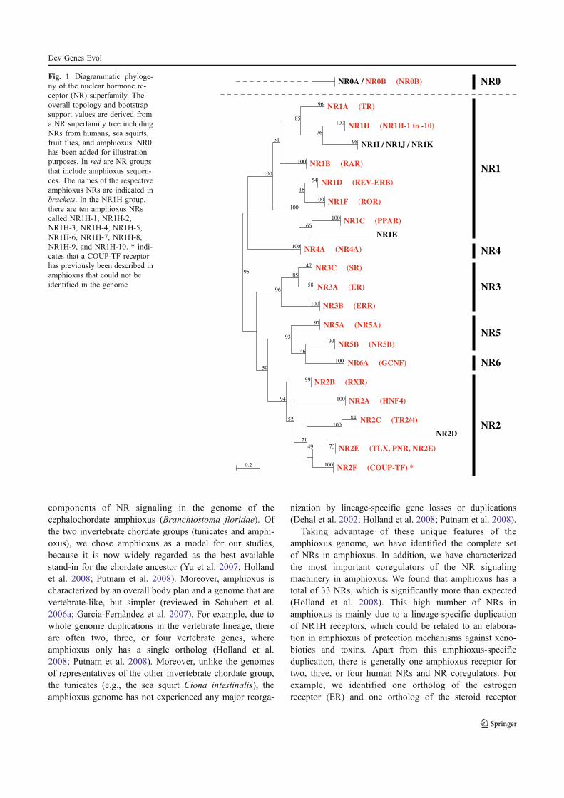
components of NR signaling in the genome of thecephalochordate amphioxus (Branchiostoma floridae). Ofthe two invertebrate chordate groups (tunicates and amphi-oxus), we chose amphioxus as a model for our studies,because it is now widely regarded as the best availablestand-in for the chordate ancestor (Yu et al. 2007; Hollandet al. 2008; Putnam et al. 2008). Moreover, amphioxus ischaracterized by an overall body plan and a genome that arevertebrate-like, but simpler (reviewed in Schubert et al.2006a; Garcia-Fernàndez et al. 2007). For example, due towhole genome duplications in the vertebrate lineage, thereare often two, three, or four vertebrate genes, whereamphioxus only has a single ortholog (Holland et al.2008; Putnam et al. 2008). Moreover, unlike the genomesof representatives of the other invertebrate chordate group,the tunicates (e.g., the sea squirt Ciona intestinalis), theamphioxus genome has not experienced any major reorga-
nization by lineage-specific gene losses or duplications(Dehal et al. 2002; Holland et al. 2008; Putnam et al. 2008).
Taking advantage of these unique features of theamphioxus genome, we have identified the complete setof NRs in amphioxus. In addition, we have characterizedthe most important coregulators of the NR signalingmachinery in amphioxus. We found that amphioxus has atotal of 33 NRs, which is significantly more than expected(Holland et al. 2008). This high number of NRs inamphioxus is mainly due to a lineage-specific duplicationof NR1H receptors, which could be related to an elabora-tion in amphioxus of protection mechanisms against xeno-biotics and toxins. Apart from this amphioxus-specificduplication, there is generally one amphioxus receptor fortwo, three, or four human NRs and NR coregulators. Forexample, we identified one ortholog of the estrogenreceptor (ER) and one ortholog of the steroid receptor
NR2F (COUP-TF) *
NR2E (TLX, PNR, NR2E)
100
73
NR2D
NR2C (TR2/4) 84
49
100
NR2A (HNF4)
71
100
NR2B (RXR)
52
99
NR6A (GCNF)
NR5B (NR5B)
100
99
NR5A (NR5A)
46
97
94
93
NR3B (ERR)
NR3A (ER)
NR3C (SR)
58
47
100
85
59
96
NR4A (NR4A)
NR1E
NR1C (PPAR) 100
NR1F (ROR)
NR1D (REV-ERB)
100
54
66
18
NR1B (RAR)
NR1I / NR1J / NR1K
NR1H (NR1H-1 to -10)
98
100
NR1A (TR)
76
96
100
85
100
51
100
100
0.2
NR2
NR6
NR5
NR3
NR4
NR1
NR0NR0A / NR0B (NR0B)
95
Fig. 1 Diagrammatic phyloge-ny of the nuclear hormone re-ceptor (NR) superfamily. Theoverall topology and bootstrapsupport values are derived froma NR superfamily tree includingNRs from humans, sea squirts,fruit flies, and amphioxus. NR0has been added for illustrationpurposes. In red are NR groupsthat include amphioxus sequen-ces. The names of the respectiveamphioxus NRs are indicated inbrackets. In the NR1H group,there are ten amphioxus NRscalled NR1H-1, NR1H-2,NR1H-3, NR1H-4, NR1H-5,NR1H-6, NR1H-7, NR1H-8,NR1H-9, and NR1H-10. * indi-cates that a COUP-TF receptorhas previously been described inamphioxus that could not beidentified in the genome
Dev Genes Evol
(SR) in amphioxus, while there are two ERs and four SRsin humans. Thus, both ER and SR have already beenpresent in the last common ancestor of all chordates andhave secondarily been lost in sea squirts and othertunicates. In sum, the data on NRs and NR coregulatorsin the amphioxus genome suggest that the ancestralchordate had a set of 22 different NRs and a single copyof each NR coregulator.
Materials and methods
Data mining
The NR data set was based on sequence alignments ofprevious studies (Bertrand et al. 2004). The alignment filealready included all NRs from humans (Homo sapiens), seasquirts (Ciona intestinalis), and fruit flies (Drosophilamelanogaster). In contrast, the NR coregulator data setwas based on the Ensembl families of the NR coregulatorsof interest (NCOA, CBP and P300, PGC1, NCOR/SMRT,MED1/TRAP220), which are available at the Ensemblgenome browser (www.ensembl.org/index.html). We usedEnsembl version v48 for this analysis. All NR andcoregulator sequences were BLASTed against the amphi-oxus genome (version v1.0), which is available at the JGIwebpage (genome.jgi-psf.org/Brafl1/Brafl1.home.html).The best hits were retrieved and aligned using MUSCLE(Edgar 2004) followed by manual refinement. We createdboth nucleotide and amino acid alignments. All alignmentfiles are available from the authors. The nucleotide align-ments were used to identify and eliminate allelic poly-morphs of the different NR and NR coregulator sequencesretrieved from the amphioxus genome. Amphioxus allelicpolymorphs are characterized by nucleotide sequences thatdiffer almost exclusively in third codon positions, whichallows their identification and elimination (Holland et al.2008; Putnam et al. 2008).
Phylogenetic analyses
The amino acid alignments of amphioxus NR and NRcoregulator sequences were used for phylogenetic treereconstruction experiments. Phylogenetic analyses on thecomprehensive NR data set including all NRs fromhumans, sea squirts, fruit flies and amphioxus were carriedout using both the neighbor joining (NJ) and maximumlikelihood (ML) method. Likewise, the different NRcoregulator alignments were analyzed by phylogenetic treereconstruction using both the NJ and ML method. The NJanalyses, carried out with the program Phylo_Win (Galtieret al. 1996), were based on a Poisson correction andpairwise gap removal. The ML trees were calculated using
PhyML (Guindon and Gascuel 2003) with default param-eters (a JTT model with a gamma distribution parameterof 2). Bootstrap support values were calculated in 100bootstrap replicates. The NJ and ML analyses yieldedidentical results and, hence, only the ML trees arediscussed.
Results
In this study, we have annotated from the amphioxusgenome all NR genes and the genes of some of the maincoregulators of NR signaling. Gene annotation was basedon phylogenetic analyses of the identified sequences in thecontext of the NR superfamily or of the coregulator proteinfamilies (Supplementary Figs. S1–S6). The sequence IDs inthe amphioxus genome of the amphioxus NRs and of theNR coregulators are given in the supplementary material(Supplementary Tables S1, S2).
Amphioxus NRs
In total, there are 33 NR genes in amphioxus, 32 of whichwe were able to identify directly in the amphioxus genome(Fig. 1, Table 1). Although amphioxus COUP-TF hadpreviously been cloned from amphioxus (Langlois et al.2000), we were not able to identify this receptor in thegenome probably because it is located in an unassembledregion of the amphioxus genome (Putnam et al. 2008). Inthe following, we will discuss the different NR subfamiliesaccording to the overall structure and nomenclature of thesuperfamily (Fig. 1).
In the NR1 subfamily, there are a total of 15 amphioxusNRs (Table 1). As expected, there is a single amphioxusortholog in most NR1 groups. For example, there is a singleamphioxus thyroid hormone receptor (TR), retinoic acidreceptor (RAR), REV-ERB receptor, retinoid-related or-phan receptor (ROR) and peroxisome proliferator-activatedreceptor (PPAR; Fig. 2a). The amphioxus TR and RARhave previously been cloned and functionally characterized(Escriva et al. 2002; Paris et al. 2008a). In contrast, wewere not able to find any amphioxus representatives of theNR1I/J/K group, which includes, for example, the verte-brate vitamin D receptors (VDRs), pregnane X receptors(PXRs) and constitutive androstene receptors (CARs). Inthe NR1H group (Fig. 2b), which contains the vertebrateliver X receptors (LXRs) and farnesoid X receptors (FXRs),the amphioxus receptors have experienced a lineage-specific duplication resulting in ten NR1H-like receptors(Table 1). Since two of these independent amphioxusduplicates are very short, containing only the DBD(NR1H-9 and NR1H-10), they could not be included inour phylogenetic analyses of the whole NR superfamily
Dev Genes Evol
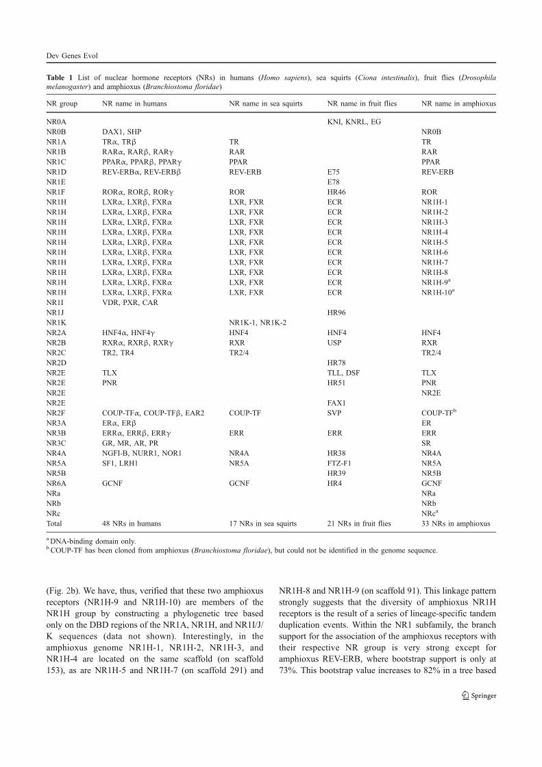
(Fig. 2b). We have, thus, verified that these two amphioxusreceptors (NR1H-9 and NR1H-10) are members of theNR1H group by constructing a phylogenetic tree basedonly on the DBD regions of the NR1A, NR1H, and NR1I/J/K sequences (data not shown). Interestingly, in theamphioxus genome NR1H-1, NR1H-2, NR1H-3, andNR1H-4 are located on the same scaffold (on scaffold153), as are NR1H-5 and NR1H-7 (on scaffold 291) and
NR1H-8 and NR1H-9 (on scaffold 91). This linkage patternstrongly suggests that the diversity of amphioxus NR1Hreceptors is the result of a series of lineage-specific tandemduplication events. Within the NR1 subfamily, the branchsupport for the association of the amphioxus receptors withtheir respective NR group is very strong except foramphioxus REV-ERB, where bootstrap support is only at73%. This bootstrap value increases to 82% in a tree based
Table 1 List of nuclear hormone receptors (NRs) in humans (Homo sapiens), sea squirts (Ciona intestinalis), fruit flies (Drosophilamelanogaster) and amphioxus (Branchiostoma floridae)
NR group NR name in humans NR name in sea squirts NR name in fruit flies NR name in amphioxus
NR0A KNI, KNRL, EGNR0B DAX1, SHP NR0BNR1A TRα, TRβ TR TRNR1B RARα, RARβ, RARγ RAR RARNR1C PPARα, PPARβ, PPARγ PPAR PPARNR1D REV-ERBα, REV-ERBβ REV-ERB E75 REV-ERBNR1E E78NR1F RORα, RORβ, RORγ ROR HR46 RORNR1H LXRα, LXRβ, FXRα LXR, FXR ECR NR1H-1NR1H LXRα, LXRβ, FXRα LXR, FXR ECR NR1H-2NR1H LXRα, LXRβ, FXRα LXR, FXR ECR NR1H-3NR1H LXRα, LXRβ, FXRα LXR, FXR ECR NR1H-4NR1H LXRα, LXRβ, FXRα LXR, FXR ECR NR1H-5NR1H LXRα, LXRβ, FXRα LXR, FXR ECR NR1H-6NR1H LXRα, LXRβ, FXRα LXR, FXR ECR NR1H-7NR1H LXRα, LXRβ, FXRα LXR, FXR ECR NR1H-8NR1H LXRα, LXRβ, FXRα LXR, FXR ECR NR1H-9a
NR1H LXRα, LXRβ, FXRα LXR, FXR ECR NR1H-10a
NR1I VDR, PXR, CARNR1J HR96NR1K NR1K-1, NR1K-2NR2A HNF4α, HNF4γ HNF4 HNF4 HNF4NR2B RXRα, RXRβ, RXRγ RXR USP RXRNR2C TR2, TR4 TR2/4 TR2/4NR2D HR78NR2E TLX TLL, DSF TLXNR2E PNR HR51 PNRNR2E NR2ENR2E FAX1NR2F COUP-TFα, COUP-TFβ, EAR2 COUP-TF SVP COUP-TFb
NR3A ERα, ERβ ERNR3B ERRα, ERRβ, ERRγ ERR ERR ERRNR3C GR, MR, AR, PR SRNR4A NGFI-B, NURR1, NOR1 NR4A HR38 NR4ANR5A SF1, LRH1 NR5A FTZ-F1 NR5ANR5B HR39 NR5BNR6A GCNF GCNF HR4 GCNFNRa NRaNRb NRbNRc NRca
Total 48 NRs in humans 17 NRs in sea squirts 21 NRs in fruit flies 33 NRs in amphioxus
aDNA-binding domain only.bCOUP-TF has been cloned from amphioxus (Branchiostoma floridae), but could not be identified in the genome sequence.
Dev Genes Evol
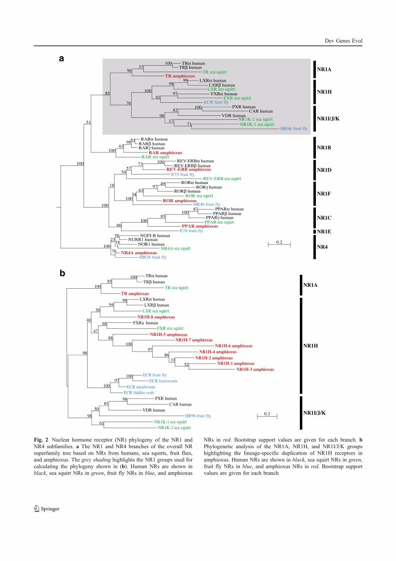
NR1H
NR1I/J/K
HR38 fruit flyNR4A amphioxus
NR4A sea squirtNOR1 human
NURR1 human NGFI-B human
16
38
76
23
E78 fruit flyPPAR amphioxus
PPAR sea squirtPPARγ human
PPARβ human PPARα human 87
100 85
100
HR46 fruit flyROR amphioxus
ROR sea squirtRORβ human
RORγ human RORα human 49
97 63
34
REV-ERB sea squirtE75 fruit fly
REV-ERB amphioxus REV-ERBβ human
REV-ERBα human 100 73
57
100
54
66
18
RAR sea squirtRAR amphioxus
RARγ human RARβ human RARα human 81
98 63
100
85
100
51
100
100
NR1K-1 sea squirtNR1K-2 sea squirt
CAR human
HR96 fruit fly 71
VDR human
PXR human 100
17
82
ECR fruit flyFXR sea squirt
FXRα human 97 LXR sea squirtLXRβ human
LXRα human 99
41
98
98
100
TR amphioxus TR sea squirt
TRβ human TRα human 100
37
76
96
0.2
NR4
NR1A
NR1H
NR1I/J/K
NR1B
NR1D
NR1F
NR1C
NR1E
NR1A
10097
5233
86
97
100
88
68
ECR fiddler crab
ECR mealworm
ECR hornworm
ECR fruit fly
NR1H-3 amphioxus
NR1H-1 amphioxus
NR1H-2 amphioxus
NR1H-4 amphioxus
NR1H-6 amphioxus
NR1H-7 amphioxus
NR1H-5 amphioxus
FXR sea squirtFXRa human
NR1H-8 amphioxus
LXR sea squirt
LXRβ human
LXRα human9894
47
50
100
40
NR1K-2 sea squirt
NR1K-1 sea squirt
HR96 fruit fly
VDR human
CAR human
PXR human9885
61
50
TR amphioxus
TR sea squirt
TRβ human
TRα human10085
98
100
980.2
a
b
Fig. 2 Nuclear hormone receptor (NR) phylogeny of the NR1 andNR4 subfamilies. a The NR1 and NR4 branches of the overall NRsuperfamily tree based on NRs from humans, sea squirts, fruit flies,and amphioxus. The grey shading highlights the NR1 groups used forcalculating the phylogeny shown in (b). Human NRs are shown inblack, sea squirt NRs in green, fruit fly NRs in blue, and amphioxus
NRs in red. Bootstrap support values are given for each branch. bPhylogenetic analysis of the NR1A, NR1H, and NR1I/J/K groupshighlighting the lineage-specific duplication of NR1H receptors inamphioxus. Human NRs are shown in black, sea squirt NRs in green,fruit fly NRs in blue, and amphioxus NRs in red. Bootstrap supportvalues are given for each branch
Dev Genes Evol
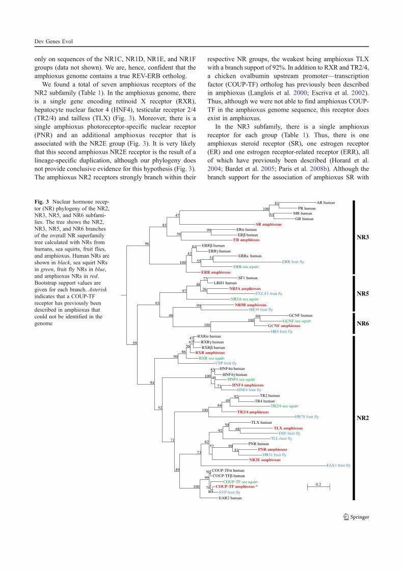
only on sequences of the NR1C, NR1D, NR1E, and NR1Fgroups (data not shown). We are, hence, confident that theamphioxus genome contains a true REV-ERB ortholog.
We found a total of seven amphioxus receptors of theNR2 subfamily (Table 1). In the amphioxus genome, thereis a single gene encoding retinoid X receptor (RXR),hepatocyte nuclear factor 4 (HNF4), testicular receptor 2/4(TR2/4) and tailless (TLX) (Fig. 3). Moreover, there is asingle amphioxus photoreceptor-specific nuclear receptor(PNR) and an additional amphioxus receptor that isassociated with the NR2E group (Fig. 3). It is very likelythat this second amphioxus NR2E receptor is the result of alineage-specific duplication, although our phylogeny doesnot provide conclusive evidence for this hypothesis (Fig. 3).The amphioxus NR2 receptors strongly branch within their
respective NR groups, the weakest being amphioxus TLXwith a branch support of 92%. In addition to RXR and TR2/4,a chicken ovalbumin upstream promoter—transcriptionfactor (COUP-TF) ortholog has previously been describedin amphioxus (Langlois et al. 2000; Escriva et al. 2002).Thus, although we were not able to find amphioxus COUP-TF in the amphioxus genome sequence, this receptor doesexist in amphioxus.
In the NR3 subfamily, there is a single amphioxusreceptor for each group (Table 1). Thus, there is oneamphioxus steroid receptor (SR), one estrogen receptor(ER) and one estrogen receptor-related receptor (ERR), allof which have previously been described (Horard et al.2004; Bardet et al. 2005; Paris et al. 2008b). Although thebranch support for the association of amphioxus SR with
EAR2 human
SVP fruit fly
COUP-TF amphioxus *
COUP-TF sea squirt
80
COUP-TFβ human
COUP-TFα human
78
50
99
FAX1 fruit fly
NR2E amphioxus
HR51 fruit fly
PNR amphioxus
PNR human
81 99
TLL fruit fly
DSF fruit fly
TLX amphioxus
TLX human
68 50
57
92
82
100
73
HR78 fruit fly
TR2/4 amphioxus
TR2/4 sea squirt
TR4 human
TR2 human 92 49
84
49
100
HNF4 fruit fly
HNF4 amphioxus
HNF4 sea squirt
71
HNF4γ human
HNF4α human
48
83
71
100
USP fruit fly
RXR sea squirt
RXR amphioxus
RXRβ human
RXRγ human
RXRα human 47 67
56
86
52
99
HR4 fruit fly
GCNF amphioxus
GCNF sea squirt
GCNF human 60 100
HR39 fruit fly
NR5B amphioxus
100
99
NR5A sea squirt
FTZ-F1 fruit fly
NR5A amphioxus
LRH1 human
SF1 human
26
77
48
46
97
94
93
ERR amphioxus
ERR sea squirt
ERR fruit fly
ERRa human 31
ERRγ human
ERRβ human
55
61
42
ER amphioxus
ERβ human
ERα human 99
SR amphioxus
GR human
MR human
PR human
AR human
53
81
100
58
47
100
85
59
96
0.2
NR2
NR6
NR5
NR3
Fig. 3 Nuclear hormone recep-tor (NR) phylogeny of the NR2,NR3, NR5, and NR6 subfami-lies. The tree shows the NR2,NR3, NR5, and NR6 branchesof the overall NR superfamilytree calculated with NRs fromhumans, sea squirts, fruit flies,and amphioxus. Human NRs areshown in black, sea squirt NRsin green, fruit fly NRs in blue,and amphioxus NRs in red.Bootstrap support values aregiven for each branch. Asteriskindicates that a COUP-TFreceptor has previously beendescribed in amphioxus thatcould not be identified in thegenome
Dev Genes Evol
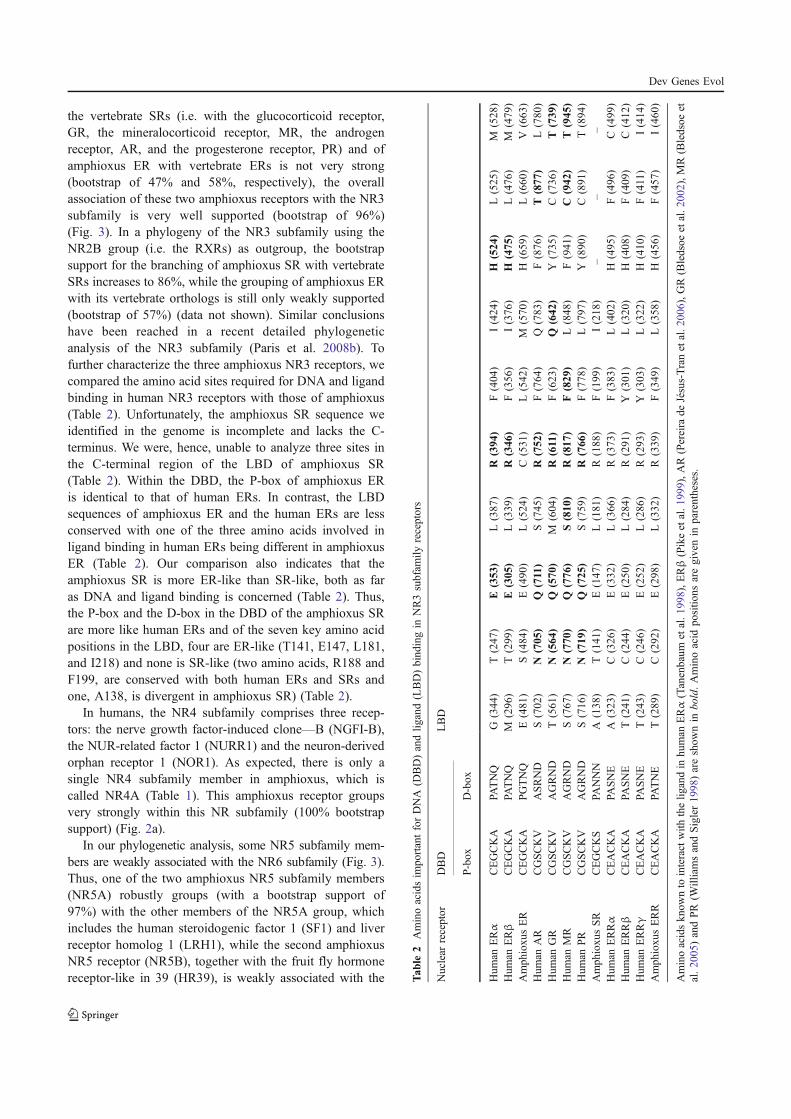
the vertebrate SRs (i.e. with the glucocorticoid receptor,GR, the mineralocorticoid receptor, MR, the androgenreceptor, AR, and the progesterone receptor, PR) and ofamphioxus ER with vertebrate ERs is not very strong(bootstrap of 47% and 58%, respectively), the overallassociation of these two amphioxus receptors with the NR3subfamily is very well supported (bootstrap of 96%)(Fig. 3). In a phylogeny of the NR3 subfamily using theNR2B group (i.e. the RXRs) as outgroup, the bootstrapsupport for the branching of amphioxus SR with vertebrateSRs increases to 86%, while the grouping of amphioxus ERwith its vertebrate orthologs is still only weakly supported(bootstrap of 57%) (data not shown). Similar conclusionshave been reached in a recent detailed phylogeneticanalysis of the NR3 subfamily (Paris et al. 2008b). Tofurther characterize the three amphioxus NR3 receptors, wecompared the amino acid sites required for DNA and ligandbinding in human NR3 receptors with those of amphioxus(Table 2). Unfortunately, the amphioxus SR sequence weidentified in the genome is incomplete and lacks the C-terminus. We were, hence, unable to analyze three sites inthe C-terminal region of the LBD of amphioxus SR(Table 2). Within the DBD, the P-box of amphioxus ERis identical to that of human ERs. In contrast, the LBDsequences of amphioxus ER and the human ERs are lessconserved with one of the three amino acids involved inligand binding in human ERs being different in amphioxusER (Table 2). Our comparison also indicates that theamphioxus SR is more ER-like than SR-like, both as faras DNA and ligand binding is concerned (Table 2). Thus,the P-box and the D-box in the DBD of the amphioxus SRare more like human ERs and of the seven key amino acidpositions in the LBD, four are ER-like (T141, E147, L181,and I218) and none is SR-like (two amino acids, R188 andF199, are conserved with both human ERs and SRs andone, A138, is divergent in amphioxus SR) (Table 2).
In humans, the NR4 subfamily comprises three recep-tors: the nerve growth factor-induced clone—B (NGFI-B),the NUR-related factor 1 (NURR1) and the neuron-derivedorphan receptor 1 (NOR1). As expected, there is only asingle NR4 subfamily member in amphioxus, which iscalled NR4A (Table 1). This amphioxus receptor groupsvery strongly within this NR subfamily (100% bootstrapsupport) (Fig. 2a).
In our phylogenetic analysis, some NR5 subfamily mem-bers are weakly associated with the NR6 subfamily (Fig. 3).Thus, one of the two amphioxus NR5 subfamily members(NR5A) robustly groups (with a bootstrap support of97%) with the other members of the NR5A group, whichincludes the human steroidogenic factor 1 (SF1) and liverreceptor homolog 1 (LRH1), while the second amphioxusNR5 receptor (NR5B), together with the fruit fly hormonereceptor-like in 39 (HR39), is weakly associated with the T
able
2Aminoacidsim
portantforDNA
(DBD)andlig
and(LBD)bind
ingin
NR3subfam
ilyreceptors
Nuclear
receptor
DBD
LBD
P-box
D-box
Hum
anERα
CEGCKA
PATNQ
G(344
)T(247
)E(353)
L(387
)R
(394)
F(404
)I(424
)H
(524)
L(525
)M
(528
)Hum
anERβ
CEGCKA
PATNQ
M(296
)T(299
)E(305)
L(339
)R
(346)
F(356
)I(376
)H
(475)
L(476
)M
(479
)Amph
ioxu
sER
CEGCKA
PGTNQ
E(481
)S(484
)E(490
)L(524
)C(531
)L(542
)M
(570
)H
(659
)L(660
)V
(663
)Hum
anAR
CGSCKV
ASRND
S(702
)N
(705)
Q(711)
S(745
)R
(752)
F(764
)Q
(783
)F(876
)T(877)
L(780
)Hum
anGR
CGSCKV
AGRND
T(561
)N
(564)
Q(570)
M(604
)R
(611)
F(623
)Q
(642)
Y(735
)C(736
)T(739)
Hum
anMR
CGSCKV
AGRND
S(767
)N
(770)
Q(776)
S(810)
R(817)
F(829)
L(848
)F(941
)C
(942)
T(945)
Hum
anPR
CGSCKV
AGRND
S(716
)N
(719)
Q(725)
S(759
)R
(766)
F(778
)L(797
)Y
(890
)C(891
)T(894
)Amph
ioxu
sSR
CEGCKS
PANNN
A(138
)T(141
)E(147
)L(181
)R(188
)F(199
)I(218
)–
––
Hum
anERRα
CEACKA
PASNE
A(323
)C(326
)E(332
)L(366
)R(373
)F(383
)L(402
)H
(495
)F(496
)C(499
)Hum
anERRβ
CEACKA
PASNE
T(241
)C(244
)E(250
)L(284
)R(291
)Y
(301
)L(320
)H
(408
)F(409
)C(412
)Hum
anERRγ
CEACKA
PASNE
T(243
)C(246
)E(252
)L(286
)R(293
)Y
(303
)L(322
)H
(410
)F(411)
I(414
)Amph
ioxu
sERR
CEACKA
PATNE
T(289
)C(292
)E(298
)L(332
)R(339
)F(349
)L(358
)H
(456
)F(457
)I(460
)
Aminoacidskn
ownto
interactwith
thelig
andin
human
ERα(Tanenbaum
etal.1
998),E
Rβ(Pikeetal.1
999),A
R(Pereira
deJésus-Tranetal.2
006),G
R(Bledsoe
etal.2
002),M
R(Bledsoe
etal.20
05)andPR(W
illiamsandSigler19
98)areshow
nin
bold.Aminoacid
positio
nsaregivenin
parentheses.
Dev Genes Evol
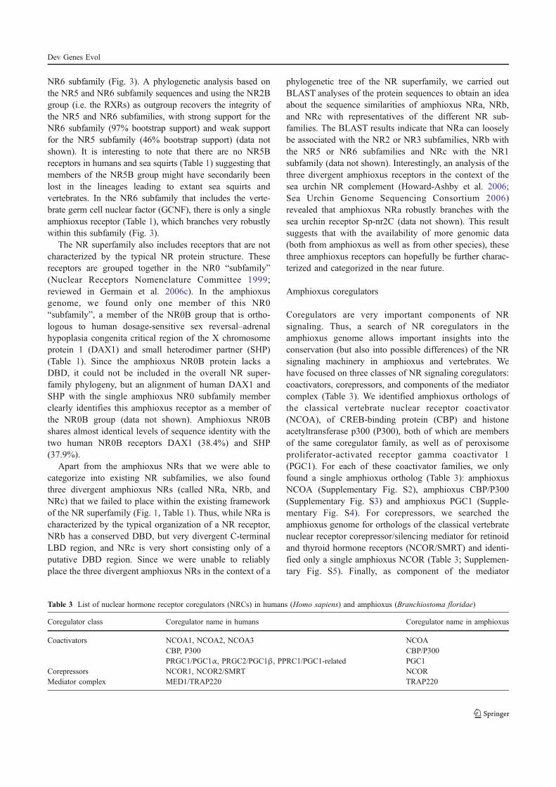
NR6 subfamily (Fig. 3). A phylogenetic analysis based onthe NR5 and NR6 subfamily sequences and using the NR2Bgroup (i.e. the RXRs) as outgroup recovers the integrity ofthe NR5 and NR6 subfamilies, with strong support for theNR6 subfamily (97% bootstrap support) and weak supportfor the NR5 subfamily (46% bootstrap support) (data notshown). It is interesting to note that there are no NR5Breceptors in humans and sea squirts (Table 1) suggesting thatmembers of the NR5B group might have secondarily beenlost in the lineages leading to extant sea squirts andvertebrates. In the NR6 subfamily that includes the verte-brate germ cell nuclear factor (GCNF), there is only a singleamphioxus receptor (Table 1), which branches very robustlywithin this subfamily (Fig. 3).
The NR superfamily also includes receptors that are notcharacterized by the typical NR protein structure. Thesereceptors are grouped together in the NR0 “subfamily”(Nuclear Receptors Nomenclature Committee 1999;reviewed in Germain et al. 2006c). In the amphioxusgenome, we found only one member of this NR0“subfamily”, a member of the NR0B group that is ortho-logous to human dosage-sensitive sex reversal–adrenalhypoplasia congenita critical region of the X chromosomeprotein 1 (DAX1) and small heterodimer partner (SHP)(Table 1). Since the amphioxus NR0B protein lacks aDBD, it could not be included in the overall NR super-family phylogeny, but an alignment of human DAX1 andSHP with the single amphioxus NR0 subfamily memberclearly identifies this amphioxus receptor as a member ofthe NR0B group (data not shown). Amphioxus NR0Bshares almost identical levels of sequence identity with thetwo human NR0B receptors DAX1 (38.4%) and SHP(37.9%).
Apart from the amphioxus NRs that we were able tocategorize into existing NR subfamilies, we also foundthree divergent amphioxus NRs (called NRa, NRb, andNRc) that we failed to place within the existing frameworkof the NR superfamily (Fig. 1, Table 1). Thus, while NRa ischaracterized by the typical organization of a NR receptor,NRb has a conserved DBD, but very divergent C-terminalLBD region, and NRc is very short consisting only of aputative DBD region. Since we were unable to reliablyplace the three divergent amphioxus NRs in the context of a
phylogenetic tree of the NR superfamily, we carried outBLAST analyses of the protein sequences to obtain an ideaabout the sequence similarities of amphioxus NRa, NRb,and NRc with representatives of the different NR sub-families. The BLAST results indicate that NRa can looselybe associated with the NR2 or NR3 subfamilies, NRb withthe NR5 or NR6 subfamilies and NRc with the NR1subfamily (data not shown). Interestingly, an analysis of thethree divergent amphioxus receptors in the context of thesea urchin NR complement (Howard-Ashby et al. 2006;Sea Urchin Genome Sequencing Consortium 2006)revealed that amphioxus NRa robustly branches with thesea urchin receptor Sp-nr2C (data not shown). This resultsuggests that with the availability of more genomic data(both from amphioxus as well as from other species), thesethree amphioxus receptors can hopefully be further charac-terized and categorized in the near future.
Amphioxus coregulators
Coregulators are very important components of NRsignaling. Thus, a search of NR coregulators in theamphioxus genome allows important insights into theconservation (but also into possible differences) of the NRsignaling machinery in amphioxus and vertebrates. Wehave focused on three classes of NR signaling coregulators:coactivators, corepressors, and components of the mediatorcomplex (Table 3). We identified amphioxus orthologs ofthe classical vertebrate nuclear receptor coactivator(NCOA), of CREB-binding protein (CBP) and histoneacetyltransferase p300 (P300), both of which are membersof the same coregulator family, as well as of peroxisomeproliferator-activated receptor gamma coactivator 1(PGC1). For each of these coactivator families, we onlyfound a single amphioxus ortholog (Table 3): amphioxusNCOA (Supplementary Fig. S2), amphioxus CBP/P300(Supplementary Fig. S3) and amphioxus PGC1 (Supple-mentary Fig. S4). For corepressors, we searched theamphioxus genome for orthologs of the classical vertebratenuclear receptor corepressor/silencing mediator for retinoidand thyroid hormone receptors (NCOR/SMRT) and identi-fied only a single amphioxus NCOR (Table 3; Supplemen-tary Fig. S5). Finally, as component of the mediator
Table 3 List of nuclear hormone receptor coregulators (NRCs) in humans (Homo sapiens) and amphioxus (Branchiostoma floridae)
Coregulator class Coregulator name in humans Coregulator name in amphioxus
Coactivators NCOA1, NCOA2, NCOA3 NCOACBP, P300 CBP/P300PRGC1/PGC1α, PRGC2/PGC1β, PPRC1/PGC1-related PGC1
Corepressors NCOR1, NCOR2/SMRT NCORMediator complex MED1/TRAP220 TRAP220
Dev Genes Evol
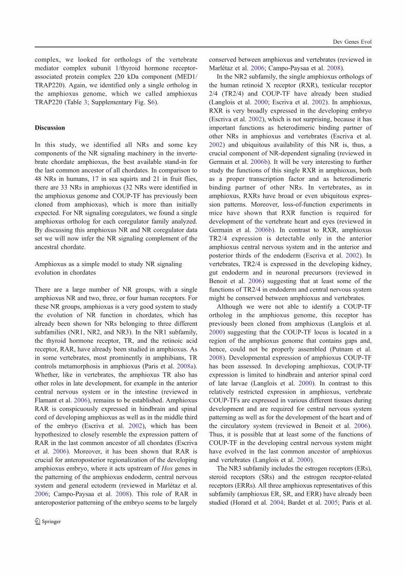
complex, we looked for orthologs of the vertebratemediator complex subunit 1/thyroid hormone receptor-associated protein complex 220 kDa component (MED1/TRAP220). Again, we identified only a single ortholog inthe amphioxus genome, which we called amphioxusTRAP220 (Table 3; Supplementary Fig. S6).
Discussion
In this study, we identified all NRs and some keycomponents of the NR signaling machinery in the inverte-brate chordate amphioxus, the best available stand-in forthe last common ancestor of all chordates. In comparison to48 NRs in humans, 17 in sea squirts and 21 in fruit flies,there are 33 NRs in amphioxus (32 NRs were identified inthe amphioxus genome and COUP-TF has previously beencloned from amphioxus), which is more than initiallyexpected. For NR signaling coregulators, we found a singleamphioxus ortholog for each coregulator family analyzed.By discussing this amphioxus NR and NR coregulator dataset we will now infer the NR signaling complement of theancestral chordate.
Amphioxus as a simple model to study NR signalingevolution in chordates
There are a large number of NR groups, with a singleamphioxus NR and two, three, or four human receptors. Forthese NR groups, amphioxus is a very good system to studythe evolution of NR function in chordates, which hasalready been shown for NRs belonging to three differentsubfamilies (NR1, NR2, and NR3). In the NR1 subfamily,the thyroid hormone receptor, TR, and the retinoic acidreceptor, RAR, have already been studied in amphioxus. Asin some vertebrates, most prominently in amphibians, TRcontrols metamorphosis in amphioxus (Paris et al. 2008a).Whether, like in vertebrates, the amphioxus TR also hasother roles in late development, for example in the anteriorcentral nervous system or in the intestine (reviewed inFlamant et al. 2006), remains to be established. AmphioxusRAR is conspicuously expressed in hindbrain and spinalcord of developing amphioxus as well as in the middle thirdof the embryo (Escriva et al. 2002), which has beenhypothesized to closely resemble the expression pattern ofRAR in the last common ancestor of all chordates (Escrivaet al. 2006). Moreover, it has been shown that RAR iscrucial for anteroposterior regionalization of the developingamphioxus embryo, where it acts upstream of Hox genes inthe patterning of the amphioxus endoderm, central nervoussystem and general ectoderm (reviewed in Marlétaz et al.2006; Campo-Paysaa et al. 2008). This role of RAR inanteroposterior patterning of the embryo seems to be largely
conserved between amphioxus and vertebrates (reviewed inMarlétaz et al. 2006; Campo-Paysaa et al. 2008).
In the NR2 subfamily, the single amphioxus orthologs ofthe human retinoid X receptor (RXR), testicular receptor2/4 (TR2/4) and COUP-TF have already been studied(Langlois et al. 2000; Escriva et al. 2002). In amphioxus,RXR is very broadly expressed in the developing embryo(Escriva et al. 2002), which is not surprising, because it hasimportant functions as heterodimeric binding partner ofother NRs in amphioxus and vertebrates (Escriva et al.2002) and ubiquitous availability of this NR is, thus, acrucial component of NR-dependent signaling (reviewed inGermain et al. 2006b). It will be very interesting to furtherstudy the functions of this single RXR in amphioxus, bothas a proper transcription factor and as heterodimericbinding partner of other NRs. In vertebrates, as inamphioxus, RXRs have broad or even ubiquitous expres-sion patterns. Moreover, loss-of-function experiments inmice have shown that RXR function is required fordevelopment of the vertebrate heart and eyes (reviewed inGermain et al. 2006b). In contrast to RXR, amphioxusTR2/4 expression is detectable only in the anterioramphioxus central nervous system and in the anterior andposterior thirds of the endoderm (Escriva et al. 2002). Invertebrates, TR2/4 is expressed in the developing kidney,gut endoderm and in neuronal precursors (reviewed inBenoit et al. 2006) suggesting that at least some of thefunctions of TR2/4 in endoderm and central nervous systemmight be conserved between amphioxus and vertebrates.
Although we were not able to identify a COUP-TFortholog in the amphioxus genome, this receptor haspreviously been cloned from amphioxus (Langlois et al.2000) suggesting that the COUP-TF locus is located in aregion of the amphioxus genome that contains gaps and,hence, could not be properly assembled (Putnam et al.2008). Developmental expression of amphioxus COUP-TFhas been assessed. In developing amphioxus, COUP-TFexpression is limited to hindbrain and anterior spinal cordof late larvae (Langlois et al. 2000). In contrast to thisrelatively restricted expression in amphioxus, vertebrateCOUP-TFs are expressed in various different tissues duringdevelopment and are required for central nervous systempatterning as well as for the development of the heart and ofthe circulatory system (reviewed in Benoit et al. 2006).Thus, it is possible that at least some of the functions ofCOUP-TF in the developing central nervous system mighthave evolved in the last common ancestor of amphioxusand vertebrates (Langlois et al. 2000).
The NR3 subfamily includes the estrogen receptors (ERs),steroid receptors (SRs) and the estrogen receptor-relatedreceptors (ERRs). All three amphioxus representatives of thissubfamily (amphioxus ER, SR, and ERR) have already beenstudied (Horard et al. 2004; Bardet et al. 2005; Paris et al.
Dev Genes Evol
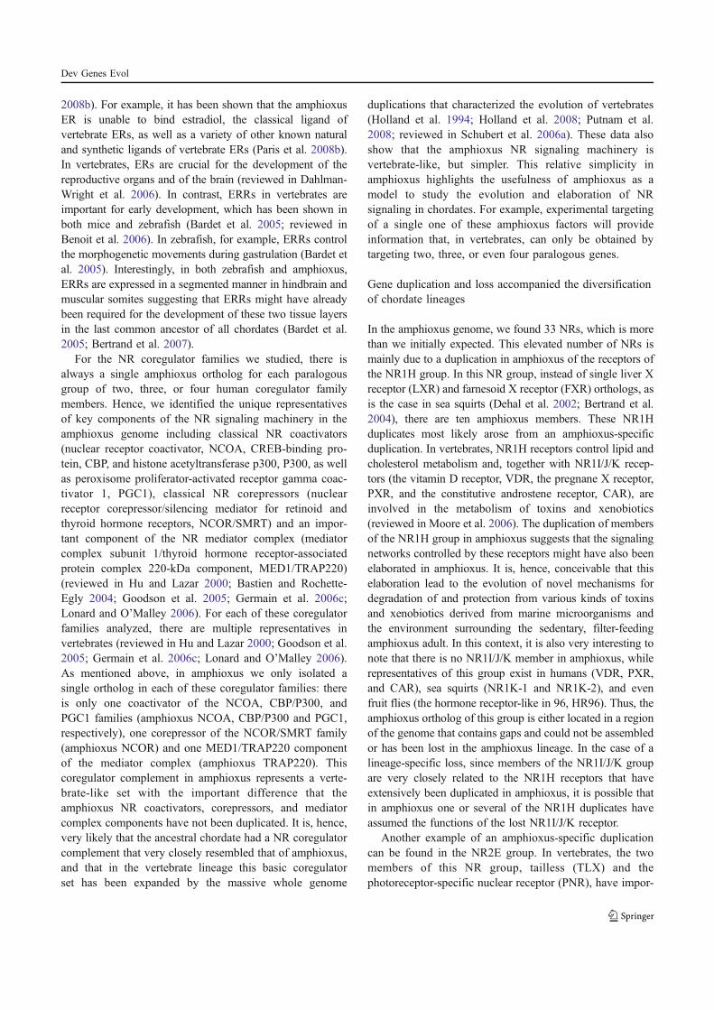
2008b). For example, it has been shown that the amphioxusER is unable to bind estradiol, the classical ligand ofvertebrate ERs, as well as a variety of other known naturaland synthetic ligands of vertebrate ERs (Paris et al. 2008b).In vertebrates, ERs are crucial for the development of thereproductive organs and of the brain (reviewed in Dahlman-Wright et al. 2006). In contrast, ERRs in vertebrates areimportant for early development, which has been shown inboth mice and zebrafish (Bardet et al. 2005; reviewed inBenoit et al. 2006). In zebrafish, for example, ERRs controlthe morphogenetic movements during gastrulation (Bardet etal. 2005). Interestingly, in both zebrafish and amphioxus,ERRs are expressed in a segmented manner in hindbrain andmuscular somites suggesting that ERRs might have alreadybeen required for the development of these two tissue layersin the last common ancestor of all chordates (Bardet et al.2005; Bertrand et al. 2007).
For the NR coregulator families we studied, there isalways a single amphioxus ortholog for each paralogousgroup of two, three, or four human coregulator familymembers. Hence, we identified the unique representativesof key components of the NR signaling machinery in theamphioxus genome including classical NR coactivators(nuclear receptor coactivator, NCOA, CREB-binding pro-tein, CBP, and histone acetyltransferase p300, P300, as wellas peroxisome proliferator-activated receptor gamma coac-tivator 1, PGC1), classical NR corepressors (nuclearreceptor corepressor/silencing mediator for retinoid andthyroid hormone receptors, NCOR/SMRT) and an impor-tant component of the NR mediator complex (mediatorcomplex subunit 1/thyroid hormone receptor-associatedprotein complex 220-kDa component, MED1/TRAP220)(reviewed in Hu and Lazar 2000; Bastien and Rochette-Egly 2004; Goodson et al. 2005; Germain et al. 2006c;Lonard and O’Malley 2006). For each of these coregulatorfamilies analyzed, there are multiple representatives invertebrates (reviewed in Hu and Lazar 2000; Goodson et al.2005; Germain et al. 2006c; Lonard and O’Malley 2006).As mentioned above, in amphioxus we only isolated asingle ortholog in each of these coregulator families: thereis only one coactivator of the NCOA, CBP/P300, andPGC1 families (amphioxus NCOA, CBP/P300 and PGC1,respectively), one corepressor of the NCOR/SMRT family(amphioxus NCOR) and one MED1/TRAP220 componentof the mediator complex (amphioxus TRAP220). Thiscoregulator complement in amphioxus represents a verte-brate-like set with the important difference that theamphioxus NR coactivators, corepressors, and mediatorcomplex components have not been duplicated. It is, hence,very likely that the ancestral chordate had a NR coregulatorcomplement that very closely resembled that of amphioxus,and that in the vertebrate lineage this basic coregulatorset has been expanded by the massive whole genome
duplications that characterized the evolution of vertebrates(Holland et al. 1994; Holland et al. 2008; Putnam et al.2008; reviewed in Schubert et al. 2006a). These data alsoshow that the amphioxus NR signaling machinery isvertebrate-like, but simpler. This relative simplicity inamphioxus highlights the usefulness of amphioxus as amodel to study the evolution and elaboration of NRsignaling in chordates. For example, experimental targetingof a single one of these amphioxus factors will provideinformation that, in vertebrates, can only be obtained bytargeting two, three, or even four paralogous genes.
Gene duplication and loss accompanied the diversificationof chordate lineages
In the amphioxus genome, we found 33 NRs, which is morethan we initially expected. This elevated number of NRs ismainly due to a duplication in amphioxus of the receptors ofthe NR1H group. In this NR group, instead of single liver Xreceptor (LXR) and farnesoid X receptor (FXR) orthologs, asis the case in sea squirts (Dehal et al. 2002; Bertrand et al.2004), there are ten amphioxus members. These NR1Hduplicates most likely arose from an amphioxus-specificduplication. In vertebrates, NR1H receptors control lipid andcholesterol metabolism and, together with NR1I/J/K recep-tors (the vitamin D receptor, VDR, the pregnane X receptor,PXR, and the constitutive androstene receptor, CAR), areinvolved in the metabolism of toxins and xenobiotics(reviewed in Moore et al. 2006). The duplication of membersof the NR1H group in amphioxus suggests that the signalingnetworks controlled by these receptors might have also beenelaborated in amphioxus. It is, hence, conceivable that thiselaboration lead to the evolution of novel mechanisms fordegradation of and protection from various kinds of toxinsand xenobiotics derived from marine microorganisms andthe environment surrounding the sedentary, filter-feedingamphioxus adult. In this context, it is also very interesting tonote that there is no NR1I/J/K member in amphioxus, whilerepresentatives of this group exist in humans (VDR, PXR,and CAR), sea squirts (NR1K-1 and NR1K-2), and evenfruit flies (the hormone receptor-like in 96, HR96). Thus, theamphioxus ortholog of this group is either located in a regionof the genome that contains gaps and could not be assembledor has been lost in the amphioxus lineage. In the case of alineage-specific loss, since members of the NR1I/J/K groupare very closely related to the NR1H receptors that haveextensively been duplicated in amphioxus, it is possible thatin amphioxus one or several of the NR1H duplicates haveassumed the functions of the lost NR1I/J/K receptor.
Another example of an amphioxus-specific duplicationcan be found in the NR2E group. In vertebrates, the twomembers of this NR group, tailless (TLX) and thephotoreceptor-specific nuclear receptor (PNR), have impor-
Dev Genes Evol
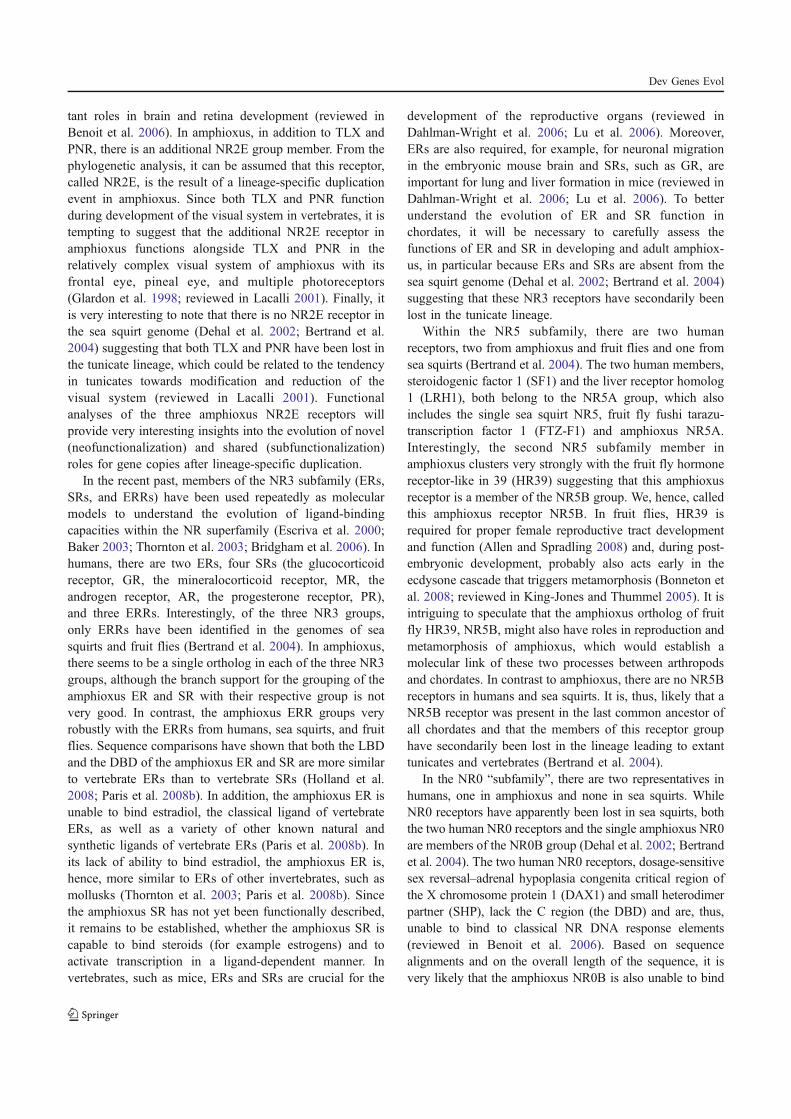
tant roles in brain and retina development (reviewed inBenoit et al. 2006). In amphioxus, in addition to TLX andPNR, there is an additional NR2E group member. From thephylogenetic analysis, it can be assumed that this receptor,called NR2E, is the result of a lineage-specific duplicationevent in amphioxus. Since both TLX and PNR functionduring development of the visual system in vertebrates, it istempting to suggest that the additional NR2E receptor inamphioxus functions alongside TLX and PNR in therelatively complex visual system of amphioxus with itsfrontal eye, pineal eye, and multiple photoreceptors(Glardon et al. 1998; reviewed in Lacalli 2001). Finally, itis very interesting to note that there is no NR2E receptor inthe sea squirt genome (Dehal et al. 2002; Bertrand et al.2004) suggesting that both TLX and PNR have been lost inthe tunicate lineage, which could be related to the tendencyin tunicates towards modification and reduction of thevisual system (reviewed in Lacalli 2001). Functionalanalyses of the three amphioxus NR2E receptors willprovide very interesting insights into the evolution of novel(neofunctionalization) and shared (subfunctionalization)roles for gene copies after lineage-specific duplication.
In the recent past, members of the NR3 subfamily (ERs,SRs, and ERRs) have been used repeatedly as molecularmodels to understand the evolution of ligand-bindingcapacities within the NR superfamily (Escriva et al. 2000;Baker 2003; Thornton et al. 2003; Bridgham et al. 2006). Inhumans, there are two ERs, four SRs (the glucocorticoidreceptor, GR, the mineralocorticoid receptor, MR, theandrogen receptor, AR, the progesterone receptor, PR),and three ERRs. Interestingly, of the three NR3 groups,only ERRs have been identified in the genomes of seasquirts and fruit flies (Bertrand et al. 2004). In amphioxus,there seems to be a single ortholog in each of the three NR3groups, although the branch support for the grouping of theamphioxus ER and SR with their respective group is notvery good. In contrast, the amphioxus ERR groups veryrobustly with the ERRs from humans, sea squirts, and fruitflies. Sequence comparisons have shown that both the LBDand the DBD of the amphioxus ER and SR are more similarto vertebrate ERs than to vertebrate SRs (Holland et al.2008; Paris et al. 2008b). In addition, the amphioxus ER isunable to bind estradiol, the classical ligand of vertebrateERs, as well as a variety of other known natural andsynthetic ligands of vertebrate ERs (Paris et al. 2008b). Inits lack of ability to bind estradiol, the amphioxus ER is,hence, more similar to ERs of other invertebrates, such asmollusks (Thornton et al. 2003; Paris et al. 2008b). Sincethe amphioxus SR has not yet been functionally described,it remains to be established, whether the amphioxus SR iscapable to bind steroids (for example estrogens) and toactivate transcription in a ligand-dependent manner. Invertebrates, such as mice, ERs and SRs are crucial for the
development of the reproductive organs (reviewed inDahlman-Wright et al. 2006; Lu et al. 2006). Moreover,ERs are also required, for example, for neuronal migrationin the embryonic mouse brain and SRs, such as GR, areimportant for lung and liver formation in mice (reviewed inDahlman-Wright et al. 2006; Lu et al. 2006). To betterunderstand the evolution of ER and SR function inchordates, it will be necessary to carefully assess thefunctions of ER and SR in developing and adult amphiox-us, in particular because ERs and SRs are absent from thesea squirt genome (Dehal et al. 2002; Bertrand et al. 2004)suggesting that these NR3 receptors have secondarily beenlost in the tunicate lineage.
Within the NR5 subfamily, there are two humanreceptors, two from amphioxus and fruit flies and one fromsea squirts (Bertrand et al. 2004). The two human members,steroidogenic factor 1 (SF1) and the liver receptor homolog1 (LRH1), both belong to the NR5A group, which alsoincludes the single sea squirt NR5, fruit fly fushi tarazu-transcription factor 1 (FTZ-F1) and amphioxus NR5A.Interestingly, the second NR5 subfamily member inamphioxus clusters very strongly with the fruit fly hormonereceptor-like in 39 (HR39) suggesting that this amphioxusreceptor is a member of the NR5B group. We, hence, calledthis amphioxus receptor NR5B. In fruit flies, HR39 isrequired for proper female reproductive tract developmentand function (Allen and Spradling 2008) and, during post-embryonic development, probably also acts early in theecdysone cascade that triggers metamorphosis (Bonneton etal. 2008; reviewed in King-Jones and Thummel 2005). It isintriguing to speculate that the amphioxus ortholog of fruitfly HR39, NR5B, might also have roles in reproduction andmetamorphosis of amphioxus, which would establish amolecular link of these two processes between arthropodsand chordates. In contrast to amphioxus, there are no NR5Breceptors in humans and sea squirts. It is, thus, likely that aNR5B receptor was present in the last common ancestor ofall chordates and that the members of this receptor grouphave secondarily been lost in the lineage leading to extanttunicates and vertebrates (Bertrand et al. 2004).
In the NR0 “subfamily”, there are two representatives inhumans, one in amphioxus and none in sea squirts. WhileNR0 receptors have apparently been lost in sea squirts, boththe two human NR0 receptors and the single amphioxus NR0are members of the NR0B group (Dehal et al. 2002; Bertrandet al. 2004). The two human NR0 receptors, dosage-sensitivesex reversal–adrenal hypoplasia congenita critical region ofthe X chromosome protein 1 (DAX1) and small heterodimerpartner (SHP), lack the C region (the DBD) and are, thus,unable to bind to classical NR DNA response elements(reviewed in Benoit et al. 2006). Based on sequencealignments and on the overall length of the sequence, it isvery likely that the amphioxus NR0B is also unable to bind
Dev Genes Evol
DNA, although DNA-binding experiments with the amphi-oxus NR0B will be necessary to validate this hypothesis.
In addition to the amphioxus NRs that group withinexisting NR subfamilies, we also identified three divergentNRs in the amphioxus genome (called NRa, NRb, andNRc). These three receptors did not branch reliably withany NR subfamily. It is possible that these three receptorsinitially originated from an amphioxus-specific duplication.After duplication, the coding sequences of the duplicatesdiverged possibly resulting in neofunctionalization orsubfunctionalization. Alternatively, these three NRs couldalso be remnants of ancient NR subfamilies, whoserepresentatives have secondarily been lost in most species,for which the NR repertoire has already been assessed. Thefact that one of the three divergent amphioxus NRs (NRa)robustly groups with a sea urchin receptor (Sp-nr2C) lendssupport to the latter hypothesis and suggests that with moregenomic data and whole genome sequences, it might becomefeasible to classify these three divergent amphioxus NRs.
Conclusions
In this study, we identified the NR and NR coregulatorcomplement of the invertebrate chordate amphioxus, whichis located at the base of the chordates and as such is the bestavailable stand-in for the last common ancestor of allchordates. Our data suggest that the chordate ancestor had22 NRs, more specifically one NR0 (NR0B), eight NR1s(TR, RAR, PPAR, REV-ERB, ROR, LXR, FXR, NR1I),six NR2s (HNF4, RXR, TR2/4, TLX, PNR, COUP-TF),three NR3s (ER, ERR, SR), one NR4 (NR4A), two NR5s(NR5A, NR5B), and one NR6 (GCNF). In addition, theancestral chordate had a single copy of the NR coactivatorsNCOA, CBP/P300, and PGC1, of the NR corepressorNCOR, and of the NR mediator complex componentTRAP220. Thus, the last common ancestor of chordateswas already characterized by a very elaborate set of NRsand NR coregulators that have independently been modi-fied and expanded both in amphioxus and vertebrates. Insum, this work highlights the utility of the amphioxusgenome for understanding the evolution of chordates andalso shows that the amphioxus genome has by no meansbeen fixed in an ancestral state: this genome has undergonevarious lineage-specific gene duplications and losses afterthe split from the lineage leading to extant tunicates andvertebrates some 550 million years ago.
Acknowledgments The authors would like to thank Michael E.Baker for critical reading of the manuscript. This work was supportedby grants from the ANR and the CNRS to M.S., the MENRT to M.S.and V.L. and by CRESCENDO, a European Union Integrated Projectof FP6, and by CASCADE, a Network of Excellence of FP6. S.B. isfunded by an ARC postdoctoral fellowship.
References
Allen AK, Spradling AC (2008) The Sf1-related nuclear hormonereceptor Hr39 regulates Drosophila female reproductive tractdevelopment and function. Development 135:311–321
Baker ME (2003) Evolution of adrenal and sex steroid action invertebrates: a ligand-based mechanism for complexity. Bioessays25:396–400
Bardet PL, Schubert M, Horard B, Holland LZ, Laudet V, HollandND, Vanacker JM (2005) Expression of estrogen-receptor relatedreceptors in amphioxus and zebrafish: implications for theevolution of posterior brain segmentation at the invertebrate-to-vertebrate transition. Evol Dev 7:223–233
Bastien J, Rochette-Egly C (2004) Nuclear retinoid receptors and thetranscription of retinoid-target genes. Gene 328:1–16
Benoit G, Cooney A, Giguere V, Ingraham H, Lazar M, Muscat G,Perlmann T, Renaud JP, Schwabe J, Sladek F, Tsai MJ, Laudet V(2006) International Union of Pharmacology. LXVI. Orphannuclear receptors. Pharmacol Rev 58:798–836
Bertrand S, Brunet FG, Escriva H, Parmentier G, Laudet V, Robinson-Rechavi M (2004) Evolutionary genomics of nuclear receptors:from twenty-five ancestral genes to derived endocrine systems.Mol Biol Evol 21:1923–1937
Bertrand S, Thisse B, Tavares R, Sachs L, Chaumot A, Bardet PL,Escriva H, Duffraisse M, Marchand O, Safi R, Thisse C, LaudetV (2007) Unexpected novel relational links uncovered byextensive developmental profiling of nuclear receptor expression.PLoS Genet 3:e188
Bledsoe RK, Montana VG, Stanley TB, Delves CJ, Apolito CJ,McKee DD, Consler TG, Parks DJ, Stewart EL, Willson TM,Lambert MH, Moore JT, Pearce KH, Xu HE (2002) Crystalstructure of the glucocorticoid receptor ligand binding domainreveals a novel mode of receptor dimerization and coactivatorrecognition. Cell 110:93–105
Bledsoe RK, Madauss KP, Holt JA, Apolito CJ, Lambert MH, PearceKH, Stanley TB, Stewart EL, Trump RP, Willson TM, WilliamsSP (2005) A ligand-mediated hydrogen bond network requiredfor the activation of the mineralocorticoid receptor. J Biol Chem280:31283–31293
Bonneton F, Chaumot A, Laudet V (2008) Annotation of Triboliumnuclear receptors reveals an increase in evolutionary rate of anetwork controlling the ecdysone cascade. Insect Biochem MolBiol 38:416–429
Bridgham JT, Carroll SM, Thornton JW (2006) Evolution ofhormone-receptor complexity by molecular exploitation. Science312:97–101
Campo-Paysaa F, Marlétaz F, Laudet V, Schubert M (2008) Retinoicacid signaling in development: tissue-specific functions andevolutionary origins. Genesis, in press
Dahlman-Wright K, Cavailles V, Fuqua SA, Jordan VC, KatzenellenbogenJA, Korach KS, Maggi A, Muramatsu M, Parker MG, GustafssonJÅ (2006) International Union of Pharmacology. LXIV. Estrogenreceptors. Pharmacol Rev 58:773–781
Dehal P, Satou Y, Campbell RK, Chapman J, Degnan B, De TomasoA, Davidson B, Di Gregorio A, Gelpke M, Goodstein DM,Harafuji N, Hastings KE, Ho I, Hotta K, Huang W, KawashimaT, Lemaire P, Martinez D, Meinertzhagen IA, Necula S, NonakaM, Putnam N, Rash S, Saiga H, Satake M, Terry A, Yamada L,Wang HG, Awazu S, Azumi K, Boore J, Branno M, Chin-Bow S,DeSantis R, Doyle S, Francino P, Keys DN, Haga S, Hayashi H,Hino K, Imai KS, Inaba K, Kano S, Kobayashi K, Kobayashi M,Lee BI, Makabe KW, Manohar C, Matassi G, Medina M,Mochizuki Y, Mount S, Morishita T, Miura S, Nakayama A,Nishizaka S, Nomoto H, Ohta F, Oishi K, Rigoutsos I, Sano M,Sasaki A, Sasakura Y, Shoguchi E, Shin-i T, Spagnuolo A,
Dev Genes Evol
Stainier D, Suzuki MM, Tassy O, Takatori N, Tokuoka M, YagiK, Yoshizaki F, Wada S, Zhang C, Hyatt PD, Larimer F, DetterC, Doggett N, Glavina T, Hawkins T, Richardson P, Lucas S,Kohara Y, Levine M, Satoh N, Rokhsar DS (2002) The draftgenome of Ciona intestinalis: insights into chordate andvertebrate origins. Science 298:2157–2167
Dehal P, Boore JL (2005) Two rounds of whole genome duplication inthe ancestral vertebrate. PLoS Biol 3:e314
Edgar RC (2004) MUSCLE: multiple sequence alignment with highaccuracy and high throughput. Nucleic Acids Res 32:1792–1797
Escriva H, Safi R, Hänni C, Langlois MC, Saumitou-Laprade P,Stehelin D, Capron A, Pierce R, Laudet V (1997) Ligand bindingwas acquired during evolution of nuclear receptors. Proc NatlAcad Sci USA 94:6803–6808
Escriva H, Delaunay F, Laudet V (2000) Ligand binding and nuclearreceptor evolution. Bioessays 22:717–727
Escriva H, Holland ND, Gronemeyer H, Laudet V, Holland LZ (2002)The retinoic acid signaling pathway regulates anterior/posteriorpatterning in the nerve cord and pharynx of amphioxus, achordate lacking neural crest. Development 129:2905–2916
Escriva H, Bertrand S, Germain P, Robinson-Rechavi M, UmbhauerM, Cartry J, Duffraisse M, Holland LZ, Gronemeyer H, Laudet V(2006) Neofunctionalization in vertebrates: the example ofretinoic acid receptors. PLoS Genet 2:e102
Flamant F, Baxter JD, Forrest D, Refetoff S, Samuels H, Scanlan TS,Vennström B, Samarut J (2006) International Union of Pharma-cology. LIX. The pharmacology and classification of the nuclearreceptor superfamily: thyroid hormone receptors. Pharmacol Rev58:705–711
Galtier N, Gouy M, Gautier C (1996) SEAVIEW and PHYLO_WIN:two graphic tools for sequence alignment and molecularphylogeny. Comput Appl Biosci 12:543–548
Garcia-Fernàndez J, D’Aniello S, Escriva H (2007) Organizingchordates with an organizer. Bioessays 29:619–624
Germain P, Chambon P, Eichele G, Evans GM, Lazar MA, Leid M, deLera AR, Lotan R, Mangelsdorf DJ, Gronemeyer H (2006a)International Union of Pharmacology. LX. Retinoic acid recep-tors. Pharmacol Rev 58:712–725
Germain P, Chambon P, Eichele G, Evans GM, Lazar MA, Leid M, deLera AR, Lotan R, Mangelsdorf DJ, Gronemeyer H (2006b)International Union of Pharmacology. LXIII. Retinoid X recep-tors. Pharmacol Rev 58:760–772
Germain P, Staels B, Dacquet C, Spedding M, Laudet V (2006c)Overview of nomenclature of nuclear receptors. Pharmacol Rev58:685–704
Glardon S, Holland LZ, Gehring WJ, Holland ND (1998) Isolationand developmental expression of the amphioxus Pax-6 gene(AmphiPax-6): insights into eye and photoreceptor evolution.Development 125:2701–2710
Goodson M, Jonas BA, Privalsky MA (2005) Corepressors: customtailoring and alterations while you wait. Nucl Recept Signal 3:e003
Gronemeyer H, Gustafsson JÅ, Laudet V (2004) Principles formodulation of the nuclear receptor superfamily. Nat Rev DrugDiscov 3:950–964
Guindon S, Gascuel O (2003) A simple, fast, and accurate algorithmto estimate large phylogenies by maximum likelihood. Syst Biol52:696–704
Holland LZ, Albalat R, Azumi K, Benito-Gutiérrez E, Blow MJ,Bronner-Fraser M, Brunet F, Butts T, Candiani S, Dishaw LJ,Ferrier DEK, Garcia-Fernàndez J, Gibson-Brown JJ, Gissi C,Godzik A, Hallböök F, Hirose D, Hosomichi K, Ikuta T, Inoko H,Kasahara M, Kasamatsu J, Kawashima T, Kimura A, KobayashiM, Kozmik Z, Kubokawa K, Laudet V, Litman GW, McHardyAC, Meulemans D, Nonaka M, Olinski RP, Pancer Z, Pennacchio
LA, Pestarino M, Rast JP, Rigoutsos I, Robinson-Rechavi M,Roch G, Saiga H, Sasakura Y, Satake M, Satou Y, Schubert M,Sherwood N, Shiina T, Takatori N, Tello J, Vopalensky P, WadaS, Xu A, Ye Y, Yoshida K, Yoshizaki F, Yu JK, Zhang Q, ZmasekCM, de Jong PJ, Osoegawa K, Putnam NH, Rokhsar DS, SatohN, Holland PWH (2008) The amphioxus genome illuminatesvertebrate origins and cephalochordate biology. Genome Res18:1100–1111
Holland PWH, Garcia-Fernàndez J, Williams NA, Sidow A (1994)Gene duplications and the origins of vertebrate development.Development Suppl 1994:125–133
Horard B, Castet A, Bardet PL, Laudet V, Cavailles V, Vanacker JM(2004) Dimerization is required for transactivation by estrogen-receptor-related (ERR) orphan receptors: evidence from amphi-oxus ERR. J Mol Endocrinol 33:493–509
Howard-Ashby M, Materna SC, Brown CT, Chen L, Cameron RA,Davidson EH (2006) Gene families encoding transcription factorsexpressed in early development of Strongylocentrotus purpur-
atus. Dev Biol 300:90–107Hu X, Lazar MA (2000) Transcriptional repression by nuclear
hormone receptors. Trends Endocrinol Metab 11:6–10King-Jones K, Thummel CS (2005) Nuclear receptors—a perspective
from Drosophila. Nat Rev Genet 6:311–323Lacalli TC (2001) New perspectives on the evolution of protochordate
sensory and locomotory systems, and the origin of brains andheads. Philos Trans R Soc Lond B 356:1565–1572
Langlois MC, Vanacker JM, Holland ND, Escriva H, Queva C, LaudetV, Holland LZ (2000) Amphicoup-TF, a nuclear orphan receptorof the lancelet Branchiostoma floridae, is implicated in retinoicacid signalling pathways. Dev Genes Evol 210:471–482
Laudet V, Gronemeyer H (2002) The nuclear receptor facts book.Academic Press, San Diego
Lonard DM, O’Malley BW (2006) The expanding cosmos of nuclearreceptor coactivators. Cell 125:411–414
Lu NZ, Wardell SE, Burnstein KL, Defranco D, Fuller PJ, Giguere V,Hochberg RB, McKay L, Renoir JM, Weigel NL, Wilson EM,McDonnell DP, Cidlowski JA (2006) International Union ofPharmacology. LXV. The pharmacology and classification of thenuclear receptor superfamily: glucocorticoid, mineralocorticoid,progesterone, and androgen receptors. Pharmacol Rev 58:782–797
Marlétaz F, Holland LZ, Laudet V, Schubert M (2006) Retinoic acidsignaling and the evolution of chordates. Int J Biol Sci 2:38–47
Michalik L, Auwerx J, Berger JP, Chatterjee VK, Glass CK, GonzalezFJ, Grimaldi PA, Kadowaki T, Lazar MA, O’Rahilly S, PalmerCN, Plutzky J, Reddy JK, Spiegelman BM, Staels B, Wahli W(2006) International Union of Pharmacology. LXI. Peroxisomeproliferator-activated receptors. Pharmacol Rev 58:726–741
Moore DD, Kato S, Xie W, Mangelsdorf DJ, Schmidt DR, Xiao R,Kliewer SA (2006) International Union of Pharmacology. LXII.The NR1H and NR1I receptors: constitutive androstane receptor,pregnene X receptor, farnesoid X receptor α, farnesoid Xreceptor β, liver X receptor α, liver X receptor β, and vitaminD receptor. Pharmacol Rev 58:742–759
Nuclear Receptors Nomenclature Committee (1999) A unifiednomenclature system for the nuclear receptor superfamily. Cell97:161–163
Paris M, Escriva H, Schubert M, Brunet F, Brtko J, Ciesielski F,Roecklin D, Vivat-Hannah V, Jamin EL, Cravedi JP, Scanlan TS,Renaud JP, Holland ND, Laudet V (2008a) Amphioxus postem-bryonic development reveals the homology of chordate meta-morphosis. Curr Biol 18:825–830
Paris M, Pettersson K, Schubert M, Bertrand S, Pongratz I, Escriva H,Laudet V (2008b) An amphioxus orthologue of the estrogenreceptor that does not bind estradiol: insights into estrogenreceptor evolution. BMC Evol Biol 8:219
Dev Genes Evol
Pereira de Jésus-Tran K, Côté PL, Cantin L, Blanchet J, Labrie F,Breton R (2006) Comparison of crystal structures of humanandrogen receptor ligand-binding domain complexed with vari-ous agonists reveals molecular determinants responsible forbinding affinity. Protein Sci 15:987–999
Pike AC, Brzozowski AM, Hubbard RE, Bonn T, Thorsell AG,Engström O, Ljunggren J, Gustafsson JÅ, Carlquist M (1999)Structure of the ligand-binding domain of oestrogen receptor betain the presence of a partial agonist and a full antagonist. EMBO J18:4608–4618
Putnam NH, Butts T, Ferrier DEK, Furlong RF, Hellsten U,Kawashima T, Robinson-Rechavi M, Shoguchi E, Terry A, YuJK, Benito-Gutiérrez È, Dubchak I, Garcia-Fernàndez J, Gibson-Brown JJ, Grigoriev IV, Horton AC, de Jong PJ, Jurka J,Kapitonov V, Kohara Y, Kuroki Y, Lindquist E, Lucas S,Osoegawa K, Pennacchio LA, Salamov AA, Satou Y, Sauka-Spengler T, Schmutz J, Shin- IT, Toyoda A, Bronner-Fraser M,Fujiyama A, Holland LZ, Holland PWH, Satoh N, Rokhsar DS(2008) The amphioxus genome and the evolution of the chordatekaryotype. Nature 453:1064–1072
Schubert M, Escriva H, Xavier-Neto J, Laudet V (2006a) Amphioxusand tunicates as evolutionary model systems. Trends Ecol Evol21:269–277
Schubert M, Holland ND, Laudet V, Holland LZ (2006b) A retinoicacid-Hox hierarchy controls both anterior/posterior patterning andneuronal specification in the developing central nervous systemof the cephalochordate amphioxus. Dev Biol 296:190–202
Sea Urchin Genome Sequencing Consortium (2006) The genome ofthe sea urchin Strongylocentrotus purpuratus. Science 314:941–952
Tanenbaum DM, Wang Y, Williams SP, Sigler PB (1998) Crystallo-graphic comparison of the estrogen and progesterone receptor’sligand binding domains. Proc Natl Acad Sci USA 95:5998–6003
Thornton JW, Need E, Crews D (2003) Resurrecting the ancestralsteroid receptor: ancient origin of estrogen signaling. Science301:1714–1717
Williams SP, Sigler PB (1998) Atomic structure of progesteronecomplexed with its receptor. Nature 393:392–396
Yagi K, Satou Y, Mazet F, Shimeld SM, Degnan B, Rokhsar D,Levine M, Kohara Y, Satoh N (2003) A genomewide survey ofdevelopmentally relevant genes in Ciona intestinalis. III. Genesfor Fox, ETS, nuclear receptors and NFκB. Dev Genes Evol213:235–244
Yu JK, Satou Y, Holland ND, Shin-I T, Kohara Y, Satoh N, Bronner-Fraser M, Holland LZ (2007) Axial patterning in cephalochor-dates and the evolution of the organizer. Nature 445:613–617
Dev Genes Evol
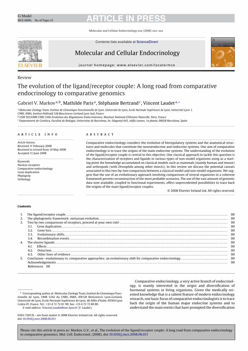
Please cite this article in press as: Markov, G.V., et al., The evolution of the ligand/receptor couple: A long road from comparative endocrinology
to comparative genomics. Mol. Cell. Endocrinol. (2008), doi:10.1016/j.mce.2008.06.011
ARTICLE IN PRESSG Model
MCE-6896; No. of Pages 12
Molecular and Cellular Endocrinology xxx (2008) xxx–xxx
Contents lists available at ScienceDirect
Molecular and Cellular Endocrinology
journa l homepage: www.e lsev ier .com/ locate /mce
Review
The evolution of the ligand/receptor couple: A long road from comparativeendocrinology to comparative genomics
Gabriel V. Markov a,b, Mathilde Paris a, Stéphanie Bertrand c, Vincent Laudet a,∗
a Molecular Zoology Team, Institut de Génomique Fonctionnelle de Lyon, Université de Lyon, Ecole Normale Supérieure de Lyon, Université Lyon 1,
CNRS, INRA, Institut Fédératif 128 Biosciences Gerland Lyon Sud, Franceb USM 501/UMR CNRS 5166-Evolution des Régulations Endocriniennes, Muséum National d’Histoire Naturelle, Paris, Francec Departament de Genètica, Facultat de Biologia, Universitat de Barcelona, Av. Diagonal 645, edifici annex, 1a planta, 08028 Barcelona, Spain
a r t i c l e i n f o
Article history:
Received 11 February 2008
Received in revised form 14 May 2008
Accepted 11 June 2008
Keywords:
Nuclear receptors
Comparative endocrinology
Gene duplication
Phylogeny
Orthology
a b s t r a c t
Comparative endocrinology considers the evolution of bioregulatory systems and the anatomical struc-
tures and molecules that constitute the neuroendocrine and endocrine systems. One aim of comparative
endocrinology is to trace the origins of the main endocrine systems. The understanding of the evolution
of the ligand/receptor couple is central to this objective. One classical approach to tackle this question is
the characterization of receptors and ligands in various types of non-model organisms using as a start-
ing point the knowledge accumulated on classical models such as mammals (mainly human and mouse)
and arthropods (with Drosophila among other insects). In this review we discuss the potential caveats
associated to this two-by-two comparison between a classical model and non-model organisms. We sug-
gest that the use of an evolutionary approach involving comparisons of several organisms in a coherent
framework permits reconstruction of the most probable scenarios. The use of the vast amount of genomic
data now available, coupled to functional experiments, offers unprecedented possibilities to trace back
the origins of the main ligand/receptor couples.
© 2008 Elsevier Ireland Ltd. All rights reserved.
Contents
1. The ligand/receptor couple . . . . . . . . . . . . . . . . . . . . . . . . . . . . . . . . . . . . . . . . . . . . . . . . . . . . . . . . . . . . . . . . . . . . . . . . . . . . . . . . . . . . . . . . . . . . . . . . . . . . . . . . . . . . . . . . . . . . . . . . . . . 00
2. The phylogenetic framework: metazoan evolution . . . . . . . . . . . . . . . . . . . . . . . . . . . . . . . . . . . . . . . . . . . . . . . . . . . . . . . . . . . . . . . . . . . . . . . . . . . . . . . . . . . . . . . . . . . . . . . . . . 00
3. Two by two comparisons of receptors, proceed at your own risk! . . . . . . . . . . . . . . . . . . . . . . . . . . . . . . . . . . . . . . . . . . . . . . . . . . . . . . . . . . . . . . . . . . . . . . . . . . . . . . . . . . 00
3.1. Gene duplication . . . . . . . . . . . . . . . . . . . . . . . . . . . . . . . . . . . . . . . . . . . . . . . . . . . . . . . . . . . . . . . . . . . . . . . . . . . . . . . . . . . . . . . . . . . . . . . . . . . . . . . . . . . . . . . . . . . . . . . . . . . . . . 00
3.2. Gene loss . . . . . . . . . . . . . . . . . . . . . . . . . . . . . . . . . . . . . . . . . . . . . . . . . . . . . . . . . . . . . . . . . . . . . . . . . . . . . . . . . . . . . . . . . . . . . . . . . . . . . . . . . . . . . . . . . . . . . . . . . . . . . . . . . . . . . . 00
3.3. Evolutionary shifts . . . . . . . . . . . . . . . . . . . . . . . . . . . . . . . . . . . . . . . . . . . . . . . . . . . . . . . . . . . . . . . . . . . . . . . . . . . . . . . . . . . . . . . . . . . . . . . . . . . . . . . . . . . . . . . . . . . . . . . . . . . . 00
3.4. Recombination events . . . . . . . . . . . . . . . . . . . . . . . . . . . . . . . . . . . . . . . . . . . . . . . . . . . . . . . . . . . . . . . . . . . . . . . . . . . . . . . . . . . . . . . . . . . . . . . . . . . . . . . . . . . . . . . . . . . . . . . . 00
4. The elusive ligands . . . . . . . . . . . . . . . . . . . . . . . . . . . . . . . . . . . . . . . . . . . . . . . . . . . . . . . . . . . . . . . . . . . . . . . . . . . . . . . . . . . . . . . . . . . . . . . . . . . . . . . . . . . . . . . . . . . . . . . . . . . . . . . . . . . 00
4.1. Effects . . . . . . . . . . . . . . . . . . . . . . . . . . . . . . . . . . . . . . . . . . . . . . . . . . . . . . . . . . . . . . . . . . . . . . . . . . . . . . . . . . . . . . . . . . . . . . . . . . . . . . . . . . . . . . . . . . . . . . . . . . . . . . . . . . . . . . . . . 00
4.2. Detection. . . . . . . . . . . . . . . . . . . . . . . . . . . . . . . . . . . . . . . . . . . . . . . . . . . . . . . . . . . . . . . . . . . . . . . . . . . . . . . . . . . . . . . . . . . . . . . . . . . . . . . . . . . . . . . . . . . . . . . . . . . . . . . . . . . . . . 00
4.3. Other lines of evidence . . . . . . . . . . . . . . . . . . . . . . . . . . . . . . . . . . . . . . . . . . . . . . . . . . . . . . . . . . . . . . . . . . . . . . . . . . . . . . . . . . . . . . . . . . . . . . . . . . . . . . . . . . . . . . . . . . . . . . . 00
5. Conclusion—evolutionary vs. comparative approaches: an evolutionary shift for comparative endocrinology. . . . . . . . . . . . . . . . . . . . . . . . . . . . . . . . . . . . 00
Acknowledgements . . . . . . . . . . . . . . . . . . . . . . . . . . . . . . . . . . . . . . . . . . . . . . . . . . . . . . . . . . . . . . . . . . . . . . . . . . . . . . . . . . . . . . . . . . . . . . . . . . . . . . . . . . . . . . . . . . . . . . . . . . . . . . . . . . 00
References 00
∗ Corresponding author at: Molecular Zoology Team, Institut de Génomique Fonc-
tionelle de Lyon, UMR 5242 du CNRS, INRA, IFR128 BioSciences Lyon-Gerland,
Université de Lyon, Ecole Normale Supérieure de Lyon, 46 Allée d’Italie, 69364 Lyon
Cedex 07, France. Tel.: +33 4 72 72 81 90; fax: +33 4 72 72 80 80.
E-mail address: [email protected] (V. Laudet).
Comparative endocrinology, a very active branch of endocrinol-ogy, is mainly interested in the origin and diversification ofhormonal systems in living organisms. Given the medically ori-ented knowledge that is a salient feature of modern endocrinologyresearch, one basic focus of comparative endocrinologists is to traceback the origin of the human major endocrine systems and tounderstand the main events that have prompted the diversification
0303-7207/$ – see front matter © 2008 Elsevier Ireland Ltd. All rights reserved.
doi:10.1016/j.mce.2008.06.011
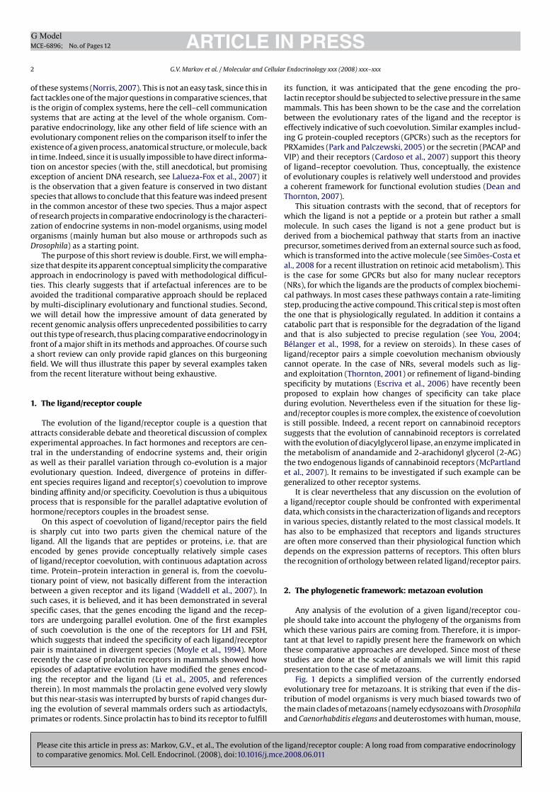
Please cite this article in press as: Markov, G.V., et al., The evolution of the ligand/receptor couple: A long road from comparative endocrinology
to comparative genomics. Mol. Cell. Endocrinol. (2008), doi:10.1016/j.mce.2008.06.011
ARTICLE IN PRESSG Model
MCE-6896; No. of Pages 12
2 G.V. Markov et al. / Molecular and Cellular Endocrinology xxx (2008) xxx–xxx
of these systems (Norris, 2007). This is not an easy task, since this infact tackles one of the major questions in comparative sciences, thatis the origin of complex systems, here the cell–cell communicationsystems that are acting at the level of the whole organism. Com-parative endocrinology, like any other field of life science with anevolutionary component relies on the comparison itself to infer theexistence of a given process, anatomical structure, or molecule, backin time. Indeed, since it is usually impossible to have direct informa-tion on ancestor species (with the, still anecdotical, but promisingexception of ancient DNA research, see Lalueza-Fox et al., 2007) itis the observation that a given feature is conserved in two distantspecies that allows to conclude that this feature was indeed presentin the common ancestor of these two species. Thus a major aspectof research projects in comparative endocrinology is the characteri-zation of endocrine systems in non-model organisms, using modelorganisms (mainly human but also mouse or arthropods such asDrosophila) as a starting point.
The purpose of this short review is double. First, we will empha-size that despite its apparent conceptual simplicity the comparativeapproach in endocrinology is paved with methodological difficul-ties. This clearly suggests that if artefactual inferences are to beavoided the traditional comparative approach should be replacedby multi-disciplinary evolutionary and functional studies. Second,we will detail how the impressive amount of data generated byrecent genomic analysis offers unprecedented possibilities to carryout this type of research, thus placing comparative endocrinology infront of a major shift in its methods and approaches. Of course sucha short review can only provide rapid glances on this burgeoningfield. We will thus illustrate this paper by several examples takenfrom the recent literature without being exhaustive.
1. The ligand/receptor couple
The evolution of the ligand/receptor couple is a question thatattracts considerable debate and theoretical discussion of complexexperimental approaches. In fact hormones and receptors are cen-tral in the understanding of endocrine systems and, their originas well as their parallel variation through co-evolution is a majorevolutionary question. Indeed, divergence of proteins in differ-ent species requires ligand and receptor(s) coevolution to improvebinding affinity and/or specificity. Coevolution is thus a ubiquitousprocess that is responsible for the parallel adaptative evolution ofhormone/receptors couples in the broadest sense.
On this aspect of coevolution of ligand/receptor pairs the fieldis sharply cut into two parts given the chemical nature of theligand. All the ligands that are peptides or proteins, i.e. that areencoded by genes provide conceptually relatively simple casesof ligand/receptor coevolution, with continuous adaptation acrosstime. Protein–protein interaction in general is, from the coevolu-tionary point of view, not basically different from the interactionbetween a given receptor and its ligand (Waddell et al., 2007). Insuch cases, it is believed, and it has been demonstrated in severalspecific cases, that the genes encoding the ligand and the recep-tors are undergoing parallel evolution. One of the first examplesof such coevolution is the one of the receptors for LH and FSH,which suggests that indeed the specificity of each ligand/receptorpair is maintained in divergent species (Moyle et al., 1994). Morerecently the case of prolactin receptors in mammals showed howepisodes of adaptative evolution have modified the genes encod-ing the receptor and the ligand (Li et al., 2005, and referencestherein). In most mammals the prolactin gene evolved very slowlybut this near-stasis was interrupted by bursts of rapid changes dur-ing the evolution of several mammals orders such as artiodactyls,primates or rodents. Since prolactin has to bind its receptor to fulfill
its function, it was anticipated that the gene encoding the pro-lactin receptor should be subjected to selective pressure in the samemammals. This has been shown to be the case and the correlationbetween the evolutionary rates of the ligand and the receptor iseffectively indicative of such coevolution. Similar examples includ-ing G protein-coupled receptors (GPCRs) such as the receptors forPRXamides (Park and Palczewski, 2005) or the secretin (PACAP andVIP) and their receptors (Cardoso et al., 2007) support this theoryof ligand–receptor coevolution. Thus, conceptually, the existenceof evolutionary couples is relatively well understood and providesa coherent framework for functional evolution studies (Dean andThornton, 2007).
This situation contrasts with the second, that of receptors forwhich the ligand is not a peptide or a protein but rather a smallmolecule. In such cases the ligand is not a gene product but isderived from a biochemical pathway that starts from an inactiveprecursor, sometimes derived from an external source such as food,which is transformed into the active molecule (see Simões-Costa etal., 2008 for a recent illustration on retinoic acid metabolism). Thisis the case for some GPCRs but also for many nuclear receptors(NRs), for which the ligands are the products of complex biochemi-cal pathways. In most cases these pathways contain a rate-limitingstep, producing the active compound. This critical step is most oftenthe one that is physiologically regulated. In addition it contains acatabolic part that is responsible for the degradation of the ligandand that is also subjected to precise regulation (see You, 2004;Bélanger et al., 1998, for a review on steroids). In these cases ofligand/receptor pairs a simple coevolution mechanism obviouslycannot operate. In the case of NRs, several models such as lig-and exploitation (Thornton, 2001) or refinement of ligand-bindingspecificity by mutations (Escriva et al., 2006) have recently beenproposed to explain how changes of specificity can take placeduring evolution. Nevertheless even if the situation for these lig-and/receptor couples is more complex, the existence of coevolutionis still possible. Indeed, a recent report on cannabinoid receptorssuggests that the evolution of cannabinoid receptors is correlatedwith the evolution of diacylglycerol lipase, an enzyme implicated inthe metabolism of anandamide and 2-arachidonyl glycerol (2-AG)the two endogenous ligands of cannabinoid receptors (McPartlandet al., 2007). It remains to be investigated if such example can begeneralized to other receptor systems.
It is clear nevertheless that any discussion on the evolution ofa ligand/receptor couple should be confronted with experimentaldata, which consists in the characterization of ligands and receptorsin various species, distantly related to the most classical models. Ithas also to be emphasized that receptors and ligands structuresare often more conserved than their physiological function whichdepends on the expression patterns of receptors. This often blursthe recognition of orthology between related ligand/receptor pairs.
2. The phylogenetic framework: metazoan evolution
Any analysis of the evolution of a given ligand/receptor cou-ple should take into account the phylogeny of the organisms fromwhich these various pairs are coming from. Therefore, it is impor-tant at that level to rapidly present here the framework on whichthese comparative approaches are developed. Since most of thesestudies are done at the scale of animals we will limit this rapidpresentation to the case of metazoans.
Fig. 1 depicts a simplified version of the currently endorsedevolutionary tree for metazoans. It is striking that even if the dis-tribution of model organisms is very much biased towards two ofthe main clades of metazoans (namely ecdysozoans with Drosophila
and Caenorhabditis elegans and deuterostomes with human, mouse,
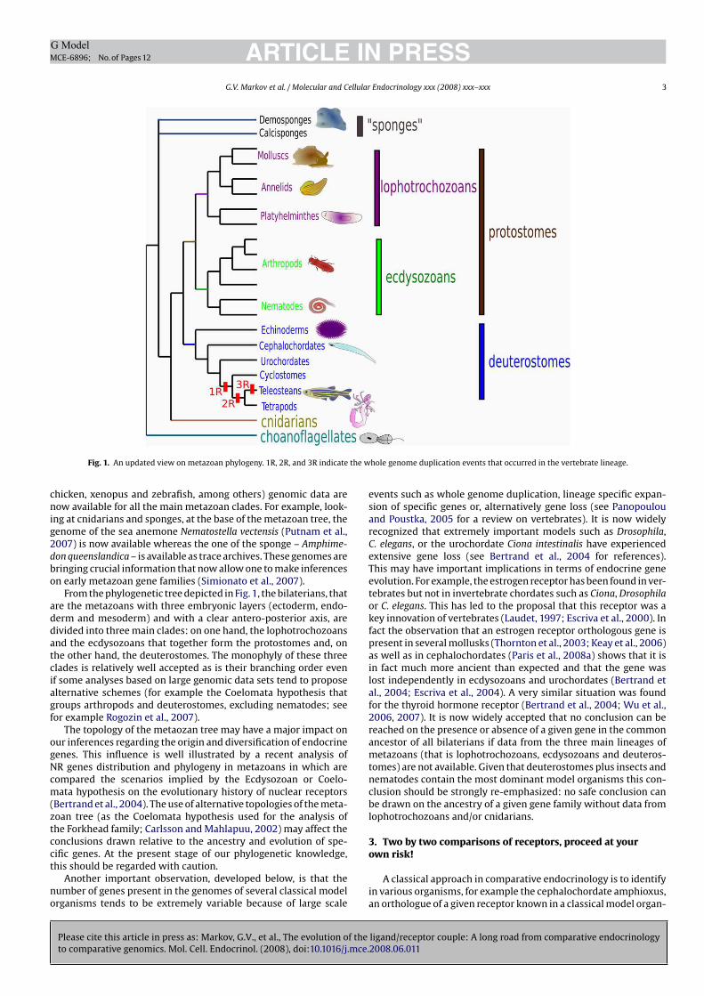
Please cite this article in press as: Markov, G.V., et al., The evolution of the ligand/receptor couple: A long road from comparative endocrinology
to comparative genomics. Mol. Cell. Endocrinol. (2008), doi:10.1016/j.mce.2008.06.011
ARTICLE IN PRESSG Model
MCE-6896; No. of Pages 12
G.V. Markov et al. / Molecular and Cellular Endocrinology xxx (2008) xxx–xxx 3
Fig. 1. An updated view on metazoan phylogeny. 1R, 2R, and 3R indicate the whole genome duplication events that occurred in the vertebrate lineage.
chicken, xenopus and zebrafish, among others) genomic data arenow available for all the main metazoan clades. For example, look-ing at cnidarians and sponges, at the base of the metazoan tree, thegenome of the sea anemone Nematostella vectensis (Putnam et al.,2007) is now available whereas the one of the sponge – Amphime-
don queenslandica – is available as trace archives. These genomes arebringing crucial information that now allow one to make inferenceson early metazoan gene families (Simionato et al., 2007).
From the phylogenetic tree depicted in Fig. 1, the bilaterians, thatare the metazoans with three embryonic layers (ectoderm, endo-derm and mesoderm) and with a clear antero-posterior axis, aredivided into three main clades: on one hand, the lophotrochozoansand the ecdysozoans that together form the protostomes and, onthe other hand, the deuterostomes. The monophyly of these threeclades is relatively well accepted as is their branching order evenif some analyses based on large genomic data sets tend to proposealternative schemes (for example the Coelomata hypothesis thatgroups arthropods and deuterostomes, excluding nematodes; seefor example Rogozin et al., 2007).
The topology of the metaozan tree may have a major impact onour inferences regarding the origin and diversification of endocrinegenes. This influence is well illustrated by a recent analysis ofNR genes distribution and phylogeny in metazoans in which arecompared the scenarios implied by the Ecdysozoan or Coelo-mata hypothesis on the evolutionary history of nuclear receptors(Bertrand et al., 2004). The use of alternative topologies of the meta-zoan tree (as the Coelomata hypothesis used for the analysis ofthe Forkhead family; Carlsson and Mahlapuu, 2002) may affect theconclusions drawn relative to the ancestry and evolution of spe-cific genes. At the present stage of our phylogenetic knowledge,this should be regarded with caution.
Another important observation, developed below, is that thenumber of genes present in the genomes of several classical modelorganisms tends to be extremely variable because of large scale
events such as whole genome duplication, lineage specific expan-sion of specific genes or, alternatively gene loss (see Panopoulouand Poustka, 2005 for a review on vertebrates). It is now widelyrecognized that extremely important models such as Drosophila,C. elegans, or the urochordate Ciona intestinalis have experiencedextensive gene loss (see Bertrand et al., 2004 for references).This may have important implications in terms of endocrine geneevolution. For example, the estrogen receptor has been found in ver-tebrates but not in invertebrate chordates such as Ciona, Drosophila
or C. elegans. This has led to the proposal that this receptor was akey innovation of vertebrates (Laudet, 1997; Escriva et al., 2000). Infact the observation that an estrogen receptor orthologous gene ispresent in several mollusks (Thornton et al., 2003; Keay et al., 2006)as well as in cephalochordates (Paris et al., 2008a) shows that it isin fact much more ancient than expected and that the gene waslost independently in ecdysozoans and urochordates (Bertrand etal., 2004; Escriva et al., 2004). A very similar situation was foundfor the thyroid hormone receptor (Bertrand et al., 2004; Wu et al.,2006, 2007). It is now widely accepted that no conclusion can bereached on the presence or absence of a given gene in the commonancestor of all bilaterians if data from the three main lineages ofmetazoans (that is lophotrochozoans, ecdysozoans and deuteros-tomes) are not available. Given that deuterostomes plus insects andnematodes contain the most dominant model organisms this con-clusion should be strongly re-emphasized: no safe conclusion canbe drawn on the ancestry of a given gene family without data fromlophotrochozoans and/or cnidarians.
3. Two by two comparisons of receptors, proceed at your
own risk!
A classical approach in comparative endocrinology is to identifyin various organisms, for example the cephalochordate amphioxus,an orthologue of a given receptor known in a classical model organ-
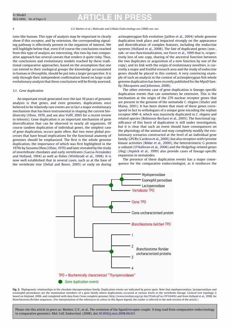
Please cite this article in press as: Markov, G.V., et al., The evolution of the ligand/receptor couple: A long road from comparative endocrinology
to comparative genomics. Mol. Cell. Endocrinol. (2008), doi:10.1016/j.mce.2008.06.011
ARTICLE IN PRESSG Model
MCE-6896; No. of Pages 12
4 G.V. Markov et al. / Molecular and Cellular Endocrinology xxx (2008) xxx–xxx
isms like human. This type of analysis may be important to clearlyshow if this receptor, and by extension, the corresponding signal-ing pathway is effectively present in the organism of interest. Wewill highlight below that, even if of course the conclusions reachedfrom this type of analysis are interesting, this two-by-two compar-ative approach has several caveats that renders it quite risky. Thus,the conclusions and evolutionary models reached by these tradi-tional comparative approaches, based on the assumption that onecan extend to their zoological groups the knowledge accumulatedin human or Drosophila, should be put into a larger perspective. It isonly through their independent confirmation based on large-scaleevolutionary analysis that these conclusions will be firmly assessed.
3.1. Gene duplication
An important result generated over the last 10 years of genomicanalysis is that genes, and even genomes, duplications oncebelieved to be relatively rare events are in fact a major evolutionarymechanism that has been instrumental in shaping the current bio-diversity (Ohno, 1970, and see also Volff, 2005 for a recent reviewin teleosts). Gene duplication is an important mechanism of genediversification that can be observed in nearly all organisms. Ofcourse tandem duplication of individual genes, the simplest caseof gene duplication, occurs quite often. But two more global pro-cesses that have broad implications for the functional anatomy ofgenomes should be emphasized. The first is the whole genomeduplication, the importance of which was first highlighted in the1970s by Susumu Ohno (Ohno, 1970) and later revealed by the studyof invertebrate chordates and early vertebrates (Garcia-Fernándezand Holland, 1994) as well as fishes (Wittbrodt et al., 1998). It isnow well established that in several cases, such as at the base ofthe vertebrate tree (Dehal and Boore, 2005) or early on during
actinopterygian fish evolution (Jaillon et al., 2004) whole genomeduplication took place and impacted strongly on the appearanceand diversification of complex features, including the endocrinesystems (Holland et al., 2008). The fate of duplicated genes (non-,sub- or neo-functionalization, see Force et al., 1999 that is, respec-tively loss of one copy, sharing of the ancestral function betweenthe two duplicates or acquisition of a new function by one of thecopy), and its link with the origin of evolutionary novelties, is cur-rently a major and fruitful research area and the study of endocrinegenes should be placed in this context. A very convincing exam-ple of such an analysis in the context of actinopterygian fish wholegenome duplication has been recently published for the MyoD fam-ily (Macqueen and Johnston, 2008).
The other extreme case of gene duplication is lineage-specificduplication events that can sometimes be extensive. This is themechanism at the origin of the 270 nuclear receptor genes thatare present in the genome of the nematode C. elegans (Sluder andMaina, 2001). It has been shown that most of these genes corre-spond in fact to orthologues of a unique gene encoding the orphanreceptor HNF-4, which was massively duplicated in C. elegans andrelated species (Robinson-Rechavi et al., 2005). The functional sig-nificance of this burst of duplication is still under investigation,but it is clear that such an event should have consequences onthe physiology of the animal and may completely modify the evo-lutionary scenarios constructed at the level of an individual genefamily. GPCRs (Cardoso et al., 2006), but also receptors with tyrosinekinase activities (Rikke et al., 2000), the heterotrimeric G proteina-subunit (O’Halloran et al., 2006) and the Hedgehog-related genes(Hog) (Aspöck et al., 1999) also provide cases of lineage-specificexpansion in nematodes.
The presence of these duplication events has a major conse-quence for the comparative endocrinologist, as it reinforces the
Fig. 2. Phylogenetic relationships in the chordate thyroperoxidase family. Duplication events are indicated by green spots. Note that myeloperoxidase, lactoperoxidase and
eosinophil peroxidases are the mammalian members of a gene family where duplications occurred at various levels in the vertebrate lineage. General tree topology is
based on Heyland, 2006, and completed with data from Ciona complete genome (http://www.treefam.org/cgi-bin/TFinfo.pl?ac=TF314416) and from Holland et al., 2008, for
Branchiostoma floridae sequences. (For interpretation of the references to colour in this figure legend, the reader is referred to the web version of the article.)
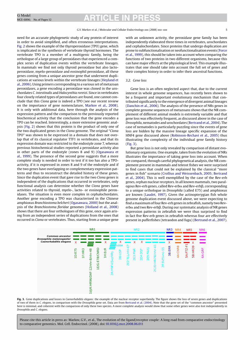
Please cite this article in press as: Markov, G.V., et al., The evolution of the ligand/receptor couple: A long road from comparative endocrinology
to comparative genomics. Mol. Cell. Endocrinol. (2008), doi:10.1016/j.mce.2008.06.011
ARTICLE IN PRESSG Model
MCE-6896; No. of Pages 12
G.V. Markov et al. / Molecular and Cellular Endocrinology xxx (2008) xxx–xxx 5
need for an accurate phylogenetic study of any protein of interestin order to avoid simplified, and often erroneous interpretations.Fig. 2 shows the example of the thyroperoxidase (TPO) gene, whichis implicated in the synthesis of vertebrate thyroid hormones. Thevertebrate TPO is a member of a multigenic family, being theorthologue of a large group of peroxidases that experienced a com-plex series of duplication events within the vertebrate lineages.In mammals we find not only thyroid peroxidase but also lacto-peroxidase, myelo-peroxidase and eosinophil peroxidase, all thesegenes coming from a unique ancestor gene that underwent dupli-cations at various levels within the vertebrate lineages (Heyland etal., 2006). Using primers corresponding to a various set of metazoanperoxidases, a gene encoding a peroxidase was cloned in the uro-chordates C. intestinalis and Halocynthia roretzi. Since in vertebratesfour closely related types of peroxidases are found, one cannot con-clude that this Ciona gene is indeed a TPO (see our recent reviewon the importance of gene nomenclature, Markov et al., 2008).It is only with additional data, here through the analysis of theexpression pattern and the comparison to the previously reportedbiochemical activity that the conclusion that the gene encodes aTPO can be reached. Interestingly, a more extensive genomic anal-ysis (Fig. 2) shows that this protein is the product of only one ofthe two duplicated genes in the Ciona genome. The original “Ciona
TPO” was shown to be expressed in a domain that does not over-lap that of its classical regulator TTF1 in vertebrates, and that itsexpression domain was restricted to the endostyle zone 7, whereasprevious histochemical studies reported a peroxidase activity alsoin other parts of the endostyle (zones 8 and 9) (Ogasawara etal., 1999). The presence of the second gene suggests that a morecomplete study is needed in order to test if it too has also a TPO-activity, if it is expressed in zones 8 and 9 of the endostyle and ifthe two genes have overlapping or complementary expression pat-terns and thus to reconstruct the detailed history of these genes.Since the duplication event that gave rise to the two Ciona genes isindependent of the duplications that occurred in vertebrates, onlyfunctional analysis can determine whether the Ciona genes haveactivities related to thyroid, myelo-, lacto- or eosinophile perox-idases. The situation is even more complex in cephalochordates.Another gene encoding a TPO was characterised in the Chineseamphioxus Branchiostoma belcheri (Ogasawara, 2000) but the anal-ysis of the Branchiostoma floridae genomes (Holland et al., 2008)shows that there are four orthologues of this gene, once again aris-ing from an independent series of duplications from the ones thatoccurred in Ciona or vertebrates. Thus, starting from a unique gene
with an unknown activity, the peroxidase gene family has beenindependently elaborated three times in vertebrates, urochordatesand cephalochordates. Since proteins that undergo duplication areprone to subfunctionalisation or neofunctionalisation events (Forceet al., 1999), this should be taken into account when comparing thefunctions of two proteins in two different organisms, because thiscan have major effects at the physiological level. This example illus-trates that one should take into account the full set of genes andtheir complex history in order to infer their ancestral functions.
3.2. Gene loss
Gene loss is an often neglected aspect that, due to the currentinterest in whole genome sequences, has recently been shown tobe a frequent and important evolutionary mechanism that con-tributed significantly to the emergence of divergent animal lineages(Danchin et al., 2006). The analysis of the presence of NRs genes incomplete genome sequences of metazoans shows that the NR com-plement of different animal models is extremely variable and thatgene loss was effectively frequent, as discussed above in the case ofDrosophila, nematodes and urochordates (Bertrand et al., 2004). Thecase of nematodes is particularly puzzling since the events of geneloss are hidden by the massive lineage specific expansion of theHNF4 gene discussed above (Robinson-Rechavi et al., 2005) thusillustrating the complexity of the individual gene family history(Fig. 3).
But gene loss is not only revealed by comparison of distant evo-lutionary organisms. One example, taken from the evolution of NRsillustrates the importance of taking gene loss into account. Whenwe compared, through careful phylogenetical analysis, the NR com-plement present in mammals and teleost fishes we were surprisedto find cases that could not be explained by the classical “moregenes in fish” scenario (Crollius and Weissenbach, 2005; Bertrandet al., 2004). This is well exemplified by the case of the Rev-erbgenes, orphan nuclear receptors. In all known mammals, two paral-ogous Rev-erb genes, called Rev-erb� and Rev-erb�, correspondingto a unique orthologue in Drosophila (called E75) and amphioxusare known (Laudet, 1997). Given the actinopterygian fish wholegenome duplication event discussed above, we were expecting tofind a maximum of four Rev-erb genes in zebrafish, namely two Rev-erb� and two Rev-erb�. During our systematic analysis of NR genesexpression patterns in zebrafish we were thus surprised to findin fact five Rev-erb genes in zebrafish whereas four are effectivelypresent in pufferfishes (tetraodon and fugu) (Bertrand et al., 2007).
Fig. 3. Gene duplications and losses in Caenorhabditis elegans: the example of the nuclear receptor superfamily. The figure shows the loss of seven genes and duplications
of two of them in C. elegans, in comparison with the Drosophila gene set. Data are from Bertrand et al. (2004). Note that the gene set of the “common ancestor” presented
here is minimal, and coherent with the comparison of only these two species. A more complete analysis would show that some other genes were also lost independently in
Drosophila and C. elegans.
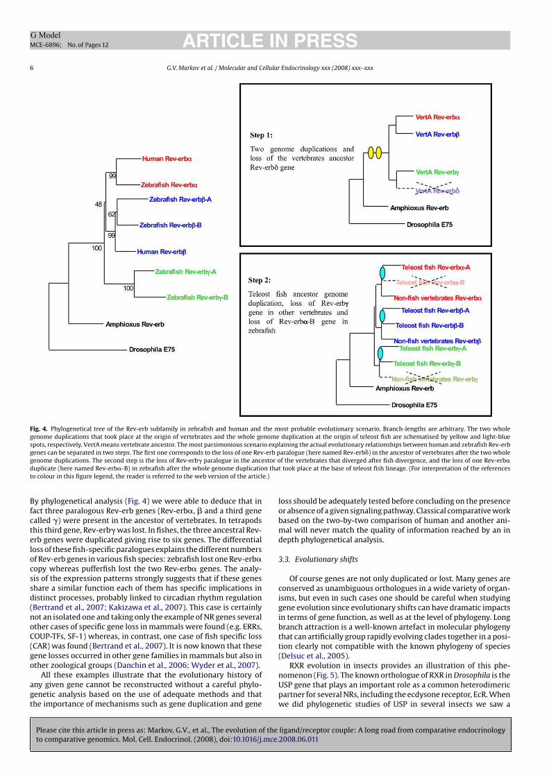
Please cite this article in press as: Markov, G.V., et al., The evolution of the ligand/receptor couple: A long road from comparative endocrinology
to comparative genomics. Mol. Cell. Endocrinol. (2008), doi:10.1016/j.mce.2008.06.011
ARTICLE IN PRESSG Model
MCE-6896; No. of Pages 12
6 G.V. Markov et al. / Molecular and Cellular Endocrinology xxx (2008) xxx–xxx
Fig. 4. Phylogenetical tree of the Rev-erb subfamily in zebrafish and human and the most probable evolutionary scenario. Branch-lengths are arbitrary. The two whole
genome duplications that took place at the origin of vertebrates and the whole genome duplication at the origin of teleost fish are schematised by yellow and light-blue
spots, respectively. VertA means vertebrate ancestor. The most parsimonious scenario explaining the actual evolutionary relationships between human and zebrafish Rev-erb
genes can be separated in two steps. The first one corresponds to the loss of one Rev-erb paralogue (here named Rev-erb�) in the ancestor of vertebrates after the two whole
genome duplications. The second step is the loss of Rev-erb� paralogue in the ancestor of the vertebrates that diverged after fish divergence, and the loss of one Rev-erb�duplicate (here named Rev-erb�-B) in zebrafish after the whole genome duplication that took place at the base of teleost fish lineage. (For interpretation of the references
to colour in this figure legend, the reader is referred to the web version of the article.)
By phylogenetical analysis (Fig. 4) we were able to deduce that infact three paralogous Rev-erb genes (Rev-erb�, � and a third genecalled �) were present in the ancestor of vertebrates. In tetrapodsthis third gene, Rev-erb� was lost. In fishes, the three ancestral Rev-erb genes were duplicated giving rise to six genes. The differentialloss of these fish-specific paralogues explains the different numbersof Rev-erb genes in various fish species: zebrafish lost one Rev-erb�copy whereas pufferfish lost the two Rev-erb� genes. The analy-sis of the expression patterns strongly suggests that if these genesshare a similar function each of them has specific implications indistinct processes, probably linked to circadian rhythm regulation(Bertrand et al., 2007; Kakizawa et al., 2007). This case is certainlynot an isolated one and taking only the example of NR genes severalother cases of specific gene loss in mammals were found (e.g. ERRs,COUP-TFs, SF-1) whereas, in contrast, one case of fish specific loss(CAR) was found (Bertrand et al., 2007). It is now known that thesegene losses occurred in other gene families in mammals but also inother zoological groups (Danchin et al., 2006; Wyder et al., 2007).
All these examples illustrate that the evolutionary history ofany given gene cannot be reconstructed without a careful phylo-genetic analysis based on the use of adequate methods and thatthe importance of mechanisms such as gene duplication and gene
loss should be adequately tested before concluding on the presenceor absence of a given signaling pathway. Classical comparative workbased on the two-by-two comparison of human and another ani-mal will never match the quality of information reached by an indepth phylogenetical analysis.
3.3. Evolutionary shifts
Of course genes are not only duplicated or lost. Many genes areconserved as unambiguous orthologues in a wide variety of organ-isms, but even in such cases one should be careful when studyinggene evolution since evolutionary shifts can have dramatic impactsin terms of gene function, as well as at the level of phylogeny. Longbranch attraction is a well-known artefact in molecular phylogenythat can artificially group rapidly evolving clades together in a posi-tion clearly not compatible with the known phylogeny of species(Delsuc et al., 2005).
RXR evolution in insects provides an illustration of this phe-nomenon (Fig. 5). The known orthologue of RXR in Drosophila is theUSP gene that plays an important role as a common heterodimericpartner for several NRs, including the ecdysone receptor, EcR. Whenwe did phylogenetic studies of USP in several insects we saw a
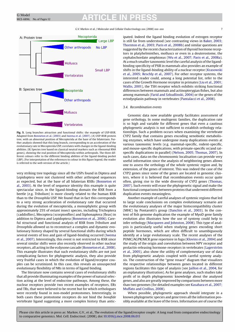
Please cite this article in press as: Markov, G.V., et al., The evolution of the ligand/receptor couple: A long road from comparative endocrinology
to comparative genomics. Mol. Cell. Endocrinol. (2008), doi:10.1016/j.mce.2008.06.011
ARTICLE IN PRESSG Model
MCE-6896; No. of Pages 12
G.V. Markov et al. / Molecular and Cellular Endocrinology xxx (2008) xxx–xxx 7
Fig. 5. Long branches attraction and functional shifts: the example of USP-RXR.
Adapted from Bonneton et al. (2003) and Iwema et al. (2007). (A) USP-RXR protein
tree, with an abnormal position of Mecopterida at the base of the bilaterians. Fur-
ther analysis showed that this long branch, corresponding to an acceleration of the
evolutionary rate in Mecopterida USP, correlates with changes in the ligand-binding
abilities. (B) Species tree based on classical neutral markers such as ribosomal RNA
genes), showing the real position of Mecopterida within arthropods. The three dif-
ferent colours refer to the different binding abilities of the ligand-binding pocket
(LBP). (For interpretation of the references to colour in this figure legend, the reader
is referred to the web version of the article.)
very striking tree topology since all the USPs found in Diptera andLepidoptera were not clustered with other arthropod sequencesas expected, but at the base of all bilaterian RXRs (Bonneton etal., 2003). At the level of sequence identity this example is quitespectacular since, in the ligand-binding domain the RXR from abeetle (e.g. Tribolium) is more closely related to the human RXRthan to the Drosophila USP. We found that in fact this correspondsto a very strong acceleration of evolutionary rate that occurredduring the evolution of mecopterida, a monophyletic group withapproximately 25% of extant insect species, including Trichoptera(caddisflies), Mecoptera (scorpionflies) and Siphonaptera (fleas) inaddition to Diptera and Lepidoptera (Bonneton et al., 2006). Care-ful structural and functional analysis of RXR from Tribolium andDrosophila allowed us to reconstruct a complex and dynamic evo-lutionary history shaped by several functional shifts during whichseveral events of loss and gain of ligand-binding occurred (Iwemaet al., 2007). Interestingly, this event is not restricted to RXR sinceseveral similar shifts were also recently observed in other nuclearreceptors, all acting in the ecdysone cascade (Bonneton et al., 2008).This example illustrates that such evolutionary shifts are not justcomplicating factors for phylogenetic analysis, they also providevery fruitful cases in which the evolution of ligand/receptor cou-ples can be scrutinized. In this case, this reveals an unsuspectedevolutionary flexibility of NRs in terms of ligand binding.
The literature now contains several cases of evolutionary shiftsthat all provide illuminating examples of the power of natural selec-tion acting at the level of endocrine pathways. Among these, thenuclear receptors provide two recent examples of receptors, ERsand TRs, that were believed to be recent but for which orthologueswere recently found in non-model protostomes. Interestingly, inboth cases these protostome receptors do not bind the bonafide
vertebrate ligand suggesting a more complex history than antic-
ipated. Indeed the ligand binding evolution of estrogen receptoris still far from understood (see contrasting views in Baker, 2003;Thornton et al., 2003; Paris et al., 2008b) and similar questions aresuggested by the recent characterization of thyroid hormone recep-tors in platyhelminthes, molluscs or even in a deuterostome, thecephalochordate amphioxus (Wu et al., 2007; Paris et al., 2008a).At a much smaller taxonomic level the careful analysis of the ligand-binding specificity of PXR in mammals also provides an example ofshifts in the ligand-binding ability of a nuclear receptor (Krasowskiet al., 2005; Reschly et al., 2007). For other receptor systems, theinterested reader could, among a long potential list, refer to thecases of the Growth Hormone receptor in primates (Liu et al., 2001;Wallis, 2001), the TSH receptor which exhibits striking functionaldifferences between mammals and actinopterygian fishes, but alsoamong mammals (Farid and Szkudlinski, 2004) or the genes of theectodysplasin pathway in vertebrates (Pantalacci et al., 2008).
3.4. Recombination events
Genomic data now available greatly facilitates assessment ofgene orthology. In some multigenic families, the duplication rateis so high and variable for different genes that even a cautiousphylogenetic analysis is not sufficient to establish orthology rela-tionships. Such a problem occurs when examining the vertebrateCYP2 family that contains genes encoding xenobiotic metabolis-ing enzymes, which have undergone many duplications events atvarious taxonomic levels (e.g. mammal-specific, rodent-specific,and mouse-specific duplications, with primate-specific or/and rat-specific duplications in parallel) (Nelson, 2005; Thomas, 2007). Insuch cases, data on the chromosomic localisation can provide veryuseful information since the analysis of neighboring genes allowsone to assess the orthology of the whole syntenic region and, byextension, of the genes of interest. This was indeed the case of theCYP2 genes since some of the genes are located in genomic clus-ters, where it is believed that recombination events occur quiteoften, giving rise to the wide diversity of CYP2 genes (Thomas,2007). Such events will erase the phylogenetic signal and make thefunctional comparisons between proteins that underwent differentduplication events meaningless.
Another example of careful analyses of syntenic regions that ledto large scale conclusions on complex evolutionary scenario arethe evolutionary analyses of the major histocompatibility cluster(MHC) by Pontarotti’s group (Danchin et al., 2003). In the con-text of fish genome duplication the example of MyoD gene familyevolution also illustrates how the use of synteny could help toinfer orthology (Macqueen and Johnston, 2008). This type of anal-ysis is particularly useful when studying genes encoding shortpeptide hormones, which are often difficult to unambiguouslyidentify at a large evolutionary scale. The recent analyses of thePOMC/AGPR/MCR gene repertoire in fugu (Klovins et al., 2004) andthe study of the origin and coevolution between NPY receptor andprolactin-releasing hormone-receptors in vertebrates (Lagerströmet al., 2005), also show the utility of complementary use of datafrom phylogenetic analysis coupled with careful synteny analy-sis. The construction of the “gene rosace” diagram that visualizesthe respective relationships between genes located in differentregions facilitates this type of analysis (see Jaillon et al., 2004, foran explanatory illustration). As for gene analyses, such studies takeprofit of in depth phylogenomic knowledge about the analysedgenes, and also are greatly improved by comparisons between morethan two genomes (for detailed examples see Kasahara et al., 2007;Muffato and Crollius, 2008).
When possible, phylogenetic approach should integrate in aknown phylogenetic species and gene trees all the information pos-sibly available at the leave of the trees. Information are of course the
Please cite this article in press as: Markov, G.V., et al., The evolution of the ligand/receptor couple: A long road from comparative endocrinology
to comparative genomics. Mol. Cell. Endocrinol. (2008), doi:10.1016/j.mce.2008.06.011
ARTICLE IN PRESSG Model
MCE-6896; No. of Pages 12
8 G.V. Markov et al. / Molecular and Cellular Endocrinology xxx (2008) xxx–xxx
sequences but also functional information such as binding proper-ties and other physiological data. In some cases it is possible tocalculate the probability to have this information at the node of thetree and from the information available at the node propagate dawnit to the non-annotated leaves (see Thornton, 2004, for a detailedreview). But even the finest probabilistic approaches are useless ifthe sampling is inappropriate.
4. The elusive ligands
The previous examples may give the impression that working onthe evolution of receptor genes is full of obstacles that complicateanalysis and interpretation. Thus, one solution to study evolutionof a specific endocrine signaling pathway could be to test directlyif the ligand of interest is present in a wide variety of organisms.This attractive solution is nevertheless also paved with obstaclesthat are not easily solved and for which a comparative genomicsapproach does not always provide clues. The two main lines of evi-dence for the existence of a given ligand in a given animal are (i) thedetection of an effect of this ligand in a given physiological or devel-opmental process, most often considered from the known effect ofthis ligand in mammals and (ii) the direct detection of the ligandthrough genomic or analytical chemistry analysis.
4.1. Effects
The rationale behind these studies, which are commonly donein traditional comparative endocrinology is that if a given moleculehas an effect in a given species this should be taken as a clear indi-cation that there is a receptor for this molecule and thus a signalingpathway and a “physiological role”. Somehow mirroring the orig-inal receptor definition by Paul Ehrlich, for which a receptor wasdefined as any molecule that is able to bind exogenous elements inhuman cells (reviewed in Prüll, 2003), people tend to consider thatany molecule of human origin acting on a living system is effec-tively proof that this molecule binds to another one, which can becalled a “receptor”. But even if this assumption is true, it cannot beused as an indication that this “receptor” is related in anyway tothe human receptor for this molecule. The problem is that manyauthors have applied this concept without taking into account thisimportant caveat.
A striking example is the case of the arboreal mycorrhizes fungiof the order Glomerales, such as Glomus intraradices, that live insymbiosis with many trees and play crucial roles at the root/soilinterface. It has been shown that the roots of the plant have a pos-itive effect in stimulating the growth of the fungal hyphe duringpre-symbiosis steps (see Requena et al., 2007, for a review). Theprecise mechanism how plant signals are perceived by the fungi isstill unknown but it has been shown that flavonoids have a strongchimio-tactic effect and that this effect can be mimicked by estro-gens and blocked by antiestrogens (Poulin et al., 1997; Scervinoet al., 2005). This was taken as an indication that the genomeof these fungi contained an estrogen receptor and that a spe-cific receptor/interacting protein with a binding site for flavonoidor structurally related compounds (estrogens and antiestrogens)exists in these fungi (Requena et al., 2007; Poulin et al., 1997;Catford et al., 2006). However, our current knowledge of NR evolu-tion indicates that the genome of Glomus (which is currently beingsequenced) should be devoid of NR genes (Escriva et al., 1997).Among the possible candidates for mediating the effect of estro-gens in Glomus, one of the most probable would be an orthologueof the estrogen binding protein already found in other fungi, includ-ing Candida albicans (Madani et al., 1994; Cheng et al., 2006). Thisprotein, well known under the name of “Old yellow enzyme” is
one of the most ancient enzymatic systems characterized, and isa cytoplasmic oxydoreductase whose activity can be regulated byestrogen binding (Cheng et al., 2006 and references therein). Thisexample demonstrates that whereas there is an effect of estro-gen in this species, this could not be taken as an indication thatthe endogenous receptor is in any way evolutionary related to thenuclear estrogen receptor found in human.
RXRs provide here again an example of the difficulties associatedwith the identification of endogenous ligands. These receptors werefirst described in the early 1990s as orphan receptors whose activ-ity could be modulated by all-trans-retinoic acid (see Laudet andGronemeyer, 2005, for references). It was later shown that 9-cis-retinoic acid, an isomeric derivative of all-trans-retinoic acid, is ableto bind with a high affinity to RXR. Indeed several rexinoids, specificligands of RXRs, were developed as pharmacological compoundsfor treatment of diseases such as diabetes and insulin resistance(Pinaire and Reifel-Miller, 2007). Ironically the detection of 9-cis-retinoic acid in vivo has proven to be difficult, casting doubts on thein vivo relevance of this ligand (see Mic et al., 2003). This has beenrecently confirmed by genetic evidence in the mouse (Calléja et al.,2006). It is now thought that RXR acts rather as a fatty acid or fattyacid derivative sensor, although the exact identity and relevance ofits ligand(s) remains a matter of debate (see references in Iwemaet al., 2007). This example depicts that even in human, it is notbecause a molecule has an effect that it indicates its endogenousexistence in a physiologically relevant manner!
These examples may appear artificial, but they illustrate a typeof assumption that has been commonly used in some comparativeendocrinology studies. The estrogen receptor of several molluscssuch as Aplysia, Octopus, and others have been cloned and con-vincingly shown to be unable to bind to estrogens (Thornton etal., 2003; Keay et al., 2006; Kajiwara et al., 2006; Matsumoto etal., 2007; Bannister et al., 2007). Nevertheless, effects of estrogensare reported in these animals (e.g. sex reversal in several molluscspecies) and this is taken as evidence in favor of the existence of aclassical nuclear estrogen receptor (see Lafont and Mathieu, 2007for references). However, the actual data suggests that if such areceptor exists it is not the unique ER orthologue already charac-terized. Among the several possible explanations we propose that:(i) the molecule used for the treatment is metabolized to anothercompound that is the active one. Indeed we recently discoveredsuch a situation in amphioxus, where thyroid hormones are metab-olized to a compound that binds the TR (Paris et al., 2008a); (ii) a NRthat binds estrogen exists but this is not an orthologue of ER; (iii)the effect of estrogen is mediated by another receptor system, e.g. atransmembrane receptor (see Revankar et al., 2005 for an example)or a cytoplasmic protein such as in the case of Glomus discussedabove. Thus one should treat claims of the existence of a signal-ing pathway based on treatment with human derived compoundwith much caution. Such data provide indications, but only indirectevidence that should be verified by more functional approachesincluding the cloning and characterisation of the relevant receptor.
4.2. Detection
Detecting a given ligand in one’s favorite non-classical modelorganism is probably an excellent option for a comparative endocri-nologist. One interesting case to illustrate the complexity of ligandevolution in different organisms is the one of the lamprey steroidhormones. For a long time, it was thought that as a vertebrate, lam-prey should produce the classical steroids, estradiol, testosterone,and progesterone, and indeed, these were effectively identifiedby radioimmunoassays (RIA) and used for physiological (for anexample see Bolduc and Sower, 1992) or functional (Thornton,2001) studies. Nevertheless, in the 1980s, chromatographic tech-
Please cite this article in press as: Markov, G.V., et al., The evolution of the ligand/receptor couple: A long road from comparative endocrinology
to comparative genomics. Mol. Cell. Endocrinol. (2008), doi:10.1016/j.mce.2008.06.011
ARTICLE IN PRESSG Model
MCE-6896; No. of Pages 12
G.V. Markov et al. / Molecular and Cellular Endocrinology xxx (2008) xxx–xxx 9
niques and more recently, blood steroid profiles analyzed byhigh performance liquid chromatography (HPLC), contradicted thisview and showed that the main circulating steroids are 15-alpha-hydroxylated (Kime and Callard, 1982; Lowartz et al., 2003). Theactual nature of the active steroid hormones and their mechanismof action in lamprey are still a matter of debate (reviewed in Bryanet al., 2008). This example shows that the diversity of ligands innon-model species is often underestimated. In this case the pres-ence of the classical steroids is not questioned, but they are not thephysiologically relevant compounds.
Many reports have shown the presence of a typical human hor-mone in early vertebrates or even in invertebrates, steroids beingthe compounds for which the number of such reports is the high-est. Most often the detection of these products is based on the useof antibodies, generated from human compounds and widely usedon human tissue samples for medical application. These antibodiesare mainly used for immunohistochemical staining and/or radioim-munoassay and two recent examples illustrate the caveats anduncertainties of such approaches. We have nevertheless to stronglyinsist on the fact that these examples were not selected becausethey are particularly dubious but rather because they illustratenumerous studies done in classical comparative endocrinology.
The first example concerns the detection of immunoreactiv-ity for progesterone in the giant Rohde cells of the amphioxusBranchiostoma belcheri (Takeda et al., 2003). Using rabbit poly-clonal antibodies against progesterone conjugated to bovine serumalbumin from two commercial suppliers, the authors performedimmunohistochemical staining on adult amphioxus and showstaining in giant neurons known as the Rohde cells. The authorscautiously conclude that “progesterone-like” substances are likelyto be present in these neurons, suggesting the existence of neu-rosteroids in this species. They performed a number of controls toavoid problems due to non-specific binding: they replaced the pri-mary antibody by normal rabbit serum, they omitted the primaryantibody from the staining reaction and they used PBS instead ofthe primary antibody. Furthermore, they also performed absorp-tion tests with the antibodies, progesterone, and BSA, respectively,corroborating the fact that the staining is specific. The main prob-lem with this type of paper is that we have no information on theactual specificity of the antibodies. Are these reagents able to recog-nize only progesterone itself or do they show cross-reactivity withother closely related molecules such as pregnenolone, deoxycor-ticosterone or 17�-hydroxyprogesterone? What if the amphioxuscontains no progesterone but related steroids, such as those foundin lamprey? The authors of this study were cautious and spokeof “progesterone-like substances”. It is interesting to note that theamphioxus genome contains several, but not all the enzymes impli-cated in steroidogenesis (e.g. a CYP21 orthologue is missing) andthat several of these enzymes are duplicated (e.g. 3�-HSD for whichsix genes were found in amphioxus, compared to three in humanand the SDR family that contains enzymes with 17�-HSD and 11�-HSD activities for which 31 genes are known in human for morethan 100 in amphioxus) (Holland et al., 2008). However, only onesteroid receptor, located at the base of the vertebrate GR, MR, PRand AR, is present in the amphioxus genome, and, to date, noth-ing is known about its specificity. Thus it would not be surprisingif amphioxus steroidogenesis has been elaborated independentlyfrom vertebrate steroidogenesis, and searching for human com-pounds in amphioxus may simply be unfruitful.
The second example concerns the presence of estrogens in mol-luscs, and more precisely in the cephalopod Octopus vulgaris, aninteresting case since it is much referred to in the debate on theligand-binding evolution of estrogen receptors (Thornton et al.,2003; Keay et al., 2006; Paris et al., 2008b). It has been shownthat, in this species, 17�-estradiol and progesterone are found in
oviduct and ovarian tissues, and that the concentration of these hor-mones in females correlates with phases of the reproductive cycle(Di Cosmo et al., 2001; D’Aniello et al., 1996). The doses of hormonesin these tissues are measured using a radioimmunoassay and we arefaced with the same uncertainties about the specificity of the detec-tion method in this study as in the previous one. In addition, thelevel of hormones detected both at the level of radioimmunoassayor by HPLC is very close to the lower limits of detection (D’Anielloet al., 1996). From all the data accumulated in Octopus, one can con-clude that steroid hormone metabolism does exist in this species,but it is difficult to be more conclusive on the precise identity ofthe steroids that will be found (see below).
These two examples emphasize the difficulties associated withdetection based on the use of antibodies. In fact, before the genomeera the same situation was found for NR and many reports claimedfor the detection of steroid receptors in a wide variety of non-metazoans organisms such as plants or fungi (see Agarwal et al.,1994; Milanesi et al., 2001; Milanesi and Boland, 2006). Despite thefact that, as in the above mentioned studies, all the controls werecorrect, all these studies displayed artefacts: there is no gene relatedto steroid receptors outside metazoans (Escriva et al., 1997, 2004).We believe that basically the same situation holds for the immuno-logical detection of NR ligands in invertebrates: perhaps some ofthese reports are correct but in the absence of more firm evidencethis cannot be confirmed (Lafont and Mathieu, 2007). Thus the useof antibodies as an evolutionary probe, even if this is attractivebecause these experiments are technically easy to perform, shouldbe avoided.
4.3. Other lines of evidence
Other methods of analysis for the presence of specific com-pounds can be used. This is for example the ability of a given tissueto metabolize a given compound, i.e. to carry out a given reaction(e.g. hydroxylation at the position 3� indicative of a 3�-HSD activitysee Di Cosmo et al., 2001). Once again the problem in these experi-ments is the conclusions that can reasonably be drawn. That a giventype of activity exists in vivo is interesting but given the diversityof steroidogenic enzymes (or enzymes using different substrates)in metazoans one cannot take for granted that because a reactionoccurs with a given labelled intermediate it actually uses this verysubstrate in vivo.
Another approach often used is searching for the presence ofthe gene encoding an enzyme responsible for a critical step inthe synthesis pathway of a given ligand. For example the exis-tence of an aromatase in amphioxus (Castro et al., 2005) arguesfor the existence of estrogens in this species. This is convincingin this particular case since the phylogeny of the isolated clonewas carefully assessed including synteny analysis. This is muchless clear in other cases, such as the recent description of putativeCYP11A and CYP17 in amphioxus (Mizuta and Kubokawa, 2007).The phylogeny of these clones, taking into account a more exhaus-tive dataset, with sequences from non-chordate species, shows thatthe phylogenetical inferences of this report were inexact and thatin both cases, the cloned gene are orthologous to a new group ofdeuterostome CYPs, that was lost in vertebrates (Markov et al., inpreparation).
Our critical arguments may well in turn be criticised as, weagree, it is easy to operate previous publications without offer-ing positive alternatives. It seems clear that the best studies arethose done with careful analytical chemistry methods. For exampleHPLC coupled with mass spectrometry allows the clear detectionand identification of compounds and offers a very strong and rigor-ous alternative (Bridgham et al., 2006 see also Lafont and Mathieu,2007 for a similar conclusion). The problem with this method is
Please cite this article in press as: Markov, G.V., et al., The evolution of the ligand/receptor couple: A long road from comparative endocrinology
to comparative genomics. Mol. Cell. Endocrinol. (2008), doi:10.1016/j.mce.2008.06.011
ARTICLE IN PRESSG Model
MCE-6896; No. of Pages 12
10 G.V. Markov et al. / Molecular and Cellular Endocrinology xxx (2008) xxx–xxx
of course that it is much more demanding and expensive thanantibody detection. But they have the unique advantage of unam-biguously assessing the presence and diversity of the compoundsfound in a given organism. The recent characterization of steroidspresent in the nematode C. elegans provides an interesting exampleof the variety of compounds that can be revealed through such adetailed analysis (Motola et al., 2006).
5. Conclusion—evolutionary vs. comparative approaches:
an evolutionary shift for comparative endocrinology
Most often, endocrinology studies are realized with a long-termmedical objective. This leads to a deformation of the evolution-ary perspective, which may be too much “human-centered”. Doingcomparative endocrinology should therefore first be accompaniedby an effort to go again this natural anthropocentric view and toconsider equally the evidences coming from different taxa. Thismay seems obvious, but in fact “rampant” anthropocentrism and agraded view of evolution (e.g. that evolution is progressing towardcomplexity) is often difficult to avoid as nicely pointed out by Gould(1996).
In fact a key solution to these methodological and interpreta-tion difficulties relies in the comparative strategy itself that shouldincorporate evidences coming from various organisms and scien-tific approaches in an integrated manner. An interesting example ofthe power of such an approach is the case of the suiform aromatases(Gaucher et al., 2004). In this study, by combining bioinformatic,molecular evolution, paleontology, cladistics, structural biologyand organic chemistry analysis, the authors propose that the con-servation of three subfunctionalised aromatase paralogues in pigsis the result of a selection for Suoidea with larger litter than theirancestors. This selective event has allowed their survival during theglobal climatic shift that began in the Eocene. Bioinformatic analy-ses (estimation of divergence times, detection of positive selectionby Ka/Ks analysis) were correlated with the presence of residuesthat were subject to positive selection in the substrate-bindingsite and with previous experimental data about different substratespecificities for these enzymes. Additionally, a detailed examina-tion of the palaeontological record and of the number of pups inmodern artiodactyls as well, correlated with data on global climatechanges, led to this quite audacious hypothesis, that is consistentwith data of many different research fields. It would be interest-ing – and possible, thanks to the availability of genome data – tocheck if such a correlation occurs in other vertebrates and could bestatistically significant.
We argue that future comparative endocrinology studies shouldcombine large-scale evolutionary analysis, with several standard-ised phylogenetic and chemical methods to ensure the robustnessof the conclusions. The classical two-by-two comparison that isprone to artefactual conclusions based on partial analysis and theuse of poorly refined detection methods, such as those based onantibodies should be replaced by such multidisciplinary studies,even if these approaches are experimentally much more difficult.Comparative endocrinology is thus now facing a real challenge: toperform multidisciplinary evolutionary approaches that will effec-tively offer solutions, using rigorous technical and conceptual basis,to the long-standing questions of the origin of the endocrine sig-nalling pathways.
Acknowledgements
We are grateful to ARC, CNRS, EMBO, MENRT, and Region Rhone-Alpes for financial support. Work from our laboratory is also fundedby the EU Cascade Network of Excellence and the integrated project
Crescendo. We thank Barbara Demeneix, Francois Bonneton andHector Escriva for critical reading of the manuscript.
References
Agarwal, M.K., Mirshahi, M., Braq, S., Jullienne, A., Leblanc, N., Stibon, F., Guern,J., 1994. Nicotiana tabacum contains a putative mineralocorticoid receptor.Biochem. Biophys. Res. Commun. 200, 1230–1238.
Aspöck, G., Kagoshima, H., Niklaus, G., Bürglin, T.R., 1999. Caenorhabditis elegans hasscores of hedgehog-related genes: sequence and expression analysis. GenomeRes. 9, 909–923.
Baker, M.E., 2003. Evolution of adrenal and sex steroid action in vertebrates: a ligand-based mechanism for complexity. Mol. Cell. Endocrinol. 25, 396–400.
Bannister, R., Beresford, N., May, D., Routledge, E.J., Jobling, S., Rand-Weaver, M., 2007.Novel estrogen receptor-related transcripts in Marisa cornuarietis, a freshwatersnail with reported sensitivity to estrogenic chemicals. Environ. Sci. Technol. 41,2643–2650.
Bélanger, A., Hum, D.W., Beaulieu, M., Lévesque, E., Guillemette, C., Tchernof, A.,Bélanger, G., Turgeon, D., Dubois, S., 1998. Characterization and regulation ofUDP-glucuronosyltransferases in steroid target tissues. J. Steroid Biochem. Mol.Biol. 65, 301–310.
Bertrand, S., Brunet, F.G., Escriva, H., Parmentier, G., Laudet, V., Robinson-Rechavi, M.,2004. Evolutionary genomics of nuclear receptors: from twenty-five ancestralgenes to derived endocrine systems. Mol. Biol. Evol. 21, 1923–1937.
Bertrand, S., Thisse, B., Tavares, R., Sachs, L., Chaumot, A., Bardet, P., Escrivà, H.,Duffraisse, M., Marchand, O., Safi, R., Thisse, C., Laudet, V., 2007. Unexpectednovel relational links uncovered by extensive developmental profiling of nuclearreceptor expression. PLoS Genet. 3, e188.
Bolduc, T.G., Sower, S.A., 1992. Changes in brain gonadotropin-releasing hormone,plasma estradiol 17-beta, and progesterone during the final reproductive cycleof the female sea lamprey, Petromyzon marinus. J. Exp. Zool. 264, 55–63.
Bonneton, F., Zelus, D., Iwema, T., Robinson-Rechavi, M., Laudet, V., 2003. Rapid diver-gence of the ecdysone receptor in Diptera and Lepidoptera suggests coevolutionbetween ECR and USP-RXR. Mol. Biol. Evol. 20, 541–553.
Bonneton, F., Brunet, F.G., Kathirithamby, J., Laudet, V., 2006. The rapid divergenceof the ecdysone receptor is a synapomorphy for Mecopterida that clarifies theStrepsiptera problem. Insect Mol. Biol. 15, 351–362.
Bonneton, F., Chaumot, A., Laudet, V., 2008. Annotation of Tribolium nuclear receptorsreveals an increase in evolutionary rate of a network controlling the ecdysonecascade. Insect Biochem. Mol. Biol. 38, 416–429.
Bridgham, J.T., Carroll, S.M., Thornton, J.W., 2006. Evolution of hormone–receptorcomplexity by molecular exploitation. Science 312, 97–101.
Bryan, M.B., Scott, A.P., Li, W., 2008. Sex steroids and their receptors in lampreys.Steroids 73, 1–12.
Calléja, C., Messaddeq, N., Chapellier, B., Yang, H., Krezel, W., Li, M., Metzger, D.,Mascrez, B., Ohta, K., Kagechika, H., Endo, Y., Mark, M., Ghyselinck, N.B., Cham-bon, P., 2006. Genetic and pharmacological evidence that a retinoic acid cannotbe the RXR-activating ligand in mouse epidermis keratinocytes. Genes Dev. 20,1525–1538.
Cardoso, J.C.R., Pinto, V.C., Vieira, F.A., Clark, M.S., Power, D.M., 2006. Evolution ofsecretin family GPCR members in the metazoa. BMC Evol. Biol. 6, 108.
Cardoso, J., de Vet, E., Louro, B., Elgar, G., Clark, M., Power, D., 2007. Persistence ofduplicated PAC1 receptors in the teleost, Sparus auratus. BMC Evol. Biol. 7, 221.
Carlsson, P., Mahlapuu, M., 2002. Forkhead transcription factors: key players indevelopment and metabolism. Dev. Biol. 250, 1–23.
Castro, L.F.C., Santos, M.M., Reis-Henriques, M.A., 2005. The genomic environmentaround the Aromatase gene: evolutionary insights. BMC Evol. Biol. 5, 43.
Catford, J.G., Staehelin, C., Larose, G., Piché, Y., Vierheilig, H., 2006. Systemicallysuppressed isoflavonoids and their stimulating effects on nodulation and myc-orrhization in alfalfa split-root systems. Plant Soil 285, 257–266.
Cheng, G., Yeater, K.M., Hoyer, L.L., 2006. Cellular and molecular biology of Candidaalbicans estrogen response. Eukaryot. Cell. 5, 180–191.
Crollius, H.R., Weissenbach, J., 2005. Fish genomics and biology. Genome Res. 15,1675–1682.
D’Aniello, A., Cosmo, A.D., Cristo, C.D., Assisi, L., Botte, V., Fiore, M.M.D., 1996. Occur-rence of sex steroid hormones and their binding proteins in Octopus vulgaris lam.Biochem. Biophys. Res. Commun. 227, 782–788.
Danchin, E.G.J., Abi-Rached, L., Gilles, A., Pontarotti, P., 2003. Conservation of theMHC-like region throughout evolution. Immunogenetics 55, 141–148.
Danchin, E., Gouret, P., Pontarotti, P., 2006. Eleven ancestral gene families lost inmammals and vertebrates while otherwise universally conserved in animals.BMC Evol. Biol. 6, 5.
Dean, A.M., Thornton, J.W., 2007. Mechanistic approaches to the study of evolution:the functional synthesis. Nat. Rev. Genet. 8, 675–688.
Dehal, P., Boore, J.L., 2005. Two rounds of whole genome duplication in the ancestralvertebrate. PLoS Biol. 3, e314.
Delsuc, F., Brinkmann, H., Philippe, H., 2005. Phylogenomics and the reconstructionof the tree of life. Nat. Rev. Genet. 6, 361–375.
Di Cosmo, A., Cristo, C.D., Paolucci, M., 2001. Sex steroid hormone fluctuations andmorphological changes of the reproductive system of the female of Octopusvulgaris throughout the annual cycle. J. Exp. Zool. 289, 33–47.
Escriva, H., Safi, R., Hänni, C., Langlois, M.C., Saumitou-Laprade, P., Stehelin, D.,Capron, A., Pierce, R., Laudet, V., 1997. Ligand binding was acquired dur-
Please cite this article in press as: Markov, G.V., et al., The evolution of the ligand/receptor couple: A long road from comparative endocrinology
to comparative genomics. Mol. Cell. Endocrinol. (2008), doi:10.1016/j.mce.2008.06.011
ARTICLE IN PRESSG Model
MCE-6896; No. of Pages 12
G.V. Markov et al. / Molecular and Cellular Endocrinology xxx (2008) xxx–xxx 11
ing evolution of nuclear receptors. Proc. Natl. Acad. Sci. U.S.A. 94, 6803–6808.
Escriva, H., Delaunay, F., Laudet, V., 2000. Ligand binding and nuclear receptor evo-lution. Mol. Cell. Endocrinol. 22, 717–727.
Escriva, H., Bertrand, S., Laudet, V., 2004. The evolution of the nuclear receptorsuperfamily. Essays Biochem. 40, 11–26.
Escriva, H., Bertrand, S., Germain, P., Robinson-Rechavi, M., Umbhauer, M., Cartry,J., Duffraisse, M., Holland, L., Gronemeyer, H., Laudet, V., 2006. Neofunctional-ization in vertebrates: the example of retinoic acid receptors. PLoS Genet. 2,e102.
Farid, N.R., Szkudlinski, M.W., 2004. Minireview: structural and functional evolutionof the thyrotropin receptor. Endocrinology 145, 4048–4057.
Force, A., Lynch, M., Pickett, F.B., Amores, A., Yan, Y.L., Postlethwait, J., 1999. Preser-vation of duplicate genes by complementary, degenerative mutations. Genetics151, 1531–1545.
Garcia-Fernández, J., Holland, P.W., 1994. Archetypal organization of the amphioxusHox gene cluster. Nature 370, 563–566.
Gaucher, E.A., Graddy, L.G., Li, T., Simmen, R.C.M., Simmen, F.A., Schreiber, D.R., Liber-les, D.A., Janis, C.M., Benner, S.A., 2004. The planetary biology of cytochromeP450 aromatases. BMC Biol. 2, 19.
Gould, S.J., 1996. Full House. Harmony Books, New York.Heyland, A., Price, D.A., Bodnarova-Buganova, M., Moroz, L.L., 2006. Thyroid hor-
mone metabolism and peroxidase function in two non-chordate animals. J. Exp.Zool. B Mol. Dev. Evol. 306, 551–566.
Holland, L.Z., Albalat, R., Azumi, K., Benito-Gutierrez, E., Bronner-Fraser, M., Brunet,F., Butts, T., Candiani, S., Dishaw, L.J., Garcia-Fernandez, J., Ferrier, D.E.K., Gibson-Brown, J.J., Gissi, C., Godzik, A., Hallbook, F., Hirose, D., Hosomichi, K., Ikuta,T., Inoko, H., Kasahara, M., Kasamatsu, J., Kawashima, T., Kimura, A., Kobayashi,M., Kozmik, Z., Kubokawa, K., Laudet, V., Litman, G.W., McHardy, A.C., Meule-mans, D., Nonaka, M., Olinski, R.P., Pancer, Z., Pestarino, M., Rast, J.P., Rigoutsos,I., Roch, G., Saiga, H., Sasakura, Y., Satake, M., Satou, Y., Schubert, M., Sher-wood, N., Shiina, T., Takatori, N., Tello, J., Vopalensky, P., Wada, S., Xu, A., Ye,Y., Yoshida, K., Yoshizaki, F., Yu, J.K., Zhang, Q., Zmasek, C.M., Putnam, N.H.,Rokhsar, D.S., Satoh, N., Holland, P.W.H., 2008. The amphioxus genome illumi-nates vertebrate origins and cephalochordate biology. Genome Res. 18, 1100–1111.
Iwema, T., Billas, I.M.L., Beck, Y., Bonneton, F., Nierengarten, H., Chaumot, A., Richards,G., Laudet, V., Moras, D., 2007. Structural and functional characterization of anovel type of ligand-independent RXR-USP receptor. EMBO J. 26, 3770–3782.
Jaillon, O., Aury, J., Brunet, F., Petit, J., Stange-Thomann, N., et al., 2004. Genomeduplication in the teleost fish Tetraodon nigroviridis reveals the early vertebrateproto-karyotype. Nature 431, 946–957.
Kajiwara, M., Kuraku, S., Kurokawa, T., Kato, K., Toda, S., Hirose, H., Takahashi, S.,Shibata, Y., Iguchi, T., Matsumoto, T., Miyata, T., Miura, T., Takahashi, Y., 2006.Tissue preferential expression of estrogen receptor gene in the marine snail,Thais clavigera. Gen. Comp. Endocrinol. 148, 315–326.
Kakizawa, T., Nishio, S., Triqueneaux, G., Bertrand, S., Rambaud, J., Laudet, V., 2007.Two differentially active alternative promoters control the expression of thezebrafish orphan nuclear receptor gene Rev-erbalpha. J. Mol. Endocrinol. 38,555–568.
Kasahara, M., Naruse, K., Sasaki, S., Nakatani, Y., Qu, W., Ahsan, B., Yamada, T.,Nagayasu, Y., Doi, K., Kasai, Y., Jindo, T., Kobayashi, D., Shimada, A., Toyoda,A., Kuroki, Y., Fujiyama, A., Sasaki, T., Shimizu, A., Asakawa, S., Shimizu, N.,Hashimoto, S., Yang, J., Lee, Y., Matsushima, K., Sugano, S., Sakaizumi, M., Narita,T., Ohishi, K., Haga, S., Ohta, F., Nomoto, H., Nogata, K., Morishita, T., Endo, T.,Shin-I, T., Takeda, H., Morishita, S., Kohara, Y., 2007. The medaka draft genomeand insights into vertebrate genome evolution. Nature 447, 714–719.
Keay, J., Bridgham, J.T., Thornton, J.W., 2006. The Octopus vulgaris estrogen receptoris a constitutive transcriptional activator: evolutionary and functional implica-tions. Endocrinology 147, 3861–3869.
Kime, D.E., Callard, G.V., 1982. Formation of 15 alpha-hydroxylated androgens bythe testis and other tissues of the sea lamprey, Petromyzon marinus, in vitro.Gen. Comp. Endocrinol. 46, 267–270.
Klovins, J., Haitina, T., Fridmanis, D., Kilianova, Z., Kapa, I., Fredriksson, R., Gallo-Payet, N., Schiöth, H.B., 2004. The melanocortin system in Fugu: determinationof POMC/AGRP/MCR gene repertoire and synteny, as well as pharmacology andanatomical distribution of the MCRs. Mol. Biol. Evol. 21, 563–579.
Krasowski, M.D., Yasuda, K., Hagey, L.R., Schuetz, E.G., 2005. Evolution of the preg-nane × receptor: adaptation to cross-species differences in biliary bile salts. Mol.Endocrinol. 19, 1720–1739.
Lafont, R., Mathieu, M., 2007. Steroids in aquatic invertebrates. Ecotoxicology 16,109–130.
Lagerström, M.C., Fredriksson, R., Bjarnadóttir, T.K., Fridmanis, D., Holmquist, T.,Andersson, J., Yan, Y., Raudsepp, T., Zoorob, R., Kukkonen, J.P., Lundin, L., Klovins,J., Chowdhary, B.P., Postlethwait, J.H., Schiöth, H.B., 2005. Origin of the prolactin-releasing hormone (PRLH) receptors: evidence of coevolution between PRLH anda redundant neuropeptide Y receptor during vertebrate evolution. Genomics 85,688–703.
Laudet, V., 1997. Evolution of the nuclear receptor superfamily: early diversificationfrom an ancestral orphan receptor. J. Mol. Endocrinol. 19, 207–226.
Laudet, V., Gronemeyer, H., 2005. The Nuclear Receptor FactsBook. Academic Press,London.
Lalueza-Fox, C., Römpler, H., Caramelli, D., Stäubert, C., Catalano, G., et al., 2007.Melanocortin 1 receptor allele suggests varying pigmentation among Nean-derthals. Science 318, 1453–1455.
Li, Y., Wallis, M., Zhang, Y., 2005. Episodic evolution of prolactin receptor gene inmammals: coevolution with its ligand. J. Mol. Endocrinol. 35, 411–419.
Liu, J.C., Makova, K.D., Adkins, R.M., Gibson, S., Li, W.H., 2001. Episodic evolution ofgrowth hormone in primates and emergence of the species specificity of humangrowth hormone receptor. Mol. Biol. Evol. 18, 945–953.
Lowartz, S., Petkam, R., Renaud, R., Beamish, F.W.H., Kime, D.E., Raeside, J., Leather-land, J.F., 2003. Blood steroid profile and in vitro steroidogenesis by ovarianfollicles and testis fragments of adult sea lamprey, Petromyzon marinus. Comp.Biochem. Physiol. A Mol. Integr. Physiol. 134, 365–376.
Macqueen, D.J., Johnston, I.A., 2008. An update on MyoD evolution in teleosts anda proposed consensus nomenclature to accommodate the tetraploidization ofdifferent vertebrate genomes. PLoS ONE 3, e1567.
Madani, N.D., Malloy, P.J., Rodriguez-Pombo, P., Krishnan, A.V., Feldman, D., 1994.Candida albicans estrogen-binding protein gene encodes an oxidoreductase thatis inhibited by estradiol. Proc. Natl. Acad. Sci. U.S.A. 91, 922–926.
Markov, G., Lecointre, G., Demeneix, B., Laudet, V., 2008. The «street light syndrome»,or how protein taxonomy can bias experimental manipulations. Bioessays 30,349–357.
Matsumoto, T., Nakamura, A.M., Mori, K., Akiyama, I., Hirose, H., Takahashi, Y., 2007.Oyster estrogen receptor: cDNA cloning and immunolocalization. Gen. Comp.Endocrinol. 151, 195–201.
McPartland, J.M., Norris, R.W., Kilpatrick, C.W., 2007. Coevolution between cannabi-noid receptors and endocannabinoid ligands. Gene 397, 126–135.
Mic, F.A., Molotkov, A., Benbrook, D.M., Duester, G., 2003. Retinoid activation ofretinoic acid receptor but not retinoid X receptor is sufficient to rescue lethaldefect in retinoic acid synthesis. Proc. Natl. Acad. Sci. U.S.A. 100, 7135–7140.
Milanesi, L., Monje, P., Boland, R., 2001. Presence of estrogens and estrogen receptor-like proteins in Solanum glaucophyllum. Biochem. Biophys. Res. Commun. 289,1175–1179.
Milanesi, L., Boland, R., 2006. Presence of vitamin D3 receptor (VDR)-like proteinsin Solanum glaucophyllum. Physiol. Plant. 128, 341–350.
Mizuta, T., Kubokawa, K., 2007. Presence of sex steroids and cytochrome P450 genesin amphioxus. Endocrinology 148, 3554–3565.
Motola, D.L., Cummins, C.L., Rottiers, V., Sharma, K.K., Li, T., Li, Y., Suino-Powell, K.,Xu, H.E., Auchus, R.J., Antebi, A., Mangelsdorf, D.J., 2006. Identification of ligandsfor DAF-12 that govern dauer formation and reproduction in C. elegans. Cell 124,1209–1223.
Moyle, W.R., Campbell, R.K., Myers, R.V., Bernard, M.P., Han, Y., Wang, X., 1994. Co-evolution of ligand–receptor pairs. Nature 368, 251–255.
Muffato, M., Crollius, H.R., 2008. Paleogenomics in vertebrates, or the recovery oflost genomes from the mist of time. Bioessays 30, 122–134.
Nelson, D.R., 2005. Gene nomenclature by default, or BLASTing to Babel. Hum.Genom. 2, 196–201.
Norris, D.O., 2007. Vertebrate Endocrinology. Elsevier Academic Press, San Diego.O’Halloran, D.M., Fitzpatrick, D.A., McCormack, G.P., McInerney, J.O., Burnell, A.M.,
2006. The molecular phylogeny and functional significance of a nematode spe-cific clade of heterotrimeric G-protein �-subunit genes. J. Mol. Evol. 63, 87–94.
Ogasawara, M., Lauro, R.D., Satoh, N., 1999. Ascidian homologs of mammalian thy-roid peroxidase genes are expressed in the thyroid-equivalent region of theendostyle. J. Exp. Zool. 285, 158–169.
Ogasawara, M., 2000. Overlapping expression of amphioxus homologs of the thyroidtranscription factor-1 gene and thyroid peroxidase gene in the endostyle: insightinto evolution of the thyroid gland. Dev. Genes. Evol. 210, 231–242.
Ohno, S., 1970. Evolution by Gene Duplication. Springer-Verlag, Heidelberg.Panopoulou, G., Poustka, A.J., 2005. Timing and mechanism of ancient vertebrate
genome duplications—the adventure of a hypothesis. Trends Genet. 21, 559–567.Pantalacci, S., Chaumont, C., Benoît, G., Sadier, A., Delsuc, F., Douzery, E., Laudet,
V., 2008. Conserved features and evolutionary shifts of the EDA signaling path-way involved in vertebrate skin appendage development. Mol. Biol. Evol. 25,912–928.
Paris, M., Escriva, H., Schubert, M., Brunet, F., Brtko, J., Cieselski, F., Roecklin, D.,Vivat-Hannah, V., Cravedi, J.-P., Scanlan, T.S., Renaud, J.-P., Holland, N.D., Laudet,V., 2008a. Amphioxus metamorphosis and the origin of the thyroid hormonesignaling pathway. Curr. Biol. 18, 825–830.
Paris, M., Pettersson, K., Schubert, M., Bertrand, S., Pongratz, I., Escriva, H., Laudet,V., 2008b. An amphioxus orthologue of the estrogen receptor that does not bindestradiol: insight into estrogen receptor evolution. BMC Evol. Biol., in press.
Park, P.S., Palczewski, K., 2005. Diversifying the repertoire of G protein-coupledreceptors through oligomerization. Proc. Natl. Acad. Sci. U.S.A. 102, 8793–8794.
Pinaire, J.A., Reifel-Miller, A., 2007. Therapeutic potential of retinoid × receptor mod-ulators for the treatment of the metabolic syndrome. PPAR Res., 941–956.
Poulin, M.J., Simard, J., Catford, J.G., Labrie, F., Piche, Y., 1997. Response of symbi-otic endomycorrhizal fungi to estrogens and antiestrogens. Mol. Plant Microb.Interact., 10.
Prüll, C.-R., 2003. Part of a scientific master plan? Paul Ehrlich and the origins of hisreceptor concept. Med. Hist. 47, 332–356.
Putnam, N.H., Srivastava, M., Hellsten, U., Dirks, B., Chapman, J., Salamov, A., Terry,A., Shapiro, H., Lindquist, E., Kapitonov, V.V., Jurka, J., Genikhovich, G., Grigoriev,I.V., Lucas, S.M., Steele, R.E., Finnerty, J.R., Technau, U., Martindale, M.Q., Rokhsar,D.S., 2007. Sea anemone genome reveals ancestral eumetazoan gene repertoireand genomic organization. Science 317, 86–94.
Reschly, E.J., Bainy, A.C.D., Mattos, J.J., Hagey, L.R., Bahary, N., Mada, S.R., Ou, J.,Venkataramanan, R., Krasowski, M.D., 2007. Functional evolution of the vitaminD and pregnane X receptors. BMC Evol. Biol. 7, 222.
Please cite this article in press as: Markov, G.V., et al., The evolution of the ligand/receptor couple: A long road from comparative endocrinology
to comparative genomics. Mol. Cell. Endocrinol. (2008), doi:10.1016/j.mce.2008.06.011
ARTICLE IN PRESSG Model
MCE-6896; No. of Pages 12
12 G.V. Markov et al. / Molecular and Cellular Endocrinology xxx (2008) xxx–xxx
Requena, N., Serrano, E., Ocón, A., Breuninger, M., 2007. Plant signals and fungalperception during arbuscular mycorrhiza establishment. Phytochemistry 68,33–40.
Revankar, C.M., Cimino, D.F., Sklar, L.A., Arterburn, J.B., Prossnitz, E.R., 2005. A trans-membrane intracellular estrogen receptor mediates rapid cell signaling. Science307, 1625–1630.
Rikke, B.A., Murakami, S., Johnson, T.E., 2000. Paralogy and orthology of tyrosinekinases that can extend the life span of Caenorhabditis elegans. Mol. Biol. Evol.17, 671–683.
Robinson-Rechavi, M., Maina, C.V., Gissendanner, C.R., Laudet, V., Sluder, A., 2005.Explosive lineage-specific expansion of the orphan nuclear receptor HNF4 innematodes. J. Mol. Evol. 60, 577–586.
Rogozin, I.B., Wolf, Y.I., Carmel, L., Koonin, E.V., 2007. Analysis of rare amino acidreplacements supports the coelomata clade. Mol. Biol. Evol. 24, 2594–2597.
Scervino, J.M., Ponce, M.A., Erra-Bassells, R., Vierheilig, H., Ocampo, J.A., Godeas, A.,2005. Arbuscular mycorrhizal colonization of tomato by Gigaspora and Glomusspecies in the presence of root flavonoids. J. Plant. Physiol. 162, 625–633.
Simionato, E., Ledent, V., Richards, G., Thomas-Chollier, M., Kerner, P., Coor-naert, D., Degnan, B.M., Vervoort, M., 2007. Origin and diversification of thebasic helix–loop–helix gene family in metazoans: insights from comparativegenomics. BMC Evol. Biol. 7, 33.
Simões-Costa, M.S., Azambuja, A.P., Xavier-Neto, J., 2008. The search for non-chordate retinoic acid signaling: lessons from chordates. J. Exp. Zool. B Mol. Dev.Evol. 310, 54–72.
Sluder, A.E., Maina, C.V., 2001. Nuclear receptors in nematodes: themes and varia-tions. Trends Genet. 17, 206–213.
Takeda, N., Kubokawa, K., Matsumoto, G., 2003. Immunoreactivity for progesteronein the giant Rohde cells of the amphioxus, Branchiostoma belcheri. Gen. Comp.Endocrinol. 132, 379–383.
Thomas, J.H., 2007. Rapid birth-death evolution specific to xenobiotic cytochromeP450 genes in vertebrates. PLoS Genet. 3, e67.
Thornton, J.W., 2001. Evolution of vertebrate steroid receptors from an ancestralestrogen receptor by ligand exploitation and serial genome expansions. Proc.Natl. Acad. Sci. U.S.A. 98, 5671–5676.
Thornton, J.W., Need, E., Crews, D., 2003. Resurrecting the ancestral steroid receptor:ancient origin of estrogen signaling. Science 301, 1714–1717.
Thornton, J.W., 2004. Resurrecting ancient genes: experimental analysis of extinctmolecules. Nat. Rev. Genet. 5, 366–375.
Volff, J., 2005. Genome evolution and biodiversity in teleost fish. Heredity 94,280–294.
Waddell, P.J., Kishino, H., Ota, R., 2007. Phylogenetic methodology for detecting pro-tein interactions. Mol. Biol. Evol. 24, 650–659.
Wallis, M., 2001. Episodic evolution of protein hormones in mammals. J. Mol. Evol.53, 10–18.
Wittbrodt, J., Meyer, A., Schartl, M., 1998. More genes in fish? Mol. Cell. Endocrinol.20, 511–512.
Wu, W., Niles, E.G., El-Sayed, N., Berriman, M., LoVerde, P.T., 2006. Schistosoma man-soni (Platyhelminthes, Trematoda) nuclear receptors: sixteen new members anda novel subfamily. Gene 366, 303–315.
Wu, W., Niles, E.G., LoVerde, P.T., 2007. Thyroid hormone receptor orthologues frominvertebrate species with emphasis on Schistosoma mansoni. BMC Evol. Biol. 7,150.
Wyder, S., Kriventseva, E., Schröder, R., Kadowaki, T., Zdobnov, E., 2007. Quantifica-tion of ortholog losses in insects and vertebrates. Genome Biol. 8, R242.
You, L., 2004. Steroid hormone biotransformation and xenobiotic inductionof hepatic steroid metabolizing enzymes. Chem. Biol. Interact. 147, 233–246.
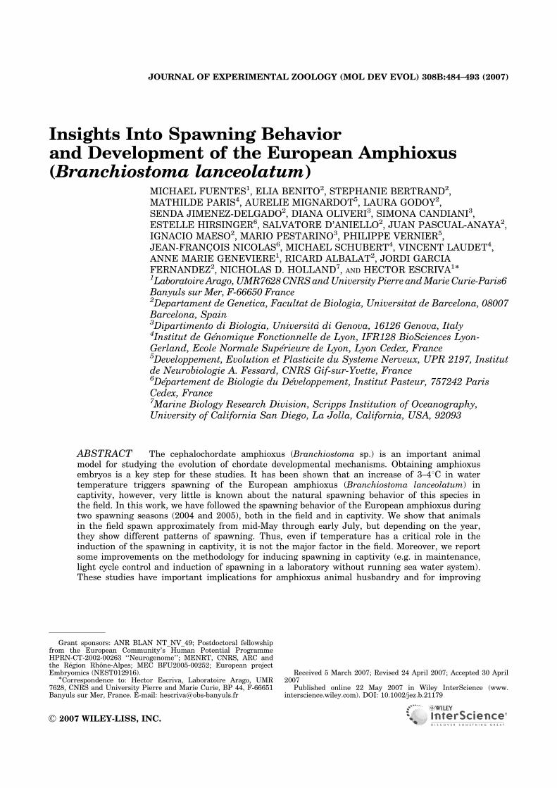
Insights Into Spawning Behaviorand Development of the European Amphioxus(Branchiostoma lanceolatum)
MICHAEL FUENTES1, ELIA BENITO2, STEPHANIE BERTRAND2,MATHILDE PARIS4, AURELIE MIGNARDOT5, LAURA GODOY2,SENDA JIMENEZ-DELGADO2, DIANA OLIVERI3, SIMONA CANDIANI3,ESTELLE HIRSINGER6, SALVATORE D’ANIELLO2, JUAN PASCUAL-ANAYA2,IGNACIO MAESO2, MARIO PESTARINO3, PHILIPPE VERNIER5,JEAN-FRANC-OIS NICOLAS6, MICHAEL SCHUBERT4, VINCENT LAUDET4,ANNE MARIE GENEVIERE1, RICARD ALBALAT2, JORDI GARCIAFERNANDEZ2, NICHOLAS D. HOLLAND7, AND HECTOR ESCRIVA1�
1Laboratoire Arago,UMR7628CNRSandUniversity Pierre andMarieCurie-Paris6Banyuls sur Mer, F-66650 France2Departament de Genetica, Facultat de Biologia, Universitat de Barcelona, 08007Barcelona, Spain3Dipartimento di Biologia, Universita di Genova, 16126 Genova, Italy4Institut de Genomique Fonctionnelle de Lyon, IFR128 BioSciences Lyon-Gerland, Ecole Normale Superieure de Lyon, Lyon Cedex, France5Developpement, Evolution et Plasticite du Systeme Nerveux, UPR 2197, Institutde Neurobiologie A. Fessard, CNRS Gif-sur-Yvette, France6Departement de Biologie du Developpement, Institut Pasteur, 757242 ParisCedex, France7Marine Biology Research Division, Scripps Institution of Oceanography,University of California San Diego, La Jolla, California, USA, 92093
ABSTRACT The cephalochordate amphioxus (Branchiostoma sp.) is an important animalmodel for studying the evolution of chordate developmental mechanisms. Obtaining amphioxusembryos is a key step for these studies. It has been shown that an increase of 3–41C in watertemperature triggers spawning of the European amphioxus (Branchiostoma lanceolatum) incaptivity, however, very little is known about the natural spawning behavior of this species inthe field. In this work, we have followed the spawning behavior of the European amphioxus duringtwo spawning seasons (2004 and 2005), both in the field and in captivity. We show that animalsin the field spawn approximately from mid-May through early July, but depending on the year,they show different patterns of spawning. Thus, even if temperature has a critical role in theinduction of the spawning in captivity, it is not the major factor in the field. Moreover, we reportsome improvements on the methodology for inducing spawning in captivity (e.g. in maintenance,light cycle control and induction of spawning in a laboratory without running sea water system).These studies have important implications for amphioxus animal husbandry and for improving
Published online 22 May 2007 in Wiley InterScience (www.interscience.wiley.com). DOI: 10.1002/jez.b.21179
Received 5 March 2007; Revised 24 April 2007; Accepted 30 April2007
Grant sponsors: ANR BLAN NT_NV_49; Postdoctoral fellowshipfrom the European Community’s Human Potential ProgrammeHPRN-CT-2002-00263 ‘‘Neurogenome’’; MENRT, CNRS, ARC andthe Region Rhone-Alpes; MEC BFU2005-00252; European projectEmbryomics (NEST012916).
�Correspondence to: Hector Escriva, Laboratoire Arago, UMR7628, CNRS and University Pierre and Marie Curie, BP 44, F-66651Banyuls sur Mer, France. E-mail: [email protected]
r 2007 WILEY-LISS, INC.
JOURNAL OF EXPERIMENTAL ZOOLOGY (MOL DEV EVOL) 308B:484–493 (2007)
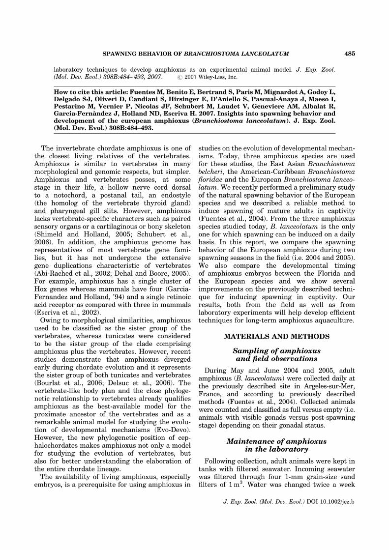
laboratory techniques to develop amphioxus as an experimental animal model. J. Exp. Zool.(Mol. Dev. Evol.) 308B:484– 493, 2007. r 2007 Wiley-Liss, Inc.
How to cite this article: Fuentes M, Benito E, Bertrand S, Paris M, Mignardot A, Godoy L,Delgado SJ, Oliveri D, Candiani S, Hirsinger E, D’Aniello S, Pascual-Anaya J, Maeso I,Pestarino M, Vernier P, Nicolas JF, Schubert M, Laudet V, Geneviere AM, Albalat R,Garcia-Fernandez J, Holland ND, Escriva H. 2007. Insights into spawning behavior anddevelopment of the european amphioxus (Branchiostoma lanceolatum). J. Exp. Zool.(Mol. Dev. Evol.) 308B:484–493.
The invertebrate chordate amphioxus is one ofthe closest living relatives of the vertebrates.Amphioxus is similar to vertebrates in manymorphological and genomic respects, but simpler.Amphioxus and vertebrates posses, at somestage in their life, a hollow nerve cord dorsalto a notochord, a postanal tail, an endostyle(the homolog of the vertebrate thyroid gland)and pharyngeal gill slits. However, amphioxuslacks vertebrate-specific characters such as pairedsensory organs or a cartilaginous or bony skeleton(Shimeld and Holland, 2005; Schubert et al.,2006). In addition, the amphioxus genome hasrepresentatives of most vertebrate gene fami-lies, but it has not undergone the extensivegene duplications characteristic of vertebrates(Abi-Rached et al., 2002; Dehal and Boore, 2005).For example, amphioxus has a single cluster ofHox genes whereas mammals have four (Garcia-Fernandez and Holland, ’94) and a single retinoicacid receptor as compared with three in mammals(Escriva et al., 2002).Owing to morphological similarities, amphioxus
used to be classified as the sister group of thevertebrates, whereas tunicates were consideredto be the sister group of the clade comprisingamphioxus plus the vertebrates. However, recentstudies demonstrate that amphioxus divergedearly during chordate evolution and it representsthe sister group of both tunicates and vertebrates(Bourlat et al., 2006; Delsuc et al., 2006). Thevertebrate-like body plan and the close phyloge-netic relationship to vertebrates already qualifiesamphioxus as the best-available model for theproximate ancestor of the vertebrates and as aremarkable animal model for studying the evolu-tion of developmental mechanisms (Evo-Devo).However, the new phylogenetic position of cep-halochordates makes amphioxus not only a modelfor studying the evolution of vertebrates, butalso for better understanding the elaboration ofthe entire chordate lineage.The availability of living amphioxus, especially
embryos, is a prerequisite for using amphioxus in
studies on the evolution of developmental mechan-isms. Today, three amphioxus species are usedfor these studies, the East Asian Branchiostomabelcheri, the American-Caribbean Branchiostomafloridae and the European Branchiostoma lanceo-latum. We recently performed a preliminary studyof the natural spawning behavior of the Europeanspecies and we described a reliable method toinduce spawning of mature adults in captivity(Fuentes et al., 2004). From the three amphioxusspecies studied today, B. lanceolatum is the onlyone for which spawning can be induced on a dailybasis. In this report, we compare the spawningbehavior of the European amphioxus during twospawning seasons in the field (i.e. 2004 and 2005).We also compare the developmental timingof amphioxus embryos between the Florida andthe European species and we show severalimprovements on the previously described techni-que for inducing spawning in captivity. Ourresults, both from the field as well as fromlaboratory experiments will help develop efficienttechniques for long-term amphioxus aquaculture.
MATERIALS AND METHODS
Sampling of amphioxusand field observations
During May and June 2004 and 2005, adultamphioxus (B. lanceolatum) were collected daily atthe previously described site in Argeles-sur-Mer,France, and according to previously describedmethods (Fuentes et al., 2004). Collected animalswere counted and classified as full versus empty (i.e.animals with visible gonads versus post-spawningstage) depending on their gonadal status.
Maintenance of amphioxusin the laboratory
Following collection, adult animals were kept intanks with filtered seawater. Incoming seawaterwas filtered through four 1-mm grain-size sandfilters of 1m3. Water was changed twice a week
SPAWNING BEHAVIOR OF BRANCHIOSTOMA LANCEOLATUM 485
J. Exp. Zool. (Mol. Dev. Evol.) DOI 10.1002/jez.b
and the animals were subjected to a 14-hr-light/10-hr dark cycle.Animals with empty gonads (post-spawning
stage) were maintained at 18–191C temperaturein 15-L rectangular plastic tanks (100–200 ani-mals/tank) without sand. Water was changedtwice a day and food was always added just afterthe water change. Animals were fed with crushedTetramicrofood (0.5 g/feeding) (www.tetra-online.-com) supplemented with 15.000–40.000 cells/mL/feeding of mixed algae (equal parts of Dunaliellatertiolecta, Isochrysis galbana and Tetraselmissuecica). Algal cultures were grown in f/2 mediumas described in Guillard et al. (Guillard andRyther, ’62). The f/2 medium was eliminated bycentrifugation before feeding the algae to theamphioxus. The animals used in temperatureshock experiments were maintained in starvationat 18–191C temperature in 15-L plastic tankswithout sand in a room with ambient light. Thewater on these animals was changed twice a week.
Temperature measurements in the field
A thermo data logger (HOBO Pendant Temp/Light Intensity Data logger, ONSET ComputerCorporation) was used to measure continuouschanges of seawater temperature in the field. Thisbattery-powered thermometer can report 6,500consecutive data records. The initial settings,including measurement intervals and data acqui-sition, were set with a computer using thesoftware platform HOBOwares (V. 2.1.1_18),ONSET Computer Corporation. For this study,measurements were taken at 30-min intervals.At this setting, about 135 days of temperaturedata can be recorded and the battery power couldlast for about 4 years. After initialization, weanchored the data loggers approximately 5 cmabove the bottom at the amphioxus collection siteat Argeles-sur-Mer, France, at a depth of 4m.
Video recordings
To observe the spawning behavior of amphioxusadults, we placed ten amphioxus adults withfull gonads (previously maintained at 191C for atleast 7 days) individually in 12.5-cm2 cell cultureplastic bottles (BD FalconTM Cell Culture Flasks,BD Biosciences) filled with seawater. The flaskswere placed in a temperature-controlled bath andthe temperature was raised to 231C to initiatespawning 36hr later. Throughout this 36-hr inter-val, the 14-hr light/10-hr dark cycle was maintainedwhile the locomotory behavior of the amphioxusadults was recorded with an infrared video camera
(Sony DCR-TRV70 MiniDV Camcorder, Sony).The infrared light permitted observation in other-wise dark conditions (a modification of thetechnique of Kubokawa et al. [2003]). The move-ments of the animals were subsequently countedand plotted on a graph.
Maintenance and spawning of amphioxusin a laboratory without running seawater
Amphioxus adults collected at Argeles-sur-Mer,France, were transported both to the University ofBarcelona, Spain and to the University of Genova,Italy, where culturing and spawning were per-formed. At the University of Barcelona, Spain, theamphioxus were kept at 171C in 5-L glass tankswith continuous aeration in filtered seawaterobtained from the Laboratoire Arago, Banyuls-sur-Mer, France. Seawater was changed daily andanimals were kept in an artificial 14-hr light/10-hrdark cycle in phase with the natural light cycle.Animals were fed with Marine Snow food supple-mented with 15.000–40.000 cells/mL/feeding ofmixed algae (1:1:1 parts of D. tertiolecta, I. galbanaand T. suecica). Spawning was induced by a 36-hrthermal shock at 211C without aeration. At theUniversity of Genova, Italy, the amphioxus werekept at 191C in 50-L glass tanks with filteredseawater obtained locally in a 14-hr light/10-hrdark artificial cycle. Animals were fed according toFuentes et al. (2004). Spawning was induced by a36-hr thermal shock at 231C without aeration.
Inversion of the day/night cycle
Amphioxus adults with full gonads were collectedin Argeles-sur-Mer, France, and brought to theUniversity of Barcelona, Spain. Adults were kept at171C to prevent temperature stress and undesiredspawning events. Starting with their arrival in thelaboratory, the amphioxus adults were exposed to a14-hr light/10-hr dark cycle that was phase shiftedrelative to the natural photoperiod such that thelight period started at 10 PM and ended at noon.After 3 weeks on this phase-shifted light regime,the animals were induced to spawn by thetemperature shock method of Fuentes et al. (2004).
DNA staining
Embryos were fixed for 10min in a methanol-glycerol solution (75:25 v:v) containing 0.1 mg/mL4,6-diamidino-2-phenylindole (DAPI) (4,6-diamidino-2-phenylindole, stock concentration was at2.5 mg/mL). The suspension was further completedwith 1 volume of phosphate-buffered saline
M. FUENTES ET AL.486
J. Exp. Zool. (Mol. Dev. Evol.) DOI 10.1002/jez.b
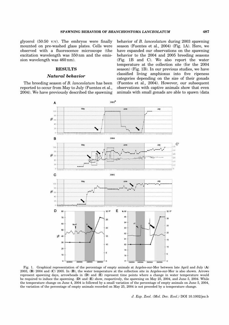
glycerol (50:50 v:v). The embryos were finallymounted on pre-washed glass plates. Cells wereobserved with a fluorescence microscope (theexcitation wavelength was 350nm and the emis-sion wavelength was 460nm).
RESULTS
Natural behavior
The breeding season of B. lanceolatum has beenreported to occur from May to July (Fuentes et al.,2004). We have previously described the spawning
behavior of B. lanceolatum during 2003 spawningseason (Fuentes et al., 2004) (Fig. 1A). Here, wehave expanded our observations on the spawningbehavior to the 2004 and 2005 breeding seasons(Fig. 1B and C). We also report the watertemperature at the collection site (for the 2004season) (Fig. 1B). In our previous studies, we haveclassified living amphioxus into five ripenesscategories depending on the size of their gonads(Fuentes et al., 2004). However, our subsequentobservations with captive animals show that evenanimals with small gonads are able to spawn (data
Fig. 1. Graphical representation of the percentage of empty animals at Argeles-sur-Mer between late April and July (A)2003, (B) 2004 and (C) 2005. In (B), the water temperature at the collection site in Argeles-sur-Mer is also shown. Arrowsrepresent spawning days, arrowheads in (D) and (E) represent time points where a change in water temperature wouldbe required to induce the spawning. (D) and (E) show, respectively, the spawning on May 25, 2004, and June 5, 2004. Whilethe temperature change on June 4, 2004 is followed by a small variation of the percentage of empty animals on June 5, 2004,the variation of the percentage of empty animals recorded on May 25, 2004 is not preceded by a temperature change.
SPAWNING BEHAVIOR OF BRANCHIOSTOMA LANCEOLATUM 487
J. Exp. Zool. (Mol. Dev. Evol.) DOI 10.1002/jez.b
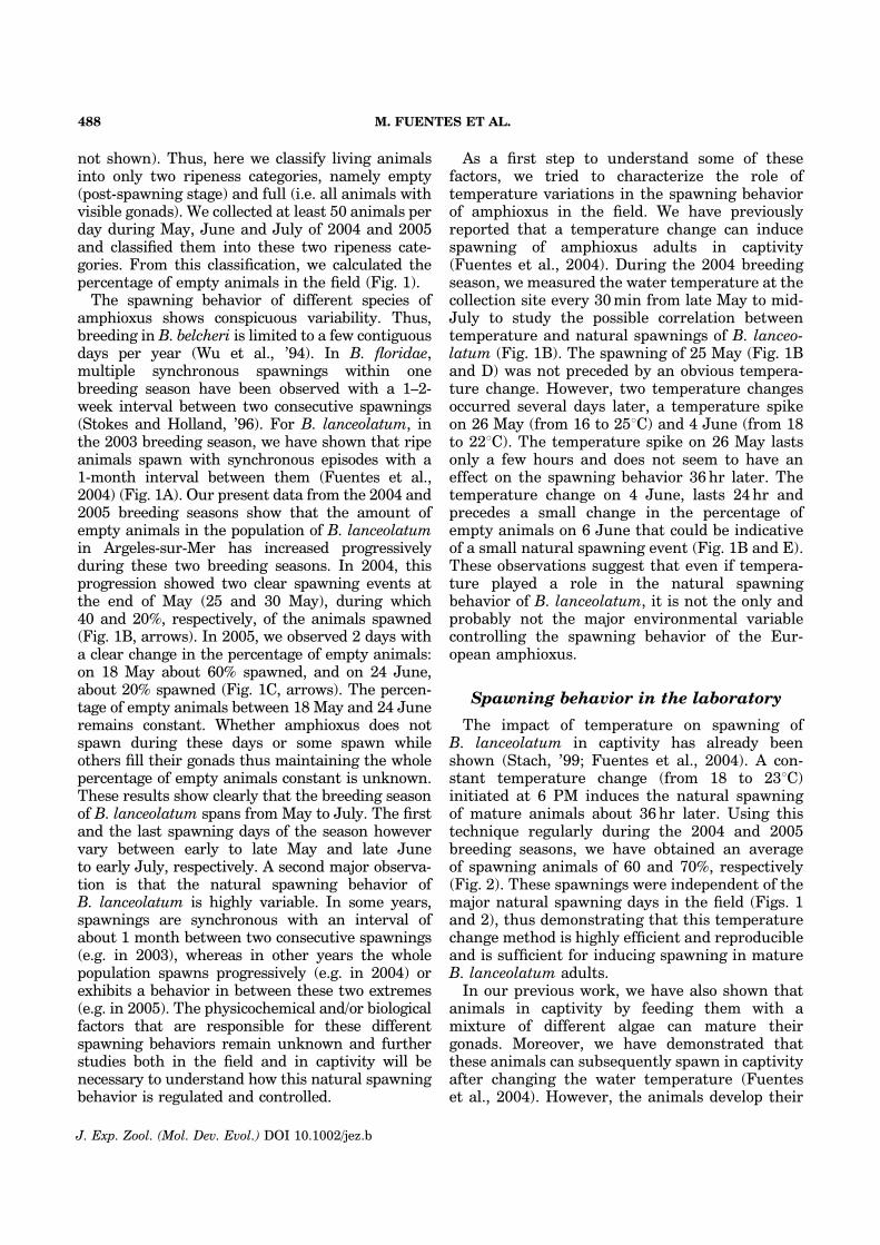
not shown). Thus, here we classify living animalsinto only two ripeness categories, namely empty(post-spawning stage) and full (i.e. all animals withvisible gonads). We collected at least 50 animals perday during May, June and July of 2004 and 2005and classified them into these two ripeness cate-gories. From this classification, we calculated thepercentage of empty animals in the field (Fig. 1).The spawning behavior of different species of
amphioxus shows conspicuous variability. Thus,breeding in B. belcheri is limited to a few contiguousdays per year (Wu et al., ’94). In B. floridae,multiple synchronous spawnings within onebreeding season have been observed with a 1–2-week interval between two consecutive spawnings(Stokes and Holland, ’96). For B. lanceolatum, inthe 2003 breeding season, we have shown that ripeanimals spawn with synchronous episodes with a1-month interval between them (Fuentes et al.,2004) (Fig. 1A). Our present data from the 2004 and2005 breeding seasons show that the amount ofempty animals in the population of B. lanceolatumin Argeles-sur-Mer has increased progressivelyduring these two breeding seasons. In 2004, thisprogression showed two clear spawning events atthe end of May (25 and 30 May), during which40 and 20%, respectively, of the animals spawned(Fig. 1B, arrows). In 2005, we observed 2 days witha clear change in the percentage of empty animals:on 18 May about 60% spawned, and on 24 June,about 20% spawned (Fig. 1C, arrows). The percen-tage of empty animals between 18 May and 24 Juneremains constant. Whether amphioxus does notspawn during these days or some spawn whileothers fill their gonads thus maintaining the wholepercentage of empty animals constant is unknown.These results show clearly that the breeding seasonof B. lanceolatum spans from May to July. The firstand the last spawning days of the season howevervary between early to late May and late Juneto early July, respectively. A second major observa-tion is that the natural spawning behavior ofB. lanceolatum is highly variable. In some years,spawnings are synchronous with an interval ofabout 1 month between two consecutive spawnings(e.g. in 2003), whereas in other years the wholepopulation spawns progressively (e.g. in 2004) orexhibits a behavior in between these two extremes(e.g. in 2005). The physicochemical and/or biologicalfactors that are responsible for these differentspawning behaviors remain unknown and furtherstudies both in the field and in captivity will benecessary to understand how this natural spawningbehavior is regulated and controlled.
As a first step to understand some of thesefactors, we tried to characterize the role oftemperature variations in the spawning behaviorof amphioxus in the field. We have previouslyreported that a temperature change can inducespawning of amphioxus adults in captivity(Fuentes et al., 2004). During the 2004 breedingseason, we measured the water temperature at thecollection site every 30min from late May to mid-July to study the possible correlation betweentemperature and natural spawnings of B. lanceo-latum (Fig. 1B). The spawning of 25 May (Fig. 1Band D) was not preceded by an obvious tempera-ture change. However, two temperature changesoccurred several days later, a temperature spikeon 26 May (from 16 to 251C) and 4 June (from 18to 221C). The temperature spike on 26 May lastsonly a few hours and does not seem to have aneffect on the spawning behavior 36hr later. Thetemperature change on 4 June, lasts 24hr andprecedes a small change in the percentage ofempty animals on 6 June that could be indicativeof a small natural spawning event (Fig. 1B and E).These observations suggest that even if tempera-ture played a role in the natural spawningbehavior of B. lanceolatum, it is not the only andprobably not the major environmental variablecontrolling the spawning behavior of the Eur-opean amphioxus.
Spawning behavior in the laboratory
The impact of temperature on spawning ofB. lanceolatum in captivity has already beenshown (Stach, ’99; Fuentes et al., 2004). A con-stant temperature change (from 18 to 231C)initiated at 6 PM induces the natural spawningof mature animals about 36hr later. Using thistechnique regularly during the 2004 and 2005breeding seasons, we have obtained an averageof spawning animals of 60 and 70%, respectively(Fig. 2). These spawnings were independent of themajor natural spawning days in the field (Figs. 1and 2), thus demonstrating that this temperaturechange method is highly efficient and reproducibleand is sufficient for inducing spawning in matureB. lanceolatum adults.In our previous work, we have also shown that
animals in captivity by feeding them with amixture of different algae can mature theirgonads. Moreover, we have demonstrated thatthese animals can subsequently spawn in captivityafter changing the water temperature (Fuenteset al., 2004). However, the animals develop their
M. FUENTES ET AL.488
J. Exp. Zool. (Mol. Dev. Evol.) DOI 10.1002/jez.b
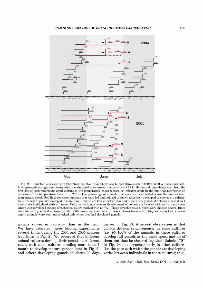
gonads slower in captivity than in the field.We have repeated these feeding experimentsseveral times during the 2004 and 2005 seasons(red lines in Fig. 2). We observed that differentanimal cultures develop their gonads at differentrates, with some cultures needing more than 1month to develop mature gonads (star in Fig. 2)and others developing gonads in about 20 days
(arrow in Fig. 2). A second observation is thatgonads develop synchronously in some cultures(i.e. 95–100% of the animals in these culturesdevelop full gonads at the same speed and all ofthem can thus be shocked together) (labeled ‘‘S’’in Fig. 2), but asynchronously in other cultures(i.e. the pace with which the gonads are developingvaries between individuals in these cultures; thus,
Fig. 2. Induction of spawning in laboratory-maintained amphioxus by temperature shock in 2004 and 2005. Each horizontalline represents a single amphioxus culture maintained at a constant temperature of 181C. Horizontal lines always span from thefirst day of each amphioxus adult culture to the temperature shock (shown as inflexion point in the line that represents anincrease in the temperature from 18 to 231C). The percentage of animals that spawned is indicated above the line for eachtemperature shock. Red lines represent animals that were fed and induced to spawn after they developed the gonads in culture.Cultures where gonads developed in more than 1 month are labeled with a star and those where gonads developed in less than 1month are highlighted with an arrow. Cultures with synchronous development of gonads are labeled with an ‘‘S’’ and thosewhere they developed gonads asynchronously are marked with an ‘‘A.’’ These asynchronous cultures were shocked several times(represented by several inflexion points in the lines): once animals in these cultures became full, they were shocked, whereasempty animals were kept and shocked only when they had developed gonads.
SPAWNING BEHAVIOR OF BRANCHIOSTOMA LANCEOLATUM 489
J. Exp. Zool. (Mol. Dev. Evol.) DOI 10.1002/jez.b
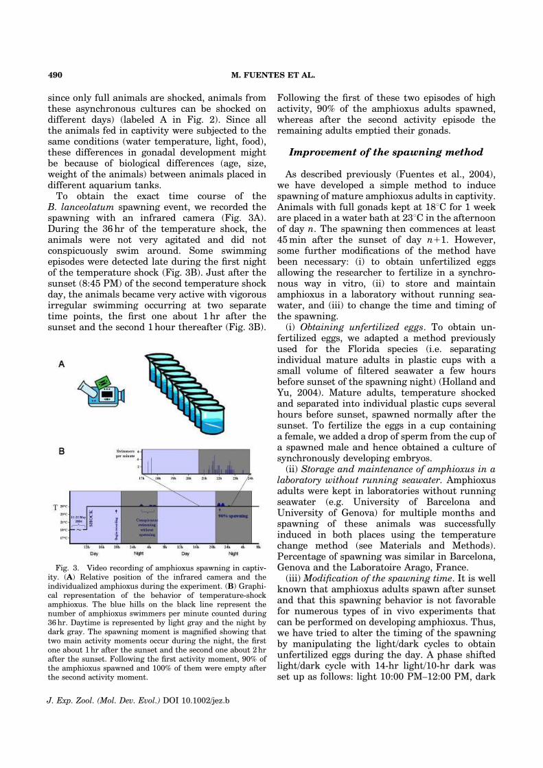
since only full animals are shocked, animals fromthese asynchronous cultures can be shocked ondifferent days) (labeled A in Fig. 2). Since allthe animals fed in captivity were subjected to thesame conditions (water temperature, light, food),these differences in gonadal development mightbe because of biological differences (age, size,weight of the animals) between animals placed indifferent aquarium tanks.To obtain the exact time course of the
B. lanceolatum spawning event, we recorded thespawning with an infrared camera (Fig. 3A).During the 36hr of the temperature shock, theanimals were not very agitated and did notconspicuously swim around. Some swimmingepisodes were detected late during the first nightof the temperature shock (Fig. 3B). Just after thesunset (8:45 PM) of the second temperature shockday, the animals became very active with vigorousirregular swimming occurring at two separatetime points, the first one about 1 hr after thesunset and the second 1hour thereafter (Fig. 3B).
Following the first of these two episodes of highactivity, 90% of the amphioxus adults spawned,whereas after the second activity episode theremaining adults emptied their gonads.
Improvement of the spawning method
As described previously (Fuentes et al., 2004),we have developed a simple method to inducespawning of mature amphioxus adults in captivity.Animals with full gonads kept at 181C for 1 weekare placed in a water bath at 231C in the afternoonof day n. The spawning then commences at least45min after the sunset of day n11. However,some further modifications of the method havebeen necessary: (i) to obtain unfertilized eggsallowing the researcher to fertilize in a synchro-nous way in vitro, (ii) to store and maintainamphioxus in a laboratory without running sea-water, and (iii) to change the time and timing ofthe spawning.(i) Obtaining unfertilized eggs. To obtain un-
fertilized eggs, we adapted a method previouslyused for the Florida species (i.e. separatingindividual mature adults in plastic cups with asmall volume of filtered seawater a few hoursbefore sunset of the spawning night) (Holland andYu, 2004). Mature adults, temperature shockedand separated into individual plastic cups severalhours before sunset, spawned normally after thesunset. To fertilize the eggs in a cup containinga female, we added a drop of sperm from the cup ofa spawned male and hence obtained a culture ofsynchronously developing embryos.(ii) Storage and maintenance of amphioxus in a
laboratory without running seawater. Amphioxusadults were kept in laboratories without runningseawater (e.g. University of Barcelona andUniversity of Genova) for multiple months andspawning of these animals was successfullyinduced in both places using the temperaturechange method (see Materials and Methods).Percentage of spawning was similar in Barcelona,Genova and the Laboratoire Arago, France.(iii) Modification of the spawning time. It is well
known that amphioxus adults spawn after sunsetand that this spawning behavior is not favorablefor numerous types of in vivo experiments thatcan be performed on developing amphioxus. Thus,we have tried to alter the timing of the spawningby manipulating the light/dark cycles to obtainunfertilized eggs during the day. A phase shiftedlight/dark cycle with 14-hr light/10-hr dark wasset up as follows: light 10:00 PM–12:00 PM, dark
Fig. 3. Video recording of amphioxus spawning in captiv-ity. (A) Relative position of the infrared camera and theindividualized amphioxus during the experiment. (B) Graphi-cal representation of the behavior of temperature-shockamphioxus. The blue hills on the black line represent thenumber of amphioxus swimmers per minute counted during36hr. Daytime is represented by light gray and the night bydark gray. The spawning moment is magnified showing thattwo main activity moments occur during the night, the firstone about 1hr after the sunset and the second one about 2hrafter the sunset. Following the first activity moment, 90% ofthe amphioxus spawned and 100% of them were empty afterthe second activity moment.
M. FUENTES ET AL.490
J. Exp. Zool. (Mol. Dev. Evol.) DOI 10.1002/jez.b
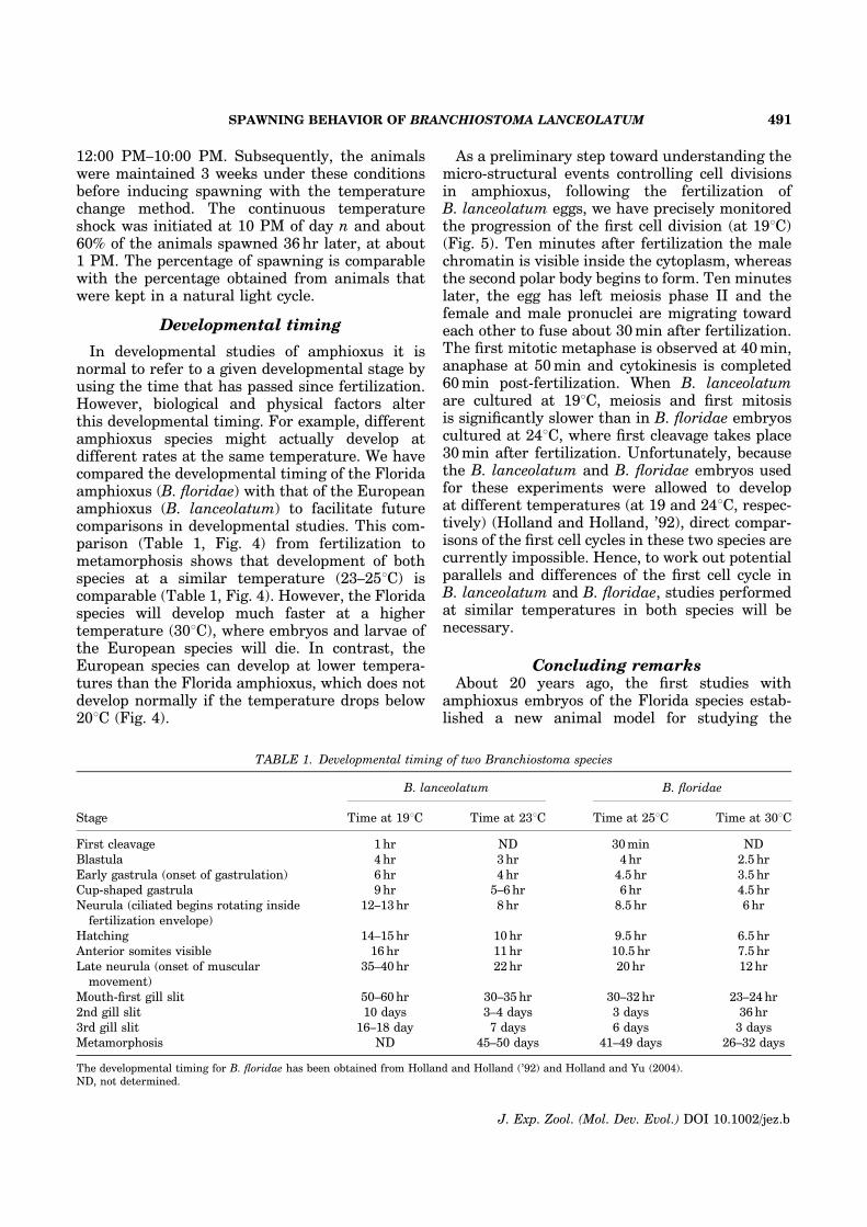
12:00 PM–10:00 PM. Subsequently, the animalswere maintained 3 weeks under these conditionsbefore inducing spawning with the temperaturechange method. The continuous temperatureshock was initiated at 10 PM of day n and about60% of the animals spawned 36hr later, at about1 PM. The percentage of spawning is comparablewith the percentage obtained from animals thatwere kept in a natural light cycle.
Developmental timing
In developmental studies of amphioxus it isnormal to refer to a given developmental stage byusing the time that has passed since fertilization.However, biological and physical factors alterthis developmental timing. For example, differentamphioxus species might actually develop atdifferent rates at the same temperature. We havecompared the developmental timing of the Floridaamphioxus (B. floridae) with that of the Europeanamphioxus (B. lanceolatum) to facilitate futurecomparisons in developmental studies. This com-parison (Table 1, Fig. 4) from fertilization tometamorphosis shows that development of bothspecies at a similar temperature (23–251C) iscomparable (Table 1, Fig. 4). However, the Floridaspecies will develop much faster at a highertemperature (301C), where embryos and larvae ofthe European species will die. In contrast, theEuropean species can develop at lower tempera-tures than the Florida amphioxus, which does notdevelop normally if the temperature drops below201C (Fig. 4).
As a preliminary step toward understanding themicro-structural events controlling cell divisionsin amphioxus, following the fertilization ofB. lanceolatum eggs, we have precisely monitoredthe progression of the first cell division (at 191C)(Fig. 5). Ten minutes after fertilization the malechromatin is visible inside the cytoplasm, whereasthe second polar body begins to form. Ten minuteslater, the egg has left meiosis phase II and thefemale and male pronuclei are migrating towardeach other to fuse about 30min after fertilization.The first mitotic metaphase is observed at 40min,anaphase at 50min and cytokinesis is completed60min post-fertilization. When B. lanceolatumare cultured at 191C, meiosis and first mitosisis significantly slower than in B. floridae embryoscultured at 241C, where first cleavage takes place30min after fertilization. Unfortunately, becausethe B. lanceolatum and B. floridae embryos usedfor these experiments were allowed to developat different temperatures (at 19 and 241C, respec-tively) (Holland and Holland, ’92), direct compar-isons of the first cell cycles in these two species arecurrently impossible. Hence, to work out potentialparallels and differences of the first cell cycle inB. lanceolatum and B. floridae, studies performedat similar temperatures in both species will benecessary.
Concluding remarksAbout 20 years ago, the first studies with
amphioxus embryos of the Florida species estab-lished a new animal model for studying the
TABLE 1. Developmental timing of two Branchiostoma species
B. lanceolatum B. floridae
Stage Time at 191C Time at 231C Time at 251C Time at 301C
First cleavage 1hr ND 30min ND
Blastula 4 hr 3hr 4hr 2.5 hr
Early gastrula (onset of gastrulation) 6 hr 4 hr 4.5 hr 3.5 hr
Cup-shaped gastrula 9 hr 5–6 hr 6hr 4.5 hr
Neurula (ciliated begins rotating inside
fertilization envelope)
12–13hr 8hr 8.5 hr 6 hr
Hatching 14–15hr 10hr 9.5 hr 6.5 hr
Anterior somites visible 16 hr 11hr 10.5 hr 7.5 hr
Late neurula (onset of muscular
movement)
35–40hr 22hr 20hr 12hr
Mouth-first gill slit 50–60hr 30–35 hr 30–32hr 23–24hr
2nd gill slit 10 days 3–4 days 3 days 36 hr
3rd gill slit 16–18 day 7 days 6 days 3 days
Metamorphosis ND 45–50 days 41–49 days 26–32 days
The developmental timing for B. floridae has been obtained from Holland and Holland (’92) and Holland and Yu (2004).ND, not determined.
SPAWNING BEHAVIOR OF BRANCHIOSTOMA LANCEOLATUM 491
J. Exp. Zool. (Mol. Dev. Evol.) DOI 10.1002/jez.b
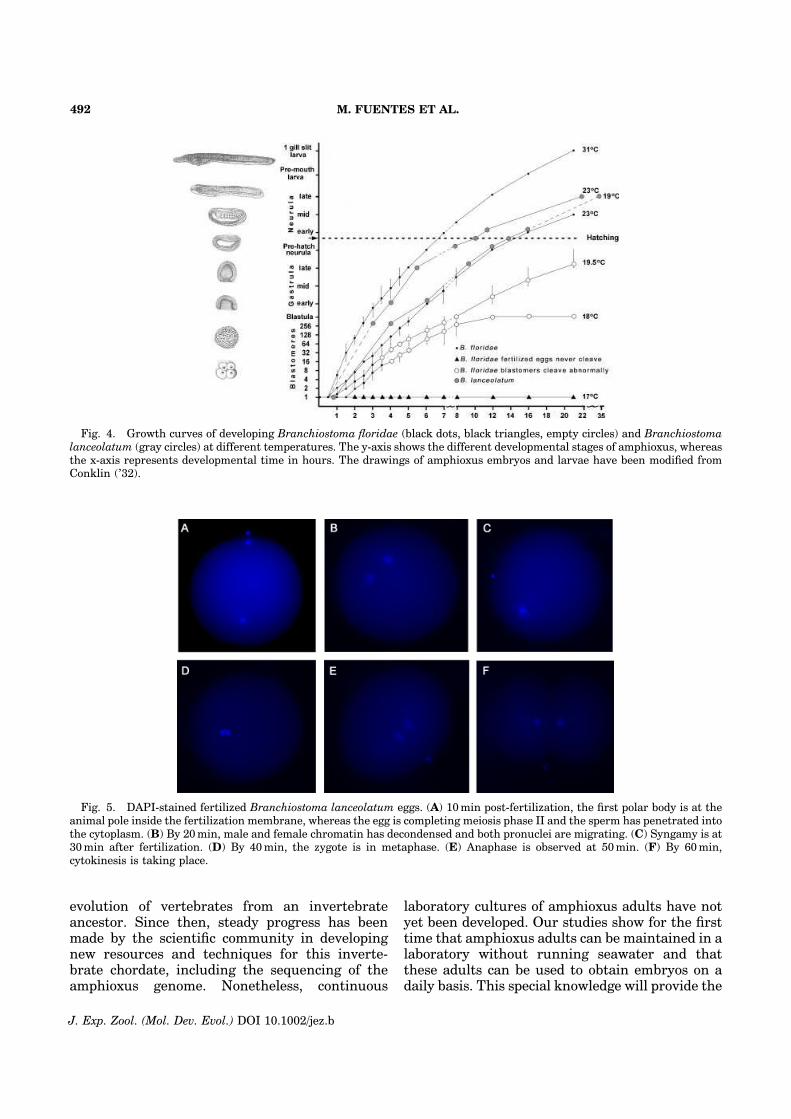
evolution of vertebrates from an invertebrateancestor. Since then, steady progress has beenmade by the scientific community in developingnew resources and techniques for this inverte-brate chordate, including the sequencing of theamphioxus genome. Nonetheless, continuous
laboratory cultures of amphioxus adults have notyet been developed. Our studies show for the firsttime that amphioxus adults can be maintained in alaboratory without running seawater and thatthese adults can be used to obtain embryos on adaily basis. This special knowledge will provide the
Fig. 5. DAPI-stained fertilized Branchiostoma lanceolatum eggs. (A) 10min post-fertilization, the first polar body is at theanimal pole inside the fertilization membrane, whereas the egg is completing meiosis phase II and the sperm has penetrated intothe cytoplasm. (B) By 20min, male and female chromatin has decondensed and both pronuclei are migrating. (C) Syngamy is at30min after fertilization. (D) By 40min, the zygote is in metaphase. (E) Anaphase is observed at 50min. (F) By 60min,cytokinesis is taking place.
Fig. 4. Growth curves of developing Branchiostoma floridae (black dots, black triangles, empty circles) and Branchiostomalanceolatum (gray circles) at different temperatures. The y-axis shows the different developmental stages of amphioxus, whereasthe x-axis represents developmental time in hours. The drawings of amphioxus embryos and larvae have been modified fromConklin (’32).
M. FUENTES ET AL.492
J. Exp. Zool. (Mol. Dev. Evol.) DOI 10.1002/jez.b
basis for the development of new experimentalapproaches and will certainly make amphioxus amuch more attractive animal model for studies of theevolution of the chordate and vertebrate body plan.
ACKNOWLEDGMENTS
We are very grateful to Neus Abella for fruitfuldiscussions about the day/night cycle inversionexperiments. We also thank the fishermen of the‘‘Nereis’’ for their help with collection of adultamphioxus. HE is supported by ANR BLANNT_NV_49. SDA is supported by a postdoctoralfellowship from the European Community’sHuman Potential Programme HPRN-CT-2002-00263 ‘‘Neurogenome’’. EH and JFN are suppor-ted by the European project Embryomics NEST012916. SB is an EMBO postdoctoral fellow.We are also grateful for funding from MENRT,CNRS, ARC and the Region Rhone-Alpes as wellas FPU, CIRIT, and FPI fellowships of SJD, JPAand IM respectively.
LITERATURE CITED
Abi-Rached L, Gilles A, Shiina T, Pontarotti P, Inoko H. 2002.Evidence of en bloc duplication in vertebrate genomes. NatGenet 31:100–105.
Bourlat SJ, Juliusdottir T, Lowe CJ, Freeman R, Aronowicz J,Kirschner M, Lander ES, Thorndyke M, Nakano H, KohnAB, Heyland A, Moroz LL, Copley RR, Telford MJ. 2006.Deuterostome phylogeny reveals monophyletic chordatesand the new phylum Xenoturbellida. Nature 444:85–88.
Conklin EG. 1932. The embryology of amphioxus. J Morphol54:69–151.
Dehal P, Boore JL. 2005. Two rounds of whole genomeduplication in the ancestral vertebrate. PLoS Biol 3:e314.
Delsuc F, Brinkmann H, Chourrout D, Philippe H. 2006.Tunicates and not cephalochordates are the closest livingrelatives of vertebrates. Nature 439:965–968.
Escriva H, Holland ND, Gronemeyer H, Laudet V, HollandLZ. 2002. The retinoic acid signaling pathway regulates
anterior/posterior patterning in the nerve cord and pharynxof amphioxus, a chordate lacking neural crest. Development129:2905–2916.
Fuentes M, Schubert M, Dalfo D, Candiani S, Benito E,Gardenyes J, Godoy L, Moret F, Illas M, Patten I,Permanyer J, Oliveri D, Boeuf G, Falcon J, Pestarino M,Fernandez JG, Albalat R, Laudet V, Vernier P, Escriva H.2004. Preliminary observations on the spawning condi-tions of the European amphioxus (Branchiostoma
lanceolatum) in captivity. J Exp Zool B Mol Dev Evol 302:384–391.
Garcia-Fernandez J, Holland PWH. 1994. Archetypal organi-zation of the amphioxus Hox gene cluster. Nature 370:563–566.
Guillard RRL, Ryther JH. 1962. Studies of marine planktonicdiatoms. I. Cyclotella nana Hustedt and Detonula conferva-cea Cleve. Can J Microbiol 8:229–239.
Holland LZ, Holland ND. 1992. Early development in thelancelet (5Amphioxus) Branchiostoma floridae from spermentry through pronuclear fusion: presence of vegetal poleplasm and lack of conspicuous ooplasmic segregation. BiolBull 182:77–96.
Holland LZ, Yu JK. 2004. Cephalochordate (amphioxus)embryos: procurement, culture, and basic methods. MethodsCell Biol 74:195–215.
Kubokawa K, Mizuta T, Morisawa M, Azuma N. 2003.Gonadal state of wild amphioxus populations and spawningsuccess in captive conditions during the breeding period inJapan. Zool Sci 20:889–895.
Schubert M, Escriva H, Xavier-Neto J, Laudet V. 2006.Amphioxus and tunicates as evolutionary model systems.Trends Ecol Evol 21:269–277.
Shimeld SM, Holland ND. 2005. Amphioxus molecularbiology: insights into vertebrate evolution and developmen-tal mechanisms. Can J Zool 83:90–100.
Stach T. 1999. The ontogeny of the notochord of Branchios-toma lanceolatum. Acta Zoologica (Stockholm) 80:25–33.
Stokes MD, Holland ND. 1996. Reproduction of the Floridalancelet (Branchiostoma floridae): spawning patterns andfluctuations in gonad indexes and nutritional reserves.Invert Biol 115:349–359.
Wu XH, Zhang SC, Wang YY, Zhang BL, Qu YM, Jiang XJ.1994. Laboratory observation on spawning, fecundity andlarval development of amphioxus (Branchiostoma belcheri
tsingtauense). Chin J Oceanol Limnol 12:289–294.
SPAWNING BEHAVIOR OF BRANCHIOSTOMA LANCEOLATUM 493
J. Exp. Zool. (Mol. Dev. Evol.) DOI 10.1002/jez.b
Combined analysis of fourteen nuclear genes refinesthe Ursidae phylogeny
Marie Pages a,b, Sebastien Calvignac a, Catherine Klein a, Mathilde Paris c,Sandrine Hughes a, Catherine Hanni a,*
aPaleogenetique et Evolution Moleculaire, Universite de Lyon, Institut de Genomique Fonctionnelle, INRA, CNRS, Universite Lyon 1,
Ecole Normale Superieure, 46 allee d’Italie, 69364 Lyon cedex 07, FrancebLaboratoire de Paleontologie, Phylogenie et Paleobiologie, Institut des Sciences de l’Evolution de Montpellier, UMR CNRS 5554,
Universite Montpellier II, Place Eugene Bataillon, 34095 Montpellier Cedex 05, FrancecZoologie Moleculaire, Universite de Lyon, Institut de Genomique Fonctionnelle, INRA, CNRS, Universite Lyon 1,
Ecole Normale Superieure, 46 allee d’Italie, 69364 Lyon cedex 07, France
Received 22 February 2007; revised 11 October 2007; accepted 17 October 2007
Available online 5 November 2007
Abstract
Despite numerous studies, questions remain about the evolutionary history of Ursidae and additional independent genetic markerswere needed to elucidate these ambiguities. For this purpose, we sequenced ten nuclear genes for all the eight extant bear species. Bycombining these new sequences with those of four other recently published nuclear markers, we provide new insights into the phyloge-netic relationships of the Ursidae family members. The hypothesis that the giant panda was the first species to diverge among ursids isdefinitively confirmed and the precise branching order within the Ursus genus is clarified for the first time. Moreover, our analyses indi-cate that the American and the Asiatic black bears do not cluster as sister taxa, as had been previously hypothesised. Sun and sloth bearsclearly appear as the most basal ursine species but uncertainties about their exact relationships remain. Since our larger dataset did notenable us to clarify this last question, identifying rare genomic changes in bear genomes could be a promising solution for further studies.� 2007 Elsevier Inc. All rights reserved.
Keywords: Molecular phylogeny; Nuclear DNA; Hibernation; Ursidae
1. Introduction
Among Carnivores, the family of Ursidae compriseseight extant species, including the giant panda (Ailuropodamelanoleuca), the spectacled bear (Tremarctos ornatus) andsix ursine species: the polar bear (Ursus maritimus), thebrown bear (Ursus arctos), the American black bear (Ursus
americanus), the Asiatic black bear (Ursus thibetanus), thesun bear (Helarctos malayanus) and the sloth bear (Melur-
sus ursinus). Although the belonging of the giant panda to
the Ursidae is not a riddle anymore (Davis, 1964; O’Brienet al., 1985; Van Valen, 1986; Mayr, 1986), the phyloge-netic relationships among ursids still remain unclear. Thefact that ursine species underwent a rapid diversification5 Myr1 ago (Kurten, 1968; Wayne et al., 1991; Waitset al., 1999) could explain why resolving this phylogenyrepresents a tricky challenge. Despite previous molecularinvestigations, mainly based on mitochondrial DNA(Zhang and Ryder, 1994; Vrana et al., 1994; Talbot andShields, 1996a; Waits et al., 1999; Yu et al., 2004; Fultonand Strobeck, 2006), three thorny questions are still pend-ing: (Q1) what is the exact relationship between the giantpanda and the spectacled bear at the base of the Ursidae
1055-7903/$ - see front matter � 2007 Elsevier Inc. All rights reserved.
doi:10.1016/j.ympev.2007.10.019
* Corresponding author. Address: Institut de Genomique Fonctionnelle,
INRA, CNRS, Universite Lyon 1, Ecole Normale Superieure, F-69364,
France. Fax: +33 0 4 72 72 80 80.E-mail address: [email protected] (C. Hanni). 1 Abbreviations used: Myr, million years; kb, kilobases.
www.elsevier.com/locate/ympev
Available online at www.sciencedirect.com
Molecular Phylogenetics and Evolution 47 (2008) 73–83
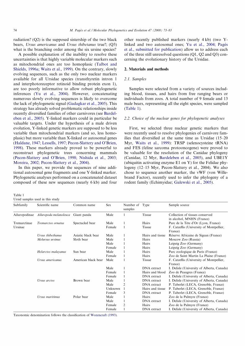
radiation? (Q2) is the supposed sistership of the two blackbears, Ursus americanus and Ursus thibetanus true?; (Q3)what is the branching order among the six ursine species?
A possible explanation of the inability to resolve theseuncertainties is that highly variable molecular markers suchas mitochondrial ones are too homoplasic (Talbot andShields, 1996a; Waits et al., 1999). On the contrary, slowlyevolving sequences, such as the only two nuclear markersavailable for all Ursidae species (transthyretin intron 1and interphotoreceptor retinoid binding protein exon 1),are too poorly informative to allow robust phylogeneticinferences (Yu et al., 2004). However, concatenatingnumerous slowly evolving sequences is likely to overcomethe lack of phylogenetic signal (Gadagkar et al., 2005). Thisstrategy has already solved problematic relationships insiderecently diversified families of other carnivores (see Bardel-eben et al., 2005). Y-linked markers could in particular bevaluable targets. Under the hypothesis of a male drivenevolution, Y-linked genetic markers are supposed to be lessvariable than mitochondrial markers (and so, less homo-plasic) but more variable than X-linked or autosomal genes(Haldane, 1947; Lessells, 1997; Pecon-Slattery and O’Brien,1998). These markers already proved to be powerful toreconstruct phylogenetic trees concerning mammals(Pecon-Slattery and O’Brien, 1998; Nishida et al., 2003;Moreira, 2002; Pecon-Slattery et al., 2004).
In this paper, we provide the sequences of nine addi-tional autosomal gene fragments and one Y-linked marker.Phylogenetic analyses performed on a concatenated datasetcomposed of these new sequences (nearly 6 kb) and four
other recently published markers (nearly 4 kb) (two Y-linked and two autosomal ones; Yu et al., 2004; Pageset al., submitted for publication) allow us to address eachof the three still unresolved questions (Q1, Q2 and Q3) con-cerning the evolutionary history of the Ursidae.
2. Materials and methods
2.1. Samples
Samples were selected from a variety of sources includ-ing blood, tissues, and hairs from free ranging bears orindividuals from zoos. A total number of 9 female and 13male bears, representing all the eight species, were sampled(Table 1).
2.2. Choice of the nuclear genes for phylogenetic analyses
First, we selected three nuclear genetic markers thatwere recently used to resolve phylogenies of carnivore fam-ilies that diversified at the same time as Ursidae (15–20Myr, Waits et al., 1999): TRSP (selenocysteine tRNA)and FES (feline sarcoma protooncogene) were proved tobe valuable for the resolution of the Canidae phylogeny(Canidae, 12 Myr, Bardeleben et al., 2005), and UBE1Y(ubiquitin activating enzyme E1 on Y) for the Felidae phy-logeny (12–15 Myr, Pecon-Slattery et al., 2004). We alsochose to sequence another marker, the vWF (von Wille-brand Factor), recently used to infer the phylogeny of arodent family (Echimyidae; Galewski et al., 2005).
Table 1
Ursid samples used in this study
Subfamily Scientific name Common name Sex Number of
samples
Type Sample source
Ailuropodinae Ailuropoda melanoleuca Giant panda Male 1 Tissue Collection of tissues conserved
in alcohol, MNHN (France)
Tremarctinae Tremarctos ornatus Spectacled bear Male 1 Hairs Parc de la Tete d’Or (Lyon, France)
Ursinae Female 1 Tissue F. Catzeflis (University of Montpellier,
France)
Ursus thibethanus Asiatic black bear Male 1 Hairs and tissue Reserve Africaine de Sigean (France)
Melursus ursinus Sloth bear Male 1 Hairs Moscow Zoo (Russia)
Male 1 Hairs Leipzig Zoo (Germany)
Female 1 Hairs Leipzig Zoo (Germany)
Helarctos malayanus Sun bear Male 1 Hairs Parc zoologique de Paris (France)
Female 1 Hairs Zoo de Saint Martin La Plaine (France)
Ursus americanus American black bear Male 1 Tissue F. Catzeflis (University of Montpellier,
France)
Male 1 DNA extract I. Delisle (University of Alberta, Canada)
Female 1 Hairs and blood Zoo de Peaugres (France)
Female 1 DNA extract I. Delisle (University of Alberta, Canada)
Ursus arctos Brown bear Male 1 DNA extract I. Delisle (University of Alberta, Canada)
Male 2 DNA extract P. Taberlet (LECA, Grenoble, France)
Unknown 1 Hairs and tissue P. Taberlet (LECA, Grenoble, France)
Female 3 DNA extract P. Taberlet (LECA, Grenoble, France)
Ursus maritimus Polar bear Male 1 Hairs Zoo de la Palmyre (France)
Male 1 DNA extract I. Delisle (University of Alberta, Canada)
Female 2 Hairs Zoo de la Palmyre (France)
Female 1 DNA extract I. Delisle (University of Alberta, Canada)
Taxonomic denomination follows the classification of Wozencraft (1993).
74 M. Pages et al. /Molecular Phylogenetics and Evolution 47 (2008) 73–83
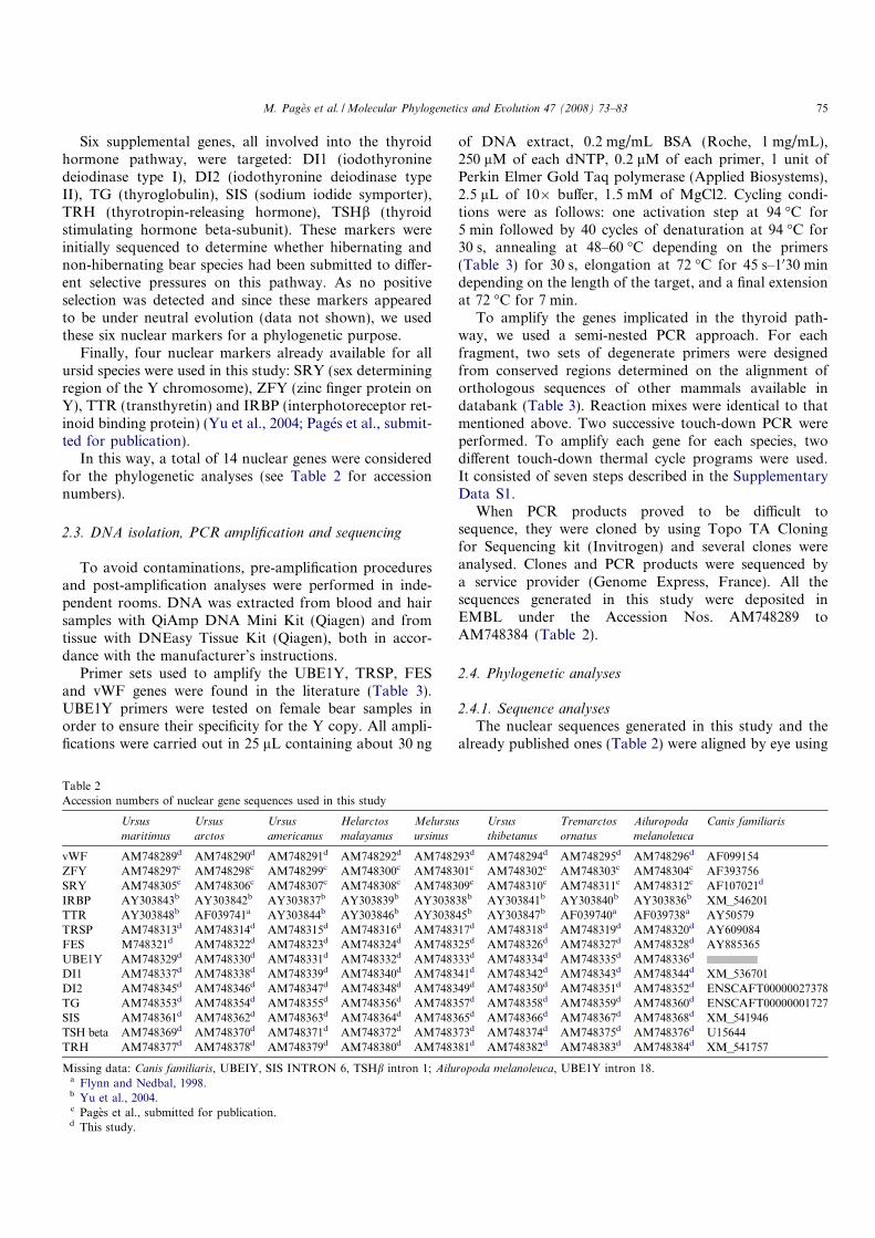
Six supplemental genes, all involved into the thyroidhormone pathway, were targeted: DI1 (iodothyroninedeiodinase type I), DI2 (iodothyronine deiodinase typeII), TG (thyroglobulin), SIS (sodium iodide symporter),TRH (thyrotropin-releasing hormone), TSHb (thyroidstimulating hormone beta-subunit). These markers wereinitially sequenced to determine whether hibernating andnon-hibernating bear species had been submitted to differ-ent selective pressures on this pathway. As no positiveselection was detected and since these markers appearedto be under neutral evolution (data not shown), we usedthese six nuclear markers for a phylogenetic purpose.
Finally, four nuclear markers already available for allursid species were used in this study: SRY (sex determiningregion of the Y chromosome), ZFY (zinc finger protein onY), TTR (transthyretin) and IRBP (interphotoreceptor ret-inoid binding protein) (Yu et al., 2004; Pages et al., submit-ted for publication).
In this way, a total of 14 nuclear genes were consideredfor the phylogenetic analyses (see Table 2 for accessionnumbers).
2.3. DNA isolation, PCR amplification and sequencing
To avoid contaminations, pre-amplification proceduresand post-amplification analyses were performed in inde-pendent rooms. DNA was extracted from blood and hairsamples with QiAmp DNA Mini Kit (Qiagen) and fromtissue with DNEasy Tissue Kit (Qiagen), both in accor-dance with the manufacturer’s instructions.
Primer sets used to amplify the UBE1Y, TRSP, FESand vWF genes were found in the literature (Table 3).UBE1Y primers were tested on female bear samples inorder to ensure their specificity for the Y copy. All ampli-fications were carried out in 25 lL containing about 30 ng
of DNA extract, 0.2 mg/mL BSA (Roche, 1 mg/mL),250 lM of each dNTP, 0.2 lM of each primer, 1 unit ofPerkin Elmer Gold Taq polymerase (Applied Biosystems),2.5 lL of 10� buffer, 1.5 mM of MgCl2. Cycling condi-tions were as follows: one activation step at 94 �C for5 min followed by 40 cycles of denaturation at 94 �C for30 s, annealing at 48–60 �C depending on the primers(Table 3) for 30 s, elongation at 72 �C for 45 s–1030 mindepending on the length of the target, and a final extensionat 72 �C for 7 min.
To amplify the genes implicated in the thyroid path-way, we used a semi-nested PCR approach. For eachfragment, two sets of degenerate primers were designedfrom conserved regions determined on the alignment oforthologous sequences of other mammals available indatabank (Table 3). Reaction mixes were identical to thatmentioned above. Two successive touch-down PCR wereperformed. To amplify each gene for each species, twodifferent touch-down thermal cycle programs were used.It consisted of seven steps described in the SupplementaryData S1.
When PCR products proved to be difficult tosequence, they were cloned by using Topo TA Cloningfor Sequencing kit (Invitrogen) and several clones wereanalysed. Clones and PCR products were sequenced bya service provider (Genome Express, France). All thesequences generated in this study were deposited inEMBL under the Accession Nos. AM748289 toAM748384 (Table 2).
2.4. Phylogenetic analyses
2.4.1. Sequence analyses
The nuclear sequences generated in this study and thealready published ones (Table 2) were aligned by eye using
Table 2
Accession numbers of nuclear gene sequences used in this study
Ursus
maritimus
Ursus
arctos
Ursus
americanus
Helarctos
malayanus
Melursus
ursinus
Ursus
thibetanus
Tremarctos
ornatus
Ailuropoda
melanoleuca
Canis familiaris
vWF AM748289d AM748290d AM748291d AM748292d AM748293d AM748294d AM748295d AM748296d AF099154
ZFY AM748297c AM748298c AM748299c AM748300c AM748301c AM748302c AM748303c AM748304c AF393756
SRY AM748305c AM748306c AM748307c AM748308c AM748309c AM748310c AM748311c AM748312c AF107021d
IRBP AY303843b AY303842b AY303837b AY303839b AY303838b AY303841b AY303840b AY303836b XM_546201
TTR AY303848b AF039741a AY303844b AY303846b AY303845b AY303847b AF039740a AF039738a AY50579
TRSP AM748313d AM748314d AM748315d AM748316d AM748317d AM748318d AM748319d AM748320d AY609084
FES M748321d AM748322d AM748323d AM748324d AM748325d AM748326d AM748327d AM748328d AY885365
UBE1Y AM748329d AM748330d AM748331d AM748332d AM748333d AM748334d AM748335d AM748336d
DI1 AM748337d AM748338d AM748339d AM748340d AM748341d AM748342d AM748343d AM748344d XM_536701
DI2 AM748345d AM748346d AM748347d AM748348d AM748349d AM748350d AM748351d AM748352d ENSCAFT00000027378
TG AM748353d AM748354d AM748355d AM748356d AM748357d AM748358d AM748359d AM748360d ENSCAFT00000001727
SIS AM748361d AM748362d AM748363d AM748364d AM748365d AM748366d AM748367d AM748368d XM_541946
TSH beta AM748369d AM748370d AM748371d AM748372d AM748373d AM748374d AM748375d AM748376d U15644
TRH AM748377d AM748378d AM748379d AM748380d AM748381d AM748382d AM748383d AM748384d XM_541757
Missing data: Canis familiaris, UBEIY, SIS INTRON 6, TSHb intron 1; Ailuropoda melanoleuca, UBE1Y intron 18.a Flynn and Nedbal, 1998.b Yu et al., 2004.c Pages et al., submitted for publication.d This study.
M. Pages et al. /Molecular Phylogenetics and Evolution 47 (2008) 73–83 75
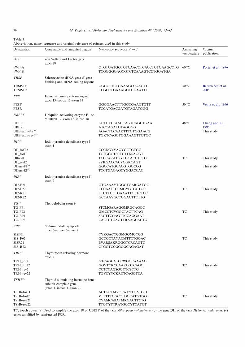
Table 3
Abbreviation, name, sequence and original reference of primers used in this study
Designation Gene name and amplified region Nucleotide sequence 50 ? 30 Annealing
temperature
Original
publication
vWF von Willebrand Factor gene
exon 28
vWF-A CTGTGATGGTGTCAACCTCACCTGTGAAGCCTG 60 �C Porter et al., 1996
vWF-B TCGGGGGAGCGTCTCAAAGTCCTGGATGA
TRSP Selenocyteine tRNA gene 50 gene-
flanking and tRNA coding regions
TRSP-1F
TRSP-1R
GGGCTTCTGAAAGCCGACTT
CCGCCCGAAAGGTGGAATTG
50 �C Bardeleben et al.,
2005
FES Feline sarcoma protooncogene
exon 13–intron 13–exon 14
FESF
FESR
GGGGAACTTTGGCGAAGTGTT
TCCATGACGATGTAGATGGG
50 �C Venta et al., 1996
UBE1Y Ubiquitin activating enzyme E1 on
Y intron 17–exon 18–intron 18
UBEF
UBER
GCTCTTCAAGCAGTCAGCTGAA
ATCCAGATGTAGGGG
48 �C Chang and Li,
1995
UBE-exon-forl(a) AGACTCCAAKTTTGTGGAACG This study
UBE-exon-revl(a) TGKTCAGGTGGAAAGTTGTGC
DI1(c) Iodothyronine deiodinase type I
exon 1
DIl_forT2 CCCDGYYAGYGCTGTGG
DIl_forl3 TCTGGGTSCTCTTKSAGGT
Dllrevll TCCCARATGYTGCACCTCTG TC This study
DIl_revl2 SYKGACCACVGGRCAGT
DIlurs-Fl(b) GGCCATGCACGTGGCCG This study
DIlurs-Rl(b) TCCTGAGAGCYGGACCAC
DI2(c) Iodothyronine deiodinase type II
exon 2
DI2-F21 GTGAAAYTGGGTGARGATGC
DI2-F22 CCCAATTCCMGYGTGGTGC TC This study
DI2-R21 CTCTTGCTGAAATTCTTCTCC
DI2-R22 GCCAAYGCCGGACTTCTTG
TG(c) Thyroglobulin exon 9
TG-F91 STCMGARAGGMRGCAGGC
TG-F92 GMCCTCVGGCTACTTCAG TC This study
TG-R91 SRCTTCGAGTTCCAGGAAT
TG-R92 CACTCTGAGTTRAAGCACTG
SIS(c) Sodium iodide symporter
exon 6–intron 6–exon 7
SISF61 CYKGACCCGMGGMGCCG
SIS_F62 GCCGCTAYACMTTCTGGAC TC This study
SISR71 BVARSAKRGGGTCRCAGTC
SIS_R72 CTGGTCCGGGGCAGAGAT
TRH(c) Thyrotropin-releasing hormone
exon 2
TRH_for2 GTCAGCATCCWGGCAAAAG
TRH_for22 GGYTCKCCAARCGTCAGC TC This study
TRH_rev2 CCTCCAGSGGYTCSCTG
TRH_rev22 TGVCYYCKRCTCAGGTCA
TSHB(c) Thyroid stimulating hormone beta-
subunit complete gene
(exon 1–intron 1–exon 2)
TSHb-for11 ACTGCTMYCTWYYTGATGTC
TSHb-forl2 VTTTTTGGCCTDGCATGTGG TC This study
TSHb-rev21 CYAMCABATMRGACTTCTG
TSHb-rev22 TTGYYTTRATGGCYTCATGT
TC, touch down. (a) Used to amplify the exon 18 of UBE1Y of the taxa Ailuropoda melanoleuca; (b) the gene DI1 of the taxa Helarctos malayanus. (c)
genes amplified by semi-nested PCR.
76 M. Pages et al. /Molecular Phylogenetics and Evolution 47 (2008) 73–83
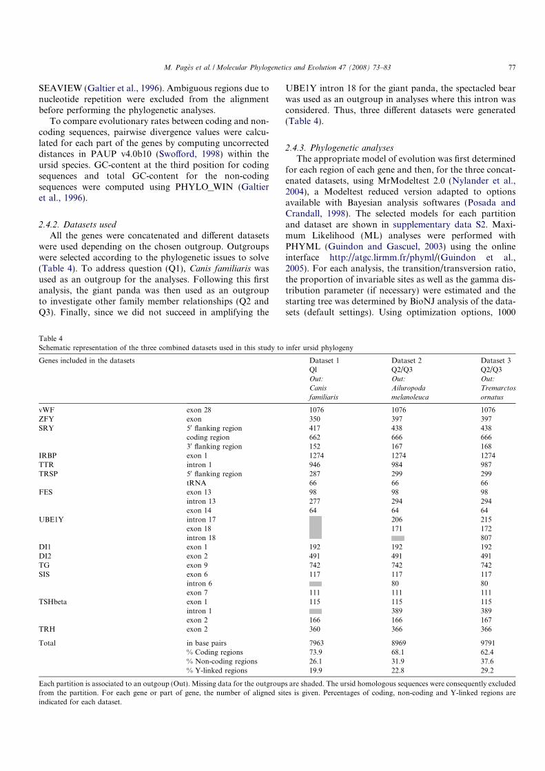
SEAVIEW (Galtier et al., 1996). Ambiguous regions due tonucleotide repetition were excluded from the alignmentbefore performing the phylogenetic analyses.
To compare evolutionary rates between coding and non-coding sequences, pairwise divergence values were calcu-lated for each part of the genes by computing uncorrecteddistances in PAUP v4.0b10 (Swofford, 1998) within theursid species. GC-content at the third position for codingsequences and total GC-content for the non-codingsequences were computed using PHYLO_WIN (Galtieret al., 1996).
2.4.2. Datasets used
All the genes were concatenated and different datasetswere used depending on the chosen outgroup. Outgroupswere selected according to the phylogenetic issues to solve(Table 4). To address question (Q1), Canis familiaris wasused as an outgroup for the analyses. Following this firstanalysis, the giant panda was then used as an outgroupto investigate other family member relationships (Q2 andQ3). Finally, since we did not succeed in amplifying the
UBE1Y intron 18 for the giant panda, the spectacled bearwas used as an outgroup in analyses where this intron wasconsidered. Thus, three different datasets were generated(Table 4).
2.4.3. Phylogenetic analyses
The appropriate model of evolution was first determinedfor each region of each gene and then, for the three concat-enated datasets, using MrModeltest 2.0 (Nylander et al.,2004), a Modeltest reduced version adapted to optionsavailable with Bayesian analysis softwares (Posada andCrandall, 1998). The selected models for each partitionand dataset are shown in supplementary data S2. Maxi-mum Likelihood (ML) analyses were performed withPHYML (Guindon and Gascuel, 2003) using the onlineinterface http://atgc.lirmm.fr/phyml/(Guindon et al.,2005). For each analysis, the transition/transversion ratio,the proportion of invariable sites as well as the gamma dis-tribution parameter (if necessary) were estimated and thestarting tree was determined by BioNJ analysis of the data-sets (default settings). Using optimization options, 1000
Table 4
Schematic representation of the three combined datasets used in this study to infer ursid phylogeny
Genes included in the datasets Dataset 1 Dataset 2 Dataset 3
Ql Q2/Q3 Q2/Q3
Out: Out: Out:
Canis Ailuropoda Tremarctos
familiaris melanoleuca ornatus
vWF exon 28 1076 1076 1076
ZFY exon 350 397 397
SRY 50 flanking region 417 438 438
coding region 662 666 666
30 flanking region 152 167 168
IRBP exon 1 1274 1274 1274
TTR intron 1 946 984 987
TRSP 50 flanking region 287 299 299
tRNA 66 66 66
FES exon 13 98 98 98
intron 13 277 294 294
exon 14 64 64 64
UBE1Y intron 17 206 215
exon 18 171 172
intron 18 807
DI1 exon 1 192 192 192
DI2 exon 2 491 491 491
TG exon 9 742 742 742
SIS exon 6 117 117 117
intron 6 80 80
exon 7 111 111 111
TSHbeta exon 1 115 115 115
intron 1 389 389
exon 2 166 166 167
TRH exon 2 360 366 366
Total in base pairs 7963 8969 9791
% Coding regions 73.9 68.1 62.4
% Non-coding regions 26.1 31.9 37.6
% Y-linked regions 19.9 22.8 29.2
Each partition is associated to an outgoup (Out). Missing data for the outgroups are shaded. The ursid homologous sequences were consequently excluded
from the partition. For each gene or part of gene, the number of aligned sites is given. Percentages of coding, non-coding and Y-linked regions are
indicated for each dataset.
M. Pages et al. /Molecular Phylogenetics and Evolution 47 (2008) 73–83 77
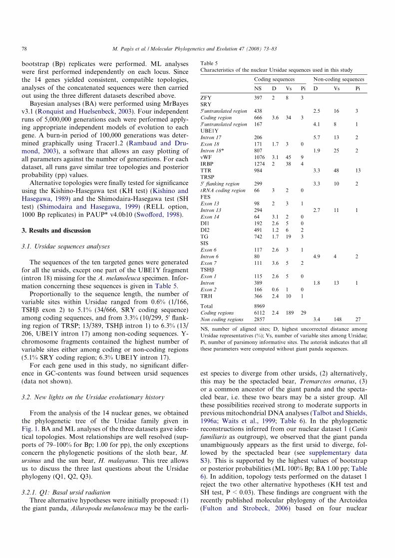
bootstrap (Bp) replicates were performed. ML analyseswere first performed independently on each locus. Sincethe 14 genes yielded consistent, compatible topologies,analyses of the concatenated sequences were then carriedout using the three different datasets described above.
Bayesian analyses (BA) were performed using MrBayesv3.1 (Ronquist and Huelsenbeck, 2003). Four independentruns of 5,000,000 generations each were performed apply-ing appropriate independent models of evolution to eachgene. A burn-in period of 100,000 generations was deter-mined graphically using Tracer1.2 (Rambaud and Dru-mond, 2003), a software that allows an easy plotting ofall parameters against the number of generations. For eachdataset, all runs gave similar tree topologies and posteriorprobability (pp) values.
Alternative topologies were finally tested for significanceusing the Kishino-Hasegawa test (KH test) (Kishino andHasegawa, 1989) and the Shimodaira-Hasegawa test (SHtest) (Shimodaira and Hasegawa, 1999) (RELL option,1000 Bp replicates) in PAUP* v4.0b10 (Swofford, 1998).
3. Results and discussion
3.1. Ursidae sequences analyses
The sequences of the ten targeted genes were generatedfor all the ursids, except one part of the UBE1Y fragment(intron 18) missing for the A. melanoleuca specimen. Infor-mation concerning these sequences is given in Table 5.
Proportionally to the sequence length, the number ofvariable sites within Ursidae ranged from 0.6% (1/166,TSHb exon 2) to 5.1% (34/666, SRY coding sequence)among coding sequences, and from 3.3% (10/299, 50 flank-ing region of TRSP; 13/389, TSHb intron 1) to 6.3% (13/206, UBE1Y intron 17) among non-coding sequences. Y-chromosome fragments contained the highest number ofvariable sites either among coding or non-coding regions(5.1% SRY coding region; 6.3% UBE1Y intron 17).
For each gene used in this study, no significant differ-ence in GC-contents was found between ursid sequences(data not shown).
3.2. New lights on the Ursidae evolutionary history
From the analysis of the 14 nuclear genes, we obtainedthe phylogenetic tree of the Ursidae family given inFig. 1. BA and ML analyses of the three datasets gave iden-tical topologies. Most relationships are well resolved (sup-ports of 79–100% for Bp; 1.00 for pp), the only exceptionsconcern the phylogenetic positions of the sloth bear, M.
ursinus and the sun bear, H. malayanus. This tree allowsus to discuss the three last questions about the Ursidaephylogeny (Q1, Q2, Q3).
3.2.1. Q1: Basal ursid radiation
Three alternative hypotheses were initially proposed: (1)the giant panda, Ailuropoda melanoleuca may be the earli-
est species to diverge from other ursids, (2) alternatively,this may be the spectacled bear, Tremarctos ornatus, (3)or a common ancestor of the giant panda and the specta-cled bear, i.e. these two bears may be a sister group. Allthese possibilities received strong to moderate supports inprevious mitochondrial DNA analyses (Talbot and Shields,1996a; Waits et al., 1999; Table 6). In the phylogeneticreconstructions inferred from our nuclear dataset 1 (Canisfamiliaris as outgroup), we observed that the giant pandaunambiguously appears as the first ursid to diverge, fol-lowed by the spectacled bear (see supplementary dataS3). This is supported by the highest values of bootstrapor posterior probabilities (ML 100% Bp; BA 1.00 pp; Table6). In addition, topology tests performed on the dataset 1reject the two other alternative hypotheses (KH test andSH test, P < 0.03). These findings are congruent with therecently published molecular phylogeny of the Arctoidea(Fulton and Strobeck, 2006) based on four nuclear
Table 5
Characteristics of the nuclear Ursidae sequences used in this study
Coding sequences Non-coding sequences
NS D Vs Pi D Vs Pi
ZFY 397 2 8 3
SRY
50untranslated region 438 2.5 16 3
Coding region 666 3.6 34 3
30untranslated region 167 4.1 8 1
UBE1Y
Intron 17 206 5.7 13 2
Exon 18 171 1.7 3 0
Intron 18* 807 1.9 25 2
vWF 1076 3.1 45 9
IRBP 1274 2 38 4
TTR 984 3.3 48 13
TRSP
50 flanking region 299 3.3 10 2
tRNA coding region 66 3 2 0
FES
Exon 13 98 2 3 1
Intron 13 294 2.7 11 1
Exon 14 64 3.1 2 0
DI1 192 2.6 5 0
DI2 491 1.2 6 2
TG 742 1.7 19 3
SIS
Exon 6 117 2.6 3 1
Intron 6 80 4.9 4 2
Exon 7 111 3.6 5 2
TSHb
Exon 1 115 2.6 5 0
Intron 389 1.8 13 1
Exon 2 166 0.6 1 0
TRH 366 2.4 10 1
Total 8969
Coding regions 6112 2.4 189 29
Non coding regions 2857 3.4 148 27
NS, number of aligned sites; D, highest uncorrected distance among
Ursidae representatives (%); Vs, number of variable sites among Ursidae;
Pi, number of parsimony informative sites. The asterisk indicates that all
these parameters were computed without giant panda sequences.
78 M. Pages et al. /Molecular Phylogenetics and Evolution 47 (2008) 73–83
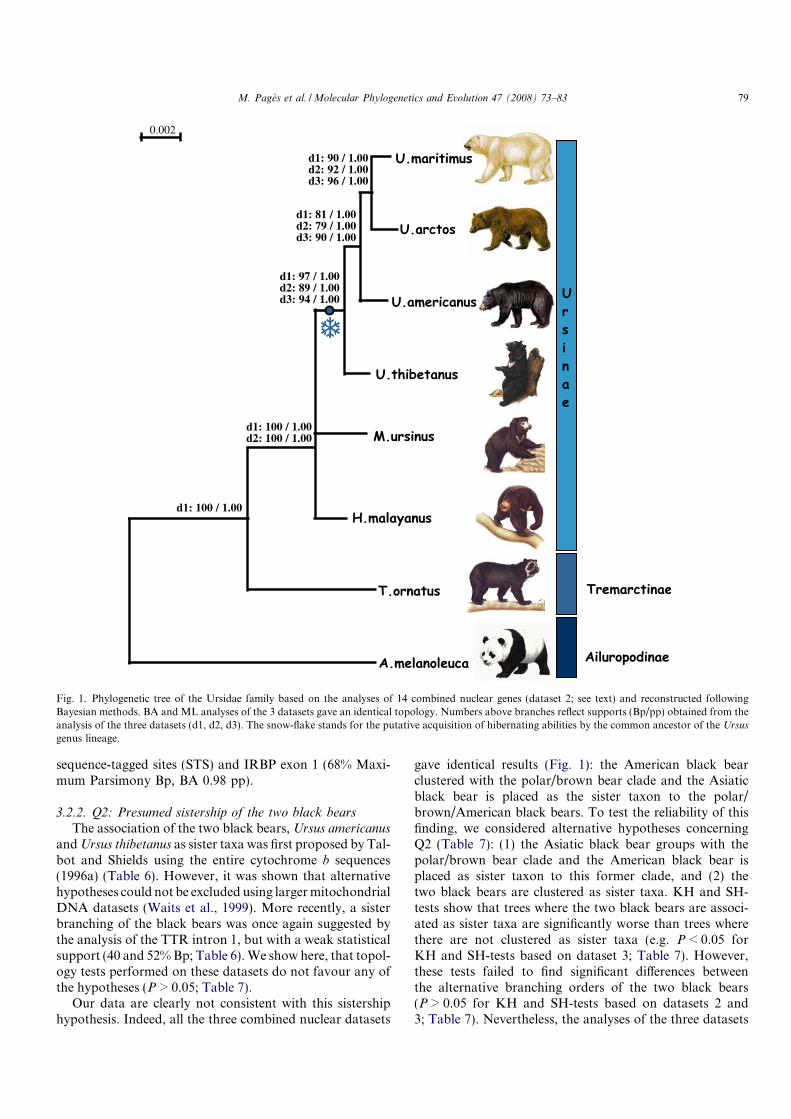
sequence-tagged sites (STS) and IRBP exon 1 (68% Maxi-mum Parsimony Bp, BA 0.98 pp).
3.2.2. Q2: Presumed sistership of the two black bears
The association of the two black bears,Ursus americanus
andUrsus thibetanus as sister taxa was first proposed by Tal-bot and Shields using the entire cytochrome b sequences(1996a) (Table 6). However, it was shown that alternativehypotheses could not be excluded using largermitochondrialDNA datasets (Waits et al., 1999). More recently, a sisterbranching of the black bears was once again suggested bythe analysis of the TTR intron 1, but with a weak statisticalsupport (40 and 52%Bp; Table 6).We show here, that topol-ogy tests performed on these datasets do not favour any ofthe hypotheses (P > 0.05; Table 7).
Our data are clearly not consistent with this sistershiphypothesis. Indeed, all the three combined nuclear datasets
gave identical results (Fig. 1): the American black bearclustered with the polar/brown bear clade and the Asiaticblack bear is placed as the sister taxon to the polar/brown/American black bears. To test the reliability of thisfinding, we considered alternative hypotheses concerningQ2 (Table 7): (1) the Asiatic black bear groups with thepolar/brown bear clade and the American black bear isplaced as sister taxon to this former clade, and (2) thetwo black bears are clustered as sister taxa. KH and SH-tests show that trees where the two black bears are associ-ated as sister taxa are significantly worse than trees wherethere are not clustered as sister taxa (e.g. P < 0.05 forKH and SH-tests based on dataset 3; Table 7). However,these tests failed to find significant differences betweenthe alternative branching orders of the two black bears(P > 0.05 for KH and SH-tests based on datasets 2 and3; Table 7). Nevertheless, the analyses of the three datasets
0.002
d1: 90 / 1.00
d2: 92 / 1.00
d3: 96 / 1.00
d1: 81 / 1.00
d2: 79 / 1.00
d3: 90 / 1.00
d1: 97 / 1.00
d2: 89 / 1.00
d3: 94 / 1.00
d1: 100 / 1.00
d2: 100 / 1.00
d1: 100 / 1.00
Fig. 1. Phylogenetic tree of the Ursidae family based on the analyses of 14 combined nuclear genes (dataset 2; see text) and reconstructed following
Bayesian methods. BA and ML analyses of the 3 datasets gave an identical topology. Numbers above branches reflect supports (Bp/pp) obtained from the
analysis of the three datasets (d1, d2, d3). The snow-flake stands for the putative acquisition of hibernating abilities by the common ancestor of the Ursus
genus lineage.
M. Pages et al. /Molecular Phylogenetics and Evolution 47 (2008) 73–83 79
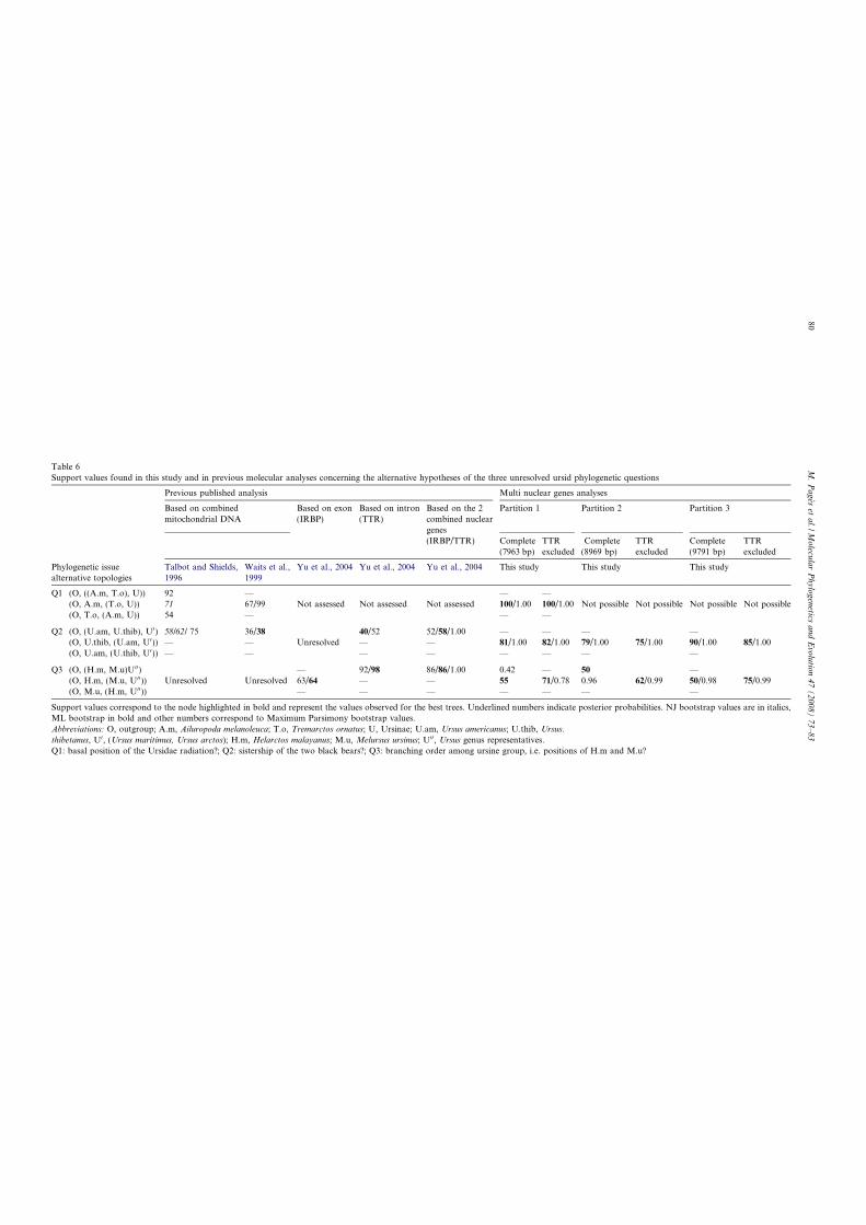
Table 6
Support values found in this study and in previous molecular analyses concerning the alternative hypotheses of the three unresolved ursid phylogenetic questions
Previous published analysis Multi nuclear genes analyses
Based on combined
mitochondrial DNA
Based on exon
(IRBP)
Based on intron
(TTR)
Based on the 2
combined nuclear
genes
Partition 1 Partition 2 Partition 3
(IRBP/TTR) Complete
(7963 bp)
TTR
excluded
Complete
(8969 bp)
TTR
excluded
Complete
(9791 bp)
TTR
excluded
Phylogenetic issue
alternative topologies
Talbot and Shields,
1996
Waits et al.,
1999
Yu et al., 2004 Yu et al., 2004 Yu et al., 2004 This study This study This study
Q1 (O, ((A.m, T.o), U)) 92 — — —
(O, A.m, (T.o, U)) 71 67/99 Not assessed Not assessed Not assessed 100/1.00 100/1.00 Not possible Not possible Not possible Not possible
(O, T.o, (A.m, U)) 54 — — —
Q2 (O, (U.am, U.thib), U0) 58/62/ 75 36/38 40/52 52/58/1.00 — — — —
(O, U.thib, (U.am, U0)) — — Unresolved — — 81/1.00 82/1.00 79/1.00 75/1.00 90/1.00 85/1.00
(O, U.am, (U.thib, U0)) — — — — — — — —
Q3 (O, (H.m, M.u)U00) — 92/98 86/86/1.00 0.42 — 50 —
(O, H.m, (M.u, U00)) Unresolved Unresolved 63/64 — — 55 71/0.78 0.96 62/0.99 50/0.98 75/0.99
(O, M.u, (H.m, U00)) — — — — — — —
Support values correspond to the node highlighted in bold and represent the values observed for the best trees. Underlined numbers indicate posterior probabilities. NJ bootstrap values are in italics,
ML bootstrap in bold and other numbers correspond to Maximum Parsimony bootstrap values.
Abbreviations: O, outgroup; A.m, Ailuropoda melanoleuca; T.o, Tremarctos ornatus; U, Ursinae; U.am, Ursus americanus; U.thib, Ursus.
thibetanus, U0, (Ursus maritimus, Ursus arctos); H.m, Helarctos malayanus; M.u, Melursus ursinus; U00, Ursus genus representatives.
Q1: basal position of the Ursidae radiation?; Q2: sistership of the two black bears?; Q3: branching order among ursine group, i.e. positions of H.m and M.u?
80
M.Pages
etal./M
olecu
larPhylogenetics
andEvolutio
n47(2008)73–83
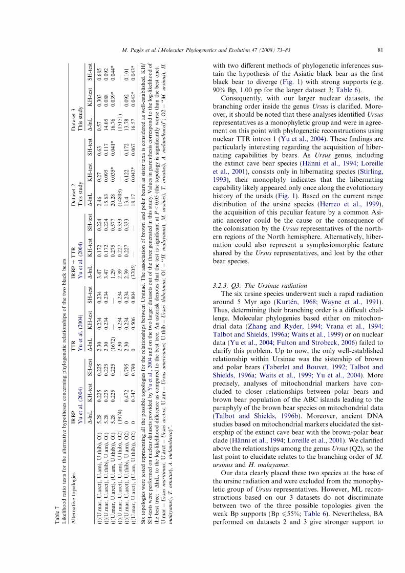
with two different methods of phylogenetic inferences sus-tain the hypothesis of the Asiatic black bear as the firstblack bear to diverge (Fig. 1) with strong supports (e.g.90% Bp, 1.00 pp for the larger dataset 3; Table 6).
Consequently, with our larger nuclear datasets, thebranching order inside the genus Ursus is clarified. More-over, it should be noted that these analyses identified Ursus
representatives as a monophyletic group and were in agree-ment on this point with phylogenetic reconstructions usingnuclear TTR intron 1 (Yu et al., 2004). These findings areparticularly interesting regarding the acquisition of hiber-nating capabilities by bears. As Ursus genus, includingthe extinct cave bear species (Hanni et al., 1994; Loreilleet al., 2001), consists only in hibernating species (Stirling,1993), their monophyly indicates that the hibernatingcapability likely appeared only once along the evolutionaryhistory of the ursids (Fig. 1). Based on the current rangedistribution of the ursine species (Herreo et al., 1999),the acquisition of this peculiar feature by a common Asi-atic ancestor could be the cause or the consequence ofthe colonisation by the Ursus representatives of the north-ern regions of the North hemisphere. Alternatively, hiber-nation could also represent a symplesiomorphic featureshared by the Ursus representatives, and lost by the otherbear species.
3.2.3. Q3: The Ursinae radiation
The six ursine species underwent such a rapid radiationaround 5 Myr ago (Kurten, 1968; Wayne et al., 1991).Thus, determining their branching order is a difficult chal-lenge. Molecular phylogenies based either on mitochon-drial data (Zhang and Ryder, 1994; Vrana et al., 1994;Talbot and Shields, 1996a; Waits et al., 1999) or on nucleardata (Yu et al., 2004; Fulton and Strobeck, 2006) failed toclarify this problem. Up to now, the only well-establishedrelationship within Ursinae was the sistership of brownand polar bears (Taberlet and Bouvet, 1992; Talbot andShields, 1996a; Waits et al., 1999; Yu et al., 2004). Moreprecisely, analyses of mitochondrial markers have con-cluded to closer relationships between polar bears andbrown bear population of the ABC islands leading to theparaphyly of the brown bear species on mitochondrial data(Talbot and Shields, 1996b). Moreover, ancient DNAstudies based on mitochondrial markers elucidated the sist-ersphip of the extinct cave bear with the brown-polar bearclade (Hanni et al., 1994; Loreille et al., 2001). We clarifiedabove the relationships among the genus Ursus (Q2), so thelast point to elucidate relates to the branching order of M.
ursinus and H. malayanus.Our data clearly placed these two species at the base of
the ursine radiation and were excluded from the monophy-letic group of Ursus representatives. However, ML recon-structions based on our 3 datasets do not discriminatebetween two of the three possible topologies given theweak Bp supports (Bp 655%; Table 6). Nevertheless, BAperformed on datasets 2 and 3 give stronger support toT
able
7
Likelihoodratiotestsforthealternativehypotheses
concerningphylogenetic
relationshipsofthetw
oblack
bears
Alternativetopologies
IRBP
TTR
IRBP+
TTR
Dataset2
Dataset3
Yuet
al.(2004)
Yuet
al.(2004)
Yuet
al.(2004)
Thisstudy
Thisstudy
D-lnL
KH-test
SH-test
D-lnL
KH-test
SH-test
D-lnL
KH-test
SH-test
D-lnL
KH-test
SH-test
D-lnL
KH-test
SH-test
((((U.m
ar,U.arct),U.am),U.thib),Ol)
5.28
0.225
0.225
2.30
0.234
0.234
3.47
0.172
0.224
2.46
0.27
0.63
0.57
0.303
0.685
((((U.m
ar,U.arct),U.thib),U.am),Ol)
5.28
0.225
0.225
2.30
0.234
0.234
3.47
0.172
0.224
15.63
0.095
0.117
14.05
0.088
0.092
(((U
.mar,U.arct),(U
.am,U.thib)),Ol)
5.28
0.225
0.225
(1672)
——
1.29
0.275
0.577
20.28
0.035*
0.041*
16.76
0.039*
0.044*
((((U.m
ar,U.arct),U.am),U.thib),O2)
(1974)
——
2.30
0.234
0.234
2.39
0.227
0.333
(14803)
——
(15151)
——
((((U.m
ar,U.arct),U.thib),U.am),O2)
00.472
0.795
2.30
0.234
0.234
2.39
0.227
0.333
13.4
0.122
0.172
13.78
0.092
0.101
(((U
.mar,U.arct),(U
.am,U.thib)),O2)
00.347
0.790
00.506
0.804
(3705)
——
18.17
0.042*
0.067
16.57
0.042*
0.043*
Six
topologiesweretested
representingallthepossibletopologiesfortherelationshipsbetweenUrsinae.Theassociationofbrownandpolarbears
assister
taxaisconsidered
aswell-established.KH/
SH-testswereperform
edonnucleardatasetsprovided
byYuet
al.,2004andonthetw
olarger
datasetsoutofthethreegeneratedin
thisstudy.Values
inparenthesiscorrespondto
thelog-likelihoodof
thebesttree;�DlnL,to
thelog-likelihooddifference
ascomparedto
thebesttree.Anasteriskdenotesthatthetest
issignificantatP<0.05(thetopologyissignificantlyworsethanthebestone).
U.m
ar=
Ursusmaritimus;
U.arct=
Ursusarctos;
U.am
=Ursusamericanus;
U.thib
=Ursusthibetanus;
O1=
‘‘H.malayanus),M.ursinus),T.ornatus),A.melanoleuca)”;O2=
‘‘M.ursinus),H.
malayanus),T.ornatus),A.melanoleuca)”.
M. Pages et al. /Molecular Phylogenetics and Evolution 47 (2008) 73–83 81
M. ursinus clustered with the representatives of the genusUrsus (pp values of 0.96 and 0.98).
Combined genes could lead to an unresolved topology ifthe genes give contradictory results. In this case, Jeffroyet al. (2006) showed that it could be better to remove theproblematic genes from the analyses. In their study com-bining IRBP and TTR, Yu et al. (2004) observed that bothgenes taken separately gave contradictory results: IRBPsustained the hypothesis of M. ursinus clustered with therepresentatives of the genus Ursus, whereas TTR supportedthe M. ursinus and H. malayanus sistership (Table 6). Inorder to assess whether TTR is responsible for the low sup-ports observed, we performed the same phylogenetic anal-yses on our three datasets but excluding this marker. In thiscase, a single topology is well supported: M. ursinus clus-ters with the representatives of the genus Ursus. The exclu-sion of TTR increases all the support values for thishypothesis in both ML and BA analyses (e.g. Bp from50–75% for dataset 3; Table 6). Nevertheless, further workis required to confirm the positions of the sloth and sunbears within the ursid tree.
3.2.4. Further investigations
In this study, we resolved the main ambiguities amongthe phylogeny of Ursidae (Q1 and Q2) and clarified the lastone (Q3). Even the combination of 14 nuclear gene frag-ments was not informative enough to solve definitivelythe relationships between all the representatives of theUrsinae sub-family (Q3). This lack of resolution is certainlydue to the rapid burst of ursine species during bear evolu-tionary history. A solution to circumvent this difficultycould be to identify Rare Genomic Changes (RGCs) inthe Ursidae genomes (Springer et al., 2004). RGCs wouldbe perfect arbiters and will certainly be needed, as thereis little chance that increasing the nuclear sequence datasetwill provide any further information. Searching RGCscould be directed by two theoretical and empirical points:(1) insertions, deletions, and retrotransposon integrationsoccur more likely in non-coding regions (Springer et al.,2004); (2) as other restricted regions of the genome withlimited recombination, the non-recombining region of theY chromosome is expected to accumulate more retrotrans-poson elements (Pecon-Slattery and O’Brien, 1998; Pecon-Slattery et al., 2004). So, scanning Y-linked chromosomeintron sequences to find RGCs in bear genomes couldprove to be useful to overcome the lack of phylogenetic sig-nal among ursids. We are confident that these additionalindependent markers will consolidate the Ursidae tree.
Acknowledgments
We thank all the institutions that gave samples for thisstudy: the MNHN (Paris), the Mammals Research Insti-tute of Poland and Zoos of Moscow, Leipzig, Berlin, andin France: La Palmyre, Peaugres, Saint Martin La Plaine,Vincennes, Parc de la Tete d’Or de Lyon, Reserve Africainede Sigean and more particularly I. Delisle (University of
Alberta, Canada), G. Veron (MNHN, France), F. Catzeflis(CNRS, ISEM, France), and P. Taberlet (LECA, France).We are particularly grateful to M. Duffraisse for help in thelab, to P. Chevret, L. Orlando, A. Chaumont and V. Lau-det for helpful discussions and critical reading, and to J.J.Jaeger for constant support. We wish to acknowledge J.Carew for English corrections. We finally thank the CNRS,Universite Lyon 1, ENS de Lyon and MNRT for financialsupports. M.P. holds a fellowship from the ARCFoundation.
Appendix A. Supplementary data
Supplementary data associated with this article can befound, in the online version, at doi:10.1016/j.ympev.2007.10.019.
References
Bardeleben, C., Moore, R.L., Wayne, R.K., 2005. A molecular phylogeny
of the Canidae based on six nuclear loci. Mol. Phylogenet. Evol. 37,
815–831.
Chang, B.H., Li, W.H., 1995. Estimating the intensity of male-driven
evolution in rodents by using X-linked and Y-linked Ube 1 genes and
pseudogenes. J. Mol. Evol. 40, 70–77.
Davis, D., 1964. The giant panda: a morphological study of evolutionary
mechanisms. Fieldiana. Zool. Mem. 3, 1–339.
Flynn, J.J., Nedbal, M.A., 1998. Phylogeny of the Carnivora (Mammalia):
congruence vs incompatibility among multiple data sets. Mol. Phylo-
genet. Evol. 9, 414–426.
Fulton, T.L., Strobeck, C., 2006. Molecular phylogeny of the Arctoidea
(Carnivora): effect of missing data on supertree and supermatrix
analyses of multiple gene data sets. Mol. Phylogenet. Evol. 41, 165–
181.
Gadagkar, S.R., Rosenberg, M.S., Kumar, S., 2005. Inferring species
phylogenies from multiple genes: concatenated sequence tree versus
consensus gene tree. J. Exp. Zoolog. B Mol. Dev. Evol. 304, 64–74.
Galewski, T., Mauffrey, J.F., Leite, Y.L., Patton, J.L., Douzery, E.J.,
2005. Ecomorphological diversification among South American spiny
rats (Rodentia; Echimyidae): a phylogenetic and chronological
approach. Mol. Phylogenet. Evol 34, 601–615.
Galtier, N., Gouy, M., Gautier, C., 1996. SEAVIEW and PHYLO_WIN:
two graphic tools for sequence alignment and molecular phylogeny.
Comput. Appl. Biosci. 12, 543–548.
Guindon, S., Gascuel, O., 2003. A simple, fast, and accurate algorithm to
estimate large phylogenies by maximum likelihood. Syst. Biol. 52, 696–
704.
Guindon, S., Lethiec, F., Duroux, P., Gascuel, O., 2005. PHYML
Online—a web server for fast maximum likelihood-based phylogenetic
inference. Nucleic Acids Res. 33, W557–W559.
Haldane, J.B.S., 1947. The mutation rate of the gene for haemophilia, and
its segregation ratios in males and females. Ann. Eugen. 13, 262–271.
Hanni, C., Laudet, V., Stehelin, D., Taberlet, P., 1994. Tracking the
origins of the cave bear (Ursus spelaeus) by mitochondrial DNA
sequencing. Proc. Natl. Acad. Sci. USA 91, 12336–12340.
Herreo, S. Compiled by Servheen, C., Herrero, H., Peyton, B. and the
IUCN/SSC Bear and Polar Bear Specialist Groups (1999). Introduc-
tion, In: Bears: Status Survey and Conservation Action Plan, pp. 1–6.
IUCN.
Jeffroy, O., Brinkmann, H., Delsuc, F., Philippe, H., 2006. Phylogenomics:
the beginning of incongruence? Trends Genet. 22, 225–231.
Kishino, H., Hasegawa, M., 1989. Evaluation of the maximum likelihood
estimate of the evolutionary tree topologies from DNA sequence data,
and the branching order in Hominoidea. J. Mol. Evol. 29, 170–179.
82 M. Pages et al. /Molecular Phylogenetics and Evolution 47 (2008) 73–83
Kurten, B., 1968. The Pleistocene Mammals of Europe. Aldine, Chicago,
III.
Lessells, K., 1997. More mutations in males. Nature 390, 236–237.
Loreille, O., Orlando, L., Patou-Mathis, M., et al., 2001. Ancient DNA
analysis reveals divergence of the cave bear, Ursus spelaeus, and brown
bear, Ursus arctos, lineages. Curr. Biol. 11, 200–203.
Mayr, E., 1986. Uncertainty in science: is the giant panda a bear or a
raccoon? Nature 323, 769–771.
Moreira, M.A., 2002. SRY evolution in Cebidae (Platyrrhini: Primates). J.
Mol. Evol. 55, 92–103.
Nishida, S., Pastene, A.L., Goto, M., Koike, H., 2003. SRY gene
structure and phylogeny in the cetacean species. Mammal Study
28, 57–66.
Nylander, J.A., Ronquist, F., Huelsenbeck, J.P., Nieves-Aldrey, J.L.,
2004. Bayesian phylogenetic analysis of combined data. Syst. Biol. 53,
47–67.
O’Brien, S.J., Nash, W.G., Wildt, D.E., Bush, M.E., Benveniste, R.E.,
1985. A molecular solution to the riddle of the giant panda’s
phylogeny. Nature 317, 140–144.
Pages, M., Maudet, C., Bellemain, E., Taberlet, P., Hughes, S., Hanni, C.,
submitted for publication. A system for sex-determination from
degraded DNA: a useful tool for paleogenetics and conservation
genetics of Ursids.
Pecon-Slattery, J., O’Brien, S.J., 1998. Patterns of Y and X chromosome
DNA sequence divergence during the Felidae radiation. Genetics 148,
1245–1255.
Pecon-Slattery, J., Pearks Wilkerson, A.J., Murphy, W.J., O’Brien, S.J.,
2004. Phylogenetic assessment of introns and SINEs within the Y
chromosome using the cat family felidae as a species tree. Mol. Biol.
Evol. 21, 2299–2309.
Porter, C.A., Goodman, M., Stanhope, M.J., 1996. Evidence on
mammalian phylogeny from sequences of exon 28 of the von
Willebrand factor gene. Mol. Phylogenet. Evol. 5, 89–101.
Posada, D., Crandall, KA., 1998. MODELTEST: testing the model of
DNA substitution. Bioinformatics 14, 817–818.
Rambaud A., Drumond A., 2003. Tracer, version v1.2.
Ronquist, F., Huelsenbeck, J.P., 2003. MrBayes 3: Bayesian phylogenetic
inference under mixed models. Bioinformatics 19, 1572–1574.
Shimodaira, H., Hasegawa, M., 1999. Multiple comparisons of log-
likelihoods with applications to phylogenetic inference. Mol. Biol.
Evol. 16, 1114–1116.
Springer, M.S., Stanhope, M.J., Madsen, O., de Jong, W.W., 2004.
Molecules consolidate the placental mammal tree. Trends Ecol. Evol.
19, 430–438.
Stirling, I., 1993. Bears-majestic creatures of the wild. Rodale Press
Emmaus, Pennsylvania.
Swofford, D.L., 1998. PAUP*. Phylogenetic Analysis using Parsimony
(*and other methods). Version 4. Sinauer Associates, Sunderland,
Massachusetts.
Taberlet, P., Bouvet, J., 1992. Genetique de l’Ours brun des Pyenees
(Ursus arctos): premiers resultats. CR Acad. Sci. III. 314, 15–21.
Talbot, S.L., Shields, G.F., 1996a. A phylogeny of the bears (Ursidae)
inferred from complete sequences of three mitochondrial genes. Mol.
Phylogenet. Evol. 5, 567–575.
Talbot, S.L., Shields, G.F., 1996b. Phylogeography of brown bears (Ursus
arctos) of Alaska and paraphyly within the Ursidae. Mol. Phylogenet.
Evol. 5, 477–494.
Van Valen, L.M., 1986. Palaeontological and molecular views of panda
phylogeny. Nature 319, 428.
Venta, P.J., Brouillette, J.A., Yuzbasiyan-Gurkan, V., Brewer, G.J., 1996.
Gene-specific universal mammalian sequence-tagged sites: application
to the canine genome. Biochem. Genet. 34, 321–341.
Vrana, P.B., Milinkovitch, M.C., Powell, J.R., Wheeler, W.C., 1994.
Higher level relationships of the arctoid Carnivora based on sequence
data and ‘‘total evidence”. Mol. Phylogenet. Evol. 3, 47–58.
Waits, L.P., Sullivan, J., O’Brien, S.J., Ward, R.H., 1999. Rapid radiation
events in the family Ursidae indicated by likelihood phylogenetic
estimation from multiple fragments of mtDNA. Mol. Phylogenet.
Evol. 13, 82–92.
Wayne, R.K., Van Valkenburgh, B., O’Brien, S.J., 1991. Molecular
distance and divergence time in carnivores and primates. Mol. Biol.
Evol. 8, 297–319.
Wozencraft, W.C., 1993. Order Carnivora. In: Wilson, D.E., Reeder,
D.M. (Eds.), Mammal Species of the World: A Taxonomic and
Geographic Reference. Smithsonian Institution Press, Washington, pp.
279–349.
Yu, L., Li, Q.W., Ryder, O.A., Zhang, Y.P., 2004. Phylogeny of the bears
(Ursidae) based on nuclear and mitochondrial genes. Mol. Phylogenet.
Evol. 32, 480–494.
Zhang, Y., Ryder, O., 1994. Phylogenetic relationships of bears (the
Ursidae) inferred from mitochondrial DNA sequences. Mol. Phyloge-
net. Evol. 3, 351–359.
M. Pages et al. /Molecular Phylogenetics and Evolution 47 (2008) 73–83 83
Published online 10 April 2007 Nucleic Acids Research, 2007, Vol. 35, No. 9 e65
doi:10.1093/nar/gkm152
A hybrid CMV-H1 construct improves efficiencyof PEI-delivered shRNA in the mouse brain
Zahra Hassani1,y, Jean-Christophe Francois2,3,y, Gladys Alfama1, Ghislaine Morvan
Dubois1, Mathilde Paris4, Carine Giovannangeli2,3 and Barbara A. Demeneix1,�
1CNRS, UMR 5166, 7 rue Cuvier CP32 Paris Cedex 05, F-75231 France; MNHN, USM 501, Departement de
‘‘Regulations, developpement et diversite moleculaire’’, Evolution des Regulations Endocriniennes, 7 rue Cuvier
CP32 Paris Cedex 05, F-75231 France, 2INSERM, U565, Acides nucleiques: dynamique, ciblage et fonctions
biologiques, 57 rue Cuvier, CP26, Paris Cedex 05, F-75231, France, 3MNHN, USM503, Departement de
‘‘Regulations, developpement et diversite moleculaire’’, Laboratoire des Regulations et dynamique des genomes,
57 rue Cuvier, CP26, Paris Cedex 5, F-75231, France; CNRS, UMR5153, Acides nucleiques : dynamique, ciblage
et fonctions biologiques, 57 rue Cuvier, CP26, Paris Cedex 5, F-75231, France and 4ENS Lyon, Laboratoire de
Biologie Moleculaire de la Cellule, UMR CNRS 5161, 46 allee d’Italie, 69364 Lyon, France
Received July 26, 2006; Revised February 26, 2007; Accepted February 27, 2007
ABSTRACT
RNA-interference-driven loss of function in specific
tissues in vivo should permit analysis of gene
function in temporally and spatially defined con-
texts. However, delivery of efficient short hairpin
RNA (shRNA) to target tissues in vivo remains
problematic. Here, we demonstrate that efficiency
of polyethylenimine (PEI)-delivered shRNA depends
on the regulatory sequences used, both in vivo and
in vitro. When tested in vivo, silencing of a
luciferase target gene by shRNA produced from a
hybrid construct composed of the CMV enhancer/
promoter placed immediately upstream of an
H1 promoter (50%) exceeds that obtained with
the H1 promoter alone (20%). In contrast, in NIH
3T3 cells, the H1 promoter was more efficient than
the hybrid construct (75 versus 60% inhibition
of target gene expression, respectively). To test
CMV-H1 shRNA efficiency against an endogenous
gene in vivo, we used shRNA against thyroid
hormone receptor a1 (TRa1). When vectorized in
the mouse brain, the hybrid construct strongly
derepressed CyclinD1-luciferase reporter gene
expression, CyclinD1 being a negatively regulated
thyroid hormone target gene. We conclude that
promoter choice affects shRNA efficiency distinctly
in different in vitro and in vivo situations and that a
hybrid CMV-H1 construct is optimal for shRNA
delivery in the mouse brain.
INTRODUCTION
RNA interference (RNAi) is now a well-describedmechanism by which a double-stranded RNA (dsRNA)leads to the sequence-specific inhibition of its homologousgene. First described in plants (1), it has since been appliedto numerous invertebrate and vertebrate models (2). Inmammalian cells, the introduction of long dsRNAsinto mammalian cells activates protein kinase PKR andRNase L, leading to an interferon response and hence tothe non-specific extinction of genes resulting in cell death(3,4). This non-specific effect of long dsRNAs intomammalian cells can be bypassed by using small RNAduplexes of 19–21 nt, which are sufficient to trigger specificRNAi in mammalian cells without activating the inter-feron response (5).This highly efficient and specific technology opens up a
broad spectrum of experimental and therapeutic possibi-lities (6,7). However, the lack of suitable delivery systemsfor short interfering RNAs (siRNAs) in vivo hashampered the advance of such applications.To date two main methods have been developed to
obtain sufficient intracellular levels of RNAi for genesilencing in mammals in vivo. The first method involvesdirect vectorization of siRNA into the target cell. Thealternative is to vectorize plasmids containing shorthairpin RNA (shRNA) cassettes, which leads to shRNAtranscription, followed by Dicer-dependent loop cleavageand siRNA production. Most often, the promoters usedto direct the expression of the shRNAs are H1 (8) and U6(9–11) or CMV (12). So far these promoter systems havebeen tested with a variety of viral delivery systems
*To whom correspondence should be addressed. Tel: þ33 1 40 79 36 07; Fax: þ33 1 40 79 36 18; Email: [email protected] authors wish it to be known that, in their opinion, the first and second authors contributed equally to this work.
� 2007 The Author(s)
This is an Open Access article distributed under the terms of the Creative Commons Attribution Non-Commercial License (http://creativecommons.org/licenses/
by-nc/2.0/uk/) which permits unrestricted non-commercial use, distribution, and reproduction in any medium, provided the original work is properly cited.
including adenoviruses (12,13), retroviruses (14) andlentiviruses (15). However, there are only few reports oftheir use with non-viral delivery of shRNA-carryingplasmids, neither in vitro nor in vivo.We previously demonstrated that cationic lipids could
provide efficient delivery of siRNAs into the brain ofnewborn mice brains, producing 480% inhibition of anexogenous gene with only 5 pmol of siRNAs (16).However, generally speaking cationic lipoplexes are lessefficient than polyplexes for plasmid vectorization in vivo.For instance in the adult mammalian brain, the cationicpolymer polyethylenime (PEI), and particularly linear22-kDa PEI (L-PEI), easily outclasses other lipid- orpolymer-based vectors (16) [for review see (17)]. We thuschose to use PEI vectorization to evaluate the capacity oftwo different constructs: either H1 alone or a hybrid CMVenhancer and minimal promoter/H1 promoter construct(hereafter called the CMV/H1 construct). Each constructwas tested for its capacity to direct the expression ofshRNA-carrying plasmids in vitro and in vivo in the brainsof newborn and adult mice.We show that, when tested in vitro on two different cell
lines, the efficiency of target silencing is independent ofthe promoter used. In contrast, when tested in vivo ineither the developing or the adult brain, the efficiency ofthe shRNAs depends markedly on the promoter used todrive production. We found that the H1-shRNAconstruct provided only a slight decrease in luciferasetarget gene expression. However, surprisingly, we foundthat the hybrid CMV-H1 construct directing shRNAproduction provided a significant inhibition of theco-transfected luciferase gene in both in vivo situationstested.Further, we tested the efficiency of a shRNA directed
against endogenous thyroid hormone a1 (shTRa1) in cellcultures and in the newborn mouse brain. We firstdemonstrated that the shTRa1 efficiently inhibits theexpression of endogenous TRa1 in cell culture. Then weshow that the presence of the shTRa1-carrying plasmid inthe newborn mouse brain strongly increases the expressionof a co-transfected CyclinD1-luciferase reporter gene,CyclinD1 being a negatively regulated thyroid hormonetarget gene in our experimental paradigm. These datademonstrate that shRNAs under the control of a hybridCMV-H1 construct efficiently inhibit endogenous geneexpression in vivo, and that this technology can beused for further gene function analysis in the brain ofnewborn mice.
METHODS
Plasmid constructions and siRNAs
The Renilla reniformis (RLluc)- and Photinus pyralis(PPluc)-luciferases carrying plasmids are respectivelypRL-CMV (Promega) and a pGL2-basic vector with acytomegalovirus (CMV) promoter inserted in the multiplecloning site referred in the following as pGL2-CMV. TheshRNA sequence directed against pGL2 luciferase (shLucin the text) is the following: 50CGTACGCGGAATACTTCGATTCAAGAGATCGAAGTATTCCGCGTACG30.
The H1-shLuc construction based on a pSUPERbasicbackbone (8) was kindly provided by Dr A Harel-Bellan(Institut Andre Lwoff, Villejuif, France).
For the CMV-H1-shLuc construction, the H1-shLucplasmid was digested by BamH1 and HindIII. The 280-bpinsert containing the shLuc and the H1 promotersequences was inserted into the luciferase-free pRL-CMV vector obtained after the digestion of pRL-CMVwith PstI and XbaI. This digestion releases the Renillaluciferase gene sequence but conserves the full CMVenhancer and early promoter. The selected clones weresequenced using the CMV forward primer (MWGBiotech). One clone, clone 6 was found to carry both theshLuc sequence and the H1 promoter just following theCMV enhancer/early promoter (see scheme, Figure 1).
The CMV-shLuc construction was obtained by ligationof a synthetic double-stranded oligonucleotide shLuccarrying PstI and XbaI ends (Eurogentec) inside thePstI/XbaI sites of pRL-CMV.
Two 21-nt-long double-stranded siRNA targeted tofirefly luciferases siLuc and siLucmt were synthesizedaccording to the sequences previously described (5,16).For the inhibition of TRa1, we designed a 21-bp sequencedirected against a specific sequence for TRa1 gene(siTRa1). The sequence of the siTRa1 was thefollowing: siTRa1(up): 50CGCUCUUCCUGGAGGUCUUTT30; siTRa1(down): 50AAGACCUCCAGGAAGAGCGTT30. For the CMV-H1-shTRa1 construction,we designed a 100-bp palindromic sequence based onthe siTRa1 sequence and containing an 8-bp loop(TCAAGAG). This 100-bp sequence was designed withBsa1 and Not1 cohesive ends, which allowed its insertioninside the Bsa1/Not1 site of CMV-H1-shLuc plasmidwhere the shLuc sequence was deleted previously.
Preparation of complexes and gene transfer in vivo
All animal experiments were conducted in accordancewith the principles and procedures described in Guidelinesfor Care and Use of Experimental Animals.
DNA was complexed L-PEI 22 kDa with a 6N/P chargeratio (where N¼ the positive charges carried by proton-able amines of PEI and P¼ the negative charges carried byDNA phosphates). For this, 0.18ml of L-PEI 100mMwere added per mg of DNA used. All solutions wereprepared in 5% glucose.
The protocol of stereotaxic injection into the lateralventricles of newborn mice brains was as describedpreviously. Briefly, 2 ml of a solution containing 0.1mg ofeach luciferase (i.e. pGL2-CMV and pRL-CMV) and0.2–0.8 mg of either the shLuc-containing plasmid tested ora plasmid carrying an irrelevant shRNA under a CMVenhancer/promoter, were stereotaxically injected into thelateral ventricles of newborn mice brains. Here, 24–72 hpost-transfection, the animals were dissected, the brainswere removed and the dual luciferase assay was performedto detect the activity of Photinus (pGL2) and Renilla(pRL) luciferases separately. Five animals were injectedper group, and the two hemispheres were analysedseparately for reporter gene expression (i.e. n¼ 10 hemi-spheres per group). Each experiment was performed at
e65 Nucleic Acids Research, 2007, Vol. 35, No. 9 PAGE 2 OF 9
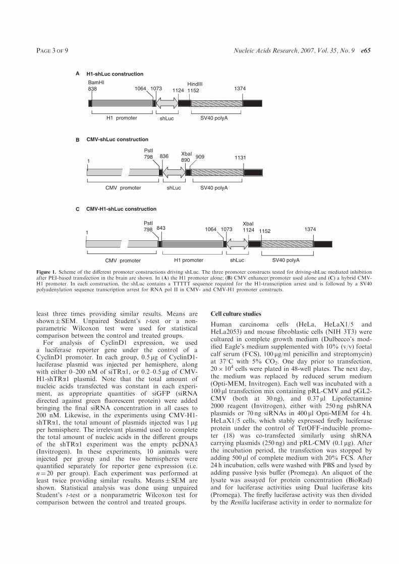
least three times providing similar results. Means areshown� SEM. Unpaired Student’s t-test or a non-parametric Wilcoxon test were used for statisticalcomparison between the control and treated groups.
For analysis of CyclinD1 expression, we useda luciferase reporter gene under the control of aCyclinD1 promoter. In each group, 0.5 mg of CyclinD1-luciferase plasmid was injected per hemisphere, alongwith either 0–200 nM of siTRa1, or 0.2–0.5 mg of CMV-H1-shTRa1 plasmid. Note that the total amount ofnucleic acids transfected was constant in each experi-ment, as appropriate quantities of siGFP (siRNAdirected against green fluorescent protein) were addedbringing the final siRNA concentration in all cases to200 nM. Likewise, in the experiments using CMV-H1-shTRa1, the total amount of plasmids injected was 1 mgper hemisphere. The irrelevant plasmid used to completethe total amount of nucleic acids in the different groupsof the shTRa1 experiment was the empty pcDNA3(Invitrogen). In these experiments, 10 animals wereinjected per group and the two hemispheres werequantified separately for reporter gene expression (i.e.n¼ 20 per group). Each experiment was performed atleast twice providing similar results. Means�SEM areshown. Statistical analysis was done using unpairedStudent’s t-test or a nonparametric Wilcoxon test forcomparison between the control and treated groups.
Cell culture studies
Human carcinoma cells (HeLa, HeLaX1/5 andHeLa2053) and mouse fibroblastic cells (NIH 3T3) werecultured in complete growth medium (Dulbecco’s mod-ified Eagle’s medium supplemented with 10% (v/v) foetalcalf serum (FCS), 100 mg/ml penicillin and streptomycin)at 378C with 5% CO2. One day prior to transfection,20� 104 cells were plated in 48-well plates. The next day,the medium was replaced by reduced serum medium(Opti-MEM, Invitrogen). Each well was incubated with a100 ml transfection mix containing pRL-CMV and pGL2-CMV (both at 30 ng), and 0.37 ml Lipofectamine2000 reagent (Invitrogen), either with 250 ng pshRNAplasmids or 70 ng siRNAs in 400 ml Opti-MEM for 4 h.HeLaX1/5 cells, which stably expressed firefly luciferaseprotein under the control of TetOFF-inducible promo-ter (18) was co-transfected similarly using shRNAcarrying plasmids (250 ng) and pRL-CMV (0.1 mg). Afterthe incubation period, the transfection was stopped byadding 500 ml of complete medium with 20% FCS. After24 h incubation, cells were washed with PBS and lysed byadding passive lysis buffer (Promega). An aliquot of thelysate was assayed for protein concentration (BioRad)and for luciferase activities using Dual luciferase kits(Promega). The firefly luciferase activity was then dividedby the Renilla luciferase activity in order to normalize for
BamHI
A
B
C
838 1064 1073 1124HindIII
1152 1374
H1 promoter shLuc SV40 polyA
H1-shLuc construction
1
PstI
798 836 XbaI890
909 1131
CMV promoter shLuc SV40 polyA
CMV-shLuc construction
1
PstI
798 843 1064 1073XbaI1124 1152 1374
CMV promoter H1 promoter shLuc SV40 polyA
CMV-H1-shLuc construction
Figure 1. Scheme of the different promoter constructions driving shLuc. The three promoter constructs tested for driving-shLuc mediated inhibitionafter PEI-based transfection in the brain are shown. In (A) the H1 promoter alone; (B) CMV enhancer/promoter used alone and (C) a hybrid CMV-H1 promoter. In each construction, the shLuc contains a TTTTT sequence required for the H1-transcription arrest and is followed by a SV40polyadenylation sequence transcription arrest for RNA pol II in CMV- and CMV-H1 promoter constructs.
PAGE 3 OF 9 Nucleic Acids Research, 2007, Vol. 35, No. 9 e65
transfection efficiencies of HeLa and NIH 3T3. Allexperiments were done in triplicate.For siTRa1 transfections, the same protocol was
adapted to transfect siTRa1 into NIH 3T3 cellsstably expressing both TRa1 and TRa2 isoforms. Threeconcentrations of siTRa1 were tested ranging from 5 to100 nM (500ml/well). In this experiment, the transfectionvector used was jetSI/DOPE, using the complexationprotocol described previously (Preparation of complexesand gene transfer in vivo section). One day post-transfection, cells were harvested and total RNAsextracted. For all experiments, reverse transcription wasperformed with MMLV-RT (Invitrogen) using 1 mg oftotal RNA mixed with 500 ng of hexamer oligonucleotidesaccording to the manufacturer’s instructions. Here, 1% ofcDNAs was used for quantitative PCR (Q-PCR) usingspecific primers (as listed below) and SYBR-Green PCRkit (Qiagen). Q-PCR reactions were performed at leastthree times in duplicate on a DNA engine Opticon system(MJ Research). Each point was repeated three times(i.e. three wells for each condition) and each well wasmeasured in triplicate. Data were analysed by normalizingTRa1 and TRa2 expression to that of a control gene 36B4.The sequences of the oligonucleotides used are as follows:TRa1 forward: 50CAGAGGGTGTGCGGAGCTGGT30;TRa1 reverse: 50CCTGTCCAAGGGCTGGAGGGT30;TRa2 forward: 50GCATGTTGTTCAGGGTCCGCAGGT30; TRa2 reverse: 50GGGCTCTTCGGGCTCTGGTGCT30; 36B4 forward: 50ACCTCCTTTCCAGGCTTT30,36B4 reverse: 50CCCACCTTGTCTCCAGTCTTT30.Specificity of the amplification was optimized by determin-ing melting curves of the amplicons.
RESULTS AND DISCUSSION
Our objectives in this study were to compare the capacitiesof different constructs to direct efficient production ofshRNA in vitro and in vivo in the mammalian brain. Theexpression cassettes of shRNA delivery vectors typicallyexploit RNA polymerase III (Pol III) promoters (8–11),(19–20), whilst some authors have suggested that use of aPol II promoter can be used successfully in certain cellcontexts using non-viral (21) or lentiviral vectors (22).However, the use of Pol II promoters for shRNAproduction is subject to certain constraints. For instance,some authors have demonstrated a need for both a veryshort distance (6 bp) between the Pol II promoter and theshRNA sequence as well as a short polyadenylation signal(21). Others have shown that the presence of an intronbetween the Pol II promoter and the shRNA sequence isnecessary for efficient production (22). These constraintsdo not apply to Pol III constructs. We therefore tested theefficiency of a Pol III construct (H1) in different in vivoand in vitro situations. However, we found that the H1promoter alone was not very efficient in vivo. So we alsotried a hybrid CMV enhancer minimal promoter/H1promoter construct (CMV/H1 construct). We chose thisstrategy because this enhancer had previously been shownto increase promoter efficiency in vivo, such as for thePDGF beta promoter (23) or to increase U6-mediated
transcription of shRNAs in plasmids (24). Other fusionpromoters have also been described to increase theshRNA production efficiency by recombinant SV40derivative viruses (25) or by lentiviral vectors (26). Inour experimental set up, a construct with CMV enhancer/promoter (CMV-shRNA) alone was used as a control forthe enhancement of the H1 promoter, knowing that in ourconstruct the CMV promoter was too far from the hairpinto be efficient in producing shRNA, as shown by Xia et al.(12). Given that one of the major advantages of using anshRNA approach is that it should be able to be exploitedto provide tissue-specific and developmental-stage-specificgene knockdown, we examined the efficiencies of theseconstructs in the context of the developing and the adultmouse brain.
Design of different promoter constructs drivingthe transcription of shLuc
Three different promoter constructs were tested for theproduction of shLuc (Figure 1). The first was a classic H1promoter transcribing shLuc from a pSUPER vectorbackbone (8). In this construction, the type III RNApolymerase synthesizes the shRNA and is stopped by afive-T stretch at the 30 end of the shRNA (Figure 1a). Inthis H1-shLuc construct, the first nucleotide of the shLucsequence is located 30 bp from the TATA box of the H1promoter (27). Second, in order to improve H1-mediatedproduction of shRNAs, we also constructed a CMV-H1hybrid construct driving the production of the shRNA.In this construction, the full enhancer and promotersequence of CMV has been integrated upstream of the H1promoter (CMV-H1-shLuc). Finally, as a control tovalidate that in the CMV-H1 construct, the productionof the hairpin is generated by the H1 promoter and not bythe CMV promoter, we designed a CMV-shLuc plasmidwhere the CMV enhancer/promoter is too far from thehairpin sequence to allow efficient shRNA production. Wethus did not expect the CMV-shLuc plasmid to provideany inhibition of the target luciferase (Figure 1c).
In cell culture, both H1-shLuc and CMV-H1-shLuc triggerefficient target gene knockdown
We tested the efficiency of H1-shLuc to trigger theinhibition of a co-transfected luciferase target gene byRNAi in a mouse fibroblastic cell line (NIH 3T3), as thisconstruct has been shown to be effective in certain cultureconditions (28). We then compared the H1-shLucefficiency to that of the CMV-H1-shLuc in the samecultures.
As described in the Methods section, two luciferasereporter genes (pGL2-CMV and pRL-CMV at 30 ng each)were co-transfected in NIH 3T3 cells (Figure 2a),along with the different constructions describedin Figure 1. Quantification of luciferase expressionrevealed strong inhibition efficiency for both H1-shLucand CMV-H1-shLuc (0.25 mg–100 ml transfectionmixture). This 80% inhibition of the target gene withH1- and CMV-H1-shLuc was stronger than obtained withsiLuc used at 70 ng (Figure 4a). Similar results wereobtained using HeLa cells instead of NIH 3T3 cells
e65 Nucleic Acids Research, 2007, Vol. 35, No. 9 PAGE 4 OF 9
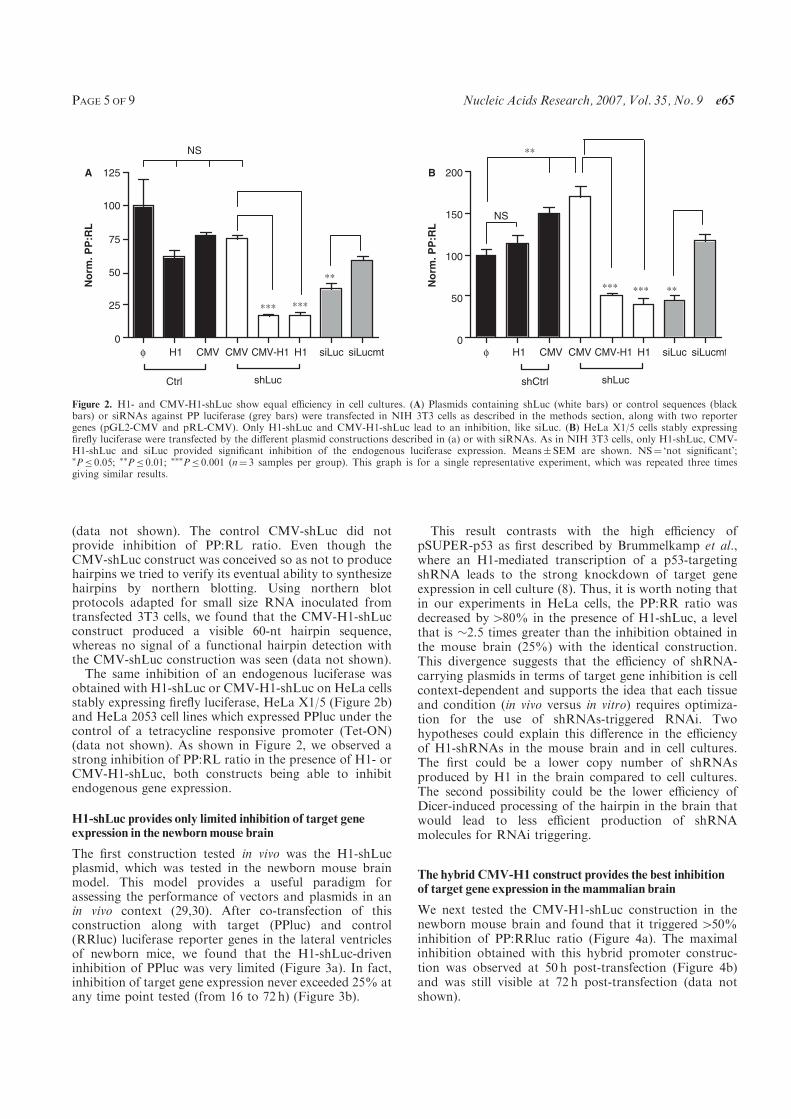
(data not shown). The control CMV-shLuc did notprovide inhibition of PP:RL ratio. Even though theCMV-shLuc construct was conceived so as not to producehairpins we tried to verify its eventual ability to synthesizehairpins by northern blotting. Using northern blotprotocols adapted for small size RNA inoculated fromtransfected 3T3 cells, we found that the CMV-H1-shLucconstruct produced a visible 60-nt hairpin sequence,whereas no signal of a functional hairpin detection withthe CMV-shLuc construction was seen (data not shown).
The same inhibition of an endogenous luciferase wasobtained with H1-shLuc or CMV-H1-shLuc on HeLa cellsstably expressing firefly luciferase, HeLa X1/5 (Figure 2b)and HeLa 2053 cell lines which expressed PPluc under thecontrol of a tetracycline responsive promoter (Tet-ON)(data not shown). As shown in Figure 2, we observed astrong inhibition of PP:RL ratio in the presence of H1- orCMV-H1-shLuc, both constructs being able to inhibitendogenous gene expression.
H1-shLuc provides only limited inhibition of target geneexpression in the newborn mouse brain
The first construction tested in vivo was the H1-shLucplasmid, which was tested in the newborn mouse brainmodel. This model provides a useful paradigm forassessing the performance of vectors and plasmids in anin vivo context (29,30). After co-transfection of thisconstruction along with target (PPluc) and control(RRluc) luciferase reporter genes in the lateral ventriclesof newborn mice, we found that the H1-shLuc-driveninhibition of PPluc was very limited (Figure 3a). In fact,inhibition of target gene expression never exceeded 25% atany time point tested (from 16 to 72 h) (Figure 3b).
This result contrasts with the high efficiency ofpSUPER-p53 as first described by Brummelkamp et al.,where an H1-mediated transcription of a p53-targetingshRNA leads to the strong knockdown of target geneexpression in cell culture (8). Thus, it is worth noting thatin our experiments in HeLa cells, the PP:RR ratio wasdecreased by480% in the presence of H1-shLuc, a levelthat is �2.5 times greater than the inhibition obtained inthe mouse brain (25%) with the identical construction.This divergence suggests that the efficiency of shRNA-carrying plasmids in terms of target gene inhibition is cellcontext-dependent and supports the idea that each tissueand condition (in vivo versus in vitro) requires optimiza-tion for the use of shRNAs-triggered RNAi. Twohypotheses could explain this difference in the efficiencyof H1-shRNAs in the mouse brain and in cell cultures.The first could be a lower copy number of shRNAsproduced by H1 in the brain compared to cell cultures.The second possibility could be the lower efficiency ofDicer-induced processing of the hairpin in the brain thatwould lead to less efficient production of shRNAmolecules for RNAi triggering.
The hybrid CMV-H1 construct provides the best inhibitionof target gene expression in the mammalian brain
We next tested the CMV-H1-shLuc construction in thenewborn mouse brain and found that it triggered450%inhibition of PP:RRluc ratio (Figure 4a). The maximalinhibition obtained with this hybrid promoter construc-tion was observed at 50 h post-transfection (Figure 4b)and was still visible at 72 h post-transfection (data notshown).
H1φ CMV CMV CMV-H1 H1 siLuc siLucmt H1φ CMV CMV CMV-H1 H1 siLuc siLucmt
0
25
50
75
100
125A
No
rm
. P
P:R
L
0
50
100
150
200B
No
rm
. P
P:R
L
Ctrl shLuc shCtrl shLuc
NS
**
*** ***
**
NS
**
*** ***
Figure 2. H1- and CMV-H1-shLuc show equal efficiency in cell cultures. (A) Plasmids containing shLuc (white bars) or control sequences (blackbars) or siRNAs against PP luciferase (grey bars) were transfected in NIH 3T3 cells as described in the methods section, along with two reportergenes (pGL2-CMV and pRL-CMV). Only H1-shLuc and CMV-H1-shLuc lead to an inhibition, like siLuc. (B) HeLa X1/5 cells stably expressingfirefly luciferase were transfected by the different plasmid constructions described in (a) or with siRNAs. As in NIH 3T3 cells, only H1-shLuc, CMV-H1-shLuc and siLuc provided significant inhibition of the endogenous luciferase expression. Means� SEM are shown. NS¼ ‘not significant’;�P� 0.05; ��P� 0.01; ���P� 0.001 (n¼ 3 samples per group). This graph is for a single representative experiment, which was repeated three timesgiving similar results.
PAGE 5 OF 9 Nucleic Acids Research, 2007, Vol. 35, No. 9 e65
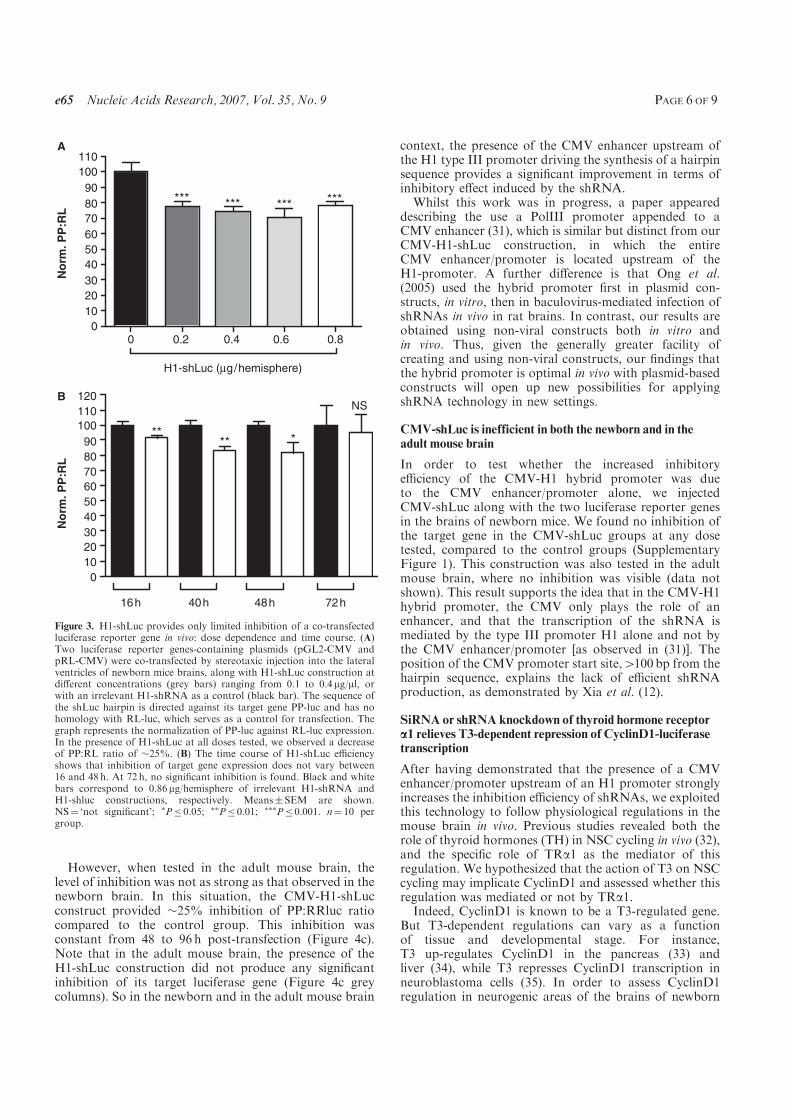
However, when tested in the adult mouse brain, thelevel of inhibition was not as strong as that observed in thenewborn brain. In this situation, the CMV-H1-shLucconstruct provided �25% inhibition of PP:RRluc ratiocompared to the control group. This inhibition wasconstant from 48 to 96 h post-transfection (Figure 4c).Note that in the adult mouse brain, the presence of theH1-shLuc construction did not produce any significantinhibition of its target luciferase gene (Figure 4c greycolumns). So in the newborn and in the adult mouse brain
context, the presence of the CMV enhancer upstream ofthe H1 type III promoter driving the synthesis of a hairpinsequence provides a significant improvement in terms ofinhibitory effect induced by the shRNA.
Whilst this work was in progress, a paper appeareddescribing the use a PolIII promoter appended to aCMV enhancer (31), which is similar but distinct from ourCMV-H1-shLuc construction, in which the entireCMV enhancer/promoter is located upstream of theH1-promoter. A further difference is that Ong et al.(2005) used the hybrid promoter first in plasmid con-structs, in vitro, then in baculovirus-mediated infection ofshRNAs in vivo in rat brains. In contrast, our results areobtained using non-viral constructs both in vitro andin vivo. Thus, given the generally greater facility ofcreating and using non-viral constructs, our findings thatthe hybrid promoter is optimal in vivo with plasmid-basedconstructs will open up new possibilities for applyingshRNA technology in new settings.
CMV-shLuc is inefficient in both the newborn and in theadult mouse brain
In order to test whether the increased inhibitoryefficiency of the CMV-H1 hybrid promoter was dueto the CMV enhancer/promoter alone, we injectedCMV-shLuc along with the two luciferase reporter genesin the brains of newborn mice. We found no inhibition ofthe target gene in the CMV-shLuc groups at any dosetested, compared to the control groups (SupplementaryFigure 1). This construction was also tested in the adultmouse brain, where no inhibition was visible (data notshown). This result supports the idea that in the CMV-H1hybrid promoter, the CMV only plays the role of anenhancer, and that the transcription of the shRNA ismediated by the type III promoter H1 alone and not bythe CMV enhancer/promoter [as observed in (31)]. Theposition of the CMV promoter start site,4100 bp from thehairpin sequence, explains the lack of efficient shRNAproduction, as demonstrated by Xia et al. (12).
SiRNA or shRNA knockdown of thyroid hormone receptora1 relieves T3-dependent repression of CyclinD1-luciferasetranscription
After having demonstrated that the presence of a CMVenhancer/promoter upstream of an H1 promoter stronglyincreases the inhibition efficiency of shRNAs, we exploitedthis technology to follow physiological regulations in themouse brain in vivo. Previous studies revealed both therole of thyroid hormones (TH) in NSC cycling in vivo (32),and the specific role of TRa1 as the mediator of thisregulation. We hypothesized that the action of T3 on NSCcycling may implicate CyclinD1 and assessed whether thisregulation was mediated or not by TRa1.
Indeed, CyclinD1 is known to be a T3-regulated gene.But T3-dependent regulations can vary as a functionof tissue and developmental stage. For instance,T3 up-regulates CyclinD1 in the pancreas (33) andliver (34), while T3 represses CyclinD1 transcription inneuroblastoma cells (35). In order to assess CyclinD1regulation in neurogenic areas of the brains of newborn
0 0.2 0.4 0.6 0.80
10
20
30
40
50
60
70
80
90
100
110A
H1-shLuc (µg/hemisphere)
*** *** *** ***
No
rm
. P
P:R
L
0
10
20
30
40
50
60
70
80
90
100
110
120
16h 40h 48h 72h
***
NS
**
B
No
rm
. P
P:R
L
Figure 3. H1-shLuc provides only limited inhibition of a co-transfectedluciferase reporter gene in vivo: dose dependence and time course. (A)Two luciferase reporter genes-containing plasmids (pGL2-CMV andpRL-CMV) were co-transfected by stereotaxic injection into the lateralventricles of newborn mice brains, along with H1-shLuc construction atdifferent concentrations (grey bars) ranging from 0.1 to 0.4 mg/ml, orwith an irrelevant H1-shRNA as a control (black bar). The sequence ofthe shLuc hairpin is directed against its target gene PP-luc and has nohomology with RL-luc, which serves as a control for transfection. Thegraph represents the normalization of PP-luc against RL-luc expression.In the presence of H1-shLuc at all doses tested, we observed a decreaseof PP:RL ratio of �25%. (B) The time course of H1-shLuc efficiencyshows that inhibition of target gene expression does not vary between16 and 48 h. At 72 h, no significant inhibition is found. Black and whitebars correspond to 0.86 mg/hemisphere of irrelevant H1-shRNA andH1-shluc constructions, respectively. Means� SEM are shown.NS¼ ‘not significant’; �P� 0.05; ��P� 0.01; ���P� 0.001. n¼ 10 pergroup.
e65 Nucleic Acids Research, 2007, Vol. 35, No. 9 PAGE 6 OF 9
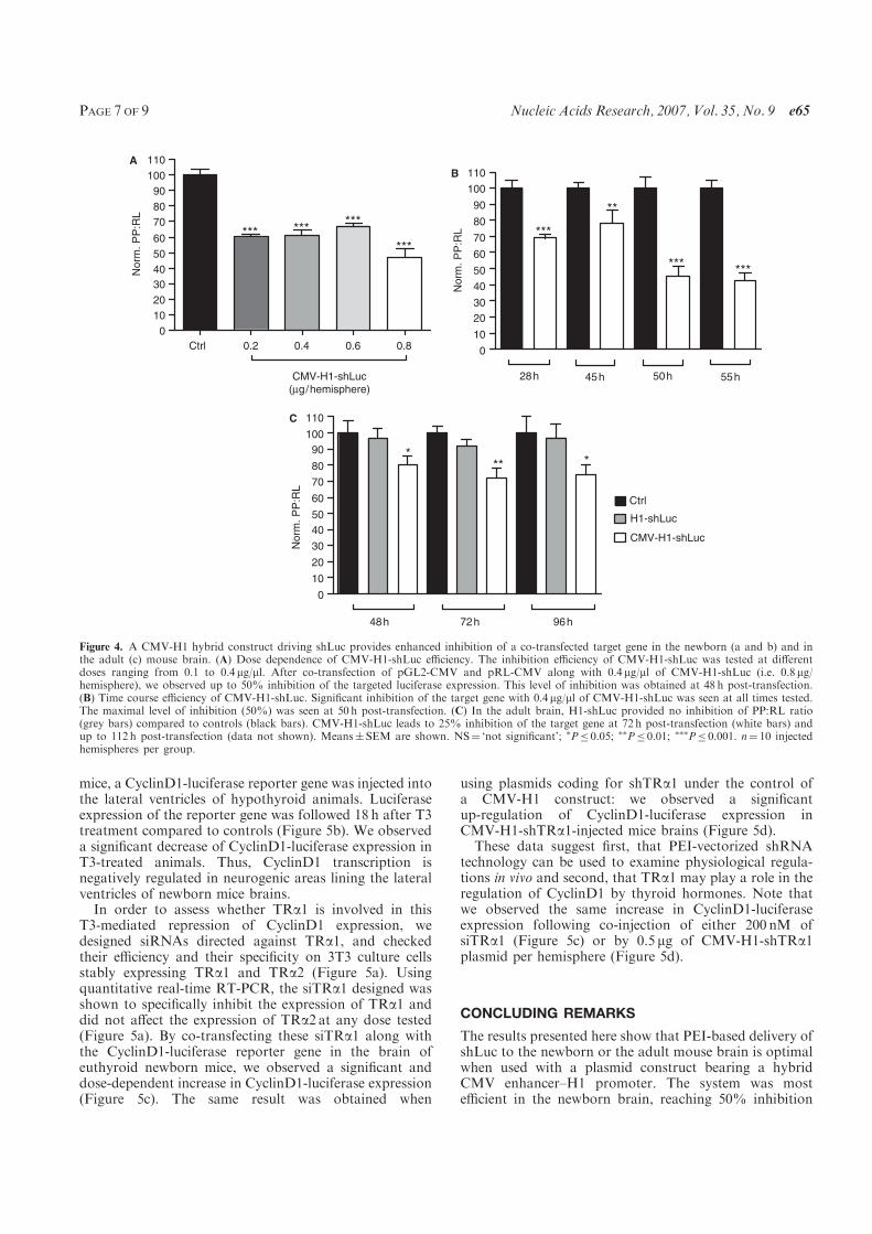
mice, a CyclinD1-luciferase reporter gene was injected intothe lateral ventricles of hypothyroid animals. Luciferaseexpression of the reporter gene was followed 18 h after T3treatment compared to controls (Figure 5b). We observeda significant decrease of CyclinD1-luciferase expression inT3-treated animals. Thus, CyclinD1 transcription isnegatively regulated in neurogenic areas lining the lateralventricles of newborn mice brains.
In order to assess whether TRa1 is involved in thisT3-mediated repression of CyclinD1 expression, wedesigned siRNAs directed against TRa1, and checkedtheir efficiency and their specificity on 3T3 culture cellsstably expressing TRa1 and TRa2 (Figure 5a). Usingquantitative real-time RT-PCR, the siTRa1 designed wasshown to specifically inhibit the expression of TRa1 anddid not affect the expression of TRa2 at any dose tested(Figure 5a). By co-transfecting these siTRa1 along withthe CyclinD1-luciferase reporter gene in the brain ofeuthyroid newborn mice, we observed a significant anddose-dependent increase in CyclinD1-luciferase expression(Figure 5c). The same result was obtained when
using plasmids coding for shTRa1 under the control ofa CMV-H1 construct: we observed a significantup-regulation of CyclinD1-luciferase expression inCMV-H1-shTRa1-injected mice brains (Figure 5d).These data suggest first, that PEI-vectorized shRNA
technology can be used to examine physiological regula-tions in vivo and second, that TRa1 may play a role in theregulation of CyclinD1 by thyroid hormones. Note thatwe observed the same increase in CyclinD1-luciferaseexpression following co-injection of either 200 nM ofsiTRa1 (Figure 5c) or by 0.5 mg of CMV-H1-shTRa1plasmid per hemisphere (Figure 5d).
CONCLUDING REMARKS
The results presented here show that PEI-based delivery ofshLuc to the newborn or the adult mouse brain is optimalwhen used with a plasmid construct bearing a hybridCMV enhancer–H1 promoter. The system was mostefficient in the newborn brain, reaching 50% inhibition
Ctrl 0.2 0.4 0.6 0.8
0
10
20
30
40
50
60
70
80
90
100
110
Norm
. P
P:R
L
A
CMV-H1-shLuc(µg/hemisphere)
*** ******
***
No
rm.
PP
:RL
B
0
10
20
30
40
50
60
70
80
90
100
110
28h 45h 50h 55h
***
*** ***
**
Ctrl
H1-shLuc
CMV-H1-shLuc
No
rm.
PP
:RL
C
0
10
20
30
40
50
60
70
80
90
100
110
72h 96h48h
*** *
Figure 4. A CMV-H1 hybrid construct driving shLuc provides enhanced inhibition of a co-transfected target gene in the newborn (a and b) and inthe adult (c) mouse brain. (A) Dose dependence of CMV-H1-shLuc efficiency. The inhibition efficiency of CMV-H1-shLuc was tested at differentdoses ranging from 0.1 to 0.4 mg/ml. After co-transfection of pGL2-CMV and pRL-CMV along with 0.4 mg/ml of CMV-H1-shLuc (i.e. 0.8 mg/hemisphere), we observed up to 50% inhibition of the targeted luciferase expression. This level of inhibition was obtained at 48 h post-transfection.(B) Time course efficiency of CMV-H1-shLuc. Significant inhibition of the target gene with 0.4 mg/ml of CMV-H1-shLuc was seen at all times tested.The maximal level of inhibition (50%) was seen at 50 h post-transfection. (C) In the adult brain, H1-shLuc provided no inhibition of PP:RL ratio(grey bars) compared to controls (black bars). CMV-H1-shLuc leads to 25% inhibition of the target gene at 72 h post-transfection (white bars) andup to 112 h post-transfection (data not shown). Means� SEM are shown. NS¼ ‘not significant’; �P� 0.05; ��P� 0.01; ���P� 0.001. n¼ 10 injectedhemispheres per group.
PAGE 7 OF 9 Nucleic Acids Research, 2007, Vol. 35, No. 9 e65
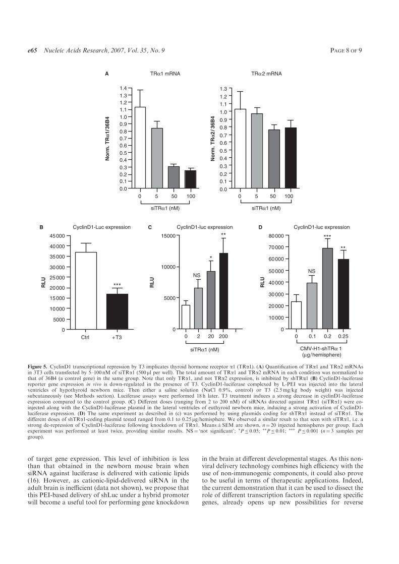
of target gene expression. This level of inhibition is lessthan that obtained in the newborn mouse brain whensiRNA against luciferase is delivered with cationic lipids(16). However, as cationic-lipid-delivered siRNA in theadult brain is inefficient (data not shown), we propose thatthis PEI-based delivery of shLuc under a hybrid promoterwill become a useful tool for performing gene knockdown
in the brain at different developmental stages. As this non-viral delivery technology combines high efficiency with theuse of non-immunogenic components, it could also proveto be useful in terms of therapeutic applications. Indeed,the current demonstration that it can be used to dissect therole of different transcription factors in regulating specificgenes, already opens up new possibilities for reverse
B CyclinD1-Luc expression
Ctrl
RL
U
+T3
0
5000
10000
15000
20000
25000
30000
35000
40000
45000
D CyclinD1-luc expression
RL
U
0 0.1 0.2 0.25
0
10000
20000
30000
40000
50000
60000
70000
80000
CMV-H1-shTRα 1 (µg/hemisphere)
NS
***
**
***
C CyclinD1-luc expression
RL
U
0 2 20 200
0
5000
10000
15000
NS
*
**
siTRα1 (nM)
TRα1 mRNAA TRα2 mRNA
0 5 50 100
0.0
0.1
0.2
0.3
0.4
0.5
0.6
0.7
0.8
0.9
1.0
1.1
1.2
1.3
1.4
No
rm
. T
Ra
1/3
6B
4
No
rm
. T
Ra
2/3
6B
4
0 5 50 100
0.0
0.1
0.2
0.3
0.4
0.5
0.6
0.7
0.8
0.9
1.0
1.1
1.2
1.3
siTRα1 (nM) siTRα1 (nM)
Figure 5. CyclinD1 transcriptional repression by T3 implicates thyroid hormone receptor a1 (TRa1). (A) Quantification of TRa1 and TRa2 mRNAsin 3T3 cells transfected by 5–100 nM of siTRa1 (500 ml per well). The total amount of TRa1 and TRa2 mRNA in each condition was normalized tothat of 36B4 (a control gene) in the same group. Note that only TRa1, and not TRa2 expression, is inhibited by shTRa1 (B) CyclinD1-luciferasereporter gene expression in vivo is down-regulated in the presence of T3. CyclinD1-luciferase complexed by L-PEI was injected into the lateralventricles of hypothyroid newborn mice. Then either a saline solution (NaCl 0.9%, control) or T3 (2.5mg/kg body weight) was injectedsubcutaneously (see Methods section). Luciferase assays were performed 18 h later. T3 treatment induces a strong decrease in cyclinD1-luciferaseexpression compared to the control group. (C) Different doses (ranging from 2 to 200 nM) of siRNAs directed against TRa1 (siTRa1) were co-injected along with the CyclinD1-luciferase plasmid in the lateral ventricles of euthyroid newborn mice, inducing a strong activation of CyclinD1-luciferase expression. (D) The same experiment as described in (c) was performed by using plasmids coding for shTRa1 instead of siTRa1. Thedifferent doses of shTRa1-coding plasmid tested ranged from 0.1 to 0.25mg/hemisphere. We observed a similar result to that seen with siTRa1, i.e. astrong de-repression of CyclinD1-luciferase following knockdown of TRa1. Means�SEM are shown, n¼ 20 injected hemispheres per group. Eachexperiment was performed at least twice, providing similar results. NS¼ ‘not significant’; �P� 0.05; ��P� 0.01; ��� P� 0.001 (n¼ 3 samples pergroup).
e65 Nucleic Acids Research, 2007, Vol. 35, No. 9 PAGE 8 OF 9
genetic studies focused on specific target tissues at defineddevelopmental stages.
ACKNOWLEDGEMENTS
Funding to pay the open access publication charge wasprovided by the CNRS. ZH was a fellow of theAssociation Francaise contre les Myopathies (AFM) andthe Fondation pour la Recherche Medicale. This workwas supported by the AFM and EU contract, LSHM CT2005-018652, Crescendo.
Conflict of interest statement. None declared.
REFERENCES
1. Napoli,C., Lemieux,C. and Jorgensen,R. (1990) Introduction of achimeric chalcone synthase gene into petunia results in reversibleco-suppression of homologous genes in trans. Plant Cell, 2, 279–289.
2. Hutvagner,G. and Zamore,P.D. (2002) RNAi: nature abhors adouble-strand. Curr. Opin. Genet. Dev., 12, 225–232.
3. Manche,L., Green,S.R., Schmedt,C. and Mathews,M.B. (1992)Interactions between double-stranded RNA regulators and theprotein kinase DAI. Mol. Cell. Biol., 12, 5238–5248.
4. Minks,M.A., West,D.K., Benvin,S. and Baglioni,C. (1979)Structural requirements of double-stranded RNA for the activationof 2’,5’-oligo(A) polymerase and protein kinase of interferon-treatedHeLa cells. J. Biol. Chem., 254, 10180–10183.
5. Elbashir,S.M., Harborth,J., Lendeckel,W., Yalcin,A., Weber,K. andTuschl,T. (2001) Duplexes of 21-nucleotide RNAs mediate RNAinterference in cultured mammalian cells. Nature, 411, 494–498.
6. Song,E., Lee,S.K., Wang,J., Ince,N., Ouyang,N., Min,J., Chen,J.,Shankar,P. and Lieberman,J. (2003) RNA interference targeting Fasprotects mice from fulminant hepatitis. Nat. Med., 9, 347–351.
7. Dorn,G., Patel,S., Wotherspoon,G., Hemmings-Mieszczak,M.,Barclay,J., Natt,F.J., Martin,P., Bevan,S., Fox,A. et al. (2004)siRNA relieves chronic neuropathic pain. Nucleic Acids Res., 32,e49.
8. Brummelkamp,T.R., Bernards,R. and Agami,R. (2002) A systemfor stable expression of short interfering RNAs in mammalian cells.Science, 296, 550–553.
9. Paul,C.P., Good,P.D., Winer,I. and Engelke,D.R. (2002) Effectiveexpression of small interfering RNA in human cells. Nat.Biotechnol., 20, 505–508.
10. Sui,G., Soohoo,C., Affarel,B., Gay,F., Shi,Y. and Forrester,W.C.(2002) A DNA vector-based RNAi technology to suppress geneexpression in mammalian cells. Proc. Natl Acad. Sci. USA, 99,5515–5520.
11. Yu,J.Y., DeRuiter,S.L. and Turner,D.L. (2002) RNA interferenceby expression of short-interfering RNAs and hairpin RNAs inmammalian cells. Proc. Natl. Acad. Sci. USA, 99, 6047–6052.
12. Xia,H., Mao,Q., Paulson,H.L. and Davidson,B.L. (2002)siRNA-mediated gene silencing in vitro and in vivo.Nat. Biotechnol., 20, 1006–1010.
13. Uprichard,S.L., Boyd,B., Althage,A. and Chisari,F.V. (2005)Clearance of hepatitis B virus from the liver of transgenic miceby short hairpin RNAs. Proc. Natl. Acad. Sci. USA, 102, 773–778.
14. Gurzov,E.N. and Izquierdo,M. (2006) RNA interference againstHec1 inhibits tumor growth in vivo. Gene Ther., 13, 1–7.
15. Raoul,C., Abbas-Terki,T., Bensadoun,J.C., Guillot,S., Haase,G.,Szulc,J., Henderson,C.E. and Aebischer,P. (2005) Lentiviral-mediated silencing of SOD1 through RNA interference retardsdisease onset and progression in a mouse model of ALS. Nat. Med.,11, 423–428.
16. Hassani,Z., Lemkine,G.F., Erbacher,P., Palmier,K., Alfama,G.,Giovannangeli,C., Behr,J.P. and Demeneix,B.A. (2005) Lipid-
mediated siRNA delivery down-regulates exogenous geneexpression in the mouse brain at picomolar levels. J. Gene Med., 7,198–207.
17. Lemkine,G.F. and Demeneix,B.A. (2001) Polyethylenimines forin vivo gene delivery. Curr. Opin. Mol. Ther., 3, 178–182.
18. Gossen,M. and Bujard,H. (1992) Tight control of gene expression inmammalian cells by tetracycline-responsive promoters. Proc. Natl.Acad. Sci. USA, 89, 5547–5551.
19. Boden,D., Pusch,O., Lee,F., Tucker,L., Shank,P.R. andRamratnam,B. (2003) Promoter choice affects the potency of HIV-1specific RNA interference. Nucleic Acids Res., 31, 5033–5038.
20. Kawasaki,H. and Taira,K. (2003) Short hairpin type of dsRNAsthat are controlled by tRNA(Val) promoter significantly induceRNAi-mediated gene silencing in the cytoplasm of human cells.Nucleic Acids Res., 31, 700–707.
21. Zhou,H., Xia,X.G. and Xu,Z. (2005) An RNA polymerase IIconstruct synthesizes short-hairpin RNA with a quantitativeindicator and mediates highly efficient RNAi. Nucleic Acids Res.,33, e62.
22. Yang,S. and Hecht,N.B. (2004) Translin associated protein X isessential for cellular proliferation. FEBS Lett., 576, 221–225.
23. Liu,B.H., Wang,X., Ma,Y.X. and Wang,S. (2004) CMV enhancer/human PDGF-beta promoter for neuron-specific transgeneexpression. Gene Ther., 11, 52–60.
24. Xia,X.G., Zhou,H., Ding,H., Affarel,B., Shi,Y. and Xu,Z. (2003)An enhanced U6 promoter for synthesis of short hairpin RNA.Nucleic Acids Res., 31, e100.
25. Strayer,D.S., Feitelson,M., Sun,B. and Matskevich,A.A. (2005)Paradigms for conditional expression of RNA interference mole-cules for use against viral targets. Methods Enzymol., 392, 227–241.
26. Unwalla,H.J., Li,H.T., Bahner,I., Li,M.J., Kohn,D. and Rossi,J.J.(2006) Novel Pol II fusion promoter directs human immuno-deficiency virus type 1-inducible coexpression of a short hairpinRNA and protein. J. Virol., 80, 1863–1873.
27. Hannon,G.J., Chubb,A., Maroney,P.A., Hannon,G., Altman,S. andNilsen,T.W. (1991) Multiple cis-acting elements are required forRNA polymerase III transcription of the gene encoding H1 RNA,the RNA component of human RNase P. J. Biol. Chem., 266,22796–22799.
28. Wyatt,C.A., Geoghegan,J.C. and Brinckerhoff,C.E. (2005) Shorthairpin RNA-mediated inhibition of matrix metalloproteinase-1 inMDA-231 cells: effects on matrix destruction and tumor growth.Cancer Res., 65, 11101–11108.
29. Boussif,O., Lezoualc’h,F., Zanta,M.A., Mergny,M.D.,Scherman,D., Demeneix,B. and Behr,J.P. (1995) A versatilevector for gene and oligonucleotide transfer into cells in cultureand in vivo: polyethylenimine. Proc. Natl. Acad. Sci. USA, 92,7297–7301.
30. Schwartz,B., Benoist,C., Abdallah,B., Scherman,D., Behr,J.P. andDemeneix,B.A. (1995) Lipospermine-based gene transfer into thenewborn mouse brain is optimized by a low lipospermine/DNAcharge ratio. Hum. Gene. Ther., 6, 1515–1524.
31. Ong,S.T., Li,F., Du,J., Tan,Y.W. and Wang,S. (2005) Hybridcytomegalovirus enhancer-h1 promoter-based plasmid and baculo-virus vectors mediate effective RNA interference. Hum. Gene. Ther.,16, 1404–1412.
32. Lemkine,G.F., Raj,A., Alfama,G., Turque,N., Hassani,Z.,Alegria-Prevot,O., Samarut,J., Levi,G. and Demeneix,B.A. (2005)Adult neural stem cell cycling in vivo requires thyroid hormone andits alpha receptor. FASEB J., 19, 863–865.
33. Ledda-Columbano,G.M., Perra,A., Pibiri,M., Molotzu,F. andColumbano,A. (2005) Induction of pancreatic acinar cellproliferation by thyroid hormone. J. Endocrinol., 185, 393–399.
34. Alisi,A., Demori,I., Spagnuolo,S., Pierantozzi,E., Fugassa,E. andLeoni,S. (2005) Thyroid status affects rat liver regeneration afterpartial hepatectomy by regulating cell cycle and apoptosis. CellPhysiol. Biochem., 15, 69–76.
35. Garcia-Silva,S. and Aranda,A. (2004) The thyroid hormonereceptor is a suppressor of ras-mediated transcription, proliferation,and transformation. Mol. Cell. Biol., 24, 7514–7523.
PAGE 9 OF 9 Nucleic Acids Research, 2007, Vol. 35, No. 9 e65
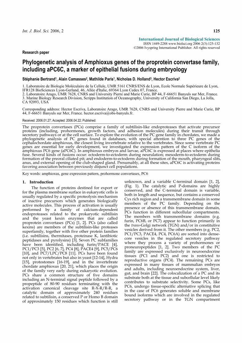
Int. J. Biol. Sci. 2006, 2 125
International Journal of Biological Sciences ISSN 1449-2288 www.biolsci.org 2006 2(3):125-132
©2006 Ivyspring International Publisher. All rights reserved
Research paper
Phylogenetic analysis of Amphioxus genes of the proprotein convertase family, including aPC6C, a marker of epithelial fusions during embryology
Stéphanie Bertrand1, Alain Camasses2, Mathilde Paris1, Nicholas D. Holland3, Hector Escriva2
1. Laboratoire de Biologie Moléculaire de la Cellule, UMR 5161 CNRS/ENS de Lyon, Ecole Normale Supérieure de Lyon, IFR128 BioSciences Lyon-Gerland, 46, Allée d'Italie, 69364 Lyon Cedex 07, France. 2. Laboratoire Arago, UMR 7628, CNRS and University Pierre and Marie Curie, BP 44, F-66651 Banyuls sur Mer, France. 3. Marine Biology Research Division, Scripps Institution of Oceanography, University of California San Diego, La Jolla, CA 92093, USA
Corresponding address: Hector Escriva, Laboratoire Arago, UMR 7628, CNRS and University Pierre and Marie Curie, BP 44, F-66651 Banyuls sur Mer, France. [email protected].
Received: 2006.01.27; Accepted: 2006.04.22; Published:
The proprotein convertases (PCs) comprise a family of subtilisin-like endoproteases that activate precursor proteins (including, prohormones, growth factors, and adhesion molecules) during their transit through secretory pathways or at the cell surface. To explore the evolution of the PC gene family in chordates, we made a phylogenetic analysis of PC genes found in databases, with special attention to three PC genes of the cephalochordate amphioxus, the closest living invertebrate relative to the vertebrates. Since some vertebrate PC genes are essential for early development, we investigated the expression pattern of the C isoform of the amphioxus PC6 gene (aPC6C). In amphioxus embryos and larvae, aPC6C is expressed at places where epithelia fuse. Several kinds of fusions occur: ectoderm-to-ectoderm during neurulation; mesoderm-to-ectoderm during formation of the preoral ciliated pit; and endoderm-to-ectoderm during formation of the mouth, pharyngeal slits, anus, and external opening of the club-shaped gland. Presumably, at all these sites, aPC6C is activating proteins favoring association between previously disjunct cell populations.
Key words: amphioxus, gene expression pattern, prohormone convertases, PC6
1. Introduction
The function of proteins destined for export or for the plasma membrane surface in eukaryotic cells is usually regulated by a specific proteolysis mechanism of inactive precursors which generates biologically active molecules. This process of activation is usually performed by a family of calcium-dependent endoproteases related to the prokaryotic subtilisin and the yeast kexin enzymes that are called proprotein convertases (PCs) [1, 2]. PCs (also called kexins) are members of the subtilisin-like proteases superfamily, together with five other protein families (i.e. subtilisins, thermitases, proteinase K, lantibiotic peptidases and pyrolysins) [3]. Seven PC subfamilies have been identified, including furin/PACE [4], PC1/PC3 [5], PC2 [6, 7], PC4 [8], PACE4 [9], PC5/PC6 [10], and PC7/LPC/PC8 [11]. PCs have been found not only in vertebrates but also in yeast [12-14], Hydra [15], protostomes [16-19], and in the invertebrate chordate amphioxus [20, 21], which places the origin of the family very early during eukaryotic evolution. PCs share a common structure of five domains including an N-terminal signal peptide followed by a propeptide of 80-90 residues terminating with the activation canonical cleavage site R-X-R/R-R, a catalytic domain of approximately 240 residues related to subtilisin, a conserved P or Homo B domain of approximately 150 residues which function is still
unknown, and a variable C-terminal domain [1, 2], (Fig. 1). The catalytic and P-domains are highly conserved, and the C-terminal domain is variable, both in length and sequence, but contains a conserved Cys rich region and a transmembrane domain in some members of the PC family. Depending on the presence or absence of this transmembrane domain, PCs function in different subcellular compartments. The members with transmembrane domains (e.g. furin, PC6B, or PC7) appear to function primarily in the trans-Golgi network (TGN) and/or in constitutive vesicles derived from it. The other members (e.g. PC2, PC1/PC3, PACE4, PC4, PC6A) are sorted into dense-core vesicles in the regulated secretory pathway where they process a variety of prohormones or proneuropeptides [1, 2]. Two members of the PC family are expressed exclusively in neuroendocrine tissues (PC1 and PC2) and one is restricted to reproductive organs (PC4). The remaining PCs are expressed in many tissues of mammalian embryos and adults, including neuroendocrine system, liver, gut, and brain [22]. The colocalization of a PC and its substrate both at the tissue and subcellular level likely contributes to substrate selectivity. Some PCs, like PC6, undergo tissue-specific alternative splicing that in the case of PC6 generates soluble and membrane bound isoforms which are involved in the regulated secretory pathway or in the TGN compartment
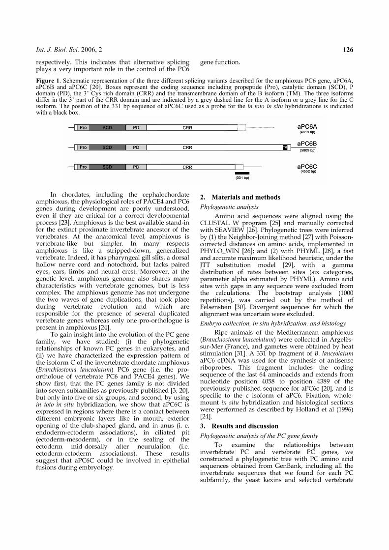
Int. J. Biol. Sci. 2006, 2 126
respectively. This indicates that alternative splicing plays a very important role in the control of the PC6
gene function.
Figure 1. Schematic representation of the three different splicing variants described for the amphioxus PC6 gene, aPC6A, aPC6B and aPC6C [20]. Boxes represent the coding sequence including propeptide (Pro), catalytic domain (SCD), P domain (PD), the 3’ Cys rich domain (CRR) and the transmembrane domain of the B isoform (TM). The three isoforms differ in the 3’ part of the CRR domain and are indicated by a grey dashed line for the A isoform or a grey line for the C isoform. The position of the 331 bp sequence of aPC6C used as a probe for the in toto in situ hybridizations is indicated with a black box.
In chordates, including the cephalochordate
amphioxus, the physiological roles of PACE4 and PC6 genes during development are poorly understood, even if they are critical for a correct developmental process [23]. Amphioxus is the best available stand-in for the extinct proximate invertebrate ancestor of the vertebrates. At the anatomical level, amphioxus is vertebrate-like but simpler. In many respects amphioxus is like a stripped-down, generalized vertebrate. Indeed, it has pharyngeal gill slits, a dorsal hollow nerve cord and notochord, but lacks paired eyes, ears, limbs and neural crest. Moreover, at the genetic level, amphioxus genome also shares many characteristics with vertebrate genomes, but is less complex. The amphioxus genome has not undergone the two waves of gene duplications, that took place during vertebrate evolution and which are responsible for the presence of several duplicated vertebrate genes whereas only one pro-orthologue is present in amphioxus [24].
To gain insight into the evolution of the PC gene family, we have studied: (i) the phylogenetic relationships of known PC genes in eukaryotes, and (ii) we have characterized the expression pattern of the isoform C of the invertebrate chordate amphioxus (Branchiostoma lanceolatum) PC6 gene (i.e. the pro-ortholoue of vertebrate PC6 and PACE4 genes). We show first, that the PC genes family is not divided into seven subfamilies as previously published [3, 20], but only into five or six groups, and second, by using in toto in situ hybridization, we show that aPC6C is expressed in regions where there is a contact between different embryonic layers like in mouth, exterior opening of the club-shaped gland, and in anus (i. e. endoderm-ectoderm associations), in ciliated pit (ectoderm-mesoderm), or in the sealing of the ectoderm mid-dorsally after neurulation (i.e. ectoderm-ectoderm associations). These results suggest that aPC6C could be involved in epithelial fusions during embryology.
2. Materials and methods
Phylogenetic analysis
Amino acid sequences were aligned using the CLUSTAL W program [25] and manually corrected with SEAVIEW [26]. Phylogenetic trees were inferred by (1) the Neighbor-Joining method [27] with Poisson-corrected distances on amino acids, implemented in PHYLO_WIN [26]; and (2) with PHYML [28], a fast and accurate maximum likelihood heuristic, under the JTT substitution model [29], with a gamma distribution of rates between sites (six categories, parameter alpha estimated by PHYML). Amino acid sites with gaps in any sequence were excluded from the calculations. The bootstrap analysis (1000 repetitions), was carried out by the method of Felsenstein [30]. Divergent sequences for which the alignment was uncertain were excluded.
Embryo collection, in situ hybridization, and histology
Ripe animals of the Mediterranean amphioxus (Branchiostoma lanceolatum) were collected in Argelès-sur-Mer (France), and gametes were obtained by heat stimulation [31]. A 331 bp fragment of B. lanceolatum aPC6 cDNA was used for the synthesis of antisense riboprobes. This fragment includes the coding sequence of the last 64 aminoacids and extends from nucleotide position 4058 to position 4389 of the previously published sequence for aPC6c [20], and is specific to the c isoform of aPC6. Fixation, whole-mount in situ hybridization and histological sections were performed as described by Holland et al (1996) [24].
3. Results and discussion
Phylogenetic analysis of the PC gene family
To examine the relationships between invertebrate PC and vertebrate PC genes, we constructed a phylogenetic tree with PC amino acid sequences obtained from GenBank, including all the invertebrate sequences that we found for each PC subfamily, the yeast kexins and selected vertebrate
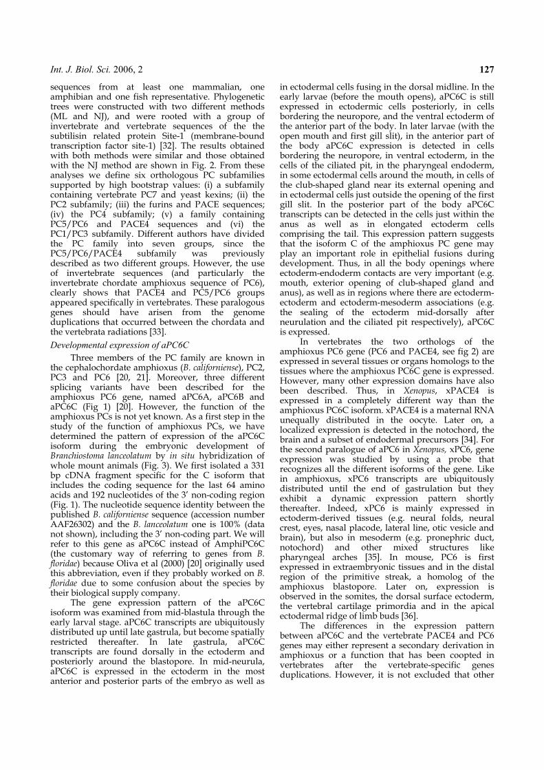
Int. J. Biol. Sci. 2006, 2 127
sequences from at least one mammalian, one amphibian and one fish representative. Phylogenetic trees were constructed with two different methods (ML and NJ), and were rooted with a group of invertebrate and vertebrate sequences of the the subtilisin related protein Site-1 (membrane-bound transcription factor site-1) [32]. The results obtained with both methods were similar and those obtained with the NJ method are shown in Fig. 2. From these analyses we define six orthologous PC subfamilies supported by high bootstrap values: (i) a subfamily containing vertebrate PC7 and yeast kexins; (ii) the PC2 subfamily; (iii) the furins and PACE sequences; (iv) the PC4 subfamily; (v) a family containing PC5/PC6 and PACE4 sequences and (vi) the PC1/PC3 subfamily. Different authors have divided the PC family into seven groups, since the PC5/PC6/PACE4 subfamily was previously described as two different groups. However, the use of invertebrate sequences (and particularly the invertebrate chordate amphioxus sequence of PC6), clearly shows that PACE4 and PC5/PC6 groups appeared specifically in vertebrates. These paralogous genes should have arisen from the genome duplications that occurred between the chordata and the vertebrata radiations [33].
Developmental expression of aPC6C
Three members of the PC family are known in the cephalochordate amphioxus (B. californiense), PC2, PC3 and PC6 [20, 21]. Moreover, three different splicing variants have been described for the amphioxus PC6 gene, named aPC6A, aPC6B and aPC6C (Fig 1) [20]. However, the function of the amphioxus PCs is not yet known. As a first step in the study of the function of amphioxus PCs, we have determined the pattern of expression of the aPC6C isoform during the embryonic development of Branchiostoma lanceolatum by in situ hybridization of whole mount animals (Fig. 3). We first isolated a 331 bp cDNA fragment specific for the C isoform that includes the coding sequence for the last 64 amino acids and 192 nucleotides of the 3’ non-coding region (Fig. 1). The nucleotide sequence identity between the published B. californiense sequence (accession number AAF26302) and the B. lanceolatum one is 100% (data not shown), including the 3’ non-coding part. We will refer to this gene as aPC6C instead of AmphiPC6C (the customary way of referring to genes from B. floridae) because Oliva et al (2000) [20] originally used this abbreviation, even if they probably worked on B. floridae due to some confusion about the species by their biological supply company.
The gene expression pattern of the aPC6C isoform was examined from mid-blastula through the early larval stage. aPC6C transcripts are ubiquitously distributed up until late gastrula, but become spatially restricted thereafter. In late gastrula, aPC6C transcripts are found dorsally in the ectoderm and posteriorly around the blastopore. In mid-neurula, aPC6C is expressed in the ectoderm in the most anterior and posterior parts of the embryo as well as
in ectodermal cells fusing in the dorsal midline. In the early larvae (before the mouth opens), aPC6C is still expressed in ectodermic cells posteriorly, in cells bordering the neuropore, and the ventral ectoderm of the anterior part of the body. In later larvae (with the open mouth and first gill slit), in the anterior part of the body aPC6C expression is detected in cells bordering the neuropore, in ventral ectoderm, in the cells of the ciliated pit, in the pharyngeal endoderm, in some ectodermal cells around the mouth, in cells of the club-shaped gland near its external opening and in ectodermal cells just outside the opening of the first gill slit. In the posterior part of the body aPC6C transcripts can be detected in the cells just within the anus as well as in elongated ectoderm cells comprising the tail. This expression pattern suggests that the isoform C of the amphioxus PC gene may play an important role in epithelial fusions during development. Thus, in all the body openings where ectoderm-endoderm contacts are very important (e.g. mouth, exterior opening of club-shaped gland and anus), as well as in regions where there are ectoderm-ectoderm and ectoderm-mesoderm associations (e.g. the sealing of the ectoderm mid-dorsally after neurulation and the ciliated pit respectively), aPC6C is expressed.
In vertebrates the two orthologs of the amphioxus PC6 gene (PC6 and PACE4, see fig 2) are expressed in several tissues or organs homologs to the tissues where the amphioxus PC6C gene is expressed. However, many other expression domains have also been described. Thus, in Xenopus, xPACE4 is expressed in a completely different way than the amphioxus PC6C isoform. xPACE4 is a maternal RNA unequally distributed in the oocyte. Later on, a localized expression is detected in the notochord, the brain and a subset of endodermal precursors [34]. For the second paralogue of aPC6 in Xenopus, xPC6, gene expression was studied by using a probe that recognizes all the different isoforms of the gene. Like in amphioxus, xPC6 transcripts are ubiquitously distributed until the end of gastrulation but they exhibit a dynamic expression pattern shortly thereafter. Indeed, xPC6 is mainly expressed in ectoderm-derived tissues (e.g. neural folds, neural crest, eyes, nasal placode, lateral line, otic vesicle and brain), but also in mesoderm (e.g. pronephric duct, notochord) and other mixed structures like pharyngeal arches [35]. In mouse, PC6 is first expressed in extraembryonic tissues and in the distal region of the primitive streak, a homolog of the amphioxus blastopore. Later on, expression is observed in the somites, the dorsal surface ectoderm, the vertebral cartilage primordia and in the apical ectodermal ridge of limb buds [36].
The differences in the expression pattern between aPC6C and the vertebrate PACE4 and PC6 genes may either represent a secondary derivation in amphioxus or a function that has been coopted in vertebrates after the vertebrate-specific genes duplications. However, it is not excluded that other
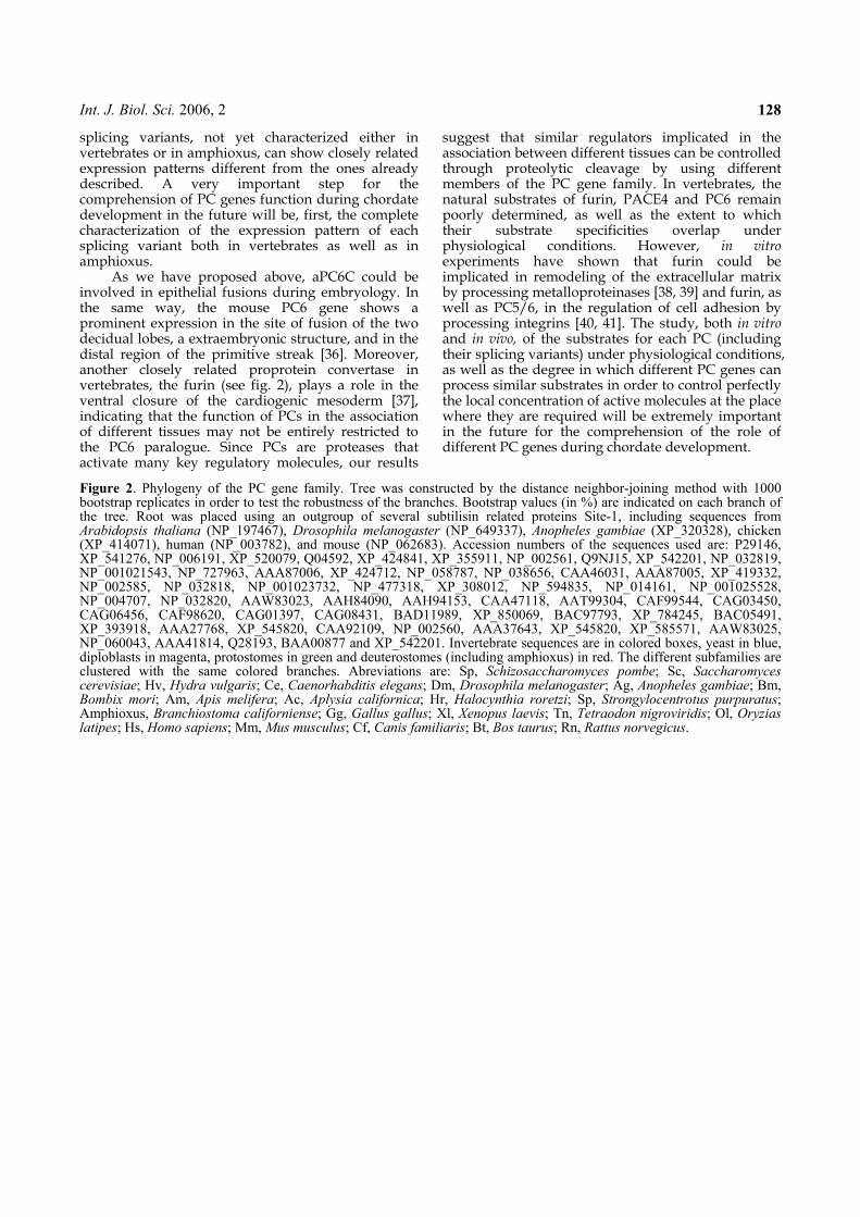
Int. J. Biol. Sci. 2006, 2 128
splicing variants, not yet characterized either in vertebrates or in amphioxus, can show closely related expression patterns different from the ones already described. A very important step for the comprehension of PC genes function during chordate development in the future will be, first, the complete characterization of the expression pattern of each splicing variant both in vertebrates as well as in amphioxus.
As we have proposed above, aPC6C could be involved in epithelial fusions during embryology. In the same way, the mouse PC6 gene shows a prominent expression in the site of fusion of the two decidual lobes, a extraembryonic structure, and in the distal region of the primitive streak [36]. Moreover, another closely related proprotein convertase in vertebrates, the furin (see fig. 2), plays a role in the ventral closure of the cardiogenic mesoderm [37], indicating that the function of PCs in the association of different tissues may not be entirely restricted to the PC6 paralogue. Since PCs are proteases that activate many key regulatory molecules, our results
suggest that similar regulators implicated in the association between different tissues can be controlled through proteolytic cleavage by using different members of the PC gene family. In vertebrates, the natural substrates of furin, PACE4 and PC6 remain poorly determined, as well as the extent to which their substrate specificities overlap under physiological conditions. However, in vitro experiments have shown that furin could be implicated in remodeling of the extracellular matrix by processing metalloproteinases [38, 39] and furin, as well as PC5/6, in the regulation of cell adhesion by processing integrins [40, 41]. The study, both in vitro and in vivo, of the substrates for each PC (including their splicing variants) under physiological conditions, as well as the degree in which different PC genes can process similar substrates in order to control perfectly the local concentration of active molecules at the place where they are required will be extremely important in the future for the comprehension of the role of different PC genes during chordate development.
Figure 2. Phylogeny of the PC gene family. Tree was constructed by the distance neighbor-joining method with 1000 bootstrap replicates in order to test the robustness of the branches. Bootstrap values (in %) are indicated on each branch of the tree. Root was placed using an outgroup of several subtilisin related proteins Site-1, including sequences from Arabidopsis thaliana (NP_197467), Drosophila melanogaster (NP_649337), Anopheles gambiae (XP_320328), chicken (XP_414071), human (NP_003782), and mouse (NP_062683). Accession numbers of the sequences used are: P29146, XP_541276, NP_006191, XP_520079, Q04592, XP_424841, XP_355911, NP_002561, Q9NJ15, XP_542201, NP_032819, NP_001021543, NP_727963, AAA87006, XP_424712, NP_058787, NP_038656, CAA46031, AAA87005, XP_419332, NP_002585, NP_032818, NP_001023732, NP_477318, XP_308012, NP_594835, NP_014161, NP_001025528, NP_004707, NP_032820, AAW83023, AAH84090, AAH94153, CAA47118, AAT99304, CAF99544, CAG03450, CAG06456, CAF98620, CAG01397, CAG08431, BAD11989, XP_850069, BAC97793, XP_784245, BAC05491, XP_393918, AAA27768, XP_545820, CAA92109, NP_002560, AAA37643, XP_545820, XP_585571, AAW83025, NP_060043, AAA41814, Q28193, BAA00877 and XP_542201. Invertebrate sequences are in colored boxes, yeast in blue, diploblasts in magenta, protostomes in green and deuterostomes (including amphioxus) in red. The different subfamilies are clustered with the same colored branches. Abreviations are: Sp, Schizosaccharomyces pombe; Sc, Saccharomyces cerevisiae; Hv, Hydra vulgaris; Ce, Caenorhabditis elegans; Dm, Drosophila melanogaster; Ag, Anopheles gambiae; Bm, Bombix mori; Am, Apis melifera; Ac, Aplysia californica; Hr, Halocynthia roretzi; Sp, Strongylocentrotus purpuratus; Amphioxus, Branchiostoma californiense; Gg, Gallus gallus; Xl, Xenopus laevis; Tn, Tetraodon nigroviridis; Ol, Oryzias latipes; Hs, Homo sapiens; Mm, Mus musculus; Cf, Canis familiaris; Bt, Bos taurus; Rn, Rattus norvegicus.
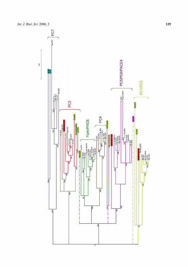
Int. J. Biol. Sci. 2006, 2 129
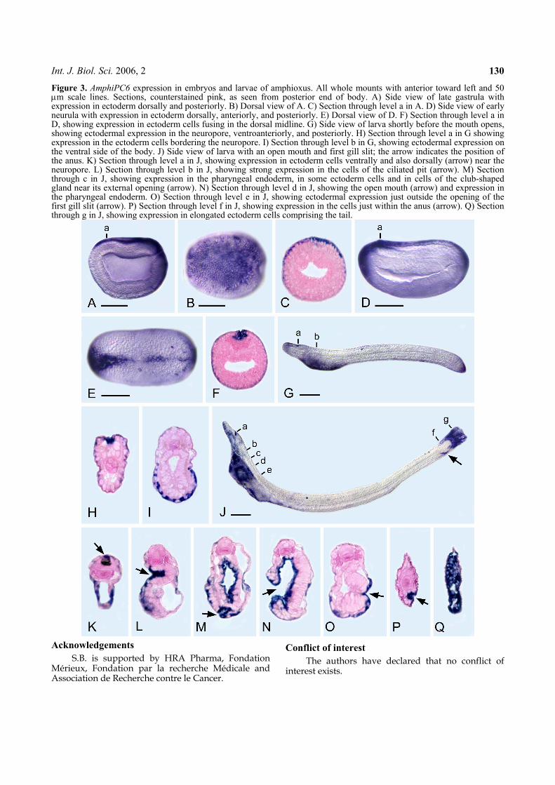
Int. J. Biol. Sci. 2006, 2 130
Figure 3. AmphiPC6 expression in embryos and larvae of amphioxus. All whole mounts with anterior toward left and 50 μm scale lines. Sections, counterstained pink, as seen from posterior end of body. A) Side view of late gastrula with expression in ectoderm dorsally and posteriorly. B) Dorsal view of A. C) Section through level a in A. D) Side view of early neurula with expression in ectoderm dorsally, anteriorly, and posteriorly. E) Dorsal view of D. F) Section through level a in D, showing expression in ectoderm cells fusing in the dorsal midline. G) Side view of larva shortly before the mouth opens, showing ectodermal expression in the neuropore, ventroanteriorly, and posteriorly. H) Section through level a in G showing expression in the ectoderm cells bordering the neuropore. I) Section through level b in G, showing ectodermal expression on the ventral side of the body. J) Side view of larva with an open mouth and first gill slit; the arrow indicates the position of the anus. K) Section through level a in J, showing expression in ectoderm cells ventrally and also dorsally (arrow) near the neuropore. L) Section through level b in J, showing strong expression in the cells of the ciliated pit (arrow). M) Section through c in J, showing expression in the pharyngeal endoderm, in some ectoderm cells and in cells of the club-shaped gland near its external opening (arrow). N) Section through level d in J, showing the open mouth (arrow) and expression in the pharyngeal endoderm. O) Section through level e in J, showing ectodermal expression just outside the opening of the first gill slit (arrow). P) Section through level f in J, showing expression in the cells just within the anus (arrow). Q) Section through g in J, showing expression in elongated ectoderm cells comprising the tail.
Acknowledgements
S.B. is supported by HRA Pharma, Fondation Mérieux, Fondation par la recherche Médicale and Association de Recherche contre le Cancer.
Conflict of interest
The authors have declared that no conflict of interest exists.
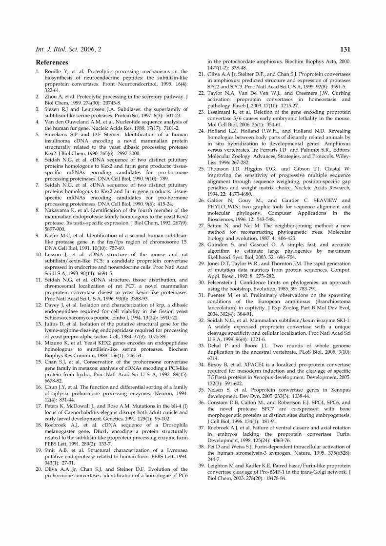
Int. J. Biol. Sci. 2006, 2 131
References
1. Rouille Y, et al. Proteolytic processing mechanisms in the biosynthesis of neuroendocrine peptides: the subtilisin-like proprotein convertases. Front Neuroendocrinol, 1995. 16(4): 322-61.
2. Zhou A, et al. Proteolytic processing in the secretory pathway. J Biol Chem, 1999. 274(30): 20745-8.
3. Siezen R.J and Leunissen J.A. Subtilases: the superfamily of subtilisin-like serine proteases. Protein Sci, 1997. 6(3): 501-23.
4. Van den Ouweland A.M, et al. Nucleotide sequence analysis of the human fur gene. Nucleic Acids Res, 1989. 17(17): 7101-2.
5. Smeekens S.P and D.F Steiner. Identification of a human insulinoma cDNA encoding a novel mammalian protein structurally related to the yeast dibasic processing protease Kex2. J Biol Chem, 1990. 265(6): 2997-3000.
6. Seidah N.G, et al. cDNA sequence of two distinct pituitary proteins homologous to Kex2 and furin gene products: tissue-specific mRNAs encoding candidates for pro-hormone processing proteinases. DNA Cell Biol, 1990. 9(10): 789.
7. Seidah N.G, et al. cDNA sequence of two distinct pituitary proteins homologous to Kex2 and furin gene products: tissue-specific mRNAs encoding candidates for pro-hormone processing proteinases. DNA Cell Biol, 1990. 9(6): 415-24.
8. Nakayama K, et al. Identification of the fourth member of the mammalian endoprotease family homologous to the yeast Kex2 protease. Its testis-specific expression. J Biol Chem, 1992. 267(9): 5897-900.
9. Kiefer M.C, et al. Identification of a second human subtilisin-like protease gene in the fes/fps region of chromosome 15. DNA Cell Biol, 1991. 10(10): 757-69.
10. Lusson J, et al. cDNA structure of the mouse and rat subtilisin/kexin-like PC5: a candidate proprotein convertase expressed in endocrine and nonendocrine cells. Proc Natl Acad Sci U S A, 1993. 90(14): 6691-5.
11. Seidah N.G, et al. cDNA structure, tissue distribution, and chromosomal localization of rat PC7, a novel mammalian proprotein convertase closest to yeast kexin-like proteinases. Proc Natl Acad Sci U S A, 1996. 93(8): 3388-93.
12. Davey J, et al. Isolation and characterization of krp, a dibasic endopeptidase required for cell viability in the fission yeast Schizosaccharomyces pombe. Embo J, 1994. 13(24): 5910-21.
13. Julius D, et al. Isolation of the putative structural gene for the lysine-arginine-cleaving endopeptidase required for processing of yeast prepro-alpha-factor. Cell, 1984. 37(3): 1075-89.
14. Mizuno K, et al. Yeast KEX2 genes encodes an endopeptidase homologous to subtilisin-like serine proteases. Biochem Biophys Res Commun, 1988. 156(1): 246-54.
15. Chan S.J, et al. Conservation of the prohormone convertase gene family in metazoa: analysis of cDNAs encoding a PC3-like protein from hydra. Proc Natl Acad Sci U S A, 1992. 89(15): 6678-82.
16. Chun J.Y, et al. The function and differential sorting of a family of aplysia prohormone processing enzymes. Neuron, 1994. 12(4): 831-44.
17. Peters K, McDowall J., and Rose A.M. Mutations in the bli-4 (I) locus of Caenorhabditis elegans disrupt both adult cuticle and early larval development. Genetics, 1991. 129(1): 95-102.
18. Roebroek A.J, et al. cDNA sequence of a Drosophila melanogaster gene, Dfur1, encoding a protein structurally related to the subtilisin-like proprotein processing enzyme furin. FEBS Lett, 1991. 289(2): 133-7.
19. Smit A.B, et al. Structural characterization of a Lymnaea putative endoprotease related to human furin. FEBS Lett, 1994. 343(1): 27-31.
20. Oliva A.A Jr, Chan S.J, and Steiner D.F. Evolution of the prohormone convertases: identification of a homologue of PC6
in the protochordate amphioxus. Biochim Biophys Acta, 2000. 1477(1-2): 338-48.
21. Oliva A.A Jr, Steiner D.F., and Chan S.J. Proprotein convertases in amphioxus: predicted structure and expression of proteases SPC2 and SPC3. Proc Natl Acad Sci U S A, 1995. 92(8): 3591-5.
22. Taylor N.A, Van De Ven W.J., and Creemers J.W. Curbing activation: proprotein convertases in homeostasis and pathology. Faseb J, 2003. 17(10): 1215-27.
23. Essalmani R, et al. Deletion of the gene encoding proprotein convertase 5/6 causes early embryonic lethality in the mouse. Mol Cell Biol, 2006. 26(1): 354-61.
24. Holland L.Z, Holland P.W.H., and Holland N.D. Revealing homologies between body parts of distantly related animals by in situ hybridization to developmental genes: Amphioxus versus vertebrates. In: Ferraris J.D and Palumbi S.R., Editors. Molecular Zoology: Advances, Strategies, and Protocols. Wiley-Liss. 1996: 267-282.
25. Thomson J.D, Higgins D.G., and Gibson T.J. Clustal W: improving the sensitivity of progressive multiple sequence alignment through sequence weighting, position-specific gap penalties and weight matrix choice. Nucleic Acids Research, 1994. 22: 4673-4680.
26. Galtier N, Gouy M., and Gautier C. SEAVIEW and PHYLO_WIN: two graphic tools for sequence alignment and molecular phylogeny. Computer Applications in the Biosciences, 1996. 12: 543-548.
27. Saitou N. and Nei M. The neighbor-joining method: a new method for reconstructing phylogenetic trees. Molecular biology and evolution, 1987. 4: 406-425.
28. Guindon S. and Gascuel O. A simple, fast, and accurate algorithm to estimate large phylogenies by maximum likelihood. Syst. Biol, 2003. 52: 696–704.
29. Jones D.T, Taylor W.R., and Thornton J.M. The rapid generation of mutation data matrices from protein sequences. Comput. Appl. Biosci, 1992. 8: 275–282.
30. Felsenstein J. Confidence limits on phylogenies: an approach using the bootstrap. Evolution, 1985. 39: 783-791.
31. Fuentes M, et al. Preliminary observations on the spawning conditions of the European amphioxus (Branchiostoma lanceolatum) in captivity. J Exp Zoolog Part B Mol Dev Evol, 2004. 302(4): 384-91.
32. Seidah N.G, et al. Mammalian subtilisin/kexin isozyme SKI-1: A widely expressed proprotein convertase with a unique cleavage specificity and cellular localization. Proc Natl Acad Sci U S A, 1999. 96(4): 1321-6.
33. Dehal P and Boore J.L. Two rounds of whole genome duplication in the ancestral vertebrate. PLoS Biol, 2005. 3(10): e314.
34. Birsoy B, et al. XPACE4 is a localized pro-protein convertase required for mesoderm induction and the cleavage of specific TGFbeta proteins in Xenopus development. Development, 2005. 132(3): 591-602.
35. Nelsen S, et al. Proprotein convertase genes in Xenopus development. Dev Dyn, 2005. 233(3): 1038-44.
36. Constam D.B, Calfon M., and Robertson E.J. SPC4, SPC6, and the novel protease SPC7 are coexpressed with bone morphogenetic proteins at distinct sites during embryogenesis. J Cell Biol, 1996. 134(1): 181-91.
37. Roebroek A.J, et al. Failure of ventral closure and axial rotation in embryos lacking the proprotein convertase Furin. Development, 1998. 125(24): 4863-76.
38. Pei D and Weiss S.J. Furin-dependent intracellular activation of the human stromelysin-3 zymogen. Nature, 1995. 375(6528): 244-7.
39. Leighton M and Kadler K.E. Paired basic/Furin-like proprotein convertase cleavage of Pro-BMP-1 in the trans-Golgi network. J Biol Chem, 2003. 278(20): 18478-84.

Int. J. Biol. Sci. 2006, 2 132
40. Lehmann M, et al. Lack of integrin alpha-chain endoproteolytic cleavage in furin-deficient human colon adenocarcinoma cells LoVo. Biochem J, 1996. 317: 803-9.
41. Stawowy P, et al. Endoproteolytic activation of alpha(v) integrin by proprotein convertase PC5 is required for vascular smooth muscle cell adhesion to vitronectin and integrin-dependent signaling. Circulation, 2004. 109(6): 770-6.
Structural and Functional Insights into the Ligand-bindingDomain of a Nonduplicated Retinoid X Nuclear Receptorfrom the Invertebrate Chordate Amphioxus*□S
Received for publication, July 24, 2008, and in revised form, October 17, 2008 Published, JBC Papers in Press, November 4, 2008, DOI 10.1074/jbc.M805692200
Giuseppe D. Tocchini-Valentini‡1,2, Natacha Rochel‡1, Hector Escriva§3, Pierre Germain¶, Carole Peluso-Iltis‡,Mathilde Paris§, Sarah Sanglier-Cianferani!, Alain Van Dorsselaer!, Dino Moras‡4, and Vincent Laudet§5
From the ‡Departement de Biologie et de Genomique Structurales and ¶Departement Biologie du Cancer, Institut de Genetique et deBiologie Moleculaire et Cellulaire, UMR7104 CNRS, U596 INSERM, Universite Louis Pasteur, 67400 Illkirch, France, §Institut de GenomiqueFonctionnelle de Lyon, Ecole Normale Superieure de Lyon, Universite Lyon 1, CNRS, INRA, Institut Federatif 128 Biosciences Gerland,69364 Lyon Sud, France, and !Laboratoire de Spectrometrie de Masse Bio-Organique, UMR/CNRS 7178, Institut PluridisciplinaireHubert CURIEN, Universite Louis Pasteur, Ecole de Chimie, Polymeres et Materiaux, 67087 Strasbourg, France
Retinoid X nuclear receptors (RXRs), as well as their insect
orthologue, ultraspiracle protein (USP), play an important role
in the transcription regulation mediated by the nuclear recep-
tors as the common partner of many other nuclear receptors.
Phylogenetic and structural studies have shown that the several
evolutionary shifts have modified the ligand binding ability of
RXRs. To understand the vertebrate-specific character of RXRs,
we have studied the RXR ligand-binding domain of the cepha-
lochordate amphioxus (Branchiostoma floridae), an inverte-
brate chordate that predates the genome duplication that pro-
duced the three vertebrates RXRs (!, ", and #). Here we report
the crystal structure of a novel apotetramer conformation of
the AmphiRXR ligand-binding domain, which shows some
similarity with the structures of the arthropods RXR/USPs.
AmphiRXR adopts an apo antagonist conformation with a
peculiar conformation of helix H11 filling the binding pocket.
In contrast to the arthropods RXR/USPs, which cannot be
activated by any RXR ligands, our functional data show that
AmphiRXR, like the vertebrates/mollusk RXRs, is able to
bind and be activated by RXR ligands but less efficiently than
vertebrate RXRs. Our data suggest that amphioxus RXR is,
functionally, an intermediate between arthropods RXR/USPs
and vertebrate RXRs.
The nuclear receptor (NR)6 superfamily is specific to animalsand performs an abundance of functions, from embryonicdevelopment to metamorphosis and from homeostasis of vari-ous physiological functions to the control of metabolism (for areview, see Ref. 1). Nuclear receptors are ligand-activated tran-scription factors. Many members of the superfamily thus bindmajor hormones, such as steroids, thyroid hormones, or reti-noids. These occupy a special position in gene regulation byproviding a direct link between the ligand, which they bind, andthe target gene, whose expression they regulate. There are alsomany nuclear receptors that are not known to bind any ligandand are thus called “orphan nuclear receptors” (2–4). Somenuclear receptors which were originally discovered as orphanshave since been shown to bind small hydrophobic molecules,such as fatty acids, but others appear to be true orphans (4).Nuclear receptors are composed of several functional
domains. The N-terminal A/B domain is highly variable inlength and sequence and contains a constitutively active trans-activation functionAF-1.Most conserved are domains C and E,corresponding grossly to the DNA-binding domain and theligand-binding domain (LBD), respectively. The LBD containsthe ligand-dependent transactivation function AF-2. TheDNA-binding domain and LBD are connected via a flexiblehinge (domain D). NRs act in vivo and in vitro as ligand-de-pendent transcriptional transregulators through binding, mostoften as heterodimers, to cis-acting response elements presentin cognate target genes. Activation of gene transcription occursafter binding of ligand, which results in a conformationalchange of the LBD, leading to release of corepressor and bind-ing of coactivator to the LBD (5).
* This study was supported by CNRS, INSERM, Universite Louis Pasteur, Euro-pean Commission on Structural Proteomics in Europe (SPINE) Grant QLG2-CT-220-0098, and SPINE2-Complexes Grant LSHG-CT-2006-031220 underthe integrated program, Quality of Life and Management of LivingResources. The costs of publication of this article were defrayed in part bythe payment of page charges. This article must therefore be herebymarked “advertisement” in accordance with 18 U.S.C. Section 1734 solely toindicate this fact.
□S The on-line version of this article (available at http://www.jbc.org) containssupplemental Table 1 and Figs. 1 and 2.
1 Both of these authors contributed equally to this work.2 Present address: Istituto di Biologia Cellulare, Consiglio Nazionale delle
Ricerche, Campus A, Buzzati-Traverso, Via Ramarini 32, MonterotondoScalo, 00016 Rome, Italy and Istituto di Genetica Molecolare, ConsiglioNazionale delle Ricerche, Via Abbiategrasso 207, I-27100 Pavia, Italy.
3 Present address: Laboratoire Arago, UMR 7628, CNRS and University Pierreand Marie Curie, BP 44, F-66651 Banyuls sur Mer, France.
4 To whom correspondence may be addressed: Laboratoire de Biologie etGenomique Structurales, IGBMC, 1 Rue Laurent Fries, BP 10142, Illkirch67404, France. Tel.: 33-388-653-220; Fax: 33-388-653-276; E-mail:[email protected].
5 To whom correspondence may be addressed: Institut de GenomiqueFonctionnelle de Lyon, Molecular Zoology team, Ecole NormaleSuperieure de Lyon 1, Institut Federatif 128 Biosciences Gerland, 69364Lyon Sud, France. Tel.: 33-472-728-190; Fax: 33-472-728-080; E-mail:[email protected].
6 The abbreviations used are: NR, nuclear receptor; RXR, retinoid X nuclearreceptor; AmphiRXR, amphioxus RXR; HsRXR, Homo sapiens RXR; MmRXR,Mus musculus RXR; Bg, RXR, B. glabrata RXR; LBD, ligand-binding domain;LBP, ligand-binding pocket; USP, ultraspiracle protein; BtUSP, B. tabaci;TcUSP, T. castaneum USP; Hx, helix x; RA, retinoic acid; DHA, docohexanoicacid; ESI, electrospray ionization; MS, mass spectrometry.
THE JOURNAL OF BIOLOGICAL CHEMISTRY VOL. 284, NO. 3, pp. 1938 –1948, January 16, 2009© 2009 by The American Society for Biochemistry and Molecular Biology, Inc. Printed in the U.S.A.
1938 JOURNAL OF BIOLOGICAL CHEMISTRY VOLUME 284 • NUMBER 3 • JANUARY 16, 2009
at C
NR
S o
n F
ebru
ary
23, 2
009
ww
w.jb
c.o
rgD
ow
nlo
aded fro
m
http://www.jbc.org/cgi/content/full/M805692200/DC1Supplemental Material can be found at:
To date, the crystal structures of !40 different NR LBDshave been solved, mainly mammalian nuclear receptors. Allstructures are very similar, sharing a common fold containing11–12 helices (6–7). The majority of the LBD crystals wereobtained in the presence of a ligand (holo-LBD) and only a fewwithout ligand (apo-LBD). Comparison of apo- and holo-LBDstructures led to the so-called “mouse trapmodel” of the ligand-dependent conformational switch (8). In apo-LBD, differentpositions of the C-terminal helix 12 (H12; also called AF-2helix) highlight the large flexibility of this region (7, 9). In holo-LBD, H12 is folded back on the core of the LBD, closing theLBP. This holo-position ofH12 is stabilized by direct or indirectinteractions with the ligand. The tightened position of H12,together with H3 and H4, results in the formation of a hydro-phobic groove inwhich coactivator proteins can bind through aconserved LXXLL motif, forming an !-helix. The ligand-in-duced conformational change concomitantly leads to destruc-tion of the surface that binds the corepressor, resulting in itsrelease from the LBD.The conserved domains present in NRs allow relatively easy
identification of nuclear receptors in genomic sequences (10)and also robust phylogenetic reconstruction on the scale of thesuperfamily (11, 12). Such analysis shows that NRs for similarligands do not group in the tree but are interspersedwith recep-tors for totally different ligands, whereas orphans are widelydistributed in the tree. This observation has led to the proposalthat the superfamily evolved from an orphan receptor thatgained the capacity of ligand binding several times independ-ently (13–15). The case of retinoid receptors is interesting inthat respect. Indeed, at least two distinct types of nuclear recep-tors, the RARs (! (NR1B1), " (NR1B2), and # (NR1B3); acti-vated by all-trans-retinoic acid (RA)) and the RXRs (! (NR2B1)," (NR2B2) and # (NR2B3); activated by 9-cis-RA) apparentlytransduce the retinoid message (16–18). It has thus been pro-posed that the capacity to bind, for example, retinoic acids,would have been acquired several times by mutation of anorphan nuclear receptor, allowing the establishment of nuclearretinoic acid signaling pathways (RARs and RXRs). The case ofRXR and USP, its arthropod homologue, is especially interest-ing in that context, because this receptor plays a pivotal roleinside the NR superfamily as a ubiquitous heterodimer partnerfor class II NRs (19).It is noteworthy that structural analysis can provide more
insight into the evolution of a protein domain (e.g. throughthe comparative structural analysis of domains from animalslocated at critical positions in the evolutionary tree). In thatrespect, the study of RXR LBD is important, since RXR is anancient gene that was subjected to several functional shiftsduring evolution. In addition, the status of RXR as a bona fideliganded receptor and the identity of its endogenous ligandare still under discussion (18, 20). Several experiments sug-gest that there is a very small amount of 9-cis-RA in vivo,casting doubts on its physiological relevance as an RXRligand (21). In addition, it has been suggested, from in vivo
detection of RXR-activating molecules, that docohexaneoicacid (DHA) could be a ligand for RXR (22). Thus, it may bepossible that 9-cis-RA is only a pharmacological activator ofRXR.
A large number of homologues of RXR have been identifiedin a variety of animals including early metazoans, such assponges and cnidarians, but also arthropods, mollusks, andinvertebrate chordates. In cnidarians, RXR was shown to bind9-cis-RA (23), and it activates transcription7 in response to it. Incontrast, in insects, the orthologue of RXR, called USP, isunable to bind 9-cis-RA and experienced a very high evolution-ary rate in dipterans and lepidopterans (see Ref. 24 and refer-ences therein). Detailed comparative structural analysis ofUSPs from insects have shown that these can be separated intotwo different types of receptors: (i) in basal insects, such ashemipterans and coleopterans, the USP ligand-binding pocketis partly filled by residues of unfoldedH6 andH11 helices and isthus likely to be an orphan receptor; (ii) in mecopterida (a ter-minal group of insects that includes dipteras and lepidopterasamong others), a phospholipid is present as a structural ligandinside the LBD that may be indicative of the existence of anendogenous ligand yet to be identified (28). In mollusks, RXRcan be activated by 9-cis-RA and DHA, at least using transienttransfection experiments (25). The case of amphioxus is espe-cially interesting, since this species is clearly located in the lin-eage giving rise to vertebrates but before the complete genomeduplications of vertebrates (26) (see Fig. 7 for a phylogenetictree). Thus, AmphiRXR is closely related to the three vertebrateRXR genes but represents a nonduplicated version of thesegenes. Consequently, determining the structure of the LBDencoded by the unique amphioxus AmphiRXR gene and thecomparison of this structure with its vertebrate orthologueswill infer the main structural features of RXR in the commonancestor of vertebrates (i.e. before gene duplication events).In this paper, we report the crystal structure of the apo form
AmphiRXR LBD, revealing a stable antagonist conformationnot driven by either a fortuitous or an antagonist ligand. Adetailed analysis of the comparison with previous crystal struc-tures of HsRXR (29–34), of nonvertebrate RXR (25) and USPs(27, 28) in different conformational states allows us to betterunderstand the evolution and functional dynamics of theseorphan receptors and to identify the structural basis of theirfunctional differences.
EXPERIMENTAL PROCEDURES
Cloning and Purification—The cDNA coding for the Bran-
chiostoma floridae RXR (accession number AF378829),AmphiRXR (35) ligand-binding domain (residues 266–484)was PCR-amplified from pSG5 vector and cloned by homolo-gous recombination into the vector pET15b. The residue’sposition number given here corresponds to the AmphiRXRsequence with the corresponding human sequence numberswithin parentheses, (HsAA-xxx), for some residues. Therecombinant protein was overproduced and purified in twosteps. Briefly, the LBD of AmphiRXR (residues 266–484) wascloned in pET15b expression vector to obtain an N-terminalhexahistidine-tagged fusion protein and was overproduced inEscherichia coli BL21 (DE3). Cells were grown in LB mediumand subsequently induced for 6 h at 20 °C with 1 mM isopropylthio-"-D-galactoside. Protein purification included a metal
7 H. Escriva and V. Lauder, unpublished results.
Amphioxus RXR LBD Structure
JANUARY 16, 2009 • VOLUME 284 • NUMBER 3 JOURNAL OF BIOLOGICAL CHEMISTRY 1939
at C
NR
S o
n F
ebru
ary
23, 2
009
ww
w.jb
c.o
rgD
ow
nlo
aded fro
m
affinity chromatography step on a nickel-Hitrap column and agel filtration on Superdex 75 16/60. The cell pellets were resus-pended in binding buffer (20 mM Tris-HCl, pH 8.0, 300 mM
NaCl, 10% glycerol, 5 mM imidazole), lysed by sonication, andclarified by centrifugation. The cleared supernatant was loadedon a nickel-Hitrap column (GE Healthcare). The protein waseluted using 150 and 300 mM imidazole, pH 8.0. The proteinwas subjected to gel filtration on a Superdex 75 16/60 column(GE Healthcare) in 20 mM Tris, pH 8, 200 mM NaCl, and 5 mM
dithiothreitol. The protein was then concentrated to 10 mg/mlusing a 10K Centricon device. The purity and homogeneity ofthe protein were assessed by SDS-PAGE and native PAGE andelectrospray ionization mass spectrometry. The protein is inequilibriumbetween homotetramer and homodimer accordingto analytical gel filtration and native PAGE. The homodimerdissociates into a monomer when characterized by mass spec-trometry studies in 50 mM ammonium acetate.Transient Transfection Assays—293T cells were maintained
in Dulbecco’s modified Eagle’s medium supplemented with 5%of charcoal-treated fetal calf serum. The cells were transfectedat 70% confluence in 24-well plates using 4 $l of ExGen 500(Euromedex) with 1.0 $g of total DNA, including 0.1 $g ofreporter plasmid (17m)x5-tk-luc, and 10 ng of cytomegalovi-rus-"-galactosidase as an internal control to account for varia-tions of transfection efficiency. The culture medium waschanged 6 h after transfection, and when appropriate, ligandsdissolved in EtOH were added to different final concentrations(10"9 to 10"6
M). Cells were lysed 24 h after transfection andassayed for luciferase activity. For the test of TIF2 (transcrip-tional intermediary factor 2) action, 10–500 ng of TIF2 expres-sion vector were assessed.Pull-down Assays—DNAs of TIF2.1 and TIF2.1m123, kindly
provided by Hinrich Gronemeyer (36), were both transcribedand translated in vitro using the TNT T7-coupled reticulocytelysate system (Promega) following the instructions of the man-ufacturer. Batches of 50 $M Ni2#-nitrilotriacetic acid-agarosegel (Qiagen) washed and equilibrated in buffer A (50 mM Tris-HCl (pH 8.0), 100 mM NaCl, 10 mM MgCl2, 5 mM imidazole,10% glycerol, and 0.1% Nonidet P-40) were loaded with 2 $g ofE. coli-purified His-tagged LBDs (HsRXR! or AmphiRXR).Incubation with TIF2.1 or TIF2.1m123 was done in a final vol-ume of 50 $l of buffer A containing 2 $l of in vitro translatedprotein and 10$g of bovine serum albumin and in the presenceof 2 $M ligand or 1% of ethanol. After incubation for 1 h at 4 °Cand threewashing steps in bufferA, boundproteinswere recov-ered with SDS sample buffer and revealed by autoradiographyof 12% SDS-polyacrylamide gels.Electrospray Ionization Mass Spectrometry—Before mass
analysis, amphioxus and human RXR LBDs purification bufferswere exchanged against 50 mM ammonium acetate on two suc-cessive gel filtration columns (NAP-10; GE Healthcare) andthen concentrated onCentricon 10-kDa devices. ESI-MSmeas-urements were performed on an electrospray time-of-flightmass spectrometer (LCT; Waters, Manchester, UK). The massmeasurements of the noncovalent complexes were performedin ammonium acetate (50mM, pH 6.8). Samples were diluted to20$M and continuously infused into the ESI ion source at a flowrate of 6 $l/min through a Harvard syringe pump. Titration
experiments were done by adding 2.5 mol eq of ligands toHsRXRorAmphiRXR, followed by a 15-min incubation at 4 °C.Competition experiments were performed by adding anequimolar mixture of two ligands (each in a 2.5-fold molarexcess) to the protein, followed by a 15-min incubation. A 2.5-fold molar excess of ligand was used as a standard in our massspectrometry experiments for two reasons: (i) to be able todetect ligands even with poor affinities (50–100 $M) and (ii) toassess the specificity of the binding (with a 2.5-fold molarexcess, nonspecific binding would also lead to the appearanceof bis-adducts, which could not be seen with only a 1:1 ratio).The ligands tested were synthetic (BMS649 and CD3254) ornatural (DHA, 9-cis-RA, and oleic acid) agonist ligands ofHsRXR. 9-cis-RA, all-trans-RA, oleic acid, and DHA werediluted in 10 mM EtOH. BMS649 and CD3254 were diluted in10mMEtOH/DMSO (90:10, v/v). To prevent dissociation in thegas phase during the ionization and desorption process, thecone voltage was optimized to 5 V. Mass data were acquired inthe positive ion mode on them/z 1000–5000 mass range. Cal-ibration of the instrument was performed using the multiplycharged ions produced by a separate injection of horse heartmyoglobin diluted to 2 $M in 1:1 water/acetonitrile (v/v) acid-ified with 1% (v/v) formic acid. The relative abundance of thedifferent species present on ESI mass spectra were measuredfrom their respective peak heights (37). All experiments werereproduced at least three times.An important feature concerning the noncovalent MS
approach is whether ESI mass spectra obtained in nondenatur-ing conditions in the gas phase of the mass spectrometer faith-fully reflect direct in solution binding. Several control experi-ments were performed in order to argue in favor of structurallyspecific interactions. A series of “successive in solution compe-titions” strongly argue in favor of site-specific interactions.Instead of the simultaneous addition of a mixture of twoligands, as realized in the direct in solution competitions, suc-cessive ligand addition was achieved; a first ligand, L1, is boundto the protein (formation of a 1:1 RXR"L1 complex), and a sec-ond one, L2, serves to displace the equilibrium toward the for-mation of the RXR"L2 complex. Such experiments allow us tounambiguously assess that both ligands compete for the samebinding site and that ESI-MS-observed interactions do notresult from any artifactual gas phase aggregation. Finally,experiments with different binding partners (either differentligands or human versus amphioxus proteins) were also per-formed, leading to subsequent changes on ESI mass spectra.Structure Determination—The homodimeric protein frac-
tions were used for crystallization trials. The purified proteinconditioned in 20 mM Tris, pH 8.0, 200 mM NaCl, 5 mM dithi-othreitol and concentrated to 5 mg/ml. Crystals were grown bythe hanging drop diffusion method and appeared in 4 days at17 °C. The initial drop contained 5mg/ml protein, 1.5M sodiumacetate, 50mMN-(2-acetamido)iminodiacetic acid, pH 6.5, andwas equilibrated versus a reservoir containing 1.8 M sodiumacetate, 100 mM N-(2-acetamido)iminodiacetic acid, pH 6.5.Two different crystals forms, hexagonal and orthorhombic,were obtained in the same crystallization conditions. Only theorthorhombic form resulted in being suitable for structuraldetermination. The solvent content of the crystals is 52.2%with
Amphioxus RXR LBD Structure
1940 JOURNAL OF BIOLOGICAL CHEMISTRY VOLUME 284 • NUMBER 3 • JANUARY 16, 2009
at C
NR
S o
n F
ebru
ary
23, 2
009
ww
w.jb
c.o
rgD
ow
nlo
aded fro
m
two LBD dimers (homotetramer) per asymmetric unit. Thus,the homodimeric protein may reassociate with time and saltconcentration of the precipitant agent. Crystals were flashcooled in ethane after cryoprotection in a solution containingthe reservoir solution plus 25% glycerol. A 2.7 Å resolution dataset was obtained merging high and low resolution data setsfromonly one crystal at the BM29Synchrotron line of the Euro-pean Synchrotron Radiation Facility facility in Grenoble. Datawere integrated and scaled using the DENZO-SCALEPACKpackage (38). The program CNS-SOLVE was used throughoutthe structure determination and refinement calculations (39).An initial phase estimate was obtained by molecular replace-ment using a partial structure of HsRXR dimer. After a rigidbody refinement, the solution resulted in Rcryst of 34% and Rfreeof 35%. The model obtained was used to determine the NCSmatrix of the four monomer molecules in the asymmetric unitin order to include averaging and solvent flattening procedureof densitymodification to calculate a first electron densitymap.The map unequivocally showed the missing parts of the modeland allowed the completemanual building of the structure. Thephase bias of the initial model was reduced by torsion anglemolecular dynamics simulated annealing refinement. Subse-quent refinement cycles alternated least square minimizationand model building using the program O (40). The NCS wasconstrained in the first round of refinement and subsequentlyrestrained and released.B factorswere refined using two groupsper residue. In the last cycle, 27 water molecules were locatedfrom a difference Fourier map. The loop between H1 and H3(residues 281–289) and the lateral chains of the residues at posi-tions 271–274, 299, 478, and 482–484 could not be constructedbecause of poor electron density. The quality of the final modelquality was assessed with Procheck. Because of the absence ofelectron density for the loop between H1 and H3, the integrityof the protein in the crystals was checked by SDS-PAGE (datanot shown). For the structure comparison, C! traces of themodels were superimposed using the lsq commands of O anddefault parameters. The figures were generated with Pymol.Crystallographic data and refinement statistics are summa-rized in supplemental Table 1. The chemical structures of allof the ligands described in this paper are shown in supple-mental Fig. 1.
RESULTS
Residues That Contact 9-cis-Retinoic Acid Are All Conserved
between AmphiRXR and HsRXR—Previous phylogeneticalanalysis clearly shows that, as expected, the amphioxus RXRsequence is positioned before the duplications giving rise to thethree vertebrate genes (35) (supplemental Fig. 2). In addition,amphioxus RXRwas shown to heterodimerize with amphioxusretinoic acid nuclear recepter or thyroid nuclear receptor andto bind to the relevant direct repeat sequence (41, 42).To determine the crystal structure of the ligand-binding
domain we selected a region that encompasses residues 266–484, leaving out the last 30 amino acid of themolecule that forman F-domain with no sequence identity with other NRs. Fig. 1represents the structure-based sequence alignment of RXRLBDs from different organisms. The alignment reveals a highdegree of sequence identity except for the length and composi-
tion of the loop between H1 and H3. Based on the structure ofhuman RXR!, the residues that contact the 9-cis-RA are allconserved. The ability to bind this ligand seems thus to bepreserved, and this is supported by mass spectrometry stud-ies as well as by transient transfection assays (see below). Thesame degree of conservation is encountered also in the resi-dues that form the hydrophobic groove in contact with theLXXLL motif activator helix (43). The only exception is a Valresidue that in amphioxus RXR substitutes at position 306 aPhe, forming Van der Waals contacts with the coactivatorhelix. The amino acids of the dimerization interface arestrictly conserved (19).AmphiRXR Is Activated by 9-cis-RA in Cultured Cells but at
Higher Concentrations than Vertebrate RXRs—Since the mainresidues implicated in ligand binding are conserved betweenAmphiRXR and vertebrates RXRs, we tested whether 9-cis-RAandDHAwere able to activate AmphiRXR in transient tranfec-tion assays in mammalian cells. To avoid any interference withendogenous RXR expressed in 293T cells, we used fusionsbetween the Gal4 DNA-binding domain and RXR-LBD. Asshown in Fig. 2A, both 9-cis-RA and all-trans-RA activate 2–4-fold Gal4-MmRXR! and Gal4-AmphiRXR. The effect of all-trans-RA at the concentration we used can be explained by itsisomerization in 9-cis-RA (46). Nevertheless, in the experimen-tal condition we used, we did not observe any reproducibleeffect of DHA (lanes 4 and 8). Of note, using strictly the sameconditions as those described in the original paper (22), weobserved that DHA activates MmRXR! but not AmphiRXR(not shown). Dose response experiments (Fig. 2B) show that9-cis-RA activatesAmphiRXR in a dose-dependentmanner butwith a range of concentration higher than mouse RXR. A weakactivation by 9-cis-RA is also observed for the molluskBiomphalaria glabrata RXR (45). Given that amphioxus islocated at the base of chordates in phylogenetical tree, we canconclude from these data that the ability of RXR to bind and tobe activated by 9-cis-RA is thus conserved in all chordates. Thisis in accordancewith the conservation in amphioxusRXRof theamino acid residues implicated in ligand binding in vertebrateRXRs.We thus explored the interaction between RXR and coacti-
vators, such as TIF2. The activity of both Gal4-MmRXR! andGal4-AmphiRXR was potentiated by the cotransfection of ahuman TIF2 expression vector (Fig. 2B). These findings wereconfirmed by pull-down assays (Fig. 2C) showing that in vitro
amphioxus RXR, like human RXR!, can interact with TIF2 in aligand-dependent manner. Interestingly, specific rexinoids,such as BMS649, induce the recruitment of TIF2, but not itsmutant TIF2.1m123, mutated in the domain known to interactwith NRs. Rexinoids are ligands that selectively bind and acti-vate RXRs but not RARs in contrast to retinoids. This is inaccordance with our observation that the recently sequencedamphioxus genome contains a unique homologue of the p160co-activators (51). All of these observations suggest that (i) themechanisms by which RXR activates transcription are con-served between amphioxus and mammals and (ii) the bindinginterface between AmphiRXR and TIF2 is functionallyconserved.
Amphioxus RXR LBD Structure
JANUARY 16, 2009 • VOLUME 284 • NUMBER 3 JOURNAL OF BIOLOGICAL CHEMISTRY 1941
at C
NR
S o
n F
ebru
ary
23, 2
009
ww
w.jb
c.o
rgD
ow
nlo
aded fro
m
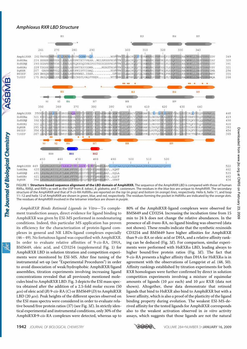
AmphiRXR Binds Retinoid Ligands in Vitro—To comple-ment transfection assays, direct evidence for ligand binding toAmphiRXR was given by ESI-MS performed in nondenaturingconditions. Indeed, this particular MS application has provenits efficiency for the characterization of protein-ligand com-plexes in general and NR LBDs-ligand complexes especially(46–49). No fortuitous ligand was copurified with AmphiRXR.In order to evaluate relative affinities of 9-cis-RA, DHA,BMS649, oleic acid, and CD3254 (supplemental Fig. 1) forAmphiRXR LBD in solution titration and competition experi-ments were monitored by ESI-MS. After fine tuning of theinstrumental set-up (see “Experimental Procedures”) in orderto avoid dissociation of weak/hydrophobic AmphiRXR/ligandassemblies, titration experiments involving increasing ligandconcentrations revealed that all previously mentioned mole-cules bind to AmphiRXR LBD. Fig. 3 depicts the ESImass spec-tra obtained after the addition of a 2.5-fold molar excess (50$M) of oleic acid (B) 9-cis-RA (C) or BMS649 (D) to AmphiRXRLBD (20 $M). Peak heights of the different species observed onthe ESI mass spectra were considered in order to evaluate rela-tive bound/free protein ratios (37) (see Fig. 3E). In strictly iden-tical experimental and instrumental conditions, only 30% of theAmphiRXR"9-cis-RA complexes were detected, whereas up to
80% of the AmphiRXR-ligand complexes were observed forBMS649 and CD3254. Increasing the incubation time from 15min to 24 h does not change the relative abundances. In thepresence of all-trans-RA, no ligand binding was observed (datanot shown). These results indicate that the synthetic rexinoidsCD3254 and BMS649 have higher affinities for AmphiRXRthan 9-cis-RA or oleic acid or DHA, and a relative affinity rank-ing can be deduced (Fig. 3E). For comparison, similar experi-ments were performed with HsRXR! LBD, leading always tohigher bound/free protein ratios (Fig. 3E). The fact that9-cis-RA presents a higher affinity than DHA for HsRXR! is inagreement with the observations of Lengqvist et al. (48, 50).Affinity rankings established by titration experiments for bothRXR homologues were further confirmed by direct in solutioncompetition experiments involving a mixture of equimolaramounts of ligands (10 $M each) and 10 $M RXR (data notshown). Altogether, these data demonstrate that retinoidligands that bind to HsRXR also bind to AmphiRXR but with alower affinity, which is also a proof of the plasticity of the ligandbinding property during evolution. The weakest ESI-MS-de-rived affinity for the tested ligands for AmphiRXR correspondsalso to the weakest activation observed in in vitro activityassays, which suggests that those ligands are not the natural
FIGURE 1. Structure-based sequence alignment of the LBD domain of AmphiRXR. The sequence of the AmphiRXR LBD is compared with those of humanRXR!, RXR", and RXR# as well as the USP from B. tabaci, B. glabatra, and T. castaneum. The residues in the blue box are unique to AmphiRXR. The secondarystructure of the AmphiRXR and that of 9-cis-RA HsRXR! are reported on the top (in gray) and bottom (in orange) lines, respectively. Helix 6, helix 11, and loop11–12 and helix 12 of AmphiRXR are shown in green, blue, and red, respectively. The residues forming the pocket in HsRXR! are indicated by the orange dots.The residues of AmphiRXR involved in the tetramer interface are shown in purple.
Amphioxus RXR LBD Structure
1942 JOURNAL OF BIOLOGICAL CHEMISTRY VOLUME 284 • NUMBER 3 • JANUARY 16, 2009
at C
NR
S o
n F
ebru
ary
23, 2
009
ww
w.jb
c.o
rgD
ow
nlo
aded fro
m
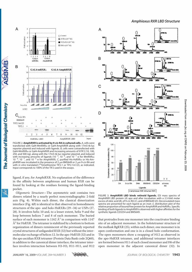
ligand, if any, for AmphiRXR. No explanation of the differencein the affinity between amphioxus and human RXR can befound by looking at the residues forming the ligand-bindingpocket.Oligomeric Structure—The asymmetric unit contains two
dimers related by a nearly perfect noncrystallographic 2-foldaxis (Fig. 4). Within each dimer, the classical dimerizationinterface (Fig. 4B) is identical to that observed in homodimericstructures of the apo- and holo-HsRXRs (29–34) or USPs (27,28). It involves helix 10 and, to a lesser extent, helix 9 and theloop between helices 7 and 8 of each monomer. The buriedsurface of each monomer is 1162 Å2 in comparison with 1147Å2 forHsRXR. The tetramer is stabilized by a bottom to bottomorganization of dimers reminiscent of the previously reportedcrystal structures of unligandedRXR (32) butwithout the inter-molecular exchange of helices 12. In the apo-HsRXR (32) and inthe apo ascidian RXR tetramer (Protein Data Bank code 2Q60),in addition to the canonical dimer interface, the tetramer inter-face involves interaction between H3-H3, H11-H11, and H12
that protrudes from onemonomer into the coactivator bindingsite of an adjacent monomer. In the holotetramer structure ofthemollusk BgRXR (25), within each dimer, onemonomer is inopen conformation and one is in a closed holo conformation.The open monomers show a swapping of H12 as observed inthe apo-HsRXR tetramer, and additional tetramer interfacesare formed betweenH11 of each closedmonomer andH6of theopen monomer in the adjacent canonical dimer (32). In
FIGURE 2. AmphiRXR is activated by 9-cis-RA in cultured cells. A, cells weretransfected with Gal4-MmRXR! or Gal4-AmphiRXR along with 17m5-tk-lucreporter plasmid and induced with ligands. B, cells were cotransfected withGal4-MmRXR! or Gal4-AmphiRXR and increasing amounts of hTIF2 (10, 100,250, and 500 ng), along with the 17m5-tk-luc reporter plasmid, and inducedwith increasing amounts of ligands (10"9, 10"8, and 10"7
M for MmRXR!;10"8, 10"7, and 10"6
M for AmphiRXR). C, purified His-HsRXR! or His-Am-phiRXR was incubated in the presence of 2 $M BMS649 or 2 $M 9-cis-RA andwith in vitro translated [35S]methionine TIF2.1 or TIF2.1m123, as indicated.Input corresponds to 100% of the TIF2 used in the assays.
FIGURE 3. AmphiRXR LBD binds retinoid ligands. ESI mass spectra ofAmphiRXR LBD protein (A) apo and after incubation with a 2.5-fold molarexcess of oleic acid (B), of 9-cis-RA (C), and of BMS649 (D). Deconvulated massspectra are presented for each ligand as an inset. E, distribution plot of therelative proportion of bound/free protein for AmphiRXR and HsRXR!. Specificbinding of both ligands to AmphiRXR is observed with higher affinities for thesynthetic ligands CD3254 and BMS649.
Amphioxus RXR LBD Structure
JANUARY 16, 2009 • VOLUME 284 • NUMBER 3 JOURNAL OF BIOLOGICAL CHEMISTRY 1943
at C
NR
S o
n F
ebru
ary
23, 2
009
ww
w.jb
c.o
rgD
ow
nlo
aded fro
m
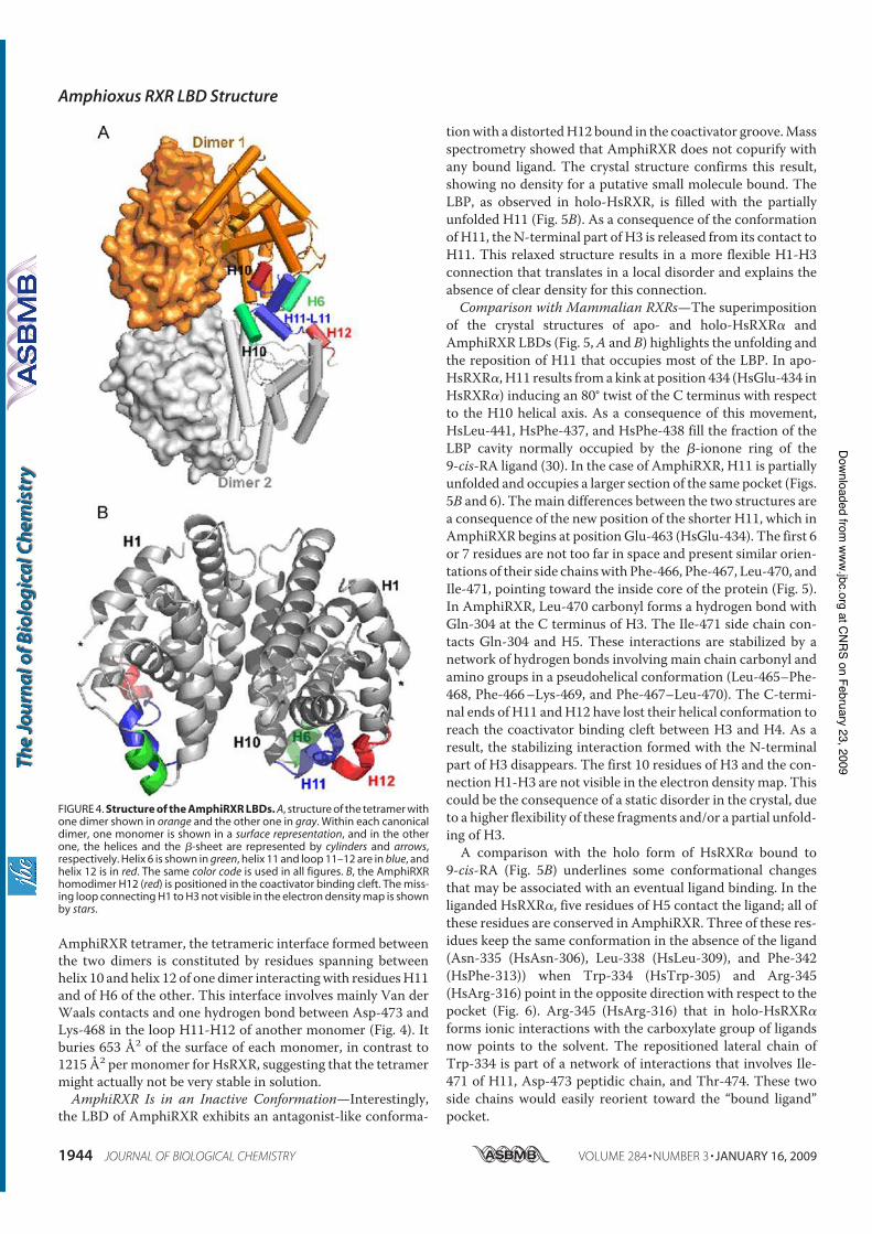
AmphiRXR tetramer, the tetrameric interface formed betweenthe two dimers is constituted by residues spanning betweenhelix 10 and helix 12 of one dimer interactingwith residuesH11and of H6 of the other. This interface involves mainly Van derWaals contacts and one hydrogen bond between Asp-473 andLys-468 in the loop H11-H12 of another monomer (Fig. 4). Itburies 653 Å2 of the surface of each monomer, in contrast to1215 Å2 permonomer for HsRXR, suggesting that the tetramermight actually not be very stable in solution.AmphiRXR Is in an Inactive Conformation—Interestingly,
the LBD of AmphiRXR exhibits an antagonist-like conforma-
tionwith a distortedH12 bound in the coactivator groove.Massspectrometry showed that AmphiRXR does not copurify withany bound ligand. The crystal structure confirms this result,showing no density for a putative small molecule bound. TheLBP, as observed in holo-HsRXR, is filled with the partiallyunfolded H11 (Fig. 5B). As a consequence of the conformationof H11, theN-terminal part of H3 is released from its contact toH11. This relaxed structure results in a more flexible H1-H3connection that translates in a local disorder and explains theabsence of clear density for this connection.Comparison with Mammalian RXRs—The superimposition
of the crystal structures of apo- and holo-HsRXR! andAmphiRXR LBDs (Fig. 5,A and B) highlights the unfolding andthe reposition of H11 that occupies most of the LBP. In apo-HsRXR!, H11 results froma kink at position 434 (HsGlu-434 inHsRXR!) inducing an 80° twist of the C terminus with respectto the H10 helical axis. As a consequence of this movement,HsLeu-441, HsPhe-437, and HsPhe-438 fill the fraction of theLBP cavity normally occupied by the "-ionone ring of the9-cis-RA ligand (30). In the case of AmphiRXR, H11 is partiallyunfolded and occupies a larger section of the same pocket (Figs.5B and 6). Themain differences between the two structures area consequence of the new position of the shorter H11, which inAmphiRXR begins at positionGlu-463 (HsGlu-434). The first 6or 7 residues are not too far in space and present similar orien-tations of their side chainswith Phe-466, Phe-467, Leu-470, andIle-471, pointing toward the inside core of the protein (Fig. 5).In AmphiRXR, Leu-470 carbonyl forms a hydrogen bond withGln-304 at the C terminus of H3. The Ile-471 side chain con-tacts Gln-304 and H5. These interactions are stabilized by anetwork of hydrogen bonds involving main chain carbonyl andamino groups in a pseudohelical conformation (Leu-465–Phe-468, Phe-466–Lys-469, and Phe-467–Leu-470). The C-termi-nal ends of H11 andH12 have lost their helical conformation toreach the coactivator binding cleft between H3 and H4. As aresult, the stabilizing interaction formed with the N-terminalpart of H3 disappears. The first 10 residues of H3 and the con-nection H1-H3 are not visible in the electron density map. Thiscould be the consequence of a static disorder in the crystal, dueto a higher flexibility of these fragments and/or a partial unfold-ing of H3.A comparison with the holo form of HsRXR! bound to
9-cis-RA (Fig. 5B) underlines some conformational changesthat may be associated with an eventual ligand binding. In theliganded HsRXR!, five residues of H5 contact the ligand; all ofthese residues are conserved in AmphiRXR. Three of these res-idues keep the same conformation in the absence of the ligand(Asn-335 (HsAsn-306), Leu-338 (HsLeu-309), and Phe-342(HsPhe-313)) when Trp-334 (HsTrp-305) and Arg-345(HsArg-316) point in the opposite direction with respect to thepocket (Fig. 6). Arg-345 (HsArg-316) that in holo-HsRXR!
forms ionic interactions with the carboxylate group of ligandsnow points to the solvent. The repositioned lateral chain ofTrp-334 is part of a network of interactions that involves Ile-471 of H11, Asp-473 peptidic chain, and Thr-474. These twoside chains would easily reorient toward the “bound ligand”pocket.
FIGURE 4. Structure of the AmphiRXR LBDs. A, structure of the tetramer withone dimer shown in orange and the other one in gray. Within each canonicaldimer, one monomer is shown in a surface representation, and in the otherone, the helices and the "-sheet are represented by cylinders and arrows,respectively. Helix 6 is shown in green, helix 11 and loop 11–12 are in blue, andhelix 12 is in red. The same color code is used in all figures. B, the AmphiRXRhomodimer H12 (red) is positioned in the coactivator binding cleft. The miss-ing loop connecting H1 to H3 not visible in the electron density map is shownby stars.
Amphioxus RXR LBD Structure
1944 JOURNAL OF BIOLOGICAL CHEMISTRY VOLUME 284 • NUMBER 3 • JANUARY 16, 2009
at C
NR
S o
n F
ebru
ary
23, 2
009
ww
w.jb
c.o
rgD
ow
nlo
aded fro
m
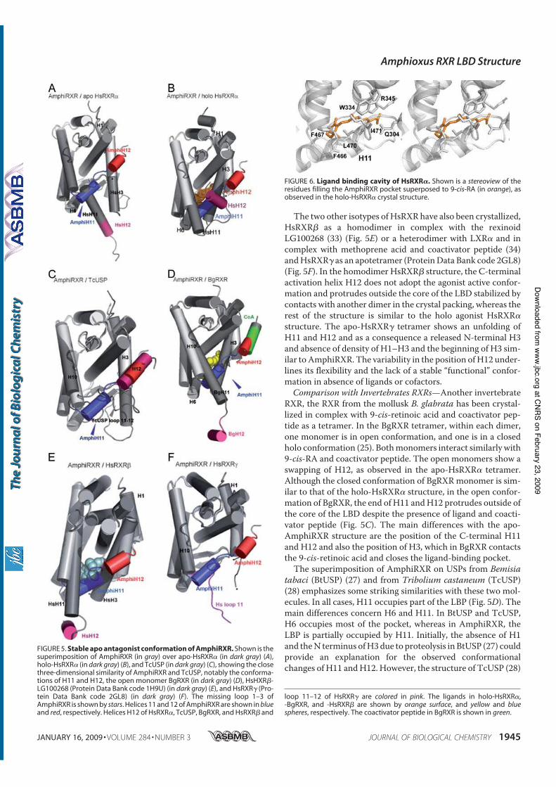
The two other isotypes of HsRXR have also been crystallized,HsRXR" as a homodimer in complex with the rexinoidLG100268 (33) (Fig. 5E) or a heterodimer with LXR! and incomplex with methoprene acid and coactivator peptide (34)andHsRXR# as an apotetramer (ProteinData Bank code 2GL8)(Fig. 5F). In the homodimer HsRXR" structure, the C-terminalactivation helix H12 does not adopt the agonist active confor-mation and protrudes outside the core of the LBD stabilized bycontacts with another dimer in the crystal packing, whereas therest of the structure is similar to the holo agonist HsRXR!
structure. The apo-HsRXR# tetramer shows an unfolding ofH11 and H12 and as a consequence a released N-terminal H3and absence of density of H1–H3 and the beginning of H3 sim-ilar to AmphiRXR. The variability in the position ofH12 under-lines its flexibility and the lack of a stable “functional” confor-mation in absence of ligands or cofactors.Comparison with Invertebrates RXRs—Another invertebrate
RXR, the RXR from the mollusk B. glabrata has been crystal-lized in complex with 9-cis-retinoic acid and coactivator pep-tide as a tetramer. In the BgRXR tetramer, within each dimer,one monomer is in open conformation, and one is in a closedholo conformation (25). Bothmonomers interact similarly with9-cis-RA and coactivator peptide. The open monomers show aswapping of H12, as observed in the apo-HsRXR! tetramer.Although the closed conformation of BgRXRmonomer is sim-ilar to that of the holo-HsRXR! structure, in the open confor-mation of BgRXR, the end of H11 andH12 protrudes outside ofthe core of the LBD despite the presence of ligand and coacti-vator peptide (Fig. 5C). The main differences with the apo-AmphiRXR structure are the position of the C-terminal H11and H12 and also the position of H3, which in BgRXR contactsthe 9-cis-retinoic acid and closes the ligand-binding pocket.The superimposition of AmphiRXR on USPs from Bemisia
tabaci (BtUSP) (27) and from Tribolium castaneum (TcUSP)(28) emphasizes some striking similarities with these two mol-ecules. In all cases, H11 occupies part of the LBP (Fig. 5D). Themain differences concern H6 and H11. In BtUSP and TcUSP,H6 occupies most of the pocket, whereas in AmphiRXR, theLBP is partially occupied by H11. Initially, the absence of H1and theN terminus ofH3due to proteolysis in BtUSP (27) couldprovide an explanation for the observed conformationalchanges of H11 andH12. However, the structure of TcUSP (28)
FIGURE 5. Stable apo antagonist conformation of AmphiRXR. Shown is thesuperimposition of AmphiRXR (in gray) over apo-HsRXR! (in dark gray) (A),holo-HsRXR! (in dark gray) (B), and TcUSP (in dark gray) (C), showing the closethree-dimensional similarity of AmphiRXR and TcUSP, notably the conforma-tions of H11 and H12, the open monomer BgRXR (in dark gray) (D), HsHXR"-LG100268 (Protein Data Bank code 1H9U) (in dark gray) (E), and HsRXR# (Pro-tein Data Bank code 2GL8) (in dark gray) (F). The missing loop 1–3 ofAmphiRXR is shown by stars. Helices 11 and 12 of AmphiRXR are shown in blueand red, respectively. Helices H12 of HsRXR!, TcUSP, BgRXR, and HsRXR" and
loop 11–12 of HsRXR# are colored in pink. The ligands in holo-HsRXR!,-BgRXR, and -HsRXR" are shown by orange surface, and yellow and bluespheres, respectively. The coactivator peptide in BgRXR is shown in green.
FIGURE 6. Ligand binding cavity of HsRXR!. Shown is a stereoview of theresidues filling the AmphiRXR pocket superposed to 9-cis-RA (in orange), asobserved in the holo-HsRXR! crystal structure.
Amphioxus RXR LBD Structure
JANUARY 16, 2009 • VOLUME 284 • NUMBER 3 JOURNAL OF BIOLOGICAL CHEMISTRY 1945
at C
NR
S o
n F
ebru
ary
23, 2
009
ww
w.jb
c.o
rgD
ow
nlo
aded fro
m
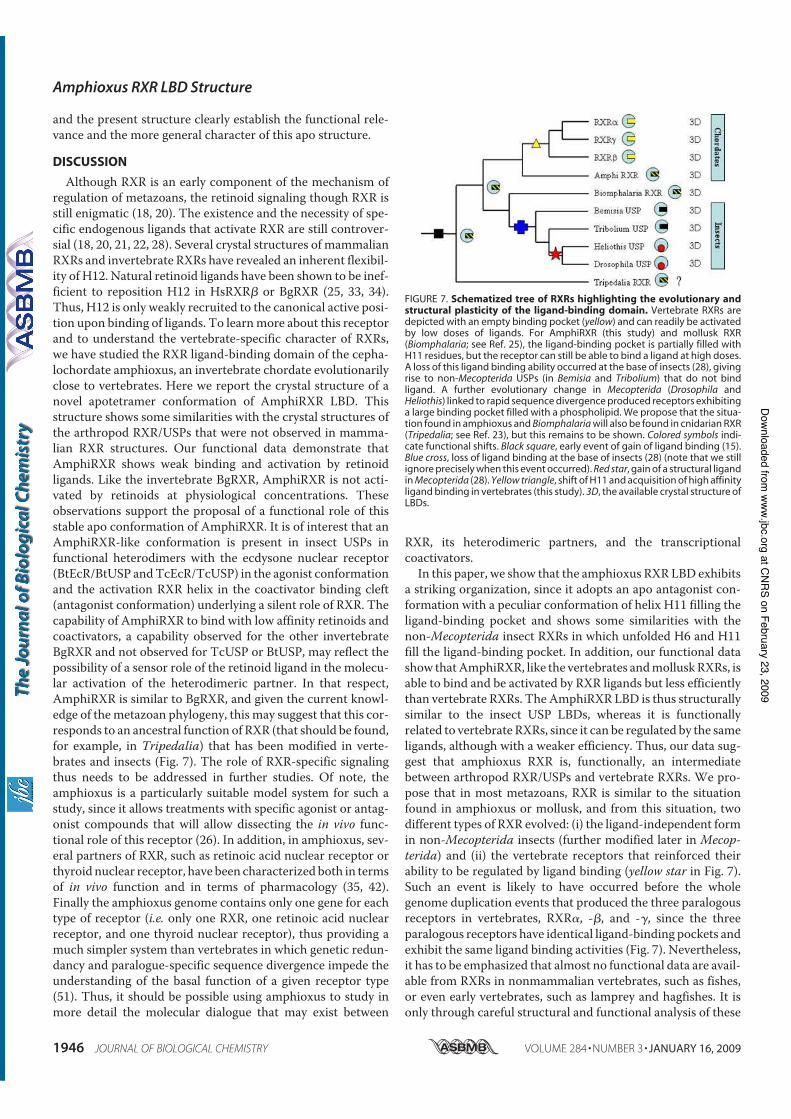
and the present structure clearly establish the functional rele-vance and the more general character of this apo structure.
DISCUSSION
Although RXR is an early component of the mechanism ofregulation of metazoans, the retinoid signaling though RXR isstill enigmatic (18, 20). The existence and the necessity of spe-cific endogenous ligands that activate RXR are still controver-sial (18, 20, 21, 22, 28). Several crystal structures of mammalianRXRs and invertebrate RXRs have revealed an inherent flexibil-ity of H12. Natural retinoid ligands have been shown to be inef-ficient to reposition H12 in HsRXR" or BgRXR (25, 33, 34).Thus, H12 is only weakly recruited to the canonical active posi-tion upon binding of ligands. To learnmore about this receptorand to understand the vertebrate-specific character of RXRs,we have studied the RXR ligand-binding domain of the cepha-lochordate amphioxus, an invertebrate chordate evolutionarilyclose to vertebrates. Here we report the crystal structure of anovel apotetramer conformation of AmphiRXR LBD. Thisstructure shows some similarities with the crystal structures ofthe arthropod RXR/USPs that were not observed in mamma-lian RXR structures. Our functional data demonstrate thatAmphiRXR shows weak binding and activation by retinoidligands. Like the invertebrate BgRXR, AmphiRXR is not acti-vated by retinoids at physiological concentrations. Theseobservations support the proposal of a functional role of thisstable apo conformation of AmphiRXR. It is of interest that anAmphiRXR-like conformation is present in insect USPs infunctional heterodimers with the ecdysone nuclear receptor(BtEcR/BtUSP andTcEcR/TcUSP) in the agonist conformationand the activation RXR helix in the coactivator binding cleft(antagonist conformation) underlying a silent role of RXR. Thecapability of AmphiRXR to bind with low affinity retinoids andcoactivators, a capability observed for the other invertebrateBgRXR and not observed for TcUSP or BtUSP, may reflect thepossibility of a sensor role of the retinoid ligand in the molecu-lar activation of the heterodimeric partner. In that respect,AmphiRXR is similar to BgRXR, and given the current knowl-edge of themetazoan phylogeny, this may suggest that this cor-responds to an ancestral function of RXR (that should be found,for example, in Tripedalia) that has been modified in verte-brates and insects (Fig. 7). The role of RXR-specific signalingthus needs to be addressed in further studies. Of note, theamphioxus is a particularly suitable model system for such astudy, since it allows treatments with specific agonist or antag-onist compounds that will allow dissecting the in vivo func-tional role of this receptor (26). In addition, in amphioxus, sev-eral partners of RXR, such as retinoic acid nuclear receptor orthyroid nuclear receptor, have been characterized both in termsof in vivo function and in terms of pharmacology (35, 42).Finally the amphioxus genome contains only one gene for eachtype of receptor (i.e. only one RXR, one retinoic acid nuclearreceptor, and one thyroid nuclear receptor), thus providing amuch simpler system than vertebrates in which genetic redun-dancy and paralogue-specific sequence divergence impede theunderstanding of the basal function of a given receptor type(51). Thus, it should be possible using amphioxus to study inmore detail the molecular dialogue that may exist between
RXR, its heterodimeric partners, and the transcriptionalcoactivators.In this paper, we show that the amphioxus RXRLBD exhibits
a striking organization, since it adopts an apo antagonist con-formation with a peculiar conformation of helix H11 filling theligand-binding pocket and shows some similarities with thenon-Mecopterida insect RXRs in which unfolded H6 and H11fill the ligand-binding pocket. In addition, our functional datashow thatAmphiRXR, like the vertebrates andmolluskRXRs, isable to bind and be activated by RXR ligands but less efficientlythan vertebrate RXRs. The AmphiRXR LBD is thus structurallysimilar to the insect USP LBDs, whereas it is functionallyrelated to vertebrate RXRs, since it can be regulated by the sameligands, although with a weaker efficiency. Thus, our data sug-gest that amphioxus RXR is, functionally, an intermediatebetween arthropod RXR/USPs and vertebrate RXRs. We pro-pose that in most metazoans, RXR is similar to the situationfound in amphioxus or mollusk, and from this situation, twodifferent types of RXR evolved: (i) the ligand-independent formin non-Mecopterida insects (further modified later in Mecop-
terida) and (ii) the vertebrate receptors that reinforced theirability to be regulated by ligand binding (yellow star in Fig. 7).Such an event is likely to have occurred before the wholegenome duplication events that produced the three paralogousreceptors in vertebrates, RXR!, -", and -#, since the threeparalogous receptors have identical ligand-binding pockets andexhibit the same ligand binding activities (Fig. 7). Nevertheless,it has to be emphasized that almost no functional data are avail-able from RXRs in nonmammalian vertebrates, such as fishes,or even early vertebrates, such as lamprey and hagfishes. It isonly through careful structural and functional analysis of these
FIGURE 7. Schematized tree of RXRs highlighting the evolutionary andstructural plasticity of the ligand-binding domain. Vertebrate RXRs aredepicted with an empty binding pocket (yellow) and can readily be activatedby low doses of ligands. For AmphiRXR (this study) and mollusk RXR(Biomphalaria; see Ref. 25), the ligand-binding pocket is partially filled withH11 residues, but the receptor can still be able to bind a ligand at high doses.A loss of this ligand binding ability occurred at the base of insects (28), givingrise to non-Mecopterida USPs (in Bemisia and Tribolium) that do not bindligand. A further evolutionary change in Mecopterida (Drosophila andHeliothis) linked to rapid sequence divergence produced receptors exhibitinga large binding pocket filled with a phospholipid. We propose that the situa-tion found in amphioxus and Biomphalaria will also be found in cnidarian RXR(Tripedalia; see Ref. 23), but this remains to be shown. Colored symbols indi-cate functional shifts. Black square, early event of gain of ligand binding (15).Blue cross, loss of ligand binding at the base of insects (28) (note that we stillignore precisely when this event occurred). Red star, gain of a structural ligandin Mecopterida (28). Yellow triangle, shift of H11 and acquisition of high affinityligand binding in vertebrates (this study). 3D, the available crystal structure ofLBDs.
Amphioxus RXR LBD Structure
1946 JOURNAL OF BIOLOGICAL CHEMISTRY VOLUME 284 • NUMBER 3 • JANUARY 16, 2009
at C
NR
S o
n F
ebru
ary
23, 2
009
ww
w.jb
c.o
rgD
ow
nlo
aded fro
m
receptors that a precise model of ligand binding evolution ofchordate RXRs will be determined.According to the strong conservation of the residues lining
the putative canonical LBP, one would predict that all RXR-likeLBD, including those from insects, such as the hemipteranBemisia (BtUSP) or the coleopteranTribolium (TcUSP), wouldbind most of the known ligands of HsRXR. This contrasts withthe experimental observations in Tribolium USP, which doesnot bind and is not activated by any RXR ligands (25). Of note,these observations have been reinforced by an evolutionaryanalysis of the pattern of substitution rate in the ligand-bindingdomains of RXRs and USPs from a wide variety of metazoans(25). Indeed, these data indicate that in TcUSP or BtUSP, resi-dues thatwould belong to a potential RXR-like LBP exhibit highevolutionary rates, suggesting a relaxation of the evolutionarypressure in this region that may be related to a loss in ligandbinding capability. AmphiRXR appears to be in a paradoxicalsituationwhen comparedwith the receptors from insects, sinceit adopts a similar apo conformation (withH12 in an antagonistposition and the ligand-binding pocket filled by H11 residues),whereas it is able to bind to RXR-specific ligands, such as 9-cis-retinoic acid, BMS649, or oleic acid. Of note, in silicomodelinganalysis cannot provide a simple steric explanation of thisobservation, since a homologymodel that would accommodateligands like 9-cis-RA or fatty acids can easily be built for boththe insect receptors and AmphiRXR. Additional experimentsare necessary to elucidate themechanism of ligand binding andmore generally the question of the necessity of a ligand for thisorphan receptor. RXR provides a very clear example showingthat sequence conservation is a necessary prerequisite for theprediction of functional properties, such as ligand binding, butis not sufficient alone to reach a conclusion.Taken together, the data available on RXRs illustrate the
remarkable evolutionary plasticity of this LBD that can adapt todifferent functional shifts, such as changes in the ligand bindingabilities, together with a conservation of other functions, suchas heterodimerization. This certainly calls for other compara-tive structural and functional studies of this receptor that willserve to illustrate basic concepts in NR function, such as therole of ligand regulation, the definition of endogenous ligands,or the structural basis of ligand affinity.
REFERENCES
1. Gronemeyer, H., Gustafsson, J. A., and Laudet, V. (2004) Nat. Rev. Drug
Discov. 3, 950–964
2. Gustafsson, J. A. (1999) Science 284, 1285–1286
3. Kliewer, S. A., Lehmann, J. M., and Willson, T. M. (1999) Science 284,
757–760
4. Benoit, G., Cooney, A., Giguere, V., Ingraham, H., Lazar, M., Muscat, G.,
Perlmann, T., Renaud, J. P., Schwabe, J., Sladek, F., Tsai, M. J., and Laudet,
V. Pharmacol. Rev. (2006) 58, 798–836
5. Laudet, V., and Gronemeyer, H. (2002) The Nuclear Receptor Facts Book,
Academic Press, London
6. Wurtz, J. M., Bourguet, W., Renaud, J. P., Vivat, V., Chambon, P., Moras,
D., and Gronemeyer, H. (1996) Nat. Struct. Biol. 3, 87–94
7. Renaud, J. P., and Moras, D. (2000) Cell Mol. Life Sci. 57, 1748–1769
8. Renaud, J. P., Rochel, N., Ruff, M., Vivat, V., Chambon, P., Gronemeyer,
H., and Moras, D. (1995) Nature 378, 681–689
9. Kallenberger, B. C., Love, J. D., Chatterjee, V. K., and Schwabe, J.W. (2003)
Nat. Struct. Biol. 10, 136–140
10. Robinson-Rechavi, M., Carpentier, A. S., Duffraisse, M., and Laudet, V.
(2001) Trends Genet. 17, 554–556
11. Laudet, V., Hanni, C., Coll, J., Catzeflis, F., and Stehelin, D. (1992) EMBO
J. 11, 1003–1013
12. Escriva, H., Bertrand, S., and Laudet, V. (2004) Essays Biochem. 40, 11–26
13. Escriva, H., Safi, R., Hanni, C., Langlois, M. C., Saumitou-Laprade, P.,
Stehelin, D., Capron, A., Pierce, R., and Laudet, V. (1997) Proc. Natl. Acad.
Sci. U. S. A. 94, 6803–6808
14. Laudet, V. (1997) J. Mol. Endocrinol. 19, 207–226
15. Escriva, H., Delaunay, F., and Laudet, V. (2000) BioEssays 22, 717–727
16. Germain, P., Staels, B., Dacquet, C., Spedding, M., and Laudet, V. (2006)
Pharmacol. Rev. 58, 685–704
17. Germain, P., Chambon, P., Eichele, G., Evans, R.M., Lazar,M. A., Leid,M.,
De Lera, A. R., Lotan, R., Mangelsdorf, D. J., and Gronemeyer, H. (2006)
Pharmacol. Rev. 58, 712–725
18. Germain, P., Chambon, P., Eichele, G., Evans, R.M., Lazar,M. A., Leid,M.,
De Lera, A. R., Lotan, R., Mangelsdorf, D. J., and Gronemeyer, H. (2006)
Pharmacol. Rev. 58, 760–772
19. Brelivet, Y., Kammerer, S., Rochel, N., Poch, O., and Moras, D. (2004)
EMBO Rep. 5, 423–429
20. Calleja, C., Messaddeq, N., Chapellier, B., Yang, H., Krezel, W., Li, M.,
Metzger, D., Mascrez, B., Ohta, K., Kagechika, H., Endo, Y., Mark, M.,
Ghyselinck, N. B., and Chambon, P. (2006) Genes Dev. 20, 1525–1538
21. Mic, F. A., Molotkov, A., Benbrook, D. M., and Duester, G. (2003) Proc.
Natl. Acad. Sci. U. S. A. 100, 7135–7140
22. de Urquiza, A. M., Liu, S., Sjoberg, M., Zetterstrom, R. H., Griffiths, W.,
Sjovall, J., and Perlmann, T. (2000) Science 290, 2140–2144
23. Kostrouch, Z., Kostrouchova, M., Love,W., Jannini, E., Piatigorsky, J., and
Rall, J. E. (1998) Proc. Natl. Acad. Sci. U. S. A. 95, 13442–13447
24. Bonneton, F., Zelus, D., Iwema, T., Robinson-Rechavi, M., and Laudet, V.
(2003)Mol. Biol. Evol. 20, 541–553
25. de Groot, A., de Rosny, E., Juillan-Binard, C., Ferrer, J. L., Laudet, V.,
Pierce, R. J., Pebay-Peyroula, E., Fontecilla-Camps, J. C., and Borel, F.
(2005) J. Mol. Biol. 354, 841–853
26. Schubert, M., Escriva, H., Xavier-Neto, J., and Laudet, V. (2006) Trends
Ecol. Evol. 21, 269–277
27. Carmichael, J. A., Lawrence,M. C., Graham, L. D., Pilling, P. A., Epa, V. C.,
Noyce, L., Lovrecz, G., Winkler, D. A., Pawlak-Skrzecz, A., Eaton, R. E.,
Hannan, G. N., and Hill, R. J. (2005) J. Biol. Chem. 280, 22258–22269
28. Iwema, T., Billas, I.M., Beck, Y., Bonneton, F., Nierengarten, H., Chaumot,
A., Richards, G., Laudet, V., andMoras, D. (2007) EMBO J. 26, 3770–3782
29. Bourguet, W., Ruff, M., Chambon, P., Gronemeyer, H., and Moras, D.
(1995) Nature 375, 377–382
30. Egea, P. F.,Mitschler, A., Rochel, N., Ruff,M., Chambon, P., andMoras, D.
(2000) EMBO J. 19, 2592–2601
31. Bourguet, W., Vivat, V., Wurtz, J. M., Chambon, P., Gronemeyer, H., and
Moras, D. (2000)Mol Cell. 5, 289–298
32. Gampe, R. T., Jr., Montana, V. G., Lambert, M. H., Wisely, G. B., Milburn,
M. V., and Xu, H. E. (2000) Genes Dev. 14, 2229–2241
33. Love, J. D., Gooch, J. T., Benko, S., Li, C., Nagy, L., Chatterjee, V. K., Evans,
R. M., and Schwabe, J. W. (2002) J. Biol. Chem. 277, 11385–11391
34. Svensson, S., Ostberg, T., Jacobsson, M., Norstrom, C., Stefansson, K.,
Hallen, D., Johansson, I. C., Zachrisson, K., Ogg, D., and Jendeberg, L.
(2003) EMBO J. 22, 4625–4633
35. Escriva, H., Holland, N. D., Gronemeyer, H., Laudet, V., andHolland, L. Z.
(2002) Development 12, 2905–2916
36. Voegel, J. J., Heine, M. J., Zechel, C., Chambon, P., and Gronemeyer, H.
(1996) EMBO J. 15, 3667–3675
37. Peschke, M., Verkerk, U. H., and Kebarle, P. (2004) J. Am. Soc. Mass Spec-
trom. 15, 1424–1434
38. Otwinowski, Z., and Minor, W. (1997)Methods Enzymol. 276, 307–326
39. Brunger, A. T., Adams, P. D., Clore, G. M., DeLano, W. L., Gros, P.,
Grosse-Kunstleve, R.W., Jiang, J. S., Kuszewski, J., Nilges,M., Pannu,N. S.,
Read, R. J., Rice, L. M., Simonson, T., and Warren, G. L. (1998) Acta
Crystallogr. Sect. D 54, 905–921
40. Jones, T. A., Zou, J. Y., Cowan, S. W., and Kjeldgaard, M. (1991) Acta
Crystallogr. Sect. A 47, 110–119
41. Escriva, H., Bertrand, S., Germain, P., Robinson-Rechavi, M., Umbhauer,
M., Cartry, J., Duffraisse, M., Holland, L., Gronemeyer, H., and Laudet, V.
Amphioxus RXR LBD Structure
JANUARY 16, 2009 • VOLUME 284 • NUMBER 3 JOURNAL OF BIOLOGICAL CHEMISTRY 1947
at C
NR
S o
n F
ebru
ary
23, 2
009
ww
w.jb
c.o
rgD
ow
nlo
aded fro
m
(2006) PLoS Genet. 2, e102
42. Paris, M., Escriva, H., Schubert, M., Brunet, F., Brtko, J., Ciesielski, F.,
Roecklin, D., Vivat-Hannah, V., Jamin, E. L., Cravedi, J. P., Scanlan,
T. S., Renaud, J. P., Holland, N. D., and Laudet, V. (2008) Curr. Biol. 18,
825–830
43. Egea, P. F., Mitschler, A., and Moras, D. (2002) Mol Endocrinol. 16,
987–997
44. Deleted in proof
45. Bouton, D., Escriva, H., de Mendonca, R. L., Glineur, C., Bertin, B., Noel,
C., Robinson-Rechavi, M., de Groot, A., Cornette, J., Laudet, V., and
Pierce, R. J. (2005) J. Mol. Endocrinol. 34, 567–582
46. Bovet, C.,Wortmann, A., Eiler, S., Granger, F., Ruff,M., Gerrits, B.,Moras,
D., and Zenobi, R. (2007) Protein Sci. 16, 938–946
47. Sanglier, S., Bourguet, W., Germain, P., Chavant, V., Moras, D., Gronem-
eyer, H., Potier, N., and Van Dorsselaer, A. (2004) Eur. J. Biochem. 271,
4958–4967
48. Lengqvist, J., Mata DeUrquiza, A., Bergman, A. C.,Willson, T.M., Sjovall,
J., Perlmann, T., and Griffiths, W. (2004) J. Mol. Cell Proteomics 3,
692–703
49. Bitsch, F., Aichholz, R., Kallen, J., Geisse, S., Fournier, B., and Schlaeppi,
J. M. (2003) Anal. Biochem. 323, 139–149
50. Lengqvist, J., Alvelius, G., Jornvall, H., Sjovall, J., Perlmann, T., and Grif-
fiths, W. J. (2005) J. Am. Soc. Mass Spectrom. 16, 1631–1640
51. Holland, L. Z., Albalat, R., Azumi, K., Benito-Gutierrez, E., Blow, M. J.,
Bronner-Fraser, M., Brunet, F., Butts, T., Candiani, S., Dishaw, L. J., Fer-
rier, D. E., Garcia-Fernandez, J., Gibson-Brown, J. J., Gissi, C., Godzik, A.,
Hallbook, F., Hirose, D., Hosomichi, K., Ikuta, T., Inoko, H., Kasahara, M.,
Kasamatsu, J., Kawashima, T., Kimura, A., Kobayashi, M., Kozmik, Z.,
Kubokawa, K., Laudet, V., Litman, G.W.,McHardy, A. C.,Meulemans, D.,
Nonaka, M., Olinski, R. P., Pancer, Z., Pennacchio, L. A., Pestarino, M.,
Rast, J. P., Rigoutsos, I., Robinson-Rechavi, M., Roch, G., Saiga, H.,
Sasakura, Y., Satake, M., Satou, Y., Schubert, M., Sherwood, N., Shiina, T.,
Takatori, N., Tello, J., Vopalensky, P., Wada, S., Xu, A., Ye, Y., Yoshida, K.,
Yoshizaki, F., Yu, J. K., Zhang, Q., Zmasek, C. M., de Jong, P. J., Osoegawa,
K., Putnam, N. H., Rokhsar, D. S., Satoh, N., and Holland, P. W. (2008)
Genome Res. 18, 1100–1111
Amphioxus RXR LBD Structure
1948 JOURNAL OF BIOLOGICAL CHEMISTRY VOLUME 284 • NUMBER 3 • JANUARY 16, 2009
at C
NR
S o
n F
ebru
ary
23, 2
009
ww
w.jb
c.o
rgD
ow
nlo
aded fro
m
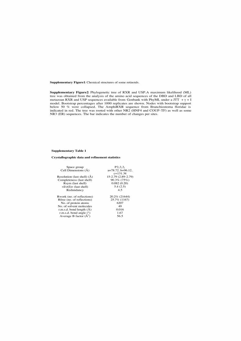
Supplementary Figure1: Chemical structures of some retinoids.
Supplementary Figure2: Phylogenetic tree of RXR and USP.A maximum likelihood (ML) tree was obtained from the analysis of the amino acid sequences of the DBD and LBD of all metazoan RXR and USP sequences available from Genbank with PhyML under a JTT + γ + I model. Bootstrap percentages after 1000 replicates are shown. Nodes with bootstrap support below 50 % were collapsed. The AmphiRXR sequence from Branchiostoma floridae is indicated in red. The tree was rooted with other NR2 (HNF4 and COUP-TF) as well as some NR3 (ER) sequences. The bar indicates the number of changes per sites.
Supplementary Table 1
Crystallographic data and refinement statistics
Space group P212121
Cell Dimensions (Å) a=78.72, b=96.12, c=131.34
Resolution (last shell) (Å) 15-2.79 (2.89-2.79)Completeness (last shell) 90.3% (75%)
Rsym (last shell) 0.082 (0.20)<I/σ(I)> (last shell) 5.4 (2.5)
Redundancy 4.5
Rwork (no. of reflections) 20.2% (21644)Rfree (no. of reflections) 25.7% (1167)
No. of protein atoms 6207No. of solvent molecules 49r.m.s.d. bond length (Å) 0.016r.m.s.d. bond angle (°) 1.67Average B factor (Å2) 56.5
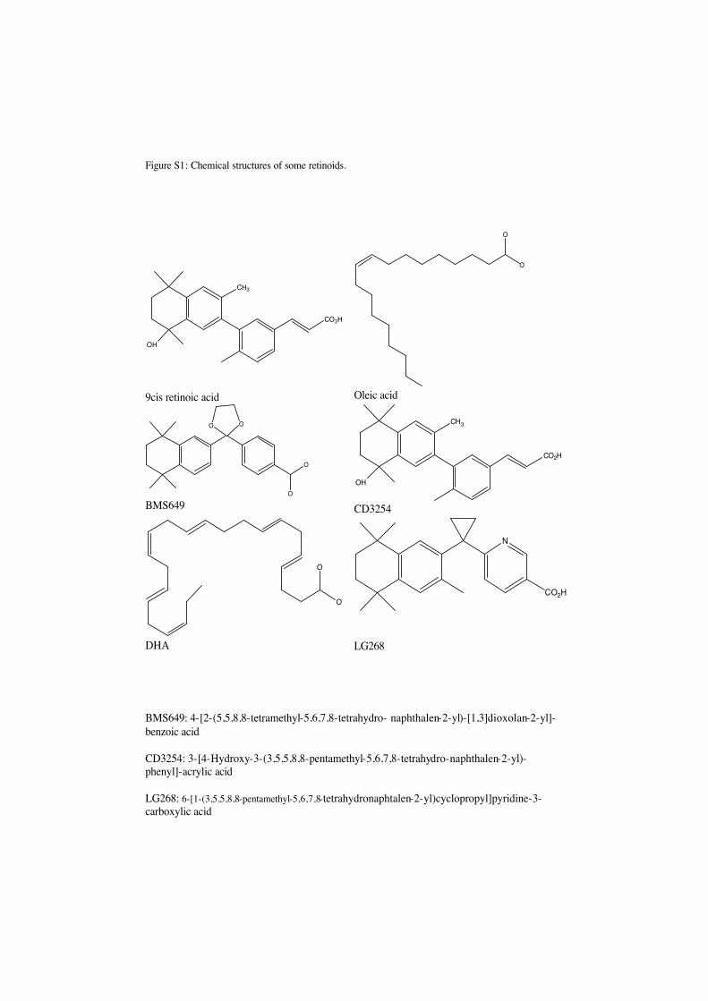
Figure S1: Chemical structures of some retinoids.
BMS649: 4-[2-(5,5,8,8-tetramethyl-5,6,7,8-tetrahydro- naphthalen-2-yl)-[1,3]dioxolan-2-yl]-
benzoic acid
CD3254: 3-[4-Hydroxy-3-(3,5,5,8,8-pentamethyl-5,6,7,8-tetrahydro-naphthalen-2-yl)-phenyl]-acrylic acid
LG268: 6-[1-(3,5,5,8,8-pentamethyl-5,6,7,8-tetrahydronaphtalen-2-yl)cyclopropyl]pyridine-3-carboxylic acid
CO2H
OH
CH3
9cis retinoic acid
O
O
Oleic acid
O O
O
O
BMS649
CO2H
OH
CH3
CD3254
O
O
DHA
N
CO2H
LG268
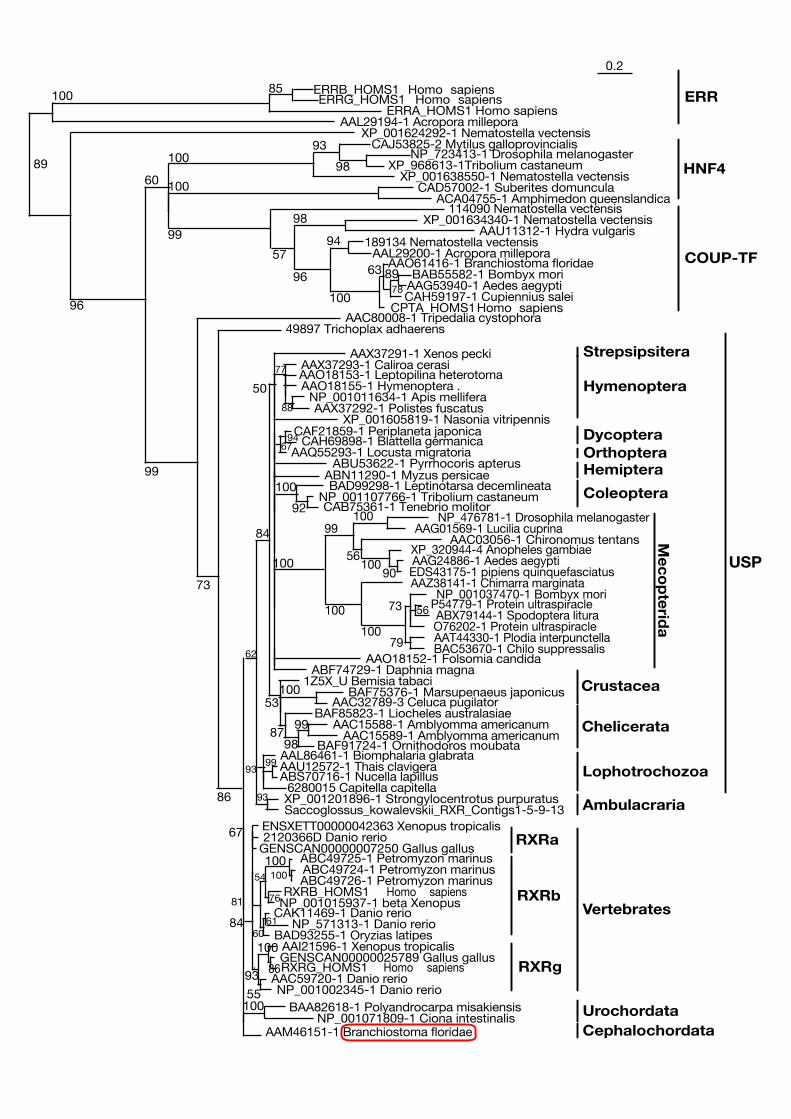
AAM46151-1 Branchiostoma floridaeNP_001071809-1 Ciona intestinalis
BAA82618-1 Polyandrocarpa misakiensis100
6280015 Capitella capitellaABS70716-1 Nucella lapillusAAU12572-1 Thais clavigera99AAL86461-1 Biomphalaria glabrata
Saccoglossus_kowalevskii_RXR_Contigs1-5-9-13XP_001201896-1 Strongylocentrotus purpuratus
93
93
BAF91724-1 Ornithodoros moubataAAC15589-1 Amblyomma americanum
AAC15588-1 Amblyomma americanum99BAF85823-1 Liocheles australasiae
98
AAC32789-3 Celuca pugilatorBAF75376-1 Marsupenaeus japonicus
87
1001Z5X_U Bemisia tabaci
ABF74729-1 Daphnia magnaAAO18152-1 Folsomia candida
ABN11290-1 Myzus persicae
XP_001605819-1 Nasonia vitripennisAAX37292-1 Polistes fuscatus
NP_001011634-1 Apis melliferaAAO18155-1 Hymenoptera .
88
AAO18153-1 Leptopilina heterotomaAAX37293-1 Caliroa cerasi77
AAX37291-1 Xenos pecki
53
50
84
62
NP_001002345-1 Danio rerioAAC59720-1 Danio rerio
RXRG_HOMS1 Homo sapiensGENSCAN00000025789 Gallus gallusAAI21596-1 Xenopus tropicalis
86
55
100BAD93255-1 Oryzias latipes
NP_571313-1 Danio rerioCAK11469-1 Danio rerio
61
NP_001015937-1 beta XenopusRXRB_HOMS1 Homo sapiens
ABC49726-1 Petromyzon marinusABC49724-1 Petromyzon marinusABC49725-1 Petromyzon marinus
100
76
100
60
54
GENSCAN00000007250 Gallus gallus2120366D Danio rerioENSXETT00000042363 Xenopus tropicalis
81
67
93
84
49897 Trichoplax adhaerens
86
AAC80008-1 Tripedalia cystophora
73
CPTA_HOMS1Homo sapiensCAH59197-1 Cupiennius saleiAAG53940-1 Aedes aegyptiBAB55582-1 Bombyx mori89
AAO61416-1 Branchiostoma floridae
78
63
AAL29200-1 Acropora millepora189134 Nematostella vectensis
100
94AAU11312-1 Hydra vulgaris
XP_001634340-1 Nematostella vectensis
96
98114090 Nematostella vectensis
57
ACA04755-1 Amphimedon queenslandicaCAD57002-1 Suberites domuncula
99
100XP_001638550-1 Nematostella vectensis
XP_968613-1Tribolium castaneumNP_723413-1 Drosophila melanogaster
CAJ53825-2 Mytilus galloprovincialis
98
93100
99
60
XP_001624292-1 Nematostella vectensis
96
AAL29194-1 Acropora milleporaERRA_HOMS1 Homo sapiens
ERRG_HOMS1 Homo sapiensERRB_HOMS1 Homo sapiens85
100
89
0.2
HNF4
ERR
COUP-TF
Mecopterid
a
USP
Hymenoptera
Dycoptera
HemipteraOrthoptera
Strepsipsitera
Coleoptera
Crustacea
Chelicerata
Lophotrochozoa
Ambulacraria
Vertebrates
Urochordata
RXRg
RXRb
Cephalochordata
ABU53622-1 Pyrrhocoris apterusAAQ55293-1 Locusta migratoria
CAH69898-1 Blattella germanicaCAF21859-1 Periplaneta japonica
9467
BAC53670-1 Chilo suppressalisAAT44330-1 Plodia interpunctellaO76202-1 Protein ultraspiracleABX79144-1 Spodoptera litura
P54779-1 Protein ultraspiracle56
NP_001037470-1 Bombyx mori
79
73
AAZ38141-1 Chimarra marginata
100
EDS43175-1 pipiens quinquefasciatusAAG24886-1 Aedes aegyptiXP_320944-4 Anopheles gambiae
90
AAC03056-1 Chironomus tentans
100
AAG01569-1 Lucilia cuprinaNP_476781-1 Drosophila melanogaster
56
100
100
99
100
CAB75361-1 Tenebrio molitorNP_001107766-1 Tribolium castaneum
BAD99298-1 Leptinotarsa decemlineata
92
100
RXRa
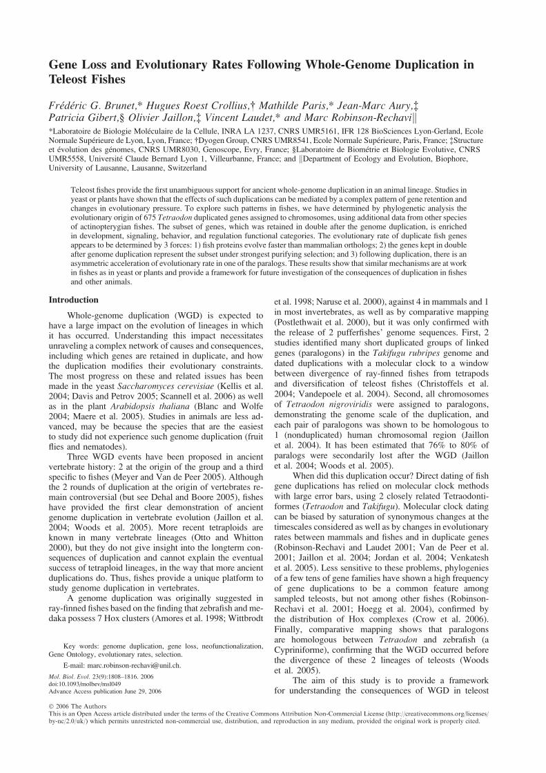
Gene Loss and Evolutionary Rates Following Whole-Genome Duplication in
Teleost Fishes
Frederic G. Brunet,* Hugues Roest Crollius,� Mathilde Paris,* Jean-Marc Aury,�Patricia Gibert,§ Olivier Jaillon,� Vincent Laudet,* and Marc Robinson-Rechavik*Laboratoire de Biologie Moleculaire de la Cellule, INRA LA 1237, CNRS UMR5161, IFR 128 BioSciences Lyon-Gerland, EcoleNormale Superieure de Lyon, Lyon, France; �Dyogen Group, CNRS UMR8541, Ecole Normale Superieure, Paris, France; �Structureet evolution des genomes, CNRS UMR8030, Genoscope, Evry, France; §Laboratoire de Biometrie et Biologie Evolutive, CNRSUMR5558, Universite Claude Bernard Lyon 1, Villeurbanne, France; and kDepartment of Ecology and Evolution, Biophore,University of Lausanne, Lausanne, Switzerland
Teleost fishes provide the first unambiguous support for ancient whole-genome duplication in an animal lineage. Studies inyeast or plants have shown that the effects of such duplications can be mediated by a complex pattern of gene retention andchanges in evolutionary pressure. To explore such patterns in fishes, we have determined by phylogenetic analysis theevolutionary origin of 675 Tetraodon duplicated genes assigned to chromosomes, using additional data from other speciesof actinopterygian fishes. The subset of genes, which was retained in double after the genome duplication, is enrichedin development, signaling, behavior, and regulation functional categories. The evolutionary rate of duplicate fish genesappears to be determined by 3 forces: 1) fish proteins evolve faster than mammalian orthologs; 2) the genes kept in doubleafter genome duplication represent the subset under strongest purifying selection; and 3) following duplication, there is anasymmetric acceleration of evolutionary rate in one of the paralogs. These results show that similar mechanisms are at workin fishes as in yeast or plants and provide a framework for future investigation of the consequences of duplication in fishesand other animals.
Introduction
Whole-genome duplication (WGD) is expected tohave a large impact on the evolution of lineages in whichit has occurred. Understanding this impact necessitatesunraveling a complex network of causes and consequences,including which genes are retained in duplicate, and howthe duplication modifies their evolutionary constraints.The most progress on these and related issues has beenmade in the yeast Saccharomyces cerevisiae (Kellis et al.2004; Davis and Petrov 2005; Scannell et al. 2006) as wellas in the plant Arabidopsis thaliana (Blanc and Wolfe2004; Maere et al. 2005). Studies in animals are less ad-vanced, may be because the species that are the easiestto study did not experience such genome duplication (fruitflies and nematodes).
Three WGD events have been proposed in ancientvertebrate history: 2 at the origin of the group and a thirdspecific to fishes (Meyer and Van de Peer 2005). Althoughthe 2 rounds of duplication at the origin of vertebrates re-main controversial (but see Dehal and Boore 2005), fisheshave provided the first clear demonstration of ancientgenome duplication in vertebrate evolution (Jaillon et al.2004; Woods et al. 2005). More recent tetraploids areknown in many vertebrate lineages (Otto and Whitton2000), but they do not give insight into the longterm con-sequences of duplication and cannot explain the eventualsuccess of tetraploid lineages, in the way that more ancientduplications do. Thus, fishes provide a unique platform tostudy genome duplication in vertebrates.
A genome duplication was originally suggested inray-finned fishes based on the finding that zebrafish and me-daka possess 7 Hox clusters (Amores et al. 1998; Wittbrodt
et al. 1998; Naruse et al. 2000), against 4 in mammals and 1in most invertebrates, as well as by comparative mapping(Postlethwait et al. 2000), but it was only confirmed withthe release of 2 pufferfishes’ genome sequences. First, 2studies identified many short duplicated groups of linkedgenes (paralogons) in the Takifugu rubripes genome anddated duplications with a molecular clock to a windowbetween divergence of ray-finned fishes from tetrapodsand diversification of teleost fishes (Christoffels et al.2004; Vandepoele et al. 2004). Second, all chromosomesof Tetraodon nigroviridis were assigned to paralogons,demonstrating the genome scale of the duplication, andeach pair of paralogons was shown to be homologous to1 (nonduplicated) human chromosomal region (Jaillonet al. 2004). It has been estimated that 76% to 80% ofparalogs were secondarily lost after the WGD (Jaillonet al. 2004; Woods et al. 2005).
When did this duplication occur? Direct dating of fishgene duplications has relied on molecular clock methodswith large error bars, using 2 closely related Tetraodonti-formes (Tetraodon and Takifugu). Molecular clock datingcan be biased by saturation of synonymous changes at thetimescales considered as well as by changes in evolutionaryrates between mammals and fishes and in duplicate genes(Robinson-Rechavi and Laudet 2001; Van de Peer et al.2001; Jaillon et al. 2004; Jordan et al. 2004; Venkateshet al. 2005). Less sensitive to these problems, phylogeniesof a few tens of gene families have shown a high frequencyof gene duplications to be a common feature amongsampled teleosts, but not among other fishes (Robinson-Rechavi et al. 2001; Hoegg et al. 2004), confirmed bythe distribution of Hox complexes (Crow et al. 2006).Finally, comparative mapping shows that paralogonsare homologous between Tetraodon and zebrafish (aCypriniforme), confirming that the WGD occurred beforethe divergence of these 2 lineages of teleosts (Woodset al. 2005).
The aim of this study is to provide a frameworkfor understanding the consequences of WGD in teleost
Key words: genome duplication, gene loss, neofunctionalization,Gene Ontology, evolutionary rates, selection.
E-mail: [email protected].
Mol. Biol. Evol. 23(9):1808–1816. 2006doi:10.1093/molbev/msl049
Advance Access publication June 29, 2006
� 2006 The AuthorsThis is an Open Access article distributed under the terms of the Creative Commons Attribution Non-Commercial License (http://creativecommons.org/licenses/by-nc/2.0/uk/) which permits unrestricted non-commercial use, distribution, and reproduction in any medium, provided the original work is properly cited.
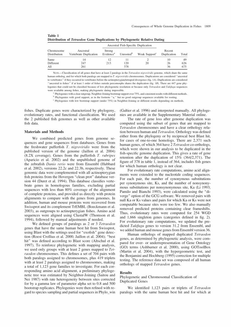
fishes. Duplicate genes were characterized by phylogeny,evolutionary rates, and functional classification. We usedthe 2 published fish genomes as well as other availablefish data.
Materials and Methods
We combined predicted genes from genome se-quences and gene sequences from databases. Genes fromthe freshwater pufferfish T. nigroviridis were from thepublished version of the genome (Jaillon et al. 2004)(8.2X coverage). Genes from the pufferfish T. rubripes(Aparicio et al. 2002) and the unpublished genome ofthe zebrafish Danio rerio were from Ensembl (Hubbardet al. 2002), versions 22.2c and 22.3b, respectively. Thesegenomic data were complemented with all actinopterygianfish proteins from the Hovergen ‘‘clean prot’’ database ver-sion 44 (Duret et al. 1994). This database classifies verte-brate genes in homologous families, excluding partialsequences with less than 80% coverage of the alignmentof complete proteins, and provided us directly with proteinalignments to compare with the genes from genomes. Inaddition, human and mouse proteins were recovered fromSwissprot and its complement TrEMBL (Boeckmann et al.2003), as outgroups to actinopterygian fishes. Amino acidsequences were aligned using ClustalW (Thomson et al.1994), followed by manual adjustments if needed.
We defined groups of paralogs as 2 or 3 Tetraodongenes that have the same human best hit from Swissprot,using Blast with the settings used for ‘‘exofish’’ gene detec-tion (Roest Crollius et al. 2000; Jaillon et al. 2004); ‘‘besthit’’ was defined according to Blast score (Altschul et al.1997). To reinforce phylogenetic with mapping analysis,we used only groups with at least 2 genes mapped to Tet-raodon chromosomes. This defines a set of 704 pairs withboth paralogs assigned to chromosomes, plus 419 tripletswith at least 2 paralogs assigned to chromosomes, makinga total of 1,123 gene families to investigate. For each cor-responding amino acid alignment, a preliminary phyloge-netic tree was estimated by Neighbor-Joining (Saitou andNei 1987) with rate heterogeneity between sites correctedfor by a gamma law of parameter alpha set to 0.8 and 500bootstrap replicates. Phylogenies were then refined with re-gard to species sampling and partial sequences in Phylo_win
(Galtier et al. 1996) and interpreted manually. All phyloge-nies are available in the Supplementary Material online.
The rate of gene loss after genome duplication wascomputed using the subset of genes that are mapped toTetraodon chromosomes and have a clear orthology rela-tion between human and Tetraodon. Orthology was definedeither from the phylogeny or by reciprocal best Blast hit,for cases of one-to-one homologs. There are 2,371 suchhuman genes, of which 364 have 2 Tetraodon co-orthologs,which were shown in our analysis to be duplicated in thefish-specific genome duplication. This gives a rate of generetention after the duplication of 15% (364/2,371). Thefigure of 378 in table 1, instead of 364, includes fish genesfor which human orthology is not clear.
For evolutionary rate computations, amino acid align-ments were extended to the nucleotide coding sequences.For each pair, the number of synonymous substitutionsper synonymous site, Ks, and the number of nonsynony-mous substitutions per nonsynonymous site, Ka (Li 1993;Pamilo and Bianchi 1993), were calculated using the ‘‘di-verge’’ option of the GCG software. We removed pairs withnull Ka or Ks values and pairs for which Ka or Ks were notcomputable because sites were too few. We also manuallyremoved predicted proteins containing clear frameshifts.Thus, evolutionary rates were computed for 254 WGDand 1,666 singleton genes (categories defined in fig. 2).For evolutionary rate computations, we updated the pre-dicted Takifugu genes to version 31.2 from Ensembl, andwe added human andmouse genes fromEnsembl version 36.
Human orthologs of mapped duplicated Tetraodongenes, as determined by phylogenetic analysis, were com-pared for over- or underrepresentation of Gene Ontology(GO) terms (Ashburner et al. 2000), using GOToolBox(Martin et al. 2004), with the hypergeometric test, andthe Benjamini and Hochberg (1995) correction for multipletesting. The reference data set was composed of all humanorthologs of mapped Tetraodon genes.
ResultsPhylogenetic and Chromosomal Classification ofDuplicated Genes
We identified 1,123 pairs or triplets of Tetraodonparalogs with the same human best hit and for which at
Table 1Distribution of Tetraodon Gene Duplications by Phylogenetic Relative Dating
Ancestral Fish-Specific Duplication
ChromosomeDistribution
AncestralVertebrate Duplication
StrongEvidencea Unrootedb Weak Supportc
RecentDuplication Total
Same 14 12 11 2 10 49Different 247 213 120 20 26 626All 261 378 36 675
NOTE.—Classification of all genes that have at least 2 paralogs in the Tetraodon nigroviridis genome, which share the same
human ortholog, and for which both paralogs are mapped to T. nigroviridis chromosomes. Duplications are considered ‘‘ancestral
to vertebrates’’ if they occurred in vertebrates before the actinopterygian/tetrapod divergence (fig. 1A). Duplications are considered
‘‘ancestral to fishes’’ if at least 1 order of fishes outside percomorphs shares the duplication (fig. 1B). There are 447 gene phy-
logenies that could not be classified because of low phylogenetic resolution or because only Tetraodon and Takifugu sequences
were available among fishes, making phylogenetic dating impossible.a Phylogenies with a clear outgroup, Neighbor-Joining bootstrap support over 75%, and consistent results with different methods.b Phylogenies with good support, as in the footnote ‘‘a,’’ but no good outgroup sequences available for rooting.c Phylogenies with low bootstrap support (under 75%) in Neighbor-Joining or different results depending on methods.
Consequences of Whole Genome Duplication in Fishes 1809
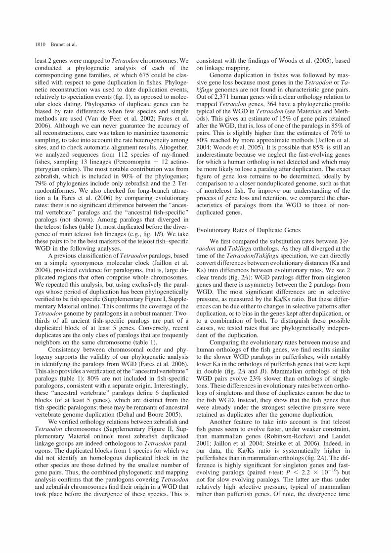
least 2 genes were mapped to Tetraodon chromosomes. Weconducted a phylogenetic analysis of each of thecorresponding gene families, of which 675 could be clas-sified with respect to gene duplication in fishes. Phyloge-netic reconstruction was used to date duplication events,relatively to speciation events (fig. 1), as opposed to molec-ular clock dating. Phylogenies of duplicate genes can bebiased by rate differences when few species and simplemethods are used (Van de Peer et al. 2002; Fares et al.2006). Although we can never guarantee the accuracy ofall reconstructions, care was taken to maximize taxonomicsampling, to take into account the rate heterogeneity amongsites, and to check automatic alignment results. Altogether,we analyzed sequences from 112 species of ray-finnedfishes, sampling 13 lineages (Percomorpha 1 12 actino-pterygian orders). The most notable contribution was fromzebrafish, which is included in 90% of the phylogenies;79% of phylogenies include only zebrafish and the 2 Tet-raodontiformes. We also checked for long-branch attrac-tion a la Fares et al. (2006) by comparing evolutionaryrates: there is no significant difference between the ‘‘ances-tral vertebrate’’ paralogs and the ‘‘ancestral fish-specific’’paralogs (not shown). Among paralogs that diverged inthe teleost fishes (table 1), most duplicated before the diver-gence of main teleost fish lineages (e.g., fig. 1B). We takethese pairs to be the best markers of the teleost fish–specificWGD in the following analyses.
A previous classification of Tetraodon paralogs, basedon a simple synonymous molecular clock (Jaillon et al.2004), provided evidence for paralogons, that is, large du-plicated regions that often comprise whole chromosomes.We repeated this analysis, but using exclusively the paral-ogs whose period of duplication has been phylogeneticallyverified to be fish specific (Supplementary Figure I, Supple-mentary Material online). This confirms the coverage of theTetraodon genome by paralogons in a robust manner. Two-thirds of all ancient fish-specific paralogs are part of aduplicated block of at least 5 genes. Conversely, recentduplicates are the only class of paralogs that are frequentlyneighbors on the same chromosome (table 1).
Consistency between chromosomal order and phy-logeny supports the validity of our phylogenetic analysisin identifying the paralogs from WGD (Fares et al. 2006).This also provides a verification of the ‘‘ancestral vertebrate’’paralogs (table 1): 80% are not included in fish-specificparalogons, consistent with a separate origin. Interestingly,these ‘‘ancestral vertebrate’’ paralogs define 6 duplicatedblocks (of at least 5 genes), which are distinct from thefish-specific paralogons; these may be remnants of ancestralvertebrate genome duplication (Dehal and Boore 2005).
We verified orthology relations between zebrafish andTetraodon chromosomes (Supplementary Figure II, Sup-plementary Material online): most zebrafish duplicatedlinkage groups are indeed orthologous to Tetraodon paral-ogons. The duplicated blocks from 1 species for which wedid not identify an homologous duplicated block in theother species are those defined by the smallest number ofgene pairs. Thus, the combined phylogenetic and mappinganalysis confirms that the paralogons covering Tetraodonand zebrafish chromosomes find their origin in a WGD thattook place before the divergence of these species. This is
consistent with the findings of Woods et al. (2005), basedon linkage mapping.
Genome duplication in fishes was followed by mas-sive gene loss because most genes in the Tetraodon or Ta-kifugu genomes are not found in characteristic gene pairs.Out of 2,371 human genes with a clear orthology relation tomapped Tetraodon genes, 364 have a phylogenetic profiletypical of the WGD in Tetraodon (see Materials and Meth-ods). This gives an estimate of 15% of gene pairs retainedafter the WGD, that is, loss of one of the paralogs in 85% ofpairs. This is slightly higher than the estimates of 76% to80% reached by more approximate methods (Jaillon et al.2004; Woods et al. 2005). It is possible that 85% is still anunderestimate because we neglect the fast-evolving genesfor which a human ortholog is not detected and which maybe more likely to lose a paralog after duplication. The exactfigure of gene loss remains to be determined, ideally bycomparison to a closer nonduplicated genome, such as thatof nonteleost fish. To improve our understanding of theprocess of gene loss and retention, we compared the char-acteristics of paralogs from the WGD to those of non-duplicated genes.
Evolutionary Rates of Duplicate Genes
We first compared the substitution rates between Tet-raodon and Takifugu orthologs. As they all diverged at thetime of the Tetraodon/Takifugu speciation, we can directlyconvert differences between evolutionary distances (Ka andKs) into differences between evolutionary rates. We see 2clear trends (fig. 2A): WGD paralogs differ from singletongenes and there is asymmetry between the 2 paralogs fromWGD. The most significant differences are in selectivepressure, as measured by the Ka/Ks ratio. But these differ-ences can be due either to changes in selective patterns afterduplication, or to bias in the genes kept after duplication, orto a combination of both. To distinguish these possiblecauses, we tested rates that are phylogenetically indepen-dent of the duplication.
Comparing the evolutionary rates between mouse andhuman orthologs of the fish genes, we find results similarto the slower WGD paralogs in pufferfishes, with notablylower Ka in the orthologs of pufferfish genes that were keptin double (fig. 2A and B). Mammalian orthologs of fishWGD pairs evolve 23% slower than orthologs of single-tons. These differences in evolutionary rates between ortho-logs of singletons and those of duplicates cannot be due tothe fish WGD. Instead, they show that the fish genes thatwere already under the strongest selective pressure wereretained as duplicates after the genome duplication.
Another feature to take into account is that teleostfish genes seem to evolve faster, under weaker constraint,than mammalian genes (Robinson-Rechavi and Laudet2001; Jaillon et al. 2004; Steinke et al. 2006). Indeed, inour data, the Ka/Ks ratio is systematically higher inpufferfishes than in mammalian orthologs (fig. 2A). The dif-ference is highly significant for singleton genes and fast-evolving paralogs (paired t-test: P , 2.2 3 10
�16) butnot for slow-evolving paralogs. The latter are thus underrelatively high selective pressure, typical of mammalianrather than pufferfish genes. Of note, the divergence time
1810 Brunet et al.

between Tetraodon and Takifugu was recently reassessedat 85MYA(Yamanoue et al. 2006), similar to the divergencebetween human and mouse. This affects neither relative ratetests (Robinson-Rechavi and Laudet 2001; Steinke et al.2006) nor comparisons of Ka/Ks ratios (this study), whichare both independent of divergence time.
Once we have established contributions to evolution-ary rate, which are not a direct result of duplication (biasedretention and species-specific differences), can we alsomeasure the direct effect of duplication? Lower purifyingselection is expected on redundant genes after duplica-tion. In principle, both paralogs could experience released
0.05COG4_MOUSE
COG4_HUMAN
TN_17687_5
TN_32094_10
TR_150018
99
DR_15947_u
83
100
zebrafish
Fugu
Tetraodon
mammals
C
0.05 O42278
O57341
Q80ZL6
100
BAC36859
99
TN_24466_20
TR_148211
Q7SX66
100
FGFI_HUMAN
FGFI_MOUSE
Q7T2N7
TN_05197_1
TR_15533089
100
100
100
93
100
mouse
mouse
zebrafish
zebrafish
zebrafish
Tetraodon
Fugu
Tetraodon
Fugu
goldfish
mammals
0.02 AAH52348
Q8CAD8
REQU_HUMAN
REQU_MOUSE
TN_31158_1
TR_142024
AAQ91267
100
DR_03264_u
TN_19087_7
TR_159362100
100
53
100
77
100
mouse
mammals
Tetraodon
Fugu
Tetraodon
Fugu
zebrafish
zebrafish
BA
FIG. 1.—Examples of relative dating of gene duplications by phylogeny. Representative examples of the main phylogenetic scenarios observed.Neighbor-Joining phylogenetic trees, with substitutions corrected for heterogeneity between sites by a gamma law (a5 0.8); Maximum Likelihood givesidentical topologies (not shown). Branch length is proportional to the number of substitutions per site (see measure bar for each tree); numbers at nodes aresupport in percentage of 500 bootstrap replicates. The stars represent the duplication that led to 2 Tetraodon paralogs in each case. (A) Fibroblast growthfactor-18 precursor, duplicated before the tetrapod–actinopterygian split (ancestral vertebrate duplication), with secondary loss of 1 copy in mammals,leading to 2 copies in fishes but only 1 in human; clade BAC36859–O42278 is a more distant paralog used as an outgroup. (B) Zinc-finger protein ubi-d4(Requiem), duplicated anciently in fishes; clade Q8CAD8–AAH52348 is a more distant paralog used as an outgroup. (C) Conserved oligomeric Golgi
complex component 4, duplicated specifically in Tetraodon, after the divergence with other fishes.
Consequences of Whole Genome Duplication in Fishes 1811
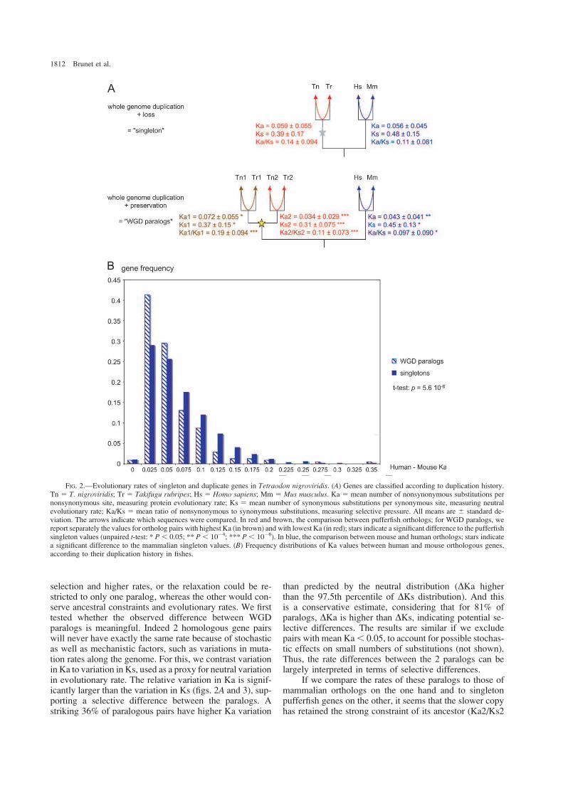
selection and higher rates, or the relaxation could be re-stricted to only one paralog, whereas the other would con-serve ancestral constraints and evolutionary rates. We firsttested whether the observed difference between WGDparalogs is meaningful. Indeed 2 homologous gene pairswill never have exactly the same rate because of stochasticas well as mechanistic factors, such as variations in muta-tion rates along the genome. For this, we contrast variationin Ka to variation in Ks, used as a proxy for neutral variationin evolutionary rate. The relative variation in Ka is signif-icantly larger than the variation in Ks (figs. 2A and 3), sup-porting a selective difference between the paralogs. Astriking 36% of paralogous pairs have higher Ka variation
than predicted by the neutral distribution (DKa higherthan the 97.5th percentile of DKs distribution). And thisis a conservative estimate, considering that for 81% ofparalogs, DKa is higher than DKs, indicating potential se-lective differences. The results are similar if we excludepairs with mean Ka, 0.05, to account for possible stochas-tic effects on small numbers of substitutions (not shown).Thus, the rate differences between the 2 paralogs can belargely interpreted in terms of selective differences.
If we compare the rates of these paralogs to those ofmammalian orthologs on the one hand and to singletonpufferfish genes on the other, it seems that the slower copyhas retained the strong constraint of its ancestor (Ka2/Ks2
FIG. 2.—Evolutionary rates of singleton and duplicate genes in Tetraodon nigroviridis. (A) Genes are classified according to duplication history.Tn 5 T. nigroviridis; Tr 5 Takifugu rubripes; Hs 5 Homo sapiens; Mm 5 Mus musculus. Ka 5 mean number of nonsynonymous substitutions pernonsynonymous site, measuring protein evolutionary rate; Ks 5 mean number of synonymous substitutions per synonymous site, measuring neutralevolutionary rate; Ka/Ks 5 mean ratio of nonsynonymous to synonymous substitutions, measuring selective pressure. All means are 6 standard de-viation. The arrows indicate which sequences were compared. In red and brown, the comparison between pufferfish orthologs; for WGD paralogs, wereport separately the values for ortholog pairs with highest Ka (in brown) and with lowest Ka (in red); stars indicate a significant difference to the pufferfish
singleton values (unpaired t-test: * P, 0.05; ** P, 10�4; *** P, 10�6). In blue, the comparison between mouse and human orthologs; stars indicatea significant difference to the mammalian singleton values. (B) Frequency distributions of Ka values between human and mouse orthologous genes,according to their duplication history in fishes.
1812 Brunet et al.
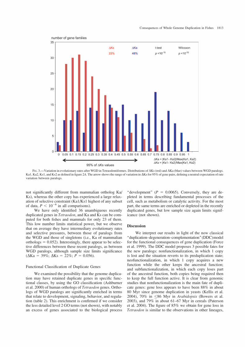
not significantly different from mammalian ortholog Ka/Ks), whereas the other copy has experienced a large relax-ation of selective constraint (Ka1/Ks1 highest of any subsetof data, P , 10�6 in all comparisons).
We have only identified 36 unambiguous recentlyduplicated genes in Tetraodon, and Ka and Ks can be com-puted for both fishes and mammals for only 23 of them.This low number limits statistical power, but we observethat on average they have intermediary evolutionary ratesand selective pressures, between those of paralogs fromthe WGD and those of singletons (i.e., Ka of mammalianorthologs 5 0.052). Interestingly, there appear to be selec-tive differences between these recent paralogs, as betweenWGD paralogs, although sample size limits significance(DKa 5 39%; DKs 5 22%; P 5 0.056).
Functional Classification of Duplicate Genes
We examined the possibility that the genome duplica-tion may have retained duplicate genes in specific func-tional classes, by using the GO classification (Ashburneret al. 2000) of human orthologs of Tetraodon genes. Ortho-logs of WGD paralogs are significantly enriched in termsthat relate to development, signaling, behavior, and regula-tion (table 2). This enrichment is confirmed if we considerthe less detailed level 2 GO terms (not shown), with notablyan excess of genes associated to the biological process
‘‘development’’ (P 5 0.0065). Conversely, they are de-pleted in terms describing fundamental processes of thecell, such as metabolism or catalytic activity. For the mostpart, the same terms are enriched or depleted in the recentlyduplicated genes, but low sample size again limits signif-icance (not shown).
Discussion
We interpret our results in light of the now classical‘‘duplication–degeneration–complementation’’(DDC)modelfor the functional consequences of gene duplication (Forceet al. 1999). The DDC model proposes 3 possible fates forthe new paralogs: nonfunctionalization, in which 1 copyis lost and the situation reverts to its preduplication state;neofunctionalization, in which 1 copy acquires a newfunction while the other keeps the ancestral function;and subfunctionalization, in which each copy loses partof the ancestral function, both copies being required thento keep the full function active. It is clear from genomicstudies that nonfunctionalization is the main fate of dupli-cate genes: gene loss appears to have been 88% in about80 Myr since genome duplication in yeasts (Kellis et al.2004), 70% in �86 Myr in Arabidopsis (Bowers et al.2003), and 79% in about 61–67 Myr in cereals (Patersonet al. 2004). The figure of 85% we obtain for gene loss inTetraodon is similar to the observations in other lineages,
FIG. 3.—Variation in evolutionary rates after WGD in Tetraodontiformes. Distributions of DKs (red) andDKa (blue) values betweenWGD paralogs.
Ka1, Ka2, Ks1, and Ks2 as defined in figure 2A. The arrow shows the range of variation inDKs for 95% of gene pairs, defining a neutral expectation of ratevariation between paralogs.
Consequences of Whole Genome Duplication in Fishes 1813
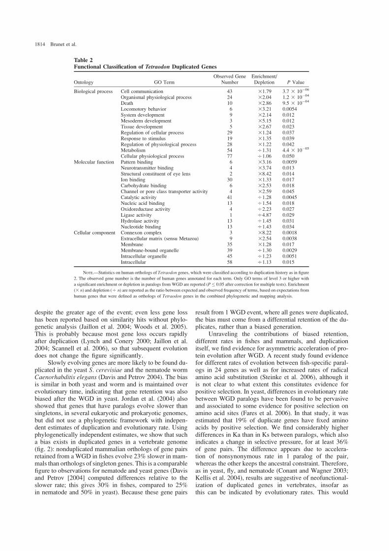
despite the greater age of the event; even less gene losshas been reported based on similarity hits without phylo-genetic analysis (Jaillon et al. 2004; Woods et al. 2005).This is probably because most gene loss occurs rapidlyafter duplication (Lynch and Conery 2000; Jaillon et al.2004; Scannell et al. 2006), so that subsequent evolutiondoes not change the figure significantly.
Slowly evolving genes are more likely to be found du-plicated in the yeast S. cerevisiae and the nematode wormCaenorhabditis elegans (Davis and Petrov 2004). The biasis similar in both yeast and worm and is maintained overevolutionary time, indicating that gene retention was alsobiased after the WGD in yeast. Jordan et al. (2004) alsoshowed that genes that have paralogs evolve slower thansingletons, in several eukaryotic and prokaryotic genomes,but did not use a phylogenetic framework with indepen-dent estimates of duplication and evolutionary rate. Usingphylogenetically independent estimates, we show that sucha bias exists in duplicated genes in a vertebrate genome(fig. 2): nonduplicated mammalian orthologs of gene pairsretained from a WGD in fishes evolve 23% slower in mam-mals than orthologs of singleton genes. This is a comparablefigure to observations for nematode and yeast genes (Davisand Petrov [2004] computed differences relative to theslower rate; this gives 30% in fishes, compared to 25%in nematode and 50% in yeast). Because these gene pairs
result from 1 WGD event, where all genes were duplicated,the bias must come from a differential retention of the du-plicates, rather than a biased generation.
Unraveling the contributions of biased retention,different rates in fishes and mammals, and duplicationitself, we find evidence for asymmetric acceleration of pro-tein evolution after WGD. A recent study found evidencefor different rates of evolution between fish-specific paral-ogs in 24 genes as well as for increased rates of radicalamino acid substitution (Steinke et al. 2006), although itis not clear to what extent this constitutes evidence forpositive selection. In yeast, differences in evolutionary ratebetween WGD paralogs have been found to be pervasiveand associated to some evidence for positive selection onamino acid sites (Fares et al. 2006). In that study, it wasestimated that 19% of duplicate genes have fixed aminoacids by positive selection. We find considerably higherdifferences in Ka than in Ks between paralogs, which alsoindicates a change in selective pressure, for at least 36%of gene pairs. The difference appears due to accelera-tion of nonsynonymous rate in 1 paralog of the pair,whereas the other keeps the ancestral constraint. Therefore,as in yeast, fly, and nematode (Conant and Wagner 2003;Kellis et al. 2004), results are suggestive of neofunctional-ization of duplicated genes in vertebrates, insofar asthis can be indicated by evolutionary rates. This would
Table 2Functional Classification of Tetraodon Duplicated Genes
Ontology GO TermObserved Gene
NumberEnrichment/Depletion P Value
Biological process Cell communication 43 31.79 3.7 3 10�06
Organismal physiological process 24 32.04 1.2 3 10�04
Death 10 32.86 9.5 3 10�04
Locomotory behavior 6 33.21 0.0054System development 9 32.14 0.012Mesoderm development 3 35.15 0.012Tissue development 5 32.67 0.023
Regulation of cellular process 29 31.24 0.037Response to stimulus 19 31.35 0.039Regulation of physiological process 28 31.22 0.042Metabolism 54 O1.31 4.4 3 10�05
Cellular physiological process 77 O1.06 0.050Molecular function Pattern binding 6 33.16 0.0059
Neurotransmitter binding 4 33.74 0.013
Structural constituent of eye lens 2 38.42 0.014Ion binding 30 31.33 0.017Carbohydrate binding 6 32.53 0.018Channel or pore class transporter activity 4 32.59 0.045Catalytic activity 41 O1.28 0.0045Nucleic acid binding 13 O1.54 0.018
Oxidoreductase activity 4 O2.23 0.027Ligase activity 1 O4.87 0.029Hydrolase activity 13 O1.45 0.031Nucleotide binding 13 O1.43 0.034
Cellular component Connexon complex 3 38.22 0.0018Extracellular matrix (sensu Metazoa) 9 32.54 0.0038Membrane 35 31.28 0.017
Membrane-bound organelle 39 O1.30 0.0029Intracellular organelle 45 O1.23 0.0051Intracellular 58 O1.13 0.015
NOTE.—Statistics on human orthologs of Tetraodon genes, which were classified according to duplication history as in figure
2. The observed gene number is the number of human genes annotated for each term. Only GO terms of level 3 or higher with
a significant enrichment or depletion in paralogs from WGD are reported (P� 0.05 after correction for multiple tests). Enrichment
(3 n) and depletion (O n) are reported as the ratio between expected and observed frequency of terms, based on expectations from
human genes that were defined as orthologs of Tetraodon genes in the combined phylogenetic and mapping analysis.
1814 Brunet et al.
be in agreement with the original evolutionary theoryof Ohno (1970) as well as with a more recent model ofsubfunctionalization followed by neofunctionalization(He and Zhang 2005).
Retention of duplicates is also biased with regardto function (table 2). The excess of development and sig-naling functions supports the much discussed idea thatgenome duplication may have played an important rolein establishing developmental diversity of fishes (Vogel1998; Wittbrodt et al. 1998; Meyer and Schartl 1999;Venkatesh 2003; Postlethwait et al. 2004; Meyer andVan de Peer 2005; Volff 2005). The duplication may alsohave been important in establishing behavioral diversity.Interestingly, by a quite different approach, Steinke et al.(2006) found an excess of transcription factors amonggenes with accelerated evolution or specific duplicationin fishes. The enrichment in communication and devel-opmental genes is consistent with observations in insectsand yeasts (Jordan et al. 2004) and Arabidopsis (Maereet al. 2005), although another study found an enrichmentof metabolic genes in Arabidopsis (Blanc and Wolfe2004), and contrasting results were found in mammals(Nembaware et al. 2002; Jordan et al. 2004).
In conclusion, the consequences of WGD in fisheshave been mediated both by biased gene loss, resultingin enrichment of development, signaling, or behavioralgenes compared with mammals, and by changes in selectivepressure, asymmetric between duplicates. The functionalconsequences of these changes remain to be explored.
Supplementary Material
Supplementary Table I and Figures I and II are avail-able at Molecular Biology and Evolution online (http://www.mbe.oxfordjournals.org/).
Acknowledgments
We are grateful to the Sanger Institute for makingzebrafish genome data publicly available. Laurent Duretand Simon Penel helped with fish data from Hovergen.F.G.B. and V.L. acknowledge support from RegionRhone-Alpes, Ministere de l’education nationale, de larecherche et de la technologie, European Union, and Institutnational de la recherche agronomique.
Funding to pay the Open Access publication chargesfor this article was provided by Etat de Vaud.
Literature Cited
Altschul SF, Madden TL, Schaffer AA, Zhang J, Zhang Z,MillerW, Lipman DJ. 1997. Gapped BLAST and PSI-BLAST:a new generation of protein database search programs. NucleicAcids Res 25:3389–402.
Amores A, Force A, Yan YL, et al. (13 co-authors). 1998. Zebra-fish hox clusters and vertebrate genome evolution. Science282:1711–4.
Aparicio S, Chapman J, Stupka E, et al. (41 co-authors). 2002.Whole-genome shotgun assembly and analysis of the genomeof Fugu rubripes. Science 297:1301–10.
Ashburner M, Ball CA, Blake JA, et al. (20 co-authors). 2000.Gene ontology: tool for the unification of biology. Nat Genet25:25–9.
Benjamini Y, Hochberg Y. 1995. Controlling the false discoveryrate: a practical and powerful approach to multiple testing. J RStat Soc 57:289–300.
Blanc G, Wolfe KH. 2004. Functional divergence of duplicatedgenes formed by polyploidy during Arabidopsis evolution.Plant Cell 16:1679–91.
Boeckmann B, Bairoch A, Apweiler R, et al. (12 co-authors). 2003.The SWISS-PROT protein knowledgebase and its supplementTrEMBL in 2003. Nucleic Acids Res 31:365–70.
Bowers JE, Chapman BA, Rong J, Paterson AH. 2003. Unrav-elling angiosperm genome evolution by phylogenetic analysisof chromosomal duplication events. Nature 422:433–8.
Christoffels A,KohEG,Chia JM,Brenner S,Aparicio S,VenkateshB. 2004. Fugu genome analysis provides evidence for a whole-genome duplication early during the evolution of ray-finnedfishes. Mol Biol Evol 21:1146–51.
Conant GC, Wagner A. 2003. Asymmetric sequence divergenceof duplicate genes. Genome Res 13:2052–8.
Crow KD, Stadler PF, Lynch VJ, Amemiya C, Wagner GP. 2006.The ‘‘fish-specific’’ Hox cluster duplication is coincident withthe origin of teleosts. Mol Biol Evol 23:121–36.
Davis JC, Petrov DA. 2004. Preferential duplication of conservedproteins in eukaryotic genomes. PLoS Biol 2:e55.
Davis JC, Petrov DA. 2005. Do disparate mechanisms of dupli-cation add similar genes to the genome? Trends Genet21:548–51.
Dehal P, Boore JL. 2005. Two rounds of whole genome duplica-tion in the ancestral vertebrate. PLoS Biol 3:e314.
Duret L, Mouchiroud D, Gouy M. 1994. HOVERGEN: a data-base of homologous vertebrate genes. Nucleic Acids Res22:2360–5.
Fares MA, Byrne KP, Wolfe KH. 2006. Rate asymmetry aftergenome duplication causes substantial long-branch attractionartifacts in the phylogeny of Saccharomyces species. Mol BiolEvol 23:245–53.
Force A, LynchM, Pickett FB, Amores A, Yan Y-l, Postlethwait J.1999. Preservation of duplicate genes by complementary,degenerative mutations. Genetics 151:1531–1545.
Galtier N, Gouy M, Gautier C. 1996. SEAVIEW and PHYLO_WIN: two graphic tools for sequence alignment and molecularphylogeny. Comput Appl Biosci 12:543–8.
He X, Zhang J. 2005. Rapid subfunctionalization accompaniedby prolonged and substantial neofunctionalization in duplicategene evolution. Genetics 169:1157–64.
Hoegg S, Brinkmann H, Taylor JS, Meyer A. 2004. Phylogenetictiming of the fish-specific genome duplication correlateswith the diversification of teleost fish. J Mol Evol 59:190–203.
Hubbard T, Barker D, Birney E, et al. (35 co-authors). 2002. TheEnsembl genome database project. Nucleic Acids Res 30:38–41.
Jaillon O, Aury J-M, Brunet F, et al. (61 co-authors). 2004. Ge-nome duplication in the teleost fish Tetraodon nigroviridisreveals the early vertebrate proto-karyotype. Nature 431:946–57.
Jordan IK, Wolf YI, Koonin EV. 2004. Duplicated genes evolveslower than singletons despite the initial rate increase. BMCEvol Biol 4:22.
Kellis M, Birren BW, Lander ES. 2004. Proof and evolutionaryanalysis of ancient genome duplication in the yeast Saccharo-myces cerevisiae. Nature 428:617–24.
LiWH. 1993. Unbiased estimation of the rates of synonymous andnonsynonymous substitution. J Mol Evol 36:96–9.
Lynch M, Conery JS. 2000. The evolutionary fate and con-sequences of duplicate genes. Science 290:1151–5.
Maere S, De Bodt S, Raes J, Casneuf T, Van Montagu M,Kuiper M, Van de Peer Y. 2005. Modeling gene and genome
Consequences of Whole Genome Duplication in Fishes 1815
duplications in eukaryotes. Proc Natl Acad Sci USA 102:5454–9.
Martin D, Brun C, Remy E, Mouren P, Thieffry D, Jacq B. 2004.GOToolBox: functional analysis of gene datasets based onGene Ontology. Genome Biol 5:R101.
Meyer A, Schartl M. 1999. Gene and genome duplications in ver-tebrates: the one-to-four (-to-eight in fish) rule and the evolu-tion of novel gene functions. Curr Opin Cell Biol 1:699–704.
Meyer A, Van de Peer Y. 2005. From 2R to 3R: evidence for a fish-specific genome duplication (FSGD). Bioessays 27:937–45.
Naruse K, Fukamachi S, Mitani H, et al. (20 co-authors). 2000. Adetailed linkage map of medaka, Oryzias latipes: comparativegenomics and genome evolution. Genetics 154:1773–84.
Nembaware V, Crum K, Kelso J, Seoighe C. 2002. Impact of thepresence of paralogs on sequence divergence in a set of mouse-human orthologs. Genome Res 12:1370–6.
Ohno S. 1970. Evolution by gene duplication. Heidelberg:Springer-Verlag.
Otto SP, Whitton J. 2000. Polyploid incidence and evolution.Annu Rev Genet 34:401–37.
Pamilo P, Bianchi NO. 1993. Evolution of the Zfx and Zfy genes:rates and interdependence between the genes. Mol Biol Evol10:271–81.
Paterson AH, Bowers JE, Chapman BA. 2004. Ancient polyploid-ization predating divergence of the cereals, and its conse-quences for comparative genomics. Proc Natl Acad SciUSA 101:9903–8.
Postlethwait J, Amores A, Cresko W, Singer A, Yan Y-L. 2004.Subfunction partitioning, the teleost radiation and the annota-tion of the human genome. Trends Genet 20:481–90.
Postlethwait JH, Woods IG, Ngo-Hazelett P, Yan YL, Kelly PD,Chu F, Huang H, Hill-Force A, Talbot WS. 2000. Zebrafishcomparative genomics and the origins of vertebrate chromo-somes. Genome Res 10:1890–902.
Robinson-Rechavi M, Laudet V. 2001. Evolutionary rates of du-plicate genes in fish and mammals. Mol Biol Evol 18:681–3.
Robinson-Rechavi M, Marchand O, Escriva H, Bardet P-L,Zelus D, Hughes S, Laudet V. 2001. Euteleost fish genomesare characterized by expansion of gene families. Genome Res11:781–8.
Roest Crollius H, Jaillon O, Bernot A, et al. (12 co-authors). 2000.Estimate of human gene number provided by genome-wideanalysis using Tetraodon nigroviridis DNA sequence. NatGenet 25:235–8.
Saitou N, Nei M. 1987. The neighbor-joining method: a newmethod for reconstructing phylogenetic trees. Mol Biol Evol4:406–25.
Scannell DR, Byrne KP, Gordon JL, Wong S, Wolfe KH. 2006.Multiple rounds of speciation associated with reciprocal geneloss in polyploid yeasts. Nature 440:341–5.
Steinke D, Salzburger W, Braasch I, Meyer A. 2006. Many genesin fish have species-specific asymmetric rates of molecularevolution. BMC Genomics 7:20.
Thomson JD, Higgins DG, Gibson TJ. 1994. CLUSTAL W:improving the sensitivity of progressive multiple sequencealignment through sequence weighting, position-specific gappenalties and weight matrix choice. Nucleic Acids Res22:4673–80.
Van de Peer Y, Frickey T, Taylor JS, Meyer A. 2002. Dealingwith saturation at the amino acid level: a case study basedon anciently duplicated zebrafish genes. Gene 295:205–11.
Van de Peer Y, Taylor JS, Braasch I, Meyer A. 2001. The ghostsof selection past: rates of evolution and functional divergencein anciently duplicated genes. J Mol Evol 53:434–44.
Vandepoele K, De Vos W, Taylor JS, Meyer A, Van de Peer Y.2004. Major events in the genome evolution of vertebrates:paranome age and size differ considerably between ray-finnedfishes and land vertebrates. Proc Natl Acad Sci USA101:1638–43.
Venkatesh B. 2003. Evolution and diversity of fish genomes. CurrOpin Genet Dev 13:588–92.
Venkatesh B, Tay A, Dandona N, Patil JG, Brenner S. 2005. Acompact cartilaginous fish model genome. Curr Biol15:R82–3.
Vogel G. 1998. Doubled genes may explain fish diversity. Science281:1119–21.
Volff JN. 2005. Genome evolution and biodiversity in teleost fish.Heredity 94:280–94.
Wittbrodt J, Meyer A, Schartl M. 1998. More genes in fish?Bioessays 20:511–5.
Woods IG, Wilson C, Friedlander B, Chang P, Reyes DK, Nix R,Kelly PD, Chu F, Postlethwait JH, Talbot WS. 2005. Thezebrafish gene map defines ancestral vertebrate chromosomes.Genome Res 15:1307–14.
Yamanoue Y, Miya M, Inoue JG, Matsuura K, Nishida M. 2006.The mitochondrial genome of spotted green pufferfish Tetrao-don nigroviridis (Teleostei: Tetraodontiformes) and diver-gence time estimation among model organisms in fishes.Genes Genet Syst 81:29–39.
Kenneth Wolfe, Associate Editor
Accepted June 23, 2006
1816 Brunet et al.
colophon
This thesis was typeset with LATEX 2ε using Hermann Zapf’s Palatino and Euler type
faces (Type 1 PostScript fonts URW Palladio L and FPL were used). The listings are
typeset in Bera Mono, originally developed by Bitstream, Inc. as “Bitstream Vera”.
(Type 1 PostScript fonts were made available by Malte Rosenau and Ulrich Dirr.)
The typographic style was inspired by ? as presented in The Elements of Typo-
graphic Style (?). It is available for LATEX via CTAN as “classicthesis”.
Copyright ©2008 André Miede http://www.miede.de
Final Version as of March 5, 2009 at 11:14.


![[Gastrointestinal hormones in food intake control]](https://img.pdfslide.net/doc/110x75/635ee880dcf4a1629e036ce6/gastrointestinal-hormones-in-food-intake-control.jpg)


