Embed Size (px)
Citation preview

JOURNAL OF VIROLOGY, Aug. 2010, p. 8209–8218 Vol. 84, No. 160022-538X/10/$12.00 doi:10.1128/JVI.00656-10Copyright © 2010, American Society for Microbiology. All Rights Reserved.
Limited Contribution of Mucosal IgA to Simian ImmunodeficiencyVirus (SIV)-Specific Neutralizing Antibody Response and Virus
Envelope Evolution in Breast Milk of SIV-Infected,Lactating Rhesus Monkeys�
Sallie R. Permar,1,2* Andrew B. Wilks,1 Elizabeth P. Ehlinger,1 Helen H. Kang,1Tatenda Mahlokozera,1 Rory T. Coffey,1 Angela Carville,3
Norman L. Letvin,1 and Michael S. Seaman1
Division of Viral Pathogenesis, Beth Israel Deaconess Medical Center, Harvard Medical School, Boston, Massachusetts 021151;Division of Infectious Disease, Children’s Hospital Boston, Harvard Medical School, Boston, Massachusetts 021152; and
New England Regional Primate Research Center, Harvard Medical School, Southborough, Massachusetts 017723
Received 26 March 2010/Accepted 25 May 2010
Breast milk transmission of human immunodeficiency virus (HIV) remains an important mode of infant HIVacquisition. Interestingly, the majority of infants remain uninfected during prolonged virus exposure viabreastfeeding, raising the possibility that immune components in milk prevent mucosal virus transmission.HIV-specific antibody responses are detectable in the milk of HIV-infected women and simian immunodefi-ciency virus (SIV)-infected monkeys; however, the role of these humoral responses in virus neutralization andlocal virus quasispecies evolution has not been characterized. In this study, four lactating rhesus monkeys wereinoculated with SIVmac251 and monitored for SIV envelope-specific humoral responses and virus evolution inmilk and plasma throughout infection. While the kinetics and breadth of the SIV-specific IgG and IgAresponses in milk were similar to those in plasma, the magnitude of the milk responses was considerably lowerthan that of the plasma responses. Furthermore, a neutralizing antibody response against the inoculation viruswas not detected in milk samples at 1 year after infection, despite a measurable autologous neutralizingantibody response in plasma samples obtained from three of four monkeys. Interestingly, while IgA is thepredominant immunoglobulin in milk, the milk SIV envelope-specific IgA response was lower in magnitude anddemonstrated more limited neutralizing capacity against a T-cell line-adapted SIV compared to those of themilk IgG response. Finally, amino acid mutations in the envelope gene product of SIV variants in milk andplasma samples occurred in similar numbers and at similar positions, indicating that the humoral immunepressure in milk does not drive distinct virus evolution in the breast milk compartment.
Breastfeeding is an important component of the maternal-infant immune system, providing the infant with passive ma-ternal immunity and protection against infectious pathogens.In fact, non-breast-fed infants in developing nations experi-ence higher mortality due to respiratory and diarrheal illnesses(45). However, breastfeeding is also a mode of infant humanimmunodeficiency virus (HIV) acquisition, contributing to alarge proportion of infant HIV infections in areas of high HIVprevalence. Therefore, development of feeding strategies thatpromote HIV-free survival of infants born to HIV-infectedmothers in developing nations poses a major public healthchallenge.
Interestingly, in the absence of antiretroviral prophylaxis,HIV is transmitted via breast milk to only 10% of infantschronically exposed to the virus via breastfeeding (19, 25). Thislow rate of HIV transmission suggests that antiviral immunefactors in milk may protect the majority of infants from mu-cosal HIV acquisition. HIV envelope-specific antibody re-sponses have been identified in milk, but the magnitude of
these responses is similar in women who transmit the virus viabreast milk and women whose infants remain uninfectedthroughout breastfeeding (3, 11, 23). Likewise, the magnitudeof simian immunodeficiency virus (SIV) envelope-specific an-tibody responses in the milk of SIV-infected, lactating rhesusmonkeys did not differ in those mothers that did and did nottransmit the virus to their suckling infant (1, 42). Proposedmechanisms for HIV-specific breast milk antibody functioninclude virus neutralization and impairment of virus transcy-tosis through an epithelial cell layer (3, 7, 17). Therefore, thefunction, rather than the magnitude, of the HIV-specific breastmilk antibody response may be the critical feature in protec-tion against infant mucosal transmission. Importantly, passivetransfer of broadly neutralizing HIV-specific antibody to neo-natal monkeys protected the infants against oral simian-humanimmunodeficiency virus (SHIV) challenge, indicating that pas-sively transferred humoral immunity can protect infants fromvirus transmission through breastfeeding (18, 41).
Vertically transmitted HIV variants, including those trans-mitted via breast milk, have been reported to be resistant toneutralization by systemic maternal antibody responses (9, 38).However, HIV-specific neutralizing antibody responses inbreast milk have not been characterized. In fact, the ability ofmucosal IgA to neutralize HIV remains an important question
* Corresponding author. Mailing address: 330 Brookline Ave., Re-search East, Beth Israel Deaconess Medical Center, Boston, MA02115. Phone: (617) 735-4403. Fax: (617) 735-4527. E-mail: [email protected].
� Published ahead of print on 2 June 2010.
8209

in the HIV field. While an HIV-specific mucosal IgA responsein the genital tracts of exposed-uninfected individuals has beendescribed, the role of mucosal IgA in protection against mu-cosal transmission of HIV is unclear and controversial (5,8–10). Furthermore, the contribution of locally replicating vi-rus at mucosal surfaces to the divergence of the systemic andmucosal antibody responses is unknown. Similarly, the role ofmucosal antibody in the shaping of mucosal virus quasispeciesevolution is not well characterized. Delineation of the functionand role of mucosal antibody responses in defining the pool oftransmitted virus will be crucial for the design of immunologicinterventions to reduce breast milk transmission of HIV.
SIV infection of lactating rhesus monkeys provides an ex-cellent model to characterize virus-specific immune responsesand virus evolution in milk, as the sequence of the virus inoc-ulum, the timing of the infection, and the virus-specific immu-nodominant responses are well defined in this model. Further-more, SIV-infected, lactating rhesus monkeys transmit thevirus to their suckling infants via breastfeeding (1). We havedeveloped a pharmacologic protocol to induce lactation innonpregnant rhesus monkeys, facilitating these studies withoutreliance on breeder monkeys. Moreover, the milk produced byhormone-induced, lactating monkeys has immunoglobulincontent and a lymphocyte phenotype similar to that producedby naturally lactating monkeys (35). In this study, we charac-terized the neutralizing potency of the SIV envelope-specificIgG and IgA responses in milk and their role in shaping theSIV envelope gene evolution of local virus variants.
MATERIALS AND METHODS
Animals and virus. Four female Mamu-A*01� rhesus monkeys underwenthormone induction of lactation, as previously described (35), and were inocu-lated intravenously with 2.11 � 105 copies of the previously described stock ofSIVmac251 (9). Blood and milk samples were collected two to three times perweek until 54 to 76 weeks after infection. Milk samples were separated intocellular, supernatant, and fat fractions by centrifugation, as previously described(35). To measure the SIV virus loads in milk and plasma samples, RNA wasisolated from milk supernatant, amplified with SIV gag-specific primers andprobes, and compared to amplification of dilutions of a known quantity of gagRNA, as previously described (35). Finally, peripheral blood mononuclear cells(PBMC) were isolated and stained with lymphocyte phenotyping antibodies aspreviously described (35). The proportion of CD4� T lymphocytes was multi-plied by the automated total lymphocyte count to obtain the CD4� T-lymphocytecounts.
Quantitation of total and SIV-specific Ig in milk and plasma samples. Plasmaand milk supernatant IgM and IgG levels were measured in duplicate usingmonkey-specific enzyme-linked immunosorbent assay (ELISA) kits (Alpha Di-agnostics) and standards per protocol. The plasma and milk supernatant IgAlevels were measured by noncommercial ELISA, as previously described (35).Breast milk supernatant was diluted between 1:10 and 1:2,000 for the assays.SIV-specific IgG and IgA were measured by incubation of serial 3-fold dilutionsof plasma and milk supernatants in duplicate in a 96-well plate coated withrecombinant SIVmac239 gp130 or p27 (ImmunoDiagnostics). After beingblocked with phosphate-buffered saline (PBS) with 5% nonfat dried milk and10% fetal bovine serum, SIV-specific antibody was detected by a horseradishperoxidase (HRP)-conjugated, polyclonal goat anti-monkey IgG (Alpha Diag-nostics) or an anti-monkey IgA (Rockland) antibody and by the addition of theABTS-2 peroxidase substrate system (KPL). The optical density (OD) at 410 nmwas measured. The SIV envelope-specific antibody titer was calculated as theinverse of the lowest dilution of plasma or milk supernatant which had anaverage OD greater than two times the OD of the PBS-negative control. TheSIV-specific IgG and IgA titers were normalized by dividing the titer by theaverage of the total IgG or IgA concentration (mg/ml) measured at three timepoints during infection for each animal.
SIV Western blotting. SIV Western blotting was performed with milk andplasma samples using an SIV Western blot assay (ZeptoMetrix) per the manu-
facturer’s instructions and the following modifications. Acute breast milk sam-ples (5 to 10 weeks after infection) and all plasma samples were diluted 1:20,and chronic breast milk samples (40 to 54 weeks after infection) were diluted1:3 in PBS. For IgA detection, an alkaline phosphatase-conjugated, poly-clonal goat anti-monkey IgA antibody (Rockland) was used as the secondaryantibody in the kit.
Ig isolation from milk and plasma samples. A 1:3 dilution of milk supernatantand 1:10 dilution of plasma samples were run over a NAb protein G spin column(Pierce), according to the manufacturer’s instructions. The protein G columnelution fractions were then run over a protein L spin column (Pierce). Theprotein G flowthrough and the protein L elution fraction were both concentratedwith Amicon Ultra centrifugal filter units (Millipore) by centrifugation at1,300 � g and then sterilized through a Durapore Millex 13-mm filter unit(Millipore). The concentrations of the antibody fractions were determined byspectrophotometry (NanoDrop) at A280. An extinction coefficient of 1.36 wasused to calculate the antibody concentrations for all fractions (extinctioncoefficient of monomeric IgG � 1.35 to 1.45; extinction coefficient of secre-tory component � 1.26) (20, 22) The calculated concentrations of IgG andIgA were validated by total IgA or IgG ELISA, as described above. Total IgGELISA (Alpha Diagnostic) also confirmed that all non-IgG fractions werecomposed of less than 1% IgG.
SIV neutralization assays. Stocks of molecularly cloned primary isolateSIVmac251, T-cell line-adapted (TCLA) SIVmac251, and murine leukemia virus(MuLV) envelope-pseudotyped viruses were prepared by transfection in 293Tcells and titration in TZM-bl cells, as previously described (26). Neutralizationwas measured by a reduction in luciferase reporter gene expression after a singleround of infection in TZM-bl cells, as previously described (26). Briefly, 200 50%tissue culture infective doses (TCID50) of virus were incubated with 3-fold serialdilutions of plasma or milk supernatant or purified preparations of plasma ormilk IgG and IgA in duplicates for 1 h at 37°C in 96-well flat-bottom cultureplates. TZM-bl cells were then added (1 � 104 cells/well in a 100-�l volume) in10% Dulbecco modified Eagle growth medium containing DEAE-Dextran(Sigma) at a final concentration of 11 �g/ml. Assay controls included replicatewells of TZM-bl cells alone (cell control) and TZM-bl cells with virus (viruscontrol). Following 48 h of incubation at 37°C, 150 �l of assay medium wasremoved from each well, and 100 �l of Bright-Glo luciferase reagent (Promega)was added. The cells were allowed to lyse for 2 min, then 150 �l of the cell lysatewas transferred to a 96-well black solid plate, and luminescence was measured.The 50% inhibitory dose (ID50) titer was calculated as the plasma dilution thatcaused a 50% reduction in the number of relative luminescence units (RLU)compared to the virus control wells after subtraction of the number of cellcontrol RLU. The 50% inhibitory concentration (IC50) titer was calculated as thepurified Ig concentration that caused a 50% reduction in the number of RLU.
Single-genome amplification, virus sequencing, and analysis of the SIV enve-lope gene. Cassettes containing the SIV envelope open reading frame wereamplified by single-genome amplification (SGA) and sequenced from simulta-neous samples of plasma and milk supernatants collected from 3 chronicallySIV-infected, lactating rhesus monkeys between 48 and 74 weeks after SIVmac251infection, as previously described (36). Sequences were trimmed, translated, andaligned with the sequence of the major inoculation virus species to identify acquiredmutations using ClustalW version 2 (2). Amino acid mutations included within 3minor variants of the inoculation virus population were not included in the analysis.
Statistical analysis. Virus loads, antibody content, and SIV-specific antibodytiters were compared by paired, nonparametric t tests (Mann-Whitney U test).Using this test, the lowest obtainable P value in this four-monkey study is 0.12.The numbers of amino acid mutations in plasma and milk virus variants werecompared by a nonparametric t test (Wilcoxon’s rank sum test).
RESULTS
Breast milk Ig content remains 1 to 2 logs lower than that inplasma samples throughout acute and chronic SIV infection.Four Mamu-A*01�, lactating rhesus monkeys inoculated withSIVmac251 had ongoing virus replication in milk samples thatremained 1 to 2 logs lower than that in plasma samplesthroughout acute and chronic infection (Fig. 1A) (all P val-ues � 0.12). IgG content in breast milk samples remainedapproximately 2 logs lower than that in plasma samples prior toand during acute and chronic infection (all P values � 0.12).Interestingly, the expected increase in plasma IgG (28) was
8210 PERMAR ET AL. J. VIROL.

detected during chronic SIV infection, but the milk IgG con-centration remained stable (Fig. 2A). IgA content in milksamples was approximately 1 log lower than that in plasmasamples prior to infection and during chronic infection (bothP values � 0.12) but was not significantly different than that inplasma samples 8 weeks after infection (P � 0.62) (Fig. 2B).Finally, the IgM content in milk samples was approximately 1log lower than that in plasma samples prior to and during acuteand chronic infection (all P values � 0.12) (Fig. 2C). As ex-pected, IgA remained the predominant antibody isotype inmilk samples throughout the SIV infection. Importantly, thetotal antibody concentration in milk samples remained consid-erably lower than that in plasma samples throughout the in-fection in all animals.
Low-magnitude SIV envelope-specific IgA response in milksamples compared to that in plasma samples during acute andchronic SIV infection. The SIV envelope-specific IgG responsein plasma appeared in all monkeys by 5 to 8 weeks afterinfection and remained relatively constant throughout the SIVinfection (Fig. 3A, C, E, and G). However, the SIV envelope-
specific plasma IgG response declined in monkey 403 by 48weeks after infection (Fig. 3G), likely due to significant CD4�
T-lymphocyte loss in this monkey (Fig. 1B). The SIV envelope-specific milk IgG response had kinetics similar to that inplasma samples, with a detectable response at 5 to 8 weeksafter infection, but the magnitude of this response in milksamples remained 2 logs lower than that in plasma samplesthroughout the infection (P � 0.12 for all time points). Inter-estingly, the milk SIV envelope-specific IgG response in mon-key 403 did not display a decrease by 48 weeks after infectionsimilar to that in the plasma SIV envelope-specific IgG re-sponse (Fig. 3G and H). When the SIV envelope-specific IgGresponses in plasma and milk samples were normalized for theIgG content in each compartment, the plasma and breast milkIgG responses were nearly identical in magnitude (Fig. 3B, D,F, and H).
In contrast, the SIV envelope-specific IgA response wasdetectable only in two of four animals during early infection(Fig. 3A and G, monkeys 206 and 403) and remained at lowtiters throughout acute and chronic infection in all animals
FIG. 1. Plasma and milk virus loads and peripheral CD4� T-lymphocyte counts of SIV-infected, hormone-induced, lactating monkeys. Plasma(closed symbols) and milk (open symbols) virus loads (A) were measured by quantitative reverse transcriptase PCR (RT-PCR). Peripheral bloodCD4 counts (B) were measured by flow cytometry and complete blood counting. wks, weeks.
FIG. 2. The total immunoglobulin isotype concentration in breast milk samples is 1 to 2 logs lower than that in plasma samples throughout acuteand chronic SIV infection. Total IgG (A), IgA (B), and IgM (C) concentrations were measured by isotype-specific Ig ELISA in plasma samples(shaded box plot) and breast milk samples (unshaded box plot) collected prior to SIV infection and at the indicated time points followingSIVmac251 infection. Box plots display values of the median (black line), 25 to 75% interquartile range (box), and range (error bars).
VOL. 84, 2010 LIMITED SIV NEUTRALIZATION BY BREAST MILK ANTIBODY 8211

(Fig. 3A, C, E, and G). Furthermore, although breast milk hashigher total IgA content than IgG content throughout infec-tion, the SIV envelope-specific IgA response remained consid-erably lower than the SIV envelope-specific IgG response inbreast milk samples during acute and chronic infection (Fig.3B, D, F, and H). Therefore, although IgA is the predominantantibody isotype in milk samples, the SIV envelope-specificIgG response is considerably more robust than the SIV enve-lope-specific IgA response in breast milk samples throughoutinfection (P � 0.12 for all time points).
As the SIV envelope IgA response in milk samples wassurprisingly minimal compared to the SIV envelope IgG re-sponse in milk samples, we assessed the magnitude of the IgAand IgG responses in milk samples directed against anotherSIV antigen, Gag p27. The anti-Gag p27 IgG response in milksamples was approximately 2 logs lower than that in plasmasamples at 12 and 48 weeks after infection (Fig. 4A) and wassimilar to that in plasma samples after normalization forplasma and milk IgG content (Fig. 4B). In contrast to the lowSIV envelope gp130 IgA responses in milk samples, the anti-Gag p27 IgA responses in milk samples were similar in mag-nitude to the anti-Gag p27 IgG responses in milk samples (Fig.4C) at both 12 and 48 weeks after infection. Furthermore, themagnitudes of the plasma and milk anti-Gag p27 IgA re-sponses were similar at both of these time points after normal-ization for total IgA content in each compartment (Fig. 4D).
FIG. 3. A limited SIV envelope-specific IgA response is detected in breast milk samples obtained from acute and chronically SIV-infected,lactating rhesus monkeys. (A, C, E, and G) SIVmac239 gp130-specific plasma IgG (filled square), milk IgG (open circle), and milk IgA (opentriangle) titers were measured by ELISA from samples collected at the indicated time points following SIVmac251 infection. (B, D, F, and H) Inaddition, SIVmac239 gp130-specific plasma IgG, milk IgG, and milk IgA titers were normalized for Ig isotype-specific concentrations. Monkeyidentification numbers are indicated above each graph.
FIG. 4. Similar-magnitude SIV Gag-specific IgA and IgG responsesin milk samples obtained from acute and chronically SIV-infected, lactat-ing rhesus monkeys. SIVmac239 Gag p27-specific plasma (closed sym-bols) and milk (open symbols) IgG (A) and IgA (C) titers were measuredby ELISA at 12 and 48 weeks after infection. In addition, SIV Gag p27plasma and milk IgG (B) and IgA (D) titers were normalized for total IgGand IgA content in each compartment, respectively.
8212 PERMAR ET AL. J. VIROL.

This discrepancy in the magnitudes of the anti-SIV envelopeand Gag IgA binding responses in milk suggests poor immu-nogenicity of the SIV envelope protein in mucosal compart-ments.
The breadth of the SIV-specific IgG and IgA responses inmilk and plasma samples was assessed by SIV Western blottingduring acute and chronic infection. A plasma IgG response toeight or nine major SIV proteins was detected by Westernblotting by 10 weeks after infection. In breast milk samples, anIgG antibody response was detected at a 1:20 dilution to be-tween four and seven of nine of the major SIV proteins by 10weeks after infection (Fig. 5). There was no single SIV protein-specific IgG response that was not detected in the breast milksamples obtained from these monkeys. Therefore, the plasmaand breast milk IgG responses likely have a similar breadthduring early infection, but the lack of detection of responses toall major SIV proteins in breast milk samples by 10 weeks afterinfection by Western blotting may be to a lower concentrationof IgG in milk samples than that in plasma samples (Fig. 2A).
An SIV-specific breast milk IgA antibody response in milksamples against two to seven of the major SIV antigens wasdetected by Western blotting by five to 10 weeks after infection(Fig. 5). The low number of SIV protein-specific IgA responsesdetected by Western blotting during acute infection in somemonkeys is consistent with the lack of detection of SIV enve-lope-specific IgA responses in breast milk samples by ELISAearly after infection in two of four animals (Fig. 3). However,as the total immunoglobulin level is lower in milk samples thanin plasma samples (Fig. 2B), the antibody concentration inmilk samples may be too low to detect limited responsesagainst all SIV antigens in this assay.
To further define the breadth of the SIV-specific antibodyresponse in breast milk samples, we repeated the SIV Western
blot assay using a less dilute breast milk sample (1:3 dilution)collected during chronic SIV infection. During chronic SIVinfection, a plasma IgG response to all major SIV proteins wasdetectable (Fig. 5). Importantly, breast milk IgG and IgA re-sponses to nearly all major SIV proteins in all animals byapproximately 1 year after infection were detected. A breastmilk IgG response to all major SIV proteins in three of threemonkeys was detected, and a breast milk IgA response tobetween seven and eight out of nine proteins in four monkeysduring chronic infection was detected. There was no single SIVprotein-specific response that was not detected in the IgG orthe IgA fraction of breast milk samples during chronic infec-tion, indicating similar breadth of the humoral responses inmilk and plasma samples during chronic infection.
The inoculation virus-specific neutralizing antibody re-sponse was not detected in breast milk during acute orchronic SIV infection. A neutralizing antibody response tothe SIVmac251 inoculation virus in plasma in three of fourmonkeys by 35 weeks after infection was detected and in-creased in magnitude in the three animals at 1 year afterinfection (Fig. 6A). This autologous neutralizing antibody re-sponse remained undetectable in the plasma obtained fromone animal with significant CD4� T-lymphocyte loss by 1 yearafter infection (monkey 403) (Fig. 1B). The magnitudes of theneutralizing antibody responses against the autologous chal-lenge virus in plasma samples in our study are consistent witha recent report describing the evolution of this response duringSIVmac251 infection (2). In contrast, an inoculation virus-specific neutralizing antibody response was not detected inbreast milk samples obtained from any of the four SIV-in-fected, lactating monkeys through 1 year after infection (Fig.6B). This finding may indicate that the SIV-specific antibody in
FIG. 5. The breadth of the SIV-specific humoral response is similar in plasma and breast milk samples. SIV Western blot of plasma and breastmilk samples during acute (weeks 5 to 10) and chronic (weeks 40 to 54) infection. All plasma samples are diluted 1:20, whereas acute breast milksamples are diluted 1:20 and chronic breast milk samples are diluted 1:3, due to limited sample availability.
VOL. 84, 2010 LIMITED SIV NEUTRALIZATION BY BREAST MILK ANTIBODY 8213

breast milk is not effective in neutralizing autologous virus inmilk at its intrinsic concentration.
Low neutralizing potency of breast milk IgA compared toplasma or breast milk IgG. In order to further compare themagnitudes of the SIV-specific neutralizing antibody responsesin milk and plasma samples, we employed a more easily neu-tralized, T-cell line-adapted (TCLA) SIVmac251 envelopepseudovirus in the TZM-bl neutralizing antibody assay. Whilethe TCLA SIV-specific neutralizing antibody response in milksamples had kinetics similar to that in plasma samples, themagnitude of the response was approximately 2 logs lower thanthat in plasma samples obtained from all monkeys (Fig. 7A, C,E, and G). Interestingly, when the TCLA SIV-specific neutral-izing antibody responses in plasma and milk samples werenormalized for compartment-specific total antibody content,this response remained slightly lower in magnitude in milksamples than in plasma samples in all monkeys (Fig. 7B, D, F,and H) (P � 0.12 for all time points at 16 weeks after infec-tion).
Next, we compared the SIV-neutralizing potency of breastmilk IgG and IgA to plasma IgG and IgA. We isolated IgG andIgA fractions from plasma and milk samples obtained from thefour animals at various time points during acute and chronicSIV infection. We then measured the neutralizing potency ofeach Ig fraction against the TCLA SIVmac251 envelopepseudovirus and a murine leukemia virus (MuLV) envelopepseudovirus (negative control). Using dilutions of the quanti-tated IgA and IgG fractions, the IC50 titer for each pseudoviruswas calculated.
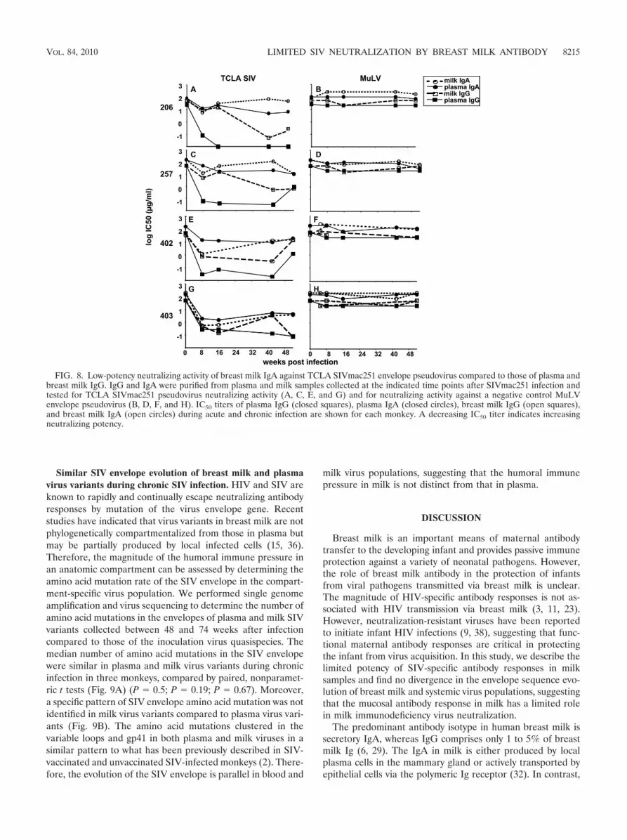
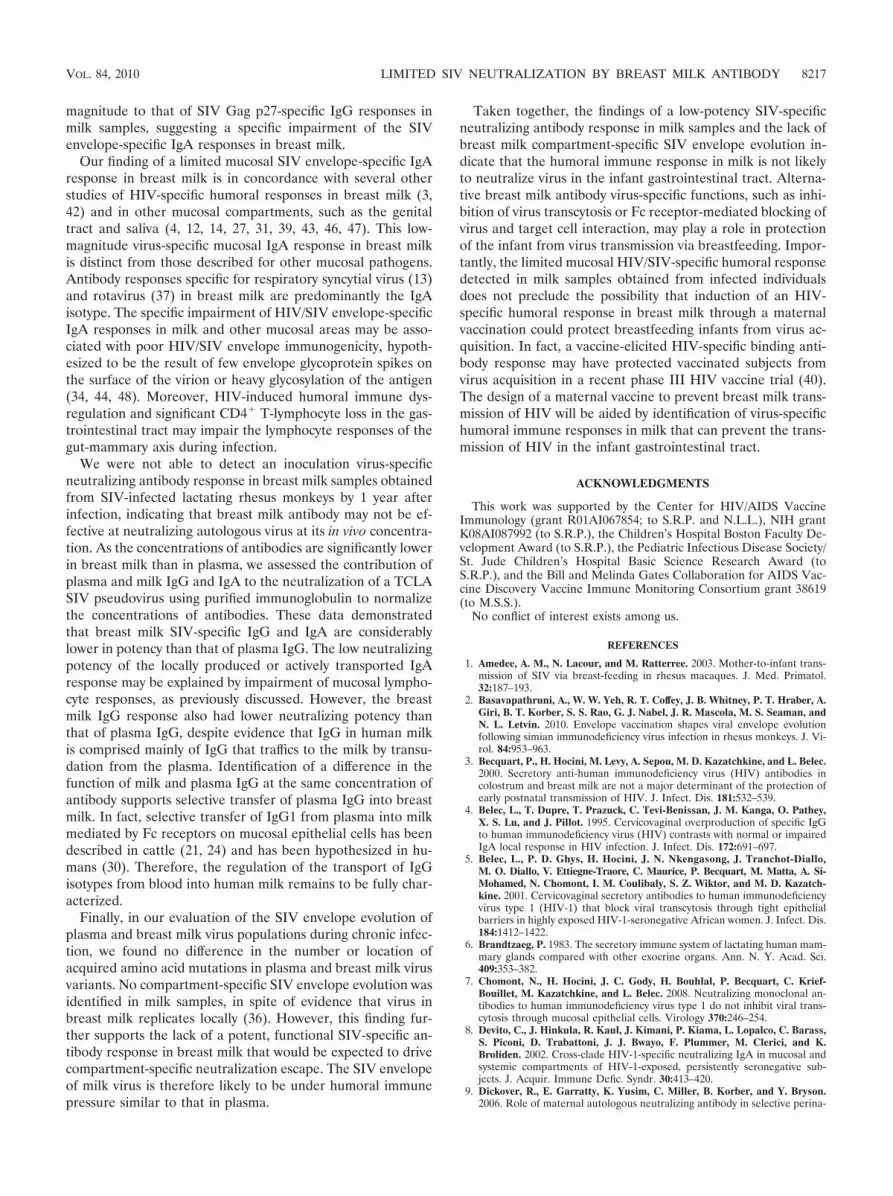
The most potent TCLA SIV-specific neutralizing responsewas detected in the plasma IgG fraction, with a median IC50
titer of 0.06 �g/ml (range, 0.03 to 0.26 �g/ml) by 8 weeks afterinfection (Fig. 8A, C, E, and G; Table 1). A potent TCLASIV-specific neutralizing response was also detected in thebreast milk IgG fraction. TCLA SIV-neutralizing activity wasdetected in the breast milk IgG fraction at 8 weeks after in-fection (median IC50, 4.9 �g/ml; range, 0.48 to 8.8 �g/ml) butincreased in potency during chronic infection (week 42 afterinfection) (median IC50, 0.32 �g/ml; range, 0.05 to 3.4 �g/ml).Importantly, minimal or no nonspecific neutralizing activitywas detected against the MuLV envelope pseudovirus (Fig. 8B,D, F, and H; Table 1).
The activities of the milk and plasma IgA neutralizing re-sponses were considerably less potent than those of the milkand plasma IgG neutralizing responses throughout acute andchronic infection (Fig. 8B, D, F, and H; Table 1). However, theTCLA SIV-neutralizing potency of breast milk IgA was similarin magnitude and kinetics to that of plasma IgA throughoutinfection. Interestingly, the neutralizing potency of the milkIgA peaked at 8 weeks after infection (median IC50, 5.5 �g/ml;range, 0.56 to 11.9 �g/ml) and remained static or decreased atlater time points, requiring a similar or larger amount of IgA toneutralize the TCLA SIV pseudovirus at week 42 (medianIC50, 70.8 �g/ml; range, 3.3 to 109.5 �g/ml) than at week 8 afterinfection. This pattern is in contrast to the plasma and breastmilk IgG responses, which continued to increase in potency at42 weeks after infection. Importantly, approximately 2- to 3-logmore milk IgA (median IC50, 70.8 �g/ml; range, 3.3 to 109.5�g/ml) than milk IgG (median IC50, 0.32 �g/ml; range, 0.05 to3.4 �g/ml) was required to neutralize the TCLA SIV pseudovi-rus at 42 weeks after infection in three of four monkeys, con-firming that the SIV-specific milk IgA response is considerablyless potent than the SIV-specific milk IgG response. The re-duced neutralizing potency of the milk IgA response comparedto that of the milk IgG response is similar to the reducedneutralizing potency of the plasma IgA response compared tothat of the plasma IgG response. Therefore, this mucosal SIV-specific IgA response does not seem to be of higher functionalquality than the plasma SIV-specific IgA response.
FIG. 6. No autologous neutralizing antibody response is detectedin breast milk samples during acute or chronic SIV infection. Autol-ogous SIVmac251 envelope pseudovirus neutralization was measuredin plasma and milk samples collected at the indicated time pointsfollowing SIVmac251. ID50 titers in plasma (A) and milk (B) samplesare plotted for each animal.
FIG. 7. The neutralizing antibody response in breast milk samplesagainst TCLA SIVmac251 envelope pseudovirus remains lower thanthat in plasma samples after normalization for total Ig content. TCLASIVmac251 envelope-specific neutralization was measured in plasmaand milk samples collected at the indicated time points followinginfection. (A, C, E, and G) ID50 titers in plasma (filled squares) andmilk (open circles) samples are shown for each monkey; (B, D, F, andH) ID50 titers in plasma and milk samples normalized for total Igcontent are shown in each graph.
8214 PERMAR ET AL. J. VIROL.
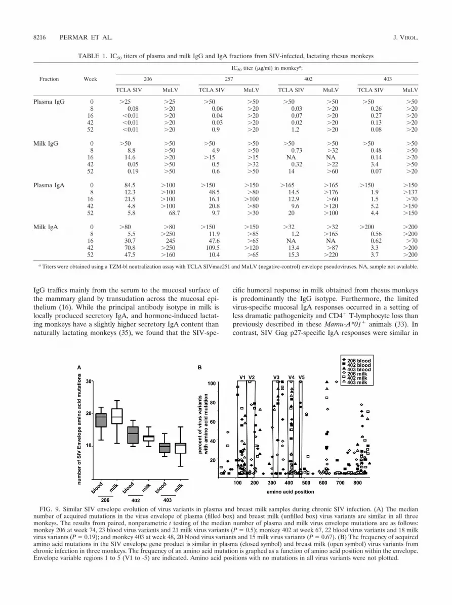
Similar SIV envelope evolution of breast milk and plasmavirus variants during chronic SIV infection. HIV and SIV areknown to rapidly and continually escape neutralizing antibodyresponses by mutation of the virus envelope gene. Recentstudies have indicated that virus variants in breast milk are notphylogenetically compartmentalized from those in plasma butmay be partially produced by local infected cells (15, 36).Therefore, the magnitude of the humoral immune pressure inan anatomic compartment can be assessed by determining theamino acid mutation rate of the SIV envelope in the compart-ment-specific virus population. We performed single genomeamplification and virus sequencing to determine the number ofamino acid mutations in the envelopes of plasma and milk SIVvariants collected between 48 and 74 weeks after infectioncompared to those of the inoculation virus quasispecies. Themedian number of amino acid mutations in the SIV envelopewere similar in plasma and milk virus variants during chronicinfection in three monkeys, compared by paired, nonparamet-ric t tests (Fig. 9A) (P � 0.5; P � 0.19; P � 0.67). Moreover,a specific pattern of SIV envelope amino acid mutation was notidentified in milk virus variants compared to plasma virus vari-ants (Fig. 9B). The amino acid mutations clustered in thevariable loops and gp41 in both plasma and milk viruses in asimilar pattern to what has been previously described in SIV-vaccinated and unvaccinated SIV-infected monkeys (2). There-fore, the evolution of the SIV envelope is parallel in blood and
milk virus populations, suggesting that the humoral immunepressure in milk is not distinct from that in plasma.
DISCUSSION
Breast milk is an important means of maternal antibodytransfer to the developing infant and provides passive immuneprotection against a variety of neonatal pathogens. However,the role of breast milk antibody in the protection of infantsfrom viral pathogens transmitted via breast milk is unclear.The magnitude of HIV-specific antibody responses is not as-sociated with HIV transmission via breast milk (3, 11, 23).However, neutralization-resistant viruses have been reportedto initiate infant HIV infections (9, 38), suggesting that func-tional maternal antibody responses are critical in protectingthe infant from virus acquisition. In this study, we describe thelimited potency of SIV-specific antibody responses in milksamples and find no divergence in the envelope sequence evo-lution of breast milk and systemic virus populations, suggestingthat the mucosal antibody response in milk has a limited rolein milk immunodeficiency virus neutralization.
The predominant antibody isotype in human breast milk issecretory IgA, whereas IgG comprises only 1 to 5% of breastmilk Ig (6, 29). The IgA in milk is either produced by localplasma cells in the mammary gland or actively transported byepithelial cells via the polymeric Ig receptor (32). In contrast,
FIG. 8. Low-potency neutralizing activity of breast milk IgA against TCLA SIVmac251 envelope pseudovirus compared to those of plasma andbreast milk IgG. IgG and IgA were purified from plasma and milk samples collected at the indicated time points after SIVmac251 infection andtested for TCLA SIVmac251 pseudovirus neutralizing activity (A, C, E, and G) and for neutralizing activity against a negative control MuLVenvelope pseudovirus (B, D, F, and H). IC50 titers of plasma IgG (closed squares), plasma IgA (closed circles), breast milk IgG (open squares),and breast milk IgA (open circles) during acute and chronic infection are shown for each monkey. A decreasing IC50 titer indicates increasingneutralizing potency.
VOL. 84, 2010 LIMITED SIV NEUTRALIZATION BY BREAST MILK ANTIBODY 8215
IgG traffics mainly from the serum to the mucosal surface ofthe mammary gland by transudation across the mucosal epi-thelium (16). While the principal antibody isotype in milk islocally produced secretory IgA, and hormone-induced lactat-ing monkeys have a slightly higher secretory IgA content thannaturally lactating monkeys (35), we found that the SIV-spe-
cific humoral response in milk obtained from rhesus monkeysis predominantly the IgG isotype. Furthermore, the limitedvirus-specific mucosal IgA responses occurred in a setting ofless dramatic pathogenicity and CD4� T-lymphocyte loss thanpreviously described in these Mamu-A*01� animals (33). Incontrast, SIV Gag p27-specific IgA responses were similar in
TABLE 1. IC50 titers of plasma and milk IgG and IgA fractions from SIV-infected, lactating rhesus monkeys
Fraction Week
IC50 titer (�g/ml) in monkeya:
206 257 402 403
TCLA SIV MuLV TCLA SIV MuLV TCLA SIV MuLV TCLA SIV MuLV
Plasma IgG 0 �25 �25 �50 �50 �50 �50 �50 �508 0.08 �20 0.06 �20 0.03 �20 0.26 �20
16 �0.01 �20 0.04 �20 0.07 �20 0.27 �2042 �0.01 �20 0.03 �20 0.02 �20 0.13 �2052 �0.01 �20 0.9 �20 1.2 �20 0.08 �20
Milk IgG 0 �50 �50 �50 �50 �50 �50 �50 �508 8.8 �50 4.9 �50 0.73 �32 0.48 �50
16 14.6 �20 �15 �15 NA NA 0.14 �2042 0.05 �50 0.5 �32 0.32 �22 3.4 �5052 0.19 �50 0.6 �50 14 �60 0.07 �20
Plasma IgA 0 84.5 �100 �150 �150 �165 �165 �150 �1508 12.3 �100 48.5 �80 14.5 �176 1.9 �137
16 21.5 �100 16.1 �100 12.9 �60 1.5 �7042 4.8 �100 20.8 �80 9.6 �120 5.2 �15052 5.8 68.7 9.7 �30 20 �100 4.4 �150
Milk IgA 0 �80 �80 �150 �150 �32 �32 �200 �2008 5.5 �250 11.9 �85 1.2 �165 0.56 �200
16 30.7 245 47.6 �65 NA NA 0.62 �7042 70.8 �250 109.5 �120 13.4 �87 3.3 �20052 47.5 �160 10.4 �65 15.3 �220 3.7 �200
a Titers were obtained using a TZM-bl neutralization assay with TCLA SIVmac251 and MuLV (negative-control) envelope pseudoviruses. NA, sample not available.
FIG. 9. Similar SIV envelope evolution of virus variants in plasma and breast milk samples during chronic SIV infection. (A) The mediannumber of acquired mutations in the virus envelope of plasma (filled box) and breast milk (unfilled box) virus variants are similar in all threemonkeys. The results from paired, nonparametric t testing of the median number of plasma and milk virus envelope mutations are as follows:monkey 206 at week 74, 23 blood virus variants and 21 milk virus variants (P � 0.5); monkey 402 at week 67, 22 blood virus variants and 18 milkvirus variants (P � 0.19); and monkey 403 at week 48, 20 blood virus variants and 15 milk virus variants (P � 0.67). (B) The frequency of acquiredamino acid mutations in the SIV envelope gene product is similar in plasma (closed symbol) and breast milk (open symbol) virus variants fromchronic infection in three monkeys. The frequency of an amino acid mutation is graphed as a function of amino acid position within the envelope.Envelope variable regions 1 to 5 (V1 to -5) are indicated. Amino acid positions with no mutations in all virus variants were not plotted.
8216 PERMAR ET AL. J. VIROL.
magnitude to that of SIV Gag p27-specific IgG responses inmilk samples, suggesting a specific impairment of the SIVenvelope-specific IgA responses in breast milk.
Our finding of a limited mucosal SIV envelope-specific IgAresponse in breast milk is in concordance with several otherstudies of HIV-specific humoral responses in breast milk (3,42) and in other mucosal compartments, such as the genitaltract and saliva (4, 12, 14, 27, 31, 39, 43, 46, 47). This low-magnitude virus-specific mucosal IgA response in breast milkis distinct from those described for other mucosal pathogens.Antibody responses specific for respiratory syncytial virus (13)and rotavirus (37) in breast milk are predominantly the IgAisotype. The specific impairment of HIV/SIV envelope-specificIgA responses in milk and other mucosal areas may be asso-ciated with poor HIV/SIV envelope immunogenicity, hypoth-esized to be the result of few envelope glycoprotein spikes onthe surface of the virion or heavy glycosylation of the antigen(34, 44, 48). Moreover, HIV-induced humoral immune dys-regulation and significant CD4� T-lymphocyte loss in the gas-trointestinal tract may impair the lymphocyte responses of thegut-mammary axis during infection.
We were not able to detect an inoculation virus-specificneutralizing antibody response in breast milk samples obtainedfrom SIV-infected lactating rhesus monkeys by 1 year afterinfection, indicating that breast milk antibody may not be ef-fective at neutralizing autologous virus at its in vivo concentra-tion. As the concentrations of antibodies are significantly lowerin breast milk than in plasma, we assessed the contribution ofplasma and milk IgG and IgA to the neutralization of a TCLASIV pseudovirus using purified immunoglobulin to normalizethe concentrations of antibodies. These data demonstratedthat breast milk SIV-specific IgG and IgA are considerablylower in potency than that of plasma IgG. The low neutralizingpotency of the locally produced or actively transported IgAresponse may be explained by impairment of mucosal lympho-cyte responses, as previously discussed. However, the breastmilk IgG response also had lower neutralizing potency thanthat of plasma IgG, despite evidence that IgG in human milkis comprised mainly of IgG that traffics to the milk by transu-dation from the plasma. Identification of a difference in thefunction of milk and plasma IgG at the same concentration ofantibody supports selective transfer of plasma IgG into breastmilk. In fact, selective transfer of IgG1 from plasma into milkmediated by Fc receptors on mucosal epithelial cells has beendescribed in cattle (21, 24) and has been hypothesized in hu-mans (30). Therefore, the regulation of the transport of IgGisotypes from blood into human milk remains to be fully char-acterized.
Finally, in our evaluation of the SIV envelope evolution ofplasma and breast milk virus populations during chronic infec-tion, we found no difference in the number or location ofacquired amino acid mutations in plasma and breast milk virusvariants. No compartment-specific SIV envelope evolution wasidentified in milk samples, in spite of evidence that virus inbreast milk replicates locally (36). However, this finding fur-ther supports the lack of a potent, functional SIV-specific an-tibody response in breast milk that would be expected to drivecompartment-specific neutralization escape. The SIV envelopeof milk virus is therefore likely to be under humoral immunepressure similar to that in plasma.
Taken together, the findings of a low-potency SIV-specificneutralizing antibody response in milk samples and the lack ofbreast milk compartment-specific SIV envelope evolution in-dicate that the humoral immune response in milk is not likelyto neutralize virus in the infant gastrointestinal tract. Alterna-tive breast milk antibody virus-specific functions, such as inhi-bition of virus transcytosis or Fc receptor-mediated blocking ofvirus and target cell interaction, may play a role in protectionof the infant from virus transmission via breastfeeding. Impor-tantly, the limited mucosal HIV/SIV-specific humoral responsedetected in milk samples obtained from infected individualsdoes not preclude the possibility that induction of an HIV-specific humoral response in breast milk through a maternalvaccination could protect breastfeeding infants from virus ac-quisition. In fact, a vaccine-elicited HIV-specific binding anti-body response may have protected vaccinated subjects fromvirus acquisition in a recent phase III HIV vaccine trial (40).The design of a maternal vaccine to prevent breast milk trans-mission of HIV will be aided by identification of virus-specifichumoral immune responses in milk that can prevent the trans-mission of HIV in the infant gastrointestinal tract.
ACKNOWLEDGMENTS
This work was supported by the Center for HIV/AIDS VaccineImmunology (grant R01AI067854; to S.R.P. and N.L.L.), NIH grantK08AI087992 (to S.R.P.), the Children’s Hospital Boston Faculty De-velopment Award (to S.R.P.), the Pediatric Infectious Disease Society/St. Jude Children’s Hospital Basic Science Research Award (toS.R.P.), and the Bill and Melinda Gates Collaboration for AIDS Vac-cine Discovery Vaccine Immune Monitoring Consortium grant 38619(to M.S.S.).
No conflict of interest exists among us.
REFERENCES
1. Amedee, A. M., N. Lacour, and M. Ratterree. 2003. Mother-to-infant trans-mission of SIV via breast-feeding in rhesus macaques. J. Med. Primatol.32:187–193.
2. Basavapathruni, A., W. W. Yeh, R. T. Coffey, J. B. Whitney, P. T. Hraber, A.Giri, B. T. Korber, S. S. Rao, G. J. Nabel, J. R. Mascola, M. S. Seaman, andN. L. Letvin. 2010. Envelope vaccination shapes viral envelope evolutionfollowing simian immunodeficiency virus infection in rhesus monkeys. J. Vi-rol. 84:953–963.
3. Becquart, P., H. Hocini, M. Levy, A. Sepou, M. D. Kazatchkine, and L. Belec.2000. Secretory anti-human immunodeficiency virus (HIV) antibodies incolostrum and breast milk are not a major determinant of the protection ofearly postnatal transmission of HIV. J. Infect. Dis. 181:532–539.
4. Belec, L., T. Dupre, T. Prazuck, C. Tevi-Benissan, J. M. Kanga, O. Pathey,X. S. Lu, and J. Pillot. 1995. Cervicovaginal overproduction of specific IgGto human immunodeficiency virus (HIV) contrasts with normal or impairedIgA local response in HIV infection. J. Infect. Dis. 172:691–697.
5. Belec, L., P. D. Ghys, H. Hocini, J. N. Nkengasong, J. Tranchot-Diallo,M. O. Diallo, V. Ettiegne-Traore, C. Maurice, P. Becquart, M. Matta, A. Si-Mohamed, N. Chomont, I. M. Coulibaly, S. Z. Wiktor, and M. D. Kazatch-kine. 2001. Cervicovaginal secretory antibodies to human immunodeficiencyvirus type 1 (HIV-1) that block viral transcytosis through tight epithelialbarriers in highly exposed HIV-1-seronegative African women. J. Infect. Dis.184:1412–1422.
6. Brandtzaeg, P. 1983. The secretory immune system of lactating human mam-mary glands compared with other exocrine organs. Ann. N. Y. Acad. Sci.409:353–382.
7. Chomont, N., H. Hocini, J. C. Gody, H. Bouhlal, P. Becquart, C. Krief-Bouillet, M. Kazatchkine, and L. Belec. 2008. Neutralizing monoclonal an-tibodies to human immunodeficiency virus type 1 do not inhibit viral trans-cytosis through mucosal epithelial cells. Virology 370:246–254.
8. Devito, C., J. Hinkula, R. Kaul, J. Kimani, P. Kiama, L. Lopalco, C. Barass,S. Piconi, D. Trabattoni, J. J. Bwayo, F. Plummer, M. Clerici, and K.Broliden. 2002. Cross-clade HIV-1-specific neutralizing IgA in mucosal andsystemic compartments of HIV-1-exposed, persistently seronegative sub-jects. J. Acquir. Immune Defic. Syndr. 30:413–420.
9. Dickover, R., E. Garratty, K. Yusim, C. Miller, B. Korber, and Y. Bryson.2006. Role of maternal autologous neutralizing antibody in selective perina-
VOL. 84, 2010 LIMITED SIV NEUTRALIZATION BY BREAST MILK ANTIBODY 8217

tal transmission of human immunodeficiency virus type 1 escape variants.J. Virol. 80:6525–6533.
10. Dorrell, L., A. J. Hessell, M. Wang, H. Whittle, S. Sabally, S. Rowland-Jones,D. R. Burton, and P. W. Parren. 2000. Absence of specific mucosal antibodyresponses in HIV-exposed uninfected sex workers from the Gambia. AIDS14:1117–1122.
11. Duprat, C., Z. Mohammed, P. Datta, W. Stackiw, J. O. Ndinya-Achola, J. K.Kreiss, K. K. Holmes, F. A. Plummer, and J. E. Embree. 1994. Humanimmunodeficiency virus type 1 IgA antibody in breast milk and serum.Pediatr. Infect. Dis. J. 13:603–608.
12. Fiore, J. R., V. Laddago, A. Lepera, L. La Grasta, M. Di Stefano, A.Saracino, P. Lopalco, G. Pastore, and G. Angarano. 2000. Limited secretory-IgA response in cervicovaginal secretions from HIV-1 infected, but not highrisk seronegative women: lack of correlation to genital viral shedding. NewMicrobiol. 23:85–92.
13. Fishaut, M., D. Murphy, M. Neifert, K. McIntosh, and P. L. Ogra. 1981.Bronchomammary axis in the immune response to respiratory syncytial virus.J. Pediatr. 99:186–191.
14. Haimovici, F., K. H. Mayer, and D. J. Anderson. 1997. Quantitation ofHIV-1-specific IgG, IgA, and IgM antibodies in human genital tract secre-tions. J. Acquir. Immune Defic. Syndr. Hum. Retrovirol. 15:185–191.
15. Heath, L., S. Conway, L. Jones, K. Semrau, K. Nakamura, J. Walter, W. D.Decker, J. Hong, T. Chen, M. Heil, M. Sinkala, C. Kankasa, D. M. Thea, L.Kuhn, J. I. Mullins, and G. M. Aldrovandi. 2010. Restriction of HIV-1genotypes in breast milk does not account for the population transmissiongenetic bottleneck that occurs following transmission. PLoS One 5:e10213.
16. Hochwald, G. M., and G. J. Thorbecke. 1964. Occurrence of myeloma-likegamma-globulin in C.S.F. of a four-month old infant with hydrocephalus.Pediatrics 33:435–440.
17. Hocini, H., P. Becquart, H. Bouhlal, H. Adle-Biassette, M. D. Kazatchkine,and L. Belec. 2000. Secretory leukocyte protease inhibitor inhibits infectionof monocytes and lymphocytes with human immunodeficiency virus type 1but does not interfere with transcytosis of cell-associated virus across tightepithelial barriers. Clin. Diagn. Lab. Immunol. 7:515–518.
18. Hofmann-Lehmann, R., R. A. Rasmussen, J. Vlasak, B. A. Smith, T. W.Baba, V. Liska, D. C. Montefiori, H. M. McClure, D. C. Anderson, B. J.Bernacky, T. A. Rizvi, R. Schmidt, L. R. Hill, M. E. Keeling, H. Katinger, G.Stiegler, M. R. Posner, L. A. Cavacini, T. C. Chou, and R. M. Ruprecht.2001. Passive immunization against oral AIDS virus transmission: an ap-proach to prevent mother-to-infant HIV-1 transmission? J. Med. Primatol.30:190–196.
19. John, G. C., B. A. Richardson, R. W. Nduati, D. Mbori-Ngacha, and J. K.Kreiss. 2001. Timing of breast milk HIV-1 transmission: a meta-analysis.East Afr. Med. J. 78:75–79.
20. Johnstone, A., and R. Thorpe. 1982. Immunochemistry in practice. BlackwellScientific Publications, Oxford, England.
21. Kemler, R., H. Mossmann, U. Strohmaier, B. Kickhofen, and D. K. Ham-mer. 1975. In vitro studies on the selective binding of IgG from differentspecies to tissue sections of the bovine mammary gland. Eur. J. Immunol.5:603–608.
22. Kobayashi, K. 1971. Studies on human secretory IgA comparative studies ofthe IgA-bound secretory piece and the free secretory piece protein. Immu-nochemistry 8:785–800.
23. Kuhn, L., D. Trabattoni, C. Kankasa, M. Sinkala, F. Lissoni, M. Ghosh, G.Aldrovandi, D. Thea, and M. Clerici. 2006. HIV-specific secretory IgA inbreast milk of HIV-positive mothers is not associated with protection againstHIV transmission among breast-fed infants. J. Pediatr. 149:611–616.
24. Leary, H. L., Jr., B. L. Larson, and D. R. Nelson. 1982. Immunohistochem-ical localization of IgG1 and IgG2 in prepartum and lactating bovine mam-mary tissue. Vet. Immunol. Immunopathol. 3:509–514.
25. Leroy, V., M. L. Newell, F. Dabis, C. Peckham, P. Van de Perre, M. Bulterys,C. Kind, R. J. Simonds, S. Wiktor, and P. Msellati. 1998. Internationalmulticentre pooled analysis of late postnatal mother-to-child transmission ofHIV-1 infection. Ghent International Working Group on Mother-to-ChildTransmission of HIV. Lancet 352:597–600.
26. Li, M., F. Gao, J. R. Mascola, L. Stamatatos, V. R. Polonis, M. Koutsoukos,G. Voss, P. Goepfert, P. Gilbert, K. M. Greene, M. Bilska, D. L. Kothe, J. F.Salazar-Gonzalez, X. Wei, J. M. Decker, B. H. Hahn, and D. C. Montefiori.2005. Human immunodeficiency virus type 1 env clones from acute and earlysubtype B infections for standardized assessments of vaccine-elicited neu-tralizing antibodies. J. Virol. 79:10108–10125.
27. Lu, F. X. 2000. Predominate HIV1-specific IgG activity in various mucosalcompartments of HIV1-infected individuals. Clin. Immunol. 97:59–68.
28. Martinez-Maza, O., E. Crabb, R. T. Mitsuyasu, J. L. Fahey, and J. V. Giorgi.1987. Infection with the human immunodeficiency virus (HIV) is associatedwith an in vivo increase in B lymphocyte activation and immaturity. J. Im-munol. 138:3720–3724.
29. McClelland, D. B., R. R. Warwick, and D. J. Shearman. 1973. IgA concen-tration. Am. J. Dig. Dis. 18:347–348.
30. Mehta, P. D., S. P. Mehta, and C. E. Isaacs. 1989. Distribution of IgGsubclasses in human colostrum and milk. Immunol. Lett. 22:235–238.
31. Mestecky, J., S. Jackson, Z. Moldoveanu, L. R. Nesbit, R. Kulhavy, S. J.Prince, S. Sabbaj, M. J. Mulligan, and P. A. Goepfert. 2004. Paucity ofantigen-specific IgA responses in sera and external secretions of HIV-type1-infected individuals. AIDS Res. Hum. Retroviruses 20:972–988.
32. Mostov, K. E., and G. Blobel. 1983. Biosynthesis, processing, and function ofsecretory component. Methods Enzymol. 98:458–466.
33. O’Connor, D. H., B. R. Mothe, J. T. Weinfurter, S. Fuenger, W. M. Rehrauer,P. Jing, R. R. Rudersdorf, M. E. Liebl, K. Krebs, J. Vasquez, E. Dodds, J.Loffredo, S. Martin, A. B. McDermott, T. M. Allen, C. Wang, G. G. Doxiadis,D. C. Montefiori, A. Hughes, D. R. Burton, D. B. Allison, S. M. Wolinsky, R.Bontrop, L. J. Picker, and D. I. Watkins. 2003. Major histocompatibilitycomplex class I alleles associated with slow simian immunodeficiency virusdisease progression bind epitopes recognized by dominant acute-phase cy-totoxic-T-lymphocyte responses. J. Virol. 77:9029–9040.
34. Pantophlet, R., and D. R. Burton. 2006. GP120: target for neutralizingHIV-1 antibodies. Annu. Rev. Immunol. 24:739–769.
35. Permar, S. R., H. H. Kang, A. Carville, K. G. Mansfield, R. S. Gelman, S. S.Rao, J. B. Whitney, and N. L. Letvin. 2008. Potent simian immunodeficiencyvirus-specific cellular immune responses in the breast milk of simian immu-nodeficiency virus-infected, lactating rhesus monkeys. J. Immunol. 181:3643–3650.
36. Permar, S. R., H. H. Kang, A. B. Wilks, L. V. Mach, A. Carville, K. G.Mansfield, G. H. Learn, B. H. Hahn, and N. L. Letvin. 2010. Local replica-tion of simian immunodeficiency virus in the breast milk compartment ofchronically-infected, lactating rhesus monkeys. Retrovirology 7:7.
37. Rahman, M. M., M. Yamauchi, N. Hanada, K. Nishikawa, and T. Mor-ishima. 1987. Local production of rotavirus specific IgA in breast tissue andtransfer to neonates. Arch. Dis. Child. 62:401–405.
38. Rainwater, S. M., X. Wu, R. Nduati, R. Nedellec, D. Mosier, G. John-Stewart, D. Mbori-Ngacha, and J. Overbaugh. 2007. Cloning and character-ization of functional subtype A HIV-1 envelope variants transmitted throughbreastfeeding. Curr. HIV Res. 5:189–197.
39. Raux, M., L. Finkielsztejn, D. Salmon-Ceron, H. Bouchez, J. L. Excler, E.Dulioust, J. M. Grouin, D. Sicard, and C. Blondeau. 1999. Comparison ofthe distribution of IgG and IgA antibodies in serum and various mucosalfluids of HIV type 1-infected subjects. AIDS Res. Hum. Retroviruses 15:1365–1376.
40. Rerks-Ngarm, S., P. Pitisuttithum, S. Nitayaphan, J. Kaewkungwal, J. Chiu,R. Paris, N. Premsri, C. Namwat, M. de Souza, E. Adams, M. Benenson, S.Gurunathan, J. Tartaglia, J. G. McNeil, D. P. Francis, D. Stablein, D. L.Birx, S. Chunsuttiwat, C. Khamboonruang, P. Thongcharoen, M. L. Robb,N. L. Michael, P. Kunasol, and J. H. Kim. 2009. Vaccination with ALVACand AIDSVAX to prevent HIV-1 infection in Thailand. N. Engl. J. Med.361:2209–2220.
41. Ruprecht, R. M., F. Ferrantelli, M. Kitabwalla, W. Xu, and H. M. McClure.2003. Antibody protection: passive immunization of neonates against oralAIDS virus challenge. Vaccine 21:3370–3373.
42. Rychert, J., and A. M. Amedee. 2005. The antibody response to SIV inlactating rhesus macaques. J. Acquir. Immune Defic. Syndr. 38:135–141.
43. Schafer, F., S. Kewenig, N. Stolte, C. Stahl-Hennig, A. Stallmach, F. J. Kaup,M. Zeitz, and T. Schneider. 2002. Lack of simian immunodeficiency virus(SIV) specific IgA response in the intestine of SIV infected rhesus macaques.Gut 50:608–614.
44. Wei, X., J. M. Decker, S. Wang, H. Hui, J. C. Kappes, X. Wu, J. F. Salazar-Gonzalez, M. G. Salazar, J. M. Kilby, M. S. Saag, N. L. Komarova, M. A.Nowak, B. H. Hahn, P. D. Kwong, and G. M. Shaw. 2003. Antibody neutral-ization and escape by HIV-1. Nature 422:307–312.
45. WHO Collaborative Study Team on the Role of Breastfeeding on the Pre-vention of Infant Mortality. 2000. Effect of breastfeeding on infant and childmortality due to infectious diseases in less developed countries: a pooledanalysis. Lancet 355:451–455.
46. Williams, S. B., T. P. Flanigan, S. Cu-Uvin, K. Mayer, P. Williams, C. A.Ettore, A. W. Artenstein, A. Duerr, and T. C. VanCott. 2002. Human immu-nodeficiency virus (HIV)-specific antibody in cervicovaginal lavage speci-mens obtained from women infected with HIV type 1. Clin. Infect. Dis.35:611–617.
47. Wright, P. F., P. A. Kozlowski, G. K. Rybczyk, P. Goepfert, H. F. Staats, T. C.VanCott, D. Trabattoni, E. Sannella, and J. Mestecky. 2002. Detection ofmucosal antibodies in HIV type 1-infected individuals. AIDS Res. Hum.Retroviruses 18:1291–1300.
48. Wyatt, R., P. D. Kwong, E. Desjardins, R. W. Sweet, J. Robinson, W. A.Hendrickson, and J. G. Sodroski. 1998. The antigenic structure of the HIVgp120 envelope glycoprotein. Nature 393:705–711.
8218 PERMAR ET AL. J. VIROL.


