Embed Size (px)
Citation preview
This article appeared in a journal published by Elsevier. The attachedcopy is furnished to the author for internal non-commercial researchand education use, including for instruction at the authors institution
and sharing with colleagues.
Other uses, including reproduction and distribution, or selling orlicensing copies, or posting to personal, institutional or third party
websites are prohibited.
In most cases authors are permitted to post their version of thearticle (e.g. in Word or Tex form) to their personal website orinstitutional repository. Authors requiring further information
regarding Elsevier’s archiving and manuscript policies areencouraged to visit:
http://www.elsevier.com/copyright

Author's personal copy
Review article
Lipid nanoparticles for parenteral delivery of actives
Medha D. Joshi *, Rainer H. MüllerDepartment of Pharmaceutical Technology, Biopharmaceutics and NutriCosmetics, Freie Universitäte Berlin, Berlin, Germany
a r t i c l e i n f o
Article history:Received 10 July 2008Accepted in revised form 2 September 2008Available online 13 September 2008
Keywords:Lipid nanoparticlesSolid lipid nanoparticles (SLN)Nanostructured lipid carriers (NLC)Lipid drug conjugates (LDC)Parenteral delivery
a b s t r a c t
The present review compiles the applications of lipid nanoparticles mainly solid lipid nanoparticles (SLN),nanostructured lipid carriers (NLC) and lipid drug conjugates (LDC) in parenteral delivery of pharmaceu-tical actives. The attempts to incorporate anticancer agents, imaging agents, antiparasitics, antiarthritics,genes for transfection, agents for liver, cardiovascular and central nervous system targeting have beensummarized. The utility of lipid nanoparticles as adjuvant has been discussed separately. A special focusof this review is on toxicity caused by these kinds of lipid nanoparticles with a glance on the fate of lipidnanoparticles after their parenteral delivery in vivo viz the protein adsorption patterns.
� 2008 Elsevier B.V. All rights reserved.
1. Introduction
Parenteral drug delivery took a major leap after successfuldevelopment of the submicronic parenteral fat emulsion (Intrali-pid) in 1960s. Quick commercialization of submicron emulsion-based products, such as Diazemuls (Diazepam) and Diprivan (Pro-pofol), was the indicator of the interest of pharmaceutical industryin colloidal carriers. Since then, there have been continuous effortsto develop novel colloidal nanocarriers for improved parenteraldelivery. The concept of lipid nanoparticles for injectable deliverywas developed from submicron sized parenteral fat o/w emulsionused for parenteral nutrition viz Intralipid� in 1960s [1]. This gavebirth to the idea of encapsulating lipophilic drugs into oil droplets.Products such as Diazemuls� contain diazepam and Diprivan� con-tain propofol [2]. The only drawback associated with these submi-cron emulsions was the low viscosity of the droplets causing fastrelease and susceptibility of the incorporated actives towards deg-radation by the aqueous continuous phase [3].
Liposomes represent the first generation of the novel colloidalcarriers, which revolutionized the scenario in parenteral drugdelivery. Liposomes offered several advantages such as encapsula-tion of hydrophobic and hydrophilic drugs, controlled drug releaseand reduction in toxicity/increased therapeutic efficacy of drugsmost of which were not offered by submicronic emulsions. Thesuccessful commercialization ff various injectable liposomal prod-ucts such as AmBisome� (Amphotericin B), Doxi�/Caelyx� (Doxo-
rubicin) [6] and DaunoXome� (Daunorubicin) [7] and a largearray of investigational products clearly indicates the potentialadvantages of liposomes as novel lipid carriers. However, complex-ity associated with the manufacturing of liposomes, difficulties inscale-up, limited physical stability and enormous cost of the lipo-somal formulation are the major barriers in the successful com-mercialization of liposomes [5].
Polymeric nanoparticles are also included in the first generationnovel colloidal carriers developed with the objective to improveparenteral delivery. The particulate nature of polymeric nanoparti-cles, their ability to control the release of drugs and amenability forsurface modifications were the drivers for active research on thesecarriers. However, due to various disadvantages such as difficultiesin scale-up, high cost of biodegradable polymers, potentially toxic/allergic end products of biodegradable polymers [4], there is nocommercial product based on polymeric ‘nano’ particles even after35 years of their discovery and the first description of polymericnanocapsules by Speiser and Birenbach. Nevertheless, polymeric‘micro’ particle-based depot formulations are available in the mar-ket, e.g., Lupron� containing leuprolide and Parlodel� containingbromocriptin [5].
In 1990s, researchers (Mueller and coworkers and Gasco andcoworkers) started exploring the potential of nanoparticles-basedsolid lipids or solid lipid nanoparticles (SLN) in the drug delivery.SLN are colloidal particles composed of a biocompatible/biodegrad-able lipid matrix that is solid at body temperature and exhibit sizerange in between 100 and 400 nm. SLN combine advantages ofaforementioned colloidal drug carrier systems like liposomes, poly-meric nanoparticles and emulsions, but at the same time avoid orminimize the drawbacks associated with them [8]. The variousadvantages such as particulate nature of SLN, amenability to encap-
0939-6411/$ - see front matter � 2008 Elsevier B.V. All rights reserved.doi:10.1016/j.ejpb.2008.09.003
* Corresponding author. Department of Pharmaceutical Technology, Biopharma-ceutics and NutriCosmetics, Freie Universitäte Berlin, Kelchstrasse 31, 12169 Berlin,Germany. Tel.: +49 30 838 50703; fax: +49 30 838 506 16.
E-mail address: [email protected] (M.D. Joshi).
European Journal of Pharmaceutics and Biopharmaceutics 71 (2009) 161–172
Contents lists available at ScienceDirect
European Journal of Pharmaceutics and Biopharmaceutics
journal homepage: www.elsevier .com/locate /e jpb

Author's personal copy
sulate hydrophilic and hydrophobic drugs, ability to sustain the re-lease of incorporated drug, ability to prevent chemical, photochem-ical or oxidative degradation of drug, ability to immobilize drug inthe solid matrix, ease of scale-up and manufacture and low cost ofsolid lipids as compared to phospholipids and biodegradable poly-mers give SLN an edge over aforementioned colloidal carriers [8].
Solid lipid nanoparticles (SLN) are colloidal particles of a lipidmatrix that is solid at body temperature. They were first intro-duced by Müller et al. in 1993 [9,10] produced by high pressurehomogenization and in parallel by Gasco by diluting warm micro-emulsion [11]. SLN have been exploited for delivery of actives viathe dermal [12–14], peroral [15], parenteral [8,16], ocular [17–19], pulmonary [20–22] and rectal [23,24] route. Upon administra-tion of SLN via the parenteral route of administration, improvedbioavailability, targeting, enhanced cytotoxicity against multidrugresistant cancer cells have been observed.
Nanostructured lipid carriers (NLC) are composed of binarymixture of solid lipid and a spatially different liquid lipid as thecarrier [12,14]. The major advantage of NLC is increased drug load.By now NLC are mainly investigated for dermal application [12,14]with seldom investigations focused on the parenteral route [65,66].
Lipid drug conjugates (LDC) were developed especially for thehydrophilic drug molecules, wherein an insoluble drug–lipid con-jugate bulk is synthetically prepared either by salt formation(e.g., with a fatty acid) or by covalent linking (e.g., to the estersor ethers) [8,25]. LDC bulk is then homogenized in the presenceof a stabilizer in water using high pressure homogenization.
The production of lipid particles in micrometer size range wasreported in the late 1950s and in the beginning of 1960s [26,27].Recently, a review [28] on advances in lipid nanodispersions forparenteral drug delivery and targeting has been published, whichfocuses on nanoemulsions, nanosuspensions and polymeric mi-celles. The first report on the use of SLN for oral delivery is by Spe-iser who termed them as nanopellets [29]. As the science of SLNtechnology progressed, different methods of production for themwere developed and stable formulations of SLN were discovered.Earlier, the utilization of SLN for parenteral drug delivery [8] withfocus on the definition of lipid nanoparticles and their differenttypes such as SLN, NLC, and LDC, their production techniques,scale-up feasibilities, stability of the incorporated drug, releaseand the biological and biopharmaceutical aspects have been re-viewed. In this review, we have summarized the efforts made bydifferent groups to incorporate the actives in lipid nanoparticles,and the success of the drug delivery system has achieved till datewith the focus on parenteral route of administration. We will alsobe discussing the new Pathfinder� technology, which can be usedto target nanoparticles to the desired organ by modifying the sur-face protein adsorption on them.
The injectable lipid nanoparticles that have been studied so farhave been encapsulated with anticancer agents, imaging agents,anti-parkinsonism, antiHIV, antipsychotics, anti-rheumatoid ar-thritic agents, antiparasitics, antihypertensives and antibiotics assummarized in Table 1. We will discuss the results obtained afterencapsulating these therapeutic agents in lipid nanoparticles cate-
Table 1Overview of various actives incorporated in injectable lipid nanoparticles
Drug Disease Type of lipid nanoparticle Route of administration Reference
30 ,50-Dioctanoyl-5-fluoro-20-deoxyuridine Cancer SLN IV [60]3-Azido-3-deoxythymidine palmitate/
azidothymidineAntiHIV SLN IV [101]
5-FU Cancer SLN IV [34]99mTc/188Re Imaging agent Nanocapsules IV [79]Actarit Rheumatoid
ArthritisSLN IV [97]
All trans retinoic acid Cancer SLN IV [50,58]Beta-element Cancer SLN IV [59]Bromocriptine Anti-parkinsonism SLN IP [87]Camptothecin Cancer SLN IV [16]CdSEe/ZnS Imaging agent QDs encapsulated in SLN IV [81]Clozapine Antipsychotic SLN ID [85]Dexamethasone acetate Pulmonary disease SLN IV [100]Diminazene Antitrypanosomal LDC - [95]DNA Cancer catinoic SLN - [68–72]Doxorubicin Cancer Stealth and non-stealth SLN, SLN IV [35,36,42,53,54]Etoposide Cancer SLN IV/SC/IP [47,48]Idarubicin Cancer SLN IV or ID [56]Iron oxide Imaging agent SLN - [78]Magnetite Imaging agent SLN - [77]Methotrexate Cancer LMBVs IV [67]Mitoxantrone Cancer SLN Local injection in breast Cancer
tissue[49]
Nitrendipine Antihypertensive SLN IV or ID [61,62]Oxymatrine Antihepatitis SLN IV [74]Paclitaxel Cancer SLN/Wax NP/sterically stabilized
SLNIV [37–41]
Paclitaxel and doxorubicin Cancer SLN – [42,43]Paclitaxel and doxorubicin and cholestryl
butyrateCancer SLN – [44]
Quinine dihydrochloride Malaria Transferring conjugated SLN IV [94,95]Tamoxifen Cancer SLN IV [45,46]Tashione II A Vasodialator SLN IV [91,92]Temoxifen citrate Cancer SLN IV [46]Temozolomide Cancer SLN IV [64]Testosterone 125 I radiolabelled Imaging agent SLN IV [80]Tobramycin Antibiotic SLN IV or ID [86]Vinorelbine bitartate Cancer PEG-modified SLN – [51]
IV, intravenous; ID, intrdeodenum; IP, intraperitoneum; SC, subcutaneous.
162 M.D. Joshi, R.H. Müller / European Journal of Pharmaceutics and Biopharmaceutics 71 (2009) 161–172
Author's personal copy
gory wise and the sequence is based on the amount of work donein the literature. Of the various purposes of these investigations,enhanced AUC and MRT, enhanced anticancer efficacy, enhancedbrain targeting and enhanced targeting to diseased organ/cell werethe main observations.
2. Application of lipid nanoparticles for parenteral drug delivery
2.1. Lipid nanoparticles for treatment of cancer
Several anticancer agents have been encapsulated in lipid nano-particles, and their in vitro and in vivo efficacy has been evaluatedby suitable studies. By and large, SLN have been shown to improvethe efficacy and residence time of the cytotoxic drugs with con-comitant reduction in the side-effects associated with them. Thissection would summarize the major studies reported in the litera-ture, and for the detailed description readers are requested to referto reviews by Wong et al. and Shenoy et al. [30,31]. The salient fea-tures of SLN which make them a suitable carrier for antitumor drugdelivery are their ability to encapsulate antitumor agents of diversephysiochemical properties, improved stability of the drug, lessin vitro toxicity, enhanced drug efficacy and improved pharmacoki-netics [30].
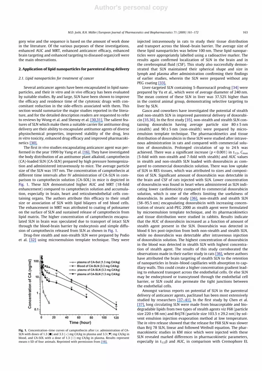
The first in vivo studies encapsulating anticancer agent was per-formed in the year 1999 by Yang et al. [16]. They have investigatedthe body distribution of an antitumor plant alkaloid, camptothecin(CA)-loaded SLN (CA-SLN) prepared by high pressure homogeniza-tion and administered intravenously to mice. The average particlesize of the SLN was 197 nm. The concentration of camptothecin atdifferent time intervals after IV administration of CA-SLN in com-parison to camptothecin solution (CA-SOL) in mice is depicted inFig. 1. These SLN demonstrated higher AUC and MRT (18-foldenhancement) compared to camptothecin solution and accumula-tion, especially in brain, heart and reticuloendothelial cells con-taining organs. The authors attribute this efficacy to their smallsize or association of SLN with lipid bilayers of red blood cells.The enhancement in MRT was attributed to coating of poloxameron the surface of SLN and sustained release of camptothecin fromlipid matrix. The higher concentration of camptothecin encapsu-lated SLN in brain was speculated due to transport of intact SLNthrough the blood-brain barrier by endocytosis and simple diffu-sion of camptothecin released from SLN as shown in Fig. 1.
Drug-free stealth and non-stealth SLN were prepared by Podioet al. [32] using microemulsion template technique. They were
injected intravenously in rats to study their tissue distributionand transport across the blood–brain barrier. The average size ofthese lipid nanoparticles was below 100 nm. These lipid nanopar-ticles were appropriately labelled using a radioactive marker. Theresults again confirmed localization of SLN in the brain and inthe cerebrospinal fluid (CSF). This study also successfully demon-strated that SLN maintained their spherical shape and size inlymph and plasma after administration confirming their findingsof earlier studies, wherein the SLN were prepared without anyPEG coating [33].
Liver-targeted SLN containing 5-fluorouracil prodrug [34] wereprepared by Yu et al., which were of average diameter of 240 nm.The mean content of these SLN in liver was 37.52% higher thanin the control animal group, demonstrating selective targeting toliver by SLN.
Gasco and coworkers have investigated the potential of stealthand non-stealth SLN in improved parenteral delivery of doxorubi-cin [35,36]. In the first study [35], non-stealth and stealth SLN con-taining doxorubicin having average particle size 80 ± 5 nm(stealth) and 90 ± 5 nm (non-stealth) were prepared by micro-emulsion template technique. The pharmacokinetics and tissuedistribution of doxorubicin in these SLN were studied after intrave-nous administration in rats and compared with commercial solu-tion of doxorubicin. Prolonged circulation of up to 24 h wasachieved. There was a significant increase (p < 0.001) in t1/2, Cmax
(5-fold with non-stealth and 7-fold with stealth) and AUC valuesin stealth and non-stealth SLN loaded with doxorubicin as com-pared to commercial doxorubicin solution. There was low uptakeof SLN in RES tissues, which was attributed to sizes and composi-tion of SLN. Significant amount of doxorubicin was detectable inthe brain and CSF of rats injected with SLN. Lower concentrationof doxorubicin was found in heart when administered as SLN indi-cating lower cardiotoxicity compared to commercial doxorubicinsolution, which is one of the effects limiting the usefulness ofdoxorubicin. In another study [36], non-stealth and stealth SLN(58–95.5 nm) encapsulating doxorubicin with increasing concen-tration of stearic acid-PEG 2000 as stealth agent were formulatedby microemulsion template technique, and its pharmacokineticsand tissue distribution were studied in rabbits. Results indicatethat the AUC of doxorubicin increased as a function of amount ofstealth agent present in the SLN. Doxorubicin was detected inblood 6 hrs post-injection from both non-stealth and stealth SLN,while no doxorubicin was detectable after intravenous injectionof doxorubicin solution. The highest concentration of doxorubicinin the blood was detected in stealth SLN with highest concentra-tion of stealth agent. The results of this study corroborated theobservations made in their earlier study in rats [36], where authorshave attributed the brain targeting of stealth SLN to the retentionof nanoparticles in brain–blood capillaries with absorption to cap-illary walls. This could create a higher concentration gradient lead-ing to enhanced transport across the endothelial cells. Or else SLNmay be endocytosed or transcytosed through the endothelial cellbarrier, or SLN could also permeate the tight junctions betweenthe endothelial cells.
Amongst various reports on potential of SLN in the parenteraldelivery of anticancer agents, paclitaxel has been most extensivelystudied by researchers [37–41]. In the first study by Chen et al.[37], long circulating SLN were made from bioacceptable and bio-degradable lipids from two types of stealth agents viz F68 (particlesize 220 ± 98 nm) and Brij78 (particle size 103.5 ± 29.2 nm) by sol-vent emulsion injection evaporation method at low temperature.The in vitro release showed that the release for F68 SLN was slowerthan Brij 78 SLN, linear and followed Weibull equation. The phar-macokinetic studies in KM mice which were injected with theseSLN revealed marked differences in pharmacokinetic parameters,especially in t1/2b and AUC, in comparison with Cremophore EL
Fig. 1. Concentration–time curves of camptothecin after i.v. administration of CA-SLN with doses of 1.3 (j) and 3.3 (�) mg CA/kg in plasma and 3.3 (.) mg CA/kg inblood, and CA-SOL with a dose of 1.3 (s) mg CA/kg in plasma. Results representmeans ± SD of four animals. Reprinted with permission from [16].
M.D. Joshi, R.H. Müller / European Journal of Pharmaceutics and Biopharmaceutics 71 (2009) 161–172 163

Author's personal copy
containing solution of paclitaxel. F68 SLN and Brij 78-SLN werefound to be long circulating with 7.4-fold and 3.6-fold highert1/2b, respectively. This was attributed to the reduced clearancerate and hence to the reduced uptake of SLN by mononuclear phag-ocytic system. Of the two stealth agents, SLN prepared with F68were longer circulating than Brij 78 which was attributed to thelonger PEG chain length.
In the studies by Koziara et al. [38,39], paclitaxel lipid nanopar-ticles having a particle size less than 100 nm were prepared bymicroemulsion template technique. In the first study [38], the po-tential of these SLN in the treatment of brain tumors was evaluatedin vitro. They have tested the cytotoxicity of paclitaxel entrapped innovel cetyl alcohol/polysorbate SLN on human glioblastoma celllines viz U-118 and HCT-15. The brain uptake of these SLN wasevaluated using an in situ rat brain perfusion model. The resultsindicate that SLN increase the uptake in the brain and its cytotox-icity towards p-glycoprotein expressing tumor cells. In the otherstudy by the same group [39], paclitaxel SLN prepared using emul-sifying wax were tested in vivo in a HCT-15 mouse xenograft mod-el. These SLN were demonstrated to overcome drug resistance inhuman colon adenocarcinoma cell line (HCT-15) and in vivo HCT-15 mouse xenograft model when injected intratumorally. In theresults of the endothelial cell differentiation assays significant
efficacy was achieved. The results of in vivo experiments are de-picted in Fig. 2A and B.
Sterically stabilized SLN [40] comprising of trymyristin and eggphosphotidylcholine and pegylated phospholipids as stabilizersand having average particle size around 200 nm were preparedusing high pressure homogenization. The important finding ofthe study was that in the in vitro release studies SLN showed a slowbut time-dependent release, and their in vitro cytotoxicities againsthuman ovarian and breast cancer cell lines as determined by MTTassay were comparable to those of a commercially available crem-ophor EL-based paclitaxel formulation.
SLN loaded with doxorubicin or paclitaxel were found to haveless cytotoxicity, and were found to be taken up by human promy-elocytic leukemia (HL60) and human breast carcinoma (MCF-7)cells [42]. Similar results have been reported with HT-29 colorectalcancer cells too [43]. In a very recent study by Zhang et al. [44], theactivity of nanostructured lipid carriers loaded with paclitaxel anddoxorubicin was tested for cytotoxicities and reversal of drug resis-tance against different cell lines. The reversal power of NLC-loadedpaclitaxel was 31 and for doxorubicin loaded NLC was 2.5 on test-ing it in a multi drug resistant cancer cell line (SKOV3-TR30).
Tamoxifen [45,46] is another anticancer agent that has beenstudied with injectable lipid nanoparticles owing to its applicationin breast cancer therapy. In a study by Fontana et al. [45], SLN wereproduced by two different production methods viz microemulsiontemplates and by precipitation technique with an average particlesize of 118 and 69 nm, respectively. The in vitro antitumoral activ-ity assay carried out on MCF-7 cell line (human breast cancer cells)suggests that tamoxifen SLN maintain an antitumoral activity com-parable to free drug. The prolonged release exhibited by tamoxifenSLN indicates their usefulness in breast cancer therapy. The secondstudy incorporated tamoxifen citrate in SLN [46], and studied thepharmacokinetic parameters of tamoxifen citrate-loaded SLN afterintravenous administration in rats. The t1/2 and mean residencetime of these SLN in plasma were approximately 3.5-fold and 3-fold higher, respectively, than the free tamoxifen revealing theirlong circulating nature.
Tripalmitin SLN loaded with etoposide have been studied forbiodistribution [47] and efficacy against Dalton’s lymphoma tu-mor-bearing mice [48]. The biodistribution studies suggested thatpositively charged SLN have a high blood concentration and pro-longed blood residence time and significantly lower uptake inreticuloendothelial system organs such as liver and spleen.
These positively charged SLN had 14-fold higher distribution inbone and brain than negatively charged SLN and etoposide 4 hours
Fig. 2. Mean tumor volume as a function of time and treatment. (A) In vivo efficacystudy #1 (study carried out 14 day after tumor implantation). Mice carrying HCT-15tumors received direct intratumoral injections of saline, E78 NPs (blank nanopar-ticles incorporated in cetyl alcohol and stabilized by polysorbate), Taxol or PX NPs(Paclitaxel SLN) beginning 14 days after cell implantation and every 3 days for 13days for a total of 5 injections. There were no significant differences in tumorvolume between all the groups, with the exception of saline on day 13 (*p < 0.05between saline and remaining groups; two-way ANOVA with repeated measures,Fisher’s LSD post-test). Data represent means ± SEM (n = 6–7). (B) In vivo efficacystudy #2. Mice were dosed via direct intratumoral injections over the course of 19days beginning 9 days after cell implantation. There was a significant inhibition oftumor growth in Taxol treated groups from controls (*p < 0.05). Additionally, therewas a significant difference between PX NPs and Taxol treatment on day 19(#p < 0.05). Data represent means ± SEM (n = 3–6). Reprinted with permission from[39].
Fig. 3. Tumor concentrations of 99mTc-etoposide and 99mTc-ETPL nanoparticlesafter subcutaneous injection in Dalton’s lymphoma tumor-bearing mice. Each valueis the mean (FSD) of three experiments. ET, etoposide; ETPL, etoposide loadedtripalmitin nanoparticles. Reprinted with permission from [48].
164 M.D. Joshi, R.H. Müller / European Journal of Pharmaceutics and Biopharmaceutics 71 (2009) 161–172

Author's personal copy
after injection. The observed phenomenon was explained to be dueto enhanced permeability and retention (EPR) effect. In the otherstudy by the same group [48], these SLN were injected by variousroutes of administration like subcutaneous, intravenous or intra-peritoneal, and their biodistribution and tumor uptake were deter-mined, Fig. 3. The tissue distribution was found to be different fordifferent routes of administration, and was found to be highest inthe order of intravenous, intraperitoneal and subcutaneous admin-istration. But the tumor uptake of etoposide SLN was 59-fold high-er after subcutaneous injection in comparison with intravenousadministration and 8-fold higher in comparison with intraperito-neal administration at 24-h post-injection.
A study [49] was carried out with local injection of mitoxan-trone SLN against breast cancer and its lymph node metastases.It revealed that the drug concentration using SLN as the carrierswas much higher in local lymph nodes, and the drug concentrationin other tissue was lower than that of mitoxantrone solution.Moreover, there was no observed toxicity to the main tissues afterlocal injection of SLN loaded with mitoxantrone compared tomitoxantrone solution. The percentage inhibition against breastcancer was 2-fold higher than that of mitoxantrone solution. Thelymph node size of the mice after treatment with mitoxantroneSLN was approximately three times lower compared to treatmentwith mitoxantrone solution.
Another study was carried out on all-trans retinoic acid [50] inorder to increase the chemical stability in powder form. The anti-proliferative effects of SLN powder formulation tested on a rangeof cancer cell line were not significantly different from that of freeall-trans retinoic acid. Moreover, its incorporation in SLN powderreduced the haemolytic potential.
An in vitro cellular uptake studies on vinorelbine bitartrate [51]encapsulated in PEG-modified SLN revealed that there was nophagocytosis of these SLN by RAW264.7 cells, but there was a sig-nificant improvement in the uptake by cancer cells (MCF-7 andA549) due to this PEG modification. Also the in vitro studies anti-cancer activity of vinorelbine bitartrate was found to be enhancedsignificantly after its incorporation in SLN and pegylated SLN.
SLN complexed with anionic polymer were formed to impartcharge or enhance encapsulation, especially of water soluble. Suchtypes of SLN have been used for encapsulation of drugs that havebeen studied for the delivery of cationic chemotherapeutic agentsand chemosensitizers [52]. These polymer-complexed SLN had aparticle size of around 180–300 nm. The efficacy of this systemagainst multidrug resistant (MDR) cancer cells was determined[53]. These SLN were 8 times more efficient in killing the MDR cellscompared to doxorubicin solution. The uptake and retention byMDR cells were both significantly enhanced by doxorubicin SLN.The authors have also evaluated the mechanism of overcomingMDR by these polymer lipid nanoparticles (PLN) [54]. Doxorubicinwas found to be physically associated with SLN and can bypass themembrane associated Pgp (the cause of MDR) when delivered asdoxorubicin PLN, the drug is better retained within Pgp-overexpressing cells than the free drug.
PEG-coated gadolinium SLN were prepared using microemul-sion templates [55], and were coated with folate. These SLN werefound not to aggregate platelets or activate neutrophils. Both un-coated PEGylated and folate-coated PEGylated SLN were found tohave enhanced cellular uptake and tumor retention. Higheramounts of folate-coated SLN, Fig. 4, were retained in the tumortissue in comparison to PEG-coated SLN. The study suggests thepotential of coated SLN in tumor-targeted delivery of gadoliniumand therefore the increased therapeutic efficacy of neutron capturetherapy.
Other literature examples of improving biodistribution and tar-geting effect of anticancer agents to brain following their adminis-tration as lipid nanoparticles are idarubicin [56], 40-O-
tetrahydropyranyl adramycin [57], 30,50-dioctanoyl-5-fluoro-20-deoxyuridine [58], oridonin [59,60], aclaciomycin [61], and tem-ozolomide [62].
The second generation of lipid nanoparticles viz nanostructuredlipid carriers have not been extensively studied as delivery systemsfor anticancer agents. In a study by Bondi et al. [63], NLC encapsu-lating two synthetic derivatives of antitumor drug temozolomideviz compound A and compound B were prepared. These NLC re-vealed an enhancement of the cytotoxic effects of compounds Aand B on human prostate cancer (PC-3) and human hepatocellularcarcinoma (HuH-6, HuH-7) cell lines with respect to free drug. Inanother study [64], encapsulating 9-nitrocamptothecin in NLCshowed that stealth 9-nitrocamptothecin had sustained releasecharacteristics and could resist the adsorption of plasma proteinsto a certain extent. In the tissue distribution studies 9-nitrocam-ptothecin was mainly found in the lung, liver, pancreas, ovariesand uterus, and the AUC of 9-nitrocamptothecin-loaded NLC washigher than that of the solution. Also these NLC were shown toeffectively target liver and lung.
An interesting type of SLN called as lipoprotein-mimicking bio-vectorized systems (LMBVs) was prepared for the delivery of meth-otrexate [65]. These LMBVs were prepared by microemulsioncongealing technique, and palmitoylpolyethylene glycol 400 wasanchored on LMBVs as apoprotein analogue. The pharmacokineticstudies carried out in rats suggested that the circulation half life ofmethotrexate was enhanced. Furthermore, methotrexate in LBMVwas found to reside in tissues for a longer period of time as com-pared to that of control. These observations were attributed tothe lipidic composition LMBVs and palmitoylpolyethylene glycol400 anchoring which mimicks natural lipoproteins.
2.2. Lipid nanoparticles for transfection
The utilization of lipid nanoparticles for transfection has beendocumented in the literature [66–75]. Cationic SLN have beenshown to be efficacious in transfecting COS-1 cells in vitro Olbrichet al. for the first time in the year 2001 [67]. These 100 nm SLNwere able to bind DNA to form a stable complex of 300–800 nmsize. The transfection efficacy as determined using COS-1 cells indi-cated that these cationic SLN complexes with DNA containing be-tween 10 and 200 weight equivalents of SII 13 matrix lipidefficiently transfected galactosidase expression plasmid pCMcBin the presence and absence of endoosmolytic agent chloroquineas depicted in Fig. 5.
Tabatt et al. [68] have compared cationic SLN with liposomesfor their transfection efficiency in vitro. The size of liposome was
Fig. 4. Retention of folate-coated (empty bars) and PEG-coated nanoparticles (filledbar) after intratumor injection into KB tumor tissue developed in athymic mice.After 8 and 24 h, the mice were sacrificed and the amount of Gd NPs in the tumortissue measured by a gamma counter. Each value represents means ± SD (n = 6–7mice). (*Tumor retention of folate-coated nanoparticles was statistically higherthan PEG-coated nanoparticles; p < 0.02; t-test.) Reprinted with permission from[55].
M.D. Joshi, R.H. Müller / European Journal of Pharmaceutics and Biopharmaceutics 71 (2009) 161–172 165

Author's personal copy
found to be around 84 nm whereas that of the SLN was 148 nm. Inthe DNA binding efficacy and transfection studies both of themwere found to be comparable. Also the same authors [69] havetried to optimize the formulation parameter of SLN viz the cationicdetergent used and the lipid matrix used to enhance the transfec-tion efficacy of SLN. The combination of acetylpalmitate as the lipidmatrix and N-[1-(2,3-dioleoyloxy)propyl]-N,N,N-trimethylammo-nium chloride as the cationic detergent resulted in highest trans-fection least toxic. The authors also found out that two-tailedcationic detergents are less toxic than one tailed ones.
The Mumper and coworkers have also investigated SLN for theirtransfection efficiency [70,71]. In one of the studies, emulsifyingwax-based nanoparticles stabilized by cationic detergent cetyltri-methylammonium bromide (CTAB) were prepared from micro-emulsion templates, and they were coated with plasmid DNA onthe surface. These SLN were also coated with mannan as a ligandto target dendritic cells. The ability of these SLN to increase the im-mune response was checked both in vitro by transfection efficacystudies and in vivo by measuring the immunization response aftersubcutaneous injection in mice. SLN resulted in 300% increase incytokine production in vitro and 16-fold higher IgG titre and T-helper cells in comparison with the naked DNA. In another studyby Cui et al. [71], two types of cationic surfactants viz DOTAPand DDAB were used to complex the plasmid DNA and encapsu-lated in the SLN prepared from emulsifying wax using microemul-sion templates. They were also coated with pullulan, a hepatocytetargeting ligand. The in vitro transfection studies carried out in HepG2 cells showed enhanced luciferase expression. Moreover, thein vivo studies carried out in mice demonstrated 40% transfectionefficiency in case of SLN, which was significantly higher than thatof naked DNA (16%).
As an approach for effective treatment of nasopharyngeal andprostate cancer, suicide gene therapy by local injection using fo-late-linked lipid nanoparticles has been used. This approach caneffectively deliver genes extensively to FR-negative LNCaP andPC-3 as well as FR-positive KB and HeLa cells. This has been sum-marized in a review by Hattori et al. [72]. Specific surface receptortargeted DNA-SLN, which are stable under physiological conditionsand low in cytotoxicity, and are capable of binding biotinylated li-gands and interacting with surface receptors, have been investi-gated [75]. In their original work, [73] Bondi et al. [74]investigated cationic SLN as a non-viral transfection agent for genedelivery. These cationic SLN formed stable complexes with DNAand were found to protect DNA against DNAase I ligation. They
were found to have very low toxicity and were found to promotetransfection of liver cancer cells.
2.3. Lipid nanoparticles for liver targeting
Particulate carriers (including SLN) usually accumulate in the li-ver by passive targeting on parenteral administration. However,passive targeting leads to entrapment of the drug in the Kupffercells and not in the hepatocytes which is the major target for thetreatment of hepatic diseases such as cancers. Hence, for liver tar-geting, SLN containing galactosylated or mannosylated lipids areemployed. To date, there are very few studies which have system-atically explored the liver targeting of SLN. Shen et al. [76] fabri-cated blank monostearin SLN with or without PEG 2000modification and the pharmacokinetics of these radiolabelled SLNwas studied in rats. The PEG-coated SLN showed 2.2 times longercirculation than that of unmodified SLN. However, unmodifiedSLN showed significantly higher accumulation in liver as comparedto that of PEG-modified SLN. Oxymatrine, a hepatoprotectiveagent, was incorporated in SLN and its liver-targeting efficacywas determined in rats [77]. The mean content of oxymatrine in li-ver at 30 min was 12 times higher in SLN group than that of oxy-matrine solution group. The relative-targeting efficiency to theliver tissues compared to solution was 360%. This targeting wasachieved without any coating on SLN, and it could be attributedto the general clearance of nanoparticles by the phagocytic cells.Similarly, liver targeting of 5-fluorouracil prodrug has also beendemonstrated by Yu et al. [34]. The 5-fluorouracil content in liverwas 37.52% higher in case of SLN as compared to that of solution.
The utility of galactosylated excipients in the liver targeting hasbeen investigated recently for an anticancer drug, taspine [78]. TheSLN with and without galactosylation were evaluated for in vitroactivity and in vivo biodistribution as compared to that of free drug.Both the SLN showed higher liver targeting as compared to that offree drug, and galactosylated SLN showed further enhancement inliver targeting.
2.4. Lipid nanoparticles for imaging
Magnetite was the first imaging agent which was incorporatedin SLN [79]. The cytotoxicity of this magnetite-loaded SLN wascompared with that of the magnetite-loaded polylactide/glycolide(PLA/GA) particles to determine toxicological acceptance as intra-venous formulation for magnetic resonance imaging and as poten-tial carrier for drug targeting. The magnetite-loaded SLN werefound to be least cytotoxic with effective concentration (ED50%)above 10%, whereas that of the polymeric nanoparticles werefound to be in the range of 0.15–0.38%. However, no in vivo studieshave been carried out to validate in vitro observations.
First in vivo study incorporating iron oxide in SLN [80] was car-ried out in rats by Peira et al. Iron oxide SLN demonstrated similarrelaxometric properties as that of Endorem�. In vivo magnetic res-onance Imaging (MRI) of the central nervous system with both SLNand Endorem� showed that supermagnetic SLN have slower bloodclearance than Endorem�. The retention in the CNS was found tobe till the end of experiment that lasted for 135 min. These obser-vations also boosted other findings like brain targeting by SLN.Intravenously injected 125I radiolabelled SLN were found to remainin the blood for higher time in comparison to other colloidal carri-ers [81]. Luminescent lipophilic CdSe/ZnS core shell quantum dots(QDs) were encapsulated into SLN to prepare fluorescent nanocom-posite particles [82]. The properties of QDs viz high fluorescenceand narrow and symmetric emission spectra were retained evenafter encapsulation. Also due to encapsulation of several QDs intosingle nanoparticle structure, the fluorescence signal and the signalto background ratio were found to be enhanced. These QD-loaded
Fig. 5. Transfection efficacy of SLN (batch SII-13). Semiconfluent COS-1 cells wereincubated for 4 h with transfection complexes in the absence and presence ofchloroquine and analyzed after 48 h (n = 6). Reprinted with permission from [69]pLL, poly L-lysine; pEI, polyethylenimine (pEI); DMEM, Dulbecco’s modified eagle’smedium; CQ, chloroquine; LCPS, luminescence light counts per second; Batch SII-13was composed of Compritol 4%, Tween 80/Span 85 (7:3) 4% and, N,N-di-(b-stearoylethyl)-N,N-dimethylammonium chloride (EQ1) 1%.
166 M.D. Joshi, R.H. Müller / European Journal of Pharmaceutics and Biopharmaceutics 71 (2009) 161–172
Author's personal copy
SLN were found to be stable, and the velocity of photobleachingwas found to be reduced. The QDs were found to be biocompatible,and therefore have great potential in biological imaging.
2.5. Lipid nanoparticles for targeting the central nervous system
Various drugs ranging from antipsychotics, anti-parkinson,antieschemic to antibiotics have been encapsulated in lipid nano-particles with the aim to either modify the biodistribution or forbrain targeting [83,84]. Recently, a comprehensive review [85] cov-ering various aspects of brain targeting using SLN has been pub-lished. Gasco and coworkers in a series of experiments evaluatedbiodistribution of radiolabelled non-stealth and stealth SLN afterintravenous injection [32,33]. It was observed that stealth SLN cir-culated in plasma for a longer time. Furthermore, significantly high-er concentrations of stealth SLN were observed in brain andcerebrospinal fluid. In another investigation, Gasco and coworkersevaluated biodistribution of tobramycin SLN after intravenousand intraduodenal administration [87]. Tobramycin-loaded SLNwere found to cross the blood–brain barrier after intravenousadministration which was significantly higher than intraduodenaladministration.
Clozapine, a lipophilic antipsychotic drug, was encapsulated invarious types of SLN, and its pharmacokinetic and biodistributionwere studied on intravenous and intraduodenal administration[86]. Sterylamine-containing clozapine SLN were found to give sig-nificantly higher plasma levels and AUC as compared to clozapineSLN without sterylamine and clozapine suspension. Furthermore,biodistribution studies indicated that sterylamine containing cloza-pine SLN result in significantly higher amount of clozapine in brain(AUC) as compared to that of clozapine SLN without sterylamineand clozapine suspension. Furthermore, the mean residence timeof these SLN was also higher than that of the other formulations.
Bromocriptine, an anti-parkinson agent, was encapsulated inNLC, and the in vivo efficacy of these formulations was tested.The in vivo efficacy of these lipidic nanoparticles was studied in6-hydroxydopamine hemilesioned rats by giving intraperitonealinjection [88], which are models for Parkinson’s disease. The re-sults indicated that the anti-parkinsonian bromocriptin NLC haverapid onset of action as compared to that of solution. Furthermore,the anti-parkinson effect was retained for longer duration in caseof NLC as compared to that of solution. This study indirectly dem-onstrated that higher brain levels of bromocriptine were achievedafter administration as NLC. Suitable pharmacokinetic studieswould be required to validate this observation. Cloricromene[89], which is used to reduce brain lipid peroxidation and the for-mation of post-ischemic brain edema, was encapsulated in SLN,and the in vitro release studies were carried out using human plas-ma. About 70% of drug was released within 15 min and immedi-ately degraded by esterase to acid form, whereas after 4 h 30% ofthe drug remained entrapped in nanoparticles. Therefore, accord-ing to the authors SLN could be very useful for CNS targeting byintravenous administration. However, the speculations would havebeen more supportive if provided with in vivo data.
In a study by Chen et al. [90], dexamethasone-loaded SLN wereadministered by intratympanic (via inner ear) and intravenousroutes. The AUC of dexamethasone acetate following intratym-panic SLN administration was 13 times higher than the dexameth-asone solution. Moreover, the AUC of dexamethasone in perilymphfollowing intratympanic SLN administration was 76% lower com-pared to dexamethasone solution given by the same route. Allthese studies indicate great potential of lipid nanoparticles to-wards targeting the central nervous system and hence the brain.
Lockman et al. fabricated emulsifying wax and Brij 78 contain-ing SLN. These SLN were further coated on surface with thiamine,and the brain uptake of the SLN with and without thiamine was
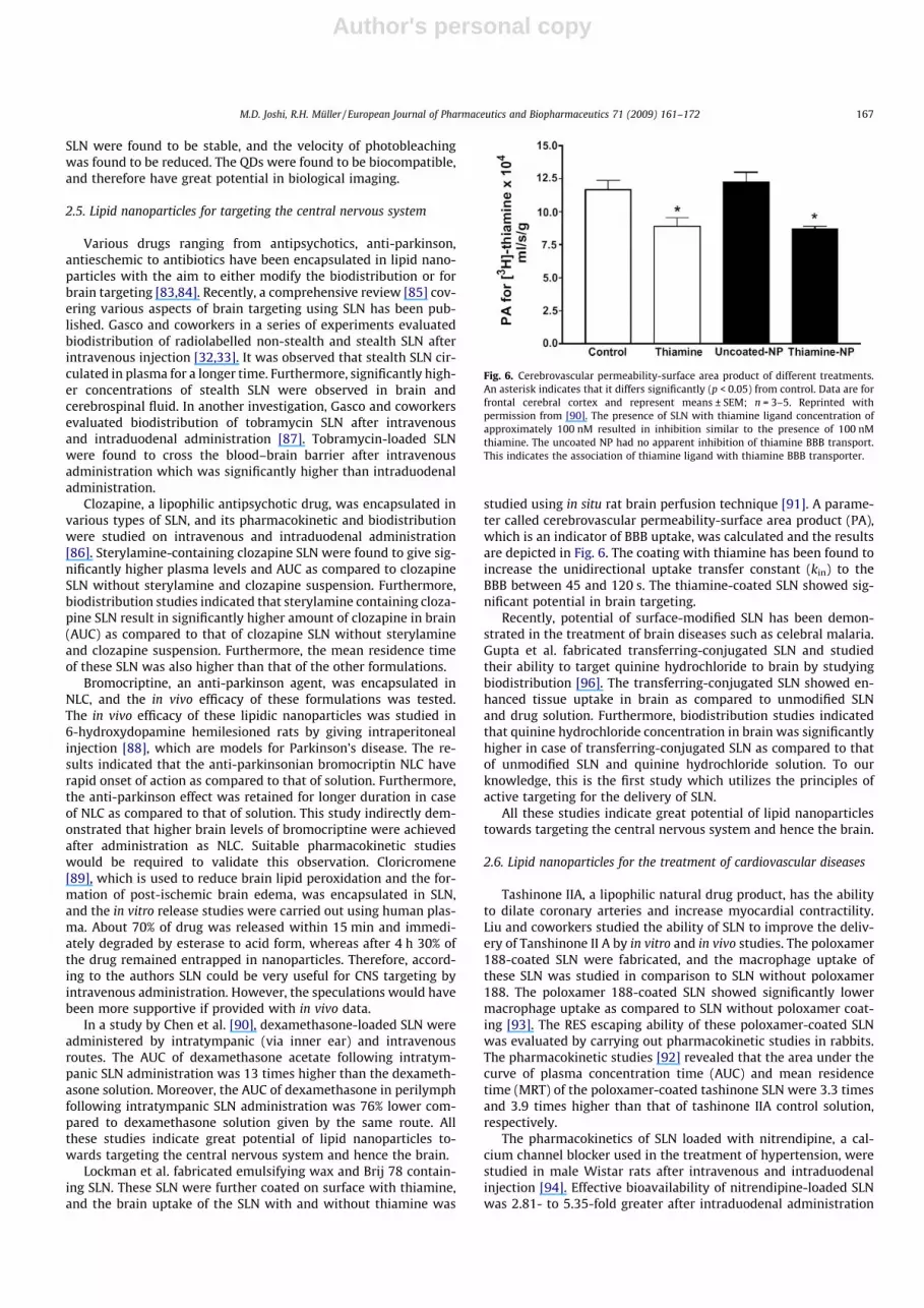
studied using in situ rat brain perfusion technique [91]. A parame-ter called cerebrovascular permeability-surface area product (PA),which is an indicator of BBB uptake, was calculated and the resultsare depicted in Fig. 6. The coating with thiamine has been found toincrease the unidirectional uptake transfer constant (kin) to theBBB between 45 and 120 s. The thiamine-coated SLN showed sig-nificant potential in brain targeting.
Recently, potential of surface-modified SLN has been demon-strated in the treatment of brain diseases such as celebral malaria.Gupta et al. fabricated transferring-conjugated SLN and studiedtheir ability to target quinine hydrochloride to brain by studyingbiodistribution [96]. The transferring-conjugated SLN showed en-hanced tissue uptake in brain as compared to unmodified SLNand drug solution. Furthermore, biodistribution studies indicatedthat quinine hydrochloride concentration in brain was significantlyhigher in case of transferring-conjugated SLN as compared to thatof unmodified SLN and quinine hydrochloride solution. To ourknowledge, this is the first study which utilizes the principles ofactive targeting for the delivery of SLN.
All these studies indicate great potential of lipid nanoparticlestowards targeting the central nervous system and hence the brain.
2.6. Lipid nanoparticles for the treatment of cardiovascular diseases
Tashinone IIA, a lipophilic natural drug product, has the abilityto dilate coronary arteries and increase myocardial contractility.Liu and coworkers studied the ability of SLN to improve the deliv-ery of Tanshinone II A by in vitro and in vivo studies. The poloxamer188-coated SLN were fabricated, and the macrophage uptake ofthese SLN was studied in comparison to SLN without poloxamer188. The poloxamer 188-coated SLN showed significantly lowermacrophage uptake as compared to SLN without poloxamer coat-ing [93]. The RES escaping ability of these poloxamer-coated SLNwas evaluated by carrying out pharmacokinetic studies in rabbits.The pharmacokinetic studies [92] revealed that the area under thecurve of plasma concentration time (AUC) and mean residencetime (MRT) of the poloxamer-coated tashinone SLN were 3.3 timesand 3.9 times higher than that of tashinone IIA control solution,respectively.
The pharmacokinetics of SLN loaded with nitrendipine, a cal-cium channel blocker used in the treatment of hypertension, werestudied in male Wistar rats after intravenous and intraduodenalinjection [94]. Effective bioavailability of nitrendipine-loaded SLNwas 2.81- to 5.35-fold greater after intraduodenal administration
Fig. 6. Cerebrovascular permeability-surface area product of different treatments.An asterisk indicates that it differs significantly (p < 0.05) from control. Data are forfrontal cerebral cortex and represent means ± SEM; n = 3–5. Reprinted withpermission from [90]. The presence of SLN with thiamine ligand concentration ofapproximately 100 nM resulted in inhibition similar to the presence of 100 nMthiamine. The uncoated NP had no apparent inhibition of thiamine BBB transport.This indicates the association of thiamine ligand with thiamine BBB transporter.
M.D. Joshi, R.H. Müller / European Journal of Pharmaceutics and Biopharmaceutics 71 (2009) 161–172 167

Author's personal copy
in comparison with that of nitrendipine suspension. In tested or-gans, the AUC and MRT of nitrendipine-loaded SLN were higherthan those of nitrendipine suspension, especially in brain, heartand reticuloendothelial cells containing organs.
2.7. Lipid nanoparticles for treatment of parasitic diseases
Antiparasitic agents represent a class of drugs which had beenneglected as a model for drug delivery systems for a long time.As compared to other therapeutic agents, relatively fewer reportsare published on the delivery of antiparasitic agents. Recently, acomprehensive review dealing with the various drug delivery ap-proaches for the treatment of parasitic diseases has been published[95]. There are few investigations which state the potential of lipidnanoparticles in the delivery of antiparasitic agents.
Transferring-conjugated SLN of quinine dihydrochloride [96],an antimalarial drug, were prepared to target it to the brain forthe management of cerebral malaria. Enhanced uptake in brain tis-sues of transferring coupled SLN was observed in fluorescencestudies. Also intravenous administration with these SLN resultedin much higher concentrations of drug in serum. Thus, this studyagain confirmed the utility of SLN in brain targeting. Another typeof lipid nanoparticles which have been studied for the delivery of ahydrophilic antitrypanosimiatic drug diminazene [97] are the li-pid–drug conjugates (LDC) nanoparticles. Here the hydrophilicdrug, diminazene, was made lipophilic by conjugating it with stea-ric acid and oleic acid. From the lipophilic conjugate nanoparticlescan be made by melting the conjugate (=lipid particle matrix mate-rial) and processing it identical to SLN by high pressure homogeni-zation after pre dispersion in hot surfactant solution [25]. Thesenanoparticles were also shown to adsorb apolipoprotein E [98]after incubation with human serum, which is a key lipoproteinresponsible for the delivery of nanoparticles to brain where theparasites Trypanosoma brucei gambiense and rhodiense reside. Re-cently, Joshi et al. [99] have explored the potential of NLC for intra-venous delivery of an antimalarial drug, artemether. These NLCwere found to be less haemolytic, and had significantly higher anti-malarial activity compared to marketed intramuscular oily injec-tion with significantly higher survival rate of 60% after 31 days ofexperiment.
2.8. Lipid Nanoparticles for treatment of rheumatoid arthritis
Actarit SLN [100] were prepared with the aim of passive target-ing. These SLN were shown to enhance the therapeutic efficacywith concomitant reduction in the various adverse effects suchas nephrotoxicity and gastrointestinal disorders. The pharmacoki-netic studies in New Zealand rabbits revealed 10-fold higher meanretention time, 1.88 times higher area under curve (AUC) andaround 3-fold increase in targeting efficiency with actarit-loadedSLN as compared to that of actarit solution upon intravenousinjection.
2.9. Lipid nanoparticles for treatment of other diseases
Cholesteryl butyrate SLN [101] as a prodrug carrier for butyricacid have been prepared as an alternative to sodium butyrate inanti-inflammatory therapy of ulcerative colitis. The efficacy of cho-lesteryl butyrate SLN in inhibiting the adhesion of human neutro-phils to endothelial cells has been studied in comparison with thatof sodium butyrate. In all tests cholesteryl butyrate SLN were moreactive than sodium butyrate. Also cholesteryl butyrate SLN inhib-ited O2
� production and myeloperoxidase release by polymorpho-nuclear cells in a dose and time-dependent manner. They were alsofound to be more active than sodium butyrate. From these obser-vations, the authors concluded that cholesteryl butyrate-loaded
SLN might be a better choice over sodium butyrate in the anti-inflammatory therapy of ulcerative colitis, which can avoid thecomplications related to sodium butyrate administration.
Glyceryl behenate lipid nanoparticles were injected endotrach-eally, and their lymphatic uptake was studied by radiolabelling[102]. It was observed that these nanoparticles are rapidly elimi-nated from rat lungs, and accumulation in para-aortic, axillaryand inguinal lymph nodes starts almost immediately after admin-istration. The translocation of nanoparticles across the lung muco-sa and their uptake into the lymphatics have indicated their utilityas drug carriers for lung cancer therapy and immunization process.
Dexamethasone acetate-loaded SLN [103] were administeredby intravenous route of administration and were studied for bio-distribution in mice. Post-injection the maximum level of dexa-methasone was reached in 0.5 h. A 17.8-fold higher AUC wasachieved using dexamethasone-loaded SLN as compared to dexa-methasone solution. 30-Azido-30-deoxythymidine palmitate, anantiHIV prodrug, was encapsulated in PEG-modified SLN [104],and its biodistribution was studied by intravenous injection toCD-1 mice. The results suggest that there was an increase in bio-availability by encapsulation of 30-azido-30-deoxythymidine palmi-tate in SLN. SLN containing HIV protease inhibitor, atazanavir[105], was prepared, and its potential to deliver the brain micro-vessel endothelial cells was checked in vitro using hCMEC/D3 cellline. These SLN led to a significant accumulation in the brain endo-thelial cell as compared to the aqueous solution of drug.
3. Lipid nanoparticles as adjuvants
Lipid nanoparticles have been used as adjuvants for proteinantigens and DNA for immunization. Olbrich et al. [106] have dem-onstrated the adjuvant activity of lipid nanoparticles using myco-plasma bovis antigen in sheep. Moderate activity was found interms of antibody titre in comparison to Freud’s incomplete adju-vant (FIA), which could be improved by combination with EQ1(N,N-di-(b-stearoylethyl)-N,N-dimethyl-ammonium chloride). Asagainst the FIA, the SLN-based antigen formulations were well tol-erated. Further, our research group has also shown [107] that par-affin or biodegradable glycerides-based SLN containingmycoplasma bovis antigen and immunoglobulin G (IgG) would bea promising alternative to Freud’s complete adjuvant (FCA). Thiswas the first study which showed that the adjuvant activity ofSLN is dependent on their particle size. Particles greater than100 nm exhibited higher adjuvant activity as compared to the par-ticles lower than 100 nm.
In another study by Cui and Mumper [108,109], a catonizedmodel protein antigen was coated on anionic lipid nanoparticles.The cationized protein-coated nanoparticles yielded strongestand most reproducible antibody titre upon subcutenous injectionto mice in comparison to cationized antigen alone or non-cation-ized antigen administered together with alum as an adjuvant.The lipid nanoparticles were found to enhance both T-helper type1 and type 2 immune responses. Cui and Mumper [110] have alsostudied the effect of co-administration of adjuvants with lipidnanoparticles-based genetic vaccine delivery system on the im-mune response. They found significant enhancement in immuniza-tion over naked plasmid DNA, e.g., 300-fold for cholera toxin(100 lg) and 250-fold for lipid A (50 lg) by subcutaneous route.
4. In vivo fate of lipid nanoparticles: protein adsorption patterns
It is noteworthy that the in vivo fate of drug on parenteraladministration is no longer determined by the properties of thedrug but by the type of the drug delivery system used, which inthis case are the lipid nanoparticles [111]. The mononuclearphagocyte system (MPS) plays a vital role in clearing the nanopar-
168 M.D. Joshi, R.H. Müller / European Journal of Pharmaceutics and Biopharmaceutics 71 (2009) 161–172

Author's personal copy
ticles from blood circulation. It has been shown by our researchgroup [112–116] that the type and pattern of protein adsorptionon lipid nanoparticles determines the organ distribution of lipidnanoparticles. Hence, it can be assumed that the targeting of lipidnanoparticles is dependent on their physiochemical properties.After intravenous injection, a protein gets adsorbed on the lipidnanoparticles surface depending upon their surface properties.The adsorbed protein leads to the adherence of these nanoparticlesto cells with appropriate receptor on the surface. For example, par-ticles having adsorbed proteins with opsonic function, e.g., immu-noglobulin IgG, complement factor C4c are cleared by MPS cells,whereas the absence of opsonins in the adsorption pattern andthe presence of dysopsonins (e.g. Albumin, IgA) lead to the circula-tion of particles in the bloodstream. Coating of nanoparticles withapolipoprotein E leads to their preferential targeting to the brain asapolipoprotein E plays an important role in the transport of lipo-protein into the brain via the low density lipoprotein (LDL) recep-tor. Hence, it is likely that apolipoprotein E adsorbing drug carriersmimic lipoprotein particles, leading to brain uptake by endocyticprocesses.
A two-dimensional polyacrylamide gel electrophoresis (2-DE)has been proven to be an effective tool for detecting all proteins ad-sorbed onto a nanoparticulate carrier. The technology named Path-finder� [117,96] has been developed by our research group usingthe principle of differential protein adsorption. The Pathfinder�
technology identifies the naturally occurring mechanism for local-ization of material in different parts of the body via adsorbed bloodproteins. Controlled production of carriers with appropriate surfaceproperties leads in vivo to preferential adsorption of the targetingprotein and subsequently to site specific intravenous delivery.
Surface-modified particles which were able to deliver drugs tothe brain have been reported by the Kreuter group [118–122]. Itwas confirmed by 2D-PAGE that the uptake of nanoparticles tothe brain endothelial cells was mediated by apolipoprotein E. Infurther experiments, the negative control of this experiment wasprecoated with apolipoprotein E on the surface and it was foundto target the brain, thus providing the proof of the principle. ThePathfinder technology was successfully implicated to target thebrain using nanocrystals [123], lipid drug conjugate nanoparticles[124], but its utility to target blood–brain barrier (BBB) usingNLC remains to be elucidated. The efficiency of apolipoprotein Ecoated LDC to selectively target brain as determined by fluores-cence microscopy is depicted in Fig. 7.
To target nanoparticles to the brain, one can either modify thesurface of nanoparticles so that they can selectively adsorb the pro-tein of interest to target it to the organ of interest or the protein it-self can be preferentially adsorbed on the particles prior toinjection, alternatively the receptor binding moiety, e.g. class Gamphiphilic helixes for apolipoprotein E, can be coupled covalentlyto the particle surface.
In a study by Göppert et al. [116], the influence of different sur-factants on the in vitro adsorption of human plasma proteins wasinvestigated using 2-DE. There was a correlation with differentamounts of adsorbed apolipoprotein E and the hydrophilic lipo-philic balance (HLB) of the different polysorbates used in the prep-aration of SLN.
5. Toxicity of lipid nanoparticles
For the successful regulatory clearance of SLN for parenteraldelivery, it is essential to establish their biocompatibility withblood components and other tissues. Hence, to study the toxicityof lipid nanoparticles various in vitro and in vivo efforts would bediscussed herein. The interaction of SLN and their respective cyto-toxicities was studied with human granulocytes [125], HL60 cellline [126], RAW 264.7 macrophage [127] and murine peritonealmacrophages [128]. The experiments with the interaction of SLNwith human granulocytes [125] revealed that SLN had distinctlylower uptake by phagocytosis resulting in prolonged circulationtime in blood. Also the study pointed out the�10-fold low cytotox-icity of glyceride SLN in comparison with that of polylactide/glyco-lide nanoparticles. Thus, SLN can be used as intravenous carriersbecause of their prolonged circulation time and high toxicologicalacceptance. The interaction of SLN with HL60 cells [126] gave sim-ilar results showing lower cytotoxicity compared to polymericnanoparticles, whereas the interaction of Dynasan 114 SLN onRAW 264.7 macrophages [127] has revealed that those SLN stabi-lized with pharmaceutically acceptable surfactants like poloxamer188, Lipoid S75, sodium cholate, Tween 80 are very well toleratedby RAW 264.7 macrophages. The tolerance was judged in terms ofcytokine production, which was reduced and stimulation, ex-pressed in elevated cytokine levels, could not be found. Similar re-sults were obtained earlier when Dynasan 114 SLN were interactedwith peritoneal mouse macrophages [129–131]. In a study bySchöler et al. [132], the behaviour of murine macrophages in thepresence of different concentration and size and various concen-
Fig. 7. Confocal laser scanning micrograph of mouse brain tissue, upper: control,lower: after i.v. injection of Nile Red-labelled LDC nanoparticles. Arrows indicatethe nanoparticles adhering to the endothelial cells of the brain vessels and thediffusion of the dye into the brain tissue. Reprinted with permission from [121].
M.D. Joshi, R.H. Müller / European Journal of Pharmaceutics and Biopharmaceutics 71 (2009) 161–172 169

Author's personal copy
trations of SLN was studied. SLN made from lipids consisting ofstearic acid or dimethyl-dioctadecylammonium bromide werefound to be cytotoxic at the concentration of 0.01%. On the otherhand, SLN made from lipids consisting of triglycerides, cetylpalm-itate or paraffin were found to be safe at the same concentration.Authors speculate that decrease in IL-6 production based on theconcentration of the lipid could be the reason for such kind of cyto-toxicity. However, the cytotoxicity was independent of the size ofSLN. In an in vivo study by Weyhers et al. [128], a greater depth wasachieved in understanding the toxicity created by SLN in vivo whentwo types of SLN, one made from Compritol (GRAS approved) andthe other made from Cetyl palmitate (less physiologic), wereadministered six times within a period of 20 days via bolus injec-tion. The injected doses were extremely high, i.e., up to 1 g of solidlipid per kg of body weight. In humans, this would be compared tobolus injection of 75 g of solid particles intravenously. The resultsof histopathology suggested that the toxicity is dependent on thelipid matrix as well as the dose administered despite this highdose. No untoward results were obtained with cetyl palmitateSLN, while the high dose compritol-containing formulation led tothe accumulation of lipid in liver and spleen of mice and to subse-quent pathological alterations. However, these alterations werereversible within six weeks after intravenous administration,which was attributed to the slow degradation of compritol. Inin vitro degradation studies it could be shown that cetyl palmitatewas degraded much faster than compritol corroborating the resultsof in vivo study. By reducing the compritol dose to 0.25 g, still beinga high dose, all these observed alterations did not occur any more.In another study by Heydenreich et al. [133], cationic SLN contain-ing sterylamine and different triglycerides were prepared andpurified using different methods such as ultrafiltration, ultracentri-fugation and dialysis. As it is suspected that toxicity of SLN isdependent on the composition and the purification method used,their cellular toxicity and physical stability were compared. Thecell toxicity was found to be dependent on the composition ofSLN and the method of purification used. Dialysis was found tobe the most efficient to remove excess surfactant. Also, a correla-tion could be obtained on the basis of the amount of sterylamineto toxicity produced which was not observed in the case ofliposome.
6. Conclusion
The potential of lipid nanoparticles for the parenteral deliveryof various therapeutic agents has been successfully established.The successful fabrication of transferring-conjugated SLN hasopened a new era for active targeting using SLN, although exten-sive studies are required to be carried out. The parenteral accept-ability of the lipids and other surfactants is the major hurdle intheir successful commercialization. It is possible to use glyceridesof the fatty acids which are present in the lipid phase of parenteralfat emulsions (e.g., C14-C18 or C22 fatty acids) to fabricate SLNwith high biocompatibility [134,135]. However, extensive in vivostudies are required to establish their in vivo parenteral acceptabil-ity. The other aspects of SLN such as sterilization [136], freeze dry-ing [137,138], shelf life [139,140] and large-scale industrialproduction [139] have already been developed to a sufficient stan-dard with the view of commercialization. Thus, although there isno lipid nanoparticle-based injectable product in the market tilldate, with the progress which they have seen so far, the day forthe marketed arrival of SLN might not be too far.
References
[1] A. Wretlind, Development of fat emulsions, J. Parenter. Enteral. Nutr. 5 (1981)230–235.
[2] J. Schmidt, Parenterale Fettemulsionen als Arzneistofftrager, in: R.H. Müller,G.E. Hildebrand (Eds.), Pharmazeutische Technologie: ModerneArzneiformen, Lehrbuch fur Studierende der Pharmazie, WissenschaftlicheVerlagsgesellschaft, Stuttgart, 1998, pp. 189–194.
[3] L. Collins-Gold, N. Feichtinger, T. Wärnheim, Are lipid emulsions the drugdelivery solution?, Mod Drug Discov. 3 (2000) 44–48.
[4] E. Allemann, R. Gurny, E. Doelker, Drug-loaded nanoparticles – preparationmethods drug targeting issues, Eur. J. Pharm. Biopharm. 39 (1993) 173–191.
[5] H. Bunjes, B. Siekmann, Manufacture, characterization and application of solidlipid nanoparticles as drug delivery system, in: M. Deleers, Y. Pathak, D.Thassu (Eds.), Nanoparticulate Drug Delivery Systems, Informa Healthcare,2007, pp. 213–268.
[6] D.D. Lasic, Doxorubicin in sterically stabilized liposomes, Nature 380 (1996)561–562.
[7] P.S. Gill, B.M. Espina, F. Muggia, S. Cabriales, A. Tulpule, J.A. Esplin, H.A.Liebman, E. Forssen, M.E. Ross, A.M. Levine, Phase I/II clinical andpharmacokinetic evaluation of liposomal daunorubicin, J. Clin. Oncol. 13(1995) 996–1003.
[8] S.A. Wissing, O. Kayser, R.H. Müller, Solid lipid nanoparticles for parenteraldrug delivery, Adv. Drug Deliv. Rev. 56 (2004) 1257–1272.
[9] R.H. Müller, J.S. Lucks, Arzneistoffträger aus festen Lipidteilchen, FesteLipidnanosphären (SLN), Medication vehicles made of solid lipid particles(solid lipid nanospheres – SLN), European Patent No. 0605497, 1993(published as German patent).
[10] R.H. Müller, Colloidal Carriers for Controlled Drug Delivery and Targeting –Modification, Characterization and in vivo Distribution, CRC Press, BocaRaton, 1991.
[11] M.R. Gasco, Method for producing solid lipid microspheres having a narrowdistribution, US Patent No. 5,250,236, 1993.
[12] R.H. Müller, R.D. Petersen, A. Hommoss, J. Pardeike, Nanostructured lipidcarriers (NLC) in cosmetic dermal products, Adv. Drug Deliv. Rev. 59 (2007)522–530.
[13] M. Schäfer-Korting, W. Mehnert, H.C. Korting, Lipid nanoparticles forimproved topical application of drugs for skin diseases, Adv. Drug Deliv.Rev. 59 (2007) 427–443.
[14] E.B. Souto, A.J. Almeida, R.H. Müller, Lipid nanoparticles (SLN�, NLC�) forcutaneous drug delivery: structure, protection and skin effects, J. Biomed.Nanotech. 3 (2007) 317–331.
[15] V. Jenning, S.H. Gohla, Encapsulation of retinoids in solid lipid nanoparticles(SLN�), J. Microenviron. 18 (2001) 149–158.
[16] S.C. Yang, L.F. Lu, Y. Cai, J.B. Zhu, B.W. Liang, C.Z. Yang, Body distribution inmice of intravenously injected camptothecin solid lipid nanoparticles andtargeting effect on brain, J. Control. Release 59 (1999) 299–307.
[17] R. Cavalli, M.R. Gasco, P. Chetoni, S. Burgalassi, M.F. Saettone, Solid lipidnanoparticles (SLN) as ocular delivery system for tobramycin, Int. J. Pharm.238 (2002) 241–245.
[18] M.R. Gasco, G.P. Zara, M.F. Saettone, Pharmaceutical compositions suitablefor the treatment of ophthalmic diseases, PCT Int. Appl. WO 2004039351,2004.
[19] A.A. Attama, S. Reichl, C.C. Müller-Goymann, Diclofenac sodium delivery tothe eye: in vitro evaluation of novel solid lipid nanoparticle formulation usinghuman cornea construct, Int. J. Pharm. 355 (2008) 307–313.
[20] J. Liu, T. Gong, H. Fu, C. Wang, X. Wang, Q. Chen, Q. Zhang, Q. He, Z. Zhang,Solid lipid nanoparticles for pulmonary delivery of insulin, Int. J. Pharm. 356(2008) 333–344.
[21] P. Chattopadhyay, B.Y. Shekunov, D. Yim, D. Cipolla, B. Boyd, S. Farr,Production of solid lipid nanoparticle suspensions using supercritical fluidextraction of emulsions (SFEE) for pulmonary delivery using the AERx system,Adv. Drug Deliv. Rev. 6 (2007) 444–453.
[22] M.A. Videira, M.F. Botelho, A.C. Santos, L.F. Gouveia, J.J. Pedroso De Lima, A.J.Almeida, Lymphatic uptake of pulmonary delivered radiolabelled solid lipidnanoparticles, J. Drug Target. 8 (2002) 607–613.
[23] M. Sznitowska, M. Gajewska, S. Janicki, A. Radwanska, G. Lukowski,Bioavailability of diazepam from aqueous-organic solution, submicronemulsion and solid lipid nanoparticles after rectal administration in rabbits,Eur. J. Pharm. Biopharm. 52 (2001) 159–163.
[24] M. Sznitowska, S. Janicki, M. Gajewska, M. Kulik, Investigation of diazepamlipospheres based on Witepsol and lecithin intended for oral or rectaldelivery, Acta Pol. Pharm. – Drug Res. 57 (2000) 61–64.
[25] R.H. Müller, C. Olbrich, Drug-delivery vehicle for the controlledadministration of an active agent, produced from lipid matrix-medicamentconjugates, PCT Int. Appl. WO 2000067800, 2000.
[26] M.J. Robinson, A. Bondi Jr., J.V. Swintosky, Sulfamethylthiadiazole: humanblood concentration urinary excretion data following oral doses, J. Am.Pharm. Assoc. (Baltim.) 47 (1958) 874–888.
[27] C.R. Kowarski, B. Volberger, J. Versanno, A. Kowarski, A method of preparingsustained release sulfamethazine in small size batches, Am. J. Hosp. Pharm.21 (1964).
[28] P.P. Constantinides, M.V. Chaubal, R. Shorr, Advances in lipid nanodispersionsfor parenteral drug delivery and targeting, Adv. Drug Deliv. Rev. 60 (2008)757–767.
[29] P. Speiser, Lipidnanopellets als Tragersystem fur Arzneimittel zur peroralenAnwendung, European Patent Application EP 0,167,825, 1986.
[30] H.L. Wong, Y. Li, R. Bendayan, M.A. Rauth, X.Y. Wu, Solid lipid nanoparticlesfor antitumor drug delivery, in: M. Amiji (Ed.), Nanotechnology for CancerTherapy, CRC Press, 2006, pp. 714–776.
170 M.D. Joshi, R.H. Müller / European Journal of Pharmaceutics and Biopharmaceutics 71 (2009) 161–172

Author's personal copy
[31] V.S. Shenoy, I.K. Vijay, R.S.R. Murthy, Tumour targeting: biological factors andformulation advances in injectable lipid nanoparticles, J. Pharm. Pharmacol.57 (2005) 411–422.
[32] V. Podio, G.P. Zara, M. Carazzonet, R. Cavalli, M.R. Gasco, Biodistribution ofstealth and non-stealth solid lipid nanospheres after intravenousadministration to rats, J. Pharm. Pharmacol. 52 (2000) 1057–1063.
[33] A. Bargoni, R. Cavalli, O. Caputo, A. Fundaro, M.R. Gasco, G.P. Zara, Solid lipidnanoparticles in lymph and plasma after duodenal administration to rats,Pharm. Res. 15 (1998) 745–750.
[34] B.T. Yu, Z.R. Zhang, R.J. Zeng, Study on the liver targeted 5-fluorouracil solidlipid nanoparticles, Yaoxue Xuebao 35 (2000) 704–705.
[35] A. Fundaro, R. Cavalli, A. Bargoni, D. Vighetto, G.P. Zara, M.R. Gasco, Non-stealth and stealth solid lipid nanoparticles (SLN) carrying doxorubicin:pharmacokinetics and tissue distribution after i.v. administration to rats,Pharmacol. Res. 42 (2000) 337–343.
[36] G.P. Zara, R. Cavalli, A. Bargoni, A. Fundaro, D. Vighetto, M.R. Gasco,Intravenous administration to rabbits of non-stealth and stealthdoxorubicin-loaded solid lipid nanoparticles at increasing concentrations ofstealth agent: pharmacokinetics and distribution of doxorubicin in brain andother tissues, J. Drug Target. 10 (2002) 327–335.
[37] D.B. Chen, T.Z. Yang, W.L. Lu, Q. Zhang, In vitro and in vivo study of two typesof long-circulating solid lipid nanoparticles containing paclitaxel, Chem.Pharm. Bull. 49 (2001) 1444–1447.
[38] J.M. Koziara, P.R. Lockman, D.D. Allen, R.J. Mumper, Paclitaxel nanoparticlesfor the potential treatment of brain tumors, J. Control. Release 99 (2004) 259–269.
[39] J.M. Koziara, T.R. Whisman, M.T. Tseng, R.J. Mumper, In-vivo efficacy of novelpaclitaxel nanoparticles in paclitaxel-resistant human colorectal tumors, J.Control. Release 112 (2006) 312–319.
[40] M.K. Lee, S.J. Lim, C.K. Kim, Preparation, characterization and in vitrocytotoxicity of paclitaxel-loaded sterically stabilized solid lipidnanoparticles, Biomaterials 28 (2007) 2137–2146.
[41] D. Chen, T. Yang, W. Lu, Q. Zhang, In vitro and in vivo study of two kinds oflong-circulating solid lipid nanoparticles containing paclitaxel, Acta Pharm.Sin. 37 (2002) 54–58.
[42] A. Miglietta, R. Cavalli, C. Bocca, L. Gabriel, M.R. Gasco, Cellular uptake andcytotoxicity of solid lipid nanospheres (SLN) incorporating doxorubicin orpaclitaxel, Int. J. Pharm. 210 (2000) 61–67.
[43] L. Serpe, M.G. Catalano, R. Cavalli, E. Ugazio, O. Bosco, R. Canaparo, E. Muntoni,R. Frairia, M.R. Gasco, M. Eandi, G.P. Zara, Cytotoxicity of anticancer drugsincorporated in solid lipid nanoparticles on HT-29 colorectal cancer cell line,Eur. J. Pharm. Biopharm. 58 (2004) 673–680.
[44] X.-g. Zhang, J. Miao, Y.-Q. Dai, Y.-Z. Du, H. Yuan, F.-Q. Hu, Reversal activity ofnanostructured lipid carriers loading cytotoxic drug in multi-drug resistantcancer cells, Int. J. Pharm 361 (2008) 239–244.
[45] G. Fontana, L. Maniscalco, D. Schillaci, G. Cavallaro, G. Giammona, Solid lipidnanoparticles containing tamoxifen characterization and in vitro antitumoralactivity, Drug Deliv. 12 (2005) 385–392.
[46] L.H. Reddy, K. Vivek, N. Bakshi, R.S. Murthy, Tamoxifen citrate loaded solidlipid nanoparticles (SLN): preparation, characterization, in vitro drug release,and pharmacokinetic evaluation, Pharm. Dev. Technol. 11 (2006) 167–177.
[47] L.H. Reddy, R.K. Sharma, K. Chuttani, A.K. Mishra, R.S.R. Murthy, Etoposide-incorporated tripalmitin nanoparticles with different surface charge:formulation, characterization, radiolabeling, and biodistribution studies,AAPS J. 6 (2004) 23.
[48] L. Harivardhan Reddy, R.K. Sharma, K. Chuttani, A.K. Mishra, R.S.R. Murthy,Influence of administration route on tumor uptake and biodistribution ofetoposide loaded solid lipid nanoparticles in Dalton’s lymphoma tumorbearing mice, J. Control. Release 105 (2005) 185–198.
[49] B. Lu, S.B. Xiong, H. Yang, X.D. Yin, R.B. Chao, Solid lipid nanoparticles ofmitoxantrone for local injection against breast cancer and its lymph nodemetastases, Eur. J. Pharm. Sci. 28 (2006) 86–95.
[50] S.J. Lim, M.K. Lee, C.K. Kim, Altered chemical and biological activities of all-trans retinoic acid incorporated in solid lipid nanoparticle powders, J. Control.Release 100 (2004) 53–61.
[51] F. Wan, J. You, Y. Sun, X.-G. Zhang, F.-D. Cui, Y.-Z. Du, H. Yuan, F.-Q. Hu,Studies on PEG-modified SLN loading vinorelbine bitartrate (I): preparationand evaluation in vitro, Int. J. Pharm. 359 (2008) 104–110.
[52] H.L. Wong, R. Bendayan, A.M. Rauth, X.Y. Wu, Development of solid lipidnanoparticles containing ionically complexed chemotherapeutic drugs andchemosensitizers, J. Pharm. Sci. 93 (2004) 1993–2008.
[53] H.L. Wong, A.M. Rauth, R. Bendayan, J.L. Manias, M. Ramaswamy, Z. Liu, S.Z.Erhan, X.Y. Wu, A new polymer–lipid hybrid nanoparticle system increasescytotoxicity of doxorubicin against multidrug-resistant human breast cancercells, Pharm. Res. 23 (2006) 1574–1585.
[54] H.L. Wong, R. Bendayan, A.M. Rauth, H.Y. Xue, K. Babakhanian, X.Y. Wu, Amechanistic study of enhanced doxorubicin uptake and retention inmultidrug resistant breast cancer cells using a polymer–lipid hybridnanoparticle system, J. Pharmacol. Exp. Ther. 317 (2006) 1372–1381.
[55] M.O. Oyewumi, R.A. Yokel, M. Jay, T. Coakley, R.J. Mumper, Comparison of celluptake, biodistribution and tumor retention of folate-coated and PEG-coatedgadolinium nanoparticles in tumor-bearing mice, J. Control. Release 95(2004) 613–626.
[56] G.P. Zara, A. Bargoni, R. Cavalli, A. Fundaro, D. Vighetto, M.R. Gasco,Pharmacokinetics and tissue distribution of idarubicin-loaded solid lipid
nanoparticles after duodenal administration to rats, J. Pharm. Sci. 91 (2002)1324–1333.
[57] N. Hodoshima, C. Udagawa, T. Ando, H. Fukuyasu, H. Watanabe, S.Nakabayashi, Lipid nanoparticles for delivering antitumor drugs, Int. J.Pharm. 146 (1997) 81–92.
[58] J.X. Wang, X. Sun, Z.R. Zhang, Enhanced brain targeting by synthesis of 30 ,50-dioctanoyl-5-fluoro-20-deoxyuridine and incorporation into solid lipidnanoparticles, Eur. J. Pharm. Biopharm. 54 (2002) 285–290.
[59] D. Zhang, T. Ren, H. Lou, J. Xing, Determination of oridonin in mice liverfollowing tail intravenous injection of oridonin solid lipid nanoparticles,Zhongguo Yaoxue Zazhi 40 (2005) 622–624.
[60] D. Zhang, T. Ren, H. Lou, J. Xing, The tissue distribution in mice andpharmacokinetics in rabbits of oridonin-solid lipid nanoparticles, Acta Pharm.Sin. 40 (2005) 573–576.
[61] L. He, S. Li, L. Huang, S. Sun, X. Jiang, Pharmacokinetics of lyophilized injectionof aclacinomycin A solid lipid nanoparticles in rabbits, Zhongguo YiyuanYaoxue Zazhi 26 (2006) 3–5.
[62] G. Huang, N. Zhang, X. Bi, M. Dou, Solid lipid nanoparticles of temozolomide:potential reduction of cardial and nephric toxicity, Int. J. Pharm. 355 (2008)314–320.
[63] M.L. Bondi, E.F. Craparo, G. Giammona, M. Cervello, A. Azzolina, P. Diana, A.Martorana, G. Cirrincione, Nanostructured lipid carriers-containinganticancer compounds: preparation, characterization, and cytotoxicitystudies, Drug Deliv. 14 (2007) 61–67.
[64] J.C. Li, X.Y. Sha, L.J. Zhang, X.L. Fang, 9-Nitrocamptothecin nanostructuredlipid carrier system: in vitro releasing characteristics, uptake by cells, andtissue distribution in vivo, Yao Xue Xue Bao 40 (2005) 970–975.
[65] S. Utreja, A.J. Khopade, N.K. Jain, Lipoprotein-mimicking biovectorizedsystems for methotrexate delivery, Pharm. Acta Helv. 73 (1999) 275–279.
[66] S.Y. Jeong, I.C. Kwon, H. Chung, Lipid emulsion and solid lipid nanoparticle asa gene or drug carrier, PCT Int. Appl, WO 2000006120, 2000.
[67] C. Olbrich, U. Bakowsky, C.M. Lehr, R.H. Müller, C. Kneuer, Cationic solid-lipidnanoparticles can efficiently bind and transfect plasmid DNA, J. Control.Release 77 (2001) 345–355.
[68] K. Tabatt, C. Kneuer, M. Sameti, C. Olbrich, R.H. Müller, C.M. Lehr, U.Bakowsky, Transfection with different colloidal systems: comparison ofsolid lipid nanoparticles and liposomes, J. Control. Release 97 (2004) 321–332.
[69] K. Tabatt, M. Sameti, C. Olbrich, R.H. Müller, C.M. Lehr, Effect of cationic lipidand matrix lipid composition on solid lipid nanoparticle-mediated genetransfer, Eur. J. Pharm. Biopharm. 57 (2004) 155–162.
[70] Z. Cui, R.J. Mumper, Genetic immunization using nanoparticles engineeredfrom microemulsion precursors, Pharm. Res. 19 (2002) 939–946.
[71] Z. Cui, R.J. Mumper, Plasmid DNA-entrapped nanoparticles engineered frommicroemulsion precursors: in vitro and in vivo evaluation, Biocon. Chem. 92(2004) 1319–1327.
[72] Y. Hattori, Y. Maitani, Folate-linked lipid-based nanoparticle for targeted genedelivery, Curr. Drug Deliv. 2 (2005) 243–252.
[73] Y. Hattori, M. Sakaguchi, Y. Maitani, Folate-linked lipid-based nanoparticlesdeliver a NFkB decoy into activated murine macrophage-like RAW264.7 cells,Biol. Pharm. Bull. 29 (2006) 1516–1520.
[74] M.L. Bondi, A. Azzolina, E.F. Craparo, N. Lampiasi, G. Capuano, G. Giammona,M. Cervello, Novel cationic solid-lipid nanoparticles as non-viral vectors forgene delivery, J. Drug Target. 15 (2007) 295–301.
[75] N. Pedersen, S. Hansen, A.V. Heydenreich, H.G. Kristensen, H.S. Poulsen, Solidlipid nanoparticles can effectively bind DNA, streptavidin and biotinylatedligands, Eur. J. Pharm. Biopharm. 62 (2006) 155–162.
[76] B. Shen, Y. Ye, J. Ding, In vivo tissues distribution pharmacokinetics: survey of2 kinds of monostearin solid lipid nanoparticles, Zhongguo Yaofang 17 (2006)253–255.
[77] J.Y. Sun, Z.F. Zhou, F. Liu, G.S. Chen, Pharmacokinetics and tissue distributionof oxymatrine-SLN, Chin. Pharm. J. 42 (2007) 1091–1095.
[78] W. Lu, L.C. He, C.H. Wang, Y.H. Li, S.Q. Zhang, The Use of Solid LipidNanoparticles to Target a Lipophilic Molecule to the Liver after IntravenousAdministration to Mice, International Journal of Biological Macromolecules43 (2008) 320–324.
[79] R.H. Müller, S. Maaen, H. Weyhers, F. Specht, J.S. Lucks, Cytotoxicity ofmagnetite-loaded polylactide, polylactide/glycolide particles and solid lipidnanoparticles, Int. J. Pharm. 138 (1996) 85–94.
[80] E. Peira, P. Marzola, V. Podio, S. Aime, A. Sbarbati, M.R. Gasco, In vitro andin vivo study of solid lipid nanoparticles loaded with superparamagnetic ironoxide, J. Drug Target. 11 (2003) 19–24.
[81] H. Weyhers, R. Lobenberg, W. Mehnert, S.E.B.J. Kreuter, In vivo distribution of125I-radiolabelled solid lipid nanoparticles, Pharmazeutische Industrie 68(2006) 889–894.
[82] W. Liu, Z. He, J. Liang, Y. Zhu, H. Xu, X. Yang, Preparation and characterizationof novel fluorescent nanocomposite particles: CdSe/ZnS core-shell quantumdots loaded solid lipid nanoparticles, J. Biomed. Mater. Res. A 84 (2008) 1018–1025.
[83] R.H. Müller, C.M. Keck, Drug delivery to the brain-realization by novel drugcarriers, J. Nanosci. Nanotechnol. 4 (2004) 471–483.
[84] J. Panyam, M. Chavanpatil, Lipid-derived nanoparticles for brain-targeteddrug delivery, PCT WO2008024753, 2008.
[85] I.P. Kaur, R. Bhandari, S. Bhandari, V. Kakkar, Potential of solid lipidnanoparticles in brain targeting, J. Control. Release 127 (2008) 97–109.
M.D. Joshi, R.H. Müller / European Journal of Pharmaceutics and Biopharmaceutics 71 (2009) 161–172 171

Author's personal copy
[86] K. Manjunath, V. Venkateswarlu, Pharmacokinetics, tissue distribution andbioavailability of clozapine solid lipid nanoparticles after intravenous andintraduodenal administration, J. Control. Release 107 (2005) 215–228.
[87] A. Bargoni, R. Cavalli, G.P. Zara, A. Fundaro, O. Caputo, M.R. Gasco,Transmucosal transport of tobramycin incorporated in solid lipidnanoparticles (SLN) after duodenal administration to rats. Part II–tissuedistribution, Pharmacol. Res. 43 (2001) 497–502.
[88] E. Esposito, M. Fantin, M. Marti, M. Drechsler, L. Paccamiccio, P. Mariani, E.Sivieri, F. Lain, E. Menegatti, M. Morari, R. Cortesi, Solid lipid nanoparticles asdelivery systems for bromocriptine, Pharm. Res. 25 (2008) 1521–1530.
[89] M.L. Bondi, G. Fontana, B. Carlisi, G. Giammona, Preparation andcharacterization of solid lipid nanoparticles containing cloricromene, DrugDeliv. 10 (2003) 245–250.
[90] G. Chen, S.X. Hou, P. Hu, M.Z. Jin, J. Liu, Preliminary study on brain-targeteddrug delivery via inner ear, Yao Xue Xue Bao 42 (2007) 1102–1106.
[91] P.R. Lockman, M.O. Oyewumi, J.M. Koziara, K.E. Roder, R.J. Mumper, D.D.Allen, Brain uptake of thiamine-coated nanoparticles, J. Control. Release 93(2003) 271–282.
[92] J. Liu, J. Zhu, Z. Du, B. Qin, Preparation and pharmacokinetic evaluation ofTashinone IIA solid lipid nanoparticles, Drug Dev. Ind. Pharm. 31 (2005) 551–556.
[93] W. Zhang, J. Liu, S. Li, M. Chen, H. Liu, Preparation and evaluation of stealthTashinone IIA-loaded solid lipid nanoparticles: influence of Poloxamer 188coating on phagocytic uptake, J. Microencapsul. 25 (2008) 203–209.
[94] K. Manjunath, V. Venkateswarlu, Pharmacokinetics, tissue distribution andbioavailability of nitrendipine solid lipid nanoparticles after intravenous andintraduodenal administration, J. Drug Target. 14 (2006) 632–645.
[95] A.A. Date, M.D. Joshi, V.B. Patravale, Parasitic diseases: liposomes andpolymeric nanoparticles versus lipid nanoparticles, Adv. Drug Deliv. Rev. 59(2007) 505–521.
[96] Y. Gupta, A. Jain, S.K. Jain, Transferrin-conjugated solid lipid nanoparticles forenhanced delivery of quinine dihydrochloride to the brain, J. Pharm.Pharmacol. 59 (2007) 935–940.
[97] C. Olbrich, A. Gessner, W. Schroder, O. Kayser, R.H. Müller, Lipid–drugconjugate nanoparticles of the hydrophilic drug diminazene-cytotoxicitytesting and mouse serum adsorption, J. Control. Release 96 (2004) 425–435.
[98] R.H. Müller, M. Luck, J. Kreuter, Medicament excipient particles for tissue-specific application of a medicament, US Patent 6,288,040, 2001.
[99] M.D. Joshi, V.B. Patravale, Design and in vivo pharmacodynamic evaluation ofnanostructured lipid carriers for parenteral delivery of artemether: nanoject,Int. J. Pharm., in press.
[100] J. Ye, Q. Wang, X. Zhou, N. Zhang, Injectable actarit-loaded solid lipidnanoparticles as passive targeting therapeutic agents for rheumatoidarthritis, Int. J. Pharm. 352 (2008) 273–279.
[101] C. Dianzani, R. Cavalli, G.P. Zara, M. Gallicchio, G. Lombardi, M.R. Gasco, P.Panzanelli, R. Fantozzi, Cholesteryl butyrate solid lipid nanoparticles inhibitadhesion of human neutrophils to endothelial cells, Br. J. Pharmacol. 148(2006) 648–656.
[102] M.A. Videira, L. Gano, C. Santos, M. Neves, A.J. Almeida, Lymphatic uptake oflipid nanoparticles following endotracheal administration, J. Microencap. 23(2006) 855–862.
[103] Q.Y. Xiang, M.T. Wang, F. Chen, T. Gong, Y.L. Jian, Z.R. Zhang, Y. Huang, Lung-targeting delivery of dexamethasone acetate loaded solid lipid nanoparticles,Arch. Pharm. Res. 30 (2007) 519–525.
[104] H. Heiati, R. Tawashi, N.C. Phillips, Solid lipid nanoparticles as drug carriers:II. Plasma stability and biodistribution of solid lipid nanoparticles containingthe lipophilic prodrug 30-azido-30-deoxythymidine palmitate in mice, Int. J.Pharm. 174 (1998) 71–80.
[105] N. Chattopadhyay, Z. J., H. Wong, X. Wu, R. Bendayan, Solid Lipid NanoparticlesEnhance the Delivery of the HIV Protease Inhibitor, Atazanavir, by a HumanBrain Endothelial Cell Line, Pharm. Res 25 (2008) 2262–2271.
[106] C. Olbrich, O. Kayser, R.H. Müller, N. Grubhofer, Solid lipid nanoparticles (SLN)as vaccine adjuvant – study in sheep with mycoplasma bovis antigen andstability testing. 27th Int. Symp. Contr. Rel. Bioact. Matr, 2000, pp. 293-294.
[107] C. Olbrich, R.H. Müller, K. Tabatt, O. Kayser, C. Schulze, R. Schade, Stablebiocompatible adjuvants – a new type of adjuvant based on solid lipidnanoparticles: a study on cytotoxicity, compatibility and efficacy in chicken,Altern. Lab. Anim. 30 (2002) 443–458.
[108] Z. Cui, R.J. Mumper, Coating of cationized protein on engineerednanoparticles results in enhanced immune responses, Int. J. Pharm. 238(2002) 229–239.
[109] R. Mumper, Z. Cui, Nanoparticle-based vaccine delivery system containingadjuvant, US Patent 2008124350, 2008.
[110] Z. Cui, R.J. Mumper, The effect of co-administration of adjuvants with ananoparticle-based genetic vaccine delivery system on the resulting immuneresponses, Eur. J. Pharm. Biopharm. 55 (2003) 11–18.
[111] R.H. Müller, T.M. Göppert, Protein adsorption patterns on parenteral lipidformulations – key factor determining the in vivo fate, in: K.M. Wasan (Ed.),Role of Lipid Excipients in Modifying Oral and Parenteral Drug Delivery: BasicPrinciples and Biological Examples, 2007, pp. 124–159.
[112] S. Hanisch, R.H. Müller, Plasma protein adsorption patterns on emulsions forparenteral administration: establishment of a protocol for two-dimensionalpolyacrylamide electrophoresis, Electrophoresis 19 (1998) 349–354.
[113] T.M. Göppert, R.H. Müller, Alternative sample preparation prior to two-dimensional electrophoresis protein analysis on solid lipid nanoparticles,Electrophoresis 25 (2004) 134–140.
[114] T.M. Göppert, R.H. Müller, Adsorption kinetics of plasma proteins on solidlipid nanoparticles for drug targeting, Int. J. Pharm. 302 (2005) 172–186.
[115] T.M. Göppert, R.H. Müller, Protein adsorption patterns on poloxamer- andpoloxamine-stabilized solid lipid nanoparticles (SLN), Eur. J. Pharm.Biopharm. 60 (2005) 361–372.
[116] T.M. Göppert, R.H. Müller, Polysorbate-stabilized solid lipid nanoparticles ascolloidal carriers for intravenous targeting of drugs to the brain: comparisonof plasma protein adsorption patterns, J. Drug Target. 13 (2005) 179–187.
[117] R.H. Müller, C.M. Keck, Challenges and solutions for the delivery of biotechdrugs – a review of drug nanocrystal technology and lipid nanoparticles, J.Biotechnol. 113 (2004) 151–170.
[118] R.N. Alyautdin, E.B. Tezikov, P. Ramge, D.A. Kharkevich, D.J. Begley, J. Kreuter,Significant entry of tubocurarine into the brain of rats by adsorption topolysorbate 80-coated polybutylcyanoacrylate nanoparticles: an in situ brainperfusion study, J. Microencapsul. 15 (1998) 67–74.
[119] R.N. Alyautdin, V.E. Petrov, K. Langer, A. Berthold, D.A. Kharkevich, J. Kreuter,Delivery of loperamide across the blood–brain barrier with polysorbate 80-coated polybutylcyanoacrylate nanoparticles, Pharm. Res. 14 (1997) 325–328.
[120] J. Kreuter, R.N. Alyautdin, D.A. Kharkevich, A.A. Ivanov, Passage of peptidesthrough the blood–brain barrier with colloidal particles (nanoparticles),Brain Res. 674 (1995) 171–174.
[121] J. Kreuter, R.N. Alyautdin, D.A. Karkevich, B.A. Sabel, Drug targeting to thenervous system by nanoparticles, US Patent 6,117,454, 2000.
[122] J. Kreuter, D.A. Karkevich, B. Sabel, R.N. Alyautdin, Drug targeting system,method for preparing same and its use, European Patent EP0740548, 1996.
[123] N. Schöler, K. Krause, O. Kayser, R.H. Müller, K. Borner, H. Hahn, O. Liesenfeld,Atovaquone nanosuspensions show excellent therapeutic effect in a newmurine model of reactivated toxoplasmosis, Antimicrob. Agents Chemother.45 (2001) 1771–1779.
[124] A. Gessner, C. Olbrich, W. Schroder, O. Kayser, R.H. Müller, The role of plasmaproteins in brain targeting: species dependent protein adsorption patterns onbrain-specific lipid drug conjugate (LDC) nanoparticles, Int. J. Pharm. 214(2001) 87–91.
[125] R.H. Müller, S. Maassen, C. Schwarz, W. Mehnert, Solid lipid nanoparticles(SLN) as potential carrier for human use: interaction with humangranulocytes, J. Control. Release 47 (1997) 261–269.
[126] R.H. Müller, D. Ruhl, S. Runge, K. Schulze-Forster, W. Mehnert, Cytotoxicity ofsolid lipid nanoparticles as a function of the lipid matrix and the surfactant,Pharm. Res. 14 (1997) 458–462.
[127] C. Olbrich, N. Schöler, K. Tabatt, O. Kayser, R.H. Müller, Cytotoxicity studies ofDynasan 114 solid lipid nanoparticles (SLN) on RAW 264.7 macrophages-impact of phagocytosis on viability and cytokine production, J. Pharm.Pharmacol. 56 (2004) 883–891.
[128] H. Weyhers, S. Ehlers, H. Hahn, E.B. Souto, R.H. Müller, Solid lipidnanoparticles (SLN) – effects of lipid composition on in vitro degradationand in vivo toxicity, Pharmazie 61 (2006) 539–544.
[129] N. Schöler, E. Zimmermann, U. Katzfey, H. Hahn, R.H. Müller, O. Liesenfeld,Effect of solid lipid nanoparticles (SLN) on cytokine production and theviability of murine peritoneal macrophages, J. Microencapsul. 17 (2000) 639–650.
[130] N. Schöler, E. Zimmermann, U. Katzfey, H. Hahn, R.H. Müller, O. Liesenfeld,Preserved solid lipid nanoparticles (SLN) at low concentrations do causeneither direct nor indirect cytotoxic effects in peritoneal macrophages, Int. J.Pharm. 196 (2000) 235–239.
[131] N. Schöler, C. Olbrich, K. Tabatt, R.H. Müller, H. Hahn, O. Liesenfeld,Surfactant, but not the size of solid lipid nanoparticles (SLN) influencesviability and cytokine production of macrophages, Int. J. Pharm. 221 (2001)57–67.
[132] N. Schöler, H. Hahn, R.H. Müller, O. Liesenfeld, Effect of lipid matrix and sizeof solid lipid nanoparticles (SLN) on the viability and cytokine production ofmacrophages, Int. J. Pharm. 231 (2002) 167–176.
[133] A.V. Heydenreich, R. Westmeier, N. Pedersen, H.S. Poulsen, H.G. Kristensen,Preparation and purification of cationic solid lipid nanospheres – effects onparticle size, physical stability and cell toxicity, Int. J. Pharm. 254 (2003) 83–87.
[134] J. Eckart, M. Adolph, U. van der Muhlen, V. Naab, Fat emulsions containingmedium chain triglycerides in parenteral nutrition of intensive care patients,J. Parenter. Enteral. Nutr. 4 (1980) 360–366.
[135] G. Wolfram, M. Adolph, J. Eckart, Distribution and elimination of medium-and long-chain fatty acids of a fat emulsion in lipoproteins of severely injuredpatients following bolus injection, Z Ernahrungswiss 28 (1989) 173–180.
[136] C. Schwarz, W. Mehnert, J.S. Lucks, R.H. Müller, Solid lipid nanoparticles (SLN)for controlled drug delivery. I. Production, characterization and sterilization,J. Control. Release 30 (1994) 83–96.
[137] E. Zimmermann, R.H. Müller, K. Mader, Influence of different parameters onreconstitution of lyophilized SLN, Int. J. Pharm. 196 (2000) 211–213.
[138] C. Schwarz, W. Mehnert, Freeze-drying of drug-free and drug-loaded solidlipid nanoparticles (SLN), Int. J. Pharm. 157 (1997) 171–179.
[139] R.H. Müller, A. Dingler, T. Schneppe, S. Gohla, in: D. Wise (Ed.), Large scaleproduction of solid lipid nanoparticles (SLNTM) and nanosuspensions(DissoCubesTM), Handbook of Pharmaceutical Controlled Release Technology,2000, pp. 359–376.
[140] M. Üner, S.A. Wissing, G. Yener, R.H. Müller, Influence of surfactants on thephysical stability of Solid Lipid Nanoparticle (SLN) formulations, Pharmazie59 (2004) 331–332.
172 M.D. Joshi, R.H. Müller / European Journal of Pharmaceutics and Biopharmaceutics 71 (2009) 161–172


