Embed Size (px)
Citation preview
~ Pergamon
0305.0491(95)00065-8
Comp. Biochem. Physiol. Vol. II2B, No. 3, pp. 441-449, 1995 Copyright © 1995 Elsevier Science Inc.
Printed in Great Britain. All rights reserved 0305-0491/95 $9.50 + 0.00
Lipophorin and apolipophorin-III in solitary and gregarious phases of Schistocerca gregaria
Dorington O. Ogoyi,* Ellie O. Osir* and Norah K. OlemboY
*The International Center of Insect Physiology and Ecology (ICIPE), P.O. Box 30772, Nairobi, Kenya; and tDepartment of Biochemistry, University of Nairobi, P.O. Box 30197, Nairobi, Kenya
Lipophorin was isolated from the hemolymph by KBr density gradient ultracentrifugation. Apolipophorin-lll (apoLp-lll) was isolated from low-density lipophorin (LDLp). High-density lipophorin (HDLp) (Mr ~620,000) consisted of two apoproteins, apoLp-I (M r - -224 ,000) and apoLp-ll (M r -81,000). Isolated apoLp-IIl (Mr ~20,000) had two isoforms in non-denaturing gels. Injection of adipokinetic hormone-I resulted in the formation of LDLp in both locust phases. Lipid content of HDLp was estimated at 48.4% and 51.8% for solitary and gregarious locusts, respectively. In LDLp, lipids constituted 59.8% and 57.1% for gregarious and solitary locusts, respectively. The results suggest that the gregarious locusts were more efficient in the conversion of HDLp to LDLp.
Key words: Lipophorin; ApoLp-III; Schistocerca gregaria phases; Adipokinetic hormone.
Comp. Biochem. Physiol. I12B, 441-449, 1995.
Introduction
In locusts, lipids are the main source of energy during prolonged flights. Lipophorin, the ma- jor insect hemolymph lipoprotein, plays an im- portant role in transporting mobilized lipids from the fat body to the flight muscles for utili- zation during flight (Beenakkers et al., 1985; Shapiro et al., 1988). Lipophorin functions as a reusable lipid shuttle between tissues with- out the destruction of its basic structure, which consists of apolipophorin-I (apoLp-I) (M r -210,000-250,000) and apoLp-II (M r -70,000-85,000) and a mixture of lipids (Sha- piro et al., 1988; Ryan, 1990; Van der Horst, 1990). The various forms of lipophorin differ in lipid contents and apoprotein composition (Ryan and Law, 1984; Prasad et al., 1986; Beenakkers et al., 1988). In most resting in- sects, the high-density lipophorin (HDLp) is the predominant species. However, in insects
Correspondence to: E.O. Osir, The International Centre of Insect Physiology and Ecology (ICIPE), P.O. Box 30772, Nairobi, Kenya.
Received 29 September 1994; accepted 1 March 1995.
that use lipids in flight, such as Locusta migra- toria and Manduca sexta, adipokinetic hor- mone (AKH) released at the onset of flight mobilizes the lipids from the fat body. These are then loaded onto the HDLp. Concomi- tant with this loading is the association of HDLp with an apoprotein, apoLp-III (Mr
18,000-20,000). This results in the formation of larger but less dense low-density lipophorin (LDLp)(Van der Horst, 1990). The presence of one or two molecules of apoLp-III has also been demonstrated in the HDLp of some in- sects (Shapiro et al., 1984; Surholt et al., 1992).
Variations in lipophorin particle size and density have been shown to be correlated with the physiological state of the insects. Telfer et al. (1991) observed that in diapausing Hyalo- phora cecropia larvae with low metabolic ac- tivity, HDLp was the predominant species, whereas before and at eclosion, the lipopro- tein density was drastically reduced. Earlier studies by Ryan and Law (1984) and Prasad et al. (1986) also demonstrated that the char-
441
442 D. O. Ogoyi et al.
acteristics of the lipophorin subspecies pres- ent in the hemolymph during the development of M. sexta reflected the physiological state of the insect with respect to lipid metabolism. Similarly, lipid mobilization and lipophorin conversions after AKH injection have been shown to be lacking in insects that have lost the ability to fly (Ziegler et al., 1988). A lim- ited response to AKH has also been observed in non-flying stages of Locusta (Van der Horst et al., 1987). Recently, Chino et al. (1992) re- ported that solitary Locusta were unable to form LDLp. Because of the need for an effi- cient lipid transport system, especially during long distance flights as in locust migration, it was considered pertinent to investigate the ef- fect that the locust phase status has on lipo- phorin structure and function. Here, we re- port on the isolation and characterization of lipophorin and apoLp-III from both locust phases and on lipophorin interconversion in response to AKH.
Materials and Methods Experimental insects
The locusts Schistocerca gregaria (Orthop- tera: acrididae) were supplied by the Insect and Animal Breeding Unit of The Interna- tional Centre of Insect Physiology and Ecol- ogy. These locusts were originally obtained from the Red Sea and Addis-Ababa regions. Gregarious locusts were maintained under crowded conditions (-200 per cage). Both lo- cust phases were kept singly in cages a few hours after the emergence of the first nymphal instars. The two locust phases were kept in separate rooms maintained at 28-34°C, 40- 50% relative humidity and daily illumination of 12 hr. The diet consisted of fresh sorghum plants or wheat brans. Adult male locusts were used 3-4 weeks after maturation. Each generation of the insects used in this study was monitored at the insectary for their phase status by morphometric measurements, pig- mentation and the existence of an extra molt in the solitary phase.
Hemolymph collection
Chilled insects were bled as described by Chino et al. (1987). In the flush method, ice-cold bleeding buffer (130 mM NaCI, 5 mM KCI, 1.9 mM NaH2PO4.H2 O, 1.7 mM K2HPO 4, I0 mM EDTA, 0.02% NaN 3, pH 7.5) was injected into the abdominal hemocoel through an incision made by severing one of the metathoracic legs. The hemolymph was drained off into Microfuge tubes containing bleeding buffer to which 50 mM glutathione, 1.0 mM diisopropylphosphofluoridate, 2 mM
aprotinin and 5 mM phenylmethylsulphonyl fluoride and phenylthiourea had been added. The samples were then centrifuged (5000 g, 4°C, 10 min) to remove the hemocytes.
Lipophorin purification
The hemolymph obtained from resting adult insects was used in the purification of HDLp, whereas LDLp was isolated from the hemo- lymph of insects that had been injected 90 min earlier with 10 pmol AKH-I isolated from L. migratoria (Peninsula Laboratories, Belmont, CA, U.S.A.). In both cases, the samples were subjected to density gradient ultracentrifuga- tion as previously described (Shapiro et al., 1984). After centrifugation, fractions (1.0 ml) were obtained from the tube and used to deter- mine absorbances at 450 nm and the refractive indices. Density values (D) were calculated from the formula D = 6.4786 RI - 7.6430 (where RI is the refractive index at 25°C). Fractions that contained lipophorin (yellow band) were subsequently pooled. Protein esti- mation was carried out using bicinchoninic acid protein assay method (Pierce, Rockford, IL, U.S.A.) with bovine serum albumin (Frac- tion V, Calbiochem, San Diego, CA, U.S.A.) as the standard.
Isolation o f apoLp- l l l
ApoLp-III was isolated from LDLp using a modification of the method described by Wells et al. (1985). Briefly, lipids were extracted from LDLp with chloroform:methanol (2:1). The resulting pellet was resuspended in 10 mM ammonium carbonate solution and ap- plied onto concanavalin-A-Sepharose column. The unbound fractions were then heated (70°C, 5 min) and the contaminating denatured proteins centrifuged (500 g, 10 min). The final step involved microfiltration using 3-/xm Milli- pore filters (Bedford, MA) (Kanost et al., 1988; Telfer et al., 1991).
Gel electrophoresis
Polyacrylamide gel electrophoresis (PAGE) was performed as described by Laemmli (1970). Gradient gels were cast using gradient maker (BRL, Gaithersburg, MD, U.S.A.) and the gels stained for protein by Coomassie Bril- liant Blue or silver nitrate (Wray et al., 1981).
Gel permeation chromatography
Gel permeation chromatography was car- ried out on an AcA22 column (Pharmacia, Ups- sala, Sweden) (Van der Horst et al., 1987). The column (160 × 1.6 cm) was equilibrated with elution buffer (130 mM NaCI, 5 mM KC1, 1.7 mM K2HPO 4, 1.9 mM NaH2PO4, 5 mM EDTA, 0.02% NaN 3, pH 7.5) and calibrated
Lipophorin from the locust phases 443
using Bio-Rad standards (Richmond, CA, U.S.A.). The flow rate was maintained at 12 ml/hr and the fractions monitored continu- ously at 280 nm.
Analysis o f the lipid moiety
Lyophilized lipophorin sample (-8.0 mg) was extracted for lipids according to Grapes et al. (1989). The extracts was evaporated to dryness in pre-weighed vials under nitrogen and then by SpeedVac concentrator (Savant, Farmingdale, NY, U.S.A.). Total lipids were determined gravimetrically. The extracts were separated into various classes using Bond- Elut amino columns (Jones Chromatography, Hengoed, U.K.) (Grapes et al., 1989). In this procedure, pooled lipid sample (8 mg) was ap- plied to the column and sequentially eluted with different solvent mixtures into the vari- ous fractions. The efficiency of the fraction- ation was monitored by thin-layer chroma- tography (Skipski and Barclay, 1959). Transesterification of the fractions was carried out using acetyl chloride (Lepage and Roy, 1986: Grapes et al., 1989).
Separation of the fatty acid methyl esters (FAMES) was carried out using a CP Sil 8 capillary column (50 m, ID 0.22 mm, film thickness 0.21 p,m; Chrompack, Middelburg, Netherlands) using a Hewlett Packard 5890A gas chromatograph (Hewlett Packard, Palo Alto, CA). The carder gas was hydrogen at an average velocity of 50 cm/s. Injection block and the detector were maintained at 250°C. Samples for separation were diluted 1 : l with hexane and 1-2/~l injected. On injection, an oven temperature program was initiated from 120°C rising linearly at 5°C/min to 230°C. The temperature was thereafter maintained until the end of the separation at 49 min. FAMES were quantified by integration of peak areas using Hewlett Packard integrator model 3393A. Percent composition of the various lipid classes was computed by comparison of the total peak areas of the FAMES from a given lipid class and corresponding to stan- dard FAMES (Sigma, Poole, UK) and the to- tal peak area of all the lipid classes arising from a lipid extract.
Analysis o f the carbohydrate moiety
The presence of covalently bound carbo- hydrates was tested by staining proteins separated by SDS-PAGE with periodic acid-Schiff's reagent (PAS) (Kapitany and Zebrowski, 1973). The carbohydrate content was determined according to the method of Dubois et al. (1956) with o-mannose as the standard. Further analysis of the carbohydrate
moiety was performed on concanavalin A- Sepharose column (Osir et al., 1989).
Immunological studies
Antibodies against HDLp and apoLp-III were raised in New Zealand White rabbit us- ing a previously described protocol (Osir et al., 1989). Antibodies to L. migratoria HDLp and apoLp-III were kindly donated by A. Th. Beenakkers, University of Utrecht, The Neth- erlands. Double radial immunodiffusion was carried out using 1% agarose in phosphate- buffered saline on glass slides (Osir et al., 1989).
Electrophoretic transfer of protein samples separated by SDS-PAGE onto nitrocellulose (Hybond C, Amersham, Amersham, UK) was carried using a NOVA blot system (LKB, Broma, Sweden). The non-specific sites on the blot were blocked using 3% gelatin in TBS (20 mM Tris, 500 mM NaC1, pH 7.5). The blot was reacted with the antibody diluted 300 times with a TBS-tween-20 (0.1%) (TTBS) containing 0.3% gelatin for 2 hr. This was fol- lowed by incubation of the blot with biotinyl- ated goat anti-rabbit IgG in TTBS for 60 min then with Streptavidin-labeled horseradish peroxidase conjugate for 30 min. Color de- velopment was carried out using 0.3% 4- chloronaphthol.
Results Isolation and characterization of lipophorin
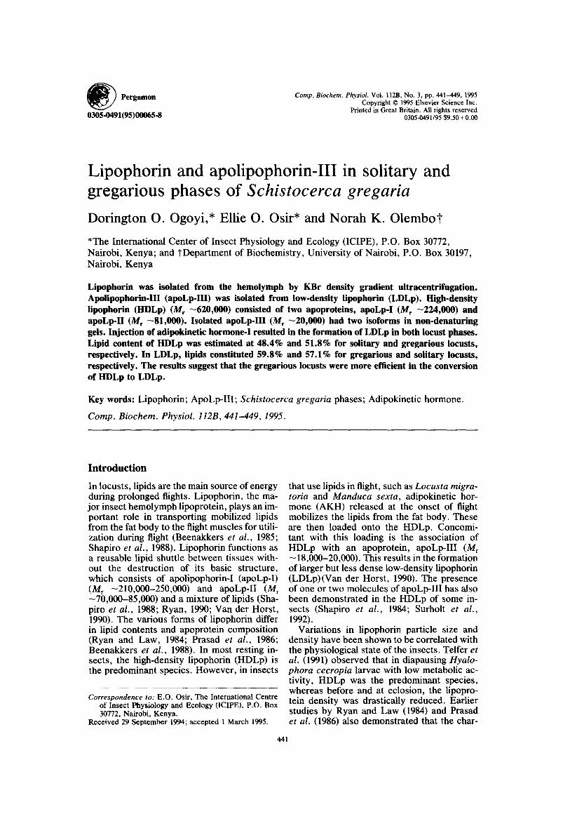
In density gradient ultracentrifugation of hemolymph from solitary and gregarious lo- custs, HDLp separated out in the upper por- tion of the centrifuge tube with a characteris- tic yelow banding (Fig. 1A). The HDLp had densities of 1.114 and 1.105 g/ml for solitary and gregarious locusts, respectively. On the other hand, LDLp had lower densities of 1.074 and 1.066 g/ml in solitary and gregarious locusts, respectively (Fig. 1B). Thus, the ad- ministration of AKH-I resulted into a lipopro- tein molecule of lower density. In the case of LDLp, yellow banding occurred at positions corresponding to HDLp, which suggested that AKH-I administration did not result into total conversion of HDLp to LDLp. This was espe- cially so with the solitary locusts (Fig. 1B). Fractions containing HDLp or LDLp were con- firmed by SDS-PAGE profiles and the appro- priate lipoprotein peaks pooled and dialysed.
From the SDS-PAGE of the ultracentrifuga- tion fractions, there was no evidence of con- tamination from the sub-phase. HDLp was shown to correspond to fractions 9-19, with a peak concentration observed on fraction number 13. On the other hand, LDLp was ob-
444 D. O. Ogoyi et al.
0.8
o 0.6
o~.1 i i ~ / / / / --
/i /// ///1t
5 9 13 17 21 25 29 33 37 Fraction number
~32
128
12&
1.20
116
112 Q
108
IO&
kD 1
6
2 :
14
I/*
1.2
1.0
® 0.8
.~ 0.6
02
/
/
5 9 13 17 21 25 29 33 37 Fraction number
132
/ / - ' " --4128
/ " '1.2& /
112 o
108
1.0t,
Fig. 1. KBr density gradient ultracentrifugation profile of hemolymph. Fractions (1.0 ml) were taken from the top of the tube and absorbance at 450 nm and refractive indices monitored for odd-numbered fractions. (A) Hemolymph from resting locusts; (B) hemolymph from insects injected with AKH-I. o o, Absorbance for solitary locusts; e e, absorbance for gregarious locusts; - . - , density
(g/ml).
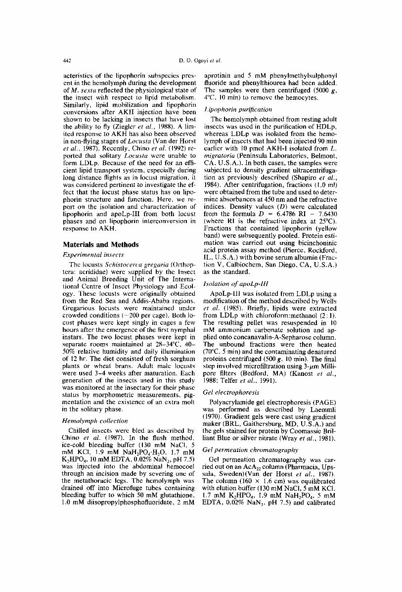
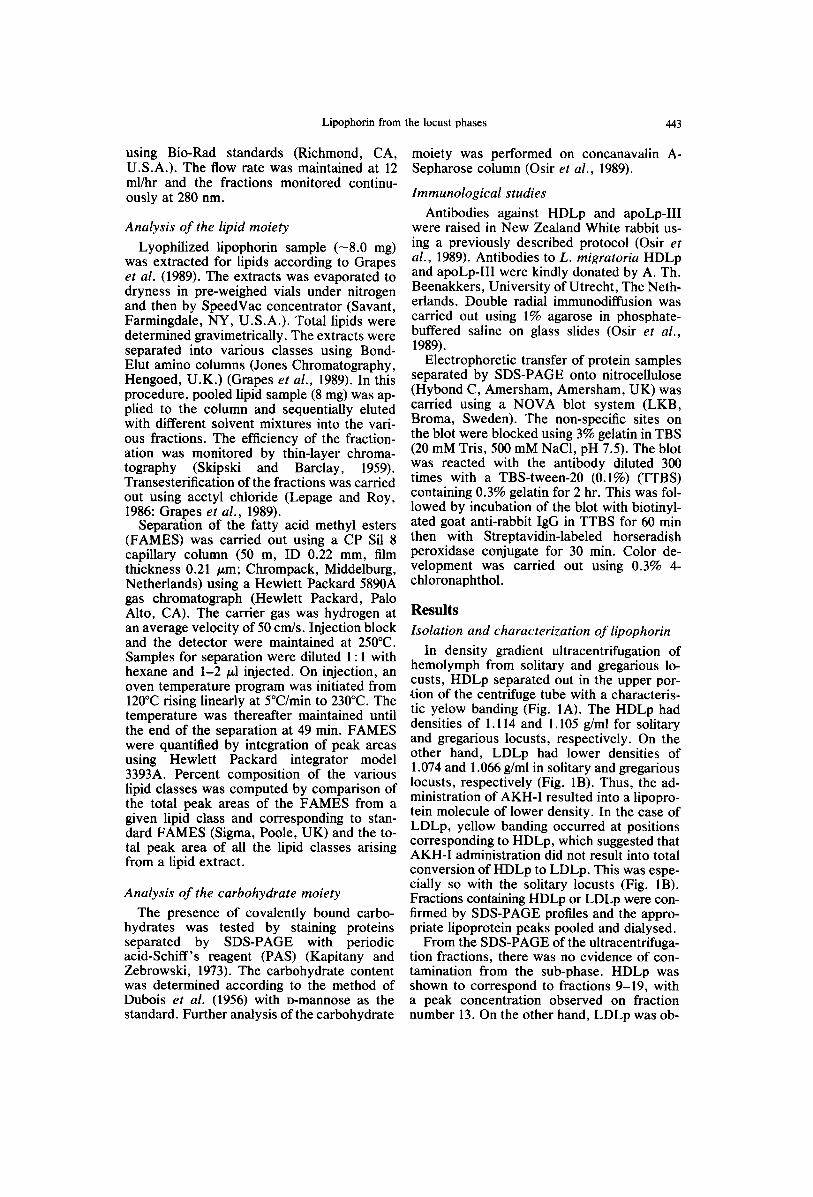
tained between fractions, (4-9) with a peak concentration on fraction number 8. The iso- lated lipoproteins were shown to be pure us- ing both non-denaturing (Fig. 2) and denatur- ing PAGE (Fig. 3). The HDLp isolated from the two phases of S. gregaria showed struc- tural similarity in both the native form and in apoprotein composition. Non-denaturing PAGE revealed a native molecular weight of M r -620,000, whereas SDS-PAGE showed the presence of two apoproteins (apoLp-I, M r, -224,000, and apoLp-II, Mr -81,000). A third apoprotein, apoLp-III ( M r - 2 0 , 0 0 0 ) , w a s present only in trace amounts that could be detected with silver but not with Coomassie Brilliant Blue staining.
LDLp isolated from both solitary and gre- garious locusts had the same apoprotein com- position. The apoproteins, apoLp-I and apoLp-II, were of the same molecular weight and intensity on staining as in HDLp de- scribed above. However, the presence of the third apoprotein, apoLp-III (Mr -20,000), was more intense even on staining with Coomassie brilliant blue (Fig. 3). The native molecular
6 7 - -
Fig. 2. Non-denaturing PAGE (4-20%) of isolated lipo- phorin. Lane 1, HDLp (gregarious) (20/zg); lane 2, HDLp
(solitary) (20/~g).
weight of LDLp as estimated by gel perme- ation chromatography on an AcA22 column was Mr - 1 . 7 x 10 6 for solitary locusts. On the other hand, the LDLp from the gregarious locusts was totally excluded in this column. Thus, the gregarious locusts LDLp had a higher molecular weight compared with the solitary locusts. In most preparations of HDLp and LDLp, apoLp-I appeared as a dou- blet. A degradation product of Mr -50,000 was also observed in SDS-PAGE of some preparations of HDLp or LDLp.
A p o L p - I ~
A B 1 2 3 4 5 6
I- D
0 0
ApoLp_l I
1 6 7
A p o L p - I I I~
Fig. 3. SDS-PAGE (4-15%) of isolated LDLp and HDLp. Lanes 3 and 6: Molecular weight standards. (A) Solitary locusts. Lane 1, LDLp (20 tzg); lane 2, HDLp (20 p,g). (B) Gregarious locusts. Lane 4, LDLp (20 /xg); lane 5,
HDLp (20/xg).
Lipophorin from the locust phases 445
Carbohydrate and lipid moieties LDLp isolated from both solitary and gre-
garious locusts was separated by SDS-PAGE and stained with PAS for carbohydrates. All three apoproteins were glycosylated (data not shown). The carbohydrate content of HDLp expressed as a percentage of the protein was estimated at 3.43% and 3.12% for solitary and gregarious locusts, respectively (Table 1). The carbohydrate content in LDLp was estimated at 7.99% and 5.10% for gregarious and solitary locusts, respectively. The higher carbohy- drate content in LDLp than in HDLp could be because of an increase in molecules of apoLp-III associating. The carbohydrate moi- eties had mannose-rich oligosaccharide chains as shown by the binding to concanavalin A.
The lipid moiety constituted 51.8% and 48.4% (wt%) of HDLp from gregarious and solitary locusts, respectively (Table 1). In the case of LDLp, lipids constituted 57.1% and 59.8% (wt%) for solitary and gregarious lo- custs, respectively. These results showed a higher lipid content in LDLps than in HDLps, indicating that the administration of AKH-I resulted in more lipid associating with the li- poprotein molecule. In both HDLp and LDLp, the major lipid classes present were phospholipids and diacylglycerides (Table 2).
The other lipids present included triacylglyc- erides, monoacylglycerides, steryl esters and free fatty acids. In the gregarious locusts, phospholipids and diacylglycerides in HDLp constituted 26.7% and 43.1% of the total lip- ids, respectively, whereas in LDLp, phospho- lipids and diacylglycerides were estimated at 22% and 60%, respectively. In the case of the solitary locusts, the percent composition of phospholipids and diacylglycerides in HDLp were 25.8% and 35.5%, respectively, whereas in LDLp, the percent phospholipids and dia- cylglycerides were 31.4% and 48%, respec- tively. Evidently, the major lipid mobilized from the fat bOdy was diacylglycerides, re- suiting into LDLp particles with a much higher level of diacylglycerides as compared with that in HDLp. The diacylglycerides per- cent composition in LDLp was significantly higher (p < 0.05) in gregarious than solitary locusts. Minor differences were also evident between the phases for the other lipid classes.
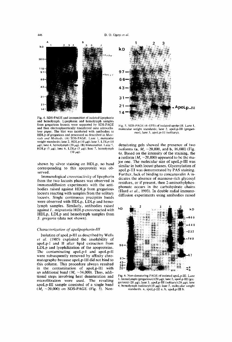
Immunological characterization of lipophorin In immunoblotting experiments, the anti-
bodies reacted with both apoLp-I and apoLp-II (Fig. 4). The banding pattern was the same for hemolymph, HDLp and LDLp. Al- though the presence of apoLp-III had been
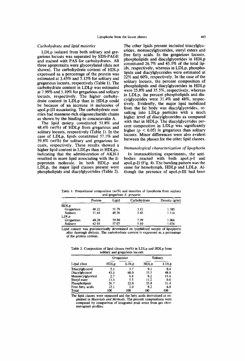
Table 1. Proportional composition (wt%) and densities of iipophorin from solitary and gregarious S. gregaria
Protein Lipid Carbohydrate Density (g/ml)
HDLp Gregarious 48.22 51.78 3.12 1.105 Solitary 51.64 48.36 3.43 1.114
LDLp Gregarious 40.20 59.80 7.99 1.066 Solitary 42.93 57.07 5.10 1.074
Lipid content was gravimetrically determined on lyophilized sample of lipophorin after thorough dialysis. The carbohydrate content is expressed as a percentage of the protein content.
Table 2. Composition of lipid classes (wt%) in LDLp and HDLp from solitary and gregarious locusts
Gregarious Solitary
Lipid class HDLp LDLp HDLp LDLp
Triacylglycerol 2.1 3.7 9.1 0.4 Diacylglycerol 43.1 60.0 35.5 48.0 Monoacylglycerol 2.7 6.8 9.2 13.4 Steryl ester 13.3 5.5 11.2 0.8 Phospholipids 26.7 22.0 25.8 31.4 Free fatty acids 12.1 2.0 9.2 6.0 Total 100 100 100 100
The lipid classes were separated and the fatty acids derivatised as ex- plained in Materials and Methods. The percent compositions were computed by comparison of integrated peak areas from gas chro- matograph profiles.
446 D. O. Ogoyi et al.
A 1 2 3
B 5 6
~ApoLp_ I
"ApoLp_I I
Fig. 4. SDS-PAGE and immunoblot of isolated lipophorin and hemolymph. Lipophorin and hemolymph samples from gregarious locusts were separated by SDS-PAGE and then electrophoretically transferred onto nitrocellu- lose paper. The blot was incubated with antibodies to HDLp of gregarious and processed as described in Mate- rials and Methods. (A) SDS-PAGE. Lane 1, molecular weight standards; lane 2, HDLp (10/xg); lane 3, LDLp (10 ~zg); lane 4, hemolymph (20 p-g). (B) lmmunoblot. Lane 5, HDLp (5 p,g); lane 6, LDLp (5 tzg); lane 7, hemolymph
(10 p.g).
shown by silver staining on HDLp, no band corresponding to this apoprotein was ob- served.
Immunological crossreactivity of lipophorin from the two locusts phases was observed in immunodiffusion experiments with the anti- bodies raised against HDLp from gregarious locusts reacting with samples from the solitary locusts. Single continuous precipitin bands were observed with HDLp, LDLp and hemo- lymph samples. Similarly, antibodies raised against L. migratoria HDLp crossreacted with HDLp, LDLp and hemolymph samples from S. gregaria (data not shown).
Characterization of apolipophorin-lll
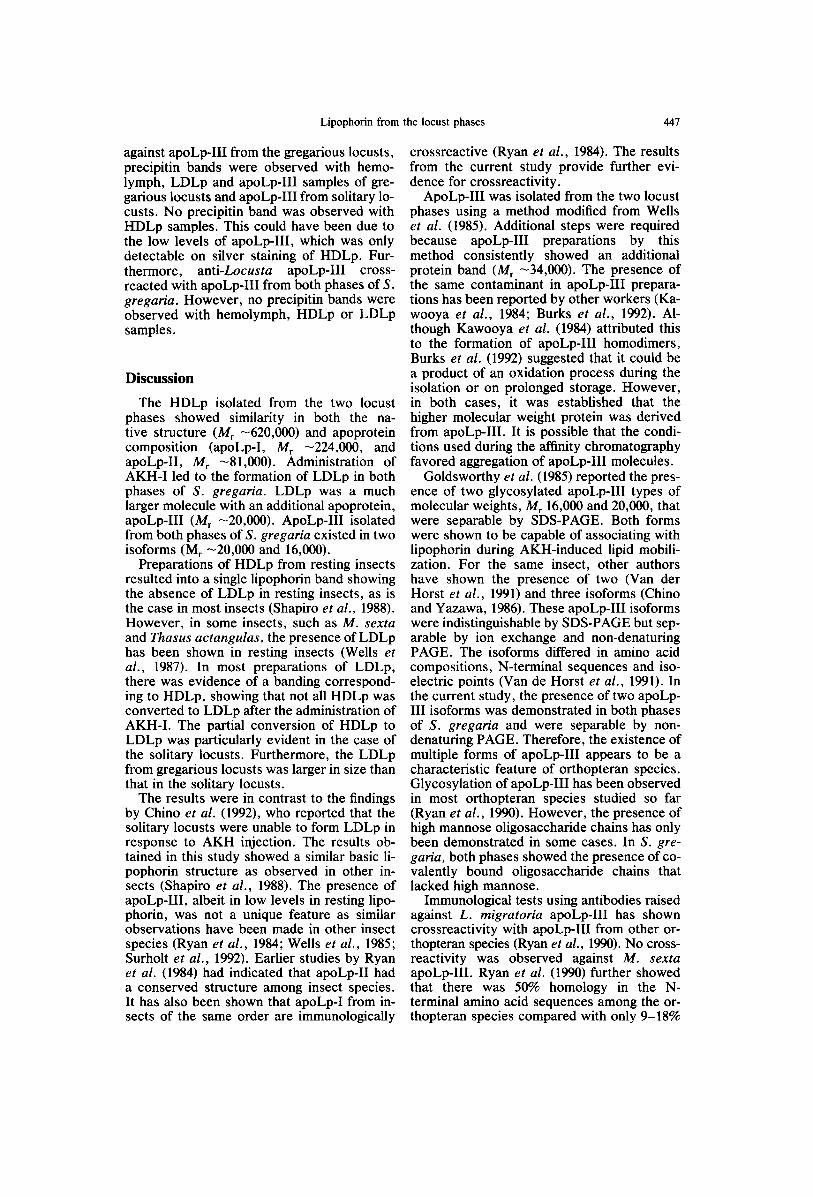
Isolation of apoLp-llI as described by Wells et al. (1985) exploited the insolubility of apoLp-I and II after lipid extraction from LDLp and lyophilization of the apoproteins. The contaminating apoLp-I and apoLp-II were subsequently removed by affinity chro- matography because apoLp-III did not bind to this column. This procedure always resulted in the contamination of apoLp-llI with an additional band (Mr -34,000). Thus, addi- tional steps involving heat denaturation and microfiltration were used. The resulting apoLp-III sample consisted of a single band (]14, -20,000) on SDS-PAGE (Fig. 5). Non-
kD 2 3
97
66
4 3
31
21
14 ApoLp_l l l
Fig. 5. SDS-PAGE (4-15%)of isolated apolp-II1. Lane 1, molecular weight standards; lane 2, apoLp-III (gregari-
ous); lane 3, apoLp-Ill (solitary).
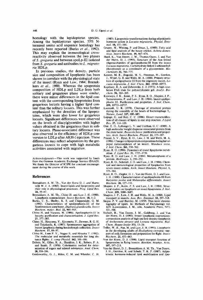
denaturing gels showed the presence of two isoforms (a, M, -20,000, and b, 16,000) (Fig. 6). Based on the intensity of the staining, the a isoform (M, -20,000) appeared to be the ma- jor one. The molecular size of apoLp-III was similar in both locust phases. Glycosylation of apoLp-III was demonstrated by PAS staining. Further, lack of binding to concanavalin A in- dicates the absence of mannose-rich glycosyl residues, or if present, then 2-aminothylphos- phonate occurs in the carbohydrate chains (Hard et al., 1993). In double radial immuno- diffusion experiments using antibodies raised
1 2 3 4 5 kD
96 - -
67-- 43-- 30-- 20--
Fig. 6. Non-denaturing PAGE of isolated apoLp-Ill. Lane 1, hemolymph (gregarious) (20 jzg); lane 2, apoLp-lll (gre- garious) (20 ~g); lane 3, apoLp-III (solitary)(20 ~g); lane 4, hemolymph (solitary)(10 ~g); lane 5, molecular weight
standards, a, apoLp-IIl a; b, apoLp-III b.
Lipophorin from the locust phases 447
against apoLp-IlI from the gregarious locusts, precipitin bands were observed with hemo- lymph, LDLp and apoLp-III samples of gre- garious locusts and apoLp-III from solitary lo- custs. No precipitin band was observed with HDLp samples. This could have been due to the low levels of apoLp-III, which was only detectable on silver staining of HDLp. Fur- thermore, anti-Locusta apoLp-III cross- reacted with apoLp-III from both phases of S. gregaria. However, no precipitin bands were observed with hemolymph, HDLp or LDLp samples.
Discussion
The HDLp isolated from the two locust phases showed similarity in both the na- tive structure (Mr -620,000) and apoprotein composition (apoLp-I, Mr -224,000, and apoLp-II, Mr -81,000). Administration of AKH-I led to the formation of LDLp in both phases of S. gregaria. LDLp was a much larger molecule with an additional apoprotein, apoLp-III (M r -20,000). ApoLp-III isolated from both phases of S. gregaria existed in two isoforms (Mr -20,000 and 16,000).
Preparations of HDLp from resting insects resulted into a single lipophorin band showing the absence of LDLp in resting insects, as is the case in most insects (Shapiro et al., 1988). However, in some insects, such as M. sexta and Thasus actangulas, the presence of LDLp has been shown in resting insects (Wells et al., 1987). In most preparations of LDLp, there was evidence of a banding correspond- ing to HDLp, showing that not all HDLp was converted to LDLp after the administration of AKH-I. The partial conversion of HDLp to LDLp was particularly evident in the case of the solitary locusts. Furthermore, the LDLp from gregarious locusts was larger in size than that in the solitary locusts.
The results were in contrast to the findings by Chino et al. (1992), who reported that the solitary locusts were unable to form LDLp in response to AKH injection. The results ob- tained in this study showed a similar basic li- pophorin structure as observed in other in- sects (Shapiro et al., 1988). The presence of apoLp-III, albeit in low levels in resting lipo- phorin, was not a unique feature as similar observations have been made in other insect species (Ryan et al., 1984; Wells et al., 1985; Surholt et al., 1992). Earlier studies by Ryan et al. (1984) had indicated that apoLp-II had a conserved structure among insect species. It has also been shown that apoLp-I from in- sects of the same order are immunologically
crossreactive (Ryan et al., 1984). The results from the current study provide further evi- dence for crossreactivity.
ApoLp-III was isolated from the two locust phases using a method modified from Wells et al. (1985). Additional steps were required because apoLp-III preparations by this method consistently showed an additional protein band (Mr -34,000). The presence of the same contaminant in apoLp-III prepara- tions has been reported by other workers (Ka- wooya et al., 1984; Burks et al., 1992). Al- though Kawooya et al. (1984) attributed this to the formation of apoLp-III homodimers, Burks et al. (1992) suggested that it could be a product of an oxidation process during the isolation or on prolonged storage. However, in both cases, it was established that the higher molecular weight protein was derived from apoLp-III. It is possible that the condi- tions used during the affinity chromatography favored aggregation of apoLp-III molecules.
Goldsworthy et al. (1985) reported the pres- ence of two glycosylated apoLp-III types of molecular weights, Mr 16,000 and 20,000, that were separable by SDS-PAGE. Both forms were shown to be capable of associating with lipophorin during AKH-induced lipid mobili- zation. For the same insect, other authors have shown the presence of two (Van der Horst et al., 1991) and three isoforms (Chino and Yazawa, 1986). These apoLp-III isoforms were indistinguishable by SDS-PAGE but sep- arable by ion exchange and non-denaturing PAGE. The isoforms differed in amino acid compositions, N-terminal sequences and iso- electric points (Van de Horst et al., 1991). In the current study, the presence of two apoLp- III isoforms was demonstrated in both phases of S. gregaria and were separable by non- denaturing PAGE. Therefore, the existence of multiple forms of apoLp-III appears to be a characteristic feature of orthopteran species. Glycosylation of apoLp-III has been observed in most orthopteran species studied so far (Ryan et al., 1990). However, the presence of high mannose oligosaccharide chains has only been demonstrated in some cases. In S. gre- garia, both phases showed the presence of co- valently bound oligosaccharide chains that lacked high mannose.
Immunological tests using antibodies raised against L. migratoria apoLp-III has shown crossreactivity with apoLp-III from other or- thopteran species (Ryan et al., 1990). No cross- reactivity was observed against M. sexta apoLp-III. Ryan et al. (1990) further showed that there was 50% homology in the N- terminal amino acid sequences among the or- thopteran species compared with only 9-18%
448 D. O. Ogoyi et al.
homology with the lepidopteran species. Among the lepidopteran species, 53% N- terminal amino acid sequence homology has recently been reported (Burks et al., 1992). This may explain the immunological cross- reactivity observed between the two phases of S. gregaria and between apoLp-III isolated from S. gregaria and antibodies to L. migrato- ria HDLp.
In previous studies, the density, particle size and composition of lipophorin has been shown to correlate with the physiological state of the insect (Ryan and Law, 1984; Beenak- kers et al., 1988). Whereas the apoprotein composition of HDLp and LDLp from both solitary and gregarious phase were similar, there were minor differences in the lipid con- tent with the corresponding lipoproteins from gregarious locusts having a higher lipid con- tent than the solitary locusts. This was further emphasized by the densities of the lipopro- teins, which were also lower for gregarious locusts. Significant differences were observed on the levels of diacylglycerides with higher values obtained in the gregarious than in soli- tary locusts. Phase-associated difference was also observed in the efficiency of HDLp con- version to LDLp after AKH-I injection. These differences may reflect adaptations by the gre- garious locusts to cope with high metabolic activities associated with migration.
Acknowledgments--This work was supported by funds from the German Academic Exchange Service (DAAD). We thank the Director of ICIPE for constant encourage- ment during the course of this work.
References
Beenakkers A. M. Th., Van der Horst D. J. and Marre- wijk W. J. A. (1985). Insect lipids and lipoproteins and their role in physiological processes. Prog. Lipid Res. 24, 19-67.
Beenakkers A. M. Th., Chino H. and Law J. H. (1988). Lipophorin nomenclature. Insect Biochem. 18, 1-2.
Burks, C. S., Shelby, K. S. and Chippendale, G. M. (1992). Characteristics of apolipophorin-III of the Southwestern corn borer, Diatraea grandiosella. Insect Biochem. molec. Biol. 22, 905-915.
Chino H. and Yazawa, M. (1986). Apolipophorin-IIl in locusts: purification and characterization. J. Lipid Res. 27, 377-385.
Chino, H., Hirayama, Y., Kiyomoto, Y., Downer, R. G. H. and Takahashi, K. (1987). Spontaneous aggregation of locust lipophorin during haemolymph collection. Insect Biochem. 17, 89-97.
Chino H., Lum P. Y., Nagao E. and Hiraoka T. (1992). The molecular and metabolic essentials for long dis- tance flights. J. comp Physiol. 162, 101-106.
Dubois, M., Gilles, K. A., Hamilton, J. K., Rebers, P. A. and Smith, F. (1956). Colorimetric method for deter- mination of sugars and related substances. Anal. Chem. 28, 350-356.
Goldsworthy, G. J., Miles, C. M. and Wheeler, C. H.
(1985). Lipoprotein transformations during adipokinetic hormone action in Locusta migratoria. Physiol. Ento- mol. 10, 151-164.
Grapes, M., Whiting, P. and Dinan, L. (1989). Fatty acid and lipid analysis of the house cricket. Acheta domes- ticus. Insect Biochem. 19, 667-674.
Hard, K., Van Doom, J. M., Thomas-Oates, J. and Van der Horst, D. J. (1993). Structure of the Asn linked oligosaccharides of apolipophorin-III from the insect Locusta migratoria. Carbohydrate linked 2-a~inoethyl- phosphonate as a constituent of a glycoprotein. Bio- chemistry 32, 766-775.
Kanost, M. R., Boguski, M. S., Freeman, M., Gordon, J., Wyatt, G. R. and Wells, M. A. (1988). Primary struc- ture of apolipophorin III from the migratory locust, Lo- custa migratoria. J. biol. Chem. 263, 10568-10573.
Kapitany, R. A. and Zebrowski, E. J. (1973). A high reso- lution PAS stain for polyacrylamide gel. Analyt. Bio- chem. 56, 361-369.
Kawooya, J. K., Keim, P. S., Ryan, R. O., Shapiro, J. P., Samaraweera, P. and Law, J. H. (1984). Insect apolipo- phorin III. Purification and properties. J. biol. Chem. 259, 10733-10737.
Laemmli, U. K. (1970). Cleavage of structural protein during the assembly of the heads of bacteriophage T4. Nature, Land. 227, 680-685.
Lepage, G. and Roy, C. C. (1986). Direct transeterifica- tion of all classes of lipids in one step reaction. J Lipid Res. 27, 114-120.
Osir, E. O., Labongo L. V. and Unithan, G. C. (1989). A high molecular weight diapause-associated protein from the stem borer, Busseolafusca: purification and proper- ties. Arch. Insect Biochem. Physiol. 11, 173-187.
Prasad, S. V., Ryan, R. O., Law, J. H. and Wells, M. A. (1986). Changes in lipoprotein composition during larval- pupal metamorphosis of an insect. Manduca sexta. J. biol. Chem. 261,558-562.
Ryan, R. O. (1990). Dynamics of insect lipophorin metab- olism. J. Lipid Res. 31, 1725-1739.
Ryan, R. O. and Law, J. H. (1984). Metamorphosis of a protein. BioEssays. 1,250-252.
Ryan, R. O., Schmidt, J. O. and Law, J. H. (1984). Chem- ical and immunological properties of lipophorins from seven insect orders. Arch. Insect Biochem. Physiol. 1, 375-384.
Ryan, R. O., Ziegler, D. J., Van der Horst, D. J. and Law, J. H. (1990). Characterization of apolipophorin-III from Barytettix psolus and Melanoplus differentialis. Insect Biochem. 20, 127-133.
Shapiro, J. P., Keim, P. S. and Law, J. H. (1984). Struc- tural studies on lipophorin an insect lipoprotein. J. biol. Chem. 259, 3680-3685.
Shapiro, J. P., Law, J. H. and Wells, M. A. (1988). Lipid transport in insects. Ann. Rev. Entomol. 33, 297-318.
Skipsi, P. V. and Barclay, M. (1959). Thin layer chroma- tography of lipids. In: Methods of Enzymology vol. XIV (Lowenstein, J. M., edn, Academic Press, NY). 531-598.
Surholt, B., Van Doorn, J. M., Goldberg, J. and Van der Horst, D. J. (1992). Insect lipophorin conversions: composition analysis of high and low density-lipophorin of Archerontia atropos and Locusta migratoria. Biol. Chem. Hoppe-Seyler 373, 13-20.
Telfer, W. H., Pan, M. and Law, J. H. (1991). Lipophorin in the developing adults of Hyalophora cecropia: sup- port of yolk formation and preparation for flight. Insect Biochem. 21, 653-663.
Van der Horst, D. J. (1990). Lipid transport function of lipoproteins in flying insects. Biochim. biophys. Acta. 107, 197-211.
Van der Horst, D. J., Beenakkers A. M. Th., Van Doom, J. H., Gerritse, K. and Schulz, T. K. F. (1987). Adipo- kinetic hormone-induced lipid mobilization and lipo-
Lipophorin from the locust phases 449
phofin interconversions in fifth larval instar locusts. In- sect Biochem. 17, 799-808.
Van der Horst, D. J., Van Doom, J. M., Voshol, H., Kanost, M. R., Ziegler, R. and Beenakkers, A. M. Th. (1991). Different isoforms of an apoprotein (apolipopho- fin-Ill) associated with lipoproteins in Locusta migrato- ria. Eur. J. Biochem. 196, 509-517.
Wells, M. A., Ryan, R. O., Prasad, S. V. and Law, J. H. (1985). A novel procedure for the purification of apoli- pophorin III. Insect Biochem. 15, 565-571.
Wells, M. A., Ryan, R. O., Kawooya, J. K. and Law,
J. H. (1987). The role of apolipophorin-III in vivo lipo- protein interconversion in adult Manduca sexta. J. biol. Chem. 262, 4172-4176.
Wray, W., Boulikas, T., Wray, V. P. and Hancock, R. (1981). Silver staining of proteins in polyacrylamide gels. Analyt. Biochem. 118, 197-203.
Ziegler, R., Ryan, R. O., Arbas, E. A. and Law, J. H. (1988). Adipokinetic response in a flightless grasshop- per (Barytettix psolus): functional components and de- fective response. Arch. Insect Biochem. Physiol. 9, 255-265.


