Embed Size (px)
Citation preview
HAL Id: hal-03371121https://hal.sorbonne-universite.fr/hal-03371121
Submitted on 8 Oct 2021
HAL is a multi-disciplinary open accessarchive for the deposit and dissemination of sci-entific research documents, whether they are pub-lished or not. The documents may come fromteaching and research institutions in France orabroad, or from public or private research centers.
L’archive ouverte pluridisciplinaire HAL, estdestinée au dépôt et à la diffusion de documentsscientifiques de niveau recherche, publiés ou non,émanant des établissements d’enseignement et derecherche français ou étrangers, des laboratoirespublics ou privés.
A review of the fossil record of caecilians Gymnophiona(Tetrapoda; Lissamphibia; Gymnophionomorpha) withcomments on its use to calibrate molecular timetrees
Rodolfo Otávio Santos, Michel Laurin, Hussam Zaher
To cite this version:Rodolfo Otávio Santos, Michel Laurin, Hussam Zaher. A review of the fossil record of caeciliansGymnophiona (Tetrapoda; Lissamphibia; Gymnophionomorpha) with comments on its use to calibratemolecular timetrees. Biological Journal of the Linnean Society, Linnean Society of London, 2020, 131(4), pp.737-755. �10.1093/biolinnean/blaa148�. �hal-03371121�
A review of the fossil record of caecilians Gymnophiona (Tetrapoda;
Lissamphibia; Gymnophionomorpha) with comments on its use to calibrate
molecular timetrees[RS1]
Rodolfo Otávio Santos1, Michel Laurin2, Hussam Zaher1
1 Instituto de Biociências, Universidade de São Paulo, São Paulo, Brazil
2 Centre de Recherches sur la Paléobiologie et les Paléoenvironnements (CR2P), Centre
national de la Recherche scientifique (CNRS)/Muséum national d’Histoire naturelle
(MNHN)/Sorbonne Université, Paris, France
Abstract:
Gymnophiona, popularly known as caecilians, the most poorly known major taxon
group[RS2][MOU3] of extant amphibians, includes are elongated and limbless tetrapods,
with compact ossified skulls and reduced eyes, mainly adapted to fossorial life as
adults(only the Typhlonectidae exhibits adaptations for an aquatic or semiaquatic
behavior). Caecilians are poorly represented in the fossil record, and despite the scarcity
low number of fossil specimens described until now (only four named taxa, in addition
to indeterminate fragmentary material), their fossils play a key role in the our
knowledge of Lissamphibia origin and evolution, as well as contribute directly to a
better understanding of phylogeny, taxonomy, and biogeography of extant
gymnophionan taxa. These records are scattered throughout geological time (from the
Jurassic to the sub-recent Neogene) and space (they are represented only on North and
South America and Africa). Here, we revisit the caecilian fossil record, providing a brief
description of all known extinct taxa described so far, along with general remarks about
their impact on systematics, time range and geographic distribution of the clade, as well
as prospects for future research. Possible calibration constraints based on the caecilian
fossil record are provided.
Key words: Amphibians – Caecilians – Fossils – Gymnophiona – Timetree calibration
- Vertebrate Paleontology
Introduction
The crown-clade Lissamphibia (see Laurin et al., 2020, for a review; but see
Dubois, 2004, for an opposing view on the use of this nomen) comprises the extant taxa
Anura, Urodela, and Gymnophiona. Although lissamphibians are diverse in present day
biotas (Frost, 2020), their fossil record is relatively scarce, and includes only a few, but
important, specimens whose preservation status is sufficiently satisfactory to allow
detailed diagnoses (e. g. Schoch & Millner, 2004; Marjanović & Laurin, 2019[MOU4]).
This scarcity is particularly pronounced for gymnophionanscaecilians. For many years,
only one gymnophionan caecilian was known in the fossil record (Estes, 1981), and to
date only four extinct taxa originally assigned to this group were have been erected
named and described in details (Estes & Wake, 1972; Jenkins & Walsh, 1993; Evans &
Sigogneau-Russel, 2001; Pardo et al., 2017).
The clade Gymnophiona is moderately diverse, with approximately 214 known
extant species (AmphibiaWeb, 2020Frost, 2020). Popularly known as caecilians, these
animals are well adapted to a fossorial existence, as shown by their elongated body, the
absence of limbs and girdles, a compact and well-ossified skull, and reduced or vestigial
eyes (e.g. Taylor, 1968; Duellman & Trueb, 1994; Wilkinson & Nussbaum, 2006;
Wilkinson et al., 2011). However, a subgroup of gymnophionans, the typhlonectids,
exhibits an aquatic or semi-aquatic lifestyles[MOU5] (Taylor, 1968; Tanner, 1971). Other
distinct characteristics of caecilians include a dual mechanism for jaw closing and a pair
of sensitive organs between the eyes and nostrils, known as tentacles (Wilkinson &
Nussbaum, 2006).
The first now-accepted caecilian fossil species described was Apodops pricei
Estes & Wake, 1972, a crown-gymnophionan from the Early Eocene of Brazil
consisting only of an isolated pre-cloacal vertebra (see Estes & Wake, 1972 and Estes,
1981, for comments about two fossils named earlier, a silurid catfish and a cephalopod,
previously misidentified as caeciliansDuellman & Trueb, 1994). Later, Eocaecilia
micropodia Jenkins & Walsh, 1993, found in Early Lower Jurassic rocks of Arizona,
the United States, was described based on numerous specimens with cranial and
postcranial elements, including limbs and girdles, both completely lost in all extant
species, but predictable in stem-gymnophionans (Jenkins et al., 2007). Subsequently, a
taxon from the Lower Cretaceous of Morocco, Rubricacaecilia monbaroni Evans &
Sigogneau-Russel, 2001, was erected based on a nearly complete pseudodentary,
although with other isolated jaw elements, vertebrae, and a possible femur have
beenalso attributed to it.
Chinlestegophis jenkinsi Pardo et al. 2017, from the Triassic of Colorado,the
United States, was initially interpreted as the sister-group of caecilians. It is represented
by partially preserved skulls, jaws and disarticulated postcranial elements. This
enigmatic taxon may be important to understand gymnophionan evolution, because it is
interpreted as showings a combination of caecilian synapomorphies and lissamphibian
plesiomorphies that suggests polyphyly of extant amphibians, according to (Pardo et al.
(2017). However, the affinities close relationship of Chinlestegophis with and
gymnophionans are is controversial. Marjanović & Laurin (2019: 144 and figure 30)
reanalyzed the data and showed that this is just one of four equally most parsimonious
results, with the others being highly incongruent with the hypothesis of Pardo et al.
(2017) Chinlestegophis is more likely to be a stereospondyl, but its affinities with
gymnophionans are more dubious. Similarly, Carroll & Currie (1975), and more
recently Anderson et al. (2008), suggested a sister-group relationship between the Early
Permian lepospondyl Rhynchonkos and caecilians. However, a detailed CT-scan
analysis of Rhynchonkosits morphology suggested that similarities previously regarded
as synapomorphies between the recumbirostran microsaurs and gymnophionans (such
as the presence of a retroarticular process, an expanded ossification in the antotic
region, and a cultriform process of parasphenoid) result from ambiguities in previous
character definitions and convergent evolution due to a fossorial ecomorph
(Szostakiwskyj et al., 2015).
Although the fossil record of caecilians is undoubtedly scarcepoor, our present
knowledge is provides sufficient clues of their past history to allow a more
comprehensive approach, combining information from both extinct and extant taxa, to
provides important clues to their past history. Here, we provide a review of the
gymnophionan caecilian fossil record and discuss aspects of the anatomy, taxonomy,
phylogeny, and biogeography of extinct groups, as well as their implications for our
understanding of the biology and relationships of extant gymnophionanscaecilians.
Phylogeny and Classification of caecilians
The classification and definition of Gymnophionan caecilian clades varies
according to authors. Trueb & Cloutier (1991) proposed to restrict the term names
Apoda Oppel, 1811 for the crown-group of caecilians and Gymnophiona Rafinesque-
Schmaltz, 1814 for the stem-total group including Apoda. However, the fact that the
former name was preoccupied by several earlier nomina (Dubois, 2004), along with a
possible misunderstanding in some statements about caecilian characteristics (such as
the generalization of the limbless condition of gymnophionans), led some authors to
reject these definitions (e.g. Dubois, 2004; Wilkinson & Nussbaum, 2006).
Furthermore, as pointed out by Wilkinson et al. (2011), the use of the name
Gymnophiona for the crown-group is already well established in the literature, and a
changinge in it wouldill probably bring more problems than create solutions. To avoid
this problematic situation, Thus, Marjanović & Laurin (2008a) proposed the term
Gymnophionomorpha for the a branchstem-based clade that comprises extant caecilians
plus extinct taxa, such as Eocaecilia. micropodia and Rubricacaecilia. monbaroni.,
Marjanović & Laurin (2008a) proposed the term Gymnophionomorpha.
According to the stembranch-based definition of Gymnophionomorpha, this
clade comprises all lineages more closely related to the crown-clade Gymnophiona than
to Batrachia. Therefore, this taxon encompasses Eocaecilia. micropodia,
Rubricacaecilia. monbaroni, and extant caecilians (Figure 1). Under the phylogeny
proposed by Pardo et al. (2017), it would also include Chinlestegophis. jenkinsi, and all
other eryopiform temnospondyls (including stereospondyls and
archegosaurids)stereospondyls, plus other (but not all) temnospondyls, such as
archegosaurids and eryopoids. On other phylogenies, Mmembers of the
Gymnophionomorpha (under their currently accepted delimitation) are characterized by
numerous bone fusions, such as the lower jaw consisting in only two bones, known as
pseudodentary and pseudoangular; and most of the braincase of only one, called os
basale (Jenkins et al. 2007).the presence of pseudodentary and pseudoangular forming
the lower jaw, os basale and absence of tympanic ear.
Recent large-scale molecular analyses strongly corroborate the monophyly of
extant Lissamphibia with respect to Amniota, and most also find caecilians placed as
the sister-group of Batrachia, which consists of includes Anura and Urodela (Irisarri et
al., 2017; Vijayakumaret al., 2019Frost et al., 2006; Pyron & Wiens, 2011). These
results also stand in a total evidence analysis (Pyron, 2011) based on a molecular data
set designed to be combined with a mainly fossil-oriented data matrix (Vallin & Laurin,
2004). However, recent morphological approaches designed to test the phylogenetic
affinities of lissamphibians within an expanded taxon sampleing of Paleozoic tetrapods
have resulted in fundamentally distinct hypotheses on the origin of the group (most
recently reviewed in see Ruta & Coates, 2007; Marjanović & Laurin, 2019). Currently
there are three main phylogenetic hypotheses try to explain this question, all of which
display minor variants (Figure 2). The first (Figure 2A) considers that Lissamphibia is a
monophyletic group inside Temnospondyli (e.g. Trueb & Cloutier, 1991; Ruta &
Coates, 2007). The second (Figure 2B) also recognizes the monophyly of
lissamphibians, but nested within Lepospondyli (e.g. Marjanović & Laurin, 2008a,
2009, 2019). The third (Figures 2C and 2D) suggests that extant amphibians do not
actually form a monophyletic group, because frogs and salamanders are temnospondyls
and whereas caecilians are lepospondyls (e.g. Anderson et al., 2008; Huttenlocker et al.,
2013) and thus more closely related to Amniota. In the variant of Anderson et al.
(2008), In some variants, gymnophionans are more closely related to amniotes than to
batrachians (e.g. Anderson et al., 2008), whereas this is contradicted by nearly all
molecular (e.g. Irisarri et al., 2017), total evidence (Pyron, 2011), and some
paleontological (Marjanović & Laurin, 2009, 2019) phylogenies.
The hypothesis recently proposed by Pardo et al. (2017) is compatible with
molecular phylogenies to the extentin that the extant amphibians form a clade that
excludes the amniotes (Figure 2E). According to this hypothesis, caecilians and
batrachians had separate origins, with caecilians being nested among stereospondyls,
whereas batrachians are dissorophoids. However, as showned by Marjanović & Laurin
(2019) after a reanalyzises of these data (including the modifications suggested by
Dilkes, 2015), this topology is only one of four equally most parsimonious resultsnot
robust. In the other three scenarios: 1) Chinlestegophis is a stereospondyl, unlike
caecilians; 2) neither C. jenkinsi nor the caecilians are stereospondyls, but both are
nested within Lissamphibia; and 3) the entire Lissamphibia are stereospondyls.
In any case, tThere is no consensus about the phylogenetic relationships between
the three extant groups of amphibians and their Paleozoic relatives, and more evidence
from distinct data sources, such as developmental biology, CT-Scan, and molecular
data, can must be used to discriminate between the various hypotheses (e.g.
Szostakiwskyj et al., 2015).
Time Range of Gymnophionomorpha
Due to the scarcity limited nature of the amphibian fossil record, time
divergence estimates are relatively inaccurate and vary considerably according to the
methodology and data source used (Marjanović & Laurin, 2007). Some works suggest
that the appearance of the amphibian crown occurred most likely in the Early
Carboniferous, approximately 318–-359 Ma (e.g. Pyron, 2011; Pardo et al., 2017).
However, other subsequent studies found a much younger origin for amphibians, in the
Permian, approximately 300–250 Ma ago (e.g. Marjanović & Laurin, 2007; 2008b).
There is no consensus on this, but any further tests should use fossil data, including
stem caecilians, to achieve robust results.
If Chinlestegophis. jenkinsi (along with many other temnospondyls) is indeed a
gymnophionomorph, then the origin of Lissamphibia and thus Gymnophionomorpha
occurred during the Late Carboniferous (Pardo et al., 2017). Although these results age
estimates are congruent with some previous time divergence time estimates based on
molecular data (e.g. Roelants et al., 2007; Zhang & Wake, 2009; San Mauro, 2010), it
isthey are incompatible with others (e.g. Marjanović & Laurin, 2007). It isAlthough
Pardo et al.’s (2007) estimates are compatible with the divergence times obtained from
total- evidence tip dating of by Pyron (2011), but itthey is incompatible with its the
latter’s topology. Clearly, more evidence is required to corroborate this Pardo et al.
(2017) hypothesis has not been sufficiently corroborated to be considered well-
supported.
The two other caecilian stem lineagestaxa, represented by Eocaecilia.
micropodia and Rubricacaecilia. monbaroni, date from the Early Jurassic and the Early
Cretaceous, respectively. The age of the crown-group Gymnophiona is poorly
constrained, with estimates ranging from Early Lower Jurassic, approximately 188 Ma
(Kamei et al., 2012) to about 100 Ma, near the Jurassic/Cretaceous boundary
(Marjanović & Laurin, 2007; Pyron, 2011). Fossils attributed to the caecilian crown are
limited to isolated remains, mainly vertebral elements too fragmentary to allow a more
specific taxonomic assignment. They are known, in time sequence, from the Cretaceous
of Sudan and Bolivia (Evans et al., 1996; Gayet et al., 2001), Paleocene of Bolivia
(Rage, 1991), Eocene of Brazil (Estes & Wake, 1972) and Algeria (Gardner & Rage,
2016), Miocene of Uganda and Colombia (Hetch & LaDuke, 1997; Rage & Pickford,
2011), and Quaternary of Mexico (Wake et al., 1999).
ThereforeIn summary, the gymnophionomorph fossil record is poor, most of it
associated withconsisting of stem-group taxa of Mesozoic age and fragmentary remains
of Cenozoic crown-group taxa (Figure 3). This low quality in thelimited fossil record
hinders limits its use for molecular clock calibrations, and limits hinders interpretations
of paleobiogeographical patterns.
Gymnophionomorpha Geographic Distribution
Extant caecilians show have a pantropical distribution, occurring in South and,
Central and North America, East and West Africa, Seychelles, India, Sri Lanka and
Southeast Asia (Wilkinson & Nussbaum, 2006; Zhang & Wake, 2009). The entirely
crown-group Cenozoic fossils were found in places within or near the geographic
distribution area of modern taxa, while the most ancient remains of undoubted
gymnophionomorphs come from nortsouthern [ML6]North America and northern Africa
(Figure 4), regions not occupied by any extantantin which caecilians are now extinct
(Pyron, 2014).
Accounting for the gymnophionomorph fossil record at the beginning of the
Mesozoic, a distribution concentrated at least in northern Pangea is well established
(Pyron, 2014). However, either a northern origin followed by dispersal into
Gondwanaaustral lands or a southern origin and subsequent radiation to the Laurasia
septentrional areas were proposed , as observed by (e.g. Feller & Hedges, 1998; Evans
& Sigogneau-Russell, (2001). The gymnophionan crown-clade was probably already
widespread in southern landmasses prior to its breakup during the Cretaceous, as shown
by the predominantly Ggondwanan distribution of extant taxa (Duellman & Trueb,
1994), and the Cretaceous record of Rubricacaecilia. monbaroni (Duellman & Trueb,
1994; Evans & Sigogneau-Russell, 2001). However, the presence of Eocaecilia in
North America is compatible raises the possibility ofwith a Laurasian origin of
Gymnophionomorpha.
Therefore, bBecause of its scarcitypaucity, the gymnophionomorph fossil record
provides limited biogeographical data, and informations from extant taxa, instead of
fossils, are preferably usedmore helpful in evaluating biogeographic hypotheses (e.g.
Gower et al., 2002; Loader et al., 2007). Mainly with the dDiscovery of new and more
complete caecilian crown fossils and ancient Gondwanan stem-group remains,
paleontological data cancould help to elucidate the biogeographic patterns of
Ggymnophionomorphan biogeography and evolution. [ML7]
Reasons for the scarcity of Gymnophionomorpha in the fossil record[RS8]
Although the ecology and behavior of caecilians remain poorly documented
(e.g., Jared et al., 1999, 2018; Measey & Herrel, 2006; Wilkinson et al., 2008; Kouete et
al., 2012), when adults most they are known to be fossorial or surface cryptic, except for
the highly nested typhlonectids that are aquatic or semi-aquatic typhlonectids
(Ramaswami, 1941; Taylor, 1968; Tanner, 1971Ducey et al., 1993), whereas the larval
stage of some speciestaxa is aquatic (e.g. Wake, 1977). The A fossorial lifestyle could,
under some circumstances, facilitate fossilization by reducing significantly the negative
effects associated with transport that generally occurs prior to burial (e.g. Fernandez et
al., 2013). However, this would enhance fossilization only if caecilians lived in
environments where sedimentation occurs, like floodplains. Given that fossorial
caecilians live in the uppermost layers of emerged soil (e.g. Hebrard et al., 1992; Jared
et al., 2019), they are unlikely to be fossilized there, and transport of the carcass to an
environment more conducive to fossilization is unlikely, unless their body is exposed by
a scavenger or by quick erosion prior to decay. Aquatic or semi-aquatic caecilians are
more likely to be fossilized because some of their environments, like braided rivers and
lakes, are often preserved in fossiliferous deposits (Behrensmeyer et al., 2000).
Therefore, the gymnophionan fossil record is probably biased in favor of aquatic or
semi-aquatic aquatic taxa who live in an aquatic or semi-aquatic environment (either as
adults or larvae), and of the earliest (stem) caecilians that must probably have beenwere
surface dwellers.
Other factors may contribute to the scarcity of the caecilian fossil record. One
being is the fact that extant caecilians are mainly distributed in tropical regions around
the world, a type of environment characterized by high levels of biological activity in
decomposition and carbon cycling of remains in acid soils, hampering the fossilization
process (Tappen, 1994;, but see Peterhans et al., 1993, for a different perspective). The
second is related with size, because usually, larger vertebrate fossils are more noticeable
than smaller ones and have a greater fossilization potential (Behrensmeyer et al., 2000),
although this effect should be offset to an extent by the much greater number of small
animals, which reflects obvious resource limitations (Kozlowski & Gawelczyk, 2002).
Although a few caecilian species reach more than 1 m, almost the entire group is formed
bymost are much smaller animals, with about a few decimeters of length (Renous &
Gasc, 1989).Finally, Gymnophiona represents one of the least studied tetrapod groups,
with a limited number of scientists dedicated to their study (Wilkinson & Nussbaum,
2006). Thus, the combined effects of all these factors can helpmay explain the rarity of
caecilians in the fossil record.
Comparisons between extinct and extant Gymnophionomorpha
Chinlestegophis: a true gymnophionomorphan?
The skull of the Triassic Chinlestegophis jenkinsi has been interpreted as
displaying a combination of stereospondyli plesiomorphies, along with gymnophionan
and lissamphibian synapomorphies, but also exhibiting uniquely derived features (Pardo
et al., 2017). Autapomorphies include a dorsomedial orbital margin formed mainly by a
long anterior process of the postfrontal, a short contact between parietal and tabular, and
a finger-like process of the prefrontal connected with a notch on the postfrontal.
However, plesiomorphic features typical of Ttriassic stereospondylsgocephalians
also occur in concedes quite conservative traits to the skull of Chinlestegophis. jenkinsi.
For instance, unlike extant caecilians (Wake & Hanken, 1982; Nussbaum, 1983), the
lower jaw of C. jenkinsi is composed by almost all typical tetrapod bones, including a
distinct dentary, three coronoids, a splenial, an angular, a surangular, and a prearticular
(Pardo et al., 2017). Additional plesiomorphies include separated supratemporal,
postparietal, tabular and occipital bones, the presence of an girdles, limbs, an otic notch,
and a lateral- line sulcus (never rarely present in lissamphibians nor in most
lepospondyls), even though it is restricted only to the suborbital margins of the jugal
and, postorbital, girdles, and limbs. An archaic os basale, comprising only exoccipitals
and opisthotics, is also interpreted as being present in C. jenkinsi (Pardo et al., 2017).
However, fusion of exoccipital and opisthotic fusion occurs in most extant amphibians
(Duellman & Trueb, 1994) and may well be an autapomorphy of Lissamphibia
(Marjanović & Laurin, 2013); it is not restricted to Gymnophionomorpha.
Chinlestegophis. jenkinsi also displays possible synapomorphies with
gymnophionans, including a primitive incomplete maxillopalatine (formed by the fusion
of lacrimal and maxilla, but the palatine remains distinct); a double tooth row in the
lower jaw; a broad cultriform process with parasagittal edges, a possible
pterygoquadrate, ; and saddle-shaped occipital condyles projected relatively far beyond
the posterior margin of the skull., a double tooth row in the lower jaw and a primitive,
incomplete maxillopalatine (formed by the fusion of lacrimal and maxilla, but the
palatine remains distinct). However, all these potential synapomorphies are problematic,
as shown below.
As Pardo et al. (2017) clarifiedy in their supplements, what they interpret as a
LEP (Llateral Eexposure of the Ppalatine), a structure found in several temnospondyls,
could be a separate lacrimal. Indeed, the presumed LEP of Rileymillerus cosgriffi, an
inferred close relative of Chinlestegophis. jenkinsi, was reinterpreted as a lacrimal by
Schoch (2008: 103). The lacrimal is absent in most lissamphibians (Duellman & Trueb,
1994), although it is retained in several urodeles and albanerpetids. This bone is also
absent in various stereospondyl taxa, such as brachyopoids, rhytidosteids, and Laidleria
(Schoch, 2008). Furthermore, it is not certain that the gymnophionan maxillopalatine
incorporates a lacrimal; Wake & Hanken (1982) failed did notto find one in any
ontogenetic stage of Dermophis mexicanus, but Müuller (2006) found a small
condensation above the maxilla in Hypogeophis rostratus and interpreted it as a
lacrimal. Therefore, the status of the lacrimal (separate or fused to the maxilla) is
uncertain, and the nature of the “maxillopalatine” in Chinlestegophis is even more
dubious. The maxillopalatine certainly results from the fusion of maxilla and palatine,
but both bones remain distinct in C. jenkinsi and Eocaecilia. micropodia.
Teeth of Chinlestegophis. jenkinsi located on the coronoids, along with the
dentary row, form the typical caecilian double tooth row (Pardo et al., 2017). This
character shows much homoplasy. According to Yates & Warren (2000), several other
stereospondyls also have a row of coronoid teeth. These include (according to their
matrix) Dvinosaurus, Almasaurus, Plagiosauridae, Siderops, and an undescribed genus.
According to their phylogeny (Yates & Warren, 2000: fig. 1), such , a continuous
coronoid tooth row appeared evolved four times in the stereospondyls that were then
known: once in an unnamed clade that includes Dvinosaurus, Tupilakosaurus (in which
this character was scored as unknown in the latter), and the undescribed genus, a second
time in Almasaurus, a third time in Plagiosauridae, and a fourth time in Siderops.
Chinlestegophis jenkinsi probably represents a fifth independent appearance of this
character in stereospondyls, even though comparisons are hampered by differences in
taxonomic sampling and topology between Yates & Warren (2000) and Pardo et al.
(2017). Gymnophionans may thus represent a sixth, independent development of this
character.[RS9][ML10]
AnoOther claimed shared features with caecilians comprise is a broad cultriform
process with parasagittal edges, but a similar condition is seen in lysrorsophians
(Wellstead, 1991; Pardo & Anderson 2016), then this character has at least some degree
of homoplasy. Pardo et al. (2017) also mention a possible pterygoquadrate, but this is
based partly on their inferred absence of a quadratojugal. This interpretation seems
dubious because this part of the skull is not clearly shown by their scan, as illustrated by
the dashed drawing of the back of the cheek in their cranial reconstruction.
Another suggested synapomorphy with gymnophionans is tThe saddle-shaped
occipital condyles projecting relatively far beyond the posterior margin of the skull.
This areis visible in the skull of Chinlestegophis jenkinsi, but it this is is present in many
lepospondyls (Carroll & Gaskill, 1978), albanerpetontids (Maddin et al., 2013), and
urodeles (Carroll & Holmes, 1980), so this character may diagnose a much larger clade
and displays some homoplasy. Unfortunately, Pardo et al.’s (2017) matrix did not
incorporate any of the scoring changes recommended by Marjanović & Laurin (2009),
which resulted in important changes in the tree. IndeedWhereas, the original version of
the matrix of Anderson et al. (2008), which was modified in a few intermediate versions
before being incorporated into the matrix of Pardo et al. (2017), initially supported
diphyly of extant amphibians, after. However, as modifications ed by Marjanović &
Laurin (2009), it corroborated results retrieved a monophyletic Lissamphibia originating
among lepospondyls.
If Chinlestegophis. jenkinsi were indeed closely related to caecilians, this would
fill a major temporal gap in the fossil record of Gymnophionomorpha would be filled,
but another gap would be created, underaccording to the phylogeny advocated by of
Pardo et al. (2017) another major gap would be created, on the batrachian stem, between
the Early Permian Gerobatrachus and Batrachia. However, Llissamphibians also
display a large gap in their fossil record under the lepospondyl hypothesis (Marjanović
& Laurin, 2008b, 2009, 2013, 2019). If C. jenkinsi is closely related to caecilians,
features of gymnophionans interpreted as adaptations for a fossorial lifestyle, including
bone fusions and loss or as well as reduction of limbs, girdles, and orbits occurred
appeared more gradually than previously thought. Note however thatBut in any case,
these characters show are homoplastichomoplasy and developed early in at least
someother lineages because they are present in some Permo-Carboniferous
lepospondyls (Carroll & Gaskill, 1978). In summary, It seems thatthere is sufficient
uncertainty that the affinities of C. jenkinsi still representsremains an open question that
will need to be evaluated in subsequent phylogenetic analyses with further revised data
matrices.
Stem-Gymnophionomorpha
Several features in caecilian morphology, such as their stegokrotraphic (closed
and compact skull structure, without temporal fenestrae) skull, fusion or loss of bones,
and serpentiform body, were identified as adaptations for a fossorial lifestyle (e.g.
Wilkinson & Nussbaum, 2006; Sherratt et al., 2014). According to phylogenies that
include only extant taxa (e.g. Wilkinson, 1997), a closed skull roof evolved later in
caecilian lineages, while the primitive rhinatrematids retain the plesiomorphic
zygokrotraphic pattern (configuration in which temporal fenestrae are present in the
posterodorsal portion of the skull). This scenario of gradual evolution towards the
closure of cranial fenestrae was not corroborated with the description of Eocaecilia.
micropodia, which bears a well-ossified stegokrotraphic skull (Jenkins & Walsh, 1993).
According to the currently accepted topology illustrated in Figure 1, the closed skull
roof of extant and stem caecilians evolved independently, and therefore the primitive
condition of the crown group is zygokrotraphyic (Maddin et al., 2012see Kleinteich,
2012 for a more detailed discussion on this subject).
The distinct skull morphology of caecilians skull results from numerous bone
fusion and/or loss events, forming a compact cranial structure fully well adapted for a
head-first burrowing style of life (Nussbaum, 1983). We can cite as examples the os
basale (formed by the fusion of exoccipitals, opisthotics, prootics, parasphenoid, and
basisphenoid), the maxillopalatine (comprising lacrimal, maxillae, and palatine, and
possibly lacrimal), the nasopremaxillae (formed by theincluding nasals and premaxillae,
but remaing as separate bones in some extant speciestaxa), the pseudodentary (formed
by the coronoids and, dentary, splenial and Meckel’s cartilage) and the pseudoangular
(encompassing angular, articular, and prearticular) (Duellman & Trueb, 1994). Extinct
taxa, despite their highly ossified skulls, also show bones that are lacking or are
completely fused in extant taxa, such as jugal and quadratojugal, as expected in such
ancient lineages (Jenkins et al., 2007).
The caecilian affinities of the Jurassic Eocaecilia. micropodia were never
seriously questioned, even though Wilkinson & Nussbaum (2006) pointed out that E.
micropodia should not be allocated in the crown-clade Gymnophiona because it lacked
several of its that clade’s main diagnostic characters, like especially limblessness.
Indeed, sSubsequent studies confirmed that E. micropodia belongs to
Gymnophionomorpha (e.g. Marjanović & Laurin, 2009, 2019) and that it shares a high
number of braincase characteristics with gymnophionans (Maddin et al., 2012). Indeed,
tThe general skull morphology of E.[ML11] micropodia closely resembles extant
caecilians, including the presence of a tentacular sulcus, a completely formed os basale,
and a lower jaw composed solely by a pseudoangular and pseudodentary. However,
some features, like distinct palatine, jugal, quadratojugal, postparietal, and
supratemporal (the last, which can alsomay be a tabular, of uncertain homology), are
primitive, because in gymnophionans, these bones were lost or incorporated into in
elements of compound elementsorigin (Jenkins & Walsh, 1993). Additionally, some
characteristics, such as a well-developedrobust internal process in the lower jaw, are
apparently unique for this taxonEocaecilia (Jenkins et al., 2007).
Stem-Gymnophioniformes Marjanović and Laurin, 2008a
The fragmentaryed condition of the Cretaceous Rubricacaecilia. monbaroni
skull clouds morphological analyses, as because only the palatine, pseudodentary, and
pseudoangular are preserved. Unlike extant caecilians (Wilkinson et al., 2011), but
similarly to Eocaecilia. micropodia, the palatine of Rubricacaecilia. monbaroni stays
remains as a distinct bone rather than beingnot fused to the maxilla. However,
differently fromcontrary to E. micropodia, the number of inner teeth in the
pseudodentary is reduced, as in some extant gymnophionans. As in extant
gymnophionans, especially rhinatrematids, the pseudoangular bears a long, straight, and
well-developed retroarticular process (Evans & Sigogneau-Russell, 2001).
The number of teeth and their surface morphology (e.g. number of cusps) in
gymnophionans were tentatively used for phylogenetic inferences, although some
degree of intraspecific variation and numerous events of parallel acquisition likely
occurred in these complexes (Wilkinson, 1991). As a rule, adult gymnophionans bear a
double tooth row in the upper jaw, whereas in the lower jaw the tooth row can be either
single or double, depending on the species (Wilkinson et a., 2011), and thesewith teeth
are ornamented by one or two cusps (Wake & Wurst, 1979). The general tooth
morphology of Eocaecilia. micropodia displays similarities with extant taxa, even
though the teeth are more numerous and smaller than in most gymnophionans. While
Whereas in Chinlestegophis. jenkinsi the teeth are monocuspid and apparently not
pedicellate (Pardo et al., 2017), E. micropodia exhibits has bicuspid pedicellate teeth.
Tooth morphology is poorly known in Rubricacaecilia. monbaroni, as because only the
pedicels were preserved, but these clearly show that the teeth were pedicellate. The
number of splenial inner mandibular teeth (only two per side in R. monbaroni) is
considerably lower than in E. micropodia, (22 or 23 positions on each side); the
systematic significance of this is difficult to assess because in the crown-group, this
number varies from up to 29 in some ichthyophiids to 0zero in some ichthyophiids,
dermophiids, and siphonopids and caeciliids (Evans & Sigogneau-Russell, 2001;
Jenkins et al., 2007; Wilkinson et al., 2011).
Body elongation, an important gymnophionan diagnostic character (e.g.
Duellman & Trueb, 1994), is also indirectly observed in ancient taxa, despite some
degree of uncertainty because of the incompleteness of materials, as this trait is
measured mainly by vertebrale count (Wake, 1980; Renous & Gasc, 1989). Considering
extant taxa, estimates for vertebral count vary according to the authors, ranging between
70-283 (Nussbaum & Naylor, 1982) and 86-285 (Wake, 2003). A study of the evolution
of the number of presacral vertabrae in lissamphibians and their presumed close
relatives shows that Eocaecilia. micropodia shares with gymnophionans a significant
increase in number of presacral vertebrae (at least 64, according to Jenkins et al., 2007);
the first lissamphibian is inferred to have had about 18–-19 presacral vertebrae, whereas
the last common ancestor of E. micropodia and gymnophionans must have had about 41
(Ascarrunz et al. 2016).
Primitive vertebrae, with a high neural spine, well-developed transverse
processes, and late [ML12]neurocentral fusion (as shown by the preservation of a string of
three articulated neural arches without accompanying centra), are known only for
Chinlestegophis. jenkinsi, among potential close relatives of gymnophionans (Pardo et
al., 2017). The general morphology of vertebrae in Eocaecilia. micropodia and
Rubricacaecilia. monbaroni closely somewhat resembles extant groups (both present
amphicoelous centra, low neural arches, medial constriction, and a ventral keel, the
latter very incipient in E. micropodia); however, unlike extant caecilians, intercentra are
retained at least in E. micropodia, and an interglenoid tuberculum on the atlas is present
in both taxa (Evans & Sigogneau-Russell, 2001; Jenkins & Walsh, 1993; Jenkins et al.,
2007).
Probably, one of the most obvious differences between stem and crown
caecilians are is the presence of limbs and girdles in the former and their complete
absence in the latter. Without exceptions, all extant caecilians lack both structures, but
limbs were retained in most or all known stem-caecilians. In For Chinlestegophis.
jenkinsi, preserved disarticulated appendicular elements include a clavicle, interclavicle,
and a putative ulna; these are morphologically similar to those of other temnospondyls
(Pardo et al., 2017). For Eocaecilia. micropodia, the limb size is relatively reduced, an
indicative indication towards the future process that culminated in limblesnesss state
condition of gymnophionans (Jenkins & Walsh, 1993). The presence of limbs is less
certain in Rubricacaecilia. monbaroni because a femur was only tentatively attributed
to it, based mainly oin the presence of trochanteric crest, a trait also observed in E.
micropodia (Evans & Sigogneau-Russell, 2001).
Crown-Gymnophiona
With exception of the cranial material from Uganda, all crown-gymnophionan
fossils are limited to isolated vertebrae. Due to their typical morphology, caecilian
vertebrae are easily distinguishable, bearing in having an amphicoelous ,and medially
constricted centrum, large parapophysis, low and flat neural arch, short neural spine
short and a well-developed ventral keel (Wake, 1980). However, the caecilian
postcranial elements, with exception of the atlas and other anteriormost vertebrae
(Taylor, 1977; Wake, 1980), are not frequently used as a source of phylogenetic data,
because the lack of knowledge of their variation among gymnophionan subgroups
makes taxonomic assignment difficult are quite conservative among gymnophionan
subgroups, according some authors (e.g. Wilkinson et al., 2011). Therefore, the
attribution of such fossils to Gymnophiona seems to be unequivocal, but a more
accurate and specific identifications are uncertain.
Evans et al. (1996) noted that the Sudanese fossil trunk vertebrae lack a
characteristic common in Scolecomorphidae, namely the presence of a posteriorly
projectinged process in the basapophyses, and thus cannot be assigned to, at least, the
scolecomorphid crown. However, exclusively shared features with other African taxa
are absent. The vertebra fromof Tiupampa described by Rage (1991) exhibits an
amphicoelous centrum and a well-developed parapophyses; however, it is too much
damaged to allow a more detailed precise identification.
In the description of the vertebra assigned to A. pricei, Estes & Wake (1972)
noticed considered that the vertebra assigned to Apodops pricei its morphology and size
proportions closely resemble some extant genera fromof West Africa (Geotrypetes) and
Central America (Dermophis and Gymnopis) in morphology and proportions. Curiously,
similarities with Siphonops and other taxa commonly found in Brazil were considered
less compelling. The Colombian fossil vertebrae described by Hecht & LaDuke (1997)
are morphologically similar to extant South American speciesexhibit the typical
caecilian morphology, except for its size, as they arewhich is three to four times larger
than the compared taxair presumed close relatives[ML13].
The caecilian material from Uganda (Rage & Pickford, 2011) represents the
most complete fossil crown-gymnophionan known so far. Despite its far betterwell-
[ML14]preserved condition, Rage & Pickford (2011) decided not erecting to erect a new
taxon for this material. It exhibits features that are typical of caecilians, such as the
pseudodentary and os basale. The Ugandan taxon retains displays [ML15]fused nasals and
premaxillae and lacks a dorsal exposure of the mesethmoid. Both features are absent in
the African Herpele and Idiocranium, but present in Boulengerula. Despite
uncertainties regarding the skull roof shape, it the skull of Ugandan taxon is certainly
not as zygokrotaphic as in Scolecomorphus. As noted by Rage & Pickford (2011), some
parts of the skull are still embedded in the rock, and apparently, a new description of
this material with CT-scan data is ongoing by H. Maddin (see Gardner & Rage, 2016).
Systematic Paleontology
Tetrapoda Jaekel, 1909Haworth, 1825
Amphibia De Blainville, 1816
Gymnophionomorpha, Marjanović & Laurin, 2008a
Eocaecilia micropodia Jenkins & Walsh, 1993
HypodigmReferred material: represented by 40Forty specimens, stored at the
Museum of Comparative Zoology, Harvard University (Massachusetts) and Museum of
Northern Arizona (Jenkins & Walsh, 1993; Jenkins et al., 2007; Maddin et al., 2012).
Locality: Gold Spring, Kayenta Formation, Coconino Country, Arizona, United States.
Age: Lower Early Jurassic (Pliensbachian-Toarcian 183.7 +/- 2.7 Ma).
Main characteristics: When discovered, Eocaecilia. micropodia was the first stem-
caecilian ever described. Of the 40 specimens assigned to this species, two preserve are
almost complete skulls (Figure 6). Exclusive features of extant caecilians were observed
in E. micropodia, as well as primitive and uniquely derived traits. Diagnostic
gymnophionan characters include the presence of a sulcus anterior to the orbits
(tentatively associated to with the tentacles), a complete os basale (formed by the fusion
of six bones, namely the supraoccipital, exoccipital, basisphenoid, basioccipital,
pleurosphenoid, and parasphenoid), and a lower jaw formed by pseudoangular and
pseudodentary.
Observed primitive ancestral traits include separated unfused jugal,
quadratojugal, postparital, tabular (or supratemporal), maxilla, and palatine bones (all of
which are lost or fused to other bones in all extant gymnophionans) and the retention of
girdles and limbs. However, as expected for a stem-gymnophionan, even the retained
girdles and limbs are shortsmall, possibly indicating specialization towards the fossorial
behavior. Uniquely derived characters comprise the presence of a fused stapes-quadrate,
an oblique and almost planar jaw joint, a tough robust internal process of the lower jaw
projected towards the adductor chamber, and a higher tooth counttotal number of teeth.
Remarks: The caecilian affinities of E. micropodia have never been seriously
questioned and these were corroborated by various phylogenetic analyses (Maddin et
al., 2012; Marjanović & Laurin, 2019), based on several braincase characters (putatively
a more reliable source of morphological phylogenetic information) shared with extant
taxa, such as paired dorsal and ventral olfactory nerve foramina, an elongated
anterolateral processs of the sphenethmoid, and the ossifications of the nasal septum and
anterior wall of sphenethmoid.
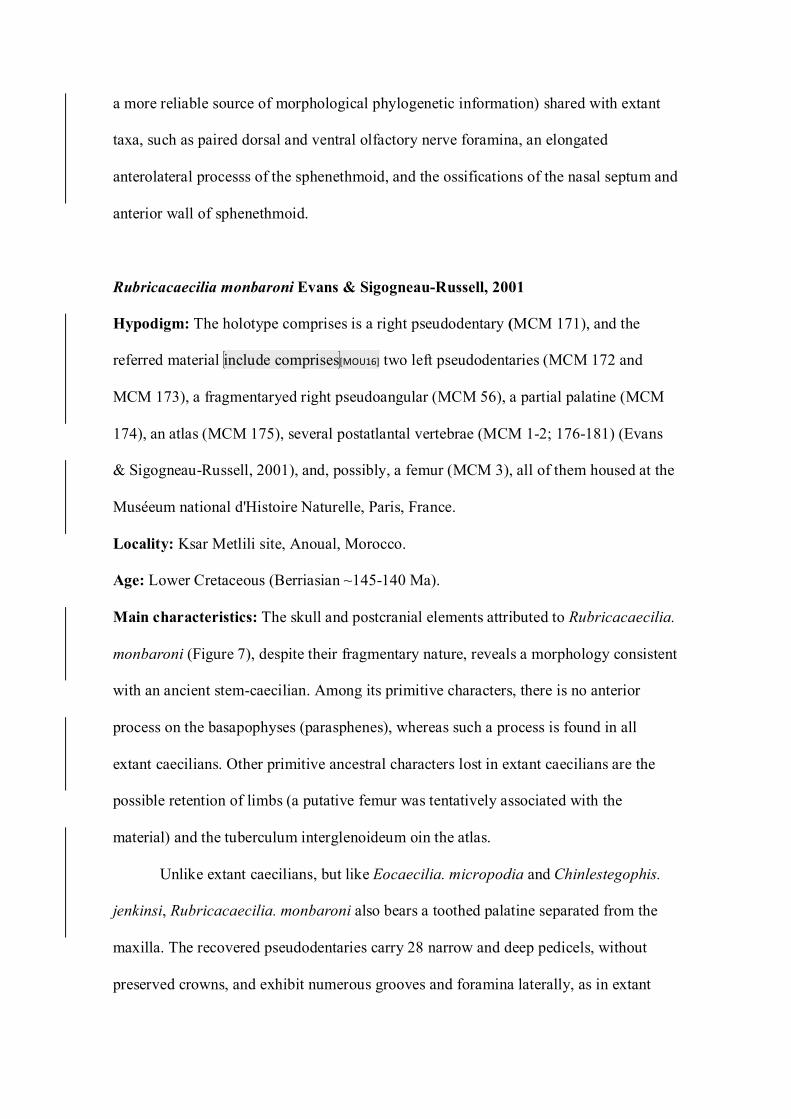
Rubricacaecilia monbaroni Evans & Sigogneau-Russell, 2001
Hypodigm: The holotype comprises is a right pseudodentary (MCM 171), and the
referred material include comprises[MOU16] two left pseudodentaries (MCM 172 and
MCM 173), a fragmentaryed right pseudoangular (MCM 56), a partial palatine (MCM
174), an atlas (MCM 175), several postatlantal vertebrae (MCM 1-2; 176-181) (Evans
& Sigogneau-Russell, 2001), and, possibly, a femur (MCM 3), all of them housed at the
Muséeum national d'Histoire Naturelle, Paris, France.
Locality: Ksar Metlili site, Anoual, Morocco.
Age: Lower Cretaceous (Berriasian ~145-140 Ma).
Main characteristics: The skull and postcranial elements attributed to Rubricacaecilia.
monbaroni (Figure 7), despite their fragmentary nature, reveals a morphology consistent
with an ancient stem-caecilian. Among its primitive characters, there is no anterior
process on the basapophyses (parasphenes), whereas such a process is found in all
extant caecilians. Other primitive ancestral characters lost in extant caecilians are the
possible retention of limbs (a putative femur was tentatively associated with the
material) and the tuberculum interglenoideum oin the atlas.
Unlike extant caecilians, but like Eocaecilia. micropodia and Chinlestegophis.
jenkinsi, Rubricacaecilia. monbaroni also bears a toothed palatine separated from the
maxilla. The recovered pseudodentaries carry 28 narrow and deep pedicels, without
preserved crowns, and exhibit numerous grooves and foramina laterally, as in extant
caecilians. The pseudoangular bears internal process was small, while the caecilian
typical retroarticular process, which is well developed and straight, as in rhinatrematids
(not dorsally arched, as in other extant caecilians), and also has a short internal process.
Remarks: Evans & Sigogneau-Russell (2001) According the several morphologic
characteristics mentioned above,interpreted Rubricacaecilia. monbaroni was interpreted
as a stem- caecilian, but more closelyr related to the crown groups than is Eocaecilia.
micropodia., but considered that its The incompleteness of the specimens, along
withand the tentative association of some materials justified, were used by Evans &
Sigogneau-Russell (2001) to justify not carrying out a phylogenetic analysis, which
could test such positioningthis hypothesis. Some recent phylogenetic analyses with
morphological data have emphasized braincase characters, which are undocumented in
R. monbaroni (e.g. Maddin, 2011; Maddin et al., 2012). Therefore, the relationship
between Rubricacaecilia and the other gymnophionomorphans must be interpreted with
caution until a cladistic analysis that includes it is carried out.
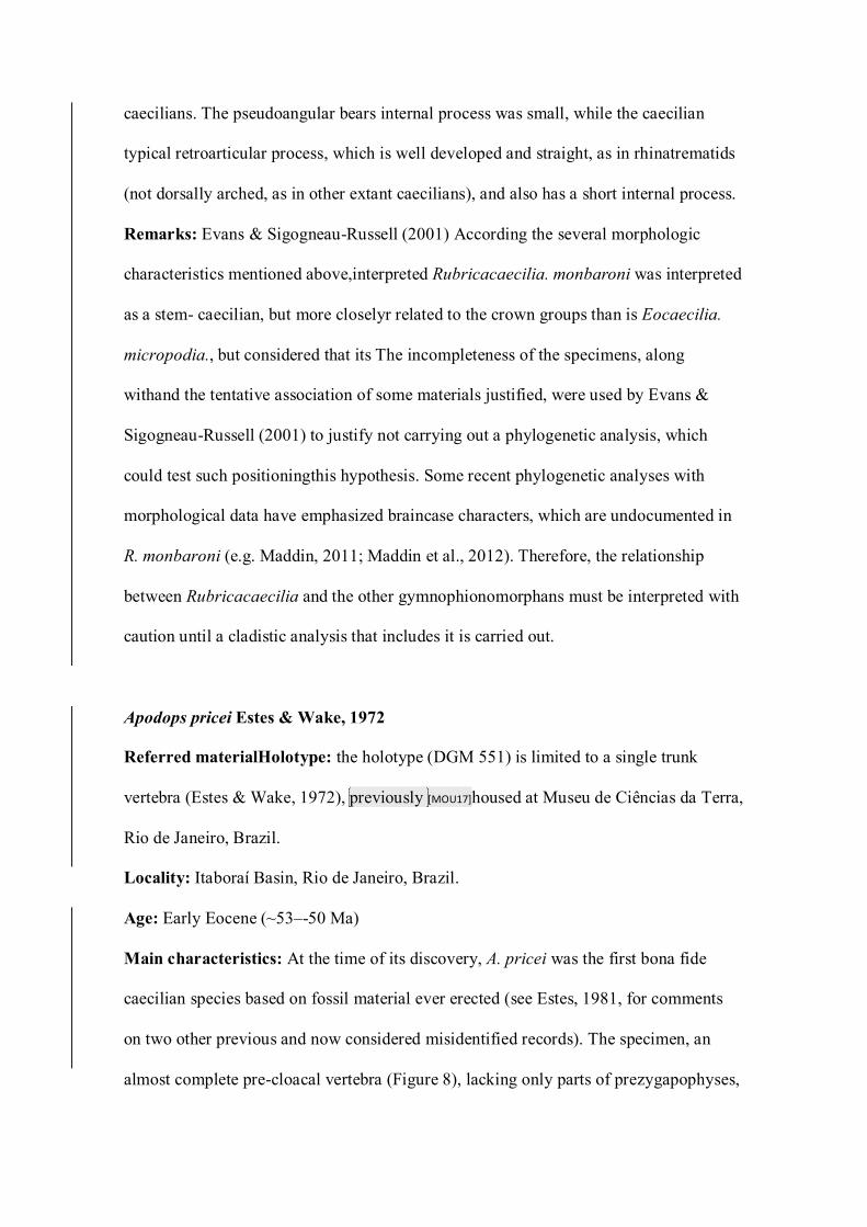
Apodops pricei Estes & Wake, 1972
Referred materialHolotype: the holotype (DGM 551) is limited to a single trunk
vertebra (Estes & Wake, 1972), previously [MOU17]housed at Museu de Ciências da Terra,
Rio de Janeiro, Brazil.
Locality: Itaboraí Basin, Rio de Janeiro, Brazil.
Age: Early Eocene (~53–-50 Ma)
Main characteristics: At the time of its discovery, A. pricei was the first bona fide
caecilian species based on fossil material ever erected (see Estes, 1981, for comments
on two other previous and now considered misidentified records). The specimen, an
almost complete pre-cloacal vertebra (Figure 8), lacking only parts of prezygapophyses,
parapophyses, and the posterior edge of the tip of neural arch posterior edge, and closely
resemblingese extant taxa, due to shared features such as highly excavated cotyles (with
chordal foramina filled by bone), general shape and proportions of centrum, neural arch,
and ventral keel, as well as large and anteriorly projectinged parapophyses
basapophyses.[MOU18]
Besides the well-developed ventral spine, the amphicoelous centrum also
exhibits a pronounced medial constriction. Even though most of the both parapophyses
have beenwere lost during the fossilization process, their broad bases can be used to
infer theits large size of the processes. The neural arch is flat and low, with a short
neural spine limited to its anterior half and two lateral deep groves that extends to the
rib-bearing surface. The vertebra also bears two large flanges (one on each side)
connecting the pre- and postzygapophyses.
Remarks: Unfortunately, the holotype (DGM 551) is currently lost (Lílian P.
Bergqvist, personal communication[MOU19]). Estes & Wake (1972) recognized
similarities between the vertebra of Apodops. pricei and some extant taxa, such as
Geotrypetes and Dermophis, but they considered that features like a high degree of
extensive ossification, a long and deep ventral keel, and deepest blood vessel grooves
were uniquely derived, and, therefore, erected a new genus and species taxon for it.
HoweverSubsequently, Taylor (1977) described some vertebrae of Siphonops, an extant
caecilian taxon widely distributed in Brazil, and mentioned features like the well-
developed ventral keel and the presence of lateral foramina that resemble those present
in A. pricei. Accounting for the current knowledge of caecilian vertebrae, tThese
structures are not unique among caecilians, and such variations can can also can be
related to the positioning of the vertebra alongin the column or ontogeny (Wake, 1980),
and therefore the holotype lacks diagnostic features to justify its specific status.
Furthermore, Wilkinson et al. (2011: p. 43) argued that such isolated fossilized
gymnophionan vertebrae cannot confidently be attributed to families due to our
incomplete knowledge of the morphological variation in extant taxa. Several collections
of isolated fossil vertebrae associated with caecilians were have subsequently been
described, but none werewas used to erect new species. Therefore, the designation of A.
pricei as a valid species needs to be revaluated. However, this is out of the scope of this
work and will be carried out in a further study.[MOU20]Thus, based on such
considerations, we proposed that A. pricei be considered a nomen dubium until new and
more complete materials are found.[RS21]
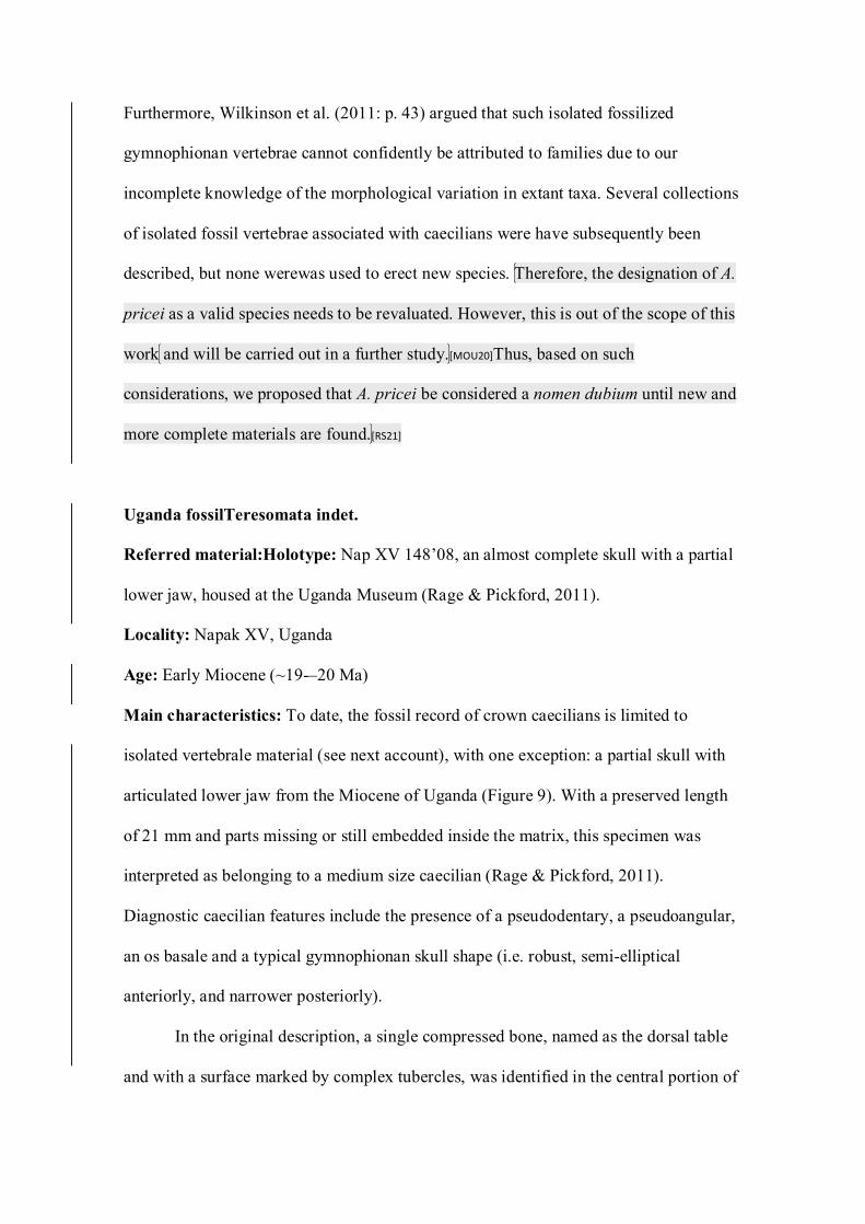
Uganda fossilTeresomata indet.
Referred material:Holotype: Nap XV 148’08, an almost complete skull with a partial
lower jaw, housed at the Uganda Museum (Rage & Pickford, 2011).
Locality: Napak XV, Uganda
Age: Early Miocene (~19-–20 Ma)
Main characteristics: To date, the fossil record of crown caecilians is limited to
isolated vertebrale material (see next account), with one exception: a partial skull with
articulated lower jaw from the Miocene of Uganda (Figure 9). With a preserved length
of 21 mm and parts missing or still embedded inside the matrix, this specimen was
interpreted as belonging to a medium size caecilian (Rage & Pickford, 2011).
Diagnostic caecilian features include the presence of a pseudodentary, a pseudoangular,
an os basale and a typical gymnophionan skull shape (i.e. robust, semi-elliptical
anteriorly, and narrower posteriorly).
In the original description, a single compressed bone, named as the dorsal table
and with a surface marked by complex tubercles, was identified in the central portion of
the skull roof. This arrangement, although common in other lissamnphibianurans[MOU22]
(sometimes the bone is called the frontoparietal), would be unique among caecilians, as
because all of them exhibit separated parietals and frontals. However, based on the
published images, a suture marks can be observed partially delimiting the parietals and
the frontals, as in all other gymnophionans. Notches on either side of Bordering laterally
the frontals, two notches related toare the dorsalmost portions of the orbits were
preservedorbital margins. The compact skull structure implies in a stegokrotraphic skull
or the presence of short a narrow upper temporal fenestrae. Posteriorly, tThe specimen
bears posteriorly preserves the parasphenoid portion of the os basale and two occipital
condyles. Only anterior fragments of a pseudodentary (including some teeth cross
sections of marginal dentitionteeth) and parts of anterodorsal and anteroventral
processes of thea pseudoangular were are preserved. Between these two bones, lies a
long and oblique suture.
Remarks: Specimen Nap XV 148’08 was initially attributedprovisionally referred to
Caeciliidae , following the delimitation proposed by sensu Frost et al. (2006), in which
Typhlonectidae and Scolecomorphidae were considered subjective synonyms of
Caeciliidae to ensure monophyly of the latter. According to this taxonomy, Caeciliidae
included all the gymnophionan taxa, except Rhinatrematidae and Ichthyophiidae.
Wilkinson et al. (2011) proposed a different strategy to prevent Caeciliidae paraphyly,
by restricting its content. Considering this scenario, Caeciliidae Frost et al., 2006 is
roughly, a group equivalent to the Teresomata of Wilkinson & Nussbaum (2006). , a
larger clade that comprises eight caecilian families: Caeciliidae (with a reduced content
that includes only Caecilia and Oscaecilia), Herpelidae, Siphonopidae, Dermophiidae,
Indotyphlidae, Chikilidae, Typhlonectidae and Scolecomorphidae (i. e. Wilkinson et al.,
2011; Kamei et al., 2012). As the features of Uganda fossil caecilian, such as the
presence of orbits, nasopremaxilla, posteriorly separated vomer, short or absent
temporal fenestrae, also occurs in numerous teresomatan taxa, we assign Nap XV
148’08 to this clade (it can also be considered part of Caeciliidae sensu Frost et al.,
2006). Given that some portions of the skull remain Furthermore, as several skull areas
are embedded inside the matrix, with the use of modern techniques, as CT Scan further
preparation and imaging (i.e. CT scanning), may help to reveal informativeadditional
phylogenetic characters[MOU23] for making a more accurate identification phylogenetic
characters can be potentially revealed and used for a more accurate assignment.
Isolated postatlantal vertebraeGymnophiona indet.
Referred material: Four trunk vertebrae (Vb-659; Vb-660; Vb-661; and Vb-781,
housed at Technical University of Berlin) from Sudan (Werner, 1994; Evans et al.
1996); an isolated damaged vertebra along with (MHNC-2635, housed at the Museo de
Historia Natural de Cochabamba) and other seven other vertebrae (the material is
deposited in the Museo de Historia Natural de Cochabamba, but in the paper, only the
collection number of a single vertebra, MHNC 8583, is mentioned,) from two localities
in Bolivia (Rage, 1991; Gayet et al., 2001);, three isolated and large anterior vertebrae
(IGM 183404; IGM 184791; and IGM 182186, housed at Florida Museum of Natural
History) from Colombia (Hecht & LaDuke, 1997); and a single vertebra (the authors did
not provide the identification number of the specimen nor the institution in which it is
stored) from Mexico (Wake et al., 1999).
Localities: Wadi Milk Formation, Wadi Abu Hashim, Sudan; Santa Lucía Formation,
Tiupampa, Bolivia; El Molino Formation, Pajcha Pata, Bolivia; Santa Lucía Formation,
Tiupampa, Bolivia; Honda Group, La Venta, Colombia; and Paso de la Amada site,
Chiapas, Mexico.
Ages: Campanian (~79.2 Ma) for Sudanese fossils; mMiddle Maastrichtian (~68.4 Ma)
and Eearly Paleocene (~64–-62 Ma) for Bolivian material, Mmiddle Miocene (~13.8–-
11.6 Ma) for Colombian specimens fossils, and Quaternary (1200–-1350 B.C.) for
Mexican vertebrafossil.
Main characteristics: Morphologically, the caecilian postcranial axial skeleton is quite
conservative. Not surprisingly, all the fossil isolated vertebrae assigned to the group
exhibit the same diagnostic features, including amphicoelous centrum, broad
anteroventral parapophyses, a well-developed ventral keel, and a low and flattened
neural arch.
Remarks: According to Taylor (1977), anterior trunk vertebrae and, more importantly,
the atlas of gymnophionans contain phylogenetic information. Unfortunately, none of
these materials were preserved in a crown caecilian fossilno examples of these vertebrae
are known for published fossil crown-caecilians. Therefore, due to the lack of most
more specific diagnostic characters, all of the above-listed fossil vertebrae can only be
these specimens were assigned to Gymnophiona indet.
Unpublished and Ppossible indeterminate records
Gardner & Rage (2016) mentioned a caecilian vertebra from the lower–middle
Eocene of Glib Zegdou, Algeria. The description of this vertebra is part of a larger
project on the herpetofauna from Glib Zegdou not yet published (James D. Gardner,
personal communication[MOU24]). Therefore, due to the lack of information (e.g. detailed
description, images, and collecting number) on this specimen, it is presented here, in a
separate section from the other already published records.
An undescribed atlas (without collection number and housed at Muséum
National d'Histoire Naturelle) from Colombia, was collected by Hoffstetter in 1966 and
figured in Hetch & LaDuke (1997). This specimen was tentatively associated with the
other vertebrae from La Venta, based mainly on their large size.
In a faunal list forof Maboko Island, Kenya, originally published by Andrews et
al. (1981, table 1), a record assigned to a Miocene Nnectrideian [MOU25]was reported, but
without images or detailed descriptions. Due to the significant temporal gap that this
record would imply in the nectrideian fossil record (nectrideians were presumably
extinct at the end of the Permian), subsequent works considered that this material is
actually a lissamphibian, probably either a salamander (Van Dijk, 1995) or a caecilian
(Gardner & Rage, 2016). However, until the material is reevaluated, such assignments
remain uncertain, though both hypotheses are much more likely than the initial
nectrideian assignment.
Possible calibration points constraints [MOU26]for Gymnophionomorpha
Calibration constraints based on the caecilian fossil record are uncommon,.
Pprobably due to its scarcity, usuallybecause few fossil occurrences are available.
Instead, other taxa are used, such as batrachians (e.g. San Mauro et al., 2014).
Following the recommendations of Parham et al. (2012), here we provide calibrations
for four three nodes of the gymnophionomorphans. We attempted to use the most recent
or widely accepted age estimates for each location. Calibrations are highly dependent of
on phylogeny and stratigraphy, and thus the latest, best-supported dating and
phylogenetic hypotheses were considered.
GYMNOPHIONOMORPHA Marjanovic & Laurin, 2008[RS27]
Node Calibration: Divergence between the total clade of caecilians and its nearest
crown sister taxon (Batrachia).
Oldest fossil: Triadobatrachus massinoti (Piveteau, 1936), from the Sakamena Group,
Madagascar (the holotype, MHNH MAE 126, comprises an almost complete individual,
and is housed at Muséum National d’Histoire Naturelle)Eocaecilia micropodia Jenkins
& Walsh, 1993
Phylogenetic Justification: If lissamphibians and batrachians are in facare botht a
monophyletic group, then the divergence time betweenage of Batrachia and
Gymnopionomorpha is the same. In this scenario, the oldest fossil available is the stem-
batrachian Triadobatrachus, from the Early Triassic of Madagascar. However,
considering the polyphyletic hypothesis for the origin of lissamphibians, only members
of the Gymnophionomorpha lineage can be used for providing constraints. In this case,
the oldest fossil available is Eocaecilia micropodia, from the Early Jurassic of
USA.[MOU28] Phylogenetic analyzes have repeatedly confirmed the relationship between
E. micropodia and extant caecilians (e.g. Maddin et al., 2012), and its position as a
sister group of all other gymnophionomorphs is supported by many characters
(discussed in more detail above) and is widely accepted.
Minimum age: 181 Ma251.2 Ma (assuming lissamphibian monophyly)
Soft Maximum age: 186.4 Ma251.9 Ma[MOU29]
Age Justification: The siltstones and sandstones deposits of Kayenta Formation were
historically thought to be Triassic-Jurassic or Early Jurassic age (see Lucas et al., 2005)
in age. Due to the lack of available ash beds and useful stratigraphic fossils, the age
estimates of such deposits were not very accurate. However, recently, the first
radiometric date for the unit estimated an Early Jurassic age of 183.7 +/- 2.7, changing
the temporal range of Kayenta Formation from Sinemurian-Pliensbachian to
Pliensbachian-Toarcian (Marsh et al. 2015). Traditionally, estimates for the age of the
Sakamena Group in Madagascar range from the Late Permian to the Middle Triassic.
Due to the absence of radiometric and magnetostratigraphic dating, age estimates are
not so accurate (Benton et al., 2015). Based on palynological evidence, an Induan age
was proposed for this unit (Wescott & Diggens, 1998; Nowak et al., 2018). Considering
the polyphyletic hypothesis, then the oldest Gymnophionomorpha fossil available is
Eocaecilia micropodia, from the Kayenta Formation, recently dated by Marsh et al.
(2015) in 183.7 +/- 2.7 Ma (Pliensbachian-Toarcian).[MOU30]
Comment: If the hypothesis proposed by Pardo et al., (2017) were correct, the
minimum age for the group would be extended to the Norian (208-227 Ma). However,
we consider that calibration of the stem-node Gymnophionomorpha with C. jenkinsi to
estimate the divergence time of gymnophionans should be avoided until its phylogenetic
affinities is thoroughly tested and clarified. Until that happens, Eocaecilia macropodia
seems to represent a better fit for this calibration constraint (Marjanović & Laurin,
2009, 2019). A large gap (approximately 67 Ma), between the origin of Lissamphibia
and the first record of Gymnophionomorpha, still persists[MOU31]. It is a strong indicative
on the existence of a hidden diversity, which Chinlestegophis jenkinsi could fill, at least
partially.
GYMNOPHIONA Rafinesque-Schmaltz, 1814
Node Calibration: Divergence between Rhinatrematidae Nussbaum, 1977 and
Rhinatrema bivittatum and Stegokrotaphia Canatella and Hillis, 1993. Caecilia
tentaculata
Oldest fossil: Gymnophiona indet., based on four trunk vertebrae from the Late
Cretaceous (Campanian) of Sudan (Werner, 1994; Evans et al., 1996).
Phylogenetic Justification: The Sudanese vertebrae exhibit a morphology typically
associated with the Gymnophiona crown, including features like an amphicoelous
centra, flat neural spine, prominent ventral keel and, mainly, a parapophyses strongly
projected anteroventraly. Such characteristics allow safely assign the specimens to this
taxon.
Minimum age: 76.82.1 Ma
Soft Maximum age: 81.6 Ma
Age Justification: Wadi Milk Formation, located in the nNorthern Sudan, includes
deposits of braided river systems, interspersed laterally with sediments associated
todeposited in flood plains, lakes, and meandering rivers., Hhistorically, it was
considered to be Albian-Santonian in age, on the basis of palynological evidence data
estimates (Schank, 1990). However, recent U-Pb radiometric dating found a much
younger age for such unitof: 79.2 ± 2.4 Ma, or equivalent to theassigning it to middle
Campanian (Agyemang et al., 2019).
Comment: Previously, Benton et al. (2015) proposed a Paleogene minimum age for
this cladeGymnophiona, based on the record of Apodops. pricei. Nevertheless, t The
Sudanese vertebrae share with crown-gymnophionans the presence of well-developed
parapophyses, which are absent in both Eocaecilia and Rubricacaecilia, and this
supports the extension ofpushing the temporal range of crown-Gymnophiona back to
the middle Campanian. However, this remains tentative, because these taxa the
Sudanese taxon alternatively could also fit just outside the gymnophionan crown.
TERESOMATA Wilkinson & Nussbaum, 2006
Node Calibration: Divergence between Scolecomorphidae Taylor, 1969 and a
nameless group formed by Caeciliidae, Typhlonectidae, Herpelidae, Chikilidae,
Dermophiidae, Indotyphlidae, and Siphonopidae.Scolecomorphus kirkii and Caecilia
tentaculata.
Oldest fossil: Napak gymnophionan Nap XV 148’08, an almost complete skull
articulated with a partial lower jaw from the Miocene (Burdigalian) of Uganda (Rage
and Pickford, 2011).
Phylogenetic Justification: Rage and Pickford (2011) assigned this fossil to the
Caeciliidae based on the combination of the following characters: posterior part of
vomers clearly separated, presence of orbits, fusion of premaxillae and nasals. However,
these features are present in several families of crown-caecilians (Wilkinson et al.,
2011), rejecting the original allocation until a more detailed study of the material is
concluded. Thus, the fossil skull from Uganda is here assigned to Teresomata based on
the present understanding of caecilian systematics. As studies with the material are
underway (Gardner & Rage, 2016), it has the potential to be assigned again to a less
inclusive taxon within Terosomata. As Nap XV 148’08 has not yet been included in
phylogenetic analyzes, its positioning is still uncertain, as it could lie outside the
Teresomata crown. Therefore, this specimen can only be used to constrain the split
between Ichthyophiidae and Teresomata.
Minimum age: 19 Ma
Soft Maximum age: 20 Ma[MOU32]
Age Justification: Napak XV, located in northeastern Uganda, comprises multiple
fossiliferous deposits intercalated with tuffs, located on and around an ancient volcano.
Dates on these tuffs, calculated with potassium-argon dating (Bishop et al., 1969), vary
between 19.5 ± 2 Ma and 18.3 ± 0.4; however, the latter date is considered an
anomalous result, and currently these deposits are assigned to the early Burdigalian,
with an estimated age between 19–-20 Ma (Werdelin, 2010, fig. 3.4).
Comment: There is currently insufficient evidence to refer Oolder unnamed fossil
caecilians from the Eearly Paleocene (Rage, 1991) and MaastrichtianLate Cretaceous
(Gayet et al., 2001) of Bolivia, and Early CretaceousCampanian of Sudan (Evans et al.,
1996) can potentially be associated to Teresomata. However, the evidence supporting
this attribution is insufficient. Therefore, the oldest fossil that can be undoubtedly
assigned to this clade is the skull Nap XV 148’08.
Conclusions
Gymnophionans remain are the most poorly known group of tetrapods,
particularly in aspects of their evolutionary history. Despite the paucity of its fossil
record, fossil caecilians directly affect our understanding of taxonomy, phylogeny and
biogeography of extant caecilians, and they help to discriminate between hypotheses
about the origin of Lissamphibia.
The gymnophionan fossil record shows wide temporal gaps, even in the
Cenozoic, in which (not considering Apodops. pricei) no diagnostic material has been
assigned to an extant species, genus, or even family so far. Therefore, the discovery of
new, more complete and diagnostic fossils assignableed to the caecilian crown-group
may would likely make a substantial contribution to contribute to resolvinge
phylogenetic and biogeographic questions about caecilian clades and better constraining
molecular clocks.
Acknowledgements[RS33]
We thank M. H. Wake D. Marjanović, A. O. Maciel, James Gardner and the threewo
anonymous reviewers[MOU34] for her their comments on the Apodops specimen
manuscript, the Editor Prof. John A Allen for his efficient handling of the draft, and the
Conselho Nacional de Desenvolvimento Científico e Tecnológico (CNPq) for their
financial support.
References
AmphibiaWeb. 2020. Information on amphibian biology and conservation. Berkeley,
CA: AmphibiaWeb. Available at: https://amphibiaweb.org.
Anderson JS, Reisz RR, Scott D, Fröbisch NB, Sumida SS. 2008. A stem batrachian
from the Early Permian of Texas and the origin of frogs and salamanders. Nature
453, 515–-518.
Andrews P, Meyer GE, Pilbeam DR, Couvering JAV, Couvering JAHV. 1981. The
Miocene fossil beds of Maboko Island, Kenya: geology, age, taphonomy and
palaeontology. Journal of Human Evolution 10: 35–-48.
Agyemang PCO, Roberts EM, Bussert R, Evans D, Muller J. 2019. U-Pb detrital
zircon constraints on the depositional age and provenance of the dinosaur-bearing
Upper Cretaceous Wadi Milk formation of Sudan. Cretaceous Research 97: 52–-
72.
Ascarrunz E, Rage JC, Legreneur P, Laurin M. 2016. Triadobatrachus massinoti,
the earliest known lissamphibian (Vertebrata: Tetrapoda) re-examined by µCT-
sScan, and the evolution of trunk length in batrachians. Contributions to Zoology
85: 201–234.
Behrensmeyer AK, Kidwell SM, Gastaldo RA. 2000. Taphonomy and pPaleobiology.
Paleobiology 26: 103–-147.
Benton MJ, Donoghue PCJ, Asher RJ, Friedman M, Near TJ, Vinther J. 2015.
Constraints on the timescale of animal evolutionary history. Palaeontologia
ElectronicaPalaeontol Electron 18: 1–-116.
Bishop WW, Miller JA, Fitch FJ. 1969. New potassium-argon age determinations
relevant to the Miocene fossil mammal sequence in East Africa. American
Journal of Science 267: 669–-699.
Cannatella DC. Hillis DM. 1993. Amphibian relationships: phylogenetic analysis of
morphology and molecules. Herpetological Monographs 7: 1-7.
Carroll RL, Currie PJ. 1975. Microsaurs as possible apodan ancestors. Zool J Linn
Soc 57: 229–-247.
Carroll RL, Gaskill P. 1978. The oOrder Microsauria. Philadelphia: American
Philosophical Society.
Carroll RL, Holmes R. 1980. The skull and jaw musculature as guides to the ancestry
of salamanders. Zoological Journal of the Linnean Society 68: 1–40.
Cogger HG, Zweifel RG. 1998. Encyclopedia of rReptiles & aAmphibians.
Cambridge: Academic Press.
Cogger HG, Zweifel RG. 1998. Encyclopedia of reptiles & amphibians. Cambridge:
Academic Press.
De Blainville HM. 1816. Prodrome d'une nouvelle distribution systématique du règne
animal. Bulletin de la Societe philomathique de Paris 8: 105–124.
Dilkes D. 2015. Carpus and tarsus of Temnospondyli. Vertebrate Anatomy Morphology
Palaeontology 1: 51-87.
Dubois A. 2004. The higher nomenclature of recent amphibians. Alytes 22: p. 1–14.
Ducey PK, Formanowicz DR, Boyet L, Mailloux J, Nussbaum RA. 1993.
Experimental examination of burrowing behavior in caecilians (Amphibia:
Gymnophiona): effects of soil compaction on burrowing ability of four species.
Herpetologica 49: 450-457.
Dubois A. 2004. The higher nomenclature of recent amphibians. Alytes 22: p. 1-14.
Duellman WE, Trueb L. 1994. Biology of Amphibians. Baltimore: The Johns Hopkins
University Press.
Estes R. 1981. Encyclopedia of Herpetology Part 2, Gymnophiona, Caudata. Stuttgart:
Gustav Fischer Verlag.
Estes R, Wake MH. 1972. The first fossil record of caecilian amphibians. Nature 239:
228–231.
Evans SE, Milner AR, Werner C. 1996. Sirenid salamanders and a gymnophionan
amphibian from the Cretaceous of the Sudan. Palaeontology 39: 77–95.
Evans SE, Sigogneau-Russell D. 2001. A stem-group caecilian (Lissamphibia:
Gymnophiona) from the Lower Cretaceous of North Africa. Paleontology 44:
259–273.
Feller AE, Hedges SB. 1998. Molecular evidence for the early history of living
amphibians. Molecular Phylogenetics and Evolution 9: 509–516.
Fernandez V, Abdala F, Carlson KJ, Rubidge BS, Yates A, Tafforeau P. 2013.
Synchrotron reveals Early Triassic odd couple: injured amphibian and aestivating
therapsid share burrow. PLoS One 8: e64978.
Frost DR, Taran G, Faivovich J, Bain RH, Haas A, Haddad CFB, De Sá RO,
Channing A, Wilkinson M, Donnellan SC, Raxworthy CJ, Campbell JA,
Blotto BL, Moler P, Drewes RC, Nussbaum RA, Lynch JD, Green DM,
Wheeler WC. 2006. The amphibian tree of life. Bulletin of the American Museum
of Natural History 297: 1–-370.
Frost DR. 2020. Amphibian Species of the World: an Online Reference. Version 6.1
(June 6 2020). Electronic Database accessible at
https://amphibiansoftheworld.amnh.org/index.php. American Museum of Natural
History, New York, 2020. doi.org/10.5531/db.vz.0001
Gardner JD, Rage JC. 2016. The fossil record of lissamphibians from Africa,
Madagascar, and the Arabian Plate. Palaeobiodiversity and Palaeoenvironments
96: 169–-220.
Gayet M, Marshall LG, Sempere T, Meunier FJ, Cappetta H, Rage JC. 2001.
Middle Maastrichtian vertebrates (fishes, amphibians, dinosaurs and other
reptiles, mammals) from Pajcha Pata (Bolivia). Biostratigraphic, palaeoecologic
and palaeobiogeographic implications. Palaeogeography Palaeoclimatology
Palaeoecology 169: 39–-68.
Gower DJ, Kupfer A, Oommen OV, Himstedt W, Nussbaum RA, Loader SP,
Presswell B, Muller H, Krishna SB, Boistel R, Wilkinson M. 2002. A
molecular phylogeny of ichthyophiid caecilians (Amphibia: Gymnophiona:
Ichthyophiidae): out of India or out of South East Asia? Proceedings of Royal
Society of London 269: 1563-–1569.
Haddoumi H, Allain R, Meslouh S, Metais G, Monbaron M, Pons D, Rage JC,
Vullo R, Zouhi S, Gheerbran E. 2016. Guelb el Ahmar (Bathonian, Anoual
Syncline, eastern Morocco): First continental flora and fauna including mammals
from the Middle Jurassic of Africa. Gondwana Research 29: 290–-319.
Hebrard JJ, Maloiy GM, Alliangana DM. 1992. Notes on the habitat and diet of
Afrocaecilia taitana (Amphibia: Gymnophiona). Journal of Herpetology 26: 513–
515.
Hecht MK, Laduke TC. 1997. Limbless Tetrapods. In: Kay RF, Madden RH, Cifelli
RL, Flynn JJ, eds. Vertebrate paleontology in the Neotropics: The Miocene fauna
of La Venta, Colombia. Washington: Smithsonian Institution Press, 95–-99.
Irisarri I, Baurain D, Brinkmann H, Delsuc F, Sire JY, Kupfer A, Petersen J,
Jarek M, Meyer A, Vences M. 2017. Phylotranscriptomic consolidation of the
jawed vertebrate timetree. Nature Ecology & Evolution 1: 1370–-1378.
Jaekel O. 1909. Über die Klassen der Tetrapoden. Zoologischer Anzeiger 34: 193–212.
Jared C, Navas CA, Toledo RC. 1999. An appreciation of the physiology and
morphology and physiology of caecilians (Amphibia: Gymnophiona).
Comparative Biochemistry and Physiology A 123: 313–328.
Jared C, Mailho-Fontana PL, Marques-Porto R, Sciani JM, Pimenta DC, Brodie
EDJ, Antoniazzi MM. 2018. Skin gland concentrations adapted to different
evolutionary pressures in the head and posterior regions of the caecilian
Siphonops annulatus. Scientific Reports 8: 1-–7.
Jared C, Mailho‐Fontana PL, Jared SG, Kupfer A, Delabie JHC, Wilkinson M,
Antoniazzi MM. 2019. Life history and reproduction of the neotropical caecilian
Siphonops annulatus (Amphibia, Gymnophiona, Siphonopidae), with special
emphasis on parental care. Acta Zoologica 100: 292–302.
Jenkins FA, Walsh DM. 1993. An Early Jurassic caecilian with limbs. Nature 365:
246–250.
Jenkins FA, Walsh DM, Carroll RL. 2007. Anatomy of Eocaecilia micropodia, a
limbed caecilian of the Early Jurassic, Bulletin of the Museum of Comparative
Zoology 158: 285–-365.
Kamei RG, San Mauro D, Gower DJ, Bocxlaer IV, Sherrat E, Thomas A, Babu S,
Bossuyt F, Wilkinson M, Biju SD. 2012. Discovery of a new family of
amphibians from northeast India with ancient links to Africa. Proceedings
Biological Sciences 279: 2396–2401.
Kleinteich T, Maddin HC, Herzen J, Beckmann F, Summers AP. 2012. Is solid
always best? Cranial performance in solid and fenestrated caecilian skulls.
Journal of Experimental Biology 215: 833–844.
Kouete MT, Wilkinson M, Gower DJ. 2012. First reproductive observations for
Herpele Peters, 1880 (Amphibia: Gymnophiona: Herpelidae): evidence of
extended parental care and maternal dermatophagy in H. squalostoma
(Stutchbury, 1836). International Scholarly Research Notices 2012: 1–7.
Kozlowski J, Gawelczyk AT. 2002. Why are species’ body size distributions usually
skewed to the right? Functional Ecology 16: 419–32.
Laurin M, Lapauze O, Marjanović D. 2019. What do ossification sequences tell us
about the origin of extant amphibians? bioRxiv 352609, ver. 4 peer-reviewed by
PCI Paleo. DOIdoi: https://doi.org/10.1101/3[MOU35]52609.
Lasseron M, Allain R, Gheerbrant E, Haddoumi H, Jalil NE, Metais G, Rage JC,
Vullo R, Zouhi S. 2019. New data on the microvertebrate fauna from the Upper
Jurassic or lowest Cretaceous of Ksar Metlili (Anoual Syncline, eastern Morocco).
Geological Magazine 157: 367-392.
Laurin M, Arntzen JW, Báez AM, Bauer AM, Damiani R, Evans SE, Kupfer A,
Larson A, Marjanović D, Müller H, Olsson L, Rage JC, Walsh D. 2020.
Lissamphibia. In: De Queiroz K, Cantino PD, Gauthier JA, eds. Phylonyms: An
Implementation of PhyloCode. Boca Raton: CRC Press, 755–-760.
Loader SP, Pisani D, Cotton JA, Gower DJ, Day JJ, Wilkinson M. 2007. Relative
time scales reveal multiple origins of parallel disjunct distributions of African
caecilian amphibians. Biology Letters 3: 505–508.
Maddin HC. 2011. Deciphering morphological variation in the braincase of caecilian
amphibians (gymnophiona). Journal of Morphology 272: 850-–871.
Maddin HC, Jenkins FA, Anderson JS. 2012. The braincase of Eocaecilia
micropodia (Lissamphibia, Gymnophiona) and the origin of caecilians. PLoS
ONE 7: 1–-12.
Maddin HC, Venczel M, Gardner JD, Rage JC. 2013. Micro-computed tomography
study of a three-dimensionally preserved neurocranium of Albanerpeton
(Lissamphibia, Albanerpetontidae) from the Pliocene of Hungary. Journal of
Vertebrate Paleontology 33: 568–587.
Marjanović D, Laurin M. 2007. Fossils, molecules, divergence times, and the origin
of lissamphibians. Systematic Biology 56: 369–-388.
Marjanović D, Laurin M. 2008a. A reevaluation of the evidence supporting an
unorthodox hypothesis on the origin of extant amphibians. Contributions to
Zoology 77: 149–-199.
Marjanović D, Laurin M. 2008b. Assessing confidence intervals for stratigraphic
ranges of higher taxa: the case of Lissamphibia. Acta Palaeontologica Polonica
Acta Palaeont Pol 53: 413–432. doi: http://dx.doi.org/10.4202/app.2008.0305.
Marjanović D, Laurin M. 2009. The origin(s) of modern amphibians: a commentary.
Evolutionary Biology 36: 336–338.
Marjanović D, Laurin M. 2013. The origin(s) of extant amphibians: a review with
emphasis on the “lepospondyl hypothesis”. Geodiversitas 35: 207–72.
Marjanović D, Laurin M. 2019. Phylogeny of Paleozoic limbed vertebrates reassessed
through revision and expansion of the largest published relevant data matrix.
PeerJ 6:e5565. 10.7717/peerj.5565.
Marsh AD, Rowe T, Simonetti A, Stockli D, Stockli L. 2015. Preliminary U-Pb
detrital zircon dates from the Kayenta Formation of Arizona. In: Sankey J, Biewer
J, eds. Western Association of Vertebrate Paleontology Annual Meeting: Program
with Abstracts. Turlock: California Digital Library, 10.
Measey GJ, Herrel A. 2006. Rotational feeding in caecilians: putting a spin on the
evolution of cranial design. Biology Letters 2: 485–487.
Müller H. 2006. Ontogeny of the skull, lower jaw, and hyobranchial skeleton of
Hypogeophis rostratus (Amphibia: Gymnophiona: Caeciliidae) revisited. Journal
of Morphology 267: 968–-986.
Nowak H, Schneebeli-Hermann E, Kustatscher E. 2018. Correlation of Lopingian to
Middle Triassic Palynozones. Journal of Earth Science 29: 755–777.
Nussbaum RA. 1977. Rhinatrematidae: A new family of caecilians (Amphibia:
Gymnophiona). Occasional Papers of the Museum of Zoology, University of
Michigan 682: 1-30.
Nussbaum RA, Naylor BG. 1982. Variation in the trunk musculature of caecilians
(Amphibia: Gymnophiona). Journal of Zoology 198: 383–398.
Nussbaum RA. 1983. The evolution of a unique dual jaw‐closing mechanism in
caecilians (Amphibia: Gymnophiona) and its bearing on caecilian ancestry.
Journal of Zoology 199: 545–-554.
Oppel M. 1811. Die ordnungen, familien und gattungen der reptilien als prodrom einer
naturgeschichte derselben. Munchen: Joseph Lindauer.
Pardo JD, Anderson JS. 2016. Cranial Morphology of the Carboniferous-Permian
Tetrapod Brachydectes newberryi (Lepospondyli, Lysorophia): New Data from
µCT. PloS one 11: e0161823.
Pardo JD, Small BJ, Huttenlocker AK. 2017. Stem caecilian from the Triassic of
Colorado sheds light on the origins of Lissamphibia. Proceedings of the National
Academy of Sciences of the United States of America 114: 1-–7.

Parham JF, Donoghue PCJ, Bell CJ, Calway TD, Head JJ, Holroyd PA, Inoue JG,
Irmis RB, Joyce WG, Ksepka DT, Patané JSL, Smith ND, Tarver JE, Van
Tuinen M, Yang Z, Angielczyk KD, Greenwood J, Hipsley CA, Jacobs L,
Makovicky PJ, Müller J, Smith KT, Theodor JM, Warnock, RCM, Benton
MJ. 2012. Best practices for justifying fossil calibrations. Systematic Biology 61:
346–-359.
Peterhans JCP, Wrangham RW, Carter ML, Hauser MD. 1993. A contribution to
tropical rain forest taphonomy: retrieval and documentation of chimpanzee
remains from Kibale Forest, Uganda. Journal of Human Evolution 25: 485–514.
Pyron RA. 2010. A likelihood method for assessing molecular divergence time
estimates and the placement of fossil calibrations. Systematic Biology 59: 185-
194.
Pyron RA. 2011. Divergence time estimation using fossils as terminal taxa and the
origins of Lissamphibia. Systematic Biology 60: 466–-481.
Pyron RA, Wiens JJ. 2011. A large-scale phylogeny of Amphibia including over 2800
species, and a revised classification of extant frogs, salamanders, and caecilians
Molecular Phylogenetics and Evolution 61: 543-583.
Pyron RA. 2014. Biogeographic analysis reveals ancient continental vicariance and
recent oceanic dispersal in amphibians. Systematic Biology 63: 779–-797.
Rafinesque-Schmaltz. 1814. Fine del Prodromo d’Erpetologia Siciliana. Specchio delle
Scienze o Giornale Enciclopedico di Sicilia 2: 102–104.
Rage JC. 1991. Gymnophionan Amphibia from the Early Paleocene (Santa Lucia
Formation) of Tiupampa (Bolivia): the oldest known Gymnophiona. Revista
Tecnica de YPFB 12: 3–-4.
Rage JC, Pickford M. 2011. Discovery of a Gymnophionan skull (?Caeciliidae,
Amphibia) in the Early Miocene of Uganda. Geo-Pal Uganda 4: 1–-9.
Ramaswami LS. 1941. Some aspects of the cranial morphology of Uraeotyphlus
narayani (Apoda). Records of the Indian Museum 43: 143–207.
Renous S, Gasc JP. 1989. Body and vertebral proportions in Gymnophiona
(Amphibia): diversity of morphological types. Copeia 1989: 837–-847.
Roelants K, Gower DJ, Wilkinson M, Loader SP, Biju SD, Guillaume K, Bossuyt
F. 2007. Global patterns of diversification in the history of modern amphibians.
Proceedings of the National Academy of Sciences 104: 887–-892.
Ruta M, Coates I. 2007. Dates, nodes and character conflict: addressing the
lissamphibian origin problem. Journal of Systematic Palaeontology 5: 69-122.
San Mauro D. 2010. A multilocus timescale for the origin of extant amphibians.
Molecular Phylogenetic Evolution 56: 554–-561.
San Mauro D, Gower DJ, Muller H, Loader SP, Zardoya R, Nussbaum RA,
Wilkinson M. 2014. Life-history evolution and mitogenomic phylogeny of
caecilian amphibians. Molecular Phylogenetic Evolution 73: 177–-189.
Schoch RR. 2008. A nNew Stereospondyl from the German Middle Triassic, and the
oOrigin of the Metoposauridae. Zoological Journal of the Linnean Society 152:
79–113.
Schoch RR, Milner AR. 2004. Structure and implications of theories on the origin of
lissamphibians. In: Arratia G, Wilson MVH, Cloutier R, eds. Recent Advances in
the Origin and Early Radiation of Vertebrates. Munich: Dr Friedrich Pfeil Verlag,
345–-377.
Schrank E. 1990. Palynology of the clastic Cretaceous sediments between Dongola and
Wadi Muqaddam, northern Sudan. Berliner Geowissenschaftliche Abhandlungen,
Reihe A 120: 149–-168.
Sherrat E, Gower DJ, Klingerberg CP, Wilkinson M. 2014. Evolution of cranial
shape in caecilians (Amphibia: Gymnophiona). Evolutionary Biology 41: 528–-
545.
Sigogneau-Russell D, Monbaron M, Kaenel ED. 1990. Nouvelles Données sur le
Gisement à Mammifères mésozoïques du Haut-Atlas marocain. Geobios 23: 461–-
483.
Szostakiwskyj M, Pardo JD, Anderson JS. 2015. Micro-CT study of Rhynchonkos
stovalli (Lepospondyli, Recumbirostra), with description of two new genera. PLoS
ONE 10: e0127307. doi:10.1371/journal.pone.0127307
Tanner K. 1971. Notizen zur Pflege und zum Verhalten einiger Blindwühlen
(Amphibia: Gymnophiona). Salamandra 7: 91-100.
Tappen M. 1994. Bone weathering in the tropical rain forest. Journal of
Archaeological Science 21: 667–-673.
Taylor EH. 1968. The Caecilians of the World: a taxonomic review. Lawrence:
University of Kansas Press.
Taylor EH. 1969. A new family of African Gymnophiona. University of Kansas
Science Bulletin, 48: 297–305
Taylor EH. 1977. Comparative anatomy of caecilian anterior vertebrae. The University
of Kansas Science Bulletin 51: 209–-231.
Trueb L, Cloutier R. 1991. A phylogenetic investigation of the inter and
intrarelationships of the Lissamphibia (Amphibia: Temnospondyli). In: Schultze
HP, Trueb L, eds. Origins of the Higher Groups of Tetrapods: Controversy and
Consensus. Ithaca: Cornell University Press, 233–- 313.
Vallin G, Laurin M. 2004. Cranial morphology and affinities of Microbrachis, and a
reappraisal of the phylogeny and lifestyle of the first amphibians. Journal of
Vertebrate Paleontology 24: 56–-72.
Van Dijk DE. 1995. African fossil Lissamphibia. Palaeontologica Africana 32: 39–-
43.
Vijayakumar SP, Pyron RA, Dinesh KP, Torsekar VR, Srikanthan AN, Swamy P,
Stanley EL, Blackburn DC, Shanker K. 2019. A new ancient lineage of frog
(Anura: Nyctibatrachidae: Astrobatrachinae subfam. nov.) endemic to the Western
Ghats of Peninsular India. PeerJ 7: e6457.
Wake MH. 1993. Evolution of oviductal gestation in amphibians. Journal of
Experimental Biology 266: 394–413.
Wake MH, Wurst GZ. 1979. Tooth crown morphology in caecilians (Amphibia:
Gymnophiona). Journal of Morphology 159: 331–-341.
Wake MH. 1980. Morphometrics of the skeleton of Dermophis mexicanus (Amphibia:
Gymnophiona). Part I. The vertebrae, with comparisons to other species. Journal
of Morphology 165: 117–-130.
Wake MH, Hanken J. 1982. Development of the skull of Dermophis mexicanus
(amphibia: Gymnophiona), with comments on skull kinesis and amphibian
relationships. Journal of Morphology 173: 203–-223.
Wake TA, Wake MH, Lesure RG. 1999. First quaternary fossil record of caecilians
from a Mexican archaeological site. Quaternary Research 52: 138–-140.
Wake MH. 2003. The osteology of caecilians. In: Heatwole H, ed. Amphibian Biology
(Volume 5), Osteology. Chipping Norton: Surrey Beatty & Sons, 1809–1876.
Wellstead CF. 1991. Taxonomic revision of the Lysorophia, Permo-Carboniferous
lepospondyl amphibians. Bulletin of the American Museum of Natural History
209: 1–-90.
Werdelin L. 2010. Chronology of Neogene Mammal localities. In Werdelin L, Sanders
WJ, eds. Cenozoic mammals of Africa. Berkeley: University of California Press,
27–-43.
Werner C. 1994. Der erste Nachweis von Gymnophionen (Amphibia) in der Kreide
(Wadi-Milk-Formation, Sudan). Neues Jahrbuch fur Geologie und Palaontologie
10: 633–-640.
Wescott WA, Diggens JN. 1998. Depositional history and stratigraphical evolution of
the Sakamena Group (Middle Karoo Supergroup) in the southern Morondava
Basin, Madagascar. Journal of African Earth Sciences 27: 461–479.
Wilkinson M. 1991. Adult tooth crown morphology in the Typhlonectidae (Amphibia:
Gymnophiona). Journal of Zoological Systematics and Evolutionary Research 29:
304–-311.
Wilkinson M. 1997. Characters, congruence and quality: a study of neuroanatomical
and traditional data in caecilian phylogeny. Biological Reviews 72: 423–470.
Wilkinson M, Nussbaum RA. 2006. Caecilian phylogeny and classification. In:
Exbrayat JM, ed. Reproductive Biology and Phylogeny of Gymnophiona
(Caecilians). Enfield: Science Publishers, 39–-78.
Wilkinson M, San Mauro D, Sherratt E, Gower DJ. 2011. A nine-family
classification of caecilians (Amphibia: Gymnophiona). Zootaxa 2874: 41–64.
Wilkinson M, Kupfer A, Marques-Porto R, Jeffkins H, Antoniazzi MM, Jared C.
2008. One hundred million years of skin feeding? Extended parental care in a
Neotropical caecilian (Amphibia: Gymnophiona). Biology Letters 4: 358–361.
Yates AM, Warren A. 2000. The Phylogeny of the 'higher' temnospondyls (Vertebrata:
Choanata) and its implications for the monophyly and origins of the
Stereospondyli. Zoological Journal of the Linnean Society 128: 77-–121.
Zhang P, Zhou H, Liu YF, Qu LH. 2005. Mitogenomic perspectives on the origin and
phylogeny of living amphibians. Systematic Biology 54: 391-400.
Zhang P, Wake MH. 2009. A mitogenomic perspective on the phylogeny and
biogeography of living caecilians (Amphibia: Gymnophiona), Molecular
Phylogenetic Evolution 53: 479–-491.
Figure Caption
Fig. 1: Time-calibrated phylogeny of Gymnophionomorpha, considering only
paleontological data, including stem lineages plus the crown-group Gymnophiona,
compiled from based on the topologies of Wilkinson et al. (2011) and Evans &
Sigogneau-Russell (2001). Red stippled doted lines indicates K-Pg boundary.
Fig. 2: The dDifferent proposals for the phylogenetic relationships of Lissamphibia,
based on Marjanović & Laurin (2008a), Laurin et al. (2019) and Pardo et al. (2017). A)
temnospondyl hypothesis. B) Lepospondyl hypothesis. C) One of the examples of
polyphyly hypothesis. D) Alternative topology also related toversion of the polyphyly
hypothesis. E) Topology recovered presented [MOU36]by Pardo et al. (2017). Silhouettes
have been downloaded from phylopic.org. All images are under creative Commons
Attribution 3.0 Unported. Eocaecilia and Triadobatrachus by Nobu Tamura;
Diplocaulus by Gareth Monger; Metoposaurus, Archaeovenator and Edops by Dmitry
Bogdanov.
Fig. 3: The Summary of the fossil record of Gymnophionomorpha through geological
time. Solid bBlack circles indicate crown-group taxa, while whereas white ones circles
with a black outline and white filling indicate stem-lineages.
Fig. 4: Geographic Ddistribution of extant caecilians, along with the locations where
fossils attributed to the group were foundand fossil occurrences. Orange marks circles
indicate stem- lineages, while whereas white ones circles correspond to fossils assigned
toindicate crown- group taxa. 1) Eocaecilia (Jenkins & Walsh, 1993). 2)
Rubricacaecilia (Evans & Sigogneau-Russel, 2001). 3) Apodops (Estes & Wake, 1972).
4) Isolated vertebra (Rage, 1991). 5) Seven vertebrae (Gayet et al., 2001). 6) Three
vertebrae (Hecht & LaDuke, 1997). 7) One vertebra (Wake et al., 1999). 8) An
incomplete skull (Rage & Pickford, 2011). 9) Four vertebrae (Evans et al., 1996).
Modified from Cogger & Zweifel (1998). 1) Forty specimens assigned to Eocaecilia
micropodia, from United States of America (Jenkins & Walsh, 1993). 2) Incomplete
skull and postcranial elements assigned to Rubricacaecilia monbaroni, from Morocco
(Evans & Sigogneau-Russell, 2001). 3) Four vertebrae assigned to Gymnophiona indet.,
from Sudan (Evans et al., 1996). 4) Seven vertebrae assigned to Gymnophiona indet.,
from Bolivia (Gayet et al., 2001). 5) Isolated vertebra of Gymnophiona indet., from
Bolivia (Rage, 1991). 6) Single vertebra assigned to Apodops pricei, from Brazil (Estes
& Wake, 1972). 7) An incomplete skull assigned to Teresomata indet., from Uganda
(Rage & Pickford, 2011). 8) Three vertebrae assigned to Gymnophiona indet., from
Colombia (Hecht & LaDuke, 1997). 9) One vertebra assigned to Dermophis
mexicanus[MOU37], an extant caecilian species of Mexico (Wake et al., 1999). Modified
from Cogger & Zweifel (1998) and Loader et al. (2007).
Fig. 5: Reconstructions of the Chinlestegophis. jenkinsi skulls in dorsal (A) and left
lateral (B) views. Redrawn from Pardo et al. (2017). Abbreviations (for Figures 5, 6, 7,
and 9): a, angular; ch, choana; d, dentary; dt, dorsal table; dta, dorsal tubercle of atlas;
eo, exoccipital; f, frontal; fh, femoral head; imt, inner mandibular teeth; j, jugal; lep,
lateral exposure of palatine; m, maxilla; mp, maxillopalatine; n, nasal; ns, nasal septum;
ob, os basale; oc, occipital condyle; p, parietal; pa, pseudoangular; paf, parasphenoid;
pd, pseudodentary; pf, post-frontal; pm, premaxilla; po, postorbital; pob, parasphenoid
portion of os basale ; pp, post-parietal; pq, pterygoquadrate; prf, prefrontal; qj,
quadratojugal; rep, retroarticular process; rpp, rostral process of parasphenoid; sa,
surangular; sn, spinal nerve exit foramen; sp, splenial; sph, sphenethmoid; spm,
septomaxilla; sq, squamosal; st-q, stapes-quadrate; st, supratemporal; t, tabular; tig,
tuberculum interglenoideum; to, tooth. Scale bar = 5 mm.
Fig. 6: Reconstruction of Eocaecilia. micropodia skull in dorsal (A) and right lateral (B)
views. Redrawn from Jenkins et al. (2007). See Ffigure 5 for abbreviations. Scale bar =
1 mm.
Fig. 7: Reconstructions Drawnings of Rubcricacaecilia. monbaroni holotype (MCM
171), a right pseudodentary (MCM 171) in lingual view (A), the proximal head of a left
femur (MCM 3) (B) tentatively associated referred towith the species (B), (MCM 3) and
MCM 175, an referred atlas (MCM 175) in right lateral view (C). Redrawn from Evans
& Sigogneau-Russell (2001). See Figure 5 for abbreviations. Scale bar = 1 mm.
Fig. 8: Holotype precloacal vertebra (DGM 551) of Apodops pricei in left Llateral (A),
ventral (B) and dorsal (C) views of DGM 551, a precloacal vertebra assigned to A.

pricei. Missing parts were markedindicated with a stippleddotted lines. Redrawn from
Estes & Wake (1972). Scale bar = 1 mm.
Fig. 9: Teresomata indet. incomplete skull and right lower jaw (Nap XV 148’08), in
Ddorsal (A) and ventral (B) views of Nap XV 148’08, a Teresomata indet. Areas
covered by rock are indicated in grey. Redrawn from Rage & Pickford (2011) and
Gardner & Rage (2016). See Ffigure 5 for abbreviations. Scale bar = 1 cm.
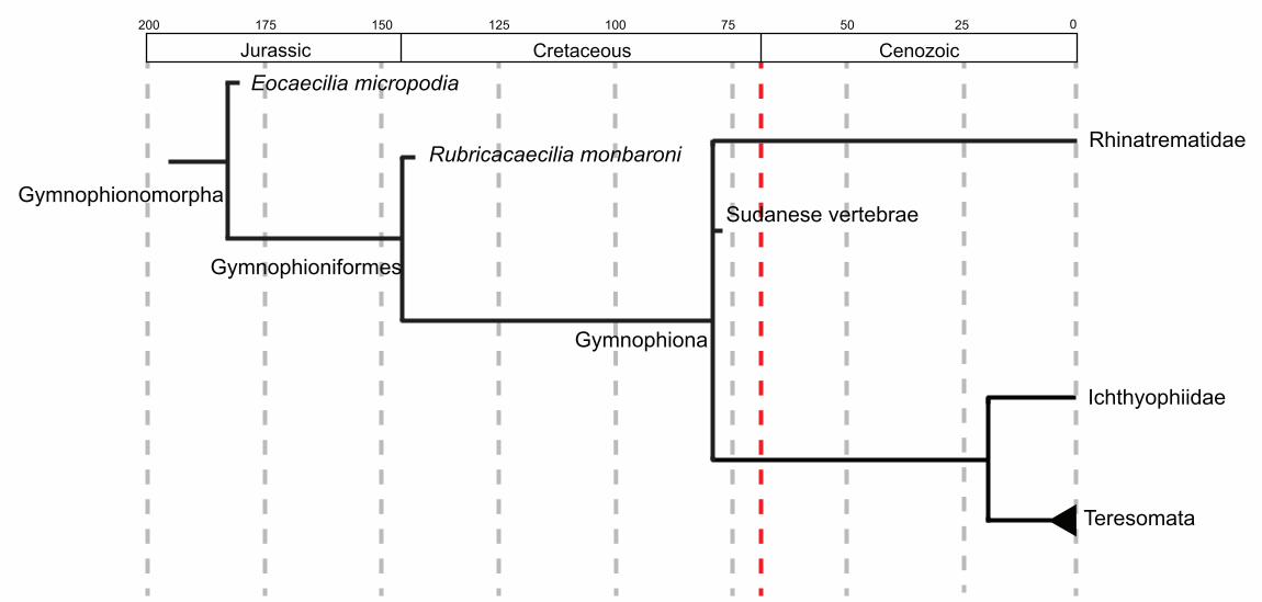
Gymnophiona
Rubricacaecilia monbaroni
Gymnophionomorpha
Gymnophioniformes
Eocaecilia micropodia
Sudanese vertebrae
Rhinatrematidae
Ichthyophiidae
Teresomata
200 175 150 125 100 75 50 25 0
CenozoicCretaceousJurassic
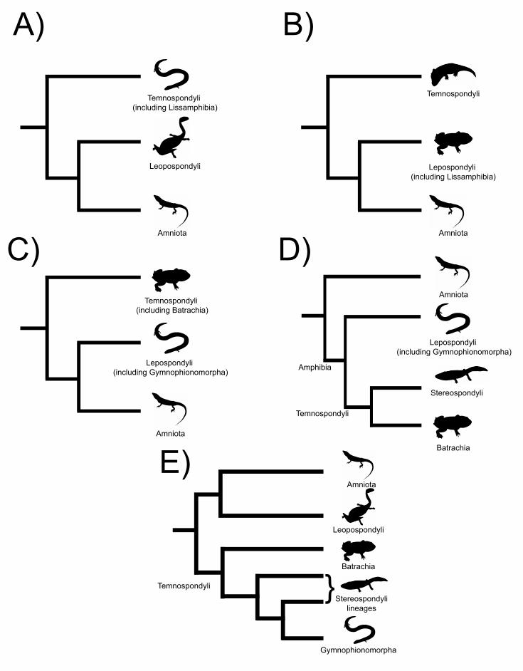
Temnospondyli (including Lissamphibia)
Leopospondyli
Amniota
Temnospondyli
Lepospondyli (including Lissamphibia)
Amniota
Temnospondyli (including Batrachia)
Lepospondyli (including Gymnophionomorpha)
Amniota
Amniota
Lepospondyli (including Gymnophionomorpha)
Stereospondyli
Batrachia
Amniota
Leopospondyli
Batrachia
Stereospondylilineages
Gymnophionomorpha
Amphibia
Temnospondyli
Temnospondyli }
A) B)
C) D)
E)

Cenozo
icM
eso
zoic
Triass
icJu
rass
icC
reta
ceous
Pale
ogene
Neogene
Quate
rnary Holocene
Pleistocene
Pliocene
Miocene
Oligocene
Eocene
Paleocene
Upper
Lower
Upper
Lower
Middle
Upper
Middle
Lower
Dermophis mexicanus, Chiapas, Mexico (1200–1350 B.C.)
Gymnophiona indet., Honda Group, Colombia (~13.8–11.6 Ma)
Teresomata indet., Napak XV, Uganda (~19–20 Ma)
Apodops pricei, Itaboraí Basin, Rio de Janeiro (~53–50 Ma)
Gymnophiona indet., Santa Lúcia Fm., Bolívia (~64–62 Ma)
Gymnophiona indet., El Molino Fm., Bolívia (~68.4 Ma)
Gymnophiona indet., Wadi Milk Fm., Sudan (~79.2 Ma)
Rubricacaecilia monbaroni, Anoual, Morocco (~145–140 Ma)
Eocaecilia micropodia, Kayenta Fm., USA (~183.7 Ma)
Ma
0.01
2.5
5
26
36
56
66
100
145
163
174
201
237
247
252
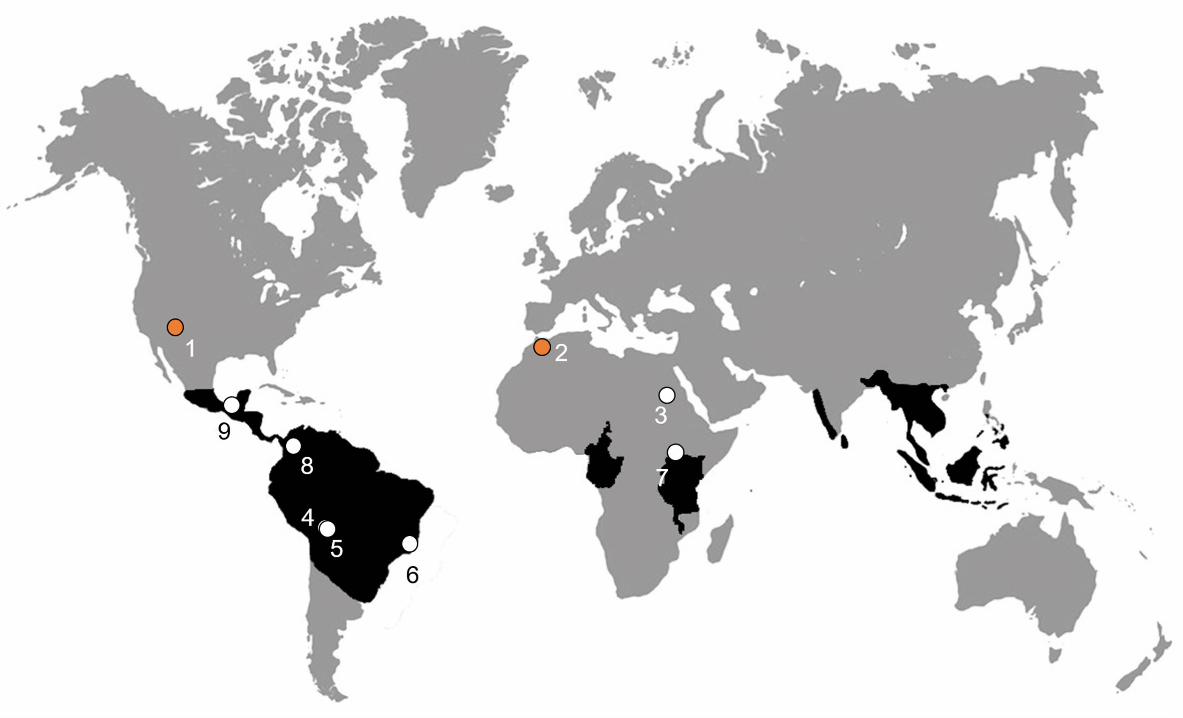
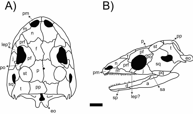
A) B)pm
nm
prflep?
j pf
post
sqt pp
eo
p
f
sqeo
pq
tstp
pp
pfpo
j
asa
lep?sp
pm
d
n prf
f
m
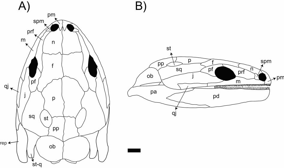
A) B)
n
f
pf
pj
sq st
pp
qj
st-q
ob
m
prf
spmpm
fp
st
pp
ob
papd
m
spm
pmjsq pf prf
n
qj
rep
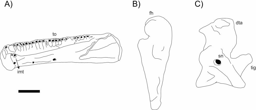
A) B) C)
tig
sn
dta
to
imt
fh
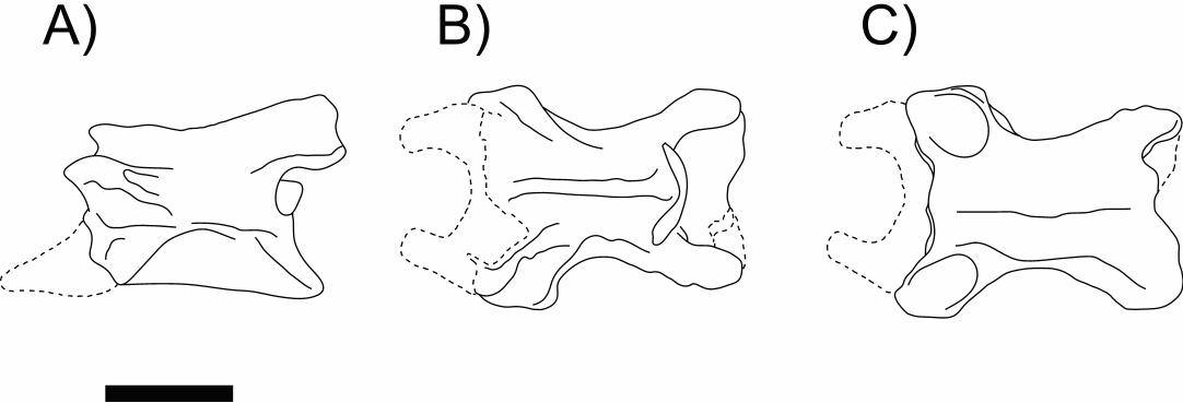
A) B) C)
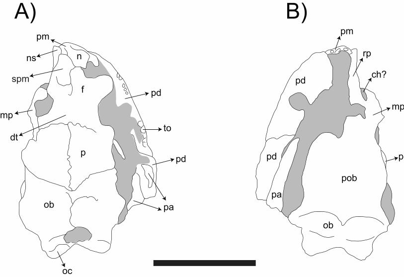
A) B)pm
ns
mp
dt
pd
to
pd
pa
p
ob
oc
f
n
pob
pd
ob
p
mp
ch?
rp
pm
pd
pa
spm


