Embed Size (px)
Citation preview

Anesthesiology 2008; 108:921–8 Copyright © 2008, the American Society of Anesthesiologists, Inc. Lippincott Williams & Wilkins, Inc.
Local Myotoxicity from Sustained Release of Bupivacainefrom MicroparticlesRobert Padera, M.D., Ph.D.,* Evangelia Bellas, B.S.,† Julie Y. Tse, B.S.,‡ Daphne Hao, B.S.,‡Daniel S. Kohane, M.D., Ph.D.§
Background: Sustained release of local anesthetics is fre-quently associated with myotoxicity. The authors investigatedthe role of particulate delivery systems and of the pattern ofdrug release in causing myotoxicity.
Methods: Rats were given sciatic nerve blocks with bupiva-caine solutions, two types of bupivacaine-containing micropar-ticles (polymeric microspheres and lipid–protein–sugar parti-cles), or blank particles with or without bupivacaine in thecarrier fluid. Myotoxicity was scored in histologic sections ofthe injection sites. Bupivacaine release kinetics from the parti-cles were measured. Myotoxicity of a range of bupivacaineconcentrations from exposures up to 3 weeks was assessed inC2C12 myotubes, with or without microparticles.
Results: Both types of bupivacaine-loaded microparticles, butnot blank particles, were associated with myotoxicity. Whereas0.5% bupivacaine solution caused little myotoxicity, a concen-tration of bupivacaine that mimicked the amount of bupiva-caine released initially from particles caused myotoxicity. Localanesthetics showed both concentration and time-dependentmyotoxicity in C2C12s. Importantly, even very low concentra-tions that were nontoxic over brief exposures became highlytoxic after days or weeks of exposure. The presence of particlesdid not increase bupivacaine myotoxicity in vitro but did invivo. Findings applied to both particle types.
Conclusions: Whereas the release vehicles themselves werenot myotoxic, both burst and extended release of bupivacainewere. A possible implication of the latter finding is that myo-toxicity is an inevitable concomitant of sustained release oflocal anesthetics. Particles, and perhaps other vehicles, mayenhance local toxicity through indirect mechanisms.
A WIDE variety of controlled-release formulations havebeen developed, including surgically implantable pel-lets,1 liposomes,2–6 lipospheres,7 cross-linkable hyal-uronic acid matrices,8 lipid–protein–sugar particles,9
and polymeric microspheres.10–17 These formulationsprolonged the duration of local anesthesia to varyingdegrees, ranging from prolongation by a number of
hours to nerve blockade lasting several weeks. We havefound that muscle injury is a concomitant of a widerange of formulation types.8,16,17 Myotoxicity is a well-recognized side effect of local anesthetic administration,perhaps particularly of extended exposure, whetherfrom controlled-release methodologies or from catheter-related methods.18 Occasionally, the consequences canbe clinically significant.19 Polymeric microspheres them-selves produce an acute local inflammatory response(neutrophils and macrophages), which is followed by achronic response (macrophages and lymphocytes) afterapproximately 7 days.20 Hydrogels can also induce aninflammatory response.8 However, neither carrier in-duces the characteristic finding of local anesthetic myo-toxicity.8 In fact, the muscle injury from controlled-release local anesthetic formulations seems to be largelydue to the encapsulated drug, rather than the vehicleitself.8,17 It is not clear whether the vehicles contributeindirectly to the severe muscle injury that can develop.
Here we examine the relative contributions of drugand vehicle in the development of muscle injury, focus-ing on two microparticulate formulations with very dif-ferent compositions and morphologies: polymeric (poly-[lactic-co-glycolic]) microspheres and lipid–protein–sugar particles. We investigate the potential impacts ofboth burst release and continuous release on the devel-opment and maintenance of myotoxicity from con-trolled-release devices.
Materials and Methods
Animal Care CommitteeAnimals were cared for in accordance with protocols
approved by the Animal Care and Use Committee at theMassachusetts Institute of Technology (Cambridge, Massa-chusetts), and the Guide for the Care and Use of Labora-tory Animals of the US National Research Council.
Chemicals1,2-Dipalmitoyl-sn-glycero-3-phosphocholine was ob-
tained from Avanti Polar Lipids (Alabaster, AL). Bupiva-caine hydrochloride, bovine serum albumin, and �-lac-tose monohydrate were from Sigma-Aldrich (St. Louis,MO). Poly(lactic-co-glycolic) acid (PLGA; molecularweight 90 kd, G:L � 65:35) was obtained from Medisorb(Alkermes; Cambridge, MA).
Microparticle PreparationPLGA microparticles were prepared as described.9 In
brief, PLGA dissolved in methylene chloride was homog-
* Assistant Professor, Department of Pathology, Brigham and Women’s Hospi-tal, Boston, Massachusetts; Harvard-MIT Division of Health Sciences and Tech-nology, Cambridge, Massachusetts. † Technician, ‡ Undergraduate Student,Department of Chemical Engineering, Massachusetts Institute of Technology,Cambridge, Massachusetts. § Associate Professor of Anaesthesiology, Labora-tory for Biomaterials and Drug Delivery, Department of Anesthesiology, Divisionof Critical Care Medicine, Children’s Hospital, Harvard Medical School.
Received from the Laboratory for Biomaterials and Drug Delivery, Departmentof Anesthesiology, Division of Critical Care Medicine, Children’s Hospital, Har-vard Medical School, Boston, Massachusetts. Submitted for publication July 30,2007. Accepted for publication January 8, 2008. Supported by grant No.GM073626 (to Dr. Kohane) from the National Institute of General MedicalSciences, Bethesda, Maryland.
Address correspondence to Dr. Kohane: Laboratory for Biomaterials and DrugDelivery, Department of Anesthesiology, Division of Critical Care Medicine,Children’s Hospital, Harvard Medical School, 300 Longwood Avenue, Boston,Massachusetts 02115. [email protected]. Information on pur-chasing reprints may be found at www.anesthesiology.org or on the mastheadpage at the beginning of this issue. ANESTHESIOLOGY’s articles are made freelyaccessible to all readers, for personal use only, 6 months from the cover date ofthe issue.
Anesthesiology, V 108, No 5, May 2008 921

enized at 3,000 rpm (Silverson L4RT-A; Longmeadow,MA) for 1 min in 1% poly(vinylalcohol) (molecularweight 25 kd; Polysciences, Inc., Warrington, PA) con-taining 100 mM Tris pH 8.5. The methylene chloride wasremoved by rotary evaporation. Particles of the desiredsize range were separated by wet sieving and were thenlyophilized to dryness. In particles containing drug, bu-pivacaine free base was substituted for 50% of the poly-meric mass dissolved in methylene chloride.
Blank 1,2-dipalmitoyl-sn-glycero-3-phosphocholine-al-bumin-lactose (DAL) particles were prepared as de-scribed9 using a Buchi 190 Mini Spray Dryer (Buchi Co.,Flawil, Switzerland). In brief, 1,2-dipalmitoyl-sn-glycero-3-phosphocholine dissolved in 100% ethanol was mixedwith an aqueous solution of bovine serum albumin and�-lactose monohydrate, such that the final solute com-position was 60% 1,2-dipalmitoyl-sn-glycero-3-phospho-choline, 20% albumin, and 20% lactose in 70:30 (vol/vol)ethanol:water. This solution was spray dried under thefollowing conditions: inlet temperature, 110°–115°C; airflow, 600 l/h; flow rate, 12 ml/min; and aspirator pres-sure, �20 mbar. The resulting outlet temperature wasbetween 50° and 55°C. In particles containing drug,bupivacaine free base was added to the ethanolic solu-tion, to an amount that equaled 10% of the total solutemass. The total mass of the other three solutes wasreduced by an equal amount.
Particle size was determined with a Coulter multisizer(Coulter Electronics Ltd., Luton, United Kingdom).
In Vitro Release of Bupivacaine from MicroparticlesFifty milligrams of DAL particles or PLGA microspheres
was suspended in 1 ml phosphate-buffered saline, pH7.4, at 37°C and inserted into the lumen of a Spectra/Por1.1 Biotech Dispodialyzer (Spectrum Laboratories, Ran-cho Dominguez, CA) with an 8,000 molecular weightcutoff. The dialysis bag was placed into a test tube with12 ml phosphate-buffered saline and incubated at 37°Con a tilt table (Ames Aliquot Mixer; Miles LaboratoriesInc., Elkhart, IN). At predetermined intervals, the dialysisbag was transferred to a test tube with fresh phosphate-buffered saline. The bupivacaine concentration in thedialysate was quantitated by measuring absorbance at272 nm and referring to a standard curve. Observation ofthe entire spectrum and performance of a protein assay(BCA Protein Assay Reagent Kit; Pierce Chemical Co.,Rockford, IL) confirmed the absence of albumin fromthe samples that were measured. Infinite sink conditionswere maintained during the release experiments as evi-denced by low concentrations of released drug (�0.17mg/ml).
Animal Care and Sciatic Blockade TechniqueAdult male Sprague-Dawley rats (Charles River Labora-
tories, Wilmington, MA) weighing 310–420 g werehoused in groups, in a 6 AM–6 PM light–dark cycle. Under
brief isoflurane–oxygen anesthesia, a 23-gauge needlewas introduced posteromedial to the greater trochanter,the needle was withdrawn approximately 1 mm, and 0.3ml drug-containing solution was injected.
Assessment of Nerve BlockadeThermal nociception was assessed by a modified hot
plate test.1,21 Hind paws were exposed in sequence (left,then right) to a 56°C hot plate (model 39D Hot PlateAnalgesia Meter; IITC Inc., Woodland Hills, CA). Thetime (latency) until paw withdrawal was measured by astopwatch. (Thermal latency in the uninjected leg was acontrol for systemic effects of the injected agents.) If theanimal did not remove its paw from the hot plate within12 s, it was removed by the experimenter to avoid injuryto the animal or the development of hyperalgesia. Theexperimenter was blinded as to what treatment specificrats were receiving.
The duration of thermal nociceptive block was calcu-lated as the time required for thermal latency to return toa value of 7 s from a higher value. Seven seconds is themidpoint between a baseline thermal latency of approx-imately 2 s in adult rats and a maximal latency of 12 s.
Tissue Harvesting and HistologyThe sciatic nerve and adjacent tissues were harvested
after euthanasia with carbon dioxide and processed toproduce hematoxylin and eosin–stained slides.17 In all anal-yses, the pathologist (R.P.) was not aware of the nature ofthe samples before examination. Samples were scored formyotoxicity (0–6).22 The myotoxicity score reflected twoseparate but related processes that are hallmarks of localanesthetic myotoxicity: nuclear internalization and regen-eration. The former is characterized by normal size andcytoplasm chromicity, but with nuclei located away fromtheir normal location at the periphery of the cell. In thelatter case, cells are shrunken, with more basophilic cyto-plasm. Scoring was as follows: 0 � normal, 1 � perifas-cicular internalization, 2 � deep internalization (�5 celllayers), 3 � perifascicular regeneration, 4 � deep regener-ation, 5 � hemifascicular regeneration, 6 � holofascicularregeneration.
Cell CultureC2C12 mouse myoblasts (American Type Culture Col-
lection CRL-1772; Manassas, VA) were cultured to pro-liferate in Dulbecco’s modified Eagle’s medium supple-mented with 20% fetal bovine serum and 1% penicillinstreptomycin. All cell culture supplies were purchasedfrom Invitrogen (Carlsbad, CA) unless otherwise noted.Cells were then plated in 24-well tissue culture plateswith 50,000 cells/ml/well in Dulbecco’s modified Eagle’smedium supplemented with 2% horse serum and 1%pen-strep, and were left to differentiate into myotubulesfor 10–14 days. During differentiation media were ex-changed every 2–3 days.
922 PADERA ET AL.
Anesthesiology, V 108, No 5, May 2008

Bupivacaine hydrochloride was added to Dulbecco’smodified Eagle’s medium (with 2% horse serum and 1%Penn strep) at a concentration of 0.125% wt/vol. Themedium was then filtered through a 0.22-�m celluloseacetate membrane and then serially diluted to preparethe remaining concentrations. Particles were irradiatedwith ultraviolet light for 2 h before suspension in mediaand serial dilution. The medium had a neutral pH at theoutset; subsequently, pH was monitored by use of apH-sensitive dye in the medium.
For time points longer than 4 days, the drug mediumwas exchanged every 2–3 days. Cells were maintained at37°C in 5% CO2 with the remainder being room air.
Assessing ViabilityTo quantitatively assess cell viability after adding drug-
or particle-containing media, a colormetric assay (3-(4,5-dimethylthiazol-2-yl)-2,5-diphenyl tetrazolium bromide[MTT] kit, Promega G4100; Madison, WI) was per-formed at selected time points. The yellow tetrazoliumsalt is metabolized in live cells to form insoluble purpleformazan crystals. The purple crystals are solubilized bythe addition of a detergent. The color can then be quan-tified by spectrophotometric means. At each time point,150 �l MTT was added, and then cells were incubated at37°C for 4 h before 1 ml solubilization solution (deter-gent) was added. The absorbance was read at 570 nmusing the SpectraMax 384 Plus fluorometer (MolecularDevices, Sunnyvale, CA) after being incubated at 37°C inthe dark overnight. Cells were also monitored visually toconfirm the results of the assay. Each plate had wells thatcontained medium without cells or other additiveswhose absorbance was subtracted from the rest of theplate as background. Each plate also had wells thatcontained medium and cells but no additives; all exper-imental groups were normalized to those wells.
Statistical AnalysisNeurobehavioral data are presented as median, with
25th and 75th percentiles in parentheses, because theywere not normally distributed. They were analyzed withthe Mann–Whitney U test, or a Wilcoxon signed rank testwhen comparing sensory and motor tests in the same
animals. Results of cell survival assays were describedwith parametric measures and tests (mean, SD, t test,analysis of variance). A P value less than 0.05 indicatedstatistical significance. Multiple comparisons were donein a planned manner (i.e., comparisons were selectedindividually), and the P value required for statisticalsignificance (�) was determined by dividing 0.05 by thenumber of comparisons. Therefore, for myotoxicityscores (10 comparisons), � � 0.05/10 � 0.005, so P �0.005 was required for statistical significance; for dura-tions of block and cell culture data, there was only onecomparison, so the � remained 0.05. For ease of under-standing, the � for each comparison is provided witheach result. All P values are two-tailed. Statistical analyseswere performed with SPSS 12.0 (SPSS Inc., Chicago, IL).
Results
Production and Characterization of MicroparticlesThe DAL particles were produced as a fine white
powder, with a median volume-weighted diameter of4–5 �m. The PLGA particles were also a white powder,with a median volume-weighted diameter of approxi-mately 60 �m. These values are in agreement with ourprevious reports for similar particles.9
Myotoxicity from Microparticles ContainingBupivacaineAnimals were injected with 0.5% wt/vol bupivacaine
hydrochloride, 10% wt/wt bupivacaine DAL particles, or50% wt/wt bupivacaine PLGA microspheres (table 1).The sensory and motor blocks from encapsulated bupiv-acaine were longer than those from free bupivacaine. Ongross dissection, tissues injected with free drug ap-peared normal. In the other groups, particles were foundin discrete pockets adjacent to the sciatic nerve, but thetissue appeared otherwise normal. On histologic exam-ination, there was evidence of perifascicular myotoxicityin all animals. The injury was limited to the site of injec-tion, at the surface cell layers of the muscle adjacent to thedepot of particles. It was considerably more pronounced inanimals injected with encapsulated bupivacaine than with
Table 1. Durations of Nerve Blockade from Local Anesthetic Formulations
Treatment Sensory Motor P Value*
Bupivacaine 0.5% wt/vol 188 (154–195) 189 (177–209) 0.18Bupivacaine 50% wt/wt PLGA microsphere 613 (474–944) 711 (584–1,306) 0.046Bupivacaine 10% wt/wt DAL 251 (219–285) 348 (296–501) 0.07Bupivacaine 1.25 % wt/vol 266 (235–285) 293 (288–301) 0.028Bupivacaine 0.5% wt/vol � blank PLGA microsphere 152 (140–167) 183 (177–197) 0.028Bupivacaine 0.5% wt/vol � blank DAL 98 (21–163) 168 (41–175) 0.14
Data are presented as median with 25th and 75th percentiles, in minutes. P values compare the durations of sensory and motor block in animals in the samegroup, and result from a Wilcoxon signed ranks test. n � 6 for all groups except n � 4 for bupivacaine 10% wt/wt DAL.
* � � 0.05.
DAL � DPPC-albumin-lactose particles; PLGA � poly(lactic-co-glycolic) acid.
923MYOTOXICITY FROM LOCAL ANESTHETIC MICROPARTICLES
Anesthesiology, V 108, No 5, May 2008
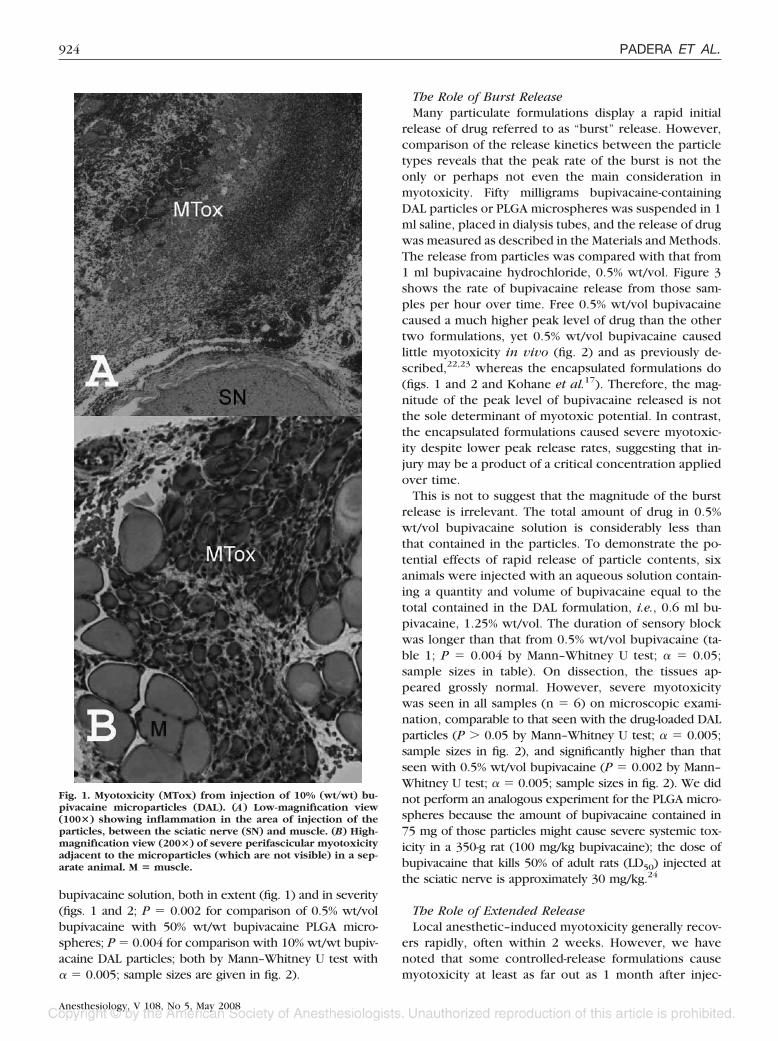
bupivacaine solution, both in extent (fig. 1) and in severity(figs. 1 and 2; P � 0.002 for comparison of 0.5% wt/volbupivacaine with 50% wt/wt bupivacaine PLGA micro-spheres; P � 0.004 for comparison with 10% wt/wt bupiv-acaine DAL particles; both by Mann–Whitney U test with� � 0.005; sample sizes are given in fig. 2).
The Role of Burst ReleaseMany particulate formulations display a rapid initial
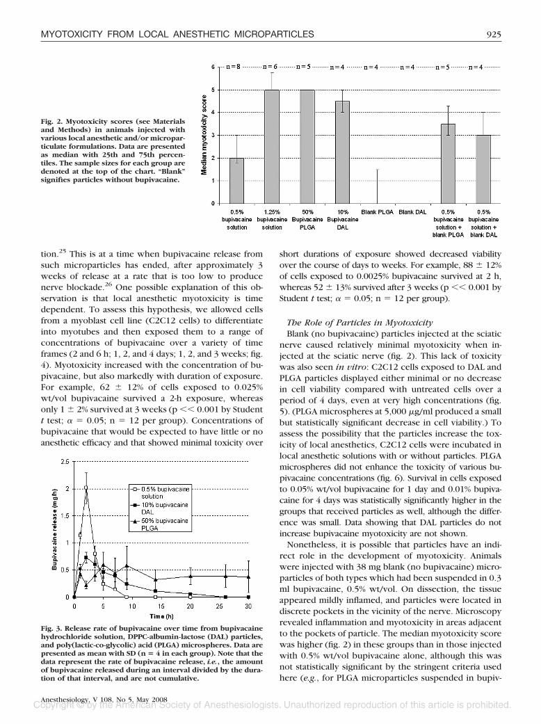
release of drug referred to as “burst” release. However,comparison of the release kinetics between the particletypes reveals that the peak rate of the burst is not theonly or perhaps not even the main consideration inmyotoxicity. Fifty milligrams bupivacaine-containingDAL particles or PLGA microspheres was suspended in 1ml saline, placed in dialysis tubes, and the release of drugwas measured as described in the Materials and Methods.The release from particles was compared with that from1 ml bupivacaine hydrochloride, 0.5% wt/vol. Figure 3shows the rate of bupivacaine release from those sam-ples per hour over time. Free 0.5% wt/vol bupivacainecaused a much higher peak level of drug than the othertwo formulations, yet 0.5% wt/vol bupivacaine causedlittle myotoxicity in vivo (fig. 2) and as previously de-scribed,22,23 whereas the encapsulated formulations do(figs. 1 and 2 and Kohane et al.17). Therefore, the mag-nitude of the peak level of bupivacaine released is notthe sole determinant of myotoxic potential. In contrast,the encapsulated formulations caused severe myotoxic-ity despite lower peak release rates, suggesting that in-jury may be a product of a critical concentration appliedover time.
This is not to suggest that the magnitude of the burstrelease is irrelevant. The total amount of drug in 0.5%wt/vol bupivacaine solution is considerably less thanthat contained in the particles. To demonstrate the po-tential effects of rapid release of particle contents, sixanimals were injected with an aqueous solution contain-ing a quantity and volume of bupivacaine equal to thetotal contained in the DAL formulation, i.e., 0.6 ml bu-pivacaine, 1.25% wt/vol. The duration of sensory blockwas longer than that from 0.5% wt/vol bupivacaine (ta-ble 1; P � 0.004 by Mann–Whitney U test; � � 0.05;sample sizes in table). On dissection, the tissues ap-peared grossly normal. However, severe myotoxicitywas seen in all samples (n � 6) on microscopic exami-nation, comparable to that seen with the drug-loaded DALparticles (P � 0.05 by Mann–Whitney U test; � � 0.005;sample sizes in fig. 2), and significantly higher than thatseen with 0.5% wt/vol bupivacaine (P � 0.002 by Mann–Whitney U test; � � 0.005; sample sizes in fig. 2). We didnot perform an analogous experiment for the PLGA micro-spheres because the amount of bupivacaine contained in75 mg of those particles might cause severe systemic tox-icity in a 350-g rat (100 mg/kg bupivacaine); the dose ofbupivacaine that kills 50% of adult rats (LD50) injected atthe sciatic nerve is approximately 30 mg/kg.24
The Role of Extended ReleaseLocal anesthetic–induced myotoxicity generally recov-
ers rapidly, often within 2 weeks. However, we havenoted that some controlled-release formulations causemyotoxicity at least as far out as 1 month after injec-
Fig. 1. Myotoxicity (MTox) from injection of 10% (wt/wt) bu-pivacaine microparticles (DAL). (A) Low-magnification view(100�) showing inflammation in the area of injection of theparticles, between the sciatic nerve (SN) and muscle. (B) High-magnification view (200�) of severe perifascicular myotoxicityadjacent to the microparticles (which are not visible) in a sep-arate animal. M � muscle.
924 PADERA ET AL.
Anesthesiology, V 108, No 5, May 2008
tion.25 This is at a time when bupivacaine release fromsuch microparticles has ended, after approximately 3weeks of release at a rate that is too low to producenerve blockade.26 One possible explanation of this ob-servation is that local anesthetic myotoxicity is timedependent. To assess this hypothesis, we allowed cellsfrom a myoblast cell line (C2C12 cells) to differentiateinto myotubes and then exposed them to a range ofconcentrations of bupivacaine over a variety of timeframes (2 and 6 h; 1, 2, and 4 days; 1, 2, and 3 weeks; fig.4). Myotoxicity increased with the concentration of bu-pivacaine, but also markedly with duration of exposure.For example, 62 � 12% of cells exposed to 0.025%wt/vol bupivacaine survived a 2-h exposure, whereasonly 1 � 2% survived at 3 weeks (p �� 0.001 by Studentt test; � � 0.05; n � 12 per group). Concentrations ofbupivacaine that would be expected to have little or noanesthetic efficacy and that showed minimal toxicity over
short durations of exposure showed decreased viabilityover the course of days to weeks. For example, 88 � 12%of cells exposed to 0.0025% bupivacaine survived at 2 h,whereas 52 � 13% survived after 3 weeks (p �� 0.001 byStudent t test; � � 0.05; n � 12 per group).
The Role of Particles in MyotoxicityBlank (no bupivacaine) particles injected at the sciatic
nerve caused relatively minimal myotoxicity when in-jected at the sciatic nerve (fig. 2). This lack of toxicitywas also seen in vitro: C2C12 cells exposed to DAL andPLGA particles displayed either minimal or no decreasein cell viability compared with untreated cells over aperiod of 4 days, even at very high concentrations (fig.5). (PLGA microspheres at 5,000 �g/ml produced a smallbut statistically significant decrease in cell viability.) Toassess the possibility that the particles increase the tox-icity of local anesthetics, C2C12 cells were incubated inlocal anesthetic solutions with or without particles. PLGAmicrospheres did not enhance the toxicity of various bu-pivacaine concentrations (fig. 6). Survival in cells exposedto 0.05% wt/vol bupivacaine for 1 day and 0.01% bupiva-caine for 4 days was statistically significantly higher in thegroups that received particles as well, although the differ-ence was small. Data showing that DAL particles do notincrease bupivacaine myotoxicity are not shown.
Nonetheless, it is possible that particles have an indi-rect role in the development of myotoxicity. Animalswere injected with 38 mg blank (no bupivacaine) micro-particles of both types which had been suspended in 0.3ml bupivacaine, 0.5% wt/vol. On dissection, the tissueappeared mildly inflamed, and particles were located indiscrete pockets in the vicinity of the nerve. Microscopyrevealed inflammation and myotoxicity in areas adjacentto the pockets of particle. The median myotoxicity scorewas higher (fig. 2) in these groups than in those injectedwith 0.5% wt/vol bupivacaine alone, although this wasnot statistically significant by the stringent criteria usedhere (e.g., for PLGA microparticles suspended in bupiv-
Fig. 2. Myotoxicity scores (see Materialsand Methods) in animals injected withvarious local anesthetic and/or micropar-ticulate formulations. Data are presentedas median with 25th and 75th percen-tiles. The sample sizes for each group aredenoted at the top of the chart. “Blank”signifies particles without bupivacaine.
Fig. 3. Release rate of bupivacaine over time from bupivacainehydrochloride solution, DPPC-albumin-lactose (DAL) particles,and poly(lactic-co-glycolic) acid (PLGA) microspheres. Data arepresented as mean with SD (n � 4 in each group). Note that thedata represent the rate of bupivacaine release, i.e., the amountof bupivacaine released during an interval divided by the dura-tion of that interval, and are not cumulative.
925MYOTOXICITY FROM LOCAL ANESTHETIC MICROPARTICLES
Anesthesiology, V 108, No 5, May 2008

acaine solution, P � 0.016 by Mann–Whitney U test; � �0.005; sample sizes in fig. 2). We note, however, thatwhen the results for both particle types suspended inbupivacaine were pooled and compared with 0.5% bu-pivacaine, the difference was statistically significant (P �0.004 by Mann–Whitney U test; � � 0.005; n � 9 whencombining the two particle-containing groups).
Discussion
The initial burst release of bupivacaine seems to causemyotoxicity, but only if the magnitude or some productof magnitude and duration of exposure are above acertain undefined threshold. This contribution to localinjury is theoretically amenable to engineering, i.e., itshould be possible to minimize that burst release. Ofmuch greater concern is the observation that very low—
even subanesthetic—concentrations of bupivacaine canbecome myotoxic over extended periods of time. Thisfinding raises the possibility that myotoxicity could be aninevitable concomitant of long-term exposure to conven-tional (amino-amide and amino-ester) local anesthetics,irrespective of the technology used to deliver them.Myotoxicity is a well-known occurrence in clinical19 orinvestigational27 use of conventional local anesthetics.Although it can have severe consequences,19 it has notgenerated much clinical concern. In fact, intramuscularlocal anesthetic injection is a standard treatment fortrigger points in myofascial pain syndromes,28 and localanesthetic myotoxicity is generally reversible. The dis-tinction that must be made, however, is that those treat-ments generally involve a single-shot drug injection witha brief duration, whereas microparticulate systems canresult in very high local concentrations and/or weeks of
Fig. 4. (A) Mean survival (z-axis) of C2C12cells exposed to a range of concentra-tions of bupivacaine (y-axis) for 2 and6 h; 1, 2, and 4 days; and 1, 2, and 3 weeks(x-axis). n � 12 for each point; SDs arenot shown for the sake of clarity. Thecolor code on the right reflects the per-centage cell survival in the z-axis. Statis-tical inferences on the data are discussedin the text. (B) Effect of 2 days of expo-sure of a range of bupivacaine concentra-tions on cell survival. Data are presentedas mean with SD. (C) Effect of duration ofexposure to on cytotoxicity of 0.025% wt/vol bupivacaine. Data are presented asmean with SD. B and C are cross-sectionsof A.
926 PADERA ET AL.
Anesthesiology, V 108, No 5, May 2008

local anesthetic exposure. An even greater concern isthat local anesthetics also have considerable local neu-rotoxicity29–31; the potential for controlled-release de-
vices to injure nerves has not been examined exten-sively, but injury to muscle suggests that nerve injurymight also be possible. Of note, animals that achievedsciatic nerve blocks lasting approximately 9 days afterinjection with microparticles containing tetrodotoxin,bupivacaine, and dexamethasone frequently had one ormore cycles of block recurrence after the initial blockwore off,25 a pattern potentially attributable to nerveinjury. We have also seen this pattern with high con-centrations of tricyclic antidepressants used as localanesthetics,23 which have been shown to be highlyneurotoxic.23,32
The effect of the particles themselves on myotoxicityis difficult to explain fully. It would seem from ourresults here and from previous experience that the par-ticles themselves cause little direct myotoxicity. How-ever, the fact that the myotoxicity of bupivacaine solu-tion is increased in the presence of particles in vivosuggests that they might enhance that toxicity. Onepossibility is that the particles release some agent (e.g.,lactic or glycolic acids, residual organic solvent, excipi-ents) that potentiates local anesthetic toxicity, but ourcell culture data do not support that conclusion. Anotherpossibility is that the presence of discrete pockets ofparticles allows more reliable identification of siteswhere the local anesthetic was deposited, thus improv-ing the accuracy of sampling. However, we do not seeany sign of such severe toxicity in any animal injectedwith bupivacaine solution. Furthermore, we did not seesuch toxicity in an animal model specifically designed toremove sampling bias by injecting very large volumes oflocal anesthetic solutions (1.5 ml).23 It is possible thatthe inflammation caused by the particles worsens myo-toxicity by some unknown mechanism, perhaps by theirproinflammatory effects.17,20,25 Finally, the macroscopicdeposits of particles—as opposed to the individual par-ticles—may slow the decline of the local concentrationof drug, thereby increasing the toxicity of bupivacainesolution. The merits of the last two possibilities cannot
Fig. 5. Survival of C2C12 cells after 1 or4 days of exposure to a range of con-centrations of blank (no bupivacaine)particles. The diamonds denote concen-trations of particles that were not tested(black � poly(lactic-co-glycolic) acid[PLGA]; white � DPPC-albumin-lactose[DAL] particles). Data are presented asmean with SD (n � 8).
Fig. 6. Survival of C2C12 cells after 1 or 4 days of exposure tobupivacaine solutions with or without blank (no bupivacaine)poly(lactic-co-glycolic) acid (PLGA) particles. Data are pre-sented as mean with SD (n � 8 in all groups). Groups with andwithout bupivacaine were compared by t test; there were nodifferences.
927MYOTOXICITY FROM LOCAL ANESTHETIC MICROPARTICLES
Anesthesiology, V 108, No 5, May 2008

be evaluated by the methods used in this study. Theinflammatory response to particles may prove to beproblematic in its own right, irrespective of myotoxicityor neurotoxicity, given the large mass that may have tobe injected to achieve clinically relevant nerve blocks inhumans.
Although we cannot rule out the possibility that resid-ual organic solvents from the particle production pro-cess contributed to the observed myotoxicity, it is un-likely that they play a major role. Particles of both typesdo not cause myotoxicity in the absence of local anes-thetics.17 Furthermore, vehicles that do not involve or-ganic solvents (e.g., cross-linked hyaluronic acid) onlycause myotoxicity when they contain local anesthetics.8
Myotoxicity seems to be related to both the releasekinetics of bupivacaine (burst and duration of release),and perhaps the presence of the particles themselves.Even very low concentrations of bupivacaine seem to bemyotoxic if the duration of exposure is sufficiently pro-longed. One possible implication of these findings is thatany type of prolonged duration local anesthesia usingdrugs of this type will be myotoxic, and potentiallyneurotoxic.
References
1. Masters DB, Berde CB, Dutta SK, Griggs CT, Hu D, Kupsky W, Langer R:Prolonged regional nerve blockade by controlled release of local anesthetic froma biodegradable polymer matrix. ANESTHESIOLOGY 1993; 79:1–7
2. Grant GJ, Vermeulen K, Langerman L, Zakowski M, Turndorf H: Prolongedanalgesia with liposomal bupivacaine in a mouse model. Reg Anesth 1994;19:264–9
3. Mashimo T, Uchida I, Pak M, Shibata A, Nishimura S, Ingaki Y, Yoshiya I:Prolongation of canine epidural anesthesia by liposome encapsulation of lido-caine. Anesth Analg 1992; 74:827–34
4. Mowat JJ, Mok MJ, MacLeod BA, Madden TD: Liposomal bupivacaine:Extended duration nerve blockade using large unilamellar vesicles that exhibit aproton gradient. ANESTHESIOLOGY 1996; 85:635–43
5. Sharma BB, Jain SK, Vyas SP: Topical liposome system bearing local anaes-thetic lignocaine: Preparation and evaluation. J Microencapsul 1994; 11:279–86
6. Yanez AM, Wallace M, Ho R, Shen D, Yaksh TL: Touch-evoked agitationproduced by spinally administered phospholipid emulsion and liposomes in rats.ANESTHESIOLOGY 1995; 82:1189–98
7. Masters DB, Domb AJ: Liposphere local anesthetic timed-release for peri-neural site application. Pharm Res 1998; 15:1038–45
8. Jia X, Colombo G, Padera R, Langer R, Kohane DS: Prolongation of sciaticnerve blockade by in situ cross-linked hyaluronic acid. Biomaterials 2004; 25:4797–804
9. Kohane DS, Lipp M, Kinney RC, Lotan N, Langer R: Sciatic nerve blockadewith lipid-protein-sugar particles containing bupivacaine. Pharm Res 2000; 17:1243–9
10. Le Corre P, Le Guevello P, Gajan V, Chevanne F, Le Verge R: Preparationand characterization of bupivacaine-loaded polylactide and polylactide-coglycol-ide microspheres. Int J Pharm 1994; 107:41–9
11. Le Corre P, Rytting JH, Gajan V, Chevanne F, Le Verge R: In vitrocontrolled release kinetics of local anaesthetics from poly(D,L-lactide) and poly-(lactide-co-glycolide) microspheres. J Microencapsul 1997; 14:243–55
12. Curley J, Castillo J, Hotz J, Uezono M, Hernandez S, Lim J-O, Tigner J,Chasin M, Langer R, Berde C: Prolonged regional nerve blockade: Injectablebiodegradable bupivacaine/polyester microspheres. ANESTHESIOLOGY 1996;84:1401–10
13. Estebe J-P, Le Corre P, Malledant Y, Chevanne F, Leverge R: Prolongationof spinal anesthesia with bupivacaine-loaded (DL-lactide) microspheres. AnesthAnalg 1995; 81:99–103
14. Wakiyama N, Juni K, Nakana M: Preparation and evaluation in vitro ofpolylactic acid microspheres containing local anesthetics. Chem Pharm Bull1981; 29:3363–8
15. Wakiyama N, Juni K, Nakano M: Preparation and evaluation in vitro and invivo of polylactic acid microspheres containing dibucaine. Chem Pharm Bull1982; 30:3719–27
16. Kohane DS, Plesnila N, Thomas SS, Le D, Langer R, Moskowitz MA:Lipid-sugar particles for intracranial drug delivery: Safety and biocompatibility.Brain Res 2002; 946:206–13
17. Kohane DS, Lipp M, Kinney RC, Anthony DC, Louis DN, Lotan N, LangerR: Biocompatibility of lipid-protein-sugar particles containing bupivacaine in theepineurium. J Biomed Mater Res 2002; 59:450–9
18. Pere P, Watanabe H, Pitkanen M, Wahlstrom T, Rosenberg PH: Localmyotoxicity of bupivacaine in rabbits after continuous supraclavicular brachialplexus blocks. Reg Anesth 1993; 18:304–7
19. Hogan Q, Dotson R, Erickson S, Kettler R, Hogan K: Local anestheticmyotoxicity: A case and review. ANESTHESIOLOGY 1994; 80:942–7
20. Anderson JM: In vivo biocompatibility of implantable delivery systems andbiomaterials. Eur J Pharm Biopharm 1994; 40:1–8
21. Kohane DS, Yieh J, Lu NT, Langer R, Strichartz GR, Berde CB: A re-examination of tetrodotoxin for prolonged duration local anesthesia. ANESTHESI-OLOGY 1998; 89:119–31
22. Padera R, Tse J, Bellas E, Kohane DS: Tetrodotoxin for prolonged localanesthesia with minimal myotoxicity. Muscle Nerve 2006; 34:747–53
23. Barnet C, Louis DN, Kohane DS: Tissue injury from tricyclic antidepres-sants used as local anesthetics. Anesth Analg 2005; 101:1838–43
24. Kohane DS, Sankar WN, Shubina M, Hu D, Rifai N, Berde CB: Sciatic nerveblockade in infant, adolescent, and adult rats: A comparison of ropivacaine withbupivacaine. ANESTHESIOLOGY 1998; 89:1199–208
25. Kohane DS, Smith SE, Louis DN, Colombo G, Ghoroghchian P, HunfeldNG, Berde CB, Langer R: Prolonged duration local anesthesia from tetrodotoxin-enhanced local anesthetic microspheres. Pain 2003; 104:415–21
26. Castillo J, Curley J, Hotz J, Uezono M, Tigner J, Chasin M, Wilder R, LangerR, Berde C: Glucocorticoids prolong rat sciatic nerve blockade in vivo frombupivacaine microspheres. ANESTHESIOLOGY 1996; 85:1157–66
27. Benoit PW, Yagiela A, Fort NF: Pharmacologic correlation between localanesthetic-induced myotoxicity and disturbances of intracellular calcium distri-bution. Toxicol Appl Pharmacol 1980; 52:187–98
28. Iwama H, Ohmori S, Kaneko T, Watanabe K: Water-diluted local anestheticfor trigger-point injection in chronic myofascial pain syndrome: Evaluation oftypes of local anesthetic and concentrations in water. Reg Anesth Pain Med 2001;26:333–6
29. Kalichman MW, Powell HC, Myers RR: Pathology of local anesthetic-induced nerve injury. Acta Neuropathol (Berl) 1988; 75:583–9
30. Kalichman MW, Moorhouse DF, Powell HC, Myers RR: Relative neuraltoxicity of local anesthetics. J Neuropathol Exp Neurol 1993; 52:234–40
31. Bainton CR, Strichartz GR: Concentration dependence of lidocaine-in-duced irreversible conduction loss in frog nerve. ANESTHESIOLOGY 1994; 91:657–67
32. Estebe JP, Myers RR: Amitriptyline neurotoxicity: Dose-related pathologyafter topical application to rat sciatic nerve. ANESTHESIOLOGY 2004; 100:1519–25
928 PADERA ET AL.
Anesthesiology, V 108, No 5, May 2008


