Embed Size (px)
Citation preview

ORIGINAL RESEARCH ARTICLEpublished: 17 March 2014
doi: 10.3389/fnana.2014.00012
Localization of peroxisome proliferator-activated receptoralpha (PPARα) and N-acyl phosphatidylethanolaminephospholipase D (NAPE-PLD) in cells expressing theCa2+-binding proteins calbindin, calretinin, andparvalbumin in the adult rat hippocampusPatricia Rivera1,2, Sergio Arrabal1,2, Antonio Vargas1, Eduardo Blanco3, Antonia Serrano1,2,
Francisco J. Pavón1,2, Fernando Rodríguez de Fonseca1,2* and Juan Suárez1,2*
1 Laboratorio de Investigación (UGC Salud Mental), Instituto de Investigación Biomédica (IBIMA), Universidad de Málaga-Hospital Regional Universitario de Málaga,Málaga, Spain
2 CIBER OBN, Instituto de Salud Carlos III, Ministerio de Ciencia e Innovación, Madrid, Spain3 Departament de Pedagogia i Psicologia, Facultat de Ciències de l’Educació, Universitat de Lleida, Lleida, Spain
Edited by:
Alfonso Fairén, University MiguelHernandez, Spain
Reviewed by:
Pedro Grandes, Basque CountryUniversity, SpainFloris G. Wouterlood, VrijeUniversity Medical Centre,Netherlands
*Correspondence:
Fernando Rodríguez de Fonseca andJuan Suárez, Laboratorio deInvestigación (UGC Salud Mental),Instituto de Investigación Biomédica(IBIMA), Universidad deMálaga-Hospital RegionalUniversitario de Málaga, AvenidaCarlos Haya 82, Pabellón deGobierno, Sótano,29010 Málaga, Spaine-mail: [email protected];[email protected]
The N-acylethanolamines (NAEs), oleoylethanolamide (OEA) and palmithylethanolamide(PEA) are known to be endogenous ligands of PPARα receptors, and theirpresence requires the activation of a specific phospholipase D (NAPE-PLD) associatedwith intracellular Ca2+ fluxes. Thus, the identification of a specific population ofNAPE-PLD/PPARα-containing neurons that express selective Ca2+-binding proteins(CaBPs) may provide a neuroanatomical basis to better understand the PPARα system inthe brain. For this purpose, we used double-label immunofluorescence and confocal laserscanning microscopy for the characterization of the co-existence of NAPE-PLD/PPARα
and the CaBPs calbindin D28k, calretinin and parvalbumin in the rat hippocampus. PPARα
expression was specifically localized in the cell nucleus and, occasionally, in the cytoplasmof the principal cells (dentate granular and CA pyramidal cells) and some non-principalcells of the hippocampus. PPARα was expressed in the calbindin-containing cells ofthe granular cell layer of the dentate gyrus (DG) and the SP of CA1. These principalPPARα+/calbindin+ cells were closely surrounded by NAPE-PLD+ fiber varicosities.No pyramidal PPARα+/calbindin+ cells were detected in CA3. Most cells containingparvalbumin expressed both NAPE-PLD and PPARα in the principal layers of the DGand CA1/3. A small number of cells containing PPARα and calretinin was found alongthe hippocampus. Scattered NAPE-PLD+/calretinin+ cells were specifically detectedin CA3. NAPE-PLD+ puncta surrounded the calretinin+ cells localized in the principalcells of the DG and CA1. The identification of the hippocampal subpopulations ofNAPE-PLD/PPARα-containing neurons that express selective CaBPs should be consideredwhen analyzing the role of NAEs/PPARα-signaling system in the regulation of hippocampalfunctions.
Keywords: PPAR alpha, NAPE-PLD, calcium-binding protein, hippocampus, rat, immunohistochemistry, confocal
microscopy
INTRODUCTIONN-acylethanolamines (NAEs), including oleoylethanolamide(OEA) and palmithylethanolamide (PEA), exert a variety of bio-logical activities in the central nervous system (Calignano et al.,1998; Combs et al., 2001; Inoue et al., 2001; Rodríguez deFonseca et al., 2001; Okamoto et al., 2007). NAE biosynthesishas been proposed to occur on demand via a two-step enzy-matic process. First, a Ca2+-activated N-acyltransferase transfersthe ns-1 acyl chain of a phospholipid to the amine of phos-phatidylethanolamine (PE) to generate an N-acyl PE (NAPE) (DiMarzo et al., 1994). Then, NAPE is converted into an NAE andphosphatidic acid by phospholipase D (PLD) (Schmid, 2000).
The molecular identity of the Ca2+-activated N-acyltransferase iscurrently unknown, while the N-acyl phosphatidylethanolaminephospholipase D (NAPE-PLD) that is expressed in specificregions of the brain has been identified (Di Marzo et al., 1996;Okamoto et al., 2004). The brain tissue of NAPE-PLD knock-out mice showed a five-fold reduction in the Ca2+-dependentconversion of NAPEs to NAEs, which affected both endocannabi-noids, such as anandamide, and the PPARα receptor ligands PEAand OEA (Leung et al., 2006). Accordingly, NAPE-PLD activ-ity is highly associated with intracellular Ca2+ stores in severaltypes of hippocampal excitatory axon terminals (Nyilas et al.,2008).
Frontiers in Neuroanatomy www.frontiersin.org March 2014 | Volume 8 | Article 12 | 1
NEUROANATOMY

Rivera et al. PPARα and Ca2+-binding proteins in the hippocampus
The PPARα receptor, which shows specific patterns oflocalization in the brain (Braissant et al., 1996; Cullingfordet al., 1998; Moreno et al., 2004), appears to mediate the sig-naling effects of monounsaturated NAEs. Its activation resultsin the up-regulation of target genes involved in learning,memory, aging, neurodegeneration and inflammation (Combset al., 2001; Inoue et al., 2001). Several reports showed thatPPARα activation resulted in prevention of cognitive impair-ment (Greene-Schloesser et al., 2014) and neuroprotection afterneuronal damage in animal models of Parkinson’s diseases orafter excitotoxic lesions and ischemia (Lombardi et al., 2007;Sun et al., 2007; Bisogno and Di Marzo, 2008; Galán-Rodríguezet al., 2009; Garg et al., 2011; Koch et al., 2011; Esposito et al.,2012; Zhou et al., 2012). Moreover, PPARα activation inducesbiological mechanisms that require the participation of intra-cellular Ca2+ (Di Marzo et al., 1994; Khasabova et al., 2012),such as the induction of peroxisomal proliferation, attenuationof neurotoxicity, decreases in intraneuronal ROS production andprevention of the calcium influx induced by H2O2 (Santos et al.,2005; Galán-Rodríguez et al., 2009; Esposito et al., 2012; Scuderiet al., 2012).
Calcium-binding proteins (CaBPs) belonging to the calmod-ulin superfamily play important roles as intracellular Ca2+ buffersand sensors in mediating Ca2+-dependent events, such as synap-tic transmission and axonal transport (Nakamura et al., 1980).Several CaBPs, including calbindin D28k, calretinin and parval-bumin, have been found in high concentrations in the brain(Baimbridge et al., 1982; García-Segura et al., 1984). CaBPsdeficits have been related to relevant neurodegenerative pro-cesses, such as Alzheimer’s disease, Parkinson’s disease, age-related cognitive defects, schizophrenia, epilepsy, and some formsof tumors (Maglóczky et al., 1997; Cates et al., 2006; Nakazawaet al., 2012; Verret et al., 2012). These CaBPs usually corre-late with neurotransmitter content, cell morphology, distribu-tion, and function (Baimbridge et al., 1982; Celio, 1990; Gulyáset al., 1991) and are used to classify neurons into specificsubpopulations. For instance, hippocampal non-pyramidal cellscontaining calretinin and parvalbumin are usually GABAergicneurons (Kosaka et al., 1987; Miettinen et al., 1992; Wouterloodet al., 2001). Moreover, it was demonstrated that parvalbumin-positive CA1 interneurons are required for spatial workingbut not for reference memory (Murray et al., 2011), whereascalbindin-positive granule cells of the dentate gyrus (DG) con-tribute to verbal memory impairments in temporal lobe epilepsy(Karádi et al., 2012).
It has been hypothesized that CaBPs buffer the intracellu-lar Ca2+ levels, resulting in the regulation of NAE productionthrough the Ca2+-sensitive enzyme NAPE-PLD, and as a con-sequence, CaBPs can influence PPARα activity. This hypothesissupports the need for the identification of the cells that co-expressor do not co-express the CaBPs and the NAPE-PLD/PPARα
signaling system. In the present study, we described and system-atically characterized specific hippocampal PPARα and NAPE-PLD-containing cells that express the three CaBPs, calbindinD28k, calretinin, and parvalbumin. For this purpose, we useddouble-label immunofluorescence and confocal laser scanningmicroscopy.
MATERIALS AND METHODSANIMALSAdult male Wistar rats (n = 5), weighing approximately 250 gand 10–12 weeks old (Charles River Laboratories, Barcelona,Spain), were used in this study. Male total-NAPE-PLD-KO miceand male total-PPARα-KO mice (n = 2), weighing approximately25 g and 8 weeks old, were used in this study. The animals werekept in standard conditions (Servicio de Estabulario, Facultad deMedicina, Universidad de Málaga) at 20 ± 2◦C room tempera-ture, 40 ± 5% relative humidity and a photoperiod of 12L:12D;the rats were given free access to food and water. All experi-mental animal procedures were performed in compliance withthe European Communities directive 86/609/ECC and Spanishlegislation (BOE 252/34367-91, 2005) regulating animal research.
TISSUE PROCESSINGThe animals were anesthetized with sodium pentobarbi-tal (50 mg/kg, i.p.) and transcardially perfused with 0.1 Mphosphate-buffered saline (PBS; pH 7.3), followed by 4%formaldehyde in PBS. The brains were dissected and incubatedin the same fixative solution overnight at 4◦C, then cryoprotectedin 0.1 M phosphate-buffered saline pH 7.3 (PBS) containing 30%sucrose and 0.01% sodium azide (NaN3) for 48 h. Then, thebrains were cut into 30-μm thick transverse sections using a slid-ing microtome. The sections were stored at 4◦C in PBS containing0.002% (w/v) NaN3 until immunohistochemistry analysis.
IMMUNOHISTOCHEMISTRYFor the analysis of the immunohistochemical expression ofPPARα, NAPE-PLD and the Ca2+-binding proteins (calbindin,calretinin, and parvalbumin) in the hippocampus, free-floating,30-μm thick coronal sections from the −3.00 to −4.80 mmBregma levels were used (Paxinos and Watson, 2007). The sec-tions were first washed several times with 0.1 M PBS (pH 7.3) toremove the NaN3 and were incubated in H2O containing 50 mMsodium citrate (pH 6) for 30 min at 80◦C, followed by severalwashes in 0.1 M PBS (pH 7.3). Then, the sections were incubatedin a solution of 3% hydrogen peroxide and 10% methanol in0.1 M PBS for 20 min at room temperature in the dark to inacti-vate the endogenous peroxidase, followed by washes in PBS. Thesections were then blocked with 10% donkey or goat serum inPBS containing 0.1% NaN3 and 0.2% Triton X-100 and incu-bated with a primary antibody overnight at room temperature(for details regarding the antibodies used, see Tables 1, 2).
The following day, the sections were washed in PBS andincubated with a biotinylated secondary antibody diluted 1:500for 1 h (Table 2). The sections were washed again in PBSand incubated with a 1:2000 dilution of ExtrAvidin peroxidase(Sigma, St. Louis, MO) for 1 h. After several washes, immuno-labeling was revealed by exposure to 0.05% diaminobenzidine(DAB; Sigma), 0.05% nickel ammonium sulfate and 0.03%H2O2 in PBS. After several washes in PBS, the sections weremounted on slides treated with poly-l-lysine solution (Sigma),air-dried, dehydrated in ethanol, cleared with xylene and cover-slipped with Eukitt mounting medium (Kindler GmBH & Co,Freiburg, Germany). Digital high-resolution photomicrographsof the rodent brains were taken under the same conditions of
Frontiers in Neuroanatomy www.frontiersin.org March 2014 | Volume 8 | Article 12 | 2

Rivera et al. PPARα and Ca2+-binding proteins in the hippocampus
Table 1 | Primary antibodies used.
Antigen Immunogen Manufacturing details Dilution References
PPARα Synthetic Peptide:M(1)VDTESPICPLSPLEADD (18)C
FitzgeraldAffinity purifiedpolyclonal IgG antibodyDeveloped in rabbitCode No.: 20R-PR021Lot. No.: P11120812
1:100 Suardíaz et al., 2007
NAPE-PLD Mouse N-terminal 1-41aa polypeptide(AB112350):MDEYEDSQSPAPSYQYPKETLRKRQNSVQNSGGSVSSRFSR
Frontier InstituteAffinity purifiedpolyclonal IgG antibodyDeveloped in guinea pigCode No. GP-Af720Lot. No.: Not provided
1:500 Leung et al., 2006Nyilas et al., 2008
Calbindin Calbindin D28k purified from chicken gut:MTAETHLQGVEISAAQFFEIWHHYDSDGNGYMDGKELQNFIQELQQARKKAGLDLTPEMKAFVDQYGKATDGKIGIVELAQVLPTEENFLLFFRCQQLKSSEDFMQTWRKYDSDHSGFIDSEELKSFLKDLLQKANKQIEDSKLTEYTEIMLRMFDANNDGKLELTELARLLPVQENFLIKFQGVKMCAKEFNKAFEMYDQDGNGYIDENELDALLKDLCEKNKKELDINNLATYKKSIMALSDGGKLYRAELALILCAEEN
SwantMonoclonal IgG antibodyProduced in mousemyeloma cellsCode No.: 300Lot. No.: 07 (F)
1:500 Celio, 1990Rüttimann et al., 2004Suárez et al., 2005
Calretinin Recombinant human calretinin 22k(epitope within the first 4 EF-handsdomains):MAGPQQQPPYLHLAELTASQFLEIWKHFDADGNGYIEGKELENFFQELEKARKGSGMMSKSDNFGEKMKEFMQKYDKNSDGKIEMAELAQILPTEENFLLCFRQHVGSSAEFMEAWRKYDTDRSGYIEANELKGFLSDLLKKANRPYDEPKLQEYTQTILRMFDLNGDGKLGLSEMSRLLPVQENFLLKFQGMKLTSEEFNAIFTFYDKDRSGYIDEHELDALLKDLYEKNKKEINIQQLTNYRKSVMSLAEAGKLYRKDLEIVLCSEPPM
SwantMonoclonal antibodyDeveloped in mouseCode No.: 6B3Lot. No.: 010399
1:500 Zimmermann andSchwaller, 2002Rüttimann et al., 2004Suárez et al., 2006
Parvalbumin Parvalbumin purified from carp muscles:MAFAGILNDADITAALQGCQAADSFDYKSFFAKVGLSAKTPDDIKKAFAVIDQDKSGFIEEDELKLFLQNFSAGARALTDAETKAFLKAGDSDGDGKIGVDEFAALVKA
SwantMonoclonal IgG antibodyProduced in mousemyeloma cellsCode No. 235Lot. No.: 10–11 (F)
1:500 Celio, 1986Bouilleret et al., 2000
light and brightness/contrast using an Olympus BX41 micro-scope equipped with an Olympus DP70 digital camera (OlympusEuropa GmbH, Hamburg, Germany).
DOUBLE IMMUNOFLUORESCENCEHippocampal sections were pretreated as described above andincubated overnight at room temperature with a cocktail ofprimary antibodies (Table 1). After washing in 0.1 M PBS(pH 7.3), the sections were incubated for 2 h at room tem-perature with a cocktail of fluorescent secondary antibodies
(Table 2) for 2 h. In some cases, we used the nuclei marker4′,6-diamine-2-phenylindole dihydrochloride (DAPI, ref. no.D9542, SIGMA) to identify the cell nuclei of specific hip-pocampal cell populations. For epifluorescence analysis, digitalhigh-resolution microphotographs were taken using an OlympusBX41 fluorescence microscope equipped with an Olympus DP70digital camera (Olympus). For a more detailed analysis, thesections that were doubly labeled were visualized using a con-focal laser (spectral) scanning microscope (Leica TCS NT; LeicaMicrosystems) equipped with a 561 nm DPM laser (argon 30%)
Frontiers in Neuroanatomy www.frontiersin.org March 2014 | Volume 8 | Article 12 | 3

Rivera et al. PPARα and Ca2+-binding proteins in the hippocampus
and a 63 × objective (HCX PL APO CS 63.0×1.40 OIL UV). Thenumerical aperture was 1.40. The emission filter settings were430–483 nm for PMT1 (blue), 504–545 nm for PMT2 (green),and 570–630 nm for PMT3 (red). The channels of the images weretaken sequentially with a frame average of 3. Depending on thelevel of zoom used in each image, the XY voxel size ranged from240.5 nm (zoom = 1) to 29.4 nm. The pinhole (airy) was 1. Thesection thickness (Z) was 772 nm. Thus, we could discriminatethe labeling of those structures whose size was larger than theimage resolution. Settings of light and brightness/contrast wereadjusted by using the Leica LAS AF Lite imaging software.
ANTIBODY SPECIFIC AND CONTROLSWe performed Western blot analyses to demonstrate that thePPARα, NAPE-PLD, calbindin, calretinin, and parvalbumin anti-bodies recognized the corresponding antigen in the rat hip-pocampus. To perform Western blot analysis, we used freshtissue from Wistar male rats. The animals were sacrificed using2,2,2-tribromoethanol (Fluka, Steinheim, Germany), and the hip-pocampi were immediately isolated, snap frozen in liquid nitro-gen and stored at −80◦C until use. Protein extracts of the rathippocampi were prepared in RIPA buffer (50 mM Tris-HClpH 7.4, 150 mM NaCl, 0.25% NaDOC, 1% Triton-X100, 1 mMEDTA, 10% aprotinin) using a homogenizer. After 2 h of incu-bation with agitation at 4◦C, the homogenate was centrifuged at20,800 g for 20 min at 4◦C, and the supernatant was collected.
For immunoblot analysis, equivalent amounts of proteinextract (75 μg) were separated on 4–20% precast polyacrylamide
Table 2 | Secondary antibodies used.
Antigen Produced Conjugate Manufacturing Dilution
in to details
Anti-rabbitIgG
Donkey Biotin GE HealthcareCode No.: RPN1004Lot. No.: 5356499
1:500
Anti-mouseIgG
Goat Biotin SIGMACode No.: B 7264Lot. No.: 125K6063
1:500
Anti-guineapig IgG
Goat Biotin Vector LaboratoriesCode No.: BA-7000Lot. No.: W0726
1:500
Anti-rabbitIgG
Donkey Cy3 bis-NHSester
JacksonImmunoResearchCode No.: 711-166-152Lot. No.: 101675
1:300
Anti-mouseIgG
Goat FluoresceinIsothiocyanate(FITC)
SIGMACode No.: F2012Lot. No.: 107K6058
1:300
Anti-guineapig IgG
Goat Cy3 bis-NHSester
JacksonImmunoResearchCode No.: 106-165-003Lot. No.: 106592
1:300
gels (Criterion™ TGX™ Precast Gel, Bio-Rad, cat. no. 567-1093),electroblotted onto nitrocellulose membranes and stained withPonceau red to ensure equal loading. The blots were first incu-bated with a blocking buffer containing 2% bovine serum albu-min (Merck) in PBS and 0.1% Tween 20 at room temperaturefor 1 h. Then, each blotted membrane lane was incubated sep-arately with the specific PPARα (1:500), NAPE-PLD (1:200),calbindin D28k (1:500), calretinin (1:1000), or parvalbumin(1:1000) antibodies. Peroxidase-conjugated goat anti-rabbit, goatanti-mouse, and goat anti-guinea pig antibodies (dilution 1:2000;Promega, Madison, WI, USA) were added for 1 h at room tem-perature. The specific protein bands were visualized using theenhanced chemiluminescence technique (ECL, Amersham) andthe Auto-Biochemi Imaging System (LTF Labortechnik GmbH,Wasserburg/Bodensee, Germany). Western blot analysis showedthat each primary antibody detected a protein of the expectedmolecular size (Figure 1A).
Because of the described variability of NAPE-PLD distribu-tion with respect to the different antibodies used (Egertová et al.,2008; supplementary figures in Suárez et al., 2008; present study),we carried out additional control experiments to ensure the
FIGURE 1 | (A) Western blot analysis of protein extracts from the rathippocampus showing the prominent immunoreactive bands of theexpected molecular masses of 52 kDa for PPARα, 46 kDa for NAPE-PLD,28 kDa for calbindin, 29 kDa for calretinin, and 10 kDa for parvalbumin. Thepositions of the molecular markers are indicated on the left. (B,C)
Comparative immunohistochemical analysis of NAPE-PLD expression in thehippocampus (DG, CA1, CA3) of the NAPE-PLD wild-type mouse (B) andthe NAPE-PLD knockout male mouse (C). (D,E) Comparative immuno-histochemical analysis of PPARα expression in the hippocampus (DG, CA1,CA3) of the PPARα wild-type mouse (D) and the PPARα knockout malemouse (E). For abbreviations, see Table 3 legend. Scale bars are indicatedin each image.
Frontiers in Neuroanatomy www.frontiersin.org March 2014 | Volume 8 | Article 12 | 4

Rivera et al. PPARα and Ca2+-binding proteins in the hippocampus
specificity of the antibodies. To this end, the hippocampus of awild-type mouse was compared with those of NAPE-PLD knock-out mice (Leung et al., 2006; Nyilas et al., 2008). The specificityof the PPARα antibody in the brains of the PPARα knock-outmice was also tested (Suardíaz et al., 2007). The immunohisto-chemical protocol for NAPE-PLD and PPARα was carried outas described above (Table 1). We observed that immunostainingwas completely absent in the hippocampus from the NAPE-PLDknock-out mouse (Figures 1B,C) and PPARα knock-out mouse(Figures 1D,E) when compared with those of the respective wild-type mice (see references in Table 1 for further information).
Calbindin D28k, calretinin, and parvalbumin antibodies wereevaluated for specificity and potency (see references in Table 1)using several methods: (a) by indirect immunofluorescent orimmunoperoxidase labeling, as well as biotin-avidin labeling,of 4% formaldehyde fixed brains; (b) by immunoenzymaticlabeling of immunoblots; (c) by radioimmunoassay; or (d) byimmunohistochemistry of the brain tissue of calbindin knock-outmice, calretinin knock-out mice or parvalbumin knock-out mice,respectively.
RESULTSIn the present study, we first analyzed the distribution and the co-expression of PPARα and NAPE-PLD with the CaBPs calbindin,calretinin, and parvalbumin in the rat hippocampus. The inten-sity of the immunoreactivity for each antibody was similar in allbrains analyzed in the present study. The results for this studyare described in the text and are summarized using a rating scale(Table 3). The gray-scale values measured in the hippocampusare represented using an arbitrary scale of three labeling intensi-ties, from “+” meaning “low” (above the background density) to“+ + +” meaning “high” (according to the highest signal densityin the specimen). Previously, we performed Western blot analysisto ensure that the PPARα, NAPE-PLD, calbindin, calretinin, andparvalbumin antibodies recognized the corresponding antigens in
the rat hippocampus (Figure 1A). Thus, Western blot analyses ofthe protein extracts from the rat hippocampus revealed PPARα
immunostaining as a prominent band at approximately 52 kDa.Immunoblots for NAPE-PLD also revealed a single band with amolecular mass of 46 kDa. Analysis of calbindin D28k, calretininand parvalbumin confirmed the expected bands of 28, 29, and10 kDa, respectively (Figure 1A).
DISTRIBUTION OF PPARα, NAPE-PLD, AND CaBPs IN THE ADULT RATHIPPOCAMPUSTo address the cell distribution of PPARα, NAPE-PLD and theCaBPs calbindin D28k, calretinin and parvalbumin in the rat hip-pocampus, coronal sections of the hippocampus were subjectedto immunohistochemical analysis (Figure 2). Along the hip-pocampal complex, an intense and specific nuclear immunore-activity for PPARα was detected in most cells localized in thegranular cell layer (gcl) of the DG and stratum pyramidale (SP) ofthe CA1/3 fields (Figures 2A–F). Nuclear PPARα labeling was alsoobserved in a number of non-principal cells widely distributed inthe remaining layers and strata of the hippocampus, such as themolecular layer (ml) of the DG and the strata oriens (SO), radia-tum (SR) and lacunosum-moleculare (SL-M) of the CA fields.Interestingly, it should be noted that some hippocampal cells inthe SP showed additional staining for PPARα in their cytoplasm(Figure 2F).
NAPE-PLD immunoreactivity was specifically associated withneuropil and puncta in the ml and gcl of the DG, and the SOand SP of the CA1/3 fields (Figures 2G–I). In the SP of CA3,this neuropil surrounded the unstained profiles of the pyrami-dal cells. Weaker staining for NAPE-PLD was observed in punctalocalized in the remaining hippocampal layers, and noteworthystaining was found in the basal part of the CA3 stratum radiatum,probably stratum lucidum (Figure 2I). Interestingly, a number ofNAPE-PLD+ cells were observed in the dentate granular (gcl) andpolymorphic (pcl or hilus) cell layers and the SP of the CA1/3
Table 3 | Immunoreactivity in the rat hippocampusa.
PPARα NAPE-PLD Calbindin Calretinin Parvalbumin
Nuclei Somata Fibers Somata Fibers Somata Fibers Somata Fibers
DG ml + − + + + + + − + − −gcl + + + + − + + + + + + − − + + + +pcl + ++ − + + + + − −
CA3 SO + + + − − − + − +SP + + + ++ + − − + + ++ + + +SR + − −/+ (SL) + −/ + ++ (SL) + + − +
CA1 SO + − + + − − − + −SP + + + + + + − + − + ++SR + + − + − + − − −SL-M + + + + + + − − −
aRating scale of the immunoreactivity in nuclei, somata and fibers of each layer and stratum of the hippocampus. Symbols are as follows: high (+ + +), medium
(++), low (++), and without immunoreactivity (−). Abbreviations: DG, dentate gyrus; gcl, granular cell layer; ml, molecular layer; pcl, polymorphic cell layer (hilus);
SL, stratum lucidum; SL-M, stratum lacunosum-moleculare; SO, stratum oriens; SP, stratum pyramidale; SR, stratum radiatum.
Frontiers in Neuroanatomy www.frontiersin.org March 2014 | Volume 8 | Article 12 | 5

Rivera et al. PPARα and Ca2+-binding proteins in the hippocampus
FIGURE 2 | Immunohistochemical expression of PPARα (A–F), NAPE-PLD
(G–L) and the CaBPs calbindin (M–O), calretinin (P–R) and parvalbumin
(S–U) in the medial rat hippocampus. (A–F) Arrows indicate PPARα
expression in the cell nuclei of the dentate granular, pyramidal, andnon-pyramidal (interneurons) cells. The arrowhead in (F) indicates thecytoplasmic labeling of PPARα. (G–L) NAPE-PLD was associated withneuropil and puncta in the ml and gcl of the DG and the SO and SP of theCA1/3 fields. The arrows indicate the NAPE-PLD+ cells observed in the pcl of
the DG and the SP of the CA1/3 fields. (M–O) Fibers in the ml of the DG andthe SL of CA3 and cells in the gcl of the DG and the SO, SP, and SR of theCA1/3 fields were stained for calbindin. (P–R) A discrete number of cells inthe pcl of the DG and all strata of the CA1/3 fields expressed calretinin. (S–U)
Cells stained for parvalbumin were found in the gcl of the DG and the SP andSO of the CA1/3 fields, whereas fibers were observed in the gcl of the DGand the SP of the CA1/3 fields. For abbreviations, see Table 3 legend. Scalebars are indicated in each image.
Frontiers in Neuroanatomy www.frontiersin.org March 2014 | Volume 8 | Article 12 | 6

Rivera et al. PPARα and Ca2+-binding proteins in the hippocampus
fields (Figures 2J–L). The remaining hippocampal layers showeda very small number of immunostained cells.
A large number of cells immunoreactive for calbindin D28kwere detected in the gcl of the DG (Figure 2M). These calbindin+cells showed moderate staining in comparison with the intenseimmunoreactivity observed in a few cells localized in the SO,SP, and SR of the CA1/3 fields (Figures 2M–O, insets). Thereappeared to be a slightly higher number of calbindin stained cellsin the more temporal aspect of CA1. A high density of fibers wasintensely stained for calbindin in the ml of the DG and the SL ofCA3 (Figures 2M,O). Weaker calbindin staining was observed inthe fibers of the pcl of the DG and in a band situated in the limitbetween the SR and SL-M of CA1 (Figures 2M,N).
Intense immunoreactivity for calretinin was associated withthe somata and the proximal projections in a discrete number ofcells localized in the pcl of the DG and all strata of the CA1/3fields (Figures 2P–R, insets). A weak network of calretinin+ fiberswas detected in the border between the gcl and ml of the DG(Figure 2P).
A discrete number of intensely stained cells positive for parval-bumin, which comprised the somata and proximal projections,was observed in the gcl of DG and the SP and SO of the CA1/3fields (Figures 2S–U). Parvalbumin immunoreactivity was alsoassociated with a dense meshwork of fibers localized in the gcl ofthe DG and the SP of the CA1/3 fields. These fibers surroundedthe unstained profiles of the principal cells (Figures 2S–U, insets).
CO-LOCALIZATION OF PPARα AND CaBPs IN THE ADULT RATHIPPOCAMPUSTo study the co-expression of PPARα and the CaBPs calbindinD28k, calretinin and parvalbumin in the hippocampus, coronalsections were subjected to double-immunolabeling and confo-cal microscope analysis. DAPI immunofluorescence was used todetermine the subcellular localization of PPARα and the CaBPs.PPARα and calbindin were highly co-expressed in most cell bod-ies of the gcl and ml of the DG (Figures 3A–E). Thus, theimmunofluorescent signal of most PPARα+ cells (red) was local-ized in their cell nuclei, whereas the immunofluorescent signalof the calbindin+ cells (green) was mainly found in their cyto-plasm (Figures 3D,E). In contrast, the PPARα+ cells in the pcl ofthe DG did not co-express calbindin, but they were surroundedby calbindin+ fibers (Figure 3F). Co-existence of PPARα and cal-bindin was observed in a small number of cells of the SP ofCA1 but was not observed in the SP of CA3 (Figures 3G–L).Co-expression of PPARα and calbindin was scarce in the cellsof the remaining strata in the CA1/3 fields (Figures 3M–Q).Interestingly, the intense network of calbindin+ fibers observedin the SL of CA3 was closely arranged around cells exhibitingPPARα immunofluorescence in the cell nucleus and cytoplasm(Figure 3R).
Regarding calretinin and parvalbumin expression, co-expression in PPARα+ cells was scarce in the rat hippocampus(Figures 4, 5). PPARα and calretinin were co-expressed in somecells of the pcl of the DG (Figures 4A–F), the SO, SP, and SR ofCA1 (Figures 4G–L) and the SO and SP of CA3 (Figures 4M–R).PPARα+/parvalbumin+ cells were mainly localized in the innerborder of the gcl of the DG (Figures 5A–F, insets), the SP of CA1
(Figures 5G–L, insets) and the SP and SR of CA3 (Figures 5M–R,insets). In all cases, similar to calbindin, both calretinin and par-valbumin were localized in the cytoplasm (Figures 5D,K,Q,R).The meshwork of parvalbumin+ fibers localized in the gcl of theDG and the SP of the CA1/3 fields surrounded the PPARα+ cells(Figures 5D,J,P).
CO-LOCALIZATION OF NAPE-PLD AND CaBPs IN THE ADULT RATHIPPOCAMPUSTo study the co-existence of NAPE-PLD and the CaBPs calbindinD28k, calretinin and parvalbumin in the hippocampus, coronalsections were also subjected to double-immunolabeling and con-focal microscope analysis. The high number of calbindin+ cellslocalized in the gcl of the DG was surrounded by a meshwork ofNAPE-PLD+ neuropil and puncta (Figures 6A–E). We could alsofind co-expression of NAPE-PLD and calbindin in some cells ofthe gcl of the DG (Figure 6F). In the SP of CA1, NAPE-PLD+neuropil and puncta also surrounded some calbindin+ cells and anumber of unstained profiles of pyramidal cells (Figures 6G–L).In contrast, we could not find NAPE-PLD+ puncta surround-ing calbindin+ cells in the SP of CA3 (Figures 6M–P), but weobserved NAPE-PLD labeling in the somata of the calbindin+cells in the SR of CA3 (Figures 6Q,R).
We also observed NAPE-PLD labeling in the somata andproximal axons of the calretinin+ cells localized in the gcl ofthe DG (Figures 7A–E) and the SP and SR of the CA1/3 fields(Figures 7G–J,M–R). NAPE-PLD+ puncta were detected on thesurface of some somata, as well as in the proximal axons, of cells inthe gcl of the DG (Figure 7F) and the SP of CA1 (Figures 7K,L).
Most NAPE-PLD+ cells localized in the gcl and pcl of the DGand the SP of the CA1/3 fields also exhibited parvalbumin expres-sion (Figures 8A–E,G–R). Some NAPE-PLD+/parvalbumin−cells in the gcl of the DG were surrounded by parvalbumin+puncta (Figure 8F). Moreover, the unstained profiles of granu-lar cells in the DG and the pyramidal cells in the CA fields weresurrounded by parvalbumin+ fibers and puncta that were closelyintercalated with NAPE-PLD+ puncta (Figures 8D,J,P).
QUANTIFICATION OF CO-LOCALIZATION IN THE ADULT RATHIPPOCAMPUSWe quantified the proportion of PPARα or NAPE-PLD-labeledcells that express calbindin, calretinin or parvalbumin in eachlayer of the DG, CA3, and CA1 (Figure 9). All PPARα-labeled cellsexpress calbindin in the granular cell layer of the DG. A lowerproportion of PPARα-calbindin-labeled cells was also found inthe molecular (32.2%) and polymorphic (7.9%) cell layers ofthe DG (Figure 9A), SR of CA3 (15.4%) (Figure 9B), and SO(30.5%), SP (18.4%), SR (39.6%), and SL-M (29.65) of CA1(Figure 9C). We observed a low percentage of cells that expressPPARα and calretinin in the polymorphic cell layer of the DG(3.7%) (Figure 9A), and SO (10.5%), SP (2.3%), and SR (11.2%)of CA1 (Figure 9C). We also observed a very low percentage ofcells that express PPARα and parvalbumin in the subgranularzone (sgz) of the DG (5.1%) (Figure 9A), SP (4.3%) and SR(7.1%) of CA3 (Figure 9B), and SO (6.3%) and SP (3.9%) ofCA1 (Figure 9C). A proportion of NAPE-PLD-calbindin-labeledcells was obtained in the sgz of the DG (20.5%) (Figure 9D), and
Frontiers in Neuroanatomy www.frontiersin.org March 2014 | Volume 8 | Article 12 | 7

Rivera et al. PPARα and Ca2+-binding proteins in the hippocampus
FIGURE 3 | PPARα and calbindin co-expression in the rat hippocampus.
Low-resolution epifluorescence photomicrographs (A–C,G–I,M–O) andhigh-resolution confocal laser scanning photomicrographs (D–F,J–L,P–R)
showing the immunoreactivity of calbindin (green) and PPARα (red) in thedentate gyrus (DG), CA1, and CA3 areas. DAPI fluorescence (blue) was used
to identify the cell nuclei of specific hippocampal cell populations. The arrowsindicate PPARα expression in the cell nucleus. The arrowheads indicate thecytoplasmic labeling of PPARα or calbindin. The asterisks indicate examplesof cells that co-express PPARα and calbindin. For abbreviations, see Table 3
legend. Scale bars are indicated in each image.
Frontiers in Neuroanatomy www.frontiersin.org March 2014 | Volume 8 | Article 12 | 8

Rivera et al. PPARα and Ca2+-binding proteins in the hippocampus
FIGURE 4 | PPARα and calretinin co-expression in the rat
hippocampus. Low-resolution epifluorescence photomicrographs(A–C,G–I,M–O) and high-resolution confocal laser scanningphotomicrographs (D–F,J–L,P–R) showing the labeling of calretinin (green)and PPARα (red) in the dentate gyrus (DG), CA1, and CA3 areas. DAPI
fluorescence (blue) is also shown. The arrows indicate PPARα expressionin the cell nucleus. The arrowheads indicate the cytoplasmic labeling ofcalretinin. The asterisks indicate examples of cells that co-express PPARα
and calretinin. For abbreviations, see Table 3 legend. Scale bars areindicated in each image.
Frontiers in Neuroanatomy www.frontiersin.org March 2014 | Volume 8 | Article 12 | 9

Rivera et al. PPARα and Ca2+-binding proteins in the hippocampus
FIGURE 5 | PPARα and parvalbumin co-expression in the rat
hippocampus. Low-resolution epifluorescence photomicrographs(A–C,G–I,M–O) and high-resolution confocal laser scanningphotomicrographs (D–F,J–L,P–R) showing the labeling of parvalbumin(green) and PPARα (red) in the dentate gyrus (DG), CA1, and CA3
areas. DAPI fluorescence (blue) is also shown. The arrows indicatePPARα expression in the cell nucleus. The asterisks indicateexamples of cells that co-express PPARα and parvalbumin. Forabbreviations, see Table 3 legend. Scale bars are indicated in eachimage.
Frontiers in Neuroanatomy www.frontiersin.org March 2014 | Volume 8 | Article 12 | 10

Rivera et al. PPARα and Ca2+-binding proteins in the hippocampus
FIGURE 6 | NAPE-PLD and calbindin co-expression in the rat
hippocampus. Low-resolution epifluorescence photomicrographs(A–C,G–I,M–O) and high-resolution confocal laser scanningphotomicrographs (D–F,J–L,P–R) showing the labeling of calbindin(green) and NAPE-PLD (red) in the dentate gyrus (DG), CA1,
and CA3 areas. The arrows indicate NAPE-PLD expression in thecalbindin+ cells. The arrowheads indicate the NAPE-PLD+ fibervaricosities on the surface of the calbindin+ cells. Forabbreviations, see Table 3 legend. Scale bars are indicated ineach image.
Frontiers in Neuroanatomy www.frontiersin.org March 2014 | Volume 8 | Article 12 | 11

Rivera et al. PPARα and Ca2+-binding proteins in the hippocampus
FIGURE 7 | NAPE-PLD and calretinin co-expression in the rat
hippocampus. Low-resolution epifluorescence photomicrographs(A–C,G–I,M–O) and high-resolution confocal laser scanningphotomicrographs (D–F,J–L,P–R) showing the labeling of calretinin(green) and NAPE-PLD (red) in the dentate gyrus (DG), CA1,
and CA3 areas. The arrows indicate NAPE-PLD expression in thecalretinin+ cells. The arrowheads indicate the NAPE-PLD+ fibervaricosities on the surface of the calretinin+ cells. Forabbreviations, see Table 3 legend. Scale bars are indicated ineach image.
Frontiers in Neuroanatomy www.frontiersin.org March 2014 | Volume 8 | Article 12 | 12

Rivera et al. PPARα and Ca2+-binding proteins in the hippocampus
FIGURE 8 | NAPE-PLD and parvalbumin co-expression in the rat
hippocampus. Low-resolution epifluorescence photomicrographs(A–C,G–I,M–O) and high-resolution confocal laser scanningphotomicrographs (D–F,J–L,P–R) showing the labeling of parvalbumin
(green) and NAPE-PLD (red) in the dentate gyrus (DG), CA1, and CA3areas. The arrows indicate NAPE-PLD expression in the parvalbumin+cells. For abbreviations, see Table 3 legend. Scale bars are indicated ineach image.
Frontiers in Neuroanatomy www.frontiersin.org March 2014 | Volume 8 | Article 12 | 13

Rivera et al. PPARα and Ca2+-binding proteins in the hippocampus
FIGURE 9 | Quantification of the proportion of cells labeled with PPARα (A–C) or NAPE-PLD (D–F) that express calbindin, calretinin, or parvalbumin in
each layer of the dentate gyrus, CA3, and CA1. Bars represent the percentage of labeled cells in each layer.
SR of CA3 (7.2%) (Figure 9E). We also observed a representativepercentage of NAPE-PLD-labeled cells that express parvalbuminin the granular (21.1%) and polymorphic (16.9%) cell layersof the DG (Figure 9D). Finally, a percentage of NAPE-PLD-labeled cells expressing parvalbumin or calretinin was observedin SO (11.6 and 10.7%, respectively), SP (20.3 and 32.9%, respec-tively) and SR (20.1%) of CA3 (Figure 9E), and SO (15.4 and12.8%, respectively) and (11.3 and 18.8%, respectively) SP of CA1(Figure 9F).
DISCUSSIONThe biological activities of monounsaturated NAEs require bothrelease from membrane precursors by the Ca2+-dependentNAPE-PLD and the activation of PPARα receptors that mod-ulate Ca2+-dependent mechanisms. Thus, the identification ofspecific populations of NAPE-PLD/PPARα-containing neuronsthat express selective Ca2+-binding proteins (CaBPs) may providea neuroanatomical basis to better understand the NAEs/PPARα
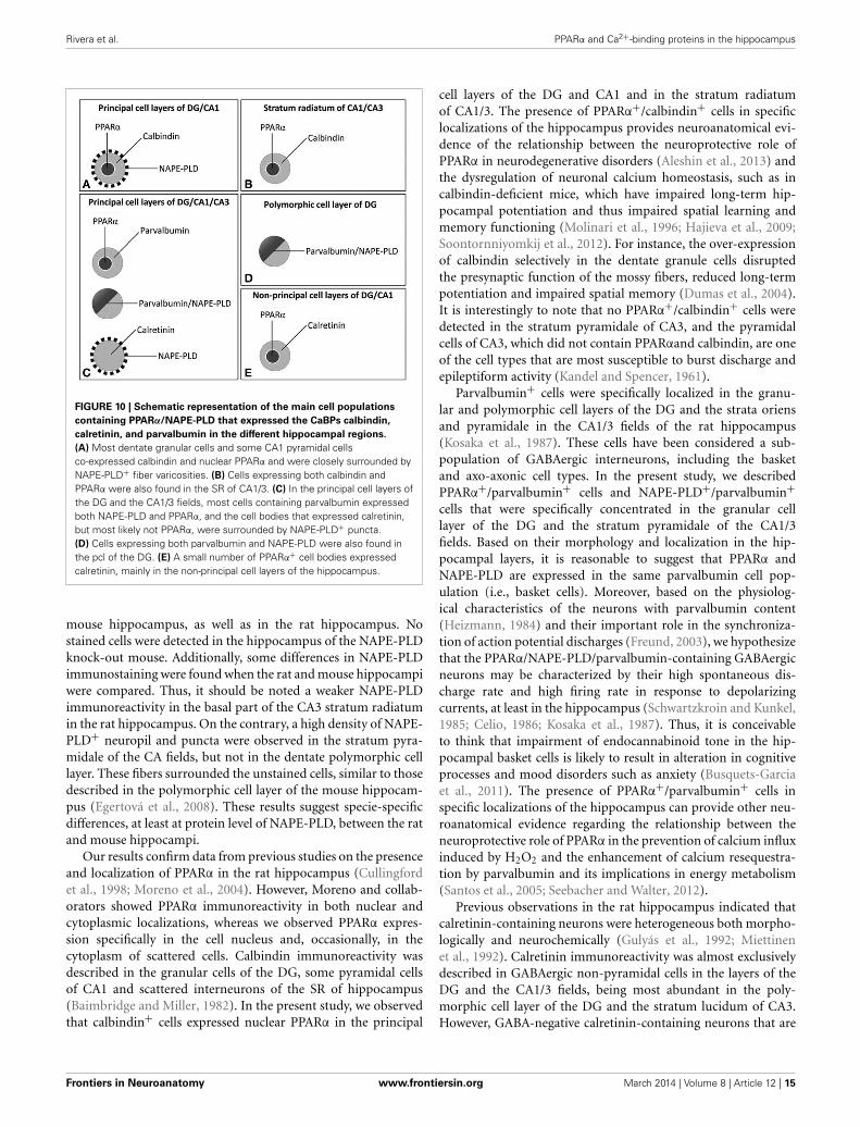
signaling system in the brain. Our study provides evidence foran anatomical segregation of the PPARα signaling system in hip-pocampal cells by identifying the localization and co-existence ofPPARα, NAPE-PLD, and the CaBPs calbindin, calretinin and par-valbumin, which have not been previously described. Due to thespecific localization of PPARα in the cell nuclei, NAPE-PLD inparticular puncta and cells, and CaBPs in certain cell bodies andfibers, we were able to highlight at least 4 different cell popula-tions in the rat hippocampus (Figure 10). First, we observed that
most dentate granular cells and some CA1 pyramidal cells co-expressed both calbindin and nuclear PPARα. These calbindin+cells containing PPARα were closely surrounded by NAPE-PLD+fiber varicosities (Figure 10A). Cells expressing both calbindinand PPARα were also found in the SR of CA1/3 (Figure 10B).Second, we observed that most cells containing parvalbuminexpressed both NAPE-PLD and PPARα in the principal cell layersof the DG and CA1/3 fields (Figure 10C). Cells expressing bothparvalbumin and NAPE-PLD were also found in the pcl of theDG (Figure 10D). Third, cell bodies that expressed calretinin, butmost likely not PPARα, were also surrounded by NAPE-PLD+puncta in the principal cell layers of the DG and CA1/3 fields(Figure 10C). Fourth, a small number of PPARα+ cell bodiesexpressed calretinin, mainly in the non-principal cell layers of thehippocampus (Figure 9E).
We observed that NAPE-PLD immunostaining was completelyabsent in the NAPE-PLD knock-out mouse hippocampus (Leunget al., 2006; Nyilas et al., 2008), whereas the wild type mouseshowed similar labeling to that described by Egertová et al.(2008). Thus, in the mouse hippocampus, very intense NAPE-PLD immunoreactivity was localized in the mossy fibers of thepolymorphic cell layer of the DG and the stratum lucidum ofCA3. Weaker staining was also observed in the molecular layerof the DG and the strata oriens and radiatum of the CA fields.In contrast to Egertová et al. (2008), we observed a number ofNAPE-PLD+ cells in the granular and polymorphic cell layersof the DG and the stratum pyramidale of the CA fields in the
Frontiers in Neuroanatomy www.frontiersin.org March 2014 | Volume 8 | Article 12 | 14
Rivera et al. PPARα and Ca2+-binding proteins in the hippocampus
FIGURE 10 | Schematic representation of the main cell populations
containing PPARα/NAPE-PLD that expressed the CaBPs calbindin,
calretinin, and parvalbumin in the different hippocampal regions.
(A) Most dentate granular cells and some CA1 pyramidal cellsco-expressed calbindin and nuclear PPARα and were closely surrounded byNAPE-PLD+ fiber varicosities. (B) Cells expressing both calbindin andPPARα were also found in the SR of CA1/3. (C) In the principal cell layers ofthe DG and the CA1/3 fields, most cells containing parvalbumin expressedboth NAPE-PLD and PPARα, and the cell bodies that expressed calretinin,but most likely not PPARα, were surrounded by NAPE-PLD+ puncta.(D) Cells expressing both parvalbumin and NAPE-PLD were also found inthe pcl of the DG. (E) A small number of PPARα+ cell bodies expressedcalretinin, mainly in the non-principal cell layers of the hippocampus.
mouse hippocampus, as well as in the rat hippocampus. Nostained cells were detected in the hippocampus of the NAPE-PLDknock-out mouse. Additionally, some differences in NAPE-PLDimmunostaining were found when the rat and mouse hippocampiwere compared. Thus, it should be noted a weaker NAPE-PLDimmunoreactivity in the basal part of the CA3 stratum radiatumin the rat hippocampus. On the contrary, a high density of NAPE-PLD+ neuropil and puncta were observed in the stratum pyra-midale of the CA fields, but not in the dentate polymorphic celllayer. These fibers surrounded the unstained cells, similar to thosedescribed in the polymorphic cell layer of the mouse hippocam-pus (Egertová et al., 2008). These results suggest specie-specificdifferences, at least at protein level of NAPE-PLD, between the ratand mouse hippocampi.
Our results confirm data from previous studies on the presenceand localization of PPARα in the rat hippocampus (Cullingfordet al., 1998; Moreno et al., 2004). However, Moreno and collab-orators showed PPARα immunoreactivity in both nuclear andcytoplasmic localizations, whereas we observed PPARα expres-sion specifically in the cell nucleus and, occasionally, in thecytoplasm of scattered cells. Calbindin immunoreactivity wasdescribed in the granular cells of the DG, some pyramidal cellsof CA1 and scattered interneurons of the SR of hippocampus(Baimbridge and Miller, 1982). In the present study, we observedthat calbindin+ cells expressed nuclear PPARα in the principal
cell layers of the DG and CA1 and in the stratum radiatumof CA1/3. The presence of PPARα+/calbindin+ cells in specificlocalizations of the hippocampus provides neuroanatomical evi-dence of the relationship between the neuroprotective role ofPPARα in neurodegenerative disorders (Aleshin et al., 2013) andthe dysregulation of neuronal calcium homeostasis, such as incalbindin-deficient mice, which have impaired long-term hip-pocampal potentiation and thus impaired spatial learning andmemory functioning (Molinari et al., 1996; Hajieva et al., 2009;Soontornniyomkij et al., 2012). For instance, the over-expressionof calbindin selectively in the dentate granule cells disruptedthe presynaptic function of the mossy fibers, reduced long-termpotentiation and impaired spatial memory (Dumas et al., 2004).It is interestingly to note that no PPARα+/calbindin+ cells weredetected in the stratum pyramidale of CA3, and the pyramidalcells of CA3, which did not contain PPARαand calbindin, are oneof the cell types that are most susceptible to burst discharge andepileptiform activity (Kandel and Spencer, 1961).
Parvalbumin+ cells were specifically localized in the granu-lar and polymorphic cell layers of the DG and the strata oriensand pyramidale in the CA1/3 fields of the rat hippocampus(Kosaka et al., 1987). These cells have been considered a sub-population of GABAergic interneurons, including the basketand axo-axonic cell types. In the present study, we describedPPARα+/parvalbumin+ cells and NAPE-PLD+/parvalbumin+cells that were specifically concentrated in the granular celllayer of the DG and the stratum pyramidale of the CA1/3fields. Based on their morphology and localization in the hip-pocampal layers, it is reasonable to suggest that PPARα andNAPE-PLD are expressed in the same parvalbumin cell pop-ulation (i.e., basket cells). Moreover, based on the physiolog-ical characteristics of the neurons with parvalbumin content(Heizmann, 1984) and their important role in the synchroniza-tion of action potential discharges (Freund, 2003), we hypothesizethat the PPARα/NAPE-PLD/parvalbumin-containing GABAergicneurons may be characterized by their high spontaneous dis-charge rate and high firing rate in response to depolarizingcurrents, at least in the hippocampus (Schwartzkroin and Kunkel,1985; Celio, 1986; Kosaka et al., 1987). Thus, it is conceivableto think that impairment of endocannabinoid tone in the hip-pocampal basket cells is likely to result in alteration in cognitiveprocesses and mood disorders such as anxiety (Busquets-Garciaet al., 2011). The presence of PPARα+/parvalbumin+ cells inspecific localizations of the hippocampus can provide other neu-roanatomical evidence regarding the relationship between theneuroprotective role of PPARα in the prevention of calcium influxinduced by H2O2 and the enhancement of calcium resequestra-tion by parvalbumin and its implications in energy metabolism(Santos et al., 2005; Seebacher and Walter, 2012).
Previous observations in the rat hippocampus indicated thatcalretinin-containing neurons were heterogeneous both morpho-logically and neurochemically (Gulyás et al., 1992; Miettinenet al., 1992). Calretinin immunoreactivity was almost exclusivelydescribed in GABAergic non-pyramidal cells in the layers of theDG and the CA1/3 fields, being most abundant in the poly-morphic cell layer of the DG and the stratum lucidum of CA3.However, GABA-negative calretinin-containing neurons that are
Frontiers in Neuroanatomy www.frontiersin.org March 2014 | Volume 8 | Article 12 | 15

Rivera et al. PPARα and Ca2+-binding proteins in the hippocampus
specifically localized in the polymorphic cell layer of the DGand the stratum lucidum of CA3 have been described (Miettinenet al., 1992). In the present study, we observed that very fewcalretinin+ cells expressed PPARα in the hippocampus; most ofthe calretinin+ cells were in the polymorphic cell layer of the DGand the stratum oriens of CA3. Thus, further analysis is necessaryto determine whether PPARα+/calretinin+ cells are GABAergicneurons, at least in the polymorphic cell layer of the DG.
In conclusion, our data indicated the presence of hippocam-pal cell subpopulations with specific co-expression patterns of theNAPE-PLD/PPARα system and selective CaBPs. As the CaBPs cal-bindin, calretinin and parvalbumin are found in a limited numberof hippocampal cell types that express PPARα and NAPE-PLD,we suggest that these CaBPs-expressing cells, especially thoseexpressing calbindin and parvalbumin, may be involved in morespecialized Ca2+-regulated functions related to the biologicalroles of the PPARα signaling system.
AUTHOR CONTRIBUTIONSAll authors had full access to all the data in the study and takeresponsibility for the integrity of the data and the accuracy of thedata analysis. Study concept and design: Fernando Rodríguez deFonseca and Juan Suárez. Acquisition of the data: Patricia Rivera,Sergio Arrabal, Eduardo Blanco, Antonia Serrano, and FranciscoJ. Pavón. Analysis and interpretation of the data: Patricia Riveraand Juan Suárez. Drafting of the manuscript: Patricia Riveraand Juan Suárez. Critical revision of the manuscript for impor-tant intellectual content, obtained funding and study supervision:Fernando Rodríguez de Fonseca and Juan Suárez.
ACKNOWLEDGMENTSThis work was supported by the 7th Framework Programmeof European Union [grant number HEALTH-F2-2008-223713,REPROBESITY], Ministerio de Ciencia e Innovación [grantnumbers SAF2010-19087, SAF 2010-20521], Instituto de SaludCarlos III, Ministerio de Economía y Competitividad, UE-ERDF[grant number CP12/03109], Red de Trastornos Adictivos [grantnumbers RD12/0028/0001, RD12/0028/0009], CIBERobn, PlanNacional Sobre Drogas, Ministerio de Sanidad y Consumo [grantnumber PNSD2010/143], Consejería de Economía, Innovación yCiencia, Junta de Andalucía, UE/ERDF [grant number CTS-433,P-11-CVI-07637], Consejería de Salud, Junta de Andalucía [grantnumbers PI0232/2008, PI0029/2008, SAS111224], and FundacióLa Marató de TV3 [grant number 386/C/2011]. Juan Suárez isrecipient of a “Miguel Servet” research contract from the NationalSystem of Health (Instituto de Salud Carlos III, grant numberCP12/03109).
REFERENCESAleshin, S., Strokin, M., Sergeeva, M., and Reiser, G. (2013). Peroxisome
proliferator-activated receptor (PPAR)β/δ, a possible nexus of PPARα-and PPARγ-dependent molecular pathways in neurodegenerative dis-eases: review and novel hypotheses. Neurochem. Int. 63, 322–330. doi:10.1016/j.neuint.2013.06.012
Baimbridge, K. G., and Miller, J. J. (1982). Immunohistochemical localization ofcalcium-binding protein in the cerebellum, hippocampal formation and olfac-tory bulb of the rat. Brain Res. 245, 223–229. doi: 10.1016/0006-8993(82)90804-6
Baimbridge, K. G., Miller, J. J., and Parkes, C. O. (1982). Calcium-binding proteindistribution in the rat brain. Brain Res. 239, 519–525. doi: 10.1016/0006-8993(82)90526-1
Bisogno, T., and Di Marzo, V. (2008). The role of the endocannabinoid system inAlzheimer’s disease: facts and hypotheses. Curr. Pharm. Des. 14, 2299–3305. doi:10.2174/138161208785740027
Bouilleret, V., Schwaller, B., Schurmans, S., Celio, M. R., and Fritschy, J. M. (2000).Neurodegenerative and morphogenic changes in a mouse model of tempo-ral lobe epilepsy do not depend on the expression of the calcium-bindingproteins parvalbumin, calbindin, or calretinin. Neuroscience 97, 47–58. doi:10.1016/S0306-4522(00)00017-8
Braissant, O., Foufelle, F., Scotto, C., Dauça, M., and Wahli, W. (1996). Differentialexpression of peroxisome proliferator-activated receptors (PPARs): tissue distri-bution of PPAR-alpha, -beta, and -gamma in the adult rat. Endocrinology 137,354–366.
Busquets-Garcia, A., Puighermanal, E., Pastor, A., de la Torre, R., Maldonado,R., and Ozaita, A. (2011). Differential role of anandamide and 2-arachidonoylglycerol in memory and anxiety-like responses. Biol. Psychiatry 70,479–486. doi: 10.1016/j.biopsych.2011.04.022
Calignano, A., La Rana, G., Giuffrida, A., and Piomelli, D. (1998). Controlof pain initiation by endogenous cannabinoids. Nature 394, 277–281. doi:10.1038/28393
Cates, J. M., Coffing, B. N., Harris, B. T., and Black, C. C. (2006). Calretininexpression in tumors of adipose tissue. Hum. Pathol. 37, 312–321. doi:10.1016/j.humpath.2005.11.006
Celio, M. R. (1986). Parvalbumin in most gamma-aminobutyric acid-containingneurons of the rat cerebral cortex. Science 231, 995–997. doi: 10.1126/sci-ence.3945815
Celio, M. R. (1990). Calbindin D-28k and parvalbumin in the rat nervous system.Neuroscience 35, 375–475. doi: 10.1016/0306-4522(90)90091-H
Combs, C. K., Bates, P., Karlo, J. C., and Landreth, G. E. (2001). Regulationof beta-amyloid stimulated proinflammatory responses by peroxisomeproliferator-activated receptor alpha. Neurochem. Int. 39, 449–457. doi:10.1016/S0197-0186(01)00052-3
Cullingford, T. E., Bhakoo, K., Peuchen, S., Dolphin, C. T., Patel, R., and Clark,J. B. (1998). Distribution of mRNAs encoding the peroxisome proliferator-activated receptor alpha, beta, and gamma and the retinoid X receptor alpha,beta, and gamma in rat central nervous system. J. Neurochem. 70, 1366–1375.doi: 10.1046/j.1471-4159.1998.70041366.x
Di Marzo, V., De Petrocellis, L., Sepe, N., and Buono, A. (1996). Biosynthesis ofanandamide and related acylethanolamides in mouse J774 macrophages andN18 neuroblastoma cells. Biochem. J. 316, 977–984.
Di Marzo, V., Fontana, A., Cadas, H., Schinelli, S., Cimino, G., Schwartz, J. C., et al.(1994). Formation and inactivation of endogenous cannabinoid anandamide incentral neurons. Nature 372, 686–691. doi: 10.1038/372686a0
Dumas, T. C., Powers, E. C., Tarapore, P. E., and Sapolsky, R. M. (2004).Overexpression of calbindin D(28k) in dentate gyrus granule cells altersmossy fiber presynaptic function and impairs hippocampal-dependent mem-ory. Hippocampus 14, 701–709. doi: 10.1002/hipo.10210
Egertová, M., Simon, G. M., Cravatt, B. F., and Elphick, M. R. (2008). Localizationof N-acyl phosphatidylethanolamine phospholipase D (NAPE-PLD) expressionin mouse brain: a new perspective on N-acylethanolamines as neural signalingmolecules. J. Comp. Neurol. 506, 604–615. doi: 10.1002/cne.21568
Esposito, E., Impellizzeri, D., Mazzon, E., Paterniti, I., and Cuzzocrea, S. (2012).Neuroprotective activities of palmitoylethanolamide in an animal model ofParkinson’s disease. PLoS ONE 7:e41880. doi: 10.1371/journal.pone.0041880
Freund, T. F. (2003). Interneuron diversity series: rhythm and mood in perisomaticinhibition. Trends Neurosci. 26, 489–495. doi: 10.1016/S0166-2236(03)00227-3
Galán-Rodríguez, B., Suarez, J., Gonzalez-Aparicio, R., Bermudez-Silva, F. J.,Maldonado, R., Robledo, P., et al. (2009). Oleoylethanolamide exerts par-tial and dose-dependent neuroprotection of substantia nigra dopamineneurons. Neuropharmacology 56, 653–664. doi: 10.1016/j.neuropharm.2008.11.006
García-Segura, L. M., Baetens, D., Roth, J., Norman, A. W., and Orci, L. (1984).Immunohistochemical mapping of calcium-binding protein immunoreactivityin the rat central nervous system. Brain Res. 296, 75–86. doi: 10.1016/0006-8993(84)90512-2
Garg, P., Duncan, R. S., Kaja, S., Zabaneh, A., Chapman, K. D., and Koulen, P.(2011). Lauroylethanolamide and linoleoylethanolamide improve functional
Frontiers in Neuroanatomy www.frontiersin.org March 2014 | Volume 8 | Article 12 | 16

Rivera et al. PPARα and Ca2+-binding proteins in the hippocampus
outcome in a rodent model for stroke. Neurosci. Lett. 492, 134–138. doi:10.1016/j.neulet.2011.01.073
Greene-Schloesser, D., Payne, V., Peiffer, A. M., Hsu, F. C., Riddle, D. R., Zhao, W.,et al. (2014). The peroxisomal proliferator-activated receptor (PPAR) α agonist,fenofibrate, prevents fractionated whole-brain irradiation-induced cognitiveimpairment. Radiat. Res. 181, 33–44. doi: 10.1667/RR13202.1
Gulyás, A. I., Miettinen, R., Jacobowitz, D. M., and Freund, T. F. (1992). Calretininis present in non-pyramidal cells of the rat hippocampus–I. A new type of neu-ron specifically associated with the mossy fibre system. Neuroscience 48, 1–27.doi: 10.1016/0306-4522(92)90334-X
Gulyás, A. I., Tóth, K., Dános, P., and Freund, T. F. (1991). Subpopulationsof GABAergic neurons containing parvalbumin, calbindin D28k, and chole-cystokinin in the rat hippocampus. J. Comp. Neurol. 312, 371–378. doi:10.1002/cne.903120305
Hajieva, P., Kuhlmann, C., Luhmann, H. J., and Behl, C. (2009). Impaired calciumhomeostasis in aged hippocampal neurons. Neurosci. Lett. 451, 119–123. doi:10.1016/j.neulet.2008.11.068
Heizmann, C. W. (1984). Parvalbumin, a relaxing factor in muscle and a neuronalmarker in brain. Prog. Clin. Biol. Res. 168, 205–210.
Inoue, I., Goto, S., Matsunag, T., Nakajima, T., Awata, T., Hokari, S., et al.(2001). The ligands/activators for peroxisome proliferator-activated receptoralpha (PPARalpha) and PPARgamma increase Cu2+,Zn2+-superoxide dismu-tase and decrease p22phox message expressions in primary endothelial cells.Metabolism 50, 3–11. doi: 10.1053/meta.2001.19415
Kandel, E. R., and Spencer, W. A. (1961). The pyramidal cell during hippocampalseizure. Epilepsia 2, 63–69. doi: 10.1111/j.1528-1167.1961.tb06247.x
Karádi, K., Janszky, J., Gyimesi, C., Horváth, Z., Lucza, T., Dóczi, T., et al. (2012).Correlation between calbindin expression in granule cells of the resected hip-pocampal dentate gyrus and verbal memory in temporal lobe epilepsy. EpilepsyBehav. 25, 110–119. doi: 10.1016/j.yebeh.2012.06.007
Khasabova, I. A., Xiong, Y., Coicou, L. G., Piomelli, D., and Seybold, V. (2012).Peroxisome proliferator-activated receptor α mediates acute effects of palmi-toylethanolamide on sensory neurons. J. Neurosci. 32, 12735–12743. doi:10.1523/JNEUROSCI.0130-12.2012
Koch, M., Kreutz, S., Böttger, C., Benz, A., Maronde, E., Ghadban, C., et al.(2011). Palmitoylethanolamide protects dentate gyrus granule cells via per-oxisome proliferator-activated receptor-α. Neurotox. Res. 19, 330–340. doi:10.1007/s12640-010-9166-2
Kosaka, T., Katsumaru, H., Hama, K., Wu, J. Y., and Heizmann, C. W. (1987).GABAergic neurons containing the Ca2+-binding protein parvalbumin in therat hippocampus and dentate gyrus. Brain Res. 419, 119–130. doi: 10.1016/0006-8993(87)90575-0
Leung, D., Saghatelian, A., Simon, G. M., and Cravatt, B. F. (2006). Inactivationof N-acyl phosphatidylethanolamine phospholipase D reveals multiple mech-anisms for the biosynthesis of endocannabinoids. Biochemistry 45, 4720–4726.doi: 10.1021/bi060163l
Lombardi, G., Miglio, G., Varsaldi, F., Minassi, A., and Appendino, G. (2007).Oxyhomologation of the amide bond potentiates neuroprotective effects of theendolipid N-palmitoylethanolamine. J. Pharmacol. Exp. Ther. 320, 599–606.doi: 10.1124/jpet.106.112987
Maglóczky, Z., Halász, P., Vajda, J., Czirják, S., and Freund, T. F. (1997).Loss of Calbindin-D28K immunoreactivity from dentate granule cells inhuman temporal lobe epilepsy. Neuroscience 76, 377–385. doi: 10.1016/S0306-4522(96)00440-X
Miettinen, R., Gulyás, A. I., Baimbridge, K. G., Jacobowitz, D. M., and Freund, T. F.(1992). Calretinin is present in non-pyramidal cells of the rat hippocampus–II.Co-existence with other calcium binding proteins and GABA. Neuroscience 48,29–43. doi: 10.1016/0306-4522(92)90335-Y
Molinari, S., Battini, R., Ferrari, S., Pozzi, L., Killcross, A. S., Robbins, T. W.,et al. (1996). Deficits in memory and hippocampal long-term potentiation inmice with reduced calbindin D28K expression. Proc. Natl. Acad. Sci. U.S.A. 93,8028–8033. doi: 10.1073/pnas.93.15.8028
Moreno, S., Farioli-Vecchioli, S., and Cerù, M. P. (2004). Immunolocalizationof peroxisome proliferator-activated receptors and retinoid Xreceptors in the adult rat CNS. Neuroscience 123, 131–145. doi:10.1016/j.neuroscience.2003.08.064
Murray, A. J., Sauer, J. F., Riedel, G., McClure, C., Ansel, L., Cheyne, L., et al. (2011).Parvalbumin-positive CA1 interneurons are required for spatial working butnot for reference memory. Nat. Neurosci. 14, 297–299. doi: 10.1038/nn.2751
Nakamura, Y., Nakahama, T., Ushiwata, A., Takeda, M., and Nakaya, K. (1980).Isolation and partial characterization of an acidic calcium-binding proteinfrom synaptic plasma membranes of rat brain. FEBS Lett. 112, 155–158. doi:10.1016/0014-5793(80)80169-4
Nakazawa, K., Zsiros, V., Jiang, Z., Nakao, K., Kolata, S., Zhang, S., et al.(2012). GABAergic interneuron origin of schizophrenia pathophysiology.Neuropharmacology 62, 1574–1583. doi: 10.1016/j.neuropharm.2011.01.022
Nyilas, R., Dudok, B., Urbán, G. M., Mackie, K., Watanabe, M., Cravatt, B. F., et al.(2008). Enzymatic machinery for endocannabinoid biosynthesis associated withcalcium stores in glutamatergic axon terminals. J. Neurosci. 28, 1058–1063. doi:10.1523/JNEUROSCI.5102-07.2008
Okamoto, Y., Morishita, J., Tsuboi, K., Tonai, T., and Ueda, N. (2004). Molecularcharacterization of a phospholipase D generating anandamide and its con-geners. J. Biol. Chem. 279, 5298–5305. doi: 10.1074/jbc.M306642200
Okamoto, Y., Wang, J., Morishita, J., and Ueda, N. (2007). Biosynthetic path-ways of the endocannabinoid anandamide. Chem. Biodivers. 4, 1842–1857. doi:10.1002/cbdv.200790155
Paxinos, G., and Watson, C. (2007). The Rat Brain in Stereotaxic Coordinates, 6thEdn. Sidney: Academic Press.
Rodríguez de Fonseca, F., Navarro, M., Gómez, R., Escuredo, L., Nava, F., Fu, J., et al.(2001). An anorexic lipid mediator regulated by feeding. Nature 414, 209–212.doi: 10.1038/35102582
Rüttimann, E., Vacher, C. M., Gassmann, M., Kaupmann, K., Van der Putten, H.,and Bettler, B. (2004). Altered hippocampal expression of calbindin-D-28k andcalretinin in GABA(B(1))-deficient mice. Biochem. Pharmacol. 68, 1613–1620.doi: 10.1016/j.bcp.2004.07.019
Santos, M. J., Quintanilla, R. A., Toro, A., Grandy, R., Dinamarca, M. C., Godoy,J. A., et al. (2005). Peroxisomal proliferation protects from beta-amyloid neu-rodegeneration. J. Biol. Chem. 280, 41057–41068. doi: 10.1074/jbc.M505160200
Schmid, H. H. (2000). Pathways and mechanisms of N-acylethanolamine biosyn-thesis: can anandamide be generated selectively? Chem. Phys. Lipids 108, 71–87.doi: 10.1016/S0009-3084(00)00188-2
Schwartzkroin, P. A., and Kunkel, D. D. (1985). Morphology of identified interneu-rons in the CA1 regions of guinea pig hippocampus. J. Comp. Neurol. 232,205–218. doi: 10.1002/cne.902320206
Scuderi, C., Valenza, M., Stecca, C., Esposito, G., Carratù, M. R., and Steardo,L. (2012). Palmitoylethanolamide exerts neuroprotective effects in mixedneuroglial cultures and organotypic hippocampal slices via peroxisomeproliferator-activated receptor-α. J. Neuroinflammation 9:49. doi: 10.1186/1742-2094-9-49
Seebacher, F., and Walter, I. (2012). Differences in locomotor performance betweenindividuals: importance of parvalbumin, calcium handling and metabolism.J. Exp. Biol. 215, 663–670. doi: 10.1242/jeb.066712
Soontornniyomkij, V., Risbrough, V. B., Young, J. W., Soontornniyomkij, B., Jeste,D. V., and Achim, C. L. (2012). Hippocampal calbindin-1 immunoreactivitycorrelates of recognition memory performance in aged mice. Neurosci. Lett. 516,161–165. doi: 10.1016/j.neulet.2012.03.092
Suardíaz, M., Estivill-Torrús, G., Goicoechea, C., Bilbao, A., and Rodríguez deFonseca, F. (2007). Analgesic properties of oleoylethanolamide (OEA) in vis-ceral and inflammatory pain. Pain 133, 99–110. doi: 10.1016/j.pain.2007.03.008
Suárez, J., Bermúdez-Silva, F. J., Mackie, K., Ledent, C., Zimmer, A., Cravatt, B. F.,et al. (2008). Immunohistochemical description of the endogenous cannabinoidsystem in the rat cerebellum and functionally related nuclei. J. Comp. Neurol.509, 400–421. doi: 10.1002/cne.21774
Suárez, J., Dávila, J. C., Real, M. A., and Guirado, S. (2005). Distribution of GABA,calbindin and nitric oxide synthase in the developing chick entopallium. BrainRes. Bull. 66, 441–444. doi: 10.1016/j.brainresbull.2005.02.014
Suárez, J., Dávila, J. C., Real, M. A., Guirado, S., and Medina, L. (2006).Calcium-binding proteins, neuronal nitric oxide synthase, and GABA helpto distinguish different pallial areas in the developing and adult chicken. I.Hippocampal formation and hyperpallium. J. Comp. Neurol. 497, 751–771. doi:10.1002/cne.21004
Sun, Y., Alexander, S. P., Garle, M. J., Gibson, C. L., Hewitt, K., Murphy, S. P.,et al. (2007). Cannabinoid activation of PPAR alpha; a novel neuroprotectivemechanism. Br. J. Pharmacol. 152, 734–743. doi: 10.1038/sj.bjp.0707478
Verret, L., Mann, E. O., Hang, G. B., Barth, A. M., Cobos, I., Ho, K., et al.(2012). Inhibitory interneuron deficit links altered network activity and cogni-tive dysfunction in Alzheimer model. Cell 149, 708–721. doi: 10.1016/j.cell.2012.02.046
Frontiers in Neuroanatomy www.frontiersin.org March 2014 | Volume 8 | Article 12 | 17

Rivera et al. PPARα and Ca2+-binding proteins in the hippocampus
Wouterlood, F. G., Grosche, J., and Härtig, W. (2001). Co-localization ofcalretinin and calbindin in distinct cells in the hippocampal forma-tion of the rat. Brain Res. 922, 310–314. doi: 10.1016/S0006-8993(01)03220-6
Zhou, Y., Yang, L., Ma, A., Zhang, X., Li, W., Yang, W., et al. (2012). Orally admin-istered oleoylethanolamide protects mice from focal cerebral ischemic injury byactivating peroxisome proliferator-activated receptor α. Neuropharmacology 63,242–249. doi: 10.1016/j.neuropharm.2012.03.008
Zimmermann, L., and Schwaller, B. (2002). Monoclonal antibodies recognizingepitopes of calretinins: dependence on Ca2+-binding status and differencesin antigen accessibility in colon cancer cells. Cell Calcium 31, 13–25. doi:10.1054/ceca.2001.0255
Conflict of Interest Statement: The authors declare that the research wasconducted in the absence of any commercial or financial relationships that couldbe construed as a potential conflict of interest.
Received: 21 December 2013; accepted: 28 February 2014; published online: 17 March2014.Citation: Rivera P, Arrabal S, Vargas A, Blanco E, Serrano A, Pavón FJ, Rodríguezde Fonseca F and Suárez J (2014) Localization of peroxisome proliferator-activatedreceptor alpha (PPARα) and N-acyl phosphatidylethanolamine phospholipase D(NAPE-PLD) in cells expressing the Ca2+-binding proteins calbindin, calretinin, andparvalbumin in the adult rat hippocampus. Front. Neuroanat. 8:12. doi: 10.3389/fnana.2014.00012This article was submitted to the journal Frontiers in Neuroanatomy.Copyright © 2014 Rivera, Arrabal, Vargas, Blanco, Serrano, Pavón, Rodríguez deFonseca and Suárez. This is an open-access article distributed under the terms of theCreative Commons Attribution License (CC BY). The use, distribution or reproductionin other forums is permitted, provided the original author(s) or licensor are creditedand that the original publication in this journal is cited, in accordance with acceptedacademic practice. No use, distribution or reproduction is permitted which does notcomply with these terms.
Frontiers in Neuroanatomy www.frontiersin.org March 2014 | Volume 8 | Article 12 | 18


