Embed Size (px)
Citation preview
Vaccine 22 (2004) 3258–3269
Long-term protection against SHIV89.6P replication in HIV-1 Tatvaccinated cynomolgus monkeys
Maria Teresa Maggiorellaa, Silvia Baroncellia, Zuleika Michelinia, Emanuele Fanales-Belasioa,Sonia Morettia, Leonardo Sernicolaa, Andrea Caraa, Donatella R.M. Negria, Stefano Buttòa,
Valeria Fiorellia, Antonella Tripicianoa, Arianna Scoglioa, Antonella Caputob,Alessandra Borsettia, Barbara Ridolfia, Roberta Bonaa, Peter ten Haaftc, Iole Macchiaa,
Pasqualina Leonea, Maria Rosaria Pavone-Cossuta, Filomena Nappia, Massimo Ciccozzid,Jonathan Heeneyc, Fausto Tittia, Aurelio Cafaroa, Barbara Ensolia,∗
a Laboratory of Virology, Istituto Superiore di Sanità, Viale Regina Elena, 299, 00161 Rome, Italyb Department of Experimental and Diagnostic Medicine, University of Ferrara, 44100 Ferrara, Italy
c Department of Virology, Biomedical Primate Research Centre, ZH 2288 GJ Rijswijk, The Netherlandsd Laboratory of Epidemiology and Biostatistics, Istituto Superiore di Sanità, 00161 Rome, Italy
Received 15 July 2003; received in revised form 19 February 2004; accepted 10 March 2004
Available online 8 April 2004
Abstract
Vaccination with a biologically active Tat protein ortatDNA contained infection with the highly pathogenic SHIV89.6P virus, preventingCD4 T-cell decline and disease onset. Here we show that protection was prolonged, since neither CD4 T-cell decline nor active virusreplication was observed in all vaccinated animals that controlled virus replication up to week 104 after the challenge. In contrast, viruspersisted and replicated in peripheral blood mononuclear cells and lymph nodes of infected animals, two of which died. Tat-specificantibody, CD4 and CD8 T-cell responses were high and stable only in the animals controlling the infection. In contrast, Gag-specificantibody production and CD4 and CD8 T-cell responses were consistently and persistently positive only in the monkeys that did notcontrol primary virus replication. These results indicate that vaccination with Tat protein or DNA induced long-term memory Tat-specificimmune responses and controlled primary infection at its early stages allowing a long-term containment of virus replication and spread inblood and tissues.© 2004 Elsevier Ltd. All rights reserved.
Keywords:HIV vaccine; Tat;Macaca fascicularis
1. Introduction
Tat is a key viral regulatory protein of human immunode-ficiency virus (HIV) produced very early after infection evenprior to provirus integration[1–3]. Tat is essential for viralreplication, transmission and disease progression[1,4–7].In addition, Tat can modulate the expression of cytokinesand cellular genes[6–11], and chemokine receptors[12–15],which are responsible for the transmission of macrophage-and T-cell-tropic HIV-1 strains, respectively. In addition, theTat protein also plays key roles in viral pathogenesis and in
∗ Corresponding author. Tel.:+39-06-4990-3209;fax: +39-06-4990-3002.
E-mail address:[email protected] (B. Ensoli).
the pathogenesis of acquired immune deficiency syndrome(AIDS)-associated Kaposi’s sarcoma[8,16–22].
Several studies suggest that an immune response to Tathas a protective role and may control the progression of thedisease in vivo. Specifically, antibody (Ab) responses to Tathave been associated with non-progression to AIDS[23–28].Similarly, anti-Tat CTLs have been shown to inversely cor-relate with progression to AIDS[29,30]. Of importance,Tat is conserved in its immunogenic regions among all Msubtypes, and Tat B clade is recognized to the same extentby sera from South African, Ugandan and Italian individ-uals infected with A, B, C and D virus clades[28]. Fur-ther, studies in humans[29,30]and in monkeys indicate thatanti-Tat CTLs are key to control virus replication early af-ter primary infection, and that they exert a selective immunepressure on the virus leading to the appearance of slowly
0264-410X/$ – see front matter © 2004 Elsevier Ltd. All rights reserved.doi:10.1016/j.vaccine.2004.03.009
M.T. Maggiorella et al. / Vaccine 22 (2004) 3258–3269 3259
replicating and apparently less pathogenic escape mutants[31].
Taken together, this body of evidence suggests that im-munization with Tat may block HIV replication both inindividuals exposed to the virus after vaccination and inseropositive patients, reducing HIV infection and favouringits control by the mounting immune response. In this regard,some features of the Tat protein are relevant to Tat-basedvaccine development. First, we have recently shown that bi-ologically active native HIV-1 Tat protein, but not its oxi-dized counterpart, is taken up very efficiently and selectivelyby monocyte-derived dendritic cells and induces their mat-uration and antigen presenting function, driving Th-1 typeimmune responses[32]. Secondly, soluble Tat protein hasbeen shown to enter the major histocompatibility complex(MHC) class I pathway of antigen presentation, leading toelicitation of cytotoxic T lymphocytes (CTLs)[33].
Pre-clinical studies in mice and monkeys have shown thatvaccination with a biologically active native HIV-1 Tat pro-tein or with tat DNA is safe[34–38], elicits both humoraland cell-mediated Tat-specific immune responses[34–38],and, in cynomolgus monkeys, controls infection with thehighly pathogenic SHIV89.6P virus, preventing CD4 T-celldecline and disease onset in the protected animals[34–36].Similar results of protection were reported in the SIV modelwith viral vectors (Semliki Forest Virus and Modified Vac-cinia Ankara Virus) expressing the SIV-Tat and -Rev genes[39]. Of importance, injection of plasmid DNA coding forthe HIV-1 regulatory genes Nef, Rev and Tat was reportedto be safe and immunogenic in HIV-1 infected individuals[40,41].
Since Tat vaccination does not prevent infection butcontrols virus replication and disease onset, it is key toinvestigate whether protection is long-lasting, and to deter-mine the level of virus infection, reservoir, and replicationover time in the protected animals. Therefore, we extendedthe analysis of the vaccinated and control macaques up to2 years after the challenge, looking for signs of virus per-sistence and replication in blood and lymph nodes and forpersistence and development of CD4 and CD8-mediatedimmune responses to HIV-1 Tat and SIV Gag, respectively.
The results indicate that vaccination with either a biolog-ically active Tat protein ortat DNA induces a long-termprotection (i.e. containment of infection) against challengewith the highly pathogenic SHIV89.6P virus, which is asso-ciated with the absence of detectable virus replication bothin blood and tissues and with the persistence of Tat-specificT- (both CD4 and CD8) and B-cell memory responses.
2. Materials and methods
2.1. Vaccination protocol and virus challenge
Adult male cynomolgus monkeys (Macaca fascicularis)were housed in single cages within level three biosafety fa-
cilities according to the European guidelines for non-humanprimate care (EEC, Directive No. 86-609, 24 November1986). Blood samples were obtained from the inguinal veinwhile the animals were under ketamine hydrochloride anes-thesia (10 mg/kg). Clinical examination and weight mea-surement were performed at the time of blood collection. Ateach time-point bleedings were performed for routine bloodchemistry tests and immunological determinations. The vac-cination protocols and schedules have been reported else-where[34–36]. After 14–18 weeks from the last boost, allanimals were challenged intravenously (i.v.) with 10 MID50of the SHIV89.6P virus, a highly pathogenic SHIV contain-ing thetat gene of HIV-1. The virus stock used was derivedfrom a cynomolgus macaque inoculated with the originalSHIV89.6P from rhesus monkeys[42]. Both the original vi-ral stock grown in a rhesus macaque and the one grown ina cynomolgus monkey were highly pathogenic inM. fasci-cularis [34,35]. The pathogenicity in cynomolgus monkeysof both virus stocks was confirmed by the progression toAIDS and death (within 46 weeks from the inoculum) offour out of six and four out of seven cynomolgus monkeysinfected, respectively, with the rhesus- or cynos-derived vi-ral stock[35]. The two naive control animals, monkeys 2and 12, included as additional control at the time of the chal-lenge, were inoculated with a three-fold higher (28 MID50)or three-fold lower (2.8 MID50) viral inoculum, respectively[34–36].
2.2. Detection of anti-SIV and anti-HIV Env and p27 Gagantigen in plasma
Ab titers to the whole SIV were determined by endpointdilution of plasma samples using a HIV-2 enzyme-linkedimmunosorbent assay (ELISA) (Elavia AC-Ab-Ak II Kit,Diagnostic, Pasteur) according to the manufacturer’s instruc-tions. Ab titers against the HIV-1 Env were detected by anHIV-1 ELISA assay (HIV-1/HIV-2 Third Generation Plus,Abbott, Chicago, IL). The mean of the negative control plus3 S.D. represented the cut-off value.
Levels of p27 Gag protein were measured in plasma byusing a capture ELISA assay (Innotest, Innogenetics, Zwi-jndrecht, Belgium) with a detection limit of 20 pg/ml.
2.3. Quantitation of the SHIV RNA copies in plasma
Until week 14 post-challenge quantitation of SHIV89.6PRNA copies was performed only in the Bayer-Chiron Di-agnostics reference Testing Laboratory (Amsterdam, TheNetherlands) by a branched DNA (bDNA) signal amplifi-cation assay recognizing the pol region of the SIVmac251strain, as described[34], with a cut-off of 1,500 RNAcopies/ml. Between week 14 and 28 post-challenge quanti-tation of SHIV89.6P RNA copies was performed by boththe bDNA method and by a more sensitive quantitative com-petitive RNA-polymerase chain reaction (QC-RNA-PCR,cut-off: 50 RNA copies/ml) as already described[43]. Since
3260 M.T. Maggiorella et al. / Vaccine 22 (2004) 3258–3269
week 35 post-challenge, quantitation of SHIV89.6P RNAcopies was performed only by QC-RNA-PCR.
2.4. Proviral DNA detection
DNA was extracted from whole blood using the QIAampBlood Kit (QIAGEN, GmbH, Hilden, Germany) or fromlymph node cells using the phenol–chloroform method, fol-lowed by precipitation with 3 M sodium acetate and coldethanol. SHIV proviral copy number was determined by asemi-quantitative DNA PCR utilizing 1�g of DNA and am-plifying a 496 bp region of thegaggene of SIVmac239, asalready described[34,36]. The lower limit of detection was1 SHIV proviral copy/�g of DNA.
2.5. Unintegrated virus DNA detection
For analysis of viral extrachromosomal circular DNA(E-DNA), DNA was extracted from lymphocytes ((2–5) ×106) using the QIAprep Spin Miniprep Kit in order to enrichfor the low molecular weight DNA fraction, and analyzedby PCR for the presence of circular forms of viral DNA asdescribed[44,45]. Twenty microliters of the total volume(50�l) were denaturated at 95◦C for 5 min, and subjectedto 40 cycles of PCR with an annealing temperature of 60◦Cfor 30 s, an extension temperature of 72◦C for 30 s and adenaturation temperature of 95◦C for 30 s, using primersFORN15′-GTGACTCCACGCTTGTTTGC-3′ (forward),located in the R LTR region, and REVN15′-CTCCTGTGC-CTCATCTGATACA-3′ (reverse), mapping in the U3 LTRregion of the SIVmac239. The amplified fragment was361 bp in length. As positive control, a reference standardwas prepared by cloning the PCR product, amplified fromDNA of CEMx174 chronically infected with SHIV89.6P,into the pCR 2.1 vector (Invitrogen, Carlsbad, CA).
Hybridization was performed according to establishedprocedures[46] utilizing the FORN1/REVN1 PCR productas probe.
2.6. Virus isolation and cell associated viral load(cytoviremia)
Virus isolation and cytoviremia were performed byco-culturing CD8-depleted PBMCs with CEMx174 cells inthe presence of phytohemoagglutinin (PHA, 2�g/ml) andrecombinant human interleukin-2 (rhIL-2) (50 IU/ml), asalready described[34–36].
2.7. Lymphocyte subsets determination
Citrated peripheral blood cells were stained with ph-icoerythrin PE-conjugated anti-CD4 (Biosource Interna-tional, Camarillo, CA) and peridin chlorophyll protein(PerCP)-conjugated anti-CD8 mAb (Becton-Dickinson,Mountain View, CA), and analyzed with a FACScan cy-tometer and software (Becton-Dickinson) as described
[34,36]. Absolute cell numbers were calculated from bloodcell counts.
2.8. Recombinant proteins
The Tat protein (1–86 aa) from the HTLV-IIIB isolate,BH-10 clone (clade B), was expressed inE. coli, purifiedand stored as described earlier[9,32,34]. The purified Tatprotein was fully monomeric and had full biological activ-ity as assessed by virus transactivation assays and by uptakestudies in monocyte-derived dendritic cells[6,9,32,34]. Tatused for in vitro studies was resuspended in degassed bufferbefore use as described[6,28,34]. The SIVmac251 p55 Gagrecombinant protein was derived from baculovirus was ob-tained through Quality Biological, Inc. and the Vaccine Re-search and Development Branch, Division of AIDS, NIAID,NIH (Bethesda, MD).
2.9. T-cell proliferation assays
To evaluate the proliferative response to Tat, Gag and therecall antigen tetanus toxoid (TT), Ficoll-purified PBMCs(2 × 10⁵ per well) were seeded in flat bottomed 96-wellmicrotiter culture plates in triplicates in a final volume of200�l of RPMI containing 10% FCS (medium) and cul-tured either alone or in the presence of PHA (2�g/ml),TT (5�g/ml, Connaught, Ontario, Canada), Tat protein(5 mg/ml), SIVmac251 p55 Gag protein (5 mg/ml) or SIV-mac239 p27 Gag 15mers peptides (aminoacid 226–240,231–245, 236–250, 283–297, 289–303, 295–309 at 5 mg/mleach) or buffer [phosphate buffered saline (PBS) containing0.1% BSA]. After 5 days of incubation at 37◦C in 5% CO2,cell cultures were pulsed with 1�Ci/well of [3H]thymidine(Amersham Life Science, Buckingamshire, UK) and theincorporated radioactivity was measured 18 h later, as pre-viously described[34,36]. A stimulation index (SI)≥3 wasconsidered positive.
2.10. Interferon (IFN)-γ ELISpot assay
The number of IFN-� producing cells (spot formingcells, sfc) was measured by a commercial kit (HumanIFN-� ELISpot, Euroclone, Paignton, UK), following themanufacturer’s instructions. Briefly, PBMCs (2× 10⁵ perwell, in duplicate) were cultured with medium alone or inthe presence of PHA (2�g/ml), TT (5�g/ml), Gag or Tatprotein (5�g/ml), or buffer in flat bottomed 96-well platespreviously coated with a mAb to IFN-�. After 18 h, cellswere removed and locally produced IFN-� was revealed byan immunoenzymatic reaction into a gel matrix as colouredspots. The spots, a measure of IFN-� producing cells, werecounted under a light microscope and expressed as num-ber of sfc per 106 PBMCs upon subtraction of backgroundcounts. Based on data obtained with PBMCs from 24 naivemonkeys, a number of sfc greater than 20/106 cells wasconsidered as a positive response to the antigen.
M.T. Maggiorella et al. / Vaccine 22 (2004) 3258–3269 3261
2.11. Lymph node biopsies
Inguinal lymph nodes were removed surgically under ke-tamine hydrochloride anesthesia at week 55 after challengefrom monkeys vaccinated with the pCV-tat, and at week 71after challenge from monkeys vaccinated with the Tat pro-tein and from control monkeys. The tissue was minced toobtain single cell suspension for virus isolation, quantitativedetection of proviral DNA, and analysis for the presence ofE-DNA forms, which were performed as described above.
2.12. Statistical analyses
The statistical significance of the differences between vac-cinated and control monkeys during the course of infectionwas determined by the Fisher’s exact test for quantitationof SHIV RNA copies in plasma and for SHIV DNA PCRand E-DNA analyses in lymph node biopsies. The Student’st-test for independent samples was used to assess differencesbetween groups concerning the proliferative response andthe IFN-� ELISpot to Tat and Gag antigens.
3. Results
3.1. Long-term containment of virus replication invaccinated and protected animals
A total of 12 macaques were vaccinated with the Tat pro-tein or the pCV-tat plasmid as two arms of the same pro-tocol and challenged i.v. with SHIV89.6P, as reported inSection 2. The results of the immune response to Tat beforethe challenge, challenge outcome and virological data upto 46 weeks after challenge have been published elsewhere[34–36]. These results indicated that, although vaccinationwith the biologically active Tat protein or pCV-tat did notprevent virus entry, it contained primary infection in 9 outof 12 vaccinated animals. Therefore, the term protection,as used in the present study, refers to protection from virusreplication and disease onset, that is control of infection, andnot to prevention of infection. Consequently, animals weredefined ascontrollersor non-controllersaccording to theirvirological status early after challenge.
To investigate whether control of infection was durable,all macaques (both vaccinated and controls) were monitoredup to 104 weeks after the challenge for plasma viremia,circular viral DNA, proviral DNA and virus isolation bothin blood and lymph nodes.
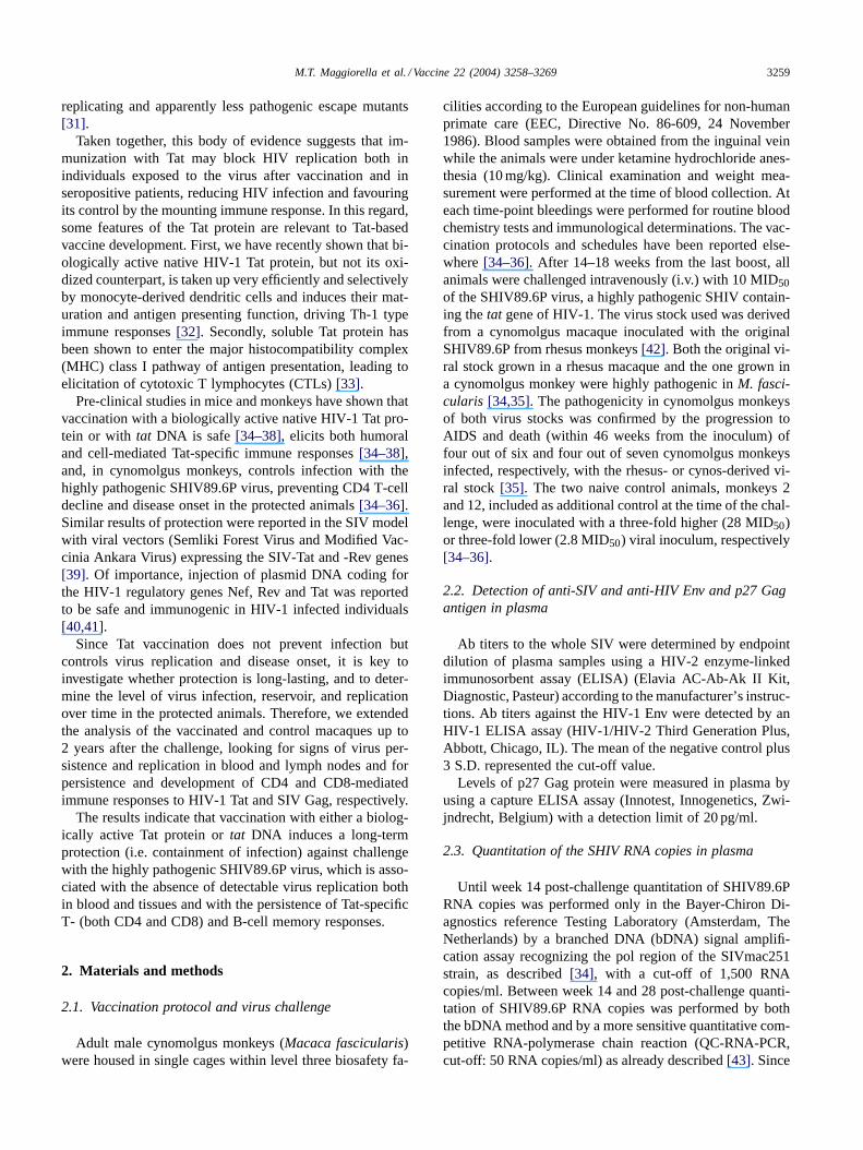
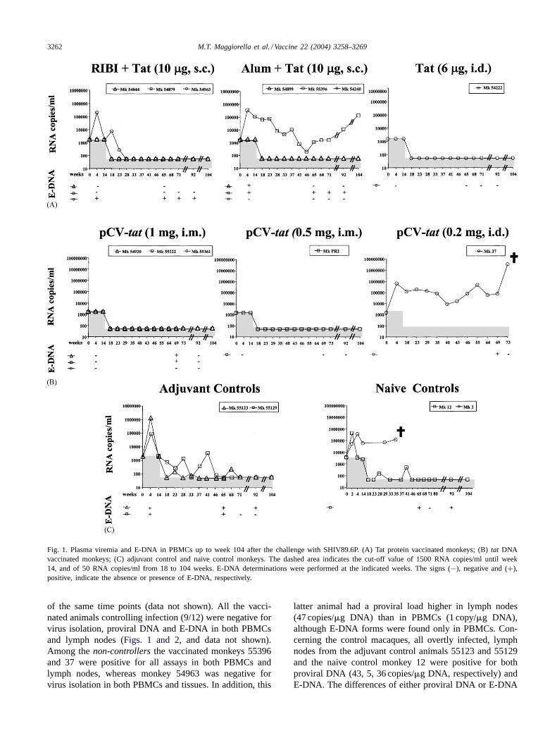
As shown inFig. 1, plasma viremia remained undetectablein all (9/12) animals, which controlled primary virus repli-cation (vaccinated and protected animals), whereas in thethree vaccinated animals unable to control virus replication(monkeys 54963, 55396, and 37) plasma viremia either be-came undetectable early in the follow-up (week 23 for mon-key 54963), or remained detectable with a further increaselate in the follow-up (monkeys 55396 and 37) (Fig. 1A and
B). Viral RNA was also detected over time in all adjuvantcontrol and naive control macaques. In particular, monkey2 had high levels of viral RNA up to week 35 when waseuthanised due to AIDS (Fig. 1C). Similarly, monkey 37died of AIDS at week 75 after challenge. The comparisonof plasma viremia data between vaccinated and control an-imals at week 4 after challenge revealed a statistically sig-nificant difference, as determined by the Fisher exact test(P-value= 0.019).
To better evaluate virus replication, E-DNA, a param-eter more sensitive than the evaluation of plasma viremia[44,45], was measured in PBMCs of all macaques (with theexception of monkey 2) and compared with plasma viremiaat early and/or late time points post-challenge (Fig. 1). Agood correlation was observed between the presence ofE-DNA and plasma viremia. However, E-DNA was alsofound in animals with undetectable plasma viremia, indicat-ing the presence of low levels of persistent viral replication.In particular, E-DNA was positive at multiple time pointspost-challenge in all thenon-controllers, both viremic andaviremic, whereas it was detected only once in three outof the ninecontrollers (plasma viremia always below thedetection threshold) (Fig. 1).
The frequency of viral isolation also correlated with thepresence of viral RNA in plasma. In fact, the virus wasnot isolated from any of the macaques controlling infection,even after depletion of the CD8+ T-cells. Conversely, it wasfrequently isolated from all thenon-controllers ([34–36],and data not shown).
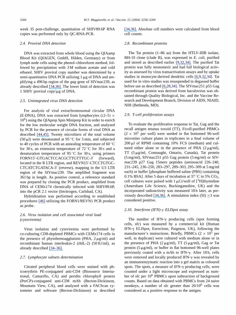
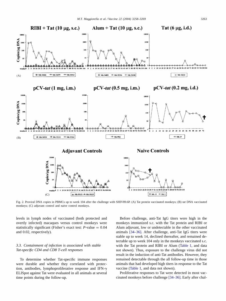
Similarly, after the challenge the provirus was either un-detectable or sporadically present at a very low level (<10copies/�g of DNA) in the controllers, and always detectedin all thenon-controllers(Fig. 2A–C).
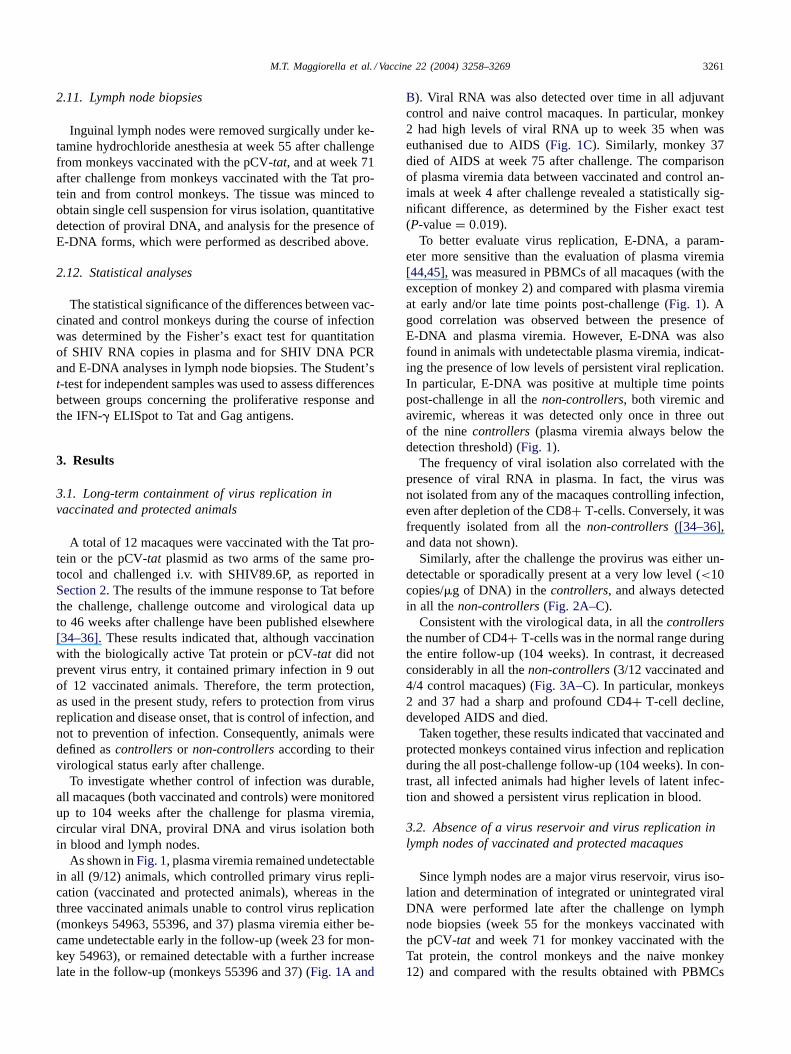
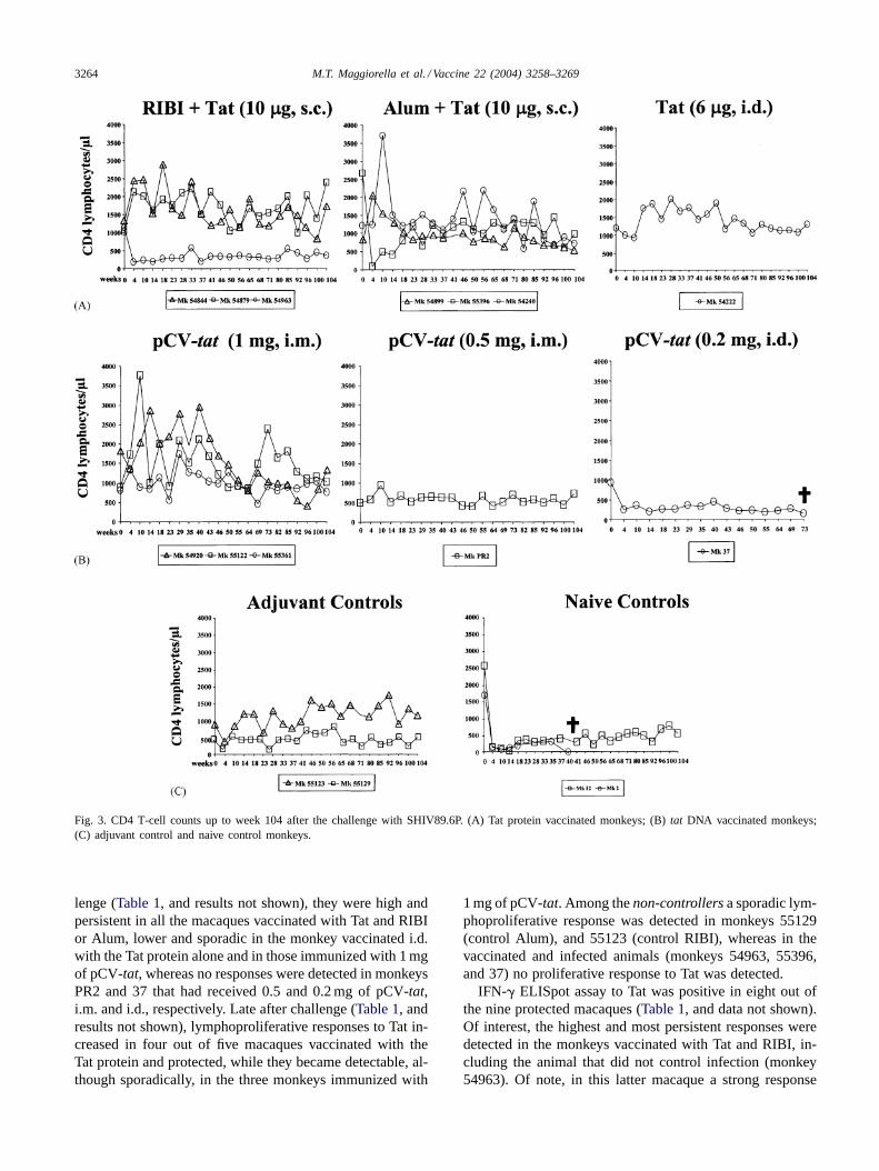
Consistent with the virological data, in all thecontrollersthe number of CD4+ T-cells was in the normal range duringthe entire follow-up (104 weeks). In contrast, it decreasedconsiderably in all thenon-controllers(3/12 vaccinated and4/4 control macaques) (Fig. 3A–C). In particular, monkeys2 and 37 had a sharp and profound CD4+ T-cell decline,developed AIDS and died.
Taken together, these results indicated that vaccinated andprotected monkeys contained virus infection and replicationduring the all post-challenge follow-up (104 weeks). In con-trast, all infected animals had higher levels of latent infec-tion and showed a persistent virus replication in blood.
3.2. Absence of a virus reservoir and virus replication inlymph nodes of vaccinated and protected macaques
Since lymph nodes are a major virus reservoir, virus iso-lation and determination of integrated or unintegrated viralDNA were performed late after the challenge on lymphnode biopsies (week 55 for the monkeys vaccinated withthe pCV-tat and week 71 for monkey vaccinated with theTat protein, the control monkeys and the naive monkey12) and compared with the results obtained with PBMCs
3262 M.T. Maggiorella et al. / Vaccine 22 (2004) 3258–3269
Fig. 1. Plasma viremia and E-DNA in PBMCs up to week 104 after the challenge with SHIV89.6P. (A) Tat protein vaccinated monkeys; (B)tat DNAvaccinated monkeys; (C) adjuvant control and naive control monkeys. The dashed area indicates the cut-off value of 1500 RNA copies/ml until week14, and of 50 RNA copies/ml from 18 to 104 weeks. E-DNA determinations were performed at the indicated weeks. The signs (−), negative and (+),positive, indicate the absence or presence of E-DNA, respectively.
of the same time points (data not shown). All the vacci-nated animals controlling infection (9/12) were negative forvirus isolation, proviral DNA and E-DNA in both PBMCsand lymph nodes (Figs. 1 and 2, and data not shown).Among thenon-controllers the vaccinated monkeys 55396and 37 were positive for all assays in both PBMCs andlymph nodes, whereas monkey 54963 was negative forvirus isolation in both PBMCs and tissues. In addition, this
latter animal had a proviral load higher in lymph nodes(47 copies/�g DNA) than in PBMCs (1 copy/�g DNA),although E-DNA forms were found only in PBMCs. Con-cerning the control macaques, all overtly infected, lymphnodes from the adjuvant control animals 55123 and 55129and the naive control monkey 12 were positive for bothproviral DNA (43, 5, 36 copies/�g DNA, respectively) andE-DNA. The differences of either proviral DNA or E-DNA
M.T. Maggiorella et al. / Vaccine 22 (2004) 3258–3269 3263
Fig. 2. Proviral DNA copies in PBMCs up to week 104 after the challenge with SHIV89.6P. (A) Tat protein vaccinated monkeys; (B)tat DNA vaccinatedmonkeys; (C) adjuvant control and naive control monkeys.
levels in lymph nodes of vaccinated (both protected andovertly infected) macaques versus control monkeys werestatistically significant (Fisher’s exact test:P-value= 0.04and 0.02, respectively).
3.3. Containment of infection is associated with stableTat-specific CD4 and CD8 T-cell responses
To determine whether Tat-specific immune responseswere durable and whether they correlated with protec-tion, antibodies, lymphoproliferative response and IFN-�ELISpot against Tat were evaluated in all animals at severaltime points during the follow-up.
Before challenge, anti-Tat IgG titers were high in themonkeys immunized s.c. with the Tat protein and RIBI orAlum adjuvant, low or undetectable in the other vaccinatedanimals[34–36]. After challenge, anti-Tat IgG titers werestable up to week 14, declined thereafter, and remained de-tectable up to week 104 only in the monkeys vaccinated s.c.with the Tat protein and RIBI or Alum (Table 1, and datanot shown). Thus, exposure to the challenge virus did notresult in the induction of anti-Tat antibodies. However, theyremained detectable through the all follow-up time in thoseanimals that had developed high titers in response to the Tatvaccine (Table 1, and data not shown).
Proliferative responses to Tat were detected in most vac-cinated monkeys before challenge[34–36]. Early after chal-
3264 M.T. Maggiorella et al. / Vaccine 22 (2004) 3258–3269
Fig. 3. CD4 T-cell counts up to week 104 after the challenge with SHIV89.6P. (A) Tat protein vaccinated monkeys; (B)tat DNA vaccinated monkeys;(C) adjuvant control and naive control monkeys.
lenge (Table 1, and results not shown), they were high andpersistent in all the macaques vaccinated with Tat and RIBIor Alum, lower and sporadic in the monkey vaccinated i.d.with the Tat protein alone and in those immunized with 1 mgof pCV-tat, whereas no responses were detected in monkeysPR2 and 37 that had received 0.5 and 0.2 mg of pCV-tat,i.m. and i.d., respectively. Late after challenge (Table 1, andresults not shown), lymphoproliferative responses to Tat in-creased in four out of five macaques vaccinated with theTat protein and protected, while they became detectable, al-though sporadically, in the three monkeys immunized with
1 mg of pCV-tat. Among thenon-controllersa sporadic lym-phoproliferative response was detected in monkeys 55129(control Alum), and 55123 (control RIBI), whereas in thevaccinated and infected animals (monkeys 54963, 55396,and 37) no proliferative response to Tat was detected.
IFN-� ELISpot assay to Tat was positive in eight out ofthe nine protected macaques (Table 1, and data not shown).Of interest, the highest and most persistent responses weredetected in the monkeys vaccinated with Tat and RIBI, in-cluding the animal that did not control infection (monkey54963). Of note, in this latter macaque a strong response
M.T.
Magg
iore
llae
ta
l./Va
ccine
22
(20
04
)3
25
8–
32
69
3265
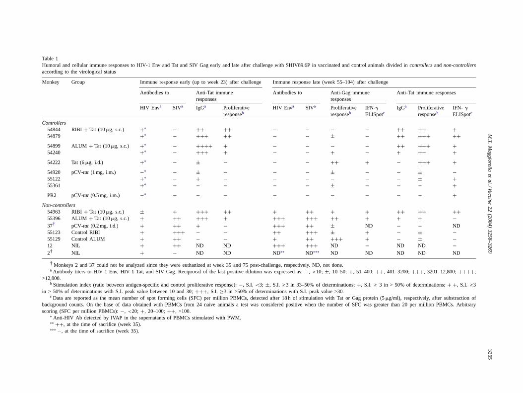
Table 1Humoral and cellular immune responses to HIV-1 Env and Tat and SIV Gag early and late after challenge with SHIV89.6P in vaccinated and control animals divided in controllersandnon-controllersaccording to the virological status
Monkey Group Immune response early (up to week 23) after challenge Immune response late (week 55–104) after challenge
Antibodies to Anti-Tat immuneresponses
Antibodies to Anti-Gag immuneresponses
Anti-Tat immune responses
HIV Enva SIVa IgGa Proliferativeresponseb
HIV Enva SIVa Proliferativeresponseb
IFN-�ELISpotc
IgGa Proliferativeresponseb
IFN- �
ELISpotc
Controllers54844 RIBI+ Tat (10�g, s.c.) +∗ − ++ ++ − − − − ++ ++ +54879 +∗ − +++ ++ − − ± − ++ +++ ++54899 ALUM + Tat (10�g, s.c.) +∗ − ++++ + − − − − ++ +++ +54240 +∗ − +++ + − − + − + ++ +54222 Tat (6�g, i.d.) +∗ − ± − − − ++ + − +++ +54920 pCV-tat (1 mg, i.m.) −∗ − ± − − − ± − − ± −55122 +∗ − + − − − − − − ± +55361 +∗ − − − − − ± − − − +PR2 pCV-tat (0.5 mg, i.m.) −∗ − − − − − − − − − +
Non-controllers54963 RIBI+ Tat (10�g, s.c.) ± + +++ ++ + ++ + + ++ ++ ++55396 ALUM + Tat (10�g, s.c.) + ++ +++ + +++ +++ ++ + + + −37† pCV-tat (0.2 mg, i.d.) + ++ + − +++ ++ ± ND − − ND55123 Control RIBI + +++ − − ++ +++ ± + − ± −55129 Control ALUM + ++ − − + ++ +++ + − ± −12 NIL + ++ ND ND +++ +++ ND − ND ND −2† NIL + − ND ND ND∗∗ ND∗∗∗ ND ND ND ND ND
† Monkeys 2 and 37 could not be analyzed since they were euthanized at week 35 and 75 post-challenge, respectively. ND, not done.a Antibody titers to HIV-1 Env, HIV-1 Tat, and SIV Gag. Reciprocal of the last positive dilution was expressed as:−, <10; ±, 10–50;+, 51–400;++, 401–3200;+++, 3201–12,800;++++,
>12,800.b Stimulation index (ratio between antigen-specific and control proliferative response):−, S.I. <3; ±, S.I. ≥3 in 33–50% of determinations;+, S.I. ≥ 3 in > 50% of determinations;+ +, S.I. ≥3
in > 50% of determinations with S.I. peak value between 10 and 30;+++, S.I. ≥3 in >50% of determinations with S.I. peak value >30.c Data are reported as the mean number of spot forming cells (SFC) per million PBMCs, detected after 18 h of stimulation with Tat or Gag protein (5�g/ml), respectively, after substraction of
background counts. On the base of data obtained with PBMCs from 24 naive animals a test was considered positive when the number of SFC was greater than 20per million PBMCs. Arbitraryscoring (SFC per million PBMCs):−, <20; +, 20–100;++, >100.
∗ Anti-HIV Ab detected by IVAP in the supernatants of PBMCs stimulated with PWM.∗∗ ++, at the time of sacrifice (week 35).∗∗∗ −, at the time of sacrifice (week 35).
3266 M.T. Maggiorella et al. / Vaccine 22 (2004) 3258–3269
to Tat correlated with negative virus isolation and no de-tectable virus replication (E-DNA) in lymph nodes. In con-trast, in monkey 55396 (Tat+ Alum), from which virus wasfrequently isolated, no production of IFN-� in response toTat was detected. Similarly, none of the adjuvant control ornaive control animals produced IFN-� in response to Tat.Thus, late after challenge eight out of ninecontrollerspro-duced IFN-� in response to Tat, as compared to one out ofthe fivenon-controllerstested.
Overall, the Tat-specific proliferative and IFN-� ELISpotresponses were higher and more frequent in the animals thatcontrolled infection and either sporadic, absent, or lost overtime in the monkeys unable to contain primary infection.
3.4. Immune responses to structural SIV/HIV antigensin protected monkeys reveal a pattern suggestive of anabortive infection
During the follow-up the Ab against SIV antigens or theHIV envelope became undetectable in thecontrollers, whichshowed very low titers (<1:10) early after challenge. Con-versely, anti-SIV and anti-HIV Env titers remained highor increased in thenon-controllers (Table 1, and data notshown). Similarly, lymphoproliferative responses to the Gagprotein or to a pool of Gag peptides were absent or sporadicin 7/9 of the controllers (Table 1, and data not shown). Incontrast, three out of the six non-controllers analysed hadhigher and persistent responses. This difference was statis-tically significant (Student’st-testP < 0.05).
IFN-� ELISpot assay against Gag were positive in 1/9vaccinated and protected monkeys and in four out of thefive non-controllersanalyzed. Of note, a direct correlationwas found between IFN-� production and the frequencyof virus isolation. The difference between the mean valuesof Gag-specific IFN-� secreting cells detected in thecon-trollers versus thenon-controllers was statistically signifi-cant (Student’st-test:P-value<0.05).
Thus, robust and persistent Gag-specific B- and T-cellresponses were detected only in animals unable to controlvirus replication. In contrast, they were low, transient or un-detectable in all the vaccinated animals containing primaryvirus replication. This pattern is suggestive of an abortiveinfection in protected animals.
4. Discussion
Vaccination with a biologically active Tat protein orpCV-tat controlled infection with the highly pathogenicSHIV89.6P preventing CD4 T-cell decline and diseaseonset. Here we show that protection after challenge wasprolonged since no CD4 T-cell decline nor active virusreplication was detected in all protected animals neither inblood nor in lymph nodes for the entire follow-up period(104 weeks). In addition, Tat vaccination elicited long-termmemory T-cell responses in the absence of any vaccineboosting.
Containment of viral load is considered a critical param-eter in the assessment of vaccine efficacy[47–50], sincehigh levels of viral load early after infection can be predic-tive of disease progression[43,51–58]. Indeed, in the vac-cinated and protected monkeys from this study, viral load(either cell- or plasma-associated) was persistently low orundetectable and CD4 T-cell decline did not occur. Thesedata were confirmed by the determination of unintegratedforms of DNA, a very sensitive marker for active viral repli-cation [59] as compared to plasma viremia[44,45]. In ourstudy, detection of circular E-DNA correlated with the pres-ence of a detectable plasma viremia during the acute phaseof infection, in agreement with previous reports in both theHIV [60,61] and the SIV model[62]. In fact, in the mon-key model the presence of circular E-DNA was found tocorrelate with low CD4:CD8 ratio in lymphoid organs andwith AIDS development[62]. Noteworthy, while plasmaviremia became undetectable, circular E-DNA remained de-tectable during chronic infection in thenon-controllersindi-cating persistency of an active infection. In contrast, E-DNAwas detected only once in PBMCs from 3/9 protected ani-mals. The results of virus containment were confirmed bythe analyses of lymph node biopsies showing undetectableproviral DNA, virus isolation and circular viral DNA in allthecontrollers, whereas one or more of these viral parame-ters were positive in thenon-controllers. Finally, two of thesevennon-controllersshowed severe worsening of the clin-ical conditions and died.
Immune responses to the viral antigens showed a differ-ent and opposite pattern in the two groups of animals. Inparticular, Tat-specific CD4 and CD8 T-cell responses (pro-liferative response and IFN-� ELISpot, respectively) wereconsistently detected in all thecontrollers, as compared tothe non-controllers (vaccinated, adjuvant controls or naivecontrols) in which they were absent or transient. Vice versa,CD4 and CD8 T-cell responses to Gag were generally highand persistent in thenon-controllers, whereas they were un-detectable or low and transient in the protected vaccinees.Thus, the presence of CD4 and CD8 T-cell responses to Gagcorrelated significantly with active virus replication.
The relevance of CTL responses against HIV-1 and SIVearly regulatory proteins Tat, Rev and Nef has been ad-dressed in recent reports aimed at defining the role of CTLsin controlling viremia and the factors governing the selec-tion of escaping mutants, the only parameter presently avail-able to measure CTLs efficacy. In a SIV model utilizing amolecular clone for the challenge, a strong correlation wasfound between the resolution of plasma viremia and the de-tection of a dominant CTL response against a Tat epitope(Tat28–35SL8) leading to the appearance of viral variantswith an apparently reduced fitness[31]. Subsequent studiesdemonstrated that CTLs with high functional avidity for theearly and intermediate proteins Tat, Nef and Vpr were themajor factor driving the selection of immune escape vari-ants during acute SIV infection[63]. More recent data mea-suring inhibition of HIV-1 replication by CTLs clones with
M.T. Maggiorella et al. / Vaccine 22 (2004) 3258–3269 3267
known avidity and specificity indicate that, in vitro, the fineepitope specificity might be the most important factor at de-termining control of virus replication and selection of virusvariants[64]. In both cases, CTLs against regulatory pro-teins were responsible of rapid selection of escape variantsindicating a strong immune pressure. It has been proposedthat because of their early expression, targeting regulatoryproteins, rather than structural antigens, may be extremelyadvantageous to the host because CTLs will impact on thevirus replication cycle at a critical step and for a prolongedtime[65]. Therefore, vaccines aimed at eliciting a large num-ber of polyclonal CTLs against these antigens may prove ef-fective at blocking virus expression during the early phasesof the acute infection, minimizing the selection of escapevariants. Along this line, a novel vaccine approach based ona polyvalent chimeric protein in which the genes coding forHIV-1 Tat, Rev and Nef has been recently developed andfound safe and immunogenic in macaques[66].
It is tempting to speculate that in our study vaccinationwith either Tat protein ortat-DNA controlled primary infec-tion at its early stages allowing containment of virus spreadin blood and tissues to undetectable levels, which were suf-ficient to ensure development and maintenance of long-termanti-Tat memory and to elicit weak anti-Gag T-helper butnot CTL responses in the protected monkeys. This pattern ofimmune responses, together with the virological data, sug-gests that in vaccinated and protected animals infection oc-curred, but it was abortive. In addition, it suggests that im-mune responses to Gag are not necessary to ensure long-termprotection in monkeys that have contained infection earlyafter challenge. However, development of anti-Gag humoraland cellular immune responses in overtly infected monkeys(non-controllers) may contribute to lower the viral load andpartially restore the CD4 T-cell number over time, mim-icking the transitory control that marks the resolution ofthe acute phase in the natural history of HIV-1 infectionin humans.
Recently, results have been reported showing that the im-munization of rhesus monkeys with the Tat protein[67,68]was safe and immunogenic but failed to induce a significantcontrol of viral replication following challenge with SIV orSHIV89.6P pathogenic viruses. However, several and keydifferences in the study design including dose and type of Tat(SIV and HIV), monkey species, adjuvant, dose and sched-ule of immunization, virus dose and route of challenge mayaccount for these conflicting results. Another key differencemay be represented by the conformation of the Tat protein.Specifically, we found that small differences in Tat whichcan be detected only by analytical HPLC and by uptake bydendritic cells (and not by other assays), make importantdifferences in the targeting and effects of Tat on dendriticcells which are key to initiate proper immune responses([32], and unpublished data). Further, our results are inagreement with reports from pre-clinical[39,69] and clini-cal [40,41] trials indicating that vaccination with Tat is safe,immunogenic and effective at controlling virus replication
and disease progression in the monkey model. These datasuggest that a native Tat protein ortat DNA should be in-cluded in future vaccine designs, particularly in prime-boostregimens. Based on these data, and on our recent resultsfrom field studies in Africa indicating cross-clade recogni-tion of Tat by anti-Tat Ab[28], preventive and therapeuticphase I trials are currently under way in Italy. In addition,a second generation of vaccine approaches combining Tatwith other antigens, such as Env and Gag, are under de-velopment to verify whether a rationally designed antigencombination can increase efficacy and long-term protectionagainst homologous as well as heterologous virus clades.
Acknowledgements
We thank A. Comini for the veterinary assistance; E. Iale,F. Incitti, F. Varano, N. Verrone, A. Marini, A. Avitabile, M.Chiodi, M. Azzetti and S. Alessandrini, for hematoclinicalanalysis of cynomolgus samples and for the handling of theanimal facility; R. Belli, F. De Angelis, M. Pace, E. SalviC. Rovetto, F. Carlini, M.G. Mancini, S. Farcomeni and D.Fulgenzi (Laboratory of Virology, Istituto Superiore di San-ità) for technical help; N. Esposito for statistical analyses.We also thank A. Lippa and P. Sergiampietri for the editorialassistance. This work was supported by grants from the IXAIDS Project, the Italian Concerted Action on HIV-AIDSvaccine development (ICAV), ISS, Rome, Italy.
References
[1] Arya SK, Guo C, Josephs SF, Wong-Staal F. Trans-activatorgene of human T-lymphotropic virus type III (HTLV-III). Science1985;229(4708):69–73.
[2] Fisher AG, Feinberg MB, Josephs SF, Harper ME, Marselle LM,Reyes G, et al. The trans-activator gene of HTLV-III is essential forvirus replication. Nature 1986;320(6060):367–71.
[3] Wu Y, Marsh JW. Selective transcription and modulation ofresting T cell activity by preintegrated HIV DNA. Science2001;293(5534):1503–6.
[4] Sodroski J, Patarca R, Rosen C, Wong-Staal F, Haseltine W. Locationof the trans-activating region on the genome of human T-celllymphotropic virus type III. Science 1985;229(4708):74–7.
[5] Cullen BR, Greene WC. Regulatory pathways governing HIV-1replication. Cell 1989;58(3):423–6.
[6] Ensoli B, Buonaguro L, Barillari G, Fiorelli V, Gendelman R,Morgan RA, et al. Release, uptake, and effects of extracellular humanimmunodeficiency virus type 1 Tat protein on cell growth and viraltransactivation. J Virol 1993;67(1):277–87.
[7] Chang HK, Gallo RC, Ensoli B. Regulation of cellular geneexpression and function by the human immunodeficiency virus type1 Tat protein. J Biomed Sci 1995;2(3):189–202.
[8] Ensoli B, Barillari G, Salahuddin SZ, Gallo RC, Wong-Staal F. Tatprotein of HIV-1 stimulates growth of cells derived from Kaposi’ssarcoma lesions of AIDS patients. Nature 1990;345(6270):84–6.
[9] Chang HC, Samaniego F, Nair BC, Buonaguro L, Ensoli B. HIV-1Tat protein exits from cells via a leaderless secretory pathway andbinds to extracellular matrix-associated heparan sulfate proteoglycansthrough its basic region. AIDS 1997;11(12):1421–31.

3268 M.T. Maggiorella et al. / Vaccine 22 (2004) 3258–3269
[10] Frankel AD, Pabo CO. Cellular uptake of the tat protein from humanimmunodeficiency virus. Cell 1988;55(6):1189–93.
[11] Fawell S, Seery J, Daikh Y, Moore C, Chen LL, Pepinsky B, et al.Tat-mediated delivery of heterologous proteins into cells. Proc NatlAcad Sci USA 1994;91(2):664–8.
[12] Huang L, Bosch I, Hofmann W, Sodroski J, Pardee AB. Tatprotein induces human immunodeficiency virus type 1 (HIV-1)coreceptors and promotes infection with both macrophage-tropic andT-lymphotropic HIV-1 strains. J Virol 1998;72(11):8952–60.
[13] Secchiero P, Zella D, Capitani S, Gallo RC, Zauli G. ExtracellularHIV-1 tat protein up-regulates the expression of surface CXC-chemo-kine receptor 4 in resting CD4+ T cells. J Immunol 1999;162(4):2427–31.
[14] Weiss JM, Nath A, Major EO, Berman JW. HIV-1 Tatinduces monocyte chemoattractant protein-1-mediated monocytetransmigration across a model of the human blood–brain barrierand up-regulates CCR5 expression on human monocytes. J Immunol1999;163(5):2953–9.
[15] de Paulis A, De Palma R, Di Gioia L, Carfora M, Prevete N, Tosi G,et al. Tat protein is an HIV-1-encoded beta-chemokine homolog thatpromotes migration and up-regulates CCR3 expression on human Fcepsilon RI+ cells. J Immunol 2000;165(12):7171–9.
[16] Viscidi RP, Mayur K, Lederman HM, Frankel AD. Inhibition ofantigen-induced lymphocyte proliferation by Tat protein from HIV-1.Science 1989;246(4937):1606–8.
[17] Subramanyam M, Gutheil WG, Bachovchin WW, Huber BT.Mechanism of HIV-1 Tat induced inhibition of antigen-specific Tcell responsiveness. J Immunol 1993;150(6):2544–53.
[18] Ensoli B, Gendelman R, Markham P, Fiorelli V, Colombini S,Raffeld M, et al. Synergy between basic fibroblast growth factorand HIV-1 Tat protein in induction of Kaposi’s sarcoma. Nature1994;371(6499):674–80.
[19] Li CJ, Friedman DJ, Wang C, Metelev V, Pardee AB. Induction ofapoptosis in uninfected lymphocytes by HIV-1 Tat protein. Science1995;268(5209):429–31.
[20] Zagury JF, Sill A, Blattner W, Lachgar A, Le Buanec H, RichardsonM, et al. Antibodies to the HIV-1 Tat protein correlated withnonprogression to AIDS: a rationale for the use of Tat toxoid as anHIV-1 vaccine. J Hum Virol 1998;1(4):282–92.
[21] Barillari G, Sgadari C, Fiorelli V, Samaniego F, Colombini S, ManzariV, et al. The Tat protein of human immunodeficiency virus type-1promotes vascular cell growth and locomotion by engaging thealpha5beta1 and alphavbeta3 integrins and by mobilizing sequesteredbasic fibroblast growth factor. Blood 1999;94(2):663–72.
[22] Barillari G, Sgadari C, Palladino C, Gendelman R, Caputo A, MorrisCB, et al. Inflammatory cytokines synergize with the HIV-1 Tatprotein to promote angiogenesis and Kaposi’s sarcoma via inductionof basic fibroblast growth factor and the alpha v beta 3 integrin. JImmunol 1999;163(4):1929–3195.
[23] Reiss P, Lange JM, de Ronde A, de Wolf F, Dekker J, Debouck C, etal. Speed of progression to AIDS and degree of antibody response toaccessory gene products of HIV-1. J Med Virol 1990;30(3):163–8.
[24] Rodman TC, To SE, Hashish H, Manchester K. Epitopes fornatural antibodies of human immunodeficiency virus (HIV)-negative(normal) and HIV-positive sera are coincident with two keyfunctional sequences of HIV Tat protein. Proc Natl Acad Sci USA1993;90(16):7719–23.
[25] Re MC, Furlini G, Vignoli M, Ramazzotti E, Roderigo G, De RV,et al. Effect of antibody to HIV-1 Tat protein on viral replication invitro and progression of HIV-1 disease in vivo. J Acquir ImmuneDefic Syndr Hum Retrovirol 1995;10(4):408–16.
[26] Zagury D, Lachgar A, Chams V, Fall LS, Bernard J, Zagury JF, etal. Interferon alpha and Tat involvement in the immunosuppressionof uninfected T cells and C-C chemokine decline in AIDS. ProcNatl Acad Sci USA 1998;95(7):3851–6.
[27] Re MC, Vignoli M, Furlini G, Gibellini D, Colangeli V, VitoneF, et al. Antibodies against full-length Tat protein and some
low-molecular-weight Tat-peptides correlate with low or undetectableviral load in HIV-1 seropositive patients. J Clin Virol 2001;21(1):81–9.
[28] Buttò S, Fiorelli V, Tripiciano A, Ruiz-Alvarez MJ, Scoglio A, EnsoliF, et al. Cross-recognition of the Clade B HIV-1 Tat protein vaccinecandidate by antibodies from HIV-1-infected Italian. Ugandan andSouth African individuals. J Infect Dis 2003;188:1171–80.
[29] van Baalen CA, Pontesilli O, Huisman RC, Geretti AM, Klein MR,de Wolf F, et al. Human immunodeficiency virus type 1 Rev- andTat-specific cytotoxic T lymphocyte frequencies inversely correlatewith rapid progression to AIDS. J Gen Virol 1997;78(Pt 8):1913–8.
[30] Addo MM, Altfeld M, Rosenberg ES, Eldridge RL, Philips MN,Habeeb K, et al. The HIV-1 regulatory proteins Tat and Rev arefrequently targeted by cytotoxic T lymphocytes derived from HIV-1-infected individuals. Proc Natl Acad Sci USA 2001;98(4):1781–6.
[31] Allen TM, O’Connor DH, Jing P, Dzuris JL, Mothe BR, Vogel TU, etal. Tat-specific cytotoxic T lymphocytes select for SIV escape variantsduring resolution of primary viraemia. Nature 2000;407(6802):386–90.
[32] Fanales-Belasio E, Moretti S, Nappi F, Barillari G, Micheletti F,Cafaro A, et al. Native HIV-1 Tat protein targets monocyte-deriveddendritic cells and enhances their maturation, function, andantigen-specific T cell responses. J Immunol 2002;168(1):197–206.
[33] Kim DT, Mitchell DJ, Brockstedt DG, Fong L, Nolan GP, FathmanCG, et al. Introduction of soluble proteins into the MHC classI pathway by conjugation to an HIV tat peptide. J Immunol1997;159(4):1666–8.
[34] Cafaro A, Caputo A, Fracasso C, Maggiorella MT, Goletti D,Baroncelli S, et al. Control of SHIV-89.6P-infection of cynomolgusmonkeys by HIV-1 Tat protein vaccine. Nat Med 1999;5(6):643–50.
[35] Cafaro A, Caputo A, Maggiorella MT, Baroncelli S, Fracasso C,Pace M, et al. SHIV89.6P pathogenicity in cynomolgus monkeys andcontrol of viral replication and disease onset by human immunodefi-ciency virus type 1 Tat vaccine. J Med Primatol 2000;29(3–4):193–208.
[36] Cafaro A, Titti F, Fracasso C, Maggiorella MT, Baroncelli S, CaputoA, et al. Vaccination with DNA containing tat coding sequencesand unmethylated CpG motifs protects cynomolgus monkeys uponinfection with simian/human immunodeficiency virus (SHIV89.6P).Vaccine 2001;19(2022):2862–77.
[37] Caselli E, Betti M, Grossi MP, Balboni PG, Rossi C, Boarini C, etal. DNA immunization with HIV-1 tat mutated in the trans activationdomain induces humoral and cellular immune responses againstwild-type Tat. J Immunol 1999;162(9):5631–8.
[38] Caputo A, Betti M, Altavilla G, Bonaccorsi A, Boarini C, MarchisioM, et al. Micellar-type complexes of tailor-made synthetic blockcopolymers containing the HIV-1 tat DNA for vaccine application.Vaccine 2002;20(17–18):2303–17.
[39] Osterhaus AD, van Baalen CA, Gruters RA, Schutten M, SiebelinkCH, Hulskotte EG, et al. Vaccination with Rev and Tat against AIDS.Vaccine 1999;17(20–21):2713–4.
[40] Calarota S, Bratt G, Nordlund S, Hinkula J, Leandersson AC,Sandstrom E, et al. Cellular cytotoxic response induced by DNAvaccination in HIV-1-infected patients. Lancet 1998;351(9112):1320–5.
[41] Calarota SA, Leandersson AC, Bratt G, Hinkula J, Klinman DM,Weinhold KJ, et al. Immune responses in asymptomatic HIV-1-infected patients after HIV-DNA immunization followed by highlyactive antiretroviral treatment. J Immunol 1999;163(4):2330–8.
[42] Karlsson GB, Halloran M, Li J, Park IW, Gomila R, ReimannKA, et al. Characterization of molecularly cloned simian-humanimmunodeficiency viruses causing rapid CD4+ lymphocyte depletionin rhesus monkeys. J Virol 1997;71(6):4218–25.
[43] Ten Haaft P, Verstrepen B, Uberla K, Rosenwirth B, HeeneyJ. A pathogenic threshold of virus load defined in simianimmunodeficiency virus- or simian-human immunodeficiencyvirus-infected macaques. J Virol 1998;72(12):10281–5.
M.T. Maggiorella et al. / Vaccine 22 (2004) 3258–3269 3269
[44] Sharkey ME, Teo I, Greenough T, Sharova N, Luzuriaga K, SullivanJL, et al. Persistence of episomal HIV-1 infection intermediatesin patients on highly active anti-retroviral therapy. Nat Med2000;6(1):76–81.
[45] Cara A, Vargas Jr J, Keller M, et al. Circular viral DNA andanomalous junction sequence in PBMC of HIV-infected individualswith no detectable plasma HIV RNA. Virology 2002;292(1):1–5.
[46] Titti F, Sernicola L, Geraci A, Panzini G, Di Fabio S, BelliR, et al. Live attenuated simian immunodeficiency virus preventssuper-infection by cloned SIVmac251 in cynomolgus monkeys. JGen Virol 1997;78(Pt 10):2529–39.
[47] Heeney JL. Primate models for AIDS vaccine development. AIDS1996;10(Suppl A):S115–22.
[48] Ensoli B, Cafaro A. Novel strategies toward the development of aneffective vaccine to prevent human immunodeficiency virus infectionor acquired immunodeficiency virus. In: Volberding PA, JacobsonMA, editors. AIDS clinical review 2000/2001. New York: MarcelDekker; 2001. p. 23–61.
[49] Kent SJ, Ada GL, Hayes E, Lewis IM. Determining the immunemechanisms of protection from AIDS: correlates of immunity and thedevelopment of syngeneic macaques. Immunol Rev 2001;183(1):94–108.
[50] Mooij P, Heeney JL. Rational development of prophylactic HIVvaccines based on structural and regulatory proteins. Vaccine2001;20(3–4):304–21.
[51] Henrard DR, Phillips JF, Muenz LR, Blattner WA, Wiesner D,Eyster ME, et al. Natural history of HIV-1 cell-free viremia. JAMA1995;274(7):554–8.
[52] Mellors JW, Rinaldo Jr CR, Gupta P, White RM, Todd JA, KingsleyLA. Prognosis in HIV-1 infection predicted by the quantity of virusin plasma. Science 1996;272(5265):1167–70.
[53] Lifson JD, Nowak MA, Goldstein S, Rossio JL, Kinter A, VasquezG, et al. The extent of early viral replication is a critical determinantof the natural history of simian immunodeficiency virus infection. JVirol 1997;71(12):9508–14.
[54] Watson A, Ranchalis J, Travis B, McClure J, Sutton W, JohnsonPR, et al. Plasma viremia in macaques infected with simianimmunodeficiency virus: plasma viral load early in infection predictssurvival. J Virol 1997;71(1):284–90.
[55] Kaufmann GR, Cunningham P, Zaunders J, Law M, Vizzard J, CarrA, et al. Impact of early HIV-1 RNA and T-lymphocyte dynamicsduring primary HIV-1 infection on the subsequent course of HIV-1RNA levels and CD4+ T-lymphocyte counts in the first year ofHIV-1 infection. Sydney Primary HIV Infection Study Group. JAcquir Immune Defic Syndr 1999;22(5):437–44.
[56] Anastos K, Kalish LA, Hessol N, Weiser B, Melnick S, Burns D,et al. The relative value of CD4 cell count and quantitative HIV-1RNA in predicting survival in HIV-1-infected women: results of thewomen’s interagency HIV study. AIDS 1999;13(13):1717–26.
[57] Lyles CM, Dorrucci M, Vlahov D, Pezzotti P, Angarano G, SiniccoA, et al. Longitudinal human immunodeficiency virus type 1 load inthe italian seroconversion study: correlates and temporal trends ofvirus load. J Infect Dis 1999;180(4):1018–24.
[58] Staprans SI, Dailey PJ, Rosenthal A, Horton C, Grant RM, LercheN, et al. Simian immunodeficiency virus disease course is predictedby the extent of virus replication during primary infection. J Virol1999;73(6):4829–39.
[59] Cara A, Reitz Jr MS. New insight on the role of extrachromosomalretroviral DNA. Leukemia 1997;11(9):1395–9.
[60] Panther La , Coombs RW, Zeh JE, Collier AC, Corey L. Unintegratedcircular HIV-1 DNA in the peripheral mononuclear cells of HIV-1infected subjects: association with high levels of plasma HIV-1 RNA,rapid decline in CD4 count, and clinical progression to AIDS. JAcquir Immune Defic Syndr Hum Retrovirol 1998;17(4):303–13.
[61] Panther LA, Coombs RW, Aung SA, Dela RC, Gretch D, CoreyL. Unintegrated HIV-1 circular 2-LTR proviral DNA as a markerof recently infected cells: relative effect of recombinant CD4,zidovudine, and saquinavir in vitro. J Med Virol 1999;58(2):165–73.
[62] Rosenberg YJ, Lewis MG, Leon EC, Cafaro A, Eddy GA,Greenhouse JJ. Viral DNA burden and decline in percentage ofCD4 positive cells in the lymphoid compartment of SIV-infectedmacaques. AIDS Res Hum Retroviruses 1994;10(10):1269–77.
[63] O’Connor DH, Allen TM, Vogel TU, Jing P, DeSouza IP, Dodds E,et al. Acute phase cytotoxic T lymphocyte escape is a hallmark ofsimian immunodeficiency virus infection. Nat Med 2002;8(5):493–9.
[64] Yang OO, Sarkis PT, Ali A, Harlow JD, Brander C, Kalams SA,et al. Determinants of HIV-1 mutational escape from cytotoxic Tlymphocytes. J Exp Med 2003;197(10):1365–75.
[65] Gruters RA, van Baalen CA, Osterhaus ADME. The advantage ofearly recognition of HIV-infected cells by cytotoxic T-lymphocytes.Vaccine 2002;20(15):2011–5.
[66] Hel Z, Tryniszewska E, Tsai WP, Johnson JM, Harrod R, Fullen J,et al. Design and in vivo immunogenicity of a polyvalent vaccinebased on SIVmac regulatory genes. DNA Cell Biol 2002;21(9):619–26.
[67] Allen TM, Mortara L, Mothe BR, Liebl M, Jing P, Calore B, et al.Tat-vaccinated macaques do not control simian immunodeficiencyvirus SIVmac239 replication. J Virol 2002;76(8):4108–12.
[68] Silvera P, Richardson MW, Greenhouse J, Yalley-Ogunro J, Shaw N,Mirchandani J, et al. Outcome of Simian-human immunodeficiencyvirus strain 89.6p challenge following vaccination of rhesusmacaques with human immunodeficiency virus Tat protein. J Virol2002;76(8):3800–9.
[69] Pauza CD, Trivedi P, Wallace M, Ruckwardt TJ, Le BuanecH, Lu W, et al. Vaccination with tat toxoid attenuates diseasein simian/HIV-challenged macaques. Proc Natl Acad Sci USA2000;97(7):3515–9.


