Embed Size (px)
Citation preview
Volume 8 Number 1 1980 Nucleic Acids Research
Loopstructures in synthetic oligodeoxynucleotidesl
Cornelis A.G.Haasnoot, Jeroen H.J.den Hartog, Jan F.M.de Rooij, Jacques H.van Boom andCornelis Altona
Gorlaeus Laboratories, State University, P.O. Box 9502, 2300 RA Leiden, The Netherlands
Received 17 October 1979
ABSTRACT.A comparative nuclear magnetic resonance study of the hydrogen-bonded iminoprotons in a series of synthetic DNA fragments is presented. The fragmentsATCCTA(Tn)TAGGAT are in principle capable of forming either a self-complemen-tary hairpin loop structure (monomer form) or an interior loop structure (di-meric form). It has been shown2, that for n=1 only the dimer structure ispresent in aqueous solution, whereas the exclusive existence of the hairpinloop structure is indicated for n=3, 4 & 5. Surprisingly, for n=2 two differ-ent structures appear to be present in solution. Concentration studies showthat both monomers and dimers exist side by side in this case. Hairpins aswell as interior loops form extra "melting sites' in addition to the well-known fraying phenomenon at the terminus of the double helix.
INTRODUCTION.
Double helical complexes formed from tailor-made syntheticoligodeoxynucleotides are convenient models for the investigationof the structural features and dynamics of base recognition innucleic acids. In a previous paper2 we investigated the helix-coil transition of the (partially) self-complementary oligodeoxy-nucleotides fragment 12 (fig. la, n=0) and fragment 13 (fig. la,
n=1) by monitoring the Watson-Crick hydrogen-bonded ring iminoprotons by high-field nuclear magnetic resonance (NMR) techniques.It was found2 that the insertion of a TxT wobble pair (i.e. an
interior loop) in the middle of the strands introduces an extra
melting site in the double helical structure (Form II, fig. lb;see also fig. 2), which gives rise to an overall lowering of theNMR "melting" temperature of the double helix.
The relative thermodynamic stability of the various typesof loops which can occur in DNA is a matter of debate. For RNAit has been found that interior loops are more stable than
hairpin loops3. If this were true for DNA, elongation of the
©) IRL Press Umited, 1 Falconberg Court, London W1V 5FG, U.K.
Nucleic Acids ResearchVolurne 8 Number 1 1980
169
Nucleic Acids Research
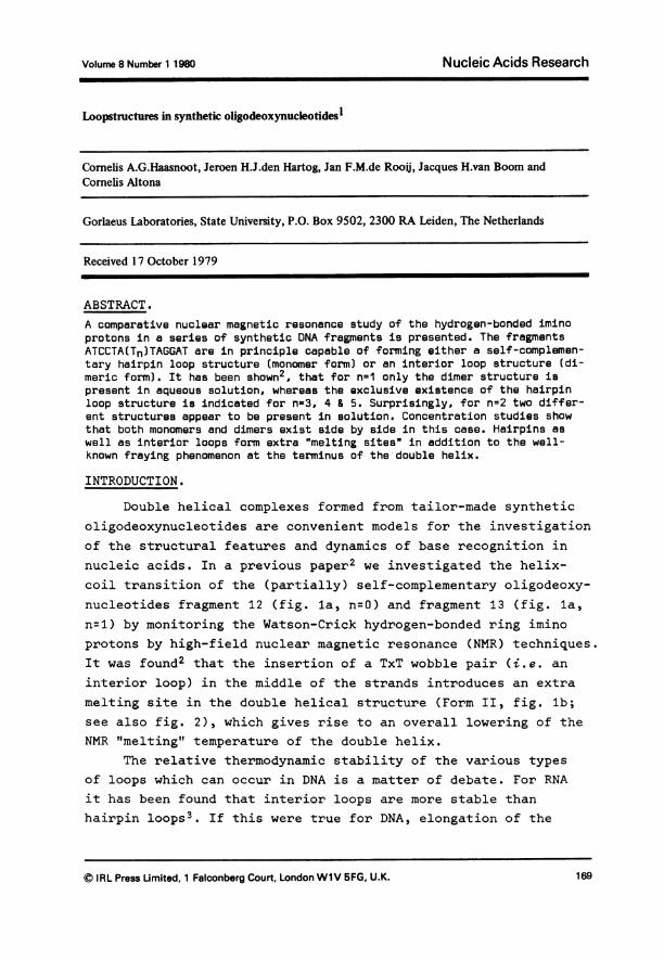
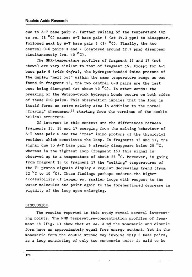
(a) FRAGMENT (12+ n):
ATCCTA MT)nTAGGAT1 2 3 4 5 6 65'4 Y3 21'
(b) FORM I-MONOMER:
1 234S6r-II I WI Tn1' 2'3'-C5i\6'J
FORM El-DIMER:
112 6Tn
6 3211
1'2'346< 64 1
FORM m-"OLIGOMER":
zTn""
TnTm n
'. t
Figure 1. (a) General formula denoting the base-sequence of the synthetic oli-gonucleotides studied.
(b) Three possible secondary structures for these oligonucleotides.Base pair numbering as shown5 note the pairwise equivalency ofthe base pairs in Form II and III because of the symmetry of thedouble helix
non-complementary site of thymidylyl residues in the middle of
fragment 13 (Tnt n=1,2,...) would not give rise to major confor-
mational changes, i.e. Form II (fig. lb) being preferred for all
values of n. On the other hand, if for a certain chain length n
the hairpin loop (Form I, fig. lb) becomes more stable than the
interior loop one expects a discontinuity in conformationalbehaviour to occur.
In the present paper we wish to report the results of a
comparative proton magnetic resonance study of fragments 13
(n=1), 14 (n=2), 15 (n=3), 16 (n=4) and 17 (n=5), undertaken in
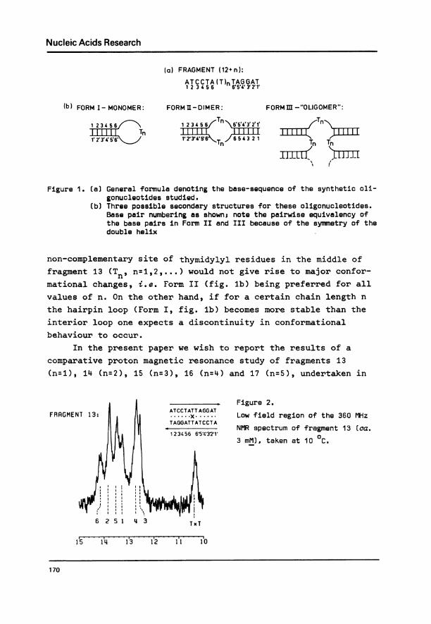
FRAGMENT 13:
Figure 2.
Low field region of the 360 MHz
NMR spectrum of fragment 13 (ca.
3 mM), taken at 10 0C.
I
6 2 5 1 4 3 TxT
I1s 1L4 1'3 1'2 1'1 I-o
170
Nucleic Acids Research
order to evaluate the conformational behaviour of these fragmentsas function of chain length and temperature. The possible forma-tion of "oligomers" (Form III, fig. lb) is also investigated.
MATERIALS AND METHODS.
The oligodeoxynucleotides were synthesized via an improvedphosphotriester approach4. Full details of preparation and puri-fication are published elsewhere5. The DNA fragments were con-verted into their Na+-salts by slow passage over a column (12 cmx 1 cm2) of Dowex cation-exchange resin, Na+-form; elution waswith distilled water. Lyophilization gave the nucleotide materialas white fluffy compounds.
The NMR samples were prepared by dissolving the appropriateamount of the oligodeoxynucleotide in 270 il of a buffer solutioncontaining 0.5 M NaCl, 0.1 M sodium cacodylate (pH = 7.0) and5 % v/v D20. High-resolution IH NMR spectra were recorded on aBruker HX-360 NMR spectrometer interfaced with a BNC-12 computersystem and equipped with a standard temperature controller, usingmicro NMR tubes with a cylindrical cavity (Wilmad 508-CP). Thespectrometer was operated in the correlation mode6, using aninternal D20 field/frequency lock. Chemical shifts were measuredrelative to the solvent H20 peak and converted to DSS-referenceby correcting for the H20 to DSS chemical shift, using the appro-priate temperature calibration curve. All reported shifts aredenoted according to the 6-convention, i.e. positive values repre-sent shifts to Zower field.
RESULTS.
Secondary structure of the synthetic DNA fragments.
The chemical shift of a given hydrogen-bonded imino protonin an intact double-helical DNA fragment is mainly determined bythe effects of ring current and diamagnetic anisotropies7t8 ofthe aromatic bases which it links together and those directlyabove and below in the vertical stacking array (nearest neighboureffects), with the next-nearest neighbours playing a relativelyminor role. Various additivity rules have been proposed as an
171
Nucleic Acids Research
aid in predicting imino-proton shifts in RNA and DNA helices9'10based upon effects exerted by nearest-neighbour bases. Barringgross changes in either geometry or stability of the doublehelix for different values of n, one should expect that the DNA
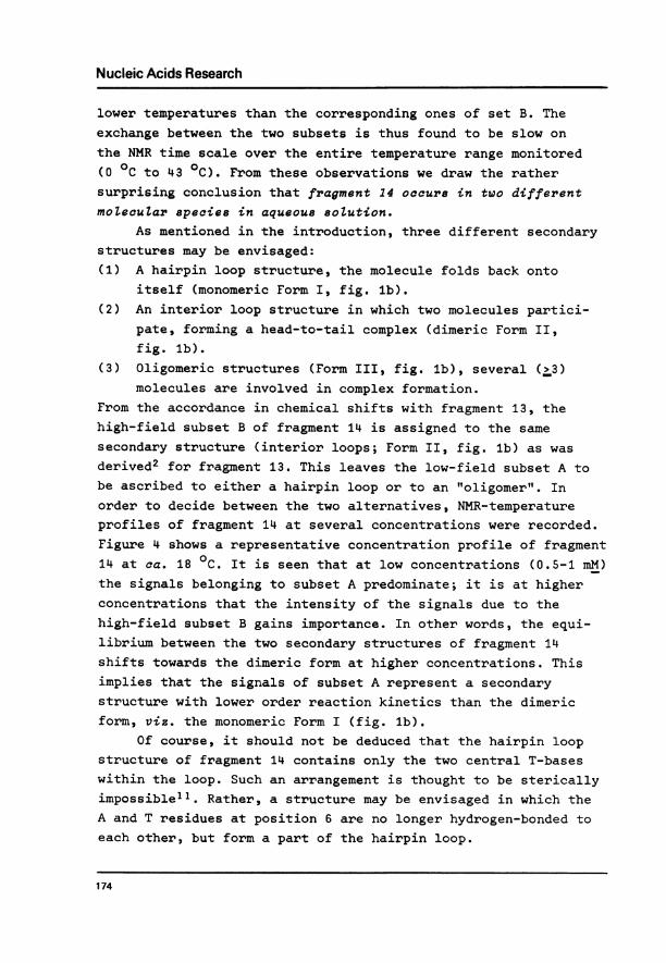
fragments presently under investigation display a very similarNMR-pattern, as all fragments contain the same sequence of sixhydrogen-bonded base pairs and differ only in the number ofthymidylyl residues in the middle of the strands. Our expectationis indeed borne out by experiment. Let us leave aside for the
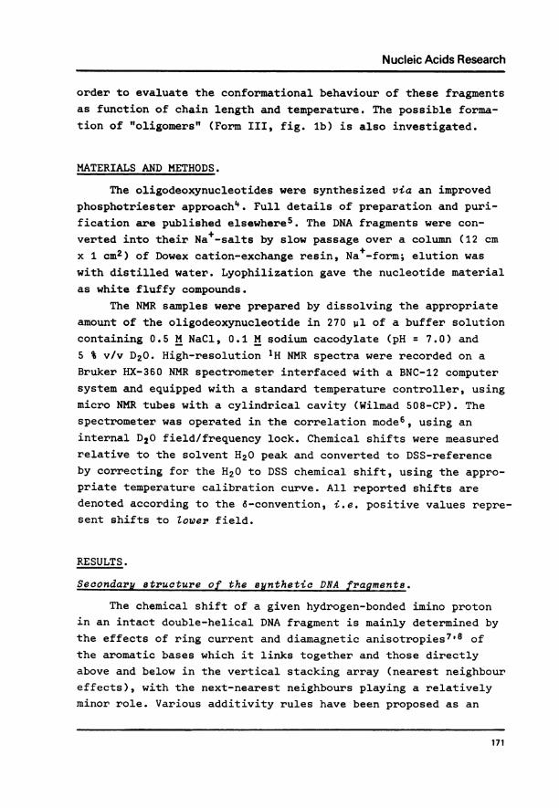
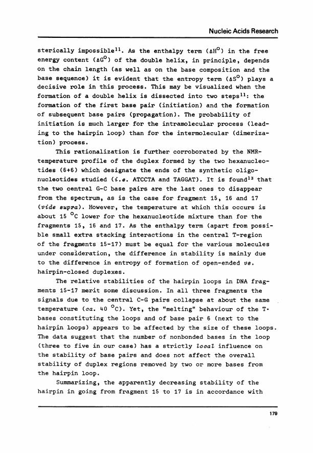
moment the results for fragment 14 (for reasons to be discussedbelow) and concentrate on the six signals displayed by fragments13,-15, 16 and 17 in the 12.6 - 14.3 ppm region (figs. 2 & 3).The chemical shifts observed at low temperature are collectedin Table 1. It is readily seen that the signal patterns of thethree larger fragments are quite similar to that of fragment 13and we therefore conclude that our previous assignment of the
T= 60CFigure 3.
The 360 MHz 1H-NMR spectra (low field
part) of fragments 14, 15, 16 and 17(X3 mM), recorded at 6 0C.
16
15
11.
15 14. 13 12 11 10 PPM
FRAGMENT 17
E
FRAGMENT
FRAGMENT
FRAGMENT
172
II AA
Nucleic Acids Research
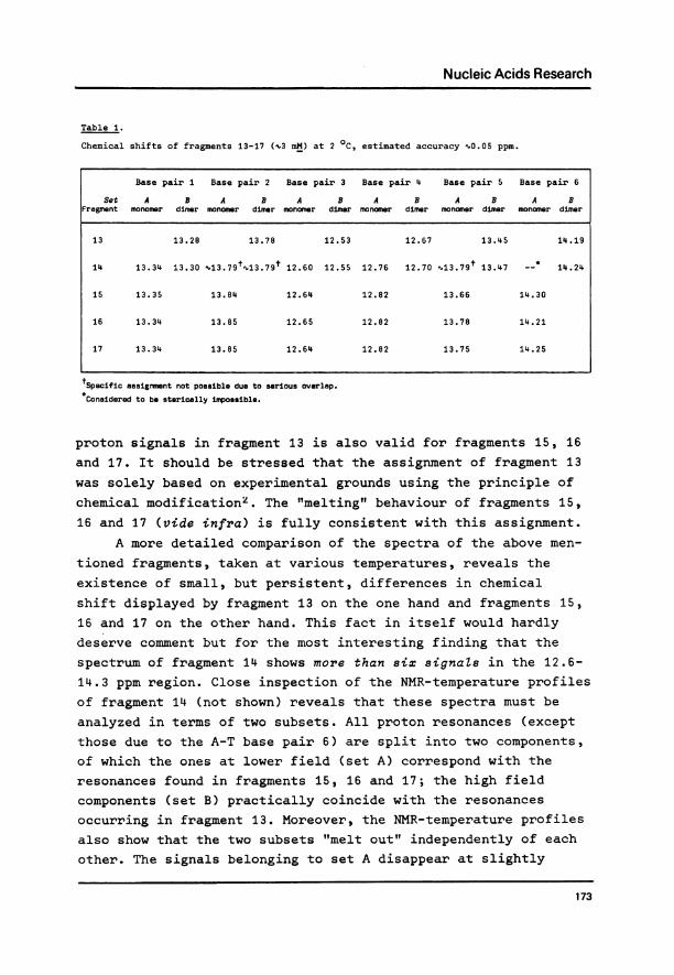
Table 1.
Chemical shifts of fragments 13-17 (.3 mM) at 2 0C, estimated accuracy ".0.05 ppm.
Base pair 1 Base pair 2 Base pair 3 Base pair 4 Base pair 5 Base pair 6
Set A B A B A B A B A B A BFragment monomer dimer monomer dimer monomer dimer monomer dimer monomer dimer monomer dimer
13 13.28 13.78 12.53 12.67 13.45 14.19
14 13.34 13.30 %.13.79t v13.79t 12.60 12.55 12.76 12.70 '.13.79t 13.47 --* 14.24
15 13.35 13.84 12.64 12.82 13.66 14.30
16 13.34 13.85 12.65 12.82 13.78 14.21
17 13.34 13.85 12.64 12.82 13.75 14.25
tSpecific assigrment not possible due to serious overlap.Considered to be sterically impossible.
proton signals in fragment 13 is also valid for fragments 15, 16
and 17. It should be stressed that the assignment of fragment 13
was solely based on experimental grounds using the principle ofchemical modification2. The "melting" behaviour of fragments 15,
16 and 17 (vide infra) is fully consistent with this assignment.A more detailed comparison of the spectra of the above men-
tioned fragments, taken at various temperatures, reveals the
existence of small, but persistent, differences in chemicalshift displayed by fragment 13 on the one hand and fragments 15,16 and 17 on the other hand. This fact in itself would hardly
deserve comment but for the most interesting finding that the
spectrum of fragment 14 shows more than six signaZs in the 12.6-
14.3 ppm region. Close inspection of the NMR-temperature profilesof fragment 14 (not shown) reveals that these spectra must be
analyzed in terms of two subsets. All proton resonances (exceptthose due to the A-T base pair 6) are split into two components,of which the ones at lower field (set A) correspond with the
resonances found in fragments 15, 16 and 17; the high fieldcomponents (set B) practically coincide with the resonances
occurring in fragment 13. Moreover, the NMR-temperature profilesalso show that the two subsets "melt out" independently of each
other. The signals belonging to set A disappear at slightly
173
Nucleic Acids Research
lower temperatures than the corresponding ones of set B. Theexchange between the two subsets is thus found to be slow onthe NMR time scale over the entire temperature range monitored(O 0C to 43 0C). From these observations we draw the rathersurprising conclusion that fragment 14 occurs in two differentmolecuZar species in aqueous soZution.
As mentioned in the introduction, three different secondarystructures may be envisaged:(1) A hairpin loop structure, the molecule folds back onto
itself (monomeric Form I, fig. lb).(2) An interior loop structure in which two molecules partici-
pate, forming a head-to-tail complex (dimeric Form II,fig. lb).
(3) Oligomeric structures (Form III, fig. lb), several (Z3)molecules are involved in complex formation.
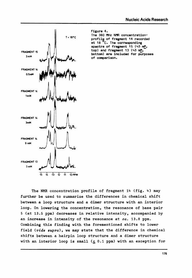
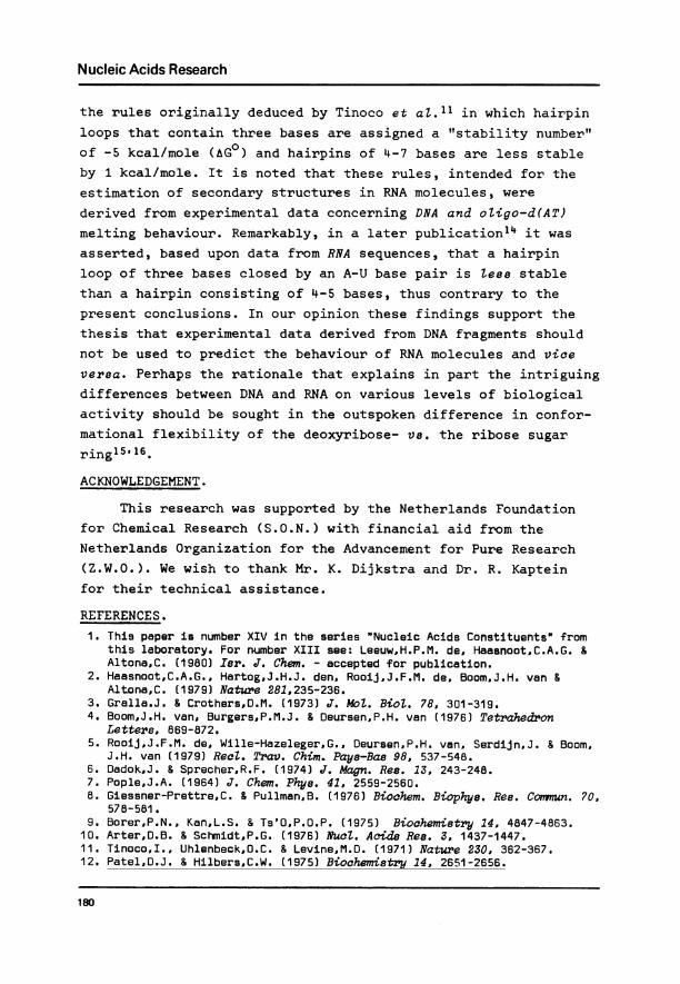
From the accordance in chemical shifts with fragment 13, thehigh-field subset B of fragment 14 is assigned to the samesecondary structure (interior loops; Form II, fig. lb) as wasderived2 for fragment 13. This leaves the low-field subset A tobe ascribed to either a hairpin loop or to an "oligomer". Inorder to decide between the two alternatives, NMR-temperatureprofiles of fragment 14 at several concentrations were recorded.Figure 4 shows a representative concentration profile of fragment14 at ca. 18 0C. It is seen that at low concentrations (0.5-1 mM)the signals belonging to subset A predominate; it is at higherconcentrations that the intensity of the signals due to thehigh-field subset B gains importance. In other words, the equi-librium between the two secondary structures of fragment 14shifts towards the dimeric form at higher concentrations. Thisimplies that the signals of subset A represent a secondarystructure with lower order reaction kinetics than the dimericform, viz. the monomeric Form I (fig. lb).
Of course, it should not be deduced that the hairpin loopstructure of fragment 14 contains only the two central T-baseswithin the loop. Such an arrangement is thought to be stericallyimpossiblell. Rather, a structure may be envisaged in which theA and T residues at position 6 are no longer hydrogen-bonded toeach other, but form a part of the hairpin loop.
174
Nucleic Acids Research
Figure 4.The 360 MHz NMR concentration-profile of fragment 14 recordedat 18 0C. The correspondingspectra of fragment 15 ("'3 mM,top) and fragment 13 ("3 mM.bottom) are included for purposesof comparison.
15 141 13 12 11 10 PPM
The NMR concentration profile of fragment 14 (fig. 4) may
further be used to summarize the differences in chemical shift
between a loop structure and a dimer structure with an interior
loop. On lowering the concentration, the resonance of base pair5 (at 13.5 ppm) decreases in relative intensity, accompanied byan increase in intensity of the resonance at ca. 13.8 ppm.
Combining this finding with the forementioned shifts to lower
field (vide aupra), we may state that the difference in chemical
shifts between a hairpin loop structure and a dimer structure
with an interior loop is small (< 0.1 ppm) with an exception for
175
FRAGMENT 15
3mM
FRAGMENT 14
0.5mM
FRAGMENT 14
1mM
FRAGMENT 14
3mM
FRAGMENT 14
9 mM
FRAGMENT 13
3mM
Nucleic Acids Research
base pair 5 for which the downfield shift amounts to approximate-ly 0.3 ppm (Table 1).
Close examination of the spectra of fragments 15, 16 and 17(see e.g. fig. 3) shows that these spectra exhibit all featuresascribed above to the monomer structure. Therefore it is conclud-ed that the fragments 15-17 adopt a hairpin-loop secondarystructure in aqueous solution. Our preliminary molecular weightdeterminations by ultra-centrifuge experiments carried out at10 0C support this conclusion as it is evinced that the molecularweight of fragment 12 (dimer2) is higher than that of fragment16 (monomer).
The upfield resonances (10-11 ppm) of the "free" imino pro-tons (T-) of the thymidylyl residues in the middle of the stranddeserve some attention. Fragment 15 in particular displays three
well-resolved signals in this range, indicating a (hairpin-)structure in which each of the three T- protons takes up a speci-fic non-equivalent site, in some measure protected against rapidexchange with the solvent protons. We note that the relativeintensity of the "free" imino protons decrease upon furtherinsertions of thymidylyl residues in the middle of the strand(fragment 15 > fragment 17, fig. 3). Tentatively, we take thisto indicate that upon enlarging the loop loses rigidity andbecomes more penetrable to the water molecules (vide infra).
Still, several puzzling questions remain unsolved for thetime being. For example, one expects to observe several signalsfrom the thymidylyl imino protons (T') in the hairpin loop offragment 14, whereas only a single line that can be ascribed tothis species is seen: the intensity of the second (downfield) T-signal increases with increasing concentration and is thereforeassigned to the dimer structure.
Another unexpected phenomenon concerns the intensity of thesignal due to base pair 6. Figure 3 clearly shows that, at agiven temperature, this intensity increases in the series frag-ment 15 + fragment 17, contrary to the rationale that a smallerhairpin loop should be tighter and induce a greater stabilityand/or less exchange with the surrounding water than might bethe case with a larger hairpin. Still, the "melting" behaviourof this particular signal accords with expectations (vide infra).
176
Nucleic Acids Research
Further studies are currently being planned in order to shed
more light upon these problems.
Melting behaviour of the loop structures.
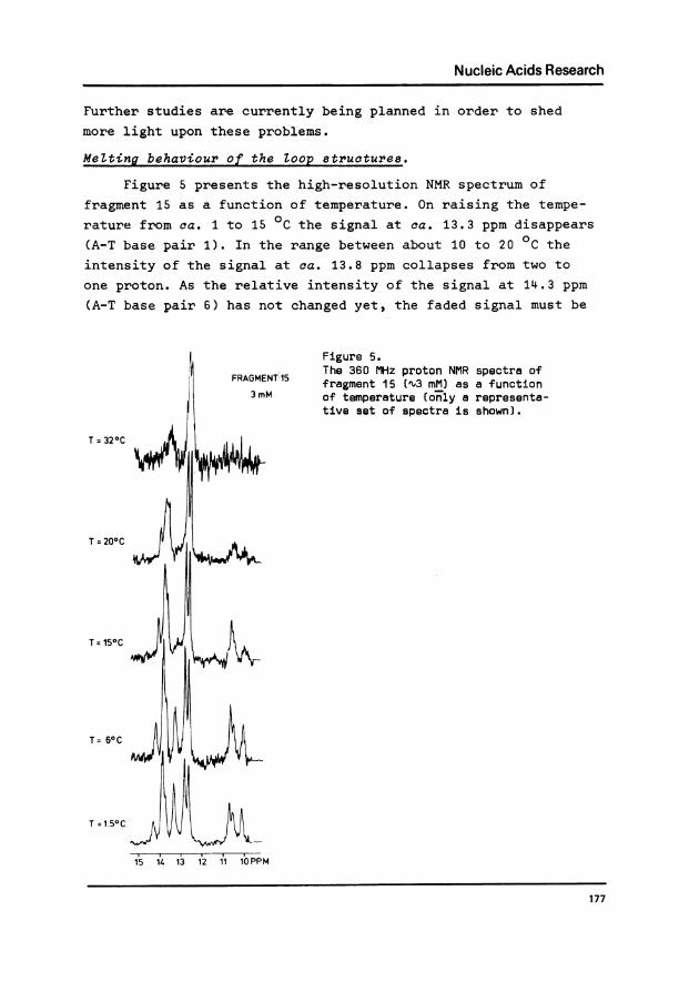
Figure 5 presents the high-resolution NMR spectrum of
fragment 15 as a function of temperature. On raising the tempe-
rature from ca. 1 to 15 0C the signal at ca. 13.3 ppm disappears
(A-T base pair 1). In the range between about 10 to 20 0C the
intensity of the signal at ca. 13.8 ppm collapses from two to
one proton. As the relative intensity of the signal at 14.3 ppm
(A-T base pair 6) has not changed yet, the faded signal must be
Figure 5.FRAGMEN.'-15 The 360 MHz proton NMR spectra ofFRAGMENt15 fragment 15 (X3 mM) as a function
3mM of temperature ( only a representa-tive set of spectra is shown).
T = 320C
1 L200C
T =150C
T =1.50C
15 14. 1 12 11 1bPPM
177
Nucleic Acids Research
due to A-T base pair 2. Further raising of the temperature (upto ca. 26 0C) causes A-T base pair 6 (at 14.3 ppm) to disappear,followed next by A-T base pair 5 (34 0C). Finally, the twocentral C-G pairs 3 and 4 (centered around 12.7 ppm) disappearsimultaneously (ca. 40 0C).
The NMR-temperature profiles of fragment 16 and 17 (notshown) are very similar to that of fragment 15. Except for A-Tbase pair 6 (vide infra), the hydrogen-bonded imino protons ofthe duplex "melt out" within the same temperature range as wasfound in fragment 15, the two central C-G pairs are the lastones being disrupted (at about 40 0C). In other words: thebreaking of the Watson-Crick hydrogen bonds occurs on both sidesof these C-G pairs. This observation implies that the loop initself forms an extra melting site in addition to the normal"fraying" phenomenon12 starting from the terminus of the doublehelical structure.
Of interest in this context are the differences betweenfragments 15, 16 and 17 emerging from the melting behaviour ofA-T base pair 6 and the "free" imino protons of the thymidylylresidues which constitute the loop. In fragments 16 and 17, thesignal due to A-T base pair 6 already disappears below 20 0C,whereas in the tightest loop (fragment 15) this signal isobserved up to a temperature of about 26 0C. Moreover, in goingfrom fragment 15 to fragment 17 the "melting" temperatures ofthe T- proton signals display a regular decreasing trend (from22 0C to 10 0C). These findings perhaps endorse the higheraccessibility of larger vs. smaller loops with respect to thewater molecules and point again to the forementioned decrease inrigidity of the loop upon enlarging.
DISCUSSION.
The results reported in this study reveal several interest-ing points. The NMR temperature-concentration profiles of frag-ment 14 (fig. 4) show that at ca. 3 mM the monomeric and dimericform have an approximately equal free energy content. Yet in the
monomeric form the double strand may involve only 5 base pairs,as a loop consisting of only two monomeric units is said to be
178
Nucleic Acids Research
sterically impossiblell. As the enthalpy term (&HO) in the freeenergy content (AG0) of the double helix, in principle, dependson the chain length (as well as on the base composition and thebase sequence) it is evident that the entropy term (AS0) plays adecisive role in this process. This may be visualized when theformation of a double helix is dissected into two steps1l: theformation of the first base pair (initiation) and the formationof subsequent base pairs (propagation). The probability ofinitiation is much larger for the intramolecular process (lead-ing to the hairpin loop) than for the intermolecular (dimeriza-tion) process.
This rationalization is further corroborated by the NMR-temperature profile of the duplex formed by the two hexanucleo-tides (6+6) which designate the ends of the synthetic oligo-nucleotides studied (i.e. ATCCTA and TAGGAT). It is found13 thatthe two central G-C base pairs are the last ones to disappearfrom the spectrum, as is the case for fragment 15, 16 and 17(vide supra). However, the temperature at which this occurs isabout 15 0C lower for the hexanucleotide mixture than for thefragments 15, 16 and 17. As the enthalpy term (apart from possi-ble small extra stacking interactions in the central T-regionof the fragments 15-17) must be equal for the various moleculesunder consideration, the difference in stability is mainly dueto the difference in entropy of formation of open-ended ve.hairpin-closed duplexes.
The relative stabilities of the hairpin loops in DNA frag-ments 15-17 merit some discussion. In all three fragments thesignals due to the central C-G pairs collapse at about the sametemperature (ca. 40 0C). Yet, the "melting" behaviour of the T-bases constituting the loops and of base pair 6 (next to thehairpin loops) appears to be affected by the size of these loops.The data suggest that the number of nonbonded bases in the loop(three to five in our case) has a strictly ZocaZ influence onthe stability of base pairs and does not affect the overallstability of duplex regions removed by two or more bases fromthe hairpin loop.
Summarizing, the apparently decreasing stability of thehairpin in going from fragment 15 to 17 is in accordance with
179
Nucleic Acids Research
the rules originally deduced by Tinoco et at.11 in which hairpinloops that contain three bases are assigned a "stability number"of -5 kcal/mole (AG0) and hairpins of 4-7 bases are less stableby 1 kcal/mole. It is noted that these rules, intended for the
estimation of secondary structures in RNA molecules, werederived from experimental data concerning DNA and oZigo-d(AT)melting behaviour. Remarkably, in a later publication14 it was
asserted, based upon data from RNA sequences, that a hairpinloop of three bases closed by an A-U base pair is Zess stablethan a hairpin consisting of 4-5 bases, thus contrary to thepresent conclusions. In our opinion these findings support thethesis that experimental data derived from DNA fragments shouldnot be used to predict the behaviour of RNA molecules and viceversa. Perhaps the rationale that explains in part the intriguingdifferences between DNA and RNA on various levels of biologicalactivity should be sought in the outspoken difference in confor-mational flexibility of the deoxyribose- vs. the ribose sugarring15 16*
ACKNOWLEDGEMENT.
This research was supported by the Netherlands Foundationfor Chemical Research (S.O.N.) with financial aid from theNetherlands Organization for the Advancement for Pure Research(Z.W.O.). We wish to thank Mr. K. Dijkstra and Dr. R. Kapteinfor their technical assistance.
REFERENCES.1. This paper is number XIV in the series "Nucleic Acids Constituents" from
this laboratory. For number XIII see: Leeuw,H.P.M. de, Haasnoot,C.A.G. &Altona,C. (1980) Isr. J. Chem. - accepted for publication.
2. Haasnoot,C.A.G., Hartog,J.H.J. den, Rooij,J.F.M. de, Boom,J.H. van &Altona,C. (1979) Nature 281,235-236.
3. Gralla.J. & Crothers,D.M. (1973) J. Mot. BioZ. 78, 301-319.4. Boom,J.H. van, Burgers,P.M.J. & Oeursen,P.H. van (1976) Tetrahedron
Letters, 869-872.5. Rooij,J.F.M. de, Wille-Hazeleger,G., Deursen,P.H. van, Serdijn,J. & Boom,
J.H. van (1979) RecZ. Trav. Chim. Pays-Bas 98, 537-548.6. Dadok,J. & Sprecher,R.F. (1974) J. Magn. Res. 13, 243-248.7. Pople,J.A. (1964) J. Chem. Phys. 41, 2559-2560.8. Giessner-Prettre,C. & Pullman,B. (1976) Biochem. Biophys. Res. Commzun. 70,
578-581.9. Borer,P.N., Kan,L.S. & Ts'O,P.O.P. (1975) Biochemistry 14, 4847-4863.
10. Arter,D.B. & Schmidt,P.G. (1976) Nucl. Acids Res. 3, 1437-1447.11. Tinoco,I., Uhlenbeck,O.C. & Levine,M.D. (1971) Nature 230, 362-367.12. Patel,D.J. & Hilbers,C.W. (1975) Biochemistry 14, 2651-2656.
180
Nucleic Acids Research
13. Haasnoot,C.A.G., Hartog,J.H.J. den & Altona,C. - urpubli8hed results.14. Tinoco,I., Borer,P.N., Dengler,B., Levine,M.D., Uhlenbeck,O.C., Crothers,
D.M. & Gralla,J. (1973) Nature New Biology 246, 40-41.15. Altona,C., Boom,J.H. van & Haasnoot,C.A.G. (1976) Eur. J. Biochem. 71,
557-562.16. Altona,C. & Sundaralingam,M. (1973) J. Amer. Chem. Soc. 95, 2333-2344.
181


