Embed Size (px)
Citation preview
J Cell Sci . Author manuscript
Page /1 12
Contribution of MT1-MMP and of human laminin-5 2 chain degradation toγ
mammary epithelial cell migrationChristine Gilles 1 * # , Myriam Polette 2 # , Christelle Coraux 2 , Jean-Marie Tournier 2 , Guerrino Meneguzzi 3 , Carine Munaut 1 ,Laure Volders 1 , Patricia Rousselle 4 , Philippe Birembaut 2 , Jean-Michel Foidart 1
Laboratory of Tumor and Developmental Biology 1 CHU Sart-Tilman , Universit de Li ge é è , 35B - 4000 LIEGE,BE
Dynamique cellulaire et mol culaire de la muqueuse respiratoire 2 é INSERM : U514 , IFR53 , Universit de Reims - Champagne Ardenne é ,Hopital Maison Blanche 45, Rue Cognacq Jay 51092 Reims Cedex,FR
Biologie et physiopathologie de la peau 3 INSERM : U385 , Universit de Nice Sophia-Antipolis é , Nice,FR
lnstitut de Biologie et de Chimie des Prot ines 4 é CNRS : UPR412 , Lyon, FR
* Correspondence should be adressed to: Christine Gilles <[email protected] >
# These authors contributed equally to this research
AbstractSUMMARY
Membrane-type matrix metalloproteinase 1 (MT1-MMP) is a membrane-anchored matrix metalloproteinase (MMP) that is
frequently associated with processes involving tissue remodelling and cell migration. We have examined MT1-MMP expression and
subcellular distribution as a function of MCF10A mammary epithelial cell migration using an in vitro outgrowth migration assay.
Stronger expression of MT1-MMP was observed at the mRNA and at the protein level in cells at the periphery of the outgrowth. As
shown by videomicroscopy, these cells were involved in an orientated cell migration, in contrast to stationary cells distant from the
periphery. Furthermore, MT1-MMP was mainly distributed in lamellipodia of migratory cells, as well as at their basal surface in
contact with the substrate. Laminin-5 (Ln-5), a recently described substrate for MT1-MMP, was deposited preferentially in the
matrix by migratory cells. Fragments of the 2 subunit of Ln-5 were also identified in migratory cultures of MCF10A cells, attestingγto its proteolytic degradation. These fragments corresponded in size to those we observed after incubation of purified human Ln-5
with the recombinant catalytic domain of human MT1-MMP. We also show that anti-Ln5 blocking antibodies, MMP inhibitors
(BB94 and TIMP-2) and MT1-MMP antisense oligonucleotides significantly decreased MCF10A cell migration. Taken together, these
observations demonstrate that MT1-MMP is spatially and temporally regulated during MCF10A cell migration, and suggest that
MT1-MMP-mediated pericellular proteolysis of Ln-5 2 chain could contribute to this process.γ
MESH Keywords Blotting, Western ; Breast ; cytology ; enzymology ; Cell Adhesion Molecules ; metabolism ; Cell Line ; Cell Movement ; Epithelial Cells ; cytology ;
enzymology ; Humans ; In Situ Hybridization ; Matrix Metalloproteinases, Membrane-Associated ; Metalloendopeptidases ; genetics ; metabolism ; Microscopy, Video ; Protein
Processing, Post-Translational ; Protein Subunits ; Pseudopodia ; enzymology ; RNA, Messenger ; genetics ; metabolism ; Reverse Transcriptase Polymerase Chain Reaction
Author Keywords MT1-MMP ; Laminin-5 ; Migration ; MCF10A ; Epithelial cells
INTRODUCTION
Epithelial cell migration plays a central role in several physiological or pathological processes, including wound healing,
organogenesis, placentation or tumour invasion. The acquisition of a migratory phenotype by epithelial cells is a spatially and temporally
regulated process that involves integrated modifications of the cells and of their surrounding environment.
The remodelling of the extracellular matrix (ECM) surrounding epithelial cells is accordingly a mechanism frequently associated with
cell migration. Matrix metalloproteases (MMPs), a family of proteases that degrades specific components of the ECM, have largely been
implicated in the ECM degradation associated with many processes that involve epithelial cell migration ( ; ; Matrisian, 1992 Werb, 1997
; , ). Most MMPs are secreted as inactive proenzymes and theirShapiro, 1998 Murphy and Gavrilovic, 1999 Nagase and Woessner, 1999
activation requires the proteolytic removal of the N-terminal pro-fragment ( ). By contrast, MT1-MMPNagase and Woessner, 1999
(membrane type-MMP) contains a transmembrane domain, which ensures the anchorage of the protein at the cell membrane (Sato et al.,
; ). MT1-MMP has been described as a major activator of MMP-2 ( ; ) but1994 Sato and Seiki, 1996 Sato et al., 1994 Sato and Seiki, 1996
MT1-MMP also possesses the ability to degrade ECM components, including gelatin, K-elastin, fibronectin, vitronectin, native fibrillar
type I, II and III collagen, and laminin-5 (Ln-5; ; ; ; ; Imai et al., 1996 Pei and Weiss, 1996 Sato and Seiki, 1996 Ohuchi et al., 1997
). The activity of MMPs, including MT1-MMP, is regulated by specific tissue inhibitors of MMPs (TIMPs).Koshikawa et al., 2000
MMP-2 is unique in its binding of TIMP-2, and a trimolecular complex formed by MT1-MMP, TIMP-2 and MMP-2 has been described (
). The expression of MT1-MMP has been detected in processes that involve epithelial cell migration and ECMStrongin et al., 1995
remodelling, and has been correlated to high invasive abilities in tumour cell lines ( ; ; Gilles et al., 1996 Sato and Seiki, 1996 Gilles et al.,
; ; ; ). However, the mechanisms by which MMPs and particularly1997 Pulyaeva et al., 1997 Polette et al., 1998 Quaranta, 2000
MT1-MMP facilitate migration are still poorly understood. The anchoring of MT1-MMP at the plasma membrane is thought to contribute
J Cell Sci . Author manuscript
Page /2 12
to a pericellular ECM degradation, either by its own degradative potential or through its ability to activate MMP-2 at the cell surface (
; ; ). A crucial issue is therefore the determination of the expression andNakahara et al., 1997 Chen and Wang. 1999 Quaranta, 2000
spatial distribution of MT1-MMP in epithelial cells during cell migration. Indeed, a pericellular degradation now appears as a key event,
generating an adequate substrate for cell migration in the immediate vicinity of migrating cells ( ; Nakahara et al., 1997 Chen and Wang,
; ; ; ).1999 Murphy and Gavrilovic, 1999 Hotary et al., 2000 Quaranta, 2000
Accordingly, rat laminin-5 (Ln-5) has recently been identified as a substrate for both MT1-MMP and MMP-2 ( ; Giannelli et al., 1997
). Ln-5 is a heterotrimer found in basement membranes (BM), which consists in the association of 3, 3 and 2Koshikawa et al., 2000 α β γsubunits ( ). It is a multifunctional protein that, in apparent contrast, is involved in the static adhesion of epithelialRousselle et al., 1991
cells to the basement membrane (BM; ; ; ; ) andBaker et al., 1996 Jones et al., 1998 Borradori and Sonnenberg, 1999 Nievers et al., 1999
also in the promotion of epithelial cell migration ( ; ; ; Miyazaki et al., 1993 Zhang and Kramer, 1996 Giannelli et al., 1997 Grassi et al.,
; ; ). The proteolytic degradation of Ln-5 3 and 2 subunits generates different1999 Goldfinger et al., 1999 Koshikawa et al, 2000 α γheterotrimeric forms of Ln-5 ( ; ; ; ), and appears to beRousselle et al, 1991 Marinkovich et al, 1992 Vailly et al, 1994 Matsui et al, 1995
responsible for the contrasting activities (i.e. adhesion or migration) attributed to Ln-5 ( ; ; Giannelli et al, 1997 Goldfinger et al, 1998
; ; ). Recent data have shown that the degradation of 2 subunit ofGiannelli et al, 1999 Goldfinger et al, 1999 Koshikawa et al, 2000 γexogenously provided rat Ln-5 by MMP-2 and/or MT1-MMP promotes cell migration in vitro ( ; Giannelli et al, 1997 Giannelli et al, 1999
; ). Furthermore, evidence that the degradation of Ln-5 2 chain could promote cell migration in vivo has beenKoshikawa et al, 2000 γgiven by the detection of specific fragments of 2 in remodelling but not in quiescent mouse tissues and in rodent skin carcinoma (γ
; ).Giannelli et al, 1997 Giannelli et al, 1999
In this study, we have used an in vitro migration assay coupled to videomicroscopy analyses to study the expression and cellular
distribution of MT1-MMP in association with cell migration of human epithelial mammary MCF10A cells. We show a stronger expression
of MT1-MMP mRNA and protein in migratory cells, and a distribution of MT1-MMP in lamellipodia and at the basal surface of these
migratory cells. Furthermore, we have found that Ln-5 was deposited around migratory cells and also detected degraded fragments of Ln-5
2 subunit in migratory cultures of MCF10A cells. We have also used MMPs inhibitors (BB94 and TIMP-2), MT1-MMP antisenseγoligonucleotides and Ln-5 blocking antibodies, which emphasized a functional contribution of MT1-MMP and Ln-5 in cell migration.
MATERIALS AND METHODSCell culture
Human mammary epithelial MCF10A cells were obtained from the American Type Culture Collection (Rockville, MD). Their growth
medium was composed of HAM F12 and Dulbecco s modified Eagle medium (DMEM) 1:3 (v/v) supplemented with 20 g/ml of adenine,’ μ5 g/ml of insulin, 0.5 g/ml of hydrocortisone, 2 ng/ml of EGF, 5 g/ml of transferrin, 1.5 ng/ml of triiodothyronine and 10 foetal calfμ μ μ %serum.
In vitro migration assay
5 10 cells were seeded in growth medium inside a 6 mm glass ring placed in the middle of a glass coverslip (22 mm in diameter).× 4
Twenty-four hours after plating, the glass ring was removed and the cells were covered with growth medium. In this model, the cells
migrate as an outgrowth from the confluent area initially delimited by the ring.
For epidermal growth factor (EGF)-induced migration, cells were plated inside the ring in complete growth medium for 24 hours.
After the removal of the ring, cells were washed for 1 hour twice in serum-free medium and then covered with serum-free medium
supplemented, or not, with EGF at 20 ng/ml.
For TIMP-2 and BB94 inhibition experiments, 72 hours after the ring removal TIMP-2 (at 1 g/ml, Calbiochem, La Jolla, CA) andμ
BB-94 (5 10 M, kindly provided by British Biotech, Oxford, UK) were added to the growth medium for 1 hour, before the× 6 −
quantification of cell migration.
For MT1-MMP antisense experiments, a T1-MMP antisense oligonucleotides kit was used (Biognostik, Gottingen, Germany). The
control oligonucleotides were scrambled oligonucleotides of the MT1-MMP antisense oligonucleotides. The oligonucleotides were added
in the growth medium at 4 M (as recommended by the manufacturer) after the removal of the ring. Using FITC-labelled oligonucleotides,μwe determined that 144 hours were necessary to ensure an uptake of the oligonucleotides by at least 20 of the cells. The effect of control%and MT1-MMP antisense oligonucleotides on cell migration were therefore measured 6 days after the removal of the ring and the addition
of the oligonucleotides.
For Ln-5 antibody inhibition experiments, mouse Ln-5 blocking antibodies (clone P3H9-2, Bioproducts, Heidelberg, Germany) were
added to the culture medium (at 25 g/ml) 72 hours after the removal of the ring and incubated for 4 hours before quantification of the cellμmigration speed. Controls were incubated with mouse IgG.
J Cell Sci . Author manuscript
Page /3 12
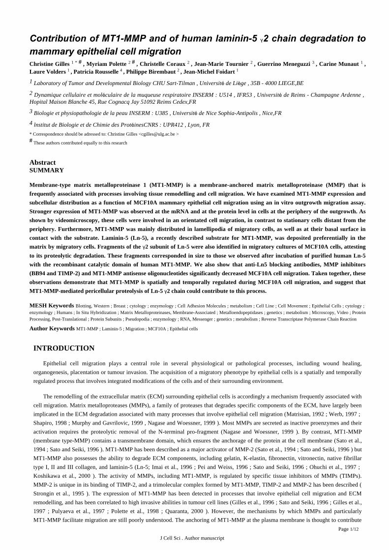
Quantification of cell migration speed and cell trajectories
To quantify the expansion of the outgrowth, migratory monolayers were placed on the stage of an inverted microscope (Nikon TMS-F,
Tokyo, Japan), connected to a video CCD camera (Cohu 4700, San Diego, CA) and a video monitor (PVM 1371, Sony, Japan), so as to
measure the outgrowth area.
To analyse and quantify the migratory speeds and trajectories of the cells, the monolayers were first incubated with a fluorescent
nuclear dye (Hoechst 33258, Molecular Probes, Eugene, OR). They were then placed in the environmental chamber (37 C, 5 CO ) of a° % 2
Zeiss IM35 inverted microscope (Zeiss, Oberkochen, Germany) equipped with an epifluorescence illumination source (excitation filter at
360 nm; emission filter at 510 nm) and a low level SIT camera (Lhesa 4036) controlled by a microcomputer (SparcClassic Workstation).
An image was collected every 10 minutes for 30 minutes. Twenty cell nuclei selected in different zones of the culture were then labelled
manually on the computer for each time point. Cell migration was characterized and quantified using a previously described software
program ( ) that measures the nuclei trajectories, as well as the cell migration speed. As the cell nuclei were labelledZahm et al, 1997
manually by the experimenter, the relative position of the label on a nucleus pictured at different time points varied slightly. Consequently,
a movement of the cells nuclei could be detected (corresponding to a speed of about 10 m/hour) in the areas distant from the outgrowthμperiphery. This movement was however random and was not considered as migration. Hence, the corresponding areas were identified as
stationary. By contrast, the movement of the nuclei at the periphery of the outgrowth corresponding to cell migration was clearly oriented
towards the outside of the outgrowth.
RT-PCR analyses
RNA extraction was performed from total migratory cultures using the RNA miniprep kits as recommended by the manufacturer
(Qiagen, Hilden, Germany). RT-PCR was performed using 10 ng of total RNA. An internal control RNA template containing the
sequences of the different primers used to amplify different MMPs was introduced in each sample for the standardization and
quantification of each RT-PCR reaction.
RT-PCR was performed using the GeneAmp Thermostable RNA PCR Kit (Perkin Elmer, Foster City, CA), and with pairs of primers
for six MMPs and for 28S control amplification (Eurogentec, Seraing, Belgium). Forward and reverse primers for human MT1-MMP,
MMP-2, MMP-9, MMP-1, MMP-3 and MMP-11 and 28S were designed as follows: MT1-MMP primers (forward 5 ′-CCATTGGGCATCCAGA-AGAGAGC-3 ; reverse 5 -GGATACCCAATGCCCATTGGCCA-3 ), MMP-2 primers (forward 5′ ′ ′ ′-GGCTGGTCAGTGGCTTGGGGTA-3 ; reverse 5 -AGATCTTCTTCTTCAAGGACCGGTT-3 ), MMP-1 primers (forward 5′ ′ ′ ′-GAGCAAACACATCTGAGGTACAGGA-3 ; reverse 5 -TTGTCCCGATGATCTCCCCTGACA-3 ), MMP-3 primers (forward 5′ ′ ′ ′-GATCTCTTCATTTTGGCCATCTCTTC-3 ; reverse 5 -CTCCAGTATTTGTCCTCTACAAAGAA-3 ), MMP-11 primers (forward 5′ ′ ′ ′-ATTTGGTTCTTCCAAGGTGCTCAGT-3 : reverse 5 -CCTCGGAAGAAGTAGATCTTGTTCT-3 ) and 28S primers (forward 5′ ′ ′ ′-GTTCACCCACTAATAGGGAACGTGA-3 ; reverse 5 -GGATTCTGACTTAGAGGCGTTCAGT-3 ). Reverse transcription was′ ′ ′performed at 70 C for 15 minutes. Amplification cycles were as follows: 15 seconds at 94 C, 15 seconds at 68 C, 10 seconds at 72 C.° ° ° °Twenty-five cycles were allowed for MT1-MMP amplification, up to 35 cycles for MMP-2, MMP-1, MMP-3 and MMP-11 amplification,
and 18 cycles for 28S amplification. Products were separated on acrylamide gels, stained with Gelstar (FMC, Bioproducts) and quantified
by fluorimetric scanning (LAS-1000, Fuji). The ratio of each endogenous signal to its specific internal control was calculated and
normalized to its the ratio to the 28S. These values were multiplied by the number of copies of internal controls added to the RT-PCR
reactions. Results were therefore expressed as a number of copies per 10 ng of RNA, allowing the comparison of the expression of
different MMPs that have been amplified using different PCR parameters.
Immunofluorescence
Monolayers were fixed with 4 paraformaldehyde in phosphate-buffered saline (PBS) for 10 minutes at 37 C then treated with 0.1% ° %Triton X-100 for MT1-MMP and integrin labelling. For Ln-5 labelling, the cells were fixed with methanol for 10 minutes at 20 C. The− °coverslips were then saturated for 30 minutes with 3 BSA in PBS.%
For MT1-MMP immunostaining, monolayers were successively (after intermediate washes in PBS) incubated for 1 hour with a
monoclonal antibody to MT1-MMP (clone 113-5B7 Chemicon, Temecula, CA), a biotinylated-sheep anti-mouse antibody (Amersham,
Aylesbury, UK) and a Texas Red-conjugated streptavidin (Amersham).
For 6 integrin staining, cells were successively incubated with anti 6 integrin rat antibody (clone GoH3, Immunotech, Marseille,α αFrance), a biotinylated goat anti-rat antibody (Sigma) and a Texas Red-conjugated streptavidin (Amersham). For 3 integrin labelling,αcells were incubated subsequently with an anti 3 monoclonal antibody (clone P1B5, Dako) and with a TRITC-conjugated rabbitαanti-mouse antibody.
J Cell Sci . Author manuscript
Page /4 12
For Ln-5 labelling, the cells were successively incubated with an anti Ln-5 monoclonal antibody (clone GB3, described in Verrando et
), a biotinylated-sheep anti-mouse antibody (Amersham) and FITC-conjugated streptavidin (Amersham).al., 1987
After incubation with the different antibodies, nuclei were labelled with 4 ,6-diamidino-2-phenylindole (DAPI; 1 g/ml) for 20′ μminutes. The coverslips were then mounted with Aquapolymount antifading solution (Agar, UK) onto glass slides and the slides were
observed under a Zeiss fluorescence microscope or with a MRC 600 confocal laser scanning microscope (BioRad, Richmond, CA).
In situ hybridization
The cultures were fixed for 10 minutes in 4 paraformaldehyde in PBS, dehydrated in ethanol 50 and 70 , rehydrated and treated% % %with 0.2 N HC1 for 20 minutes at room temperature. They were then washed in 2xSSC, acetylated in 0.25 acetic anhydride in 0.1 M%triethanolamine for 10 minutes and hybridized overnight with S -labelled MT1-MMP antisense RNA transcripts. This probe was[35 ]prepared from the MT1-MMP cDNA insert that had been cloned into pBluescript. The samples were then treated with RNase (20 g/ml)μfor 1 hour at 37 C to remove the unhybridized probes, washed under stringent conditions and detected autoradiographically by exposure to°D19 emulsion (Kodak, Rochester, NY) for 15 days. The control slides were treated under the same conditions but were hybridized with [35
S -labelled sense probes. Quantification of the in situ hybridizations was performed using the Discovery system automated image analyser](Becton-Dickinson, Mountain View, CA) that allowed the determination of the mean area of the in situ hybridization grains per cell. The
mean area of the in situ hybridization grains per cell was measured automatically on 10 fields (500 cells) at high magnification ( 500). In×order to compare MT1-MMP mRNA expression in stationary versus migratory cells, we performed these measurements in the two rows of
cells at the periphery of the outgrowth and in the following rows of the cells (between the fourth and the tenth rows of cells) on three
independent experiments.
Degradation of purified human Ln-5 by recombinant MT1-MMP
Human Ln-5 was purified from human squamous carcinoma SCC25 cell conditioned medium as described previously (Rousselle et al.,
). Purified Ln-5 (1 g) was incubated with the recombinant catalytic domain of human MT1-MMP (100 500 ng, Chemicon) in 501991 μ –mM Tris pH 7.5, 5 mM CaCl , 150 mM NaCl for 16 hours at 37 C. The samples were then analysed by western blotting for the expression2 °
of the 2 chain of Ln-5.γ
Western blotting analyses
Analyses of MT1 -MMP and Ln-5 2 chain expression were performed on protein extracts performed 72 hours after the removal of theγring on migratory cultures of MCF10A cells (cultivated in complete growth medium or in serum-free medium supplemented with EGF) or
on stationary cultures of MCF10A cells (cultivated in EGF/FCS-free medium) for 72 hours after the removal of the ring.
Extracts were prepared by scraping the cells in RIPA buffer (50mM Tris (pH 7.4), 150 mM NaCl, 1 Igepal (v/v), 1 sodium% %deoxycholate (w/v), 5 mM iodoacetamide, 0.1 SDS (w/v)) containing protease inhibitors (1mM phenylmethylsulfonyl fluoride, 10 g/ml% μleupeptin and 10 g/ml aprotinin).μ
Samples (8 g for Ln-5 2 chain analyses and 20 g for MT1-MMP analyses) were mixed with 1/5 sample buffer (0.31 M Trisμ γ μ(pH6.8), 10 SDS (w/v), 25 glycerol (v/v), 12.5 -mercaptoethanol (v/v) and 0.125 bromophenol blue (w/v)) and boiled for 5% % % β %minutes. They were then separated on 7.5 and 12 SDS-PAGE gels for Ln-5 and MT1-MMP analyses respectively and transferred to a% %PVDF filter (NEN, Boston, MA). The membranes were then blocked with 5 milk (w/v), 0.1 Tween 20 (w/v) in PBS for 2 hours before% %exposure to the primary antibody overnight at 4 C: a rabbit antibody (clone L 2-1) generated against the C-terminal region of Ln-5 2° γ γsubunit or a monoclonal antibody directed against the hemopexin-like domain of MT1-MMP (clone 2D7 kindly provided by Dr Rio,
IGBMC, Illkirch, France). The filters were then incubated either with a horseradish peroxydase-conjugated swine anti-rabbit or goat
anti-mouse antibody (Dako). Signals were detected with an enhanced chemoluminescence (ECL) kit (NEN, Boston, MA).
Statistical analyses
Experiments were performed in triplicate at least three times each. Data are expressed as means s.d. Student s test was used to± ’ t
compare the migration speeds of the cells under various experimental conditions. <0.05 was considered to be significant.P
RESULTSMT1-MMP is a major MMP expressed in migratory cultures of MCF10A cells
In order to examine the implication of MT1-MMP in epithelial cell migration, we used the human mammary MCF10A cells plated in
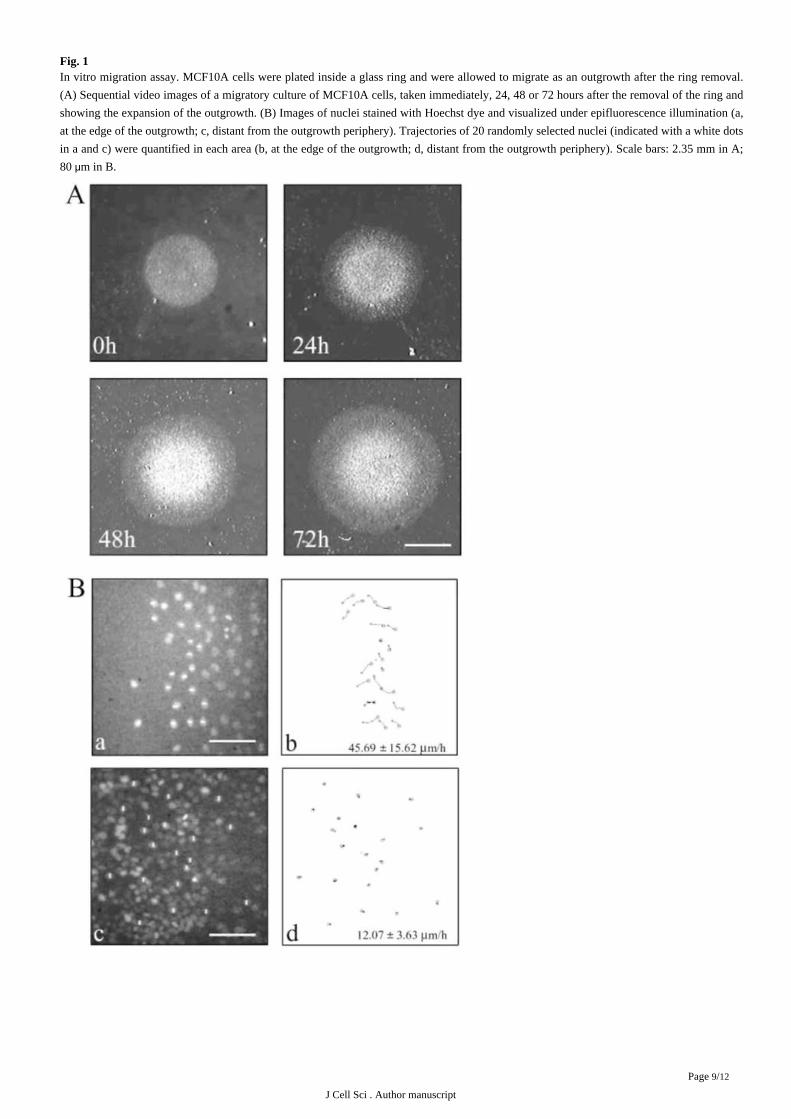
an in vitro migration assay allowing the visualization and quantification of cell migration ( ). In this migration assay,Gilles et al., 1999
cells are plated at a high density in a glass ring and migrate as an outgrowth after the removal of the ring (we will refer to these cultures as
migratory cultures, ). Using videomicroscopy, we demonstrated that cells at the periphery of the outgrowth are involved in anFig. 1A
J Cell Sci . Author manuscript
Page /5 12
orientated migration whereas cells distant from that periphery are basically stationary ( ). Comparing the expression of MT1-MMPFig. 1B
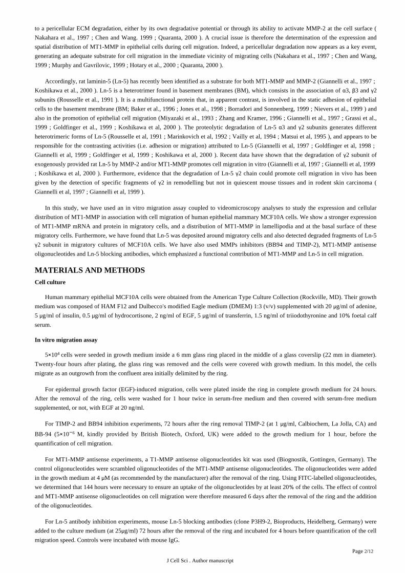
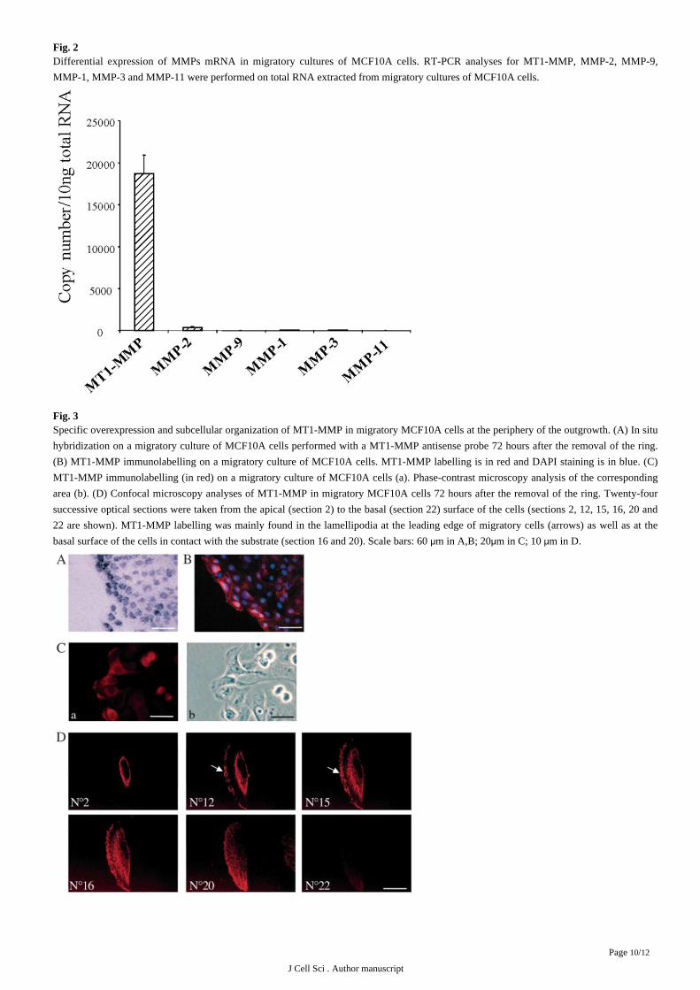
by quantitative RT-PCR with the expression of other MMPs, we found that MT1-MMP is a major MMP expressed in our model compared
with other MMPs (MMP-9. MMP-2, MMP-1, MMP-3 and MMP-11), which were barely detectable ( ).Fig. 2
MT1-MMP is overexpressed in migratory MCF10A cell
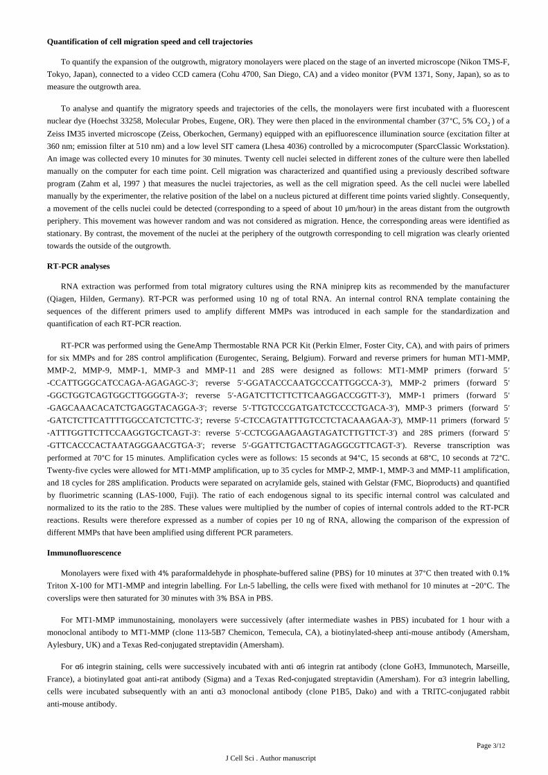
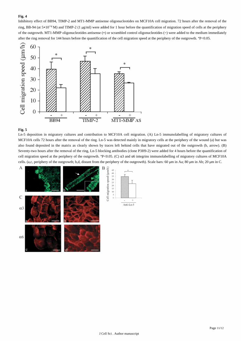
In order to study more precisely the relationship between MT1-MMP and cell migration, we performed in situ hybridization and
immunofluorescence analyses on migratory cultures of MCF10A cells. A stronger expression of MT1-MMP was clearly observed both at
the mRNA ( ) and protein level ( ) in cells at the periphery of the outgrowth, which has been shown to be a subpopulation ofFig. 3A Fig. 3B
migratory cells. Densitometric quantification of the in situ hybridizations revealed a 2- to 2.4-fold increase in the density of grains in the
rows of cells at the periphery of the outgrowth when compared to the density in the eight subsequent rows. Moreover, immunofluorescence
combined with phase contrast analyses clearly revealed that MT1-MMP is present in lamellipodia identified as a phase-dark rim at the
leading edge of the cells ( ). Confocal microscopy confirmed this particular distribution of MT1-MMP in lamellipodia at theFig. 3C
leading edge of migratory cells but also revealed its presence at the basal surface of these migratory cells in contact with the substrate (Fig.
).3D
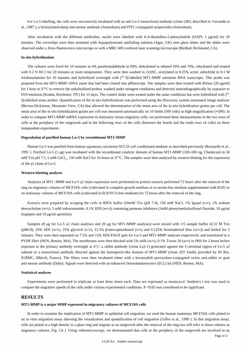
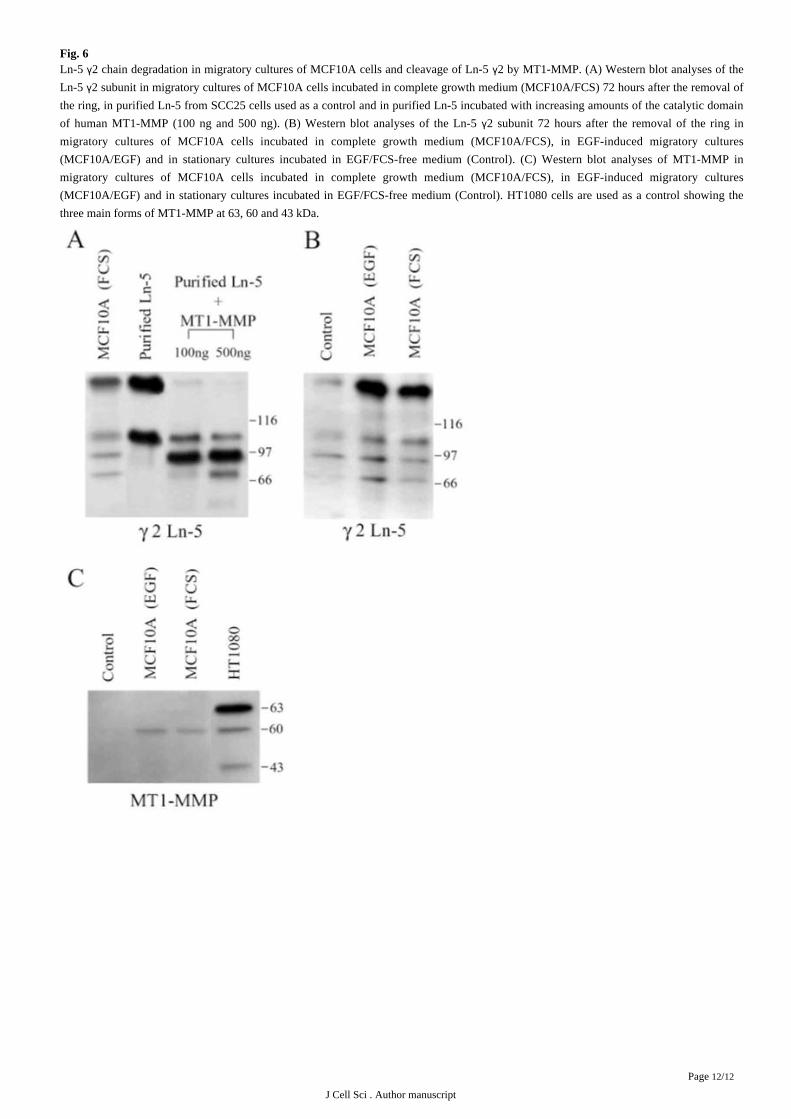
Furthermore, using MMP inhibitors BB94 and TIMP-2, we showed a decrease of MCF10A cell migration ( ). The addition ofFig. 4
MT1-MMP antisense oligonucleotides (with a maximal uptake of the oligonucleotides by about 20 of the cells) resulted in a decrease of%MT1-MMP expression, as evaluated by immunofluorescence, and also diminished cell migration ( ). These data suggest a functionalFig. 4
contribution of MT1-MMP overexpression and activity to MCF10A cell migration.
Ln-5 and MCF10A cell migration
AS rat Ln-5 has recently been described as a potential substrate for MT1-MMP ( ), the presence and depositionKoshikawa et al., 2000
of endogenously produced human Ln-5 was investigated in migratory cultures of MCF10A cells. By immunofluorescence, Ln-5 appeared
to be overexpressed by migratory cells at the periphery of the outgrowth ( , part a). The deposition of Ln-5 was also emphasized byFig. 5A
the presence of traces of Ln-5 behind cells which had migrated out of the outgrowth ( , part b). Ln-5 blocking antibodies were alsoFig. 5A
found to decrease MCF10A cell migration suggesting a contribution of the interactions between MCF10A cells and Ln-5 in their migratory
behaviour ( ).Fig. 5B
The distribution of 6 and 3 integrins, which constitute the 6 4 and 3 l receptors known as two major receptors for Ln-5, was alsoα α α β α βexamined by immunofluorescence as a function of MCF10A cell migration. These integrins were not only present in MCF10A migratory
cultures but also reorganized in function of cell migration. They were indeed mainly found in lamellipodia of migratory cells, whereas in
stationary cells, 3 was detected rather in places cell-cell contact and 6 at the basal surface ( ). These results show that Ln-5 andα α Fig. 5C
its major receptors are expressed in our model but also display a specific distribution during cell migration.
Ln-5 degradation and MCF10A cell migration
In order to examine the potential contribution of MT1-MMP on the degradation of Ln-5 2 chain degradation during epithelial cellγmigration, the pattern of endogenously produced 2 chain of Ln-5 was examined by western blotting analyses in migratory cultures ofγMCF10A cells. Four Ln-5 2 fragments could clearly be identified in total extracts of migratory MCF10A cultures ( ). The twoγ Fig. 6A
largest fragments corresponded in size to those detected in Ln-5 purified from squamous carcinoma SCC25 cell-conditioned medium, and
have previously been described as the unprocessed human Ln-5 2 subunit (155 kDa) and a processed fragment of human 2 (105 kDa; γ γ).Rousselle et al., 1991
Furthermore, the incubation of purified Ln-5 with the recombinant catalytic domain of human MT1-MMP generated fragments
corresponding in size to those identified in our migratory cultures of MCF10A cells ( ).Fig. 6A
In order to relate the presence of the degraded fragments of Ln-5 2 chain to cell migration, we compared the Ln-5 2 pattern inγ γmigratory cultures (incubated in complete medium) with the one obtained in cells from cultures incubated in EGF/serum-free medium
previously characterized as stationary cultures ( ). In order to minimize the potential contribution of serum-derivedGilles et al., 1999
proteases in the processing of Ln-5 2 chain, we also examined the 2 chain pattern in cells cultivated in serum-free medium supplementedγ γwith EGF, which has previously been shown to promote cell migration in our model ( ). It was verified that, as observedGilles et al., 1999
for migratory cultures of MCF10A cells incubated in complete medium, both MT1-MMP and Ln-5 expression was increased specifically
in cells at the periphery of the outgrowth in EGF-induced migratory cultures (data not shown). Using these different culture conditions, we
could show increased amounts of Ln-5 2 and its three degraded fragments in migratory cultures (incubated in serum- or inγEGF-containing medium) in comparison with those detected in EGF/serum-free stationary cultures ( ). As shown by MT1-MMPFig. 6B
western blotting analyses, this increase in Ln-5 fragments in migratory cultures of MCF10A cells clearly correlated with increased amount
of MT1-MMP ( ). These results also revealed that MT1-MMP was mostly present as the 60 kDa form, known to be the active formFig. 6C
of the enzyme. As MMP-2 has also been shown to cleave rat Ln-5 2, we looked at MMP-2 expression by zymography analyses and didγ
J Cell Sci . Author manuscript
Page /6 12
not find any detectable levels of MMP-2 in cell extracts of MCF10A cells cultivated with or without EGF in the migration assay (data not
shown). These data therefore suggest that MT1-MMP could contribute to the degradation of endogenously produced Ln-5 2 during cellγmigration.
DISCUSSION
In the present study, we have shown that MT1-MMP is overexpressed and redistributed during human epithelial mammary MCF10A
cell migration in association with Ln-5 2 chain degradation. We indeed observed: (1) a stronger expression of MT1-MMP in migratoryγMCF10A cells versus stationary ones, by in situ hybridization, immunofluorescence and western blotting; (2) a subcellular localization of
MT1-MMP in cellular protrusions (lamellipodia) and at the basal surface of migratory cells; (3) an inhibition of epithelial cell migration by
BB94, TIMP-2 and MT1-MMP antisense oligonucleotides; and (4) degraded fragments of Ln-5 2 chain in migratory cultures of MCF10Aγcells.
Overexpression of MT1-MMP in epithelial cells associates with their migratory status
Using an in vitro migration assay, we have shown a stronger expression of MT1-MMP in cells located at the outgrowth periphery that,
as determined by time lapse videomicroscopy, represents a subpopulation of migratory cells. In close relation to our observations obtained
with the MCF10A cells, a downregulation of MT1-MMP associated with increased cell-cell contacts has been reported in a mouse
mammary epithelial cell line ( ). In vivo, MT1-MMP has also been reported in epithelial cells involved in processes thatTanaka et al., 1997
require cell migration, such as nephrogenesis ( ; ), placentation ( ; Tanney et al., 1998 Kanwar et al., 1999 Nawrocki et al., 1996 Bjorn et al.,
; ) or tumour invasion ( ; ). Our in vitro model coupled to the1997 Tanaka et al., 1998 Sato and Seiki, 1996 Ellerbroek and Stack, 1999
videomicroscopy analyses nevertheless allowed us to demonstrate a specific induction of MT1-MMP both at the protein and mRNA level
within the same cell line, in association with the expression of migratory properties. Furthermore, our results showing that BB94, TIMP-2
and MT1-MMP antisense oligonucleotides diminished MCF10A cell migration clearly emphasized a functional contribution of
MT1-MMP overexpression and activity in MCF10A cell migration. It can therefore be suggested that MT1-MMP expression in epithelial
cells can be temporally and spatially regulated during cell migration, and that it functionally contributes to this process.
MT1-MMP is distributed in lamellipodia and at the basal surface of migratory cells in contact with the substrate
More than demonstrating an increase of MT1-MMP expression in relation with cell migration, our results also showed a particular
subcellular organization of MT1-MMP in migratory epithelial cells. Immunofluorescence analyses indeed clearly showed that MT1-MMP
is mostly located in lamellipodia. Confocal microscopy confirmed these data but also revealed a punctiform labelling of MT1-MMP at the
basal surface of migratory cells. Accordingly, Nakahara et al. have shown that RPMI-7951 melanoma cells transfected with MT1-MMP
organized the protein in invadopodia at the basal surface of the cells and in lamellipodia at the leading edge of the cells, and displayed an
enhanced ability to degrade FITC-labelled gelatin films ( ). By contrast, they reported that ConA-inducedNakahara et al., 1997
overexpression of MT1-MMP, which did not generate the subcellular localisation in invadopodia, did not enhance gelatin degradation. It
has also been shown that the degradation of the gelatin film was mostly accomplished by the enzyme present in the invadopodia rather
than by the enzyme present in lamellipodia ( ). Belien et al. ( ) have also shown a distribution ofChen and Wang, 1999 Belien et al., 1999
MT1-MMP in the lamellipodia of MT1-MMP transfected rat glioma cells. Taken together with our results, these data suggest that the
subcellular organization of MT1-MMP plays a major role in its degradative ability and emphasize the importance of a pericellular
proteolysis in cell migration. Accordingly, Hotary et al. ( ) have shown that MT-MMPs, but not soluble MMPs,Hotary et al., 2000
participate to cell invasion and morphogenesis of MDCK cells in collagen gels. A high level of expression of MT1-MMP and a subcellular
organization at the leading edge of migratory cells and at their basal surface would thus contribute to a pericellular proteolysis involved in
cell migration.
Laminin-5 deposition associates with MCF10A cell migration
We found that Ln-5, a potential substrate for MT1-MMP, is preferentially deposited by MT1-MMP-overexpressing migratory
MCF10A cells at the periphery of the outgrowth. In agreement with our findings, several reports have shown that Ln-5 is expressed and
deposited by migratory epithelial cells during wound healing both in vivo ( ; ) and in vitro (Larjava et al., 1993 Kainulainen et al., 1998
; ; ). Ln-5 has also been shown to be deposited adjacent to carcinomaZhang and Kramer, 1996 Lotz et al., 1997 Qin and Kurpakus, 1998
cell clusters and has been related to the invasiveness of several types of tumours ( ; ; Pyke et al., 1995 Sordat et al., 1998 Kosmehl et al.,
; ; ; ). Other evidence that the interaction between Ln-5 and MCF10A cells1999 Maatta et al., 1999 Skyldberg et al, 1999 Lohi et al, 2000
could be involved in their migration comes from our present results and those of Goldfinger et al. ( ) showing thatGoldfinger et al, 1999
the 6 and 3 integrin are not only present in MCF10A cells but also reorganized in relation with cell migration and that Ln-5 blockingα αantibodies decreased MCF10A cell migration. Accordingly, 6 4 and 3 1 are known as the main receptors for Ln-5 and have beenα β α βimplicated in adhesion but also in migration of epithelial cell lines on Ln-5 ( ; ).Zhang and Kramer, 1996 Goldfinger et al, 1999
J Cell Sci . Author manuscript
Page /7 12
Furthermore, the overexpression and cellular redistribution of 6 4 has been described in invasive tumours (α β Rabinovitz and Mercurio,
) and in wound healing ( ). It can thus be suggested that an increased deposition of Ln-5 by MCF10A cells at1996 Kainulainen et al, 1998
the periphery of the outgrowth and a reorganization of the Ln-5 integrin receptors in such cells are involved in their migratory properties.
Laminin-5 2 chain degradation associates with MCF10A cell migrationγ
Supporting the concept that MT1-MMP-mediated pericellular proteolysis could be involved in epithelial cell migration, we identified
by western blotting increased amounts of fragments of the 2 chain in migratory cultures of MCF10A cells versus stationary cultures. Theγincubation of the recombinant catalytic domain of human MT1-MMP with purified human Ln-5 generated degraded fragments
corresponding in size to those observed in our migratory cultures of MCF10A cells, suggesting a contribution of MT1-MMP in the
generation of the Ln-5 2 fragments associated with MCF10A cell migration. In apparent contrast to our data, Goldfinger et al. (γ Goldfinger
) did not find the two smallest fragments of Ln-5 in the ECM of wounded MCF10A cell cultures that were allowed to heal for 8et al, 1999
hours. This discrepancy could be explained by the fact that, in our model, cells were allowed to migrate for 72 hours. This could indeed
lead to an enrichment of the ECM with degraded Ln-5 2 chain if, as suggested by our data, migratory cells newly synthesize Ln-5,γoverexpress MT1-MMP that subsequently cleaves the 2 chain of Ln-5. However, these authors found that plasmin-mediatedγmodifications of the 3 subunit of Ln-5 regulated cell migration ( ). Taken together with our results, these dataα Goldfinger et al, 1999
suggest that modifications of both 2 and 3 chains of Ln-5 can regulate MCF10A epithelial cell migration. Supporting our findings thatγ αLn-5 2 degradation associates with the overexpression of MT1-MMP and cell migration, the cleavage of the 2 chain of rat Ln-5 has beenγ γshown to be mediated by MMP-2 and/or MT1-MMP in a dose-dependent manner ( ; ), and notGiannelli et al, 1997 Koshikawa et al, 2000
by other proteases such as plasmin or MMP-9 ( ). The degraded fragments identified by Koshikawa et al. (Giannelli et al, 1997 Koshikawa
) after the cleavage of rat Ln-5 2 chain by MT1-MMP differed in number and in size from those we observed afteret al, 2000 γMT1-MMP-mediated degradation of human Ln-5 2 chain. Also in agreement with our data, rat Ln-5 cleaved by MMP-2, but not intact ratγLn-5, has bee reported to promote the migration of epithelial cells ( ). It has also been shown using a transwell assayGiannelli et al, 1997
that human tumour cells constitutively expressing MT1-MMP display a higher migrating ability towards exogenously provided rat Ln-5
than MT1-MMP negative cells ( ). We report an enhanced production of both Ln-5 and MT1-MMP and a cleavageKoshikawa et al, 2000
of Ln-5 2 chain specifically associated with the expression of a migratory status by MCF10A epithelial cells.γ
In conclusion, our data demonstrate that the acquisition of a migratory phenotype by MCF10A epithelial cells is accompanied by an
overexpression of MT1-MMP and a localization of the protein in the lamellipodia and at the basal surface of the cells in contact with the
ECM substrate. This could participate to a pericellular degradation of the 2 chain of Ln-5, which is more specifically deposited by theγmigratory cells themselves, thereby providing a modified substrate that promotes cell migration.
Ackowledgements:
We thank Dr Rio (Strasbourg, France) for the 2D7 MT1-MMP antibody and British Biotech (Oxford, UK) for BB94. This work was
supported by grants from the Fonds National de la Recherche Scientifique (FNRS, Brussels, Belgium), the Fonds de la Recherche Scientifique
M dicale, the Communaut Fran aise de Belgique (Actions de Recherches Concert es), the F d ration Belge Contre le Cancer, the Centreé é ç é é éAnti-canc reux pr s l Universit de Li ge, the CGER-Assurances, the Fonds d Investissements de la Recherche Scientifique (CF U, Li ge,é è ’ é è ’ Γ èBelgium) and from the Lions Club of Soissons (France). C.G. is a Research Associate from the FNRS (Belgium).
References: Baker SE , DiPasquale AP , Stock EL , Quaranta V , Fitchmun M , Jones JC . 1996 ; Morphogenetic effects of soluble laminin-5 on cultured epithelial cells and tissue expiants
. Exp Cell Res . 228 : 262 - 270 Belien AT , Paganetti PA , Schwab ME . 1999 ; Membrane-type 1 matrix metalloprotease (MT1-MMP) enables invasive migration of glioma cells in central nervous system
white matter . J Cell Biol . 144 : 373 - 384 Bjorn SE , Hastrup N , Lund LR , Dano K , Larsen JF , Pyke C . 1997 ; Co-ordinated expression of MMP-2 and its putative activator, MT1-MMP, in human placentation . Mol
Hum Reprod . 3 : 713 - 723 Borradori L , Sonnenberg A . 1999 ; Structure and function of hemidesmosomes: more than simple adhesion complexes . J Invest Dermatol . 112 : 411 - 418
Chen WT , Wang JY . 1999 ; Specialized surface protrusions of invasive cells, invadopodia and lamellipodia, have differential MT1-MMP, MMP-2, and TIMP-2 localization . Ann New York Acad Sci . 878 : 361 - 371 Ellerbroek SM , Stack MS . 1999 ; Membrane associated matrix metalloproteinases in metastasis . BioEssays . 21 : 940 - 949
Giannelli G , Falk-Marzillier J , Schiraldi O , Stetler-Stevenson WG , Quaranta V . 1997 ; Induction of cell migration by matrix metalloprotease-2 cleavage of laminin-5 . Science . 211 : 225 - 228
GiannelU G , Pozzi A , Stetler-Stevenson WG , Gardner HA , Quaranta V . 1999 ; Expression of matrix metalloprotease-2-cleaved laminin-5 in breast remodeling stimulated by sex steroids . Am J Pathol . 154 : 1193 - 1201
Gilles C , Polette M , Piette J , Munaut C , Thompson EW , Birembaut P , Foidart JM . 1996 ; High level of MT-MMP expression is associated with invasiveness of cervical cancer cells . Int J Cancer . 65 : 209 - 213
Gilles C , Polette M , Seiki M , Birembaut P , Thompson EW . 1997 ; Implication of collagen type I-induced membrane-type 1-matrix metalloproteinase expression and matrix metalloproteinase-2 activation in the metastatic progression of breast carcinoma . Lab Invest . 76 : 651 - 660
Gilles C , Polette M , Zahm JM , Tournier JM , Volders L , Foidart JM , Birembaut P . 1999 ; Vimentin contributes to human mammary epithelial cell migration . J Cell Sci . 112 : 4615 - 4625
Goldfinger LE , Stack MS , Jones JC . 1998 ; Processing of laminin-5 and its functional consequences: role of plasmin and tissue-type plasminogen activator . J Cell Biol . 141 : 255 - 265
J Cell Sci . Author manuscript
Page /8 12
Goldfinger LE , Hopkinson SB , deHart GW , Collawn S , Couchman JR , Jones JC . 1999 ; The alpha3 laminin subunit, alpha6beta4 and alpha3betal integrin coordinately regulate wound healing in cultured epithelial cells and in the skin . J Cell Sci . 112 : 2615 - 2629
Grassi M , Moens G , Rousselle P , Thiery JP , Jouanneau J . 1999 ; The SFL activity secreted by metastatic carcinoma cells is related to laminin 5 and mediates cell scattering in an integrin-independent manner . J Cell Sci . 112 : 2511 - 2520
Hotary K , Allen E , Punturieri A , Yana I , Weiss SJ . 2000 ; Regulation of cell invasion and morphogenesis in a three-dimensional type I collagen matrix by membrane-type matrix metalloproteinases 1, 2, and 3 . J Cell Biol . 149 : 1309 - 1323
Imai K , Ohuchi E , Aoki T , Nomura H , Fujii Y , Sato H , Seiki M , Okada Y . 1996 ; Membrane-type matrix metalloproteinase 1 is a gelatinolytic enzyme and is secreted in a complex with tissue inhibitor of metalloproteinases 2 . Cancer Res . 56 : 2707 - 2710
Jones JC , Hopkinson SB , Goldflnger LE . 1998 ; Structure and assembly of hemidesmosomes . BioEssays . 20 : 488 - 494 Kainulainen T , Hakkinen L , Hamidi S , Larjava K , Kallioinen M , Peltonen J , Salo T , Larjava H , Oikarinen A . 1998 ; Laminin-5 expression is independent of the injury
and the microenvironment during reepithelialization of wounds . J Histochem Cytochem . 46 : 353 - 360 Kanwar YS , Ota K , Yang Q , Wada J , Kashihara N , Tian Y , Wallner EI . 1999 ; Role of membrane-type matrix metalloproteinase 1 (MT-l-MMP), MMP-2, and its inhibitor
in nephrogenesis . Am J Physiol . 111 : F934 - F947 Koshikawa N , Giannelli G , CiruUi V , Miyazaki K , Quaranta V . 2000 ; Role of cell surface metalloprotease MT1-MMP in epithelial cell migration over laminin-5 . J Cell
Biol . 148 : 615 - 624 Kosmehl H , Berndt A , Strassburger S , Borsi L , Rousselle P , Mandel U , Hyckel P , Zardi L , Katenkamp D . 1999 ; Distribution of laminin and fibronectin isoforms in oral
mucosa and oral squamous cell carcinoma . Br J Cancer . 81 : 1071 - 1079 Larjava H , Salo T , Haapasalmi K , Kramer RH , Heino J . 1993 ; Expression of integrins and basement membrane components by wound keratinocytes . J Clin Invest . 92 :
1425 - 1435 Lohi J , Oivula J , Kivilaakso E , Kiviluoto T , Frojdman K , Yamada Y , Burgeson RE , Leivo I , Virtanen I . 2000 ; Basement membrane laminin-5 is deposited in colorectal
adenomas and carcinomas and serves as a ligand for alpha3betal integrin . APMIS . 108 : 161 - 172 Lotz MM , Nusrat A , Madara JL , EzzeU R , Wewer UM , Mercurio AM . 1997 ; Intestinal epithelial restitution. Involvement of specific laminin isoforms and integrin laminin
receptors in wound closure of a transformed model epithelium . Am J Pathol . 150 : 747 - 760 Maatta M , Soini Y , Paakko P , Salo S , Tryggvason K , Autio-Harmainen H . 1999 ; Expression of the laminin gamma2 chain in different histological types of lung
carcinoma. A study by immunohistochemistry and in situ hybridization . J Pathol . 188 : 361 - 368 Marinkovich MP , Lunstrum GP , Burgeson RE . 1992 ; The anchoring filament protein kalinin is synthesized and secreted as a high molecular weight precursor . J Biol Chem .
267 : 17900 - 17906 Matrisian LM . 1992 ; The matrix-degrading metalloproteinases . BioEssays . 14 : 455 - 463
Matsui C , Wang CK , Nelson CE , Bauer EA , Hoeffler WK . 1995 ; The assembly of laminin-5 subunits . J Biol Chem . 270 : 23496 - 23503 Miyazaki K , Kikkawa Y , Nakamura A , Yasumitsu H , Umeda M . 1993 ; A large cell-adhesive scatter factor secreted by human gastric carcinoma cells . Proc Natl Acad Sci
USA . 90 : 11767 - 11771 Murphy G , Gavrilovic J . 1999 ; Proteolysis and cell migration: creating a path? . Curr Opin Cell Biol . 11 : 614 - 621 Nagase H , Woessner JF . 1999 ; Matrix metalloproteinases . J Biol Chem . 274 : 21491 - 21494
Nakahara H , Howard L , Thompson EW , Sato H , Seiki M , Yeh Y , Chen WT . 1997 ; Transmembrane/cytoplasmic domain-mediated membrane type 1-matrix metalloprotease docking to invadopodia is required for cell invasion . Proc Natl Acad Sci USA . 94 : 7959 - 7964
Nawrocki B , Polette M , Marchand V , Maquoi E , Beorchia A , Tournier JM , Foidart JM , Birembaut P . 1996 ; Membrane-type matrix metalloproteinase-1 expression at the site of human placentation . Placenta . 17 : 565 - 572
Nievers MG , Schaapveld RQ , Sonnenberg A . 1999 ; Biology and function of hemidesmosomes . Matrix Biol . 18 : 5 - 17 Ohuchi E , Imai K , FujU Y , Sato H , Seiki M , Okada Y . 1997 ; Membrane type 1 matrix metalloproteinase digests interstitial collagens and other extracellular matrix
macromolecules . J Biol Chem . 272 : 2446 - 2451 Pei D , Weiss SJ . 1996 ; Transmembrane-deletion mutants of the membrane-type matrix metalloproteinase-1 process progelatinase A and express intrinsic matrix-degrading
activity . J Biol Chem . 271 : 9135 - 9140 Polette M , Gilles C , de Bentzmann S , Gruenert D , Tournier JM , Birembaut P . 1998 ; Association of fibroblastoid features with the invasive phenotype in human bronchial
cancer cell lines . Clin Exp Metastasis . 16 : 105 - 112 Pulyaeva H , Bueno J , Polette M , Birembaut P , Sato H , Seiki M , Thompson EW . 1997 ; MT1-MMP correlates with MMP-2 activation potential seen after epithelial to
mesenchymal transition in human breast carcinoma cells . Clin Exp Metastasis . 15 : 111 - 120 Pyke C , Salo S , Ralfklaer E , Romer J , Dano K , Tryggvason K . 1995 ; Laminin-5 is a marker of invading cancer cells in some human carcinomas and is coexpressed with
the receptor for urokinase plasminogen activator in budding cancer cells in colon adenocarcinomas . Cancer Res . 55 : 4132 - 4139 Qin P , Kurpakus MA . 1998 ; The role of laminin-5 in TGF alpha/EGF-mediated corneal epithelial cell motility . Exp Eye Res . 66 : 569 - 579
Quaranta V . 2000 ; Cell migration through extracellular matrix: membrane-type metalloproteinases make the way . J Cell Biol . 149 : 1167 - 1170 Rabinovitz I , Mercurio AM . 1996 ; The integrin alpha 6 beta 4 and the biology of carcinoma . Biochem Cell Biol . 74 : 811 - 821
Rousselle P , Lunstrum GP , Keene DR , Burgeson RE . 1991 ; Kalinin: an epithelium-specific basement membrane adhesion molecule that is a component of anchoring filaments . J Cell Biol . 114 : 567 - 576
Sato H , Takino T , Okada Y , Cao J , Shinagawa A , Yamamoto E , Seiki M . 1994 ; A matrix metalloproteinase expressed on the surface of invasive tumour cells . Nature . 370 : 61 - 65
Sato H , Seiki M . 1996 ; Membrane-type matrix metalloproteinases (MT-MMPs) in tumor metastasis . J Biochem . 119 : 209 - 215 Shapiro SD . 1998 ; Matrix metalloproteinase degradation of extracellular matrix: biological consequences . Curr Opin Cell Biol . 10 : 602 - 608
Skyldberg B , Salo S , Eriksson E , Aspenblad U , Moberger B , Tryggvason K , Auer G . 1999 ; Laminin-5 as a marker of invasiveness in cervical lesions . J Natl Cancer Inst . 91 : 1882 - 1887
Sordat I , Bosnian FT , Dorta G , Rousselle P , Aberdam D , Blum AL , Sordat B . 1998 ; Differential expression of laminin-5 subunits and integrin receptors in human colorectal neoplasia . J Pathol . 185 : 44 - 52
Strongin AY , Collier I , Bannikov G , Manner BL , Grant GA , Goldberg GI . 1995 ; Mechanism of cell surface activation of 72-kDa type IV collagenase. Isolation of the activated form of the membrane metalloprotease . J Biol Chem . 270 : 5331 - 5338
Tanaka SS , Mariko Y , Mori H , Ishijhna J , Tachi S , Sato H , Seiki M , Yamanouchi K , Tojo H , Tachi C . 1997 ; Cell-cell contact down-regulates expression of membrane type metalloproteinase-1 (MT1-MMP) in a mouse mammary gland epithelial cell line . Zool Sci . 14 : 95 - 99
Tanaka SS , Togooka Y , Sato H , Seiki M , Tojo H , Tachi C . 1998 ; Expression and localization of membrane type matrix metalloproteinase-1 (MT1-MMP) in trophoblast cells of cultured mouse blastocysts and ectoplacental cones . Placenta . 19 : 41 - 48
Tanney DC , Feng L , Pollock AS , Lovett DH . 1998 ; Regulated expression of matrix metalloproteinases and TIMP in nephrogenesis . Dev Dyn . 213 : 121 - 129 Vailly J , Verrando P , Champliaud MF , Gerecke D , Wagman DW , Baudoin C , Aberdam D , Burgeson R , Bauer E , Ortonne JP . 1994 ; The 100-kDa chain of nicein/kalinin
is a laminin B2 chain variant . Eur J Biochem . 219 : 209 - 218 Verrando P , Hsi BL , Yeh CJ , Pisani A , Serieys N , Ortonne JP . 1987 ; Monoclonal antibody GB3, a new probe for the study of human basement membranes and
hemidesmosomes . Exp Cell Res . 170 : 116 - 128 Werb Z . 1997 ; ECM and cell surface proteolysis: regulating cellular ecology . Cell . 91 : 439 - 442
Zahm JM , Kaplan H , Herard AL , Doriot E , Pierrot D , Somelette P , Puchelle E . 1997 ; Cell migration and proliferation during the in vitro wound repair of the respiratory epithelium . Cell Motil Cytoskeleton . 37 : 33 - 43
Zhang K , Kramer RH . 1996 ; Laminin 5 deposition promotes keratinocyte motility . Exp Cell Res . 227 : 309 - 322
J Cell Sci . Author manuscript
Page /9 12
Fig. 1In vitro migration assay. MCF10A cells were plated inside a glass ring and were allowed to migrate as an outgrowth after the ring removal.
(A) Sequential video images of a migratory culture of MCF10A cells, taken immediately, 24, 48 or 72 hours after the removal of the ring and
showing the expansion of the outgrowth. (B) Images of nuclei stained with Hoechst dye and visualized under epifluorescence illumination (a,
at the edge of the outgrowth; c, distant from the outgrowth periphery). Trajectories of 20 randomly selected nuclei (indicated with a white dots
in a and c) were quantified in each area (b, at the edge of the outgrowth; d, distant from the outgrowth periphery). Scale bars: 2.35 mm in A;
80 m in B.μ
J Cell Sci . Author manuscript
Page /10 12
Fig. 2Differential expression of MMPs mRNA in migratory cultures of MCF10A cells. RT-PCR analyses for MT1-MMP, MMP-2, MMP-9,
MMP-1, MMP-3 and MMP-11 were performed on total RNA extracted from migratory cultures of MCF10A cells.
Fig. 3Specific overexpression and subcellular organization of MT1-MMP in migratory MCF10A cells at the periphery of the outgrowth. (A) In situ
hybridization on a migratory culture of MCF10A cells performed with a MT1-MMP antisense probe 72 hours after the removal of the ring.
(B) MT1-MMP immunolabelling on a migratory culture of MCF10A cells. MT1-MMP labelling is in red and DAPI staining is in blue. (C)
MT1-MMP immunolabelling (in red) on a migratory culture of MCF10A cells (a). Phase-contrast microscopy analysis of the corresponding
area (b). (D) Confocal microscopy analyses of MT1-MMP in migratory MCF10A cells 72 hours after the removal of the ring. Twenty-four
successive optical sections were taken from the apical (section 2) to the basal (section 22) surface of the cells (sections 2, 12, 15, 16, 20 and
22 are shown). MT1-MMP labelling was mainly found in the lamellipodia at the leading edge of migratory cells (arrows) as well as at the
basal surface of the cells in contact with the substrate (section 16 and 20). Scale bars: 60 m in A,B; 20 m in C; 10 m in D.μ μ μ
J Cell Sci . Author manuscript
Page /11 12
Fig. 4Inhibitory effect of BB94, TIMP-2 and MT1-MMP antisense oligonucleotides on MCF10A cell migration. 72 hours after the removal of the
ring, BB-94 (at 5 10 M) and TIMP-2 (1 g/ml) were added for 1 hour before the quantification of migration speed of cells at the periphery× 6 − μof the outgrowth. MT1-MMP oligonucleotides antisense ( ) or scrambled control oligonucleotides ( ) were added to the medium immediately+ −after the ring removal for 144 hours before the quantification of the cell migration speed at the periphery of the outgrowth. P<0.05.*
Fig. 5Ln-5 deposition in migratory cultures and contribution to MCF10A cell migration. (A) Ln-5 immunolabelling of migratory cultures of
MCF10A cells 72 hours after the removal of the ring. Ln-5 was detected mainly in migratory cells at the periphery of the wound (a) but was
also found deposited in the matrix as clearly shown by traces left behind cells that have migrated out of the outgrowth (b, arrow). (B)
Seventy-two hours after the removal of the ring, Ln-5 blocking antibodies (clone P3H9-2) were added for 4 hours before the quantification of
cell migration speed at the periphery of the outgrowth. P<0.05. (C) 3 and 6 integrins immunolabelling of migratory cultures of MCF10A* α αcells. (a,c, periphery of the outgrowth; b,d, distant from the periphery of the outgrowth). Scale bars: 60 m in Aa; 80 m in Ab; 20 m in C.μ μ μ
J Cell Sci . Author manuscript
Page /12 12
Fig. 6Ln-5 2 chain degradation in migratory cultures of MCF10A cells and cleavage of Ln-5 2 by MT1-MMP. (A) Western blot analyses of theγ γLn-5 2 subunit in migratory cultures of MCF10A cells incubated in complete growth medium (MCF10A/FCS) 72 hours after the removal ofγthe ring, in purified Ln-5 from SCC25 cells used as a control and in purified Ln-5 incubated with increasing amounts of the catalytic domain
of human MT1-MMP (100 ng and 500 ng). (B) Western blot analyses of the Ln-5 2 subunit 72 hours after the removal of the ring inγmigratory cultures of MCF10A cells incubated in complete growth medium (MCF10A/FCS), in EGF-induced migratory cultures
(MCF10A/EGF) and in stationary cultures incubated in EGF/FCS-free medium (Control). (C) Western blot analyses of MT1-MMP in
migratory cultures of MCF10A cells incubated in complete growth medium (MCF10A/FCS), in EGF-induced migratory cultures
(MCF10A/EGF) and in stationary cultures incubated in EGF/FCS-free medium (Control). HT1080 cells are used as a control showing the
three main forms of MT1-MMP at 63, 60 and 43 kDa.


