Embed Size (px)
Citation preview

Cough: Causes, Mechanisms andTherapy
Edited by
Kian Fan ChungMD DSc FRCP
National Heart and Lung Institute
Imperial College and Royal Brompton & Harefield NHS Trust
London
UK
John G. WiddicombeMA DPhil DM FRCP
University of London and
116 Pepys Road
London
UK
Homer A. BousheyMD
Department of Medicine
University of California San Francisco
San Francisco
California
USA


Cough: Causes, Mechanisms and Therapy

Cough: Causes, Mechanisms andTherapy
Edited by
Kian Fan ChungMD DSc FRCP
National Heart and Lung Institute
Imperial College and Royal Brompton & Harefield NHS Trust
London
UK
John G. WiddicombeMA DPhil DM FRCP
University of London and
116 Pepys Road
London
UK
Homer A. BousheyMD
Department of Medicine
University of California San Francisco
San Francisco
California
USA

© 2003 by Blackwell Publishing LtdBlackwell Publishing, Inc., 350 Main Street, Malden, Massachusetts 02148-5020, USABlackwell Publishing Ltd, 9600 Garsington Road, Oxford OX4 2DQ, UKBlackwell Publishing Asia Pty Ltd, 550 Swanston Street, Carlton, Victoria 3053, Australia
The right of the Authors to be identified as the Authors of this Work has been assertedin accordance with the Copyright, Designs and Patents Act 1988.
All rights reserved. No part of this publication may be reproduced, stored in aretrieval system, or transmitted, in any form or by any means, electronic, mechanical,photocopying, recording or otherwise, except as permitted by the UK Copyright,Designs and Patents Act 1988, without the prior permission of the publisher.
First published 2003
Library of Congress Cataloging-in-Publication Data
Cough : causes, mechanisms, and therapy / edited by Kian Fan Chung, John G. Widdicombe,Homer A. Boushey.
p. ; cm.Includes bibliographical references and index.
ISBN 1-4051-1634-X1. Cough.
[DNLM: 1. Cough. WF 143 C854 2003] I. Chung, K. Fan, 1951– II. Widdicombe, John G. III. Boushey, Homer A.
RC741.5.C68 2003616.2 —dc21 2003011068
ISBN 1-4051-1634-X
A catalogue record for this title is available from the British Library
Set in 9/111/2 pt Sabon by SNP Best-set Typesetter Ltd., Hong KongPrinted and bound in Great Britain at CPI Bath, Bath
Commissioning Editor: Maria KhanManaging Editor: Rupal MaldeProduction Editor: Nick MorganProduction Controller: Kate Charman
For further information on Blackwell Publishing, visit our website:http://www.blackwellpublishing.com


v
Contents
Contributors, viiPreface, xi
Section 1: Introduction1 The clinical and pathophysiological challenge of cough, 3
Kian Fan Chung
2 Epidemiology of cough, 11Alyn H. Morice
3 A brief overview of the mechanisms of cough, 17John G. Widdicombe
Section 2: Cough in the Clinic4 Clinical assessment of cough, 27
Lorcan P.A. McGarvey
5 Measurement and assessment of cough, 39Kian Fan Chung
6 Cough sensitivity: the use of provocation tests, 49Rick W. Fuller
7 Causes, assessment and measurement of cough in children, 57Anne B. Chang
8 The quality of life in coughers, 75Richard S. Irwin, Cynthia L. French & Kenneth E. Fletcher
Section 3: Clinical Conditions with Cough9 Cough in lower airway infections, 83
Wee-Yang Pek & Homer A. Boushey
10 Cough and gastro-oesophageal reflux, 97Alvin J. Ing
11 Cough in postnasal drip, rhinitis and rhinosinusitis, 107Bruno C. Palombini & Elisabeth Araujo
12 Cough and airway hyperresponsiveness, 115Paul M. O’Byrne
13 Cough in chronic obstructive pulmonary disease, 125Kian Fan Chung & Peter M.A. Calverley
14 Cough in suppurative airway diseases, 137Robert Wilson
15 Cough in cancer patients, 147Sam H. Ahmedzai & Nisar Ahmed

CONTENTS
Section 4: Pathophysiology16 Sensory pathways for the cough reflex, 161
Stuart B. Mazzone, Brendan J. Canning & John G. Widdicombe
17 Neurogenesis of cough, 173Donald C. Bolser, Paul W. Davenport, Francis J. Golder, David M. Baekey,
Kendall F. Morris, Bruce G. Lindsey & Roger Shannon
18 Plasticity of vagal afferent fibres mediating cough, 181Marian Kollarik & Bradley J. Undem
19 Motor mechanisms and the mechanics of cough, 193Giovanni A. Fontana
20 Mucus hypersecretion and mucus clearance in cough, 207W. Michael Foster
21 Animal models of cough, 217Maria G. Belvisi & David J. Hele
Section 5: Therapy22 Mechanisms of actions of centrally acting antitussives —electrophysiological and
neurochemical analysis, 225Kazuo Takahama
23 Pharmacology of peripherally acting antitussives, 237Sandra M. Reynolds, Domenico Spina & Clive P. Page
24 Current and potential future antitussive therapies, 247Peter V. Dicpinigaitis
25 Placebo effects of antitussive treatments on cough associated with acute upper respiratory tract infection, 259Ronald Eccles
26 Mucoactive agents for the treatment of cough, 269Bruce K. Rubin
27 Management of cough, 283Kian Fan Chung
Index, 299
vi

Nisar AhmedAcademic Palliative Medicine Unit, Division ofClinical Sciences, Royal Hallamshire Hospital,Glossop Road, Sheffield S10 2JF, UK
Sam H. AhmedzaiAcademic Palliative Medicine Unit, Division ofClinical Sciences, Royal Hallamshire Hospital,Glossop Road, Sheffield S10 2JF, UK
Elisabeth AraujoOtorhinolaryngology and Respiratory Diseases,Universidade Federal do Rio Grande do Sul, 90020-090 Porto Alegre, RS Brazil
David M. BaekeyDepartment of Physiology and Biophysics, College ofMedicine, University of South Florida, Tampa, FL33612, USA
Maria G. BelvisiRespiratory Pharmacology Group, CardiothoracicSurgery, National Heart & Lung Institute, Faculty ofMedicine, Imperial College of Science, Technologyand Medicine, London SW3 6LY, UK
Donald C. BolserDepartment of Physiological Sciences, College ofVeterinary Medicine, University of Florida, PO Box100144, Gainesville, FL 32610-0144, USA
Homer A. BousheyProfessor of Medicine, Department of Medicine,University of California San Francisco, 505 ParnassusAvenue, San Francisco, CA 94143, USA
Peter M.A. CalverleyDepartment of Medicine, University of Liverpool, andUniversity Hospital Aintree, Liverpool, UK
Brendan J. CanningJohns Hopkins Asthma and Allergy Center, 5501Hopkins Bayview Circle, Baltimore, MD 21224, USA
Anne B. ChangPaediatric Respiratory Consultant, Department ofRespiratory Medicine, Royal Children’s HospitalFoundation, Herston, and Associate Professor ofPaediatrics, University of Queensland, Queensland4029, Australia
Kian Fan ChungNational Heart & Lung Institute, Imperial Collegeand Royal Brompton & Harefield NHS Trust,Dovehouse Street, London SW3 6LY, UK
Paul W. DavenportDepartment of Physiological Sciences, College ofVeterinary Medicine, University of Florida,Gainesville, FL 32610, USA
Peter V. DicpinigaitisAssociate Professor of Clinical Medicine, AlbertEinstein College of Medicine, and Director, IntensiveCare Unit, Jack D. Weiler Hospital of the AlbertEinstein College of Medicine, Bronx, New York, NY10461, USA
Ronald EcclesCommon Cold Centre, Cardiff School of Biosciences,Cardiff University, Cardiff CF10 3US, UK
Kenneth E. FletcherAssociate Professor of Psychiatry and the GraduateSchool of Nursing, University of MassachusettsMedical School, 55 Lake Avenue North, Worcester,MA 01655, USA
vii
Contributors

CONTRIBUTORS
Giovanni A. FontanaDipartimento di Area Critica Medico Chirurgica,Unità Funzionale di Medicina Respiratoria, Universitàdegli Studi di Firenze, Firenze, Italy
W. Michael FosterPulmonary and Critical Care Medicine, MSRBRoom #275, Research Drive, Duke UniversityMedical Center, Durham, NC 27710, USA
Cynthia T. FrenchUniversity of Massachusetts Memorial Health Care,55 Lake Avenue North, Worcester, MA 01655, USA
Rick W. FullerDirector of Science Funding, The Wellcome Trust, 183Euston Road, London NW1 2BE, UK
Francis J. GolderDepartment of Physiological Sciences, College ofVeterinary Medicine, University of Florida,Gainesville, FL 32610, USA
David J. HeleRespiratory Pharmacology Group, CardiothoracicSurgery, National Heart & Lung Institute, Faculty ofMedicine, Imperial College of Science, Technologyand Medicine, London SW3 6LY, UK
Alvin J. IngThoracic Physician, Clinical Senior Lecturer,University of Sydney, Concord Hospital, Concord,NSW 2139, Australia
Richard S. IrwinProfessor of Medicine, University of MassachusettsMedical School, 55 Lake Avenue North, Worcester,MA 01655-0330, USA
Marian KollarikJohns Hopkins Asthma and Allergy Center, 5501Hopkins Bayview Circle, Baltimore, MD 21224, USA
Bruce G. LindseyDepartment of Physiology and Biophysics, College ofMedicine, University of South Florida, Tampa, FL33612, USA
Stuart B. MazzoneHoward Florey Institute of Experimental Physiologyand Medicine, University of Melbourne, Melbourne,Victoria 3010, Australia
Lorcan P.A. McGarveySenior Lecturer/Consultant Physician, Department ofMedicine, The Queen’s University of Belfast, Belfast,Northern Ireland
Alyn H. MoriceHead of Academic Department of Medicine, CastleHill Hospital, Castle Road, Cottingham, EastYorkshire HU16 5JQ, UK
Kendall F. MorrisDepartment of Physiology and Biophysics, College ofMedicine, University of South Florida, Tampa, FL33612, USA
Paul M. O’ByrneFirestone Institute for Respiratory Health, St Joseph’sHospital, 50 Charlton Avenue East, Hamilton,Ontario L8N 4A6, Canada
Clive P. PageThe Sackler Institute of Pulmonary Pharmacology, 5thFloor Hodgkin Building, GKT School of BiomedicalSciences, Guy’s Campus, King’s College London,London SE1 1UL, UK
Bruno C. PalombiniPavilhao Pereira Filho, Irmandade da Santa Casa deMisericordia de Porto Alegre, Universidade Federal doRio Grande do Sul, Rua Prof. Annes Dias 285, 90020-090 Porto Alegre, RS Brazil
Wee-Yang PekVisiting Postgraduate Fellow in Pulmonary andAllergy/Immunology, Department of Medicine,University of California San Francisco, 505 ParnassusAvenue, San Francisco, CA, 94143, USA
viii

CONTRIBUTORS
Sandra M. ReynoldsThe Sackler Institute of Pulmonary Pharmacology, 5thFloor Hodgkin Building, GKT School of BiomedicalSciences, Guy’s Campus, King’s College London,London SE1 1UL, UK
Bruce K. RubinProfessor and Vice-Chair of Pediatrics, Professor ofBiomedical Engineering, Physiology andPharmacology, Department of Pediatrics, Wake ForestUniversity School of Medicine, Medical CenterBoulevard, Winston-Salem, NC 27157-1081, USA
Roger ShannonDepartment of Physiology and Biophysics, College ofMedicine, University of South Florida, Tampa, FL33612, USA
Domenico SpinaThe Sackler Institute of Pulmonary Pharmacology, 5thFloor Hodgkin Building, GKT School of BiomedicalSciences, Guy’s Campus, King’s College London,London SE1 1UL, UK
Kazuo TakahamaDepartment of Environmental and Molecular HealthSciences, Graduate School of Pharmaceutical Sciences,Kumamoto University, 5-1 Oe-honmachi, Kumamoto862-0973, Japan
Bradley J. UndemJohns Hopkins Asthma and Allergy Center, 5501Hopkins Bayview Circle, Baltimore, MD 21224, USA
John G. WiddicombeUniversity of London and 116 Pepys Road, LondonSW20 8NY, UK
Robert WilsonConsultant Physician, Royal Brompton Hospital, andReader, National Heart & Lung Institute at ImperialCollege of Science, Technology and Medicine, SydneyStreet, London SW3 6NP, UK
ix


xi
Preface
Cough is the most common symptom of airway andlung disease. More money is spent at the pharmacy on‘coughs and colds’ than on any other symptom exceptperhaps ‘aches and pains’. It can be a presenting symp-tom of more than 100 clinical conditions of the respira-tory system. At international meetings of, for example,the American Thoracic Society and the European Res-piratory Society, cough is one of the most frequentitems listed in the Proceedings’ indices. Surprisingly, ithas seldom been comprehensively reviewed, althoughmost textbooks of respiratory medicine contain a shortchapter on the subject, as do texts on respiratory pharmacology and therapeutics. Physiology textbookslargely ignore it.
In 1970, Salem and Aviado published a three-volumetext on Antitussive Agents [1] which has become a clas-sic that, while of limited scope, badly needs updating.In 1974, Korpas and Tomori published Cough andOther Respiratory Reflexes [2], a valuable text dealingmainly with animal studies from distinguished researchcentres in Eastern Europe. There is an excellent shortbook (published in 2001 but in Italian!) La Tosse Fisiopatologia e Clinica by Fontana, Lavorini, Panta-leo and Pistolesi [3]. There have been a number of pub-lished symposium proceedings dedicated to cough[4–9]. All these publications are somewhat restricted intheir approach and most need to be brought up to date.Two of the symposia [7,8], based on the 1st and 2nd International Symposia on Cough held in London in1996 and 2001, attracted much support and interest;this response was in large part the stimulus to the present book.
We therefore believe that this book is the first com-prehensive review, by internationally distinguished au-thors, of a subject of great importance to respiratorybasic science and medicine, and to health care. We havetried to make it inclusive, but are aware that there maybe gaps. We have particularly tried to review the sig-nificance of cough in the wider context of clinical med-icine, a subject which has seen impressive advances inunderstanding in recent years; and in the basic physio-logical mechanisms of cough. Understanding of the latter has been transformed recently by studies of the
plasticity of cough mechanisms at peripheral and cen-tral nervous levels, research that has important rele-vance to what happens in acute, subacute and chronicairways’ disease. Basic pharmacological studies arealso having a significant impact on the therapy ofcough, an influence which is likely to be dramaticallyextended in the near future. With such an approach andcoverage, we hope that our book will be helpful to awide readership of clinicians (particularly general practitioners), respiratory and paediatric physicians,respiratory physiologists and scientists interested incough and lung reflexes, and pharmacologists in searchof better treatments for cough.
In compiling this volume, we have ourselves learnt alot more but have also realized that there are large gapsboth at the clinical and basic level of understanding andtreatment of this common symptom. We hope that thereaders will be similarly challenged.
We are grateful for the cooperation, diligence and expertise of our many contributors; for the valuable advice and support of our colleague Tim Higenbottamat AstraZeneca UK; and for the skill and efficiency ofour publishers, Blackwell Publishing, in particularMaria Khan, Rupal Malde and Nick Morgan.
Fan ChungJohn Widdicombe
Homer Boushey
References
1 Salem H, Aviado DM, eds. Antitussive Agents. Interna-tional Encyclopedia of Pharmacology and Therapeutics,section 27, Volumes I–III. Oxford: Pergamon Press, 1970.
2 Korpas J, Tomori Z. Cough and other respiratory reflexes.In: Progress in Respiratory Research, Vol. 12. Basel: Karger,1978.
3 Fontana GA, Lavorini F, Pantaleo T et al., eds. La Tosse Fisiopatologia e Clinica. Pisa: Primula, 2001.
4 Berglund E, Nilsson BS, Mossberg B et al. Cough and ex-pectoration. Eur J Respir Dis 1980; 61 (Suppl. 110): 1–262.
5 Widdicombe JG, Korpas J, Salat D, eds. The cough reflex.Bull Eur Physiopath Respir 1987; 23 (Suppl. 10): 11S–76S.

PREFACE
6 Korpas J, Widdicombe JG, eds. Cough and related phenom-ena. Respir Med 1991; 85 (Suppl. A):1–68.
7 Widdicombe J, ed. Cough: methods and measurements.Pulm Pharmacol 1996; 9: 261–392.
8 Widdicombe J, Chung F, eds. Cough: pharmacology andtherapeutics. Pulm Pharmacol Ther 2002; 15: 185–338.
9 Korpas J, Widdicombe JG, eds. Cough: recent advances inunderstanding. Eur Respir Rev 2002; 85: 221–82.
xii

SECTION 1
Introduction


3
Introduction
Cough is a symptom that has been experienced by everyhuman and is an essential protective and defensive actwhose action secures the removal of mucus, noxioussubstances and infections from the larynx, trachea and larger bronchi. Coughing is the most efficientmechanism for clearing the upper airways, and can beconsidered to be an innate inbuilt defence mechanism.Impairment or absence of the coughing mechanism canbe harmful and even fatal in disease. On the other hand,cough may be the first overt sign of disease of the air-ways or lungs, when it represents more than a defencemechanism, and by its persistence becomes a helpfulpointer for both patient and physician of potential disease. Nearly all conditions affecting the respiratorysystem and some extrapulmonary conditions maycause cough, but to the physician it is most important to exclude the most serious conditions that needprompt treatment. Cough may be persistent in incur-able diseases such as in terminal lung cancer, whenother chronic symptoms are also present such as dys-pnoea and pain. Cough may be the most prominentsymptom complained of by patients with chronic respi-ratory disease such as asthma [1]. Excessive persistentcough may also be present in association with chronicnon-malignant disease with or without excessivemucus production. The effect of persistent cough itselfmay be harmful and deleterious to the patient by inter-fering with breathing, social activities and sleep, and bycausing deterioration in the quality of life and socialembarrassment, not to mention syncopal episodes, uri-nary incontinence, muscle ache, insomnia and fatigue.Thus, cough is a symptom with many facets: a protec-
tive mechanism for the lungs, a warning sign of disease,and a detrimental symptom when persistent.
With these several aspects of the problem of cough,the challenge to understand and adequately treat coughhas been and remains daunting, from both the investi-gational and clinical angles. Our pathophysiologicalunderstanding of the genesis of cough is incomplete,particularly with reference to disease. This volume willfocus on all the clinical, pathogenetic and treatment aspects of cough. To approach the challenge presentedby cough, this first chapter will discuss the followingthemes: (i) the normal cough; (ii) the significance ofcough in the community; (iii) the spectrum of coughpresenting to the clinician; (iv) the mechanisms of increased cough sensitivity; and (v) the treatment ofcough.
The normal cough
What is a normal cough pattern? How often do healthypeople cough? There are no clear data. Cough is notnecessarily an ‘abnormal’ symptom with clinical sig-nificance. But when does a normal pattern of cough be-comes an abnormal one? One presumes that a cough isnecessary to clear the airways of the mucus and fluid secretion from the airways (estimated to be 20–30mLover 24h), and the amount of airway secretion may berelated to the amount of exposure to daily irritants.City dwellers exposed to increasing pollution such asparticulate materials may cough more. The fact thathealthy people need to cough may be judged from theoccurrence of cough during large gatherings for a lec-ture or concert or during intervals of concerts, and a
3
The clinical and pathophysiologicalchallenge of coughKian Fan Chung
1

CHAPTER 1
rate of 2.5 coughs/min has been quoted for a gatheringof 100 people in a lecture room [2]. In a small group ofhealthy people in whom cough was monitored using aportable cough counter, the frequency of cough over a24-h period was found to be less than 16 coughs [3] and11 bursts of cough per 24h (range 1–34) in children [4],or a range of 0–141 (median of 10) coughs per day ifchildren with respiratory infections are included [5].We do not know whether the normal protective coughreflex occurs during sleep, particularly during rapid eyemovement sleep; presumably it does, since there is noother obvious protective mechanism that can be in-voked. The smoker with a chronic morning cough usu-ally considers his or her cough to be normal, necessaryto clear the airways of the excessive secretions inducedby cigarette smoke. However, to study the normality ofcough, it is important to exclude the possibility of anenhanced cough reflex response which may be inducedby cigarette smoking or even perhaps daily exposure topollutants. A cough may be provoked by a tussivestimulus, and the likelihood of this cough occurring isincreased by the presence of an enhanced cough reflex.The definition of normality of cough experience needsto be studied from the point of view of the event, thetype of cough and the cough sensitivity reflex, so thatthe significance of cough prevalence studies can be determined.
Prevalence of cough in the community
In several epidemiological surveys, persistent cough isreported as a symptom that affects a large proportionof the general population, being prevalent for examplein 18% of the US population, in up to 16% of a south-east English population and in 11% of the Swedishpopulation [6–8]. In these surveys, there has been no es-timate of how many of these reported symptoms havereceived medical attention, and whether the personconsiders the cough to be a ‘normal’ symptom. In inves-tigations of the Swedish part of the European Commu-nity Respiratory Health Survey (ECRHS) [9,10], ahigher prevalence of nocturnal and non-productivecough was reported in females than in males. In a morerecent trans-European survey of ECRHS, about one-third of subjects reported that they had been woken upby an attack of cough in the last 12 months, and about20% reported a non-productive or productive coughduring the winter months [11]. There was again a pre-
ponderance of females with nocturnal and non-productive cough, and nocturnal cough was related toasthma, tobacco smoking, exposure to environmentaltobacco smoke and obesity. However, there has been no detailed community survey of the potential diseasesunderlying the cough reported.
Exposure to pollutants or environmental irritantsappears to be important, judging from the epidemio-logical information linking dry or productive coughwith outdoor air pollution, particularly in children. Inadults and schoolchildren, productive cough or chronicnocturnal dry cough has been associated with levels ofthe PM10 particulates [12,13]. Increases in levels ofPM10 are also related with reductions in peak expirat-ory flowsandwithincreasedreportingof cough,sputumproduction and sore throat in children with or withoutasthma [14]. Living close to heavy traffic may be associ-ated with increased asthma symptoms and longstand-ing cough compared to not living close to heavy traffic[15]. In the Italian Po Valley district, the increase in airpollution has been associated with an increase in coughincidence amongst females but not males [16]. Noctur-nal cough in relation to indoor exposure to cat allergenswas observed not only in sensitized but also in non-sensitized subjects [17]. It is possible that populationsurveys are picking up more cough in the communityfrom subjects being exposed to environmental pollu-tants and allergens, but we do not know whether thesefactors induce or sensitize cough. It would seem mostimportant to determine the prevalence of sensitivity ofthe cough reflex in the community and relate this to thepresence of cough, and thus determine levels of normalvs. abnormal cough in the community. For example, exposure of workers to capsaicin in a chilli processingfactory was associated with an increase in self-reporting of cough but no increase in capsaicin coughresponsiveness in these workers, indicating a ‘normal’response to exposure to capsaicin [18].
When evaluating any community surveys, it is worthremembering that up to 25–30% of the population areusually cigarette smokers. In a survey in southeast Eng-land, up to 16% of 9077 responders had cough everyday or half of the days of the year, and up to 13.2% hadsputum every day or on half the days of the year; in thiscohort, 54% were current cigarette smokers [7]. Thechronic cough of cigarette smokers is a well-known fea-ture, and is accompanied by hypersecretion of mucusand possibly by slowing of mucociliary clearance.Chronic bronchitis has been defined as the expectora-
4

THE CLINICAL AND PATHOPHYSIOLOGICAL CHALLENGE
tion of sputum on most days during at least three con-secutive months per year over two successive years.Apart from mucus stimulation of cough, chronic smok-ing may increase airway sensitivity to capsaicin [19],and patients with chronic obstructive airways diseasecaused by cigarette smoking show an increased tussiveresponse to capsaicin [20]. Cigarette smokers withchronic cough do not usually seek medical help unlessthe pattern of their cough changes.
More detailed analysis of the factors that may causecough, its severity and an assessment of the distributionof cough responsiveness is needed in epidemiologicalsurveys. This indicates the need to have better methodsof recording the severity of cough, objective measure-ments and a rapid easy and safe method of measuringthe cough sensitivity reflex. There are no epidemiologi-cal data on the distribution of cough responsiveness inthe population, and this is important to know.
Cough presenting to the clinician
Cough is one of the most frequent reasons for seeking a consultation with a doctor. In the US, a national medical case survey reported that in 1991 cough wasthe most common complaint for which patients sought medical attention, and the second most com-mon reason for a general medical consultation [21].Much of this is likely to be due to cough occurring withthe common cold, which is a self-limiting disease. In theUS, for a chest specialist practice, patients with persis-tent chronic cough probably account for 10–38% ofoutpatient practice [22,23]. In addition, treatment of cough is a substantial proportion of health care ex-penditure. In the UK, about 3 million prescriptions forcough preparations are given annually by general prac-titioners, representing a cost of £1.9 million [24]. Thisis, however, an underestimate of the use of coughpreparations since cough mixtures can be obtainedover the counter.
Faced with a patient with a chronic cough, utilizationof an anatomical diagnostic protocol has been advo-cated [25], and application of such a protocol oftenleads to a cause identified in 88–100% of cases, withtreatment success rates of between 84 and 98%[22,23]. Such figures are perhaps overoptimistic, de-pendent on the referral practice of the clinic. Othercough clinics report that in 12–31% of their patients no underlying cause of the cough can be found
despite investigation and empirical treatment for gastro-oesophageal reflux, postnasal drip and asthma[26–28]. In patients referred with a chronic cough inwhom the diagnosis of cancer has been excluded, a pro-portion will respond to the institution of inhaled corti-costeroid therapy, on the basis of which one could beconfident of the diagnosis of asthma, cough-variantasthma or eosinophilic bronchitis as underlying thecough [29]. Whether the eosinophil is the pathogenicfactor in the induction of the cough remains unclear.Other prevalent diagnoses that are reported to ‘cause’cough are chronic rhinosinusitis often with a postnasaldrip, gastro-oesophageal reflux, chronic bronchitis,bronchiectasis or taking an angiotensin-converting enzyme inhibitor medication [30]. There appears to be little value in separating chronic dry cough fromchronic cough with excessive sputum production interms of the diagnostic yield since the frequency of thecauses of both symptoms appear to be similar [31].Postnasal drip, asthma, gastro-oesophageal reflux andbronchitis were the most frequent cause of a chronicproductive cough. When a ‘cause’ is established, one as-sumes that the specific treatment of that cause shouldrelieve the cough, and it sometimes does not. This raises the issue as to whether the ‘cause’ is really in-volved in the genesis of cough, or whether the treatmentis really effective for the ‘cause’.
The role of postnasal drip in causing cough meritssome discussion [32]. Postnasal drip is defined as thepresence of secretions at the back of the throat associ-ated with frequent throat-clearing. How could it trig-ger cough when there is no vagal afferent innervation ofthe throat and there is no evidence for aspiration of se-cretions from the sinuses into the lungs in patients withsinusitis [33]? Patients with gastro-oesophageal refluxand cough may not respond to treatment with protonpump inhibitors that suppress acid production, per-haps because other factors in the refluxate such as thecontent of pepsin may be important in the pathogenesisof cough. In spite of these uncertainties, there is a groupof chronic coughers in whom there appears to be no as-sociated cause. There are other causes that remainunidentified. For example, a condition of eosinophilicbronchitis without bronchial hyperresponsiveness hasrecently been established as a cause of chronic cough[34].Bordetella pertussis infection has been flagged as apotential cause, when nasopharyngeal aspirates wereexamined for the presence of Bordetella pertussis bypolymerase chain reaction [35].
5

CHAPTER 1
The increased tussive response
Many patients with a chronic cough have a sensitizedcough reflex response, and patients whose cough disap-pears or is controlled with appropriate treatments nor-malize their cough reflex response [36,37]. A sensitizedcough reflex is also described in many other conditionssuch as asthma, pulmonary fibrosis and chronic ob-structive pulmonary disease (COPD) [20,38,39]. How-ever, the prevalence of sensitized cough reflex in thepopulation and its association with different types ofcough are still to be determined. It is not necessary forthe cough to be ‘dry’ in order for the cough to be sensi-tized since patients with COPD with sputum produc-tion can demonstrate sensitization.
The sensitized cough reflex has various clinical con-notations. Often, there is a history that the cough wasfirst triggered following an upper respiratory virus infection or by a cold, and that the cough persisted, despite the clearing of the infective period, which indicates the notion of a central cough sensitizationprocess. Such patients usually present with a history ofpersistent cough which may have lasted for many years.The cough is described as an irritation in the throat orupper chest, ranging from a feeling of wanting to coughto severe episodes of violent coughing that cannot bestopped. The severity of the cough may be variable overperiods of months with mild or asymptomatic periodsinterspersed with periods of severe symptoms. Typi-cally, the cough may be triggered by stimuli such aschanges in temperature of inhaled air, taking a deepbreath, talking over the telephone, laughing, eatingcrumbly food, certain smells or perfumes and lying in asupine posture. Often, any suspected associated causehas been treated with no success in controlling thecough.
Pathophysiology of cough
Understanding of the cough response and of howcough may become persistent is of the utmost impor-tance. The anatomy of the cough reflex has been dis-sected extensively, with the airway afferents andreceptors, a central pathway and efferent pathways de-fined. The cough reflex is subserved mainly by vagalprimary afferent nerves such as bronchopulmonaryrapidly adapting receptors (RARs) which can beevoked by mechanical stimulation and deformation of
the airway epithelium such as particulate matter ormucus, and by airway smooth muscle contraction in-duced by constrictor agents [40,41]. These are pre-dominantly present in the larynx, trachea and carina.Activation of bronchopulmonary C fibres by chemicalssuch as bradykinin and capsaicin can evoke cough inconscious animals and humans [42], although thesemay also activate RARs. C fibres and RARs project todifferent subnuclei in the nucleus tractus solitarius inthe brainstem, considered to be a cough centre, al-though these are not anatomically discrete, and theconcept of a cough centre is not universally accepted.Various second-order neurones project to other nucleiassociated with the regulation of breathing. Integrationof the various inputs occur centrally. For example,slow-adapting receptor afferent input may have a facil-itatory effect on the genesis of cough [43].
This work has paved the way for an understanding ofthe cough response, but there is more to be understoodregarding the cough reflex in the clinical situation. Onecould take the cough associated with gastro-oesophageal reflux as an example of how one couldwork out how the refluxate could induce cough [44].Protons within the refluxate or other components of the refluxate could directly stimulate cough receptorsin the upper airways; alternatively, there could be a dis-tal oesophageal–tracheobronchial reflex mechanismwhich would be mediated through the usual pathwayof the cough receptor. The biochemical counterparts ofthe cough reflex arc remain to be elucidated, and suchinformation may provide therapeutic targets.
One defining concept of the chronic cougher is thatof an enhanced cough reflex in patients with persistentcough as being the fundamental abnormality. Throughunderstanding how this occurs, more targeted specificsuppressants of the cough response, irrespective of thecause, can be devised. The process of cough ‘sensitiza-tion’ remains unclear, and this may invoke both ‘peri-pheral’ (i.e. in the airways) and/or ‘central’ (i.e. in thebrain) mechanisms, according to our current under-standing of the cough reflex pathways. Central sensiti-zation may occur by integration from various sensorynerve subtypes in the central nervous system to initiateexaggerated reflexes and sensation [45]. Substance Pmay be involved as an important central mechanism forsensitization of the cough reflex, and its persistence. Ina model of allergic inflammation, neuroplastic changesin the response of vagal primary afferent neurones weredescribed, such that Ad mechanosensor RARs released
6

THE CLINICAL AND PATHOPHYSIOLOGICAL CHALLENGE
substance P, when under normal conditions, they donot [46]. Substance P in the nucleus tractus solitariuscan increase bronchopulmonary C fibre reflex activity[47]. Peripheral mechanisms that can heighten coughreflex mechanisms have been mainly envisaged as an effect of altered environment of the cough receptorsuch as the release of inflammatory mediators such asprostaglandins or bradykinin that could enhance theresponse of the cough receptor [48,49]. Alternatively,there may be direct interactions between inflammatorycells and the cough receptor such that the threshold ofthe cough receptor is altered.
It has not been possible to determine whether thecough receptor itself is abnormal in terms of the transduction of the stimulatory signals. Examinationof neural afferent profiles in the airways of persistentcoughers has been limited. A recent study of airwaybiopsies reported an increase in total intraepithelialnerve density together with augmented staining for theneuropeptide calcitonin gene-related peptide (CGRP)but not for substance P [27]. However, the significanceof the increase in intraepithelial nerves is unclear sincewe do not know whether any of these profiles are in-deed cough receptors. Detailed localization of cough fi-bres and their ultrastucture, particularly in an airwaythat demonstrates augmented tussive response, is cru-cial to undertake.
Approach to the treatment of cough
The treatment of cough would be more efficacious ifone could understand the mechanisms of the phenome-non of cough sensitivity, since suppression of theheightened responses would control the urge to cough.The search for new antitussives has progressed alongseveral lines, from studies of airway afferent nerve ac-tivity in vitro (with the disadvantage that the activityobserved is not necessarily that of a ‘cough’ receptor) tostudies in whole organisms using tussive challenges,and more rarely using patients with enhanced cough re-flex or with chronic cough. The predictive value of anti-tussives tested in animals to their effectiveness inhumans is not known. Some investigators study ro-dents such as mice that cannot cough, while the bestmodel for studying cough is the guinea-pig which pro-duces the explosive noise similar to the human cough.The use of the capsaicin or citric acid challenge in nor-mal subjects cannot be predictive of antitussive activity
since normal volunteers do not possess an enhancedcough reflex. Even if we do not currently have an un-derstanding of the enhanced cough reflex, it is impera-tive that novel antitussives be tested in patients with anenhanced cough reflex. The aim would be to inhibit theenhanced component of the cough reflex to maintainthe ‘normal’ cough reflex. This task would be easier ifthe enhanced cough reflex was superimposed on thenormal reflex by entirely different mechanisms ratherthan representing an amplification of ‘normal’ mechanisms.
Many targets can be defined if one considers treatingthe ‘cause’ of the cough first. Treatment of the causes ofcough may include anti-inflammatory approaches suchas the cough associated with eosinophilic inflammationin the airways as in asthma, cough-variant asthma oreosinophil bronchitis [29]. Inhaled corticosteroids areparticularly effective here, although the eosinophil as acausation of the cough is not entirely proven. Coughand enhanced cough with a neutrophilic inflammation[20] which do not respond to inhaled corticosteroids,and approaches that suppress the neutrophilic re-sponse or neutrophil activation could be helpful. Thetreatment of cough associated with gastro-oesophagealreflux with acid suppressants such as histamine H2-receptor antagonists or proton pump inhibitors, or associated with rhinosinusitis with antihistamines orwith nasal steroid drops, is not always successful. Thismay mean either that these conditions are not the causeof the cough or that a central mechanism of cough sen-sitization has occurred.
Partly because there is no apparent cause associatedwith a persistent cough in many patients, it is clear that the development of effective antitussives (i.e. drugsthat specifically block cough whatever the cause) isneeded. Currently, the most commonly used anti-tussives are the centrally acting opiates such as codeine,dihydrocodeine or pholcodeine. Sometimes for the intractable cough in terminal disease, morphine or diamorphine is used, with the advantage that these alsopossess analgesic properties. However, at their effectivedoses, these antitussives cause drowsiness, nausea andvomiting, and constipation, and often cause physicaldependence. Development of new antitussives has oc-curred on a number of fronts. An improved under-standing of the cough reflex pathways and of themechanisms by which the reflex is enhanced has led tothe identification of potential targets [50]. Finally, theassessment of antitussives in clinical studies has also
7

CHAPTER 1
been improved with the use of cough reflex challengeprotocols such as capsaicin or citric acid, or of measur-ing the cough itself in ambulatory patients [51], but ex-perience needs to be gathered.
It would be useful in clinical practice to divide the an-titussive drugs into those that are acute relievers ofcough such as the opiates; and those that can preventcough (‘preventors’) when taken on a regular basis,such as corticosteroid therapy for eosinophil-associated cough or proton pump inhibitors for gastro-oesophageal reflux-associated cough (these preventorswould be specific to the cause of cough).
Conclusion
This chapter has provided an overview of the clinicaland pathophysiological challenges of cough, and high-lighted areas of unmet needs and of future inves-tigations. Because of the high prevalence of cough,physicians commonly encounter patients presentingwith a cough problem for diagnosis and management.The potentially wide diagnostic possibilities make itnecessary for a systematic protocol to be used to ensurethat diagnoses are not missed. New causes of chronicpersistent cough will certainly be discovered. The toolsto determine the severity of a chronic cough are beingdeveloped, and these can be applied to the clinic as wellas to the evaluation of new antitussive therapies. Incough pathophysiology, an understanding of the path-ways that enhance the cough reflex is needed, and whatbiochemical or structural abnormalities constitute ahypertussive cough receptor should be determined. Weneed to apply the knowledge obtained from studyingthe cough reflex to clinical practice, which includes thediscovery of new treatments for suppressing cough.
References
1 Osman LM, McKenzie L, Cairns J, Friend JA, Godden DJ,Legge JS etal. Patient weighting of importance of asthmasymptoms. Thorax 2001; 56: 138–42.
2 Loudon RG. Cough in health and disease. Current re-search in chronic obstructive lung disease. In: Proceedingsof the 10th Emphysema Conference. US Department ofHealth, Education & Welfare, 1967: 41–53.
3 Hsu J-Y, Stone RA, Logan-Sinclair R, Worsdell M, BusstC, Chung KF. Coughing frequency in patients with persis-
tent cough using a 24-hour ambulatory recorder. EurRespir J 1994; 7: 1246–53.
4 Munyard P, Bush A. How much coughing is normal? ArchDis Child 1996; 74: 531–4.
5 Chang AB, Phelan PD, Robertson CF, Newman RG,Sawyer SM. Frequency and perception of cough severity. JPaediatr Child Health 2001; 37: 142–5.
6 Barbee RA, Halonen M, Kaltenborn WT, Burrows B. Alongitudinal study of respiratory symptoms in a commu-nity population sample. Correlations with smoking, al-lergen skin-test reactivity, and serum IgE. Chest 1991; 99:20–6.
7 Cullinan P. Persistent cough and sputum: prevalence andclinical characteristics in south east England. Respir Med1992; 86: 143–9.
8 Lundback B, Nystrom L, Rosenhall L, Stjernberg N. Ob-structive lung disease in northern Sweden: respiratorysymptoms assessed in a postal survey. Eur Respir J 1991;4: 257–66.
9 Bjornsson E, Plaschke P, Norrman E, Janson C, LundbackB, Rosenhall A etal. Symptoms related to asthma andchronic bronchitis in three areas of Sweden. Eur Respir J1994; 7: 2146–53.
10 Ludviksdottir D, Bjornsson E, Janson C, Boman G. Habitual coughing and its associations with asthma, anxiety, and gastroesophageal reflux. Chest 1996; 109:1262–8.
11 Janson C, Chinn S, Jarvis D, Burney P. Determinants ofcough in young adults participating in the European Com-munity Respiratory Health Survey. Eur Respir J 2001; 18:647–54.
12 Zemp E, Elsasser S, Schindler C, Kunzli N, Perruchoud AP,Domenighetti G etal. Long-term ambient air pollutionand respiratory symptoms in adults (SAPALDIA study).The SAPALDIA Team. Am J Respir Crit Care Med 1999;159: 1257–66.
13 Braun-Fahrlander C, Wuthrich B, Gassner M, Grize L,Sennhauser FH, Varonier HS etal. Validation of a rhinitissymptom questionnaire (ISAAC core questions) in a population of Swiss school children visiting the schoolhealth services. SCARPOL team. Swiss study on child-hood allergy and respiratory symptom with respect to airpollution and climate. International study of asthma andallergies in childhood. Pediatr Allergy Immunol 1997; 8:75–82.
14 Vedal S, Petkau J, White R, Blair J. Acute effects of ambi-ent inhalable particles in asthmatic and nonasthmatic chil-dren. Am J Respir Crit Care Med 1998; 157: 1034–43.
15 Montnemery P, Bengtsson P, Elliot A, Lindholm LH, Nyberg P, Lofdahl CG. Prevalence of obstructive lung diseases and respiratory symptoms in relation to living environment and socio-economic group. Respir Med2001; 95: 744–52.
8

THE CLINICAL AND PATHOPHYSIOLOGICAL CHALLENGE
16 Viegi G, Pedreschi M, Baldacci S, Chiaffi L, Pistelli F, Modena P etal. Prevalence rates of respiratory symptomsand diseases in general population samples of North andCentral Italy. Int J Tuberc Lung Dis 1999; 3: 1034–42.
17 Gehring U, Heinrich J, Jacob B, Richter K, Fahlbusch B,Schlenvoigt G etal. Respiratory symptoms in relation toindoor exposure to mite and cat allergens and endotoxins.Indoor Factors and Genetics in Asthma (INGA) StudyGroup. Eur Respir J 2001; 18: 555–63.
18 Blanc P, Liu D, Juarez C, Boushey HA. Cough in hot pep-per workers. Chest 1991; 99: 27–32.
19 Bergren DR. Enhanced lung C-fiber responsiveness in sen-sitized adult guinea pigs exposed to chronic tobaccosmoke. J Appl Physiol 2001; 91: 1645–54.
20 Doherty MJ, Mister R, Pearson MG, Calverley PM. Capsaicin responsiveness and cough in asthma and chronic obstructive pulmonary disease. Thorax 2000; 55:643–9.
21 Schappert SM. National ambulatory medical care survey: 1991: Summary. In: Vital and Health Statistics,Publication No. 230. US Department of Health andHuman Services, 1993: 1–20.
22 Irwin RS, Curley FJ, French CL. Chronic cough: the spec-trum and frequency of causes, key components of the diag-nostic evaluation, and outcome of specific therapy. AmRev Respir Dis 1990; 141: 640–7.
23 Irwin RS, Carrao WM, Pratter MR. Chronic persistentcough in the adult: the spectrum and frequency of causesand successful outcome of specific therapy. Am Rev RespirDis 1981; 123: 413–17.
24 British Thoracic Society. The Burden of Lung Disease.London: BMJ Publishing, 2001.
25 Irwin RS, Madison JM. The diagnosis and treatment ofcough. N Engl J Med 2000; 343: 1715–21.
26 Poe RH, Harder RV, Israel RH, Kallay MC. Chronic persistent cough. Experience in diagnosis and outcomeusing an anatomic diagnostic protocol. Chest 1989; 95:723–8.
27 O’Connell F, Springall DR, Moradoghli-Haftvani A,Krausz T, Price D, Fuller RW etal. Abnormal intraepithe-lial airway nerves in persistent unexplained cough? Am JRespir Crit Care Med 1995; 152: 2068–75.
28 McGarvey LP, Heaney LG, Lawson JT, Johnston BT, Scally CM, Ennis M etal. Evaluation and outcome of patients with chronic non-productive cough using a comprehensive diagnostic protocol. Thorax 1998; 53:738–43.
29 Chung KF. Assessment and measurement of cough: thevalue of new tools. Pulm Pharmacol Ther 2002; 15:267–72.
30 Chung KF, Lalloo UG. Diagnosis and management ofchronic persistent dry cough. Postgrad Med J 1996; 72:594–8.
31 Smyrnios NA, Irwin RS, Curley FJ. Chronic cough with ahistory of excessive sputum production. The spectrum andfrequency of causes, key components of the diagnosticevaluation, and outcome of specific therapy. Chest 1995;108: 991–7.
32 Campanella SG, Asher MI. Current controversies: sinusdisease and the lower airways. Pediatr Pulmonol 2001;31: 165–72.
33 Bardin PG, Van Heerden BB, Joubert JR. Absence of pulmonary aspiration of sinus contents in patients withasthma and sinusitis. J Allergy Clin Immunol 1990; 86:82–8.
34 Brightling CE, Pavord ID. Eosinophilic bronchitis: an im-portant cause of prolonged cough. Ann Med 2000; 32:446–51.
35 Birkebaek NH, Kristiansen M, Seefeldt T, Degn J, MollerA, Heron I etal. Bordetella pertussis and chronic cough inadults. Clin Infect Dis 1999; 29: 1239–42.
36 Choudry NB, Fuller RW. Sensitivity of the cough reflex in patients with chronic cough. Eur Respir J 1992; 5:296–300.
37 O’Connell F, Thomas VE, Pride NB, Fuller RW. Capsaicincough sensitivity decreases with successful treatment ofchronic cough. Am J Respir Crit Care Med 1994; 150:374–80.
38 Lalloo UG, Lim S, DuBois R, Barnes PJ, Chung KF. Increased sensitivity of the cough reflex in progressive systemic sclerosis patients with interstitial lung disease.Eur Respir J 1998; 11: 702–5.
39 Doherty MJ, Mister R, Pearson MG, Calverley PM. Capsaicin induced cough in cryptogenic fibrosing alveoli-tis. Thorax 2000; 55: 1028–32.
40 Widdicombe JG. Neurophysiology of the cough reflex.Eur Respir J 1995; 8: 1193–202.
41 Karlsson J-A, Sant’Ambrogio G, Widdicombe J. Afferentneural pathways in cough and reflex bronchoconstriction.J Appl Physiol 1988; 65: 1007–23.
42 Fox AJ. Modulation of cough and airway sensory fibres.Pulm Pharmacol 1996; 9: 335–42.
43 Hanacek J, Davies A, Widdicombe JG. Influence of lungstretch receptors on the cough reflex in rabbits. Respira-tion 1984; 45: 161–8.
44 Ing AJ, Ngu MC. Cough and gastro-oesophageal reflux.Lancet 1999; 353: 944–6.
45 Canning BJ. Interactions between vagal afferent nervesubtypes mediating cough. Pulm Pharmacol Ther 2002;15: 187–92.
46 Myers AC, Kajekar R, Undem BJ. Allergic inflammation-induced neuropeptide production in rapidly adapting afferent nerves in guinea pig airways. Am J Physiol LungCell Mol Physiol 2002; 282: L775–L781.
47 Mutoh T, Bonham AC, Joad JP. Substance P in the nucleusof the solitary tract augments bronchopulmonary C fiber
9

CHAPTER 1
reflex output. Am J Physiol Regul Integr Comp Physiol2000; 279: R1215–R1223.
48 Nichol GM, Nix A, Barnes PJ, Chung KF. Enhancement of capsaicin-induced cough by inhaled prostaglandin F2a: modulation by beta-adrenergic agonist and anti-cholinergic agent. Thorax 1990.
49 Fox AJ, Lalloo UG, Bernareggi M, Belvisi MG, Chung KF,
Barnes PJ. Bradykinin and captopril-induced cough inguinea-pigs. Nature Med 1996; 2: 814–7.
50 Chung KF. Cough: potential pharmacological devel-opments. Expert Opin Investig Drugs 2002; 11:955–63.
51 Chung KF. Methods of assessing cough and antitussives inman. Pulm Pharmacol 1996; 9: 373–7.
10

11
Introduction
Cough is a universal experience common to us all. It is also the commonest symptom for which medical advice is sought [1]. For the purpose of classificationcough may be divided into defined, acute, self-limitingepisodes and chronic persistent cough. This distinctionis clinically useful since the aetiology of the two syn-dromes is very different. An arbitrary cut-off of 8 weeksis taken to separate acute from chronic cough.
Acute cough
The majority of consultations with acute cough are dueto viral respiratory tract infections. The UK MorbidityStatistics from General Practice survey [2] suggests anaverage of two consultations per year for every adultand six consultations for each child under 4. Of totalprimary care workload Morrell [3] found cough to bethe presenting complaint in 527 per 1000 consultationswith new symptoms. Even this staggering statistic, thatover half of new consultations are due to cough, under-estimates the true socioeconomic impact of of thissymptom. Many of us do not consult a physician forviral cough, and if driven to treatment self-medicatewith over-the-counter products of dubious efficacy.The market in cough and cold products in the US is esti-mated at over 2 billion dollars.
This enormous morbidity is caused by a wide arrayof viral pathogens including influenza, parainfluenza,rhinovirus, adenovirus, respiratory syncytial virus andthe respiratory corona virus [4]. All of these virusesshare a common short incubation period of between 1
and 4 days. Because of its ability to undergo antigenicshift and genetic recombination influenza occurs in epi-demics. Why these epidemics occur during the wintermonths is unknown but may be related to cooling of theairway epithelium decreasing host defence [5]. Parain-fluenza viruses differ from influenza viruses in that theyare much more antigenically stable and of the four sub-types types one and three are particularly important,causing serious lower respiratory tract infections,croup and tracheobronchitis in infants and young chil-dren. In all, parainfluenza viruses are thought to be re-sponsible for a fifth of all non-bacterial respiratorytract disease in childhood. Since immunity to reinfec-tion is only transient it is one of the commonest causesof the typical infective cough which plagues familieswith small children.
Respiratory syncytial virus (RSV) is the single mostimportant aetiological agent in cough in infancy [6].RSV occurs in epidemics, usually between the autumnand early spring. Each outbreak lasts between 2 and 3months and can involve as many as half of all familieswith children. There is also a second peak of infectivityin the elderly. Unfortunately, immunity to RSV infec-tion is transient so reinfection is common.
Of the over 100 serotypes of adenovirus eight are im-portant in the production of a syndrome of severe acutecough. Immunity following infection is good but sinceso many serotypes are associated with cough the likeli-hood of repeated adenovirus-induced cough is high.The common cold or rhinovirus again represents over100 serotypes and is the major cause of mild upper res-piratory tract infection and cough in both children andadults. Unsurprisingly, rhinovirus immunity is verypoor. Coronaviruses are similar in clinical presentation
11
Epidemiology of coughAlyn H. Morice2

CHAPTER 2
to rhinoviruses and may be responsible for between 5and 10% of human coughs and colds.
At the age of 15 a gender difference appears in consultation rates reported in the morbidity statistics in general practice. During the reproductive years consultations are twice for women what they are formen. After the age of 55 consultation rates in both menand women become equal again. Whilst this could beexplicable by societal differences in that women mayconsult more, there are intriguing clues to the fact that women may have a heightened cough reflex com-pared with men. Women exposed to cough challengewith protussive agents cough twice as much as men or,conversely, cough the same amount at a lower dose[7,8]. This could be an artefact of inhaling drug into a smaller lung volume but women are also over-represented in those patients who develop angiotensin-converting enzyme (ACE) inhibitor cough [9]. In themajority of cough clinics women outnumber men andwhen tested with cough challenge they exhibit a height-ened cough response [10]. It is possible that womenhave an intrinsically heightened cough reflex comparedwith men.
Chronic cough
Several surveys have attempted to quantify the inci-dence of chronic cough within populations [11–14].The objective of most of these surveys is to assess thesymptomatology associated with cigarette smokingand so questions are directed towards discovering theprevalence of chronic bronchitis. This leads to confu-sion in the data analysis between cough and chronicsputum production. Despite these methodologicalshortcomings it is clear that chronic cough is a commonproblem with reported prevalence varying between 40and 6%. In the UK four general practices undertook apostal and telephone survey of over 11000 patients.Cough was reported every day or on over half of thedays of the year by 14% of males and 10% of females.A worldwide study from 16 countries surveyed 18277subjects aged between 20 and 48. Nocturnal cough waspresent in 30%, productive cough in 10%, and non-productive cough in 10%. There was a clear dose-related effect of cigarette smoking on cough. The veryhigh prevalence of nocturnal cough arose because apositive response to the question ‘Have you beenwoken by an attack of coughing at any time in the last
12 months?’ was taken as indicating the symptom.However, since acute cough could also lead to a positiveresponse, significant nocturnal cough was probablyoverestimated in this study. Another problem is in thechoice of age group in which to study cough. Chroniccough is present in older subjects as may be seen fromthe average age of patients seen in cough clinics (Table2.1). Whatever the failings of individual surveys chronic cough is clearly a very common symptomwhich although associated with considerable morbid-ity goes largely unheeded.
Those patients presenting to specialist cough clinics,however, represent a subgroup of this population.Most smokers assume that their morning cough reflectstheir smoking habit and do not consult. Conditionssuch as chronic bronchitis are therefore grossly under-represented, even though they cause considerable mor-bidity within the population. Similarly, such tertiaryreferral clinics are unlikely to represent the true preva-lence of conditions such as asthma as a cause of chroniccough, since at least in European practice, a therapeutictrial of antiasthma medication is usually performed bythe primary physician. If such therapy is successful the patient remains in primary care and the prevalenceof the condition is hidden from the tertiary referral centre.
The three common causes of chronic cough
All of the reported series from tertiary referral centresidentify the same three common causes of cough. Thisdiagnostic triad underlies the vast majority of chroniccough seen within the population. The problem of thehigh morbidity from chronic cough is the failure of doc-tors, both generalists and specialists, to recognize thatcough as an isolated symptom may be generated fromany of three anatomical areas. The relative incidence ofasthma or asthma-like syndrome, gastro-oesophagealreflux and rhinitis is illustrated in Table 2.1 which liststhe reported prevalence.
The individual reports from cough clinics illustrate awide variety in the prevalence of each syndrome. Thisvariation may represent different patient populations,or the different prevalence of the underlying diseasessuch as asthma in each individual population and dif-ferent diagnostic methods. A further source of error isthe criteria for diagnosis. Some clinics only accept diag-nosis when a therapeutic trial of appropriate treatmenthas been successful [15]. Other clinics report a positive
12

EPIDEMIOLOGY OF COUGH
diagnosis as a positive result during investigation, lead-ing to apparent multiple diagnoses [16].
There is no doubt that a positive result in terms of di-agnosis and therapy can suggest multiple causes ofcough in individual patients. I believe, however, thatthis illustrates the plasticity of the cough reflex. Low-grade, subclinical, cough may be present in an individ-ual but only become apparent when an additionalprovoking feature occurs. A good example of this phe-nomenon is ACE inhibitor cough, where ACE in-hibitors alter the cough reflex sensitivity as shown by shift in the capsaicin dose–response curve [27] andmay reveal previous low-grade cough due to gastro-oesophageal reflux or rhinitis [28].
Cough-predominant asthmaThe term cough-predominant asthma has been intro-duced to illustrate that cough may be one facet of anasthma syndrome which is variously represented in in-dividual patients. In classic asthma where bronchocon-striction, and conversely bronchodilator response, canbe demonstrated cough may be an additional and important feature. However, cough as an isolatedsymptom without bronchoconstriction or breathless-ness, but with the characteristic pathological featuresof asthmatic airway inflammation, is the other end of
the spectrum [29,30]. This so-called cough variantasthma is merely one end of a continuum. The termcough-predominant asthma may be preferred since thisterminology includes patients in whom the major prob-lem is cough but who also illustrate some or all of theother features of classic asthma [31]. Why individualsshould vary so much in the expression of cough as asymptom of asthma is at present unclear. An elegant se-ries of papers by Brightling has explored the extremeend of this spectrum, eosinophilic bronchitis. Here airways inflammation is present but there are no fea-tures of classic asthma, including bronchial hyper-responsiveness. However there are subtle differences in the cellular composition and location of the inflam-mation, particularly concerning mast cells and theirmediators [26,32].
Between a quarter and a third of patients presentingto a tertiary referral centre with chronic cough will be suffering from cough-predominant asthma (Table2.1). This rate of detection probably does not reflect the prevalence of cough-predominant asthma sincemany patients, particularly those who have features ofclassic asthma, are diagnosed and treated in the com-munity. Indeed it is unusual for patients with chroniccough to be seen in tertiary clinics who have not had anunsuccessful trial of inhaled medication. The reasons
13
Table 2.1 Commonest causes of chronic cough in patients investigated in specialist clinics.
Author Mean age in years Diagnosis (% of total)(range)
Asthma syndrome GOR Rhinitis Most common other causes (%)
Irwin et al. 1981 [18] 50.3 (17–88) 25 10 29 Chronic bronchitis (12)Poe et al. 1982 [20] ? (15–89) 36 0 8 Postinfectious (27)Poe et al. 1989 [21] 44.8 (19–79) 35 5 26 Idiopathic (12)Irwin et al. 1990 [19] 51 (6–83) 24 21 41 Chronic bronchitis (5)Hoffstein 1994 [22] 47 25 24 26 Postinfectious (21)O’Connell et al. 1994 [23] 49 (19–83) 6 10 13 Idiopathic (22)Smyrnios et al. 1995 [24] 58 (18–86) 24 15 40 Chronic bronchitis (11)Mello et al. 1996 [17] 53.1 (15–83) 14 40 38 Bronchiectasis (4)Marchesani et al. 1998 [25] 51 14 5 56 Chronic bronchitis (16)McGarvey et al. 1998 [15] 47.5 (18–77) 23 19 21 Idiopathic (18)Palombini et al. 1999 [16] 57 (15–81) 59 41 58 Bronchiectasis (18)Brightling et al. 1999 [26] * 31 8 24 Postviral (13)
GOR, gastro-oesophageal reflux.* No figures given for the total sample but mean age of 12/91 patients with eosinophilic bronchitis given as 52 (28–76) years.

CHAPTER 2
for failure of therapy, even when the underlying diag-nosis is of cough-predominant asthma, are all thoseusually associated with poor asthma control: compli-ance, poor inhaler technique, inappropriate choice ofdevice, etc. In addition there are other features ofcough-predominant asthma, which unless recognized,lead to failure of therapy. Clearly the usual diagnosticmeasures of reversibility testing or home peak flowmonitoring are frequently unhelpful. Even metha-choline challenge may not identify patients who re-spond adequately to corticosteroid therapy since thosewith eosinophilic bronchitis are not hypersensitive[33]. Whilst sputum examination in expert hands clearly has a role the methodological difficulties obviate its routine use. Ultimately, the diagnosis andtherefore prevalence of cough-predominant asthmarests on the use of a therapeutic trial of antiasthma medication. Here again the differences between cough-predominant asthma and classic asthma may lead toconfusion. Since bronchospasm may only be a minorfeature or even absent, add-on therapy with long-acting b-agonists rarely proves successful andleukotriene antagonists may be the preferred add-ontherapy [34]. The response to leukotriene antagonistsmay illustrate the hypothesized role of lipoxygenaseproducts in the direct modulation of the putative VR1cough receptor [35]. Ultimately, diagnosis of cough-predominant asthma may rely on the demonstration ofa response to parenteral steroids.
The oesophagus and coughA considerable portion of patients presenting withchronic cough have a disorder of the oesophagus. It is clearly poorly recognized by many physicians, yetcough as the sole presentation of gastro-oesophagealreflux has been well described [36,37]. In addition toreflux it is becoming increasingly clear that a number ofoesophageal disorders, broadly classified as dysmoti-lity and including abnormal peristalsis and abnormallower oesophageal sphincter tone, may give rise tocough [38]. That acid reflux alone is not the cause ofcough in oesophageal disease explains the partial re-sponse seen in many patients with even high doses ofproton pump inhibitors.
As with other causes of cough, diagnosis may be dif-ficult because there can be few clues from the history.However, whilst there is some disagreement [17], in in-dividual patients there may be a strong association withother symptoms, particularly heartburn. More unusual
characteristics such as an association with hoarseness,choking sensation and postnasal symptoms are increas-ingly recognized as being part of a reflux phenomenonby ENT specialists. Indeed, a striking reduction ofcough during sleep, which initially may be thought tocount against a diagnosis of oesophageal cough, mayindicate an oesophageal origin. Lower oesophagealsphincter pressure increases physiologically in recum-bency preventing reflux in the early stages of the disease[39]. The clues to the diagnosis of cough of oesophagealorigin may be obtained by looking for associations be-tween food, eating and cough.
Rhinitis and postnasal dripThere is marked geographical variation in the incidenceof rhinitis and postnasal drip in the reported series ofpatients presenting to cough clinics. Patients in theAmericas present with symptoms of postnasal drip inup to 50% of cases, whereas rhinitis is reported in approximately 10% in most European experience. The difference for this may be in part societal in that patients from North America are far more likely to describe upper respiratory tract symptoms as postnasaldrip [18,19]. In addition, the diagnosis of postnasaldrip or rhinitis is frequently accepted because of a response to ‘specific therapy’ with broad-spectrum,centrally acting antihistamines and systemic deconges-tants [40]. Of course, such treatment is anything butspecific. Such therapy may act in upper airway diseaseand in asthma. Centrally acting antihistamines maywork either on the central pathways of the cough orsimply through a sedating mechanism unrelated to theanatomical site of cough generation.
Until such problems in the definition of postnasaldrip and its subsequent specific diagnosis are resolved,rhinitis or rhinosinusitis is probably the preferred termdescribing this syndrome. One further problem is thenumerous observations from animal species, whichpoint to the absence of afferent cough sensory neuroneswithin the territory of the glossopharyngeal andtrigeminal nerves [41]. It is possible that the symptomsof rhinitis and postnasal drip are epiphenomena associ-ated with inflammation in the territory of the vagus.
Other rare causes of cough
Whilst the rarer causes of cough may not matter in theoverall epidemiology of cough, they are extremely im-portant to the individual patient since prolonged suf-
14

EPIDEMIOLOGY OF COUGH
fering may result because of a lack of firm diagnosis.The whole panoply of disorders within the territory ofthe vagus nerve must be considered in patients with ob-scure cough. The ear (from the nerve of Arnold), the oe-sophagus, and even the heart may be the seat of thegeneration of cough.
Some of the more important causes of chronic cough,which lead to considerable diagnostic and social conse-quences are:• Inhaled foreign body. Whilst this can occur in any age group, typically it affects boys aged 3 [42]. Theremay be little or no clue from the history or chest radiog-raphy. Foreign bodies can be radiolucent and thosewith a lumen can produce wheezing without distal atelectasis and may be frequently misdiagnosed as asthma.• Habit cough, a syndrome almost exclusively re-stricted to children and young people. The physicalcharacteristics of the cough are unlike those of an ‘organic’ cough and the sound produced has been described as a ‘honk’. The cough characteristically dis-appears when the patient is asleep. Whilst most paedia-tricians treat habit cough with simple measures such asreassurance and breathing exercises there remains ahard core of patients who are resistant to therapy. Inthese habit cough has many of the characteristics of a tic and may on close observation be associated withother mannerisms. Cough is a feature of Gilles de laTourette syndrome in approximately 10% of cases andtherapy with haloperidol or pimozide may be highly effective.• Postviral cough. Whilst most cough associated with upper respiratory tract viral infection abates within 1 week, in some patients coughing is prolonged and may take several months to settle. Such patientshave a heightened cough reflex and may have some sub-clinical cause of cough which is exacerbated by virus-induced vagal hypersensitivity. There is no specifictherapy [43].• ACE inhibitor cough. Similar to postviral cough,ACE inhibitor cough causes a shift in the cough sensi-tivity to tussigenic agents [44]. This alteration in thecough reflex may take several months to settle. Thus,although patients usually improve within a week of ces-sation of ACE inhibitors, a number of individuals con-tinue coughing weeks and months after the cessation oftreatment. As with postviral cough such individualsfrequently have a low-grade or subclinical coughcaused by other aetiologies.
References
1 Schappert KT. National Ambulatory Medical Care Survey: 1991, Summary. Advance Data 93 A.D., Number230. US Department of Health and Human Services, National Center for Health Statistics, 1993.
2 Office of Population Censuses and Surveys. MorbidityStatistics from General Practice: 4th National Study1991–1992. London: HMSO, 1995: Series MB5, 3.
3 Morrell DC. Symptom interpretation in general practice. JR Coll General Pract 1972; 22: 297–309.
4 Ison MG, Mills J, Openshaw P, Zambon M, Osterhaus A, Hayden F. Current research on respiratory viral infec-tions: Fourth International Symposium. Antiviral Res2002; 55 (2): 227–78.
5 Eccles R. An explanation for the seasonality of acute upperrespiratory tract viral infections. Acta Otolaryngol 2002;122 (2): 183–91.
6 Law BJ, Carbonell-Estrany X, Simoes EA. An update onrespiratory syncytial virus epidemiology: a developedcountry perspective. Respir Med 2002; 96 (Suppl. B):S1–S7.
7 Fujimura M, Kasahara K, Kamio Y, Naruse M, Hashimoto T, Matsuda T. Female gender as a determinantof cough threshold to inhaled capsaicin. Eur Respir J1996; 9: 1624–6.
8 Dicpinigaitis PV, Allusson VRC, Baldanti A, Nalamati JR.Ethnic and gender differences in cough reflex sensitivity.Respiration 1901; 68 (5): 480–2.
9 Yeo WW, Maclean D, Richardson PJ, Ramsay LE. Coughand enalapril: assessment by spontaneous reporting andvisual analogue scale under double-blind conditions. Br JClin Pharmacol 1991; 31 (3): 356–9.
10 Kastelik JA, Thompson RH, Aziz I, Ojoo JC, RedingtonAE, Morice AH. Sex-related differences in cough reflexsensitivity in patients with chronic cough. Am J Respir CritCare Med 2002; 166: 961–4.
11 Cullinan P. Persistent cough and sputum: prevalence andclinical characteristics in south east England. Respir Med1992; 86 (2): 143–9.
12 Janson C, Chinn S, Jarvis D, Burney P. Determinants of cough in young adults participating in the EuropeanCommunity Respiratory Health Survey. Eur Respir J1991; 18 (4): 647–54.
13 Littlejohns P, Ebrahim S, Anderson R. Prevalence and di-agnosis of chronic respiratory symptoms in adults. Br MedJ 1989; 298: 1556–60.
14 Boezen HM, Schouten JP, Postma DS, Rijcken B. Relationbetween respiratory symptoms, pulmonary function and peak flow variability in adults. Thorax 1995; 50:121–6.
15 McGarvey LP, Heaney LG, Lawson JT, Johnston BT, Scally CM, Ennis M, Shepherd DR, MacMahon J. Evalua-
15
CHAPTER 2
tion and outcome of patients with chronic non-productivecough using a comprehensive diagnostic protocol. Thorax1998; 53 (9): 738–43.
16 Palombini BC, Villanova CA, Araujo E, Gastal OL, Alt DC, Stolz DP, Palombini CO. A pathogenic triad inchronic cough: asthma, postnasal drip syndrome, and gastroesophageal reflux disease. Chest 1999; 116 (2):279–84.
17 Mello CJ, Irwin RS, Curley FJ. Predictive values of thecharacter, timing, and complications of chronic cough indiagnosing its cause. Arch Intern Med 1996; 156 (9):997–1003.
18 Irwin RS, Corrao WM, Pratter MR. Chronic persistentcough in the adult: the spectrum and frequency of causesand successful outcome of specific therapy. Am Rev RespirDis 1981; 123 (4 Part 1): 413–17.
19 Irwin RS, Curley FJ, French CL. Chronic cough. The spectrum and frequency of causes, key components of thediagnostic evaluation, and outcome of specific therapy.Am Rev Respir Dis 1990; 141 (3): 640–7.
20 Poe RH, Israel RH, Utell MJ, Hall WJ. Chronic cough:bronchoscopy or pulmonary function testing? Am RevRespir Dis 1982; 126 (1): 160–2.
21 Poe RH, Harder RV, Israel RH, Kallay MC. Chronic persistent cough. Experience in diagnosis and outcomeusing an anatomic diagnostic protocol. Chest 1989; 95(4): 723–8.
22 Hoffstein V. Persistent cough in nonsmoker. Can Respir J1994; 1: 40–7.
23 O’Connell F, Thomas VE, Pride NB, Fuller RW. Capsaicincough sensitivity decreases with successful treatment ofchronic cough. Am J Respir Crit Care Med 1994; 150:374–80.
24 Smyrnios NA, Irwin RS, Curley FJ. Chronic cough with ahistory of excessive sputum production. The spectrum andfrequency of causes, key components of the diagnosticevaluation, and outcome of specific therapy. Chest 1995;108 (4): 991–7.
25 Marchesani F, Cecarini L, Pela R, Sanguinetti CM. Causesof chronic persistent cough in adult patients: the results ofa systematic management protocol. Monaldi Arch ChestDis 1998; 53 (5): 510–14.
26 Brightling CE, Ward R, Goh KL, Wardlaw AJ, Pavord ID.Eosinophilic bronchitis is an important cause of chroniccough. Am J Respir Crit Care Med 1999; 160 (2): 406–10.
27 Morice AH, Lowry R, Brown MJ, Higenbottam T. An-giotensin converting enzyme and the cough reflex. Lancet1987; ii: 1116–18.
28 Ojoo JC, Kastelik JA, Morice AH. Duration of an-giotensin converting enzyme inhibitor (ACEI) inducedcough. Thorax 1902; 56: 89.
29 Glauser FL. Variant asthma. Ann Allergy 1972; 30 (8):457–9.
30 Corrao WM, Braman SS, Irwin RS. Chronic cough as thesole presenting manifestation of bronchial asthma. N EnglJ Med 1979; 300 (12): 633–7.
31 Pratter MR, Bartter T, Akers S, Dubois J. An algorithmicapproach to chronic cough. Ann Intern Med 1993; 119(10): 977–83.
32 Brightling CE, Ward R, Woltmann G, Bradding P, ShellerJR, Dworski R, Pavord ID. Induced sputum inflammatorymediator concentrations in eosinophilic bronchitis andasthma. Am J Respir Crit Care Med 2000; 162 (3 Part 1):878–82.
33 Gibson PG, Dolovich J, Denburg J, Ramsdale EH, Hargreave FE. Chronic cough: eosinophilic bronchitiswithout asthma. Lancet 1989; 1: 1346–8.
34 Dicpinigaitis PV, Dobkin JB. Effect of zafirlukast on coughreflex sensitivity in asthmatics. J Asthma 1999; 36 (3):265–70.
35 Hwang SW, Cho H, Kwak J, Lee SY, Kang CJ, Jung J, Cho S, Min KH, Suh YG etal. Direct activation of cap-saicin receptors by products of lipoxygenases: endoge-nous capsaicin-like substances. Proc Natl Acad Sci USA2000; 97 (11): 6155–60.
36 Irwin RS, Zawacki JK, Curley FJ, French CL, Hoffman PJ.Chronic cough as the sole presenting manifestation of gas-troesophageal reflux. Am Rev Respir Dis 1989; 140 (5):1294–300.
37 Ing AJ, Ngu MC. Cough and gastro-oesophageal reflux.Lancet 1999; 353: 944–6.
38 Kastelik JA, Aziz I, Thompson R etal. Gastroesophagealdysmotility as a cause of chronic persistent cough. Thorax2003 (in press).
39 Mittal RK, Balaban DH. The esophagogastric junction. NEngl J Med 1997; 336 (13): 924–32.
40 Irwin RS, Madison JM. Anatomical diagnostic protocol in evaluating chronic cough with specific reference to gastroesophageal reflux disease. Am J Med 2000; 108(Suppl. 4a): 126S–30S.
41 Widdicombe JG. Afferent receptors in the airways andcough. Respir Physiol 1998; 114 (1): 5–15.
42 Hoeve LJ, Rombout J, Pot DJ. Foreign body aspiration inchildren. The diagnostic value of signs, symptoms and pre-operative examination. Clin Otolaryngol 1993; 18 (1):55–7.
43 Ojoo JC, Kastelik JA, Morice AH. A boy with a disablingcough. Lancet 2003; 361: 674.
44 Morice AH, Brown MJ, Higenbottam T. Cough associ-ated with angiotensin converting enzyme inhibition. JCardiovasc Pharmacol 1989; 13 (Suppl. 3): S59–S62.
16

17
Introduction
The aim of this chapter is to review briefly the patho-physiological mechanisms of cough so that the clinicalreader can see their relevance to the understanding ofthe conditions being studied. It is an introduction to,but not a substitute for, the detailed description of thepathophysiology of cough given in Section 4 of thisbook; the latter will provide the detailed basis of themechanisms of cough, and point to future develop-ments in the understanding and treatment of cough.
Of all the specialized and forceful acts of breathing(ignoring vocalization) —e.g. cough, sneeze, sigh/gasp,yawn, hiccup —cough has special distinctive features: itusually signals disease; it is not stereotyped but can takemany forms; it has a voiceprint that identifies the sub-ject; it can be produced and mimicked voluntarily andaccurately; and it is used as a form of communication.Possibly the last two features apply in part also toyawns and sighs.
Definition and description
Cough has three defining features: an initial deepbreath, a brief powerful expiratory effort against aclosed glottis, and opening of the glottis with closure ofthe nasopharynx and vigorous expiration through themouth. Within this definition there are several variants.The act may be a single deep inspiration followed by asingle glottic closure interrupting an almost completeexpiration near to residual volume; the same but withmultiple glottic closures during the single expiration; ora ‘bout’ of coughing with each expiratory effort either
completed or partial. Other acts, such as the ‘huff’ ofclearing the throat and the expiratory effort with glotticclosure due to touching the vocal folds or trachea (the‘expiration reflex’), are by definition not cough but maybe fragments of a cough.
The problem is that we know virtually nothing aboutthe differences in activation of neural mechanisms thatdetermine the patterns of cough. Nor do we understandthe secondary mechanisms whereby a cough, once initi-ated, may itself strongly influence its own pattern byfeedback from the airway receptors stimulated by thecough. Just as we do not understand the physiologicalbasis for different patterns of cough, the clinician canseldom define the underlying causes of cough by ob-serving and measuring it, apart from the broad distinc-tion between ‘wet’ and ‘dry’ cough.
Cough is the most vigorous respiratory act to involvethe body. The commonest cause is probably cigarettesmoking, which has been very little studied becausesubjects do not usually go to the physician. Acute coughdue to upper respiratory tract infection inflicts virtuallyeveryone in developed countries at least once a year, butagain has been little studied because patients prefer thepharmacist to the physician. Chronic cough, the com-monest symptom of respiratory disease, can have over100 underlying causes, but the complexity of its mecha-nisms has baffled both the clinician and the basic scientist.
Physiological mechanisms of cough
Cough is said to be exclusively mediated via the vagusnerves [1]. If so, this may explain cough due to irritation
17
A brief overview of the mechanismsof coughJohn G. Widdicombe
3

CHAPTER 3
of the external ear, which is innervated by a smallbranch of the vagus. It should follow that pharyngealirritation due, for example, to postnasal drip cannot it-self cause cough, since the pharynx has no vagal inner-vation; the mucus and its inflammatory mediators mustfirst reach the larynx. In experimental animals no-onehas shown that stimulation restricted to the pharynx induces cough.
Cough can be initiated from the larynx, including itssupraglottal part, from the trachea and from the largerbronchi. Irritation of the smaller bronchi, the bronchi-oles and the alveoli does not cause cough; cough initi-ated from such sites would not be very effective becausethe luminal airflows and velocities would be too low tohave shear forces adequate to clear airway mucus anddebris [2,3].
Sensory mechanisms of cough
The airway zones that can initiate cough, from larynxto bronchi, all contain rapidly adapting pulmonarystretch receptors (RARs), and there is abundant evidence that these mediate cough [4–6]. They havesmall-diameter myelinated nerve fibres, the block ofwhich prevents cough, and all the mechanical, chemicaland pathological conditions that stimulate them alsoinduce cough. As their name implies, they adapt rapidly to a maintained stimulus, which might limit acontinuous bout of coughing that could be harm-
ful; however, they do not accommodate to repeated stimulation [7].
The morphology of RARs has not been clearly delin-eated [4,8–10]. However, they are almost certainly thebranching terminals of non-myelinated nerve fibresunder, and possibly in, the epithelium [10] (Fig. 3.1).This gives them an ideal site for sensing inhaled irritantsand locally released inflammatory mediators. Theseterminals link to vagal thin myelinated fibres, with cellbodies mainly in the nodose ganglia.
The RARs are exquisitely mechanosensitive, andalso respond to acid and to non-isosmolar solutions[7,11,12]. Their membranes have receptors and chan-nels for these stimuli, all of which can cause cough.However, in vitro the RARs are insensitive to a widerange of chemicals and mediators that can producecough: histamine, bradykinin, prostaglandins, 5-hy-droxytryptamine, capsaicin, tachykinins, etc. The ex-planation of this paradox is that RARs can be sensitizedor stimulated by mechanical events in the airway wall,including smooth muscle contraction, vasodilatationand oedema, mucus secretion and decrease in lungcompliance. All these changes occur in inflammatoryconditions such as asthma, either by the direct action ofinflammatory mediators released in the tissues, or bythe local actions of tachykinins released from C-fibrereceptors in the epithelium and mucosa (mediating thelocal reflex effects known as neurogenic inflammation)[13].
18
E
?R
BrVLg
r
L
SM
VA
?R
Fig. 3.1 Hypothetical location andstructure of a rapidly adapting pul-monary stretch receptor that causescough. It is proposed that the receptoris localized in the extracellular spacein close proximity to bronchialvenules. An increase in interstitialpressure in the venules, or vasoactivesubstances such as histamine or sub-stance P, will increase extravascularfluid volume and stimulate the recep-tors. Irritant gases in the airways willalso stimulate the endings. VA, vagalafferent fibre; BrV, bronchial venule;L, lymphatic; g, gaps between en-dothelial cells; r, pharmacological re-ceptors; SM, smooth muscle, E,epithelial cells. From [9].

MECHANISMS OF COUGH
There has been some advocacy for the view that theseC-fibre receptors can directly cause cough [14], basedon the relative insensitivity of RARs in vitro to tussi-genic chemical stimuli [15]. However, as explainedabove, C-fibre receptors can release tachykinins withmucosal responses that secondarily excite the RARs.There is no convincing evidence that C-fibre receptorscan directly cause cough, and much evidence againstthis belief [3,4,6]. The issue is an important one in viewof the current interest in peripherally acting antitussivedrugs; we need to know how they act on the RAR–C-fibre mediator–mucosal response complex, and thiscomplex must first be mapped out.
Other airway receptors are unlikely to play an im-portant role in cough, although slowly adapting recep-tors (SARs), responsible for the Hering–Breuer reflexand for controlling breathing pattern, can have a facili-tatory effect on the cough reflex [4].
(See also Chapter 16.)
Central nervous mechanisms of cough
The old-fashioned concept of a ‘cough centre’ in thebrainstem has been discarded in the light of recent understanding of the neuronal circuitry of cough andits relation to the respiratory rhythm generator[16–21]. The subject is complex and at least some con-clusions are hypothetical. But since this is the site wherecentrally acting antitussive agents act it is a topic withimportant clinical implications.
The vagal fibres for cough enter the brainstem andrelay in the nucleus of the solitary tract with connec-tions to second-order neurones [4,21]. Here their path-ways overlap and interact with those from other airwayafferents —C-fibre receptors and SARs. The interactionof multiple inputs is such that, for example, C-fibre ac-tivity will potentiate that of RARs, i.e. augment thecough reflex. The neurotransmitters involved havebeen studied and include glutamate and the tachykininssubstance P and neurokinin A [4,21].
If the activity of RARs increases sufficiently thencoughing is produced. This involves:1 a gating process which determines whether the‘cough input’ is adequate to cause cough; the gates arein turn controlled by afferent inputs, for example fromSARs, and probably from higher centres;2 a complex interaction with the respiratory rhythmgenerator. Obviously we cannot cough and breathe atthe same time so, if cough is induced, breathing has to
be switched off, even although the same motor outputsare involved in both activities; and3 a coordination of motor activities to diaphragm, abdominal and intercostal muscles, and the larynx and upper airway muscles mainly via the nucleiretroambigualis and ambiguus; here further integrativeprocesses take place and there are distinctive differ-ences between motor controls for breathing and forcough.
Recent studies on the brainstem cough generator cir-cuit show several important features.1 The gating systems for cough from the tracheo-bronchial tree and for that from the larynx are differentand with different control activities; at the same timecentrally acting antitussive agents can have differentactions on the cough from the two regions [18]. If humans behave like experimental animals, at least potentially physicians should give different coughremedies for laryngeal compared with lower airway inflammation.2 Although cough and breathing have the same finalcommon integrative pathways, they can be dissoci-ated both physiologically and pharmacologically[18,19]; this explains why appropriate antitussiveagents can block cough without inhibiting breathing,and why agents that depress breathing may not be antitussive.3 Antitussive drugs can inhibit some components of the cough while leaving others intact [19,22]; for example, codeine can block the frequency of coughwithout affecting the pattern of the individual coughcycle. This fits with experience of antitussive agents on the pattern of cough in humans, where cough fre-quency is usually affected more than is cough pattern [23].
Increasing knowledge of the neurotransmitters in-volved in the central cough complex has incri-minated, as well as the tachykinins and glutamate, 5-hydroxytryptamine, g-aminobutyric acid, N-methyl-D-aspartate and dopamine; various types of opioid re-ceptors have been described, and also nociceptinreceptors [8,21]. The scope for development of novelcentrally acting antitussive drugs is great.
Recently the role of the cerebral cortex in influencingcough has been studied [24,25]. Of course we can vol-untarily produce cough, but voluntary inhibition ofcough due to upper respiratory tract infection (URTI)has now been measured. Of course there are limits; itwould be impossible to inhibit cough due to touching
19

CHAPTER 3
the vocal folds. These studies should be compared withrecent evidence that the standard dose of over-the-counter dextromethorphan is little more effective thanplacebo [25] (although this view is disputed [23,26]). Itis proposed that both a small dose of the antitussive andthe placebo act on cortical or subcortical pathways; thisopens another avenue for antitussive research. Figure3.2 illustrates a possible neuronal system that includesthe cerebral cortex [24].
This view of the importance of higher centres incough is supported by the observation that some pa-tients with stroke exhibit a weak or absent cough reflex[27], which may increase the likelihood of aspirationpneumonias. A similar condition is seen in Parkinson’sdisease, where the cough reflex may be inhibited and itspattern altered [22].
(See also Chapters 17, 22 and 25.)
Motor actions of cough
Typical cough consists of four phases [1] (Fig. 3.3).1 Inspiratory, when a near-maximal deep inspiration istaken.2 Compressive, when the glottis closes and forced ex-piration takes place. The closure is for about 200ms,and intrapleural and intra-alveolar pressures as high as300mmHg can be achieved.3 Expulsive, when the glottis opens and forced expira-tion takes place. Velocities as great as 28000cm/s(85% of the speed of sound) have been reported, but itis impossible to determine the gas velocity at points ofairway constriction, where the greatest shearing forceswill be developed. During this phase there is dynamiccollapse in the bronchial tree, with large pressure gradi-ents across the collapsed segment. Maximum expirato-
20
Voluntarycontrolof cough
Sensationofirritation
Cerebral cortex
Placebo effect
Endogenousopioids Exogenous
opioids
Vagusnerve
Respiratory area of brainstem
Cough controlcentre
+ve –ve
Airway irritationRespiratory
muscles
COUGH
Fig. 3.2 Cough model to illustrate re-flex and voluntary control mecha-nisms. Irritation of airway receptorsmay cause reflex cough via a brainstemcontrol area. A sensation of irritationmay cause cough via higher centressuch as the cerebral cortex. Cough canbe voluntarily initiated and inhibitedvia the cerebral cortex that influencescough by two pathways: via the brain-stem and via a descending pathway tothe spinal cord. Cough can also be in-hibited by endogenous or exogenousopioids. Cough associated with com-mon cold may be a mixture of bothvoluntary and reflex cough. From [24].

MECHANISMS OF COUGH
ry flow is effort independent, because it is limited by thisdynamic compression. The dynamic compression in-creases velocity, kinetic energy of the gas, and turbu-lence of the air passing through the compression point;these features will improve the clearing capacity of thecough.4 Relaxation, when functional residual capacity is resumed, possibly before another cough effort.
As already stated, the exact pattern of coughing canvary greatly, and little is known about the mechanismsof this variation. Certainly ‘cough’ from the larynx andtrachea can be either a single expiration (the expirationreflex) or a full cough, while that from the bronchiseems always to be a full cough. This may be related tothe undesirability of cough from the larynx or tracheastarting with an inspiration, which might draw foreignmaterial into the lungs; this would be a lesser problem ifthe foreign material is already deep in the lungs, when alarge alveolar volume would be desirable to make the
cough from this bronchial site effective. It is disputedwhether closure of the glottis is essential for an effectivecough [28]; a forced expiratory effort with an openglottis can be effective, and tracheostomized patientscan ‘cough’ quite efficiently.
The action of skeletal muscles in cough, includingthose in the larynx, have been frequently studied anddescribed [29,30]. Other motor outputs associatedwith cough include bronchoconstriction, mucus secre-tion and submucosal vasodilatation [6]. The bron-choconstriction may be masked by the dramaticchanges in airway calibre induced mechanically by thecough. Since these changes can sensitize or stimulateRARs there is the possibility of an enhanced cough re-flex, which could be a factor in cough hyperreactivity inconditions such as asthma (see Chapter 19).
The importance of secreted mucus in coughing is thatit will be a stimulus to cough, it will remove inhaled par-ticles, irritants and inflammatory mediators, and itcould act as a physicochemical barrier between lumenand mucosa [31] (see also Chapter 21). However, theseconcepts are largely speculative; the increase in mucussecretion in conditions associated with cough has neverbeen measured accurately, perhaps because of the lackof appropriate methods. ‘Wet’ and ‘dry’ cough areterms based almost entirely on subjective evaluation,by the patient or by his or her attendant.
Plasticity of the cough reflex
One of the most important recent advances in our understanding of the cough reflex has been the demon-stration of plasticity, at receptor, ganglionic and centralnervous levels [7,32,33]. This phenomenon as so farstudied always leads to an enhancement of the activityof cough pathways, and may cause or contribute to thehypersensitive cough reflex seen in airways diseases.Whether similar mechanisms in other conditions couldlead to desensitization of cough pathways does notseem to have been studied.
At the level of the airway mucosa, the mechanicalsensitivity of cough-mediating RARs can be increasedby administration of histamine, ozone or allergen challenge in sensitized animals [7,8,32,33]. At the ganglionic level both respiratory tract viral infectionand allergen challenge in sensitized animals can pro-mote the appearance of tachykinins in RAR cell bodiesin the nodose ganglion, presumably with changes in
21
Airvolume
Subglotticpressure
Flowrates
cm H
2OL/s
0.0
Negativeflow phase
Inspiratoryphase
Glottisclosure
Expiratoryphase
(explosive)
Min flowphase
Positiveflow phase
1.0
2.0
3.0
4.0
5.0
6.0
0
10
20
30
40
50
0.1 s
1 2 3
Sound
Fig. 3.3 Diagrammatic representation of the changes of thefollowing variables during a representative cough: flow rate,volume, subglottic pressure and sound level. During inspira-tion, the flow rate is negative; at the glottic closure, the flow iszero; and during the expiratory phase the flow is positive. Thelast phase can be divided into three parts: growing, constantand decreasing. From [30].

CHAPTER 3
sensitivity and function [7,8,32,33]. At the centralnervous level, the expression of tachykinins in fibresfrom RARs could ‘tune’ the cough and bronchomotorpathways, presumably by facilitating them [18].
To date studies on plasticity have usually been with invitro models, and any deductions with regard to an ef-fect on cough in vivo have been inferential. However itseems certain that in time the mechanisms of enhancedcough reflexes in a wide range of airways diseases willbe clarified.
Conclusions
The old concept that cough was a rather stereotyped ac-tivity with a separate coordinating ‘cough centre’ hasbeen discarded. In its peripheral sensory mechanisms,in its neuronal circuitry in the brainstem and interac-tions with the respiratory rhythm generator, and in itsmotor outputs with possibilities for feedback activity,cough emerges as a highly complex and variableprocess. Acute studies in healthy animals may bear littlequantitative relationship to what happens in humandisease, when the plasticity of the reflex may becomeparamount.
References
1 Irwin RS, Widdicombe J. Cough. In: Murray, JF, Nadel,JA, eds. Textbook of Respiratory Medicine, 3rd edn.Philadelphia: W. B. Saunders, 2000: 553–66.
2 Widdicombe JG. Afferent receptors in the airways andcough. Respir Physiol 1998; 114: 5–15.
3 Widdicombe JG. Airway receptors. Respir Physiol 2001;125: 3–15.
4 Widdicombe JG. Functional morphology and physiologyof rapidly adapting receptors (RARs). Anat Rec 2003;270A: 2–10.
5 Sant’Ambrogio G, Widdicombe JG. Reflexes from airwayrapidly adapting receptors. Respir Physiol 2001; 125:33–45.
6 Karlsson J-A, Sant’Ambrogio G, Widdicombe JG. Affe-rent neural pathways in cough and reflex bronchocon-striction. J Appl Physiol 1988; 65: 1007–23.
7 Undem BJ, Carr MJ, Kollarik M. Physiology and plasti-city of putative cough fibres in the guinea pig. Pulm Phar-macol Ther 2002; 15: 193–8.
8 Widdicombe JG. Neuroregulation of cough: implicationsfor drug therapy. Curr Opin Pharmacol 2002; 2: 256–63.
9 Ravi K, Kappagoda CT. Reflex effects of pulmonary ve-nous congestion: role of vagal afferents. News Physiol Sci1990; 5: 95–9.
10 Baluk P, Nadel JA, McDonald DM. Substance P im-munoreactive sensory axons in the rat respiratory tract: a quantitative study of their distribution and role in neurogenic inflammation. J Comp Neurol 1992; 319:586–98.
11 Carr MJ, Undem BJ. Ion channels in airway afferent neu-rons. Respir Physiol 2001; 125: 83–97.
12 Undem BJ, Carr MJ. Pharmacology of afferent nerve ac-tivity. Respir Res 2001; 2: 234–44.
13 Barnes PJ. Neurogenic inflammation in the airways.Respir Physiol 2001; 125: 145–54.
14 Karlsson J-A, Fuller RW. Pharmacological regulation of the cough reflex. Pulm Pharmacol Ther 1999; 12:215–28.
15 Fox AJ. Modulation of cough and airway sensory fibres.Pulm Pharmacol Ther 1996; 9: 335–42.
16 Shannon R, Baekey DM, Morris KF, Lindsey BJ. Ventro-lateral medullary respiratory network and a model ofcough motor pattern generation. J Appl Physiol 1998; 84:2020–35.
17 Shannon R, Baekey DM, Morris KF, Lindsey BJ. Func-tional connectivity among ventrolateral medullary neu-rons and responses during fictive cough in the cat. JPhysiol (Lond) 2000; 525: 207–24.
18 Bolser DC, Davenport PW. Functional organization of thecentral cough generation mechanism. Pulm PharmacolTher 2002; 15: 221–5.
19 Bolser DC, Hey JA, Chapman RW. Influence of central an-titussive drugs on the cough motor pattern. J Appl Physiol1999; 86: 1017–24.
20 Pantaleo T, Bongianni F, Mutolo D. Central nervousmechanisms of cough. Pulm Pharmacol Ther 2002; 15:227–33.
21 Mazzone SB, Canning BJ. Central nervous system controlof the airways: pharmacological implications. Curr OpinPharmacol 2002; 2: 220–8.
22 Fontana GA, Lavorini F, Pistolesi M. Water aerosols andcough. Pulm Pharmacol Ther 2002; 15: 205–11.
23 Pavesi L, Subburaj S, Porter-Shaw K. Application and val-idation of a computerised cough acquisition system forobjective monitoring of acute cough: a meta-analysis.Chest 2001; 120: 1121–8.
24 Lee PCL, Cotterill-Jones C, Eccles R. Voluntary control ofcough. Pulm Pharmacol Ther 2002; 15: 317–20.
25 Eccles R. The powerful placebo. Do we need antitussives?Pulm Pharmacol Ther 2002; 15: 303–8.
26 Morice AH, Widdicombe J, Dicpinigaitis P, Groenke L.Understanding cough. Eur Respir J 2002; 19: 6–7.
27 Stephens RE, Addington WR, Widdicombe JG. The effectof acute unilateral MCA infarcts on voluntary cough and
22

MECHANISMS OF COUGH
the laryngeal cough reflex. Am J Phys Med Rehabil 2003(in press).
28 Young S, Abdul-Sattar N, Caric D. Glottic closure andhigh flows are not essential for productive cough. ClinRespir Physiol 1987; 23 (Suppl. 10): 11S–17S.
29 Leigh DE, Butler JP, Sneddon SL, Brain JD. Cough. In:Cherniack, NS, Widdicombe, JG, eds. Handbook ofPhysiology, Section 3: Respiration, Vol. 3: Mechanicsof Breathing (Part I). Baltimore: American PhysiologicalSociety, 1986: 315–36.
30 Bianco S, Robuschi M. Mechanics of cough. In: Braga, PC,
Allegra, L, eds. Cough. New York: Academic Press, 1987:95–108.
31 Rogers D. Motor control of airway goblet cells and glands.Respir Physiol 2001; 125: 129–44.
32 Carr MJ, Hunter DD, Jacoby DB, Undem BJ. Expressionof tachykinins in nonnociceptive vagal afferent neuronsduring respiratory viral infection in the guinea pig. Am JRespir Crit Care Med 2002, 1071–5.
33 Carr MJ, Undem BJ. Inflammation induced plasticity ofthe afferent innervation of the airways. Environ HealthPerspect 2001: 109 (Suppl. 4): 567–71.
23


SECTION 2
Cough in the Clinic


27
Introduction
Cough frequently accompanies the common cold and isusually self-limiting, causing little more than a nuis-ance [1]. In this circumstance, many individuals self-medicate, as is implied by the considerable annual expenditure on ‘over-the-counter’ antitussive prepara-tions [2,3]. Although there is a close association be-tween smoking and cough, smokers may become soaccustomed to their cough that it becomes less apparentto them on a daily basis [4].
Despite these two well-recognized scenarios, coughremains one of the most common symptoms for whichpatients seek medical attention [5]. Patients requestingmedical help for a chronic cough are often concernedthat ‘something is wrong’, a number report exhaustionfrom sleep deprivation and many become socially self-conscious [6]. Coughing can be so severe as to inducevomiting, incontinence and syncope, and is known tosignificantly impair quality of life [7].
In the last 30 years there has been expanding clinicaland research interest in the whole area of cough. Rec-ommendations on the management of cough have beenpublished, but the cost-effectiveness of the suggestedstrategies needs to be evaluated [8]. Acceptable guide-lines must consider the availability of laboratory teststo the general and specialist physician in both hospitaland general practice and address the role of empiricaltherapy.
To date, no completely satisfactory agreement on theclinical assessment and treatment of cough exists. Theaim of this chapter is to review the current diagnosticapproach to the adult presenting with a cough, providesome additional suggestions for effective clinical as-
sessment and consider the areas of contention that remain to be resolved.
An overview of current diagnostic protocols
An effective cough involves a complex reflex arc ini-tiated by stimulation of afferent structures, innervatedby the vagus nerve and its branches [9] (see Fig. 4.1).The neurophysiological mechanisms underlying coughhave already been extensively covered elsewhere in thisbook. In 1981, a protocol to evaluate patients withchronic cough was devised [10]. This approach wasbased on the systematic evaluation, using history, examination and laboratory investigations directed at the anatomical sites of cough receptors which com-prise the afferent limb of the cough reflex. Suspected aetiologies were confirmed if the cough resolved or sig-nificantly improved after a trial of diagnosis-specifictherapy. It was termed the ‘anatomic diagnostic proto-col’ and encouraged physicians to consider both pul-monary and extrapulmonary conditions as potentialcauses for cough. The findings have contributed signifi-cantly to what are now accepted as the main diseaseprocesses which underpin chronic cough. The main observations have been supported by a repeat study al-most 10 years later [11] and by a number of prospectivestudies in both community and hospital settings, usingmodifications of the protocol [12–16]. The consistentfindings from all these studies are summarized as follows.1 A cause for chronic cough could be determined inmost cases (82–100%).
27
Clinical assessment of coughLorcan P.A. McGarvey4

CHAPTER 4
2 In nearly every case, postnasal drip syndrome(PNDS), asthma, and gastro-oesophageal reflux disease (GORD) either alone or in combination accounted for the cough.3 Multiple causes for cough were frequently identified(approximately one-quarter of cases).4 Despite extensive investigation and treatment a sub-group of patients with idiopathic or unexplained coughremained (up to 30%).
Despite the consistencies between studies, a widevariation in the frequencies of the three most commoncauses of cough exists. This is most likely attributableto differences in how diagnoses are established andsome variation in the study populations. In oneprospective study, patients were treated empirically forPNDS as first line with cough improvement in 87% ofcases [17]. This extremely high prevalence of upper air-way disease contrasts with other studies reporting fre-
quencies of between 13% and 21% [13,15]. The failureto use optimal diagnostic methods may also explain dif-ferences in diagnostic frequencies reported. One groupsuggested that their use of barium swallow rather thana more sensitive and specific test, namely ambulatoryoesophageal pH monitoring, contributed to a lowerfrequency of GORD-associated cough [13]. Coughsymptom duration may differ between study popula-tions and consequently modify the diagnostic out-comes. One study group with relatively short averagecough duration reported postviral cough in 27% of pa-tients evaluated [12]. It is widely appreciated that asthma, gastro-oesophageal reflux (GOR) and postnasal drip coexist and may simultaneously contributeto the cough, and failure to appreciate this leads totreatment failure. However, a complex protocol, whichserially investigated these aetiologies in all patients, re-ported all three causes concurrently causing cough in15% of patients [16].
Alternative management strategies include trials ofempirical therapy alone or in combination with labora-tory investigations. There is some evidence that empiri-cal treatment of GORD is more cost-effective thaninvestigation followed by treatment in chronic cough[18]. There is at least general agreement as to the im-portance of a protocol, which considers the most com-mon causes of cough. In general clinics and hospitalswhere no such approach exists, extrapulmonary causesare often overlooked and consequently treatment suc-cess is poor [19,20].
What follows is a commentary on the current ap-proach to clinical assessment of patients with cough including a suggested management pathway incorpo-rating courses of empirical therapy.
The clinical assessment of an adult with cough
History
It is unlikely for the history alone to provide sufficientinformation to support a diagnosis for the cough.Nonetheless, a careful history should include question-ing of the duration, characteristics, associated symp-toms and timing of the cough.
Cough durationClassification of cough based on symptom duration issomewhat arbitrary. A cough lasting less than 3 weeks
28
Fig. 4.1 The anatomy of the afferent limb of the cough reflex.Solid dots represent receptors, circled dot represents thecough centre. N, cortical input; VN, vagus nerve. (Reprintedfrom Gibson et al. (2003), Respiratory Medicine, 3rd Edn. ©(2003), with permission from Elsevier.)
N
VNVN
VN
VN
VN
VN

CLINICAL ASSESSMENT OF COUGH
is termed acute and one lasting longer defined as chronic. Acute cough is usually a result of a viral upperrespiratory tract infection as almost all such coughs re-solve within this time period [1]. A postinfective coughmay, however, persist for considerable periods of timeIt may be more satisfactory to define chronic cough asone lasting greater than 8 weeks and to recognize anoverlap period of between 3 and 8 weeks.
Sputum productionAttempts to accurately measure sputum volume may bedifficult, as the amount swallowed is variable. Thecough may be dry or productive of only scanty amountsof phlegm. A productive cough has been defined as one productive of more than 30mL of sputum per day[14]. In practical terms what is more relevant is the find-ing of expectoration of large volumes of purulentphlegm on a daily basis or the presence of haemoptysis.Furthermore, the diagnostic approach and outcome isalmost identical, whether the cough is productive ornot [14].
Cough characteristicsExperience from specialist cough clinics suggests thatthe character and timing of a cough are not diagnosti-cally helpful [21]. The history may establish that thecough is associated with frequent throat clearing or thesensation of postnasal drip, occurs mainly at night orafter meals, or is made worse with exercise or cold air.Reliance solely on the characteristics may be mislead-ing: the symptoms of postnasal drip in a patient may reflect only coexistent rhinitis and the absence of dys-pepsia does not rule out reflux as the cause of cough. Inone study, the predictive values for cough characteris-tics and associated symptoms were calculated and thefindings have been outlined in Table 4.1 [15].
A cough with a ‘honking’ or ‘barking’ quality andwhich disappears with sleep has been suggested as typi-cal of a psychogenic or habit cough. Such characteris-tics have been frequently reported in the paediatricliterature. Such features obtained at history in adultswith cough are not helpful [21]. Furthermore, sleep isknown to suppress the cough reflex. In a series of pa-tients with lung disease and nocturnal cough, sponta-neous cough was almost abolished during stage 3 and 4sleep [22].
Smoking historyThere is a threefold greater prevalence of cough amongsmokers compared to never smokers. The causes ofcough identified in current or ex-smokers are quite dif-ferent from never smokers and usually due to chronicbronchitis. A cough which is productive of phlegm onmost days for 3 months for 2 consecutive years, satisfiesthe diagnosis of chronic bronchitis [23]. However,smokers account for only about 5% of those seekingspecialist help for their cough [11]. Over time, presum-ably they attribute their symptom to the direct effects oftobacco exposure. In one study, half of the smokerswith an initial cough complaint denied it as a problem 8years later [4]. In a smoker, the onset of a new cough, achange in cough character, or presence of haemoptysisshould raise the suspicion of a bronchogenic car-cinoma. Smoking cessation is associated with resolu-tion or improvement of the cough in over 90% ofindividuals, with over 50% reporting disappearance of cough within 1 month [24].
Occupational historyA thorough occupational history may identify a poss-ible aetiological factor. Cough has been reported asconsequence of working with tussive irritants [25,26].
29
Table 4.1 Predictive value of symptom characteristics obtained from history. Reproduced from [15] with permission.
Number with positive Number correctly Positive predictive history identified value (%)
Asthma (nocturnal cough, precipitated by cold air, 27 15 56exercise, aerosols)
PNDS (throat clearing, sensation of postnasal drip, 27 14 52nasal discharge, previous sinusitis)
GORD (dyspepsia, cough worse after meals) 20 8 40
PNDS, postnasal drip syndrome; GORD, gastro-oesophageal reflux disease.

CHAPTER 4
Sensitization to a growing list of agents in the workplace may explain the increasing prevalence of occupa-tional asthma and asthma-like symptoms includingcough [27].
Drug historyAngiotensin-converting enzyme (ACE) inhibitors areincreasingly used in the treatment of hypertension andheart failure and although usually well tolerated, a per-sistent cough has been reported as the most commonside-effect, occurring in around 10% patients treated[28]. Typically the cough is dry and tickly and morecommon among women. It can develop within a weekor not for some months after starting therapy. The drugshould be withdrawn or substituted with another family of antihypertensive or heart failure medication.When this is necessary, the cough usually subsides within a few days but may take a few months in somecases. Any drug that induces parenchymal lung damagemay be associated with cough.
Physical examination
The physical examination of the patient with coughmay demonstrate clinical signs of obstructive lung dis-ease, lung cancer, bronchiectasis, pulmonary fibrosis orcardiac failure. More often though, the examination re-veals less specific findings.
Acute coughAt the outset of the common cold there may be clinicalevidence of a rhinitis and pharyngitis with inflamednasal mucosa and posterior pharynx with adherent ordraining secretions. Inspection of the ears may reveal aserous otitis. A computed tomographic (CT) study ofthe nasal passages and sinuses in the common cold hasdemonstrated that a widespread rhinosinusitis, whichclears on resolution of the infection, is most typical[29]. The findings on high-resolution CT of the lunghave been reported in a group of 76 young adults with the common cold [30]. No important pulmonarychanges were reported which is consistent with the nor-mal findings usually reported on examination of thelower respiratory tract.
Acute cough is common in any patient who has apneumonia. Physical findings on examination of thechest are often very helpful and include dullness on percussion, bronchial breathing and crackles on auscultation.
Rarely, on examination of a patient with an acutecough there may be distinguishing features suggestingspecific infective aetiologies. These include bullousmyringitis in patients with a Mycoplasma pneumoniaeinfection and subconjunctival haemorrhages describedin acute pertussis infection.
Chronic coughPhysical examination should concentrate on the affer-ent sites identified as most commonly associated withchronic cough.
An ear, nose and throat (ENT) examination may reveal evidence of nasal obstruction due to inflamedturbinates or the presence of polyps. The appearance of secretions draining in the posterior pharynx may be apparent. A ‘cobblestone’ appearance of theoropharyngeal mucosa has been suggested but is an uncommon finding in the routine examination of patients with chronic cough [15]. A more comprehen-sive discussion on the physical findings in rhinitis, sinusitis and postnasal drip can be found elsewhere in the text. Evidence of irritation of the larynx andpharynx on indirect laryngoscopy could suggest proximal GOR [31].
Examination of the chest is not useful in differentiat-ing reversible airflow obstruction from fixed or par-tially reversible airflow limitation. Likewise, there are no features that easily distinguish cough-variantasthma. Asking the patient to inhale may trigger parox-ysms of coughing. Chest auscultation may revealrhonchi and a prolonged expiratory phase on ausculta-tion. Coarse crackles may be a prominent finding on examination of a patient with bronchiectasis whilewidespread fine late inspiratory crackles are typical ofdiffuse parenchymal lung disease.
The presence of finger clubbing in a smoker togetherwith evidence of a pleural effusion or lobar collapse onexamination almost certainly points to a diagnosis ofbronchogenic carcinoma.
Assessing frequency and severity of cough
Clinical assessment involves an appreciation of theseverity of cough in terms of its frequency and intensityand any impact on psychological well-being. A cough-specific quality of life questionnaire has recently beenvalidated and may prove a useful tool in routine clinicalassessment of both acute and chronic cough [32]. Sub-jective assessment including cough diaries and symp-
30

CLINICAL ASSESSMENT OF COUGH
tom scores have been used but are often inaccurate. Inrelation to nocturnal cough, for example, scoring ofcough severity by children and their parents correlatedpoorly with objective monitoring [33]. Ambulatorycough monitoring is more objective but not widelyavailable and limited by cost and size of the device to-gether with insufficient computer memory. Current ad-vances in technology should resolve these problemsand widen the opportunity to accurately record coughfrequency.
A wide range of tussigenic agents may be delivered byinhalation challenge to assess cough severity and treat-ment efficacy [34]. A full discussion of these is outsidethe scope of this chapter and has been covered else-where within the book.
Clinical investigations
The routine clinical assessment of cough should includea number of baseline investigations. These, togetherwith any additional tests, should reflect the pulmonaryand extrapulmonary diseases known to commonlycause chronic cough. Investigations should be re-quested until a cause is identified but given that coughmay have a multiplicity of aetiologies, the diagnosticstrategy may become unwieldy and expensive.
The sequence in which investigations are orderedand the degree to which they are pursued will dependon the clinician’s impression of each case. However, thedominant factor in many scenarios will be what testsare available to the physician either in hospital or in
general practice. The investigations relevant to themost common causes of chronic cough, namely asthma, PNDS and GOR, will be dealt with in detail inthe pertinent chapters of this book.
However, the following section aims to provide anoverview of the diagnostic tests that may be available tothe physician. An outline of the most common investi-gations is provided in Table 4.2.
Chest radiographA chest radiograph is mandatory at an early stage as asignificant abnormality will alter the diagnostic algo-rithm and avoid unnecessary investigation. In the se-lected population assessed at specialist clinics, the chestradiograph is almost always normal. In a series of stud-ies the chest radiograph was abnormal less than 10% ofthe time [10,11]. The frequency of abnormal chest ra-diographs among unselected cases presenting withchronic cough to a general respiratory clinics has beenreported to be 30% [19].
Spirometry and peak expiratory flow measurementsWhen available, spirometry both before and after an in-haled bronchodilator should be performed at an early stage in the routine diagnostic testing of all pa-tients with cough. Testing may demonstrate significantreversibility, establishing the diagnosis of asthma.Spirometry is not readily available in primary care andit has been advocated that peak flow meters should beused as an alternative [35]. It is unclear if daily peak ex-piratory flow (PEF) monitoring at home or the use of a
31
Table 4.2 Investigations used in the evaluation of patients with chronic cough.
Investigation Comment
Chest radiograph Essential investigation in all patientsSpirometry and reversibility testing A baseline investigation; not readily available in primary carePeak flow recording May reliably demonstrate diurnal variabilityBronchial challenge A negative study effectively rules out asthma but not steroid responsive
cough24-h ambulatory pH monitoring If available; helpful in patients with no reflux symptoms to assess duration
and frequency of reflux episodes and any temporal association with cough
Paranasal sinus radiograph (or CT scan) May reveal sinus opacity, mucosal thickening and air–fluid levelsFibreoptic bronchoscopy Diagnostic utility is low if chest radiograph is normalNon-invasive assessment of airway inflammation Induced sputum useful in identifying an eosinophilic bronchitis; best
reserved for patients with negative bronchoprovocation test

CHAPTER 4
peak flow meter to assess bronchodilator reversibilityare diagnostically reliable for patients with persistentcough. An analysis of the value of measuring diurnalpeak flow variability in patients with cough concludedthat this method provided accurate diagnostic infor-mation [36]. However, peak expiratory flow measurements are not reliable for use in assessing bronchodilator response in primary care patients withpersistent cough [37].
Bronchoprovocation testingIf doubt remains about the diagnosis of asthma, bron-choprovocation testing should be considered. As bron-chial hyperresponsiveness is invariably present insymptomatic asthma, a positive test is persuasive evi-dence to begin treatment with inhaled steroids and afavourable response confirms the diagnosis of asthma.A negative test reliably rules out asthma [15] but does not exclude a steroid-responsive cough [38].Eosinophilic bronchitis has none of the airway dys-function typically associated with asthma. Critically,the diagnosis relies in part on the demonstration of nor-mal airway hyperresponsiveness (provocative concen-tration of methacholine producing a 20% decrease inFEV1 (PC20) >16mg/mL) [38]. Therefore, a negativebronchial challenge may trigger additional tests aimedat assessing the cellular characteristics of the airway.The nature of such investigations will be discussedbelow.
Upper airway provocation studiesIt has been suggested that asthma-like symptoms inparticular cough are associated with extrathoracic air-way dysfunction. Using flow–volume loops, variableextrathoracic upper airway obstruction has been ob-served in a series of patients with cough due to PNDS,which improved with treatment [39]. Extrathoracicairway responsiveness can be assessed by recording themaximal inspiratory flow/volume curve during con-ventional bronchial challenge testing [40]. Extratho-racic airway hyperresponsiveness in the absence ofbronchial hyperresponsiveness may be an indicator ofupper airway disease as a cause for cough [41].
Sinus imagingEmpirical trials of therapy for postnasal drip have beenadvocated but the place of sinus imaging in coughevaluation has yet to be established [17]. A plain radio-graph of the sinuses may reveal evidence of opacity,
mucosal thickening and air–fluid levels in individualswith sinusitis but is rather less helpful when rhinitis isthe prominent element. Plain sinus radiography alonehas low specificity but improves when combined withhistory and findings at ENT inspection [42]. When thecough is productive, sinusitis accounts for postnasaldrip in 60% of cases [14] and in this circumstance or-dering sinus radiograph has been recommended [8].
CT imaging of the sinuses and nasal passages has su-perior specificity to plain radiography [43]. Screeningcoronal CT studies have reduced both cost and radia-tion exposure without compromising accuracy [44]. Ina prospective evaluation of a diagnostic protocol forcough which included routine CT sinus imaging, thepredictive value of the scan was no better than ENT ex-amination in accurately identifying upper airway dis-ease [15]. Sinus CT scanning should be reserved forrefractory cases, which may require surgical referral.
Gastrointestinal investigationsIf symptoms of GOR seem prominent from the historythen an empirical trial of an antireflux regimen shouldprecede investigation of the upper gastrointestinaltract. In a double blind randomized study of patientswith chronic cough, empirical treatment with a high-dose proton pump inhibitor proved cost-effective whencompared with routine gastrointestinal investigation[18]. However, typical symptoms of GOR are fre-quently absent and investigations may be necessary.
Barium swallow has been extensively used in the di-agnosis of GOR. However, free reflux of barium is notuncommonly observed in healthy individuals and oftennot at all in those with known GOR [45]. The low sen-sitivity and specificity exclude barium swallow as a rou-tine diagnostic tool for cough. Endoscopy accuratelyidentifies oesophagitis but does not establish that thecough is due to oesophageal reflux. Twenty-four-houroesophageal pH monitoring is regarded as the mostsensitive and specific test for diagnosing GOR. Hypo-thetically it has the additional advantage of identifyingany temporal association between an acid reflux eventand a cough episode. A series of prospective studies ofdiagnostic cough protocols have reported positive pre-dictive values for the investigation in the range of68–100% [11,15,16]. All studies agreed, however, thata negative test effectively excluded GOR as the causefor the cough. Nevertheless, as the oesophageal pHprofile may not predict response to antireflux therapy,and factors other than acid may be contributing to
32

CLINICAL ASSESSMENT OF COUGH
oesophageal cough, caution is needed in particularwhen interpreting a negative study [46]. The role ofambulatory 24-h oesophageal pH monitoring in theevaluation of chronic cough needs further scrutiny.
Fibreoptic bronchoscopyFibreoptic bronchoscopy together with a chest radio-graph are the first tests to consider in evaluating asmoker with cough. However, the diagnostic yield frombronchoscopy in the routine evaluation of chroniccough is low, about 5% [8]. This reflects the low fre-quency of smokers and radiograph abnormalitiesamong patients seeking medical attention for theircough. In spite of this, bronchoscopy has significant di-agnostic potential in selected patients where the morecommon causes have been rigorously excluded [47]. Inthis study diagnoses were found in 7 of 25 patients un-dergoing bronchoscopy for unexplained cough. Theyincluded broncholithiasis, tracheobronchopathia andlaryngeal dyskinesia. Aspirated foreign bodies may gounrecognized for prolonged periods of time althoughbronchoscopy is probably only indicated when there isa clue in the medical history.
Fibreoptic bronchoscopy provides the opportunityfor airway sampling by either mucosal biopsy orbronchial lavage. There has been expanding interest inthe role of non-invasive methods to assess airway in-flammation and this will be discussed below.
Thoracic CT scanningDiagnoses including diffuse parenchymal lung diseaseor bronchiectasis not appreciated on history or chestradiograph may be identified on high-resolution CTscanning of the thorax. Only one study has employedthoracic CT scanning routinely in the evaluation of pa-tients referred to a specialist cough clinic [16]. In this report, an abnormality was identified in over a quarterof tests performed with a positive predictive value of83%. The diagnosis in each case was bronchiectasis butit was not clear if this could have been made on history,examination and/or chest radiograph alone. The addi-tion of thoracic CT scanning to baseline investigationsis unlikely to be cost-effective.
Assessing airway inflammationEosinophilic bronchitis has now been added to thespectrum of diseases that commonly cause chroniccough [48]. It may account for between 10 and 15% ofcases referred for specialist attention [38,41]. It has
therefore been suggested that the assessment of airwayinflammation should be an important addition to theclinical evaluation of cough [38]. The development ofnon-invasive techniques such as induced sputum pro-vides a means by which this may be achieved. Themethodology involved has been well documented [49]but is not widely employed outside of specialist coughclinics. A greater than 3% sputum eosinophil count hasbeen taken as indicative of eosinophilic bronchitis andwarranting a trial of corticosteroid therapy [38].
Exhaled nitric oxide (NO) represents an alternativenon-invasive technique for assessing airway inflamma-tion. Exhaled NO levels appear to be lower in non-asthmatic coughers allowing some differentiation fromasthmatic cough [50]. Conversely, elevated levels of ex-haled NO have been reported in eosinophilic bronchitis[51]. The technique and interpretation of measure-ments has yet to be fully evaluated. For the near future,in the evaluation of cough at least, the role of exhaledNO outside of a research capacity is likely to remainlimited.
Psychological assessmentA psychogenic or ‘habit’ cough has been most fre-quently reported in children and adolescents [52]. In avery few case reports detailing adults with refractorycough, psychotherapy was the only successful inter-vention [53,54]. Criteria to help identify patients withunexplained cough, or even those in whom a cause hasbeen established, who may benefit from psychologicalintervention, have not been established.
Suggestions for the effective clinicalassessment of cough
A revised protocol for evaluating patients with chroniccough should continue to appreciate the three mostcommon aetiologies, which may operate singly or sim-ultaneously. It should ensure both correct interpreta-tion of diagnostic tests and the timely inclusion ofempirical therapeutic trials which must be of adequatedose and duration. One such protocol has been out-lined in Fig. 4.2.
The objective of the history, physical examinationand baseline investigations (which at a minimumshould include chest radiograph and spirometry) is toidentify any primary pulmonary pathology. ACE in-hibitor therapy should be stopped or replaced with an
33

CHAPTER 4
34
History and physical examination
Is patient taking an ACE-I?YES
Stop ACE-I andconsider alternativeto ACE-I Review in 3 months
Chest radiograph Spirometry + reversibility testing (hospital setting) Home PEF recording (general practice or spirometry unavailable)
Any obvious primary pulmonary pathology?
Manage according to treatment guidelines
Cough stillpresent
Coughresolved
Is patient currently taking any therapy for cough?
NO
YES
NO
YES
NO
NO
Stop therapy
Coughworse
Coughpersists
Diagnostic testing/Empirical therapy
Reintroducetherapy, ensurecompliance,maximize dose andconsider additionaldiagnosis
Does patient have a symptomcomplex suggestive of
PNDS or GORD?Empirical trial of therapy
Cough stillpresent
Coughresolved
Review
Diagnostic testing in the following order:1)2)
3)
PNDSAsthma–induced sputum if bronchoprovocationchallenge negativeGORD
Consider anadditionaldiagnosis actingsimultaneously
Review
Completeresolution
No resolutionPartial resolution
Additional investigations: Fibre-opticbronchoscopy, high-resolution CT thorax,cardiac studies, psychiatric appraisal
YES
Fig. 4.2 Protocol for the evaluation of chronic cough in an adult. Adapted with permission from [8]. ACE-I, angiotensin-converting enzyme inhibitor; PEF, peak expiratory flow; PNDS, postnasal drip syndrome; GORD, gastro-oesophageal reflux disease; CT, computed tomography.

CLINICAL ASSESSMENT OF COUGH
alternative. As patients attending specialist cough clin-ics are rarely treatment naïve, stopping any existingcough treatment can be a useful next step. If as a conse-quence, symptoms worsen, treatment should be rein-troduced, compliance checked, dose maximized and ifnecessary a coexistent aetiology sought.
The next stage involves a combination of diagnostictesting and trials of empirical therapy. Patients report-ing symptoms suggestive of GORD or PNDS should befirst offered a trial of empirical therapy. In a placebo-controlled trial in patients with GORD-associatedcough, omeprazole 40mg o.d. for 8 weeks produced aneffective and sustained resolution of symptoms [55].Older (first-generation) antihistamines in combinationwith decongestants have been recommended as empiri-cal therapy for PNDS [8,17].
In the absence of symptoms, diagnostic testingshould be arranged systematically in an order which re-flects the three most likely possibilities, i.e. PNDS, asth-ma and GORD. A further advance in current protocolsis the place of induced sputum in the diagnosis ofeosinophilic bronchitis. Where available, induced spu-tum should be reserved for patients with a negativebronchoprovocation test.
In the event of any positive investigation, an appro-priate trial of therapy should be commenced, ensuringthat both patient compliance and effective doses areprescribed. If the outcome of treatment effects only apartial response, then another cause acting simul-taneously should be considered. Each case should beconsidered on its own merits but testing should be con-tinued until a cause is determined. In general, once themost common causes for cough have been reliably ex-cluded, additional tests may prove valuable. These maycomprise fibreoptic bronchoscopy, high-resolutionthoracic CT scanning and psychiatric appraisal.
Cost-effectiveness of clinical assessment of cough
Current management strategies for cough have under-gone cost-effectiveness analysis [56]. The approach to‘test all then treat’ similar to that advocated by Palom-bini and colleagues [16] was the most expensive ap-proach but had the shortest treatment duration. Incontrast, treating sequentially starting with PNDS asrecommended by Pratter and coworkers [17], was thecheapest option but had the longest treatment dura-
tion. The challenge therefore appears to be balance ofcost against time to treatment success.
Conclusions
Although the clinical approach to cough has been welldefined it needs to be continually refined. Based on cur-rent experience, a large proportion of patients withcough can be successfully managed using existingstrategies. However, important questions remain as tothe complexity and cost-effectiveness of cough algo-rithms. Identifying more precise and reliable diagnostictests, determining the specific role of empirical thera-peutic trials and understanding more completely path-ogenic mechanisms of cough remain the challenges forfuture research.
References
1 Curley FJ, Irwin RS, Pratter MR. Cough and the commoncold. Am Rev Respir Dis 1988; 138: 305–11.
2 Couch RB. The common cold: control? J Infect Dis 1984;150: 167–73.
3 Higgenbottom T. Cough induced by changes of ionic com-position of airway surface liquid. Bull Eur PhysiopatholRespir 1984; 20: 553–62.
4 Barbee RA, Halonen M, Kaltenborn WT, Burrows B. Alongitudinal study of respiratory symptoms in a com-munity population sample. Correlations with smoking allergen skin-test reactivity and serum IgE. Chest 1991;99: 20–6.
5 Schappert SM. National ambulatory medical care survey1991; Summary. In: Adv Data Stat No. 230. US Depart-ment of Health and Human Services. March 29, 1993:1–20.
6 Irwin RS, Curley FJ. The treatment of cough: a compre-hensive review. Chest 1991; 99: 1477–84.
7 French CL, Irwin RS, Curley FJ, Krikorian CJ. Impact ofchronic cough on quality of life. Arch Intern Med 1998;158: 1657–61.
8 Irwin RS, Boulet LP, Cloutier MM etal. Managing coughas a defence mechanism and as a symptom. A consensuspanel report of the American College of Chest Physicians.Chest 1998; 114: 133S–181S.
9 Korpas J, Tomori Z. Cough and Other Respiratory Re-flexes, 12th edn. Basel: S. Karger, 1979.
10 Irwin RS, Corrao WM, Pratter MR. Chronic persistentcough in the adult: the spectrum and frequency of casesand successful outcome of specific therapy. Am Rev RespirDis 1981; 123: 414–7.
35

CHAPTER 4
11 Irwin RS, Curley FJ, French CL. Chronic cough: the spectrum and frequency of causes, key components of the diagnostic evaluation and outcome of specific therapy.Am Rev Resp Dis 1990; 141: 640–7.
12 Poe HR, Harder RV, Israel RH. Chronic persistent cough:experience in diagnosis and outcome using an anatomicdiagnostic protocol. Chest 1989; 95: 723–7.
13 O’Connell F, Thomas VE, Fuller RW etal. Cough sensitiv-ity to inhaled capsaicin decreases with successful treat-ment of chronic cough. Am J Respir Crit Care Med 1993;150: 374–80.
14 Smyrnios NA, Irwin RS, Curley FJ. Chronic cough with ahistory of excessive sputum production: The spectrum andfrequency of causes, key components of the diagnosticevaluation, and outcome of specific therapy. Chest 1995;108: 991–7.
15 McGarvey LPA, Heaney LG, Lawson JT etal. Evaluationand outcome of patients with chronic non-productivecough using a comprehensive diagnostic protocol. Thorax1998; 53: 738–43.
16 Palombini BC, Villanova CA, Araujo E etal. A pathogenictriad in chronic cough: asthma, postnasal drip syndromeand gastrooesophageal reflux disease. Chest 1999; 116:279–84.
17 Pratter MR, Bartter T, Akers S, Dubois J. An algorithmicapproach to chronic cough. Ann Intern Med 1993; 119:977–83.
18 Ours TM, Kavuru MS, Schilz RJ, Richter JE. A prospec-tive evaluation of esophageal testing and a double blind,randomized study of omeprazole in a diagnostic andtherapeutic algorithm for chronic cough. Am J Gastroen-terol 1999; 94: 3131–8.
19 McGarvey LPA, Heaney LG, MacMahon J. A retrospec-tive survey of diagnosis and management of patients pre-senting with chronic cough to a general chest clinic. Int JClin Pract 1997; 52: 158–61.
20 Al-Mobeireek AF, Al-Sarhani A, Al-Amri S, Bamgboye E,Ahmed S. Chronic cough at a non-teaching hospital: Areextrapulmonary causes overlooked? Respirology 2002; 7:141–6.
21 Mello CJ, Irwin RS, Curley FJ. The predictive values of the character, timing and complications of chronic coughin diagnosing its cause. Arch Intern Med 1993; 119:997–1003.
22 Power JT, Stewart IC, Connaughton JJ etal. Nocturnalcough in patients with chronic bronchitis and emphysema.Am Rev Respir Dis 1984; 130: 999–1001.
23 Medical Research Council. Committee report on the aeti-ology of chronic bronchitis: Definition and classificationof chronic bronchitis for clinical and epidemiological pur-poses. Lancet 1965: 775–8.
24 Wynder EL, Kaufman PL, Lesser RL. A short term followup study on ex-cigarette smokers: With special emphasis
on persistent cough and weight gain. Am Rev Respir Dis1967; 96: 645–55.
25 Blanc P, Liu D, Juarez C, Boushey HA. Cough in hot pep-per workers. Chest 1991; 99: 27–32.
26 Gordon SB, Curran AD, Turley A, Wong C, Rahman S,Wiley K, Morice AH. Glass bottle workers exposed to lowdose irritant fumes cough but do not wheeze. Am J RespirCrit Care Med 1997; 156: 206–10.
27 Chan-Yeung M, Malo J. Aetiological agents in occupa-tional asthma. Eur Respir J 1994; 7: 346–71.
28 Israili ZH, Hall WD. Cough and angioneurotic oedemaassociated with angiotensin-converting enzyme inhibitortherapy: a review of the literature and pathophysiology.Ann Intern Med 1992; 117: 234–42.
29 Gwaltney JM Jr, Phillips CD, Miller RD, Riker DK. Com-puted tomographic study of the common cold. N Engl JMed 1994; 330: 25–30.
30 Puhakka T, Lavonius M, Varpula M etal. Pulmonary im-aging and function in the common cold. Scand J Infect Dis2001; 33: 211–4.
31 Koufman JA. The otolaryngologic manifestations of gas-troesophageal reflux disease (GERD): a clinical investiga-tion of 225 patients using ambulatory 24-hour pHmonitoring and an experimental investigation of the roleof acid and pepsin in the development of laryngeal injury.Laryngoscope 1991; 101: 1–78.
32 French CT, Irwin RS, Fletcher KE, Adams TM. Evaluationof a cough specific quality of life questionnaire. Chest2002; 121: 1123–31.
33 Chang AB, Newman RG, Carlin JB, Phelan PD, Robertson CF. Subjective scoring of cough in children:parent-completed vs child-completed cards vs an objectivemethod. Eur Respir J 1998; 11: 462–6.
34 Morice AH, Kastelik JA, Thompson R. Cough challengein the assessment of cough reflex. Br J Clin Pharmacol2001; 52: 365–75.
35 Global Initiative for Asthma. Global Strategy for Asth-ma Management and Prevention, Publ. 95-3659. Washington, DC: National Heart, Lung and Blood Insti-tute, National Institutes of Health, 1995: 1–176.
36 Thiadens HA, De Bock GH, Dekker FW etal. Value ofmeasuring diurnal peak flow variability in the recognitionof asthma: a study in general practice. Eur Respir J 1998;12: 842–7.
37 Thiadens HA, De Bock GH, Van Houwelingen JC etal. Can peak expiratory flow measurements reliablyidentify the presence of obstruction and bronchodilatorresponse as assessed by FEV(1) in primary care patientspresenting with a persistent cough? Thorax 1999; 54:1055–60.
38 Brightling C, Ward R, Goh KL, Wardlaw AJ, Pavord ID.Eosinophilic bronchitis is an important cause of chroniccough. Am J Respir Crit Care Med 1999; 160: 406–10.
36

CLINICAL ASSESSMENT OF COUGH
39 Irwin RS, Pratter MR, Holland PS, Corwin RW, HughesJP. Postnasal drip causes cough and is associated with re-versible upper airway obstruction. Chest 1984; 85:346–52.
40 Bucca C, Rolla G, Brussino L, De Rose V, Bugiani M. Areasthma-like symptoms due to bronchial or extrathoracicairway dysfunction? Lancet 1995; 346: 791–5.
41 Carney IK, Gibson PG, Murree-Allen K, Saltos N, OlsonLG, Hensley MJ. A systematic evaluation of mechanismsin chronic cough. Am J Respir Crit Care Med 1997; 156:211–6.
42 Pratter M, Bartter T, Lotano R. The role of sinus imagingin the treatment of chronic cough in adults. Chest 1999;116: 1287–91.
43 Davidson TM, Brahme FJ, Gallagher ME. Radiographicevaluation for nasal dysfunction: computed tomographyversus plain films. Head Neck 1989; 11: 405–9.
44 Goodman GM, Martin DS, Klein J, Awwad E, Druce HM,Sharafuddin M. Comparison of a screening coronal CTversus contiguous coronal CT for the evaluation of pa-tients with presumptive sinusitis. Ann Allergy Asthma Im-munol 1995; 74: 178–82.
45 Richter J, Castell DO. Gastroesophageal reflux. Patho-genesis, diagnosis and therapy. Ann Intern Med 1982; 97:93–103.
46 Irwin RS, Zawacki JK, Wilson MM, French CT, CalleryMP. Chronic cough due to gastroesophageal reflux dis-ease: failure to resolve despite total/near-total eliminationof esophageal acid. Chest 2002; 121: 1132–40.
47 Sen RP, Walsh TE. Fibreoptic bronchoscopy for refractorycough. Chest 1991; 99: 33–5.
48 Gibson PG, Dolovich J, Denberg JA, Ramsdale EH, Harg-reave FE. Chronic cough: eosinophilic bronchitis withoutasthma. Lancet 1989, 1346–8.
49 Pavord I, Pizzichini MM, Pizzichini E, Hargreave FE. Theuse of induced sputum to investigate airway inflamma-tion. Thorax 1997; 52: 498–501.
50 Chatkin JM, Ansarin K, Silkoff PE etal. Exhaled nitricoxide as a noninvasive assessment of chronic cough. Am JRespir Crit Care Med 1999; 159: 1810–3.
51 Berlyne GS, Parmeswaran K, Kamada D, Efthimiadis A,Hargreave FE. A comparison of exhaled nitric oxide andinduced sputum as markers of airway inflammation. J Allergy Clin Immunol 2000; 106: 638–44.
52 Lokshin B, Lindgren S, Weinberger M, Koviach J. Out-come of habit cough in children with a brief session of sug-gestion therapy. Ann Allergy 1991; 67: 579–82.
53 Riegal B, Warmoth JE, Middaugh SJ etal. Psychogeniccough treated with biofeedback and psychotherapy. A re-view and case report. Am J Phys Med Rehabil 1995; 74:155–8.
54 Mastrovich JD, Greenberger PA. Psychogenic cough inadults: a report of two cases and review of the literature.Allergy Asthma Proc 2002; 23: 27–33.
55 Kiljander T, Salomaa ERM, Hietanen EK, Terho EO.Chronic cough and gastro-oesophageal reflux: a double-blind placebo-controlled study with omeprazole. EurRespir J 2000; 16: 633–8.
56 Lin L, Poh KL, Lim TK. Empirical treatment of chroniccough: A cost-effective analysis. Proc AMIA Symp 2001,383–7.
37


39
Introduction
The assessment of cough by the clinician should includeboth tools that measure the amount and severity of thecough, and also investigations that may lead to unrav-elling the cause of the cough, in terms of both diseaseand disease processes. An anatomical approach to theinvestigation of the patient with chronic cough hasbeen successfully advocated [1], and many investiga-tions that are part of the work-up of the patient withpersistent cough (Table 5.1) contribute to this ap-proach. However, not only anatomical evaluation butassessing disease processes must be part of the investi-gation. In this chapter, I will focus on the measurementof the cough itself, and on the assessment of airway in-flammation in chronic cough. Other aspects of meas-urement and assessment of patients with cough arecovered elsewhere.
Measuring cough
The measurement of cough is important in order to de-termine its severity, following which an approach totreating the cough can be planned. A reliable measure isneeded so that the evolution of a persistent cough in aparticular patient can be measured and the effective-ness of treatments can be determined.
Cough severity can be measured in several ways(Table 5.2) but the clinician can simply ask the patienthow the cough affects his or her daily living and activi-ties, the frequency and intensity of episodes of cough,and his or her own appreciation of the overall severity,and thus obtain a subjective evaluation of the patient’s
perception of this symptom. Perception of severity on a linear cough symptom score scale ranging from mild to severe has been widely used, but there has beenfew comparative studies performed with other meas-ures of cough. The notation of the patient, scaling ofcough intensity and frequency, and patients’ diarieshave been used to assess severity [2–4]. Table 5.3 showsa scale that rests on the frequency and intensity of thecough, although the intensity component is not in-cluded at the lower scores. The table also uses a scoringsystem for the daytime as well as the night-time al-though the reliability of such a measure at night-time isnot known.
Although this is the most convenient tool that the cli-nician has to assess severity of cough, it remains a rela-tively unvalidated measure. For example, one does notknow whether this scale is linear and whether this rep-resents the physical or mental effects of the persistentcough. The sensitivity of the scale is not known and thebasis for any changes reported on the score is unclear(for example, psychological factors or the intensity orthe frequency of the cough itself).
The impact of cough on patients has been evaluatedusing a cough-specific health-related quality of lifequestionnaire [5]. Such a tool provides a more quanti-tative reflection, but what do changes in the score re-flect? In a study of chronic persistent coughers ofunknown cause, we found that according to a generalhealth questionnare (the SF-36) the mental but not thephysical scores were impaired (R.G. Stirling & K.F.Chung, unpublished data), which is not surprisingsince chronic coughing itself is unlikely to be associatedwith any physical impairment, except in the very severecougher. It also indicated the potential mental effects of
Measurement and assessment of coughKian Fan Chung
5

CHAPTER 5
40
chronic cough. For this reason, it is important to exam-ine objective measures of cough so as to determine whatcomponents of the cough response contribute to the ‘in-tegrated’ severity profiles measured either from thecough symptom score or from a cough-specific qualityof life assessment. One has to hypothesize that theseverity of the cough symptom may depend on its fre-quency of occurrence and on its intensity. The cough re-flex measurement may be considered as similar to therelationship of methacholine or histamine bronchialhyperresponsiveness to asthma severity but there ismuch less investigation of the cough reflex. Other po-tential contributions to the severity of cough may in-clude bronchial hyperresponsiveness and submucosalinflammatory changes.
Monitoring cough counts
The quantitative recording of cough over a representa-tive period of time is necessary for the objective evalua-tion of cough associated with different diseases and forthe assessment of the efficacy of different treatments forchronic cough. Early methods recorded cough in non-ambulatory patients, usually limited to short periods oftime [6–11]. Pneumographic recording of thoracicpressure change during cough and measurement of air-flow have been used to count cough numbers and theuse of the cassette recorder using a free-air microphonewas described in the 1960s [12,13]. A variety of meth-ods have been developed initially recording cough inthe non-ambulatory subject while sitting in a room andusually limited to short periods of time by having an ob-server count cough sounds as they occur. Such record-ings are limited because the patients are not exposed tothe presumed tussive stimuli that they encounter intheir daily activities. Monitoring of patients with a coldwhile sitting in a room shows that cough counts fall sig-nificantly over the first 60min [14].
The intensity and duration of cough have been exam-ined by recording of the pneumogram on a kymograph[15], but the most common method has been to recordthe coughs onto a tape recorder either fixed on the wallof the patient’s room or placed in close contact to thepatient’s throat [16,17]. Cough sounds have also beenrecorded with a dynamic microphone placed in theacoustic focus of a paraboloid mirror [18]. Cough in-tensity has also been measured using an integrated sur-face abdominal electromyogram [19].
Table 5.1 Tests used to investigate cough.
Chest radiographLung function testsBronchodilator reversibilityMethacholine or histamine bronchial responsivenessExercise-induced bronchoconstrictionExhaled nitric oxideInduced sputum eosinophiliaFibreoptic bronchoscopyHigh-resolution computed tomography of thoraxHigh-resolution computed tomography of the nose and
sinusesBarium swallow studiesGastro-oesophagoscopy24-h monitoring of acid in oesophagusOesophageal manometryTotal serum immunoglobulin ESkin prick tests to common allergens
Table 5.2 Analysis of cough severity.
1 Clinical history2 Cough symptom score3 Cough-specific quality of life4 Ambulatory cough counts5 Ambulatory cough intensity6 Spectral analysis of cough sound7 Cough sensitivity (to capsaicin or citric acid)
Table 5.3 Cough symptom score.
Daytime0 No cough1 Cough for one short period2 Cough for more than two short periods3 Frequent cough not interfering with usual activities4 Frequent cough interfering with usual activities5 Distressing cough most of the day
Night-time0 No cough1 Cough on waking only/cough on going to sleep only2 Awoken once or woken early due to coughing3 Frequent waking due to coughing4 Frequent coughs most of the night5 Distressing cough

MEASUREMENT AND ASSESSMENT
41
Recent developments have given rise to ambulatorymethods of monitoring cough over a period of days[20,21]. A 24-h ambulatory system using a solid-state,multiple channel recorder to measure the number ofcoughs has been devised. Coughs were measured as thesimultaneous occurrence of the digitized cough soundrecorded by a microphone and the electromyographicsignals from the lower respiratory muscles. The signalswere analysed visually, and it was possible in this wayto distinguish a cough from sneezing, Valsalva manoeu-vre, laughing and speaking loudly. What probably re-mains most difficult to distinguish from cough isthroat-clearing but the intensity of the noise induced bythroat-clearing is less. The data could be analysed ei-ther as single cough events or as episodes of successiveburst of coughs, termed epochs, and there was an excel-lent correlation between the cough epochs and the totalnumber of coughs.
Another system that has been described is the acqui-sition of only the cough sound from a computerized au-diotimed portable recorder connected to a transmitterusing telemetry to send the collected sound signals to acomputer in the home that digitizes and stores the sig-nals [22]. The volunteer is free to move within 100m ofthe computer. The parameters that were measured in-cluded the cough count, the cough latency (periods be-tween coughs), the cough effort (integral of the coughacoustic power spectrum), cough intensity (cough ef-fort divided by cough count) and the ‘wetness’ of thecough. It is interesting that there is no system commer-cially available for ambulatory cough monitoring forclinical use. The automatic computerized analysis ofcough events is still an issue. The ability to record high-fidelity cough sounds acquired on a sound card with
subsequent computerized analysis is likely to be theway forward. Cough frequency, in addition to other pa-rameters from the cough sound such as the range of fre-quencies of the cough, spectral bursts and duration ofcough, can be measured [23].
Analysis of cough counts and intensity
Most of the coughs of patients with chronic persistentcough occurred during the awake hours, with reducedor little activity during the sleeping hours [20]. This is inagreement with studies showing a depression of thecough reflex during rapid eye movement sleep [24]. Inchronic coughers, there was a good correlation be-tween daytime coughs and the self-assessment coughscores (Fig. 5.1). In both adults and children, a correla-tion has been demonstrated between a cough scoringsystem and the cough counts, particularly during theday [20,25]. However, the correlation is not perfect, in-dicating that the cough scoring system may also reflectother parameters than just the cough numbers, such asthe intensity of the cough or the physical effects ofcough.
Pharmacological assessment
In children with recurrent cough, the effects of salbuta-mol or beclomethasone (beclometasone) were exam-ined on the cough counts. Overall, these drugs did notinhibit cough. However, a 70% reduction in coughcounts was taken as representing success of treatmentand, on individual assessment, 4 out of 21 and 12 out of
10
P < 0.01
2Daytime cough symptom score
Nu
mb
er o
f co
ug
hs
(05:
00–2
3:00
)
3 4 50
500
1000
1500
2500
3500
2000
3000
10
P = 0.842
2Night-time cough symptom score
Nu
mb
er o
f co
ug
hs
(23:
00–0
5:00
)
3 4 50
50
100
150
250
350
200
300
Fig. 5.1 Relationship of cough countswith cough symptom scores during theday or night. From [20].

CHAPTER 5
42
22 children had a therapeutic response following salbu-tamol and beclomethasone, respectively [26] (Fig. 5.2).The telemetric method of acquisition of cough soundshas been used mainly to test the effects of antitussivesduring acute cough associated with an upper respirat-ory tract virus infection. The effects of a reliever anti-tussive,dextromethorphan,wereexaminedbycompar-ing recordings over a 1-h baseline period with a 3-hpostdose period. In a meta-analysis of six studies in-volving 710 subjects, dextromethorphan was signifi-cantly effective in reducing cough counts (by 13%),cough intensity (by 6%) and cough effort (by 17%),and increasing cough latency period (by 17%) [27]. In asimilar study of 43 subjects who were observed in anon-ambulatory study, dextromethorphan had no sig-nificant effect on the cough counts [28]. The study withthe ambulatory monitoring indicates that large cohortsare needed to show an effect of dextromethorphan. Theremaining question is how significant are the changes inthe objective measurements observed?
The use of ambulatory monitoring of chronic persis-tent cough is still surprisingly limited. What are the is-sues raised and the potential advantages that the 24-hambulatory cough monitoring system will provide?First, it is necessary to determine the variability of thecough count and intensity: patients with chronic coughoften mention a variable course of chronic cough. Thiscould be related to various environmental factors, andtriggers may be identified. Secondly, it would be pos-sible to relate temporally specific triggers with the
cough event, such as an episode of gastro-oesophagealreflux, timed with a decrease in oesophageal pH. In onestudy where cough was noted by the patient as it oc-curred, about 46% of coughs were temporally associat-ed with acid reflux as measured by oesophageal pHmonitoring [29]. Thirdly, it would be possible to relatethe measures of count and intensity to the cough re-sponsiveness to capsaicin, to the cough symptom scoreand to cough-associated quality of life score. Finally,we may obtain useful ways of determining the effects oftherapy in particular patients and also for trials of drugor other therapies as illustrated above.
Can quantitative analysis of the coughsound help in the diagnosis?
An interest in quantitative analysis of the cough soundhas been generated with the hope that such analysismay be used for diagnostic purposes, as well as for as-sessing the severity of the disease process [30]. Analysisof the cough sound (the tussiphonogram) can often dis-cern two components, with the first sound originatingat the level of the tracheal bifurcation or below, whilethe second sound probably from the vocal cords. Thesecond cough sound is often absent in voluntary cough-ing, in patients following laryngectomy or chordec-tomy, during laryngeal paralysis and in patients with acough due to psychological reasons [31]. Abnormali-ties of the first cough sound such as a prolongation is
AHR +salbutamolSuccess=0Failure =6
Ch
ang
e in
fre
qu
ency
of
cou
gh
s/24
h (
%)
AHR –salbutamolSuccess=4Failure =11
AHR +placebo
Success =1Failure =7
AHR –placebo
Success=4Failure=10
–200
–100
–70
0
100
200
Failure
Success
Fig. 5.2 Effect of salbutamol treatmenton cough counts in children with achronic cough. AHR, airway hyper-responsive. From [26].

MEASUREMENT AND ASSESSMENT
43
due to tracheobronchial collapse. The presence ofmucus in the airways could lead to doubling or triplingof the first cough sound. Changes in airway calibre resulting from pharmacological drugs do not appear to change the quality of the cough sound. Various char-acteristics have been described in terms of the coughsounds associated with tracheitis, bronchitis and laryngitis, but these have not been put to test in clinicalpractice. A barking cough is typical of subglottic steno-sis with the deep hollow cough sounds also comingfrom the trachea. Whoops are typical of pertussis infec-tions. A brassy sound is characteristic of bronchialcompression.
The intensity of the sound at a wide range of fre-quency levels can be analysed using a fast Fourier trans-formation (spectral analysis). Using this analysis, thespontaneous cough of an asthmatic has been character-ized by relatively long duration with a prolongedwheezing sound and by a lower frequency than thosepatients with chronic bronchitis or with tracheo-bronchial collapse [32,33]. Differences in the coughspectrogram have also been reported between asth-matic and non-asthmatic children [34] and exercisechanged cough sound of the asthmatic child but notthat of the non-asthmatic child [35]. Spectrographicdifferences in children with cystic fibrosis, acute bron-chiolitis and whooping cough have been described. Dif-ferent patterns of cough spectra may provide possiblediscrimination between normal and abnormal coughsounds [36]. Higher frequencies of voluntary coughspectrograms were reported in patients with asthma,chronic bronchitis, bronchial carcinoma and laryngealnerve paralysis compared with healthy volunteers [37].However, there are inherent difficulties in the use of thisanalysis in diagnostic work, and these have limited theusefulness of such an approach. The frequency distri-bution of the cough sound is variable between subjectsand also within the same subject under different condi-tions. One disappointing feature of spectral analysis ofcough is that it does not appear to be different withinthe same subject when challenged with different tussivestimuli such as with capsaicin or prostaglandin F2a orchloride-deficient solutions.
Assessment of airway inflammation inchronic cough
Many of the tests in the assessment of airway inflam-
mation relate to the diagnosis of asthma or cough-variant asthma, because these conditions are associatedtypically with airway eosinophilia. Sputum can be in-duced by inhalation of hypertonic solutions of salt, andthe resulting expectorate can be examined for inflam-matory cells such as eosinophils [38]. The value of per-forming fibreoptic bronchoscopy in patients withpersistent cough of indeterminate cause remains un-clear [39], mainly because the significance of inflamma-tory cells in the submucosa apart from eosinophilsremains uncertain. Other non-invasive measures thatmay help in the diagnostic process of confirming asth-ma or asthma-associated cough include exhaled nitricoxide.
Assessment of airway inflammation by induced sputum
In asthma, one expects to see high levels of eosinophilcounts, often related to the severity of the disease [40]; in addition, raised levels of neutrophils can also be seen in patients with more severe asthma needingoral corticosteroid therapy [41]. There are three con-ditions of persistent cough associated with eosino-philic inflammation as assessed by induced sputum(Table 5.4).
Cough-variant asthma, first described in six patientswith chronic persistent cough without wheezing or dyspnoea or airflow obstruction, but with bronchialhyperresponsiveness [42], responds well to bron-chodilator therapy and inhaled corticosteroids. In acomparative study, serum eosinophil cationic proteinlevels and the percentage of eosinophils in bron-choalveolar lavage fluid and in bronchial biopsy speci-mens were elevated in patients with cough-variantasthma and comparable to those levels found in pa-tients with classic asthma associated with wheeze [43].Increased thickness of the bronchial basement mem-brane has also been described in patients with cough-variant asthma, indicating that a similar process of‘airway remodelling’ as observed in asthma may bepresent in cough-variant asthma [44]. The relationshipof bronchial hyperrresponsiveness to the cough reflexto capsaicin in this group of patients is interesting.These patients have been divided into those hyperre-sponsive to methacholine whose coughs were respon-sive to bronchodilators, and into those who werenormoresponsive and whose cough did not respond to

CHAPTER 5
44
bronchodilators [45]. The former group had a normalcapsaicin cough threshold, while the latter had a hyper-tussive response to capsaicin, which reverted to normalwhen treated with steroids or anti-H1 histamine drugs.In this group, eosinophilia was not present in bron-choalveolar lavage fluid, but there was a small numberof eosinophils in the subepithelium of the trachea andbronchi [46].
Eosinophilic bronchitis is a condition presentingwith chronic cough, and characterized by sputumeosinophilia but without any evidence of variable airflow obstruction or airway hyperresponsiveness[47,48]. It is found in 10–20% of patients presentingwith a persistent cough to a hospital cough clinic [49].Sputum eosinophilia ranging between 3 and 95% hasbeen reported with normal bronchial responsiveness tohistamine but with a hypertussive response to cap-saicin. With inhaled corticosteroid treatment, thecough improves with a reduction in capsaicin tussiveresponse and with a significant reduction (not suppres-sion) of the sputum eosinophilia [50].
In patients with eosinophilic bronchitis, similar to
asthmatics, gene expression of the cytokines inter-leukin-5 (IL-5) and granulocyte–macrophage colony-stimulating factor (GM-CSF), measured by in situhybridization were expressed in bronchoalveolarlavage cells from most of these patients [51]. By con-trast, in cells obtained from patients whose cough didnot respond to inhaled corticosteroids, these cytokineswere not expressed. Similar to patients with asthma,patients with steroid-responsive cough demonstrateexpression of IL-5 and GM-CSF. On the other hand, ina study of patients with chronic cough without asthmaor asthma-related conditions, examination of sputumrevealed no eosinophils but an excess of neutrophils[52]. These included patients with ‘idiopathic’ cough orcough associated with postnasal drip or gastro-oesophageal reflux. There were also increased levels ofinterleukin-8 and tumour necrosis factor-a which areneutrophil-associated cytokines (Fig. 5.3). Interest-ingly, patients with chronic obstructive pulmonary disease of moderate severity, a condition associatedwith neutrophilic inflammation, have an enhanced cap-saicin tussive response [53].
Table 5.4 Common causes of cough with eosinophilia.
Peak flow variability BHR Sputum Sputum eosinophils Steroid responsiveness
Asthma + + ± + +Cough-variant asthma ± + - + +Eosinophilic bronchitis - - + + +
Eosinophil-associated cough: +, present; ±, variable; -, absent.BHR, bronchial hyperresponsiveness.
0
1
2
3
4
5
6
7 (a)
IL-8
(n
g/m
L)
P < 0.05
0
50
100
150
200 (b)TN
Fa (
pg
/mL)
P < 0.01
Fig. 5.3 Levels of interleukin-8 (IL-8)and tumour necrosis factor-a (TNFa)in supernatants of induced sputumfrom non-asthmatic chronic coughers(closed symbols) and from control non-coughing volunteers (open symbols).From [52].

MEASUREMENT AND ASSESSMENT
45
Wark et al. have looked at the measurement of induced eosinophil count in the assessment of asthmaand chronic cough, and concluded that this may be auseful guide to therapy, especially in the assessment ofpersistent symptoms in asthmatics on corticosteroids,and in the assessment of non-asthmatic subjects withcough [54]. Of interest, in cough patients without spu-tum eosinophilia, inhaled corticosteroids had no effecton the cough [55].
Overall, one could define a group of chronic cough associated with eosinophilic inflammation(‘eosinophilic cough’), an enhanced cough reflex tocapsaicin, and a suppression of the cough to inhaledcorticosteroids. These patients reflect the spectrum ofconditions ranging from asthma to eosinophilic bron-chitis. Clinically, these conditions can be grouped to-gether because of their good therapeutic response to inhaled steroids. We do not know whether cough due to gastro-oesophageal reflux is an eosinophil-associated condition: in a small study that included twopatients with gastro-oesophageal reflux and in anotherlarger study there was no sputum eosinophilia [52,56]but, in a bronchoalveolar lavage study, eosinophiliawas reported [57].
Exhaled nitric oxide
Nitric oxide is an intracellular messenger with actionsas an inflammatory mediator, vasodilator and a non-adrenergic non-cholinergic neurotransmitter. In-creased levels of exhaled nitric oxide are observed inpatients with asthma, after upper airway viral infec-tions and in bronchiectasis [58]. Raised levels of ex-haled nitric oxide have been used as a non-invasivemarker of airway inflammation. Exhaled nitric oxidelevels are raised in patients presenting with chroniccough in whom asthma has been documented with an increased methacholine hyperresponsiveness [59],while in chronic cough not attributable to asthma, levels are within normal. The sensitivity and specificityof exhaled nitric oxide for detecting asthma in patientswith chronic cough were 75% and 87%, respectively.Exhaled nitric oxide is not elevated in patients withcough associated with gastro-oesophageal reflux [56],nor in non-asthmatic coughing children [60,61].Therefore, a normal level of exhaled nitric oxide mayconfidently exclude the diagnosis of asthma-associatedcough, although these measurements should be made
with patients not taking inhaled corticosteroids sincethis inhibits exhaled nitric oxide levels [62]. The levelsof exhaled nitric oxide in eosinophilic bronchitis arenot known. The relationship between exhaled nitricoxide levels and induced sputum eosinophils or submu-cosal eosinophils in chronic asthmatic coughers is notknown. In mild asthma, exhaled nitric oxide correlateswith bronchial hyperresponsiveness but not with spu-tum eosinophilia [63,64].
Bronchial mucosal biopsies
There are limited data regarding the inflammation observed in bronchial biopsies from patients withchronic cough, and therefore the interpretation of thepresence of inflammatory cells in these biopsies re-mains unclear. In addition, it is possible that coughingitself could perpetuate a chronic inflammatory airwayresponse. This issue remains unresolved. Bronchialbiopsies from 25 patients with a chronic dry cough asan isolated symptom over a 3-week period revealed in21 patients an infiltrate with eosinophils, of whom fivewere hyperresponsive to methacholine; in the otherfour, a lymphocytic infiltrate was found [65]. The sig-nificance of airway lymphocytic inflammation inchronic cough is not known but this has also been observed by other groups [66]. In a group of non-asthmatic patients with chronic cough associated with postnasal discharge, chronic sinusitis, gastro-oesophageal reflux, or without any associated cause,endobronchial biopsies showed increased epithelialdesquamation and the presence of inflammatory cells,particularly mononuclear cells [66]. In addition, sub-mucosal fibrosis, squamous cell metaplasia and loss ofcilia are also described. The significance of thesechanges are unclear and more detailed analysis is necessary.
Conclusion
This chapter has reviewed the measurement and assess-ment of airway inflammation in chronic persistentcough. Measurement of cough severity needs more validated tools, which are also required for the proper evaluation of antitussive drugs. Assessment ofairway inflammation by invasive and non-invasivetechniques is required for investigating the cause of

CHAPTER 5
46
cough, but may also provide clues to the pathogenesisof cough.
References
1 Irwin RS, Curley FJ, French CL. Chronic cough: the spec-trum and frequency of causes, key components of the diag-nostic evaluation, and outcome of specific therapy. AmRev Respir Dis 1990; 141: 640–7.
2 Aylward M, Maddock J, Davies DE, Protheroe DA, Leide-man T. Dextromethorphan and codeine: comparison ofplasma kinetics and antitussive effects. Eur J Respir Dis1984; 65: 283–91.
3 Gulsvik A, Refvem OK. A scoring system on respiratorysymptoms. Eur Respir J 1988; 1: 428–32.
4 Ellul-Micallef R. Effect of terbutaline sulphate in chronic ‘allergic’ cough. Br Med (Clin Res Ed) 1983; 287:940–3.
5 French CL, Irwin RS, Curley FJ, Krikorian CJ. Impact ofchronic cough on quality of life. Arch Intern Med 1998;158: 1657–61.
6 Barach AL, Bickerman HA, Beck GJ. Clinical and physio-logical studies on the use of metacortandracin in respira-tory disease. 1. Bronchial asthma. DC 1955; 27: 515–22.
7 Bickerman HA, Borach AL. The experimental productionof cough in human subjects induced by citric acid aerosols.Preliminary studies on the evaluation of antitussiveagents. Am J Med Sci 1954; 228: 156–63.
8 Morris DJ, Shane SJ. Human bioassay of a new antitussiveagent. Can Med Assoc J 1960; 83: 1093–5.
9 Prime FJ. The assessment of antitussive drugs in man. BrMed J 1961; 1: 1149–51.
10 Calesnik B, Christensen JA, Munch JC. Antitussive actionof 1-propoxyphene in citric acid-induced cough response.Am J Med Sci 1961; 242: 560–4.
11 Chernish SM, Lewis G, Kraft B, Howe J. Clinical evalua-tion of a new antitussive preparation. Ann Allergy 1963;21: 677–82.
12 Loudon RG, Brown LC. Cough frequency in patients with respiratory disease. Am Rev Respir Dis 1967; 96:1137–43.
13 Reece CA, Cherry AC, Reece AT, Hatcher AT, Diehl AM.Tape recorder for evaluation of cough in children. Am JDis Child 1966; 112: 124–8.
14 Eccles R, Morris S, Jawad M. Lack of effect of codeine inthe treatment of cough associated with acute upper respi-ratory tract infection. J Clin Pharm Ther 1992; 17:175–80.
15 Gravenstein JS, Devloo RA, Beecher HK. Effect of antitus-sive agents on experimental and pathological cough inman. J Appl Physiol 1954; 7: 119–39.
16 Woolf CR, Rosenberg A. The cough suppressant effect ofheroin and codeine: a controlled clinical study. Can MedAssoc J 1962; 87: 810–4.
17 Sevelius H, Colmore JP. Objective assessment of antitus-sive agents in patients with chronic cough. J New Drugs1966; 6: 216–33.
18 Salmi T, Sovijarvi ARA, Brander P, Piirila P. Long-termrecording and automatic analysis of cough using filteredacoustic sign movements on static charge sensitive bed.Chest 1988; 94: 970–5.
19 Cox ID, Wallis PJW, Apps MCP, Hughes DTD, EmpeyDW, Osman RCA etal. An electromyographic method of objectively assessing cough intensity and use of themethod to assess effects of codeine on the dose–response curve to citric acid. Br J Clin Pharmacol 1984;18: 377–82.
20 Hsu J-Y, Stone RA, Logan-Sinclair R, Worsdell M, BusstC, Chung KF. Coughing frequency in patients with persis-tent cough using a 24-hour ambulatory recorder. Eur RespJ 1994; 7: 1246–53.
21 Chang AB, Phelan PD, Robertson CF, Newman RG,Sawyer SM. Frequency and perception of cough severity. JPaediatr Child Health 2001; 37: 142–5.
22 Subburaj S, Parvez L, Rajagopalan TG. Methods ofrecording and analysing cough sounds. Pulm Pharmacol1996; 9: 269–79.
23 Dalmasso F, Isnardi E, Sudaro L, Bellantoni R. Bio-acoustics of cough during bronchial inhalation challenge(BIC) with methacholine. Eur Resp J 2001; 18 (33):135s.
24 Power JT, Stewart IC, Connaughton JJ, Brash HM,Shapiro CM, Flenley DC etal. Nocturnal cough in patientswith chronic bronchitis and emphysema. Am Rev RespirDis 1984; 130: 999–1001.
25 Chang AB, Newman RG, Carlin JB, Phelan PD, Robertson CF. Subjective scoring of cough in children:parent-completed vs child-completed diary cards vs an objective method. Eur Respir J 1998; 11: 462–6.
26 Chang AB, Phelan PD, Carlin JB, Sawyer SM, RobertsonCF. A randomised, placebo controlled trial of inhaledsalbutamol and beclomethasone for recurrent cough.Arch Dis Child 1998; 79: 6–11.
27 Pavesi L, Subburaj S, Porter-Shaw K. Application and vali-dation of a computerized cough acquisition system for objective monitoring of acute cough: a meta-analysis.Chest 2001; 120: 1121–8.
28 Lee PCL, Jawad MS, Eccles R. Antitussive efficacy of dex-tromethorphan in cough associated with acute upper res-piratory tract infection. J Pharm Pharmacol 2000; 52:1137–42.
29 Avidan B, Sonnenberg A, Schnell TG, Sontag SJ. Temporalassociations between coughing or wheezing and acid re-flux in asthmatics. Gut 2001; 49: 767–72.

MEASUREMENT AND ASSESSMENT
47
30 Korpas J, Sadlonova J, Vrabec M. Analysis of the coughsound: an overview. Pulm Pharmacol 1996; 9: 261–8.
31 Korpas J, Sadlonova J, Salat D, Masarova E. The origin ofcough sounds. Bull Eur Physiopathol Respir 1987; 23(Suppl. 10): 47s–50s.
32 Piirila P, Sovijarvi AR. Differences in acoustic and dy-namic characteristics of spontaneous cough in pulmonarydiseases. Chest 1989; 96: 46–53.
33 Salat D, Korpas J, Salatova V, Korpasova Sadlonova J,Palecek D. The tussiphonogram during asthmatic attack.Acta Physiol Hung 1987; 70: 223–5.
34 Thorpe CW, Toop LJ, Dawson KP. Towards a quantitativedescription of asthmatic cough sounds. Eur Respir J 1992;5: 685–92.
35 Toop LJ, Dawson KP, Thorpe CW. A portable system forthe spectral analysis of cough sounds in asthma. J Asthma1990; 27: 393–7.
36 Debreczeni LA, Korpas J, Salat D. Spectral analysis ofcough sounds recorded with and without a nose clip. Bull Eur Physiopathol Respir 1987; 23 (Suppl. 10):57s–61s.
37 Debreczeni LA, Korpas J, Salat D, Sadlonova KorpasovaJ, Vertes C, Masarova E etal. Spectra of the voluntary firstcough sounds. Acta Physiol Hung 1990; 75: 117–31.
38 Jayaram L, Parameswaran K, Sears MR, Hargreave FE.Induced sputum cell counts: their usefulness in clinicalpractice. Eur Respir J 2000; 16: 150–8.
39 Markovitz DH, Irwin RS. Is bronchoscopy overused in theevaluation of chronic cough? Bronchoscopy is overused. JBronchol 1997; 4: 332–6.
40 Peleman RA, Rytila PH, Kips JC, Joos GF, Pauwels RA.The cellular composition of induced sputum in chronicobstructive pulmonary disease. Eur Respir J 1999; 13:839–43.
41 Jatakanon A, Uasuf C, Maziak W, Lim S, Chung KF,Barnes PJ. Neutrophilic inflammation in severe persistentasthma. Am J Respir Crit Care Med 1999; 160: 1532–9.
42 Carrao WM, Braman SS, Irwin RS. Chronic cough as thesole presenting manifestation of bronchial asthma. N EnglJ Med 1979; 300: 633–7.
43 Niimi A, Amitani R, Suzuki K, Tanaka E, Murayama T,Kuze F. Eosinophilic inflammation in cough variant asthma. Eur Respir J 1998; 11: 1064–9.
44 Niimi A, Matsumoto H, Minakuchi M, Kitaichi M, Ami-tani R. Airway remodelling in cough-variant asthma.Lancet 2000; 356: 564–5.
45 Fujimura M, Kamio Y, Hashimoto T, Matsuda T. Coughreceptor sensitivity and bronchial responsiveness in pa-tients with only chronic non-productive cough: in view ofeffect of bronchodilator therapy. J Asthma 1994; 31:463–72.
46 Fujimura M, Ogawa H, Yasui M, Matsuda T. Eosinophilictracheobronchitis and airway cough hypersensitivity in
chronic non-productive cough. Clin Exp Allergy 2000;30: 41–7.
47 Gibson PG, Dolovich J, Denburgh J, Ramsdale EH, Hargreave FE. Chronic cough: Eosinophilic bronchitiswithout asthma. Lancet 1989; 1: 1246–7.
48 Brightling CE, Pavord ID. Eosinophilic bronchitis: an im-portant cause of prolonged cough. Ann Med 2000; 32:446–51.
49 Brightling CE, Ward R, Goh KL, Wardlaw AJ, Pavord ID.Eosinophilic bronchitis is an important cause of chroniccough. Am J Respir Crit Care Med 1999; 160: 406–10.
50 Brightling CE, Ward R, Wardlaw AJ, Pavord ID. Airwayinflammation, airway responsiveness and cough beforeand after inhaled budesonide in patients with eosinophilicbronchitis. Eur Respir J 2000; 15: 682–6.
51 Gibson PG, Zlatic K, Scott J, Sewell W, Woolley K, SaltosN. Chronic cough resembles asthma with IL-5 and granulocyte-macrophage colony-stimulating factor geneexpression in bronchoalveolar cells. J Allergy Clin Immunol 1998; 101: 320–6.
52 Jatakanon A, Lalloo UG, Lim S, Chung KF, Barnes PJ. In-creased neutrophils and cytokines, TNF-alpha and IL-8,in induced sputum of non-asthmatic patients with chronicdry cough. Thorax 1999; 54: 234–7.
53 Doherty MJ, Mister R, Pearson MG, Calverley PM. Cap-saicin responsiveness and cough in asthma and chronicobstructive pulmonary disease. Thorax 2000; 55: 643–9.
54 Wark PA, Gibson PG, Fakes K. Induced sputumeosinophils in the assessment of asthma and chroniccough. Respirology 2000; 5: 51–7.
55 Pizzichini MM, Pizzichini E, Parameswaran K, Clelland L,Efthimiadis A, Dolovich J etal. Nonasthmatic chroniccough: no effect of treatment with an inhaled cortico-steroid in patients without sputum eosinophilia. CanRespir J 1999; 6: 323–30.
56 Parameswaran K, Allen CJ, Kamada D, Efthimiadis A,Anvari M, Hargreave FE. Sputum cell counts and exhalednitric oxide in patients with gastroesophageal reflux, andcough or asthma. Can Respir J 2001; 8: 239–44.
57 McGarvey LP, Forsythe P, Heaney LG, MacMahon J,Ennis M. Bronchoalveolar lavage findings in patients withchronic nonproductive cough. Eur Respir J 1999; 13:59–65.
58 Kharitonov SA, Barnes PJ. Clinical aspects of exhaled ni-tric oxide. Eur Respir J 2000; 16: 781–92.
59 Chatkin JM, Ansarin K, Silkoff PE, McClean P, GutierrezC, Zamel N etal. Exhaled nitric oxide as a noninvasive as-sessment of chronic cough. Am J Respir Crit Care Med1999; 159: 1810–3.
60 Avital A, Uwyyed K, Berkman N, Godfrey S, Bar-Yishay E,Springer C. Exhaled nitric oxide and asthma in young chil-dren. Pediatr Pulmonol 2001; 32: 308–13.
61 Formanek W, Inci D, Lauener RP, Wildhaber JH, Frey U,

CHAPTER 5
48
Hall GL. Elevated nitrite in breath condensates of childrenwith respiratory disease. Eur Respir J 2002; 19: 487–91.
62 Kharitonov SA, Yates DH, Barnes PJ. Inhaled glucocorti-coids decrease nitric oxide in exhaled air of asthmatic pa-tients. Am J Respir Crit Care Med 1996; 153: 454–7.
63 Jatakanon A, Lim S, Kharitonov SA, Chung KF, Barnes PJ.Correlation between exhaled nitric oxide, sputumeosinophils, and methacholine responsiveness in patientswith mild asthma. Thorax 1998; 53: 91–5.
64 Dupont LJ, Rochette F, Demedts MG, Verleden GM. Ex-
haled nitric oxide correlates with airway hyperresponsive-ness in steroid-naive patients with mild asthma. Am JRespir Crit Care Med 1998; 157: 894–8.
65 Lee SY, Cho JY, Shim JJ, Kim HK, Kang KH, Yoo SH etal.Airway inflammation as an assessment of chronic nonpro-ductive cough. Chest 2001; 120: 1114–20.
66 Boulet LP, Milot J, Boutet M, St Georges F, Laviolette M.Airway inflammation in non-asthmatic subjects withchronic cough. Am J Respir Crit Care Med 1994; 149:482–9.

49
Introduction
Cough has been identified as a significant symptom inthe population. Everybody will experience cough atsome time, either during a respiratory infection or asone of a significant number of people who will havechronic cough from other causes [1]. Almost any disease of the respiratory tract, as well as some non-respiratory conditions, such as gastro-oesophageal reflux, can cause cough. Traditionally the study ofcough has relied on patient reports via questionnairesor by mechanical recording of cough events. Provoca-tion tests provide another mechanism to study cough[2]. Despite the demonstration in 1957 [3] that coughprovocation could be safely and reliably performed,such methodology has not been as well developed asthose for provoking bronchoconstriction. Neverthe-less, there are data on cough provocation studies inthree areas, which I will discuss in this chapter. They arethe epidemiology of cough, the clinical management ofcough and the clinical research of cough.
Epidemiology
Cough is a symptom which is found commonly in allcommunities with an incidence varying from 5 to 40%[1]. Environmental and lifestyle factors, e.g. smoking,appear to have the greatest influence on the frequencywith which cough is reported. A recent study in Europe[4] has shown that three distinct types of cough can beidentified in the population: productive cough, non-productive cough and nocturnal cough. These differentcough types have different associations with the data
gained in the survey, i.e. increased body mass and ciga-rette exposure contributed to all three whereas rhinitisand gender did not. The question is whether coughprovocation testing, as an index of the sensitivity of thecough reflex in those populations, would have enrichedthe information. Unfortunately no true epidemiologystudy has used cough reflex testing as a variable; thereare, however, data from small studies, which point to itspossible value.
Data in patient groups have been gathered usingchallenges such as low Cl- solutions [5], which is atimed tidal breathing challenge, and single breath chal-lenges with citric acid [6] and capsaicin [7], althoughthe methods used tend to vary from group to group.Broadly speaking, the studies show that in the non-coughing population there is little variability in thecough reflex sensitivity, i.e. very few people do notcough or have very sensitive reflexes (Fig. 6.1). There issome evidence that children are more sensitive thanadults [8], which may well be an artifact of dosing dueto their smaller airways. In addition, a clear differencecan be defined between males and females (the femalesbeing more sensitive) with low Cl- challenge [9] and insome studies with the single breath challenges [10] butnot others [7]. The explanation is likely to be anatomi-cal differences in airway size and therefore a dose issueuntil proven otherwise.
The cough reflex has been studied in a number of dif-ferent patient groups with respiratory diseases withand without cough. Patients with stable disease with-out cough, chiefly asthma, have a normal reflex (Table6.1); however, if the disease is associated with drycough then an increased sensitivity of the reflex hasbeen observed [7] (Table 6.1). The exception in the
Cough sensitivity: the use ofprovocation testsRick W. Fuller
6
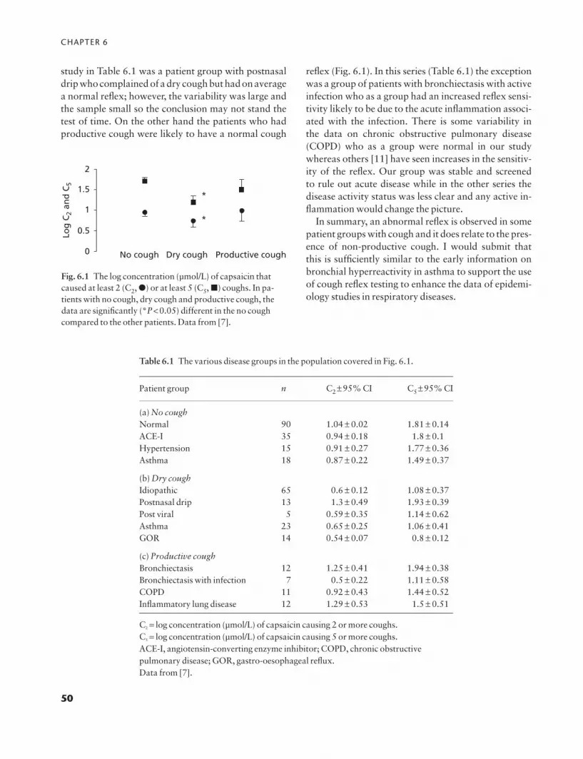
CHAPTER 6
50
study in Table 6.1 was a patient group with postnasaldrip who complained of a dry cough but had on averagea normal reflex; however, the variability was large andthe sample small so the conclusion may not stand thetest of time. On the other hand the patients who hadproductive cough were likely to have a normal cough
reflex (Fig. 6.1). In this series (Table 6.1) the exceptionwas a group of patients with bronchiectasis with activeinfection who as a group had an increased reflex sensi-tivity likely to be due to the acute inflammation associ-ated with the infection. There is some variability in the data on chronic obstructive pulmonary disease(COPD) who as a group were normal in our studywhereas others [11] have seen increases in the sensitiv-ity of the reflex. Our group was stable and screened to rule out acute disease while in the other series the disease activity status was less clear and any active in-flammation would change the picture.
In summary, an abnormal reflex is observed in somepatient groups with cough and it does relate to the pres-ence of non-productive cough. I would submit that this is sufficiently similar to the early information onbronchial hyperreactivity in asthma to support the useof cough reflex testing to enhance the data of epidemi-ology studies in respiratory diseases.
0 No cough Dry cough Productive cough
Log
C2
and
C5
0.5
1.5
1
2
*
*
Fig. 6.1 The log concentration (μmol/L) of capsaicin thatcaused at least 2 (C2, �) or at least 5 (C5, �) coughs. In pa-tients with no cough, dry cough and productive cough, thedata are significantly (*P<0.05) different in the no coughcompared to the other patients. Data from [7].
Table 6.1 The various disease groups in the population covered in Fig. 6.1.
Patient group n C2 ±95% CI C5 ±95% CI
(a) No coughNormal 90 1.04 ± 0.02 1.81 ± 0.14ACE-I 35 0.94 ± 0.18 1.8 ± 0.1Hypertension 15 0.91 ± 0.27 1.77 ± 0.36Asthma 18 0.87 ± 0.22 1.49 ± 0.37
(b) Dry coughIdiopathic 65 0.6 ± 0.12 1.08 ± 0.37Postnasal drip 13 1.3 ± 0.49 1.93 ± 0.39Post viral 5 0.59 ± 0.35 1.14 ± 0.62Asthma 23 0.65 ± 0.25 1.06 ± 0.41GOR 14 0.54 ± 0.07 0.8 ± 0.12
(c) Productive coughBronchiectasis 12 1.25 ± 0.41 1.94 ± 0.38Bronchiectasis with infection 7 0.5 ± 0.22 1.11 ± 0.58COPD 11 0.92 ± 0.43 1.44 ± 0.52Inflammatory lung disease 12 1.29 ± 0.53 1.5 ± 0.51
C2 = log concentration (μmol/L) of capsaicin causing 2 or more coughs.C5 = log concentration (μmol/L) of capsaicin causing 5 or more coughs.ACE-I, angiotensin-converting enzyme inhibitor; COPD, chronic obstructivepulmonary disease; GOR, gastro-oesophageal reflux.Data from [7].

COUGH SENSITIVITY
51
Clinical management
Cough provocation testing has a potential role in threeareas of clinical management: first, as an aid in diagno-sis; second, as a guide to prognosis; and finally, as a toolfor monitoring the course of treatment.
Diagnosis
The symptom of cough as noted in the previous sectioncan have several attributes attached to it, i.e. dry (nosputum), productive (sputum), temporal (night, etc.) orprovoking (cold air, etc.) events. Unfortunately, none ofthese attributes narrow down the cause of the cough.Standard history-taking and routine investigationsmay help identify the cause, which can then be investi-gated by more specific tests and eventually therapeutictrials. A cough-specific test, which could pinpoint thediagnosis, would therefore be of great clinical value.Does cough provocation testing meet this need? Unfor-tunately, as the data in Table 6.1 and Fig. 6.1 show,provocation testing can differentiate between produc-tive and dry coughing but not for various causes withinthose subsets. It can therefore be used only to confirmthat aspect of the history which may have some limitedvalue. In addition, it can be used to rule out psychogenicor fictitious cough if the test is performed blind to the subject and yet is still abnormal. These findings ap-pear to be true of all the provocation tests studied suffi-ciently to date. It is unlikely that provocation testingwill be used as a routine in clinical diagnosis but will bereserved as a research tool.
Prognosis
Like diagnosis, the prognosis of a patient with coughonce chronic cannot be easily judged until the successor failure of a therapeutic trial is known. This is not to say that it is not a safe bet to reassure the sufferer ofan acute cough during a respiratory infection that theirsuffering will be relatively short-lived! Theoreticallythe relative abnormality of a cough provocation testcould inform on prognosis. However, there is insuffi-cient information in the studies such as those in Fig. 6.2to know whether this is a possibility. This question isclearly an area that would benefit from systematic evaluation.
Monitoring
The monitoring of the clinical course of cough and theresponse to therapy is a more fruitful area than coughprovocation tests. The data exemplified in Figs 6.2 and6.3 show that an abnormal cough provocation test, ifidentified, will be present while the disease is active andresolve with the disease. The use of provocation testingcan clearly help in this aspect of management especially
0Pre IIIness
*
*
Recovery
0.5C2
and
C5
(mm
ol/L
)
1.5
2.5
1
2
Fig. 6.2 The log concentration (μmol/L) of capsaicin thatcaused at least 2 (C2, �) or at least 5 (C5, �) coughs. In 31 pa-tients before (Pre), during (Illness) and after (Recovery) upperrespiratory infection, the data are significantly different (*P<0.05) in the illness compared to the ‘Pre’ and ‘Recovery’data. Data from [15].
0 Successpre
Successpost
C2
and
C5
(mm
ol/L
)
Failurepre
Failurepost
0.20.4
0.60.8
11.2
1.41.6
1.82
*
*
Fig. 6.3 The log concentration (μmol/L) of capsaicin thatcaused at least 2 (C2, �) or at least 5 (C5, �) coughs. In pa-tients before (pre) and after (post) successful (success n=48)or unsuccessful (failure n=39) treatment of the underlyingcause for their cough, the data for the successfully treated pa-tients are significantly (*P<0.05) different from the ‘pre’data. Data from [42].

CHAPTER 6
52
in a patient with no clear diagnosis that may require anumber of therapeutic interventions before the diagno-sis is finally made.
A clear case for use of cough provocation comes inthe management of neurological diseases where respi-ratory infection is a common sequela [12]. The loss of afunctioning cough reflex is thought to be behind themechanism for these respiratory infections in patientswith a loss of neurological functions.
Efferent testing of cough reflex by asking the patientto cough will usually inform on the capacity of thatlimb of the reflex. However, it does not inform on thefunction of the sensory input. Cough provocation willso inform, as a negative provocation test in a patientwho can cough voluntarily identifies the perturbationof the sensory limb of the reflex. Data to support suchan abnormality are limited to date [13]; however, whatdata there are support the hypothesis and in view of itsimportance to the mobility of the patient it does requirecomprehensive assessment.
Cough provocation in clinical research
Pathophysiology
Cough provocation is able to provide an objectivemeasure of the sensitivity of the cough reflex so that itsfunction can be assessed in respiratory disease. It istherefore possible to use it to understand the patho-physiology of cough. Cough is a response either to theneed to clear the airway of unwanted material or to analteration in the sensitivity of the cough reflex as in drycough. It is therefore of value to understand what maychange the sensitivity of the reflex. Cross-sectionalstudies have shown that in some diseases the reflex canbe normal or abnormal, with a suggestion that this is related to the level of activity of the diseased state.Prospective studies have confirmed that in angiotensin-converting enzyme inhibitor-associated cough [14] andcommon cold-associated cough [15], patients withcough start with a normal reflex which becomes exag-gerated with the cough and returns to normal with theresolution of the condition.
What causes this change in the response of the reflexis not fully elucidated. Airway biopsy in a small series ofdry cough patients [16] with an abnormal reflex hasshown an increase in airway nerve and neuropeptideassociated with those nerves. However, prospective
studies have yet to be performed. Additional data has been obtained that inhaled PGE2 [17] but notbradykinin or substance P can invoke a temporarychange in the sensitivity of the reflex. This would sug-gest that hyperalgesic mediators could increase the sen-sory limb of the reflex, probably not by altering thenumber of fibres in view of the rapidity of the response.The unravelling of the mechanisms behind the changein sensitivity of the cough reflex would give a new bas-ket of targets for developing antitussive drugs.
Pharmacology
Most of the investigations of the pharmacology of thecough reflex have been made using cough provocationeither by citric acid, low Cl- solution or capsaicin innormal volunteers. In addition there has been some lim-ited investigation of the pharmacology of the exagger-ated reflex in patients with dry cough.
Table 6.2 summarizes the results of studies usingcough challenge in normal volunteers. This list maywell not contain all data, as it is likely that a number ofnegative investigations are not in the public domain.There is consistent and predictable evidence that localanaesthesia [18] and centrally acting opiates [19] re-duce the sensitivity of the reflex, however tested. Otherresults are either conflicting, e.g. bronchodilatationand inhaled diuretics which have shown positive resultsin some, but by no means all studies, or are based on theunconfirmed results of one study. If the results are con-firmed, then humans [20] like cats [21] may have a 5-HT-dependent cough-suppressing mechanism, whichcould be exploited for antitussive therapies. Other thanthat there is little of encouragement from the extensivestudies to date.
Equally disappointing is the data in patients with dry cough. A 4-week study with nedocromil sodiumwas negative [22] and despite anecdotal evidence ofnon-steroidal anti-inflammatory drugs working in angiotensin-converting enzyme inhibitor cough, con-trolled trials failed to show an effect on dry cough [23]overall.
Conclusion
Cough challenge is a safe and easy method for assessingthe sensitivity of the cough reflex. However, there arenot yet any standards agreed for the methodology,

COUGH SENSITIVITY
53
which tend to be laboratory specific making compar-isons between studies difficult. As well as data compat-ibility issues, the different methodologies occasionallylead to results that need explaining, such as differencesbetween males and females, adults and children and inCOPD. These differences could be true physiologicalvariability or artefacts of methodology and this needsto be resolved through a consensus on methodology.
Despite the methodological issues there is sufficientevidence that the provocation tests can detect differ-ences in the sensitivity of the reflex in disease. These dif-ferences make the cough challenge of likely value inepidemiology studies, some aspects of clinical manage-ment and in particular in hypothesis testing. In hypoth-esis testing it is difficult to imagine a strategy to unravelthe pathophysiology and pharmacology of the reflexwithout using cough challenge even if the results re-quire validation in large studies using more clinical end-points.
References
1 Fuller RW, Jackson DM. Physiology and treatment ofcough. Thorax 1990; 45: 425–30.
2 Prime FJ. The assessment of antitussive drugs in man. BrMed J 1961; April 22: 1149–51.
3 Tiffeneau R. The acetylcholine cough test. Dis Chest1957; 31: 404–22.
4 Janson C, Chinn S, Jarvis D, Burney P. Determinants ofcough in young adults participating in the European Com-munity Respiratory Health Survey. Eur Respir J 2001; 18:647–54.
5 Lowry R, Wood A, Johnson T, Higenbottam T. Antitussiveproperties of inhaled bronchodilators on induced cough.Chest 1988; 93/6: 1186–9.
6 Pounsford JC, Birch MJ, Saunders KB. Effect of bron-chodilators on the cough response to inhaled citric acid in normal and asthmatic subjects. Thorax 1985; 40:662–7.
7 Choudry NB, Fuller RW. Sensitivity of the cough reflex inpatients with chronic cough. Eur Respir J 1992; 5:296–300.
8 Chang AB, Phelan PD, Roberts RGD, Robertson CF. Cap-saicin cough receptor sensitivity test in children. EurRespir J 1996; 9: 2220–3.
9 Stone RA. Investigations into the neural control of thecough reflex. PhD thesis, Department of Thoracic Medi-cine, Royal Brompton National Heart and Lung Institute,University of London, December 1992.
10 Dicpinigaitis PV, Rauf K. The influence of gender on coughreflex sensitivity. Chest 1998; 113: 1319–21.
Table 6.2 Results of pharmacological studies on cough provocation tests.
Pharmacological tool (Reference) Capsaicin Low Cl- Citric acid Other
Local anaesthesia [18,24] Ø Ø ØOpiates [19,25] Ø Ø Øb2-Agonists [5,6,26,27] 0 Ø ± ØAntimuscarinics [6,26,41] 0 ØAntihistamines [28] 05-HT3 antagonists [29] 05-HT1 agonists [20] 0NSAID [30,31] 0Sodium cromoglycate [32] 0Nedocromil sodium [33] 0Baclofen [34] Øa2-Agonists [35] 0Diuretics [36] 0MAO-I [37] 0Demulcents [38,39] Ø ØAntileukotriene [40] 0
MAO-I, monoamine oxidase inhibitor; NSAID, non-steroidal anti-inflammatorydrug.Ø, Reduced; ±, equivocal or conflicting data; 0, no effect.

CHAPTER 6
54
11 Doherty MJ, Mister R, Pearson MG, Calverley PMA.Capsaicin responsiveness and cough in asthma and chronic obstructive pulmonary disease. Thorax 2000; 55:643–9.
12 Smith Hammond CA, Goldstein LB, Zajac DJ, Gray L,Davenport PW, Bolser DC. Assessment of aspiration riskin stroke patients with quantification of voluntary cough.Neurology 2001; 56: 502–6.
13 Addington WR, Stephens RE, Gilliland KA. Assessing thelaryngeal cough reflex and the risk of developing pneumo-nia after stroke —an interhospital comparison. Stroke1999; 30: 1203–7.
14 McEwan JR, Choudry N, Street R, Fuller RW. Change incough reflex after treatment with enalapril and ramipril.Br Med J 1989; 299: 13–6.
15 O’Connell F, Thomas VE, Studham JM, Pride NB, Fuller RW. Capsaicin cough sensitivity increases duringupper respiratory infection. Respir Med 1996; 90: 279–86.
16 O’Connell F, Springall DR, Moradoghli-Haftvani A,Krausz T, Price D, Fuller RW, Polak JM, Pride NB. Abnor-mal intraephithelial airway nerves in persistent unex-plained cough? Am J Respir Crit Care Med 1995; 152:2068–75.
17 Choudry NB, Fuller RW, Pride NB. Sensitivity of thehuman cough reflex: effect of inflammatory mediatorsprostaglandin E2, bradykinin and histamine. Am Rev RepDis 1989; 140: 137–41.
18 Choudry NB, Fuller RW, Anderson N, Karlsson J-A. Sepa-ration of cough and reflex bronchoconstriction by inhaledlocal anaesthetics. Eur Respir J 1990; 3: 579–83.
19 Fuller RW, Karlsson J-A, Choudry NB, Pride NB. Effect of inhaled and systemic opiates on responses to inhaledcapsaicin in humans. Am Physiol Soc 1988; 88: 1125–30.
20 Stone RA, Worsdell YM, Fuller RW, Barnes PJ. Effect of 5-hydroxytryptamine and 5-hydroxytryptophan on sensi-tivity of the human cough reflex. J Appl Physiol 1993; 74:396–401.
21 Karlsson J-A, Fuller RW. Pharmacological regulation ofthe cough reflex —from experimental models to antitus-sive effects in man. Pulm Pharmacol Ther 1999; 12:215–28.
22 Choudry NB. Investigation of the sensitivity of the coughreflex in humans. MD thesis, Faculty of Medicine, Univer-sity of London, December 1990.
23 McEwan JR, Choudry NB, Fuller RW. The effect of sulin-dac on the abnormal cough reflex associated with drycough. J Pharmacol Exp Ther 1990; 255: 161–4.
24 Addington WR, Stephens RE, Goulding RE. Anesthesia ofthe superior laryngeal nerves and tartaric acid-inducedcough. Arch Phys Med Rehabil 1999; 80: 1584–6.
25 Empey DW, Laitinen LA, Young GA, Bye CE, HughesDTD. Comparison of the antitussive effects of codeine
phosphate 20mg, dextromethorphan 30mg and noscap-ine 30mg using citric acid-induced cough in normal sub-jects. Eur J Clin Pharmacol 1979; 16: 393–7.
26 Choudry NB, Fuller RW. Effect of airway calibre on thesensitivity of the human cough reflex. Thorax 1990; 45:311P–12P.
27 Katsumata U, Sekizawa K, Inoue H, Sasaki H, TakishimaT. Inhibitory actions of procaterol, a beta-2 stimulant, onsubstance P-induced cough in normal subjects duringupper respiratory tract infection. Tohoku J Exp Med1989; 158: 105–6.
28 Studham J, Fuller RW. The effect of oral terfenadine on thesensitivity of the cough reflex in normal volunteers. PulmPharmacol, 1992; 5 (1): 51–2.
29 Choudry NB, McEwan JR, Lavender EA, Williams AJ,Fuller RW. Human responses to inhaled capsaicin are notinhibited by granisetron. Br J Clin Pharmacol 1991; 31(3): 337–9.
30 Stone RA, Barnes PJ, Fuller RW. The low-chloride coughresponse is not inhibited by a single, high dose of aspirin.Br J Clin Pharmacol 1992; 34: 370–2.
31 Dicpinigaitis PV. Effect of the cyclooxygenase-2 inhibitorcelecoxib on bronchial responsiveness and cough reflexsensitivity in asthmatics. Pulm Pharmacol Ther 2001; 14:93–7.
32 Collier JG, Fuller RW. The effect of inhaled capsaicin inman and the action of SCG on this response. Br J Pharma-col 1984; 81: 113–7.
33 Hansson L, Choudry NB, Fuller RW, Pride NB. Effect ofnedocromil sodium on the airway response to inhaled cap-saicin in normal subjects. Thorax 1988; 43: 935–6.
34 Dicpinigaitis PV, Dobkin JB. Antitussive effect of theGABA-agonist baclofen. Chest 1997; 111: 996–9.
35 O’Connell F, Thomas VE, Fuller RW, Pride NB, KarlssonJ-A. Effect of clonidine on induced cough and bron-choconstriction in guinea pigs and humans. J Appl Physiol1994; 76: 1082–7.
36 Karlsson JA, Choudry NB, Zackrisson C, Fuller RW. Acomparison of the effect of inhaled diuretics on airway reflexes in humans and guinea pigs. J Appl Physiol 1992;1: 72 (2), 434–8.
37 Choudry NB, Studham J, Harland D, Fuller RW. Modula-tion of capsaicin induced airway reflexes in humans: effectof monoamine oxidase inhibition. Br J Clin Pharmacol1993; 35: 184–7.
38 Packman EW, London SJ. The utility of artificially inducedcough as a clinical model for evaluating the antitussive effects of aromatics delivered by inunction. Eur J RespirDis 1980; 110 (Suppl.): 101–9.
39 Fuller RW, Haase G, Choudry NB. The effect of dex-tromethophan cough syrup on capsaicin-induced cough in normal volunteers. Am Rev Respir Dis 1989; 139:A11.

COUGH SENSITIVITY
55
40 Dicpinigaitis PV, Dobkin JB. Effect of zafirlukast on coughreflex sensitivity in asthmatics. J Asthma 1999; 36 (3):265–70.
41 Fuller RW. Pharmacology of inhaled capsaicin in humans.Respir Med 1991; 85 (Suppl. A): 31–4.
42 O’Connell F, Thomas VE, Pride NB, Fuller RW. Capsaicincough sensitivity decreases with successful treatment ofchronic cough. Am J Respir Crit Care Med 1994; 150:374–80.


57
Introduction
The prevalence of childhood cough without wheeze is high, 12.8–15.5% [1] in community studies based on parent-completed questionnaires, and that of nocturnal cough varies from 4.9 to 28.1% [2,3]. Thishigh prevalence raises the question as to when child-hood cough should be considered ‘normal’ or patho-logical. In childhood, cough may be ‘normal’ [4] or asymptom of any respiratory illness and rarely of a non-respiratory illness. The management of childhoodcough [4,5] differs from that of adults [6]; and pae-diatricians worldwide have long been passionate about managing childhood illness differently from that of adults, and about the dangers of extrapolatingadult data to young children. The pattern of respi-ratory illness in children can be clearly different fromthat in adults. For example, viruses associated with the common cold in adults can cause life-threateningillness in children, such as bronchiolitis and croup; thenatural history of asthma in children is dominated by decreasing severity with age and, in some, completeresolution [7] whereas asthma acquired in adulthoodusually persists. The aetiology and management ofchildhood cough should also be clearly distinguishedfrom adult cough. Extrapolation of adult cough literature to children can be harmful, for example thesuggestion of fundoplication for gastro-oesophagealreflux as the sole symptom of cough without evidenceof secondary aspiration [8] is inappropriate for chil-dren. Indeed current evidence suggests that it is erro-neous to extrapolate the three commonest causes ofcough in adults (cough-variant asthma, postnasal drip,gastro-oesophageal reflux) to children [9,10]. This
chapter discusses the issues of cough unique to children.
Causes of cough in children
The aetiology of cough can be broadly divided intogroups of primary pathophysiology, although there isundoubtedly an overlap in the pathophysiology ofsome diseases (Table 7.1). The first clinical challengethat faces the physician is deciphering whether thecough is ‘normal’ or ‘expected’, non-specific or specific.Non-specific overlaps with both ‘normal/expected’ andspecific cough, but ‘expected’ cough is distinctly sepa-rate from specific cough (Fig. 7.1). The relative fre-quency of each category will depend on the setting, andgeneral practitioners would more likely encounter ‘ex-pected/normal’ cough whereas in a tertiary setting spe-cific cough would dominate. These descriptions and thecommon and/or controversial aetiologies of childhoodcough are briefly discussed below. A complete clinicalreview of each specific aetiology is beyond the scope ofthis chapter and can be found in standard paediatricrespiratory textbooks.
Cough categories
‘Normal’ or ‘expected’ cough
Diagnosing this category of cough requires the mostskill and experience [4]. All cough is arguably represen-tative of some process. However, as the cough reflex is subjected to cortical modulation [9] and can be
57
Causes, assessment andmeasurement of cough in childrenAnne B. Chang
7
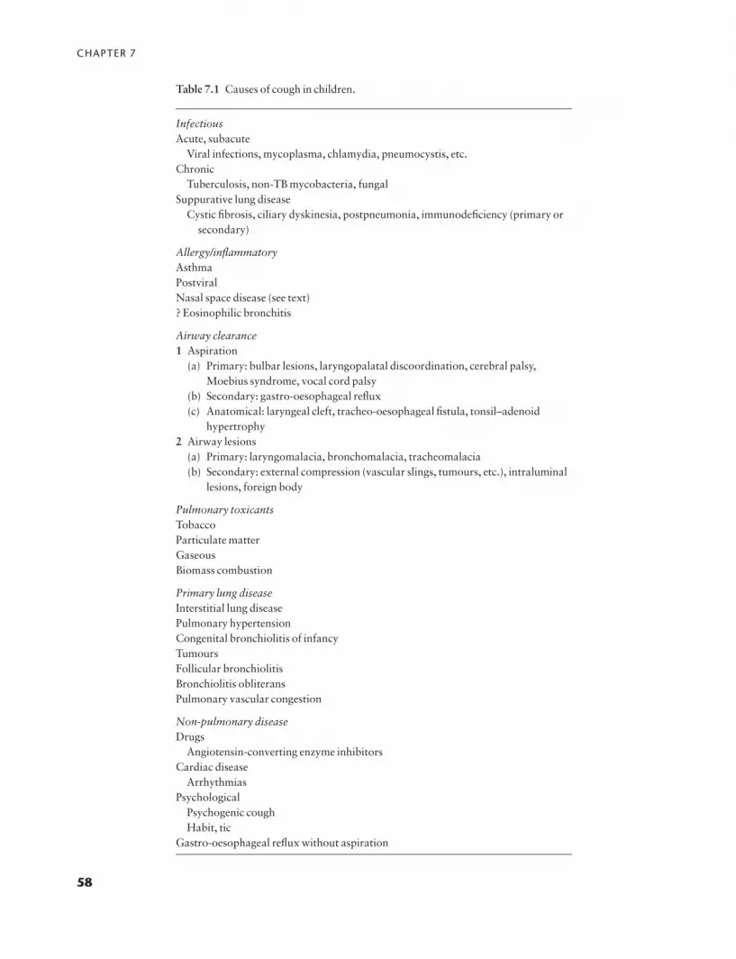
CHAPTER 7
58
Table 7.1 Causes of cough in children.
InfectiousAcute, subacute
Viral infections, mycoplasma, chlamydia, pneumocystis, etc.Chronic
Tuberculosis, non-TB mycobacteria, fungalSuppurative lung disease
Cystic fibrosis, ciliary dyskinesia, postpneumonia, immunodeficiency (primary orsecondary)
Allergy/inflammatoryAsthmaPostviralNasal space disease (see text)? Eosinophilic bronchitis
Airway clearance1 Aspiration
(a) Primary: bulbar lesions, laryngopalatal discoordination, cerebral palsy,Moebius syndrome, vocal cord palsy
(b) Secondary: gastro-oesophageal reflux(c) Anatomical: laryngeal cleft, tracheo-oesophageal fistula, tonsil–adenoid
hypertrophy2 Airway lesions
(a) Primary: laryngomalacia, bronchomalacia, tracheomalacia(b) Secondary: external compression (vascular slings, tumours, etc.), intraluminal
lesions, foreign body
Pulmonary toxicantsTobaccoParticulate matterGaseousBiomass combustion
Primary lung diseaseInterstitial lung diseasePulmonary hypertensionCongenital bronchiolitis of infancyTumoursFollicular bronchiolitisBronchiolitis obliteransPulmonary vascular congestion
Non-pulmonary diseaseDrugs
Angiotensin-converting enzyme inhibitorsCardiac disease
ArrhythmiasPsychological
Psychogenic coughHabit, tic
Gastro-oesophageal reflux without aspiration

COUGH IN CHILDREN
voluntarily initiated, it is not surprising that cough(1–34 per 24h) can be found in ‘normal’ children free ofa respiratory infection in the previous 4 weeks [11]. Inanother study, age-, sex- and season-matched ‘normalcontrols’ have 0–141 coughs per day (median 10) [12].In this study, children with recent viral infections werenot excluded but were considered well by their parents.Children can have 6–8 viral acute respiratory infections(ARIs) per year, especially when they are in daycarecentres, and arguably childhood cough within thisspectrum can be considered ‘normal’ or ‘expected’.Children with recurrent cough who present to a tertiarycentre do have a higher cough frequency than controls,and have a higher cough frequency during the day thanthe night [12].
Specific cough
Specific refers to cough to which a definable cause (or causes) can be attributed. In specific cough, pointersof respiratory impairment are usually present. The aeti-ology and necessity of further investigations is usuallyevident from the presence of these pointers (Table 7.2).
Non-specific cough
In non-specific cough, the cough is dry and pointers of
respiratory impairment are absent. The aetiology may be ill-defined and we suspect that the majority is related to postviral cough and/or increased cough re-ceptor sensitivity [13,14]. In non-specific cough, thereis no serious underlying condition and the child is otherwise well.
Brief overview of common causes and controversies
Cough and respiratory infections
The spectrum of respiratory infections causing cough varies from benign (e.g. common cold) to life-threatening infections (e.g. lung abscess, pneumocystispnuemonia). The manifestation of the infection de-pends on various pathogens, ‘host or patient’ factors(asthma, immunodeficiency, established cardiopul-monary impairment, malnutrition, genetics, etc.) andenvironmental factors (smoke, biomass combustionexposure).
ARIs and post-ARI-associated cough are the mostcommon causes of childhood cough. In Australia, thereported use of medications for coughs and colds in the2 weeks before assessment was 167 and 87 per 1000 forchildren under 5 years and children between 5 and 14years, respectively, corresponding to the most fre-quently and third most frequently used medications intheir respective age groups [15]. Approximately US $2billion per year is spent on cough and cold remedies in
59
‘Normal’ or‘expected’ cough
Non-specificcough
Specificcough
Fig. 7.1 Broad categories of childhood cough. The diagramdepicts the overlap between the categories of childhoodcough. Non-specific cough overlaps with both specific coughand ‘normal or expected cough’, whereas specific cough isdistinct from ‘normal or expected cough’.
Table 7.2 Specific cough pointers.
Presence of:HaemoptysisRecurrent pneumoniaExertional dyspnoeaChronic sputum productionWheezeStridorImmune deficiencyCardiac abnormalitySwallowing difficultiesDyspnoeaChest deformityClubbingAusculatory abnormalityPoor growth

CHAPTER 7
the US [16]. A national US survey reported that ap-proximately 35% of 8145 preschool-aged children hadused ‘over the counter’ medications for cough in thepast 30 days [16].
It is sometimes argued that ‘the common cold’, anupper respiratory infection, should not cause cough,and the presence of cough reflects the presence of alower respiratory tract infection. However, lower air-way inflammation has been demonstrated in childrenwith colds, not only in the active phase but also in thosewho were asymptomatic 1–14 days before a bron-choalveolar lavage was taken [17]. Also, clinically un-apparent alterations in lower airway function occurduring upper ARI in infants, children and adults [18].
There are few prospective epidemiological data onthe length of cough associated with a common cold inchildren. In one study, 80% of adults were cough freeby 2 weeks [19] and cough sensitivity has been shownto be increased during upper ARIs [20]. Transient en-hanced cough sensitivity has also been shown in chil-dren with influenza infection [21]. Like the clinicalmanifestation of ARIs, the length of cough most likelydepends on patient and environmental factors.
Cough and asthma in children
There is little doubt that children with asthma maypresent with cough and that wheeze may be absent during physical examination. Isolated cough has beenpostulated as a marker of asthma, and improper interpretation of cough as a symptom was previouslythought to be a key factor in the underdiagnosis of asthma. However, more recently increasing numbers ofchildren are misdiagnosed as having asthma on thebasis of cough alone [22–24], although there is also on-going concern that children with significant asthmasymptoms are still being missed and consequently undertreated [25]. The importance of cough in the diagnosis of asthma and the frequency of cough-variant asthma is debatable [23]. Current epidemiolog-ical, inflammatory and cohort studies suggest that mostcough in children is not asthma [23,26,27] and mostcough in children spontaneously resolves [26,28]. A review on this is available elsewhere [9].
Recent work on airway inflammatory markers incommunity children with isolated persistent coughshow that, unlike in the adult, most children with per-sistent isolated cough do not have asthma, since theirairway inflammatory markers are significantly differ-
ent from those in children with asthma [27,29]. Howmany children in the community with persistent coughdo indeed have asthma is unknown. There are only tworandomized controlled trials on the use of current asthma medications in children with isolated cough[30,31]. Both studies objectively measured cough,which is crucial, since cough as an outcome measure issubjective and unreliable [32]. Neither recommendedprolonged or high doses of asthma-type therapy whenmanaging these children [30,31].
Indeed, the misdiagnosis of isolated cough as asthmais not uncommon [23,24,33]. We recently showed thatthe overdiagnosis of asthma and the overuse of asthmatreatments with significant side-effects, in children withpersistent cough referred to a respiratory clinic, is com-mon [24]. While a trial of asthma-type medicationsmay be considered, the child must be reviewed and, ifthe cough does not respond to the asthma therapy, thediagnosis must be withdrawn and the medicationsstopped [4,5]. Failure to do so will lead to escalatingdoses being used with significant side-effects [24].There is however, difficulty in defining what constitutesa response to treatment, particularly when the naturalresolution of the underlying condition such as viral res-piratory infection may mimic a response to treatment.Recurrence of cough on cessation and elimination ofcough upon commencement of inhaled corticosteroidsis sometimes necessary for confident diagnosis [4]. Theoriginal studies describing cough as a manifestation ofasthma showed that the cough responded within aweek to medications used for asthma during that era(theophylline and hypnotics) [34,35]. It could be argued that the currently available more potent anti-inflammatory medications for asthma would reducethe symptoms as efficaciously, if not more so. However,following an acute exacerbation of asthma, cough waspresent in 23% of children [36]. In this group of chil-dren whether the cough is related to concurrent viralrespiratory infections, the cause of asthma exacer-bations in 80% of children [37], or a marker of non-resolution of asthma is unknown. Markers of coughseverity are known to correlate poorly with clinical, in-flammatory and spirometry markers of asthma severity[38]. The relationship between cough and asthma iscomplex [39,40] and beyond the scope of this chapter.
The existence of cough-dominant asthma, wherecough is a dominant feature of asthma exacerbations inpatients with classical asthma, is recognized in both thepaediatric [41] and the adult literature [42]. However,
60

COUGH IN CHILDREN
whether cough-dominant asthma represents a truly dif-ferent phenotype is not yet confirmed at the cellular orgenetic level. Increasingly asthma phenotypes are rec-ognized and ‘treatment response genes’ to b2-receptoragonists have recently been described. However, for cli-nicians, the aetiology of isolated cough in children withasthma can be difficult to determine, when objectivemarkers such as spirometry are unavailable. Cough isincluded in asthma severity scales [43], and in childrenwithout wheeze the presence of this isolated symptomcan categorize children into the moderately severegroup. Isolated cough in children with asthma can beassociated with any other respiratory illness, or withenvironmental exposure. Several studies on air pollu-tion (environmental tobacco smoke, bioaerosols) haveshown that air pollution is more likely to affect childrenwith asthma than those without, and cough is the major symptom manifestation [44]. Many studies haveshown a poor relationship between cough and othermarkers of asthma (day and night oxygen saturations(SpO2), lung volumes, airway resistance (Raw), peak ex-piratory flow (PEF) and variability, spirometry) in chil-dren with asthma during a non-acute phase as well inthe recovery phase of an acute exacerbation, as re-cently summarized [40].
Does ‘allergic or atopic cough’ exist in children?
This poorly defined condition is probably an overlapwith asthma and eosinophilic bronchitis, a conditionwell recognized in adults but not well defined in children. The association between atopy and respi-ratory symptoms has been the subject of many epi-demiological studies [45,46]. Some have describedgreater respiratory symptom chronicity [1] but othershave not [45,46]. In the seminal prospective study of in-fants followed up to 11 years old, the Tuscon groupshowed that recurrent cough present early in life re-solved in the majority of children [26]. These childrenwith recurrent cough and without wheeze did not haveairway hyperresponsiveness (AHR) or atopy and sig-nificantly differed from those with classical asthmawith or without cough [26]. These important differ-ences were: maternal allergy, wheezing, airway respira-tory infection, high IgE, atopy, reduced maximal flow(V·max) at functional residual capacity (FRC) (signifi-cant for asthma) and exposure to tobacco smoke (sig-nificant for recurrent cough). In laboratory and clinicalstudies, others have studied the association between
atopy, AHR, asthma and cough [14,30,47]. In thesestudies, AHR is an important confounder associatedwith atopy and is independent of other respiratorysymptoms [48,49]. Studies where confounding vari-ables were not controlled, and where standard criteriafor AHR were not observed, can be misleading in theirconclusions [47].
Cough and gastro-oesophageal reflux (GOR) in children
Aspiration lung disease can result from laryngopalataldiscoordination or discoordinated swallowing (pri-mary aspiration), or severe GOR (secondary aspira-tion). Children with primary aspiration may presentwith chronic cough but usually in the context of severedevelopmental or neurological disturbance. The inves-tigatory evidence for aspiration lung disease can be dif-ficult but it is important to exclude this diagnosis in thecorrect setting as recurrent aspiration may lead tochronic respiratory illnesses such as bronchiectasis andbronchiolitis obliterans. Oesophageal disorders canundoubtedly trigger cough in children which may causecough by at least three mechanisms: aspiration of gas-tric contents, acid reflux and volume reflux. However,while GOR can cause cough, cough can also causeGOR and causative links are hard to identify [50]. Therelationship between the two is probably complex. Theview that GOR is a frequent cause of cough in adultshas been challenged [51,52]. Like the difference be-tween the paediatric and adult literature for asthmaand cough, there is indeed little evidence that GORwithout aspiration is a specific or frequent cause ofcough in children. As cough is very common in childrenand respiratory symptoms may exacerbate GOR, it isdifficult to delineate cause and effect. Infants regularlyregurgitate [53], yet few, if any, well infants cough withthese episodes.
Cough and nasal space disease
In adults, sinusitis/postnasal drip is a common cause of cough. In children, there is little supportive evidencefor this condition [10]. There are no cough receptors inthe pharynx or postnasal space [42]. Although sinusitisis common in childhood, it is not associated with asthma or cough once allergic rhinitis, a common asso-ciation, is treated [54]. The relationship between nasalsecretions and cough is more likely linked by common
61

CHAPTER 7
aetiology (infection and/or inflammation causing both)or due to throat clearing of secretions reaching the larynx. In a prospective study, 50% of 137 childrenaged under 13 years had CT sinus scans consistent withsinusitis but all were asymptomatic [55]. Abnormalsinus radiographs may be found in 18–82% of asymp-tomatic children [56]. Using a continuous infusion of2.5mL/min of distilled water into the pharynx ofasymptomatic adults, Nishino et al. demonstrated thatlaryngeal irritation and cough only occurred in the pres-ence of hypercapnia (45–55mmHg) [57], suggestingthat pharyngeal secretion itself does not cause cough.
Cough and chronic suppurative lung disease
This is an essential diagnosis that should never bemissed. Children with bronchiectasis have a chronicmoist or productive cough and are typically but not al-ways finger-clubbed. The cough is characteristicallyworse in the mornings. Physical findings are non-specific; clubbing, pectus carinatum, and coarse crepitations and localized wheeze may or may not bepresent. Plain chest radiography will suggest features insevere disease (dilated and thickened bronchi may appear as ‘tramtracks’) but is insensitive in mild dis-ease. Confirmation is by high-resolution computed to-mography (CT) scan of the chest (routine CT scanprovides insufficient detail). A child with suspectedbronchiectasis should be referred for investigation of a specific cause and specific treatment instituted whenindicated (e.g. cystic fibrosis, primary ciliary dyskine-sia, immunodeficiency).
Cough and airway lesions in children
Cough can be due to intrinsic (tracheomalacia andbronchomalacia) [58,59] and extrinsic (foreign bodiesand other endobronchial obstruction) airway lesions.An inhaled foreign body usually presents in the acutestage, with a history of acute cough following a chokingevent, but may be found many years later [24]. Whenthe foreign body causes a ball-valve effect, air trappingseen in an expiratory film when compared with an in-spiratory chest radiograph (CXR) may be useful. When suspected, bronchoscopy allows for the definitive diagnostic and concomitant therapeutic procedure.Airway malacia disorders (tracheomalacia, bron-chomalacia) are well recognized causes of persistentand/or recurrent cough in children [24,58,59], but not
in adults [60]. In one study, 75% of children with tra-cheomalacia secondary to congenital vascular anomalyhad persistent cough at presentation [58], whereas of24 adults with tracheomalacia secondary to vascularrings 16 were symptomatic and none complained ofcough [60]. The pathophysiology of cough in the pres-ence of airway lesions is poorly understood but is poss-ibly related to a localized bronchitic process.
Environmental causes
In utero tobacco smoke exposure alters pulmonary de-velopment and physiology [61]. How this influencesthe developmental aspects of the central and peripheralcough pathways as well as the plasticity of the coughpathway is unknown. Exposure to environmentalsmoke and other ambient pollutants (particulate mat-ter [62], nitrogen dioxide, etc.) is associated with in-creased cough in children, especially in the presence ofother respiratory illness such as asthma [62]. Irrespec-tive of the primary aetiology of cough, exposure to en-vironmental smoke can exacerbate the frequency andseverity of cough.
Other causes of cough
Other than habitual cough, non-respiratory causes ofcough are rare, but should be considered when appro-priate clinical features are present.
Habitual and psychogenic cough
Habitual cough or cough as a ‘vocal tic’ may be tran-sient or chronic. While psychogenic cough is more com-mon in adolescents, the habitual cough occurs inyounger children. The mean age of diagnosis for habit-ual cough ranges from 4 to 15 years [63]. The typicalpsychogenic cough (honking cough) is recognizableand can often be heard even before the child is seen. Irrespective of the cause of cough, psychological influ-ences on severity of cough have been documented inboth children [64] and adults.
Assessment in children
Cough is a very common symptom of respiratory dis-ease. As cough is audible and can interfere with sleepand may represent serious underlying disorders such as
62

COUGH IN CHILDREN
cystic fibrosis, it is not surprising that parents are oftenanxious about their children’s cough and often seekmedical advice and remedy. Parental concerns may dif-fer significantly from physicians’ concerns. Physiciansare usually concerned about the aetiology of cough andgetting the correct diagnosis. Parental concerns, how-ever, often relate to their perceived effects of cough ontheir child (sleep, choking, permanent chest damage)[65].
Clinical assessment
When presented with a child with a cough, the keyquestions are aimed at clearly defining the nature andimpact of the cough on the child and family:1 Is it a symptom of an underlying problem?2 Are there possible modifiers of exacerbation and/orcontributing factors?3 How does the cough affect the child and parents?4 Is it necessary to investigate?5 Are any treatment modalities available or necessary?
The clinical assessment of cough can be approachedfrom many ways, e.g. dry vs. moist, age of the child andlength of cough. Each obviously influences the othersand none can be considered in isolation. A suggestedpathway primarily based on the length of cough is pre-sented (Fig. 7.2) [5]. The aetiology of cough that back-dates to infancy is clearly different to that of recentonset. A history of chronicity or exertional dyspnoeamay not be offered unless specifically asked for. Coughassociated with pointers of respiratory impairment(Table 7.2) are immediately differentiated (pathway IIIin Fig. 7.2c) irrespective of the historical length ofcough. These pathways are only a guide and based onclinical experience and available scientific literature. Inpaediatrics there are indeed insufficient data to providea comprehensive evidence-based approach.
In a child with persistent cough, the initial clinicalchallenge is deciphering whether the persistent cough isspecific or non-specific. The definition of persistent orchronic cough varies (>3–6 weeks) and the aetiologicalfactors in children vary from the benign to life-threatening causes. Specific pointers should be soughtfrom a thorough history and examination (Table 7.2).In specific cough, the aetiology and necessity of furtherinvestigations is usually evident from the presence ofcoexisting symptoms and signs. The presence of any ofthese pointers suggests that the cough is likely to repre-
sent an underlying disorder and that further complexinvestigations are probably indicated, other than sym-metrical polyphonic wheeze representative of asthma.The type of investigations depends on the clinical find-ings. Diagnoses that need to be considered includebronchiectasis, retained foreign body, aspiration lungdisease, atypical respiratory infections, cardiac anom-alies and interstitial lung disease, amongst other diseases. The diagnosis and management of cough associated with an underlying disorder is most appro-priately undertaken by paediatric respiratory physi-cians and will not be discussed further in this chapter.
In deciding the aetiology of the child’s cough, Bush[4] suggests that the child’s cough can be placed in oneof the five categories:1 Normal.2 A serious illness such as cystic fibrosis (rare but essential to get right).3 An unserious but treatable cause (e.g. GOR).4 A child with an asthma syndrome.5 Overestimation of symptoms for psychological orother reasons by either or both child or family.
An additional category to the above would be:6 A non-respiratory cause such as habitual cough,medications (ACE inhibitors), etc.
Exposure to tobacco smoke is the most importantmodifier of childhood cough and is known to increasesusceptibility to respiratory infections [66], cause ad-verse respiratory health outcomes and increase cough-ing illnesses [67]. When appropriate, a non-judgmentaldiscussion of how tobacco smoke affects childrenforms part of any clinical management of a child with acough.
Effect on parents and childIn childhood illnesses, parental expectations oftendrive presentations to medical personnel [68]. In adultswith asthma, asthma severity determined emergencypresentations whereas in paediatrics, parental anxietywas found to be the most important factor [69]. In astudy on upper respiratory tract infection, the main reason for repeated consultations for the same epi-sode occurred when parental expectations about the natural history of the illness were not fulfilled [68].When adopting a ‘wait and see’ approach in a childwith non-specific cough, discussing parental expecta-tions, concerns and anxiety often proves useful, diag-nostically and therapeutically.
63

CH
AP
TE
R 7
64
Look for symptoms of
Acute(<2 weeks)
Subacute2–4 weeks
Chronic or persistent(>4 weeks)
See pathway II See pathway IIIIs it the first presentation of a chronic illness?
Presence of 'respiratory pointers'Poor growth, chest deformity, clubbing, haemoptysis,recurrent pneumonia, dyspnoea, sputum production,exertional dyspnoea, ausculatory abnormality, cardiacabnormality, immune deficiency, swallowing difficulties
No
YesIs the cough characteristic?
Fever, rhinorhoea, coryza,sneezing, generalized aches,pains, sore throat, tachypnoea
Acute upper and/orlower respiratory tractinfection entities (ARI)
History of aspiration followedby immediate choking,coughing, +/– wheeze,stridor, tachypnoea, cyanosis
Aspiration offoreign object
No
Yes
Classical recognizable cough
• Psychogenic—honking cough [94]• LTB—barking cough (watch for other causes, e.g. tracheomalacia)• Staccato—Chlamydia (in infants)• Paroxysmal—pertussis and para pertussis
Wheeze, dyspnoea,exertional symptoms,hyperinflation, previousresponse to beta-2agonist +/– atopy
Asthma
Acute exposure,e.g. smoke,volatilecompounds
Hypoxia, chestpain, risk factors present
(a)
Inhalation injuryEmbolismhaemorrhage(rare)
Pathway I: Coughing child and length of cough

CO
UG
H IN
CH
ILDR
EN
65
Pathway II: Subacute cough not associated with other symptoms
CXRSpirometry (if >6 years old)
Both normal
Wait and see usually• post viral cough• acute bronchitis
Evaluate• smoke/other pollutants• child's activity• parental expectations• functional disorders
Either abnormal
See pathway III
(b)
Review in a week
Resolving
Resolved
Discharge
Further symptoms developor increased severity
Fig. 7.2 An approach to cough based on length of cough. These pathways are aguide to the approach to a child with a cough. However, most childhood coughsare benign and do not require drug therapy or investigations. The suggested ap-proach depends on the length of cough (pathway I) and whether any symptomsand/or signs are present (‘pointers’ listed in pathway I). Symptoms and signs canbe age dependent and thus the child’s age and severity of illness must also be considered when approaching a child with cough. It would not be possible
to factor the various possible combinations in these pathways. Note: ARI could coexist with another diagnosis. ARI, acute respiratory infection; CRS, cough receptor sensitivity; CXR, chest X-ray; FTT, failure to thrive; GOR, gastro-oesophageal reflux; HRCT, high-resolution computed tomography of the chest; LTB, laryngotracheobronchitis; TOF, tracheo-oesophageal fistula; TB, tuberculosis; UA, upper airway. Adapted with permis-sion from [5]. (Continued on p. 66.)

CH
AP
TE
R 7
66
CXRSpirometry (if >6 years old)
Both normal Either abnormal Consider early consultation with paediatricpulmonologist for assessment
Reversible airway obstruction?
Yes NoAsthmaAbsence of 'pointers', wheeze andproductive/wet cough?
Yes No
Non-specific cough 'Pointers'present?
No Yes
Consider:• post viral [20]• increased CRS [13]• asthma [40]• GOR• UA problems• functional disorders (habit cough, tics, psychogenic)
Purulent productive ormoist/wet cough andwithout wheeze
Yes No
Subacutebronchitis:needs follow-up;if recurrent orpersistent, needsinvestigation
Asthmabut reviewfor othercauses ofwheezing
Review and if 'pointers' present
If cough does notsettle consider
Assess risk factors for
Bronchiectasisor recurrentpneumonia
• cystic fibrosis• ciliary dyskinesia• previous severe pneumonia• immunodeficiency• structural airway lesions• congenital lung lesions• missed foreign body• TOF/H fistula
Sweat testBronchoscopyCilia biopsyImmune work-upHRCT chestBa swallow
Aspiration
• primary and secondary• neurologically abnormal• altered swallow• weak cough reflex• neuromuscular disease• laryngeal abnormalities• tonsil adenoid hypertrophy• TOF/H fistula• severe GOR
Ba swallowBronchoscopy and lavageVideo fluoroscopypH monitorLung milkScan/salivagram
Chronic orless commoninfections
• TB• non-tuberculous mycobacteria• mycoses
MantouxBronchoscopy and lavageHRCT chest
Interstitiallungdisease
• rheumatic diseases• cytotoxics• drugs• radiation• etc
Autoimmune markersHRCT chestLung biopsy
Cardiac
• pulmonary hypertension• cardiac oedema
Paediatriccardiologist
Echocardiaccatheter
(c) Pathway III: Chronic/persistent cough (>4 weeks) or acute/subacute cough associated with other symptoms
Fig. 7.2 Continued.

COUGH IN CHILDREN
Exploring the effects of the cough on the child in-cludes how the child deals with the cough in school, exercise and other activities. Children are sometimesrequested to leave the class and prevented from partici-pating in physical activity, and these negative elementsmay perpetuate or intensify cough. In our clinical expe-rience, parents usually restrain their child’s physical ac-tivities and we suspect that this can lead to ‘sick child’syndrome.
Investigative assessment
Investigation for a child with a cough may be appropri-ate and intensive, depending on history and examina-tion of the clinical setting (pathway III, Fig. 7.2c). Themajority of children with cough will, however, havecough of short duration and the cough is ‘expected’ orwithin the limits of normality of childhood illness [4].In the primary care setting, the majority of childrenwith cough do not require further investigations. Theapproach in a tertiary setting would clearly be different.In children with persistent cough, however, the mini-mum investigations are spirometry (if over 6 years old)and a chest radiograph. The more controversial and research-based investigations are described below.
Is AHR determination useful in children?In paediatrics, unlike work on adults, the demonstra-tion of AHR in a child with isolated cough is unhelpfulin predicting the later development of asthma [70] orthe response to asthma medications [30]. However others who have stated that the presence of AHR inchildren with cough is representative of asthma did notuse placebo-controlled studies, or confounders werenot adjusted for, or an unconventional definition ofAHR was used [35,71]. In a randomized placebo-controlled trial on the use of inhaled salbutamol andcorticosteroids in children with recurrent cough, thepresence of AHR could not predict the efficacy of thesemedications for cough [30]. A recent study has shownthat AHR to hypertonic saline is significantly associat-ed with wheeze and dyspnoea but not associated withdry cough or nocturnal cough once confounders wereaccounted for [72]. Koh et al. suggested that childrenwith ‘cough-variant asthma’ (CVA) required a greateramount of methacholine to induce a wheeze which wasonly audible when their FEV1 was lower, when com-pared with children with classical asthma [73]. How-ever in their study the investigators were not blinded to
the category of the child and wheeze was determinedsubjectively. The concept that children with CVA re-quire a greater fall in FEV1 to induce a wheeze is inter-esting but questionable. Moreover, if this was so, adecrease in FEV1 should still be detectable despite theabsence of wheeze during a coughing episode in thosewith ‘CVA’, given that PC20 (the concentration ofmethacholine that caused a 20% fall in FEV1) was notdifferent between the two groups [73].
Airways resistance by the interrupter technique (Rint)This measurement, not yet established in clinical prac-tice, may prove to be useful in detecting isolated coughassociated with asthma [74]. Despite its application inresearch, there are still problems with intersubject vari-ability and hence validity of its measurements when undertaken by different investigators [75].
Is cough sensitivity determination clinically useful?The concept of ‘hyperresponsiveness of cough recep-tors’ in childhood respiratory disease was first raised byMitsuhashi et al. [76]. Using acetic acid to assess coughreflex sensitivity (CRS), the study described a group ofchildren with asthma who had increased CRS. How-ever the groups were not matched for age and apartfrom atopy no clinical associations were made [76].Later, using nebulized citric acid in a group of childrenwho had been questioned 2 years before the cough challenge test, Riordan et al. studied the relationshipbetween CRS and respiratory symptoms, and found no relationship [77]. Since then several other studieshave described altered CRS in different diseases using better methods of measuring CRS and symptomascertainment [14,21,41].
Adults with chronic cough that are subsequently wellcontrolled demonstrate invariably a normalization oftheir pretreatment-enhanced capsaicin tussive re-sponse [78]. This has not been shown in children butnormalization of the cough sensitivity has been shownin children with asthma [41] and recurrent cough [14]upon resolution of their cough. Although childrengrouped into different respiratory illnesses have signifi-cantly different cough sensitivities (Fig. 7.3) [13], thistest has not been evaluated on an individual basis toidentify its role in the clinic.
Are inflammatory markers useful?Several groups have examined airway indices in children with cough. Fitch et al. examined the
67

CHAPTER 7
bronchoalveolar lavage in children with untreated unexplained persistent cough and showed that only aminority of children had asthma-type airway inflam-mation [29]. Zimmerman et al. [79] studied childrenwith postinfectious cough and treated and untreatedchildren with asthma, and concluded that postinfec-tious cough in children has different pathophysiologi-cal features than allergic asthma and represents adifferent disease. Although 6 of the 11 children withpostinfectious cough had AHR, airway eosinophilsand eosinophil cationic protein were normal [79]. In acommunity-based survey of children with chronic res-piratory symptoms, Gibson et al. examined airwaymarkers of four groups of children (wheeze, cough, re-current chest colds and controls), and found elevatedeosinophils (>2.5%) in all children with wheeze andAHR, but only in half of the children with wheezealone. Other airway cell differentials were similar in allthree symptom groups and sputum eosinophil cationicprotein (ECP) levels were non-discriminatory betweenthe groups [27].
In a longitudinal prospective study, children with
asthma and chronic cough were found to have ele-vated serum ECP and urinary eosinophil protein X(EPX) when compared with controls [80]. However,the authors failed to control for atopy, an importantconfounder. Also ECP may be elevated in neutrophilicinflammation, a known association in children withchronic cough [29,79].
When comparing data between groups in a cohort,differences may be found but application in the clinicalsetting for individual patients has not yet been shown inpaediatrics. As single markers are context- and time-dependent, inflammatory markers are unlikely to beuseful clinically if used in isolation [81]. This is in con-trast to adults where examination of airway cells has arole in the clinical setting for evaluation of eosinophilicbronchitis.
Measurement of cough in children
In the clinic, the severity and progress of cough is deter-mined historically. Cough, however, can be measuredand defined in other ways, most of which are used in re-search but not yet clinically. Cough measurements canbe broadly divided into objective and subjective meth-ods, but only some methods have been used in children.Of the objective methods that evaluate physical aspectsof the cough (frequency, flow, amplitude), only coughfrequency (static cough devices such as tape recordersand ambulatory cough meters) [82,83] have been usedin children. Cough sensitivities or cough thresholds to avariety of stimuli (capsaicin, citric acid, osmotic agents,tartaric acid, etc.) are the only other objective measure-ments used in children. The many other objective meas-urements of cough in adults (e.g. airflow, cough soundsand spectrographs) have not yet been tested in children,which is necessary given the difference between adultsand children in frequency of sound and airflow genera-tion. Subjective measurements include measurement of‘cough tendency’ (recording a number expressing aneed or wish to cough but not actually coughing) [84]and diary cards using a variety of scales (Likert, visualanalogue) and can be completed by parent or child.Which of these objective and/or subjective measure-ments are most clinically relevant is, however, yet to bedefined. It is possible that the most relevant outcomemeasure may depend, at least in part, on the reason forperforming the measurement, like determinants ofasthma severity and types of airway hyperresponsive-
68
0
1
2
3
0.5
1.5
2.5
C2
Mea
n lo
g [
cap
saic
in]
+/–
95%
CI
C5
Recurrent coughAsthmaCystic fibrosisControls
Fig. 7.3 Cough receptor sensitivity (CRS) outcome measuresin children with recurrent cough, asthma, cystic fibrosis andcontrols. Means and CI are adjusted for age, sex and FEV1.C2 and C5 of children with recurrent cough were significantlylower (P=0.00001) when compared with all groups. CRSwas similar in children with asthma and controls. Childrenwith recurrent cough have an enhanced cough sensitivity(thus lower threshold to cough in the capsaicin cough sensi-tivity test) when compared with those with asthma, cystic fi-brosis and controls [13]. While differences in coughsensitivity can be found in cohorts of children grouped ac-cording to disease process [13,14], their relevance for an indi-vidual child is not yet defined. Reproduced from Arch DisChild 1997; 77: 331–4, with permission from the BMJ Pub-lishing Group.
COUGH IN CHILDREN
ness. Unlike the adult literature, a cough-specific quality of life measurement is unavailable in children.
There are also few data on how these measurementsrelate to one another. Daytime cough severity subjec-tively reported correlates to objectively measuredcough, but the confidence intervals for the correlationcoefficients were wide (0.11–0.74). Thus the severity ofcough defined on diary cards does not necessarily rep-resent cough frequency [85]. Such data are unavailablein adults. Thus the different outcomes of cough wouldmost likely reflect different aspects of cough. Variousmethods of measuring cough are described in more detail below.
The need to objectively measure cough in clinical re-search is reflected in the several documented limitationswhen cough is used as an outcome measure. Firstly,questions on isolated cough are largely poorly repro-ducible [86]. The k-value relating the chance-correctedagreement between answers to questions on coughranges from 0.14 to 0.57 [86,87]. In contrast, questionson wheeze and asthma attacks are highly reproduciblewith k-values of 0.7–1.0 [86]. Secondly, nocturnalcough is unreliably reported when compared to objec-tive measurements [32,85]. Thirdly, cough is subjectedto the period effect (spontaneous resolution of coughwhen studied) [88]. Thus non-placebo-controlled in-tervention studies have to be interpreted with caution.Fourthly, cough is also subjected to psychological influ-ences [64]. In adults with cough, subjective scoring ofcough does not correlate with cough frequency over a20-min period but correlates with mood scores [89].Rietveld et al. showed that children were more likely tocough under certain psychological settings [64]. Furthermore, subjective perception of cough severitydepends on the population studied [12]. Finally, ‘nor-mal’ children do cough as described in two studies thatobjectively measured cough [11,12].
Measuring cough sensitivity in children
The method in general has been outlined by Fuller(Chapter 6). In paediatrics, several tussive agents(acetic acid [21], capsaicin [90], citric acid [77]) andmethods (astrograph, nebulizer with [90] and withoutdosimeter [77]) have been used. Cough receptors areunevenly distributed in the airways [9] and hencestandardization of airway deposition is important fortest validity. This is arguably more important in chil-dren who have smaller airways and who are less likely
to comply with complex respiratory manoeuvres likemaintaining an open glottis throughout nebulizer de-livery. For measurements to be interpretable, measuresof repeatability within the laboratory are important.Inspiratory flow has been shown to influence repeata-bility of measurements of cough sensitivity [90] andhence should be regulated. Gender is known to influ-ence cough sensitivity in adults but not in children [13].Other influences on cough sensitivity are airway calibreand age [13]. A full description of a standardized reli-able method of measuring cough sensitivity to cap-saicin is available elsewhere [90].
The commonly used outcome measures for coughsensitivity are C2 (the lowest concentration required to stimulate two or more coughs) and C5 (the lowestconcentration required to stimulate five or morecoughs). Other outcome measures that have been de-scribed are number of coughs within a specific time pe-riod (e.g. 10s) and cough latency. Unlike the C2 and C5measurements, no repeatability studies have been per-formed on these other outcome measurements. Controlsolutions (diluent) should be used initially since hypo-and hyperosmolar solutions can stimulate cough, anddoubling concentration of the chosen stimulant is thenused. The coughs following the challenge should be objectively monitored or counted by an independentobserver.
Cough meters
The traditional method of recording cough is the audiotape recorder [32], which limits portability. The firstambulatory cough meter developed by the BromptonUnit used in adults [91] was also codeveloped in children [83]. Adopting the Brompton method, acheaper alternative was described [82] and this waslater utilized for an intervention study [30]. To datethere have been no other studies that have objectivelymeasured ambulatory cough frequency as an outcome,probably because of the time and expense in using thismethod. Until such meters become more assessable,their application will most likely be limited to research.
Cough diary cards
Traditionally, in studies on children questionnaires anddiary cards are parent-filled [32,92]. In quality of lifequestionnaires there is an increasing trend for childrento fill their own questionnaires since child-completed
69

CHAPTER 7
responses have been shown to be significantly differentto those completed by parents [93]. In cough-specificdiary cards, the difference in parent-completed vs.child-completed diary cards is small in subjects (children with recurrent cough) and moderate in controls [85].
Cough-specific diary cards can be related to coughseverity in general (Likert scale, visual analogue scale)or categorical, based on limitation or effect on activity(verbal category descriptive score). In a study that com-pared these scores with objective monitoring, the mostvalid subjective method of scoring cough in childrenover 6 years old was the verbal category descriptivescore completed by children with parental assistance.The verbal category descriptive score (Table 7.3) usedwas that modified from the diary described by theBrompton group [91]. Nocturnal cough diary cardshave been repeatedly shown to be inaccurate and arehence invalid [32,85]. Indeed despite the wide use ofcough as an outcome measure there is only one vali-dated cough-specific diary card for children [85]. How-ever, this study and others [32,91] comparing objectiveand subjective scores for cough assume that subjectsand parents judge the severity of cough on the fre-quency of cough. However, it is possible that some par-ents may assess the severity of cough on the length ofthe paroxysms or the loudness of the cough, whichwould not necessarily correlate to the frequency ofcough. Although the frequency of cough is forseeablythe only objective ambulatory measurement available,it is likely that the severity of cough as defined on diarycards is not interchangeable with cough frequencymeasurements [85].
AcknowledgementDr I. Brent Masters is gratefully acknowledged for hiscritical review of the manuscript and for his valuedmentorship.
References
1 Clough JB, Williams JD, Holgate ST. Effect of atopy on thenatural history of symptoms, peak expiratory flow, andbronchial responsiveness in 7- and 8-year-old childrenwith cough and wheeze. Am Rev Respir Dis 1991; 143:755–60.
2 Ninan TK, Macdonald L, Russel G. Persistent nocturnalcough in childhood: a population based study. Arch DisChild 1995; 73: 403–7.
3 Robertson CF, Heycock E, Bishop J, Nolan T, Olinsky A,Phelan PD. Prevalence of asthma in Melbourne school-children: changes over 26 years. Br Med J 1991; 302:1116–8.
4 Bush A. Paediatric problems of cough. Pulm PharmacolTher 2002; 15: 309–15.
5 Chang AB, Asher MI. A review of cough in children. JAsthma 2001; 38: 299–309.
6 Chung KF, Lalloo UG. Diagnosis and management ofchronic persistent dry cough. Postgrad Med J 1996; 72:594–8.
7 Phelan PD, Olinsky A, Oswald H. Asthma: classification,clinical patterns and natural history. Bailliere’s Clin Pae-diatr 1995; 3: 307–18.
8 Irwin RS, Boulet LP, Cloutier MM etal. Managing coughas a defense mechanism and as a symptom. A consensuspanel report of the American College of Chest Physicians.Chest 1998; 114: 133S–81S.
70
Table 7.3 Verbal descriptive score for paediatric use. From [85].
Cough scoreDay of month 1st 2nd 3rd 4th 5th 6th 7thDaytime score (0–5)
Daytime score0 No cough during the day1 Cough for one or two short periods only2 Cough for more than two short periods3 Frequent coughing which does not interfere with school or other daytime activities4 Frequent coughing which does interfere with schoolwork or some daytime
activities5 Severe coughing that prevents most usual daytime activity

COUGH IN CHILDREN
9 Chang AB. State of the art: cough, cough receptors, andasthma in children. Pediatr Pulmonol 1999; 28: 59–70.
10 Campanella SG, Asher MI. Current controversies: sinusdisease and the lower airways. Pediatr Pulmonol 2001;31: 165–72.
11 Munyard P, Bush A. How much coughing is normal? ArchDis Child 1996; 74: 531–4.
12 Chang AB, Phelan PD, Robertson CF, Newman RG,Sawyer SM. Frequency and perception of cough severity. JPaediatr Child Health 2001; 37: 142–5.
13 Chang AB, Phelan PD, Sawyer SM, Del Brocco S, Robertson CF. Cough sensitivity in children with asthma,recurrent cough, and cystic fibrosis. Arch Dis Child 1997;77: 331–4.
14 Chang AB, Phelan PD, Sawyer SM, Robertson CF. Airwayhyperresponsiveness and cough-receptor sensitivity inchildren with recurrent cough. Am J Respir Crit Care Med1997; 155: 1935–9.
15 National Health Survey —summary of results. Canberra,Australia: Government Publishing Service, 1997: Catalogno. 4364.
16 Kogan MD, Pappas GYuSM, Kotelchuck M. Over-the-counter medication use among preschool-age children.JAMA 1994; 272: 1025–30.
17 Grigg J, Riedler J, Robertson CF. Bronchoalveolar lavagefluid cellularity and soluble intercellular adhesion molecule-1 in children with colds. Pediatr Pulmonol1999; 28: 109–16.
18 Martinez FD, Taussig LM, Morgan WJ. Infants withupper respiratory illnesses have significant reductions inmaximal expiratory flow. Pediatr Pulmonol 1990; 9:91–5.
19 Curley FJ, Irwin RS, Pratter MR, Stivers DH, Doern GV,Vernaglia PA, Larkin AB, Baker SP. Cough and the com-mon cold. Am Rev Respir Dis 1988; 138: 305–11.
20 O’Connell F, Thomas VE, Studham JM, Pride NB, FullerRW. Capsaicin cough sensitivity increases during upperrespiratory infection. Respir Med 1996; 90: 279–86.
21 Shimizu T, Mochizuki H, Morikawa A. Effect of influenzaA virus infection on acid-induced cough response in chil-dren with asthma. Eur Respir J 1997; 10: 71–4.
22 Kelly YJ, Brabin BJ, Milligan PJM, Reid JA, Heaf D, Pearson MG. Clinical significance of cough and wheeze in the diagnosis of asthma. Arch Dis Child 1996; 75:489–93.
23 McKenzie S. Cough —but is it asthma? Arch Dis Child1994; 70: 1–2.
24 Thomson F, Masters IB, Chang AB. Persistent coughin children —overuse of medications. J Paediatr Child
Health 2002; 38: 578–81.25 Siersted HC. Population based study of risk factors for un-
derdiagnosis of asthma in adolescence: Odense school-child study. Br Med J 1998; 316: 651–7.
26 Wright AL, Holberg CJ, Morgan WJ, Taussig L, HalonenM, Martinez FD. Recurrent cough in childhood and its re-lation to asthma. Am J Respir Crit Care Med 1996; 153:1259–65.
27 Gibson PG, Simpson JL, Chalmers AC, Toneguzzi RC,Wark PAB, Wilson A, Hensley MJ. Airway eosinophilia isassociated with wheeze but is uncommon in children withpersistent cough and frequent chest colds. Am J RespirCrit Care Med 2001; 164: 977–81.
28 Powell CVE, Primhak RA. Stability of respiratory symp-toms in unlabelled wheezy illness and nocturnal cough.Arch Dis Child 1996; 75: 385–91.
29 Fitch PS, Brown V, Schock BC, Taylor R, Ennis M, ShieldsMD. Chronic cough in children: bronchoalveolar lavagefindings. Eur Respir J 2000; 16: 1109–14.
30 Chang AB, Phelan PD, Carlin J, Sawyer SM, RobertsonCF. Randomised controlled trial of inhaled salbutamoland beclomethasone for recurrent cough. Arch Dis Child1998; 79: 6–11.
31 Davies MJ, Fuller P, Picciotto A, McKenzie SA. Persistentnocturnal cough: randomised controlled trial of high doseinhaled corticosteroid. Arch Dis Child 1999; 81: 38–44.
32 Archer LNJ, Simpson H. Night cough counts and diary card scores in asthma. Arch Dis Child 1985; 60:473–4.
33 Finder JD. Primary bronchomalacia in infants and chil-dren. J Paediatr 1997; 130: 59–66.
34 McFadden ER. Exertional dyspnea and cough as preludesto acute attacks of bronchial asthma. N Engl J Med 1975;292: 555–8.
35 Cloutier MM, Loughlin GM. Chronic cough in children: amanifestation of airway hyperreactivity. Pediatrics 1981;67: 6–12.
36 Stevens MW, Gorelick MH. Short-term outcomes afteracute treatment of pediatric asthma. Pediatrics 2001; 107:1357–62.
37 Johnston SL, Pattemore PK, Sanderson G etal. Commu-nity study of role of viral infections in exacerbations ofasthma in 9–11 year old children. Br Med J 1995; 310:1225–9.
38 Chang AB, Harrhy VA, Simpson JL, Masters IB. GibsonPG. Cough, airway inflammation and mild asthma exacer-bation. Arch Dis Child 2002; 86: 270–5.
39 Karlsson J-A, Sant’Ambrogio G, Widdicombe J. Afferentneural pathways in cough and reflex bronchoconstriction.J Appl Physiol 1988; 65: 1007–23.
40 Chang AB, Gibson PG. Relationship between cough,cough receptor sensitivity and asthma in children. PulmPharmacol Ther 2001; 15: 287–91.
41 Chang AB, Phelan PD, Robertson CF. Cough receptor sensitivity in children with acute and non-acute asthma.Thorax 1997; 52: 770–4.
42 Lalloo UG, Barnes PJ, Chung FK. Pathophysiology and
71

CHAPTER 7
clinical presentations of cough. J Allergy Clin Immunol1996; 98: S91–7.
43 Rosier MJ, Bishop J, Nolan T, Robertson CF, Carlin J, Phelan PD. Measurement of functional severity of asthmain children. Am J Respir Crit Care Med 1994; 149:1434–41.
44 Schwartz J, Timonen KL, Pekkanen J. Respiratory effectsof environmental tobacco smoke in a panel study of asth-matic and symptomatic children. Am J Respir Crit CareMed 2000; 161: 802–6.
45 Mertsola J, Ziegler T, Ruuskanen O, Vanto T, Koivikko A,Halonen P. Recurrent wheezy bronchitis and viral respira-tory infections. Arch Dis Child 1991; 66: 124–9.
46 Clough JB, Holgate ST. Episodes of respiratory morbidityin children with cough and wheeze. Am J Respir Crit CareMed 1994; 150: 48–53.
47 Frischer T, Studnicka M, Neumann M, Gotz M. Determi-nants of airway response to challenge with distilled waterin a population sample of children aged 7–10 years old.Chest 1992; 102: 764–70.
48 Pattemore PK, Asher MI, Harrison AC, Mitchell EA, ReaHH, Stewart AW. The interrelationship among bronchialhyperresponsiveness, the diagnosis of asthma, and asthmasymptoms. Am Rev Respir Dis 1990; 142: 549–54.
49 Lombardi E, Morgan WJ, Wright AL, Stein RT, HolbergCJ, Martinez FD. Cold air challenge at age 6 and subse-quent incidence of asthma. Am J Respir Crit Care Med1997; 156: 1863–9.
50 Johnston BT, Gideon RM, Castell DO. Excluding gastro-esophageal reflux disease as the cause of chronic cough. JClin Gastroenterol 1996; 22: 168–9.
51 Ferrari M, Olivieri M, Sembenini C etal. Tussive effect ofcapsaicin in patients with gastroesophageal reflux withoutcough. Am J Respir Crit Care Med 1995; 151: 557–61.
52 Laukka MA, Cameron AJ, Schei AJ. Gastroesophageal re-flux and chronic cough: which comes first? J Clin Gas-troenterol 1994; 19: 100–4.
53 Treem WR, Davis PM, Hyams JS. Gastroesophageal reflux in the older child: presentation, response to treat-ment and long-term follow up. Clin Pediatr 1991; 30:435–40.
54 Lombardi E, Stein RT, Wright AL, Morgan WJ, MartinezFD. The relation between physician-diagnosed sinusitis,asthma, and skin test reactivity to allergens in 8-year-oldchildren. Pediatr Pulmonol 1996; 22: 141–6.
55 Diament MJ, Senac MO, Gilsanz V, Baker S, Gillespie T,Larsson S. Prevalence of incidental paranasal sinusesopacification in pediatric patients: a CT study. J ComputAssist Tomogr 1987; 11: 426–31.
56 Shopfner CE, Rossi JO. Roentgen evaluation of theparanasal sinuses in children. AJR 1973; 118: 176–86.
57 Nishino T, Hasegawa R, Ide T, Isono S. Hypercapnia en-hances the development of coughing during continuous
infusion of water into the pharynx. Am J Respir Crit CareMed 1998; 157: 815–21.
58 Gormley PK, Colreavy MP, Patil N, Woods AE. Congeni-tal vascular anomalies and persistent respiratory symp-toms in children. Int J Pediatr Otorhinolaryngol 1999; 51:23–31.
59 Wood RE. Localised tracheomalacia or bronchomalaciain children with intractable cough. J Pediatr 1997; 116:404–6.
60 Grathwohl KW, Afifi AY, Dillard TA, Olson JP, Heric BR.Vascular rings of the thoracic aorta in adults. Am Surg1999; 65: 1077–83.
61 Stick S. Pediatric origins of adult lung disease. The contri-bution of airway development to paediatric and adult lungdisease. Thorax 2000; 55: 587–94.
62 Vedal S, Petkau J, White R, Blair J. Acute effects of ambi-ent inhalable particles in asthmatic and nonasthmatic chil-dren. Am J Respir Crit Care Med 1998; 157: 1034–43.
63 Wamboldt MZ, Wamboldt FS. Psychiatric aspects of res-piratory syndromes. In: Taussig LM, Landau LI, eds. Pedi-atric Respiratory Medicine. St Louis: Mosby, Inc., 1999:1222–34.
64 Rietveld SBI, Everaerd W. Psychological confounds inmedical research: the example of excessive cough in asthma. Behav Res Ther 2000; 38: 791–800.
65 Fuller P, Picciotto A, Davies M, McKenzie SA. Cough andsleep in inner-city children. Eur Respir J 1998; 12: 426–31.
66 Wu-Williams AH, Samet JM. Environmental tobaccosmoke: exposure–response relationships in epidemiologicstudies. Risk Anal 1990; 10: 39–48.
67 Couriel JM. Passive smoking and the health of children.Thorax 1994; 49: 731–4.
68 Stott NC. Management and outcome of winter upper res-piratory tract infections in children aged 0–9 years. BrMed J 1979; 1: 29–31.
69 Mellis CM. Can we reduce acute asthma attendances tohospital emergency departments? Aust NZ J Med 1997;27: 275–6.
70 Galvez RA, McLaughlin FJ, Levison H. The role of themethacholine challenge in children with chronic cough. JAllergy Clin Immunol 1987; 79: 331–5.
71 Paganin F, Seneterre E, Chanez P, Daures JP, Bruel JM,Michel FB, Bousquet J. Computed tomography of thelungs in asthma: influence of disease severity and etiology.Am J Respir Crit Care Med 1996; 153: 110–4.
72 Strauch E, Neupert T, Ihorst G, Van’s-Gravesande KS,Bohnet W, Hoeldke B, Karmaus W, Kuehr J. Bronchial hy-perresponsiveness to 4.5% hypertonic saline indicates apast history of asthma-like symptoms in children. PediatrPulmonol 2001; 31: 44–50.
73 Koh YY, Chae SA, Min KU. Cough variant asthma is asso-ciated with a higher wheezing threshold than classic asthma. Clin Exp Allergy 1993; 23: 696–701.
72

COUGH IN CHILDREN
74 McKenzie SA, Bridge PD, Healy MJ. Airway resistanceand atopy in preschool children with wheeze and cough.Eur Respir J 2000; 15: 833–8.
75 Klug B, Nielsen KG, Bisgaard H. Observer variability oflung function measurements in 2–6-yr-old children. EurRespir J 2000; 16: 472–5.
76 Mitsuhashi M, Mochizuki H, Tokuyama K, Morikawa A,Kuroume T. Hyperresponsiveness of cough receptors inpatients with bronchial asthma. Pediatrics 1985; 75:855–8.
77 Riordan MF, Beardsmore CS, Brooke AM, Simpson H.Relationship between respiratory symptoms and coughreceptor sensitivity. Arch Dis Child 1994; 70: 299–304.
78 O’Connell F, Thomas VE, Pride NB, Fuller RW. Capsaicincough sensitivity decreases with successful treatment ofchronic cough. Am J Respir Crit Care Med 1994; 150:374–80.
79 Zimmerman B, Silverman FS, Tarlo SM, Chapman KR,Kubay JM, Urch B. Induced sputum: comparison ofpostinfectious cough with allergic asthma in children. J Al-lergy Clin Immunol 2000; 105: 495–9.
80 Labbe A, Aublet-Cuvelier B, Jouaville L, Beaugeon G,Fiani L, Petit I, Ouchchane L, Doly M. Prospective longi-tudinal study of urinary eosinophil protein X in childrenwith asthma and chronic cough. Pediatr Pulmonol 2001;31: 354–62.
81 Martinez FD. Context dependency of markers of disease.Am J Respir Crit Care Med 2000; 162: 56S–57.
82 Chang AB, Newman RG, Phelan PD, Robertson CF. Anew use for an old Holter monitor: an ambulatory coughmeter. Eur Respir J 1997; 10: 1637–9.
83 Munyard P, Busst C, Logan-Sinclair R, Bush A. A new de-vice for ambulatory cough recording. Pediatr Pulmonol1994; 18: 178–86.
84 Ng-Man-Kwong G, Proctor A, Billings C, Duggan R, DasC, Whyte MK, Powell CV, Primhak R. Increasing preva-lence of asthma diagnosis and symptoms in children isconfined to mild symptoms. Thorax 2001; 56: 312–4.
85 Chang AB, Newman RG, Carlin J, Phelan PD, RobertsonCF. Subjective scoring of cough in children: parent-completed vs child-completed diary cards vs an objectivemethod. Eur Respir J 1998; 11: 462–6.
86 Brunekreef B, Groot B, Rijcken B, Hoek G, SteenbekkersA, de Boer A. Reproducibility of childhood respiratorysymptom questions. Eur Respir J 1992; 5: 930–5.
87 Clifford RD, Radford M, Howell JB, Holgate ST. Preva-lence of respiratory symptoms among 7 and 11 year oldschoolchildren and association with asthma. Arch DisChild 1989; 64: 1118–25.
88 Evald T, Munch EP, Kok-Jensen A. Chronic non-asthmat-ic cough is not affected by inhaled beclomethasone dipro-pionate. A controlled double blind clinical trial. Allergy1989; 44: 510–4.
89 Hutchings HA, Eccles R, Smith AP, Jawad MSM. Volun-tary cough suppression as an indication of symptom sever-ity in upper respiratory tract infections. Eur Respir J 1993;6: 1449–54.
90 Chang AB, Phelan PD, Roberts RGD, Robertson CF. Cap-saicin cough receptor sensitivity test in children. EurRespir J 1996; 9: 2220–3.
91 Hsu JY, Stone RA, Logan-Sinclair RB, Worsdell M, BusstCM, Chung KF. Coughing frequency in patients with per-sistent cough: assessment using a 24 hour ambulatoryrecorder. Eur Respir J 1994; 7: 1246–53.
92 van Essen-Zandvliet EE, Hughes MD, Waalkens HJ,Duiverman EJ, Kerrebijn KF, and the Dutch CNSLD StudyGroup. Remission of childhood asthma after long-termtreatment with an inhaled corticosteroid (budesonide):Can it be achieved? Eur Respir J 1994; 7: 63–8.
93 Juniper EF, Guyatt GH, Dolovich J. Assessment of quality of life in adolescents with allergic rhinocon-junctivitis: development and testing of a questionnaire for clinical trials. J Allergy Clin Immunol 1994; 93:413–23.
94 Weinberg G. ‘Honking’: psychogenic cough tic in chil-dren. S Afr Med J 1980; 57: 198–200.
73

75
Introduction
The methods of assessing the impact of cough on patients have been categorized as subjective and objective [1]. Subjective measures such as health-related quality of life (HRQoL) instruments are likelyto be the ones that best reflect the severity of cough from the patient’s standpoint because a subjective re-sponse most likely integrates both cough frequency and intensity. We use the term HRQoL to define a pa-tient’s perception of the impact of health and disease on multiple domains of his or her life (e.g. physicalfunction, psychosocial state). HRQoL can be distin-guished from QoL in that it is primarily concerned withfactors that fall under the responsibility or concerns ofhealth care providers and health care systems. In thischapter, we review the state of the art regarding theHRQoL in patients who seek medical attention com-plaining of cough by posing and answering a series ofquestions.
Does cough adversely affect the health-related quality of life?
While cough is an important defence mechanism thathelps clear excessive secretions and foreign materialfrom the airways, it also is a very common symptom. Infact, over the past decade, cough of undifferentiatedduration has been the most common complaint forwhich patients have sought medical attention from pri-mary care physicians in the US [2,3]. Moreover, refer-rals of patients with persistently troublesome chroniccough of at least 8 weeks’ duration have been shown to
account for up to 38% of a pulmonologist’s outpatientpractice [4].
Chronic cough
To better understand why patients with chronic coughseek medical attention so frequently, French et al. [5]performed a prospective study to determine whetherchronic cough was associated with adverse psychoso-cial or physical effects on the HRQoL and to determinewhether the elimination of chronic cough with specifictherapy for the underlying cause(s) improved these ad-verse effects. Chronic cough has been defined as onethat is persistently troublesome to the patient for atleast 8 weeks in duration [6]. To characterize the com-plications of cough, the investigators used an adverse,cough-specific outcome survey (ACOS) that reflectedphysical and psychosocial complications of cough. TheACOS is a fixed-alternative, yes/no, self-administered,29-item questionnaire. To assess the effects of cough onHRQoL, the sickness impact profile (SIP) was used. Ithad been previously shown to be a reliable and valid,generic (i.e. non-illness-specific) measure of health-related dysfunction in non-coughing disease states.
From this study, five important findings emerged.First, chronic cough was significantly associated withmeaningful adverse psychosocial and physical effectson HRQoL. Second, successful treatment of chroniccough was associated with resolution of patients’ dete-rioration in HRQoL. Third, the HRQoL induced bychronic cough was more likely to be psychosocial thanphysical in nature. While previous publications haveemphasized the physical consequences of cough [7],this was the first study to highlight and show that pa-
The quality of life in coughersRichard S. Irwin, Cynthia L. French & Kenneth E. Fletcher8

CHAPTER 8
76
tients are most troubled by the psychosocial complica-tions (Table 8.1). Fourth, the ACOS and SIP appearedto be valid tools in assessing the impact of chroniccough on patients. Fifth, based upon lack of correlationbetween adverse cough occurrences and the specific
causes of chronic cough, it appeared that the adverseoccurrences identified in this study were specific tochronic cough in general and not to chronic cough dueto any specific disease. Because of this and because therewas no difference in other characteristics of patients
Table 8.1 Spectrum and frequency of cough-associated adverse occurrences beforeand after specific treatment in patients (n=28) cured of their coughs.
Adverse occurrence Before After P valuetreatment (%) treatment (%)
Needs reassurance nothing is serious 75 19 <0.001Concerned something is wrong 68 11 <0.001Frequent retching 57 8 <0.001Exhaustion 61 15 <0.001Others think something is wrong 46 19 0.03
with meEmbarrassment 46 11 <0.004Self-consciousness 46 14 0.01Difficulty speaking on the telephone 43 22 0.10Hoarseness 46 15 0.01Had to change lifestyle 36 11 0.03Cannot sleep at night 43 15 0.02Can no longer sing in church 29 8 <0.05Spouse cannot tolerate cough 27 8 0.08Wetting pants 32 7 0.02Concerned something is seriously 68 11 <0.001
wrongDizziness 18 0 0.02Excessive sweating 29 7 0.04Achiness 21 0 0.01Hurts to breathe 18 15 0.76Stopped going to movies 18 4 0.09Headaches 15 4 0.16Fear of AIDS or TB 11 0 0.08Concern of cancer 14 4 0.17Absences from school or work 11 19 0.40Loss of appetite 11 0 0.08Nausea or vomiting 7 7 0.97Soiling of pants 7 0 0.16Broken ribs 7 0 0.16Loss of job 4 4 0.97
AIDS, acquired immune deficiency syndrome; TB, tuberculosis.In the 28 patients in whom before and after treatment data were available, c2 analysesrevealed a significant reduction in adverse occurrences. As shown above, 16 of the 29adverse occurrences had significantly decreased; the reduction in an additional 4approached significance. With successful treatment, the average (±SD) number ofcomplaints had decreased from 8.6±4.8 to 1.9±3.2 (P<0.0001).

QUALITY OF LIFE IN COUGHERS
77
(e.g. the ages, gender distribution, duration and spectraand frequencies of the causes of chronic cough) in thisstudy compared with the results of other prospectivestudies, it was concluded that the HRQoLs observed inthis study could be generalized to other adult patientswho seek medical attention complaining of cough.
Using a cough-specific HRQoL questionnaire, theCQLQ, French et al. [8] prospectively confirmed thatchronic cough does indeed adversely affect the HRQoLof patients who seek medical attention because of thiscomplaint. Before treatment, total CQLQ scores inchronic coughers were significantly higher (i.e. thehigher the scores, the worse the HRQoL) than in thecontrol group, which did not complain of cough. Afterspecific treatment, the total CQLQ scores of chroniccoughers had significantly decreased to the controlgroup level.
Acute cough
By studying acute coughers as well as chronic coughersin their study, French et al. [8] prospectively showedthat acute cough as well as chronic cough adversely ef-fects the HRQoL of subjects complaining of cough.Acute cough has been defined as cough that is trouble-some to the patient for less than 3 weeks [6]. Using theCQLQ, it was demonstrated that the HRQoL of acutecoughers was adversely affected overall and to a similardegree as for chronic coughers, and it was significantlymore affected than the control group which did notcomplain of cough. A comparison of the six subscaleand 28 individual item pretreatment scores revealedthat acute coughers complained of similar adverse oc-currences related to cough as chronic coughers.
How should the health-related quality of life of coughers be measured?
In general, HRQoL instruments can be classified as dis-ease- or symptom-specific, generic, or preference-based[9]. Disease-specific measures focus on the symptomsof the specific disease while generic measures assesshow the varied aspects of patients’ lives are affected.Because they focus on the symptoms of the specific dis-ease, disease-specific instruments have the potential tobe able to detect smaller, clinically important changes inhealth status than generic instruments. Moreover, com-pared with generic instruments, disease-specific meas-
ures may be more sensitive because they contain a higher percentage of directly relevant content to thespecific disease being evaluated. On the other hand, disease-specific instruments are not as comprehensivein their approach as generic measures, and disease-specific instruments are not able to compare health sta-tus among different diseases as generic measures can.Unlike disease-specific and generic HRQoL instru-ments, preference-based measures can also gather in-formation that reflects health benefits of interventions.This is because they can assess the willingness of sub-jects to undergo risk to reduce suffering.
While there is no general agreement about what QoLmeans and which class of measure is best [10], we con-tend that all will agree that the best instrument is onethat has been shown to be reliable and valid by a batteryof psychometric assessments. It should also be shownto have dimensionality, which means it is composed ofsubscales that assess the underlying dimensions ofquality of life related to health problems. With respectto evaluating the HRQoL in coughers, only one instru-ment, the CQLQ, has been appropriately evaluated [8].The CQLQ has been determined to have dimensional-ity that is consistent with a cough-specific HRQoL in-strument. Moreover, it has been determined to be aninternally consistent, reliable and valid instrument bywhich to assess the impact of HRQoL in chronic andacute coughers. It is a 28-item, symptom-specific, paperand pencil survey, scored on a 4-point Likert-type scale.There is an overall or total score, scores for six separatesubscales, and 28 individual item scores. The lowestpossible achievable total score indicating no adverse ef-fects of cough on HRQoL is 28, the highest possibletotal score is 112. The six subscales were identified byfactor analysis and named according to their content asfollows: physical complaints (e.g. headache), psy-chosocial issues (e.g. family and/or close friends cannottolerate it any more), functional abilities (e.g. pro-longed absences from important activities such aswork, school or volunteer services), emotional well-being (e.g. ‘I have a fear that I might have AIDS or tu-berculosis’), extreme physical complaints (e.g. ‘I wetmy pants’), and personal safety fears (e.g. ‘I want to bereassured that I do not have anything seriously the mat-ter with me’). While the ACOS and the SIP had beenuseful in determining the impact of chronic cough onthe health status of patients, thereby demonstratingtheir construct validity, they have not been fully evalu-ated as HRQoL measures for cough.

CHAPTER 8
78
What have we learned by using the CQLQ?
To date, we have learned that the CQLQ can be used asan outcome measure to assess the severity of acute andchronic cough [8] and to assess the efficacy of coughtherapies for chronic cough [8]. While the ability of theCQLQ to assess the efficacy of cough therapies in acutecoughers has not yet been evaluated, the following rea-sons suggest that it will likely be shown to be a validmeans of doing so in future studies: (i) the CQLQ wasdeveloped with data from both acute and chroniccoughers [8]; (ii) it has been determined that pretreat-ment total CQLQ scores in acute coughers are similarto those of chronic coughers [8]; and (iii) a comparisonof pretreatment subscale scores between acute andchronic coughers has revealed that both groups com-plain of similar types of adverse occurrences related tocough and both complain significantly more than acontrol group [8]. Therefore, it is reasonable to specu-late that when cough goes away as a complaint, theCQLQ scores will drop in similar fashion, whether theyreflect the impact of an acute or a chronic cough.
The CQLQ also has been used to better understandthe reasons for the gender differences in patients com-plaining of cough [10,11]. Because past studies of pa-tients with chronic cough have consistently establishedthat more women than men seek medical attention,French et al. [10] sought to determine whether this dif-ference was due to chronic cough more adversely af-fecting the HRQoL of women. Using a post hocanalysis of data collected prospectively from chroniccoughers during the psychometric testing of theCQLQ, French et al. [8] have reported in abstract formthat the HRQoL of women complaining of chroniccough is more adversely affected than men. Based upontheir results, these investigators speculated that womenwere more adversely affected because they were morelikely to suffer from physical complaints such as uri-nary stress incontinence and become embarrassed be-cause of it. These same investigators then sought todetermine if there were similar gender-specific data forwomen and men seeking medical attention complain-ing of acute cough. Using a post hoc analysis of datacollected prospectively from acute coughers during thepsychometric testing of the CQLQ [8] to whichprospectively collected data from acute coughers wereadded to enhance their database, these investigatorshave reported in abstract form [11] that both genders
appeared to seek medical attention complaining ofacute cough at approximately the same rate. Basedupon their results, they concluded that the similar fre-quency was probably related to there being no differ-ence in the overall effects of acute cough on HRQoLbetween the genders. In addition, they speculated thatthe differences in the few items (e.g. wetting of pants for women) had the potential to become much moreimportant when the cough becomes persistent andchronic.
Clinical and research implications
To optimally evaluate the impact of cough on patientsand to assess the efficacy of cough-modifying agents,investigators ideally should use both subjective and ob-jective methods because they have the potential tomeasure different aspects of cough. Subjective meas-ures are likely to be the ones that will best reflect theseverity of cough from the subject’s perspective, be-cause a subjective response most likely integrates bothcough frequency and intensity. With respect to subjec-tive methods, we believe that the 28-item CQLQshould be routinely used in adults because, to ourknowledge, it has been shown, in adults, to be the onlyreliable, valid, simple, easy-to-use and easy-to-understand outcome measure that can be self-administered by patients in a matter of minutes. Theonly other reliable and validated subjective instrumentthat we are aware of has been developed by Faniran etal. [12] for use in children for measuring the prevalence,morbidity and risk factors of persistent cough in epi-demiological studies.
References
1 Irwin RS, French CT. Cough and gastroesophageal reflux:identifying cough and assessing the efficacy of cough-modifying agents. Am J Med 2001; 111 (8A): 45S–50S.
2 Schappert SM. National ambulatory medical care survey:1991 Summary. Vital Health Stat 1993; 230: 1–20.
3 Woodwell DA. National ambulatory medical care survey:1998 Summary. Vital Health Stat 2000; 315: 1–25.
4 Irwin RS, Curley FJ, French CL. Chronic cough. Am RevRespir Dis 1990; 141: 640–7.
5 French CL, Irwin RS, Curley FJ, Krikorian CJ. Impact ofchronic cough on quality of life. Arch Intern Med 1998;158: 1657–61.

QUALITY OF LIFE IN COUGHERS
79
6 Irwin RS, Madison JM. The diagnosis and treatment ofcough. N Engl J Med 2000; 343: 1715–21.
7 Irwin RS. Cough. In: Irwin RS, Curley FJ, Grossman RF,eds. Diagnosis and Treatment of Symptoms of the Respi-ratory Tract. Armonk, NY: Futura Publishing Co., Inc.,1997: 1–54.
8 French CT, Irwin RS, Fletcher KE, Adams TM. Evaluationof a cough-specific quality-of-life questionnaire. Chest2002; 121: 1123–31.
9 Yusen RD. What outcomes should be measured in patientswith COPD? Chest 2001; 119: 327–8.
10 French C, Irwin RS, Fletcher K, Adams T. Gender differ-ences in quality of life (QoL) in patients complaining ofchronic cough. Am J Respir Crit Care Med 2001; 163:A58.
11 French CL, Fletcher KL, Irwin RS. Gender differences inquality of life (QoL) in patients complaining of acutecough. Am J Respir Crit Care Med 2002; 165: A460.
12 Faniran AO, Peat JK, Woolcock AJ. Measuring persistentcough in children in epidemiological studies: developmentof a questionnaire and assessment of prevalence in twocountries. Chest 1999; 115: 434–9.


SECTION 3
Clinical Conditions with Cough


83
Introduction
Cough is a common manifestation of acute infectionsof the lower respiratory tract, and these infections arepart of a continuum of acute respiratory infectionscommon in all human populations. The continuumranges from rhinosinusitis (the common cold) to laryn-gitis, tracheobronchitis, bronchiolitis and pneumonia.These infections are among the most common reasonsfor visits to primary care providers and for hospital ad-missions. In this chapter, ‘acute lower airway infection’refers to acute tracheobronchitis, acute bronchiolitisand community-acquired pneumonia (CAP). From aclinical standpoint, it is sometimes difficult to distin-guish these entities from each other, and one conditionmay progress into another. They may be due to the samemicrobiological agents and share many clinicopatho-logical features. Cough is a common presenting featureof these conditions, but beyond generally localizing theinfections to the respiratory tract, the symptom is non-specific, occurring with all conditions from rhinosi-nusitis (through postnasal drip) to pneumonia.
Epidemiology and aetiology of lowerairway infection
Acute tracheobronchitis and bronchiolitis
Acute tracheobronchitis is commonly diagnosed in patients presenting with cough of recent onset. In theUS, about a third of the 30 million visits for cough in1997 were attributed to acute tracheobronchitis [1]. Inthe UK, the incidence of acute bronchitis is similarly
high, ranging from 34.5 to 171.4 per 100000 [2]. Therate is particularly high during the winter months,when acute infectious respiratory illnesses are mostprevalent.
Acute tracheobronchitis refers to an acute inflamma-tory condition of the trachea and bronchi in whichcough, with or without the production of sputum, is apredominant feature [3,4]. It is usually caused by acuterespiratory infection. The infection is typically viral in origin, and is usually self-limited. All symptoms, including cough, usually resolve within 3 weeks.
Exceptions to this benign course are numerous, andmay reflect differences in host or pathogen. An exampleof the importance of differences in the host is the dif-ference in the clinical presentation of infection with respiratory syncytial virus (RSV) in infants and in older children or adults. In all but infants, infectionwith this organism typically causes nasopharyngitisfrequently associated with acute bronchitis. In infantsunder the age of 2 years, it causes bronchiolitis. Thisacute inflammatory condition of the terminal, bronchi-olar airways may be associated with respiratory dis-tress from airway narrowing from oedema, smoothmuscle contraction and mucus secretion. Viral bron-chiolitis is one of the most common reasons for hospi-talization in infancy [5–7], and RSV is its major cause,accounting for 45–75% of cases; parainfluenza virus isresponsible for up to 30% [6]. Another potentially life-threatening respiratory infection due predominantly toparainfluenza virus (types 1, 2 and 3) and also RSV isacute laryngotracheobronchitis or croup [8,9]. Theseviruses cause inflammation and swelling of the subglot-tic tissues as well as the tracheal and bronchial mucosaresulting in upper airway narrowing and obstruction.
83
Cough in lower airway infectionsWee-Yang Pek & Homer A. Boushey9

CHAPTER 9
The infection peaks in the second year of life and is associated with a hospitalization rate in up to a quarterof patients [10]. In addition to their importance as res-piratory pathogens in infants and young children, RSVand parainfluenza viruses are also important respira-tory pathogens in elderly adults, in whom they com-monly cause moderate to severe acute inflammation of the lower respiratory tract [11,12]. Another res-piratory pathogen, rhinovirus —the most commonpathogen in humans—also causes different clinical pre-sentations at the two extremes of age. Compared to thetypically limited rhinorrhoea, sneezing and sore throatof common colds caused by rhinovirus infections inadolescents and in young and middle-aged adults,acute otitis media occurs much more commonly in chil-dren [13] and prolonged lower respiratory tract symp-toms prompting physician consultation occur morecommonly in people over 60 years of age [14].
Differences in the infecting pathogen also cause dif-ferent clinical presentations. This is most infamously illustrated by influenza, which so characteristicallyprovokes fever, rigors, myalgias and malaise in addi-tion to cough and sputum production that ‘the flu’ hasentered the English language as a generic term for anacute febrile respiratory illness with systemic symp-toms. But influenza can also cause pneumonia, a com-plication thought to be responsible for deaths in the1918 pandemic, which caused the greatest total mor-tality from any acute epidemic in human history[15,16]. The differences in rates of pneumonia and inmortality among influenza epidemics from year to yearillustrate another cause of variation in clinical presen-
tation and consequence: genetic variations amongstrains of the same species of viruses.
The aetiological agents that cause acute tracheo-bronchitis are identified only in a minority of cases butare likely to be non-bacterial (Table 9.1). The supple-mentation of classical but insensitive techniques ofviral culture with modern molecular techniques, likePCR (polymerase chain reaction), has shown that res-piratory viruses account for more than 90% of acuterespiratory illness in which an agent is identified[11,17,18].
The viruses identified include those that involve primarily the lower respiratory tract, like influenza A,influenza B, parainfluenza 3 and the respiratory syncy-tial virus. Other viruses like rhinovirus, adenovirus and coronavirus infections predominantly affect theupper respiratory tract, but also commonly involve thelower airways [11,17,19,20]. Because of the tempera-ture dependence of some strains of rhinovirus, growingbest in culture at 33°C, rhinovirus infections werethought to be limited to the upper airways, causinglower airway symptoms only indirectly, through post-nasal drip of inflammatory secretions. But rhinovirushas now been demonstrated by the in situ hybridizationtechnique in bronchial mucosal biopsies from infectedpatients [21], proving that it can directly infect the subglottic, or ‘lower’ airways as well. While severe tracheobronchitis occurs in a smaller proportion of patients infected by rhinovirus than by influenza,parainfluenza and respiratory syncytial virus, the totalnumber of infections by the >100 serotypes of rhi-novirus is so great that this common virus likely con-tributes most to the overall burden of lower respiratoryinfection in the community [20,22], particularly in theelderly. In addition, rhinovirus infection is now recog-nized as the most common cause of exacerbations ofasthma in people with the disease [23,24]. It is now alsoemerging as a major cause of acute exacerbations ofchronic obstructive bronchitis (AECB) [25–27] and ofcystic fibrosis [28].
Bacterial infections account for only about 5–10%of all cases of acute tracheobronchitis in previouslyhealthy adults. Aetiological agents include Bordetellapertussis, Mycoplasma pneumoniae and Chlamydiapneumoniae. In recent years, infection with B. pertussishas been recognized as an important cause of acute butpersistent tracheobronchitis. It has been identified in asmany as 26% of cases presenting with prolonged cough[29,30]. The reasons for the apparent increase in the
84
Table 9.1 Aetiological agents of acute tracheobronchitis.
VirusesInfluenza virus A and BRhinovirusParainfluenza virusRespiratory syncytial virusAdenovirusCoronavirusCoxsackievirus A21
BacteriaMycoplasma pneumoniaeChlamydia pneumoniaeBordetella pertussis

COUGH IN LOWER AIRWAY INFECTIONS
prevalence of B. pertussis include the waning of immu-nity in previously vaccinated individuals and an increased awareness of this infection in the adult popu-lation. The role of C. pneumoniae as an important aeti-ological agent in acute tracheobronchitis has beenbetter defined in recent years, and is now thought to account for between 5 and 20% of cases of persistentcough [31,32]. Again, the use of PCR to detect this organism in airway biopsies and secretions is chang-ing the appreciation of the organism’s importance. The aetiological role of common bacterial causes of community-acquired pneumonia, like Streptococcuspneumoniae, Haemophilus influenzae and Moraxellacatarrhalis in uncomplicated acute bronchitis in pre-viously healthy patients remains uncertain.
The overwhelming predominance of viral infectionsin causing acute tracheobronchitis has not kept the condition from being an important reason for the in-appropriate prescription of antibiotics. Despite thelack of evidence supporting their routine use, antibi-otics are prescribed to a large majority of the patientswith acute tracheobronchitis who present for care [33].The Ambulatory Medical Care Survey in the US in1992 showed that 66% of patients diagnosed as havingbronchitis were treated with antibiotics [34]. The antibiotic prescription rate in patients diagnosed ashaving ‘colds’ or upper respiratory tract infections was slightly lower, at around 50%. The rate of anti-biotic use by primary care physicians for patients withcough of recent onset has remained high, actually in-creasing from 1980 to 1994 [35]. This excessive use of antibiotics in outpatient respiratory infections is believed to be an important determinant of the recentrise in antibiotic resistance among common respira-tory pathogens.
Pneumonia
Community-acquired pneumonia (CAP) is one of themost common causes of hospitalization and death frominfectious diseases in adults. About 4–6 million cases ofCAP occur each year in the US, and up to 25% of theserequire hospital admission [36,37]. Most cases presentwith acute cough in association with other respiratoryand systemic symptoms. Community-acquired pneu-monia accounts for about 6% of all ambulatory visitswith a chief complaint of cough [35].
The aetiology of CAP is identified in only half of allcases using currently available diagnostic methods
(Table 9.2). The most commonly identified organism is Streptococcus pneumoniae, followed by Haemo-philus influenzae and Staphylococcus aureus. Viruses,Chlamydia pneumoniae, Mycoplasma pneumoniaeand Legionella account for around a quarter of cases inwhich an aetiology is identified [38–40]. Enteric Gram-negative bacteria including Pseudomonas aeruginosaare not common in CAP but can be found more fre-quently in elderly patients, in patients with chronic lungdiseases and in patients with underlying comorbiditiesthat suppress immune function [41,42].
Pathophysiology
The pathogenetic mechanism that gives rise to cough in acute airway infection is not well understood but is likely to result from one or more of the following(Table 9.3).1 Infections limited to the upper airway, like many
85
Table 9.2 Microbial pathogens that cause pneumonia.
VirusesInfluenza virusCytomegalovirusRespiratory syncytial virusMeasles virusVaricella zoster virusHantavirus
BacteriaStreptococcus pneumoniaeHaemophilus influenzaeStaphylococcus aureusMycoplasma pneumoniaeChlamydia pneumoniaeChlamydia psittaciLegionella pneumophilaMoraxella catarrhalisOral anaerobesEnteric aerobic Gram-negative bacilliPseudomonas aeruginosaNocardia spp.Mycobacterium tuberculosis
FungiHistoplasmaCoccidioidesBlastomyces spp.

CHAPTER 9
86
common colds with or without rhinosinusitis, often re-sult in dripping of secretions rich in inflammatory me-diators from the nasopharynx down onto the larynx ortrachea (‘postnasal drip’). Irritation of nerve endings atthese sites commonly elicits coughing.2 Proinflammatory mediators known to be released in response to rhinovirus infection of the airway mucosa include chemokines, cytokines, histamine,bradykinins and prostaglandins [43]. Cough can result from the stimulation of nerve endings in thelower airway mucosa by inflammatory mediators released directly from airway epithelial cells (the site ofrhinovirus infection) or from inflammatory cells at-tracted to the site of infection by epithelial cell-derivedchemokines, like interleukin-8 and eotaxin [44,45](Fig. 9.1).3 Damage and destruction of the airway epithelium in the lower respiratory tract is found in infection withinfluenza and RSV [46,47]. Necrosis and shedding of the epithelial layer of the bronchi could expose sen-sory nerve endings, like rapidly adapting irritant recep-tors, decreasing the cough threshold to environmentalirritants and inflammatory secretions.4 Studies of epithelial cells in vitro and of rats andguinea-pigs in vivo have suggested that some infectionsmight lower the cough threshold by an indirect mecha-nism, through reduction in neutral endopeptidase onthe epithelial cell surface [48,49]. This enzyme de-grades neuropeptides released from adjacent afferentnerve endings, so a reduction in its concentration or activity might amplify the effects of stimulated releaseof neuropeptides. One of these effects, most clearlyidentified for substance P, is cough, and the adminis-tration of exogenous neutral endopeptidase has beenshown to increase the cough threshold to inhalation ofsubstance P in a guinea-pig model [50,51].
5 Together with the increase in mucus production, the impairment of mucociliary function during infec-tion of the airways leads to accumulation of secretionsand debris in the airway lumen. These secretions can mechanically deform irritant receptors in the adjacent bronchial mucosa, triggering cough as a defensive reflex for clearing the airways of excess secretions and foreign materials. The accumulation ofsecretions in the distal airways is also the mechanismlikely to be responsible for cough in patients with pneu-monia, as cough receptors are not present in the lungparenchyma.6 Finally, cough in acute lower airway infection maybe a manifestation of airway hyperresponsiveness andbronchospasm, as is associated with cough in asthma.The mechanism is presumed to be related to changes insmooth muscle tone, for bronchodilators are effectivein relieving cough in patients with asthma [52] and insome patients with cough associated with tracheobron-chitis [53–55]. In patients in whom acute infection ofthe lower airway is associated with abnormalities inpulmonary function, cough usually lasts for a longerperiod of time, and bronchodilator treatment is morelikely to be effective [18,56].
Clinical presentation
Acute tracheobronchitis
Cough is so consistently present in patients with acutetracheobronchitis as to be a diagnostic criterion for the condition [3,4]. A review of 346 adults with self-diagnosed colds for 48h or less (82% due to picor-navirus infection) found that cough was reported by75% within the first 2 days and by 25% on the 14th
Table 9.3 Possible mechanisms giving rise to cough in lower respiratory tractinfections.
1 Irritation of nerve endings in the larynx and trachea from ‘postnasal drip’.2 Release of proinflammatory mediators at the site of viral replication.3 Exposure of nerve endings secondary to damage and destruction of the airway
epithelium.4 Enhanced effect of neuropeptides such as substance P secondary to decrease in
neutral endopeptidase on the epithelial cell surface.5 Deformation of irritant receptors by accumulated secretion and debris.6 Airway hyperresponsiveness and bronchospasm.

COUGH IN LOWER AIRWAY INFECTIONS
87
(a) Viral replicationand epithelialactivation
(b) Activation ofresident airwaycells
(c) Cell recruitmentand activation
(d) Resolution
EpitheliumIL-6, IL-8RANTESGM-CSFmediatorsIL-11
EndotheliumSmoothmuscle
IL-11AdhesionmoleculesSerum proteins
(oedema)
MacLymph
TNFa, IL-1, 8IFNa, gSecretions
AHR
EOSPMN
CTLIL-10TGFb
Basement membranethickening
Binding toepithelium
Neurogenicand cellular inflammation
AHR
GM-CSFO2
degranulation
TNFa, IL-1, 8IFNa, g
·
Fig. 9.1 Cellular inflammatory responses to respiratory viralinfection. (a) Respiratory viral infections are initiated when avirus enters a host epithelial cell, and viral replication causesthe release of new infectious particles and activates secretionof cytokines, chemokines and mediators by the epithelialcells. (b) Viral particles released into the airway cause activa-tion of resident cells in the airway, including macrophages,lymphocytes and granulocytes. Cytokines from epithelialcells and other resident airway cells increase adhesion mole-cule expression and increase airway responsiveness. (c) Thecombination of chemokine secretion and increased adhesionmolecule expression causes recruitment and activation of ad-ditional leucocytes, which further add to airway inflamma-tion and increased responsiveness. (d) Cytotoxic T cells kill
virus-infected cells, and cytokines such as TGFb and inter-leukin (IL) 10 are likely to play a role in the down-regulationof airway inflammation after viral infection. Increased num-bers of airway lymphocytes and eosinophils may persist forweeks after the viral infection. The steps illustrated in thisdiagram can occur sequentially and/or in parallel, and thetiming depends on factors related to both the host and virus.Abbreviations: EOS, eosinophil; IFN, interferon; PMN, polymorphonuclear leucocyte; Mac, macrophage; Lymph,lymphocyte; CTL, cytotoxic lymphocyte; O·
2, superoxide;AHR, airway hyperresponsiveness. Reproduced from: Folkerts G et al. Virus-induced airway hyperresponsivenessand asthma. Am J Respir Crit Care Med 1998; 157 (6 Part 1):1708–20.

CHAPTER 9
day. Cough was rated as moderate or severe by 44%,and as the most troubling symptom in 8% [57].
The symptom usually begins early and becomesmore prominent as the illness progresses. Acute tra-cheobronchitis can begin as a severe cold or ‘flu-like’ illness with upper respiratory tract symptoms such ascoryza and sore throat, but cough appears shortly afterwards, progressing to become the predominantcomplaint. Specific descriptors of cough in acute tra-cheobronchitis have not proven useful, but it is oftendescribed as ‘chesty’ and is productive of sputum inabout 50% of cases. The sputum produced is typicallymucoid at the outset, but more purulent in the laterstages of illness. This purulence does not indicate a bacterial aetiology; it simply indicates the accumula-tion of inflammatory cells or sloughed mucosal epithe-lial cells in airway secretions [58]. Another commonsymptom of tracheal involvement in acute tracheo-bronchitis is retrosternal pain, typically worsened byrespiration and coughing. Dyspnoea and cyanosis arenot seen in healthy adults without underlying chroniclung disease.
Rhonchi and coarse rales may be heard on ausculta-tion indicating the presence of loose intraluminal secre-tions. Wheezing may be heard but is infrequent exceptin patients with prior obstructive airway disease, orwith a predisposition to asthma. Findings of lung con-solidation are absent. Wheezing with otherwise unre-markable bouts of tracheobronchitis may be the firstclue to a diagnosis of asthma. With the exception of influenza and adenovirus infection, fever is typicallyabsent in acute tracheobronchitis due to viruses.
Although the cough in acute tracheobronchitis typi-cally lasts less than 3 weeks, it may persist up to a monthor longer in some patients [59]. Viral causes still pre-dominate even in these patients, but the proportion attributable to B. pertussis and C. pneumoniae is in-creased [30,32,60,61]. In children, the first symptomsof pertussis are similar to those of a common cold withcoryza: dry cough and mild fever. A week or two afterthis innocuous onset, coughing begins to occur inparoxysms lasting for a minute or even longer. Thechild may gasp for air between coughing paroxysms,with a characteristic ‘whooping’ sound. Coughing usually continues for at least 2 weeks and may persistfor more than 3 months before gradually improving[62]. In contrast, adult pertussis usually presents withrelatively mild respiratory complaints, as of simplyprolonged ‘postviral’ cough without the classical
paroxysms seen in children. No cluster of clinical fea-tures permits a specific diagnosis of pertussis in thesepatients, for no pattern of symptoms distinguishes itfrom other causes of acute tracheobronchitis [60,63].
Acute bronchiolitis
Cough is usually not the predominant feature of thiscondition that affects primarily young children and in-fants. The key clinical features of viral bronchiolitis areacute respiratory illness with wheezing and concurrentfever, coryza or cough in a previously well infant. Aus-cultation of the lungs reveals bilateral crackles and expiratory wheezing. Severe cases can present with respiratory distress with intercostal retraction, hy-poxia and respiratory failure. Viral bronchiolitis ismost often due to RSV infection, and peaks in the win-ter months. Infants diagnosed with viral bronchiolitishave a higher rate of subsequent wheezing illnessthrough childhood, with the increase over other chil-dren declining toward nil by about the age of 13 [64].
Acute laryngotracheobronchitis
This is an important diagnosis to consider in youngchildren in the second to third year of life. The classicsigns of this condition —inspiratory stridor, hoarsenessand a peculiar brassy or ‘croupy’ cough —arise from in-flammation involving mainly the larynx and trachea.Most patients have an upper respiratory infection forseveral days before cough becomes apparent. Fever iscommon. With progressive compromise of the airway,stridor becomes continuous with a deepening cough.Suprasternal, infrasternal and intercostal retractionsare frequently observed in severe cases. There may be diminished breath sounds, rhonchi and scatteredcrackles on auscultation. Nevertheless, most patientswith croup have a relatively benign clinical course andonly progress as far as stridor and slight dyspnoea be-fore they start to recover [10]. The differential diagno-sis includes other causes of upper airway obstruction inyoung children such as acute epiglottitis, aspiration of foreign body, diphtheria, and peritonsillar andretropharyngeal abscesses.
Community-acquired pneumonia
Cough is present in about 80% of all cases of community-acquired pneumonia (CAP) and can be dry
88

COUGH IN LOWER AIRWAY INFECTIONS
or productive [65]. Other associated features includefever, sputum production, dyspnoea and pleuritic chestpain. Elderly patients and patients with impaired immune response may have few or no respiratorysymptoms. Physical findings on examination includetachypnoea, crackles, rhonchi and signs of consolida-tion. Dullness to percussion may suggest the presenceof pleural effusion, but diagnosis often becomes appar-ent only on chest radiography. While many cases ofCAP are due to viral infection, bacterial causes aremuch more prevalent than in tracheobronchitis orbronchiolitis [40].
Evaluation
A wide variety of inflammatory and infectious condi-tions of the respiratory system can present as an acutecough illness. Most common causes of acute cough canusually be inferred from a detailed history and physicalexamination. Acute tracheobronchitis is essentially aclinical diagnosis characterized by the predominance ofcough in the presence of other symptoms of upper respiratory tract infection such as sore throat and rhin-orrhoea, which help differentiate it from other condi-tions. However, it is not possible to differentiatebetween bacterial and viral tracheobronchitis based onclinical presentation alone.
The most important objective in the evaluation ofadults with acute cough illness is to exclude the pres-ence of pneumonia. Other serious diseases that shouldbe kept in mind include congestive heart failure, pul-monary embolism and aspiration. Various studies havebeen done to determine the accuracy of the patient’shistory and physical examination for diagnosing pneumonia in adults with acute cough in the outpatient and emergency room settings [66–69]. A recent reviewof these studies has concluded that in the absence of abnormalities in the vital signs and chest examina-tion, further diagnostic work-up —as by chest radiog-raphy —in an otherwise healthy adult patient withacute cough is probably not necessary [70]. Neverthe-less, the clinician is reminded of the atypical presenta-tions of pneumonia in patients with comorbidities orimmunosuppression, and in elderly patients whose pre-senting complaints may be of non-respiratory symp-toms such as confusion or frequent falls [71].
In the initial evaluation of a patient with new acuterespiratory symptoms, one has to take into account the
patient’s dermographic and epidemiological situation.B. pertussis infection has to be considered in any patient with a persistent cough when there is a historysuggestive of a positive contact. Influenza should beconsidered a possible cause of acute febrile tracheo-bronchitis in the winter months or in outbreak settings.The presence of fever together with sore throat,headache and myalgia in adult patients presenting withacute cough illness during the flu season is sufficientlyspecific for the clinician to make a definitive diagnosisof influenza [72–74]. The early diagnosis of these two conditions is important, as specific antimicrobialtherapy is available. The timely administration of anti-biotics in patients with pertussis will help to reduce the transmission of this highly infectious disease in thecommunity. In patients with influenza, the early ad-ministration of neuraminidase inhibitors such as os-eltamivir and zanamivir will shorten the course of theillness.
Some patients presenting with acute cough illnessdue to acute tracheobronchitis may have underlyingasthma. However, the diagnosis of asthma is difficult inthe acute setting because a significant number of pa-tients with otherwise uncomplicated tracheobronchitishave abnormal pulmonary function. These abnormali-ties are transient and include a bronchodilator-responsive decrease in FEV1 and an increase inbronchial reactivity that can last up to 2 months[56,75]. It is therefore prudent to defer definitive diag-nosis of asthma until at least 2 months after the onset ofacute tracheobronchitis.
Radiographic examination of the chest is stronglyrecommended when pneumonia is suspected [36,37].Standard posteroanterior and lateral chest radiographsare generally needed for all patients whose symptomsand physical examination suggest the possibility ofpneumonia. The presence of lung infiltrates excludesuncomplicated acute tracheobronchitis as the cause ofa patient’s acute cough illness and greatly increases thepossibility of bacterial infection.
Laboratory investigations are usually not helpful inthe diagnosis of acute tracheobronchitis. The yieldfrom a sputum culture is exceedingly low in this condi-tion and neither Gram stain nor culture is recom-mended. The role of sputum Gram stain and culture inthe evaluation of pneumonia is controversial. Someguidelines advocate the tests, whereas others discour-age their routine use [36,37,76]. When the clinical sus-picion of pertussis is high, a nasopharyngeal smear for
89

CHAPTER 9
direct fluorescent antibody test and culture for B. per-tussis should be sent early in the illness. Serological testsfor influenza and B. pertussis may be helpful in the set-ting of community or seasonal outbreaks. The avail-ability of PCR-based testing for routine use in the futuremay shorten the time to detection of these infections.
Cough lasting more than 3 weeks is consideredchronic. It is recommended that chest radiograph beobtained in all such cases. Irwin and colleague have de-veloped recommendations for the evaluation of suchcases [77]. The term ‘postinfectious cough’ is some-times used to describe chronic cough developing afteran upper or lower airway infection but should be re-served for those with a normal chest radiograph.
Management
Specific treatment of cough
Antibiotics in acute tracheobronchitisRandomized controlled trials conducted in the last 20 years have repeatedly failed to demonstrate bene-fit from the routine use of antibiotic treatment in uncomplicated acute tracheobronchitis. This is not unexpected, as the majority of the cases of acute tra-cheobronchitis are caused by viral infections. While it isreasonable to expect that patients with acute tracheo-bronchitis from infection with bacteria such as B. per-tussis, M. pneumoniae and C. pneumoniae wouldbenefit from antibiotic treatment, it is impossible to differentiate bacterial from viral tracheobronchitis on clinical grounds, and no method currently allowsprompt detection of bacterial causes of infection.Methods based on amplification of signature elementsof bacterial DNA (e.g. PCR) may ultimately make thispossible, but until they are developed, it will remain dif-ficult to select patients who may potentially benefitfrom antibiotic therapy for treatment.
Trials of antibiotic treatment of uncomplicated acute tracheobronchitis have been done in sufficientnumbers to permit meta-analyses of their findings. Thisapproach has confirmed that the routine antibiotictreatment does not affect the duration of illness, limita-tion of activity or loss of work [78–82]. With regard tothe specific symptoms of cough and sputum produc-tion, two meta-analyses have found a modest benefit in terms of shorter duration of these symptoms (byabout half a day) in some patients treated with anti-
biotics [81,82]. Using improvement in the health-related quality of life as a primary outcome measure, arecent randomized double blind controlled trial usingthe broad-spectrum antibiotic azithromycin failed to demonstrate any beneficial effect in healthy adultswith acute bronchitis [83]. Clinical practice guidelineswith regard to appropriate antibiotic use have recentlybeen published [84,85]. These guidelines recommendthat the evaluation of adults with an acute cough illnessor a presumptive diagnosis of uncomplicated acutebronchitis should focus on ruling out serious illness andthe routine antibiotic treatment of uncomplicatedacute bronchitis is not recommended, regardless of duration of cough. Patient satisfaction with care foracute bronchitis depends most on physician–patientcommunication than on whether an antibiotic is pre-scribed. Clinicians caring for patients with uncompli-cated acute tracheobronchitis are encouraged todiscuss with their patients the natural course of the dis-ease and the lack of benefit of antibiotics in treating thiscondition.
Bronchodilators in acute tracheobronchitisA few randomized controlled trials have indicated that the use of bronchodilator agents may be advanta-geous in selected patient with acute tracheobronchitis[53–55]. The use of b2-agonists like albuterol has beenshown to shorten the duration of cough in patients withbronchial hyperresponsiveness, wheezing or an FEV1 <80% of predicted at the initial visit. No such benefit wasseen in subjects without evidence of airflow limitationat presentation [53]. A recent Cochrane Database re-view of the literature concluded that there is no evi-dence to support the use of b2-agonists in children withacute cough who do not have evidence of airflow ob-struction. There is also little evidence to support theirroutine use in adults. While the risk of adverse effectsfrom b-agonist use is small, it is not nil [86], and the se-lection of patients for bronchodilator treatment shouldrest on the use of objective, simple tests of lung func-tion, like spirometry, when feasible. The duration oftreatment needed in these patients is generally nolonger than 1–2 weeks. In patients with b-agonist-responsive cough persisting for longer than 6 weeks,follow-up assessment of pulmonary function should be made.
Antihistamines in acute tracheobronchitisAntihistamine decongestant therapy has been shown
90

COUGH IN LOWER AIRWAY INFECTIONS
to reduce postnasal drip and significantly decreases the severity of cough in patients with uncomplicatedcommon colds [87]. The impact of this treatment incases that have progressed to acute tracheobronchitis isunknown.
Specific considerations
Pertussis. The one possible exception to antibiotic usediscussed above is when pertussis infection is suspectedin patients with acute tracheobronchitis. Althoughstudies have identified pertussis in up to 20% of pa-tients with cough longer than 2–3 weeks, there is nospecific clinical feature that identifies persistent coughdue to pertussis [30,60,88]. This is especially true inadults where the classic features of whooping cough arenot seen. In the setting of a familial or community out-break of B. pertussis infection, a high index of suspicionis required and early institution of the appropriate an-tibiotics can shorten the duration of transmission ofthis highly infectious condition by decreasing the shed-ding of the organism. The most widely used antibioticfor this infection is erythromycin at 30–40mg/kg every6h for 2 weeks. Nevertheless, there is no evidence to in-dicate that the natural course of pertussis, including theduration of cough, can be significantly altered when thetreatment is started 7–10 days after the onset of illness[88–90].
Influenza. Specific antiviral agents are available for thetreatment and prevention of acute tracheobronchitisdue to influenza. Amantadine, rimantadine and thenewer neuraminidase inhibitors such as oseltamivirand zanamivir have been shown to be effective in de-creasing the duration as well as the severity of symp-toms [72,73,91,92]. The caveat is that these agents ingeneral must be started within 24–48h to be effective.The accuracy of a clinical diagnosis of influenza in thesetting of an outbreak is usually good, and laboratoryconfirmation may not be necessary before startingtreatment [91]. This consideration is especially impor-tant for unvaccinated patients at high risk of complica-tions of influenza infection. Outside of the period ofinfluenza epidemics, however, diagnosing influenza as acause of sporadic cases of acute tracheobronchitis onclinical grounds alone is difficult.
Antiviral agents effective against other common res-piratory viruses, like rhinovirus, have been developedand tested, but are yet to be approved for clinical use
[93]. When these agents are effective only against a par-ticular family of viruses, the question is invited as to thenecessity and cost of making a specific diagnosis earlyenough in the course of the illness for the therapy tomake an impact.
Acute bronchiolitis. Treatment of RSV bronchiolitis issymptomatic, with emphasis primarily on adequate hy-dration and oxygenation. Aerosolized ribavirin hasbeen approved to treat hospitalized infants and youngchildren with severe lower respiratory infection due toRSV. Ribavirin is a guanosine analogue with good invitro activity against a number of viruses, includingRSV. However, aerosolized ribavirin is costly and themode of delivery is elaborate requiring continuous ad-ministration with the use of a special nebulization de-vice in a mist tent or hood. There is also concern aboutthe effect of occupational exposure to ribavirin inhealth care workers of childbearing age, as this drug isknown to be teratogenic and even embryocidal in mostanimal species. Most importantly, the clinical efficacyand benefit of this treatment remain uncertain despiteits widespread use in the last two decades [94]. Conse-quently the American Academy of Pediatrics recom-mends its use only in patients in specific high-riskgroups [95].
Some clinicians advocate the use of b-agonist bron-chodilators, like albuterol, but evidence is lacking that it reduces admission rates or decreases length ofhospitalization in children with acute bronchiolitis[96]. With regard to the use of corticosteroids, a meta-analysis using data from six randomized placebo-controlled trials of systemic corticosteroids in the treat-ment of patients hospitalized with RSV bronchiolitisreveals a statistically significant improvement of theclinical symptoms, length of stay and duration of symptoms [97]. However, two randomized control trials published later and not included in this meta-analysis show no evidence that corticosteroid treat-ment alters the course of the disease [98,99].
Acute laryngotracheobronchitis. Specific antiviraltherapy is not available for the treatment of this infection. Administration of nebulized racemic epi-nephrine has been demonstrated to be effective in thetreatment of airway obstruction in both outpatientsand hospitalized patients with croup [100–103]. In patients with moderate to severe laryngotracheobron-chitis, parenteral, oral and nebulized corticosteroids
91

CHAPTER 9
have been shown to lessen the symptoms and hospital-izations [104–109]. Oral dexamethasone at a dose of0.15mg/kg is also effective for outpatients with less severe disease [110].
Pneumonia. Most patients with community-acquiredpneumonia are treated empirically with antibiotics ini-tially. Pathogen-specific therapy may be possible insome patients when blood or sputum culture results be-come available. The initial choice of antibiotics is usu-ally guided by relevant clinical factors that influence thelikely aetiological pathogens. For example, in outpa-tients with no risk factors for drug-resistant S. pneu-moniae or Gram-negative organisms, macrolide ortetracycline alone can be used. In patients with risk fac-tors for these organisms, because of either underlyingchronic lung diseases or immunosuppression, thechoice is between a macrolide/b-lactam combinationand an antipneumococcal fluoroquinolone alone. De-tailed discussion of the treatment of community-acquired pneumonia is beyond the scope of this chapter and readers are referred to several guidelinespublished recently on this topic [36,37,76,111].
Symptomatic treatment of cough
Antitussive and protussive therapyWith the exception of pneumonia, the mainstay oftreatment for patients with cough as a troublesomemanifestation of acute lower airway infection will bethe use of non-specific therapies aimed at suppressingcough or facilitating sputum mobilization and clear-ance. This is particularly true when specific treatment isnot available, as in most patients with acute tracheo-bronchitis due to a viral origin.
Antitussive therapy —aimed at controlling or elimi-nating cough —is often prescribed for nocturnal use,for patients are often most bothered by cough inter-rupting sleep, but is also often prescribed for repeateduse throughout the day, on an ‘as needed’ basis. Al-though inhibition of cough carries the theoretical riskof allowing accumulation of excessive respiratory secretions, thus predisposing to bacterial infection, noassociation of the use of antitussives and the develop-ment of secondary bacterial infections has been reported in the literature. The effectiveness of medications such as codeine, dextromethorphan anddiphenhydramine has been demonstrated in random-
ized controlled trials to reduce cough in patients withbronchitis [77,112].
The use of protussive therapy is indicated whencough is deemed to perform a useful function and needsto be encouraged. Although there is some evidence that humidified air and guaifenesin are effective expec-torants, there is a paucity of good-quality studies on the use of other protussive agents in the management of cough secondary to respiratory tract infection. Therole of mucolytic and mucokinetic agents in acute tra-cheobronchitis remains unclear. While many of theseagents are used widely in cough medications availableover the counter, a recent systemic review of 15 ran-domized controlled trials involving 2166 adult patientswith acute cough illness due to upper respiratory tractinfections found no convincing evidence that over-the-counter cough preparations were helpful [113].
Although short courses of oral or inhaled cortico-steroid have been given to patients in clinical practice in an attempt to shorten the duration of cough, there isno published report to suggest that this is effective inpatients without underlying asthma.
References
1 Schappert SM. Ambulatory care visits of physician of-fices, hospital outpatient departments, and emergencydepartments: United States, 1995. Vital Health Stat 131997; 129: 1–38.
2 Ayres JG. Seasonal pattern of acute bronchitis in generalpractice in the United Kingdom 1976–83. Thorax 1986;41 (2): 106–10.
3 Oeffinger KC, Snell LM, Foster BM, Panico KG, ArcherRK. Diagnosis of acute bronchitis in adults: a nationalsurvey of family physicians. J Fam Pract 1997; 45 (5):402–9.
4 Evan AS. Clinical syndromes in adults caused by respira-tory infection. Med Clin North Am 1967; 51: 803–18.
5 Shay DK, Holman RC, Newman RD, Liu LL, Stout JW,Anderson LJ. Bronchiolitis-associated hospitalizationsamong US children, 1980–96. JAMA 1999; 282 (15):1440–6.
6 Henderson FW, Clyde WA Jr, Collier AM, Denny FW, Senior RJ, Sheaffer CI etal. The etiologic and epidemio-logic spectrum of bronchiolitis in pediatric practice. J Pe-diatr 1979; 95 (2): 183–90.
7 Welliver RC, Wong DT, Sun M, McCarthy N. Parainfluenza virus bronchiolitis. Epidemiology andpathogenesis. Am J Dis Child 1986; 140 (1): 34–40.
8 Denny FW, Murphy TF, Clyde WA Jr, Collier AM,
92

COUGH IN LOWER AIRWAY INFECTIONS
Henderson FW. Croup: an 11-year study in a pediatricpractice. Pediatrics 1983; 71 (6): 871–6.
9 Hall CB. Respiratory syncytial virus and parainfluenzavirus. N Engl J Med 2001; 344 (25): 1917–28.
10 Sendi K, Crysdale WS, Yoo J. Tracheitis: outcome of1,700 cases presenting to the emergency department dur-ing two years. J Otolaryngol 1992; 21 (1): 20–4.
11 Nicholson KG, Kent J, Hammersley V, Cancio E. Acuteviral infections of upper respiratory tract in elderly peo-ple living in the community: comparative, prospective,population based study of disease burden. Br Med J1997; 315 (7115): 1060–4.
12 Falsey AR, McCann RM, Hall WJ, Tanner MA, CriddleMM, Formica MA etal. Acute respiratory tract infectionin daycare centers for older persons. J Am Geriatr Soc1995; 43 (1): 30–6.
13 Pitkaranta A, Virolainen A, Jero J, Arruda E, HaydenFG. Detection of rhinovirus, respiratory syncytial virus,and coronavirus infections in acute otitis media by re-verse transcriptase polymerase chain reaction. Pediatrics1998; 102: 291–5.
14 Nicholson KG, Kent J, Hammersley V, Cancio E. Riskfactors for lower respiratory complications of rhinovirusinfections in elderly people living in the community:prospective cohort study. Br Med J 1996; 313: 1119–23.
15 Van Hartesveldt FR. The 1918–1919 Pandemic of Influenza: the Urban Impact in the Western World.Lewiston, NY: E. Mellen Press, 1992.
16 Crosby AW. America’s Forgotten Pandemic: the In-fluenza of 1918. Cambridge, UK: Cambridge UniversityPress, 1989.
17 Denny FW Jr. The clinical impact of human respiratoryvirus infections. Am J Respir Crit Care Med 1995; 152:S4–12.
18 Boldy DA, Skidmore SJ, Ayres JG. Acute bronchitis in thecommunity: clinical features, infective factors, changesin pulmonary function and bronchial reactivity to hista-mine. Respir Med 1990; 84 (5): 377–85.
19 Monto AS. Viral respiratory infections in the commu-nity: epidemiology, agents and interventions. Am J Med1995; 99: 24S–27S.
20 Monto AS, Sullivan KM. Acute respiratory illness in thecommunity. Frequency of illness and the agents involved.Epidemiol Infect 1993; 110 (1): 145–60.
21 Gern JE, Galagan DM, Jarjour NN, Dick EC, Busse WW.Detection of rhinovirus RNA in lower airway cells dur-ing experimentally induced infection. Am J Respir CritCare Med 1997; 155 (3): 1159–61.
22 Monto AS, Fendrick AM, Sarnes MW. Respiratory ill-ness caused by picornavirus infection: a review of clinicaloutcomes. Clin Ther 2001; 23 (10): 1615–27.
23 Johnston SL, Pattemore PK, Sanderson G, Smith S,Lampe F, Josephs L etal. Community study of role of
viral infections in exacerbations of asthma in 9–11 yearold children. Br Med J 1995; 310: 1225–9.
24 Nicholson KG, Kent J, Ireland DC. Respiratory virusesand exacerbations of asthma in adults. Br Med J 1993;307: 982–6.
25 Seemungal TA, Harper-Owen R, Bhowmik A, JeffriesDJ, Wedzicha JA. Detection of rhinovirus in induced spu-tum at exacerbation of chronic obstructive pulmonarydisease. Eur Respir J 2000; 16 (4): 677–83.
26 Wedzicha JA. Exacerbations: etiology and patho-physiologic mechanisms. Chest 2002; 121 (5 Suppl.):136S–41S.
27 Greenberg SB, Allen M, Wilson J, Atmar RL. Respira-tory viral infections in adults with and without chronicobstructive pulmonary disease. Am J Respir Crit CareMed 2000; 162 (1): 167–73.
28 Smyth AR, Smyth RL, Tong CY, Hart CA, Heaf DP. Ef-fect of respiratory virus infections including rhinoviruson clinical status in cystic fibrosis. Arch Dis Child 1995;73 (2): 117–20.
29 Mink CM, Cherry JD, Christenson P, Lewis K, Pineda E,Shlian D etal. A search for Bordetella pertussis infectionin university students. Clin Infect Dis 1992; 14 (2):464–71.
30 Nennig ME, Shinefield HR, Edwards KM, Black SB,Fireman BH. Prevalence and incidence of adult pertussisin an urban population. JAMA 1996; 275 (21): 1672–4.
31 Grayston JT, Kuo CC, Wang SP, Altman J. A newChlamydia psittaci strain, TWAR, isolated in acute respi-ratory tract infections. N Engl J Med 1986; 315 (3):161–8.
32 Wright SW, Edwards KM, Decker MD, Grayston JT,Wang S. Prevalence of positive serology for acuteChlamydia pneumoniae infection in emergency depart-ment patients with persistent cough. Acad Emerg Med1997; 4 (3): 179–83.
33 Gonzales R, Malone DC, Maselli JH, Sande MA. Exces-sive antibiotic use for acute respiratory infections in theUnited States. Clin Infect Dis 2001; 33 (6): 757–62.
34 Gonzales R, Steiner JF, Sande MA. Antibiotic prescribingfor adults with colds, upper respiratory tract infections,and bronchitis by ambulatory care physicians. JAMA1997; 278 (11): 901–4.
35 Metlay JP, Stafford RS, Singer DE. National trends in theuse of antibiotics by primary care physicians for adult pa-tients with cough. Arch Intern Med 1998; 158 (16):1813–18.
36 Niederman MS, Mandell LA, Anzueto A, Bass JB,Broughton WA, Campbell GD etal. Guidelines for themanagement of adults with community-acquired pneu-monia. Diagnosis, assessment of severity, antimicrobialtherapy, and prevention. Am J Respir Crit Care Med2001; 163 (7): 1730–54.
93

CHAPTER 9
37 Bartlett JG, Breiman RF, Mandell LA, File TM Jr. Community-acquired pneumonia in adults: guidelinesfor management. The Infectious Diseases Society ofAmerica. Clin Infect Dis 1998; 26 (4): 811–38.
38 Macfarlane JT, Finch RG, Ward MJ, Macrae AD. Hospital study of adult community-acquired pneumo-nia. Lancet 1982; 2: 255–8.
39 Ostergaard L, Andersen PL. Etiology of community-acquired pneumonia. Evaluation by transtracheal aspi-ration, blood culture, or serology. Chest 1993; 104 (5):1400–7.
40 Bartlett JG, Mundy LM. Community-acquired pneumo-nia. N Engl J Med 1995; 333 (24): 1618–24.
41 Ruiz M, Ewig S, Marcos MA, Martinez JA, Arancibia F,Mensa J etal. Etiology of community-acquired pneumo-nia: impact of age, comorbidity, and severity. Am J RespirCrit Care Med 1999; 160 (2): 397–405.
42 Ruiz M, Ewig S, Torres A, Arancibia F, Marco F, Mensa Jetal. Severe community-acquired pneumonia. Risk fac-tors and follow-up epidemiology. Am J Respir Crit CareMed 1999; 160 (3): 923–9.
43 Gwaltney JM Jr. Rhinovirus infection of the normalhuman airway. Am J Respir Crit Care Med 1995; 152:S36–9.
44 Halperin SA, Eggleston PA, Hendley JO, Suratt PM,Groschel DH, Gwaltney JM Jr. Pathogenesis of lowerrespiratory tract symptoms in experimental rhinovirusinfection. Am Rev Respir Dis 1983; 128 (5): 806–10.
45 Folkerts G, Busse WW, Nijkamp FP, Sorkness R, Gern JE.Virus-induced airway hyperresponsiveness and asthma.Am J Respir Crit Care Med 1998; 157: 1708–20.
46 Loosli CG, Stinson SF, Ryan DP, Hertweck MS, HardyJD, Serebrin R. The destruction of type 2 pneumocytes byairborne influenza PR8-A virus; its effect on surfactantand lecithin content of the pneumonic lesions of mice.Chest 1975; 67 (2 Suppl.): 7S–14S.
47 Aherne W, Bird T, Court SD, Gardner PS, McQuillin J.Pathological changes in virus infections of the lower res-piratory tract in children. J Clin Pathol 1970; 23 (1):7–18.
48 Borson DB, Brokaw JJ, Sekizawa K, McDonald DM,Nadel JA. Neutral endopeptidase and neurogenic in-flammation in rats with respiratory infections. J ApplPhysiol 1989; 66 (6): 2653–8.
49 Jacoby DB, Tamaoki J, Borson DB, Nadel JA. Influenzainfection causes airway hyperresponsiveness by decreas-ing enkephalinase. J Appl Physiol 1988; 64 (6): 2653–8.
50 Kohrogi H, Nadel JA, Malfroy B, Gorman C, Briden-baugh R, Patton JS etal. Recombinant human enkephali-nase (neutral endopeptidase) prevents cough induced bytachykinins in awake guinea pigs. J Clin Invest 1989; 84(3): 781–6.
51 Nadel JA, Borson DB. Modulation of neurogenic inflam-
mation by neutral endopeptidase. Am Rev Respir Dis1991; 143: S33–6.
52 Corrao WM, Braman SS, Irwin RS. Chronic cough as thesole presenting manifestation of bronchial asthma. NEngl J Med 1979; 300 (12): 633–7.
53 Melbye H, Aasebo U, Straume B. Symptomatic effect of inhaled fenoterol in acute bronchitis: a placebo-controlled double-blind study. Fam Pract 1991; 8 (3):216–22.
54 Hueston WJ. A comparison of albuterol and erythromy-cin for the treatment of acute bronchitis. J Fam Pract1991; 33 (5): 476–80.
55 Hueston WJ. Albuterol delivered by metered-dose in-haler to treat acute bronchitis. J Fam Pract 1994; 39 (5):437–40.
56 Williamson HA Jr. Pulmonary function tests in acutebronchitis: evidence for reversible airway obstruction. JFam Pract 1987; 25 (3): 251–6.
57 Witek TJ, Doyle C, Hayden FG. The incidence of ‘postviral cough’ following natural colds in a community set-ting. Am J Respir Crit Care Med 2002; 165: 8 (2): A130.
58 Robertson AJ. Green sputum. Lancet 1952; 1: 12–15.59 Williamson HA Jr. A randomized, controlled trial of
doxycycline in the treatment of acute bronchitis. J FamPract 1984; 19 (4): 481–6.
60 Wright SW, Edwards KM, Decker MD, Zeldin MH. Pertussis infection in adults with persistent cough. JAMA1995; 273 (13): 1044–6.
61 He Q, Viljanen MK, Arvilommi H, Aittanen B, MertsolaJ. Whooping cough caused by Bordetella pertussis andBordetella parapertussis in an immunized population.JAMA 1998; 280 (7): 635–7.
62 Feigin RD, Cherry JD. Pertussis. In: Bralow L, ed.Textbook of Pediatric Infectious Diseases, 3rd edn.Pennsylvania: W.B. Saunders, 1992: 1211–12.
63 Yaari E, Yafe-Zimerman Y, Schwartz SB, Slater PE,Shvartzman P, Andoren N etal. Clinical manifestationsof Bordetella pertussis infection in immunized childrenand young adults. Chest 1999; 115 (5): 1254–8.
64 Stein RT, Sherrill D, Morgan WJ, Holberg CJ, HalonenM, Taussig LM etal. Respiratory syncytial virus in earlylife and risk of wheeze and allergy by age 13 years. Lancet1999; 354: 541–5.
65 Marrie TJ. Community-acquired pneumonia. In: Niederman MS, ed. Respiratory Infections: a ScientificBasis for Management. Philadelphia: W.B. Saunders,1994: 125–38.
66 Heckerling PS, Tape TG, Wigton RS, Hissong KK, LeikinJB, Ornato JP etal. Clinical prediction rule for pul-monary infiltrates. Ann Intern Med 1990; 113 (9):664–70.
67 Diehr P, Wood RW, Bushyhead J, Krueger L, Wolcott B,Tompkins RK. Prediction of pneumonia in outpatients
94

COUGH IN LOWER AIRWAY INFECTIONS
with acute cough —a statistical approach. J Chronic Dis1984; 37 (3): 215–25.
68 Singal BM, Hedges JR, Radack KL. Decision rules andclinical prediction of pneumonia: evaluation of low-yieldcriteria. Ann Emerg Med 1989; 18 (1): 13–20.
69 Gennis P, Gallagher J, Falvo C, Baker S, Than W. Clinicalcriteria for the detection of pneumonia in adults: guide-lines for ordering chest roentgenograms in the emergencydepartment. J Emerg Med 1989; 7 (3): 263–8.
70 Metlay JP, Kapoor WN, Fine MJ. Does this patient havecommunity-acquired pneumonia? Diagnosing pneumo-nia by history and physical examination. JAMA 1997;278 (17): 1440–5.
71 Metlay JP, Schulz R, Li YH, Singer DE, Marrie TJ, ColeyCM etal. Influence of age on symptoms at presentation inpatients with community-acquired pneumonia. ArchIntern Med 1997; 157 (13): 1453–9.
72 Monto AS, Fleming DM, Henry D, de Groot R, Makela M, Klein T etal. Efficacy and safety of the neu-raminidase inhibitor zanamivir in the treatment of in-fluenza A and B virus infections. J Infect Dis 1999; 180(2): 254–61.
73 Hayden FG, Osterhaus AD, Treanor JJ, Fleming DM,Aoki FY, Nicholson KG etal. Efficacy and safety of theneuraminidase inhibitor zanamivir in the treatment of in-fluenzavirus infections. Gg167 Influenza Study Group.N Engl J Med 1997; 337 (13): 874–80.
74 Dolin R, Reichman RC, Madore HP, Maynard R, LintonPN, Webber-Jones J. A controlled trial of amantadineand rimantadine in the prophylaxis of influenza A infec-tion. N Engl J Med 1982; 307 (10): 580–4.
75 Melbye H, Kongerud J, Vorland L. Reversible airflowlimitation in adults with respiratory infection. EurRespir J 1994; 7 (7): 1239–45.
76 BTS Guidelines for the Management of Community Acquired Pneumonia in Adults. Thorax 2001; 56 (Suppl.4): IV1–64.
77 Irwin RS, Boulet LP, Cloutier MM, Fuller R, Gold PM,Hoffstein V etal. Managing cough as a defense mecha-nism and as a symptom. A consensus panel report of theAmerican College of Chest Physicians. Chest 1998; 114(2 Suppl. Managing): 133S–181S.
78 Fahey T, Stocks N, Thomas T. Quantitative systematicreview of randomised controlled trials comparing antibi-otic with placebo for acute cough in adults. Br Med J1998; 316: 906–10.
79 Orr PH, Scherer K, Macdonald A, Moffatt ME. Randomized placebo-controlled trials of antibiotics foracute bronchitis: a critical review of the literature. J FamPract 1993; 36 (5): 507–12.
80 MacKay DN. Treatment of acute bronchitis in adultswithout underlying lung disease. J Gen Intern Med 1996;11 (9): 557–62.
81 Smucny JJ, Becker LA, Glazier RH, McIsaac W. Are antibiotics effective treatment for acute bronchitis? Ameta-analysis. J Fam Pract 1998; 47 (6): 453–60.
82 Bent S, Saint S, Vittinghoff E, Grady D. Antibiotics inacute bronchitis: a meta-analysis. Am J Med 1999; 107(1): 62–7.
83 Evans AT, Husain S, Durairaj L, Sadowski LS, Charles-Damte M, Wang Y. Azithromycin for acute bronchitis: arandomised, double-blind, controlled trial. Lancet 2002;359: 1648–54.
84 Snow V, Mottur-Pilson C, Gonzales R. Principles of ap-propriate antibiotic use for treatment of acute bronchitisin adults. Ann Intern Med 2001; 134 (6): 518–20.
85 Gonzales R, Bartlett JG, Besser RE, Cooper RJ, Hickner JM, Hoffman JR etal. Principles of appropriateantibiotic use for treatment of uncomplicated acutebronchitis: background. Ann Intern Med 2001; 134 (6):521–9.
86 Smucny J, Flynn C, Becker L, Glazier R. Beta2-agonistsfor acute bronchitis. Cochrane Database Syst Rev 2001;(1): CD001726.
87 Curley FJ, Irwin RS, Pratter MR, Stivers DH, Doern GV,Vernaglia PA etal. Cough and the common cold. Am RevRespir Dis 1988; 138 (2): 305–11.
88 Bergquist SO, Bernander S, Dahnsjo H, Sundelof B. Erythromycin in the treatment of pertussis: a study ofbacteriologic and clinical effects. Pediatr Infect Dis J1987; 6 (5): 458–61.
89 Sprauer MA, Cochi SL, Zell ER, Sutter RW, Mullen JR,Englender SJ etal. Prevention of secondary transmissionof pertussis in households with early use of erythromy-cin. Am J Dis Child 1992; 146 (2): 177–81.
90 Wirsing von Konig CH, Postels-Multani S, Bogaerts H,Bock HL, Laukamp S, Kiederle S etal. Factors influenc-ing the spread of pertussis in households. Eur J Pediatr1998; 157 (5): 391–4.
91 Randomised trial of efficacy and safety of inhaledzanamivir in treatment of influenza A and B virus infections. The MIST (Management of Influenza in theSouthern Hemisphere Trialists) Study Group. Lancet1998; 352: 1877–81.
92 Jefferson T, Demicheli V, Deeks J, Rivetti D. Neu-raminidase inhibitors for preventing and treating in-fluenza in healthy adults. Cochrane Database Syst Rev2000; (2): CD001265.
93 Rotbart HA. Treatment of picornavirus infections. An-tiviral Res 2002; 53 (2): 83–98.
94 Randolph AG, Wang EE. Ribavirin for respiratory syn-cytial virus infection of the lower respiratory tract.Cochrane Database Syst Rev 2000; (2): CD000181.
95 American Academy of Pediatrics. Hemophilus influenzatype b and respiratory syncytial virus. In: Peter G, ed.RedBook: Report of the Committee on Infectious Diseases,
95

CHAPTER 9
24th edn. Elk Grove Village, IL: American Academy ofPediatrics, 1997: 443–7.
96 Flores G, Horwitz RI. Efficacy of beta2-agonists in bronchiolitis: a reappraisal and meta-analysis. Pediatrics1997; 100: 233–9.
97 Garrison MM, Christakis DA, Harvey E, Cummings P,Davis RL. Systemic corticosteroids in infant bronchioli-tis: a meta-analysis. Pediatrics 2000; 105 (4): E44.
98 Cade A, Brownlee KG, Conway SP, Haigh D, Short A,Brown J etal. Randomised placebo controlled trial ofnebulised corticosteroids in acute respiratory syncytialviral bronchiolitis. Arch Dis Child 2000; 82 (2): 126–30.
99 Bulow SM, Nir M, Levin E, Friis B, Thomsen LL, NielsenJE etal. Prednisolone treatment of respiratory syncytialvirus infection: a randomized controlled trial of 147 in-fants. Pediatrics 1999; 104 (6): e77.
100 Ledwith CA, Shea LM, Mauro RD. Safety and efficacy ofnebulized racemic epinephrine in conjunction with oraldexamethasone and mist in the outpatient treatment ofcroup. Ann Emerg Med 1995; 25 (3): 331–7.
101 Waisman Y, Klein BL, Boenning DA, Young GM, Chamberlain JM, O’Donnell R etal. Prospective ran-domized double-blind study comparing 1-epinephrineand racemic epinephrine aerosols in the treatment of laryngotracheitis (croup). Pediatrics 1992; 89 (2):302–6.
102 Fitzgerald D, Mellis C, Johnson M, Allen H, Cooper P,Van Asperen P. Nebulized budesonide is as effective asnebulized adrenaline in moderately severe croup. Pedi-atrics 1996; 97 (5): 722–5.
103 Wright RB, Pomerantz WJ, Luria JW. New approachesto respiratory infections in children. Bronchiolitis andcroup. Emerg Med Clin North Am 2002; 20 (1): 93–114.
104 Super DM, Cartelli NA, Brooks LJ, Lembo RM, KumarML. A prospective randomized double-blind study to
evaluate the effect of dexamethasone in acute laryngotra-cheitis. J Pediatr 1989; 115 (2): 323–9.
105 Geelhoed GC, Macdonald WB. Oral dexamethasone in the treatment of croup: 0.15mg/kg versus 0.3mg/kgversus 0.6mg/kg. Pediatr Pulmonol 1995; 20 (6): 362–8.
106 Klassen TP, Feldman ME, Watters LK, Sutcliffe T, RowePC. Nebulized budesonide for children with mild-to-moderate croup. N Engl J Med 1994; 331 (5): 285–9.
107 Johnson DW, Jacobson S, Edney PC, Hadfield P, MundyME, Schuh S. A comparison of nebulized budesonide, intramuscular dexamethasone, and placebo for mod-erately severe croup. N Engl J Med 1998; 339 (8):498–503.
108 Klassen TP, Craig WR, Moher D, Osmond MH,Pasterkamp H, Sutcliffe T etal. Nebulized budesonideand oral dexamethasone for treatment of croup: a randomized controlled trial. JAMA 1998; 279 (20):1629–32.
109 Ausejo M, Saenz A, Pham B, Kellner JD, Johnson DW,Moher D etal. Glucocorticoids for croup. CochraneDatabase Syst Rev 2000; (2): CD001955.
110 Geelhoed GC, Turner J, Macdonald WB. Efficacy of asmall single dose of oral dexamethasone for outpatientcroup: a double blind placebo controlled clinical trial. BrMed J 1996; 313: 140–2.
111 Heffelfinger JD, Dowell SF, Jorgensen JH, Klugman KP, Mabry LR, Musher DM etal. Management of community-acquired pneumonia in the era of pneu-mococcal resistance: a report from the Drug-ResistantStreptococcus pneumoniae Therapeutic WorkingGroup. Arch Intern Med 2000; 160 (10): 1399–408.
112 Irwin RS, Curley FJ. The treatment of cough. A compre-hensive review. Chest 1991; 99 (6): 1477–84.
113 Schroeder K, Fahey T. Systematic review of randomisedcontrolled trials of over the counter cough medicines foracute cough in adults. Br Med J 2002; 324: 329–31.
96

97
Gastro-oesophageal reflux
The primary event in gastro-oesophageal reflux (GOR) is the movement of acid, pepsin and other noxious substances from the stomach into the oesoph-agus [1]. In healthy individuals, reflux is a normal,mostly asymptomatic event. Gastro-oesophageal re-flux disease (GORD) is defined as occurring when reflux leads to symptoms or physical complications. In most patients this occurs when there is excessive ex-posure of the distal oesophageal mucosa to refluxedgastric contents resulting in heartburn, epigastric orretrosternal discomfort and chest pain [2]. Prolongedexposure can lead also to oesophagitis, oesophageal ulceration and its complications such as bleeding orstricture formation. However, oesophageal refluxsymptoms can also occur without oesophagitis, andthere can be significant reflux without classical symptoms [3].
GOR has long been associated with pulmonarysymptoms and diseases, many of which present withcough. These range from bronchopulmonary dysplasiain the newborn, bronchial asthma, chronic persistentcough, chronic bronchitis and diffuse pulmonary fibro-sis, through to the pulmonary aspiration syndromes,including lung abscess, bronchiectasis, aspirationpneumonitis, recurrent pneumonia and eventually res-piratory failure [4]. Pulmonary complications may re-sult from either direct micro- and/or macroaspiration,as well as from both local and centrally mediated reflexmechanisms.
As a cause of chronic cough, GORD has been docu-mented in many series to be one of the most commonaetiologies, across all age groups [5–7].
The normal antireflux barrier
The lower oesophageal sphincter (LOS), the crural dia-phragm and the phreno-oesophageal ligament are con-sidered to be the anatomical structures that play amajor role in the normal antireflux barrier [1]. The in-traluminal pressure at the gastro-oesophageal junctionreflects the strength of the antireflux barrier, and refluxonly occurs when this pressure is reduced.
The lower oesophageal sphincter is 2.5–3.5cm inlength, and is probably part intra-abdominal and part intrathoracic. It consists of a zone of thickenedmuscle with evidence of higher neuronal density thanthat of the adjacent oesophagus in animals. The end expiratory pressure at the gastro-oesophageal junctionat rest is due to the smooth muscle activity of the LOS,but the LOS pressure can also fluctuate with the migrat-ing motor activity of the stomach. The circular muscleof the LOS can generate tonic activity, which is influ-enced by neurogenic, hormonal and myogenic factors[8,9].
The crural diaphragm, phreno-oesophageal liga-ment and LOS form an anatomical and physiologi-cal antireflux barrier which prevents GOR under both resting conditions and when increased intra-abdominal pressure occurs. The transdiaphragmaticpressure (Pdi) or the pressure difference between thestomach and the oesophagus is +4–6mmHg duringtidal volume expiration, while it is +10–18mmHg during tidal volume expiration. During maximal inspi-ratory efforts, e.g. to total lung capacity, the Pdi canreach values of 60–80mmHg [1]. However, GOR does not result from raised transdiaphragmatic pres-sure alone. This is because as Pdi increases during
97
Cough and gastro-oesophageal refluxAlvin J. Ing
10

CHAPTER 10
inspiration, oesophagogastric junction pressure in-creases due to contraction of the crural diaphragm. ForGOR to occur, therefore, there must be significant de-fects in the normal antireflux barrier since increases inPdi and intra-abdominal pressures are effectively coun-teracted by intact antireflux mechanisms.
Pathogenesis of gastro-oesophageal reflux disease
It is currently thought that LOS dysfunction is themajor cause of defective gastro-oesophageal compe-tence and thus reflux, with non-sphincteric mecha-nisms having a secondary role [2]. The majority ofpatients with GORD have normal basal LOS tone. Re-flux occurs, however, because of transient relaxation ofthis tone, a phenomenon termed transient lower oe-sophageal sphincter relaxation (TLOSR) by Dent et al.[10].
Simultaneous measurements of oesophageal pH andmotility have found that, under resting conditions,LOS sphincter pressure has to be absent for reflux tooccur in normal subjects, both adults and children[10–12]. In the majority of reflux episodes this is due to TLOSR, with only a minority of episodes due tochronic absence of LOS pressure, or reduced basal LOStone. TLOSR is likely to be neurally mediated, and trig-gered by gastric distension, and perhaps pharyngeal activity [13]. There are also non-sphincteric factors re-lated to occurrence of pathological gastro-oesophagealreflux. Of particular interest in patients with chronicpersistent cough is increased Pdi during the inspiratoryphase of cough, and raised intra-abdominal pressureduring the compressive and expiratory phases ofcough. In healthy subjects, increased Pdi provokesGOR only if basal LOS pressure is less than or equal to 4mmHg [14], normal resting pressure being 10–26mmHg. Reflux was not demonstrated when LOSwas normal, and only occurred in the presence of raisedPdi when there was TLOSR or swallowing [10].
The relationship between cough and oesophageal re-flux, with regard to mechanisms by which cough mayaggravate or precipitate reflux episodes, is not known.There is no doubt that raised transdiaphragmatic pres-sure occurs as a result of chronic cough, but this alone isnot sufficient to produce reflux on a background of nor-mal basal lower esophageal sphincter tone [1,10]. Pos-sible mechanisms by which cough may precipitatereflux include cough stimulating either TLOSR orswallow-induced sphincter relaxation; as yet neither
has been proven, although some animal studies are suggestive.
Chronic persistent cough
Chronic persistent cough has been the most widelystudied entity in the investigation of the role of GORDin the pathogenesis of cough. It is defined as cough per-sisting for at least 3 weeks in patients with a normalchest X-ray and not on angiotensin-converting enzymeinhibitors. In multiple series, GOR has been docu-mented to be a cause of chronic persistent cough (eithersolely or in combination with bronchial asthma andpostnasal drip) in 38–82% of patients [3,5–7,15–17].GORD, bronchial asthma and postnasal drip accounteither singly or in combination for over 90% of patientswith chronic persistent cough. As a consequence, usingevidence-based guidelines, the American College ofChest Physicians have published a consensus statementon the management of cough, including an algorithmon the evaluation of chronic cough in immunocompe-tent adults (Fig. 10.1) [18]. At the heart of this algo-rithm is the principle that, in patients with chronicpersistent cough with a normal chest X-ray, the possi-bility of postnasal drip, bronchial asthma and GORDshould be evaluated in that order to determine the underlying aetiology of cough.
Role of GOR in the pathogenesis of cough
Chronic cough secondary to GOR has been associatedwith a wide range of disease entities. These can be cate-gorized based on the pathogenesis of the cough.
Gross aspiration or macroaspiration has been docu-mented in many pathologies including recurrent aspi-ration pneumonia, pulmonary abscess, pulmonaryfibrosis including progressive systemic sclerosis andcryptogenic fibrosing alveolitis, bronchiectasis andobliterative bronchiolitis in heart–lung transplant recipients.
Microaspiration has been documented in patientswith laryngeal inflammation (especially posteriorlaryngitis), chronic bronchitis and sinusitis.
Vagally mediated distal oesophageal–tracheo-bronchial reflex mechanisms have been documented inpatients with chronic persistent cough with otherwiseasymptomatic reflux [19], and patients with bronchialasthma [20].
98

COUGH AND GASTRO-OESOPHAGEAL REFLUX
In patients with chronic persistent cough whosecough is unexplained after a standard diagnostic evalu-ation, including history and examination, chest X-rays,laryngoscopy, paranasal sinus X-rays, lung functiontesting, bronchial provocation testing and home peakflow monitoring, cough is commonly associated with
GOR [18]. In this setting, cough has been shown to be aresult of gastric acid stimulating a distal oesophagealtracheobronchial reflex mechanism with no evidence of microaspiration or proximal oesophageal reflux[19,21]. The afferent pathway is inhibited by the oe-sophageal instillation of local anaesthetic (4% topical
99
Chronic cough
Stop ACE-I
Coughgone
Coughpersists
Chest radiograph
Normal Abnormal
Avoid irritant
Coughgone
Coughpersists
Abnormalitymay not berelated tocough
Order according to likelyclinical possibility
Sputum cytology, HRCT scan,modified BaE, bronchoscopy,
cardiac studies
Treat accordingly
Coughpersists
Coughgone
Evaluate for three most commonconditions singly in the following
order, or in combination:1) PNDS 2) Asthma 3) GORD
Coughgone
Coughpersists
Considerpostinfectious
cough
Evaluate for uncommonconditions
Sputum tests, HRCT scan,modified BaE, bronchoscopy, cardiac
studies
Coughgone
Coughpersists
Reconsider adequacy of treatmentregimens before considering habit
or psychogenic cough
HxPE ACE-I
Fig. 10.1 Guidelines for evaluatingchronic cough in immunocompetentadults. ACE-I, angiotensin-converting enzyme inhibitor; BaE, barium examina-tion; GORD, gastro-oesophageal refluxdisease; HRCT scan, high-resolution computed tomographic scan; Hx, history;PE, physical examination; PNDS, post-nasal drip syndrome. From [18].

CHAPTER 10
lidocaine), while the efferent pathway is inhibited bynebulized ipratropium bromide [19]. This reflex arc isthe likely mechanism by which GOR leads to cough inthese patients, although intraoesophageal acid may notbe the sole mediator [21].
The nature of this reflex arc has not been fully eluci-dated in patients with cough, although in patients withbronchial asthma there is evidence that GOR may initi-ate airway inflammation from the oesophagus via ax-onal reflexes [22]. Axonal reflexes are mediated bynociceptive afferent nerves that release neurotransmit-ters which act to trigger an inflammatory response.Tachykinins including substance P and neurokinin Aare associated with nociceptive afferent nerves and arepotent mediators of bronchospasm and mucus secre-tion. Whether local axonal reflexes such as this have arole in the pathogenesis of cough associated with GORis unknown, although it is likely to play a role in the de-velopment of cough secondary to GOR in patients withasthma.
There is also growing evidence that central nervousreflexes may be important in the pathogenesis of GOR-induced cough in patients with chronic persistentcough. An animal model has been developed using Wistar rats, showing that stimulation of their oesopha-gus by acid and pepsin resulted in an increase in c-Fosimmunoreactivity in brainstem regions [23,24]. In arandomized controlled fashion, Suwanprathes et al.[24] perfused the oesophagus of 10 rats with 0.1mol/LHCl and pepsin (3200–4500IU/mL). The brainstemwas then processed immunohistochemically for detec-tion of c-Fos protein, an immediate-early gene with lowbasal CNS expression, which is detected maximally inCNS neurones 30–45min after stimulation. This studyfound that c-Fos immunoreactivity was significantly increased in a number of brainstem regions in rats in-cluding the nucleus of the solitary tract, medial part(mNTS), Kolliker–Fuse nucleus (KF), central amygdalanucleus (CeC), nucleus ambiguus (Amb), retroam-bigualis nucleus (RA) and paratrigeminal nucleus(PTN). These areas represent the dorsomedial medulla(mNTS), dorsolateral medulla (PTN), ventrolateralmedulla (Amb and RA) and forebrain (CeC). Otherstudies have found that vagal efferent pathways origi-nate from the RA and Amb, and that the PTN is the initial processing centre for afferent signals, with a subpopulation of secondary neurones projecting ontothe NTS and then onto the RA and Amb which possiblyrepresent the cough efferent centre [25,26]. These stud-
ies therefore suggest that acid and pepsin in the distaloesophagus may stimulate afferent pathways whichproject to the brainstem ‘cough centre’ (including thePTN, NTS, Amb and RA), which in turn may activatecough efferent pathways. The applicability of this inhumans remains unknown.
The cough–reflux self-perpetuating cycle
GOR precipitates cough via the mechanisms describedabove. A number of investigators have proposed a self-perpetuating positive feedback cycle between coughand oesophageal reflux, whereby cough from any causemay precipitate further reflux [18,19,25]. The mecha-nisms by which GOR is worsened or triggered by coughare still unknown. Evidence for the existence of thiscycle is found in studies showing that the antitussive ac-tion of antireflux therapy is prolonged and is presentlong after the antireflux therapy has ceased [27,28].
Clinical presentation
Apart from cough, the clinical presentation in adults isvery much dependent on the underlying aetiology. Themost common clinical syndrome is due to distal oesophageal–tracheobronchial reflex mechanisms,and in such patients GOR symptoms such as heartburn,waterbrash and acid regurgitation are unusual. Be-tween 50 and 77% of patients have no reflux symptoms[15,29], while the remainder have symptoms only afterthe development of cough, suggesting cough as the ini-tiating event in the cough–reflux cycle. In one study,cough was the sole presenting manifestation of GOR innine patients [3]. The cough occurs predominantly dur-ing the day, with minimal nocturnal symptoms, as re-flux occurs generally in the upright position. This is as aresult of the preservation of the normal reflex whichsuppresses TLOSR when supine.
The cough may be productive or non-productive andis generally longstanding with a mean duration of13–58 months [3,15,29], the majority of patients re-calling its onset after an upper respiratory tract infec-tion. Often the cough has not responded to eithernon-specific or specific trials of therapy, and investiga-tions including chest X-rays, lung function testing,bronchial provocation testing and laryngoscopy havebeen unhelpful. GOR in this situation needs to be sus-pected and initial empirical therapy is appropriate and
100

COUGH AND GASTRO-OESOPHAGEAL REFLUX
cost-effective [18]. There is also evidence to suggestthat chronic cough from any cause may precipitateGOR via the cough–reflux self-perpetuating cycle, andthus GOR should be considered as a contributory causeof cough in any patients with persistent symptoms de-spite a specific diagnosis and specific therapy. Chroniccough has been documented to have multiple causes inover 25% of patients [18].
In patients with microaspiration, symptoms of GORare more prominent and may predate the onset ofcough, implying that cough may not be the initiatingevent in this group. Laryngeal symptoms such as dys-phonia, hoarseness and a sore throat are also promi-nent, and laryngoscopy may be abnormal. Posteriorvocal cord inflammation is suggestive of microaspira-tion, but not diagnostic.
Diagnosis
GOR should be considered if there are typical symp-toms or if cough remains unexplained after standard in-vestigations. This should include history, examination,chest X-ray, pulmonary function studies and laryn-goscopy. The diagnosis of GORD is usually made onthe basis of clinical grounds. There is evidence that ifcough remains unexplained after the above investiga-tions, and in particular if postnasal drip and bronchialasthma have been excluded, then an empirical trial of
antireflux therapy is justified [18] (Fig. 10.2). If empiri-cal therapy fails, it cannot be assumed that GORD hasbeen excluded as a cause of chronic cough, as empiricaltherapy may have been inadequate or medical therapymay have failed. In such a situation, objective investiga-tion for GORD is recommended, and specifically 24-hambulatory oesophageal pH monitoring while on antireflux therapy. Twenty-four-hour ambulatory oe-sophageal pH monitoring is well recognized as the mostsensitive and specific investigation for GORD, sincesignificant and/or symptomatic reflux can occur in theabsence of macroscopic mucosal damage at endoscopy,and patients may have significant reflux without symp-toms other than chronic cough. It is recommended thatpH monitoring should be performed during therapy ifthere is failure of cough to respond to empirical antire-flux treatment, since the presence of significant acid re-flux will indicate inadequate therapy, while the absenceof acid reflux would suggest that GOR is not the causeof the chronic cough.
The other advantages of pH monitoring of the oesophagus are that it may demonstrate a temporal relationship between cough and reflux, as well as quan-tifying the degree of reflux present (Fig. 10.3). It may bethe only method of diagnosing GOR in up to 32% of patients with cough [5]. There is evidencethat conventionally used diagnostic indices of GORD,such as those proposed by De Meester [30], may be mis-leadingly normal in patients with chronic cough, and
101
Chronic cough GORDSymptoms
Medicalantirefluxtherapy
pH monitoringreadily available?
Cough explained after‘Anatomic DiagnosticProtocol’ ? Cough
persists
? Antirefluxsurgery
pH study to evaluateoesophageal contacttimes and reflux-induced episodes
Coughpersists
Rx underlyingcause
Not GOR
Cough resolves
No
No
YesYes GOR
Fig. 10.2 Suggested guidelines for theevaluation of a patient with chroniccough and suspected gastro-oesophageal reflux disease (GORD).
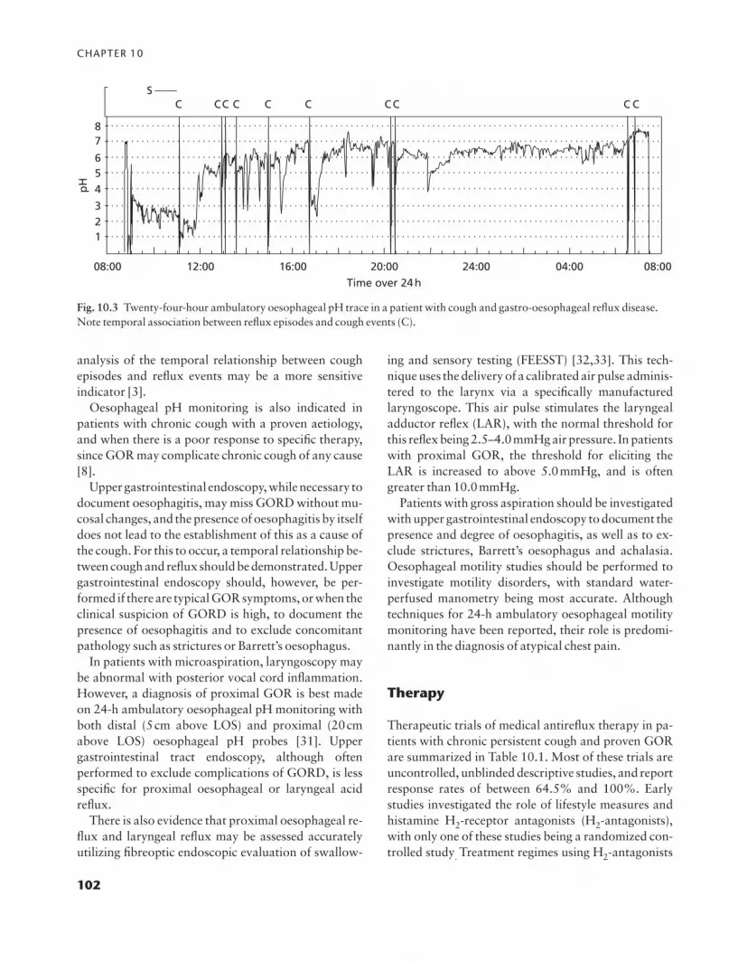
CHAPTER 10
analysis of the temporal relationship between coughepisodes and reflux events may be a more sensitive indicator [3].
Oesophageal pH monitoring is also indicated in patients with chronic cough with a proven aetiology,and when there is a poor response to specific therapy,since GOR may complicate chronic cough of any cause[8].
Upper gastrointestinal endoscopy, while necessary todocument oesophagitis, may miss GORD without mu-cosal changes, and the presence of oesophagitis by itselfdoes not lead to the establishment of this as a cause ofthe cough. For this to occur, a temporal relationship be-tween cough and reflux should be demonstrated. Uppergastrointestinal endoscopy should, however, be per-formed if there are typical GOR symptoms, or when theclinical suspicion of GORD is high, to document thepresence of oesophagitis and to exclude concomitantpathology such as strictures or Barrett’s oesophagus.
In patients with microaspiration, laryngoscopy maybe abnormal with posterior vocal cord inflammation.However, a diagnosis of proximal GOR is best made on 24-h ambulatory oesophageal pH monitoring withboth distal (5cm above LOS) and proximal (20cmabove LOS) oesophageal pH probes [31]. Upper gastrointestinal tract endoscopy, although often performed to exclude complications of GORD, is lessspecific for proximal oesophageal or laryngeal acid reflux.
There is also evidence that proximal oesophageal re-flux and laryngeal reflux may be assessed accuratelyutilizing fibreoptic endoscopic evaluation of swallow-
ing and sensory testing (FEESST) [32,33]. This tech-nique uses the delivery of a calibrated air pulse adminis-tered to the larynx via a specifically manufacturedlaryngoscope. This air pulse stimulates the laryngealadductor reflex (LAR), with the normal threshold forthis reflex being 2.5–4.0mmHg air pressure. In patientswith proximal GOR, the threshold for eliciting theLAR is increased to above 5.0mmHg, and is oftengreater than 10.0mmHg.
Patients with gross aspiration should be investigatedwith upper gastrointestinal endoscopy to document thepresence and degree of oesophagitis, as well as to ex-clude strictures, Barrett’s oesophagus and achalasia.Oesophageal motility studies should be performed toinvestigate motility disorders, with standard water-perfused manometry being most accurate. Althoughtechniques for 24-h ambulatory oesophageal motilitymonitoring have been reported, their role is predomi-nantly in the diagnosis of atypical chest pain.
Therapy
Therapeutic trials of medical antireflux therapy in pa-tients with chronic persistent cough and proven GORare summarized in Table 10.1. Most of these trials areuncontrolled, unblinded descriptive studies, and reportresponse rates of between 64.5% and 100%. Earlystudies investigated the role of lifestyle measures andhistamine H2-receptor antagonists (H2-antagonists),with only one of these studies being a randomized con-trolled study. Treatment regimes using H2-antagonists
102
08:00
pH
12:00Time over 24 h
16:00 20:00 24:00 04:00 08:00
23
456
78
1
C CC C C C CC C CS
Fig. 10.3 Twenty-four-hour ambulatory oesophageal pH trace in a patient with cough and gastro-oesophageal reflux disease.Note temporal association between reflux episodes and cough events (C).

COUGH AND GASTRO-OESOPHAGEAL REFLUX
report response rates of 80–100%, with no correlationbetween 24-h ambulatory oesophageal pH monitoringresults and response. There are also no important dif-ferences on pH monitoring between partial and com-plete responders.
The antitussive and antireflux effects of H2-antagonists are prolonged, with both cough symptomsand reflux parameters as measured by repeat 24-h oesophageal pH monitoring being significantly sup-pressed for more than 6 weeks after the drug is ceased.This implies that H2-antagonists break the cough–reflux self-perpetuating cycle in patients with coughdue to distal oesophageal–tracheobronchial reflexmechanisms [28].
There have been two published randomized, con-trolled, blinded trials of the effect of proton pump in-hibitors in patients with chronic persistent cough andGOR. Ours et al. [34] studied 17 patients with chronicpersistent cough and proven GOR on ambulatory oe-sophageal pH monitoring. They randomized 8 patientsto omeprazole 40mg p.o. b.d. and 9 patients to placebofor 12 weeks. They reported that 1 of 8 patients receiv-ing omeprazole and 0 of 9 patients on placebo res-ponded. After 12 weeks, all 17 patients received
omeprazole 40mg p.o. b.d. for another 4 weeks (openlabel medication). The authors then reported that 1 of 8patients in the initial active treatment group responded,and 5 of 9 in the initial placebo group responded. Therewere no differences noted between the active and pla-cebo groups and between the responders and non-responders for any GOR parameter (including all oesophageal pH indices), cough duration or GORsymptoms. All patients who responded did so within 2weeks of commencing active therapy. Ours et al. con-cluded that empirical high-dose proton pump inhibitortherapy for 2 weeks in all patients with chronic persist-ent cough not secondary to asthma or postnasal dripsyndrome was a ‘practical and simple approach’ [34].
Kiljander et al. [27] investigated 29 patients withchronic persistent cough and proven GOR: 21 of 29 pa-tients completed a randomized, double-blind, placebo-controlled crossover trial of omeprazole 40mg p.o.daily. Each treatment period was for 8 weeks with a 2-week washout period. In the group receiving omepra-zole in the first period there was a marked reduction incough scores when compared with baseline, and this re-duction was maintained in the second or placebo pe-riod. In the group receiving omeprazole in the second
103
Table 10.1 Summary of therapeutic trials of antireflux therapy in patients with cough and gastro-oesophageal reflux, notincluding proton pump inhibitor trials (see text). From [18].
Study Intervention No. of Study design Response Evidence gradepatients rate (%)
Irwin et al. Am Rev Respir Dis Antacid 5 PUU 100 Descriptive grade II-31981; 123: 413–7 Cimetidine
Irwin et al. Am Rev Respir Dis Metoclopramide 9 PUU 100 Descriptive grade II-31989; 140: 294–300 H2-antagonists
Fitzgerald et al. Can Med Assoc J Metoclopramide 20 PUU 70 Descriptive grade II-31989; 140: 520–4 Cimetidine
Irwin et al. Am Rev Respir Dis Metoclopramide 28 PUU 100 Descriptive grade II-31990; 141: 640–7 H2-antagonists
Ing et al. Am Rev Respir Dis Ranitidine 25 PBC 84 Grade I1992; 144: A11
Dordal et al. Allerg Immunol Cisapride 55 PUU 64.5 Descriptive grade II-31994; 26: 53–8 Domperidone
Waring et al. Dig Dis Sci PPI 25 PUU 80 Descriptive grade II-31995; 40: 1093–7 H2-antagonists
Smyrnios et al. Chest 1995; 108: Prokinetics 20 PUU 97 Descriptive grade II-3991–7 Antagonists
PUU, prospective, unrandomized, uncontrolled; PBC, prospective, blind, controlled; PPI, proton pump inhibitor.

CHAPTER 10
period there was a marked reduction in cough scoreswhen compared to baseline and when compared to theinitial placebo period. Kiljander et al. [27] concludedthat omeprazole 40mg p.o. daily for 8 weeks relievedGOR-related cough and that the reduction in coughcontinued after the treatment period. Maximal re-sponse in cough occurred after antireflux therapy wasceased, and Kiljander et al. [27] concluded that thissupported the existence of a cough–reflux positive feed-back cycle.
An empirical trial of proton pump inhibitors wouldtherefore be appropriate in patients with chronic per-sistent cough, after asthma and postnasal drip havebeen excluded. The duration of therapy, however, re-mains unclear, with some authors recommending aminimum therapeutic trial of 4 weeks.
Table 10.2 summarizes the studies investigating therole of antireflux surgery in patients with chronic per-sistent cough and GOR. Unfortunately all series arenon-randomized, uncontrolled descriptive studies,though most are prospective. The definitions of re-sponse rates for cough are variable, and range from84% to 100%. One of the more recent series was re-ported by Novitsky et al. [35]. They investigated 21 pa-tients with cough proven to be secondary to GOR.Cough was the sole presentation in 53% and persisteddespite intensive medical therapy including high-doseproton pump inhibitors and antireflux diet. After fun-doplication, 62% of patients reported complete resolu-
tion of cough, and another 24% reported significantimprovement in cough.
Irwin et al. [36] reported a subgroup of 8 of the initial21 patients that had persistent cough despite total ornear-total acid suppression using proton pump in-hibitors, prokinetic agents and antireflux diet (omepra-zole 20–80mg p.o. daily and cisapride 40–80mg p.o.daily). These 8 patients had 24-h ambulatory oe-sophageal pH monitoring while on medical therapy,and in all patients the percentage of 24h spent at pH<4.0 was zero or near zero. Despite this, all 8 pa-tients underwent antireflux surgery with marked re-duction in cough scores after surgery (as measured byvisual analogue scale and Adverse Cough OutcomeSurvey), which was maintained after 12 months of follow-up.
These descriptive studies investigating the effect ofantireflux surgery on cough in patients with provenGOR suggest that surgery may improve cough that isresistant to medical therapy, and that the improvementis sustained. In addition, there is a suggestion that acidreflux disease in patients with cough and GORD maybe a misnomer since non-acid reflux may be responsiblefor cough in some patients (volume reflux with gastricenzymes, bile salts, etc.).
It is unclear at present if antireflux surgery should be offered to all patients with cough and proven GORwho fail intensive medical therapy. In the series published by Novitsky et al. [35], 14% of patients (3 of
104
Table 10.2 Summary of trials of antireflux surgery in patients with chronic cough and gastro-oesophageal reflux.
Study Intervention No. of Study design Response rate (%) Evidence gradepatients
Pellegrini et al. Surgery 1979; 86: Fundoplication 5 PUU 100 Descriptive grade II-3110–19
De Meester et al. Ann Surg 1990; Fundoplication 17 PUU 100 Descriptive grade II-3211 (3): 337–45
Giudicelli et al. Ann Chir 1990; Fundoplication 13 PUU 84.6 Descriptive grade II-344 (7): 552–4
Thoman et al. J Gastrointest Surg Fundoplication 37 RUU 91 Descriptive grade II-32002; 6 (1): 17–21
Novitsky et al. Surg Endosc Fundoplication 21 PUU 86 Descriptive grade II-32002; 16 (4): 567–71
Irwin et al. Chest 2002; 121 (4): Fundoplication 8 PUU 100 Descriptive grade II-31132–40
PUU, prospective, unrandomized, uncontrolled; RUU, retrospective, unrandomized, uncontrolled.

COUGH AND GASTRO-OESOPHAGEAL REFLUX
21) had no response. The reasons are unclear, but fail-ure to address multiple aetiologies remains possible.There have been no randomized controlled studies onantireflux surgery in patients with chronic persistentcough and GORD, and as a result evidence-basedguidelines for management of refractory cough arelacking.
A recent development that may help predict responseto surgery is the technique of endoscopic implantationof biopolymer into the LOS. This involves the injectionof a biopolymer such as Enteryx (ethylene vinyl alco-hol) into the muscle of the gastric cardia. Although notreported in patients with cough and GORD, there havebeen reports of its efficacy in patients with GORD ingeneral. Deviere [37] reported 15 patients with provenGORD on long-term proton pump inhibitors. Afterbiopolymer injection, LOS pressure increased in 13 of15 patients at 1 month, and this was sustained for a me-dian of 6 months. Patients were able to cease protonpump inhibitors, though 4 of 15 resumed this therapyeventually. This technique has the potential of beingused to predict the response of patients with cough and GORD to antireflux surgery, but requires furtherevaluation in this specific group.
Summary
1 GORD along with bronchial asthma and postnasaldrip syndrome is one of the three most common causesof chronic persistent cough.2 Cough may be the only symptom of GORD.3 GORD most commonly causes chronic cough via an oesophageal–bronchial reflex mechanism with both local and central nervous reflexes likely to be important.4 Other components of gastric refluxate apart fromacid are likely to be important in the pathogenesis ofcough.5 Empirical medical antireflux therapy including proton pump inhibitors is likely to be successful intreating the cough in over 80% of patients.6 Patients who do not respond to empirical medicaltherapy should have 24-h ambulatory oesophageal pH monitoring while on therapy.7 Antireflux surgery may be useful in patients refractory to medical treatment, even if acid reflux is not demonstrated on follow-up oesophageal pH monitoring.
References
1 Mittal RK. Current concepts of the antireflux barrier. Gastroenterol Clin North Am 1990; 19 (3): 501–17.
2 Dent J. Recent views on the pathogenesis of gastro-oesophageal reflux disease. Bailliere’s Clinical Gastroen-terol 1987; 1 (4): 727–45.
3 Irwin RS, Zawacki JK, Curley FJ, French CL, Hoffman PJ.Chronic cough as the sole presenting manifestation of gastro-oesophageal reflux. Am Rev Respir Dis 1989; 140(5): 294–300.
4 Mansfield LE. Gastro-esophageal reflux and respiratorydisorders: a review. Ann Allergy 1989; 62: 158–63.
5 Irwin RS, Curley FJ, French CL. Chronic cough. The spec-trum and frequency of causes, key components of the diag-nostic evaluation and outcomes of specific therapy. AmRev Respir Dis 1990; 141: 640–7.
6 Palombi BC, Villanova CAC, Gastal OL, Stolz DP. A pathogenic triad in chronic cough: asthma, post nasal dripsyndrome, and gastro-oesophageal reflux disease. Chest1999; 116: 279–84.
7 McGarvey LP, Forsythe P, Heaney LG, McMahon J, EnnisM. Bronchoalveolar lavage findings in patients withchronic nonproductive cough. Eur Respir J 1999; 13 (1):59–65.
8 Liebermann-Meffert D, Allgower M, Schmid P. Muscularequivalent of the lower esophageal sphincter. Gastroen-terology 1979; 76: 31–8.
9 Tottrup A, Forman A, Uldbjerg N etal. Mechanical prop-erties of isolated human esophageal smooth muscle. Am JPhysiol 1990; 21: G329–37.
10 Dent J, Dodds WJ, Friedman RH etal. Mechanisms of gastro-oesophageal reflux in recumbent asymptomatichuman subjects. J Clin Invest 1980; 65: 256–67.
11 Mittal RK, McCallum RW. Characteristics of transientlower esophageal sphincter relaxation in humans. Am JPhysiol 1987; 252: G636.
12 Dent J, Holloway RH, Toouli J, Dodds WJ. Mechanismsof lower esophageal sphincter incompetence in patientswith symptomatic gastro-esophageal reflux. Gut 1988;29: 1020–8.
13 Holloway RH, Dent J. Pathophysiology of gastro-esophageal reflux: lower esophageal sphincter dysfunc-tion in gastroesophageal reflux disease. GastroenterolClin North Am 1990; 19 (3): 517–35.
14 Stanciu C, Bennett JR. Esophageal acid clearing: One fac-tor in production of reflux oesophagitis. Gut 1974; 15:852–7.
15 Ing AJ, Ngu MC, Breslin ABX. Chronic persistent cough and gastro-oesophageal reflux. Thorax 1991; 46:479–83.
16 Irwin RS, Corrao WM, Pratter MR. Chronic persistentcough in the adult. The spectrum and frequency of causes
105

CHAPTER 10
and successful outcome of specific therapy. Am Rev RespirDis 1981; 123: 413–7.
17 Poe RH, Harder RV, Israel RH, Kallay MC. Chronic per-sistent cough: experience in diagnosis and outcome usingan anatomic diagnostic protocol. Chest 1989; 95: 723–8.
18 American College of Chest Physicians. Managing coughas a defence mechanism and as a symptom —a consensuspanel report of the American College of Chest Physicians.Chest 1998; 114 (2): 133S–181S.
19 Ing AJ, Ngu MC, Breslin ABX. Pathogenesis of chronicpersistent cough associated with gastro-esophageal reflux.Am J Respir Crit Care Med 1994; 149: 160–7.
20 Harding SM. The role of gastro-oesophageal reflux inchronic cough and asthma. Chest 1997; 111: 1389–402.
21 Irwin RS, French CL, Curley FJ, Zawacki JK, Bennett FM.Chronic cough due to gastro-oesophageal reflux. Clinical,diagnostic and pathogenetic aspects. Chest 1993; 104 (5):1511–17.
22 Canning BJ. Role of nerves in asthmatic inflammation andpotential influence of gastro-oesophageal reflux disease.Am J Med 2001; 111 (Suppl. 8A): 13S–17S.
23 Suwanprathes P, Hunt G, Breslin A, Ing AJ, Ngu MC.Identification of brainstem regions involved with cough-ing: a study using c-Fos immunohistochemistry. Am JRespir Crit Care Med 2000; 161 (8): Abstract B52.
24 Suwanprathes P, Hunt G, Seow F, Ing AJ, Ngu MC. Coughand gastro-oesophageal reflux: evidence for a commoncentre in the brainstem. Am J Respir Crit Care Med 2001;163 (8): Abstract F57.
25 Bieger D. Muscarinic activation of rhombencephalic neu-rones controlling oesophageal peristalsis in the rat. Neu-ropharmacology 1984; 23: 1451–64.
26 Bieger D, Hopkins DA. Viscerotopic representation of theupper alimentary tract in the medulla oblongata in the rat:the nucleus ambiguus. J Comp Neurol 1987; 262: 546–62.
27 Kiljander TO, Salomaa ERM, Hietanen EK, Terho EO.Chronic cough and gastro-oesophageal reflux: a doubleblind placebo controlled study with omeprazole. EurRespir J 2000; 16: 633–8.
28 Ing AJ. Cough and gastro-oesophageal reflux. Am J Med1997; 103 (5A): 91–6.
29 Ing AJ, Ngu MC, Breslin ABX. Chronic persistent coughand clearance of oesophageal acid. Chest 1992; 102:1668–71.
30 Johnson LF, De Meester TR. Development of the 24-hourintra-oesophageal pH monitoring composite scoring sys-tem. J Clin Gastroenterol 1986; 8 (Suppl. 1): 52–8.
31 Paterson WG, Murat BW. Combined ambulatory oe-sophageal manometry and dual-probe pH-metry in evalu-ation of patients with chronic unexplained cough. Dig DisSci 1994; 39 (5): 1117–25.
32 Phua SY, McGarvey LPA, Peters MJ, Breslin ABX, Ing AJ.Assessing laryngeal sensitivity of patients with chroniccough and gastro-oesophageal reflux using fibreoptic en-doscopic evaluation of laryngeal sensitivity. Am J RespirCrit Care Med 2002; 165 (8): A406.
33 Aviv JE, Parides M, Fellowes J, Close LG. Endoscopicevaluation of swallowing as an alternative to 24-hour pHmonitoring for diagnosis of extra-oesophageal reflux.Ann Otol Rhinol Laryngol 2000; 184 (Suppl.): 25–7.
34 Ours TM, Kavuru MS, Schilz RJ, Richter JE. A prospec-tive evaluation of oesophageal testing and a double blind,randomized study of omeprazole in a diagnostic and ther-apeutic algorithm for chronic cough. Am J Gastroenterol1999; 94: 3131–8.
35 Novitsky YW, Zawacki JK, Irwin RS, French CT, HusseyVM, Callery MP. Chronic cough due to gastro-oesophageal reflux disease: efficacy of antireflux surgery.Surg Endosc 2002; 16 (4): 567–71.
36 Irwin RS, Zawacki JK, Wilson MM, French CT, CalleryMP. Chronic cough due to gastro-oesophageal reflux dis-ease: failure to resolve despite total/near total eliminationof oesophageal acid. Chest 2002; 121: 1132–40.
37 Deviere J, Pastorelli A, Louis H, de Maertelaer V, LehmanG, Cicala M, Le Moine O, Silverman D, Costamagna G.Endoscopic implantation of a biopolymer in the lower oe-sophageal sphincter for gastro-oesophageal reflux: a pilotstudy. Gastrointest Endosc 2002; 55 (3): 335–41.
106

107
Introduction
In 1998 the Consensus Panel Report of the AmericanCollege of Chest Physicians (ACCP) stated that ‘themost common causes of chronic cough in non-smokersare postnasal drip syndrome (PNDS), asthma and/orgastro-oesophageal reflux disease (GORD), whether ornot the cough is described as dry or productive’ [1–4].
The incidence of PNDS in chronic cough is disputed,with published values ranging from 2 to 57% It seems far more common in the US than in Europe [5].The reasons for this variation may be social or seman-tic. Few studies were randomized, and selection of patients might depend on the reputation and interestsof the investigator. In almost none of the studies wascough measured, either subjectively or objectively, oreven defined to distinguish between true cough andhuff.
Postnasal drip syndrome
According to the ACCP [3], the diagnosis of PNDSlargely rests on the patient reporting certain symptomsor sensations. Since PNDS is a syndrome, and sincethere are no pathognomonic findings to prove its pres-ence, the diagnosis of postnasal drip-induced cough isbest determined by considering a combination of crite-ria, including symptoms, physical examination, radi-ographic findings, and ultimately, response to specifictherapy.
A favourable response to specific therapy for PNDSwith resolution of cough is a crucial step in confirming
that PNDS was present and that it was the aetiology ofthe cough.
Cough initiated at the upper respiratory tract is usu-ally produced by stimulation of pharyngeal branchnerve endings (vagal branches). It is speculated that thecough in PNDS may be caused by chemical or mechan-ical irritation of receptors located in the larynx and/orpharynx [1,2], Proctor [6] claims that, in patients withpurulent rhinosinusitis, secretions are aspirated intothe trachea and bronchi during the night, a processwhich is further stimulated by the reduction in muco-ciliary activity. Bucca et al. [7] have demonstrated an increase in the extrathoracic reactivity of airways mediated by pharyngobronchial reflexes followingdrainage of upper airway secretions.
Clinical studies suggest that the pathogenesis ofcough from PNDS is due to mechanical or chemicalstimulation of the afferent limb of the cough reflex in the upper airway. This stimulation is secondary to secretions emanating from the nose and/or sinusesdripping down into the hypopharynx. A number ofconditions can cause PNDS. The differential diagno-sis includes seasonal allergic rhinitis, perennial aller-gic rhinitis, perennial non-allergic rhinitis, vasomotorrhinitis, postinfectious rhinitis, chronic (bacterial) rhinosinusitis, allergic fungal rhinosinusitis, non-allergic rhinitis due to medication abuse or environ-mental irritants, and non-allergic rhinitis associatedwith pregnancy [6–8].
In addition to this clinical presentation, there is usu-ally a history of rhinosinopathy. These clinical findingsare relatively sensitive, but not specific for diagnosticpurposes [3,4]. In addition to cough (which is produc-tive in 50% of cases), Villanova et al. [8,9] have found
107
Cough in postnasal drip, rhinitis and rhinosinusitisBruno C. Palombini & Elisabeth Araujo
11

CHAPTER 11
that throat-clearing (66.7%) and pharyngeal aspira-tion (26.7%) are the two most common clinical mani-festations in PNDS patients [3,4,8,9].
When chronic cough results from a multicausal asso-ciation, diagnosis is usually based on the recently de-scribed pathogenic triad of chronic cough (asthma,PNDS and gastro-oesophageal reflux (GOR)) [9,10].
The clinical presentation of patients with PNDS, inaddition to cough, commonly involves complaints of a sensation of something dripping into the throat(drainage in posterior pharynx), a need to clear thethroat, a tickle in the throat or nasal congestion and/ornasal discharge [6]. Patients sometimes complain ofhoarseness. Cough can also occur with talking, but thisis a non-specific complaint associated with essentiallyall causes of cough. A history of upper respiratory ill-ness (a cold) is often present. A history of wheeze is alsocommon. Most patients with PND-induced cough willhave symptoms or evidence of one or more of the fol-lowing: drainage in posterior pharynx, throat-clearing,nasal discharge, cobblestone appearance of theoropharyngeal mucosa and mucus in the oropharynx.These clinical findings are relatively sensitive, but theyare not specific. They are also found in many patientswith cough due to other causes [1–4].
Pharyngeal aspiration and throat-clearing
PNDS has been described and discussed in several stud-ies and publications. It is caused by disorders affectingthe nasal fossae, pharynx and paranasal sinuses. Coughin PNDS may be triggered by numerous stimuli, and inmost cases the aetiopathogenesis is clear. Cough is un-doubtedly associated with certain conditions such asbronchopneumonia, bronchiectasis, bronchial cancer,tuberculosis, use of angiotensin-converting enzyme(ACE) inhibitors, lung fibrosis and asthma. However,cases with multiple or obscure aetiopathogenesis pose achallenge. For these situations, it is useful to discusssome recent advances [9,10].
One of the characteristics of non-productive cough isthat it is not associated with expectoration and does notpresent the clinical signs of pharyngeal aspiration (PA)or throat-clearing (TC).
On the other hand, chronic productive cough is fre-quently observed in smokers, since smoking is often as-sociated with both emphysema and chronic bronchitis.
However, this type of productive cough does not pres-ent signs of PA, since there is little association betweensmoking and chronic rhinosinusitis. In turn, signs ofTC are frequently found in smokers with productivecough.
Inspiratory signs of PA, even when present in smok-ers, do not present a cause-and-effect association withsmoking. Signs of PA strongly suggest the presence ofchronic suppurative foci in the upper airway. In otherwords, since smoking does not prevent smokers fromhaving rhinosinusitis, some smokers may simultane-ously present signs of PA (due to rhinosinusitis) andsigns of TC (secondary to chronic smoker’s bronchitis).Figure 11.1 shows cinematography images of pharyn-geal aspiration and throat-clearing.
Preliminary results of research concerning the mean-ing of these clinical signs have already become avail-able. Recent studies in Brazil have employed injectionof barium contrast in the nasal fossae followed by pharyngeal aspiration and throat-clearing with simul-taneous radioscopy with digital subtraction [11]. Concerning the drainage of upper airway secretions,preliminary impressions suggest that the pharynx mayact as a pump, with inspiration and expiration move-ments, and its lumen may go from expanded dimen-sions (inspiration) to a small volume (expiration) (seeFig. 11.1).
Another major characteristic associated with thebasic physiopathogenic mechanism of PA and TC is thepossibility of airway vibration, also found in otherPND manoeuvres in general. The physical characteris-tics of such vibration and its role in clearing the airwaysare still uncertain.
One of the objectives of the present chapter is to pro-vide tools to general practitioners, pneumologists andpaediatricians to support the diagnosis of chroniccough using simple rules. Basically, the objective is toprovide further knowledge regarding PA and TC. Figure 11.1 shows the results of radiological studieswith forced inspiration, characterizing signs of PA, andforced expiration, constituting signs of TC. The clinicalrecognition of these signs is easy, and they have greatdiagnostic value, dispensing with expensive diagnosticprocedures that often delay treatment (for example,computed tomography (CT) of paranasal sinuses andpharynx and/or fibreoptic nasal endoscopy).
Therefore, the need to train resident physicians to perform the differential diagnosis of chronic pro-ductive cough is underscored, highlighting the dif-
108
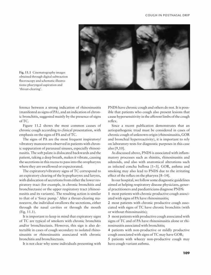
COUGH IN POSTNASAL DRIP
ference between a strong indication of rhinosinusitis(manifested as signs of PA), and an indication of chron-ic bronchitis, suggested mainly by the presence of signsof TC.
Figure 11.2 shows the most common causes ofchronic cough according to clinical presentation, withemphasis on the signs of PA and of TC.
The signs of PA are the most frequent inspiratory/vibratory manoeuvres observed in patients with chron-ic suppuration of paranasal sinuses, especially rhinosi-nusitis. The soft palate is dislocated backwards and thepatient, taking a deep breath, makes it vibrate, causingthe secretions in this recess to pass into the oropharynxwhere they are swallowed or expectorated.
The expiratory/vibratory signs of TC correspond toan expiratory clearing of the hypopharynx and larynx,with dislocation of secretions from either the lower res-piratory tract (for example, in chronic bronchitis andbronchiectasis) or the upper respiratory tract (rhinosi-nusitis and its variants). The resulting action is similarto that of a ‘force pump.’ After a throat-clearing ma-noeuvre, the individual swallows the secretions, eitherthrough the nasal cavities or through the mouth (Fig. 11.1).
It is important to keep in mind that expiratory signsof TC are typical of smokers with chronic bronchitisand/or bronchiectasis. However, this sign is also de-tectable in cases of cough secondary to isolated rhino-sinusitis or rhinosinusitis associated with chronicbronchitis and bronchiectasis.
It is not clear why some individuals presenting with
PNDS have chronic cough and others do not. It is poss-ible that patients who cough also present lesions thatcause hypersensitivity in the afferent limbs of the coughreflex.
Since a recent publication demonstrates that an aetiopathogenic triad must be considered in cases ofchronic cough of unknown origin (rhinosinusitis, GORand bronchial hyperreactivity), it is important to relyon laboratory tests for diagnostic purposes in this casealso [9,10].
As discussed above, PNDS is associated with inflam-matory processes such as rhinitis, rhinosinusitis andadenoids, and also with anatomical alterations such as infected concha bullosa [1–3]. GOR, asthma andsmoking may also lead to PNDS due to the irritating effect of the reflux on the pharynx [8–14].
In our hospital, we follow some diagnostic guidelinesaimed at helping respiratory disease physicians, gener-al practitioners and paediatricians diagnose PNDS:1 most patients with chronic productive cough associ-ated with signs of PA have rhinosinusitis;2 most patients with chronic productive cough asso-ciated with signs of TC have chronic bronchitis (with or without rhinosinusitis);3 most patients with productive cough associated withsigns of TC and of PA have rhinosinusitis alone or rhi-nosinusitis associated with bronchitis;4 patients with non-productive or mildly productivecough associated with signs of TC may have GOR;5 patients with wheezy non-productive cough mayhave cough-variant asthma.
109
Fig. 11.1 Cinematography images obtained through digital subtractionfluoroscopy and schematic illustra-tions: pharyngeal aspiration and‘throat-clearing’.

CHAPTER 11
These clinical findings are relatively sensitive, but not specific for diagnostic purposes [3,4]. In addition to chronic cough (which was productive in 50% ofcases of chronic cough, in adults), Villanova et al.[8,9] have found that the signs of TC were positive in66.7% and signs of PA in 26.7%. These were the twomost common clinical manifestations in patients withPNDS.
Rhinitis
The concept of rhinitis implies inflammation of thenasal mucosa. Clinical diagnosis is based on the pres-
ence of the following symptoms: pruritus, sneezing,rhinorrhoea and nasal obstruction.
Patients with rhinitis can be divided into ‘sneezerswith a runny nose’ and those with a ‘stuffy nose.’Cough is more frequently associated with increased secretion, especially thicker secretion with posteriordrainage. The presence of secretion with inflammatorymediators results in stimulation of receptors present inthe nasal mucosa that are innervated by the trigeminusand of receptors found in the posterior pharynx inner-vated by the glossopharyngeus.
Infectious rhinitis is the most common cause of acutecough. Viral infections due to rhinovirus, influenza andparainfluenza induce the activation of several types of
110
Ch
ron
ic c
ou
gh
With obviouspathogenicfactors
Without obviouspathogenicfactors
Non-productivechronic cough
Productivechroniccough
Non-productivechronic cough
Productivechronic cough
TuberculosisBronchial cancerLung fibrosisBronchial hyper-reactivityUse of ACE inhibitors
COPDBronchiectasisLung abscess
Bronchial hyper-reactivityGastro-oesophageal refluxACE treatment
Sign of pharyngeal aspiration
Present AbsentPr
esen
tA
bse
ntSig
n o
f th
roat
cle
arin
g
Chronicrhinosinusitis withchronic bronchitis
Chronic bronchitis
Chronicrhinosinusitis
Bronchial cancerTuberculosis
Fig. 11.2 Pathogenetic routes of the most common types of chronic cough and some accompanying signs.
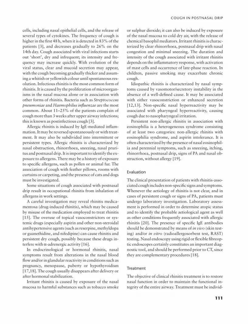
COUGH IN POSTNASAL DRIP
cells, including nasal epithelial cells, and the release ofseveral types of cytokines. The frequency of cough ishigher in the first 48h, when it is detected in 83% of thepatients [3], and decreases gradually to 26% on the14th day. Cough associated with viral infections startsout ‘short’, dry and infrequent; its intensity and fre-quency may increase quickly. With evolution of theviral status, clear and mucoid secretion may appear,with the cough becoming gradually thicker and assum-ing a whitish or yellowish colour until spontaneous res-olution. Infectious rhinitis is the most common form ofrhinitis. It is caused by the proliferation of microorgan-isms in the nasal mucosa alone or in association withother forms of rhinitis. Bacteria such as Streptococcuspneumoniae and Haemophilus influenzae are the mostcommon. About 11–25% of the patients complain ofcough more than 3 weeks after upper airway infections;this is known as postinfectious cough [3].
Allergic rhinitis is induced by IgE-mediated inflam-mation. It may be reversed spontaneously or with treat-ment. It may also be subdivided into intermittent orpersistent types. Allergic rhinitis is characterized bynasal obstruction, rhinorrhoea, sneezing, nasal pruri-tus and postnasal drip. It is important to identify the ex-posure to allergens. There may be a history of exposureto specific allergens, such as pollen or animal fur. Theassociation of cough with feather pillows, rooms withcurtains or carpeting, and the presence of cats and dogsmust be investigated.
Some situations of cough associated with postnasaldrip result in occupational rhinitis from inhalation ofallergens in work settings.
A careful investigation may reveal rhinitis medica-mentosa (drug-induced rhinitis), which may be causedby misuse of the medication employed to treat rhinitis[15]. The overuse of topical vasoconstrictors or sys-temic drugs (especially aspirin and other non-steroidalantihypertensive agents (such as reserpine, methyldopaor guanethidine, and nifedipine) can cause rhinitis andpersistent dry cough, possibly because these drugs in-terfere with a-adrenergic activity [16].
In endocrinological or hormonal rhinitis, nasalsymptoms result from alterations in the nasal bloodflow and/or in glandular reactivity in conditions such aspregnancy, menopause, puberty or hypothyroidism[17,18]. The cough usually disappears after delivery orafter hormonal stabilization.
Irritant rhinitis is caused by exposure of the nasalmucosa to harmful substances such as tobacco smoke
or sulphur dioxide; it can also be induced by exposureof the nasal mucosa to cold dry air, with the release ofchemical basophil mediators. Irritant rhinitis is charac-terized by clear rhinorrhoea, postnasal drip with nasalcongestion and minimal sneezing. The duration and intensity of the cough associated with irritant rhinitisdepends on the inflammatory response, with activationof mast cells and occurrence of late-phase reaction. Inchildren, passive smoking may exacerbate chroniccough.
Idiopathic rhinitis is characterized by nasal symp-toms caused by vasomotor/secretory instability in theabsence of a well-defined cause. It may be associatedwith either vasoconstriction or enhanced secretion[12,13]. Non-specific nasal hyperreactivity may be associated with pharyngeal hyperreactivity, causingcough due to nasopharyngeal irritation.
Persistent non-allergic rhinitis in association witheosinophilia is a heterogeneous syndrome consisting of at least two categories: non-allergic rhinitis witheosinophilia syndrome, and aspirin intolerance. It isoften characterized by the presence of nasal eosinophil-ia and perennial symptoms, such as sneezing, itching,rhinorrhoea, postnasal drip, signs of PA and nasal ob-struction, without allergy [19].
Evaluation
The clinical presentation of patients with rhinitis-asso-ciated cough includes non-specific signs and symptoms.Whenever the aetiology of rhinitis is not clear, and incases of persistent cough or signs of PA, patients mustundergo laboratory investigation. Laboratory assess-ment is performed in order to determine atopic statusand to identify the probable aetiological agent as wellas other conditions frequently associated with allergicrhinitis [20]. The presence of specific IgE antibodiesshould be demonstrated by means of in vivo (skin test-ing) and/or in vitro (radioallergosorbent test, RAST)testing. Nasal endoscopy using rigid or flexible fibreop-tic endoscopes certainly constitutes an important diag-nostic tool, and should be performed prior to CT, sincethey are complementary procedures [18].
Treatment
The objective of clinical rhinitis treatment is to restorenasal function in order to maintain the functional in-tegrity of the entire airway. Treatment must be individ-
111

CHAPTER 11
ualized and extended to associated infectious and mechanical complications. Important points includecleanliness of physical environment, pharmacotherapyand specific immunotherapy.
The drugs used to treat allergic rhinitis-associatedcough include antihistamines, oral and topical vaso-constrictors, ipratropium bromide, disodium chromo-glycate, nedochromil sodium, and oral and topicalcorticosteroids. These drugs can be used either in isola-tion or in association [13].
Rhinosinusitis
As previously mentioned, rhinosinusitis is caused by inflammation of the mucosa lining the nasal cavity and PNS. Rhinitis may occur in isolation, but cases ofsinusitis without associated rhinitis are rare.
Rhinosinusitis is a major public health problem, affecting approximately 14% of the worldwide adultpopulation, and this prevalence has been increasing[12].
Diagnosis
Acute rhinosinusitis is one of the most common reasons for medical appointments. Between 0.5% and5% of the viral infections affecting the upper airwaysresult in acute rhinosinusitis. A differential diagnosiswith viral infection is often difficult, since most symp-toms (nasal obstruction, rhinorrhoea, hyposmia, facialpressure, PND, signs of PA, cough and fever) are non-specific. Certain symptoms, such as fever, halitosis andhyposmia, considered as minor symptoms in adults,might be the sole indication of rhinosinusitis in children[13].
In chronic rhinosinusitis, chronic cough (lastinglonger than 3 weeks) and purulent or mucopurulent ex-pectoration, as well as signs of PA, are common find-ings, without evidence of suppurative foci in the lowerairways [9,10]. The time of the day in which the coughappears is also an important indication for its origin.Productive cough appearing at night and on waking up indicates rhinosinusitis. Between 18 months and 6years of age, rhinosinusitis is the most frequent cause ofchronic cough. Between 6 and 16 years, it is associatedwith asthma-variant cough and psychogenic cough asthe most common causes of chronic cough.
For adults with chronic cough due to PND, rhinosi-
nusitis is the cause in 30–60% of cases. Less frequentcauses are some forms of rhinitis, such as allergic,postinfectious, vasomotor, perennial, non-allergic anddrug-induced rhinitis [12].
In non-allergic and non-infectious rhinosinusitis, thecommon cold (acute coryza) deserves special attention.Primary symptoms include mouth dryness and nasalpruritus/itch, followed by dryness and pain in thethroat, sneezing, watery coryza and constitutionalsymptoms (febricula, indisposition). After 2 or 3 days,a secondary infection occurs, with nasal obstructionand purulent secretions, non-productive cough, febric-ula and muscle pain lasting for 5–10 days [13].
When chronic cough results from a multicausal association, diagnosis is usually based on the recentlydescribed [8–10] pathogenic triad of chronic cough(asthma, PND, GOR) (Fig. 11.3). Over 50% of thecases of chronic cough are related to this triad. In orderto recognize this condition, in addition to CT of thePNS, oesophageal pH-metry (detection of oesophagealreflux) and spirometry with or without bronchoprovo-cation testing (diagnosis of asthma without wheezing)should be performed [9,10].
Traditional nasal evaluation with a frontal lightsource and nasal speculum provides only limited infor-mation. Rigid and flexible fibreoptic endoscopes allowfor a direct and systematic evaluation of nose areas. En-doscopy must be considered whenever the patient pres-ents with severe and persistent cough, despite clinicaltreatment [17]. The presence of cough is often relatedto drainage of purulent secretions in the nasopharynx
112
Asthma Postnasal dripsyndrome
Gastro-oesophagealreflux disease
12 16 13
12
10
4 4
Fig. 11.3 Aetiological factor in 78 cases of chronic cough:15.4% of the patients had three simultaneous causes; 26%had two causes; 35% had one single cause. From [8].

COUGH IN POSTNASAL DRIP
originating at the draining ostia of the maxillary, ethmoid and sphenoid sinuses. In such situations, ourexperience has shown the presence of thick pharyng-eal secretion at endoscopy, despite the fact that somepatients refer to having dry cough.
Generally, the diagnostic relevance of simple radi-ographs is controversial, since radiological abnormal-ities in the ethmoid and sphenoid sinuses, frontal recessand osteomeatal complex may go unnoticed. A normalresult in a simple radiograph is not sufficient to rule outthe presence of rhinosinusitis. In children, it is impor-tant to underscore that the younger they are the greaterthe radiological limitations imposed [17,18].
CT is the preferred method for assessing PNS be-cause it clearly shows the relationship between soft tissues and bones. CT allows for a specific diagnosisand for the identification and systematic evaluation ofanatomical areas that are inaccessible to conventionalradiological tests (Fig. 11.4) [18,19].
Approximately 70% of the cases of acute rhinosi-nusitis are caused by Streptococcus pneumoniae andHaemophilus influenzae. In chronic rhinosinusitis,bacteriological studies demonstrate a predominance ofpolymicrobial flora and anaerobes, due to the reducedoxygen concentration in the PNS. Fungi (Aspergillus,Candida albicans and Alternaria) are isolated in3–10% of cases of chronic rhinosinusitis [20].
The main aetiological agent of infection in the air-ways is S. pneumoniae. Recent studies show that ap-
proximately 20% of the strains present intermediateresistance to penicillin; high resistance was observed inapproximately 1%. With regard to other microorgan-isms, b-lactamase was found in approximately 10% of H. influenzae strains and in more than 90% ofMoraxella catarrhalis [15,16].
A favourable response to specific therapy, with reso-lution of cough, is a crucial stage in confirming the association of bacteria with cough.
Cases of chronic cough caused by chronic rhinosi-nusitis or sinusitis resistant to treatment should be re-ferred to an otorhinolaryngologist for investigation ofstructural abnormalities of the osteomeatal complex,hypertrophy of adenoids, septal deviation and dentalabscess [15].
References
1 Irwin RS. Cough. In: Irwin RS, Curley FJ, Grossman RF,eds. Diagnosis and Treatment of Symptoms of the Respi-ratory Tract. New York: Futura Publishing Co., 1997:1–54.
2 Irwin RS, Madison JM. The diagnosis and treatment ofcough. N Engl J Med 2000; 343: 1715–21.
3 Irwin RS, Boulet LP, Cloutier MM, Fuller R, Gold PM,Hoffstein V etal. Managing cough as a defense mechanismand as a symptom —a consensus panel report of the American College of Chest Physicians. Chest 1998; 114(2 Suppl. Managing): 133S–81S.
4 Irwin RS, Curley FJ, French CL. The spectrum and fre-quency of causes, key components of the diagnostic evalu-ation and outcome of specific therapy. Am Rev Respir Dis1990; 141: 640–7.
5 Morice AH. Epidemiology of cough. Pulm PharmacolTher 2002; 15: 253–60.
6 Proctor DF. Nasal physiology and defense of the lung. AmRev Respir Dis 1977; 115: 97–102.
7 Bucca C, Rolla G, Scappaticci E, Chiampo F, Bugiani M,Magnano M etal. Extrathoracic and intrathoracic airwayresponsiveness in sinusitis. J Allergy Clin Immunol 1995;95: 52–9.
8 Villanova CA, Palombini BC, Pereira EA, Stolz DP, GastalOL, Alt DC etal. Post-nasal drip syndrome as a cause ofchronic cough: its place among other conditions. Am JRespir Crit Care Med 1996; 153: A517.
9 Palombini BC, Villanova CA, Araújo E, Gastal OL, AltDC, Stolz DP etal. A pathogenic triad in chronic cough —asthma, postnasal drip syndrome, and gastroesophagealreflux disease. Chest 1999; 116 (2): 279–84.
10 Palombini BC, Villanova CA, Araújo E, Gastal OL, Alt
113
Fig. 11.4 Computed tomography section (coronal view)showing bilateral ethmoid and maxillary opacification, withobstruction of the aorta.

CHAPTER 11
DC, Stolz DP etal. A pathogenic triad in chronic cough —asthma, post-nasal drip syndrome, and gastroesophagealreflux disease. Chest 1999; 116 (2): 279–84. (Quotation in the Journal Watch (General), N Engl J Med 1999; 19(19): 155.)
11 Irion KL, Porto NS, Palombini BC, Letti N, Pereira EA,Fraga JC etal. Documentação radiológica da falênciaeventual dos mecanismos supostamente capazes de evitaraspiração para traquéia das secreções faríngeas durante asmanobras da tosse, da aspiração faríngea e do pigarrear[abstract]. J Pneumologia 1992; 18 (1): 33.
12 Madison JM, Irwin RS. Cough. In: Albert R, Spiros S, Jett J, eds. Comprehensive Respiratory Medicine.London: Mosby, 1999: 15.1.
13 Mitchel D. Rhinitis and sinusitis. In: Albert R, Spiros S, Jett J, eds. Comprehensive Respiratory Medicine.London: Mosby, 1999: 31.1.
14 Palombini BC, Pereira EA, Alves MR, Irion KL. The need to recognize new and accurate symptoms and signs inthe diagnosis of sinobronchitis: the qualification of their
sensitivity and specificity [abstract]. Am Rev Respir Dis1992; 145 (4): 301.
15 Brandileone MC, Di Fabio JL, Vieira VS, Zanella RC,Casagrande ST, Pignatari AC etal. Geographic distribu-tion of penicillin resistance of Streptococcus pneumoniaein Brazil: genetic relatedness. Microb Drug Resist 1998; 4(3): 209–17.
16 Naclerio RM, Proud D, Togias AG, Adkinson NF Jr, Meyers DA, Kagey-Sobotka A etal. Inflammatory media-tors in late-phase antigen-induced rhinitis. N Engl J Med1985; 313: 6570.
17 McCaffrey TV. Rhinologic Diagnosis and Treatment.New York: Thieme, 1997.
18 Mackay I, ed. Rhinitis —Mechanisms and Management.London: Royal Society of Medicine, 1989.
19 Kennedy DW. International conference on sinus disease:terminology staging therapy. Ann Otol Rhinol Laryngol1995; 104 (10).
20 Mackay IS, Durham SR. Perennial rhinitis. Br Med J 1998;316: 917–20.
114

115
Introduction
Asthma is a disease which is identified by the presenceof characteristic symptoms and by the demonstrationof variable airflow obstruction, either spontaneously,or improving as a result of treatment. The symptoms ofasthma are dyspnoea, wheezing, chest tightness and, inmany patients with persistent asthma, troublesomecough. Cough has been recognized as a symptom ofasthma since the earliest descriptions of the disease.Persistent cough in asthma is often, but not always, as-sociated with the presence of airflow obstruction, andassociated with other asthma symptoms. The presenceof persistent cough, with or without sputum produc-tion, even when the only asthma symptom, is an indica-tion of poor asthma control [1].
Airway inflammation and asthma
Asthma is an airway inflammatory disease, with in-flammation present even when asthma is stable andwell controlled. This has been shown in studies whichhave provided information on airway cell populationsin mild stable asthmatics with ongoing and persistentairway hyperresponsiveness [2,3]. Common findings inall of these studies are the presence of increased num-bers of inflammatory cells such as eosinophils, lympho-cytes and mast cells compared with normal controlsubjects with normal airway responsiveness. Theeosinophils have shown signs of activation, as indicat-ed by increased airway levels of granular proteins,major basic protein (MBP) [4] and eosinophil cationicprotein (ECP) [5]. In the bronchial mucosa the
eosinophils have shown morphological features of acti-vation as indicated by heterogeneity of the granularstructure or as eosinophil granules released fromeosinophils lying free in the mucosal interstitium [6].Increased numbers of activated T lymphocytes havealso been shown in several studies [7], together with in-creased number of mast cells in the airway mucosa ex-hibiting various stages of degranulation [8]. The extentand severity of the airway inflammation increases attimes when asthma is unstable and poorly controlled,with increased numbers of airway eosinophils and/orneutrophils [9]. As will be discussed later, the presenceof persistent airway inflammation is likely to be respon-sible not only for asthma symptoms, including cough,but also for the structural changes and the associatedphysiological abnormalities of asthma.
Airway hyperresponsiveness in asthma
Airway responsiveness is a term which describes theability of the airways to narrow after exposure to con-strictor agonists. Airway hyperresponsiveness is an increased ability to develop this response and consists of the ability of a smaller concentration of an agonistneeded to initiate the bronchoconstrictor response (increased sensitivity of the airways), a steeper slope ofthe dose–response curve (increased reactivity of the air-ways), as well as a greater maximal response to the ago-nist (Fig. 12.1). The initial description of an increased responsiveness of asthmatic airways was made byAlexander and Paddock in 1921 [10], who demonstrat-ed an ‘asthmatic breathing’ in asthmatic subjects, butnot normals, after subcutaneous administration of the
115
Cough and airwayhyperresponsivenessPaul M. O’Byrne
12

CHAPTER 12
cholinergic agonist pilocarpine. This observation wasconfirmed by Weiss et al. [11], who reported that asthmatic subjects, but not normals, developed bronchoconstriction after being given intravenous histamine. Later Curry [12] described an increasedbronchoconstrictor response to histamine that oc-curred with intramuscular, intravenous and nebulizedhistamine, again only in asthmatic subjects. Tiffeneauand Beauvallet [13] were the first to describe the use ofacetylcholine inhalation tests to determine the degree ofairway responsiveness in asthmatics. Airway hyperre-sponsiveness is often considered to be non-specific, inthat asthmatic subjects will develop bronchoconstric-tion after exposure to many different chemical stimulisuch as histamine [14], the cholinergic agonists metha-choline [15] and carbachol [16], cysteinyl leukotrienes(LT) C4 and D4 [17], and prostaglandins (PG) D2 [18]and PGF2a [19]. This is to contrast the specificairway re-sponses that develop when subjects inhale substances,such as allergens [20] or occupational sensitizing agents[21], to which they have become sensitized. The termnon-specific airway hyperresponsiveness can, however,be misleading. It suggests that a common mechanismexists by which these pharmacological or physical
stimuli cause bronchoconstriction. This is clearly not the case, as most of the pharmacological agents act onspecific receptors in the airways, and the mechanisms by which receptor activation causes bronchoconstric-tion are different for different agents. However, as eachof these chemical bronchoconstrictors activates specificreceptors, they are considered to be ‘direct-acting’ stimuli. In addition, some stimuli such as exercise [22],hyperventilation of cold, dry air [23], both hypotonicand hypertonic solutions [24], and mannitol [25] alsocause bronchoconstriction in asthmatics. In contrast tothe direct-acting stimuli, these cause release of bron-choconstrictor mediators, such as histamine [26] andcysteinyl leukotrienes [27], in the airways, and there-fore are called ‘indirect-acting’ stimuli.
In studies of populations of asthmatic patients, theseverity of airway hyperresponsiveness has been shownto correlate with the severity of asthma [14] and withthe amount of treatment needed to control symptoms[28]. A variety of methods of measuring airway respon-siveness have been reported, and the clinical signifi-cance and the effects of antiasthma medications onthese measurements, and the pathophysiology andpathogenesis of airway hyperresponsiveness in asth-matic patients have been extensively studied. As a resultof this research, the methods of its measurement havebeen standardized, and are widely accepted. Almost allpatients with symptomatic asthma have airway hyper-responsiveness [14], and for this reason the absence ofairway hyperresponsiveness in a patient with symp-toms suggestive of asthma should result in an alterna-tive diagnosis being considered.
Mechanisms of airwayhyperresponsiveness
Studies of monozygotic and dizygotic twins have sug-gested that there is a genetic basis for the developmentof airway hyperresponsiveness, but that environmentalfactors are more important [29]. Also measurements ofairway hyperresponsiveness in young infants have indi-cated that airway hyperresponsiveness can be presentvery early in life, and that a family history of asthmaand parental smoking were risk factors for its develop-ment [30]. More recently, reports of genetic linkage ofairway hyperresponsiveness have been published. Onestudy has identified genetic linkage between hista-mine airway hyperresponsiveness and several genetic
116
0.25 0.5 1 2 4 8Concentration (mg/mL)
FEV
1 (%
ch
ang
e)
16 32 64 128 256
0
10
20
30
40
50 Asthmatic
Mild asthmatic
Normal
(a)
(b)
(c)
Fig. 12.1 The change in FEV1 vs. baseline, induced by in-creasing doses of a bronchoconstrictor stimulus (metha-choline) in mild, moderate and severe asthmatics vs. healthyindividuals. The PC20 value is calculated by interpolating a20% fall in FEV1 to the log–linear dose–response curve foreach individual. Asthmatic subjects have a reduced thresholdresponse (a), indicating increased sensitivity of the airways;an increased slope (b), indicating increased reactivity of theairways; and an increased maximal response (c).

COUGH AND AIRWAY HYPERRESPONSIVENESS
markers on chromosome 5q, near a locus that regulatesserum IgE levels [31]. Another study has identified link-age between a highly polymorphic marker of the B sub-unit of the high-affinity IgE receptor on chromosome11q and methacholine airway hyperresponsiveness,even in patients with non-atopic asthma [32]. One spe-cific gene polymorphism (Glu 27) of the nine identifiedfor the b2-adrenoceptor has also been associated with increased methacholine airway hyperresponsiveness[33], while another polymorphism (Gly 16) was associ-ated with the presence of nocturnal asthma [34]. Thus,a genetic basis for airway hyperresponsiveness seemsvery likely; however, the genetic linkage studies need tobe confirmed by other investigators in different patientpopulations.
In addition to a possible genetic basis causing airwayhyperresponsiveness in asthma, airway inflammatoryprocesses are thought to be important (Fig. 12.2). Mediators released from airway eosinophils have
been suggested to cause many of the tissue changes seen in the disease, including epithelial damage andthickening of the basement membrane. Eosinophilsalso produce cysteinyl leukotrienes which cause airwaysmooth muscle contraction [35] and exudation of plas-ma [36], resulting in thickening of the airway wall. It is likely that airway wall thickening, which has been de-scribed in asthmatics of varying degrees of severity,could explain some of the differences in airway hyper-responsiveness between normal individuals and asth-matic patients. The thickness of the airway wall fromautopsy specimens is greater in fatal asthmatics than inpatients with milder disease and in non-asthmatics[37]. It is not exactly clear which tissue contributesmostly to airway wall thickening in asthma. One factor that may be involved is the subepithelial thicken-ing seen in bronchial biopsy specimens from most asthmatics. Furthermore, bronchial smooth muscle hasa larger volume in asthmatics [38]. Lastly, exudation of
117
Fig. 12.2 Potential mechanisms for the development of airway hyperresponsiveness in asthma.
Inflammatorycell production,recruitment and mediator release
Dendritic cell,lymphocyteand mast cellmediated events
Initial antigen recognition,T-cell orientationand IgE production
Myofibroblasthyperplasia and
fibrosis
Oedema andloss of airwaytethering toparenchyma
Increased smooth muscleand altered function
Altered neuralmediators
Goblet cell hyperplasia,mucus production andepithelial desquamation
AHR
DCT
B
MC
T

CHAPTER 12
plasma can cause oedema, and thus thickening, of theairway wall. Together these factors may, by geometricmechanisms, enhance the airway luminal resistance induced by a certain degree of airway smooth muscleshortening. Another feature of the asthmatic airwaythat correlates with the degree of airway hyperrespon-siveness is loss of epithelial structure [6]. Possibly thepartial loss of the epithelial barrier allows greateramounts of bronchoconstrictor mediators to reach thesmooth muscle or other cells which amplify the bron-choconstricting effect of the inhaled mediators. Alter-natively, the release of bronchodilating substancesfrom the epithelium could be reduced by epithelialdamage, which could enhance bronchial smooth mus-cle contraction [39]. Indeed, it is possible that a numberof these different mechanisms interact to produce airway hyperresponsiveness, but it is also likely thatdifferent mechanisms are involved in causing differentcomponents of airway hyperresponsiveness. Thus,while airway structural changes appear to be respons-ible for the underlying persisting airway hyperrespon-siveness in asthmatic patients, other mechanisms causethe variable changes in airway hyperresponsivenessseen in asthmatic subjects during the course of the disease.
Variable airway hyperresponsiveness can last days or weeks after experimental allergen inhalation [40], or during a seasonal allergen exposure [41]. Thesechanges are temporally related to increases in airwayinflammatory cells, particularly eosinophils and ba-sophils [42]. In addition, the release of cysteinylleukotrienes, likely from these cells, is partially respon-sible for allergen-induced airway hyperresponsiveness.This has been confirmed by the demonstration thatleukotriene receptor antagonists, such as montelukast[43] or pranlukast [44], partially attenuate allergen-induced airway hyperresponsiveness, but not the per-sistent airway hyperresponsiveness of asthma.
Cough-variant asthma
Cough-variant asthma was initially described as a clini-cal entity in 1979 [45]. It was identified in six patientswho had chronic persistent cough, without airflow obstruction, but who did have methacholine airwayhyperresponsiveness. These authors argued that thepresence of airway hyperresponsiveness, together witheven one symptom of asthma, established the diagnosis
of asthma. In all subjects, the cough improved with reg-ular inhaled bronchodilators. Subsequently, the termcough-variant asthma was used to indicate a ‘formefruste’ of asthma [46]. However, it is now recognizedthat in some patients with asthma, cough is the main,and indeed sometimes the only, symptom and that vari-able airflow obstruction may not accompany the symp-tom of cough. Koh et al. [47] have suggested that it isthe level of maximal airway response to inhaled metha-choline, rather than the degree of airway hypersensi-tivity, that may be an important risk factor for theeventual development of wheezing in patients who ini-tially present with cough as the only symptom of asth-ma. It has become clear that cough-variant asthma israther not a ‘forme fruste’ of asthma, but a clinical man-ifestation of (usually mild) asthma, with similar patho-logical changes in the airway wall [48]. Consistent withthis view, Niimi et al. [49] have described that serumeosinophil cationic protein level and the percentage ofeosinophils in bronchoalveolar lavage (BAL) fluid andin bronchial biopsy specimens were elevated and com-parable to those found in patients with classic asthmaassociated with wheeze. The same group found an in-creased thickness of the bronchial basement membranein patients with cough-variant asthma, indicating thata similar process of airway wall remodelling as ob-served in classic asthma was present [48].
Eosinophilic bronchitis without airwayhyperresponsiveness
Another clinical entity, identified in 1989 by Gibson et al. [50], is the presence of troublesome persistentcough, associated with airway inflammation, as meas-ured by increased numbers of eosinophils in inducedsputum, but without airway hyperresponsiveness (Fig. 12.3). This was called eosinophilic bronchitis, toindicate a clinical condition separate from asthma. Onestudy has suggested that eosinophilic bronchitis is thecause of troublesome cough in 13% of patients present-ing to a speciality clinic [51], and has been described in an occupational setting [52]. The cough associatedwith eosinophilic bronchitis responds to treatmentwith inhaled corticosteroids [53], associated with reso-lution of the eosinophilic airway inflammation. In themajority of the patients described in the original clinicaldescription, the cough did not return after treatment.However, occasionally, the cough did not fully improve
118

COUGH AND AIRWAY HYPERRESPONSIVENESS
with even high doses of inhaled corticosteroids, and re-quired oral corticosteroids for benefit. Interestingly, in one case report, inadequately treated eosinophilicbronchitis resulted in symptomatic asthma, with par-tially fixed, irreversible airflow obstruction [54]. Thepathological features of eosinophilic bronchitis havebeen compared to asthma in airway biopsies [55].These studies have shown that the main differencesdemonstrated were an increased number of mast cellspresent in the airway smooth muscle in asthma. Gibsonand colleagues [56] found that sputum cells obtainedfrom patients with chronic cough, who had normalspirometry and normal airway responsiveness, andwhose cough responded to inhaled corticosteroids, expressed interleukin-5 (IL-5) and granulocyte–macrophage colony-stimulating factor (GM-CSF)mRNA by in situ hybridization. These patients wouldfit into the category of eosinophilic bronchitis, and suchexpression has also been observed in patients with asth-ma indicating that both diseases may be caused byeosinophils.
Airway hyperresponsiveness and cough sensitivity
The lack of relationship between airway hyperre-
sponsiveness and capsaicin cough response has beenhighlighted by a study of Fujimura and colleagues [57].They described two groups of patients with chronicnon-productive cough: those with methacholine air-way hyperresponsiveness, but with a normal capsaicincough threshold, whose cough was responsive to bron-chodilator therapy; and those had normal airway re-sponsiveness with a hypertussive response to capsaicinwhose coughs responded to inhaled corticosteroids or antihistamines. Furthermore, these patients did notdemonstrate any BAL eosinophilia, but had only asmall number of eosinophils in the subepithelium of the tracheal and bronchial biopsies [58] and in inducedsputum [59]. Thus, it appears that what Fujimuracalled ‘atopic’ cough may be part of ‘eosinophilic bron-chitis’ or at least at one spectrum of this condition. Inaddition, atopic cough, as with eosinophilic bronchitis,is responsive to inhaled corticosteroid therapy with areduction in the capsaicin tussive response [60].
This lack of concordance between airway respon-siveness and capsaicin cough response [61] has notbeen confirmed by other investigators. Doherty et al.[62] described a cohort of asthmatic patients, who as a group showed enhanced capsaicin cough responsewhen compared to a cohort of non-asthmatic volun-teers and capsaicin cough sensitivity was related tosymptomatic cough as measured by diary card score.Thus, capsaicin cough response may be increased insome patients with asthma, since some patients mayhave a persistent dry cough, even though other asthmasymptoms are well controlled on adequate therapy.Cough receptor sensitivity in children with uncon-trolled asthma, who have troublesome cough, is in-creased, but decreases when asthma is controlled tolevels similar to those in children with asthma withoutcough [63].
Mechanisms of cough in asthma
The mechanisms of cough in asthma are similar tothose causing other symptoms; that is, a consequence of airway inflammation. In some instances, cough mayoccur as a direct result of airway obstruction and in-creased mucus production that occurs during an asth-ma exacerbation. As described above, some asthmaticshave also been demonstrated to have an increasedcough reflex as measured by capsaicin inhalation [64],which suggests that axonal reflexes, the stimulation of
119
Asthma
MC
C (
%)
Non-asthmaticCS cough
Chronicbronchitis
0.0
0.5
1.0
1.5
2.0
2.5
Eosi
no
ph
ils (
%)
0
20
40
60
80
100
Fig. 12.3 Numbers of eosinophils and metachromatic cells ineither spontaneous or induced sputum in patients with asth-ma (�), eosinophilic bronchitis (�) or chronic bronchitis (�).Reproduced from [50].

CHAPTER 12
airway C fibres and substance P release in the airwaysmay be abnormal in asthmatics [65]. Sensitization ofthe cough response by mediators such as PGE2 or bybradykinin may occur [66,67]. In addition to peripher-al sensitization, central sensitization pathways havebeen proposed by integration from various sensorynerve subtypes in the central nervous system to initiateexaggerated reflexes and sensation, and substance Phas been proposed as an important central mechanismfor sensitization of the cough reflex, and its persistence[68]. Interestingly, recent studies have shown that aller-gen inhalation in atopic asthmatic subjects caused increases in eosinophilic airway inflammation and airway hyperresponsiveness, but not in capsaicin sensi-tivity [69]. This suggests that cough receptor sensitivityto capsaicin is not associated with airway eosinophilicinflammation, at least in patients with allergic asthma.By contrast, Weinfeld et al. [70] described increasedcapsaicin sensitivity in allergic asthmatic patients to beincreased during allergen exposure as during the birchpollen season, and argued that allergic inflammation inthe lower and/or upper airways may trigger neurogenicmechanisms of significant clinical importance. How-ever, the best evidence for an involvement of airway in-flammation, in particular eosinophils, in cough is theobservation that cough associated with eosinophilic in-flammation such as asthma, cough-variant asthma andan eosinophilic bronchitis responds to corticosteroidtherapy which also inhibits eosinophilic inflammation.
Management of cough in asthma
The management of cough in asthma is, generally, thesame as the management of other asthma symptoms[1]. This means treating airway inflammation, which isdone most effectively with inhaled corticosteroids andinhaled b2-agonists as needed. Inhaled b2-agonists willbe particularly and rapidly effective if the cough is a re-sult of airflow obstruction. The clinical effectiveness ofinhaled corticosteroids will take days to weeks toachieve the maximal benefit. Other treatment optionsthat have been considered for asthmatic cough includeinhaled ipratropium bromide or sodium cromoglycate.Ipratropium bromide has been shown to reduce capsaicin-induced bronchoconstriction [64], whilecromoglycate reduces angiotensin-converting enzyme-induced cough [71]. However, only inhaled cortico-steroids have been shown to improve the cough
threshold in asthmatics [72], while neither cromogly-cate [64], nor leukotriene receptor antagonists [73]have this effect.
Conclusions
Cough is an important symptom in asthmatic patients.It is often, but not always, associated with airflow ob-struction. Occasionally, cough is the only symptom ofasthma, and the diagnosis is established by demonstrat-ing the presence of airway hyperresponsiveness. Morecommonly, persistent cough as the only respiratorysymptom is caused by eosinophilic bronchitis, whereairway hyperresponsiveness is absent, but is associatedwith eosinophils as in asthma. Some asthmatics have an enhanced cough reflex as measured by capsaicin in-halation, likely as a consequence of airway inflamma-tion. The treatment of cough in patients with asthma or eosinophilic bronchitis is the same as with asthma,with inhaled corticosteroids, with or without inhaledb2-agonists.
References
1 Global Initiative for Asthma. Global Strategy for AsthmaManagement and Prevention, 2002. NIH Publication02–3659.
2 Kirby JG, Hargreave FE, Gleich GJ, O’Byrne PM. Bronchoalveolar cell profiles of asthmatic and nonasth-matic subjects. Am Rev Respir Dis 1987; 136: 379–83.
3 Beasley R, Roche WR, Roberts JA, Holgate ST. Cellularevents in the bronchi in mild asthma and after bronchialprovocation. Am Rev Respir Dis 1989; 139: 806–17.
4 Gleich GJ, Frigas E, Loegering DA, Wassom DL, Steinmuller D. Cytotoxic properties of the eosinophilmajor basic protein. J Immunol 1979; 123: 2925–7.
5 Venge P, Bystrom J. Eosinophil cationic protein (ECP). IntJ Biochem Cell Biol 1998; 30 (4): 433–7.
6 Jeffery PK, Wardlaw AJ, Nelson FC, Collins JV, Kay AB.Bronchial biopsies in asthma. An ultrastructural, quanti-tative study and correlation with hyperreactivity. Am RevRespir Dis 1989; 140: 1745–53.
7 Robinson DS, Hamid Q, Ying S, Tsicopoulos A, BarkansJ, Bentley AM etal. Predominant TH-2 like bronchoalveo-lar T-lymphocyte populations in atopic asthma. N Engl JMed 1992; 326: 298–304.
8 Jeffery PK. Comparative morphology of the airways inasthma and chronic obstructive pulmonary disease. Am JRespir Crit Care Med 1997; 150: S6–S13.
120

COUGH AND AIRWAY HYPERRESPONSIVENESS
9 Turner MO, Hussack P, Sears MR, Dolovich J, HargreaveFE. Exacerbations of asthma without sputum eosinophil-ia. Thorax 1995; 50: 1057–61.
10 Alexander HL, Paddock R. Bronchial asthma: response topilocarpine and epinephrine. Arch Intern Med 1921; 27:184–91.
11 Weiss S, Robb GP, Ellis LB. The systematic effects of histamine in man. Arch Intern Med 1932; 49: 360–96.
12 Curry JJ. Comparative action of acetyl-beta-methylcholine and histamine on the respiratory tract in normals,patients with hay fever and subjects with bronchial asth-ma. J Clin Invest 1947; 26: 430–8.
13 Tiffeneau R, Beauvallet P. Epreuve de bronchoconstric-tion et de bronchodilation par aerosols. Bull Acad Med1945; 129: 165–8.
14 Cockcroft DW, Killian DN, Mellon JJ, Hargreave FE.Bronchial reactivity to inhaled histamine: a method andclinical survey. Clin Allergy 1977; 7: 235–43.
15 Juniper EF, Frith PA, Dunnett C, Cockcroft DW, Hargreave FE. Reproductibility and comparison of re-sponses to inhaled histamine and methacholine. Thorax1978; 33: 705–10.
16 Sotomayor H, Badier M, Vervloet D, Orehek J. Seasonalincrease of carbachol airway responsiveness in patients allergic to grass pollen. Reversal by corticosteroids. AmRev Respir Dis 1984; 130: 56–8.
17 Adelroth E, Morris MM, Hargreave FE, O’Byrne PM. Airway responsiveness to leukotrienes C4 and D4 and tomethacholine in patients with asthma and normal con-trols. N Engl J Med 1986; 315: 480–4.
18 Hardy CC, Robinson C, Tattersfield AE, Holgate ST. The bronchoconstrictor effect of inhaled prostaglandinD2 in normal and asthmatic men. N Engl J Med 1984;311: 209–13.
19 Thomson NC, Roberts R, Bandouvakis J, Newball H,Hargreave FE. Comparison of bronchial responses toprostaglandin F2 alpha and methacholine. J Allergy ClinImmunol 1981; 68: 392–8.
20 O’Byrne PM, Dolovich J, Hargreave FE. Late asthmaticresponses. Am Rev Respir Dis 1987; 136: 740–51.
21 Chan-Yeung M, Malo JL. Occupational asthma. N Engl JMed 1995; 333: 107–12.
22 McFadden ER Jr, Gilbert IA. Exercise-induced asthma. NEngl J Med 1994; 330: 1362–7.
23 O’Byrne PM, Ryan G, Morris M, McCormack D, JonesNL, Morse JL etal. Asthma induced by cold air and its relation to nonspecific bronchial responsiveness to metha-choline. Am Rev Respir Dis 1982; 125: 281–5.
24 Anderson SD, Schoeffel RE, Finney M. Evaluation of ultrasonically nebulised solutions for provocation testingin patients with asthma. Thorax 1983; 38: 284–91.
25 Anderson SD, Brannan J, Spring J, Spalding N, RodwellLT, Chan K etal. A new method for bronchial-provocation
testing in asthmatic subjects using a dry powder of manni-tol. Am J Respir Crit Care Med 1997; 156: 758–65.
26 Anderson SD, Brannan JD. Exercise-induced asthma: isthere still a case for histamine? J Allergy Clin Immunol2002; 109 (5): 771–3.
27 Manning PJ, Watson RM, Margolskee DJ, Williams VC,Schwartz JI, O’Byrne PM. Inhibition of exercise-inducedbronchoconstriction by MK-571, a potent leukotrieneD4-receptor antagonist. N Engl J Med 1990; 323:1736–9.
28 Juniper EF, Frith PA, Hargreave FE. Airway responsive-ness to histamine and methacholine: relationship to mini-mum treatment to control symptoms of asthma. Thorax1981; 36: 575–9.
29 Hopp RJ, Bewtra A, Biven R, Nair NM, Townley RG.Bronchial reactivity pattern in nonasthmatic parents ofasthmatics. Ann Allergy 1988; 61: 184–6.
30 Young S, Le Souef PN, Geelhoed GC, Stick SM, TurnerKL, Landau LI. The influence of a family history of asthmaand parental smoking on airway responsiveness in earlyinfancy. N Engl J Med 1991; 324: 1168–73.
31 Postma DS, Bleeker ER, Amelung PJ, Holroyd KJ, Xu J,Panhyusen CIM etal. Genetic susceptibility to asthma-bronchial hyperresponsiveness coinherited with a majorgene for atopy. N Engl J Med 1995; 333: 894–900.
32 van Herwerden L, Harrap SB, Wong ZY, Abramson MJ, Kutin JJ, Forbes AB etal. Linkage of high-affinity IgEreceptor gene with bronchial hyperreactivity, even in theabsence of atopy. Lancet 1995; 346: 1262–5.
33 Hall IP, Wheatley A, Wilding P, Liggett SB. Association ofGlu 27 beta 2-adrenoceptor polymorphism with lowerairway reactivity in asthmatic subjects. Lancet 1995; 345:1213–4.
34 Turki J, Pak J, Green SA, Martin RJ, Liggett SB. Geneticpolymorphism of the beta-2 adrenergic receptor in noctur-nal and nonnocturnal asthma. Evidence that Gly 16 corre-lates with the nocturnal phenotype. J Clin Invest 1995; 95:1635–41.
35 Dahlen SE, Hedqvist P, Hammarstrom S, Samuelsson B.Leukotrienes are potent constrictors of human bronchi.Nature 1980; 288: 484–6.
36 Cui ZH, Pullerits T, Linden A, Skoogh BE, Lotvall J. Attenuation of early phase airway response and plasmaexudation due to reduction of leukotrienes productionafter repeated allergen exposure. J Allergy Clin Immunol2000; 105 (1): S294.
37 Carroll N, Elliot J, Morton A, James AL. The structureand function of large and small airways in nonfatal andfatal asthma. Am Rev Respir Dis 1993; 147: 405–10.
38 Dunnill MS, Massarell GR, Anderson JA. A comparisonof the quantitive anatomy of the bronchi in normal sub-jects, in status asthmaticus, in chronic bronchitis and inemphysema. Thorax 1969; 24: 176–9.
121

CHAPTER 12
39 Manning PJ, Jones GL, Otis J, Daniel EE, O’Byrne PM.The inhibitory influence of tracheal mucosa mounted inclose proximity to canine trachealis. Eur J Pharmacol1990; 178: 85–9.
40 Cartier A, Thomson NC, Frith PA, Roberts R, HargreaveFE. Allergen-induced increase in bronchial responsivenessto histamine: relationship to the late asthmatic responseand change in airway caliber. J Allergy Clin Immunol1982; 70: 170–7.
41 Monteseirin J, Guardia P, Delgado J, Llamas E, Palma J,Conde A etal. Peripheral-blood T-lymphocytes seasonalbronchial asthma. Allergy 1995; 50 (2): 152–6.
42 Gauvreau GM, Watson RM, O’Byrne PM. Kinetics of al-lergen-induced airway eosinophilic cytokine productionand airway inflammation. Am J Respir Crit Care Med1999; 160: 640–7.
43 Leigh R, Vethanayagam D, Yoshida M, Watson RM, Rerecich T, Inman MD etal. Effects of montelukast andbudesonide on airway responses and airway inflamma-tion in asthma. Am J Respir Crit Care Med 2002; 166 (9):1212–7.
44 Hamilton AL, Faiferman I, Stober P, Watson RM, O’ByrnePM. Pranlukast, a leukotriene receptor antagonist, atten-uates allergen-induced early and late phase bronchocon-striction and airway hyperresponsiveness in asthmaticsubjects. J Allergy Clin Immunol 1998; 102: 177–83.
45 Corrao WM, Braman SS, Irwin RS. Chronic cough as thesole presenting manifestation of bronchial asthma. N EnglJ Med 1979; 300 (12): 633–7.
46 Tokuyama K, Shigeta M, Maeda S, Takei K, Hoshino M,Morikawa A. Diurnal variation of peak expiratory flow inchildren with cough variant asthma. J Asthma 1998; 35(2): 225–9.
47 Koh YY, Park Y, Kim CK. The importance of maximal air-way response to methacholine in the prediction of wheez-ing development in patients with cough-variant asthma.Allergy 2002; 57 (12): 1165–70.
48 Niimi A, Matsumoto H, Minakuchi M, Kitaichi M, Amitani R. Airway remodelling in cough-variant asthma.Med J Aust 2000; 356 (9229): 564–5.
49 Niimi A, Amitani R, Suzuki K, Tanaka E, Murayama T,Kuze F. Eosinophilic inflammation in cough variant asthma. Eur Respir J 1998; 11 (5): 1064–9.
50 Gibson PG, Dolovich J, Denburg J, Ramsdale EH, Hargreave FE. Chronic cough: eosinophilic bronchitiswithout asthma. Lancet 1989; 1: 1346–8.
51 Brightling CE, Ward R, Goh KL, Wardlaw AJ, Pavord ID.Eosinophilic bronchitis is an important cause of chroniccough. Am J Respir Crit Care Med 1999; 160 (2): 406–10.
52 Lemiere C, Efthimiadis A, Hargreave FE. Occupationaleosinophilic bronchitis without asthma: an unknown occupational airway disease. J Allergy Clin Immunol1997; 100 (6): 852–3.
53 Gibson PG, Hargreave FE, Girgis-Gabardo A, Morris M,Denburg JA, Dolovich J. Chronic cough with eosinophilicbronchitis: examination for variable airflow obstructionand response to corticosteroid. Clin Exp Allergy 1995; 25:127–32.
54 Brightling CE, Woltmann G, Wardlaw AJ, Pavord ID. Development of irreversible airflow obstruction in a pa-tient with eosinophilic bronchitis without asthma. EurRespir J 1999; 14 (5): 1228–30.
55 Brightling CE, Bradding P, Symon FA, Holgate ST, Wardlaw AJ, Pavord ID. Mast-cell infiltration of airwaysmooth muscle in asthma. N Engl J Med 2002; 346 (22):1699–705.
56 Gibson PG, Zlatic K, Scott J, Sewell W, Woolley K, SaltosN. Chronic cough resembles asthma with IL-5 and granu-locyte–macrophage colony-stimulating factor gene ex-pression in bronchoalveolar cells. J Allergy Clin Immunol1998; 101 (3): 320–6.
57 Fujimura M, Kamio Y, Hashimoto T, Matsuda T. Coughreceptor sensitivity and bronchial responsiveness in pa-tients with only chronic non-productive cough: in view ofeffect of bronchodilator therapy. J Asthma 1994; 31:463–72.
58 Fujimura M, Ogawa H, Yasui M, Matsuda T. Eosinophilictracheobronchitis and airway cough hypersensitivity inchronic non-productive cough. Clin Exp Allergy 2000; 30(1): 41–7.
59 Fujimura M, Songur N, Kamio Y, Matsuda T. Detection of eosinophils in hypertonic saline-induced sputum in patients with chronic nonproductive cough. J Asthma1997; 34 (2): 119–26.
60 Brightling CE, Ward R, Wardlaw AJ, Pavord ID. Airwayinflammation, airway responsiveness and cough beforeand after inhaled budesonide in patients with eosinophilicbronchitis. Eur Respir J 2000; 15 (4): 682–6.
61 Fujimura M, Kamio Y, Hashimoto T, Matsuda T. Airwaycough sensitivity to inhaled capsaicin and bronchial re-sponsiveness to methacholine in asthmatic and bronchiticsubjects. Respirology 1998; 3: 267–72.
62 Doherty MJ, Mister R, Pearson MG, Calverley PMA.Capsaicin responsiveness and cough in asthma and chron-ic obstructive pulmonary disease. Thorax 2000; 55 (8):643–9.
63 Chang AB, Phelan PD, Robertson CF. Cough receptor sensitivity in children with acute and non-acute asthma.Thorax 1997; 52 (9): 770–4.
64 Fuller RW, Dixon CM, Barnes PJ. Bronchoconstrictor re-sponse to inhaled capsaicin in humans. J Appl Physiol1985; 58 (4): 1080–4.
65 Millqvist E, Bende M, Lowhagen O. Sensory hyperreactiv-ity —a possible mechanism underlying cough and asthma-like symptoms. Allergy 1998; 53 (12): 1208–12.
66 Fox AJ, Lalloo UG, Belvisi MG, Bernareggi M, Chung KF,
122

COUGH AND AIRWAY HYPERRESPONSIVENESS
Barnes PJ. Bradykinin-evoked sensitization of airway sensory nerves: a mechanism for ACE-inhibitor cough.Nature Med 1996; 2 (7): 814–7.
67 Nichol GM, Nix A, Barnes PJ, Chung KF. Enhancement ofcapsaicin-induced cough by inhaled prostaglandin F2a:modulation by beta-adrenergic agonist and anticholiner-gic agent. Thorax 1990; 45: 694–8.
68 Undem BJ, Hunter DD, Liu M, Haak-Frendscho M,Oakragly A, Fischer A. Allergen-induced sensory neuro-plasticity in airways. Int Arch Allergy Immunol 1999;118: 150–3.
69 Minoguchi H, Minoguchi K, Tanaka A, Matsuo H, KiharaN, Adachi M. Cough receptor sensitivity to capsaicin doesnot change after allergen bronchoprovocation in allergicasthma. Thorax 2003; 58: 19–22.
70 Weinfeld D, Ternesten-Hasseus E, Lowhagen O, MillqvistE. Capsaicin cough sensitivity in allergic asthmatic pa-tients increases during the birch pollen season. AnnAllergy Asthma Immunol 2002; 89 (4): 419–24.
71 Hargreaves MR, Benson MK. Inhaled sodium cromogly-cate in angiotensin-converting enzyme inhibitor cough.Lancet 1995; 345: 13–6.
72 Di Franco A, Dente FL, Giannini D, Vagaggini B, Conti I,Macchioni P etal. Effects of inhaled corticosteroids oncough threshold in patients with bronchial asthma. PulmPharmacol Ther 2001; 14 (1): 35–40.
73 Dicpinigaitis PV, Dobkin JB. Effect of zafirlukast on coughreflex sensitivity in asthmatics. J Asthma 1999; 36 (3):265–70.
123


125
Introduction
Chronic obstructive pulmonary disease (COPD) is aleading cause of death and disability throughout theworld, and it is predicted that COPD will increase fromthe twelfth to become the fifth most prevalent diseaseand from the sixth to become the third most commoncause of death in the world [1]. In the UK, hospital ad-missions for COPD amounted to just over 200000 in1994, with an average hospital stay of 10 days, and theestimated total direct and indirect costs were £846 mil-lion in 1996. Currently, in the US, there are 16 millionpatients with COPD, and the estimated direct and indi-rect costs are $30 billion per year.
COPD is a term that encompasses many conditionsincluding emphysema and chronic obstructive bron-chitis characterized by progressive airflow limitationthat is not substantially reversed by bronchodilators,usually resulting from an abnormal response of thelungs to noxious particles or gases [2]. The resulting endogenous inflammatory response is likely to be the driving cause for the loss of airway function. The most common cause of COPD is tobacco smoke, withits composition of noxious gases and particles. The causes of airflow limitation in COPD include acombination of airways inflammation and remodel-ling, bronchospasm, mucus hypersecretion and loss of elastic lung recoil. In this chapter, we will overviewthe pathophysiology, clinical presentation and treat-ment of COPD, with emphasis particularly on the im-portance of cough and the mechanisms underlyingcough.
Cough and COPD —clinical aspects
Before the Global Initiative for Chronic ObstructiveLung Disease (GOLD) definition of COPD was agreed,chronic persistent coughing was recognized among clinicians as a hallmark of at least the early stages of this illness. This led to the clinical concept of chronicbronchitis, with its associated sputum production, as a marker for future ill health and possibly mortality.When this was tested by the epidemiological studies of Fletcher and colleagues in the 1970s it became clearthat airflow obstruction and not cough and sputumproduction was the best predictor of subsequent mor-tality [3]. Indeed, inclusion of the data about coughproductive of sputum did not add further to the mortal-ity prediction [4].
This led many clinicians to discount cough in COPDas being of any importance, apart from its perceivednuisance value. However this view is now changingagain as further data have accumulated. There is nowbetter evidence that people who have cough regularlyproductive of sputum are more likely to develop pneu-monic complications during COPD exacerbations and subsequently die [5]. Patients spirometrically diag-nosed as having COPD are more likely to report anyform of exacerbation if they regularly cough and pro-duce sputum [6]. Even in early COPD there are nowdata suggesting that those with persistent infection, as-sociated as it is with regular cough, will have a morerapid decline in FEV1 than those who do not experiencethis problem [7]. This small effect is independent of theeffects of tobacco smoke but its mechanism remains to be explained.
Thus, there is renewed interest in cough in COPD.
125
Cough in chronic obstructivepulmonary diseaseKian Fan Chung & Peter M.A. Calverley
13

CHAPTER 13
Clinically cough does contribute independently to thereduction in quality of life (better described as healthstatus) which is typical of patients affected by COPD[8]. Chronic bronchitis is now recognized as being avery common complaint reported by over 5 millionpeople in the US and often associated with undiagnosedairflow obstruction [9]. This has led the GOLD initia-tive to propose persistent coughing even without air-flow obstruction as a warning sign of the individual’spotential for developing COPD [2]. This may not be ac-curate, at least as judged in other population data sets[10], but it does serve to raise public awareness that apersistent cough is not something that should be simplyignored or rationalized as being the result of cigarettesmoking and of no consequence.
Aetiological factors and amplifying mechanisms
Much thinking has gone into the fact that, although tobacco smoking induces airway and lung inflamma-tion in all smokers, only 15% of smokers developCOPD, indicating that there are either host or environ-mental factors (or both) that may determine the onsetof progressive airflow limitation. Genetic factors arelikely to be important as illustrated by the developmentof emphysema in non-smokers, and with accelerateddevelopment of emphysema in smokers associated with a severe deficiency of a1-protease inhibitor, amajor circulating inhibitor of serine protease [11,12].Other aetiological or interacting factors with ciga-rette smoke may be environmental pollution, bacterialor viral infections, nutritional factors, low birth weight and bronchial hyperresponsiveness. A grow-ing area of investigation is that of the innate host response factors to external environmental factors such as viruses and bacteria, as well as components of these infective organisms such as viral DNA and bacterial lipopolysaccharides, and environmental pollutants.
It has been proposed that latent adenoviral genesmay act as an enhancing transcription factor to en-hance the inflammatory response of cigarette smokeexposure [13]. Furthermore, the propensity of bac-terial and viral mucosal infections leading to exacer-bations of COPD may themselves damage the epithelialbarrier and enhance the chronic inflammation. Finally,defence mechanisms of the airways may be impaired
primarily, to allow continuing damage such as the reduction in the production of secretory IgA in the epithelium of patients with COPD [14], and the impair-ment of mucociliary clearance in smokers and chronicbronchitis [15].
The inflammatory process
The close association between tobacco smoking andthe development of emphysema, chronic bronchitisand the full spectrum of COPD has been known formany years. Increased numbers of neutrophils,macrophages and natural killer lymphocytes as com-pared with values in smokers without airflow obstruc-tion has been described, and each variable inverselycorrelated with FEV1 [16]. The association of inflam-mation and COPD is very complex. Cigarette smoking-induced inflammation is present in the lungs of allsmokers, including those with normal lung function. Inthe minority of smokers that develop COPD, the in-flammatory process is more pronounced. Airway andparenchymal inflammation are consistently found inCOPD, and the airways of patients with airflow limita-tion contain a higher number of inflammatory cellsthan do airways of patients with normal FEV1 [16].However, the inflammation can persist for a long timeafter smoking cessation [17].
The inflammatory process in the airways involvesneutrophils, macrophages, CD8+ T cells and epithelialalterations. The role of overexpressed cytokines and chemokines is particularly important in the initia-tion and maintenance of the inflammatory process[18]. Macrophages can be activated by cigarette smoketo release inflammatory mediators such as tumournecrosis factor-a (TNFa), interleukin-8 (IL-8) andleukotriene B4 (LTB4). These mediators are likely tocontribute to the recruitment of neutrophils to thelungs. There is also an increase in the number ofmacrophages in patients with COPD, and macro-phages are localized to the sites of alveolar wall destruction in patients with emphysema [19], and in theepithelium of small airways [20]. Part of the inflamma-tory process or altered tissue repair can lead to largeand small airway wall thickening, together with epithe-lial squamous and mucous cell metaplasia, excessivematrix deposition, hypertrophy of the submucosalglands, and an increased airway smooth muscle mass inlarge and small airways.
Neutrophils are increased in the airways of smokers
126

COUGH IN COPD
and of patients with COPD, especially those withchronic bronchitis [21], and this is related to the sever-ity of airflow obstruction [16]. Increased neutrophilelastase activity has been measured in both blood and lavage from patients with emphysema [22], and in smokers compared with non-smokers [23]. Otherpotential effects of neutrophils include the actions ofneutrophil elastase in causing mucus secretion, epithe-lial damage and slowing of ciliary beat frequency[24,25].
Increased numbers of CD8+ T cells in the central andperipheral airways and lung parenchyma of smokerswith COPD negatively correlated with the degree ofairflow obstruction [26]. These T cells appear acti-vated, expressing surface activation markers IL-2R andVLA-1 (very late antigen-1) [27]. Interestingly, once es-tablished, the number of CD8+ T cells and the expres-sion of these activation markers do not change oncessation of smoking for up to 1 year [28]. These CD8+ T cells coexpress interferon-g (IFNg) and thechemokine receptor, CXCR3; in addition, the ligandchemokine for this receptor, CXCL10, is overexpressedin the bronchiolar epithelium of smokers with COPD[29]. These data indicate that the CXCR3/CXCL10 in-teraction may lead to the recruitment of CD8+ T cells inthe peripheral airways of smokers with COPD.
Pathophysiological changes in COPD
With biochemical and cellular changes in the small air-ways and surrounding alveoli, structural damage leadsto a loss of elastic lung recoil [30]. The lungs start to in-crease in size and forced vital capacity (FVC) increases.In early stages of COPD, the ratio of FEV1 to FVC maydecrease without any change in FEV1. Both a loss ofelastic lung recoil and an increase in lung resistanceoccur when alveoli become damaged or lost, with a re-duction in the elastic supporting structure of the lung,since the airways are no longer tethered by the radialtraction forces of the surrounding alveolar attachments[31]. Mural inflammation of the small airways and air-ways remodelling also reduce the airway lumen [32].The site of airways obstruction in COPD is in the small-er conducting airways including bronchi and bronchi-oles of less than 2mm in diameter [33]. Therefore, thecauses of airflow obstruction in COPD are a combina-tion of airways inflammation and remodelling, bron-chospasm, mucus hypersecretion and loss of elasticlung recoil.
Clinical presentation of COPD
The diagnosis of COPD is based on the detection of air-flow limitation. Since the umbrella of COPD covers a range of overlapping conditions such as chronic bronchitis, bronchiolitis and emphysema, the clinicalpresentation can be varied. Many patients who havesmoked for many years may be asymptomatic and thediagnosis may not be made because spirometry has notbeen performed. Otherwise, patients often present withshortness of breath on effort that is chronic; these pa-tients also often present without cough. Other associ-ated symptoms of COPD may include wheezing andchest tightness.
The association of current cigarette smoking withcough is well known [34,35], and the risk of cough is increased with the amount of tobacco smoked. Coughis frequently the first symptom reported by patientswith COPD [36]. Some patients may have had a historyof chronic bronchitis, with persistent cough and spu-tum production over a period of months during thewinter, although this may or may not be associated with airflow obstruction. In fact, cough and sputumproduction may precede by many years the develop-ment of airflow limitation. Patients with COPD mayhave a persistent troublesome cough that is either pro-ductive or non-productive.
Given the enormous number of potential variables,which may play different roles at different stages of theCOPD patient’s clinical progress, it should be no sur-prise that simple questions about the frequency ofcough and how troublesome it is often yield confusingresults of uncertain value. In one selected series of pa-tients 81% complained of cough but only 12% foundthis a very troublesome symptom and only half report-ed regular sputum production [37]. This illustrateshow limited our knowledge of this important com-plaint really is in this group of patients. We urgentlyneed a validated cough questionnaire that can be ap-plied to sufficient numbers of patients for a reliable esti-mate of the severity, type and natural history of coughto be obtained. Only then will it be possible to relatesome of the currently rather fragmentary mechanisticdata to recognizable clinical settings.
A further useful development would be to agree a sat-isfactory acoustic definition of cough and apply it toCOPD patients. Attempts to do this have been made[38], and studies of objectively recorded overnightcough suggest that most coughing in COPD occurs
127

CHAPTER 13
during periods of wakefulness rather than disturbingsleep, as occurs in other patients with chronic cough[39]. Measures of lung function like the FEV1 are notwell related to self-reported cough or to measures ofcapsaicin sensitivity (Fig. 13.1) [37]. This is another illustration of how one aspect of the COPD patient’sdisability, in this case expiratory airflow obstruction,does not capture the extent of their clinical problemswith cough.
Severity of COPD
The severity of COPD is usually categorized by spiro-metric measurements, as proposed by GOLD. Stage 0 isearly disease usually presenting with persistent coughwith sputum in the absence of airflow obstruction. Ithas been proposed that these individuals with persist-ent cough and sputum have inflammation of the largeairways, as well as the small airways and alveoli, due tocigarette smoke. The presence of symptoms of coughand sputum may indicate the presence at sites of pathol-ogy which can lead later to airflow limitation. Unfortu-nately, many smokers consider their cough to be a‘normal’ accompaniment of smoking, and this may bethe reason why early diagnosis of COPD may bemissed. In addition, the use of spirometry necessary forthe diagnosis of COPD is not prevalent in most primary
care settings. The value of Stage 0 is unknown. Onestudy has suggested that Stage 0 has no predictive valuein detecting subsequent airflow obstruction [10].
The later established stages of COPD are describedaccording to the degree of airflow obstruction:Stage I (mild): Early development of airflow obstruc-
tion reflected by a reduction in the FEV1/FVCratio <70%, with FEV1 in the normal range (>80%predicted).
Stage II (moderate):A: FEV1 between 50 and 80% of predicted; FEV1/
FVC ratio <70%; with or without symptoms.B: FEV1 between 30 and 50% of predicted; FEV1/
FVC ratio <70%; with or without symptoms.Stage III (severe): FEV1 less than 30% of predicted,
usually with evidence of current or previous respira-tory failure or of right-sided heart failure. Cough andsputum production may be present at any stage ofCOPD.
Exacerbations of COPD
Patients with severe COPD may deteriorate, with in-creased dyspnoea and productive cough associatedwith increased sputum volume or purulence [40], intoan exacerbation. Cough is sometimes a very prominentsymptom. Other symptoms may include wheeze, sorethroat, nasal discharge or fever. Exacerbations of OPD that are more common in winter months are oftendue to viral or bacterial infections, and sometimes environmental pollutants, but the cold weather couldbe a predisposing trigger. Recurrent exacerbations are more usual in patients at a more severe stage of thedisease.
Cough reflex sensitivity
Data about the cough reflex in COPD have produced asomewhat contradictory picture reflecting differencesin the method of selection of the subjects included andin the technique used to test the reflex (see Chapter 5).Even when the same stimulus is applied using the sameprotocol rather different results can emerge. Thus, astudy of 11 patients with COPD with ‘productive’cough demonstrated a normal sensitivity of the coughresponse to capsaicin [41], and this lends support to theconcept that the cough of COPD was due to clearing ofexcessive secretions. However, in a larger cohort of
128
2Saline 8 31C5 response (mmol/L)
FEV
1 (p
er c
ent
pre
dic
ted
)
125 500 >5000
20
40
60
80
100COPDAsthma
Fig. 13.1 Relationship between FEV1 as a percentage of pre-dicted and directly measured capsaicin cough threshold inchronic obstructive pulmonary disease (COPD) (�) andchronic asthma (�). No significant relationship is present ineither disease. Reproduced with permission from [37].

COUGH IN COPD
current or ex-smokers with COPD with a mean FEV1 of42% of predicted values, there was an increase in coughsensitivity [42]. The capsaicin cough sensitivity was re-lated to the presence of cough, and to the patient’s as-sessment of cough severity. In general the reduction incough threshold was similar to that seen in a group ofchronic asthmatics and significantly lower than that ina control population of somewhat younger subjects(Fig. 13.2). One strength of this study is that patientswere not selected on the basis of having a clinicallytroublesome cough as in most other investigations.This may explain why they rated their cough as being ofmild to moderate severity but also suggests that reduc-tions in capsaicin cough threshold are not uncommonin established COPD. As yet there are no data regardingcough sensitivity during an exacerbation of COPD, butone presumes that it is further increased given theprominence of the cough and the triggering bacterial or viral infections which are known to increase coughsensitivity.
The increase in the cough reflex in COPD raises several interesting issues. First, the basis for the in-creased cough reflex is not known. Apart from possiblemucus stimulation of cough, chronic smoking may increase airway sensitivity to capsaicin [43], and coughsensitivity to capsaicin in awake guinea-pigs [44].
Inflammatory mechanisms may sensitize afferentnerves in the airways through several mediators such asprostaglandins, or bradykinin may be implicated. Neu-rotrophins such as nerve growth factor and ciliary neu-rotrophic factor induce proliferation of airway sensorynerves and change their phenotype, with a reducedthreshold of activation and increased expression ofneuropeptides [45]. Substance P levels are elevated ininduced sputum of patients with COPD [46], suggest-ing a role for neurogenic inflammation. Substance Pmay also be an important mechanism for augmentationand persistence of the cough reflex through ‘peripheral’and ‘central’ mechanisms which integrate various in-puts from sensory nerve subtypes in the central nervoussystem to initiate exaggerated reflexes and sensation[47]. The effect of products of activated neutrophilssuch as neutrophil myeloperoxidase or neutrophil elas-tase on the cough reflex is not known, but neutrophilelastase can cause goblet cell mucus secretion, epithe-lial damage and slowing of ciliary beat frequency[24,25].
Mucus hypersecretion
As noted above, early epidemiological studies failed tofind an association between mucus hypersecretion andrapid progression of COPD [3], but more recent popu-lation-based studies have now reported an association[48]. These latest epidemiological data support theconcept that the development of cough and sputum in asmoker may be an early indicator of the development ofCOPD. It is also possible that the underlying cause ofmucus hypersecretion and cough may also apply to theaccelerated decline in lung function. Chronic bronchi-tis is associated with hyperplasia of both epithelial goblet cells and submucosal glands [20], and with submucosal gland hypertrophy [49]. Partial or com-plete occlusion of the airways of less than 2mm in diameter with mucus plugs is commonly observed inCOPD [33], with the clinical consequence of impair-ment of gas exchange, and morphometric measure-ments of distal airways of patients dying from COPDshowed more mucus in the airway lumen when com-pared with controls without respiratory disease [49]. Apositive correlation between the amount of submucos-al glands and both the amount of mucus in the airwaylumen and the daily sputum volume was reported.Many COPD patients also have areas of localized
129
01.9
5
Capsaicin concentration (mmol/L)
Cu
mu
lati
ve f
req
uen
cy o
f re
spo
nd
ers
3.9 7.8 15.6
31.25 62
.5 125
250
500
>500
0
20
40
60
80
100
120NormalAsthmaCOPD
Fig. 13.2 Cumulative frequency plot of the capsaicin concen-tration at which the C5 threshold was reached in stablechronic obstructive pulmonary disease (COPD) patients (�),chronic asthma patients (�) and normal individuals (�).Note that at any concentration more patients with airflowobstruction have a reduced threshold to coughing. Repro-duced with permission from [37].

CHAPTER 13
bronchiectasis identifiable on computed tomographicscans but not evident on the ordinary chest radiograph[50], which could also contribute to the total amount ofsputum produced each day.
Increased mucus production could overwhelm thenormal mucociliary clearance mechanisms and lead to the pooling of secretions and activation of the coughreflex. Alternatively the direct ciliotoxic effects of to-bacco smoke coupled with delayed or ineffective ep-ithelial healing following infective injury could impairthe capacity of this system to clear more normalamounts of mucus produced physiologically in the air-ways. This would yield the same result but might ex-plain why the amount of sputum produced by manypatients is so meagre. Changes in the viscosity andphysical properties of the sputum itself, as well as theproduction of this material in more distal airways thanoccurs in health due to goblet cell hyperplasia, couldalso make it harder to develop sufficiently high expira-tory flows to clear mucus effectively. The residual mate-rial could act as a chronic irritant that repeatedlyprovokes relatively ineffective spells of coughing in theaffected patient.
Whole-lung mucociliary clearance is reduced inCOPD [51,52], and slowing of mucus clearance was observed to be greatest within the central airways[53]. In severe airflow obstruction, cough is usually an ineffective adjunct to mucociliary clearance. How-ever, in asymptomatic smokers, normal values ofmucus transport velocity were found in central airways with delays in clearance of mucus from peri-pheral airways [54]. This was reversed by b-agonisttherapy.
Airway mucus is an aqueous solution of glycocon-jugates, made predominantly of mucins, in addition to proteoglycans, enzymes and electrolytes. Mucins are important in determining the viscoelastic propertiesof airway mucus, and up to nine mucin genes have been identified so far [55]. MUC5AC is a predomi-nant mucin in airway secretions from normal healthychildren and in sputum samples from patients withchronic bronchitis [56]. In healthy smokers comparedto non-smokers a fourfold increase in mucin-like material was detected in bronchoalveolar lavage fluid[57]. Mucus hypersecretion may also be induced by inflammation in the absence of substantial gland en-largement. For example, neutrophil elastase inducesmucus secretion, together with epithelial damage andslowing of ciliary beat frequency [24,25]. Cigarette
smoke activates C fibres in airways and may result inmucus hypersecretion and goblet cell discharge [58],and tachykinins are potent stimuli of mucus secretionin human airways [59].
The differentiation of epithelial cells into goblet cells is another aspect underlying mucus hypersecre-tion and this is determined by the expression of mucingenes that encode the mucin glycoproteins in epithelialcells. Mucin genes can be up-regulated by exposure ofepithelial cells to environmental factors including in-fections and pollutants, and to neutrophil elastase,while acrolein, a component of cigarette smoke, in-duces MUC5AC gene expression, and mucus metapla-sia in rats [60, 61]. These effects may occur through theinduction of oxidative stress [62], that leads to the ligand-independent transactivation of epidermalgrowth factor receptor (EGFR) and to airway mucinsynthesis. Enhanced EGFR expression in the airway epithelium on exposure to cigarette smoke has been observed [63].
Treatment of COPD
The treatment of COPD is mainly aimed towards therelief of symptoms. Although FEV1 remains the basisfor classification of severity, the change in FEV1 withtreatment is not used as a marker of response becausesuch change may be small or within the error of themeasurement, yet still be associated with significantsymptomatic relief and improvement in well-being.Such symptomatic improvement can be obtained by theuse of bronchodilators. The rate of decline in FEV1 isused as an indication of disease progression, and thereare no treatments that appear to be able to reverse theaccelerated decline in lung function in these patients.Only cessation of cigarette smoking has been shown tolead to a normalization of the decline in FEV1 [64].Therefore, an important therapeutic approach is to getthe patient to cease smoking. Nicotine replacementtherapy with either gum, skin patches or inhaler is ben-eficial, and addition of buproprion provides additiveeffects [65].
In one study, cough markedly decreased in most pa-tients following smoking cessation, with an improve-ment in the cough noticeable within 4 weeks in morethan 50% of cases [66]. On the basis of this informa-tion, one would assume that the cough reflex would bereduced with smoking cessation.
130
COUGH IN COPD
Bronchodilators
Bronchodilators, while not modifying the rate of de-cline in FEV1, provide the most symptomatic relief inCOPD. Although a small bronchodilator response isusually observed, they may relieve symptoms of effortdyspnoea probably through a reduction in dynamic hy-perinflation. Short-acting b-agonists (e.g. salbutamolor terbutaline) and short-acting anticholinergic drugs(e.g. oxitropium or ipratropium) are often used, andmay be used in combination, usually provided on a reg-ular basis up to six times per day. Long-acting b-agonists for regular use are more conveniently used because of their 12-h duration of action, and beneficialeffects on quality of life without significant effect onFEV1 have been demonstrated [67,68]. Tiotropiumbromide, a long-acting anticholinergic, is now avail-able for the treatment of COPD. This drug is clearly su-perior to short-acting anticholinergic therapy in termsof improving FEV1, reducing the number of exacerba-tions and improving health status [69]. b-Agonists oranticholinergics may be combined with theophylline.Although little emphasized in the reports of trials withall these agents, the self-reported cough score is little in-fluenced in most cases, although occasional studies re-porting some benefit can be found [70]. This may reflectthe many different mechanisms underlying cough inCOPD (see above) as well as the insensitive nature ofthese questionnaires. However, it is possible that spe-cific mechanisms underlie cough in this disease andthese are not as closely related to those producing air-flow obstruction and hence do not change with treat-ment primarily directed at this endpoint.
There are data suggesting that patients who receiveshort-acting anticholinergic drugs have a lower coughthreshold than those on other treatments, while this test is not influenced by inhaled corticosteroids [37].However, it is not clear whether this is just a selection by severity or a true pharmacological effect.
Corticosteroids
The role of anti-inflammatory agents in the treatmentof COPD remains unclear. Several trials of inhaled cor-ticosteroid therapy have shown that these agents do notslow the decline in lung function, although they do pro-vide slightly more bronchodilator responses to short-acting b-agonists, and they may reduce the number ofexacerbations in severe COPD [71–73]. One reason
put forward for the lack of clinical response to cortico-steroid treatment is that the inflammation of COPD isnot steroid responsive [74]. Therefore, other more specific anti-inflammatory approaches may be neces-sary, such as those targeted against neutrophils ormacrophage activation.
It is not deemed necessary to use the response to acourse of oral steroids to determine whether a patientwith COPD should be given inhaled corticosteroidtherapy [72]. More severe COPD patients are morelikely to benefit, and a trial of 3–6 months is recom-mended. Inhaled corticosteroids should be consideredin patients who do not experience benefit from bronchodilators.
Combination of corticosteroids and bronchodilators
Recent studies using a combination of fluticasone andsalmeterol have shown a better improvement in FEV1improvement of the order of 50–70mL and in dyspno-eic index when compared to placebo or salmeterolalone; in addition, the combination therapy signifi-cantly improved quality of life [75]. These results provide some optimism for using such combinationtherapies in COPD. The combination of inhaledsteroids and inhaled tiotropium will be of interest.
Specific treatment for cough in COPD
If cough is an important part of COPD and contributesto deterioration in quality of life, it would make sense tocontrol the symptom. There are two approaches, whichare to suppress the amount of airway secretions and toreduce the enhanced cough reflex to a ‘normal’ range.Other potential causes of cough must be looked forsuch as rhinosinusitis, gastro-oesophageal reflux, orbeing on angiotensin-converting enzyme inhibitortherapy.
There is no specific treatment of airway mucus hypersecretion currently available. The currently avail-able anticholinergics do not affect mucus secretion. Inhaled corticosteroid therapy does not have an inhibitory effect on goblet cells or on the expression of mucins in the airways [76–78]. Steroids do not inhibit the neutrophilic inflammation [74] which may be responsible for excessive mucus secretion.Leukotriene receptor antagonists may be tried be-cause the cysteinyl leukotrienes are potent mucus
131

CHAPTER 13
secretagogues, but it is unlikely that these mediators areinvolved in COPD.
Mucolytic therapy
There has been a trend in the past to use mucolyticagents such as N-acetylcysteine or sodium 2-mercaptoethane sulphonate, which break down disul-phide bonds in mucins, to reduce the viscosity ofmucus, and therefore improve mucus clearance. N-acetylcysteine has been shown to have some beneficialeffects, particularly in reducing exacerbations [79], although this may result from its antioxidant effects.However, their efficacy is not good, and there is no general acceptance of their use in the treatment ofCOPD.
Cough suppressants
It is not recommended that centrally acting antitussivessuch as opiates be used because of their potential sup-pressive effect on breathing (although in severe breath-lessness opiates are sometimes used), and also becauseof the risk of retaining secretions and of infections.Whether the latter are serious risks is not known. Towhat degree the bronchodilators such as b-agonists oranticholinergics may influence cough in COPD is notknown, although in asthma the reduction in airwaytone achieved by bronchodilators may be a reason whycough can be controlled. The use of tachykinin receptorantagonists as antitussives has been proposed particu-larly for cough in COPD, and this action may resultfrom suppression of the effects of tachykinin on air-ways and sputum production.
References
1 Murray CJ, Lopez AD. Alternative projections of mortal-ity and disability by cause 1990–2020: Global Burden ofDisease Study. Lancet 1997; 349: 1498–504.
2 Pauwels RA, Buist AS, Calverley PM, Jenkins CR, HurdSS. Global strategy for the diagnosis, management, andprevention of chronic obstructive pulmonary disease.NHLBI/WHO Global Initiative for Chronic ObstructiveLung Disease (GOLD) Workshop summary. Am J RespirCrit Care Med 2001; 163: 1256–76.
3 Fletcher C, Peto R. The natural history of chronic airflowobstruction. Br Med J 1977; 1: 1645–8.
4 Peto R, Speizer FE, Cochrane AL, Moore F, Fletcher CM,Tinker CM etal. The relevance in adults of air-flow ob-struction, but not of mucus hypersecretion, to mortalityfrom chronic lung disease. Results from 20 years ofprospective observation. Am Rev Respir Dis 1983; 128:491–500.
5 Prescott E, Lange P, Vestbo J. Chronic mucus hypersecre-tion in COPD and death from pulmonary infection. EurRespir J 1995; 8: 1333–8.
6 Seemungal TA, Donaldson GC, Paul EA, Bestall JC,Jeffries DJ, Wedzicha JA. Effect of exacerbation on qualityof life in patients with chronic obstructive pulmonary dis-ease. Am J Respir Crit Care Med 1998; 157: 1418–22.
7 Kanner RE, Anthonisen NR, Connett JE. Lower respi-ratory illnesses promote FEV(1) decline in current smok-ers but not ex-smokers with mild chronic obstructivepulmonary disease: results from the lung health study. AmJ Respir Crit Care Med 2001; 164: 358–64.
8 Jones PW, Quirk FH, Baveystock CM, Littlejohns P. A self-complete measure of health status for chronic airflowlimitation. The St. George’s Respiratory Questionnaire.Am Rev Respir Dis 1992; 145: 1321–7.
9 Mannino DM, Gagnon RC, Petty TL, Lydick E. Obstruc-tive lung disease and low lung function in adults in theUnited States: data from the National Health and Nutri-tion Examination Survey, 1988–1994. Arch Intern Med2000; 160: 1683–9.
10 Vestbo J, Lange P. Can GOLD Stage 0 provide informationof prognostic value in chronic obstructive pulmonary dis-ease? Am J Respir Crit Care Med 2002; 166: 329–32.
11 Eriksson S. A 30-year perspective on alpha 1-antitrypsindeficiency. Chest 1996; 110: 237S–42S.
12 Sandford AJ, Weir TD, Spinelli JJ, Pare PD. Z and S mutations of the alpha1-antitrypsin gene and the risk ofchronic obstructive pulmonary disease. Am J Respir CellMol Biol 1999; 20: 287–91.
13 Retamales I, Elliott WM, Meshi B, Coxson HO, Pare PD,Sciurba FC etal. Amplification of inflammation in emphy-sema and its association with latent adenoviral infection.Am J Respir Crit Care Med 2001; 164: 469–73.
14 Pilette C, Godding V, Kiss R, Delos M, Verbeken E, Decaestecker C etal. Reduced epithelial expression of secretory component in small airways correlates with airflow obstruction in chronic obstructive pulmonary disease. Am J Respir Crit Care Med 2001; 163: 185–94.
15 Wanner A. Clinical aspects of mucociliary transport. AmRev Respir Dis 1977; 116: 73–125.
16 Di SA, Capelli A, Lusuardi M, Balbo P, Vecchio C,Maestrelli P etal. Severity of airflow limitation is asso-ciated with severity of airway inflammation in smokers.Am J Respir Crit Care Med 1998; 158: 1277–85.
17 Rutgers SR, Postma DS, ten Hacken NH, Kauffman HF,Der Mark TW, Koeter GH etal. Ongoing airway inflam-
132

COUGH IN COPD
mation in patients with COPD who do not currentlysmoke. Thorax 2000; 55: 12–8.
18 Chung KF. Cytokines in chronic obstructive pulmonarydisease. Eur Resp J 2001; 18 (Suppl. 34): 50s–59s.
19 Finkelstein R, Fraser RS, Ghezzo H, Cosio MG. Alveolarinflammation and its relation to emphysema in smokers.Am J Respir Crit Care Med 1995; 152: 1666–72.
20 Saetta M, Turato G, Baraldo S, Zanin A, Braccioni F,Mapp CE etal. Goblet cell hyperplasia and epithelial inflammation in peripheral airways of smokers with both symptoms of chronic bronchitis and chronic airflowlimitation. Am J Respir Crit Care Med 2000; 161:1016–21.
21 Confalonieri M, Mainardi E, Della PR, Bernorio S, Gandola L, Beghe B etal. Inhaled corticosteroids reduceneutrophilic bronchial inflammation in patients withchronic obstructive pulmonary disease [see comments].Thorax 1998; 53: 583–5.
22 Yoshioka A, Betsuyaku T, Nishimura M, Miyamoto K,Kondo T, Kawakami Y. Excessive neutrophil elastase inbronchoalveolar lavage fluid in subclinical emphysema.Am J Respir Crit Care Med 1995; 152: 2127–32.
23 Abboud RT, Fera T, Johal S, Richter A, Gibson N. Effect ofsmoking on plasma neutrophil elastase levels. J Lab ClinMed 1986; 108: 294–300.
24 Sommerhoff CP, Nadel JA, Basbaum CB, Caughey GH.Neutrophil elastase and cathepsin G stimulate secretionfrom cultured bovine airway gland serous cells. J Clin Invest 1990; 85: 682–9.
25 Smallman LA, Hill SL, Stockley RA. Reduction of ciliarybeat frequency in vitro by sputum from patients withbronchiectasis: a serine proteinase effect. Thorax 1984;39: 663–7.
26 Saetta M, Baraldo S, Corbino L, Turato G, Braccioni F,Rea F etal. CD8+ve cells in the lungs of smokers withchronic obstructive pulmonary disease. Am J Respir CritCare Med 1999; 160: 711–7.
27 Saetta M, Di Stefano A, Maestrelli P, Ferraresso A, DrigoR, Potena A etal. Activated T-lymphocytes and macro-phages in bronchial mucosa of subjects with chronic bron-chitis. Am Rev Respir Dis 1993; 147: 301–6.
28 Turato G, Di Stefano A, Maestrelli P, Mapp CE, RuggieriMP, Roggeri A etal. Effect of smoking cessation on airwayinflammation in chronic bronchitis. Am J Respir Crit CareMed 1995; 152: 1262–7.
29 Saetta M, Mariani M, Panina-Bordignon P, Turato G,Buonsanti C, Baraldo S etal. Increased expression of thechemokine receptor CXCR3 and its ligand CXCL10 in peripheral airways of smokers with chronic obstructivepulmonary disease. Am J Respir Crit Care Med 2002;165: 1404–9.
30 Petty TL, Silvers GW, Stanford RE. Mild emphysema is as-sociated with reduced elastic recoil and increased lung size
but not with air-flow limitation. Am Rev Respir Dis 1987;136: 867–71.
31 Mead J, Turner JM, Macklem PT, Little JB. Significance ofthe relationship between lung recoil and maximum expi-ratory flow. J Appl Physiol 1967; 22: 95–108.
32 Matsuba K, Thurlbeck WM. The number and dimensionsof small airways in emphysematous lungs. Am J Pathol1972; 67: 265–75.
33 Hogg JC, Macklem PT, Thurlbeck WM. Site and nature ofairway obstruction in chronic obstructive lung disease. NEngl J Med 1968; 278: 1355–60.
34 Barbee RA, Halonen M, Kaltenborn WT, Burrows B. Alongitudinal study of respiratory symptoms in a commun-ity population sample. Correlations with smoking, aller-gen skin-test reactivity, and serum IgE. Chest 1991; 99:20–6.
35 Cullinan P. Persistent cough and sputum: prevalence andclinical characteristics in south east England. Respir Med1992; 86: 143–9.
36 Burrows B, Earle RH. Course and prognosis of chronic obstructive lung disease. A prospective study of 200 patients. N Engl J Med 1969; 280: 397–404.
37 Doherty MJ, Mister R, Pearson MG, Calverley PM. Capsaicin responsiveness and cough in asthma and chronic obstructive pulmonary disease. Thorax 2000; 55:643–9.
38 Power JT, Stewart IC, Connaughton JJ, Brash HM,Shapiro CM, Flenley DC etal. Nocturnal cough in patientswith chronic bronchitis and emphysema. Am Rev RespirDis 1984; 130: 999–1001.
39 Hsu J-Y, Stone RA, Logan-Sinclair R, Worsdell M, BusstC, Chung KF. Coughing frequency in patients with persist-ent cough using a 24-hour ambulatory recorder. Eur RespJ 1994; 7: 1246–53.
40 Anthonisen NR, Manfreda J, Warren CP, Hershfield ES,Harding GK, Nelson NA. Antibiotic therapy in exacerba-tions of chronic obstructive pulmonary disease. AnnIntern Med 1987; 106: 196–204.
41 Choudry NB, Fuller RW. Sensitivity of the cough reflex in patients with chronic cough. Eur Respir J 1992; 5:296–300.
42 Doherty MJ, Mister R, Pearson MG, Calverley PM. Capsaicin induced cough in cryptogenic fibrosing alveoli-tis. Thorax 2000; 55: 1028–32.
43 Bergren DR. Enhanced lung C-fiber responsiveness in sen-sitized adult guinea pigs exposed to chronic tobaccosmoke. J Appl Physiol 2001; 91: 1645–54.
44 Bergren DR. Chronic tobacco smoke exposure increasescough to capsaicin in awake guinea pigs. Respir Physiol2001; 126: 127–40.
45 Carr MJ, Hunter DD, Undem BJ. Neurotrophins and asthma. Curr Opin Pulm Med 2001; 7: 1–7.
46 Tomaki M, Ichinose M, Miura M, Hirayama Y, Yamauchi
133

CHAPTER 13
H, Nakajima N etal. Elevated substance P content in in-duced sputum from patients with asthma and patientswith chronic bronchitis. Am J Respir Crit Care Med 1995;151: 613–7.
47 Canning BJ. Interactions between vagal afferent nervesubtypes mediating cough. Pulm Pharmacol Ther 2002;15: 187–92.
48 Vestbo J, Prescott E, Lange P. Association of chronicmucus hypersecretion with FEV1 decline and chronic ob-structive pulmonary disease morbidity. Copenhagen CityHeart Study Group. Am J Respir Crit Care Med 1996;153: 1530–5.
49 Aikawa T, Shimura S, Sasaki H, Takishima T, Yaegashi H,Takahashi T. Morphometric analysis of intraluminalmucus in airways in chronic obstructive pulmonary dis-ease. Am Rev Respir Dis 1989; 140: 477–82.
50 O’Brien C, Guest PJ, Hill SL, Stockley RA. Physiologicaland radiological characterisation of patients diagnosedwith chronic obstructive pulmonary disease in primarycare. Thorax 2000; 55: 635–42.
51 Camner P, Mossberg B, Philipson K. Tracheobronchialclearance and chronic obstructive lung disease. Scand JRespir Dis 1973; 54: 272–81.
52 Wanner A. Clinical aspects of mucociliary transport. AmRev Respir Dis 1977; 116: 73–125.
53 Smaldone GC, Foster WM, O’Riordan TG, Messina MS,Perry RJ, Langenback EG. Regional impairment of mucociliary clearance in chronic obstructive pulmonarydisease. Chest 1993; 103: 1390–6.
54 Foster WM, Langenback EG, Bergofsky EH. Disassocia-tion in the mucociliary function of central and peripheralairways of asymptomatic smokers. Am Rev Respir Dis1985; 132: 633–9.
55 Rose MC, Gendler SJ. Airway mucin genes and gene prod-ucts. In: Rogers DF, Lethem MI, eds. Airway Mucus: BasicMechanisms and Clinical Perspectives. Boston, MA:Birkhauser-Verlag, 1997: 41–66.
56 Hovenberg HW, Davies JR, Carlstedt I. Different mucinsare produced by the surface epithelium and the submucosain human trachea: identification of MUC5AC as a majormucin from the goblet cells. Biochem J 1996; 318 (1):319–24.
57 Steiger D, Fahy J, Boushey H, Finkbeiner WE, Basbaum C. Use of mucin antibodies and cDNA probes to quantifyhypersecretion in vivo in human airways. Am J Respir CellMol Biol 1994; 10: 538–45.
58 Kuo HP, Rohde JAL, Barnes PJ, Rogers DF. Cigarettesmoke-induced airway goblet cell secretion: dose depend-ent differential nerve activation. Am J Physiol 1992;L161–L167.
59 Rogers DF, Aursudkij B, Barnes PJ. Effect of tachykininson mucus secretion on human bronchi in vitro. Eur J Phar-macol 1989; 174: 283–6.
60 Borchers MT, Wert SE, Leikauf GD. Acrolein-inducedMUC5ac expression in rat airways. Am J Physiol 1998;274: L573–L581.
61 Voynow JA, Young LR, Wang Y, Horger T, Rose MC, Fischer BM. Neutrophil elastase increases MUC5ACmRNA and protein expression in respiratory epithelialcells. Am J Physiol 1999; 276: L835–L843.
62 Takeyama K, Dabbagh K, Jeong SJ, Dao-Pick T, Ueki IF,Nadel JA. Oxidative stress causes mucin synthesis viatransactivation of epidermal growth factor receptor: roleof neutrophils. J Immunol 2000; 164: 1546–52.
63 Barsky SH, Roth MD, Kleerup EC, Simmons M, TashkinDP. Histopathologic and molecular alterations inbronchial epithelium in habitual smokers of marijuana,cocaine, and/or tobacco. J Natl Cancer Inst 1998; 90:1198–205.
64 Anthonisen NR, Connett JE, Kiley JP, Altose MD, BaileyWC, Buist AS etal. Effects of smoking intervention and theuse of an inhaled anticholinergic bronchodilator on therate of decline of FEV1. The Lung Health Study. JAMA1994; 272: 1497–505.
65 Jorenby DE, Leischow SJ, Nides MA, Rennard SI,Johnston JA, Hughes AR etal. A controlled trial of sus-tained-release bupropion, a nicotine patch, or both forsmoking cessation. N Engl J Med 1999; 340: 685–91.
66 Wynder EL, Kaufman PL, Lesser RL. A short-term follow-up study on ex-cigarette smokers. With special emphasison persistent cough and weight gain. Am Rev Respir Dis1967; 96: 645–55.
67 Jones PW, Bosh TK. Quality of life changes in COPD patients treated with salmeterol. Am J Respir Crit CareMed 1997; 155: 1283–9.
68 Dahl R, Greefhorst LA, Nowak D, Nonikov V, Byrne AM, Thomson MH etal. Inhaled formoterol dry powderversus ipratropium bromide in chronic obstructive pul-monary disease. Am J Respir Crit Care Med 2001; 164:778–84.
69 Vincken W, van Noord JA, Greefhorst AP, Bantje TA,Kesten S, Korducki L etal. Improved health outcomes inpatients with COPD during 1 year’s treatment withtiotropium. Eur Respir J 2002; 19: 209–16.
70 Boyd G, Morice AH, Pounsford JC, Siebert M, Peslis N,Crawford C. An evaluation of salmeterol in the treatmentof chronic obstructive pulmonary disease (COPD). EurRespir J 1997; 10: 815–21.
71 Vestbo J, Sorensen T, Lange P, Brix A, Torre P, Viskum K.Long-term effect of inhaled budesonide in mild and moderate chronic obstructive pulmonary disease: a ran-domised controlled trial. Lancet 1999; 353: 1819–23.
72 Burge PS, Calverley PM, Jones PW, Spencer S, AndersonJA, Maslen TK. Randomised, double blind, placebo controlled study of fluticasone propionate in patients with moderate to severe chronic obstructive pulmonary
134

COUGH IN COPD
disease: the ISOLDE trial. Br Med J 2000; 320: 1297–303.
73 Pauwels RA, Lofdahl CG, Laitinen LA, Schouten JP, Postma DS, Pride NB. Long-term treatment with inhaledbudesonide in persons with mild chronic obstructive pulmonary disease who continue smoking. European Respiratory Society Study on Chronic Obstructive Pul-monary Disease. N Engl J Med 1999; 340: 1948–53.
74 Culpitt SV, Maziak W, Loukidis S, Nightingale JA,Matthews JL, Barnes PJ. Effect of high dose inhaledsteroid on cells, cytokines, and proteases in induced spu-tum in chronic obstructive pulmonary disease. Am JRespir Crit Care Med 1999; 160: 1635–9.
75 Calverley PMA, Pauwels RA, Vestbo J, Jones PW, Pride NB, Gulsvik A, Anderson J, Maden C. Combined salmeterol and fluticasone in the treatment ofchronic obstructive pulmonary disease: a randomisedcontrolled trial. Lancet 2003; 361: 449–56.
76 Laitinen LA, Laitinen A, Haahtela T. A comparative studyof the effects of an inhaled corticosteroid, budesonide, anda beta 2-agonist, terbutaline, on airway inflammation innewly diagnosed asthma: a randomized, double-blind,parallel-group controlled trial. J Allergy Clin Immunol1992; 90: 32–42.
77 Fahy JV, Boushey HA. Effect of low-dose beclomethasonedipropionate on asthma control and airway inflamma-tion. Eur Respir J 1998; 11: 1240–7.
78 Groneberg DA, Eynott PR, Lim S, Oates T, Wu R, Carlstedt I etal. Expression of respiratory mucins in fatalstatus asthmaticus and mild asthma. Histopathology2002; 40: 367–73.
79 Boman G, Backer U, Larsson S, Melander B, Wahlander L.Oral acetylcysteine reduces exacerbation rate in chronicbronchitis: report of a trial organized by the Swedish Soci-ety for Pulmonary Diseases. Eur J Respir Dis 1983; 64:405–15.
135

137
Introduction
Airway suppurative diseases are characterized by pro-duction of purulent sputum containing large numbersof bacteria and neutrophils. They usually occur be-cause the local host defences are in some way deficient,permitting inhaled bacteria to persist and multiply.Neutrophils are attracted into the airway lumen by the products of bacteria themselves and also by media-tors released from host cells, e.g. interleukin-8 (IL-8),C5a and leukotriene B4 (LTB4). Serum levels of the adhesion molecules E-selectin, ICAM-1 and VCAM-1are elevated, suggesting that endothelial activation occurs, probably within the lung [1]. The failure of the inflammatory response to eradicate the infectiononce it is established is due partly to the impaired defences but also to the number of bacteria present and their pathogenic determinants [2]. Chronic neu-trophilic inflammation has the potential to cause tissuedamage via spillage of proteolytic enzymes such as elastase and reactive oxygen species. Immune com-plexes are formed between antibodies that are pro-duced locally and those arriving via transudation, andbacterial antigens. These stimulate other inflammatoryprocesses. Infection and inflammation may spread toinvolve adjacent areas of normal bystander lung. Thelung defences are further weakened by the tissue dam-age caused by inflammation, and this in turn promotescontinued infection, which perpetuates the inflam-matory response. Epithelial cells, lymphocytes andmacrophages release cytokines and other factors whichorchestrate this sequence of events which has beentermed ‘a vicious circle’ (Fig. 14.1).
Bronchiectasis results from loss of structural pro-
teins such as elastin from the bronchial wall, and the muscle and cartilage layers also show signs of damage. These changes lead to abnormal chronic dilatation of the affected bronchi. Copious secretionsproduced by increased numbers of goblet cells and hy-pertrophic submucosal glands partially obstruct theairway lumen. Mucus is poorly cleared from thebronchiectatic airways for several reasons: there ispooling of excess secretions in the abnormal dilated airways; ciliated cells are lost when the epithelium isdamaged; and the mucus is less elastic and more viscousmaking it difficult to clear by ciliary beat or cough. Side branches of the tortuous airways are frequentlyobliterated, and there may be complete fibrosis of smallairways. There may be peribronchial pneumonicchanges with evidence of parenchymal damage. Lym-phocytes predominate in the bronchial wall, whichcontains lymphoid follicles and nodes, whereas neutrophils are abundant in the lumen. As well as Blymphocytes, plasma cells and CD4+ T lymphocytes inthe follicles, there is a well-developed cell-mediated immune response, with increased numbers of activatedT lymphocytes, mainly of the suppressor/cytotoxicCD8+ phenotype, antigen processing cells and maturemacrophages [3].
What is often difficult to establish is the startingpoint of the pathological processes described above.Bronchiectasis can occur as a result of an acute insultwhich damages the bronchial wall, e.g. inhalation of a toxic gas, or following a serious infection, e.g. tuber-culosis or whooping cough. In other cases, there may bea recognized deficiency in the local host defences, e.g.primary ciliary dyskinesia or cystic fibrosis; or theremay be a deficiency of the systemic host defences, e.g.
137
Cough in suppurative airway diseasesRobert Wilson
14

CHAPTER 14
hypogammaglobulinaemia, and in these cases damageis caused by repeated pneumonias. However, in many cases the origin of the bronchiectasis is much lessclear. There may be a history of childhood pneumonia,but the significance of this when the patient presents in adult life with widespread bronchiectasis is un-certain. A history of wheezy bronchitis in childhood is also common. In some patients a particularly badviral-like illness occurs at the onset of their problems,and the resultant cough and sputum never resolve. Persistent viral infection might in some way alter thehost defences causing permissive conditions for whatfollows [4].
Patients with smoking-related chronic obstructivepulmonary disease (COPD) are prone to exacerbationscaused by bacterial infection, sometimes preceded by aviral illness which impairs the airway defences by destroying ciliated cells and altering mucus rheology. Aproportion of patients, about one-third in several stud-ies, have lower airway bacterial colonization in the stable state which can be associated with chronic neu-trophilic airway inflammation [5]. One community-based study has suggested that the prevalence ofbronchiectasis in this population is much higher thanhad previously been recognized [6], and was present in29% of patients. This is also suggested by the occur-rence of Pseudomonas aeruginosa as a significantpathogen in COPD patients with severe airflow ob-struction [7]. Therefore various aspects of the manage-
ment of cough in suppurative airway diseases may havewider application in some COPD patients.
Cough in bronchiectasis
Cough in bronchiectasis may be contributed by severalfactors, of which the continuous presence of sputumand airway secretions, often containing bacteria, islikely to be the most important. Excessive productionof mucus and damage to mucociliary clearance mecha-nisms both contribute to mucus stasis in damaged air-ways of bronchiectasis. The induction of cough itself isa powerful additional mechanism for clearing mucusfrom the lungs. This cough clearance is independent ofthe action of cilia, but cough efficiency is dependent onthe volume of liquid on the airway surface and the viscosity of the intraluminal material [8]. Although patients with primary ciliary dyskinesia have abnormalities in ciliary structure that leads to absent orslow ciliary beating and defective mucociliary clear-ance [9], their cough clearance is well preserved [10]and mucus clearance is entirely achieved by repetitivecoughing. This illustrates the important therapeuticrole of coughing (e.g. when performing physiotherapy)in conditions associated with bronchiectasis. The importance of cough as a clearing mechanism inbronchiectasis is also emphasized by a group of patientsin whom cough suppression had an adverse effect ontheir clinical condition [11]. In cystic fibrosis, it hasbeen hypothesized that the reduction of periciliary liquid surface is associated with both an inefficiency of mucociliary and of cough clearance, and perhaps results in a more severe disease phenotype when compared to primary ciliary dyskinesia [12]. One possibility is that this loss of periciliary liquid fluid maycause increased attachment between certain mucincomponents, to cause bonding between mucus and theepithelial surface.
Capsaicin cough reflex
In a small group of bronchiectatic patients, the coughresponse to inhaled capsaicin was found to be compa-rable to that of normal subjects, but the severity of thebronchiectasis was not specified [13]. There may beseveral reasons why there could be an enhanced coughreflex in patients with more severe bronchiectasis. First,
138
Bacterialinfection
Tissue damage
Impaired hostdefences
Inflammation
Fig. 14.1 A vicious circle of events which begins because im-paired host defences predispose the airway to bacterial infec-tion. Persistent infection provokes chronic inflammationwhich damages lung tissue and further impairs host defencespromoting continued infection.

COUGH IN SUPPURATIVE AIRWAY DISEASES
the presence of severe neutrophilic bronchial inflam-mation and damage may be expected to stimulatecough receptors. Second, the occurrence of con-comitant diseases such a rhinosinusitis and gastro-oesophageal reflux may contribute to or exacerbatecough. These associated conditions, of course, need tobe treated specifically.
Clinical features
The most common symptoms are cough and sputumproduction. Patients suffer from recurrent bronchialinfections or may have chronic infection causing regu-lar production of purulent sputum that can total sever-al hundred millilitres in a day. Although most often anexacerbation is associated with increased purulent sputum production, sometimes the volume decreasesbecause it becomes more sticky and difficult to expec-torate. High temperature is unusual and may indicatean acute viral infection or pneumonia if it occurs.Chronic rhinosinusitis is very common. There is a posi-tive correlation between the severity of airflow obstruc-tion present and the severity of bronchiectasis [14].There may be some reversibility indicating an asthmat-ic component, but most of the obstruction is usuallyfixed. Over half of patients have airway hyperrespon-siveness to methacholine [15].
Chest pains and discomfort are common and in-crease during exacerbations. Joint pains also occur.Haemoptysis when present is usually small. Unduetiredness and difficulty concentrating usually reflectspoorly controlled disease. Symptoms of anxiety and/ordepression may be present as in any chronic illness. Wehave found that depression correlates with severity ofdisease, but the level of anxiety may be much higherthan is appropriate. This is important to recognize, because it may not improve with treatment of the lungdisease, and would therefore need to be dealt with separately [16]. Exercise tolerance, the frequency of exacerbations, requirement for hospital admission andthe presence or absence of P. aeruginosa infection arethe best predictors of quality of life in bronchiectasis[17,18].
There may be coarse inspiratory crackles heard overthe site of bronchiectasis, but sometimes there are nosigns in the lung to suggest the diagnosis. Wheezes andsqueaks may be heard due to obstructed airways. Club-bing is quite unusual nowadays, because severe cases
with cystic bronchiectasis are seen infrequently. Weightshould always be recorded because it often falls duringa spell of poor control. Patients’ description of theirsputum colour and volume is often inaccurate and a 24-h sputum collection is very informative. Mucus plugsthat form a cast of the airway may indicate allergicbronchopulmonary aspergillosis (ABPA).
The prevalence of severe cystic bronchiectasis has decreased because of the introduction of vaccinationagainst childhood infections, improved socioeconomicconditions and the availability of antibiotics, but inparts of the world where social conditions are poor andhealth care less available bronchiectasis remains amuch more common cause of morbidity and mortality.The availability of high-resolution computed tomogra-phy has increased the recognition of milder forms ofdisease. This cylindrical or tubular form has beentermed ‘modern’ bronchiectasis [19]. The disease isusually bilateral and may be diffuse, although the lowerlobes are usually worst affected. Progression of diseasemay be bimodal, with most patients stable or decliningslowly, whereas a smaller number progress more rapid-ly for reasons that may not be clear. It is important toidentify those patients that are deteriorating in orderthat treatment can be given in an attempt to halt the decline.
Investigations
Suspicion of the presence of bronchiectasis should leadto investigation of possible causes (Table 14.1) and as-sociated conditions (Table 14.2). The protocol of inves-tigations performed in our unit is given in Table 14.3. Ina study of 150 patients, Pasteur et al. [20] found thatsimilar intensive investigation influenced manage-ment in 44 instances: 12 had immunological defects, 11ABPA, 6 aspiration or reflux, 5 Young’s syndrome, 4cystic fibrosis, 3 primary ciliary dyskinesia, 2 ulcerativecolitis and 1 diffuse panbronchiolitis. Younger pa-tients, those with associated conditions, e.g. infertility,and those in whom respiratory function is deterioratingand/or infective exacerbations are becoming more frequent or prolonged should be seen by a respiratoryphysician with a special interest in bronchiectasis whohas access to all of the investigations listed.
A chest radiograph is a relatively insensitive test for bronchiectasis. In one study less than 50% of pa-tients who subsequently had positive bronchography
139

CHAPTER 14
were detected [21]. High-resolution thin-section (1–2mm) computed tomography scans, performed with afast scan time (1s or less) to reduce artefacts from respi-ratory motion and cardiac pulsation, have replacedbronchography in establishing the diagnosis and as-sessing the extent of the disease. The whole of the lungshould be examined with 10mm intersection spacing.Characteristic findings are illustrated in Fig. 14.2. Although certain features may suggest a cause ofbronchiectasis, e.g. cystic fibrosis (upper lobe disease),ABPA (proximal disease), non-tuberculous mycobacte-ria (mild disease with peripheral nodules that may becavitating) or diffuse panbronchiolitis (widespreadsmall nodules), they do not usually allow a confident diagnosis [22].
Lung function tests provide a measure of functionalimpairment and an assessment of change with time.Airflow obstruction, which is largely fixed, and gastrapping are very common. Gas transfer values that
have been adjusted for alveolar volume are usually wellpreserved unless the disease is severe. Antibody defi-ciency is a relatively common cause of bronchiectasis.Immunoglobulin G subclass deficiency by itself is not acause of susceptibility to infection. Specific antibodylevels should be measured, and if these are low the ability to respond appropriately to vaccination withpolysaccharide (pneumococcal and Haemophilusinfluenzae type b) and protein (tetanus) antigens should be tested. All cases of immune deficiency may be secondary to malignancy, particularly of the lym-phoreticular system, so a high index of suspicion mustbe maintained.
Sputum should be examined by microscopy as wellas culture for respiratory pathogens including fungiand mycobacteria, since eosinophils may cause purulence, and their presence may indicate asthmaand/or ABPA. H. influenzae, H. parainfluenzae andPseudomonas aeruginosa (which develops a mucoid
140
Table 14.1 Causes of bronchiectasis.
Congenital, e.g. defective bronchial wall, pulmonary sequestrationPostinfective, e.g. tuberculosis, whooping cough, non-tuberculous mycobacteria especially Mycobacterium avium complexMechanical obstruction within lumen (e.g. tumour or foreign body) or external compression (e.g. lymph node), bronchial
stenosisDeficient immune response, e.g. common variable hypogammaglobulinaemia, disorders of phagocyte function, human
immunodeficiency virusInflammatory pneumonitis, e.g. aspiration of gastric contents, inhalation of toxic gasesExcessive immune response, e.g. allergic bronchopulmonary aspergillosis, lung transplant rejection, chronic graft vs. host
diseaseAbnormal mucociliary clearance, e.g. primary ciliary dyskinesia, cystic fibrosis*, Young’s syndromeFibrosis, e.g. cryptogenic fibrosing alveolitis, sarcoidosis, radiation pneumonitis
*Note: Delayed mucociliary clearance in cystic fibrosis may be part of the primary defect or could be secondary to infection.Other mechanisms are likely to cause bacterial infection in cystic fibrosis, e.g. altered bacterial adherence to airway epithelium ordefective bacterial killing by defensins due to high sodium content of airway fluids.
Table 14.2 Conditions associated with bronchiectasis.
Infertility, e.g. primary ciliary dyskinesia, cystic fibrosis, Young’s syndromeInflammatory bowel disease, e.g. ulcerative colitis, Crohn’s disease, coeliac diseaseConnective tissue disorders, e.g. rheumatoid arthritis, systemic lupus erythematosus, Sjögren’s syndromeMalignancy, e.g. acute or chronic lymphatic leukaemiaDiffuse panbronchiolitis. Predominantly seen in JapaneseYellow nail syndrome. Discoloured (usually yellow) nails, lymphoedema and pleural effusionsa1-Antiproteinase deficiency. More commonly causes emphysemaMercury poisoning. May cause Young’s syndrome (obstructive azospermia, sinusitis and bronchiectasis)

COUGH IN SUPPURATIVE AIRWAY DISEASES
phenotype after chronic infection) are the species iden-tified most frequently.
The saccharin test can be used as a simple screeningtest to determine if there is a mucociliary clearanceproblem [23]. We use exhaled nasal nitric oxide, whichis very low in patients with primary ciliary dyskinesia,as another screening test. When a ciliary problem is sus-pected a sample of epithelium is taken from the inferiorturbinate of the nostril using a cytology brush. The cil-iary beat frequency and the pattern of ciliary beatingare assessed under light microscopy, and the rest of thesample is processed for electron microscopy and studyof ultrastructure [24].
Specific causes of bronchiectasis
We remain ignorant of many of the underlying causesof bronchiectasis, and about half of cases are still con-
sidered idiopathic. Some of the more important causesare briefly described in this section. Microorganismscausing severe lung infections that are particularly as-sociated with subsequent bronchiectasis are Mycobac-terium tuberculosis, M. avium complex, Bordetellapertussis, measles virus and adenovirus. A history ofchronic cough and sputum production follows the se-vere illness. Localized bronchiectasis may occur due toinfection occurring distal to a bronchial obstructionfrom any cause, and this can either be in the lumen ordue to compression from outside. Acquired commonvariable hypogammaglobulinaemia is the most com-mon immunological deficiency discovered in adults[25]. ABPA is caused by an immune reaction to As-pergillus fumigatus fungus colonizing the airway involving eosinophils. Atelectasis occurs due to obstruction by plugs of inspissated secretions contain-ing fungal hyphae. Acute episodes of fever, wheeze, ex-pectoration of viscid sputum plugs and pleuritic pain,sometimes associated with fleeting shadows on thechest radiograph, merge insidiously over time asbronchiectasis develops into chronic purulent sputumproduction where exacerbations of ABPA are difficultto distinguish from infective exacerbations ofbronchiectasis.
Three different forms of impaired mucociliary clearance result in chronic bacterial infection and
141
Table 14.3 Investigation of bronchiectasis.
All patientsChest radiograph (PA and lateral)Sinus radiographsHigh-resolution thin-section computed tomography scanRespiratory function testsBlood investigations*Sputum microscopy including eosinophilsSputum culture and sensitivitiesSputum smear and culture for acid-fast bacilliSkin tests (atopy, Aspergillus)Sweat test (nasal potential difference, genotyping)Nasal mucociliary clearance and exhaled nasal nitric oxideProceed to cilia studies if these tests are abnormal
Selected patientsFibreoptic bronchoscopyBarium swallow (video fluoroscopy)Respiratory muscle functionSemen analysisTests for associated conditionsBlood tests for rarer immune deficiencies
*To include: differential white cell count; erythrocytesedimentation rate and C-reactive protein; totalimmunoglobulin (Ig) levels of IgG, IgM, IgA, IgE and IgGsubclasses; specific antibodies; protein electrophoretic strip;Aspergillus radioallergosorbent test and precipitins;rheumatoid factor and antinuclear antibodies; a1-antiproteinase.
BA C
D
Fig. 14.2 High-resolution thin-section computed tomogra-phy scan of a patient with bronchiectasis. Several characteris-tic features are demonstrated: a non-tapering bronchus (A);airway wall thickening (B); the signet ring sign in which the airway is larger than the accompanying pulmonary artery(C); mucus filled bronchi which appear as branching tubes ornodules (D).

CHAPTER 14
bronchiectasis. Cystic fibrosis is caused by a recessivemutation of a gene on the long arm of chromosome 7,which codes for CFTR (cystic fibrosis transmembraneconductance regulator), a cyclic AMP-dependent chlo-ride channel, which has wide-ranging effects in addi-tion to mucociliary clearance. Childhood infectionswith Staphylococcus aureus and non-typeable H. in-fluenzae progress inexorably to severe bronchiectasisand chronic P. aeruginosa infection and death in earlyadult life due to respiratory failure. Primary ciliarydyskinesia is rare and is thought to be an autosomal re-cessive condition with incomplete penetrance. Ultra-structural abnormalities in the cilia cause them to beimmotile or move in a slow disorganized fashion.About half of patients have dextrocardia, and a smallerpercentage full situs inversus. They may present in theneonatal period with segmental collapse due to mucusimpaction. Cough is incessant, as it is the only way thepatient has to clear mucus. Diffuse bronchiectasis is as-sociated with chronic sinusitis, middle ear disease andoften, but not invariably, in men infertility due to im-motile sperm. Young’s syndrome is a combination ofbronchiectasis, chronic sinusitis and azoospermia dueto functional blockage of sperm in the caput epi-didymis. The condition has been linked to mercury po-sitioning, the incidence of which has fallen sincemercurous chloride was removed from teething pow-ders and worm medication in the UK.
Non-antibiotic treatment
Poor clearance of mucus from bronchiectatic airways is probably the fundamental reason that patients be-come infected. Therefore physiotherapy exercises toclear secretions are a critical aspect of management. Pa-tients are advised to perform postural drainage at leastonce daily, and increase the frequency to twice or threetimes daily if they suffer an exacerbation. Patientsshould be taught by a trained physiotherapist to adoptthe correct position to drain affected areas, and clearmucus by controlled breathing techniques, sometimesaided by chest clapping by the patient or partner. About10min in any one productive position is required. Un-derstandably compliance with physiotherapy is poor,because of the nature of the process and the time in-volved, so medical staff should regularly remind the pa-tient of its importance. Physical exercise should also beencouraged.
Any asthmatic component of bronchiectasis shouldbe treated in the usual way. Systemic corticosteroidshave unacceptable side-effects when used long-term toreduce airway inflammation, although they are usedfor short periods during severe exacerbations; however,in some patients systemic corticosteroids are required.Treatment of ABPA with high-dose inhaled cortico-steroids may prevent exacerbations. The antifungal antibiotic itraconazole may act as a steroid sparingagent [26]. Inhaled corticosteroids are commonly givento bronchiectasis patients in an attempt to reduce airway inflammation and relieve airflow obstruc-tion [27]. However, there is little evidence that this isbeneficial, and an objective assessment of symptomsand lung function tests should be made after their introduction.
Acid reflux and rhinosinusitis should be treated ifpresent. Influenza (annual) and pneumococcal vaccina-tion should be encouraged. Nebulized saline may begiven in an attempt to promote cough clearance by liquefying secretions. Nebulized recombinant humanDNase gives some benefit to cystic fibrosis patients in this way, but not in other forms of bronchiectasis,and other mucolytic agents have no proven benefit. Patients with chronic respiratory failure due tobronchiectasis are managed in the usual way. Nasal intermittent positive pressure ventilation is often sur-prisingly well tolerated despite sinusitis and excessbronchial secretions [28].
The only curative treatment of bronchiectasis is sur-gical resection. Cylindrical bronchiectasis is usually bi-lateral and surgery is rarely considered for this reason.Palliative surgical resection may be considered if a lo-calized area of severe bronchiectasis defies medicalmanagement and acts as a sump for infection, even ifless severe bronchiectasis is present elsewhere. Emer-gency surgical resection may be necessary for life-threatening haemoptysis, but embolization of theappropriate bronchial artery is usually attempted first.Lung transplantation (usually two lungs or heart–lungotherwise infection in the remaining lung may spread tothe new organ) has been used to treat end-stage respira-tory failure due to bronchiectasis.
Antibiotic management
When bronchiectasis is mild to moderate, antibioticscan reduce the bacterial numbers in the airway to low
142

COUGH IN SUPPURATIVE AIRWAY DISEASES
levels leading to resolution of the symptoms of the ex-acerbation. The lung defences are then able to controlbacterial numbers at levels which do not attract anovert inflammatory response. The next exacerbationoccurs either when a new bacterial infection occurs orwhen an event, such as a viral infection, impairs thehost defences, so that bacterial numbers can increase.When lung damage is more severe, the bronchial tree isusually chronically infected and the patient’s symptomsmay return soon after stopping the antibiotic. In thesedifferent circumstances, antibiotics may be needed onlyduring an infective exacerbation when there is a changein sputum production, breathlessness and malaise, orat the other end of the spectrum continuously if relapsefollowing antibiotic treatment is rapid.
The choice of antibiotic is influenced by the high frequency of b-lactamase production by strains of H.influenzae and Moraxella catarrhalis, isolated frombronchiectasis patients who have taken frequent an-tibiotic courses in the past, and the presence or absenceof P. aeruginosa which is resistant to most oral anti-biotics. Co-amoxyclavulanate and ciprofloxacin areour first-line choices. If patients are severely unwell at presentation and/or are infected by resistant strains,intravenous antibiotics are used. Ceftriaxone (non-Pseudomonas patients) and ceftazidime plus an amino-glycoside (Pseudomonas patients) are our first-lineantibiotics in these circumstances, with piperacillin-tazobactam and meropenem second line. Intravenousantibiotics should be commenced in hospital wheresupportive treatment such as physiotherapy can also begiven, but increasingly patients are being taught to administer their own injections, so that when they areimproving their hospital stay can be shortened by finishing the course at home.
Patients with frequent exacerbations who relapsequickly following intravenous treatment may be considered for long-term prophylactic antibiotics. This decision should only be taken after careful considera-tion, and only after other aspects of management havebeen optimized. There are several concerns about this approach: development of antibiotic resistance inthe strains already present, promotion of infection bymore antibiotic-resistant species, e.g. P. aeruginosa,and side-effects of antibiotics, e.g. Clostridium difficileinfection. Three different approaches to antibiotic prophylaxis have been used: oral antibiotics, which can either be with a single antibiotic, or a rotation ofseveral antibiotics from different classes [29]; inhaled
antibiotics given as an isotonic solution via a nebulizer[30]; and regular pulsed courses of intravenous anti-biotics [31]. Each approach has been shown to reduceexacerbation frequency, as well as having other bene-fits. In broad terms we use oral prophylaxis in patientsnot infected with Pseudomonas, nebulized antibioticsfor patients with chronic Pseudomonas infection, andpulsed intravenous antibiotics for patients with themost severe disease in whom other forms of prophy-laxis have failed.
Continuous treatment with macrolide antibioticshas been used effectively to treat diffuse panbronchioli-tis, even when there is chronic bronchial infection involving P. aeruginosa. This condition was first de-scribed in Japan, where it seems to be much more com-mon. There are unique radiological and histologicalfeatures, but it bears some resemblance clinically to idiopathic bronchiectasis. The benefit of macrolides is unlikely to involve bacterial killing, althoughmacrolides do influence P. aeruginosa virulence deter-minants; it is more likely that the anti-inflammatoryproperties of the macrolide class are responsible [32]. A recent study has shown promising results in cystic fibrosis, and further studies in bronchiectasis are underway [33].
Treatment of cough in bronchiectasis
Because cough clearance is important in bronchiec-tasis, antitussive therapy is relatively contraindicated.If cough appears to be a debilitating symptom for the patient, then various treatment approaches directedat the airway may well reduce the burden of cough. This is illustrated by treatments for the infective exacer-bations of bronchiectasis. Corticosteroid therapy may reduce cough by inhibiting the inflammatoryprocess in bronchiectatic airways, and inhaled b-agonist bronchodilator therapy by reversing airwaysmooth muscle tone and by improving mucociliaryclearance. However, evidence is lacking that either of these two approaches is beneficial in non-asthmaticpatients [34]. Macrolide antibiotics have been shownto reduce sputum volume and to normalize mucus rheology in two small studies [35,36], to suppress se-cretion of respiratory glycoconjugates from human air-way cells [37], and to reduce neutrophil accumulationand activation [38]. In the future, demonstration ofsensitization of the cough reflex may be a justification
143

CHAPTER 14
to intensify anti-inflammatory and mucus inhibitoryapproaches.
References
1 Zheng L, Tipoe G, Lam WK etal. Up-regulation of circu-lating adhesion molecules in bronchiectasis. Eur Respir J2000; 16: 691–6.
2 Wilson R, Dowling R, Jackson AD. The biology of bacter-ial colonization and invasion of the respiratory mucosa.Eur Respir J 1996; 9: 1523–30.
3 Lapa e Silva JR, Jones JAH, Cole PJ, Poulter LW. The immunological component of the cellular inflammatoryinfiltrate in bronchiectasis. Thorax 1989; 44: 668–73.
4 Elliot WM, Hayashi S, Hogg JC. Immunodetection of ade-noviral EIA proteins in human tissue. Am J Respir CellMol Biol 1995; 12: 642–8.
5 Wilson R. Bacterial infection and chronic obstructive pulmonary disease. Eur Respir J 1999; 13: 233–5.
6 O’Brien C, Guest PJ, Hill SL etal. Physiological and radio-logical characterisation of patients diagnosed with chron-ic obstructive pulmonary disease in primary care. Thorax2000; 55: 635–42.
7 Miravitlles M, Espinosa C, Ferandez-Laso E etal. Rela-tionship between bacterial flora in sputum and functionalimpairment in patients with acute exacerbations ofCOPD. Chest 1999; 116: 40–6.
8 King M, Zahm JM, Pierrot D, Vaquez-Girod S, Puchelle E.The role of mucus gel viscosity, spinnability, and adhesiveproperties in clearance by simulated cough. Biorheology1989; 26: 737–45.
9 Camner P, Mossberg B, Afzelius BA. Measurements of tracheobronchial clearance in patients with immotile-ciliasyndrome and its value in differential diagnosis. Eur JRespir Dis Suppl 1983; 127: 57–63.
10 Noone PG, Bennett WD, Regnis JA, Zeman KL, Carson JL, King M etal. Effect of aerosolized uridine-5¢-triphosphate on airway clearance with cough in patientswith primary ciliary dyskinesia. Am J Respir Crit CareMed 1999; 160: 144–9.
11 Wells A, Rahman A, Woodhead M, Pfeiffer J, Wilson R,Cole PJ. Voluntary cough suppression associated withchronic pulmonary suppuration: a new syndrome. EurRespir J 1992; 5 (Suppl. 15): 141S.
12 Knowles MR, Boucher RC. Mucus clearance as a primaryinnate defense mechanism for mammalian airways. J ClinInvest 2002; 109: 571–7.
13 Choudry NB, Fuller RW. Sensitivity of the cough reflex in patients with chronic cough. Eur Respir J 1992; 5:296–300.
14 Hansell DM. Imaging of obstructive pulmonary disease.Bronchiectasis. Radiol Clin N Am 1998; 36: 107–28.
15 Bahous J, Cortier A, Pineau L etal. Pulmonary functiontests and airway responsiveness to metacholine in chronicbronchiectasis of the adult. Bull Eur Physiopathol Respir1984; 20: 375–80.
16 O’Leary CJ, Wilson CB, Hansell DM, Cole PJ, Wilson R,Jones PW. Relationship between psychological well-beingand lung health status in patients with bronchiectasis.Respir Med 2002; 96: 686–92.
17 Wilson CB, Jones PW, O’Leary CJ etal. Health status as-sessment in bronchiectasis using the St George’s Respira-tory Questionnaire. Am J Respir Crit Care Med 1997;156: 536–41.
18 Wilson CB, Jones PW, O’Leary CJ etal. Effect of sputumbacteriology on the quality of life of patients withbronchiectasis. Eur Respir J 1998; 12: 820–4.
19 Cole PJ. Bronchiectasis. In: Brewis RAL, Corrin B, GeddesDM, Gibson GJ, eds. Respiratory Medicine, 2nd edn. London: W.B. Saunders, 1995: 1286–317.
20 Pasteur MC, Helliwell SM, Houghton SJ etal. An investi-gation into causative factors in patients with bronchiecta-sis. Am J Respir Crit Care Med 2000; 162: 1277–84.
21 Currie DC, Cooke JC, Morgan AD etal. Interpretation ofbronchograms and chest radiographs in patients withchronic sputum production. Thorax 1987; 42: 278.
22 Reiff DB, Wells AU, Carr DH etal. CT findings inbronchiectasis: limited value in distinguishing between idiopathic and specific types. Am J Roentgenol 1995; 165:261–7.
23 Stanley P, MacWilliam L, Greenstone M etal. Efficacy of asaccharin test for screening to detect abnormal mucocil-iary clearance. Br J Dis Chest 1984; 78: 62–5.
24 Rutland J, Dewar A, Cox T, Cole PJ. Nasal brushing forthe study of ultrastructure. J Clin Pathol 1982; 35: 357–9.
25 Watts WJ, Watts MB, Dai W etal. Respiratory dysfunctionin patients with common variable hypogammaglobuli-naemia. Am Rev Respir Dis 1986; 134: 699–703.
26 Stevens DA, Schwartz HJ, Lee JY etal. A randomised trialof itraconazole in allergic bronchopulmonary aspergillo-sis. N Engl J Med 2000; 342: 756–62.
27 Tsang KWT, Ho PL, Lam WK etal. Inhaled fluticasone re-duces sputum inflammatory indices in severe bronchiecta-sis. Am J Respir Crit Care Med 1998; 158: 723–7.
28 Gacouin A, Desrues B, Lena H etal. Long-term nasal in-termittent positive pressure ventilation (NIPPV) in sixteenconsecutive patients with bronchiectasis: a retrospectivestudy. Eur Respir J 1996; 9: 1246–50.
29 Rayner CFJ, Tillotson G, Cole PJ, Wilson R. Efficacy andsafety of long term ciprofloxacin in the management of severe bronchiectasis. J Antimicrob Chemother 1994; 34:149–56.
30 Mukopadhyay S, Singh M, Cater JL etal. Nebulised anti-pseudomonal antibiotic therapy in cystic fibrosis: a meta-analysis of benefits and risks. Thorax 1996; 51: 364–8.
144

COUGH IN SUPPURATIVE AIRWAY DISEASES
31 Szaff M, Holby N, Flensborg FW. Frequent antibiotic ther-apy improves survival of cystic fibrosis patients withchronic Pseudomonas aeruginosa infection. Acta PaediatrScand 1983; 72: 651–7.
32 Kudoh S, Uetake T, Hagiwara K etal. Clinical effects oflow dose long-term erythromycin chemotherapy on dif-fuse panbronchiolitis. Jpn J Thorac Dis 1987; 25: 632–42.
33 Wolter J, Seeney S, Bell S, Bowler S, Masel P, McCormackJ. Effect of long term treatment with azithromycin on dis-ease parameters in cystic fibrosis: a randomised trial. Tho-rax 2002; 57: 212–6.
34 Elborn JS, Johnston B, Allen F, Clarke J, McGarry J,Varghese G. Inhaled steroids in patients with bronchiecta-sis. Respir Med 1992; 86: 121–4.
35 Tamaoki J, Takeyama K, Tagaya E, Konno K. Effect ofclarithromycin on sputum production and its rheologicalproperties in chronic respiratory tract infections. Antimi-crob Agents Chemother 1995; 39: 1688–90.
36 Rubin BK, Druce H, Ramirez OE, Palmer R. Effect of clar-ithromycin on nasal mucus properties in healthy subjectsand in patients with purulent rhinitis. Am J Respir CritCare Med 1997; 155: 2018–23.
37 Goswami SK, Kivity S, Marom Z. Erythromycin inhibitsrespiratory glycoconjugate secretion from human airwaysin vitro. Am Rev Respir Dis 1990; 141: 72–8.
38 Culic O, Erakovic V, Parnham MJ. Anti-inflammatory ef-fects of macrolide antibiotics. Eur J Pharmacol 2001; 429:209–29.
145


147
Prevalence and epidemiology
Cough is a normal basic mechanism for self-preserva-tion, and is experienced by all people frequently andoften subconsciously. In respiratory diseases, cough issometimes necessary to expectorate excess mucus, butits frequency and severity may increase to the point thatit impinges on normal daily activities. In the cancer pa-tient, who is usually already burdened by several physi-cal and psychological symptoms, cough can become amajor source of distress.
The cancers that are most commonly associated withcough and other respiratory symptoms such as dysp-noea and haemoptysis are, naturally, those arising fromthe airways, lungs, pleura and other mediastinal struc-tures. However, cancers from many other primary sitescan metastasize to the thorax and produce the samesymptoms. As discussed below, the various anticancertreatments given for all types of malignancy can them-selves be a potent cause of cough, amongst many otherside-effects. For these reasons, it is difficult to state theexact prevalence of cough in cancer patients, as thisvaries between different primary types, their stage andtherapies used.
At presentation, cough is one of the commonestsymptoms of lung cancer. Cumulative experience of650 patients entering the UK Medical Research Cen-tre’s multicentre lung cancer trials shows that, overall,cough was the fourth commonest symptom reported atpresentation [1]. The actual frequency of cough was80% in small cell lung cancer (SCLC) and in 70% ofnon-small cell lung cancer (NSCLC). Male patients re-ported 7% more cough, and 12% more coughing up ofblood (haemoptysis), than females. A survey of patients
with a variety of other cancers attending a large US can-cer hospital found that cough was a troublesome symp-tom for 22% of patients with colon cancer, 26% withprostate cancer, 28% with ovarian cancer and 37%with breast cancer [2]. The overall prevalence of coughwas 29%.
Much of the literature on the palliation of cough in lung and other primary cancers focuses on the ‘advanced’ stages of disease. Usually this means that the cancer has failed to respond to first-line curativetreatment and has progressed either locally or meta-statically. The reporting of cough in these patients istherefore subject to bias from the impact of prior anti-cancer therapies as well as intercurrent respiratory infections that are increasingly common in patientswith increasing frailty and reduced immunity. Anothercause for lack of precision in stating the frequency ofcough —as well as most other symptoms in cancer pa-tients —is that standardized and validated measures,ideally rated by patients themselves, are not directlycomparable. Indeed, in much of the older oncologicalliterature, symptoms were evaluated by attending clini-cians rather than the patients.
A standard quality of life (QoL) questionnaire com-monly used in lung cancer studies is the European Or-ganization for Research and Treatment of Cancer(EORTC) Lung Cancer 13-item Module (LC-13). Twointernational studies, conducted for the validation ofthis patient-rated instrument, which included 883 pa-tients with inoperable NSCLC who were about to startpalliative radiotherapy or chemotherapy, found thatthe prevalence of cough which was scored as ‘quite abit’ or ‘very much’ was altogether 39% [3]. In contrast,combined data on a total of 673 patients from two US
147
Cough in cancer patientsSam H. Ahmedzai & Nisar Ahmed15
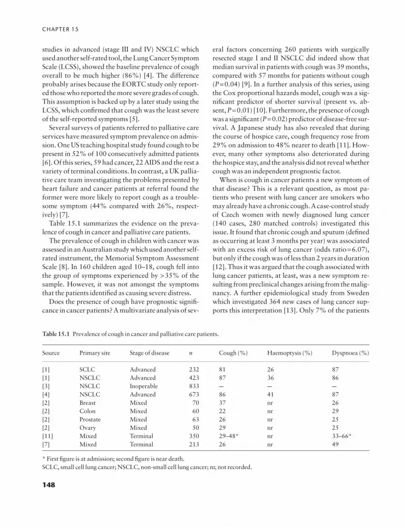
CHAPTER 15
studies in advanced (stage III and IV) NSCLC whichused another self-rated tool, the Lung Cancer SymptomScale (LCSS), showed the baseline prevalence of coughoverall to be much higher (86%) [4]. The differenceprobably arises because the EORTC study only report-ed those who reported the more severe grades of cough.This assumption is backed up by a later study using theLCSS, which confirmed that cough was the least severeof the self-reported symptoms [5].
Several surveys of patients referred to palliative careservices have measured symptom prevalence on admis-sion. One US teaching hospital study found cough to bepresent in 52% of 100 consecutively admitted patients[6]. Of this series, 59 had cancer, 22 AIDS and the rest avariety of terminal conditions. In contrast, a UK pallia-tive care team investigating the problems presented byheart failure and cancer patients at referral found theformer were more likely to report cough as a trouble-some symptom (44% compared with 26%, respect-ively) [7].
Table 15.1 summarizes the evidence on the preva-lence of cough in cancer and palliative care patients.
The prevalence of cough in children with cancer wasassessed in an Australian study which used another self-rated instrument, the Memorial Symptom AssessmentScale [8]. In 160 children aged 10–18, cough fell intothe group of symptoms experienced by >35% of thesample. However, it was not amongst the symptomsthat the patients identified as causing severe distress.
Does the presence of cough have prognostic signifi-cance in cancer patients? A multivariate analysis of sev-
eral factors concerning 260 patients with surgically resected stage I and II NSCLC did indeed show that median survival in patients with cough was 39 months,compared with 57 months for patients without cough(P=0.04) [9]. In a further analysis of this series, usingthe Cox proportional hazards model, cough was a sig-nificant predictor of shorter survival (present vs. ab-sent, P=0.01) [10]. Furthermore, the presence of coughwas a significant (P=0.02) predictor of disease-free sur-vival. A Japanese study has also revealed that duringthe course of hospice care, cough frequency rose from29% on admission to 48% nearer to death [11]. How-ever, many other symptoms also deteriorated duringthe hospice stay, and the analysis did not reveal whethercough was an independent prognostic factor.
When is cough in cancer patients a new symptom ofthat disease? This is a relevant question, as most pa-tients who present with lung cancer are smokers whomay already have a chronic cough. A case-control studyof Czech women with newly diagnosed lung cancer(140 cases, 280 matched controls) investigated thisissue. It found that chronic cough and sputum (definedas occurring at least 3 months per year) was associatedwith an excess risk of lung cancer (odds ratio=6.07),but only if the cough was of less than 2 years in duration[12]. Thus it was argued that the cough associated withlung cancer patients, at least, was a new symptom re-sulting from preclinical changes arising from the malig-nancy. A further epidemiological study from Swedenwhich investigated 364 new cases of lung cancer sup-ports this interpretation [13]. Only 7% of the patients
148
Table 15.1 Prevalence of cough in cancer and palliative care patients.
Source Primary site Stage of disease n Cough (%) Haemoptysis (%) Dyspnoea (%)
[1] SCLC Advanced 232 81 26 87[1] NSCLC Advanced 423 87 36 86[3] NSCLC Inoperable 833 — — —[4] NSCLC Advanced 673 86 41 87[2] Breast Mixed 70 37 nr 26[2] Colon Mixed 60 22 nr 29[2] Prostate Mixed 63 26 nr 25[2] Ovary Mixed 50 29 nr 25[11] Mixed Terminal 350 29–48* nr 33–66*[7] Mixed Terminal 213 26 nr 49
* First figure is at admission; second figure is near death.SCLC, small cell lung cancer; NSCLC, non-small cell lung cancer; nr, not recorded.

COUGH IN CANCER PATIENTS
were asymptomatic at diagnosis, and cough was themost common first symptom to be reported.
It is not altogether clear why cough should be such asensitive indicator of a new lung cancer and may alsosignify a poorer outcome. One possible explanationcomes from a study of the rheological properties ofmucus taken by bilateral paired sampling in patientsbeing bronchoscoped for a unilateral radiological ab-normality [14]. Eight of the 20 patients studied werefound to have lung cancer. Mucus from the side with theradiological abnormality had a lower value for the losstangent tan d100 (P=0.004), indicating greater mucusrecoil. This would be consistent with poor mucus clear-ability on the affected side. All of the eight cancer pa-tients had a low tan d100 value (P=0.007), and two ofthose who were initially negative from bronchoscopybut had abnormal mucus values were later diagnosedwith cancer on follow-up. These interesting findingsneed to be confirmed, but may have implications fortherapeutic management as well as their diagnosticvalue.
Causes and consequences of cough incancer patients
The study by Zayas et al. [14] described above shedslight on one of the possible causes of cough as a trou-blesome symptom in cancer patients. If the presence ofmalignant change in the bronchial tree can inducechanges which lead to mucus that is more difficult toclear and expectorate, then it is evident that the patientcould complain of difficulty in coughing.
Several other causative and associated factors alsoneed to be considered [15,16]. Table 15.2 gives a sum-mary of the conditions which may initiate or provokecough in cancer patients. Infection of the respiratorytract is naturally a major suspect, as is chronic obstruc-tive pulmonary disease, since most patients with lungcancer, at least, have a longstanding smoking history. Itis important to consider unusual pathogens, especiallyin immunocompromised patients. In one series of suchpatients, invasive pulmonary aspergillosis was accom-panied by cough in 54% of cases, fever in 54%,haemoptysis in 30% and dyspnoea in 8% [17]. Sincecancer in adults is a disease that is commoner in themiddle-aged and elderly, it is important to consider co-morbidity as a causative factor. Heart failure, the use ofangiotensin-converting enzyme (ACE) inhibiting drugs
for cardiac disease, systemic inflammatory diseases andpulmonary thromboembolism should be excluded.The latter is particularly relevant in view of the increased blood coaguability associated with some cancers, and the insidious nature of recurrent thromboembolic episodes which may be individuallysubclinical.
Complications of lung cancer and sometimes othermalignancies which give rise to increased coughing in-clude pleural or pericardial effusion, superior venacava obstruction, bronchopleural fistula and lymphan-gitis carcinomatosa. These are usually evident clinic-ally or on routine chest radiographs or computedtomography (CT) scan, although lymphangitis may bedifficult to diagnose in a patient with pre-existing pul-monary disease or heart failure.
Unfortunately, cough is a common consequence ofmany of the treatments which are used against canceritself. Studies of long-term survivors of cancer have re-ported cough as one of the symptoms which both chil-dren and adults suffer long after the disease has beentreated. The Childhood Cancer Survivor Study whichinvestigated 12390 ex-patients in the USA 5 years ormore after their illness found that, compared with sib-lings, survivors had significantly increased relative riskof chronic cough as well as recurrent pneumonia, lungfibrosis, pleurisy and exercise-induced breathlessness[18]. Specifically, the study identified the association ofchronic cough with prior chest radiation therapy (RR=2.0, P<0.001); with bleomycin exposure (RR=1.9, P<0.001); and with cyclophosphamide therapy (RR=1.3,P=0.004). The propensity for these anticancer thera-pies to cause pulmonary damage has been known for along time, although cyclophosphamide-induced lungdamage is relatively rare [19].
A recent study in Sweden has investigated the rela-tively new approach of allogeneic haematopoietic stemcell transplantation (HSCT) in adult cancer patients[20]. Twenty-five patients who had received HSCT 2–4years previously were surveyed using a variety of symp-tom and health-related QoL measures. More than halfthe patients reported cough, dry mouth and eye prob-lems, as well as tiredness, sexual difficulties and anxi-ety. It should be noted that, in spite of these reportedproblems, 80% of the cancer survivors estimated theirgeneral health as ‘quite good’ or ‘excellent’. In contrast,another Swedish follow-up study of 277 men who hadbeen cured of testicular teratoma, compared with 392controls, revealed that they reported significantly less
149

CHAPTER 15
cough, as well as less backache, leg pains and eye prob-lems than the controls [21].
Autologous peripheral blood stem cell transplanta-tion which uses busulphan chemotherapy can alsocause an acute or subacute idiopathic pneumonia syn-drome (IPS) characterized by cough, hypoxaemia andpulmonary infiltrates, sometimes with pleural effu-sions. Although IPS only arose in 10 out of a series of271 patients, it was fatal in eight of these [22]. A moreinsidious form of this condition has also been describedas delayed pulmonary toxicity syndrome (DPTS), following the use of high-dose chemotherapy with cyclophosphamide/cisplatin/BCNU and autologousbone marrow transplantation [23].
Table 15.3 gives a summary of the common treat-ments used for cancer that have been shown to be asso-ciated with pulmonary damage, cough and dyspnoea.Many ‘traditional’ anticancer drugs can cause intersti-tial pneumonia, leading to pulmonary fibrosis. Amongthe newer agents that are being used for cancers of thebreast and thorax are the taxanes. Six out of 30 NSCLCand mesothelioma patients who received paclitaxelwith concurrent chest radiotherapy developed signifi-cant lung injury manifested by a persistent cough [24].
150
Table 15.2 Conditions that may initiate or provoke cough in cancer patients.
System Site Pathology/mechanism
Respiratory Upper airways Laryngeal tumourTracheal tumourIatrogenic, e.g. radiation tracheitis
Bronchi Lung cancerMetastatic cancerInfectionChronic obstructive pulmonary disease
Lung Infectionparenchyma Lymphangitis carcinomatosa
Iatrogenic, e.g. pneumonitis and/or fibrosis after radiation, chemotherapy, biological therapies
Pleura MesotheliomaPleural effusion
Cardiovascular Heart Pericardial effusionPulmonary congestionPulmonary thromboembolismIatrogenic, e.g. angiotensin-converting enzyme inhibitor
Gastrointestinal Oesophagus Tracheo-oesophageal fistulaReflux
Table 15.3 Common treatments used for cancer that havebeen shown to be associated with pulmonary damage, coughand dyspnoea.
Type of therapy Pathology
Radiation Acute lymphocytic alveolitisAcute pneumonitisBronchiolitis obliterans
organizing pneumonia (BOOP)Late pulmonary fibrosis
Anticancer Acute idiopathic pneumonia chemotherapy syndrome (IPS)
Delayed pulmonary toxicity syndrome (DTPS)
Interstitial pneumonitisLate pulmonary fibrosis
Biological therapies Idiopathic cough —herceptin;interleukin

COUGH IN CANCER PATIENTS
Irinotecan is another new antineoplastic agent which isincreasingly being used in the management of colorec-tal cancer. Cumulative evidence from clinical trials inJapan and the US suggests that up to 20% of patientsmay develop pulmonary toxicity, causing dyspnoeaand cough. The drug initiates an interstitial pneumoni-tis that may not respond to corticosteroids and can befatal [25]. It is claimed that patients with pre-existinglung disease may be at a higher risk of irinotecan pul-monary toxicity.
Amplification of the human epidermal growth factorreceptor 2 protein (HER2) correlates with poor prog-nosis in breast cancer, and herceptin is a recombinanthumanized monoclonal immunoglobulin antibodythat binds to the HER2 receptor. It is being increasinglyused in breast cancer trials. One of its commoner side-effects is cough, along with fever and chills, pain andtiredness [26]. Interleukin-2 (IL-2) is used as an anti-cancer agent for metastatic melanoma. One of its manydose-dependent side-effects is cough [27].
Radiation therapy may causes an acute lymphocyticalveolitis or hypersensitivity pneumonitis, which is as-sociated with dyspnoea and cough within weeks or afew months of the dose [28]. In a study of 256 lung can-cer patients receiving radical doses of radiotherapy,49% developed acute radiation pneumonitis althoughit was severe in only 13% [29]. Late radiation damage isevident as pulmonary fibrosis, and may take severalmonths or years to develop. The fibrosis results fromthe effects of tissue repair mechanisms following pneu-monitis within the irradiated area, which involve theactivation of fibroblasts by transforming growth fac-tor-b, fibronectin and platelet-derived growth factor[28]. For longer-term survivors of cancer, radiation fibrosis is a sinister development as it can lead to severesymptoms and terminal respiratory failure. There is evidence that a smoking history can protect patientsfrom developing radiation-induced pneumonitis [30].
Apart from lung cancer, breast cancer is another malignancy that often involves thoracic radiation. Thesyndrome of bronchiolitis obliterans organizing pneumonia (BOOP) has been described as a rare (2.5%)complication of postoperative radiotherapy followingbreast-conserving surgery [31]. These patients developfever, non-productive cough and patchy unilateral orbilateral pulmonary infiltrates 5–6 months afterradiotherapy. Postradiation BOOP is a serious condi-tion which tends to recur even after long-term cortico-steroidtherapy.Modern radiation planning techniques,
which limit the total dose to normal lung, have mini-mized the risk of BOOP and IPS, but they may still be arisk for patients requiring radiation therapy, who havepre-existing chronic pulmonary disease.
There are some other less common but interesting iatrogenic causes of disordered or diminished cough incancer patients. For example, with concurrent radio-therapy and chemotherapy for head and neck cancer,14% of patients in one study exhibited aspiration priorto treatment, 65% soon after therapy and 62% in thelate post-therapy stage (aspiration rates pre-therapy vs.post-therapy P=0.0002) [32]. This typically silent as-piration was a result of disordered epiglottal and pha-ryngeal muscle coordination, leading to a diminishedcough reflex or cough which was delayed or ineffectivein expectorating the aspirated material. Reduced cougheffectiveness is also seen after unilateral vocal cordparalysis, which is often the result of mediastinal encroachment of a bronchial cancer, but may also result iatrogenically during thoracic surgery for the disease [33].
Assessment of cough in cancer patients
The formal methods of assessing cough are coveredelsewhere in this volume. Specific symptom measure-ment tools devised for cancer patients, which could be useful for evaluating the impact of cough on func-tioning and QoL include the EORTC LC-13; the LungCancer Symptom scale; and the Functional Assessmentof Cancer Treatment (FACT) lung cancer module.
Investigations used in determining the source ofcough in cancer patients are the same as for other dis-ease groups. Thus, the most useful tests will includethoracic imaging by X-ray or CT scan; ultrasound inthe case of suspected pleural or pericardial effusion;and ventilation/perfusion scanning for suspected pul-monary thromboembolism. Bronchoscopy is rarelynecessary once a diagnosis of cancer is made, unless anendobronchial metastasis from a remote primary site isbeing considered.
Management of cough in cancer patients
This topic will be approached under the following sections —specific anticancer therapies; laser and
151

CHAPTER 15
photodynamic therapy; palliative drug therapies; andmanagement in terminal care. Table 15.4 summarizesthese methods of managing cough. One of the difficul-ties of presenting and comparing different methods ofcough palliation is that most studies have used a varietyof outcome measures, timescales and definitions of‘successful’ palliation. The UK Medical ResearchCouncil has also found that success depends on thestarting point: whereas 90% of patients with lung can-cer who had severe or moderate grades of cough atbaseline reported improvement after treatment, only53% who started with mild cough did so [34].
Anticancer therapies
Radiation therapyRadiotherapy may be offered to the cancer patient fortwo reasons —with the primary aim of curing disease orextending survival (radical treatment), or with the pri-mary aim of palliating symptoms (palliative treat-ment). Table 15.5 gives a summary of the results ofradiotherapy for palliation of respiratory symptoms.Most of the radiation studies have concentrated on theformer, until recent years. Unfortunately, symptom re-lief was often considered of less importance comparedwith survival and tumour response in the case of radicaltreatment, and so the earlier research shed little light onsubjective response. Thus, a US review of 156 patientswith clinical stage I NSCLC, who were medically inop-erable and therefore treated with radical intent (mediandose 64Gy) between 1980 and 1995, states that before
treatment 23% of patients had cough [35]. However,the paper does not reveal to what extent radiotherapyrelieved the cough. The analysis did show that the pres-ence of cough prior to treatment was associated with ahigher local failure rate.
A European EORTC study, however, did investigaterespiratory symptoms as well as QoL changes in 164patients with NSCLC receiving a radical (60Gy) doseof radiotherapy [36]. This study showed that there wasa significant association between objective tumour response and the palliation of pain and physical func-tioning. During radiation treatment, many generalsymptoms and QoL deteriorated. However, after treat-
152
Table 15.4 Methods used to manage cough in cancerpatients.
Type of therapy Examples
Anticancer chemotherapy Single agents, e.g. gemcitabineCombination regimens
Radiotherapy Hypofractionated regimens
Laser therapy Nd-YAG laserPhotodynamic therapy
Palliative drugs Anti-inflammatory, e.g. corticosteroid
Suppressant, e.g. opioidAntimuscarinic, e.g. hyoscine,
glycopyrrolate
Table 15.5 Summary of the results of radiotherapy for palliation of respiratorysymptoms in non-small cell lung cancer (figures quoted are response rates for eachsymptom).
Dose Study Cough (%) Haemoptysis (%) Dyspnoea (%)
Radical (curative) [36] 31 83 3760Gy
Conventional [39] 49 79 3palliation
Hypofractionated [37] 48 95 4617Gy
Hypofractionated [42] 24 60 5517Gy
Hypofractionated [41] 68 77 4220Gy
Gy, Gray.

COUGH IN CANCER PATIENTS
ment, cough improved in 31% of patients, haemoptysisin 83% and dyspnoea in 37%. Global QoL improved in 36%.
In order to improve tumour response to radical ra-diotherapy, there has been a move towards increasingthe number of doses given daily (hyperfractionation).Continuous hyperfractionated radiotherapy (CHART)is being used in potentially curable cancer patients whoare unable to have surgical resection. Studies ofCHART have focused, not surprisingly, on long-termobjective outcomes. However, short-term monitoringof patients with head and neck cancer having CHARThas revealed that cough and hoarseness were improved,compared with conventional dose radiotherapy, at 6weeks [38].
Studies of palliative regimens for radiotherapy havenaturally focused on symptom control as a key end-point. The same EORTC group just mentioned abovealso reported on palliative responses in patients with lo-cally advanced and metastatic NSCLC [39]. In compar-ison with their radical treatment study, cough wasrelieved in 49% of patients, haemoptysis in 79% anddyspnoea in 39%. Once again, global QoL improved in37% of patients after completion of radiotherapy andthere was a tendency for patients who had an objectivetumour response to have a symptomatic benefit.
A Cochrane systematic review examined 10 pub-lished randomized trials of palliative radiotherapy forlung cancer. Meta-analysis of the results was not possi-ble because of great heterogeneity in the doses, patientcharacteristics and outcome measures used. There wasno convincing evidence that any particular regime gavea better symptom resolution. The conclusion from thisreview was that most patients should be treated withminimum doses delivered in one or two fractions (hy-pofractionation) [40]. This approach would be special-ly useful for patients who are ill, cannot be admitted tohospital or cannot attend for long outpatient courses.In India a study examined the effect of giving four smallweekly doses to a total of 20Gy to 47 inoperable pa-tients with NSCLC [41]. Not only was the regime welltolerated and convenient for patients who had long dis-tances to travel, but it also gave relief of cough in 68%,of haemoptysis in 77% and of dyspnoea in 42%. Hy-pofractionation was also used in a Greek study of 48NSCLC patients who had two doses of 8.5Gy 1 weekapart [42]. Cough was palliated in 24% with a medianpalliation duration of 70 days; haemoptysis in 60%over 133 days; and dyspnoea in 55% over 74 days.
Should palliative radiotherapy be offered routinelyat the time of diagnosis, or only as needed when symp-toms develop? This question was tackled by the UKMedical Research Council in an international study in-volving 230 patients with advanced inoperableNSCLC, who had no immediate reasons for radiother-apy [43]. The results showed that there was no differ-ence between the immediate or deferred treatmentapproaches in terms of symptom relief, psychologicaldistress or survival. Adverse effects were more likelyafter immediate radiotherapy.
When lung cancer recurs after a course of radiother-apy, it is unusual but it may be feasible to offer a secondcourse of treatment. A retrospective study evaluatingthe outcome of reirradiation showed that 60% of pa-tients achieved improvement in cough, 73% in dysp-noea, and all patients with haemoptysis had a reductionor complete resolution [44]. In recent years, the pre-ferred approach in specialist centres for such patients isto offer endobronchially delivered intraluminal irradi-ation (brachytherapy). This involves subjecting the pa-tient to a bronchoscopy and placing a catheter in theairway down to the level of the tumour. An after-load-ing device then sends a radioactive source via thecatheter to the tumour site for a short time before withdrawal [45]. Several recent small studies have documented good palliative benefit after brachytherapy —relief of cough, haemoptysis and dys-pnoea occurring in 54%, 74% and 54%, respectively[46]; 65%, 86% and 64%, respectively [47]; and 43%,95% and 80%, respectively [48]. One problem withbrachytherapy is that it is only available in a few centreswith the necessary radioisotope equipment; another isthat it has been associated with dose-dependent latetoxicities, especially massive haemoptysis. Tauelle et al.[46] reported severe or fatal haemoptysis occurring in7% of their patients; Hatlevoll et al. [47] in 27% (22%within 6 months); and Anacak et al. [48] in 11%.
ChemotherapyAs with radiation therapy, in the past the emphasis inresearch was on achieving ‘cure’, so that the end-pointswere usually survival and objective tumour responserather than symptom relief. In SCLC, chemotherapyhas long been the preferred option as the disease is oftendisseminated and, unlike NCSLC, is very sensitive toantimitotic drugs. It is well established that objectivetumour response in SCLC is associated with symptomimprovement. In various trials of standard combina-
153

CHAPTER 15
tion chemotherapy regimens, relief of cough has been reported in between 45% and 70% of patients; ofhaemoptysis in up to 92%; and of dyspnoea in between46% and 78% [45]. A recent study in poor prognosisSCLC patients comparing combination chemotherapyvs. single-agent carboplatin showed that even in thisvery sick population, good symptom palliation couldbe achieved [49]. The combination regime (CAV) gave 21% relief of cough compared with 7% after car-boplatin; dyspnoea improved in 66% and 41%, respec-tively. In terms of toxicity and objective tumourresponse, there were few differences.
Gemcitabine is a new anticancer drug which is givensingly or in combination with others. As a single agentfor inoperable NSCLC it has given relief of cough in44%, of haemoptysis in 63% and of dyspnoea in 26%[45]. In combination with cisplatin these response rateswere 44%, 75% and 36%, respectively [50]. It is likelythat other new agents such as the taxanes will be usedincreasingly for symptom palliation in NSCLC. Single-agent chemotherapy (as well as talc and tetracycline) isalso used to prevent the recurrence of pleural and peri-cardial effusions [54]. Table 15.6 summarizes the results of recent studies of palliating respiratory symp-toms in NSCLC.
Laser and photodynamic therapy
Laser therapy for the palliation of cough is delivered in-traluminally via a bronchoscope. Unlike brachyther-apy which was described above, laser therapy works bymacroscopically heating and destroying tissues. Photo-dynamic therapy (PDT) is a refinement of the laserprocess by premedicating patients with tissue sensitiz-
ers which allow greater killing of tumour as comparedwith normal cells [15]. Both of these techniques aremuch less commonly available than radiotherapy (either external or endobronchial) or chemotherapy.Studies of their effectiveness have been relatively smalland mostly uncontrolled. However, a recent random-ized controlled trial was performed in 31 patients withtracheobronchial obstruction due to inoperableNSCLC [55]. The study directly compared laser resec-tion using an Nd-YAG laser with dihaematoporphyrinether and argon dye laser photoradiation. There was nodifference between the two techniques in terms ofsymptom relief, but PDT gave a longer time till recur-rence of symptoms and a longer median survival.
Drug therapies
The drug management of cough is described in detailelsewhere in this volume, as well as in palliative medi-cine texts [15,16], so only a few points specific to cancerpatients will be highlighted. Most palliative care unitsuse codeine-based drugs for relieving cough. For patients who are resistant to codeine, other drugswhich have been recommended include hydrocodone[56], benzonatate [57] and levodropropizine [58].There are no published trials comparing the relativebenefits or side-effects of these agents in cancer pa-tients. A potential problem arises when a cancer patientis already receiving a potent opioid (morphine, hydro-morphone, fentanyl, etc.) for pain, and then complainsof a cough. It does not make sense pharmacologically to add a second opioid drug, as there is no evidence that incomplete cross-tolerance exists between opioidsfor cough (unlike the situation with pain modulation).
154
Table 15.6 Summary of the results of chemotherapy for palliation of respiratorysymptoms in non-small cell lung cancer (figures quoted are response rates for eachsymptom).
Regimen Study Cough (%) Haemoptysis (%) Dyspnoea (%)
Cisplatin/vindesine+ [51] 45 91 78mitomycin or ifosfamide
Mitomycin/ifosfamide/ [52] 70 92 46cisplatin
Gemcitabine alone [53] 44 63 26Gemcitabine/cisplatin [50] 44 75 36

COUGH IN CANCER PATIENTS
In this situation, it would be appropriate to use anotherclass of drug or another approach entirely, such as radiotherapy.
In palliative care centres, nebulized lidocaine orbupivacaine have sometimes been advocated for the re-lief of intractable unproductive cough [16]. Thismethod is thought to work by anaesthesizing sensorynerve endings in the hypopharynx, larynx and upperairways that are involved in generating the cough re-flex. Other drugs which have theoretical actions oncough and may be tried in refractory cases include baclofen and mexilitine [16].
As discussed above, cough can arise as a symptom ofpulmonary toxicity after some anticancer drugs. Re-cently there have been attempts to prevent or reverse the pulmonary damage using novel drug approaches. In an open uncontrolled US study, 63 patients withbreast cancer who were starting high-dose chemother-apy prior to autologous bone marrow transplantationwere given an inhaled corticosteroid —fluticasone pro-pionate [59]. After 12 weeks of taking this drug twicedaily, the study found that, compared with the centre’shistorical controls, DLCO (the diffusing capacity of thelungs) declined by a smaller amount (21% fluticasonevs. 33% historical controls). Delayed pulmonary toxic-ity syndrome developed in 35% of fluticasone patientscompared to 73% of historical controls. Because of the grave significance of such pulmonary toxicity, fur-ther randomized trials of this and similar regimens arerequired.
Management in terminal care
In the care of cancer patients, cough may become verydistressing in the terminal stage. The act of coughingmay cause or aggravate pain, it may disturb precioussleep and it can impede communication between pa-tients and carers. If the cough is productive, it is helpfulto position the patient so that expectoration is assisted,and gentle chest physiotherapy may be used [15]. Itmay be appropriate to offer an antibiotic for a respira-tory tract infection, if the symptoms of cough and dysp-noea are very distressing. Patients with cancer are oftenprescribed drugs which can cause dry mouth and re-duction of tracheobronchial mucus (opioids, anti-cholinergics, antihistamines) and this can causedifficulty in expectoration. It may be helpful in such sit-uations to humidify the air or oxygen, if the patientneeds the latter for hypoxaemia.
Some patients develop noisy breathing as they slipinto unconsciousness (so-called death rattle). The causeof this is thick mucus lying in the major airways or hy-popharynx, which partly obstructs the airflow, butwhich the patient is unable to cough up. The patient isusually unaware of the noise, but it can be very upset-ting to family carers or others nearby. Suction may helpif there is mucus in or above the larynx. Often the mostpractical management is to prevent further mucus pro-duction by giving an antimuscarinic agent, preferablyby subcutaneous injections or by continuous infusions.The best agents for this purpose are hysoscine butyl-bromide or glycopyrrolate [60].
References
1 Hopwood P, Stephens RJ on behalf of the Medical Re-search Council (MRC) Lung Cancer Working Party.Symptoms at presentation for treatment in patients withlung cancer: implications for the evaluation of palliativetreatment. Br J Cancer 1995; 71: 633–6.
2 Portenoy RK, Thaler HT, Kornblith AB etal. Symptomprevalence, characteristics and distress in a cancer popula-tion. Qual Life Res 1994; 3: 183–9.
3 Bergmann B, Aaronson NK, Ahmedzai S etal. TheEORTC QLC-LC13: a modular supplement to theEORTC Core Quality of Life Questionnaire (QLC-C30)for use in lung cancer clinical trials. Eur J Cancer 1994;30A: 635–42.
4 Hollen PJ, Gralla RJ, Kris MG etal. Normative data andtrends in quality of life from the Lung Cancer SymptomScale (LCSS). Support Care Cancer 1999; 7: 140–8.
5 Lutz S, Norrell R, Bertucio C etal. Symptom frequencyand severity in patients with metastatic or locally recur-rent lung cancer: a prospective study using the Lung Cancer Symptom Scale in a community hospital. PalliatMed 2001; 4: 157–65.
6 Ng K, von Gunten CF. Symptoms and attitudes of 100 consecutive patients admitted to an acute hospice/palliative care unit. J Pain Symptom Manage 1998; 16:307–16.
7 Anderson H, Ward C, Eardley A etal. The concerns of pa-tients under palliative care and a heart failure clinic are notbeing met. Palliat Med 2001; 15: 279–86.
8 Collins JJ, Byrns ME, Dunkel IJ etal. The measurement ofsymptoms in children with cancer. J Pain Symptom Man-age 2000; 19: 363–77.
9 Mehdi SA, Etxell JE, Newman NB etal. Prognostic signifi-cance of Ki-67 immunostaining and symptoms in resectedstage I and II non-small cell lung cancer. Lung Cancer1998; 20: 99–108.
155

CHAPTER 15
10 Mehdi SA, Tatum AH, Newman NB etal. Prognosticmarkers in resected stage I and II non-small-cell lung cancer: an analysis of 260 patients with 5 year follow-up.Clin Lung Cancer 1999; 1: 59–67.
11 Morita T, Tsunoda J, Inoue S etal. Contributing factors tophysical symptoms in terminally-ill cancer patients. J PainSymptom Manage 1999; 18: 338–46.
12 Kubik A, Zatloukal P, Boyle P etal. A case-control study oflung cancer among Czech women. Lung Cancer 2001; 31:111–22.
13 Koyi H, Hillerdal G, Branden E. A prospective study of atotal material of lung cancer from a county in Sweden1997–1999; gender, symptoms, type, stage and smokinghabits. Lung Cancer 2002; 36: 9–14.
14 Zayas JG, Rubin BK, York EL etal. Bronchial mucusproperties in lung cancer: relationship with site of lesion.Can Respir J 1999; 6: 246–52.
15 Ahmedzai SH. Palliation of respiratory symptoms. In:Doyle D, Hanks GWC, McDonald N, eds. Oxford Text-book of Palliative Medicine, 2nd edn. Oxford: OxfordUniversity Press, 1998: 583–616.
16 Twycross R, Wilcock A. Respiratory symptoms. In:Twycross R, Wilcock A, eds. Symptom Management inAdvanced Cancer, 2nd edn. Oxford: Radcliffe Press,1997: 141–79.
17 Pidhorecky I, Urschel J, Anderson T. Resection of invasivepulmonary aspergillosis in immunocompromised pa-tients. Ann Surg Oncol 2000; 7: 312–7.
18 Mertens AC, Yasui Y, Liu Y etal. Pulmonary complica-tions in survivors of childhood and adolescent cancer. Areport from the Childhood Cancer Survivor Study. Cancer2002; 95: 2431–41.
19 Segura A, Yuste A, Cercos A etal. Pulmonary fibrosis in-duced by cyclophosphamide. Ann Pharmacother 2001;35: 894–7.
20 Edman L, Larsen J, Hagglund H etal. Health-related qual-ity of life, symptom distress and sense of coherence in adultsurvivors of allogeneic stem-cell transplantation. Eur JCancer Care (Engl) 2001; 10: 124–30.
21 Rudberg L, Carlsson M, Nilsson S etal. Self-perceivedphysical, psychologic, and general symptoms in survivorsof testicular cancer 3–13 years after treatment. CancerNurs 2002; 25: 187–95.
22 Bilgrami SF, Metersky ML, McNally D etal. Idiopathicpneumonia syndrome following myeloablativechemotherapy and autologous transplantation. AnnPharmacother 2001; 35: 196–201.
23 Wilczynski SW, Erasmus JJ, Petros WP etal. Delayed pul-monary toxicity syndrome following high-dosechemotherapy and bone marrow transplantation forbreast cancer. Am J Respir Crit Care Med 1998; 157:565–73.
24 Herscher LL, Hahn SM, Kroog G etal. Phase I study of pa-
clitaxel as a radiation sensitizer in the treatment ofmesothelioma and non-small-cell lung cancer. J ClinOncol 1998; 16: 635–41.
25 Madarnas Y, Webster P, Shorter AM etal. Irinotecan-associated pulmonary toxicity. Anticancer Drugs 2000;11: 709–13.
26 Goldenberg MM. Trastuzumab, a recombinant DNA-derived humanized monoclonal antibody, a novel agentfor the treatment of metastatic breast cancer. Clin Ther1999; 21: 309–18.
27 Eton O, Rosenblum MG, Legha SS etal. Phase I trial ofsubcutaneous recombinant human interleukin-2 in patients with metastatic melanoma. Cancer 2002; 95:127–34.
28 Abratt RP, Morgan GW. Lung toxicity following chest irradiation in patients with lung cancer. Lung Cancer2002; 35: 103–9.
29 Inoue A, Kunitoh H, Sekine I etal. Radiation pneumonitisin lung cancer patients: a retrospective study of risk factorsand the long-term prognosis. Int J Radiat Oncol Biol Phys2001; 49: 649–55.
30 Johansson S, Bjermer L, Franzen L etal. Effects of ongoingsmoking on the development of radiation-induced pneu-monitis in breast cancer and oesophagus cancer patients.Radiother Oncol 1998; 49: 41–7.
31 Takigawa N, Segawa Y, Saeki T etal. Bronchiolitis obliter-ans organizing pneumonia syndrome in breast-conservingtherapy for early breast cancer: radiation-induced lungtoxicity. Int J Radiat Oncol Biol Phys 2000; 48: 751–5.
32 Eisbruch A, Lyden T, Bradford CR etal. Objective assess-ment of swallowing dysfunction and aspiration after radiation concurrent with chemotherapy for head-and-neck cancer. Int J Radiat Oncol Biol Phys 2002; 53:23–8.
33 Abraham MT, Bains MS, Downey RJ etal. Type I thyro-plasty for acute unilateral vocal fold paralysis followingintrathoracic surgery. Ann Otol Rhinol Laryngol 2002;111: 667–71.
34 Stephens RJ, Hopwood P, Girling DJ. Defining andanalysing symptom palliation in cancer clinical trials: a deceptively difficult exercise. Br J Cancer 1999; 79:538–44.
35 Sibley GS, Jamieson TA, Marks LB etal. Radiotherapyalone for medically inoperable stage I non-small-cell lungcancer: the Duke experience. Int J Radiat Oncol Biol Phys1998; 40: 149–54.
36 Langendijk JA, Aaronson NK, de Jong JM etal. Prospec-tive study on quality of life before and after radical radio-therapy in non-small-cell lung cancer. J Clin Oncol 2001;19: 2123–33.
37 Medical Research Council Lung Cancer Working Party.Randomized trial of palliative two-fraction versus moreintensive 13 fraction radiotherapy for patients with inop-
156

COUGH IN CANCER PATIENTS
erable non-small cell lung cancer and good performancestatus. Clin Oncol 1996; 8: 167–75.
38 Griffiths GO, Parmar MK, Bailey AJ. Physical and psy-chological symptoms of quality of life in the CHART ran-domized trial in head and neck cancer: short-term andlong-term patient reported symptoms. CHART SteeringCommittee. Continuous hyperfractionated acceleratedradiotherapy. Br J Cancer 1999; 81: 1196–205.
39 Langendijk JA, ten Velde GP, Aaronson NK etal. Qualityof life after palliative radiotherapy in non-small cell lungcancer: a prospective study. Int J Radiat Oncol Biol Phys2000; 47: 149–55.
40 Macbeth F, Toy E, Coles B etal. Palliative radiotherapyregimens for non-small cell lung cancer. [Update ofCochrane Database Syst Rev 2001; 2: CD002143; PMID: 11406035.] Cochrane Database Syst Rev Issue 4,2002.
41 Bhatt ML, Mohani BK, Kumar L etal. Palliative treat-ment of advanced non small cell lung cancer with weekly fraction radiotherapy. Indian J Cancer 2000; 37:148–52.
42 Plataniotis GA, Kouvaris JR, Dardoufas C etal. A short radiotherapy course for locally advanced non-small celllung cancer (NSCLC): effective palliation and patients’convenience. Lung Cancer 2002; 35: 203–7.
43 Falk SJ, Girling DJ, White RJ etal. Immediate versus de-layed palliative thoracic radiotherapy in patients with un-resectable locally advanced non-small cell lung cancer andminimal thoracic symptoms: randomised controlled trial.Br Med J 2002; 325: 465.
44 Gressen EL, Werner-Wasik M, Cohn J etal. Thoracicreirradiation for symptomatic relief after prior radiother-apeutic management for lung cancer. Am J Clin Oncol2000; 23: 160–3.
45 Hoskin P, Ahmedzai SH. Assessment and management ofrespiratory symptoms of malignant disease. In: AhmedzaiSH, Muers M, eds. Supportive Care of the RespiratoryDisease Patient. Oxford: Oxford University Press, 2003(in press).
46 Taulelle M, Chauvet B, Vincent P etal. High dose rate en-dobronchial brachytherapy: results and complications in189 patients. Eur Respir J 1998; 11: 162–8.
47 Hatlevoll R, Karlsen KO, Skovlund E. Endobronchial ra-diotherapy for malignant bronchial obstruction or recur-rence. Acta Oncol 1999; 38: 999–1004.
48 Anacak Y, Mogulkoc N, Ozkok S etal. High dose rate en-
dobronchial brachytherapy in combination with externalbeam radiotherapy for stage III non-small cell lung cancer.Lung Cancer 2001; 34: 253–9.
49 White SC, Cheeseman S, Thatcher N etal. Phase II study oforal topotecan in advanced non-small cell lung cancer.Clin Cancer Res 2000; 6: 868–73.
50 Jassem J, Krzakowski M, Roszkowski K etal. A phase IIstudy of gemcitabine plus cisplatin in patients with ad-vanced non-small cell lung cancer: clinical outcomes andquality of life. Lung Cancer 2002; 35: 73–9.
51 Fernandez C, Rosell R, Abad-Esteve A etal. Quality of lifeduring chemotherapy in non-small cell lung cancer. ActaOncol 1989; 28: 29–33.
52 Cullen MH. The MIC regimen in non-small cell lung can-cer. Lung Cancer Suppl 1993; 2: 81–98.
53 Thatcher N, Anderson H, Betticher DC etal. Symptomaticbenefit from gemcitabine and other chemotherapy in advanced non-small cell lung cancer: changes in perform-ance status and tumour-related symptoms. AnticancerDrugs 1995; 6 (Suppl.): 39–48.
54 Paz-Ares L, Garcia-Carbonera R. Medical emergencies.In: Cavalli F, Hansen HE, Kaye SB, eds. Textbook of Medical Oncology, 2nd edn. Martin Dunitz, 2000:619–49.
55 Diaz-Jimenez JP, Martinez-Ballarin JE, Llunell A etal. Ef-ficacy and safety of photodynamic therapy versus Nd-YAG laser resection in NSCLC with airway obstruction.Eur Respir J 1999; 14: 800–5.
56 Homsi J, Walsh D, Nelson KA etal. Pain and symptommanagement. Hydrocodone for cough in advanced can-cer. Am J Hospice Palliative Care 2000; 17: 342–6.
57 Doona M, Walsh D. Benzonatate for opioid-resistantcough in advanced cancer. Palliat Med 1998; 12: 55–8.
58 Luporini G, Barni S, Marchi E etal. Efficacy and safety oflevodropropizine and dihydrocodeine on nonproductivecough in primary and metastatic lung cancer. Eur Respir J1998; 12: 97–101.
59 McGaughey DS, Nikcevich DA, Long GD etal. Inhaledsteroids as prophylaxis for delayed pulmonary toxicitysyndrome in breast cancer patients undergoing high-dosechemotherapy and autologous stem cell transplantation.Biol Blood Marrow Transplant 2001; 7: 274–8.
60 Bennett M, Lucas V, Brennan M etal. Using anti-muscarinic drugs in the management of death-rattle: evi-dence-based guidelines for palliative care. Palliat Med2002; 16: 369–74.
157


SECTION 4
Pathophysiology


161
Introduction
The cough reflex is one of several defensive reflexes thatserve to protect the airways from the potentially dam-aging effects of inhaled particulate matter, aeroaller-gens, pathogens, aspirate and accumulated secretions.In some airways diseases, cough may become excessiveand non-productive, and is potentially harmful to theairway mucosa. An understanding of the neural path-ways involved in the cough reflex may facilitate the de-velopment of therapeutic strategies that preventexcessive and non-productive cough, whilst preservingthe important innate defensive role of this respiratoryreflex.
Much of our current understanding of the neuralpathways involved in the cough reflex is derived fromstudies in animals. In cats, dogs, rabbits, guinea-pigsand monkeys it is clear that vagal afferent nerves are responsible for initiating the cough reflex [1–6]. Al-though poorly described, afferent nerves innervatingother viscera as well as somatosensory nerves innervat-ing the chest wall, diaphragm and abdominal muscula-ture also likely play an integral role in regulating cough.In this chapter the broad classes of vagal afferent nervesinnervating the airways and their role in regulating thecough reflex will be defined.
Defining airway afferent nerve subtypes
Airway afferent nerve subtypes can be distinguishedbased on their physicochemical sensitivity, adaptationto sustained lung inflation, neurochemistry, origin,
myelination, conduction velocity and sites of ter-mination in the airways. The utility of each of these approaches for defining airway afferent nerve subtypesis limited in large part by the lack of specificity of the various characteristics studied. When used in combination, however, these physiological and mor-phological attributes can be used to identify at leastthree broad classes of airway afferent nerves: rapidlyadapting mechanoreceptors (RARs), slowly adaptingmechanoreceptors (SARs) and unmyelinated C-fibres(Fig. 16.1).
The value of this now widely accepted classificationscheme used to define airway afferent nerve subtypes isconfirmed by its established utility in all species thus farstudied. Most airway afferent nerves found in any ofthese species are described reasonably well as eitherRAR, SAR or C-fibre. The observations that stereotyp-ical reflexes are initiated by stimuli selective (even if notspecific) for these afferent nerve subtypes and that thesereflexes are modulated by interventions that preferen-tially alter the activity or actions of the afferent nervesubtypes studied is further evidence for the utility ofthis classification scheme. Studies of central nervoussystem (CNS) termination sites of the various afferentnerve subtypes identified also confirm the utility of theclassification scheme [7]. Such analyses reveal the nec-essary divergence of afferent nerve terminals within theCNS, divergence that is required to differentially andspecifically control homeostatic and defensive reflexesinitiated from the airways.
The characteristics of the afferent nerve subtypes in-nervating the airways are described briefly in Table16.1 and in detail below.
161
Sensory pathways for the cough reflexStuart B. Mazzone, Brendan J. Canning & John G. Widdicombe
16

CHAPTER 16
162
Fig. 16.1 Afferent nerve subtypes innervating the mam-malian airways. Representative traces (upper panels) andmean data (lower panels) obtained from single-fibre record-ings of afferent nerve activity in the vagus nerve of anaes-thetized rats. (a) Airway C-fibres are generally quiescentduring tidal breathing and relatively unresponsive to lung in-flation (peak activity ~1 impulse/s). C-fibres, however, re-spond vigorously to intravenous injections of the vanilloidcapsaicin (peak activity ~20 impulses/s). Rapidly adapting
receptors (RARs) (b) and slowly adapting receptors (SARs)(c) are sporadically active during the respiratory cycle (10–40 impulses/s). Neither subtype of mechanoreceptor re-sponds to capsaicin (Cap), but both fire intensely when thelungs are inflated (peak activity ~40–120 impulses/s). Notethat RARs are easily differentiated from SARs since they rap-idly adapt during sustained lung inflation. Modified with per-mission from [10].
25
20
15
10
5
0
50
40
30
20
10
0
50
40
30
20
10
0
50
40
30
20
10
0
140120100
80604020
0
2
1
0
Cap Cap Cap
Inflation Inflation Inflation
Act
ivit
y (i
mp
/s)
Act
ivit
y (i
mp
/s)
–5 0 5 10 15Time (s)
–5 0 5 10 15Time (s)
–5 0 5 10 15Time (s)
Cap Cap Cap
C-fibre RAR SAR
(a) (b) (c)
10
0
150
0
30
0
150
0
AB
P(m
mH
g)
P t(c
mH
2O)
AB
P(m
mH
g)
P t(c
mH
2O)
AP
AP
10 s

SENSORY PATHWAYS FOR THE COUGH REFLEX
Afferent nerve subtypes innervating the airways and their role in regulating cough
Rapidly adapting receptors
Although relatively little is known about the anatomi-cal arrangement of RAR terminations in the airwaywall, functional studies suggest that RARs terminatewithin or beneath the epithelium and are localized toboth intra- and extrapulmonary airways [8–11]. RARsare typically differentiated from other airway afferentnerves by their rapid (1–2s) adaptation to sustainedlung inflations (Fig. 16.1) [10–14]. Other distinguish-ing properties of RARs include their sensitivity to lungcollapse and/or lung deflation, their responsiveness toalterations in dynamic lung compliance (and thus theirsensitivity to bronchospasm), and their conduction velocity (4–18m/s, suggestive of small, myelinatedaxons) [8–11,15]. Careful analysis of the responsive-
ness of RARs to mechanical stimulation reveals thattheir adaptation to sustained lung inflation is not at-tributable to an electrophysiological adaptation, aswith sustained, dynamic mechanical stimulation,RARs are continually activated [16,17]. RARs mayalso adapt comparatively slow to lung deflation [10].Perhaps RARs are thus better defined as dynamic re-ceptors that respond to changes in airway mechanicalproperties (e.g. diameter, length, interstitial pressures).
RARs are sporadically active throughout the respira-tory cycle, activated by the dynamic mechanical forcesaccompanying lung inflation and deflation and becom-ing more active as the rate and volume of lung inflationincreases [10,16,17]. It follows therefore that RAR ac-tivity during respiration correlates to respiratory rateand is higher in guinea-pigs and rats (16–27 impulses/s)and almost unmeasurable in larger animals such asdogs (<1 impulse/s). It also follows that, at least insmaller animals, RAR-dependent reflexes require aheightened activity in the already active RARs. This
163
Table 16.1 In vivo properties of vagal afferent nerve subtypes innervating the mammalian airways.
Rapidly adapting receptor Slowly adapting receptor C-fibre
Electrophysiological propertiesConduction velocity (m/s) 14–23 15–32 1.3–1.5Fibre type Myelinated Myelinated UnmyelinatedEupnoeic activity (impulses/s) 0–20 10–40 0.3–1.5
Physical sensitivityLung deflation Increased Decreased No effectOedema Increased No effect Increased
Chemical sensitivityCO2 No effect Decreased IncreasedH+ Increased No effect IncreasedCapsaicin Increased* No effect Increased†Bradykinin Increased* No effect Increased†
Reflex effectsParasympathetic Excitatory Inhibitory ExcitatoryRespiratory Hyperpnoea Inhibit inspiration ApnoeaOedema formation Promote No effect Promote
Neurokinin positive No No Yes
* Increased RAR activity is prevented by bronchodilator pretreatment, suggesting that activation occurs secondary toobstruction in the lung.† Increased C-fibre activity is enhanced by bronchodilator pretreatment, suggesting that bradykinin and capsaicin directlystimulate C-fibres in the airways. See text for further details and references.

CHAPTER 16
sensitivity to the dynamic forces associated with lunginflation and deflation suggests a mechanism by whichstimuli activate RARs. RARs may be insensitive tomany ‘direct’ chemical stimuli (Fig. 16.1). However,RAR activity can be increased by stimuli that evokebronchospasm or obstruction resulting from mucus se-cretion or oedema [11,18–23]. Not surprisingly, then,the ability of substances such as histamine, capsaicin,substance P and bradykinin to activate RARs can bemarkedly inhibited or abolished by preventing the localend-organ effects that these stimuli evoke (e.g. mucussecretion, bronchospasm).
Activation of RARs increases parasympathetic nerveactivity in the airways [13,21]. RARs also respond tostimuli that evoke cough in many species including hu-mans (see below), and fulfil all of the accepted criteriafor mediating cough [1,4,13,24]. Further evidence fortheir role in the cough reflex comes from studies ofvagal cooling, which blocks cough at temperatures thatselectively abolish activity in myelinated fibres (includ-ing RARs) whilst preserving unmyelinated C-fibre activity [25].
Slowly adapting mechanoreceptors
SARs, like RARs, are active during the respiratorycycle, their activity increasing sharply during the inspi-ratory phase and peaking just prior to the initiation ofexpiration (Fig. 16.1) [10,26]. SARs are thus believedto be the primary afferent fibres involved in the Her-ing–Breuer reflex, which terminates inspiration and ini-tiates expiration when the lungs are adequately inflated[26]. SARs, however, can be differentiated from RARsin some species based on action potential conductionvelocity, and in most species by their lack of adaptationduring sustained lung inflations. SARs may also be dif-ferentially distributed throughout the airways [26]. Incats, guinea-pigs and rats, few if any SARs but manyRARs and C-fibres can be found in the extrapulmonaryairways. Rather, SARs appear to be associated with thesmooth muscle of the intrapulmonary airways (in dogs,SARs may also be localized to extrapulmonary air-ways). SARs also differ from RARs with respect to thereflexes they precipitate. SAR activation results in cen-tral inhibition of respiration and inhibition of choliner-gic drive to the airways, leading to decreased phrenicnerve activity and decreased airway smooth muscletone (due to a withdrawal of cholinergic nerve activity)[22,26].
The role of SARs in the cough reflex is poorly under-stood. Single-unit recordings from the vagus nerve inrabbits suggest that SAR activity does not increaseprior to or during ammonia-induced coughing [5]. Al-though this suggests that SARs are unlikely to play aprimary role in the cough reflex, their profound influ-ence over respiratory pattern makes it likely that they,in some way, play a role in cough and other airway de-fensive reflexes. It has been proposed, for example, thatenhancing baseline SAR activity with the loop diureticfrusemide (furosemide) may account for the reportedantitussive effects of this agent in animals and in humansubjects [27]. In contrast, preloading, which will likelyincrease baseline SAR activity, has been reported to in-crease expiratory efforts during cough [28,29]. Consis-tent with this latter assertion, experiments performedon rabbits in which inhaled sulphur dioxide has beenused in an attempt to selectively block SAR activityshow that the cough reflex is coincidentally attenuated[4,30]. However, it must be noted that the selectivity ofsulphur dioxide for airway SARs is questionable sinceseveral reports indicate an excitatory action of sulphurdioxide on airway C-fibres [31,32]. C-fibre activationmay be inhibitory to cough (see below).
Studies of CNS processing also suggest that SARsmay facilitate coughing. Shannon and colleagues haveproposed a central cough network in which SARs facil-itate cough via activation of brainstem second-orderneurones (termed pump cells) of the SAR reflex path-way [2]. In this model, SARs, through activation ofpump cells, open an as yet unidentified ‘gate’ in thebrainstem that is thought to promote cough. An excita-tory role of pump cells in cough, however, is difficult toreconcile with studies showing that SARs (via pumpcells) inhibit other RAR-mediated reflex pathways[20,33].
C-fibres
Unmyelinated afferent C-fibres are physiologically andmorphologically similar to the unmyelinated nocicep-tors of the somatic nervous system and comprise themajority of afferent nerves innervating the airways[11,14,34,35]. In addition to their conduction velocity,afferent C-fibres are distinguished from RARs andSARs by their relative insensitivity to mechanical stim-ulation and lung inflation and their responsiveness tobradykinin and capsaicin (Fig. 16.1) [9–12,21,34]. C-fibre afferent nerves are further distinguished from
164
SENSORY PATHWAYS FOR THE COUGH REFLEX
RARs by the observation that bradykinin and cap-saicin-evoked activation of their endings in the airwaysis not inhibited by pretreatment with bronchodilators.On the contrary, bronchodilators such as prosta-glandin E2 (PGE2), adrenaline (epinephrine) andadenosine may enhance excitability of airway afferentC-fibres [34,36]. This indicates that unlike RARs, C-fibres are directly activated by substances such asbradykinin and capsaicin.
Morphological studies in rats and in guinea-pigs in-dicate that C-fibre afferent nerves innervate the airwayepithelium as well as other effector structures withinthe airway wall [9,37–39]. C-fibres may synthesizeneuropeptides that are subsequently transported totheir central and peripheral nerve terminals[9,37,39,40]. This unique neurochemical property ofbronchopulmonary C-fibres has been exploited to de-scribe the distribution and peripheral nerve terminalsof these unmyelinated airway afferent nerve endings.Although the expression of neuropeptides in their pe-ripheral afferent nerve terminals may be species de-pendent, it seems likely that C-fibres innervating theairways of other species are morphologically (if notneurochemically) similar to those well characterized inguinea-pigs and rats [38,41].
In dogs, airway afferent C-fibres may be further subdivided into bronchial and pulmonary C-fibres, adistinction based both on sites of termination but alsoon responsiveness to chemical and mechanical stimuli[14]. Notably, pulmonary C-fibres in dogs may be unre-sponsive to histamine, whilst bronchial C-fibres (indogs at least) are activated by histamine. Whether simi-lar physiological distinctions between bronchial andpulmonary afferent C-fibres can be defined in otherspecies is unknown.
C-fibre afferent nerves play a key role in airway defensive reflexes. Although C-fibre afferent endingsare polymodal and thus respond to both chemical andmechanical stimulation, their threshold for mechanicalactivation is substantially higher than that of RARs and SARs [9,10]. Accordingly, C-fibres are generallyquiescent throughout the respiratory cycle but arereadily activated by chemical stimuli such as capsaicin,bradykinin, citric acid, hypertonic saline and sulphurdioxide [9–11,34]. Reflex responses evoked by C-fibreactivation include increased airway parasympatheticnerve activity and the chemoreflex, characterized byapnoea (followed by rapid shallow breathing), brady-cardia and hypotension [11,14,21,34]. In some species
(particularly rats and guinea-pigs) C-fibre activationevokes peripheral release of neuropeptides (via an axonreflex) leading to bronchospasm and neurogenic inflammation [34,42].
The role of bronchopulmonary C-fibres in the coughreflex is controversial. Several lines of evidence supportthe hypothesis that activation of airway C-fibres pre-cipitates cough. For example, putatively selective stim-ulants of airway C-fibres such as capsaicin, bradykininand citric acid evoke cough in conscious animals and inhumans [3,14,18,43–45]. Furthermore, capsaicin pre-treatment, a technique that is used to selectively depleteC-fibres of neuropeptides, abolishes cough in guinea-pigs induced by citric acid, but has no effect on coughevoked by mechanical probing of the airway mucosa inthese same animals [43]. Finally, pharmacologicalstudies which take advantage of the somewhat uniqueexpression of neurokinins by airway C-fibres haveshown that citric acid- and capsaicin-induced cough incats and guinea-pigs is attenuated if not abolished byprior treatment with selective neurokinin receptor antagonists [46].
Although the evidence summarized above supports arole for C-fibres in the cough reflex, there is also consid-erable evidence to indicate that airway C-fibres do notevoke cough and may actually inhibit cough evoked byRAR stimulation. In anaesthetized animals, for exam-ple, C-fibre stimulation has consistently failed to evokecoughing, even though cough can be readily induced inthese animals by mechanically probing mucosal sitesalong the airways [6,24,25,47]. Indeed, systemic ad-ministration of C-fibre stimulants has been shown toinhibit cough evoked by RAR stimulation in variousspecies [24,25,47]. Given that vagal cooling abolishescough and yet preserves C-fibre-dependent reflexesprovides further evidence against a role for C-fibres incough [25].
It is unclear why so much conflicting evidence aboutC-fibres in cough exists. It is possible that generalanaesthesia in animals selectively disrupts the ability ofC-fibres to evoke cough without adversely affectingcough induced by RAR stimulation. Consistent withthis notion, general anaesthesia can also inhibit thecough reflex in human subjects [48]. It is unlikely that anaesthesia prevents C-fibre activation and C-fibre-mediated reflex effects entirely. C-fibres are readily activated in anaesthetized animals and can precipitate profound cardiorespiratory reflexes[14,21,49–52]. Rather, anaesthesia must selectively
165

CHAPTER 16
inhibit cough-related neural pathways, or may act byaccentuating the inhibitory effects of C-fibre activationon cough. Alternatively, general anaesthesia may inter-fere with the conscious perception of airway irritationand the resulting urge to cough. In this context, it is in-teresting that capsaicin-evoked cough can be con-sciously suppressed in human subjects [53]. Yet anequally viable hypothesis is that C-fibre stimulationalone is simply insufficient to evoke cough but dependsupon airway afferent nerve interactions both in the pe-riphery and at the level of the central nervous system(see below).
Other airway afferent nerve subtypes
Not all airway afferent nerves fit into the three classes ofairway afferent nerves described above. In guinea-pigs(commonly used to study cough), a second type of noci-ceptor-like afferent nerve has been described in vitro[9,16,24,39,40]. Extracellular recording in the vagalsensory ganglia of guinea-pigs indicates that about halfof the tracheal afferent nerves responsive to bothbradykinin and capsaicin are small, myelinated, Ad-fibres [9]. Physiologically, these myelinated airway nociceptors resemble the myelinated nociceptors de-scribed in somatic tissues [54]. The guinea-pig trachealAd-nociceptors have their cell bodies in the jugular gan-glia (superior vagal ganglia, which also contains theperikarya of bronchopulmonary C-fibres) and arereadily distinguished from RAR-like Ad-fibres inner-vating the guinea-pig trachea, which have their cellbodies in the nodose ganglia (inferior vagal ganglia).Unlike the jugular Ad-fibres, nodose-derived RARs areutterly unresponsive to direct stimulation by either cap-saicin or bradykinin in vitro and are 15-fold more sensitive to mechanical stimulation than the jugularAd-fibres. The adaptation index (a measurement of af-ferent responsiveness to sustained mechanical stimula-tion) of these fibres also differs considerably.McAlexander et al. [16] reported that the nodose-derived RARs had an adaptation index that averaged95±2, whereas the adaptation index of the jugular Ad-fibres was comparable to the adaptation index oftracheal/bronchial C-fibres in the preparation, averag-ing 46±8. Histological analyses reveal that like C-fibres, Ad-nociceptors innervating the guinea-pig trachea express the capsaicin receptor VR1, but unlikeairway C-fibres, do not synthesize neuropeptides[9,39,40].
The role of Ad-nociceptors in airway homeostaticand defensive reflexes, and whether these afferent nervesubtypes are unique to the guinea-pig trachea, is un-known. No such fibres have been described in rats ordogs. Whether this is reflective of their peculiarity to theguinea-pig or of the fact that myelinated, nociceptor-like fibres innervating the airways of other species havebeen excluded from published analyses is also un-known. It is interesting, however, that only about halfof the RARs studied in other species are responsive tocapsaicin [12,18].
Subtypes of RARs and SARs have also been pro-posed [10,11,14,26,55,56]. Differences in the airwaysegments innervated and not differences in the physio-logical properties of the SARs and RARs likely accountin part for some of the subtypes described. In other in-stances, it could be argued that the evidence for SARand RAR subtypes is more an argument of semanticsthan physiology. For example, Bergren and Peterson[56] and Ho and colleagues [10] both described a population of myelinated afferent nerves innervatingthe airways of rats that were activated vigorously bylung deflation yet adapted rapidly to sustained lung inflation. These afferent nerves, which were activethroughout the respiratory cycle, appeared to be physi-ologically identical in every way and yet Bergren andPeterson classified these fibres as SARs while Ho andcolleagues called them RARs [10,56]. Such divergentinterpretations of essentially identical data by experi-enced investigators highlight the importance of estab-lishing universal criteria for identifying airway afferentnerve subtypes.
Interactions between afferent nervesubtypes evoking cough
Peripheral interactions
Activation of airway C-fibres, particularly by cap-saicin, evokes axon reflex-dependent peripheral releaseof the neuropeptides substance P, neurokinin A and cal-citonin gene-related peptide [42]. There are many con-sequences of axonal reflexes, including bronchospasm,vasodilatation, oedema, leucocyte recruitment, mucussecretion, altered parasympathetic nerve activity andstimulation of endothelial and epithelial cells [57–63].This peripheral neuropeptide release from C-fibre end-ings in the lung is prominent in rats and in guinea-pigs,
166

SENSORY PATHWAYS FOR THE COUGH REFLEX
and can be problematic when studying mechanisms ofairway defensive reflexes in these species.
Peripheral neuropeptide release in the airways mayalso activate RARs. Studies in rabbits and in guinea-pigs have shown that endogenously released or exoge-nously administered substance P increases RARactivity [22,23,64]. The neuropeptide-evoked RAR ac-tivation is unlikely to be a direct action on the afferentnerve ending. Rather, it probably occurs secondary to actions on structural cells in the airway wall that inturn indirectly activate RARs [13,19,20]. Consistentwith this notion, capsaicin- and bradykinin-inducedstimulation of RARs in anaesthetized guinea-pigs cor-relates with the increases in pulmonary insufflationpressure evoked by these agents. The associated in-creases in RAR activity can be substantially reduced orabolished by pretreating animals with isoproterenol,
thereby preventing the bronchospastic activity of theseagents [19].
Given the ability of C-fibre stimulants to activateRARs secondary to axonal reflex-mediated effects, it is predictable that preventing the axon reflex would be effective at preventing cough (Fig. 16.2). Indeed,cough evoked by capsaicin, cigarette smoke, bron-chospasm or the neutral endopeptidase inhibitor phosphoramidon is markedly inhibited in guinea-pigspretreated with b-agonists, inhaled neurokinin recep-tor antagonists or inhaled neutral endopeptidase(which enzymatically inactivates neurokinins andbradykinin) [65–69].
Although it is clear that C-fibre stimulants readilyevoke cough, it is unclear whether axon reflexes or anyperipheral interactions between C-fibres and RARsplay any role in defensive reflex responses in the
167
Cortex
Generalanaesthesia
SP/NKA Brainstem
– –
Respiratorymuscles
COUGH
C-fibre
SP/NKA
CapsaicinBradykinin
Inflammation
EAA
BronchospasmMucus secretionPlasma leakage
BreathingObstruction
Irritation
Acetylcholine RAR
Central sensitization
Central reflex
Axon reflex
Fig. 16.2 Highly schematic diagramshowing the potential roles of airwayC-fibres and rapidly adapting receptors(RARs) in the cough reflex. In vivo, thebrainstem continuously receives inputfrom RARs as a result of the mechani-cal effects of breathing. Additional ob-struction or mechanical irritation likelyevokes a distinct activity pattern inRARs which directly promotes cough-ing. C-fibres may also contribute tocough by (a) further activating RARssecondary to either central or axon re-flex mediated airways obstruction; (b)facilitating ongoing RAR activity in thebrainstem via central sensitization; or(c) directly activating cough pathwaysin the brainstem possibly by promotingthe urge to cough through the percep-tion of airway irritation. EAA, excita-tory amino acids; NKA, neurokinin A;SP, substance P.

CHAPTER 16
airways of humans or in the airways of any speciesother than guinea-pigs. In cats and dogs, for example,bradykinin and capsaicin evoke bronchospasm,bronchial vasodilatation and mucus secretion, butthese responses can be prevented entirely with atropineor by vagotomy. Unlike those seen in rats and guinea-pigs, therefore, parasympathetic reflexes account forthe end-organ effects mediated by airway C-fibre activation in the airways of dogs and cats [14,70,71].Similar findings have been reported in humans [72].Morphological studies also call into question the im-portance of the axon reflex in humans, as there are fewsubstance P-containing nerve fibres in human airways,and there is no evidence that these nerves correspond tothe terminals of capsaicin-sensitive afferent C-fibres[38]. Although studies in vitro suggest that capsaicincan evoke contractions of the airway smooth muscleand enhance mucus secretion in human tissue prepara-tions, these effects are not mediated by neurokinins[73–75].
The apparent lack of the axon reflex in humans (and other species) has to be reconciled with data show-ing that C-fibre stimulants are extremely effective atevoking cough. This would indicate that other mecha-nisms must underlie any potential involvement of C-fibres in coughing. It is possible that peripheral interactions between C-fibres and RARs may also proceed independent of axon reflexes (Fig. 16.2). Forexample, C-fibre activation evokes CNS-dependentparasympathetic reflex-induced alterations in airwayand vascular tone and also induces mucus secretion[14,21,34,71,72]. These end-organ effects are mediat-ed in large part by acetylcholine released from airwayparasympathetic nerves and may be sufficient to acti-vate RARs in the airway wall. This may account in partfor the observation that inhaled anticholinergics havesome antitussive properties in animals and in humansubjects [76,77].
Central interactions
Our understanding of central integration of airway afferent fibres is somewhat limited. Insights into howC-fibres and RARs might interact in the brainstem maybe obtained from studies in other systems, particularlythe somatosensory system. C-fibres and mechanore-ceptors arising from somatic tissues interact in thespinal cord in a process known as central sensitization[35,78]. The consequence of this central interaction
manifests as a heightened reflex responsiveness and ex-aggerated sensations of pain following cutaneous stim-ulation. Studies of central sensitization in the spinalcord have revealed two features of the somatosensorysystem that facilitate this hyperreflexia. Firstly, C-fibreand mechanoreceptor nerve terminals appear to con-verge onto common integrative neurones in the spinalcord. Secondly, this convergence allows for central am-plification of afferent signalling (i.e. synergy) followingthe coincident activation of both afferent nerve sub-types. In many systems, this synergy and resulting hy-perreflexia is precipitated by neurokinins released fromthe central terminals of somatosensory C-fibres. Thisresults in a long-lasting hyperexcitability of spinal inte-grative neurones [35,78].
Several lines of evidence suggest that a process similar to central sensitization may play a role in airwaydefensive reflexes. The morphological, electrophysio-logical and pharmacological properties of airway C-fibres and mechanoreceptors are similar to those in thesomatic nervous system [9,10,14,35,78]. In addition,anatomical and functional studies have shown consid-erable convergence of vagal afferents in brainstem integration sites such as the nucleus of the solitary tract(nTS) [7,79,80]. In many species, lung mechanorecep-tors are sporadically active throughout the respiratorycycle, whereas C-fibres are typically quiescent, evenduring large lung inflations [10–14]. The central pro-cessing of C-fibre afferent nerve activity must thereforebe integrated into a reflex pathway that is continuouslyreceiving input from airway mechanoreceptors. C-fibreactivation, via central interactions with RARs, maypromote coughing by facilitating synaptic transmissionat RAR relay neurones in the brainstem (Fig. 16.2). Insupport of this notion, substance P can facilitate synap-tic transmission between lung afferents and nTS neu-rones in guinea-pigs [81].
Direct evidence for central interactions between air-way C-fibres and RARs in the regulation of airwayparasympathetic tone has been documented [80]. Acti-vation of C-fibre afferent nerves in the lung evokes pro-found increases in cholinergic tone in the airways byfacilitating airway mechanoreceptor activity in thebrainstem. In the absence of airway mechanoreceptoractivity, C-fibres are ineffective at evoking reflex re-sponses. The facilitating effects of C-fibres on RAR re-flex pathways in the brainstem appear to be mediatedby neurokinins since the central synergistic interactionsare prevented entirely by neurokinin receptor antago-
168

SENSORY PATHWAYS FOR THE COUGH REFLEX
nists administered intracerebroventricularly. The sen-sitizing effect of nociceptor stimulation can also bemimicked, in the absence of C-fibre stimulation, by ad-ministering substance P to the brainstem [80]. Giventhat neurokinin receptor antagonists also inhibitcough, at least in part, via central mechanisms [47], it istempting to speculate that they are acting to prevent thecentral sensitizing effects of C-fibre activation on RARpathways.
Concluding remarks
Studies that have delineated the afferent neural path-ways regulating cough indicate that RARs subserve aprimary role in the cough reflex. There remains someuncertainty about the role of other afferent nerve sub-types (particularly C-fibres) in this defensive reflex. Thefailure to clearly define the role of C-fibres in cough re-lates in part to the ability of general anaesthesia to se-lectively prevent cough mediated by C-fibre stimulants.The effects of general anaesthesia appear to be uniqueto the cough reflex, since C-fibres readily evoke otherdefensive reflexes under these same conditions. Anaes-thesia must therefore selectively disrupt an as yetunidentified C-fibre-specific cough pathway in thebrain. Further studies are required to identify the affer-ent pathways responsible for initiating cough, and theinteractions between these pathways and other modu-latory afferent nerves regulating this important defen-sive reflex.
References
1 Widdicombe JG. Afferent receptors in the airways andcough. Respir Physiol 1998; 114: 5–15.
2 Shannon R, Baekey DM, Morris KF, Lindsey BG. Ventro-lateral medullary respiratory network and a model ofcough motor pattern generation. J Appl Physiol 1998; 84:2020–35.
3 Karlsson JA. The role of capsaicin-sensitive C-fibre affer-ent nerves in the cough reflex. Pulm Pharmacol 1996; 9:315–21.
4 Sant’Ambrogio G, Sant’Ambrogio FB, Davies A. Airwayreceptors in cough. Bull Eur Physiopathol Respir 1984;20: 43–7.
5 Matsumoto S. The activities of lung stretch and irritant re-ceptors during cough. Neurosci Lett 1988; 90: 125–9.
6 Deep V, Singh M, Ravi K. Role of vagal afferents in the re-
flex effects of capsaicin and lobeline in monkeys. RespirPhysiol 2001; 125: 155–68.
7 Kubin L, Davies RO. Central pathways of pulmonary andairway vagal afferents. In: Hornbein TF, ed. Regulation ofBreathing, Vol. 79. New York: Marcel Dekker, 1995:219–84.
8 Bergren DR, Sampson SR. Characterization of intrapul-monary, rapidly adapting receptors of guinea-pigs. RespirPhysiol 1982; 47: 83–95.
9 Riccio MM, Kummer W, Biglari B, Myers AC, Undem BJ.Interganglionic segregation of distinct vagal afferent fibrephenotypes in guinea-pig airways. J Physiol 1996; 496:521–30.
10 Ho CY, Gu Q, Lin YS, Lee LY. Sensitivity of vagal afferentendings to chemical irritants in the rat lung. Respir Physi-ol 2001; 127: 113–24.
11 Widdicombe J. Airway receptors. Respir Physiol 2001;125: 3–15.
12 Armstrong DJ, Luck JC. A comparative study of irritantand type J receptors in the cat. Respir Physiol 1974; 21:47–60.
13 Sant’Ambrogio G, Widdicombe J. Reflexes from airwayrapidly adapting receptors. Respir Physiol 2001; 125:33–45.
14 Coleridge JC, Coleridge HM. Afferent vagal C fibre inner-vation of the lungs and airways and its functional signifi-cance. Rev Physiol Biochem Pharmacol 1984; 99: 1–110.
15 Jonzon A, Pisarri TE, Coleridge JC, Coleridge HM.Rapidly adapting receptor activity in dogs is inversely re-lated to lung compliance. J Appl Physiol 1986; 61:1980–7.
16 McAlexander MA, Myers AC, Undem BJ. Adaptation ofguinea-pig vagal airway afferent neurones to mechanicalstimulation. J Physiol 1999; 521: 239–47.
17 Pack AI, DeLaney RG. Response of pulmonary rapidlyadapting receptors during lung inflation. J Appl Physiol1983; 55 (3): 955–63.
18 Mohammed SP, Higenbottam TW, Adcock JJ. Effects ofaerosol-applied capsaicin, histamine and prostaglandinE2 on airway sensory receptors of anaesthetized cats. JPhysiol 1993; 469: 51–66.
19 Bergren DR. Sensory receptor activation by mediators ofdefense reflexes in guinea-pig lungs. Respir Physiol 1997;108: 195–204.
20 Morikawa T, Gallico L, Widdicombe J. Actions of mogu-isteine on cough and pulmonary rapidly adapting receptoractivity in the guinea-pig. Pharmacol Res 1997; 35:113–8.
21 Canning BJ, Reynolds SM, Mazzone SB. Multiple mecha-nisms of reflex bronchospasm in guinea-pigs. J Appl Phys-iol 2001; 91: 2642–53.
22 Joad JP, Kott KS, Bonham AC. Nitric oxide contributes to substance P-induced increases in lung rapidly adapting
169
CHAPTER 16
receptor activity in guinea-pigs. J Physiol 1997; 503:635–43.
23 Bonham AC, Kott KS, Ravi K, Kappagoda CT, Joad JP.Substance P contributes to rapidly adapting receptor re-sponses to pulmonary venous congestion in rabbits. JPhysiol 1996; 493: 229–38.
24 Canning BJ, Reynolds SM, Meeker S, Undem BJ. Electro-physiological identification of tracheal (T) and laryngeal(LX) vagal afferents mediating cough in guinea-pigs (GP).Am J Respir Crit Care Med 2000; 161: A434.
25 Tatar M, Sant’Ambrogio G, Sant’Ambrogio FB. Laryn-geal and tracheobronchial cough in anesthetized dogs. JAppl Physiol 1994; 76: 2672–9.
26 Schelegle ES, Green JF. An overview of the anatomy andphysiology of slowly adapting pulmonary stretch recep-tors. Respir Physiol 2001; 125: 17–31.
27 Sudo T, Hayashi F, Nishino T. Responses of tracheo-bronchial receptors to inhaled furosemide in anesthetizedrats. Am J Respir Crit Care Med 2000; 162: 971–5.
28 Hanacek J, Korpas J. Modification of the intensity of theexpiration reflex during short-term inflation of the lungsin rabbits. Physiol Bohemoslov 1982; 31: 169–74.
29 Nishino T, Sugimori K, Hiraga K, Hond Y. Influence ofCPAP on reflex responses to tracheal irritation in anes-thetized humans. J Appl Physiol 1989; 67: 954–8.
30 Hanacek J, Davies A, Widdicombe JG. Influence of lungstretch receptors on the cough reflex in rabbits. Respira-tion 1984; 45: 161–8.
31 Atzori L, Bannenberg G, Corriga AM, Lou YP, LundbergJM, Ryrfeldt A, Moldeus P. Sulfur dioxide-induced bron-choconstriction via ruthenium red-sensitive activation ofsensory nerves. Respiration 1992; 59: 272–8.
32 Wang AL, Blackford TL, Lee LY. Vagal bronchopul-monary C-fibers and acute ventilatory response to inhaledirritants. Respir Physiol 1996; 104: 231–9.
33 Ezure K, Tanaka I. Lung inflation inhibits rapidly adaptingreceptor relay neurons in the rat. Neuroreport 2000; 11:1709–12.
34 Lee LY, Pisarri TE. Afferent properties and reflex functionsof bronchopulmonary C-fibers. Respir Physiol 2001; 125:47–65.
35 Ma QP, Woolf CJ. Involvement of neurokinin receptors inthe induction but not the maintenance of mechanical allo-dynia in rat flexor motoneurones. J Physiol 1995; 486:769–77.
36 Ho CY, Gu Q, Hong JL, Lee LY. Prostaglandin E(2) en-hances chemical and mechanical sensitivities of pul-monary C fibers in the rat. Am J Respir Crit Care Med2000; 162: 528–33.
37 Baluk P, Nadel JA, McDonald DM. Substance P-immunoreactive sensory axons in the rat respiratory tract:a quantitative study of their distribution and role in neuro-genic inflammation. J Comp Neurol 1992; 319: 586–98.
38 Lundberg JM, Hokfelt T, Martling CR, Saria A, Cuello C.Substance P-immunoreactive sensory nerves in the lowerrespiratory tract of various mammals including man. CellTissue Res 1984; 235: 251–61.
39 Hunter DD, Undem BJ. Identification and substance Pcontent of vagal afferent neurons innervating the epitheli-um of the guinea-pig trachea. Am J Respir Crit Care Med1999; 159: 1943–8.
40 Myers AC, Kajekar R, Undem BJ. Allergic inflammation-induced neuropeptide production in rapidly adapting af-ferent nerves in guinea-pig airways. Am J Physiol LungCell Mol Physiol 2002; 282: L775–81.
41 Dey RD, Altemus JB, Zervos I, Hoffpauir J. Origin andcolocalization of CGRP- and SP-reactive nerves in cat air-way epithelium. J Appl Physiol 1990; 68: 770–8.
42 Barnes PJ. Neurogenic inflammation in the airways.Respir Physiol 2001; 125: 145–54.
43 Forsberg K, Karlsson JA. Cough induced by stimulation ofcapsaicin-sensitive sensory neurons in conscious guinea-pigs. Acta Physiol Scand 1986; 128: 319–20.
44 Choudry NB, Fuller RW, Pride NB. Sensitivity of thehuman cough reflex: effect of inflammatory mediatorsprostaglandin E2, bradykinin, and histamine. Am RevRespir Dis 1989; 140: 137–41.
45 Mazzone SB, Mori N, Canning BJ. Bradykinin-inducedcough in conscious guinea-pigs. Am J Respir Crit CareMed 2002; 165: A773.
46 Bolser DC, DeGennaro FC, O’Reilly S, McLeod RL, HeyJA. Central antitussive activity of the NK1 and NK2tachykinin receptor antagonists, CP-99,994 and SR48968, in the guinea-pig and cat. Br J Pharmacol 1997;121: 165–70.
47 Tatar M, Webber SE, Widdicombe JG. Lung C-fibre recep-tor activation and defensive reflexes in anaesthetized cats.J Physiol 1988; 402: 411–20.
48 Nishino T, Tagaito Y, Isono S. Cough and other reflexes onirritation of airway mucosa in man. Pulm Pharmacol1996; 9: 285–92.
49 Roberts AM, Kaufman MP, Baker DG, Brown JK, Co-leridge HM, Coleridge JC. Reflex tracheal contraction in-duced by stimulation of bronchial C-fibers in dogs. J ApplPhysiol 1981; 51: 485–93.
50 Davis B, Roberts AM, Coleridge HM, Coleridge JC. Reflex tracheal gland secretion evoked by stimulation of bronchial C-fibers in dogs. J Appl Physiol 1982; 53:985–91.
51 Pisarri TE, Coleridge JC, Coleridge HM. Capsaicin-induced bronchial vasodilation in dogs: central and pe-ripheral neural mechanisms. J Appl Physiol 1993; 74:259–66.
52 Bergren DR. Enhanced lung C-fiber responsiveness in sensitized adult guinea-pigs exposed to chronic tobaccosmoke. J Appl Physiol 2001; 91: 1645–54.
170

SENSORY PATHWAYS FOR THE COUGH REFLEX
53 Hutchings HA, Morris S, Eccles R, Jawad MS. Voluntarysuppression of cough induced by inhalation of capsaicin inhealthy volunteers. Respir Med 1993; 87: 379–82.
54 Szolcsányi J. Actions of capsaicin on sensory receptors. In:Wood JN, ed. Capsaicin in the Study of Pain. London:Academic Press, 1993: 1–27.
55 Yu J. Spectrum of myelinated pulmonary afferents. AmJ Physiol Regul Integr Comp Physiol 2000; 279:R2142–8.
56 Bergren DR, Peterson DF. Identification of vagal sensoryreceptors in the rat lung: are there subtypes of slowlyadapting receptors? J Physiol 1993; 464: 681–98.
57 Lundberg JM, Saria A, Brodin E, Rosell S, Folkers K. Asubstance P antagonist inhibits vagally induced increase invascular permeability and bronchial smooth muscle con-traction in the guinea-pig. Proc Natl Acad Sci USA 1983;80: 1120–4.
58 Kuo HP, Rohde JA, Tokuyama K, Barnes PJ, Rogers DF.Capsaicin and sensory neuropeptide stimulation of gobletcell secretion in guinea-pig trachea. J Physiol 1990; 431:629–41.
59 Manzini S. Bronchodilatation by tachykinins and cap-saicin in the mouse main bronchus. Br J Pharmacol 1992;105: 968–72.
60 Piedimonte G, Hoffman JI, Husseini WK, Snider RM,Desai MC, Nadel JA. NK1 receptors mediate neurogenicinflammatory increase in blood flow in rat airways. J ApplPhysiol 1993; 74: 2462–8.
61 Baluk P, Bertrand C, Geppetti P, McDonald DM, NadelJA. NK1 receptors mediate leukocyte adhesion in neuro-genic inflammation in the rat trachea. Am J Physiol 1995;268: L263–L269.
62 Ricciardolo FL, Rado V, Fabbri LM, Sterk PJ, Di MariaGU, Geppetti P. Bronchoconstriction induced by citricacid inhalation in guinea-pigs: role of tachykinins,bradykinin, and nitric oxide. Am J Respir Crit Care Med1999; 159: 557–62.
63 Canning BJ, Reynolds SM, Anukwu LU, Kajekar R,Myers AC. Endogenous neurokinins facilitate synapticneurotransmission in guinea-pig airway parasympatheticganglia Am J Physiol Regul Integr Comp Physiol 2002;283: R320–30.
64 Matsumoto S, Takeda M, Saiki C, Takahashi T, Ojima K.Effects of tachykinins on rapidly adapting pulmonarystretch receptors and total lung resistance in anesthetized,artificially ventilated rabbits. J Pharmacol Exp Ther 1997;283: 1026–31.
65 Ujiie Y, Sekizawa K, Aikawa T, Sasaki H. Evidence for sub-stance P as an endogenous substance causing cough inguinea-pigs. Am Rev Respir Dis 1993; 148: 1628–32.
66 Bolser DC, DeGennaro FC, O’Reilly S, Hey JA, Chapman
RW. Pharmacological studies of allergic cough in theguinea-pig. Eur J Pharmacol 1995; 277: 159–64.
67 Sekizawa K, Ebihara T, Sasaki H. Role of substance P incough during bronchoconstriction in awake guinea-pigs.Am J Respir Crit Care Med 1995; 151: 815–21.
68 Yasumitsu R, Hirayama Y, Imai T, Miyayasu K, Hiroi J.Effects of specific tachykinin receptor antagonists on citricacid-induced cough and bronchoconstriction in un-anesthetized guinea-pigs. Eur J Pharmacol 1996; 300:215–9.
69 Kohrogi H, Nadel JA, Malfroy B, Gorman C, Bridenbaugh R, Patton JS, Borson DB. Recombinanthuman enkephalinase (neutral endopeptidase) preventscough induced by tachykinins in awake guinea-pigs. J ClinInvest 1989; 84: 781–6.
70 Russell JA, Lai-Fook SJ. Reflex bronchoconstriction in-duced by capsaicin in the dog. J Appl Physiol 1979; 47:961–7.
71 Ichinose M, Inoue H, Miura M, Yafuso N, Nogami H,Takishima T. Possible sensory receptor of nonadrenergicinhibitory nervous system. J Appl Physiol 1987; 63:923–9.
72 Fuller RW, Dixon CM, Barnes PJ. Bronchoconstrictor re-sponse to inhaled capsaicin in humans. J Appl Physiol1985; 58: 1080–4.
73 Baker B, Peatfield AC, Richardson PS. Nervous control ofmucin secretion into human bronchi. J Physiol 1985; 365:297–305.
74 Rogers DF, Barnes PJ. Opioid inhibition of neurally medi-ated mucus secretion in human bronchi. Lancet 1989; 1:930–2.
75 Ellis JL, Sham JS, Undem BJ. Tachykinin-independent ef-fects of capsaicin on smooth muscle in human isolatedbronchi. Am J Respir Crit Care Med 1997; 155: 751–5.
76 Jia YX, Sekizawa K, Sasaki H. Cholinergic influence onthe sensitivity of cough reflex in awake guinea-pigs. JAuton Pharmacol 1998; 18: 257–61.
77 Lowry R, Wood A, Johnson T, Higenbottam T. Antitussiveproperties of inhaled bronchodilators on induced cough.Chest 1988; 93: 1186–9.
78 Woolf CJ, Salter MW. Neuronal plasticity: increasing thegain in pain. Science 2000; 288: 1765–9.
79 Jordan D. Central nervous pathways and control of theairways. Respir Physiol 2001; 125: 67–81.
80 Mazzone SB, Canning BJ. Synergistic interactions be-tween airway afferent nerve subtypes mediating reflexbronchospasm in guinea-pigs. Am J Physiol Regul IntegrComp Physiol 2002; 283: R86–98.
81 Mutoh T, Bonham AC, Joad JP. Substance P in the nucleusof the solitary tract augments bronchopulmonary C fiberreflex output. Am J Physiol 2000; 279: R1215–23.
171


173
Introduction
Recent evidence has led to significant advances in ourunderstanding of the central mechanisms for the pro-duction of cough. A single network of neurones ap-pears to mediate both cough and breathing [1–4]. Theneurones in this network have distinct anatomical con-nections (or functional interactions) with one anotherthat, in combination with their intrinsic membraneproperties, regulate their discharge patterns to controltemporal and spatial distribution of motor drive to res-piratory muscle motoneurones. Clearly, cough andbreathing are different behaviours. The same networkcan produce such different behaviours by a processknown as reconfiguration, which can involve dynamicalteration of the excitability of key elements and/or recruitment of previously silent elements. We proposethat the excitability of this network during cough is ad-ditionally controlled by a ‘gating’ mechanism, which issensitive to antitussive drugs [5]. Included here is a re-view of the evidence supporting these concepts, as wellas the first effort to integrate the detailed understandingof the brainstem cough generation network with theemerging knowledge of how this reflex is functionallyorganized.
Cough network model
It is well documented that the ventrolateral medullaryrespiratory neuronal network that generates the eup-noeic breathing pattern also participates in producingcough motor patterns [1–4,6–12].
The scheme in Fig. 17.1 represents a model for the
generation and transmission of the eupnoeic and coughmotor patterns to pump and laryngeal muscles by ele-ments of the rostral and caudal ventral respiratorygroup (VRG), including Bötzinger and pre-Bötzingercomplex neurones (BÖT/rVRG). Excitatory and in-hibitory connections onto respiratory bulbospinal pre-motor neurones, that drive spinal motoneurones andupper airway (i.e. laryngeal) motoneurones, arise fromsubpopulations in the ‘core’ network (enclosed by dotted lines). Discussions of evidence supporting themodel and gaps in our knowledge are available in ourprevious reports [3,4,12].
This model is based primarily on fictive cough dataobtained in neuromuscular blocked, ventilated cats bystimulation of the intrathoracic trachea. Tracheo-bronchial cough is initiated by stimulation of rapidlyadapting receptors (RARs) [13–14]. For the purposesof this review, these RARs will be defined as ‘cough re-ceptors’, but we recognize that this population of sen-sory afferents also elicits mucus production.
In response to cough receptor excitatory input, thereare alterations in inspiratory and expiratory timing anddischarge patterns of ‘core’ neurones. These changesare transmitted via premotor neurones specific for different muscles. The pathways and connectivity of‘cough’ receptor second-order neurones in the nucleustractus solitarius (NTS) to ‘core’ neurones are un-known. In the model, cough receptor interneurones areproposed to provide excitatory input to virtually all elements of the system; this connectivity generatedcough-like motor patterns in computer simulations [3].
Slowly adapting pulmonary stretch receptors (SARsor PSRs) influence the timing and patterns of inspira-tory and expiratory motor activity during eupnoea.
Neurogenesis of coughDonald C. Bolser, Paul W. Davenport, Francis J. Golder, David M. Baekey, Kendall F. Morris, Bruce G. Lindsey & Roger Shannon
17

Connectivity key
Excitatory
2nd order 'cough'excitatory
Inhibitory
CORE NETWORK
E-Dec
I-DriverI-Plat
E-Augearly
E-Auglate
I-Dec
I-Aug
NTS
Pump
2ndorder
I-Aug
I-Dec
I-Plat E-Dec
E-Auglate
I-Aug
E-Dec
I-Dec
E-Dec
E-Auglate
I-Aug
I-Dec
I-Driver
E-DecE-Auglate
I-Aug
I-Dec
E-Augearly
E-Dec
E-Auglate
PSRE-Augearly
PSR
'Co
ug
h' r
ecep
tors
Larynx
Inte
rco
stal
s
Diaphragm
Abdominals
Lungs
ILM
ELM
I-AugPREMOTOR
E-AugPREMOTOR
InspMN
ExpMN
Fig. 17.1 Cough network model. Scheme of BÖT/VRG res-piratory neuronal network connections and hypothesized in-puts from nucleus tractus solitarius (NTS) cough receptorsecond-order neurones and pulmonary stretch receptor (PSR,SAR) pump cells. Neurone connections onto respiratory bul-bospinal premotor neurones (I-Aug and E-Aug) and laryn-geal motoneurones (ILM and ELM) arise from the corenetwork (enclosed by dotted line box). E-Aug Early and E-Aug Late, neurones that begin discharging prior to and dur-
ing the latter part of the expiratory phase, respectively. I-Driver, inspiratory neurone also active before the expira-tory–inspiratory phase transition and with a relatively con-stant discharge rate throughout the inspiratory phase (I-Plat);definition specifically limited to BÖT/rVRG neurones withpreviously identified excitatory functional links to other in-spiratory neurones [15]. I-Dec and E-Dec, inspiratory and ex-piratory modulated neurones with decrementing patterns.Other abbreviations have been described in detail in the text.

NEUROGENESIS OF COUGH
175
These effects are presumed to be due primarily tomonosynaptic connections, via NTS pump cells, ontorostral ventral respiratory group propriobulbar E-Decneurones [16] and paucisynaptic interactions with bul-bospinal premotor E-Aug neurones [17]. SARs appearnot to affect inspiratory amplitude and inspiratory andexpiratory phase timing, during multiple coughepisodes [18]. They are, however, necessary for expres-sion of the cough motor pattern [19,20]; their effect appears to involve both enhancement of cough excitability as well as central facilitation of expiratorymotor drive.
The following sections are a summary of our work-ing hypotheses on neurone responses and interactionsduring a cough (see Figs 17.1 & 17.2). A key to the abbreviations is in Table 17.1.
Inspiratory phase of cough
There is an increased firing rate and duration of inspira-tory motor activity in the diaphragm, intercostal andlaryngeal muscles. Phrenic and intercostal spinal mo-toneurones are driven by I-Aug premotor neurones,whose discharge pattern is determined by excitatory inputs from ‘core’ I-Aug and I-Driver neurones and inhibitory inputs from I-Dec neurones. Inspiratory laryngeal motoneurone activity is determined by exci-tation from ‘core’ I-Aug, I-Dec and I-Plat (not shown)neurones, resulting in enlargement of the glottis open-ing. The prolonged duration of the inspiratory phaseresults from the activity of I-Driver neurones and in-hibitory I-Dec neurones. Expiratory neurones are sup-pressed by widespread inhibitory actions of ‘core’
I-Driver
I-Dec
I-Aug
E-Dec
E-Aug
ILMPILM
ELMPELM
PHRI-Aug/BSPREMOTOR
LUME-Aug/BSPREMOTOR
COUGHI IE EC
‘CO
RE’
BO
T/rV
RG
¨
e l
Fig. 17.2 Graphical representation ofrespiratory neurones and their respons-es during fictive cough. The dischargeprofiles were estimated from integratedneurone signals (in vivo). Each profilerepresents an envelope that includesneurones with similar discharge pat-terns, but different peak rates, and du-ration of activities. C, compressivephase of cough; PILM and PELM, pre-motor inspiratory and expiratory la-ryngeal neurones; LUM, lumbar motornerve to abdominal expiratory muscles;PHR, phrenic motoneurones.

CHAPTER 17
176
I-Dec and I-Aug neurones. The inspiratory phase is ter-minated by inhibitory actions of ‘core’ E-Dec and earlyE-Aug cells. The increased E-Dec and E-Aug neuroneactivities are due in part to reduced inhibitory actionsof I-Aug and I-Dec neurones and excitation from coughreceptor activity.
Compressive phase of cough
At the transition between the inspiratory and expulsivephases, there is a coordinated increase in expiratory in-tercostal, abdominal and laryngeal muscle activity toproduce a large increase in intrathoracic pressure. In-tercostal and abdominal spinal motoneurones receiveexcitatory drive from bulbospinal premotor expiratoryneurones. The firing rate of premotor (E-Aug) neuronesincreases rapidly near the end of the inspiratory phasedue primarily to enhanced excitation from ‘core’ earlyE-Aug neurones. Other factors promoting this activityinclude reduced inhibition from ‘core’ I-Dec and I-Augneurones (postinhibitory rebound), and reduced inhi-bition from another subpopulation of E-Aug neuronesand SARs.
A short burst of expiratory laryngeal motoneu-rone activity, and closure of the glottis, are due primari-ly to excitation from ‘core’ premotor E-Dec neurones.Other factors that promote this burst of activity include
enhanced postinhibitory rebound resulting from thecessation of actions from antecedent I-Dec and I-Augneurones. During the compressive and expulsive phases, inspiratory laryngeal motoneurones are inhib-ited by decrementing and augmenting expiratory neurones.
Expulsive phase of cough
The glottis is opened quickly due to a rapid decline inexpiratory laryngeal motoneurone activity. This pat-tern is a consequence of reduced excitation from corepremotor E-Dec neurones and inhibition from other E-Dec and late E-Aug neurones.
For a short time after the compressive phase, the fir-ing rates of premotor bulbospinal E-Aug neurones, andthus spinal expiratory motoneurones, continue to in-crease. As the expiratory phase progresses, dischargerates decrease in a decrementing pattern shaped by adecline in excitation from core early E-Aug neuronesand increasing inhibition from late E-Aug neurones.Expiratory activity is terminated by enhanced activityin decrementing and augmenting inspiratory neuronesas they are released from inhibition by expiratory neurones.
Table 17.1 Key to abbreviations.
I-Driver Inspiratory neurones also active prior to the expiratory–inspiratory phase transition (I–E/I) and with a relatively constant discharge rate throughout the I phase (I-Plat). Definition specifically limited to rostral BÖT/VRG neurones with previously identified excitatory functional links to other inspiratory neurones
I-Dec Inspiratory neurone with decrementing firing rate during the phaseI-Aug Inspiratory neurone with augmenting firing rate during the phaseI-Aug premotor Premotor to spinal motoneuronesILM Inspiratory laryngeal motoneuroneELM Expiratory laryngeal motoneuroneE-Dec Expiratory neurone with decrementing firing rate; most active during the early expiratory (postinspiratory)
intervalE-Aug (early) Expiratory neurone with augmenting discharge pattern; begins activity early in phase and active
throughoutE-Aug (late) Activity limited primarily to late part of expiratory interval (stage 2 expiration)E-Aug premotor Premotor to spinal motoneuronesE-Recruit Silent neurone that is evoked during the E phase of coughPump cell Neurone excited by pulmonary stretch receptors (SARs) during lung inflationPSR Slowly adapting pulmonary stretch receptors (SARs)‘Cough’ receptors Operationally defined rapidly adapting receptors (RARs)

NEUROGENESIS OF COUGH
177
Other modulatory influences on thecough motor pattern
Respiratory neurones of the ventrolateral medulla(BÖT/rVRG) mutually interact with other respiratoryand non-respiratory modulated neurones in the medul-la, pons and cerebellum to form a larger dynamic net-work. Results from neuronal recordings and lesioningstudies are consistent with a modulatory role of neu-rones in the medullary midline (i.e. raphe nuclei and ad-joining reticular formation) and lateral tegmental field,pontine respiratory group and cerebellum on genera-tion of the cough pattern by BÖT/rVRG [7,21–25]. Ad-ditional studies are needed to elucidate pathways andconnections between these regions and their specificmodulatory roles.
Cough is a gated process
The work of Bolser et al. [5] involved perturbation ofthe cough motor pattern and provided evidence for agating mechanism in the tracheobronchial cough pat-tern generator. The location and identity of the gate areunknown. The gating mechanism is postulated to regu-late the behaviour of the cough network by raising itsexcitability above a threshold (analogous to apnoeicthreshold for breathing). Single coughs result fromtransient excitation of the gating mechanism, whereasrepetitive coughing can occur as long as the coughthreshold is exceeded. Furthermore, this gating mecha-nism regulates the magnitude of expiratory motor acti-vation during cough.
Evidence supporting the gating mechanism
Antitussive drugs do not inhibit tracheobronchialcough by suppression of the entire central cough gener-ation mechanism, rather they have very specific effectson various components of this system. For example,low doses of antitussive drugs (administered via thevertebral artery) specifically decreased the number ofcoughs elicited per stimulus trial and the expiratorymuscle electromyogram burst amplitude during tra-cheobronchial cough [5]. Inspiratory or expiratoryphase durations and inspiratory burst amplitude wereunchanged by these low doses of antitussive drugs.
Therefore, antitussive drugs must inhibit tracheo-bronchial cough number by an action ‘upstream’ fromthe components of the pattern generator that regulatecough cycle duration (Fig. 17.3a,b). This aspect of themodel accounts for the fact that antitussive drugs donot decrease breathing frequency at doses that inhibitcough [5,26].
We propose that afferent input to the pattern genera-tor is transmitted by cough receptor relay interneu-rones and pump cells through the gating mechanism(Fig. 17.3a). Direct suppression of pump cell activity byantitussive drugs is unlikely because in our study thesedrugs had no effect on eupnoeic respiratory phase du-rations or integrated diaphragm EMG amplitude [5].These drugs can also selectively decrease expiratorymotor activation during cough without reducing inspi-ratory motor activation. These findings do not supportan action of antitussive drugs on cough receptor in-terneurones in the NTS. A caveat to this argument isthat the population of NTS cough relay neurones maybe composed of subsets that separately regulate the be-haviour of inspiratory and expiratory motor pathways.The expiratory subset could have a high relative sensi-tivity to codeine. To our knowledge, no evidence existssupporting the existence of functional subsets of thispopulation of NTS neurones. As such, we have depict-ed in Fig. 17.3(a) what we believe to be the simplest hypothesis. In this model, pump cells, NTS cough receptor neurones and the core of the cough networkthat controls cough phase durations do not participatein the gating mechanism (Fig. 17.3a).
Proposed interaction of the gating mechanism with elements of thecough network
The model in Fig. 17.1 is specific to tracheobronchialcough and does not address any differences betweenthis type of cough and that elicited from stimulation ofthe larynx. Furthermore, the model is based on the re-sults of experiments in which single coughs were gener-ated in each stimulus trial, whereas the gatingmechanism (Fig. 17.3) is based largely on experimentsin which repetitive coughing was produced during eachstimulus. The differences in how elements participatingin this network may interact during single and repeti-tive coughs are currently unknown.
Reconciliation of the gating mechanism with the

CHAPTER 17
178
detailed model of the cough network is challenging be-cause the gate is a functional entity of unknownanatomical location. In essence, it is difficult to connectfunctional elements with specifically identified compo-nents of a model in the absence of specific knowledge ofsynaptic relationships between them. This problem inunifying a functional model with a specific networkmodel highlights an important concept: namely, that itis at least as important to determine how the gatingmechanism interacts with known elements of the coughpattern generator as it is to identify the neuronal groupsthat make up the gate itself. Our recent work has beenaimed at determining how the gating mechanism inter-acts with neurones that control expiratory motor driveduring cough [27]. We have not proposed a model ofthe specific synaptic mechanisms by which the gate isproposed to control the number of coughs per stimulustrial; more information is needed regarding the identityand activity patterns of neurones participating in thegate mechanism before this will become possible.
According to the model in Fig. 17.3(a,b), suppres-sion of expiratory motor activity by antitussive drugs is
accomplished by inhibition of elements of the tracheo-bronchial gating system, preventing the excitatory ef-fects of cough receptors from reaching bulbospinalpremotor expiratory neurones. There currently is noevidence in the literature indicating the mechanism bywhich antitussive drugs suppress medullary or spinalexpiratory motor activity during cough. Jakus et al.[28] have shown that the tracheobronchial cough-related discharge of caudal VRG expiratory neurones is reduced after systemic administration of codeine.These findings could be explained either by an inhibit-ory action of codeine on medullary expiratory neurones or by an action of this drug on neuronespresynaptic to medullary expiratory neurones.
The determination of the mechanism by which anti-tussive drugs decrease the activity of expiratory motorpathways during cough represents a critical piece of in-formation in determining how the gating mechanisminteracts with specific elements of the cough network.We have obtained evidence that the responsiveness ofexpiratory motor pathways to other inputs is relativelyunchanged after doses of codeine sufficient to almost
(a) (b)
2nd
orderPumpcells
Tracheobronchialgate
Cough patterngenerator
PSRs
Coughreceptors
Inspiratorypremotor
Expiratorypremotor
Inspiratorymotor
ExpiratorymotorExcitatory
Permissive
E-Dec
E-Auglate
E-Augearly
E-Augearly
E-AugBS
Tracheobronchialgate
Spinal
?
?
Excitatory
Inhibitory
Fig. 17.3 Proposed relationship of the tracheobronchial gate with elements of the cough pattern generator. (a) A generalized rep-resentation of the relationship of the gate to the cough pattern generator. (b) Potential synaptic relationships of the gate with ele-ments of the cough network that control expiratory motor drive. Abbreviations as in Table 17.1.
NEUROGENESIS OF COUGH
179
completely eliminate cough [27]. In anaesthetized cats,we determined the response of abdominal expiratoryEMG to cough and expiratory threshold loading(ETL). Expiratory loading results in increased activityof caudal ventral respiratory group expiratory pre-motor neurones and expiratory spinal motoneurones[29]. Central (intravertebral arterial) administration ofcodeine significantly suppressed expiratory muscle ac-tivation during tracheobronchial cough, but had no effect on the response to ETL [27]. This selective effectof codeine supports the concept that expiratory motorpathways activated by both stimuli are not directly in-hibited by the drug. These data are consistent with thehypothesis that codeine inhibits one or more elementspresynaptic to expiratory premotor and motoneu-rones. In the cough network (Fig. 17.1), a subset of‘core’ expiratory augmenting neurones (E-Aug early),caudal medullary expiratory premotor neurones (E-Aug), and spinal expiratory motoneurones all con-tribute to increased expiratory muscle activity duringcough, as well as the temporal regulation of the expul-sive phase. Based on our evidence, we propose that thetracheobronchial gate mechanism is presynaptic to the‘core’ early E-Aug and caudal VRG premotor neurones(Fig. 17.3b). We also hypothesize that there are, atleast, two subpopulations of early E-Aug neurones.One subpopulation is involved in the regulation of ex-piratory phase durations during cough and the othergroup receives excitatory input from the tracheo-bronchial gate and in turn excites E-Aug bulbospinal(BS) neurones. This hypothesis is supported by evi-dence that the expiratory phase duration and magni-tude of expulsive motor drive during cough areregulated independently [30].
The proposed interaction between the gate and thecough network shown in Fig. 17.3(b) is subject to sev-eral caveats. The model must be further tested by directdetermination of the excitability of selected elements ofthe expiratory network during codeine administration.A presynaptic action of codeine would be supported byan unchanged excitability of E Aug BS neurones in thepresence of this drug. However, if the excitability of E-Aug BS neurones were decreased by codeine, the modelwould have to be revised to incorporate these neuronesin the gating mechanism.
Identity of the gate elements
The gate could represent single or multiple groups of in-terneurones interposed between NTS cough receptorrelay neurones and the cough network. Alternatively,suppression of selected presynaptic terminals of NTScough receptor relay neurones by antitussive drugscould account for some of our observations. Candidatepopulations of neurones may exist in the raphe nuclei[24], pons [23], interposed nucleus of the cerebellum[25], reticular formation [31] and/or tegmental field[22]. We propose that some elements of the gatingmechanism participate in excitation of expiratory pre-motor neurones during cough. Whether this excitationoccurs by monosynaptic or multisynaptic interactionsis currently unknown. The excitability of neurones par-ticipating in the gating mechanism should be decreasedby antitussive drugs, leading to disfacilitation of ele-ments of the cough network with which they interact.Of the candidate locations listed above, the most infor-mation is available on the effect of the cerebellum onthe cough motor pattern [25]. Cerebellectomy or lesionof the interposed nucleus can elicit relative suppressionof expiratory motor discharge during cough as well as adecrease in cough number [25]. However, cerebellect-omy does not completely eliminate cough as is com-monly observed after administration of antitussivedrugs [32]. The role of the cerebellum in mediating pro-posed functions of the gate should be clarified by fur-ther investigation.
AcknowledgementsWork presented in this review was supported by Na-tional Heart, Lung, and Blood Institute grant HL-49813 and State of Florida Department of HealthBiomedical Research grant BM-040.
References
1 Shannon R, Bolser DC, Lindsey BG. Neural control ofcoughing and sneezing. In: Miller AD, Bianchi AL, BishopBP, eds. Neural Control of Breathing. Boca Raton: CRCPress, 1996: 215–24.
2 Shannon R, Baekey DM, Morris KF, Lindsey BG. Brain-stem respiratory networks and cough. Pulm Pharmacol1997; 9: 343–7.
3 Shannon R, Morris KF, Lindsey BG. Ventrolateralmedullary respiratory network and a model of cough

CHAPTER 17
180
motor pattern generation. J Appl Physiol 1998; 84:2020–35.
4 Shannon R, Baekey DM, Morris KF, Li Z, Lindsey BG.Functional connectivity among ventrolateral medullaryrespiratory neurons and responses during fictive cough inthe cat. J Physiol 2000; 525: 207–24.
5 Bolser DC, Hey JA, Chapman RW. Influence of central an-titussive drugs on the cough motor pattern. J Appl Physiol1999; 86: 1017–24.
6 Engelhorn R, Weller E. Zentrale representation husten-wirksamer Afferenzen in der Medulla oblongata derKatze. Pflug Arch 1965; 284: 224–39.
7 Jakus J, Tomori Z, Boselova L, Nagyova B, Kubinec V.Respiration and airway reflexes after transversal brainstem lesions in cats. Physiol Bohemoslov 1987; 36:329–40.
8 Dawid-Milner MS, Lara JP, Milan A, Gonzalez-Baron S.Activity of inspiratory neurons of the ambiguous complexduring cough in the spontaneously breathing decerebratecat. Exp Physiol 1993; 78: 835–8.
9 Oku Y, Tanaka I, Ezure K. Activity of bulbar respiratoryneurons during fictive coughing and swallowing in the decerebrate cat. J Physiol 1994; 480: 309–84.
10 Gestreau C, Milano S, Bianchi AL, Grelot L. Activity ofdorsal respiratory group inspiratory neurons during la-ryngeal-induced fictive coughing and swallowing in decer-ebrate cats. Exp Brain Res 1996; 108: 247–56.
11 Bongianni F, Mutolo D, Fontana GA, Pantaleo T. Dis-charge patterns of Botzinger complex neurons duringcough in the cat. Am J Physiol 1998; 274: R1015–24.
12 Baekey DM, Morris KF, Gestreau C, Lindsey BG, ShannonR. Medullary respiratory neurones and control of laryn-geal motoneurones during fictive eupnoea and cough inthe cat. J Physiol 2001; 534: 565–81.
13 Tomori Z, Widdicombe JG. Muscular, bronchomotor,and cardiovascular reflexes elicited by mechanical stimu-lation of the respiratory tract. J Physiol 1969; 200: 25–49.
14 Widdicombe JG. Afferent receptors in the airways andcough. Respir Physiol 1998; 114: 5–15.
15 Balis UJ, Morris KF, Koleski J, Lindsey BG. Simulations of ventrolateral medullary neural network for respira-tory rhythmogenesis inferred from spike train cross-correlation. Biol Cybern 1994; 70: 311–27.
16 Hayashi F, Coles SK, McCrimmon DR. Respiratory neu-rons mediating the Breuer–Hering reflex prolongation ofexpiration in rat. J Neurosci 1996; 16: 6526–36.
17 Ezure K, Tanaka I. Pump neurons of the nucleus of the solitary tract project widely to the medulla. Neurosci Lett1996; 215: 123–6.
18 Bolser DC, Davenport PW. Volume–timing relationships
during cough and resistive loading in the cat. J Appl Phys-iol 2000; 89: 785–90.
19 Hanecek J, Davies A, Widdicombe JG. Influence of lungstretch receptors on the cough reflex in rabbits. Respira-tion 1984; 45: 161–8.
20 Sant’Ambrogio G, Sant’Ambrogio FB, Davies A. Airwayreceptors in cough. Bull Eur Physiopathol Respir 1984;20: 43–7.
21 Jakus A, Stransky A, Poliacek I, Barani H, Boselova L. Effects of medullary midline lesions on cough and otherairway reflexes in anaesthetized cats. Physiol Res 1998;47: 203–13.
22 Jakus J, Stransky A, Poliacek I, Barani H, Boselova L.Kainic acid lesions to the lateral tegmental field of medul-la: effects on cough, expiration and aspiration reflexes inanesthetized cats. Physiol Res 2000; 49: 387–98.
23 Baekey DM, Morris KF, Li Z, Nuding SC, Lindsey BG,Shannon R. Concurrent changes in pontine respiratorygroup neuron activities during fictive coughing. FASEB J1999; 13: A824.
24 Baekey DM, Morris KF, Nuding SC, Segers LS, LindseyBG, Shannon R. Raphe neuron activity during fictivecoughing. FASEB J 2002; 16: 628.
25 Xu F, Frazier DT, Zhang Z, Shannon R. Influence of thecerebellum on the cough motor pattern. J Appl Physiol1997; 83: 391–7.
26 May AJ, Widdicombe JG. Depression of the cough reflexby pentobarbitone and some opium derivatives. Br J Phar-macol 1954; 9: 335–40.
27 Bolser DC, Pampo CA, Ruble MA, Golder FJ. Evidencefor disfacilitation of expiratory premotor pathways by an-titussive drugs. Am J Respir Crit Care Med 2001; 163:A629.
28 Jakus J, Tomori Z, Stransky A, Boselova L. Bulbar respira-tory activity during defensive airways reflexes in cats. ActaPhysiol Hung 1987; 70: 245–54.
29 Baker JP, Frazier DT. Response of abdominal muscle to graded mechanical loads. J Neurosci Res 1985; 13:581–9.
30 Bolser DC, Davenport PW. Determinants of cough cycleduration in the cat. FASEB J 2001; 13: 798.
31 Billig I, Foris JM, Enquist LW, Card JP, Yates BJ. Definitionof neuronal circuitry controlling the activity of phrenicand abdominal motoneurons in the ferret using recombi-nant strains of pseudorabies virus. J Neurosci 2000; 20:7446–54.
32 Bolser DC, DeGennaro FC, O’Reilly S, McLeod RL, HeyJA. Central antitussive activity of the tachykinin receptorantagonists CP 99,994 and SR 48968 in the guinea pig andcat. Br J Pharmacol 1997; 121: 165–70.

181
Introduction
The coughing associated with acute respiratory tractinfections and chronic airway diseases including bron-chitis, asthma and chronic obstructive pulmonary dis-ease (COPD) likely arises as a result of production ofvarious tussigenic agonists in the airway wall. In addi-tion, in these disorders as well as in cough related togastro-oesophageal reflux (GOR), angiotensin-converting enzyme inhibitor treatment and idiopathiccough, the sensitivity of cough reflex pathways may beincreased (Fig. 18.1). That is to say, cough is evoked bystimuli that are normally subthreshold for initiating thecough reflex. The increase in cough reflex sensitivityhas been experimentally demonstrated in numerousstudies. However, the molecular mechanisms by whichthis sensitization occurs and the structures involved remain unknown. One likely structure affected byprocesses resulting in increased sensitivity is the vagalafferent nerve.
Afferent nerves are not static entities, but rather areconstantly changing in structure and activity. In gener-al terms the change in structure and function of nervesis referred to as neuroplasticity [1]. In this chapter weuse the term ‘plasticity’ rather liberally to denotechanges in neuronal excitability, receptor expression,transmitter chemistry and the structure of the nerve.Regrettably, little is known about vagal nerve plasticityin human diseases associated with cough reflex hyper-sensitivity. Knowledge in this area has been obtainedonly by inference from studies on the somatosensorysystem, and from functional and electrophysiologicalstudies of vagal afferent nerves using various cellular,tissue and animal models. With respect to the so-
matosensory system, the general concept of cough re-flex hypersensitivity finds its analogy in the heightenedsensitivity of pain pathways, i.e. hyperalgesia and allo-dynia associated, for example, with inflammation. Amechanistic understanding of the cough reflex plasti-city may ultimately suggest novel therapeutic strategiesaimed at normalizing the heightened reflex associatedwith chronic cough. In this chapter we focus on thoseaspects of neuroplasticity that are likely to contributeto the increases in cough sensitivity. Although not dis-cussed in this chapter, it should be noted that increasesin cough sensitivity may also occur independently ofchanges in nerve structure and function. This is exem-plified by the convergent interactions among differenttypes of airway afferent nerves, and is discussed else-where in this volume (Chapter 16).
Clinical studies on cough reflex sensitivity
Cough reflex sensitivity can be quantified by severalmethods [2,3]. In general, these methods are based onthe determination of the amount of tussigenic agent required to evoke a predetermined cough response. The most common tussive agents used are capsaicinand citric acid. The use of either of these agents has re-vealed that some diseases are associated with an appre-ciable increase in cough reflex sensitivity (i.e. a decreasein the amount of tussigenic agent required to evokecough). Table 18.1 summarizes some studies on coughsensitivity [4–11].
Cough sensitivity studies need to be cautiously inter-preted, because cough reflex hypersensitivity may be
Plasticity of vagal afferent fibresmediating coughMarian Kollarik & Bradley J. Undem
18
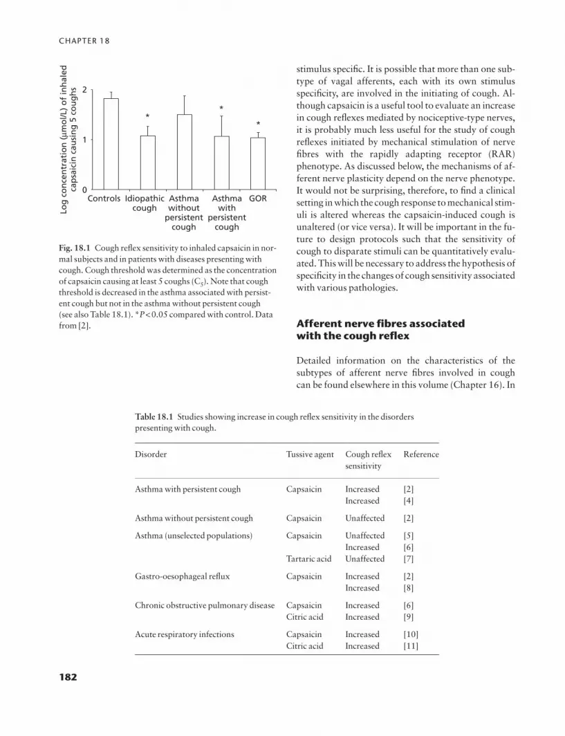
CHAPTER 18
182
stimulus specific. It is possible that more than one sub-type of vagal afferents, each with its own stimulusspecificity, are involved in the initiating of cough. Al-though capsaicin is a useful tool to evaluate an increasein cough reflexes mediated by nociceptive-type nerves,it is probably much less useful for the study of cough reflexes initiated by mechanical stimulation of nerve fibres with the rapidly adapting receptor (RAR) phenotype. As discussed below, the mechanisms of af-ferent nerve plasticity depend on the nerve phenotype.It would not be surprising, therefore, to find a clinicalsetting in which the cough response to mechanical stim-uli is altered whereas the capsaicin-induced cough isunaltered (or vice versa). It will be important in the fu-ture to design protocols such that the sensitivity ofcough to disparate stimuli can be quantitatively evalu-ated. This will be necessary to address the hypothesis ofspecificity in the changes of cough sensitivity associatedwith various pathologies.
Afferent nerve fibres associated with the cough reflex
Detailed information on the characteristics of the subtypes of afferent nerve fibres involved in cough can be found elsewhere in this volume (Chapter 16). In
0
1
2
Controls Idiopathiccough
Asthmawithout
persistentcough
Log
co
nce
ntr
atio
n (
mmo
l/L)
of
inh
aled
cap
saic
in c
ausi
ng
5 c
ou
gh
s
Asthmawith
persistentcough
GOR
**
*
Fig. 18.1 Cough reflex sensitivity to inhaled capsaicin in nor-mal subjects and in patients with diseases presenting withcough. Cough threshold was determined as the concentrationof capsaicin causing at least 5 coughs (C5). Note that coughthreshold is decreased in the asthma associated with persist-ent cough but not in the asthma without persistent cough (see also Table 18.1). *P<0.05 compared with control. Datafrom [2].
Table 18.1 Studies showing increase in cough reflex sensitivity in the disorderspresenting with cough.
Disorder Tussive agent Cough reflex Referencesensitivity
Asthma with persistent cough Capsaicin Increased [2]Increased [4]
Asthma without persistent cough Capsaicin Unaffected [2]
Asthma (unselected populations) Capsaicin Unaffected [5]Increased [6]
Tartaric acid Unaffected [7]
Gastro-oesophageal reflux Capsaicin Increased [2]Increased [8]
Chronic obstructive pulmonary disease Capsaicin Increased [6]Citric acid Increased [9]
Acute respiratory infections Capsaicin Increased [10]Citric acid Increased [11]

PLASTICITY OF FIBRES MEDIATING COUGH
183
the context of the present chapter, however, we wouldlike to stress the idea that activation of at least two fundamentally distinct types of afferent fibres can lead to cough in animal models. The ‘cough fibre’ de-scribed by the classical studies of Widdicombe and col-leagues is a myelinated fibre that conducts actionpotentials in the Ad range [12]. These fibres are verysensitive to mechanical perturbation. A defining char-acteristic of these fibres is the rapid adaptation of their action potential discharge to a prolongedsuprathreshold mechanical stimulus. Accordingly,these fibres are commonly referred to as rapidly adapt-ing receptors or RARs. The other fibre type involved incertain types of cough is the nociceptive fibre (nocicep-tor). These fibres are relatively insensitive to mechani-cal stimulation, but can be activated by inflammatorymediators, acid, changes in osmolarity, and chemicalssuch as capsaicin and bradykinin [13,14]. Nociceptorsare typically characterized by non-myelinated axons(i.e. C-fibres); however, nociceptors also comprise alarge number of Ad-fibres [13]. It is worth emphasizingthat in guinea-pig airways, nociceptive Ad-fibres differfrom RAR Ad-fibres in that the latter are exquisitelymechanically sensitive and do not respond directly tocapsaicin [13].
The RAR fibres and nociceptors are not only acti-vated by different types of stimuli, but also appear to be situated in different compartments within the extrathoracic airway. In guinea-pig airways, circum-stantial evidence supports the hypothesis that the nociceptive population of nerves appears to be the pri-mary nerve type that extends into the epithelium,whereas the RAR type of fibres are situated in the sub-mucosa just beneath the epithelium [15]. These twotypes of afferent nerves may also have different embry-ological origins. This speculation is based on the obser-vation that, in guinea-pig airways, the RAR-type fibresarise from cell bodies situated in the nodose ganglia,whereas the nociceptive C and Ad-fibres are derivedfrom cell bodies in the jugular (supranodose) ganglia[13]. Embryologically, neurones within the nodoseganglia are thought to be derived from the epibranchialplacodes, whereas the jugular ganglion neurones arederived from the neural crest as are the dorsal root gan-glia neurones [16]. Thus, based on the nature of the activating stimuli, location within the airways and perhaps embryological origin, the RAR fibres and noci-ceptive fibres represent quite distinct subpopulations ofairway afferent nerves in guinea-pig trachea/bronchus.
The characteristics and mechanisms of plasticity willlikely therefore differ between these two general typesof fibre.
Plasticity
We use the term plasticity here to denote change in theneuronal excitability, receptor expression, neurotrans-mitter chemistry and nerve structure. As such, plasti-city per se denotes change, and may lead to eitherincreases or decreases in overall responsiveness of theafferent nerves. We have chosen to focus our discussionon those aspects of plasticity that would most likelylead to an increase in afferent nerve activity and the sen-sitivity of cough reflex.
Excitability
General principles of excitabilityActivators of afferent nerves (mechanical displace-ment, certain chemicals, changes in osmolarity, etc.) in-teract with the nerve terminals in a manner that leads toa membrane depolarization. This initial terminal mem-brane depolarization is referred to as the generator potential. The generator potential is electrotonicallyconducted along the axon until it reaches the so-calledactive zone characterized by a high concentration ofvoltage-gated sodium channels. If in the active zone themagnitude of membrane depolarization is sufficient(i.e. the threshold for action potential formation isreached), action potential discharge is evoked. Thegreater the amplitude of generator potential, the higheris the frequency of action potential discharge. These are theoretical considerations inasmuch as neither thestructure nor location of the generator region and active zones has been described in vagal afferent nerveterminals. However, these mechanisms have beenworked out in considerable detail in somatosensorysystems and are likely to be shared with vagal afferentnerves [17].
Many chemical mediators affect the electrophysio-logical properties of the afferent nerve membrane with-out causing a generator potential and activating thenerve. One might say that these are not activators of thenerve, but rather are better characterized as modula-tors of nerve excitability. It is possible that a given stim-ulus may act both as an activator (causing a generator
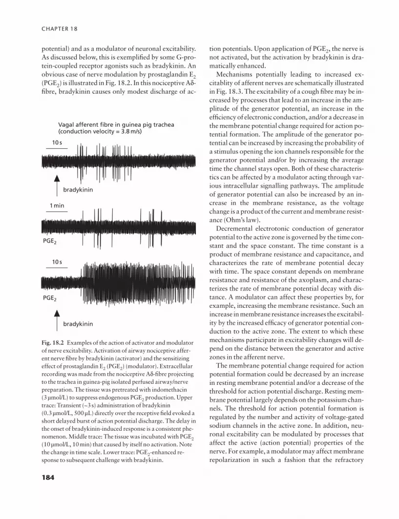
CHAPTER 18
184
potential) and as a modulator of neuronal excitability.As discussed below, this is exemplified by some G-pro-tein-coupled receptor agonists such as bradykinin. Anobvious case of nerve modulation by prostaglandin E2(PGE2) is illustrated in Fig. 18.2. In this nociceptive Ad-fibre, bradykinin causes only modest discharge of ac-
tion potentials. Upon application of PGE2, the nerve isnot activated, but the activation by bradykinin is dra-matically enhanced.
Mechanisms potentially leading to increased ex-citablity of afferent nerves are schematically illustratedin Fig. 18.3. The excitability of a cough fibre may be in-creased by processes that lead to an increase in the am-plitude of the generator potential, an increase in theefficiency of electronic conduction, and/or a decrease inthe membrane potential change required for action po-tential formation. The amplitude of the generator po-tential can be increased by increasing the probability ofa stimulus opening the ion channels responsible for thegenerator potential and/or by increasing the averagetime the channel stays open. Both of these characteris-tics can be affected by a modulator acting through var-ious intracellular signalling pathways. The amplitudeof generator potential can also be increased by an in-crease in the membrane resistance, as the voltagechange is a product of the current and membrane resist-ance (Ohm’s law).
Decremental electrotonic conduction of generatorpotential to the active zone is governed by the time con-stant and the space constant. The time constant is aproduct of membrane resistance and capacitance, andcharacterizes the rate of membrane potential decaywith time. The space constant depends on membraneresistance and resistance of the axoplasm, and charac-terizes the rate of membrane potential decay with dis-tance. A modulator can affect these properties by, forexample, increasing the membrane resistance. Such anincrease in membrane resistance increases the excitabil-ity by the increased efficacy of generator potential con-duction to the active zone. The extent to which thesemechanisms participate in excitability changes will de-pend on the distance between the generator and activezones in the afferent nerve.
The membrane potential change required for actionpotential formation could be decreased by an increasein resting membrane potential and/or a decrease of thethreshold for action potential discharge. Resting mem-brane potential largely depends on the potassium chan-nels. The threshold for action potential formation isregulated by the number and activity of voltage-gatedsodium channels in the active zone. In addition, neu-ronal excitability can be modulated by processes thataffect the active (action potential) properties of thenerve. For example, a modulator may affect membranerepolarization in such a fashion that the refractory
Vagal afferent fibre in guinea pig trachea(conduction velocity = 3.8 m/s)
10 s
10 s
1min
bradykinin
bradykinin
PGE2
PGE2
Fig. 18.2 Examples of the action of activator and modulatorof nerve excitability. Activation of airway nociceptive affer-ent nerve fibre by bradykinin (activator) and the sensitizingeffect of prostaglandin E2 (PGE2) (modulator). Extracellularrecording was made from the nociceptive Ad-fibre projectingto the trachea in guinea-pig isolated perfused airway/nervepreparation. The tissue was pretreated with indomethacin (3μmol/L) to suppress endogenous PGE2 production. Uppertrace: Transient (~3s) administration of bradykinin (0.3μmol/L, 500μL) directly over the receptive field evoked ashort delayed burst of action potential discharge. The delay inthe onset of bradykinin-induced response is a consistent phe-nomenon. Middle trace: The tissue was incubated with PGE2(10μmol/L, 10min) that caused by itself no activation. Notethe change in time scale. Lower trace: PGE2-enhanced re-sponse to subsequent challenge with bradykinin.

PLASTICITY OF FIBRES MEDIATING COUGH
185
period is decreased and consequently the frequency ofaction potential discharge is increased.
It is important to realize that a single inflammatorymediator may affect multiple factors regulating neu-ronal excitability by signalling through divergent intra-
cellular pathways. Moreover, inflammatory disorderslead to accumulation of numerous mediators at the siteof nerve terminal. Therefore, within the complexity ofairway disease, rather than an isolated change in a par-ticular mechanism, multiple effects on excitability
Stimulusintensity
Generatorpotential
Active zonedepolarization
Action potentialdischarge
Terminalmembrane
'Activezone'
membrane
Axonalmembrane
threshold
Mechanisms of normaland enhanced neuronalexcitability
Normal excitability(subthreshold stimulus)
Increased amplitude of generatorpotential in response to givenstimulus
Increased passive electrotonicconduction of generator potentialto the active zone
Decreased threshold for actionpotential formation
Alteration in repolarization —increased frequency of actionpotential discharge (suprathreshold stimulus)
Fig. 18.3 Theoretical mechanisms of normal and enhancedneuronal excitability. Stimulus interaction with the receptivefield membrane results in membrane depolarization (genera-tor potential). This depolarization propagates passively tothe active zone which is responsible for initiation of actionpotentials. If the membrane depolarization at the active zoneis of sufficient magnitude action potential discharge follows.Excitability of the nerve terminal could be enhanced by sever-al mechanisms: (i) increase in the amplitude of the generatorpotential in response to the stimulus of given intensity bymodification of transducer channels (for example by proteinkinase A-mediated phosphorylation of the capsaicin receptorTRPV1); (ii) enhancement of passive propagation of the gen-
erator potential (for example by increasing membrane resist-ance through the inhibition of certain potassium channels);(iii) reduction of threshold for action potential formation (forexample by modification of voltage-gated sodium channels);or (iv) increase in the frequency of action potential dischargedue to change in repolarization (for example by modificationof voltage-gated potassium channels involved in the mem-brane repolarization or hyperpolarization). The bars in ‘gen-erator potential’ and ‘active zone depolarization’ columnsdenote the amplitude of membrane depolarization. The hori-zontal dashed lines in the ‘active zone depolarization’ columndenote the threshold for action potential formation.

CHAPTER 18
186
could summate and result in greatly increased respon-siveness of airway afferent nerves.
Molecular mechanisms of increased excitabilityAt a phenomenological level, allergic inflammation orvarious inflammatory mediators have been shown toincrease cough reflex sensitivity as well as excitabilityof airway afferent nerves. Inhalation of PGE2, for ex-ample, enhances capsaicin-induced cough in humans[18]. In addition to these types of studies, as discussedabove, a large number of clinical studies have demon-strated that certain pathological conditions are accom-panied by a substantial increase in cough reflexsensitivity in humans (Table 18.1). This can also be ob-served in animal models. For example, in guinea-pigsallergic inflammation or inhalation of bradykinin potentiated cough evoked by capsaicin and citric acid,respectively [19,20]. An increase in the excitability ofthe afferent nerve endings in the airways is likely to contribute to this phenomenon.
Inhalation of inflammatory mediators includingPGE2 and eosinophil major basic protein results in po-tentiation of capsaicin-induced action potential dis-charge in pulmonary nociceptive fibres in rats [21,22].Some of these mediators also decrease the amount ofmechanical force required to activate nociceptive C-fi-bres in the lungs [21]. Excitability of RAR fibres is alsoenhanced by inflammatory conditions. For example,exposing the trachea isolated from sensitized guinea-pigs to the antigen causes a substantial increase in themechanosensitivity of RAR fibres [23]. Beyond thesetypes of descriptive studies, there has been relatively lit-tle published relating to the mechanisms underlying in-creases in airway afferent excitability.
The vast majority of studies on the mechanistic basisof afferent nerve excitability and plasticity have beencarried out on nociceptive-type somatosensory neu-rones isolated from the dorsal root ganglia [1]. So-matosensory nociceptors share many properties withairway nociceptive fibres, and thus may provide impor-tant clues to the mechanism of airway nociceptor ex-citability. On the other hand, the RAR phenotype fibreis not readily paralleled by any type of somatosensoryafferent. Consequently relatively little has been pub-lished on the mechanisms by which airway RAR ex-citability is modulated. It nevertheless should be kept inmind that, given the phenotypic differences betweenRAR and nociceptive fibres, the mechanisms resultingin changes in RAR excitability will likely be different
from those involved in excitability changes in airwaynociceptors.
Vanilloid receptor (TRPV1) mechanisms. Vagal affer-ent nociceptors (C-fibres and Ad-fibres) innervating the airways express the capsaicin receptor, a member ofthe transient receptor potential family, currently re-ferred to as TRPV1 [13,14]. This receptor was previ-ously termed vanilloid receptor 1 (VR1). TRPV1 is notexpressed by RAR-type fibres in guinea-pig airways[24], and therefore the extent to which capsaicin canlead to RAR activation in vivo is likely through indirectmeans.
TRPV1 is an ionotropic receptor that upon activa-tion serves as a non-selective cation channel resulting in membrane depolarization [25]. In addition to vanil-loid compounds, TRPV1 is activated by endogenouslipid mediators including anandamide and arachidonicacid metabolites of various lipoxygenase enzymes[25,26]. Certain metabotrophic receptors may also activate TRPV1 through intracellular signal transduc-tion mechanisms. For example, evidence from both somatosensory neurones and airway afferent C-fibressupports the hypothesis that bradykinin activates sensory nerves, at least in part, through production oflipoxygenase products of arachidonic acid and the subsequent activation of TRPV1 (see below) [26].TRPV1 can be stimulated by heat, but the temperaturerequired (>40°C) is unlikely to be relevant to coughphysiology. More relevant to airways physiology is thefact that hydrogen ions can activate TRPV1 (pH ~6 at37°C) [25].
A unique feature of TRPV1 is its ability to integratedisparate stimuli —i.e. action of one TRPV1 agonistpotentiates the action of the other [25]. For example,hydrogen ions were found to potentiate TRPV1-medi-ated responses to vanilloids and heat. On the otherhand, increases in temperature increase the TRPV1-mediated responses to vanilloids and lipid mediators.Integration is likely to be an important mechanism ofincreased sensitivity of nociceptive airway afferents in-volved in cough. Under various pathological condi-tions one or more TRPV1 agonists may accumulate inthe airway wall. For example, airway inflammation inasthma could lead to TRPV1-mediated responses by in-creased concentration of hydrogen ions (i.e. decreasingthe pH in the airway wall), bradykinin and certain lipidmediators.
Biophysical studies on sensory cell bodies show
PLASTICITY OF FIBRES MEDIATING COUGH
187
that inflammatory mediators that stimulate classical G-protein-coupled receptors can increase conductancethrough TRPV1. Agonists of Gq-coupled receptorshave been shown to increase TRPV1 conductance sec-ondary to phospholipase C (PLC) activation and sub-sequent phosphorylation of TRPV1 by protein kinaseC (PKC) [27]. In addition, activation of PLC has beenshown to release TRPV1 from phosphatidylinositol[12,13] phosphate inhibition [28]. This mechanism ap-pears to contribute to increases in TRPV1 activity sec-ondary to nerve growth factor (NGF) stimulation oftrk-A receptors and also bradykinin activation of B2receptors. One might speculate that bradykinin B2-receptor activation (a classical Gq-coupled receptor)may both activate the nerve via lipoxygenase-dependent gating of TRPV1, and increase the excitabil-ity of the nociceptor via induction of prostaglandinproduction and PKC-dependent phosphorylation ofTRPV1.
The amplitude of the TRPV1-mediated generatorpotential may also be increased by Gs-coupled recep-tors. Elevation in cAMP increases capsaicin-inducedconductance in rat nociceptive neurones by a mecha-nism that can be inhibited by inhibitors of protein ki-nase A (PKA) [29]. This mechanism likely contributesto the observation that PGE2 increases the capsaicin-induced action potential discharge in rat pulmonarynociceptors [21]. The generator potential evoked by TRPV1 agonists could in theory also be increased bychanges in the number of TRPV1 receptors. Neu-rotrophins such as NGF have been found to increasethe expression of TRPV1 in rat sensory neurones [30].Although the relative expression of TRPV1 in nocicep-tors found in normal and diseased airways has not beenstudied, it is known that NGF can be elevated at sites ofairway inflammation [31].
Inflammatory mediators may affect nociceptor ex-citability by mechanisms that do not involve TRPV1.Non-TRPV1 mechanisms likely contribute to ex-citability changes in RAR fibres as well as nociceptors.Various inflammatory mediators have been shown todecrease the threshold for mechanical stimulation ofairway afferent nerves [21,23]. The mechanisms under-lying this response are unknown but likely involvemodulation of various ion channels.
Potassium channels. There are a large number of different types of potassium channels in sensory nervefibres [32]. In general, inhibition of potassium channels
leads to an increase in excitability. Antigen challengeand mediators such as histamine have been found to in-hibit resting potassium current in vagal sensory neu-rones [33]. Inhibiting potassium channels that are openunder resting condition leads to an increase in mem-brane resistance and this could lead to an increase in theamplitude of the generator potential. Increased mem-brane resistance can also increase the efficacy of elec-trotonic conduction of the terminal membrane.
Other potassium channels play a key role in deter-mining the refractory period of the nerve. One suchchannel is found in vagal sensory neurones and causes aslow hyperpolarization following the action potential,referred to as the ‘slow afterspike hyperpolarization’(AHPslow) [34]. This channel is opened by calciumthat enters the cell during the action potential throughvoltage-gated N-type calcium channels. Inhibitingthese channels increases the peak frequency at whichthe sensory nerve can fire action potentials. In nodoseganglion neurones, antigen challenge, bradykinin,PGD2 and PGI2 effectively inhibit the AHPslow[34,35]. In addition, any process that leads to eleva-tions in cAMP or inhibition of N-type calcium channelswill inhibit the AHPslow current.
Potassium channels underlying the so-called maxi-K current may also affect the excitability of afferentnerves in the airways. Pharmacological opening ofthese channels with drugs such as NS1619 inhibits af-ferent nerve activity [36]. Similarly, there are variousvoltage-gated potassium channels in the airway affer-ent endings. Drugs that block some of these channels,such as 4-aminopyridine and certain dendrotoxins, canlead to increases in excitability of the nerve endings, oreven to overt activation [37]. However, there is little in-formation on how the process of airway inflammationaffects these channels.
Sodium channels. The number and activity of voltage-gated sodium channels can affect the threshold for ac-tion potential generation, as well as peak frequency ofaction potential discharge. Indeed, long before vera-trum alkaloids were known to act by increasing sodiumchannel activity (by inhibiting their inactivation), theywere used to stimulate airway afferent nerves. Thereare a large number of different voltage-gated sodiumchannels expressed in mammalian nerves. Based on the sensitivity to tetrodotoxin (TTX), these channelshave been pharmacologically divided into two families,the TTX-sensitive (potently blocked by TTX) and

CHAPTER 18
188
TTX-resistant sodium channels. Sodium channels ofeach category are present in airway afferent nerves[32]. However, TTX-resistant channels may be particu-larly relevant to regulation of excitability because oftheir modulation by inflammatory mediators [38]. Inaddition, certain TTX-resistant sodium channels arepreferentially localized to afferent nerves. These ‘sens-ory nerve specific’ (SNS) channels are found mainly in small-diameter (nociceptor-like) neurones. Thenomenclature of SNS sodium channels is confusing,with SNS1 also referred to as PN3, while SNS2 is some-times referred to as the NaN channel. Christian andTogo noted that the vast majority of neurones inguinea-pig jugular ganglia (source of nociceptive fibresinnervating the airways), have sufficient TTX-resistantsodium channels to support action potential generation[39]. The sodium channels in airway afferent nerveshave not been characterized in detail; however, prelim-inary data from our laboratory have shown that air-way-specific jugular neurone cell bodies have sufficientTTX-resistant current to support action potential for-mation [32]. Current through the SNS TTX-resistantchannels can be amplified by inflammatory mediators.For example, PGE2, adenosine and 5-hydroxytrypta-mine (5-HT) are effective in enhancing TTX-resistantsodium current in somatosensory neurones [38]. Theextent to which inflammatory mediators found in the airways modulate this current remains to be determined.
Neurotransmitter plasticity
Sensory C-fibres innervating airways characteristicallycontain neuropeptides in their peripheral and centralterminals. The most often studied sensory neuropep-tides are substance P and related tachykinins, but otherpeptides are likely to be found in C-fibres including cal-citonin gene-related peptide (CGRP), secretoneurinand various opioid peptides.
A hallmark of inflammatory disease is the up-regulation in production of various sensory neuropep-tides. This has been seen both in animal models of inflammation and in numerous inflammatory diseases,including COPD [40]. This is often found to be second-ary to increases in the expression of the prepro-tachykinin gene in the sensory neurones [41,42]. Itremains unknown as to how inflammation within theairway wall sends signals to the distant cell body in therelevant sensory ganglia to induce the transcription of
neuropeptide synthesizing enzymes. A likely mecha-nism, however, involves the action of various neu-rotrophins. Neurotrophins are known to interact withspecific tyrosine kinase-linked receptors (trk receptors)to evoke signals in the cell body. The neurotrophin–trkreceptor complex is thought to be transported from thenerve terminals to the cell body via axonal transportmechanisms, and therein to affect transcriptions of various genes including those involved in the synthesisof neuropeptides [43]. Adding to the evidence thatneurotrophins may be involved in the up-regulation ofneuropeptide synthesis in airway diseases are the observations that neurotrophins such as nerve growthfactor (NGF) and brain-derived neurotrophin factor(BDNF) are found in the airways, and their productionmay be increased at sites of allergic inflammation [31].
Sensory neuropeptides are synthesized in the cellbody and transported to both the peripheral and cen-tral terminals [1]. Neuropeptides can be released fromthe peripheral terminals of afferent nerves by a processreferred to as the axon reflex. In addition, chemical me-diators such as TRPV1 agonists and trypsin can causeneuropeptide release independent of action potentialdischarge and reflex activity. Neurokinins released inthe airways can participate in the inflammatory reac-tion by causing vasodilatation, plasma extravasationand in some species bronchial smooth muscle contrac-tion [44]. In the guinea-pig these processes may indi-rectly activate RAR nerves and thereby contribute tothe tussigenic activity of these agents [44,45].
Neuropeptides are released from the central terminalin the brainstem as a result of action potential invasionof the central release sites [1]. It is the release of neurokinins from the central terminals that likely playsan important role in regulating cough reflex sensitivity.Neurokinins released in the synapse between primaryand secondary vagal afferent neurones in the nucleus of the solitary tract can cause an increase in synaptictransmission. Typically neurokinins cause slow excita-tory postsynaptic potentials and/or increases in inputimpedance [1]. These events can lead to changes in synaptic efficacy and neurotransmission in the NTS. There is evidence that, at least in some instances,central terminals of RAR fibres converge on the samesecondary neurones as nociceptive C-fibres [46]. Thisfact, considered with the electrophysiological effects of neurokinins on postsynaptic membranes, providesthe conceptual framework for the process referred to as ‘central sensitization’ [1]. This term is used to

PLASTICITY OF FIBRES MEDIATING COUGH
189
denote the process by which one type of nerve input(e.g. nociceptive C-fibre) enhances the synaptic trans-mission of another type of input (e.g. RAR fibres). Thiscould lead to a substantive decrease in the amount ofRAR input to the central nervous system (CNS) re-quired to trigger cough. Moreover, such synergy pro-vided by disparate converging inputs in the brainstemmight explain, for example, how activation of nocicep-tive-type nerves in the oesophagus could affect thethreshold for cough evoked by activation of airway-specific afferent nerves.
The increase in sensory neuropeptides associatedwith inflammation is thought to be secondary to in-duction of preprotachykinin genes in nociceptive neurones. However, recent studies in both the somatosensory and vagal sensory systems support thehypothesis that inflammation may also lead to pheno-typic changes in the neuropeptidergic innervation[24,47,48]. Accordingly, exposure to respiratory aller-gen or virus infection increased the amount of sensoryneurokinins in the airway afferent neurones in guinea-pigs [24,42,48]. Interestingly, however, the prepro-tachykinin gene expression and neurokinin productionwere found to be increased in neurones that projectnon-nociceptive fibres into the airway. The neuronesinduced by inflammation to produce neuropeptideswere large-diameter neurones located in the nodoseganglia and had physiological characteristics consis-tent with the RAR phenotype. At sites of inflammation,therefore, neuropeptidergic innervation may consist ofnot only nociceptive C-fibres but also RAR fibres. Inaddition, histological evidence supports the hypothesisthat the neuropeptides produced in response to airwayinflammation are also transported to central terminalsof the RAR neurones [24]. This raises the possibilitythat in allergic inflammation or respiratory virus infec-tion, mechanical activation of the RAR fibres wouldlead to neurokinin release in the brainstem. Unlike no-ciceptors, many RAR fibres are activated during thebreathing cycle. This could lead to central sensitizationduring breathing independently of nociceptive stimula-tion. In the somatosensory system an analogous pheno-typic switch in neuropeptide innervation has beenreported to play a role in painful sensation to non-painful stimuli termed allodynia [47]. It is tempting tospeculate that this process in the airways could con-tribute to inappropriate or ‘allotussive’ cough sensa-tions, i.e. the urge to cough in the absence of anything inthe airway to productively cough up. The mechanism
underlying this inflammation-induced phenotypicswitch in the neurochemistry of RAR has not beenworked out; however, this effect of allergen challengeor virus exposure can be mimicked by local injection ofNGF into the airway wall [49].
Changes in the neurotransmitter content in centralterminals of primary afferents, combined with increas-es in afferent nerve excitability and activity, may lead tosubstantive changes in the synaptic input to secondaryneurones (neurones receiving input from sensory affer-ents) in the central nervous system. This, in turn, cancause changes in the excitability of the secondary neu-rones. This is referred to as ‘use-dependent’ plasticityand has been extensively studied in the somatosensorysystem where activation of nociceptive afferent fibresresults in increases in excitability of secondary neu-rones in the spinal cord, sensitizing them to noxious aswell as innocuous stimuli [50]. The mechanisms of use-dependent excitability changes in secondary neuronesare not clear, but several receptors and second messen-ger cascades may be involved [50]. With respect tocough-mediating pathways it is interesting to note thatexposing the lungs of non-human primates repeatedlyto allergen resulted in increases in excitability of sec-ondary sensory neurones in the nucleus of the solitarytract [51]. These observations are consistent with thehypothesis that allergen inhalation-induced activationof primary airway afferent nerves can result in use-dependent changes in excitability of secondary neu-rones in the brainstem.
Changes in nerve fibre density
The density of sensory innervation can change in re-sponse to its environment [52]. Tissue damage or re-lease of various growth factors can lead to increasednerve fibre growth and fibre sprouting. The question asto whether increases in afferent nerve density may par-ticipate in increased cough sensitivity has not been ex-tensively studied. To our knowledge only one study hasaddressed this issue as it relates to cough [53]. This ele-gant study was designed such that the nerve density wasdetermined in biopsies taken from the carina of theright upper lobe and a subsegmental carina of the rightlower lobe. The tissue was obtained from patients withpersistent idiopathic cough who had increased coughsensitivity to capsaicin and from controls. There was nodifference in the density of epithelial nerve fibres asquantified using the non-specific nerve marker PGP

CHAPTER 18
190
9.5, but patients with chronic cough had a higher densi-ty of CGRP-positive nerves compared with the controlsubjects. This supports the hypothesis of neurotrans-mitter plasticity contributing to cough sensitivity butdoes not favour a role for an increased nerve sproutingand density. Whether there is a higher nerve density inthe submucosa plexus or near cough regions such as inthe trachea or larynx was not investigated.
Extraneuronal effects
Most afferent nerves in the airways are mechanicallysensitive, and the lungs are exposed to extensive me-chanical forces during breathing [13,14]. Each breathcauses discharge in various populations of afferentmechanosensors. As discussed above, the action poten-tial discharge depends on the amplitude and durationof the generator potential. It is likely that the amplitudeof the generator potential evoked by mechanical forcesin the lungs will depend not only on the terminal mem-brane, but also on the viscoelastic properties of the lung tissue. In airway diseases such as COPD and asthma, airway tissue destruction and remodelling will likely affect the extent to which distension of the tissue in the microenvironment of the mechano-sensors is transduced to a generator potential. Coughreflexes could be increased if changes in airway struc-ture lead to increases in mechanotransduction ofcough-inducing fibres (e.g. RAR-type fibres), or if theylead to decreases in activity in mechanosensors that arenormally inhibitory to the cough reflex. The hypothesisthat changes in airway structure that accompanies airway diseases alters the properties of airwaymechanosensors has been studied as it pertains toCOPD, but little information is available as regardscough [54].
Conclusions
Excessive coughing accompanies many diseases includ-ing asthma, respiratory infections, lung cancer, bron-chitis, COPD and gastro-oesophageal reflux disease.The increase in coughing in many cases is likely due tothe production of tussigenic stimuli in the large air-ways. In addition, however, it is recognized that pa-tients with chronic cough often present with an increasein the cough reflex sensitivity to tussigenic stimuli. Ra-tional strategies for the treatment of chronic cough may
consider therefore both a reduction in the amount oftussigenic stimuli and a reduction in the heightened sen-sitivity of the cough reflex. Regrettably, short of non-specific centrally acting drugs, there are no drugs thatare aimed at decreasing the increased sensitivity ofcough. This is likely because relatively little is knownabout the mechanisms underlying this process. Theavailable data gathered from research on vagal sensoryand somatosensory systems indicate that multiplemechanisms may lead to hypersensitivity of reflexessuch as cough. These include increases in the excitabil-ity of the afferent terminals, changes in the neurotrans-mitter content and consequent changes in synaptictransmission in the brainstem, and extraneuronalchanges that may lead to changes in the efficiency ofmechanotransduction in afferent nerves. Adding to thecomplexity of this issue is the notion that at least twodisparate types of afferent nerves may participate incough reflexes (RARs and nociceptors). Inasmuch asthese nerve subtypes are phenotypically distinct, it islikely that mechanisms resulting in increases in their ac-tivity will also be distinct. Finally, adding height to thehurdle in front of our understanding of this issue, is thedifficulty in quantifying cough sensitivity in humans.At present most studies in this area have used classicalnociceptive stimuli to study changes in cough sensitiv-ity. This leaves open the question of the extent to whichthe sensitivity of RAR-driven cough is affected in vari-ous disease states. A combined effort from studies at the research bench and bedside will be required beforesome semblance of understanding of this complex area emerges.
References
1 Woolf CJ, Salter MW. Neuronal plasticity: increasing thegain in pain. Science 2000; 288: 1765–9.
2 Choudry NB, Fuller RW. Sensitivity of the cough reflex inpatients with chronic cough. Eur Respir J 1992; 5 (3):296–300.
3 Pounsford JC, Birch MJ, Saunders KB. Effect of bron-chodilators on the cough response to inhaled citric acid innormal and asthmatic subjects. Thorax 1985; 40 (9):662–7.
4 Chang AB, Phelan PD, Robertson CF. Cough receptor sensitivity in children with acute and non-acute asthma.Thorax 1997; 52 (9): 770–4.
5 Chang AB, Phelan PD, Sawyer SM, Del Brocco S, Robertson CF. Cough sensitivity in children with asthma,

PLASTICITY OF FIBRES MEDIATING COUGH
191
recurrent cough, and cystic fibrosis. Arch Dis Child 1997;77 (4): 331–4.
6 Doherty MJ, Mister R, Pearson MG, Calverley PM. Capsaicin responsiveness and cough in asthma and chron-ic obstructive pulmonary disease. Thorax 2000; 55 (8):643–9.
7 Fujimura M, Sakamoto S, Kamio Y, Matsuda T. Cough receptor sensitivity and bronchial responsiveness in nor-mal and asthmatic subjects. Eur Respir J 1992; 5 (3):291–5.
8 Ferrari M, Olivieri M, Sembenini C, Benini L, Zuccali V,Bardelli E etal. Tussive effect of capsaicin in patients withgastroesophageal reflux without cough. Am J Respir CritCare Med 1995; 151: 557–61.
9 Wong CH, Morice AH. Cough threshold in patients withchronic obstructive pulmonary disease. Thorax 1999; 54(1): 62–4.
10 O’Connell F, Thomas VE, Studham JM, Pride NB, Fuller RW. Capsaicin cough sensitivity increases duringupper respiratory infection. Respir Med 1996; 90 (5):279–86.
11 Empey DW, Laitinen LA, Jacobs L, Gold WM, Nadel JA.Mechanisms of bronchial hyperreactivity in normal sub-jects after upper respiratory tract infection. Am RevRespir Dis 1976; 113 (2): 131–9.
12 Widdicombe JG. Receptors in the trachea and bronchi ofthe cat. J Physiol Lond 1954; 123: 71–104.
13 Riccio MM, Kummer W, Biglari B, Myers AC, Undem BJ.Interganglionic segregation of distinct vagal afferent fibrephenotypes in guinea-pig airways. J Physiol 1996; 496 (2):521–30.
14 Fox A. Airway nerves: in vitro electrophysiology. CurrOpin Pharmacol 2002; 2 (3): 278–9.
15 Hunter DD, Undem BJ. Identification and substance P content of vagal afferent neurons innervating the epi-thelium of the guinea pig trachea. Am J Respir Crit CareMed 1999; 159 (6): 1943–8.
16 Fontaine-Perus J, Chanconie M, Le Douarin NM. Embryonic origin of substance P containing neurons incranial and spinal sensory ganglia of the avian embryo.Dev Biol 1985; 107 (1): 227–38.
17 Fain GL. Molecular and Cellular Physiology of the Neurons. Cambridge: Harvard University Press, 1999.
18 Choudry NB, Fuller RW, Pride NB. Sensitivity of thehuman cough reflex: effect of inflammatory mediatorsprostaglandin E2, bradykinin, and histamine. Am RevRespir Dis 1989; 140 (1): 137–41.
19 Fox AJ, Lalloo UG, Belvisi MG, Bernareggi M, Chung KF,Barnes PJ. Bradykinin-evoked sensitization of airway sen-sory nerves: a mechanism for ACE-inhibitor cough. NatMed 1996; 2 (7): 814–7.
20 Liu Q, Fujimura M, Tachibana H, Myou S, Kasahara K,Yasui M. Characterization of increased cough sensitivity
after antigen challenge in guinea pigs. Clin Exp Allergy2001; 31 (3): 474–84.
21 Ho CY, Gu Q, Hong JL, Lee LY. Prostaglandin E(2) enhances chemical and mechanical sensitivities of pul-monary C fibers in the rat. Am J Respir Crit Care Med2000; 162: 528–33.
22 Lee LY, Gu Q, Gleich GJ. Effects of human eosinophilgranule-derived cationic proteins on C-fiber afferents inthe rat lung. J Appl Physiol 2001; 91 (3): 1318–26.
23 Riccio MM, Myers AC, Undem BJ. Immunomodulationof afferent neurons in guinea-pig isolated airway. J Physiol1996; 491 (2): 499–509.
24 Myers AC, Kajekar R, Undem BJ. Allergic inflammation-induced neuropeptide production in rapidly adapting af-ferent nerves in guinea pig airways. Am J Physiol LungCell Mol Physiol 2002; 282 (4): L775–81.
25 Caterina MJ, Julius D. The vanilloid receptor: a moleculargateway to the pain pathway. Annu Rev Neurosci 2001;24: 487–517.
26 Shin J, Cho H, Hwang SW, Jung J, Shin CY, Lee SY etal.Bradykinin-12-lipoxygenase-VR1 signaling pathway forinflammatory hyperalgesia. Proc Natl Acad Sci USA2002; 99 (15): 10150–5.
27 Premkumar LS, Ahern GP. Induction of vanilloid receptorchannel activity by protein kinase C. Nature 2000; 408:985–90.
28 Chuang HH, Prescott ED, Kong H, Shields S, Jordt SE,Basbaum AI etal. Bradykinin and nerve growth factor release the capsaicin receptor from PtdIns(4,5)P2-mediated inhibition. Nature 2001; 411: 957–62.
29 Lopshire JC, Nicol GD. The cAMP transduction cascademediates the prostaglandin E2 enhancement of the cap-saicin-elicited current in rat sensory neurons: whole-celland single-channel studies. J Neurosci 1998; 18 (16):6081–92.
30 Michael GJ, Priestley JV. Differential expression of themRNA for the vanilloid receptor subtype 1 in cells of the adult rat dorsal root and nodose ganglia and its downregulation by axotomy. J Neurosci 1999; 19 (5):1844–54.
31 Virchow JC, Julius P, Lommatzsch M, Luttmann W, RenzH, Braun A. Neurotrophins are increased in bronchoalve-olar lavage fluid after segmental allergen provocation. AmJ Respir Crit Care Med 1998; 158 (6): 2002–5.
32 Carr MJ, Undem BJ. Ion channels in airway afferent neu-rons. Respir Physiol 2001; 125 (1–2): 83–97.
33 Undem BJ, Hubbard W, Weinreich D. Immunologically in-duced neuromodulation of guinea pig nodose ganglionneurons. J Auton Nerv Syst 1993; 44 (1): 35–44.
34 Cordoba-Rodriguez R, Moore KA, Kao JP, Weinreich D.Calcium regulation of a slow post-spike hyperpolariza-tion in vagal afferent neurons. Proc Natl Acad Sci USA1999; 96 (14): 7650–7.

CHAPTER 18
192
35 Undem BJ, Weinreich D. Electrophysiological propertiesand chemosensitivity of guinea pig nodose ganglion neu-rons in vitro. J Auton Nerv Syst 1993; 44 (1): 17–33.
36 Fox AJ, Barnes PJ, Venkatesan P, Belvisi MG. Activationof large conductance potassium channels inhibits the af-ferent and efferent function of airway sensory nerves in theguinea pig. J Clin Invest 1997; 99 (3): 513–9.
37 McAlexander MA, Undem BJ. Potassium channel block-ade induces action potential generation in guinea-pig air-way vagal afferent neurones. J Auton Nerv Syst 2000; 78(2–3): 158–64.
38 Gold MS, Reichling DB, Shuster MJ, Levine JD. Hyperal-gesic agents increase a tetrodotoxin-resistant Na+ currentin nociceptors. Proc Natl Acad Sci USA 1996; 93 (3):1108–12.
39 Christian EP, Togo JA. Excitable properties and underly-ing Na+ and K+ currents in neurons from the guinea-pigjugular ganglion. J Auton Nerv Syst 1995; 56 (1–2):75–86.
40 Tomaki M, Ichinose M, Miura M, Hirayama Y, YamauchiH, Nakajima N etal. Elevated substance P content in induced sputum from patients with asthma and patientswith chronic bronchitis. Am J Respir Crit Care Med 1995;151: 613–7.
41 Hunter DD, Castranova V, Stanley C, Dey RD. Effects ofsilica exposure on substance P immunoreactivity and pre-protachykinin mRNA expression in trigeminal sensoryneurons in Fischer 344 rats. J Toxicol Environ Health A1998; 53 (8): 593–605.
42 Fischer A, McGregor GP, Saria A, Philippin B, Kummer W.Induction of tachykinin gene and peptide expression inguinea pig nodose primary afferent neurons by allergic airway inflammation. J Clin Invest 1996; 98 (10):2284–91.
43 Klesse LJ, Parada LF. Trks: signal transduction and intra-cellular pathways. Microsc Res Tech 1999; 45 (4–5):210–6 [p. ii].
44 Advenier C, Emonds-Alt X. Tachykinin receptor antago-nists and cough. Pulm Pharmacol 1996; 9 (5–6): 329–33.
45 Joad JP, Kott KS, Bonham AC. Nitric oxide contributes to substance P-induced increases in lung rapidly adaptingreceptor activity in guinea-pigs. J Physiol 1997; 503 (3):635–43.
46 Mazzone SB, Canning BJ. Synergistic interactions be-tween airway afferent nerve subtypes mediating reflexbronchospasm in guinea pigs. Am J Physiol Regul IntegrComp Physiol 2002; 283 (1): R86–R98.
47 Neumann S, Doubell TP, Leslie T, Woolf CJ. Inflammatorypain hypersensitivity mediated by phenotypic switch inmyelinated primary sensory neurons. Nature 1996; 384:360–4.
48 Carr MJ, Hunter DD, Jacoby DB, Undem BJ. Expressionof tachykinins in non-nociceptive vagal afferent neuronsduring respiratory tract viral infection in guinea pigs. Am JRespir Crit Care Med 2002; 165: 1071–5.
49 Hunter DD, Myers AC, Undem BJ. Nerve growth factor-induced phenotypic switch in guinea pig airway sensoryneurons. Am J Respir Crit Care Med 2000; 161 (6):1985–90.
50 Willis WD. Role of neurotransmitters in sensitization ofpain responses. Ann N Y Acad Sci 2001; 933: 142–56.
51 Chen CY, Bonham AC, Schelegle ES, Gershwin LJ, Plopper CG, Joad JP. Extended allergen exposure in asth-matic monkeys induces neuroplasticity in nucleus tractussolitarius. J Allergy Clin Immunol 2001; 108 (4): 557–62.
52 Stead RH. Nerve remodelling during intestinal inflamma-tion. Ann N Y Acad Sci 1992; 664: 443–55.
53 O’Connell F, Springall DR, Moradoghli-Haftvani A,Krausz T, Price D, Fuller RW etal. Abnormal intraepithe-lial airway nerves in persistent unexplained cough? Am JRespir Crit Care Med 1995; 152: 2068–75.
54 Mansoor JK, Hyde DM, Schelegle ES. Contribution of vagal afferents to breathing pattern in rats with lung fibrosis. Respir Physiol 1997; 108 (1): 45–61.

193
‘Tussis est offendiculum spiritus in trachea arteria’(Matteo Plateario, Scuola Medica Salernitana, 12thcentury)
Introduction
Cough is a modified respiratory act that can be pro-duced voluntarily but, in most instances, it represents areflex action evoked by the activation of laryngealand/or tracheobronchial receptors. Its main functionsare protecting the lungs against aspiration and help-ing in the removal of excessive bronchial secretions.Coughing does not normally occur in healthy individu-als, and persistent cough is one of the cardinal signs ofrespiratory disease. In health, mucociliary clearanceand alveolar macrophages satisfactorily control minorinsults from exogenous noxious agents. However,when these systems fail or become overloaded with foreign matter or excessive bronchial secretions, coughintervenes as a supplemental mechanism to the mucociliary escalator.
In his 1937 paper, Coryllos [1] provided a detaileddescription of the cough cycle, and noted that ‘cough isnot a simple expiratory act but is composed of three dis-tinctive phases: inspiratory, compressive and expul-sive’. He likened the phases of the cough cycle to thedeflagration of a gun: the preparatory inspiration to theloading of the gun, the compressive phase to the blazingof the powder and production of gas under high pres-sure, and the expulsive phase to the ejection of the bul-let from the barrel of the gun.
The purposes of this chapter are to describe themotor characteristics of cough and outline the princi-
pal aspects of respiratory mechanics that are relevant tothe genesis of the high flow rates and gas velocities ofcough. Some related topics will be mentioned, whileothers, which are presented elsewhere in this book, willbe omitted.
Description of cough
When cough is performed voluntarily or initiated bystimulation of tracheobronchial receptors, it usuallybegins with the inhalation of a variable volume of air.However, if coughing is triggered by stimulation of af-ferents from the vocal folds, the preparatory inspira-tion may be absent: this has also been considered aseparate reflex, i.e. the so called ‘expiration reflex’ [2].Next, the glottis is closed and the expiratory musclescontract intensely, leading to the build-up of high in-trathoracic and abdominal pressures. The glottis isthen actively reopened, and the inspired air volumeforcefully expelled by the sustained contraction of theexpiratory muscles. At the same time the central air-ways within the thorax are compressed and, for anygiven expiratory flow rate, the reduction in their cross-sectional area results in higher gas linear velocities thanwould have occurred had there been no bronchial nar-rowing. There may be a fourth phase mainly character-ized by the cessation of expiratory muscle activity andthe appearance of some antagonistic activity [3].
During spontaneous coughing, several expiratorythrusts may follow one another with replication of theabove-mentioned events or with minor variations suchas those characterized by the absence of glottis closure.The principal muscular and mechanical events that
Motor mechanisms and themechanics of coughGiovanni A. Fontana
19

CHAPTER 19
194
characterize a single cough effort are diagrammaticallyillustrated in Fig. 19.1.
Inspiratory phase
As with most inspiratory acts, the first event of the in-spiratory phase of cough is the contraction of the ab-ductor muscles of the arytenoid cartilage leading tocomplete opening of the glottis and facilitating subse-quent inhalation of a variable air volume [4]. The in-spired air volume may range from a fraction of toseveral times the eupnoeic tidal volume.
Studies in which subjects were instructed to coughvoluntarily suggest a high degree of volitional controlover inspired volume, the latter being related to the an-ticipated forcefulness of the subsequent cough effort. Ina group of normal subjects performing a series of ‘singlegentle coughs’ [5], the mean duration of the inspiratoryphase varied from 0.45 to 1.00s, with an average valueof 0.75s. The corresponding inspiratory volumeranged from 0.09 to 0.53L (mean 0.39L), and wasfound to correlate with the duration of the preparatoryinspiration [5]. In the study by Ross et al. [6], in which
subjects were instructed to perform maximum volun-tary cough efforts, the mean duration of the inspiratoryphase was 0.65s, and resulted in a mean air intake ofabout 2.5L.
Little is known regarding the regulation of inspirat-ory volume and flow during the inspiratory phase of re-flex cough. It may be, however, that the magnitude ofthe inspired volume is related, at least in part, to the in-tensity of the stimulus causing cough (author’s unpub-lished observations).
During mechanical stimulation of dog trachea, thetypical sequence of cough’s motor acts is often precededby a sustained apnoeic period during which the di-aphragm shows only minimal activation, and lung vol-ume does not appreciably change [4].
Whatever the regulatory mechanism(s), the augmen-tation of the inspiratory volume may enhance the mechanical efficiency of the subsequent expiration by different means. At high lung volumes, the tension–length relationship of the expiratory muscles isoptimized [7,8], thus allowing them to produce greaterintrathoracic and abdominal pressures. Furthermore,activation of pulmonary stretch receptors by lung dis-tension leads to central facilitation of cough [9].
Compressive phase
Closure of the glottis by adduction of the ventricularfolds and covering of the laryngeal inlet by the epiglot-tis marks the onset of the compressive phase of cough.Contraction of the expiratory muscles against a closedglottis leads to the development of high abdominal,pleural, alveolar and subglottic pressures. When pleu-ral pressure increases, the alveolar gas is compressedand lung volume decreases. Glottis closure, rather thangas compression within the respiratory system, is re-garded as the phenomenon that mostly differentiatescoughing from a forced expiration. Indeed, marked in-creases in abdominal and intrathoracic pressures lead-ing to compression of the alveolar gas are known tooccur during expiratory thrusts with an open glottisdue to the resistive properties of the tracheobronchialtree. Expiratory muscle contraction during the com-pressive phase is accompanied by the coactivation ofthe diaphragm [10] and other inspiratory muscles op-posing further development of positive pleural andalveolar pressures [3].
Since the glottis remains closed for only 0.2s, al-though with considerable variability, the rate of change
Diaphragm
Abd. muscles
PCA
TA
Flow
PSG
I C E
Fig. 19.1 Diagrammatic representation of the motor patternof a typical cough effort. The inspiratory (I), compressive (C)and expiratory (E) phases of cough are delimited by thedashed lines, and are preceded and followed by periods ofnormal breathing. PCA, posterior cricoarytenoid muscle (laryngeal abductor); TA, tyroarytenoid muscle (laryngealadductor); PSG, subglottic pressure. On some occasions (see [41]), diaphragmatic activity may extend into the earlystages of the expiratory phase. Reprinted with permissionfrom [17].

MOTOR MECHANISMS AND MECHANICS OF COUGH
195
in alveolar pressure over this brief interval is remark-ably high. At the end of the compressive phase, alveolarpressure may exceed 20kPa [11,12], i.e. values50–100% higher than during other expulsive manoeu-vres in which the glottis is open. Thus, the corre-sponding rate of change in alveolar pressure wouldapproximate 100kPa/s. Given the relationship be-tween gas pressure and volume changes as dictated byBoyle’s law, if one assumes a lung volume of 5L at theend of the inspiratory phase, and an alveolar pressureof 20kPa at the end of the compressive phase (i.e. ~+20% of the atmospheric value), the corresponding re-duction in lung volume will be ~1L. The rate of changein lung volume during compression would then ap-proximate to 5L/s. The augmentation of pleural pres-sure during the compressive phase, as compared toother expiratory manoeuvres, may be related to a reflexincrease in agonist and decreased antagonist muscle ac-tivity, presumably brought about by glottis closure[13], and/or optimization of the force–length–velocityrelationships of the expiratory muscles [3].
As is the case for all other skeletal muscles, the forcedeveloped by the contracting expiratory muscles is proportional to their length and inversely related totheir velocity of shortening [8]. Thus, there are me-chanically advantageous conditions contributing toforce development during the compressive phase ofcough. In fact, glottis closure prevents significant de-creases in lung volume, except for those produced bygas compression, thus allowing the expiratory musclesto express their maximal force during contraction atthe length determined by the lung volume attained fol-lowing the preceding inspiratory phase. In fact, due to the relatively small changes in lung volume duringcompression, the shortening velocity of the expiratorymuscles is minimal, and muscle contraction nearly isometric.
Although glottic closure is generally considered aprerequisite for development of the high intrathoracicpressures of cough, some lines of evidence seem to denythis. For instance, the study of Gal [14] showed that, insubjects who performed maximum voluntary cough ef-forts prior to and following tracheal intubation, coughpressures were the same or even greater after intuba-tion. Furthermore, neither glottic closure nor high pres-sures appear to be crucial to effective coughing:tracheostomized and intubated patients can still expec-torate, and even normal subjects need not close theglottis for airway clearing [15].
Expiratory phase
This is the phase of the cough cycle during which theairways are cleared of secretions, debris and foreignmaterial. It is initiated by rapid (20–40ms) abductionof the arytenoid cartilages, an active phenomenon in-volving muscle recruitment [4]. Opening of the glottisat the onset of expiration is associated with passive os-cillations of gas and tissues causing the characteristicnoise of cough and setting up pressure fluctuations thatmay play a role in shaking loose secretions. Pressurewithin central airways rapidly falls to nearly atmos-pheric values (Fig. 19.1), while pleural and alveolarpressures still continue to rise for about 0.5s. The totalduration of the expiratory phase is variable, but gener-ally comprises between 0.5 and 1.0s [5,16].
In normal subjects performing a maximum volun-tary cough effort starting from near total lung capacity,expiratory flow sharply rises up to values of more than10L/s [17], and the central intrathoracic airways col-lapse. Airway compression causes rapid, transient dis-placement of the airway gas volume, and generates highsupramaximal flow rates that superimpose on the air-flow coming from the alveolar spaces. More detailedaccounts of the mechanisms contributing to the genera-tion of flow transients during maximum expiratory ef-forts will be given in a subsequent section. The timerequired to achieve these high flow transients (i.e. thetime to peak flow) is approximately 30ms [17]. Afterthis short time interval, flow rate falls to much lowervalues, approximately 50% of the cough peak flow,which may be sustained for several milliseconds, up tohalf the total duration of the expiratory phase [5,16].Lung volume and flow rate then decrease exponentiallywith time, with a time constant of ~0.5s [3]. During thelast stage of expiration, flow rapidly drops to zero andexpulsion terminates.
The violent muscular activity associated with the ex-pirations of cough may have noxious effects, includingtrauma of the larynx and airways, rib fractures andbarotrauma. Indeed, costal fractures and abdominalmuscle tears are well-known complications of intensecough, but have never been reported to occur withother expulsive efforts [3,18,19].
Cessation
The cessation phase is associated with relaxation of expiratory muscle and perhaps onset of antagonistic

CHAPTER 19
196
muscle activity with a fall in pleural and abdominalpressures. In most instances, the glottis narrows and the laryngeal structures gradually return to their inspi-ratory position [5]. The compressed central airways re-expand.
Mechanics of cough
The development of an effective cough as a clearingmechanism is thought to be critically dependent uponthe linear velocity of the gas molecules travelling downthe airway lumen [3,20]. It appears that the coughmechanism is designed to maximally increase the gasvelocity by both generating high expiratory flow ratesand dynamically compressing the airways to reducetheir cross-sectional area. In this section, we will reviewthe mechanisms implicated in the regulation of the rateand velocity of flow during the expulsive phase ofcough. Excellent and more detailed descriptions of themechanical events of cough, as well as of cough as aclearing mechanism, can be found in the literature[3,6,12,20,21].
Regulation of expiratory flow
The high intrathoracic pressures that are generatedduring the nearly isometric expiratory muscle contrac-tion of the cough compressive phase are suddenly released when the glottis opens at the onset of the expiratory phase. Pressure at the airway opening rapid-ly falls to atmospheric values, while alveolar and pleu-ral pressures remain constant or may continue to risefor a short period [3,20]. Sustained expiratory musclecontraction and concomitant cessation of antagonistaction of the inspiratory muscles allowing full trans-mission of expiratory muscle force to the pleural andalveolar spaces are the likely contributing mechanisms[3]. Due to the elastic recoil of the lungs, alveolar pres-sure is always greater than pleural pressure, while thepressure surrounding the outer wall of the airways, theperibronchial pressure, closely approximates that ofthe pleural space [22]. The pressure within the airways,the intrabronchial pressure, progressively diminishesas air travels down the airways from the alveolus to theairway opening. This pressure drop is the result of theenergy expenditure that is required to accelerate flow asthe total airway cross-sectional area decreases and air-way resistance increases, as well as the result of laminar
and turbulent energy dissipation in the flow [23]. Atsome point along the tracheobronchial tree, at theequal pressure point (EPP), the decrease in intra-bronchial pressure equals the elastic recoil of the lung(Fig. 19.2). Thus, the EPP divides the intrathoracic air-ways into two segments arranged in series, respectively,located upstream (i.e. toward the alveoli) and down-stream of the EPP. In the upstream segment, the in-trabronchial pressure is greater than peribronchialpressure, and the airways are distended. In the down-stream segment, pressure within the airways becomeslower than the pressure surrounding them, and the air-ways tend to collapse. Once the maximum expiratoryflow has been achieved, further expiratory efforts onlycause more compression of the downstream segment,but do not affect flow through the upstream segment. Infact, since the pressure drop in the upstream segmentequals the elastic recoil, the rate of flow in the upstreamsegment is dictated by the ratio between the elastic recoil of the lungs (PEL) and the resistance (R) of the upstream (US) segment:
Fig. 19.2 Description of the principal mechanical events ofcough by means of a model of the respiratory system. Duringthe compressive phase, expiratory muscle activation in-creases pleural pressure (PPL) that becomes markedly positivewith respect to atmosphere. Alveolar pressure (PA) is alwaysgreater than PPL due to the elastic recoil of the lung (arrows).Since the glottis is closed, intrabronchial pressure is similar toPA throughout the airways. When the glottis opens at the be-ginning of the expiratory phase, air starts to flow rapidlydown the pressure gradient from the alveoli to the mouth.Thus, there must be a point along the airways at which the in-trabronchial pressure and PPL pressures are equal: the equalpressure point (EPP). Downstream of the EPP, the airways aredistended; upstream of the EPP the intrathoracic airways arecompressed. Reprinted with permission from [17].
PA ++++
PPL +++
Compressivephase
Glottis closed
++++ ++++
PA ++++
PPL +++
Expiratoryphase
Glottis open
+++ ++ +

MOTOR MECHANISMS AND MECHANICS OF COUGH
197
V.
= PEL/RUS.
This phenomenon, called flow limitation or autoregu-lation of flow, is the basic mechanism that sets up theupper limits to flow during both a forced expirationand the expiration of cough. It has been likened to thebehaviour of a Starling resistor or a waterfall. The flowof water upstream of the waterfall depends on the slopeof the terrain that delivers water to the waterfall, but isindependent of the height of the waterfall or the condi-tions downstream to it. Thus, under conditions of flowlimitation, changes in downstream pressure do not af-fect flow. When the intrathoracic pressures generatedby the expiratory muscle contraction exceed the mod-est level necessary to attain maximal expiratory flow,the excess pressure markedly compresses the down-stream segment of the intrathoracic airways, but doesnot affect flow. However, narrowing of the down-stream airways augments flow velocity and kinetic en-ergy, and this may increase the effectiveness of cough asa clearing mechanism.
An additional explanation for flow limitation isbased on principles of the wave speed theory [24]. According to this theory, an elastic tube cannot carry afluid at a mean velocity greater than the speed at whichpressure waves will propagate along the tube, i.e. thetube wave speed. By analogy, this is the velocity atwhich pulse propagates in the arteries. At the site wherethe linear velocity of flow equals the velocity of propa-gation of pressure waves, a ‘choke point’ develops, andprevents further increases in flow rate. The tube wavespeed depends on the gas density (r), the tube (i.e. air-way) cross sectional area (A) and the specific elastanceof the tube wall (AdP/dA). Then, maximum flow (V
.)
can be expressed as:
V.
= A [r-0.5 (A dP/dA)]0.5.
Actual V., however, cannot be determined by using this
equation, since values of A and (AdP/dA) vary withchoke point location, the latter depending on airwaygeometry and elasticity, and on lung volume. During anexpiratory thrust performed at a large lung volume, thechoke point resides in the central airways. As lung vol-ume diminishes, the choke point moves in the upstreamdirection. In both humans and experimental animals,simultaneous measurements of pleural and intra-bronchial pressures made it possible to estimate that,above functional residual capacity, the choke point islocated at the level of lobar or segmental bronchi
[20,22]. At lung volumes near residual volume, thechoke point moves down to the fifth- to sixth-generation branches [20,25]. Thus, when a series ofcoughs is initiated at a high lung volume, secretions areinitially cleared from the larger airway; secretions arethen moved from the small to the larger airway as lungvolume progressively diminishes.
Expiratory flow velocity
Given that in any condition flow is the same throughoutthe airway, flow velocity must be increased at the levelof the compressed downstream segment. In fact, forany given flow, the velocity of flow is inversely relatedto the airway cross-sectional area:
Velocity = flow/cross-sectional area.
In theory, for a forced expiratory flow of 8L/s throughan uncompressed tracheal segment with a cross-sec-tional area of 2.0cm, the linear velocity of gas would beapproximately 2500cm/s. Since dynamic airway com-pression may reduce the tracheal cross-sectional areaby up to one-sixth of its normal value [6], the linear ve-locity would increase to over 14000cm/s. In vivo meas-urements of linear velocities in the human trachea [22]have demonstrated velocities close to 12000cm/s. Thekinetic energy of a moving airstream increases as thesquare of the velocity. Thus, in the example shown, theforce available to remove secretions from the com-pressed regions of the airways would be approximately15 times greater than that available in uncompressedregions.
Supramaximal flow
Dynamic airway compression not only plays a crucialrole in limiting the expiratory flow, but is also implicat-ed in the genesis of the flow transients that characterizethe onset of both coughs and forced expirations (Fig.19.3). The origin of such flow transients has been clear-ly understood since the work by Dayman in the early1950s [26]. However, only the use of more advanced,fast response devices allowed quantitative analysis ofthe flow events generated by manoeuvres such as maxi-mum voluntary cough, forced expirations and coughstriggered by the rapid release of a solenoid shutter valveplaced at the mouth. Knudson et al. [17] were able todemonstrate that, during a series of maximum volun-tary or triggered expiratory efforts performed from

CHAPTER 19
198
near total lung capacity to near residual volume, twodistinct components having different sites of origin, dif-ferent time courses and mechanics contribute to the ini-tial expiratory flow: the airway and the pulmonarycomponents. The moment at which alveolar pressureexceeds mouth pressure, air begins to flow from thelung parenchyma, and this flow represents the pul-monary component. At the same time, since pleuralpressure is greater than that within the central airways,these are subjected to dynamic compression and col-lapse abruptly. The flow produced by sudden displace-ment of the airway gas volume represents the airwaycomponent. This may be detected as a transient flow‘spike’ at the beginning of the expiratory phase of each
cough or forced expiration (Fig. 19.3), and is referred toas supramaximal expiratory flow, superimposed onmore sustained flow from lung parenchyma. Transientpeak flow rates, that are particularly evident at low lungvolumes and in patients with airway obstruction [17],considerably exceed the limits imposed by the standardmaximum expiratory flow–volume (MEFV) curve. Ifthe expiratory effort is sustained, however, flow rapidlyfalls back to within the limits of the MEFV curve. Thus,with the exception of the initial supramaximal tran-sients, the mechanisms limiting flow during forced ex-piration also operate to limit flow during cough.
The study by Arora and Gal [11] performed in nor-mal curarized subjects demonstrated that expiratory
0
5
10
15 Forced expiration
5
10
15
0
5
10
15 Cough
0
5
10
15 Triggered transient
23
456
7
1
0
(a)
(b)
(c)
2Volume (L) Time (ms)
Volume (L) Time (ms)Time (10 ms/div)
4 6
5
10
15
0
7 654
3 2
1
20 40 60 80 100
5
10
15
2
34
56
65
43
2
11
0 2 4 6
5
10
15
0 20 40 60 80 100
V (
L/s
)·
V (
L/s
)·
Fig. 19.3 (a) Flow–time representations of forced expiration,cough and triggered transients performed at the same lungvolume in a normal subject. (b) On the left is the flow–volumerepresentation of a series of voluntary coughs beginning attotal lung capacity and progressing sequentially down thevital capacity, superimposed on the subject’s maximum expi-
ratory flow–volume curve. On the right, the numberedcoughs are represented as flow in time. (c) A series of brief,rapid expiratory efforts are depicted in the same manner asthe coughs. Same subject provided all data shown. Reprintedwith permission from [27].

MOTOR MECHANISMS AND MECHANICS OF COUGH
199
muscle weakness markedly decreased inspiratory lungvolume and the lung’s ability to generate high pleuralpressures during voluntary coughing. However, the de-creased pleural pressure during cough following cu-rarization had minimal effects on the pulmonarycomponent of flow, whereas it markedly reduced air-way compression as judged by the loss of flow tran-sients in coughs initiated at the highest achievable lungvolume [11].
The air volume displaced by dynamic compression issmall, i.e. between 50 and 150mL, also depending onthe location of the EPP [17]. Volume acceleration (theratio of peak flow to the time to peak) during cough andforced expiration may attain values as high as 300L/s2.Volume acceleration values of 1200L/s2 could be at-tained when coughing was triggered by a shutter at themouth [17,27].
An additional phenomenon occurring during airwaycompression is that airway walls undergo substantialradial acceleration that may facilitate the interactionbetween flow and mucus (see below). As Leith et al.consider in their review on cough [3], during collapsethe airways may behave in a manner similar to a rugbeing shaken in the wind. If the airways shake with fullforce, as in the case of intense cough or shutter-trig-gered coughs, more mucus can be shaken from the air-ways into the rapidly moving airstream of the cough,thus improving cough effectiveness. However, the pos-sibility that supramaximal flow enhances cough clear-ance has recently been denied by Bennet and Zeman[27]. By using radiolabelled aerosols and gamma cam-era analysis, they compared the efficacy of voluntarycoughs, forced expirations without glottis closure(‘huffs’) and shutter-triggered coughs for clearingmucus from the airways of patients with chronic air-way obstruction. It was found that increasing supra-maximal flow during coughing did not enhance mucusclearance, and that voluntary coughing was as effectiveas huffing in airway clearing [27]. Harris and Lowson[21] have evaluated the mechanical effectiveness of suc-cessive voluntary coughs in healthy young adults by si-multaneously recording flow at the airway opening andchanges in tracheal cross section by cine-radiography.They were able to show that peak flow occurred beforemaximum tracheal narrowing, so that maximum linearvelocities were achieved during the period of more sus-tained but lower-rate flow which occurs after peak flowhad subsided. They concluded that measurements ofsustained flow rates during coughing might be of
greater importance than measurements of peak flowfor assessing the ‘scrubbing action’ of cough. In thelight of these findings [21,27], it appears that both thetime at which cough expiratory flow attains the highestvelocity and the actual relevance of flow velocity tomucus removal need to be more clearly established.
Flow–mucus interactions
In the airways flow can be either laminar (i.e. obeyingPoiseuilles’s law) or non-laminar (turbulent). Whetherflow is laminar or turbulent can be predicted by theReynolds number (Re), a dimensionless variable whichdepends upon V
., gas density (r) and viscosity (h), and
the airway radius (r):
Re = 2V.r/prh.
Turbulence is more likely to occur when Re is high, as isthe case in the central airways, particularly the trachea.Conversely, laminar flow patterns occur only in thesmall peripheral airways. When an airway is lined withmucus, laminar flow exerts only a negligible shearingforce on the lining. However, once flow becomes turbu-lent, it exercises a force on the lining layer, setting rip-ples and eventually shearing the mucus off the airwaywalls. In such conditions, where airflow exerts a me-chanical influence on the mucus lining the airways, thenit is said to be the two-phase cocurrent flow [3,25,28].This refers to flow of gas and liquid (mucus) in the samedirection within a conduit. For a gas density about thatof air and a liquid viscosity similar to that of mucus,four main two-phase cocurrent flow regimes exist (Fig.19.4). The bubble or aerated flow, in which the gas isdispersed as fine bubbles throughout the liquid, occursfor gas velocities below 60cm/s. The piston or slugflow, in which the gas flows as large plugs, occurs forgas velocities from 60 to 1000cm/s. The annular or filmflow, in which the liquid is pumped up the airways as anannulus and the gas flows as a core, occurs for gas ve-locities over 1000cm/s. Finally, for gas velocities over2500cm/s, the liquid is carried as fine drops in the gasphase: the mist flow, probably the most effective one[25]. Which types of mucus pumping occur in whatparts of the airway depends on the linear velocity of gaswhich, as outlined above, is a function of flow rate andairway cross-sectional area. Air speeds sufficient toshear mucus from the airway wall typically occur in thecollapsed airway segments downstream from the EPP.When the lungs are filled to vital capacity, the EPP is at

CHAPTER 19
200
the level of the trachea, and moves progressively up-stream to segmental and smaller bronchi as the lungsempty. Since flow rate decreases at low lung volumes, itis doubtful whether the velocity of flow in the smallerbronchi is high enough to shift mucus, and theoreticalanalysis suggests that mist flow is unlikely to occur be-yond the sixth-generation branches or for flow rateslower than 5L/s [25].
Cough motor mechanisms
The efferent outflows of cough are numerous, the mostimportant ones being those to the airway smooth mus-cle, the upper airway and respiratory muscles, and themucus-secreting apparatus. The mechanisms subserv-ing mucus secretion during cough will be reviewed inChapter 20 of this book.
Airway smooth muscle
Cough and bronchoconstriction can be regarded as adefensive and protective reflex, respectively, for the res-piratory tract [29]. Stimuli to the larynx and the tra-cheobronchial tree that cause coughing also cause
reflex bronchoconstriction ([30] and references there-in). Both these reflex responses appear to be mediatedby the same type of receptor but their afferent neuralpathways may be separate, since they can be inducedindividually and suppressed selectively by drugs [29].In cats, mechanical stimulation of the larynx and tra-cheobronchial tree causes an increase in total lung re-sistance that closely corresponds with the inspiratoryand expiratory motoneurone discharge [10]. A simul-taneous recording of bronchoconstrictor fibre activityand of total lung resistance also reveals considerablecorrelation in time and magnitude [10].
The physiological meaning of cough-related bron-choconstriction is uncertain. It may, however, enhanceboth the sensory and the motor components of cough.Animal studies [31] have shown that airway rapidlyadapting irritant receptors are stimulated or sensitizedby smooth muscle contraction, and desensitized bymuscle relaxants. Presumably, cough-related bron-choconstriction may also help to limit the progressionof inhaled noxious substances into the lungs, or to sta-bilize the airway wall during the violent movements ofcoughing. In addition, constricted airways are morerigid than in the relaxed state, and this may preventtheir total collapse when intrabronchial pressure falls
Bubble flow0–60 cm/s
0–2.16 km/h
Slug flow60–1000 cm/s2.16–36 km/h
Annular flow1000–2500 cm/s
36–90 km/h
Mist flow>2500 cm/s>90 km/h
Airflow
Fig. 19.4 The four basic regimens intwo-phase cocurrent flow. The rangeof associated gas velocities is takenfrom engineering literature (see [32]for references) for large rigid conduitsand Newtonian liquids. Reprintedwith permission from [17].
MOTOR MECHANISMS AND MECHANICS OF COUGH
201
below peribronchial pressure. This phenomenon mightcontribute to shifting the EPP upstream, and help toclear the smaller airways [32].
Upper airway and respiratory muscles
The motor pattern of coughing involves the coordinat-ed activation of several muscles, all having a prevailingrespiratory function and exerting their mechanical ac-tion on the chest wall or upper airways. Simultaneousactivation of muscles with an inspiratory or expiratorydischarge pattern during normal breathing occurs dur-ing coughing [10]. For clarity, however, their functionswill be analysed separately. Upper airway muscles donot contribute directly to airflow, but their activationneeds to be synchronized with that of the respiratorymuscles in the production of the normal pattern ofbreathing and the cough motor output. To ensure pa-tency of the upper airways, muscles of the oral andnasal passageways are also recruited during coughing.They include the alae nasi muscles, muscles loweringthe jaws to open the mouth, and soft palate muscles toclose the nasopharynx. The genioglossus muscle mayalso be activated [33].
Laryngeal musclesThe participation of individual laryngeal muscles incough has been the object of several endoscopic analy-ses of the glottal chink [13]. In contrast, the analysis oflaryngeal muscle activation has received considerablyless attention. In humans, invasive electromyographic(EMG) recordings demonstrated intense activation ofthe adductor muscles occurring prior to the onset of thetypical cough sounds, the magnitude of such activationbeing greater than that of both normal breathing andvocalization [34]. The abductor muscle contracted inreciprocal order to that of the adductors [34].
The study of the temporal relationships between thepattern of laryngeal muscle activation and the ongoingmechanical events during cough has recently been un-dertaken in anaesthetized dogs [4]. These authorsrecorded the EMG activity of the laryngeal abductorand adductor muscles, along with subglottic and in-trathoracic pressure changes, during coughing elicitedby mechanical stimulation of the tracheobronchialtree. The posterior cricoarytenoid (PCA) is the laryn-geal abductor muscle, while the thyroarytenoid (TA)and arytenoid (AR) muscles are adductors. The role ofthe cricothyroid (CT) is still controversial, but its pat-
tern of activation during cough closely resembles thatof the abductor muscle [4]. During the inspiratoryphase of cough, the PCA and the CT were activated,causing a reduction in upper airway resistance and pro-moting inspiratory flow. During glottal narrowing, theTA and AR were recruited, while both the PCA and CTdisplayed minimal activity. No consistent correlationwas found between the magnitude of TA and RA EMGactivity and intrathoracic and subglottic pressures, sug-gesting the intervention of additional mechanisms besides adductor muscle activation in the control of laryngeal resistance. Finally, during the expiratoryphase, the PCA was recruited and the adductors weresuppressed. These phenomena would open the glottisand, along with the activation of expiratory muscles,promote expulsion of air from the lung. Interestingly,this pattern of motor activation turned out to be unaf-fected by both isolating the larynx from the intratho-racic airways and sectioning of the internal branches of the superior laryngeal nerves. Therefore, the well-coordinated activation of laryngeal abductor and ad-ductor muscles during tracheobronchial cough appearsto be an entirely centrally preprogrammed event that isuninfluenced by the route of breathing and laryngealsensory feedback [4].
Diaphragm and other inspiratory musclesStudies in cats have demonstrated that, compared withnormal breathing, the EMG activity of the costal andcrural diaphragm, along with that of the parasternal in-tercostals, is markedly increased during the inspiratoryphase of coughing [35]. Large preparatory inspirationsmay optimize the precontractile lengths of the expira-tory muscle, which is likely to be an advantageous situ-ation for dynamic airway compression and high gasvelocities during the subsequent expulsive phase ofcough. The electrical activity of these inspiratory mus-cles also persists into the early expiratory portion ofmost mechanically induced cough efforts [10,34]. To-mori and Widdicombe [10] found that, at the beginningof the expiratory phase, the intensity of diaphragmaticactivity exceeded that of the preceding inspiratoryphase, lasted 0.1–0.2s, and terminated prior to the at-tainment of peak positive intrapleural pressure. Thispattern of diaphragmatic activation during cough mayserve to counterbalance the simultaneous, intense acti-vation of the expiratory musculature or delay the trans-mission of pressure from the abdomen to the thorax[10].

CHAPTER 19
202
Intercostal musclesThe conventional view of intercostal muscle actionsmaintains that, because of muscle fibre orientation, theexternal intercostals (EIC) have an inspiratory action,whereas the internal intercostals (IIC) have an expira-tory action ([34] and references therein). Indeed, recenthuman studies confirmed that the EIC in many areas of the rib cage shorten during passive inflation, where-as the IIC lengthen [36]. Thus, in agreement with Hamberger’s theory ([36] and references therein), theEIC have an inspiratory advantage, while the IIC havean expiratory advantage. However, the magnitude ofsuch mechanical advantages is such that the inspiratoryadvantage of the EIC is greatest at the rostral inter-spaces, and the expiratory advantage of the IEC isgreatest at the ventral portion of the caudal interspaces[36]. The inspiratory effect of the EIC muscles is maxi-mal at the level of the dorsal half of the second inter-space, but decreases rapidly in the caudal direction andis reversed into an expiratory effect in the ventral half of the sixth and eighth interspaces. The IIC muscles in the ventral half of the sixth and eighth interspaceshave large expiratory effects that decrease dorsally andcranially [36].
Previous EMG studies performed in experimentalanimals have demonstrated major differences in thecontrol of intercostal muscles at different thoracic lev-els. In anaesthetized cats making respiratory effortsagainst an occluded airway, the EIC of the sixth costalinterspace fired during inspiration and the IIC duringexpiration. However, both the EIC and the IIC of thethird interspace discharged during inspiration, andthose of the ninth space during expiration [37]. Duringcoughing elicited by electrical stimulation of the superi-or laryngeal nerves in the decerebrate cat, midthoracicexternal and internal intercostal muscles dischargedsynchronously with the diaphragm and the abdominalmuscles, respectively [38]. However, caudal externaland internal intercostals discharged synchronouslywith the abdominal muscles [38].
Abdominal and other expiratory musclesThe principal expiratory muscles with significant respi-ratory function in humans lie in the ventrolateral aspectof the abdominal wall. The triangularis sternii alsofunctions as an expiratory muscle. The abdominalmuscles include the transversus abdominis muscle, theexternus and internus obliquus muscles, and the rectusabdominis muscle [8].
The abdominal muscles have an important posturalfunction as rotators and flexors of the trunk. As expira-tory muscles, their contraction pulls the abdominalwall inward and increases abdominal pressure. As a re-sult, the diaphragm is pushed cranially, lung volume de-creases and pleural pressure increases. Due to theirinsertions on the rib cage, contraction of the abdominalmuscles contributes to expiration by lowering thelower ribs. In humans breathing at rest, the abdominalmuscles are silent in the supine position. In the standingposture, they often display a tonic activity unrelated tothe phases of respiration [8]. Phasic expiratory contrac-tion of these muscles occurs when ventilation reachesvery high levels, or when the expiratory pressure ishigher than 1kPa [7].
In both humans and experimental animals, intenseactivation of the abdominal muscles is an essentialcomponent of cough. When triggered by appropriateafferent inputs, the respiratory network generating thecough motor pattern conveys an excitatory drive notonly to the caudal expiratory neurones and, hence, tothe major respiratory motoneurone pools, but also tothe lower lumbar and sacral cord where pudendal mo-toneurones (nucleus of Onuf) innervating the externalurethral and anal sphincters are located ([39] and refer-ences therein). Thus, an excitatory drive to caudal expi-ratory neurones may play a role also in preventingincontinence when abdominal pressure is raised by ab-dominal muscle activation during cough [39].
Tomori and Widdicombe [10] systematically investi-gated the motor pattern of a single abdominal muscleduring coughing in cats, and showed that the rectus abdominis was strongly activated during this reflex and that such activation was associated with large in-trathoracic pressures. Electromyographic recordingsperformed in humans during either voluntary or cap-saicin-induced cough also documented strong activa-tion of the rectus abdominis ([40] and referencestherein). The pattern of activation of the anterolateralabdominal muscles during coughing induced by mechanical stimulation of the tracheobronchial or la-ryngeal lumen has recently been studied in anaes-thetized cats [40]. During cough, all four abdominalmuscles proved to be simultaneously and vigorouslyactivated and, unlike during expiratory threshold loading, the patterns of activation were very similar to one another [40]. In contrast, by means of surfaceEMG recordings performed in humans during volun-tary coughing, Floyd and Silver [41] demonstrated

MOTOR MECHANISMS AND MECHANICS OF COUGH
203
substantial activation of both the internus and externusobliquus muscles that was associated with relative in-activity of the rectus abdominis muscle. Accordingly, asubsequent study by Strohl et al. [42] also showedgreater activation of the upper and lower ventrolateralabdominal muscles compared with the rectus abdo-minis muscle during voluntary cough efforts in humans. Measurement of static expiratory airwaypressure during spinal cord stimulation, performed be-fore and after ablation of different expiratory musclegroups in the anaesthetized dog, confirmed that theoblique muscles make the largest mechanical contribu-tion to pressure generation, and that the rectus abdo-minis muscle minimally contributes to pressuregeneration [43].
Non-invasive recordings of EMG activity of humanabdominal muscle activity, particularly the obliquusexternus muscle, have been used to assess the intensityof coughs elicited by inhalation of tussigenic agents[16,44,45]. Cox et al. [44] were the first to demonstratethat the sum of the electrical activity generated by eachexpiratory muscle contraction (i.e. the ‘true’ integratedEMG activity) correlated with the volume, flow andnoise of coughs elicited by citric acid inhalation. Morerecently, the ‘moving average’ integrated EMG activityof the obliquus externus muscle has been used to evalu-ate the intensity of voluntary and reflex cough efforts[16,45]. These studies showed that the peak and rate ofrise of the obliquus externus integrated EMG activitycorrelates with both the intensity of the cough stimulus[45] and the cough maximum expiratory flow [16,45].
In dogs, both the triangularis sternii and the trans-versus abdominis are active during normal breathing,and the peak activity of both these muscles increasesapproximately threefold during coughing [46]. DeTroyer et al. [47] found that the triangularis sternii, anexpiratory rib cage muscle, is active during the expira-tory phase of coughing in humans. In tetraplegic sub-jects, contraction of the clavicular portion of thepectoralis major plays an important expiratory roleduring coughing [48].
Cardiovascular implications of coughing
The intrathoracic pressure generated during both com-pression and the subsequent expiratory phase may behigh enough to have important cardiovascular effects
([18] and references therein). If these high pressures aresustained, as may be the case in patients with bronchialobstruction, venous return, right and left cardiac fillingand afterloads, systemic arterial flow distribution andvascular reflexes are markedly influenced. In conse-quence, cardiac output and systemic blood pressure arereduced, while systemic venous pressure rises. The re-duction in blood pressure results in a decreased cerebralperfusion pressure that, along with the rise in cere-brospinal fluid pressure associated with the augmentedthoracic and abdominal pressures, may eventuallycause loss of consciousness [3,18]. The increase in ve-nous pressure may result in rupture of subconjunctival,nasal and anal veins. Cough may also be accompaniedby a reflex increase in vagal tone, also leading to brady-cardia and heart blocks [18]. Cough has thus been usedas a form of cardiopulmonary resuscitation to restore a more normal cardiac rhythm in patients with poten-tially lethal arrhythmia [18].
References
1 Coryllos PN. Action of the diaphragm in cough. Experi-mental and clinical study on the human. Am J Med Sci1937; 194: 523–35.
2 Korpas J, Tomori Z. Cough and other respiratory reflexes.In: Herzog H, ed. Progress in Respiratory Research, Vol.12. Basel: Karger, 1979: 94–105.
3 Leith DE, Butler JP, Sneddon SL, Brain JD. Cough. In: Fishman AP, Macklem PT, Mead J, Geiger SR, eds.Handbook of Physiology. The Respiratory System. Me-chanics of Breathing, Sect. 3, Vol. III, Part 1. Bethesda,MD: American Physiological Society, 1979: 315–36.
4 Sant’Ambrogio G, Kuna ST, Vanoye CR, Sant’AmbrogioF. Activation of intrinsic laryngeal muscles during cough.Am J Respir Crit Care Med 1997; 155: 637–41.
5 Yanagihara N, Von Leden H, Werner-Kukuk E. The phys-ical parameter of cough: the larynx in a normal singlecough. Acta Otolaryngol 1966; 61: 495–510.
6 Ross BB, Graniak R, Rahn H. Physical dynamics of thecough mechanism. J Appl Physiol 1955; 8: 264–8.
7 Agostoni E. Action of respiratory muscles. In: Fenn WO,Rahn H, eds. Handbook of Physiology, Vol. 1, Sect. 3,Chapter 12, Respiration. Washington, DC: AmericanPhysiological Society, 1964: 377–86.
8 De Troyer A, Loring SH. Action of respiratory muscles. In:Fishman AP, Macklem PT, Mead J, Geiger SR, eds. Hand-book of Physiology. The Respiratory System. Mechanicsof Breathing, Sect. 3, Vol. III. Bethesda, MD: AmericanPhysiological Society, 1986: 1–67.

CHAPTER 19
204
9 Hanácek J, Davies A, Widdicombe JG. Influence of lungstretch receptors on the cough reflex in rabbits. Respira-tion 1984; 45: 161–8.
10 Tomori Z, Widdicombe JG. Muscular, bronchomotor andcardiovascular reflexes elicited by mechanical stimulationof the respiratory tract. J Physiol 1969; 200: 25–49.
11 Arora NS, Gal TJ. Cough dynamics during progressive ex-piratory muscle weakness in healthy curarized subjects. JAppl Physiol 1981; 51: 494–8.
12 Lavietes MH, Smeltzer SC, Cook SD, Modak RM, Smal-done GC. Airway dynamics, oesophageal pressure andcough. Eur Respir J 1988; 11: 156–61.
13 Von Leden H, Isshiki N. An analysis of cough at the level ofthe larynx. Arch Otolaryngol 1965; 81: 616–25.
14 Gal TJ. Effects of endotracheal intubation on normalcough performance. Anaesthesiology 1980; 52: 324–9.
15 Young S, Abdul-Settar N, Caric D. Glottic closure andhigh flows are not essential for productive cough. BullEur Physiopathol Respir 1987; 23 (Suppl. 10): 11s–17s.
16 Fontana GA, Pantaleo T, Lavorini F, Polli G, Pistolesi M.Coughing in laryngectomized patients. Am J Respir CritCare Med 1999; 160: 1578–84.
17 Knudson RJ, Mead J, Knudson DE. Contribution of air-way collapse to supramaximal expiratory flow. J ApplPhysiol 1974; 36: 653–67.
18 Irwin RS, Boulet LP, Cloutier MM etal. Managing coughas a defence mechanism and as a symptom. A ConsensusPanel Report of the American College of Chest Physicians.Chest 1998; 114: 113s–81s.
19 Fontana GA, Lavorini F, Pantaleo T, Pistolesi M. Fisiologia della tosse. In: La Tosse. Fisiopatologia e Clini-ca. Pisa: Primula Multimedia, 2001: 14–43.
20 Macklem PT. Physiology of cough. Ann Otol 1974; 83:761–8.
21 Harris RS, Lawson TV. The relative mechanical effective-ness and efficiency of successive voluntary coughs inhealthy young adults. Clin Sci 1968; 34: 569–77.
22 Macklem PT, Wilson NJ. Measurement of intrabronchialpressure in man. J Appl Physiol 1965; 20: 653–63.
23 Hyatt RE. Expiratory flow limitation. J Appl Physiol1983; 55: 1–8.
24 Dawson SV, Elliot EA. Wave-speed limitation on expirat-ory flow —a unifying concept. J Appl Physiol 1977; 43:498–515.
25 Leith DE. Cough. J Am Phys Ther Assoc 1968; 48:439–47.
26 Dayman H. Mechanics of airflow in health and emphys-ema. J Clin Invest 1951; 30: 1175–90.
27 Bennet WD, Zeman KL. Effect of enhanced supramaximalflows on cough clearance. J Appl Physiol 1994; 77:1577–83.
28 Clarke SW, Jones JG, Oliver DR. Resistance to two-phase
gas–liquid flow in airways. J Appl Physiol 1970; 29:464–71.
29 Karlsson J-A, Sant’Ambrogio G, Widdicombe JG. Affer-ent neural pathways in cough and reflex bronchoconstric-tion. J Appl Physiol 1988; 65: 1007–23.
30 Widdicombe JG. Respiratory reflexes and defense. In:Brain JD, Procter DF, Reid LM, eds. Respiratory DefenseMechanisms, Part II. New York: Marcel Dekker, 1977:593–630.
31 Mills JE, Sellick H, Widdicombe JG. Epithelial irritant re-ceptors in the lungs. In: Porter R, ed. Breathing, Hering-Breuer Centenary Symposium. London: Churchill, 1970:77–92.
32 Olsen CF, Stevens AE, Iroy MB. Rigidity of the trachea andbronchi during muscular contraction. J Appl Physiol1967; 23: 27–34.
33 Tatar M, Webber SE, Widdicombe J. Lung C-fibre recep-tor activation and defensive reflexes in dogs. J Physiol1988; 402: 411–20.
34 Faaborg-Andersen KL. Electromyographic investigationof intrinsic laryngeal muscles in humans: an investigationof subjects with normally movable cords and patients withvocal cord paresis. Acta Physiol Scand 1957; 41: 1–148.
35 Van Lunteren E, Daniels R, Chandler Deal R, Haxhiou M.Role of costal and crural diaphragm and parasternal intercostals during coughing in cats. J Appl Physiol 1989;66: 135–41.
36 Wilson TA, Legarnd A, Gevenois P-A, De Troyer A. Respiratory effects of the external and internal intercostalmuscles in humans. J Physiol 2001; 530.2: 319–30.
37 Le Bars P, Duron B. Are the external and internal inter-costal muscles synergist or antagonist in the cat? NeurosciLett 1984; 51: 383–6.
38 Iscoe S, Grelot L. Regional intercostal activity duringcoughing and vomiting in decerebrate cats. Can J PhysiolPharmacol 1992; 70: 1195–9.
39 Bongianni F, Mutolo D, Fontana GA, Pantaleo T. Discharge patterns of Bötzinger complex neurons duringcough in the cat. Am J Physiol 1988; 274: R1015–R1024.
40 Bolser DC, Reier PJ, Davenport PW. Responses of the an-terolateral abdominal muscles during cough and expirat-ory threshold loading in the cat. J Appl Physiol 2000; 88:1207–14.
41 Floyd WF, Silver PHS. Electromyographic study of pat-terns of activity of the anterior abdominal wall muscles inman. J Anat 1950; 84: 132–45.
42 Strohl KP, Mead J, Banzett RB, Loring S, Kosch P. Region-al differences in abdominal muscle activity during variousmaneuvres in humans. J Appl Physiol 1981; 51: 1471–6.
43 Di Marco AF, Romaniuk JR, Kowalski KE, Supinski G.Mechanical contribution of expiratory muscles to pres-sure generation during spinal cord stimulation. J ApplPhysiol 1999; 87: 1433–9.

MOTOR MECHANISMS AND MECHANICS OF COUGH
205
44 Cox ID, Wallis PJW, Apps MCP, Hughes DTD, EmpeyDW, Osman RCA, Burke CA. An electromyographicmethod of objectively assessing cough intensity and use ofthe method to assess effects of codeine on the dose–re-sponse curve to citric acid. Br J Clin Pharmacol 1984; 8:377–82.
45 Fontana GA, Pantaleo T, Lavorini F, Boddi F, Panuccio P. Anon-invasive electromyographic study on threshold andintensity of cough in humans. Eur Respir J 1997; 10:983–9.
46 Van Lunteren E, Haxhiu MA, Cherniack NS, Arnold SJ.Role of triangularis sterni during coughing and sneezing indogs. J Appl Physiol 1988; 65: 2440–5.
47 De Troyer A, Ninane V, Gilmartin JJ, Lemerre C, EstenneM. Triangularis sterni muscle use in supine humans. J ApplPhysiol 1987; 62: 919–25.
48 Estenne M, De Troyer A. Cough in tetraplegic subjects: anactive process. Ann Intern Med 1990; 112: 22–8.


207
Introduction
The tracheobronchial airways of the human lung arelargely covered by a liquid lining of mucus. The mucusis a viscoelastic secretion that serves as a barrier for en-trapment of microorganisms and xenobiotic materialand protects the underlying mucosal tissues from dehy-dration. This fluid lining also acts as an extracellularsurface for immunological and enzymatic action, andcan absorb and neutralize toxic gases [1]. The airwaylining fluid is hyperosmolar and relatively acidic, withhigh concentrations of calcium, sodium and potassium[2,3]. Current understanding is that the liquid lining isa two-phase model in which the superficial layer is a vis-coelastic (mucins, tangled network of high molecularweight polymers) gel phase that overlays a periciliarysol phase (serous). The serous layer is approximately2–4μm in thickness and bathes the cilia that protrudefrom the epithelial surface, and thus the mucus layerwith an estimated depth between 1 and 6μm is thoughtto be propelled by ciliary beating and flows above theserous layer [1,4]. Recent in vitro studies suggest thatperhaps the periciliary layer is not stationary but maymove unidirectionally via ciliary activity [5]. The veloc-ity of mucus layer transport can be fairly rapid in thetracheal airway, i.e. velocities observed in humansrange between 4 and 21mm/min [6], and appears related in part to the transfer rates of the mucus layerfrom the peripheral lung and dependent bronchi intothe lower trachea [7]. Mucociliary transport and re-placement of the mucus layer is influenced by severalfactors, e.g. secretion rate and viscoelasticity of themucus, and synchrony and beat frequency of the cilia.Cough is a significant stimulus to mucus secretion in
health and a physical adjunct to mucociliary clearancein hypersecretory airway disease [8,9].
Sources of airway mucus
In humans cellular sources of the mucin component ofthe airway liquid layer are the serous and mucous cellsof the submucosal glands, the epithelial goblet cells and perhaps Clara cells. Submucosal glands are presentthroughout the lower respiratory tract in the airwayscontaining cartilage; whereas goblet cells are locatedwithin all airways and extend to the level of the alveolarducts, at which site Clara cells are found. Submucosalglands, due to their prominence in airway histologicalsection, are considered to secrete the major contribu-tion of mucus to airway surface liquid. Reid [10] had estimated that the volume of the glands in the airwaymucosa was 40 times greater than the volume of gobletcells. This calculation was based on several assump-tions and thus the current interpretation is that relativecontributions of goblet cells and glands to the mucincomponent of airway mucus are uncertain and likely tovary with airway level and disease state. The normaldaily output of tracheobronchial secretions does notexceed 0.5mL/kg body weight under physiologicalconditions [11].
Control of mucus secretion and transport
Due to the heterogeneity in the cell types that release secretory products onto the airway surface, control
207
Mucus hypersecretion and mucusclearance in coughW. Michael Foster
20

CHAPTER 20
factors are not fully defined for the rate(s) of secretionor for the composition of products secreted by each celltype. Mechanisms that regulate the quality and volumeof the respiratory secretions involve, for example, thetransepithelial secretion of the chloride ion across the airway epithelium with passive diffusion of water,the stimulation of secretion by a number of mediatorssuch as arachidonic acid metabolites, and the overallstability in the numbers of mucus-secreting epithelialcells present in the airways [12]. The final product, res-piratory mucus, can be complex, with differing degreesof hydration and composition of ion and sugar content,as well as variation in amino acid, glycoprotein andlipid moities. Mucin proteins, the major constituents ofairway mucus produced by goblet cells and submucosalglands, are a high molecular weight mixture of geneproducts. At least seven MUCgenes are expressed in thehuman airways, but three predominate: MUC5B,MUC1, and MUC5AC [13]. Mucin glycoproteins arethe major determinants for viscoelastic and adhe-sive properties of mucus [14]. Once synthesized, themucin glycoproteins are stored within cytoplasmicmembrane-bound granules; and upon appropriatestimulation, these granules are released via an exocyto-tic process in which the granules translocate to the cell periphery and fuse with the plasma membrane, followed by mucin release onto the epithelial surface.Intracellular protein kinases (protein kinase C andgCMP-dependent protein kinase) appear to be key determinants in the exocytotic release of mucin [15].Three of the potential pathologies or airway lesions that lead to excess mucus are: (i) increased synthesis ofmucus as a result of overexpression of mucin genes; and(ii) enhanced production of mucus secondary to hyper-plasia, hypertrophy or even metaplasia of the secretorycells; or (iii) hypersecretion of stored mucin granulesfrom surface goblet cells and submucosal glands.
A wide variety of agents and inflammatory/humoralmediators can provoke mucin secretion. In healthy sub-jects, the peripheral airways contain few goblet cells,but goblet cell metaplasia may occur in respiratory dis-orders. It is also recognized that surface goblet cells canquickly discharge vast quantities of mucus in responseto an acute insult; although integral to defence, thisoutcome may also precipitate airway diseases asso-ciated with hypersecretion of mucus, including bron-chitis [16]. Smokers with chronic bronchitis have beenshown to have greater inflammation around glandducts in bronchi larger than 4mm in diameter. One
hypothesis is that neutrophil infiltration of the airway epithelium may mediate hypersecretion by direct interaction with mucus-producing cells. This concept is supported by explants of human tracheal tissue inwhich integrin binding of neutrophils was required toinduce degranulation of mucus cells [17]. A uniformairway response following exposure to respirable irritants, e.g. oxidants or acid aerosols, is a neural-mediated increase in the rate or volume of secretionsfrom epithelial cells and/or submucosal glands [18]. Inpart this is a reflex defence mechanism to enhance thedepth of the airway mucus layer and modify the sen-sitivity of airway irritant receptors and amelioratebronchoconstrictive responses [18–20]. In addition tostimulation of neural reflexes, toxic agents can often in-teract with secretions directly. For example, an oxidantgas like ozone will react with the cross-linking bondsthat hold glycoproteins together and lower viscosity[21]. Changes in the composition of secreted glycopro-teins (either neutral or acid, depending upon specificsugars in their oligosaccharide side chains) may alsoalter the rheological properties of mucus; for example,an increase in the acidic glycoprotein content of mucinsis associated with an increase in mucus viscosity.
Control of mucociliary clearance
The mucociliary transport system is innervated pre-dominantly by the parasympathetic nervous system; efferent postganglionic parasympathetic fibres havebeen identified in association with submucosal glands[22]. Airway surface epithelial cells and submucosalgland cells express muscarinic receptors [23–25]. Thereis also a high density of b-adrenergic receptors on sur-face epithelial cells and submucosal glands [26,27].Submucosal glands also express a-adrenergic receptorswhich are localized mainly to serous cells [26]. Pep-tidergic receptors (for vasoactive intestinal peptide(VIP) and substance P) have also been identified on theairway epithelial cells [28,29]. Mucous glands pre-served from bronchial surgical specimens of smokerswith chronic bronchitis demonstrate a significant in-crease in the density of nerve fibres immunoreactive forVIP [30] and support the hypothesis for involvement ofneuropeptides in hypersecretion of airway mucus [31].Mucociliary transport does not appear to be under au-tonomic control; although it is known that atropine, amuscarinic antagonist that apparently does not change
208

MUCUS SECRETION AND COUGH
the rheological properties of mucus, can significantlyreduce the rate of lung mucus transport in humans[32,33]. Both vagal and sympathetic nerve stimulationincrease the rate of glandular secretion in severalspecies; and direct stimulation of the airway surface byexogenous agonists, i.e. adrenergic and cholinergic,and mediators such as histamine, alter transepithelialsecretory processes [1,16] and in humans increase air-way mucociliary clearance [7,34,35].
Cough can be considered as a respiratory reflex withdefensive capabilities and in general its presence is a sen-tinel of an abnormal condition or an irritant exposure.For example, an important role in epithelial membranehomeostasis and continuous effective clearance of theairway liquid layer is the interaction between mucus viscosity, periciliary fluid and the ciliary beating. Coughcan signal dysfunction of these components, especiallyin chronic bronchitis, and may serve as a back-up and/or adjunct to the mucociliary clearance system. High intrathoracic pressures generated during cough serve to compress intrathoracic airways, thereby giving riseto high gas velocities within the airway. These high velocities of airflow supply the shearing forces neces-sary to dislodge material adherent to the airway epithe-lial surfaces. It appears likely that two-phase gas–liquidflow can occur in the human airway under certain con-ditions. During coughing and rapid breathing (such aswith exercise) and maximal flow manoeuvres theReynolds numbers in the large airways will be suffi-ciently high to result in gas–liquid interaction if thethickness of the mucus layer at the epithelial surface is inexcess of approximately 300μm [36]. These depths ofmucus are believed to occur in hypersecretory airwaydisease [36] and, although the pathophysiology of per-sistent cough remains obscure, cough can be a manifes-tation of hypersecretory airway disease [37].
Cough is a rare occurrence in health (except forepisodes of acute respiratory infection), and only a few investigations have evaluated the influence ofcough on mucociliary clearance in subjects with nor-mal pulmonary function. In fact, based upon mechani-cal principles, it has generally been presumed thatvoluntary cough manoeuvres are ineffective in the nor-mal airway where moderate depths of the mucus layer(<20μm) exist as compared to the excessive airway se-cretions and mucus layer thickness found in bronchitis(>200μm) [8,38]. Camner et al. [38] observed noabrupt changes in airway clearance of a radiomarkerduring periods of short vigorous coughing (1–2min). In
similar fashion, Yeates and coworkers [39] demon-strated that voluntary coughing had no effect on tracheal velocities of mucus flow in healthy subjects.However, based upon a physical model with a turbu-lent stream of airflow through a straight tube and anon-Newtonian pseudoplastic fluid, Scherer and Burtzhave proposed that cough-related ciliary mucus veloc-ity can be enhanced down to about a 12th airway gen-eration, even in a healthy individual [40]. In thisconnection, Bennett and colleagues have investigatedthe effects of cough on mucociliary clearance in healthysubjects during the performance of respiratory mano-euvres (cough-like expiratory, vs. rapid inspiratory,breathing) [8]. Their objective was to explore the influ-ence of voluntary breathing manoeuvres on the clear-ance of airway secretions using respiratory effortsdesigned to generate directionally opposite, but instan-taneous, airflow rates (peak mean airflow measured atthe mouth of 7–10L/s). The influence of these rapidbreathing manoeuvres on lung mucociliary clearance is compared with respective control clearance curves in Fig. 20.1. Somewhat as expected, the cough-like expiratory manoeuvre significantly increased lung mucociliary clearance as compared with control clear-ance (Fig. 20.1a). However, surprisingly, the rapid in-spiratory breathing pattern likewise had a stimulatoryinfluence on transporting mucus out of the lung (Fig.20.1b). Wolff et al. [41] have shown that both exerciseand resting eucapnic hyperpnoea can enhance the rateof airway mucociliary clearance in normal subjects,though exercise provided for a more intense effect.However, the results with hyperpnoea are consistentwith the results observed by Bennett and colleagues, i.e.independent of the direction of airflow, airway mucusclearance was stimulated by high velocity of airflow.Tracheobronchial neuronal reflexes may be respon-sible, whereby the velocity of airflow through large central airways could serve to be a stimulus via rapidlyadapting receptors whose terminals lie within andunder the epithelium of the larger bronchi and whichrespond to changes in lung volume (inflation and defla-tion). Following excitation of these receptors responsesinclude cough or deep inspirations, bronchoconstric-tion, mucus secretion and airway vasodilatation [42].Thus this integrated system is well suited as a neuro-physiological line of defence that limits epithelial injuryfrom respirable irritants. For example, following rapidlung inflation or deflation or exposure to a respirable irritant (dust, ozone, smoke, capsaicin) mucus release
209

CHAPTER 20
from submucosal glands onto the epithelial surfacewould not only act to dilute the specific activity of a deposited irritant, but would serve to thicken the gelmucus layer and favour annular-type radial flow of themucus layer during the expiratory phase through bron-choconstricted airways (undergoing increased airflowvelocity, secondary to reduced airway diameter). Animal model studies support stimulation of tracheo-
bronchial gland secretion in response to rapid defla-tion; an action that is abolished by vagal cooling, vagalsection or anticholinergic treatment [43]. Thus duringexpiratory directed airflow (Fig. 20.1) the stimulatoryaction on mucociliary clearance may be reflex in natureand dependent upon cholinergic mechanisms. Supportfor this hypothesis arises from a study of stable, chronic airflow-obstructed patients who had demon-strable increases in mucociliary clearance during vol-untary cough manoeuvres. However, pretreatment ofthese patients with a cholinergic antagonist that ablat-ed vagal cholinergic efferent innervation of the airwayand reflex responses prevented the cough-related aug-mentation of mucociliary clearance [44]. Thus duringcough and near-maximal expiratory airflow mucocil-iary clearance becomes enhanced due to a combinationof factors, i.e. neurally mediated mucus secretion cou-pled with radial transport of the gel–mucus layer out ofthe lung as a result of two-phase gas–liquid interaction[36,40].
Control of mucociliary clearance inpatients with chronic obstructivepulmonary disease
The significance of spontaneous cough or its frequencywith regard to the kinetics of mucociliary clearancewithin tracheobronchial airways of patients with air-flow obstruction is not completely understood [45].Recently, in patients with chronic spontaneous coughexposure to isotonic alkaline salt solution that con-tained bicarbonate ions significantly enhanced airwaymucociliary clearance as assessed with radiomarkertechnique [46]. It was hypothesized that replacement of Cl- by HCO3
- facilitated secretion of Cl- into the airway lumen and promoted hydration of the airwaysurface layer that favoured efficiency of cough clear-ance. Involuntary (spontaneous) cough is essential forclearance of secretions from the tracheobronchial air-ways in elderly (over 60 years) patients with obstructivelung disease [47]. For example, using airway clearanceof an insoluble radiolabelled marker over a 4-h periodas a gauge of lung mucus transport, mucociliary clear-ance in a small cohort of 16 patients (characterized bythe half-time T50, i.e. time to attain 50% clearance ofthe radiolabel) had a mean value of 145min. Basedupon the T50 index and frequency of spontaneouscough during the 4-h measurement period the patients
210
00
0.10.20.30.40.50.60.70.80.91.0
30
(a)
(b)
60 90 120 150 180 21024 h
Normal subjectsControlCough
00
0.10.20.30.40.50.60.70.80.91.0
30 60 90 120 150
Time (min)
Part
icle
ret
enti
on
(fr
acti
on
dep
osi
ted
)
180 21024h
Normal subjects
(n = 8)
(n = 12)
RapidControl
Fig. 20.1 Effect of voluntary respiratory manoeuvres on lungmucociliary clearance. (a) Comparison of high expiratoryairflow (Cough) manoeuvre to relaxed breathing (Control);(b) Comparison of high inspiratory airflow (Rapid) mano-euvre to relaxed breathing (Control). Number of subjectsstudied as indicated and data represent mean and error barsas ±SEM. Assessment of mucociliary clearance on Control,Cough and Rapid study days; respiratory manoeuvres (highexpiratory and inspiratory airflow rates, respectively) wereperformed voluntarily at 10-min intervals during the firsthour of assessment of mucociliary clearance. Final measureof radiolabel retention assessed at 24h post deposition of radiomarker.

MUCUS SECRETION AND COUGH
were divided into three groupings: (i) six patients hadT50 <75min and over the 4h averaged 23 coughs; (ii)three patients had T50 >75 and <150min and averaged13 coughs; and (iii) seven patients had T50 >150 and <300min and averaged 4 coughs. There were no asso-ciations between indices of mechanical lung function ofthe patients, i.e. FEV1 or FVC, and the T50 index or thefrequency of cough. These findings suggest that for elderly patients with obstructive airway disease the frequency of spontaneous cough is associated with effective clearance of airway mucus. Thus for patientswith good preservation of their expiratory flow rates,spontaneous cough is an effective adjunct to mucocil-iary clearance of mucus from the lung. Submucosal in-flammation, a common feature of obstructive airwaydisease, is a stimulus for cough [37,48]. Understandingthe linkage in obstructive lung disease between inflam-matory cells, mucosal injury, genesis of cough and effi-cient clearance of airway secretions will require furtherstudy [17,30,44].
Recent epidemiological studies have raised interestin risk assessment of patients with asthma, chronic obstructive pulmonary disease (COPD) and cystic fibrosis, and the presence of airway mucus hypersecre-tion is now considered to be a risk factor for increasedmorbidity. In general, hypersecretion of mucus withinstem bronchi is usually related to cough and sputum;whereas inflammation and excess mucus discharge inthe distal peripheral airway contribute to airflow ob-struction and, as shown by morphological examina-tion of airways (excised or postmortem), correlate within vivo tests of small airways function [49,50]. Particu-larly for COPD patients, chronic hypersecretion ofmucus is a major manifestation of their disease; how-ever, its role in the development of chronic airflow ob-struction is unclear. Peto and coauthors suggested thatamong males with similar initial airflow obstruction,age-specific COPD death rates were not significantlyrelated to initial mucus hypersecretion, and supportedthe concept that airflow obstruction and mucus hyper-secretion are largely independent disease processes[51]. However, recent support for a role of mucus hypersecretion in the development of chronic airflowlimitation was documented by the Copenhagen CityHeart Study [52], an 11-year follow-up study of over12000 men and women, that found chronic sputumproduction to be associated with both an excessiveFEV1 decline and an increased risk of hospitalizationdue to COPD.
Guided by clinical and therapeutic interests, mu-cociliary clearance has often been studied mechanisti-cally in respiratory disease states (chronic bronchitis,COPD) to determine whether mucus hypersecretion isindeed correlated to indices of lung function, i.e. delaysin clearance and thickening of the liquid lining layercontribute to airflow dysfunction. An examination ofextremes (little or no functional impairment vs. se-verely obstructed) in these disease states provides in-sight for the hypothesis that hyperproduction of mucusis associated with airflow limitation. For example,asymptomatic bronchitis patients with short smokinghistories and functional values in the predicted normalrange for FEV1 and peak expiratory flow have beenshown to have normal values of mucus transport veloc-ity within central airways (trachea and stem bronchi);and it was the peripheral bronchi that were first ob-served to exhibit delays and non-continuous clearanceof airway mucus [53]. This peripheral abnormality inmucus transport was reversible with b2-adrenergictherapy, but its presence is consistent with epithelial re-modelling, changes in mucosal permeability and in-flammatory cellular infiltrates, the triad of abnormalpathology commonly found in the lung periphery andrespiratory bronchioles of young smokers [54]. By con-trast, in patients with advanced airway obstruction, i.e.diagnosis of COPD, and severely reduced expiratoryflow rates, delays in mucociliary clearance of airway se-cretions are predominantly found within airways ofcentral lobar and stem bronchi. For example (Fig.20.2), Smaldone and coauthors [55] using regionallung analysis techniques found that patients withchronic airflow obstruction and flow limitation, i.e.collapse of major bronchi during the expiratory phaseof a tidal exhalation, exhibited significant slowing ofmucus clearance within central airways. The patientswere compared with normal subjects without airwayobstruction and sufficient expiratory flow reserve, i.e.capable of increasing expiratory flow rates during tidalbreathing. Mucociliary clearance was also significantlyreduced in the peripheral airways of the patients (Fig.20.2), although not so impaired as within the centralairway. Although the flow-limited (expiratory) patientscoughed at random during the clearance measures, dueto their severe flow-limitation and low expiratory flowrates, cough likely would have been an ineffective ad-junct to the mucociliary clearance process. Thus thesepatients exhibited a pattern of mucus stasis in the lungthat was opposite to the normal situation: central air-
211

CHAPTER 20
ways, that in the healthy airway clear most rapidly andare assisted by cough and two-phase gas–liquid trans-port, became rate-limiting. Previous investigations[56,57] have shown reduced whole-lung clearance inCOPD; however, these studies did not try to assesswhether clearance deficiencies were of a global or a re-gional nature. In COPD, which diffusely affects lungparenchyma and airways, there is no reason to suspecta priori that a rate-limiting clearance defect would be located in central bronchi. Thus the results of the
Smaldone report, disproportionate reduction of mucusclearance centrally in the lung, certainly support specu-lation that the prolonged retention and thickening ofthe mucus layer may reduce airway diameter and con-tribute to the mechanical effects causing airflow ob-struction as suggested by the Copenhagen City HeartStudy [52]. These findings do not imply that clearanceabnormalities are confined to central airways inCOPD, as significant evidence of peripheral impair-ment was also apparent (Fig. 20.2). However, thechanges in the periphery were small as compared withthe marked slowing found centrally where the effects ofineffective cough may be superimposed on a general-ized increase in synthesis and release of mucin proteinsonto the airway surface [17].
Therefore in the healthy airway, lung inflation/defla-tion reflexes, and perhaps shearing forces at the airwaysurface during airflow at high velocity, are significantstimuli for enhancing mucociliary clearance [8,42].However, in the early stages of smoke-related airwaypathology and mucus hypersecretion in which epithe-lial remodelling is extended over time, deficiencies inthe mucociliary clearance system seem to predominateat least initially in the smaller peripheral airways [53].If there is preservation of expiratory airflow rates then even with continued exposure to environmentalirritants and cigarette smoke, mucociliary clearance re-mains effective when assisted by cough and two-phase,gas–liquid interactions [47,58]. However, for patientswith advanced airway obstruction and incapable ofgenerating forceful expiratory flows, i.e. expiratoryflow limitation, cough is ineffective and mucociliaryclearance is now disparate with markedly slowedmucus layer transport within central airways [55]. Forirritant-induced mucus hypersecretion and the laterstages of progressive airway obstruction, improvingclearance of airway secretions may largely dependupon mucoactive and mucolytic therapies designed tolimit viscous airway secretions, increase cough clear-ance, and improve ventilation [46,59,60].
Control of mucociliary clearance in asthma
In bronchial asthma the mucous glands are distributedthroughout the cartilaginous airways as they are in the normal lung, but also may extend into peripheralbronchioles where normally they are absent. Hypertro-
212
00
0.10.20.30.40.50.60.70.80.91.0
20
(a) Central airways
40 60 80 100
Flow-limited (n = 10)Normal (n = 9)
120
00
0.10.20.30.40.50.60.70.80.91.0
20
(b) Peripheral airways
40 60
Time (min)
Part
icle
ret
enti
on
(fr
acti
on
dep
osi
ted
)
80 100
Flow-limited (n = 10)Normal (n = 9)
120
Fig. 20.2 Regional mucociliary clearance in the lung. Aver-age central (a) and peripheral (b) lung clearance of insolubleradiolabelled marker in healthy subjects and expiratory air-flow-limited COPD patients. Number of subjects studied asindicated and data represent mean and error bars as±SEM.Lung regions located by radio-gas ventilation scan and divided into central (inner, 30% of total region and centredover large central airways) and peripheral (outer, remaining70% of total region) airways. Time zero was at the time pointimmediately following inhalation and deposition of the radiomarker.

MUCUS SECRETION AND COUGH
phy of the submucosal gland mass is thought to contribute to the presence of excessive mucus pro-duction found in fatal asthma. Frequently on patholog-ical examination, there is dilatation of the secretoryducts leading from the submucosal glands into thebronchial airway lumen. An increase in the number ofepithelial goblet cells also contributes to the excess ofsecretions within the airway in severe asthma [61] andthe excessive secretions present in the larger bronchimay overwhelm the normal clearance mechanisms and undergo retrograde flow and/or aspiration intosmaller airways. When mucus secretions are in excess,as in status asthmaticus, mucus plugs may extend fromthe larger second-generation bronchi into the smallerbronchioles [62].
Marked pathophysiological changes in mucociliarytransport in bronchial asthma have been observed.Mucociliary transport has been found to be depressedin experimental as well as human asthma; during acuteantigen challenge, a further reduction in mucociliarytransport is observed. The abnormal mucociliary trans-port may contribute to physiological abnormalities inairways function. Cough and residual airways dysfunc-tion found to be present in asthma patients in remissionis partly related to the presence of excessive mucus inthe peripheral airway. This is usually easily observed inscintigraphy studies of asthma patients by inhomo-geneities of radioaerosol deposition in the largerbronchi and with poor penetration of micron-sizedaerosol into the peripheral airway [63,64].
Fahy and coauthors [14] have recently addressed thequestion of whether excessive mucus is an importantcause of morbidity in moderate and mild asthmatic
subjects (severity characterized by FEV1). Based uponairway epithelial biopsy of mainstem bronchi the epithelial mucin stores were increased in mild and moderate asthma and this increase was attributable togoblet cell hyperplasia (not hypertrophy). Induced spu-tum collected in the patients suggested that secretedmucin was increased only in the moderate asthmatics[65]. An interesting observation in the diathesis of asth-ma is the finding of a broad range of airway mucociliaryclearance rates. Thus, for example, in severe asthma(mean FEV1 <50% of FVC) mucus clearance velocitiesin central airways are markedly reduced as comparedto non-asthma subjects; and the patients (Fig. 20.3) infact only exhibit measurable velocities of mucus trans-port within mainstem bronchi and tracheal airwayswhen stimulated by a b-adrenergic agonist [66]. A cor-responding increase in whole-lung clearance was alsoobserved in the patients following b-adrenergic aerosoltreatment, e.g. on average 53% of the mucus markerwas removed with treatment as compared with 22% ona control day without treatment. This represented a1.5-fold increase in lung mucus clearance and transportof the airway mucus layer was now comparable withlung clearance observed in unstimulated healthy sub-jects [53,66]. However, rapid rates of lung mucociliaryclearance have been observed in stable asthma patientswith mild to moderate airflow obstruction (mean FEV1 of 66% FVC) in which over 40% of the airwaymucus marker was cleared within the initial 30min ofassessment and on average 60% cleared after a 1-h time period [67]. These supranormal rates of airwaymucus clearance in mild to moderate asthmatics areconsistent with observations by other laboratories for
213
Control
b-Dila
tor
Bronchus
Tran
spo
rt v
elo
city
(m
m/m
in)
Trachea
b-Dila
tor
Control
0
2
4
6
8
10
12(a) Normal subjects
Control
b-Dila
tor
Bronchus Trachea
b-Dila
tor
Control
0
2
4
6
8
10
(b) Patients
Fig. 20.3 Bronchial and tracheal mucustransport velocity. (a) Healthy subjects;(b) asthma patients. Individual valuesand means ±SEM of these values areshown for control (�) velocities andafter b-adrenergic aerosol stimulation:isoproterenol (�), or isoetharine (�).Increases in velocity were significantcompared with control tracheal andbronchial mucus velocities; P<0.01).

CHAPTER 20
non-symptomatic patients at the time of their mucusclearance evaluation [68,69]. Although in mild asthmalung mucus clearance can become impaired and be se-verely compromised during a period of acute exacerba-tion that requires hospitalization, after recovery andhospital discharge, a repeat evaluation of lung mucocil-iary clearance demonstrated remarkable improvement [70] although at recovery it is difficult to separate outinfluences of rescue medications and therapeutic (b-adrenergics, theophylline, corticosteroids) interven-tion on mucus clearance. The patients voluntarilyresisted coughing during the assessment periods, soclearly physical adjuncts to therapy were not respon-sible for the return of normal clearance at recovery. Therapeutic approaches [71] to prevent exacerbationin bronchial asthma are based on long-term controlmedications and include corticosteroids, sodium cro-moglycate and nedocromil sodium, long-acting b2adrenoceptor agonists and methylxanthines, all ofwhich enhance the efficiency of mucociliary function[72]. As new information becomes available on mech-anisms of goblet cell synthesis and release of mucingranules [15] novel strategies are expected to evolve to limit factors related to secretion, i.e. inflammatorycells such as neutrophils [17], or inhibit growth recep-tors [73] located on epithelial cells and that are key tosecretory cell metaplasia and secretagogue activity.
Conclusions
1 The airway surface of the lower respiratory tract islargely protected by a liquid lining layer composed of a periciliary phase, adjacent to the luminal surface ofthe epithelial lining cells, and a gel phase superimposedon top. The thickness of this layer depends on transep-ithelial secretion of Cl- across the epithelial lining cellswith passive diffusion of water, steady-state and stimu-lated mucin secretion, and overall stability in the num-bers of mucus-secreting epithelial cells.2 Effective transport of the gel phase to the larynx limits residence time, and removes secretions and xenobiotic materials from the epithelial surface. Trans-port results from coordinated ciliary activity of the epithelial lining cells. Lung inflation/deflation reflexesand shearing forces at the airway surface during high-velocity airflow, i.e. cough, are stimulatory factors forincreasing mucociliary clearance. Thus in health andairway disease, spontaneous and/or voluntary cough is
a powerful adjunct to mucociliary activity for clearanceof airway secretions.3 In hypersecretory airway disease like bronchitis, tra-cheal mucus velocities are normal but mucus layertransport is deficient within the peripheral airways. Insevere stages of COPD, mucus clearance is preferential-ly impaired within central dependent bronchi. Cough isan effective adjunct for clearance of secretions whenhigh velocities of expiratory airflow are preserved andgas flow and airway liquid interaction favours coughclearance of secretions.4 In bronchial asthma, severity of disease in largemeasure determines whether airway clearance of secre-tions is: (i) supranormal; (ii) capable of therapeutic up-regulation; or (iii) ineffective during states of exac-erbation and hypersecretion of mucus.5 As new information develops on control mecha-nisms for mucin synthesis and mucin granule release,therapeutic measures likewise are expected to evolve tofortify mucociliary clearance and ameliorate mucus hypersecretion.
AcknowledgementsPreparation of this chapter was supported in part byawards from National Heart Lung and Blood Institute:HL-62641 and HL-68072 (Washington, DC, USA).
References
1 Kaliner M, Shelhamer JH, Borson B etal. Human respira-tory mucus. Am Rev Respir Dis 1986; 134: 612–21.
2 Robinson NP, Kyle H, Webber SE etal. Electrolyteand other chemical concentrations in tracheal airway surface liquid and mucus. J Appl Physiol 1989; 66:2129–35.
3 Knowles M, Gatzy J, Boucher R. Ion composition of air-way surface liquid of patients with cystic fibrosis as com-pared to normal and disease-control subjects. J Clin Invest1997; 100: 2588–95.
4 Widdicombe J. Airway and alveolar permeability and surf-ace liquid thickness: theory. J Appl Physiol1997;82:3–12.
5 Matsui H, Randell SH, Peretti SW etal. Coordinatedclearance of periciliary liquid and mucus from airway sur-face. J Clin Invest 1998; 102: 1125–31.
6 Wolff RK. Mucociliary clearance. In: Parent RA, ed. Com-parative Biology of the Normal Lung. Boca Raton, FL:CRC Press, 1992: 659–80.
7 Foster WM, Langenback EG, Bergofsky EH. Measure-ment of tracheal and bronchial mucus velocities in man:
214

MUCUS SECRETION AND COUGH
relation to lung clearance. J Appl Physiol 1980; 48:965–71.
8 Bennett WD, Foster WM, Chapman WF. Cough-enhanced mucus clearance in the normal lung. J ApplPhysiol 1990; 69: 1670–5.
9 Groth ML, Macri K, Foster WM. Cough and mucociliarytransport of airway particulate in chronic obstructive lungdisease. Ann Occup Hyg 1997: 41 (S): 515–21.
10 Reid L. Measurement of the bronchial mucous gland layera diagnostic yardstick in chronic bronchitis. Thorax 1960;15: 132–41.
11 Toremalm NG. The daily amount of tracheo-bronchial secretions in man. Acta Otolaryngol 1960: 158 (S):43–53.
12 Jacquot J, Hayem A, Galabert C. Functions of proteinsand lipids in airway secretions. Eur Respir J 1992; 5:343–58.
13 Rose MC, Gendler SJ. Airway mucin genes and gene products. In: Rogers DF, Lethem DI, eds. Airway Mucus:Basic Mechanisms and Clinical Perspectives. Boston:Birkhauser-Verlag, 1997: 41–66.
14 Fahy JV. Airway mucus and mucociliary system. In: Middleton E, Reed CE, Busse WW, eds. Allergy Principlesand Practice. St Louis, MO: CV Mosby, 1998: 520–31.
15 Li Y, Martin LD, Spizz G etal. MARCKS protein is a keymolecule regulating mucin secretion in human airway epithelial cells in vitro. J Biol Chem 2001; 276: 40982–90.
16 Rogers DF. Airway goblet cell: responsive and adaptablefront-line defenders. Eur Respir J 1994; 7: 1690–706.
17 Nadel JA. Role of neutrophil elastase in hypersecretionduring COPD exacerbations, and proposed therapies.Chest 2000; 117: 386S–89S.
18 Costa DL, Schelegle ES. Inhaled air pollutants. In: SwiftDH, Foster WM, eds. Air Pollutants and the RespiratoryTract. New York: Marcel Dekker, 1999: 119–45.
19 King M, Kelly S, Cosio M. Alteration of airway reactivityby mucus. Respir Physiol 1985; 62: 47–59.
20 Kim CS, Eldridge MA, Wanner A. Airway responsivenessto inhaled and intravenous carbachol in sheep: effect ofairway mucus. J Appl Physiol 1988; 65: 2744–51.
21 Last JA. Mucus production and the ciliary escalator. In:Witschi H, Nettersheim P, eds. Mechanisms in RespiratoryToxicology, Vol. 1. Boca Raton, FL: CRC Press, 1982:247–60.
22 Laitinen A. Ultrastructural organization of intraepithelialnerves in human airway tract. Thorax 1985; 40: 488–92.
23 Barnes PJ, Nadel JA, Roberts JM, Basbaum CB. Muscarinic receptors in lung and trachea: autoradio-graphic localization using quinuclidinyl benzilate. Eur JPharmacol 1983; 86: 103–6.
24 Basbaum CB, Barnes PJ, Grillo M etal. Adrenergic andcholinergic receptors in submucosal glands in ferret trachea: autoradiographic localization. Eur J Respir Dis1983; 64: 433–5.
25 Basbaum CB, Grillo M, Widdicombe JH. Muscarinic receptors: evidence for a non-uniform distribution in tracheal smooth muscle and exocrine glands. J Neurol Sci1984; 4: 508–20.
26 Barnes PJ, Basbaum CB. Mapping of adrenergic receptorsin mammalian trachea using an autoradiographicmethod. Exp Lung Res 1983; 5: 183–92.
27 Kelsen SG, Mardini IA, Zhou S etal. A technique to har-vest viable tracheo-bronchial epithelial cells from livinghuman donors. Am J Respir Cell Mol Biol 1992; 67:66–72.
28 Carstairs JR, Barnes PJ. Visualization of vasoactive intes-tinal peptide receptors in human and guinea-pig lung. J Pharmacol Exp Ther 1986; 239: 240–55.
29 Basbaum C. Innervation of the airway mucosa and sub-mucosa. Semin Respir Med 1995; 308: 313.
30 Lucchini RE, Facchini F, Turato G etal. Increased VIP-positive nerve fibers in the mucous glands of subjects withchronic bronchitis. Am J Respir Crit Care Med 1997; 156:1963–8.
31 Barnes PJ, Barianuk JN, Belvisi MG. Neuropeptides in therespiratory tract. I. State of art. Am Rev Respir Dis 1991;144: 1187–98.
32 Groth ML, Langenback EG, Foster WM. Influence of inhaled atropine on lung mucociliary function in humans.Am Rev Respir Dis 1992; 145: 215–9.
33 Foster WM, Bergofsky EH, Bohning D etal. Effect ofadrenergic agents and their mode of action on mucociliaryclearance. J Appl Physiol 1876; 41: 146–52.
34 Camner P, Strandberg K, Philipson K. Increased mucocil-iary transport by cholinergic stimulation. Arch EnvironHealth 1974; 29: 220–4.
35 Mussato DJ, Garrard CS, Lourenco RV. The effect of inhaled histamine on human tracheal mucus velocity and bronchial mucociliary clearance. Am Rev Respir Dis1988; 138: 775–9.
36 Clarke SW, Jones JG, Oliver DR. Resistance to two-phasegas-liquid flow in airways. J Appl Physiol 1970; 29:464–71.
37 Banner AS. Cough: physiology, evaluation, and treatment.Lung 1986; 164: 79–92.
38 Camner P, Mossberg M, Philipson K etal. Elimination oftest particles from the human tracheobronchial tract by voluntary coughing. Scand J Respir Dis 1979; 60:56–62.
39 Yeates D, Aspin N, Levinson H etal. Mucociliary trachealtransport rates in man. J Appl Physiol 1975; 39: 47–95.
40 Scherer P, Burtz L. Fluid mechanical experiments relevantto coughing. J Biomech 1978; 11: 183–7.
41 Wolff RK, Dolovich MB, Obminski G, Newhouse MT. Effects of exercise and eucapnic hyperventilation onbronchial clearance in man. J Appl Physiol 1977;43:46–50.
215

CHAPTER 20
42 Widdicombe JG. Neurophysiology of the cough reflex.Eur Respir J 1995; 8: 1193–202.
43 Yu J, Schultz HD, Goodman JC, Coleridge JCG, ColeridgeHM, Davis B. Pulmonary rapidly adapting receptors reflexly increase airway secretion in dogs. J Appl Physiol1989; 67: 682–7.
44 Bennett WD, Chapman WF, Mascarella JM. The acute effect of ipratropium bromide bronchodilator therapy oncough clearance in COPD. Chest 1993; 103: 488–95.
45 Irwin RS, Curley FJ, French CL. Chronic cough: spectrumand frequency of causes, key components of the diagnosticevaluation, and outcome of specific therapy. Am RevRespir Dis 1990; 141: 640–7.
46 Haidl P, Schonhofer B, Kohler D. Inhaled isotonic alkalineversus saline solution and radioaerosol clearance in chronic cough. Eur Respir J 2000; 16: 1102–8.
47 Groth ML, Macri K, Foster WM. Cough and mucociliarytransport of airway particulate in chronic obstructive lungdisease. Ann Occup Hyg 1997; 41(S1): 515–21.
48 Linden M, Ramussen JB, Piitulainen E et al. Airway inflam-mation in smokers with nonobstructive and obstructivechronic bronchitis. Am Rev Resp Dis1993;148: 1226–32.
49 Wright JL, Lawson LM, Pare PD et al. The detection ofsmall airways disease. Am Rev Respir Dis 1984;129:989–94.
50 Peto R, Speizer FE, Cochrane AL et al. The relevance inadults of air-flow obstruction, but not of mucus hyper-secretion, to mortality from chronic lung disease. Am RevRespir Dis 1983; 128: 491–500.
51 Cosio M, Ghezo H, Hogg JC etal. The relationship be-tween structural changes in small airways and pulmonaryfunction tests. N Engl J Med 1978; 298: 1277–81.
52 Vestbo J, Prescott E, Lange P. Copenhagen City Heart Study Group. Association of chronic mucus hypersecre-tion with FEV1 decline and chronic obstructive diseasemortality. Am J Respir Crit Care Med 1996; 153: 1530–5.
53 Foster WM, Langenback EG, Bergofsky EH. Disassocia-tion of mucociliary function in central and peripheral air-ways of asymptomatic smokers. Am Rev Respir Dis 1985;132: 633–9.
54 Niewoehner DE, Kleinerman J, Rice DB. Pathologicchanges in the peripheral airways of young cigarettesmokers. N Engl J Med 1974; 153: 629–32.
55 Smaldone GC, Foster WM, O’Riordan TG etal. Regionalimpairment of mucociliary clearance in chronic obstruc-tive pulmonary disease. Chest 1993; 103: 1390–6.
56 Camner P, Mossberg B, Philipson K. Tracheobronchialclearance and chronic obstructive lung disease. Scand JRespir Dis 1978; 54: 272–81.
57 Mossberg B, Strandberg K, Philipson K etal. Tracheo-bronchial clearance and beta agonist stimulation in patients with chronic bronchitis. Scand J Respir Dis1976; 57: 281–9.
58 Puchele E, Zahm JM, Girard F etal. Mucociliary transport
in vivo and in vitro: relations to sputum properties inchronic bronchitis. Eur J Respir Dis 1980; 61: 254–64.
59 Ziment I. Help for an overtaxed mucociliary system: managing abnormal mucus. J Respir Dis 1991; 12: 21–33.
60 Poole PJ, Black PN. Oral mucolytic drugs for exacerba-tions of chronic obstructive pulmonary disease: system-atic review. Br Med J 2001; 322: 1271–83.
61 Shimure S, Andoh Y, Haraguchi M etal. Continuity of air-way goblet cells and intraluminal mucus in the airways ofpatients with bronchial asthma. Eur Respir J 1996; 9:1395–401.
62 Sheehan JK, Richardson PS, Fung DC etal. Analysis of res-piratory mucus glycoproteins in asthma: a detailed studyfrom a patient who died in status asthmaticus. Am J RespirCell Mol Biol 1995; 13: 748–56.
63 Chopra SK, Taplin GV, Tashkin DP etal. Imaging sites ofairway obstruction and measuring functional responses to bronchodilator treatment in asthma. Thorax 1979; 34:493–500.
64 Agnew JE, Bateman JRM, Pavia D etal. Radionuclidedemonstration of ventilatory abnormalities in mild asthma. Clin Sci 1984; 66: 525–31.
65 Ordonez CL, Khashayar R, Wong HH etal. Mild andmoderate asthma is associated with airway goblet cell hyperplasia and abnormalities in mucin gene expression.Am J Respir Crit Care Med 2001; 163: 517–23.
66 Foster WM, Bergofsky EH. Airway mucus membrane: ef-fects of beta-adrenergic and anticholinergic stimulations.Am J Med 1986; 81 (S5A): 28–35.
67 Groth ML, Ackner V, Foster WM. Targeting of therapeu-tic aerosols in asthma: is poor penetration of aerosols as-sociated with mucociliary dysfunction in central airways?Am Rev Respir Dis 1991; 143: A634.
68 Mussato DJ, Chakravarthy VS, Masssey VJ etal. Com-puter generated scintigraphy and mucociliary clearance inhealthy and asthmatic patients. Am Rev Respir Dis 1989;139: A142.
69 O’Riordan TG, Zwang J, Smaldone GC. Mucociliaryclearance in adult asthma. Am Rev Respir Dis 1992; 146:598–603.
70 Messina MS, O’Riordan TG, Smaldone GC. Changes inmucociliary clearance during acute exacerbations of asthma. Am Rev Respir Dis 1991; 143: 993–7.
71 Bousquet J, Jeffery PK, Busse WW etal. State of the art:asthma from bronchoconstriction to airways inflamma-tion and remodeling. Am J Respir Crit Care Med 2000;161: 1720–45.
72 Wanner A, Salathe M, O’Riordan TG. State of the art: mu-cociliary clearance in the airways. Am J Respir Crit CareMed 1996; 154: 1868–902.
73 Takeyama K, Fahy JV, Nadel JA. Relationship of epider-mal growth factor receptors to goblet cell production inhuman bronchi. Am J Respir Crit Care Med 2001; 163:511–6.
216

217
Introduction
The ultimate goal of an animal model is to provide a system to elucidate mechanisms and test putativedrug candidates. The model needs to accurately reflectthe disease in humans as closely as is possible. If treat-ments are available for the disease in humans thendemonstrable activity of these treatments in the animalmodel of choice greatly enhances the credibility of that model.
Cough, a reflex defence mechanism, is an extremelycommon symptom of many inflammatory diseases ofthe airways such as asthma and chronic obstructivepulmonary disease [1]. At present there are no satisfac-tory treatments for acute cough as was outlined in a recent review [2] where over-the-counter (OTC) coughmedicines were assessed. It was concluded that the cur-rently available medicines could not be recommended,as there was no good evidence for their effectiveness.
It is therefore essential to develop animal models of cough, models that reflect the disease in humans. Areliable, robust and reproducible model of cough is essential to profile, and establish the efficacy of, novelantitussive therapies under development prior to test-ing in humans. The chosen model should also allow thestudy of the physiology of cough and the mechanismsand mediators that lead to cough or the exacerbation ofcough.
Therefore a requirement of the animal of choice forthe model is that the physiology should resemble asclosely as possible that of humans, which in modelsused to study the cough reflex means not only the struc-ture of the lungs but also the innervation of the trachea,bronchi and intrapulmonary airways.
Physiology
The cough reflex can be evoked by mechanical or chemical stimuli or by changes in ion concentration orosmolarity in the local mucosal surface. The reflexcomposes three parts, the afferent system, which sensesthe cough-inducing stimulus, the central nervous system (CNS), which in turn stimulates the efferent system that produces the cough [3]. Interference withthe afferent signal is thought to provide the best oppor-tunity for pharmacological intervention as the coughcentre in the brain has not yet been clearly identifiedand may be diffuse. Interference with the efferent signalmay affect many other processes, i.e. the gut and cardiovascular system. The afferent nerves that arethought to play an important role in the cough reflexare the myelinated Ad-fibres (also known as rapidlyadapting stretch receptors, RARs) and the non-myelinated C-fibres.
Model development
Species
In an attempt to accurately reflect the disease in humansseveral different species have been used to provide a variety of models of cough. Most preclinical studies of neural pathways involved in the cough reflex and the pharmacological regulation of those pathways have been conducted in mice, rats, guinea-pigs, rabbits,cats and dogs [4], and more recently in conscious pigs [5].
In rodents the cough reflex is difficult to study in
217
Animal models of coughMaria G. Belvisi & David J. Hele21

CHAPTER 21
anaesthetized animals as anaesthesia suppresses neuronal conduction and activity in the CNS. How-ever, several investigators have used a conscious ratmodel of cough to study the effect of potential antitus-sive therapies. Although many studies have been per-formed in conscious rats, and cough sounds recorded[6], there is much scepticism regarding the ability ofthese animals to produce a cough that resembles the re-flex seen in humans. In fact, it is thought that if coughcan be elicited in rats it would appear that the main re-flexogenic origin of the cough is the larynx rather thanthe tracheobronchial tree. Indeed, expulsive eventsoriginating from the larynx can include expiration re-flexes, which are difficult to differentiate from cough.Furthermore, the two reflexes are regulated differently[7]. Other studies have described a murine model ofcough [8] but again there are certain reservations re-garding the use of this model given that mice do nothave RARs (rapidly adapting receptors conducting inthe Ad range, the nerves traditionally believed, alongwith C-fibre afferents, to play an important role in thecough reflex) and have been found to be lacking in in-traepithelial nerve endings and thus are thought to bewithout a cough reflex [8]. It has also been shown thatmice cannot cough, as they cannot generate the energyneeded to cough. It is therefore probable that investiga-tors using the model are measuring an expiration reflexrather than a true cough.
The use of large animals such as cats, dogs and pigsinvolves a cost element, not only in their purchase pricebut also in the cost of feeding and housing and in thecost of producing large quantities of drug substance forscreening purposes. Although the use of these animalsis thus precluded for routine screening they may be ofvalue for tertiary screening of a compound selected fordevelopment, and the cat, for example, has played auseful role in determining the physiology and mecha-nisms involved in the cough response.
The most useful and commonly used model forcough studies in recent years has been the consciousguinea-pig [9,10]. Much information has now beengathered in this model regarding the pharmacologicalmodulation of the cough reflex. Various tussive stimulihave been examined, with the most commonly usedbeing inhaled citric acid or capsaicin. In these experi-ments cough can be detected by putting the guinea-pig in a transparent perspex chamber, exposing it toaerosols of tussive stimuli and measuring changes inairflow, observing the characteristic posture of an
animal about to cough and recording the cough sound[11–14]. This method is described in detail below.
Conscious vs. anaesthetized animals
As mentioned earlier, in rodents the cough reflex is dif-ficult to study in anaesthetized animals as anaesthesiasuppresses neuronal conduction and activity in theCNS. In some species (non-rodent), a suitable depth ofanaesthesia, with respiratory reflexes being essentiallyintact, can be obtained and a tussive response easilymeasured [4]. An example of this is the anaesthetizedcat, which has been utilized to analyse both the centraleffects of antitussives administered intracerebroven-tricularly [15] and the peripheral effects of compoundsadministered intravenously [16]. In these experimentscough in response to mechanical and chemical stimuli ischaracterized by a deep inspiration followed by an ac-tive expiratory effort. In other experiments cough hasbeen defined in anaesthetized animals as a large burst ofelectromyogram activity in the diaphragm immediatelyfollowed by a burst of activity in the rectus abdominismuscle [17]. Interestingly, data recently presented byCanning et al. (unpublished) demonstrated that cap-saicin and bradykinin (C-fibre stimulants) are totallyineffective at initiating and may actually inhibit thecough reflex in anaesthetized guinea-pigs even thoughthe cough reflex initiated by mechanical stimuli (largelyRAR selective) is entirely preserved in the anaesthetizedstate. However, these chemical agents do elicit a coughreflex in conscious guinea-pigs. These data illustratethe importance of accumulating evidence from differ-ent experimental settings before making firm conclu-sions with regard to the influence of certain fibre typesand their role in respiratory reflexes. Furthermore, evenif cough can be readily elicited in a given preparation,the work by Canning et al. supports the concept that the physiological state of the animal (i.e. anaesthetizedor awake) may alter how this defensive reflex is regu-lated. This further suggests that perhaps anaesthesia isbest avoided when studying the cough reflex in animalmodels.
Tussive stimuli
The cough reflex can be elicited by electrical, mechani-cal (in anaesthetized animals) or chemical stimulation,as well as by changes in ion concentration or osmolar-ity in the mucosal surface fluid, or of sensory afferents
218

ANIMAL MODELS OF COUGH
(in the larynx, trachea or bronchial mucosa), or bystimulation of the CNS. More recent studies have uti-lized the irritant capsaicin and low pH solutions (e.g.citric acid) to study the cough response. Citric acid confers the advantage of allowing repeated coughmeasurements without the occurrence of tachyphilaxiswhereas repeated exposure to capsaicin is known to re-sult in tachyphilaxis thus preventing the production ofa reproducible cough response in the same animal [18].Different methods of stimulation may involve differ-ent populations of sensory afferent and there has beenmuch discussion in the literature regarding the selectiv-ity of agents for different fibre types, e.g. the use of cap-saicin as a selective C-fibre stimulant [12,19].
Different airway levels and the sensitivity of the cough reflex
The density and type of sensory nerves present at eachairway level determines the sensitivity to tussive stimuli. The larynx of most species including cat, dog,guinea-pig and humans is particularly sensitive to me-chanical stimulation and even the most gentle pressurein this region leads to strong expiratory efforts. In thedog both chemical and mechanical irritation are morepotent tussive stimuli in the tracheobronchial tree thanin the larynx, and C-fibre stimulants such as bradykininand capsaicin have little effect when applied to the larynx which would be consistent with the sparse C-fibre innervation to the canine larynx. Furthermore, thecat intrapulmonary bronchi are much more sensitive tochemical irritation and less sensitive to mechanicalstimulation than the larynx or trachea. Interestingly,studies have been performed in human subjects, whichare in agreement with the studies performed in animalmodels in that the tracheobronchial tree is more sensi-tive to chemical stimuli than the laryngeal region [20].
The guinea-pig model of cough
Similarity to the human cough reflex
As stated above, many different species have been usedto study the cough reflex, in both the anaesthetized andthe conscious state. The electrophysiological and me-chanical characteristics of the cough reflex appear to beconserved across species. The current animal of choicefor studying pharmacological intervention in the cough
reflex is the guinea-pig. This animal has been utilizedextensively, with cough being induced in conscious animals by inhalation of aerosols of either capsaicin orlow pH solutions such as citric acid [11,14,21–23].One caveat to the use of guinea-pigs is that they need tobe pretreated with a b-adrenoceptor agonist to sup-press bronchoconstriction to ensure that only thecough reflex is being studied. With this exception theguinea-pig provides a good model of the human coughreflex and this has been confirmed by a study showingthe similarity in response to both citric acid and cap-saicin in humans and guinea-pig [21]. Furthermore, re-cent in vitro data suggest that the isolated guinea-pigvagus nerve depolarizes in response to tussive stimuli in a similar manner to the human isolated vagus [24],again providing evidence in favour of using the guinea-pig cough model.
Experimental design
Male or female guinea-pigs have been used. Animalsshould be housed under controlled conditions with fre-quent changes of bedding as the build-up of ammoniain cages has been shown to influence the cough re-sponse to citric acid [25]. Belvisi et al. (unpublisheddata) have shown that the cough response to a givenstimuli varies greatly from guinea-pig to guinea-pig butthat repeated assessments within the same animal arefairly reproducible. Therefore animals should be pre-screened to assess their level of response to the stimuliof choice before being treated with test or standardcompound. The same authors have not found it neces-sary to precondition guinea-pigs to accept aerosol ex-posure in the challenging box.
The assessment should take the form of a prescreen-ing exercise, i.e. exposure to cough stimulus for 10min,to establish the basal cough rate (expressed as numberof coughs/min) for each individual animal to be used in a study. The entire group of animals should then beranked by cough response and the non-responders ex-cluded. The remaining animals should be blocked intohigh, medium and low responders and randomly as-signed to groups from each block to ensure a spread of high, medium and low responders across the treat-ment groups. This process allows each animal to becompared with its own control (prescreening) level bypaired analysis as well as allowing meaningful com-parisons between treated groups at the post-treatmentcough screening stage.
219

CHAPTER 21
Once the prescreening is complete, animals shouldbe ‘rested’ for at least 72h before being re-exposed to the cough stimulant. Prior to the second exposure,treatment with test compound or compounds can beperformed at a time and by a route determined by thepharmacokinetic profile (if available) of the compoundor compounds under scrutiny. The most commonlyused routes of administration in this model are oral,aerosol inhalation, intraperitoneal or subcutaneous.The intravenous route may be employed via the mar-ginal ear vein but this route is not without difficulty.The aim should be to give test substances by a route thatcauses the least stress to the animal as any undue stressmay affect the response to the cough stimuli.
Methodology
The procedure for measuring cough in consciousguinea-pigs has been described and modified by severalauthors [9,12–14,21].
The protocol currently in common use involves pre-dosing the guinea-pig with terbutaline (0.05mg/kg i.p.)
3min prior to exposure to cough stimuli to inhibitbronchoconstriction and facilitate cough detection[14]. The guinea-pig is then placed in a small perspexbox (approximately 1L in volume) that allows freemovement during exposure to aerosols. Airflowthrough the box is provided by compressed medical airvia a flow regulator at 600mL/min with changes in air-flow induced by respiration and coughing detected by a pneumotachograph, amplified via a pressure trans-ducer and recorded on a chart recorder (see Fig. 21.1).Cough sounds are amplified and recorded via a micro-phone sited in the cough chamber and recorded con-currently on the chart recorder (see Fig. 21.2). Tussiveagents (capsaicin, citric acid, etc.) are delivered byaerosol using an ultrasonic nebulizer with an output of0.4mL/min and delivering a median particle diameterof 0.9μm connected to the airflow port. The animal is exposed for a defined period, usually 10min. Adose–response curve to the chosen stimuli should beconstructed and a submaximal dose chosen for furtherstudies.
Coughs are assessed and counted by a trained ob-
220
Fig. 21.1 Experimental set-up for the evaluation of the cough response to aerosolized tussive stimuli in the conscious unre-strained guinea-pig.
Transducer
Chart recorder
Microphone
Tape recorder
Challenging chamber
Exhaust
Nebulizer
Compressedair supply
Flowregulator

ANIMAL MODELS OF COUGH
server using three different methods to ensure that onlycoughs are counted and that sneezes and augmentedbreaths are excluded. The three methods are as follows:1 by observation, by an observer trained to differenti-ate between coughs and sneezes and to recognize thechanges in posture (splaying of the front feet and for-ward stretching of the neck) and the characteristicopening of the mouth associated with cough (see Fig. 21.3);2 by pressure changes, showing as full- or near full-scale deflections in both directions on the chartrecorder (see Fig. 21.2), reflecting the deep inspirationand explosive expiration occurring during cough;3 by sound, the characteristic sound of a guinea-pigcough.
Results can be expressed as coughs/10min or ascoughs/min and comparisons made with prescreen-ing cough rate or with postscreening vehicle control treated animals.
Conclusion
Cough, irrespective of which airways disease it is asso-ciated with, represents an unmet clinical need. Thereare no efficacious treatments available for cough andthose that are available have been shown to be ineffec-tive [2]. It is therefore essential to identify and developnew treatments for cough. To achieve this end it is nec-essary to develop and utilize an animal model of coughthat accurately reflects the condition in humans.
The development of an animal model always re-
quires the answering of key questions and cough is noexception. The key questions for a cough model are:• The choice of species.• The choice of tussive stimuli to elicit a functional response.• Should the model employ conscious or anaes-thetized animals?• What should the end-point measurement be?• Is the model reliable and reproducible?• Does the model represent cough in humans?
It would appear that the ‘citric acid-induced cough inguinea-pig’ model answers most if not all of these ques-tions. The guinea-pig shows distinct advantages overother small rodents in that it does cough to given stim-uli in the conscious state and that the physiology of thecough response reflects that in humans. The tussivestimulus, citric acid, used to elicit cough in this modelalso causes cough in humans and acts on C-fibres andpossibly RARs which are thought to be involved in the cough response pathway in humans. Citric acid also
221
Non-cough Cough
Pressure
Sound
10 min
Fig. 21.2 A representative trace showingboth airflow and sound recordings of theguinea-pig cough response over a 10-minperiod.
(a) (b)
Fig. 21.3 The classic change in posture seen when a guinea-
pig coughs in response to tussive stimuli. (a) Normal posture
at rest; (b) cough posture with front feet splayed, body ex-
tended and mouth open.

CHAPTER 21
has the advantage of producing reproducible cough re-sponses in the same animal within a short period of timeand unlike repeated administration of capsaicin doesnot result in tachyphilaxis. The end-point measure-ment in this model is cough and as this can be assessedin three different ways, by observed posture change, bypressure change and by sound, this adds to the repro-ducibility and reliability of the model.
In conclusion, citric acid-induced cough in the con-scious guinea-pig provides a robust model in which totest and develop much needed putative treatments forcough in humans.
References
1 Choudry NB, Fuller RW. Sensitivity of the cough reflex in patients with chronic cough. Eur Respir J 1992; 5:296–300.
2 Shroeder K, Fahey T. Systematic review of randomisedcontrolled trials of over the counter cough medicines foracute cough in adults. Br Med J 2002; 324: 1–6.
3 Widdicombe JG. Advances in understanding and treat-ment of cough. Monaldi Arch Chest Dis 1999; 54: 275–9.
4 Karlsson J-A, Fuller RW. Pharmacological regulation ofthe cough reflex —from experimental models to antitus-sive effects in man. Pulm Pharmacol Ther 1999; 12:215–28.
5 Moreaux B, Beerens D, Gustin P. Development of a coughinduction test in pigs: effects of SR 48968 and enalapril. J Vet Pharmacol Ther 1999; 22: 387–9.
6 Kamei J, Hukuhara T, Kauya Y. Dopaminergic control ofthe cough reflex as demonstrated by the effects of apomor-phine. Eur J Pharmacol 1987; 141: 511–3.
7 Korpas J. Differentiation of the expiration and the coughreflex. Physiol Bohemoslov 1972; 21: 677–80.
8 Karlsson J-A, Sant’Ambrogio G, Widdicombe J. Afferentneural pathways in cough and reflex bronchoconstriction.J Appl Physiol 1988; 65: 1007–23.
9 Forsberg K, Karlsson J-A, Theodorsen E, Lundberg JM,Persson CGA. Cough and bronchoconstriction mediatedby capsaicin-sensitive sensory neurons in guinea pigs.Pulm Pharmacol 1988; 1: 33–9.
10 Fox AJ, Barnes PJ, Urban L, Dray A. An in vivo study of theproperties of single vagal afferents innervating guinea pigairways. J Physiol 1993; 469: 21–35.
11 Fox AJ. Modulation of cough and airway sensory fibres.Pulm Pharmacol 1996; 9: 335–42.
12 Fox AJ, Lalloo UG, Belvisi MG, Bernareggi M, Chung KF,Barnes PJ. Bradykinin-evoked sensitization of airway sensory nerves: a mechanism for ACE-inhibitor cough.Nature Med 1996; 2: 814–7.
13 Fox AJ, Barnes PJ, Venkatesan P, Belvisi MG. Activationof large conductance potassium channels inhibits the afferent and efferent function of airway sensory nerves inthe guinea pig. J Clin Invest 1997; 99/3: 513–9.
14 Lalloo UG, Fox AJ, Belvisi M, Chung KF, Barnes PJ. Capsazepine inhibits cough induced by capsaicin and citric acid but not by hypertonic saline in guinea pigs. JAppl Physiol 1995; 79: 1082–7.
15 Bolser DC, Hey JA, Chapman RW. Influence of central antitussive drugs on the cough motor pattern. J Appl Physiol 1999; 86: 1017–24.
16 Bolser DC, McLeod RL, Tulshian DB, Hey JA. Antitussiveaction of nociceptin in the cat. Eur J Pharmacol 2001; 430:107–11.
17 Bolser DC, Aziz SM, DeGennaro FC, Kreutner W, EganRW, Siegel MI, Chapman RW. Antitussive effects ofGABAB agonists in the cat and guinea pig. Br J Pharmacol1993; 110: 491–5.
18 Morice AH, Kastelik JA, Thompson R. Cough challengein the assessment of cough reflex. Br J Clin Pharmacol2001; 52: 365–75.
19 Widdicombe JG. Sensory mechanisms. Pulm Pharmacol1996; 9 (5–6): 383–7.
20 Karlsson J-A. The role of capsaicin-sensitive C-fibre affer-ent nerves in the cough reflex. Pulm Pharmacol 1996; 9:315–21.
21 Laude EA, Higgins KS, Morice AH. A comparative studyof the effects of citric acid, capsaicin and resiniferatoxin onthe cough challenge in guinea pig and man. Pulm Pharma-col 1993; 6: 171–5.
22 Hay DWP, Giardina GAM, Griswold DE, UnderwoodDC, Kotzer CJ, Bush B, Potts W, Sandhu P, Lundberg D,Foley JJ, Luttmann MA, Grugni M, Raveglia LF, SarauHM. Nonpeptide tachykinin receptor antagonists. III. SB235375, a low central nervous system-penetrant, potentand selective neurokinin 3 receptor antagonist, inhibitscitric acid-induced cough and airways hyper-reactivity inguinea pigs. J Pharmacol Exp Ther 2002; 300: 314–23.
23 Emonds-Alt X, Advenier C, Cognon C, Croci T, Daoui S,Ducoux JP, Landi M, Naline E, Neliat G, Poncelet M, Proietto V, Van Broeck D, Vilain P, Soubrie P, Le Fur G,Maffrand JP, Breliere JC. Biochemical and pharmacologi-cal activities of SR 144190, a new potent non-peptidetachykinin NK2 receptor antagonist. Neuropeptides1997; 5: 449–58.
24 Belvisi MG, Venkatesan P, Barnes PJ, Fox AJ. A compari-son of the chemosensitivity of the isolated guinea pig andhuman vagus nerves. Am J Respir Crit Care Med 1998;157: A487.
25 Moreaux B, Nemmar A, Beerens D, Gustin P. Inhibiting effect of ammonia on citric acid-induced cough in pigs: apossible involvement of substance P. Pharmacol Toxicol2000; 87/6: 279–85.
222

SECTION 5
Therapy


225
Introduction
Many antitussives have been developed and used inclinics and as over-the-counter (OTC) drugs. However,there is still a need for effective antitussives that do nothave adverse effects; synthetic non-narcotic antitus-sives currently available are not very effective, and nar-cotic antitussives such as codeine often have adverseeffects. Cough can be produced through activation of the cough reflex arc consisting of afferent nerves,‘cough centre’ neurones and efferent nerves, and is reg-ulated by higher brain activity [1] and peripheral regu-lating systems such as tone of the tracheobronchialsmooth muscle. Therefore, these nerves and regulatingsystems are possible targets for antitussives. Evidencefrom in vivo pharmacological and electrophysiologicalstudies indicates that the primary sites of action of centrally acting antitussives are brain neurones. In par-ticular, evidence from comparison of equieffectivedoses administered via such varied routes as the vein,the common carotid artery, the vertebral artery and the cerebellomedullary cistern [2] suggests that the pri-mary region of action of centrally acting antitussives isthe so-called ‘cough centre’, which is located in the re-gion that includes part of the nucleus tractus solitariiand adjacent regions. This hypothesis is supported bydata from analysis of actions of antitussives on evokeddischarges in afferent and efferent nerves of the coughreflex. In addition, there have been studies of neuronalcircuitry involved in integration of cough response inthe brain [3,4]. However, the precise mechanisms ofcentrally acting antitussives have yet to be studied, par-ticularly at the neurone level. In this chapter, I discussthe results of recent studies (including ours) of neuronal
mechanisms of centrally acting antitussives, focusingon effects on single neurones.
Glycine and its receptor
Action on glycine-induced currents in single neurones
Glycine, like g-aminobutyric acid (GABA), is generallyconsidered to be an inhibitory neurotransmitter in the mammalian central nervous system (CNS), and isconsidered to be of particular importance in the lowerbrainstem and spinal cord. However, much remains unknown about the pharmacological and pathophy-siological significance of glycine receptors in the CNS,whereas much data have been accumulated regardingthe pharmacological significance of GABA receptor–ionophore complexes in the brain. Toxic doses of anti-tussives often facilitate convulsions in experimental animals, possibly due to blockade of the receptors of inhibitory neurotransmitters such as glycine andGABA. Therefore, we attempted to determine whetherantitussives influence glycine-mediated responses inthe CNS [5]. In that experiment, we used acutely dis-sociated single neurones of nucleus tractus solitarii(NTS). The neurones were freshly dissociated from7–10-day-old Hartley guinea-pigs, using standardmethods. In those neurones, all of which were voltage-clamped to a holding potential (VH) of –50mV, glycineinduced a strychnine-sensitive inward current (Igly) in aconcentration-dependent manner at concentrations of3μmol/L to 3mmol/L.
Dextromethorphan (Dex) did not induce membrane
225
Mechanisms of actions of centrallyacting antitussives —electrophysiological andneurochemical analysisKazuo Takahama
22

CHAPTER 22
currents in NTS neurones at a concentration of 0.1mmol/L. However, Dex depressed Igly induced by30μmol/L glycine in a concentration-dependent manner (Fig. 22.1). The half-maximal inhibitory con-centration (IC50) of Dex for Igly was 3.3μmol/L, 85times the IC50 of strychnine. It has been reported thatDex inhibits voltage-dependent Ca2+ and Na+ channels[6]. However, it is unlikely that the effect of Dex on Iglyis due to action on these channels, because Igly is carriedby Cl- in neurones of the CNS, including the NTS neu-rones of guinea-pigs. Based on electrophysiologicaland pharmacological analysis of the action of Dex onIgly, we speculate that low concentrations of Dex act onthe glycine receptor–ionophore complex, but not onthe Cl- channel of the complex. However, a relativelyhigh concentration of Dex may affect the Cl- channel ofthe complex. Interestingly, 100μmol/L Dex had no ef-fect on the current induced by 30μmol/L GABA. Thefact that GABA-induced current (IGABA) in NTS neu-rones was depressed by bicuculline indicates that it wasmediated by the GABAA receptor. This in turn indicatesthat centrally acting antitussives have little effect onGABAA receptors, although it has been reported thatGABAB receptor agonists have antitussive action in ex-perimental animals [7]. Other antitussives, both nar-cotic and non-narcotic, have been found to haveinhibitory action on Igly but not on IGABA, but they areless potent inhibitors of Igly than Dex [8]. Those studieswere the first to indicate that Igly inhibition is an actioncommon to several centrally acting antitussives at thebrain neurone level, and their findings raise the ques-tion of whether Igly inhibition by antitussives is part oftheir mechanism of action.
The ‘cough centre’ and its pharmacology
Prior to studying the action of antitussives on singlebrain neurones, we sought the location of the so-called‘cough centre’ and studied the pharmacology of thecough centre, using guinea-pigs. We found an area thatproduces cough-like responses when electrically stimu-lated, located within the region from 500μm rostal to100μm caudal to the obex, from 800μm to 400μmfrom the midline, and from 1000μm to 600μm belowthe dorsal surface of the medulla oblongata. This areacorresponds to a region that includes the rostal part ofthe NTS and adjacent areas, and is nearly the same asthe region in cats reported by Kase et al. [1] and Borison[2]. The responses produced by electrical stimulation of
this area were confirmed to be coughing by the fact thatthey were effectively depressed by codeine.
Interestingly, in guinea-pigs, direct administration of1μg glycine into the cough centre potentiated cough re-sponses, as indicated by an increase in the amplitude ofrespiration curves of cough caused by mechanical stim-ulation of the mucosa of the tracheal bifurcation. Thisfinding is of particular interest, because all the othersubstances such as 5-hydroxytryptamine (5-HT), atropine and dl-AP5 in that study inhibited cough responses [9].
Strychnine-sensitive glycinergic transmission
Results of direct administration of glycine into thecough centre suggested the possibility that there isglycinergic transmission in the cough centre; glycine receptors are distributed in the NTS [10]. To resolvethis question, we performed a patch clamp experi-ment using brain slice preparations. Coronal sectionsof the lower brainstem, including the NTS, 200–250μm in thickness, were prepared from 6–9-day-oldmale Hartley guinea-pigs. Prior to preparing the slices,it was confirmed that the region the slices were to betaken from included the sites where the afferent neu-rones of the cough reflex reach, using a neuronal trac-ing technique in which the fluorescent dye diI serves asa probe.
Out of 70 synaptic currents evoked by electrical stim-ulation of the solitary tract, 13 were excitatory, 28 wereinhibitory, and the remaining 29 were excitatory currents followed by inhibitory currents. Of the 28 inhibitory postsynaptic currents (IPSCs), 21 wereblocked by strychnine, suggesting that there is glyciner-gic transmission in the region of the NTS where the af-ferent neurones of the cough reflex synapse with theirpostsynaptic neurones. Dex inhibited the strychnine-sensitive IPSCs, but not the bicuculline-sensitive IPSCs[11]. Results of a microdialysis study (discussed below)also supported the hypothesis that glycine is involved incough responses.
Microdialysis experiment
We attempted to determine whether glycine levels in theNTS of guinea-pigs are increased during coughing. Forthis experiment, we developed a new device for floatinga microdialysis probe. This device keeps the tip of a microdialysis probe stably within the selected region
226

MECHANISMS OF ACTIONS OF CENTRALLY ACTING ANTITUSSIVES
227
0
0.001 0.01 1 10 1000.1
Antagonist concentration ( mmol/L)
I Gly
(%
of
con
tro
l)
20
40
60
80
100
(b)
Control
Gly
cin
e 30
mmo
l/L
GA
BA
30
mmo
l/L
Gly
cin
e 30
mmo
l/L
Gly
cin
e 30
mmo
l/L
ControlDex100 mmol/L
Dex100 mmol/L
Wash Wash
Control(a) ControlDex1 mmol/L
Dex10 mmol/L
Wash Wash
1nA
20s
Fig. 22.1 Concentration-dependent in-hibitory effect of dextromethorphan(Dex) and strychnine in NTS neurones.(a) A neurone was voltage-clamped at–50mV. Dex depressed 30μmol/Lglycine-induced Cl– current Igly in aconcentration-dependent manner, butdid not change the current induced by30μmol/L GABA. An apparent depres-sion of the IGABA was due to a rundownof the response. All responses were ob-tained from the same neurone. (b) Con-centration response curve for theinhibitory effects of Dex (�) andstrychnine (�) on Igly. Each point de-notes mean from four to five experi-ments; vertical lines show SE mean.

CHAPTER 22
of the lower brainstem, preventing the microdialysisprobe from moving as a result of fluctuations of thebrainstem. Microdialysis samples were collected every5min and analysed for amino acids, including glycine,using HPLC with electrochemical detection. In the ex-periments performed under stereotaxic fixation usinglightly anaesthetized guinea-pigs, glycine levels in theNTS were markedly elevated in the microdialysis samples collected during a period of coughing causedby inhalation of an aerosol of 2% citric acid solution.However, the levels of GABA, alanine, glutamic acidand glutamine did not change. Taurine levels also sig-nificantly increased during coughing. It is unlikely thatthese changes in the level of glycine and taurine werecaused by the respiratory excitation and rise in bloodpressure that were observed during coughing, becausedimethylphenylpiperadinium-induced respiratory ex-citation and phenyrephrine-induced increase in bloodpressure did not cause changes in the level of glycine ortaurine.
The above evidence suggests that glycine and its re-ceptor may be involved in the cough response, althoughit is unknown whether increases in glycine levels causecoughing or are a result of coughing. However, it ap-pears that inhibition of Igly by centrally acting antitus-sives other than Dex does not necessarily contribute to antitussive action, because relatively high concen-trations were needed to inhibit Igly. Further studies areneeded to resolve these questions.
5-Hydroxytryptamine and its receptors
Studies have found that 5-HT may be involved in theactivity of centrally acting antitussives. Kamei et al.have reported that the 50% antitussive effective dose(AtD50) of some antitussives in reserpinized rats is two-to fourfold higher than in normal rats [12]. Similar results have been obtained when rats treated with p-chlorphenylalanine (PCPA), which depletes 5-HTcontent in the brain, were compared to normal rats. Incontrast, treatment with a-methyl-p-tyrosine, whichdepletes catecholamines such as noradrenaline (norep-inephrine) and dopamine in the brain, has little effecton antitussive activity, suggesting that catecholaminesare not involved in antitussive activity. Based on these findings, Kamei et al. suggested that 5-HT in thebrain plays an important role in antitussive activity[12], although it is well known that activation of
serotonergic neurones in the brain causes respiratorydepression.
An important question is which subtype of 5-HT re-ceptor is involved in antitussive activity. 5-HT1A recep-tors are distributed throughout the medulla oblongata,and are reportedly involved in regulation of respira-tion. Interestingly, systemic administration of 8-hydroxy-2-(di-n-propylamino)-tetralin (8-OH-DPAT),a 5-HT1A receptor agonist, depresses coughing inguinea-pigs [13] and rats [14], suggesting that direct activation of 5-HT1A receptors may depress cough re-sponses. Also, it has been reported that the antitussiveeffects of Dex and dihydrocodeine, a narcotic antitus-sive, in rats are inhibited by 5-HT1A receptor antago-nists such as spiperone and methysergide, but not by the 5-HT2 receptor antagonist ketanserine [15].Therefore, we examined the effect of Dex on 5-HT-induced currents (I5-HT) to determine whether 5-HT1Areceptor-mediated mechanisms are involved in the action of Dex, using nystatin-perforated and conven-tional whole-cell recording techniques in acutely dis-sociated brain neurones. In the dorsal raphe (DR)neurones,voltage-clamped at a VH of –80mV, 1nmol/Lto 1μmol/L 5-HT induced an inwardly rectifying K+
current in a concentration-dependent manner. This I5-HT was blocked by spiperone, a 5-HT1A receptorantagonist, but not by ketanserine, a 5-HT2 receptorantagonist. We also confirmed that I5-HT was carried byK+, based on electrophysiological analysis.
Not only 5-HT but also 8-OH-DPAT, a 5-HT1A re-ceptor agonist, induced I5-HT in DR neurones. How-ever, 10μmol/L Dex did not cause any changes in themembrane current when administered alone. This sug-gests that Dex may not act agonistically on the 5-HT1Areceptor. Surprisingly, Dex potently inhibited the I5-HT [16]. Figure 22.2(a) shows the concentration-dependent inhibitory effect of Dex on I5-HT elicited by0.1μmol/L 5-HT. The IC50 and Hill coefficient of theDex inhibition were 14.3μmol/L and 1.0, respectively.The effect of Dex on the maximum 5-HT response andthe results obtained from the Lineweaver–Burk plot in-dicated that Dex inhibition was non-competitive [16].
Inhibitory action on GIRK channel
When 5-HT1A receptor agonists are applied to therecorded DR neurones in the brain slice and acutely dis-sociated cell preparation, they cause hyperpolarization
228

MECHANISMS OF ACTIONS OF CENTRALLY ACTING ANTITUSSIVES
and cessation of spontaneous firing, This effect iscaused by the activation of the G-protein-coupled inwardly rectifying K+ (GIRK) channels. It has beenshown that intracellular perfusion with guanosine 5¢-o-(3-thiotriphosphate) (GTPgS) causes irreversible 5-HT1A receptor-mediated activation of the GIRKchannels, in the conventional whole-cell recordingmode. The irreversible activation of K+ current by in-tracellular perfusion with GTPgS reminded us of the di-rect activation of K+ channels by G-protein, andenabled us to determine whether GIRK channels couldbe blocked by Dex even in the absence of the agonist. Asshown in Fig. 22.2(c), when we used a recording elec-trode filled with pipette solution containing GTPgS,brief application of 5-HT induced a continuous and almost irreversible inward K+ current. Dex markedlyinhibited the current activated by intracellular GTPgSin the absence of 5-HT [16], whereas spiperone, a 5-HT1A receptor antagonist, had no effect on this current.There appeared to be two possible explanations for theinhibition of the GTPgS-activated currents by Dex, be-cause it has been reported that the activation of GIRKchannels is mediated by the G-protein bg subunit. Onepossible mechanism is blocking of the GIRK channel itself. The other mechanism is blocking the G-protein-mediated activation of the channel by inhibiting the action of the G-protein bg subunits. Although the site ofaction of Dex is not clearly indicated by the availableevidence, the fact that onset and termination of the Dexresponse were relatively rapid suggests that Dex mayact at the level of the GIRK channel [16].
We felt it could be instructive to determine whetherDex also inhibits currents caused by activation of otherneurotransmitter receptors coupled to G protein, be-cause GIRK channels are coupled to other receptors, in-cluding adrenergic a2-receptors and GABAB receptors.Therefore, we examined the effect of Dex on norepi-nephrine (NE)-induced current, which is mediated bya2-adrenoceptors [17], in the locus coeruleus neurones.We found that Dex reversibly inhibits the NE-inducedcurrent, with rapid onset and termination. Although itis known that GIRK channels are heterotetramericchannels with two or more subunit isoforms, little isknown about the isoforms of GIRK channels that arecoupled with 5-HT1A receptors and a2-adrenoceptors.The above results indicate that Dex has no specific effect on GIRK channels coupled to 5-HT1A receptorsin DR neurones.
An important question is whether blocking of GIRK
229229
10–7 10–6 10–5
Dex (mol/L)R
elat
ive
I 5-H
T10–4 10–3
0
0.2
0.4
0.6
0.8
1.0
10 s
50pA
5-HT10–7
mol/L
(b)
(c)
(a) Dex10–6
mol/L
5-HT10–7 mol/L
Dex 3 ¥ 10–5 mol/L
3 ¥ 10–6 10–5 3 ¥ 10–5
GTP g s
30 s
100pA
Fig. 22.2 Concentration-dependent suppression of I5-HT bydextromethorphan (Dex). (a) Representative current tracesillustrating the effect of Dex on I5-HT. All recordings were per-formed in external solution containing 20mmol/L K+ at a VHof –80mV. (b) Concentration–inhibition relationship for theinhibitory effect of Dex on I5-HT. Each point represents meanfrom five to seven neurones; vertical bars show SE mean. Thecontinuous line was drawn according to the following equa-tion: I=1–Cn/(Cn + ICn
50) where I is the maximalized currentamplitude, n is the Hill coefficient and C is the concentrationof Dex. (c) Effect of Dex on the I5-HT irreversibly activated byintracellular GTPgS. The neurones were intracellularly per-fused with an internal solution containing 0.1mmol/L GTPgSby using the conventional whole-cell patch recording mode.Current recording was performed in external solution con-taining 20mmol/L K+ at a VH of –80mV.

CHAPTER 22
channels by antitussives causes inhibition of coughing.Interestingly, all centrally acting antitussives that westudied inhibited I5-HT caused by 5-HT1A receptor acti-vation. IC50 values of various antitussives for the cur-rent caused by 0.1μmol/L 5-HT ranged from 1μmol/Lto 10μmol/L. Also, currents irreversibly activated byintracellular perfusion with GTPgS were also inhibited,suggesting inhibition of GIRK channels by these anti-tussives, which included narcotic antitussives. It maybe significant that Dex inhibited GIRK channels, be-cause only a few GIRK channel blockers are currentlyavailable.
Many antitussives have respiratory depressant ac-tion in experimental animals. Furthermore, it is wellknown that activation of serotonergic neurones in the brain depresses respiration. Unlike antitussives,pentobarbital-Na, which has potent respiratory de-pressant action, had little effect on 5-HT-induced cur-rent, suggesting that it had little effect on the GIRKchannels. Thus, it is likely that inhibition of GIRKchannels by antitussive drugs contributes to antitussiveaction, although the mechanism by which inhibition ofGIRK channels affects coughing is not known.
As described above, the results obtained from phar-macological studies using 5-HT1A receptor agonistsand antagonists suggest that direct activation of the 5-HT1A receptor may lead to inhibition of cough. However, a patch-clamp study using acutely dissociat-ed single neurones revealed that Dex did not act direct-ly on the 5-HT1A receptor but rather blocked GIRKchannels coupled to 5-HT1A receptor and a2-adreno-ceptor via G protein. Although these findings appearcontradictory, the following discussion and findingsmay resolve this dilemma.
Significance of inhibitory action on GIRK channels
Results of a competitive binding assay indicated thatDex had no effect on 5-HT1A receptors [18]. It is wellknown that serotonergic neurones often have autore-ceptors that may have an inhibitory influence on theneurones. It has been reported that a subtype of autore-ceptors in serotonergic neurones are 5-HT1A receptors.Furthermore, activation of 5-HT1A receptors is knownto cause hyperpolarization in these neurones. There-fore, a reasonable working hypothesis is that the effectof Dex on GIRK channels coupled to 5-HT1A autore-ceptors blocks feedback inhibition in serotonergic neu-rones, in turn augmenting release of 5-HT. In fact, it has
been reported that Dex and dihydrocodeine increasethe release of 5-HT from slices prepared from the NTS,an important relay centre in the modulation of cough[19]. The agonist sensitivity of autoreceptors is alsoknown to be rather higher than that of postsynaptic receptors.
It has also been reported that GABAB receptor ago-nists such as baclofen and 3-aminopropylphosphinicacid (3-APPi) have antitussive activity in cats andguinea-pigs. Based on their study of GABAB receptorantagonists and comparison of equieffective doses administered via veins and arteries, Bolser et al. con-cluded that baclofen inhibits cough by activating CNS GABAB receptors, whereas 3-APPi inhibits coughby acting at peripheral sites [7]. GABAB agoniststhat act on presynaptic receptors are known to inhibitglutamate exocytosis from synaptosomes, primaryneuronal cultures and brain slices [20]. Here it is important to remember that N-methyl-D-aspartate(NMDA) receptor antagonists have antitussive actionin experimental animals [21], although not all centrallyacting antitussives block NMDA receptors. On theother hand, GABAB receptors are known to be coupledto GIRK channels. If antitussives inhibit GIRK chan-nels coupled to GABAB receptors, they should affect thelevel of GABA in the brain, because GABAB receptorsare also presynaptic autoreceptors. In turn, cough re-sponses may be modulated via changes in GABA levelsin the brain.
Recently, it has been reported that a d-opioid recep-tor antagonist exerts antitussive action via a CNSmechanism [3]. However, Kotzer et al. have recently re-ported that the selective d-opioid receptor agonist SB227122 has an antitussive effect in guinea-pigs [22].They have also reported that the selective d-opioidreceptor antagonist SB 244525 has little antitussive effect in guinea-pigs when administered alone. It hasalso been reported that the d1-opioid receptor agonistDPDPE has little effect on experimentally inducedcough in animals, but inhibited the antitussive effect ofthe μ-opioid receptor agonist DAMGO [23]. In con-trast, the d2-opioid receptor agonist [d-Ala2] deltor-phin II (DELT-II) potentiated the antitussive effect ofDAMGO [23]. Studies of involvement of d-opioidreceptors in antitussive activity have produced someconflicting results. Surprisingly, in our preliminarystudy, the d-opioid receptor antagonists naltrindoleand naltriben, known to have antitussive action, bothinhibited GIRK channels in single brain neurones.
230

MECHANISMS OF ACTIONS OF CENTRALLY ACTING ANTITUSSIVES
There have been several reports suggesting involve-ment of s-receptors in mechanisms of action of antitus-sive drugs [24]. It has also been reported that there is ahigh-affinity binding site for Dex on s-receptors [25].Other studies suggest that s-receptors are associatedwith K+ channels, because some s-ligands modify K+
conductances in some tissues. It has been reported thats-ligands inhibit ligand-induced hyperpolarization me-diated by a2-adrenoceptors and μ-opioid receptors inlocus coeruleus (LC) neurones [26]. As described above,Dex inhibits not only I5-HT in DR neurones but also norepinephrine-induced currents in LC neurones. Fur-thermore, a2-adrenoceptors in LC neurones are knowntobecoupledtoGIRKchannels [17]. Therefore, it is pos-sible that s-ligands exert antitussive action at least part-ly through inhibition of GIRK channels. A patch-clampstudy by Nguyen et al. revealed no correlation betweenbinding affinities of antitussives at s1- or s2-bindingsites and potency of inhibition of K+ currents [27].
Coughing is regulated by higher brain areas, and isunder voluntary control to some extent. It may be rea-sonable to speculate that action of antitussive drugs isfacilitated, at least in part, by a placebo effect [28], sincelow doses of some antitussives suppress cough with apotency little greater than that of a placebo.
Recently, it has been reported that antitussive drugsmay act, at least in part, at a cortical level. Our studyalso showed that microelectrophoretically applied antitussives inhibited spontaneous unit activities re-corded from the pyramidal layer of the cerebral cortexof guinea-pigs in vivo (Fig. 22.3). To our knowledge,the mechanism of inhibition of unit activities in thecerebral cortex has not been studied. However, it is pos-sible that inhibition of unit activities is the result of in-
hibition of GIRK channels, because GIRK channels arefound in various regions of the brain, including thecerebral cortex [29].
NMDA and neurokinin 1 (NK1) receptors
NMDA receptor
Glutamate is often an excitatory neurotransmitter inprimary afferent neurones. Netzer et al. were the first to report that Dex inhibited NMDA-induced currentsin brain neurones [6]. We confirmed this, and demon-strated that Dex had no effect on kainate-induced cur-rent in single brain neurones. Interestingly, codeine andthe non-narcotic antitussive eprazinone did not have aneffect on this current, although Dex and codeine bothhave a morphinan structure. These findings indicatethat blocking of NMDA receptors is not essential forantitussive effects.
Substance P and its receptor
There is a high density of substance P (SP)-containingnerve terminals in the NTS [30], and there is a paralleldistribution of SP receptors in the postsynaptic neu-rones of these nerve terminals in the NTS. Also, recentpharmacological and physiological studies usingguinea-pigs have revealed that SP in the NTS augmentsbronchopulmonary C-fibre reflex output, causingbronchoconstriction and changes in respiration includ-ing coughing [31]. Although it is unknown whetherclinically available centrally acting antitussives act onNK1 receptors in the NTS, it seems likely that drugs
231231
0
10
20
0
10
20
+5Dex
+5Codeine
Spik
es p
er s
+10 +15
+10 +10NaCl
+101min
1min
Fig. 22.3 Inhibitory effect of micro-electrophoretically applied antitussiveson single neurone activities in the cere-bral cortex of guinea-pigs. Dex-tromethorphan (Dex) and codeineinhibited single neurone activities in aconcentration-dependent manner. Na+
was applied to confirm that the inhibi-tory effect of antitussives was not dueto the current applied. Numerals undereach record represent the current intensity in nA.

CHAPTER 22
which block NK1 receptors in the NTS have antitussiveaction.
Sasaki et al. [32] reported that an increase in the levelof SP significantly lowered the risk of pneumonia due to aspiration of oropharyngeal bacterial pathogensamong older adults, apparently via recovery (orstrengthening) of cough and swallowing reflexes. Theyalso suggested that treatment with dopamine ana-logues or potentiating drugs such as amantadine mayaffect the incidence of pneumonia, because productionof SP is regulated by dopaminergic neurones in the cere-bral basal ganglia. SP is synthesized in the nodose andjugular ganglia, and has been shown to be transportedperipherally and released in the airway. Increased re-lease of SP is involved in cough and neurogenic inflam-mation in the airway. However, there is considerablemorphological and physiological evidence that SP isalso released at central synapses in the NTS [31]. Thissuggests that NK1 receptors in the NTS could serve astarget receptors for new antitussive drugs. Finally, stud-ies of the NK1 receptor and glycine receptor may pro-vide useful data for development of a ‘cough recovery’(or ‘strengthening’) drug for older adults with de-creased cough and swallowing reflexes, which increasesusceptibility to aspiration pneumonia.
Opioid receptors and narcotic antitussives
It is clear that antitussive drugs derived from opium al-kaloids act via opioid receptors, because (i) antitussiveaction of these drugs is effectively abolished by opioidreceptor antagonists such as levallorphan and nalox-one and (ii) the blocking action of opioid receptor antagonists occurs with a very short latency. Opioid receptors are classified into three receptor types: μ, dand k, and μ- and d-receptors are in turn divided phar-macologically into subtypes. Kamei et al. have reporteda series of pharmacological findings on involvement ofopioid receptor groups and subgroups in antitussive activities of opioid drugs. These findings can be sum-marized as follows: (i) selective agonists for μ- and k-receptors have significant antitussive action, but d-receptor agonists have little antitussive action [33,34];(ii) antitussive action of μ-agonists occurs via μ2- butnot μ1-receptors [35,36]; (iii) antitussive action via μ2-receptors is depressed via μ1-receptors [37]; (iv)DPDPE, a d1-receptor agonist, depresses the antitussive
action of μ- and k-receptor agonists [34,38]; and (v) DELT-II, a d2-receptor agonist, inhibits antitussive action that occurs via μ2-receptors, suggesting that the relationship between μ- and d-receptors in antitus-sive action may be different from their relationship in analgesic action [23]. Although these findings aresuggestive, some are controversial; for example, Kotzer et al. have recently reported that the selective d-receptor agonist SB 227122 has an antitussive effect inguinea-pigs [22].
As discussed above, action of narcotic antitussivessuch as codeine is thought to occur via opioid receptors.However, nothing is known about effects of these antitussives on opioid receptor-mediated responses in single neurones. Findings of several studies indicatethat activation of opioid receptors inhibits excitabil-ity of neurones in various regions of the brain andspinal cord, while increasing excitability in a few neurones. In addition, there have been several findingsregarding mechanisms of opioid receptor-mediated in-hibition of excitability of single neurones. For example,it has been reported that a μ-receptor agonist inhibitsCa2+ currents in dorsal root ganglion (DRG) neurones[39]. This inhibition occurred via activation of a Gi- orG(o)-type G protein, but was independent of changes in adenylate cyclase activity [39]. On the other hand, a study using DRG neurones in organotypic cultures revealed that most μ-, d- and k-opioid agonists elicit dimodal excitatory as well as inhibitory modulation ofthe action potential duration (APD) of these neurones[40]. Excitatory opioid effects have been shown to be mediated by opioid receptors that are coupled via Gs to cyclic AMP-dependent ionic conductances thatprolong APD, whereas inhibitory opioid effects are mediated by opioid receptors coupled via Gi/Go toionic conductances that shorten APD [40]. A relativelyrecent study of dissociated neurones of the peri-aqueductal grey of rats has found that a μ-opioid receptor agonist, DAMGO, inhibits high-voltage-activated (HVA) Ca2+ current [41]. It has been con-firmed that this effect is due to inhibition of N-typeHVA Ca2+ channels via pertussis toxin (PTX)-sensitiveG proteins [13]. It has also been reported that μ-opioidreceptor stimulation causes activation of inward recti-fying K+ conductance in various brain regions [42].Thus, signal transduction across cellular membranesby opioid receptors is mediated by coupling to G pro-teins. However, the actions and mechanisms of opioidsin individual brain neurones appear to vary among
232

MECHANISMS OF ACTIONS OF CENTRALLY ACTING ANTITUSSIVES
different types of neurones. Also, recent evidence indi-cates that a single receptor type can interact with sever-al G proteins [43], which in turn can couple to morethan one effector [44]. In addition, studies of the a2-adrenergic receptor indicate that receptor/G-proteininteractions are affected by receptor density [45]. Thus,actions of opioids in single neurones might be greatlyinfluenced by the region of the brain that the neuronesare from and the type of G protein involved in the signaltransduction mechanism. This also seems to be the casefor k- and d-opioid receptors. Therefore, when study-ing the actions of narcotic antitussives at the neuronelevel, it is important to use neurones that are involved inthe cough reflex. Rhim et al. [46] found that, in neu-rones of the NTS, an important relay centre in themodulation of cough, activation of μ-opioid receptorsinhibited both N- and P/Q-type Ca2+ channels. Becausethey found that inhibition of Ca2+ currents byDAMGO, a μ-receptor agonist, was reduced by treat-ment with PTX and by a conditioning prepulse to +80mV, they suggested that a PTX-sensitive G proteinwas involved in inhibition of Ca2+ currents byDAMGO. Also, they found that DAMGO-induced inhibition of Ca2+ currents, unlike somatostatin-induced inhibition of Ca2+ currents, was not attenuatedby intracellular loading with an antiserum raisedagainst the amino terminus of the a subunits of Go(Goa), suggesting that at least two different PTX-sensitive G-protein-mediated pathways are involved inreceptor-mediated inhibition of Ca2+ currents in NTSneurones.
As discussed above, narcotic antitussives such ascodeine inhibit GIRK channel currents in brain neu-rones at relatively high concentrations. Accordingly,we have conducted a preliminary study of the actions ofcodeine on HVA Ca2+ currents and delayed rectifier K+
currents in acutely dissociated NTS neurones. A highconcentration of 1mmol/L was needed to inhibit bothcurrents. In this context, it seems likely that the primarysite of action of clinically available narcotic antitussivesis in opioid receptors such as μ-opioid receptors, butnot ionic channels such as HVA Ca2+ and delayed recti-fying K+ channels. However, further study is needed todetermine whether opioid receptor-mediated actions of narcotic antitussives inhibit cough directly or viachanges in levels of other neurotransmitters. Severalstudies have found that μ-opioid receptors decreaseGABA release presynaptically in the brain [47].
A recent study using expression of the Fos-like im-
munoreactivity (FLI) has revealed that many regions ofthe brain are involved in laryngeal-induced fictivecough in cats [48], including the interstitial and ventro-lateral subdivisions of the nucleus of the tractus solitar-ius, the medial part of the lateral tegmental field, the internal division of the lateral reticular nucleus, the nucleus retroambiguus, the para-ambigual region, theretrofacial nucleus and the medial parabrachial nucle-us. Furthermore, FLI in these regions was found to besignificantly reduced by treatment with codeine [48].Also, μ- and k-opioid receptors are densely localized inthe NTS and parabrachial nucleus [49]. However, d-receptorsdonot distribute to those regions in which FLIincreases after cough stimulation, although a very lowdensity of d-receptors was found in the NTS [49]. Thesefindings seem to be consistent with findings that μ- andk-agonists, but not d-agonists, have antitussive actionsin experimental animals. However, further studies areneeded to clarify the neuronal mechanisms of the actionof narcotic antitussives, because (i) μ-, d- and k-opioidreceptors are widely distributed in the brain, includingthe cerebral cortex [49] and (ii) coughing is under thecontrol of both lower and higher brain regions [1].
Conclusions
The centrally acting antitussives currently availablehave been derived from various compounds that havedifferent pharmacological actions, such as analgesic,anticholinergic and antihistaminergic actions. Becauseof this, antitussive drugs are diverse in chemical struc-ture. For some time, researchers studying cough andantitussives have wondered whether centrally actingantitussives have a common mechanism or site of ac-tion in the brain, despite known differences between re-ceptors for narcotic and non-narcotic antitussives. Ourrecent studies have shown that centrally acting antitus-sives have inhibitory actions on the strychnine-sensitiveglycine receptor–ionophore complex and GIRK chan-nels (Table 22.1). Inhibitory action on the glycine re-ceptor does not appear to be important for the action ofantitussives other than Dex, because relatively highconcentrations are needed to inhibit glycine-inducedcurrent. However, blocking of GIRK channels appearsto contribute at least partly to the action of centrallyacting antitussives. As described above, GIRK channelsare coupled to various receptors. A recent study has revealed that GIRK channels are also coupled to
233233

CHAPTER 22
μ-receptors in the cerebral cortex [29]. GIRK channelsare tetramers, and exist as homo- and heteromeric com-plexes of the following four subunits: GIRK1, GIRK2,GIRK3 and GIRK4. However, it is not known whetherGIRK channels coupled to different receptors eachhave a unique GIRK subunit composition. Detailedstudies on the effects of antitussives on the GIRK chan-nels coupled to various receptors may help clarify neuronal mechanisms of centrally acting antitussives,in particular, non-narcotic antitussives, because GIRKchannels are coupled to receptors for many differenttransmitters, including acetylcholine, dopamine, 5-HT,GABA, somatostatin, norepinephrine, ATP and opi-oids, and because results of an FLI expression studysuggest that multiple sites (even in the brainstem) areinvolved in the cough reflex and may be involved in antitussive action [48].
Narcotic antitussives inhibit various channels, in-cluding GIRK channels. However, their primary site ofaction appears to be opioid receptors such as μ- and k-receptors, because high concentrations are needed toinhibit μ- and k-receptor-mediated channels. It is likelythat further understanding of central mechanisms ofnarcotic antitussives can be achieved by conductingstudies using neurones that are primarily or second-arily involved in cough responses, and by using prepa-rations such as cough response-related single neuroneswith synaptic buttons or brain slices containing neu-ronal circuits related to cough responses. The use of
such methods may also help elucidate the mechanismsof action of non-narcotic antitussives.
AcknowledgementsThe author thanks Professor T. Miyata and Drs H. Kai,Y. Isohama, H. Ishibashi and T. Shirasaki, Faculty ofPharmaceutical Sciences, Kumamoto University, fortheir collaboration. The author thanks the followinggraduate students for their assistance: H. Honda, H.Fukushima, M. Otsuka, S. Nagayama, Y. Terasako andF. Soeda, Department of Pharmacology, and K.Kuwano and K. Abe, Department of Hygienic Chemi-stry, Faculty of Pharmaceutical Sciences, KumamotoUniversity. The author sincerely thanks Emeritus Pro-fessor Y. Kase for his continuous encouragement.
References
1 Kase Y, Kito G, Takahama K, Miyata T. Influence of cere-bral cortex stimulation upon cough-like spasmodic expiratory response (SER) and cough in the cat. Brain Res1984; 306: 293–8.
2 Kase Y. Antitussive agents and their sites of action. TrendsPharmacol Sci 1980; 1: 237–9.
3 Widdicombe J. Neuroregulation of cough: implica-tions for drug therapy. Curr Opin Pharmacol 2002; 2:256–63.
4 Baekey DM, Morris K, Gestreau C, Li Z, Lindsey B, Shannon R. Medullary respiratory neurones and control
234
Table 22.1 Effects of antitussive drugs on receptor-mediated or ion channel-activated currents in single brain neurones.
Narcotic (codeine) Non-narcotic
Dextromethorphan Others* d-Antagonist or s-agonist
Igly ØØ ØØØ Ø n.d.IGABA Æ Æ Æ n.d.INMDA Æ ØØØ Æ n.d.IK n.d. Æ n.d. n.d.I5-HT Ø ØØØ ØØØ ØØINE n.d. ØØØ n.d. ØØGIRK channel Ø ØØØ ØØØ ØØ
A downward arrow indicates the magnitude of the inhibitory effect: the greater the number of arrows, the greater is theinhibitory action on the currents. IGly, IGABA, INMDA, IK, I5-HT and INE are the currents induced by glycine, GABA, NMDA,kainate, 5-HT and norepinephrine, respectively.n.d., not determined.
* For example, eprazinone.

MECHANISMS OF ACTIONS OF CENTRALLY ACTING ANTITUSSIVES
of laryngeal motoneurones during fictive eupnoea andcough in the cat. J Physiol 2001; 534: 565–81.
5 Takahama K, Fukushima H, Isohama Y, Kai H, Miyata T.Inhibition of glycine currents by dextromethorphan inneurones dissociated from the guinea-pig nucleus tractussolitarii. Br J Pharmacol 1997; 120: 690–4.
6 Netzer R, Pflimlin P, Trube G. Dextromethorphan blocksN-methyl D-aspartate-induced currents and voltage-operated inward currents in cultured cortical neurons. Eur J Pharmacol 1993; 238: 209–16.
7 Bolser DC, DeGennaro FC, O’Reilly S, Chapman RW,Kreutner W, Egan RW, Hey JA. Peripheral and central sites of action of GABA-B agonists to inhibit the cough reflex in the cat and guinea pig. Br J Pharmacol 1994; 113:1344–8.
8 Fukushima K, Nagayama S, Otsuka M, Takahama K, Isohama Y, Kai H, Miyata T. Inhibition of glycine-inducedcurrent by morphine in nucleus tractus solitarii neuronesof guinea pigs. Methods Find Exp Clin Pharmacol 1998;20: 125–32.
9 Honda H, Takahama K, Kawaguchi T, Fuchikami J, KaiH, Miyata T. Involvement of N-methyl-D-aspartic acid(NMDA) receptor in the central mechanism of cough re-flex. Jpn J Pharmacol 1990; 52 (Suppl. I): 308P.
10 Kubo T, Kihara M. Evidence for the presence of GABAergic and glycine-like systems responsible for car-diovascular control in the nucleus tractus solitarii of therat. Neurosci Lett 1987; 74: 331–6.
11 Takahama K, Soeda F, Usui S, Isohama Y, Kai H, Miyata T.Is glycinergic transmission in the nucleus tractus solitarii(NTS) involved in cough reflex. Naunyn-Schmiedeberg’sArch Pharmacol 1998; 358 (Suppl. 1): R64.
12 Kamei J, Ogawa M, Kasuya Y. Monoamine and the mechanisms of action of antitussive drugs in rats. Arch IntPharmacodyn 1987; 290: 117–27.
13 Stone RA, Barnes PJ, Chung KF. Effect of 5-HT1A receptoragonist, 8-OH-DPAT, on cough responses in the consciousguinea pig. Eur J Pharmacol 1997; 332: 201–7.
14 Kamei J, Mori T, Igarashi H, Kasuya Y. Effects of 8-hydroxy-2-(di-n-propylamino) tetralin, a selective agonistof 5-HT1A receptors, on the cough reflex in rats. Eur JPharmacol 1991; 203: 253–8.
15 Kamei J, Hosokawa T, Yanaura S, Hukuhara T. Effects ofmethysergide on the cough reflex. Jpn J Pharmacol 1986;42: 450–2.
16 Ishibashi H, Kuwano K, Takahama K. Inhibition of the 5-HT1Areceptor-mediated inwardly rectifying K+ current bydextromethorphan in rat dorsal raphe neurones. Neu-ropharmacology 2000; 39: 2302–8.
17 Arima J, Kubo C, Ishibashi H, Akaike N. a2-Adrenoceptor-mediated potassium currents in acutely dissociated rat locus coeruleus neurones. J Physiol (Lond)1998; 508: 57–66.
18 Craviso GL, Musacchio JM. High affinity dextromethor-phan binding sites in guinea pig brain, competition experiments. Mol Pharmacol 1983; 23: 629–40.
19 Kamei J, Mori T, Igarashi H, Kasuya Y. Serotonin releasein the nucleus of the solitary tract and its modulation byantitussive drugs. Res Comm Chem Pathol Pharmacol1992; 76: 371–4.
20 Nicholls DG, Sanchez-Prieto J. Neurotransmitter releasemechanism. In: Stephenson FA, Turner AJ, eds. Frontiersin Neurobiology 3, Amino Acid Neurotransmission.London: Portland Press, 1998: 1–24.
21 Kamei J, Tanihara H, Igarashi H, Kasuya Y. Effects of N-methyl-D-aspartate antagonists on the cough reflex.Eur J Pharmacol 1989; 168: 153–8.
22 Kotzer CJ, Hay DWP, Dondio G, Giardina G, Petrillo P,Underwood DC. The antitussive activity of d-opioid re-ceptor stimulation in guinea pigs. J Pharmacol Exp Ther2000; 292: 803–9.
23 Kamei J, Iwamoto Y, Suzuki T, Nagase H, Misawa M, Kasuya Y. Differential modulation of μ-opioid receptor-mediated antitussive activity by d-opioid receptor agonistsin mice. Eur J Pharmacol 1993; 234: 117–20.
24 Kamei J, Iwamoto Y, Misawa M, Kasuya Y. Involvementof haloperidol-sensitive s-sites in antitussive effects. Eur JPharmacol 1992; 224: 39–43.
25 Klein M, Paturzo JJ, Musacchio JM. The effects of proto-typic s-ligands on the binding of [3H]dextromethorphanto guinea pig brain. Neurosci Lett 1989; 97: 175–80.
26 Bobker DH, Shen K-Z, Surprenant A, William JT. DTGand (+)-3PPP inhibit a ligand-activated hyperpolarizationin mammalian neurons. J Pharmacol Exp Ther 1989; 251:840–5.
27 Neuyen VH, Ingram SL, Kassiou M, Christie MJ. s-Binding site ligands inhibit K+ currents in rat locuscoeruleus neurons in vitro. Eur J Pharmacol 1998; 361:157–63.
28 Eccles R. The powerful placebo in cough studies? PulmPharmacol Ther 2002; 15: 303–8.
29 Ponce A, Bueno E, Kentros C, Vega-Saenz de Miera E,Chow A, Hillman D, Chen S, Zhu L, Wu MB, Wu X, RudyB, Thornhill WB. G-protein-gated inward rectifier K+
channel proteins (GIRK1) are present in the soma and dendrites as well as in nerve terminals of specific neuronsin the brain. J Neurosci 1996; 16: 1990–2001.
30 Kawai Y, Mori S, Takagi H. Vagal afferents interact withsubstance P–immunoreactive structures in the nucleus ofthe tractus solitarius: immunoelectron microscopy com-bined with an anterograde degeneration study. NeurosciLett 1989; 101: 6–10.
31 Mutoh T, Bonham AC, Joad JP. Substance P in the nucleusof the solitary tract augments bronchopulmonary C fiberreflex output. Am J Physiol Regul Integr Comp Physiol2000; 279: R1215–23.
235235

CHAPTER 22
32 Yamaya M, Yanai M, Ohrui T, Arai H, Sasaki H. Interven-tions to prevent pneumonia among older adults. J AmGeriatr Soc 2001; 49: 85–90.
33 Kamei J, Tanihara H, Kasuya Y. Antitussive effect of twospecific k-opioid agonists, U-50,488H and U-62,066E, inrats. Eur J Pharmacol 1990; 187: 281–6.
34 Kamei J, Tanihahara H, Kasuya Y. Modulation of μ-mediated antitussive activity in rats by a d agonist. Eur JPharmacol 1991; 203: 153–6.
35 Kamei J, Iwamoto Y, Kawashima N, Suzuki T, Nagase H,Misawa M, Kasuya Y. Possible involvement of μ2-mediated mechanisms in μ-mediated antitussive activity inthe mouse. Neurosci Lett 1993; 149: 169–72.
36 Kamei J, Iwamoto Y, Suzuki T, Misawa M, Nagase H, Kasuya Y. The role of the μ2-opioid receptor in the antitus-sive effect of morphine in μ1-opioid receptor-deficientCXBK mice. Eur J Pharmacol 1993; 240: 99–101.
37 Kamei J, Saitoh A, Morita K, Nagase H. Antagonistic effect of buprenorphine on the antitussive effect of morphine is mediated via the activation of μ1-opioidreceptors. Life Sci 1995; 57: 231–5.
38 Kamei J, Tanihahara H, Kasuya Y. Modulation of k-mediated antitussive activity in rats by a d agonist. ResComm Chem Pathol Pharmacol 1992; 76: 375–8.
39 Moises HC, Rusin KI, Macdonald RL. μ-Opioid receptor-mediated reduction of neuronal calcium current occurs viaa G(o)-type GTP-binding protein. J Neurosci 1994; 14:3842–51.
40 Crain SM, Shen KF. Modulatory effects of Gs-coupled excitatory opioid receptor functions on opioid analgesia,tolerance, and dependence. Neurochem Res 1996; 21:1347–51.
41 Kim CJ, Rhee JS, Akaike N. Modulation of high-voltage
activated Ca2+ channels in the rat periaqueductal grayneurons by μ-type opioid agonist. J Neurophysiol 1997;77: 1418–24.
42 Loose MD, Kelly MJ. Opioids act at μ-receptors to hyperpolarize arcuate neurons via an inwardly rectifyingpotassium conductance. Brain Res 1990; 513: 15–23.
43 Laugwitz K, Offermanns S, Spincher K, Schultz G. μ and dopioid receptors differentially couple to G protein sub-types in membranes of human neuroblastoma SH-SY-5Ycell. Neuron 1993; 10: 233–42.
44 Hescheler J, Rosenthal W, Trautwein W, Schutz G. TheGTP-binding protein, Go, regulates neuronal calciumchannels. Nature 1987; 325: 445–7.
45 Eason MG, Kurose H, Holt BD, Raymond JR, Liggett SB.Simultaneous coupling of a2-adrenergic receptors to twoG proteins with opposing effects. J Biol Chem 1992; 267:15795–801.
46 Rhim H, Toth PT, Miller RJ. Mechanism of inhibition of calcium channels in rat nucleus tractus solitarius byneurotransmitters. Br J Pharmacol 1996; 118: 1341–50.
47 Alreja M, Shanabrough M, Liu W, Leranth C. Opioidssuppress IPSCs in neurons of the rat medial septum/diago-nal band of Broca: involvement of μ-opioid receptors andseptohippocampal GABAergic neurons. J Neurosci 2000;20: 1179–89.
48 Gestreau C, Bianchi AL, Grelot L. Differential brainstemfos-like immunoreactivity after laryngeal induced cough-ing and its reduction by codeine. J Neurosci 1997; 17:9340–52.
49 Mansour A, Khachaurian H, Lewis ME, Akil H, WatsonSJ. Anatomy of CNS opioid receptors. Trends Neurosci1988; 11: 308–14.
236

237
Introduction
Cough is a common symptom of a variety of airwaydiseases, from upper respiratory tract infections tochronic illnesses such as asthma, chronic obstructivepulmonary disease (COPD) and bronchial carcinoma.Cough is also a protective reflex, which prevents entryof foreign bodies into the respiratory tract and also aidsthe expulsion of mucus. Chronic persistent non-pro-ductive cough that lasts for longer than a month, whereno specific cause can be found, occurs in about 30–40%of patients [1], resulting in significant morbidity interms of quality of life. Drugs currently used to treatcough are among the most widely used over-the-count-er drugs in the world, despite a recent analysis suggest-ing that there is little evidence to suggest that such drugsproduce any meaningful efficacy [2]. In 1999 alone$750 million was spent on antitussive medications inthe US, Canada and Europe [2]. There is a currentunmet need for the development of safe, effective anti-tussive therapeutic options in the treatment of persist-ent cough as alternatives to existing medications.
Antitussive drugs are broadly divided into twogroups based on their site of action. Peripherally actingdrugs act outside the central nervous system (CNS) toinhibit cough by suppressing the responsiveness of oneor more types of sensory nerves that produce cough [3].Central antitussive drugs act inside the central nervoussystem to suppress the responsiveness to one or morecomponents of the central reflex pathway of cough [4].This chapter will discuss existing knowledge of the peripherally acting antitussive medications that arecurrently used in the clinic and the development ofnovel antitussives in this area (Fig. 23.1).
‘Sensory’ regulation of the cough reflex
The cough reflex can be evoked by mechanical and/orchemical stimuli in the airways. There are four maingroups of airway receptors which may initiate thecough reflex: the rapidly adapting stretch receptors(RARs), nociceptive Ad-fibres, the pulmonary andbronchial C-fibre receptors, and the slowly adaptingstretch receptors (SARs). All of these groups are locatedwithin the epithelial layer of the trachea and lower air-ways [5,6]. RARs and C-fibres have also been located inthe larynx [7], and pulmonary C-fibres are located inthe alveolar wall (Fig. 23.1).
It is well documented that RARs can cause cough asthey are activated by the same stimuli that initiatecoughing. Studies in experimental animals havedemonstrated that impulse conduction in these nervesis activated by mechanical, chemical and osmotic stim-uli that are all capable of inducing cough in humans[8–10]. SARs facilitate the cough reflex by activatingRARs as shown in cats and rabbits via interneuronescalled ‘pump cells’, which are believed to open gatesthat either permit or augment the cough reflex due toRAR activity in the larynx and trachea [11]. They arenot believed to be directly involved as their activity isnot altered by stimuli evoking cough. The C-fibres (pulmonary or bronchial, depending on their locationalong the airways) are also activated by the same stim-uli, but C-fibres are less sensitive to mechanical stimulithan are RARs. Non-myelinated C-fibre afferents con-tain neuropeptides such as substance P (SP), neurokininA (NKA) and calcitonin gene-related peptide (CGRP)and may be indirectly involved in the cough reflex, bythe release of these neuropeptides from peripheral
237
Pharmacology of peripherally acting antitussivesSandra M. Reynolds, Domenico Spina & Clive P. Page
23

CHAPTER 23
nerve terminals. The release of endogenous neuropep-tides may also induce bronchospasm and oedemawhich indirectly stimulate RARs [4]. Most of the evi-dence for the direct involvement of C-fibres in coughhas come from the fact that capsaicin, which wasthought to be a selective stimulant of C-fibres, canevoke coughing in animals and humans [12,13] although there are also suggestions that stimulation ofC-fibre receptors with capsaicin may actually inhibitcough [14]. However, it is now regarded that capsaicinis not selective for C-fibres as it has been reported to ac-tivate Ad-fibres also [15]. In addition, recent work hasshown that the vanilloid (VR1) receptor, which is acti-vated by capsaicin, is also found in airway nerves notcontaining sensory neuropeptides [16–18]. The impli-cation of these findings is that capsaicin can no longer
be considered selective for C-fibre afferents and that C-fibre involvement in the cough response is more complex than previously recognized.
Opioids and opioid-like drugs
Opioids, especially morphine and codeine, have beenshown to suppress cough in a number of animal models[19,20] and in humans [21,22] to a range of differentstimuli including capsaicin and citric acid. Of the five receptor subtypes known, it is thought that the antitussive actions of these drugs is via stimulationof μ-opioid receptors. μ-Opioid receptors are found within the cough centres of the brain and traditionallyit is believed that opioids exert their antitussive effects
238
CNS
C-fibres
Jugularganglion
Local anaesthetics
Nodoseganglion
Ad fibres
Opioids,GABA,
NociceptinNS1619, VR-1antagonistsBradykinin
CapsaicinCitric acid
SubPNKA
Tachykininantagonists
Bronchodilators
Bronchospasm, mucus secretionVasodilatation, oedema/inflammation
Epithelium
Histamine, eicosanoids
Fig. 23.1 Representation of neuronal innervation of the airways and the potential drug targets in the treatment of cough. Modified from [6].

PERIPHERALLY ACTING ANTITUSSIVES
via a central action [21,23]. However, the possibilitythat opioids suppress cough also by a peripheral mech-anism of action led to the development of BW443C, anovel peripherally acting μ-opioid receptor agonist forthe treatment of cough.
In the guinea-pig BW443C (H–Tyr–D-Arg–Gly–Phe(4-NH4)) was shown to inhibit cough by a peripheralsite of action as there appears to be little or no pene-tration of this drug into the CNS sufficient to cause respiratory depression, in comparison with codeineand morphine. Furthermore, the antitussive actions of BW443C were significantly reduced by N-methylnalorphine, a salt of nalorphine, and thereforedesigned not to penetrate the blood–brain barrier [20].Follenfant et al. also showed that BW443C was unable to cross the blood–brain barrier in chemicallyinduced writhing models [24]. BW443C inhibits acti-vity in airway sensory neurones originating from RARsand C-fibre receptors [3,4]. BW443C has not been tested as an antitussive in humans but it failed to significantly alter hyperresponsiveness in asthmaticsubjects, although the possibility of enzymatic destruc-tion following deposition on the epithelial surface mayaccount for this result [25].
Nociceptin/orphanin FQ (N/OFQ), is an opioid-likepeptide and is the endogenous ligand for the recentlydiscovered ORL1 receptor now known as NOP1 [26].Nociceptin also has low affinity for opioid receptorsbut it is not recognized as a member of the opioid group due to the fact that nociceptin does not have high binding affinity for opioid receptors and opioidantagonists such as naltrexone are unable to block theactivity of nociceptin. Nociceptin has been located inthe lung and has been found to inhibit the release of sensory neuropeptides following depolarization of C-fibres [27] and to inhibit bronchospasm in theguinea-pig. This latter effect could be due to the inhib-ition of the release of sensory neuropeptides from C-fibres [28]. More recently it has been shown thatN/OFQ inhibited cough induced by mechanical andcapsaicin stimuli in guinea-pigs and cats [29]. This suggests that NOP1 receptors are involved in the modulation of the cough reflex. N/OFQ is a peptideand probably does not cross the blood–brain barrierwhen administered by the intravenous route; it also has similar efficacy when delivered by the intracere-broventricular (i.c.v.) route [26,30]. N/OFQ-selectiveagonists that activate NOP1 show promise as poten-tially novel peripherally acting antitussives, that would
be without the side-effects associated with classical opioid antitussives.
Local anaesthetics
Local anaesthetics like lidocaine, benzonatate, bupiva-caine and mexiletine have been used experimentally to block cough, and local anaesthetics are the most consistently effective antitussive medications but theiruse is controversial and they are often used as the drugs of last resort in patients with irritable cough. If local anaesthetics are applied locally they can re-versibly inhibit pulmonary vagal afferent discharge[31]. The inhibition of action potential generation andtransmission in afferent nerves is thought to be a resultof a use-dependent inhibition of voltage-gated sodiumchannels.
Lidocaine given systemically is very effective at inhibiting cough in animal models and in humans, although it is subject to tachyphylaxis. However, high plasma concentrations are necessary for effectivecough suppression in humans [32]. Lidocaine also has a quite rapid equilibration through the blood–brain barrier and therefore an inhibition of neural activity in the CNS may contribute to these antitussiveeffects when the drug is administered systemically[33,34]. Penetration within the CNS may be reduced by direct application to the lung. Thus, inhaled and topically administered lidocaine inhibited cough in humans presumably by an action on afferent nerves inthe lung, but it also has a short duration of action. Theduration of action may be improved following oral administration.
RSD 931 (carcainium chloride), a quaternary ammo-nium molecule, has been shown to inhibit citric acidand aerosolized capsaicin-induced cough in the guinea-pig, an effect not thought to be due to local anaestheticactivity [35]. RSD 931 almost completely inhibited cit-ric acid-induced cough at the highest dose used. In rab-bits that were pretreated with ozone in order to increasecough sensitivity to citric acid, this cough response wasdistinctly inhibited by RSD 931, an effect equivalent tointravenous codeine in rabbits. RSD 931 also inhibitedhistamine-evoked discharge in Ad-fibres originatingfrom RARs in the tracheobronchial tree of rabbits in a similar way to lidocaine, which suggests a peripheralsite of action for this compound [35]. However, RSD931 differs from lidocaine in that it does not inhibit the
239

CHAPTER 23
spontaneous and capsaicin-evoked discharges in pulmonary and bronchial C-fibres, which suggests anovel mechanism of action.
Mexiletine is an orally active local anaesthetic and asingle oral dose reduced histamine-induced reflex bron-choconstriction to the same degree as intravenous lido-caine in subjects with mild asthma [36]. Furthermore, ithas been shown that oral administration of mexiletinesuppressed cough in humans induced by tartaric acidbut not by capsaicin [37]. The implications of thesefindings are that these stimuli induce cough by differentpathways which are differentially regulated by block-ade of sodium channels. Interestingly it has been re-ported that carcainium chloride, which is structurallyrelated to lidocaine and mexiletine but which lacks sig-nificant local anaesthetic effects, has a selective effecton RAR nerves in the rabbit and is considerably moreeffective than lidocaine as an antitussive drug [35].Mexiletine could prove a more long-lasting antitussiveas it has a longer equilibration period.
Bupivacaine administered by the inhaled route re-duced cough that was generated by inhalation of citricacid in humans, an effect that was not observed follow-ing intravenous administration. This seems to suggest alocal site of action for this drug [38]. Dyclonine failed toreduce the cough response to inhaled capsaicin at dosesthat did not cause bronchoconstriction in humans, de-spite adequately causing oral anaesthesia. This suggeststhat the inhibition caused by lidocaine may be due tothe difference in rate of penetration of the two differentanaesthetics through the airway mucosa [34].
Tachykinin antagonists
Tachykinins are a group of small peptides, including SP,NKA and neurokinin B (NKB), found in the peripheralendings of capsaicin-sensitive primary afferent neu-rones (C-fibres) which innervate the lung. The local re-lease of tachykinins following activation of C-fibresmay stimulate RARs to enhance the cough reflex [39].It has also been shown that substance P released fromC-fibre afferents augments the stimulatory effect ofmild pulmonary congestion on RAR activity, by en-hancing hydraulically induced microvascular leak [40].The actions of the tachykinins are mediated throughNK1, NK2 and NK3 receptors, which are located inboth the central and peripheral nervous system. Thepredominant sites containing tachykinin peptides are
capsaicin-sensitive primary afferents that are found invarious locations including the lung, skin and gastroin-testinal tract [41,42]. There are now several potenttachykinin antagonists available which have proved effective against cough in a number of experimentalconditions [43,44]. Substance P given at high concen-trations does not evoke cough in guinea-pigs, pigs,healthy humans and asthmatics [45,46]. This may not be surprising since substance P is rapidly metabo-lized by neutral endopeptidase present in airway ep-ithelium. However, substance P release seems to play asensitizing role on the cough reflex in guinea-pigsthrough the stimulation of tachykinin NK1, NK2 andNK3 receptors [46].
The antitussive effect of NK2 antagonists has beenobserved in many studies. An NK2 receptor antagonist(SR 48968) suppressed the cough reflex in a dose-dependent manner and was found to be more potentthan codeine in the guinea-pig [47]. The actions of SR48968 were not reversed by naloxone implying a lack ofinvolvement of opioid receptors in the suppression ofcough. Interestingly, both SR 48968 and codeine onlypartially inhibited the cough reflex when administeredby the inhaled route [44,48]. Although there are con-flicting views on the site of action of tachykinin receptorantagonists, a direct action on NK receptors on RARswould indirectly suppress the cough reflex, mediatedfollowing activation by NK receptors by endogenoustachykinins from stimulated C-fibres [39,48,49].
The antitussive effect of NK1 antagonists is not clear.FK 888 and CP-99994 (NK1 antagonists) have beenshown to inhibit cough induced by tobacco smoke andcitric acid in guinea-pigs and mechanical stimulation ofthe trachea in anaesthetized cats [48,49]. Another NK1antagonist SR 140333 was reported to have no in-hibitory effect on cough, but when it was combinedwith SR 48968 it was noted that there was an inhibit-ory effect more marked than with SR 48968 alone [47].The different effectiveness by these NK1 antagonistsmay be reconciled with the findings that unlike CP-99994, SR 140333 does not penetrate the blood–brainbarrier [50].
The role of NK3 receptors in the cough reflex has received scant attention. The most widely studied NK3receptor antagonist SR 142801 has high affinity, and isa selective, reversible and competitive antagonist forthe NK3 receptor [51]. SR 142801 has also been shownto have an inhibitory effect on citric acid-inducedcough in guinea-pigs and pigs although the possibility
240
PERIPHERALLY ACTING ANTITUSSIVES
of a central mechanism of action cannot be ruled out[46,52]. SB 235375, a non-peptide antagonist withhigh affinity for NK3 receptors [53], has been found to have low CNS penetration and inhibited cough in-duced by citric acid in guinea-pigs, and therefore couldbe a useful compound as a future peripheral-acting antitussive [51].
Capsaicin
Capsaicin is a pungent component of chilli peppers thathas been shown to cause pain by stimulating sensorynerves and is also able to induce neurogenic inflamma-tion in human skin and eyes and rat hind paw [54].Whilst it has been shown that low concentrations ofcapsaicin are highly selective for sensory C-fibres andhave lower activity on Ad-fibres [55], it is clear that Ad-fibres that innervate the jugular ganglion are responsiveto capsaicin [15]. Capsaicin stimulates sensory nervesfollowing binding to and activation of the vanilloid receptor (VR1) which has recently been cloned [56].Activation of VR1 in the lung can lead to modest contraction of human isolated airways [57] and hasbeen implicated in the development of airways hyperre-sponsiveness to various stimuli [58]. Moreover, chron-ic treatment with capsaicin can suppress cough inanimal models [59]. Whilst sensory neuropeptides havebeen implicated in this phenomenon, it is clear thatneuropeptides indirectly activate cough via RARs.However, the demonstration of vanilloid receptors onnon-neuropeptide-containing nerves [16] suggests thatcapsaicin may be operating through an additionalmechanism independent of sensory neuropeptide release to incite cough. The activation of the vanilloidreceptor might explain how a variety of mediators either induce cough and/or sensitize the cough reflex.This has recently been demonstrated for bradykinin[60], and other endogenous activators of the vanilloidreceptor include 12 HPETE and 15 HPETE [61], al-though whether these lipid mediators induce cough isnot yet known. Nonetheless, targeting the vanilloid re-ceptor may offer a new treatment for cough, since cap-sazepine, a competitive antagonist of the vanilloidreceptor, was found to inhibit cough induced by cap-saicin and citric acid but not by hypertonic saline inguinea-pigs [62], an observation confirming that pre-venting the activation of vanilloid-containing afferentnerves can lead to cough suppression.
Disodium cromoglycate
Disodium cromoglycate (DSCG) and the related drugnedocromil sodium have both been shown to produceantitussive effects in humans via an inhibitory activityon sensory nerves, and to suppress sensory C-fibre activation by capsaicin [63,64]. Further evidence tosuggest that DSCG and nedocromil sodium inhibit neuronal activity is that nedocromil sodium induced along-lasting chloride-dependent nerve depolarizationand clearly reduced the firing of action potentials fol-lowing desensitization of the nerve [65]. Nedocromilsodium was able to delay the onset of cough in dogs to citric acid [65], and in several clinical studies nedocromil sodium and sodium cromoglycate havebeen shown to reduce the severity of cough in patientswith asthma [66,67].
GABAB receptor agonists
GABA (g-aminobutyric acid) is a major inhibitory neurotransmitter found in the CNS, although it is also present in the lung [68]. There are two distinct receptors for GABA, known as GABAA and GABAB.Baclofen, a selective GABAB receptor agonist, inhibitedbronchospasm and also cough induced by capsaicin in healthy normal subjects [69]. In experimental studies, both baclofen and 3-aminopropylphosphinicacid (3-APPi) administered by the inhaled route werepotent antitussives in the guinea-pig against cough in-duced by capsaicin with an efficacy equivalent to thatobserved with either codeine or dextromethorphan[70]. The suppression of cough mediated by activa-tion of GABAB receptors did not involve an opioid-dependent mechanism, nor were GABAA receptorsinvolved in this response [70]. A peripheral site of ac-tion for 3-APPi and a central site of action for baclofenwere confirmed with the demonstration that i.c.v. ad-ministration of a GABAB receptor antagonist inhibitedcough following systemic administration of 3-APPi butnot baclofen [71].
K+ channel openers
Selective K+ channel openers may also be a novel class of antitussives, by acting to reduce peripheralcough reflexes. NS1619 is a selective potassium
241

CHAPTER 23
channel opener of calcium-activated K+ channels(BKCa) which evokes the efflux of K+ from these channels and leads to cell hyperpolarization. In theguinea-pig NS1619 inhibited the activity of sensorynerve-mediated reflexes [72]. Cough induced by citricacid in conscious guinea-pigs was inhibited by 60%after pretreatment with NS1619 [20]. Similarly,NS1619 effectively inhibited the cough reflex inducedby bradykinin in guinea-pigs [72].
Another family of potassium channels, the KATPchannels, has also been implicated in the regulation ofcough and therefore may also prove to be a novel targetfor the treatment of cough independent of bron-chodilatation per se [73,74]. The KATP channel opener,pinacidil, inhibited cough induced by capsaicin inguinea-pigs, and the effect was inhibited by the sulph-onylurea, glibenclamide, an inhibitor of this channel[74].
Antidiuretics
The loop diuretic furosemide has been shown to havean inhibitory effect on cough induced by inhalation oflow chloride solutions in normal subjects but had no ef-fect on capsaicin-induced cough [75]. Furosemide actsby inhibiting the Na+/K+/2Cl– cotransporter in the thickascending loop of the kidney that leads to a loss in sodium, potassium and calcium into the urine. Inhaledfurosemide may act indirectly on sensory receptors inthe airway epithelium and may alter the Cl- concentra-tion around the location of the epithelial cough recep-tors, although this is still not clear [75]. It has also beenshown to inhibit the discharge of laryngeal irritant receptors [76]. Furosemide given by the intravenousroute does not suppress cough, which suggests that itdoes not act systemically [75]. In mild asthmatics,furosemide had a much smaller effect on cough inducedby low chloride solutions compared with healthy subjects [77].
Moguisteine
Moguisteine, (R,S)-2-(2-methoxyphenoxy)-methyl-3-ethoxycarbonyl-acetyl 1-1,13 thiazolidine, is a non-narcotic antitussive. Its antitussive actions are thoughtto be via the suppression of RAR rather than C-fibre ac-tivity. Moguisteine can suppress the resting discharge
of RARs when it is administered by the intravenousroute and it also has an inhibitory effect on the coughreflex and the excitation of RAR activity due to cap-saicin [78,79]. Moguisteine, unlike codeine, does notsuppress cough when it is given into the cerebral ventri-cles and has a very low impact on respiratory and lungmechanics after oral administration, which points infavour of a peripheral site of action [80]. Moreover, ithas been suggested that the antitussive action of mogu-isteine may be via the opening of KATP channels on peripheral afferent nerves [74]. Moguisteine has been reported to be safe and well tolerated in subjectswith chronic dry or slightly productive cough and wasas effective as codeine [81].
Dopamine receptor agonists
There are currently five known dopamine receptors, including D1, D2, D3, D4 and D5 [82], and the possibil-ity that agonists for these receptors may modulatecough has been proposed [83]. It has been shown thatdopamine receptor activation inhibited the release ofneuropeptides from peripheral endings on airwaynerves and the activation of RARs in vivo [84,85].Similarly, dopamine can inhibit vagally induced airwaymicrovascular leakage, which suggests that dopaminehas the ability to inhibit sensory nerve function [86].Furthermore, it has recently been demonstrated thatdopamine receptors are present on sensory nerves with-in the human vagus [86]. Recent clinical studies withViozan®, a dual D2/b2-adrenoceptor agonist, suggestthat this type of drug may prove to be effective in sup-pressing cough in humans, although the side-effects associated with this drug have precluded further development.
Conclusion
There remains a clear need to develop a safe alternativetherapy to opiates for the treatment of cough which re-mains a very significant unmet medical need. A numberof preliminary approaches are under investigation, al-though to date none of them have matured sufficientlyto see whether this is a therapeutic promise that can be realized.
242

PERIPHERALLY ACTING ANTITUSSIVES
References
1 O’Connell F, Thomas VE, Pride NB etal. Capsaicin coughsensitivity decreases with successful treatment of chroniccough. Am J Respir Crit Care Med 1994; 150: 374–80.
2 Schroeder K, Fahey T. Systematic review of randomisedcontrolled trials of over the counter cough medicines foracute cough in adults. Br Med J 2002; 324: 329–31.
3 Adcock JJ. Peripheral opioid receptors and the cough re-flex. Respir Med 1991; 85 (Suppl. A): 43–6.
4 Bolser DC, DeGennaro FC. Effect of codeine on the inspi-ratory and expiratory burst pattern during fictive cough incats. Brain Res 1994; 662: 25–30.
5 Widdicombe JG. Advances in understanding and treat-ment of cough. Monaldi Arch Chest Dis 1999; 54: 275–9.
6 Undem BJ, Carr MJ, Kollarik M. Physiology and plastici-ty of putative cough fibres in the guinea pig. Pulm Phar-macol Ther 2002; 15: 193–8.
7 Irwin RS, Curley FJ, French CL. Chronic cough. The spec-trum and frequency of causes, key components of the diag-nostic evaluation, and outcome of specific therapy. AmRev Respir Dis 1990; 141: 640–7.
8 Widdicombe JG. Respiratory reflexes from the tracheaand bronchi of the cat. J Physiol 1954; 123: 70.
9 Widdicombe JG. Receptors in the trachea of and bronchiof the cat. J Physiol 1954; 123: 71–104.
10 Sant’Ambrogio G. Nervous receptors of the tracheo-bronchial tree. Annu Rev Physiol 1987; 49: 611–27.
11 Shannon R, Baekey DM, Morris KF etal. Functionalconnectivity among ventrolateral medullary respiratoryneurones and responses during fictive cough in the cat. J Physiol 2000; 525: 207–24.
12 Collier JG, Fuller RW. Capsaicin inhalation in man and theeffects of sodium cromoglycate. Br J Pharmacol 1984; 81:113–17.
13 Forsberg K, Karlsson JA. Cough induced by stimulation ofcapsaicin-sensitive sensory neurons in conscious guinea-pigs. Acta Physiol Scand 1986; 128: 319–20.
14 Tatar M, Webber SE, Widdicombe JG. Lung C-fibre recep-tor activation and defensive reflexes in anaesthetized cats.J Physiol 1988; 402: 411–20.
15 Riccio MM, Kummer W, Biglari B etal. Interganglionicsegregation of distinct vagal afferent fibre phenotypes inguinea-pig airways. J Physiol 1996; 496 (2): 521–30.
16 Myers AC, Kajekar R, Undem BJ. Allergic inflammation-induced neuropeptide production in rapidly adapting af-ferent nerves in guinea pig airways. Am J Physiol LungCell Mol Physiol 2002; 282: L775–81.
17 Kagaya M, Lamb J, Robbins J etal. Characterization ofthe anandamide induced depolarization of guinea-pig iso-lated vagus nerve. Br J Pharmacol 2002; 137: 39–48.
18 Guo A, Vulchanova L, Wang J etal. Immunocytochemicallocalization of the vanilloid receptor 1 (VR1): relationship
to neuropeptides, the P2X3 purinoceptor and IB4 bindingsites. Eur J Neurosci 1999; 11: 946–58.
19 Karlsson JA, Lanner AS, Persson CG. Airway opioid receptors mediate inhibition of cough and reflex bron-choconstriction in guinea pigs. J Pharmacol Exp Ther1990; 252: 863–8.
20 Adcock JJ, Schneider C, Smith TW. Effects of codeine,morphine and a novel opioid pentapeptide BW443C, oncough, nociception and ventilation in the unanaesthetizedguinea-pig. Br J Pharmacol 1988; 93: 93–100.
21 Fuller RW. Cough. Br J Hosp Med 1991; 45: 100–1.22 Sevelius H, McCoy JF, Colmore JP. Dose–response to
codeine in patients with chronic cough. Clin PharmacolTher 1971; 12: 449–55.
23 Eddy NB, Friebel H, Hahn KJ etal. Codeine and its alter-nates for pain and cough relief. 5. Discussion and summa-ry. Bull World Health Organ 1969; 40: 721–30.
24 Follenfant RL, Hardy GW, Lowe LA etal. Antinociceptiveeffects of the novel opioid peptide BW443C comparedwith classical opiates; peripheral versus central actions. BrJ Pharmacol 1988; 93: 85–92.
25 Pavord I, Hall I, Wahedna I etal. Effect of 443c81, an inhaled mu-opioid receptor agonist in asthma. Clin ExpAllergy 1994; 24: 144–8.
26 Meunier JC, Mollereau C, Toll L etal. Isolation and struc-ture of the endogenous agonist of opioid receptor-likeORL1 receptor. Nature 1995; 377: 532–5.
27 Shah S, Page CP, Spina D. Nociceptin inhibits non-adren-ergic non-cholinergic contraction in guinea-pig airway. BrJ Pharmacol 1998; 125: 510–16.
28 Corboz MR, Rivelli MA, Egan RW etal. Nociceptin in-hibits capsaicin-induced bronchoconstriction in isolatedguinea pig lung. Eur J Pharmacol 2000; 402: 171–9.
29 McLeod RL, Bolser DC, Jia Y etal. Antitussive effects ofnociceptin/orphanin FQ in experimental cough models.Pulm Pharmacol Ther 2002; 15: 213–16.
30 McLeod RL, Parra LE, Mutter JC etal. Nociceptin inhibitscough in the guinea-pig by activation of ORL(1) receptors.Br J Pharmacol 2001; 132: 1175–8.
31 DeJong RH, Robles R, Morikawa KI. Actions of immobi-lizing drugs on synaptic transmission. Exp Neurol 1968;21: 213–18.
32 Yukioka H, Yoshimoto N, Nishimura K etal. Intravenouslidocaine as a suppressant of coughing during tracheal in-tubation. Anesth Analg 1985; 64: 1189–92.
33 Gove RI, Wiggins J, Stableforth DE. A study of the use ofultrasonically nebulized lignocaine for local anaesthesiaduring fibreoptic bronchoscopy. Br J Dis Chest 1985; 79:49–59.
34 Choudry NB, Fuller RW, Anderson N etal. Separation ofcough and reflex bronchoconstriction by inhaled localanaesthetics. Eur Respir J 1990; 3: 579–83.
35 Adcock JJ, Douglas GJ, Garabret MG etal. RSD 931, a
243

CHAPTER 23
novel anti-tussive agent acting on airway sensory nerves.Br J Pharmacol 2003; 138: 407–16.
36 Groeben H, Foster WM, Brown RH. Intravenous lidocaine and oral mexiletine block reflex bronchocon-striction in asthmatic subjects. Am J Respir Crit Care Med1996; 154: 885–8.
37 Fujimura M, Kamio Y, Myou S etal. Effect of oral mexile-tine on the cough response to capsaicin and tartaric acid.Thorax 2000; 55: 126–8.
38 Cross BA, Guz A, Jain SK etal. The effect of anaesthesia ofthe airway in dog and man: a study of respiratory reflexes,sensations and lung mechanics. Clin Sci Mol Med 1976;50: 439–54.
39 Widdicombe JG. Neurophysiology of the cough reflex.Eur Respir J 1995; 8: 1193–202.
40 Bonham AC, Kott KS, Ravi K etal. Substance P con-tributes to rapidly adapting receptor responses to pul-monary venous congestion in rabbits. J Physiol 1996; 493(1): 229–38.
41 Otsuka M, Yoshioka K. Neurotransmitter functions ofmammalian tachykinins. Physiol Rev 1993; 73: 229–308.
42 Maggi CA. Tachykinins in the autonomic nervous system.Pharmacol Res 1996; 33: 161–70.
43 Advenier C, Lagente V, Boichot E. The role of tachykininreceptor antagonists in the prevention of bronchial hyperresponsiveness, airway inflammation and cough.Eur Respir J 1997; 10: 1892–906.
44 Advenier C, Emonds-Alt X. Tachykinin receptor antago-nists and cough. Pulm Pharmacol 1996; 9: 329–33.
45 Fox AJ. Modulation of cough and airway sensory fibres.Pulm Pharmacol 1996; 9: 335–42.
46 Moreaux B, Nemmar A, Vincke G etal. Role of sub-stance P and tachykinin receptor antagonists in citric acid-induced cough in pigs. Eur J Pharmacol 2000; 408:305–12.
47 Girard V, Naline E, Vilain P etal. Effect of the twotachykinin antagonists, SR 48968 and SR 140333, oncough induced by citric acid in the unanaesthetized guineapig. Eur Respir J 1995; 8: 1110–14.
48 Yasumitsu R, Hirayama Y, Imai T etal. Effects of specifictachykinin receptor antagonists on citric acid-inducedcough and bronchoconstriction in unanesthetized guineapigs. Eur J Pharmacol 1996; 300: 215–19.
49 Bolser DC. Mechanisms of action of central and peripher-al antitussive drugs. Pulm Pharmacol 1996; 9: 357–64.
50 Bolser DC, DeGennaro FC, O’Reilly S etal. Central anti-tussive activity of the NK1 and NK2 tachykinin receptorantagonists, CP-99,994 and SR 48968, in the guinea-pigand cat. Br J Pharmacol 1997; 121: 165–70.
51 Hay DW, Giardina GA, Griswold DE etal. Nonpeptidetachykinin receptor antagonists. III. SB 235375, a lowcentral nervous system-penetrant, potent and selectiveneurokinin-3 receptor antagonist, inhibits citric acid-
induced cough and airways hyper-reactivity in guineapigs. J Pharmacol Exp Ther 2002; 300: 314–23.
52 Daoui S, Cognon C, Naline E etal. Involvement oftachykinin NK3 receptors in citric acid-induced coughand bronchial responses in guinea pigs. Am J Respir CritCare Med 1998; 158: 42–8.
53 Giardina GA, Sarau HM, Farina C etal. 2-Phenyl-4-quinolinecarboxamides: a novel class of potent and selec-tive non-peptide competitive antagonists for the humanneurokinin-3 receptor. J Med Chem 1996; 39: 2281–4.
54 Jancso N, Jancso-Gabor A, Szolcsanyi J. The role of sen-sory nerve endings in neurogenic inflammation induced in human skin and in the eye and paw of the rat. Br J Phar-macol 1968; 33: 32–41.
55 Holzer P. Capsaicin: cellular targets, mechanisms of ac-tion, and selectivity for thin sensory neurons. PharmacolRev 1991; 43: 143–201.
56 Caterina MJ, Schumacher MA, Tominaga M etal. Thecapsaicin receptor: a heat-activated ion channel in the painpathway. Nature 1997; 389: 816–24.
57 Spina D, Matera GM, Riccio MM etal. A comparison ofsensory nerve function in human, guinea-pig, rabbit andmarmoset airways. Life Sci 1998; 63: 1629–42.
58 Spina D, Page CP. Pharmacology of airway irritability.Curr Opin Pharmacol 2002; 2: 264–72.
59 Karlsson JA. The role of capsaicin-sensitive C-fibre affer-ent nerves in the cough reflex. Pulm Pharmacol 1996; 9:315–21.
60 Shin J, Cho H, Hwang SW etal. Bradykinin-12-lipoxyge-nase-VR1 signaling pathway for inflammatory hyperalge-sia. Proc Natl Acad Sci USA 2002; 99: 10150–5.
61 Hwang SW, Cho H, Kwak J etal. Direct activation of capsaicin receptors by products of lipoxygenases: endoge-nous capsaicin-like substances. Proc Natl Acad Sci USA2000; 97: 6155–60.
62 Lalloo UG, Fox AJ, Belvisi MG etal. Capsazepine inhibitscough induced by capsaicin and citric acid but not by hy-pertonic saline in guinea pigs. J Appl Physiol 1995; 79:1082–7.
63 Dixon M, Jackson DM, Richards IM. The action of sodi-um cromoglycate on ‘C’ fibre endings in the dog lung. Br JPharmacol 1980; 70: 11–13.
64 Dixon M, Jackson DM, Richards IM. A study of the affer-ent and efferent nerve distribution to the lungs of dogs.Respiration 1980; 39: 144–9.
65 Jackson DM, Pollard CE, Roberts SM. The effect of ne-docromil sodium on the isolated rabbit vagus nerve. Eur JPharmacol 1992; 221: 175–7.
66 Barnes PJ. Effect of nedocromil sodium on airway sensorynerves. J Allergy Clin Immunol 1993; 92: 182–6.
67 Boulet LP, Milot J, Boutet M etal. Airway inflammation innonasthmatic subjects with chronic cough. Am J RespirCrit Care Med 1994; 149: 482–9.
244

PERIPHERALLY ACTING ANTITUSSIVES
68 Chapman RW, Hey JA, Rizzo CA etal. GABAB receptorsin the lung. Trends Pharmacol Sci 1993; 14: 26–9.
69 Dicpinigaitis PV, Dobkin JB. Antitussive effect of theGABA-agonist baclofen. Chest 1997; 111: 996–9.
70 Bolser DC, Aziz SM, DeGennaro FC etal. Antitussiveeffects of GABAB agonists in the cat and guinea-pig. Br JPharmacol 1993; 110: 491–5.
71 Bolser DC, DeGennaro FC, O’Reilly S etal. Peripheral andcentral sites of action of GABA-B agonists to inhibit thecough reflex in the cat and guinea pig. Br J Pharmacol1994; 113: 1344–8.
72 Fox AJ, Barnes PJ, Venkatesan P etal. Activation of largeconductance potassium channels inhibits the afferent andefferent function of airway sensory nerves in the guineapig. J Clin Invest 1997; 99: 513–9.
73 Poggioli R, Benelli A, Arletti R etal. Antitussive effect ofK+ channel openers. Eur J Pharmacol 1999; 371: 39–42.
74 Morita K, Kamei J. Involvement of ATP-sensitive K(+)channels in the anti-tussive effect of moguisteine. Eur JPharmacol 2000; 395: 161–4.
75 Ventresca PG, Nichol GM, Barnes PJ etal. Inhaledfurosemide inhibits cough induced by low chloride con-tent solutions but not by capsaicin. Am Rev Respir Dis1990; 142: 143–6.
76 Sant’Ambrogio FB, Sant’Ambrogio G, Anderson JW. Effect of furosemide on the response of laryngeal recep-tors to low-chloride solutions. Eur Respir J 1993; 6:1151–5.
77 Stone RA, Barnes PJ, Chung KF. Effect of frusemide oncough responses to chloride-deficient solution in normaland mild asthmatic subjects. Eur Respir J 1993; 6: 862–7.
78 Morikawa T, Gallico L, Widdicombe J. Actions of mogu-
isteine on cough and pulmonary rapidly adapting receptoractivity in the guinea pig. Pharmacol Res 1997; 35:113–18.
79 Sant’Ambrogio G, Sant’Ambrogio FB. Action of moguis-teine on the activity of tracheobronchial rapidly adaptingreceptors in the dog. Eur Respir J 1998; 11: 339–44.
80 Gallico L, Borghi A, Dalla RC etal. Moguisteine: a novelperipheral non-narcotic antitussive drug. Br J Pharmacol1994; 112: 795–800.
81 Barnabe R, Berni F, Clini V etal. The efficacy and safety ofmoguisteine in comparison with codeine phosphate in pa-tients with chronic cough. Monaldi Arch Chest Dis 1995;50: 93–7.
82 Schwartz JC, Diaz J, Bordet R etal. Functional implica-tions of multiple dopamine receptor subtypes: the D1/D3receptor coexistence. Brain Res Rev 1998; 26: 236–42.
83 Newbold P, Jackson DM, Young A etal. Dual D2 receptorand b-adrenoceptor agonists for the modulation of senso-ry nerves. In: Hansel TT, Barnes PJ, eds. New Drugs forAsthma, Allergy and COPD. Basel: Karger, 2001: 68–71.
84 Trevisani M, Tognetto M, Amadesi S etal. D2 dopaminereceptor agonists inhibit neuropeptide release fromhuman airway sensory nerves. Am J Respir Crit Care Med2001; 163: A905.
85 Jackson DM, Simpson WT. The effect of dopamine on therapidly adapting receptors in the dog lung. Pulm Pharma-col Ther 2000; 13: 39–42.
86 Birrell MA, Crispino N, Hele DJ etal. Effect of dopaminereceptor agonists on sensory nerve activity: possible thera-peutic targets for the treatment of asthma and COPD. Br JPharmacol 2002; 136: 620–8.
245


247
Introduction
Cough is usually distinguished in the medical literatureas acute or chronic, the latter often being arbitrarily de-fined as cough of greater than 3 weeks’ duration. Anti-tussive therapy is appropriately classified as specifictherapy, which is aimed at an established or presumedspecific aetiology of cough, and non-specific therapy,whose goal is to suppress the cough reflex.
The successful treatment of chronic cough is inextri-cably linked to the establishment of its aetiology. Multi-ple prospective studies have demonstrated that the useof a systematic diagnostic protocol will provide a diag-nosis in the vast majority of patients [1–3]. A definite di-agnosis will allow the initiation of specific antitussivetherapy, which is highly effective [1–3]. Non-specifictherapy, which does not address the underlying mecha-nism of a patient’s cough, is often ineffective.
Acute cough, most commonly associated with anupper respiratory tract infection (URTI), is usuallytransient and self-limited. For patients seeking relieffrom bothersome acute cough, currently available options are less than satisfactory. Contributing to thistherapeutic void are a dearth of adequately performedtrials of non-prescription cough preparations [4], aswell as a lack of consensus among clinicians regardinglogical and appropriate treatment strategies for dealingwith acute cough [5].
Chronic cough
Specific antitussive therapy (Table 24.1)
Prospective studies have shown that, in the vast
majority of patients (>80%) who are non-smokers, arenot receiving angiotensin-converting enzyme (ACE) in-hibitors, and who do not demonstrate acute pathologyon chest radiograph, chronic cough is explained by oneor more of three aetiologies: postnasal drip syndrome(PNDS), asthma and gastro-oesophageal reflux disease(GORD) [1–3,6]. Multiple causes of chronic cough arepresent simultaneously in approximately 25% of patients [1–3,6]. Therefore, the clinician must bear inmind that a partial response to specific antitussive ther-apy may indicate that only one of multiple aetiologiesof cough has been eliminated.
Postnasal drip syndromeMultiple studies have demonstrated PNDS to be the most common cause of chronic cough in adults[1–3,6,7]. PNDS may be the result of various processes,including seasonal and perennial allergic rhinitis,perennial non-allergic rhinitis, vasomotor rhinitis,postinfectious (postviral) rhinitis and chronic bacterialsinusitis. Bacterial sinusitis distinguishes itself from theother causes of PNDS since it requires aggressive antibiotic therapy.
The combination of a first-generation antihistamineand decongestant is regarded as the most consistentlyeffective sole form of therapy for PNDS-induced coughnot due to sinusitis [8]. These older, potentially sedatingantihistamine/decongestant preparations have beenshown in prospective trials to be effective in treatingchronic cough due to PNDS [1,2,9]. The newer, rela-tively non-sedating antihistamines, alone or in com-bination with a decongestant, have been shown to beineffective antitussives in controlled trials evaluatingcough due to the common cold [10–12]. The superior-
Current and potential futureantitussive therapiesPeter V. Dicpinigaitis
24

CHAPTER 24
248
ity of the first-generation antihistamines is presumablydue to their greater anticholinergic potency and pene-tration into the central nervous system.
Because PNDS has been shown to be the most com-mon cause of chronic cough [1–3,6,7], and, since coughmay be the sole presenting symptom of PNDS [2], onetherapeutic strategy shown to be successful is to empir-ically treat patients with a first-generation antihista-
mine/decongestant combination when they presentwith a chronic cough whose aetiology is not evidentfrom initial history and physical examination [2]. In aprospective study employing this strategy, 36% of 45 patients achieved complete resolution, and 87% reported an improvement in cough [2]. Patients unresponsive or only partially responsive to initial, empirical PNDS therapy were further managed accord-ing to a stepwise diagnostic algorithm [2].
Other effective treatments for rhinitis include nasallyadministered corticosteroids, cromolyn, ipratropiumbromide and decongestants, but studies evaluating theeffect of these agents specifically on PNDS-inducedcough are lacking.
Cough associated with asthmaIn most asthmatics, cough is associated with other typ-ical features of the disease, including dyspnoea andwheezing. In a subgroup of patients, however, cough isthe predominant or sole symptom [13]. This conditionhas been termed cough-variant asthma (CVA).
In general, the therapeutic approach to CVA is simi-lar to that of typical asthma. An initial, albeit partial,response is often seen after 1 week of therapy with an inhaled b2-agonist [14]. Prospective studies havedemonstrated inhaled corticosteroids to be efficaciousin CVA, although up to 8 weeks of therapy may be necessary to achieve full resolution of cough [14,15].Inhaled steroids are also effective in the treatment ofeosinophilic bronchitis, a condition that causes chroniccough in association with sputum eosinophilia, but inthe absence of bronchial hyperresponsiveness charac-teristic of asthma [16].
An important potential pitfall of inhaled steroidtherapy is the possibility that the medication itself mayinduce or exacerbate cough, likely due to a constituentof the aerosol. For example, the more common occur-rence of cough after inhalation of beclometasonedipropionate compared to triamcinolone acetonide isthought to be caused by a component of the dispersantin the former [17].
As with other conditions, there appears to exist a subgroup of CVA patients whose symptoms are particularly severe. Such patients, whose cough is refractory to inhaled steroids, have been described as having ‘malignant cough-equivalent asthma’ [18]. When cough is severe, or when inhaled steroid-induced cough is suspected, oral corticosteroid therapy(i.e. prednisone 40mg or equivalent daily for 7–14
Table 24.1 Specific antitussive therapy.*
Postnasal drip syndromeAntihistamine/decongestant†Nasal corticosteroidsNasal cromolynNasal ipratropium bromide
Asthma-associated coughInhaled bronchodilators (b2-agonists)Inhaled corticosteroidsLeukotriene receptor antagonistsSystemic (oral) corticosteroids
Gastro-oesophageal reflux diseaseProton pump inhibitorsH2-receptor antagonistsMetaclopramideConservative (non-pharmacological) measures‡Surgery (Nissen fundoplication)§
Postinfectious coughOral corticosteroidsInhaled corticosteroids¶Inhaled ipratropium bromide**
Cough due to angiotensin-converting enzyme (ACE)inhibitorsCessation of ACE inhibitor††
* See text for discussion of supporting evidence.†First-generation antihistamine/decongestant combination(see text).‡Including high-protein, low-fat diet; avoidance of coffeeand tobacco; elevation of head of bed.§When aggressive medical antireflux therapy eliminates acidreflux, but cough persists; laparoscopic procedure preferred;surgery not uniformly effective (see text).¶Efficacy not demonstrated in adequately performed clinicaltrials.**Efficacy demonstrated in one small randomized placebo-controlled trial (see text).†† Numerous agents shown partially effective (see text).

CURRENT AND FUTURE ANTITUSSIVE THERAPIES
249
days), alone or followed by inhaled therapy [19], is appropriate.
The leukotriene receptor antagonists (LTRAs) repre-sent the newest drug class available for the treatment of asthma. Initial anecdotal reports suggested thatLTRAs appeared to be quite effective in asthma-associated cough [20,21]. Subsequently, a randomizeddouble-blind placebo-controlled crossover studydemonstrated that a 14-day course of oral zafirlukast,20mg twice daily, significantly improved subjectivecough scores and diminished capsaicin-induced coughin a group of eight patients with CVA whose cough hadbeen refractory to inhaled bronchodilators and, in fiveof eight patients, was refractory to inhaled steroids[22]. The ability of zafirlukast to suppress cough thathad been refractory to inhaled bronchodilators and in-haled steroids suggests that, in subjects with CVA, theLTRAs more effectively modulate the inflammatorymilieu of the afferent cough receptors within the airwayepithelium. The mechanism by which this occurs remains to be elucidated.
Given these recent data, LTRAs appear to be an ap-propriate choice for asthmatic cough refractory to in-haled bronchodilators and steroids, before escalationof therapy to systemic steroids. Whether LTRAs shouldbe used before inhaled steroids in CVA remains unclear.Subepithelial layer thickening, a pathological feature ofairway remodelling, is present in CVA, although to alesser extent than in typical asthma [23]. Therefore,chronic anti-inflammatory therapy may be beneficial inCVA as in typical asthma. Long-term comparisons ofinhaled steroids and LTRAs in this setting are required.
Other agents shown in prospective trials to be effec-tive in CVA include inhaled nedocromil sodium [24](double-blind placebo-controlled study) and azelastinehydrochloride [25], a second-generation H1-receptorantagonist (unblinded, uncontrolled study). The mech-anism by which azelastine inhibits cough remains unclear. An antihistaminic effect is supported by thedrug’s ability to inhibit allergen-induced histamine re-lease in nasal lavage fluid [26]. Furthermore, studies inguinea-pigs suggest that the antitussive effect of azelas-tine may be due to inhibition of substance P releasefrom sensory nerves [27].
Gastro-oesophageal reflux diseaseThe successful treatment of chronic cough due toGORD presents a challenge to the clinician since, in upto 75% of cases, no associated symptoms suggestive of
GORD are present [28]. Further complicating diagno-sis and therapy of this condition is the prolonged inter-val that may be required in some patients before coughimprovement and resolution occur.
Initial studies evaluating the treatment of GORD-induced cough employed conservative measures suchas high-protein, low-fat diet, avoidance of coffee andtobacco, and elevation of the head while sleeping, incombination with pharmacological therapy consistingof histamine H2-receptor antagonists and/or meta-clopramide. This strategy led to resolution of cough in 70–100% of patients. In some cases, however, initial improvement did not occur for 2–3 months, andfull resolution of cough required up to 6 months of therapy [8].
The subsequent appearance of more potent acid-sup-pressing medications in the form of proton pump in-hibitors appears to have aided the clinician performinga diagnostic therapeutic trial of antireflux therapy for presumed GORD-induced chronic cough by morepromptly and effectively achieving initial improvementand resolution of cough. For example, a regimen ofomeprazole, 40mg twice daily, has been shown, in arandomized double-blind placebo-controlled study, to achieve significant improvement in cough within 2weeks [29]. In another placebo-controlled study, whichemployed a crossover design, therapy with a once-daily40mg dose of omeprazole led to significant improve-ment within 8 weeks [30].
In a subgroup of patients cough will persist despitedocumentation of total or near-total elimination of oe-sophageal acid with intensive medical therapy [31].Since such refractory cough often improves after an-tireflux surgery (Nissen fundoplication), it appearslikely that, at least in some patients, reflux-inducedcough is due to non-acid mediators [31]. Surgical inter-vention is not uniformly effective, however. One studyprospectively evaluating the results of antireflux sur-gery in 21 patients with reflux-induced cough refract-orytointensivepharmacological therapy demonstratedpostoperative improvement in 86%, and complete res-olution of cough in 62%, with symptomatic improve-ment persisting after 1 year [32].
Postinfectious coughAlthough cough associated with an acute URTI is usu-ally transient, in a subgroup of patients a dry coughmay persist for weeks to months after resolution of other symptoms. Viral infections likely cause most
CHAPTER 24
250
cases of postinfectious cough; in adults, Chlamydiapneumoniae, strain TWAR, Mycoplasma pneumoniaeand Bordetella pertussis have also been implicated [8].
The harsh, persistent, dry cough that may follow anacute URTI is a common stimulus for medical attentionand eventual referral to a pulmonary specialist. Postin-fectious cough can be particularly difficult to treat.Even codeine, which has been demonstrated to have an-titussive activity in chronic and induced cough, wasshown to be ineffective in the acute phase of URTI [33].
Since infection-induced, persistent airway inflamma-tion is the likely cause of enhanced cough sensitivityduring URTI [34], anti-inflammatory therapy withsteroids for severe, debilitating cough seems logical. Al-though oral steroids have been shown to be effective inthis setting [35], inhaled steroids, which are commonlyprescribed for postinfectious cough, have not beenproperly evaluated in clinical trials. One study of non-asthmatic idiopathic chronic cough, though not specif-ically postinfectious cough, demonstrated a 2–4-weekcourse of budesonide to be ineffective [36].
Inhaled ipratropium bromide has been shown in onesmall, randomized, placebo-controlled study to be effective in chronic postinfectious cough [37]. Thecombination of an oral H1-antagonist, oxatomide, anddextromethorphan demonstrated antitussive activityin a small open-label study [38].
Cough due to ACE inhibitorsThe mechanism of ACE inhibitor-induced cough re-mains unclear. Possible mediators include bradykininand substance P, which are degraded by ACE and there-fore accumulate in lung tissue when the enzyme is inhibited. Prostaglandins, whose production may beenhanced by bradykinin, have also been implicated[39].
The only uniformly effective intervention for ACEinhibitor-induced cough is cessation of the offendingagent. Numerous small studies evaluating variousdrugs have been performed. Agents demonstrating theability to attenuate ACE inhibitor-induced cough inrandomized double-blind placebo-controlled trials include sodium cromoglycate [40], theophylline [41],indometacin [42], the calcium channel antagonists amlodipine and nifedipine [42], the thromboxane re-ceptor antagonist picotamide [43], and ferrous sul-phate [44]. In open-label uncontrolled studies, drugsshown to suppress ACE inhibitor-induced cough in-clude the GABA agonist baclofen [45], the thrombox-
ane synthetase inhibitor ozagrel [46], and aspirin (500mg daily) [47].
The recently introduced drug class, the angiotensin IIreceptor antagonists, theoretically should not inducecough, since their mechanism of action does not involveinhibition of ACE with resultant elevation of tissue lev-els of bradykinin and substance P. Indeed, losartan, thefirst angiotensin II receptor antagonist approved forclinical use, has been associated with a low incidence ofcough, similar to that of the diuretic hydrochloroth-iazide [48]. Interestingly, multiple cases of angioedemaassociated with the use of angiotensin II receptor antag-onists have been reported [49].
Non-specific antitussive therapy
Because specific antitussive therapy is highly effective[1–3], an aggressive diagnostic evaluation aimed at de-termining the specific aetiology of a patient’s chroniccough is always indicated. Non-specific antitussivetherapy, whose goal is to suppress bothersome coughby inhibiting the cough reflex regardless of the cause ofcough, is appropriate only under particular circum-stances: (i) when the specific aetiology of cough cannotbe established (idiopathic); (ii) when severe coughneeds to be suppressed while awaiting the effect of spe-cific antitussive therapy or the resolution of postinfec-tious cough; and (iii) when the aetiology of cough isknown but the cause is irreversible, such as inoperablelung cancer or pulmonary fibrosis.
Non-specific antitussive agents are broadly classifiedas central or peripheral, based on their site of action(Table 24.2). The pharmacology of these agents is discussed in detail elsewhere in this text.
Centrally acting agents
Opioids. Three narcotic opioids, codeine, hy-drocodone and hydromorphone, are approved for useas antitussives. Codeine is the preferred narcotic antitussive because of its lower potential for abuse and more favourable side-effect profile. Nevertheless,codeine in antitussive doses can cause sedation, nausea,vomiting and constipation.
Codeine has been shown in randomized double-blind placebo-controlled studies to have antitussive activity against pathological cough [50,51] as well asinduced cough in healthy volunteers [52,53]. However,as mentioned above, codeine has been demonstrated to

CURRENT AND FUTURE ANTITUSSIVE THERAPIES
251
be ineffective against cough associated with acute res-piratory tract infections [33]. The minimal effectivedose of codeine appears to be 20–30mg.
Dextromethorphan is a non-narcotic opioid thatlacks the sedative, analgesic and respiratory depressanteffects of codeine, although it can cause confusion andirritability at antitussive doses. It is one of the mostwidely used antitussive agents worldwide, either aloneor as a component of numerous non-prescriptioncough and cold preparations. The drug most com-monly appears in a 20–30mg dose.
Dextromethorphan has been shown, in randomizeddouble-blind placebo-controlled studies, to be effectiveagainst pathological cough [50,51]. In a recent meta-analysis of six blinded placebo-controlled studies containing 710 subjects, a single 30mg dose of dextromethorphan demonstrated antitussive efficacyagainst cough associated with URTI [54].
Diphenhydramine. Diphenhydramine, a first-genera-tion histamine H1-receptor antagonist, is believed to have central antitussive activity [55]. Placebo-controlled crossover studies have demonstrated theability of diphenhydramine to suppress induced coughin healthy volunteers [56] as well as chronic pathologi-cal cough due to bronchitis [57]. A dose of 25mgachieved an antitussive effect in both studies.
Baclofen. Baclofen is an agonist of g-aminobutyricacid (GABA), a central inhibitory neurotransmitter.Animal studies initially demonstrated the drug’s centralantitussive mechanism [58]. Subsequently, baclofen
was shown, in randomized double-blind placebo-controlled studies, to inhibit capsaicin-induced coughin healthy human volunteers at doses as low as 20mgdaily [59,60]. Baclofen demonstrated antitussive activ-ity when evaluated in a double-blind placebo-con-trolled manner in two patients with chronic idiopathiccough [61]. In an open-label study, baclofen effectivelysuppressed ACE inhibitor-induced cough [45]. Ade-quate clinical trials in patients with pathological coughare required to assess the potential therapeutic role ofbaclofen or other GABA agonists.
Peripherally acting agents
Benzonatate. Benzonatate (Tessalon‰ perles), a long-chain polyglycol derivative chemically related to procaine, is an orally administered agent that may actthrough inhibition of stretch receptors [62]. Studiesperformed soon after the drug’s release in the 1950sdemonstrated its ability to inhibit induced cough aswell as to attenuate subjectively measured pathologicalcough [55]. The benzonatate perle must be swallowedwhole to prevent oral anaesthetic effects. Althoughmore contemporary controlled trials are lacking, ben-zonatate was recently reported to suppress refractoryopioid-resistant cough in three patients with advancedcancer [63].
Levodropropizine. Levodropropizine is a non-opioidagent whose peripheral antitussive action may be dueto its modulation of sensory neuropeptides within therespiratory tract [64]. It has been shown to inhibit ex-perimentally induced cough in healthy volunteers [65]and in subjects with obstructive lung disease [66]. Inpatients with pathological cough, levodropropizinehas been shown to be superior to placebo [67], andcomparable in antitussive activity to dextromethor-phan [68] and dihydrocodeine [69] in randomized double-blind clinical trials.
Moguisteine. Moguisteine is a novel, non-opioid com-pound whose peripheral site of action [70], which mayinvolve ATP-sensitive potassium channels [71], hasbeen established in animal studies. In human trials in-volving patients with chronic cough, moguisteine wasfound to be superior to placebo in a randomized dou-ble-blind study [72], and demonstrated antitussive activity similar to that of codeine in a double-blind parallel-group comparison trial [73].
Table 24.2 Non-specific antitussive therapy.*
Centrally acting agentsOpioids
CodeineDextromethorphan
Diphenhydramine
Peripherally acting agentsBenzonatateLevodropropizineMoguisteine
* Only agents demonstrated in controlled clinical trials tohave antitussive activity against pathological cough arelisted; see text for discussion of other agents.

CHAPTER 24
252
Inhaled anaesthetics. Nebulized lidocaine has beenshown to inhibit experimentally induced cough in vol-unteers [74] and, in multiple case reports and smallstudies, to suppress chronic refractory cough [75–77],alone or in combination with nebulized bupivacaine[77]. The requirement for nebulization provides a logistical hurdle to the common use of these agents.Prospective controlled trials are necessary to evaluatethe potential role of inhaled anaesthetics in the man-agement of cough.
Acute cough
Fortunately, acute cough, which is most commonly as-sociated with a URTI, is usually transient and self-lim-ited. However, if cough is severe, interferes with sleepor persists, patients will consult their physician or phar-macist. Indeed, cough is the most common complaintfor which outpatient medical attention is sought in theUS [78].
Despite the significance of the problem, very few ad-equate clinical trials have been performed to evaluatetreatments for acute cough [4]. Of those, only a minor-ity have clearly demonstrated the efficacy of pharmaco-logical therapy. Among non-specific antitussive agents,the narcotic opioid codeine, as discussed above, hasbeen shown to be ineffective against acute cough associated with the common cold [33]. Although ameta-analysis demonstrated antitussive efficacy of thenon-narcotic opioid dextromethorphan in acute coughdue to URTI [54], two other studies showed little or nobenefit compared to placebo [79,80]. Moguisteine, theperipherally acting non-specific antitussive agent, like-wise demonstrated little if any benefit in patients withacute cough [81]. The combination of a first-generationantihistamine and decongestant, considered the mosteffective therapy for PNDS-induced chronic cough [8],should theoretically be of some benefit in acute coughas well, by attenuating the postnasal drip which mayaccompany a URTI. This concept is supported by onestudy [82], but, interestingly, not by a subsequent trialthat employed a newer-generation, non-sedating anti-histamine in combination with a decongestant [12].
Further complicating the situation is the lack of con-sensus among clinicians regarding the optimal thera-peutic strategy for acute cough [5]. Some physiciansmaintain that antitussive therapy should be avoided incough associated with acute URTI because of a concern
that excessive respiratory secretions may accumulatewithin the airways. Although this concept may be rele-vant in conditions associated with copious sputum pro-duction, such as bronchiectasis or cystic fibrosis, acutecough due to URTI is rarely associated with significantsputum production. Clearly, adequately performedclinical trials aimed at evaluating currently availabletherapeutic agents, and determining optimal manage-ment of acute cough, are required.
Agents other than antitussives are marketed world-wide for the treatment of cough, usually as non-prescription preparations. Often these formulationscontain combinations of agents. As with antitussives,few adequately performed clinical trials are available toevaluate these products [4].
Expectorants presumably act by decreasing the vis-cosity of respiratory secretions, thereby facilitatingtheir expulsion and decreasing the intensity of coughwith its associated physical discomfort. Guaifenesin,the only expectorant considered ‘safe and effective’ by the US Food and Drug Administration [83], is acomponent of numerous cough and cold preparations.Studies (performed over two decades ago) evaluatingthe effect of guaifenesin on sputum characteristics [84],rate of mucociliary clearance [85] and cough suppres-sion [86,87] have yielded contradictory results.
Demulcents such as sugar often comprise a signifi-cant proportion of many cough products. These agentsare thought to suppress cough by coating the inflamedoral mucosa [62].
Future antitussive therapies
Since antitussive therapy aimed at a specific aetiologyof cough is highly successful [1–3], the greatest currentneed is for more effective non-specific antitussive medications. Presently available non-specific therapy isseverely limited by lack of effective agents and/or theirunacceptable side-effects. A huge unmet need exists for safe and more effective non-specific cough inhibi-tion, especially for patients with chronic idiopathiccough; cough due to irreversible causes such as inoper-able lung cancer; severe acute or persistent postinfec-tious cough associated with URTI; and severe coughrequiring transient relief while specific antitussive therapy takes effect.
Below is a brief overview of several areas of currentscientific inquiry that may eventually result in the

CURRENT AND FUTURE ANTITUSSIVE THERAPIES
253
development of novel antitussive therapeutic agents(Table 24.3).
Opioid receptor subtypes and opioid-like receptor antagonists
Agonists of the μ-opioid receptor (including codeineand hydrocodone) achieve antitussive effect at the expense of side-effects, which may include sedation,respiratory depression, nausea, constipation and potential for abuse. A compound that could inhibitcough without these associated adverse effects would offer a significant advantage over currentlyavailable narcotic antitussives. To that end, selectiveagonists of the d-opioid receptor have been developed,and have demonstrated antitussive activity in animal trials [88].
Opioid-like orphan (NOP) receptors are presentthroughout the mammalian central and peripheralnervous system, including within the lung. Noci-ceptin/orphanin FQ (N/OFQ) is an endogenous ligandfor the NOP receptor. Intravenously administeredN/OFQ has been shown to inhibit mechanically stimu-lated cough in cats [89] and, when given by either a cen-tral or peripheral route, inhibited capsaicin-inducedcough in guinea-pigs [90]. Subsequent studies demon-strated the ability of N/OFQ to block capsaicin-induced tachykinin release and bronchoconstrictionthrough a mechanism involving the activation of an inward-rectifier potassium channel [91].
Tachykinin receptor antagonists
The tachykinins include various neuropeptide trans-mitters such as substance P, neurokinin (NK) A, NKB,and calcitonin gene-related peptide (CGRP). Animalstudies have implicated that tachykinins, through stimulation of three receptor subtypes (NK1, NK2 andNK3), induce bronchial hyperresponsiveness, neuro-genic inflammation and cough [92]. In human air-ways, inflammatory cells appear to be the major sourceof tachykinins [93]. Antagonists of the three NK receptor subtypes have been isolated, and are being actively investigated for potential therapeutic effect.Although clinical trials in asthma and chronic ob-structive pulmonary disease have been disappointing[93], antagonists of all three receptor subtypes havedemonstrated antitussive activity in animal studies[92–95].
Vanilloid (VR1) receptor
Capsaicin, the pungent extract of red peppers, hasachieved common usage in clinical research because itinduces cough in a reproducible and dose-dependentmanner [96]. Recently, the target receptor of cap-saicin, the type 1 vanilloid (VR1) receptor, was dis-covered on peripheral pain-sensing neurones [97], aswell as throughout the central nervous system [98]. The isolation of the VR1 receptor now provides the opportunity for the development of potentially usefulantagonists.
Endogenous cannabinoids
Anandamide is an endogenous cannabinoid that hasbeen shown in rodents to inhibit capsaicin-inducedcough and bronchospasm, while inducing bron-chospasm in animals devoid of vagal tone [99]. Thesecontrasting effects are both mediated through periph-eral CB1 cannabinoid receptors present in airwaynerves. The development of more selective cannabinoidreceptor agonists provides a potential source of futureantitussive agents.
Eosinophil antagonists
Recent studies have demonstrated that eosinophilic air-way inflammation is an important cause of chronicnon-asthmatic cough [16]. The antiallergic agent, su-
Table 24.3 Potential future antitussive agents.*
Delta-opioid receptor antagonistsOpioid-like orphan (NOP) receptor antagonistsTachykinin receptor (NK1, NK2, NK3) antagonistsVanilloid (VR1) receptor antagonistsEndogenous cannabinoidsAntiallergic agents/eosinophil antagonistsGABAB agonists5-Hydroxytryptamine (5-HT) receptor agonistsLarge conductance calcium-activated potassium channel
openers
* See text for discussion; most agents listed have only beenevaluated in animal studies.
CHAPTER 24
254
platast tosilate, is a T-helper (Th) 2-cytokine inhibitorthat inhibits interleukin (IL)-4, IL-5 and immunoglob-ulin E production, as well as local eosinophil accumula-tion [100]. In guinea-pigs, suplatast tosilate inhibitedantigen-induced cough hypersensitivity and airwayeosinophilia [100]. Agents that prevent eosinophilicairway inflammation may offer therapeutic benefit to aparticular subgroup of patients suffering from chroniccough.
GABAB agonists
As discussed above, the central inhibitory neurotrans-mitter baclofen, an agonist of the GABAB receptor, hasdemonstrated antitussive activity against induced as well as pathological cough [45,59–61]. A commonside-effect of baclofen, especially at high doses, is seda-tion. The discovery of potent GABAB agonists with lesssedative properties than baclofen may provide clini-cally useful antitussives.
5-Hydroxytryptamine (5-HT) receptor agonists
5-HT has been shown to suppress induced cough inhealthy volunteers [101]. Furthermore, pretreatmentwith pizotifen, a 5-HT receptor antagonist, attenuatedthe inhibitory effect of morphine against capsaicin-in-duced cough, thereby suggesting a role for 5-HT recep-tors in the antitussive, but not sedative, effect of opiates[102]. Further insights into 5-HT receptor pharmacol-ogy may yield effective antitussive agents that lack theundesirable side-effects of opiates.
Large conductance calcium-activated potassium channel openers
Animal studies have provided evidence that modula-tion of potassium channels can attenuate experimental-ly induced cough. In guinea-pigs, the benzimidazolonecompound NS1619, an opener of large conductancecalcium-activated potassium (BKCa) channels, inhibited airway sensory nerve activity (as measured bysingle-fibre recording experiments) as well as citricacid-induced cough [103]. Pinacidil, an ATP-sensitivepotassium channel opener, has been shown to inhibitcough in guinea-pigs. The antitussive effect of pin-acidil, as well as that of moguisteine, were attenuatedby glibenclamide, an ATP-sensitive potassium channelblocker, thus suggesting a role for ATP-sensitive potas-
sium channels in the mechanism of action of bothagents [71].
References
1 Irwin RS, Curley FJ, French CL. Chronic cough. Thespectrum and frequency of causes, key components ofthe diagnostic evaluation, and outcome of specific ther-apy. Am Rev Respir Dis 1990; 141: 640–7.
2 Pratter MR, Bartter T, Akers S, Dubois J. An algorithmicapproach to chronic cough. Ann Intern Med 1993; 119:977–83.
3 McGarvey LPA, Heaney LG, Lawson JT etal. Evaluationand outcome of patients with chronic nonproductivecough using a comprehensive diagnostic protocol. Thorax 1998; 53: 738–43.
4 Schroeder K, Fahey T. Systematic review of ran-domised controlled trials of over the counter cough medicines for acute cough in adults. Br Med J 2002; 324:329–31.
5 Morice AH, Widdicombe J, Dicpinigaitis P, Groenke L.Understanding cough. Eur Respir J 2002; 19: 6–7.
6 Smyrnios NA, Irwin RS, Curley FJ, French CL. From aprospective study of chronic cough. Diagnostic and ther-apeutic aspects in older adults. Arch Intern Med 1998;158: 1222–8.
7 Marchesani F, Cecarini L, Pela R, Sanguinetti CM. Causes of chronic persistent cough in adult patients: theresults of a systematic management protocol. MonaldiArch Chest Dis 1998; 53: 510–14.
8 Irwin RS, Boulet LP, Cloutier MM etal. Managing coughas a defense mechanism and as a symptom: a consensuspanel report of the American College of Chest Physi-cians. Chest 1998; 114 (Suppl.): 133S–81S.
9 Smyrnios NA, Irwin RS, Curley FJ. Chronic cough with ahistory of excessive sputum production: the spectrumand frequency of causes and key components of the diag-nostic evaluation, and outcome of specific therapy. Chest1995; 108: 991–7.
10 Gaffey MJ, Kaiser DL, Hayden FG. Ineffectiveness oforal terfenadine in natural colds: evidence against hista-mine as a mediator of common cold symptoms. PediatrInfect Dis J 1988; 7: 223–8.
11 Berkowitz RB, Tinkelman DG. Evaluation of oral ter-fenadine for the treatment of the common cold. AnnAllergy 1991; 67: 593–7.
12 Berkowitz RB, Connell JT, Dietz AJ, Greenstein SM, Tinkelman DG. The effectiveness of the nonsedating antihistamine loratadine plus pseudoephedrine in thesymptomatic management of the common cold. AnnAllergy 1989; 63: 336–9.
13 Corrao WM, Braman SS, Irwin RS. Chronic cough as the

CURRENT AND FUTURE ANTITUSSIVE THERAPIES
255
sole presenting manifestation of bronchial asthma. NEngl J Med 1979; 300: 633–7.
14 Irwin RS, French CL, Smyrnios NA, Curley FJ. Inter-pretation of positive results of a methacholine inhalationchallenge and 1 week of inhaled bronchodilator use in diagnosing and treating cough-variant asthma. ArchIntern Med 1997; 157: 1981–7.
15 Cheriyan S, Greenberger PA, Patterson R. Outcome ofcough variant asthma treated with inhaled steroids. AnnAllergy 1994; 73: 478–80.
16 Brightling CE, Ward R, Goh KL, Wardlaw AJ, PavordID. Eosinophilic bronchitis is an important cause ofchronic cough. Am J Respir Crit Care Med 1999; 160:406–10.
17 Shim CS, Williams MH. Cough and wheezing from beclomethasone diproprionate aerosol are absent aftertriamcinolone acetonide. Ann Intern Med 1987; 106:700–3.
18 Millar MM, McGrath KG, Patterson R. Malignantcough-equivalent asthma: definition and case reports.Ann Allergy Asthma Immunol 1998; 80: 345–51.
19 Doan T, Patterson R, Greenberger PA. Cough variantasthma: usefulness of a diagnostic therapeutic trial withprednisone. Ann Allergy 1992; 69: 505–9.
20 Tan RA, Spector SL. Chronic cough. Compr Ther 1997;23: 467–71.
21 Nishi K, Watanabe K, Ooka T, Fujimura M, Matsuda T.Cough-variant asthma successfully treated with a pep-tide leukotriene receptor antagonist (Japanese). Jpn JThorac Dis 1997; 35: 117–23.
22 Dicpinigaitis PV, Dobkin JB, Reichel J. Antitussive effect of the leukotriene receptor antagonist zafirlukastin subjects with cough-variant asthma. J Asthma 2002;39: 291–7.
23 Niimi A, Matsumoto H, Minakuchi M, Kitaichi M, Amitani R. Airway remodeling in cough-variant asthma.Lancet 2000; 356: 564–5.
24 North American Tilade Study Group. A double-blindmulticenter group comparative study of the efficacy andsafety of nedocromil sodium in the management of asth-ma. Chest 1990; 97: 1299–306.
25 Shioya T, Ito N, Watanabe A etal. Antitussive effect ofazelastine hydrochloride in patients with bronchial asth-ma. Arzneimittel-Forschung 1998; 48: 149–53.
26 Jacobi HH, Skov PS, Poulsen LK, Malling HJ, Mygind N.Histamine and tryptase in nasal lavage fluid after aller-gen challenge: effect of 1 week of pretreatment with in-tranasal azelastine or systemic cetirizine. J Allergy ClinImmunol 1999; 103: 768–72.
27 Ito N, Shioya T, Watanabe A, Sano M, Sasaki M, Miura M. Mechanism of the antitussive effect of azelas-tine in guinea pigs. Arzneimittel-Forschung 2002; 52:441–7.
28 Irwin RS, French CL, Curley FJ, Zawacki JK, BennettFM. Chronic cough due to gastroesophageal reflux: clinical, diagnostic, and pathogenetic aspects. Chest1993; 104: 1511–17.
29 Ours TM, Kavuru MS, Schilz RJ, Richter JE. A prospec-tive evaluation of esophageal testing and double-blind,randomized study of omeprazole in a diagnostic andtherapeutic algorithm for chronic cough. Am J Gastroen-terol 1999; 94: 3131–8.
30 Kiljander TO, Salomaa ERM, Hietanen EK, Terho EO.Chronic cough and gastro-oesophageal reflux: a double-blind, placebo-controlled study with omeprazole. EurRespir J 2000; 16: 633–8.
31 Irwin RS, Zawacki JK, Wilson MM, French CT, CalleryMP. Chronic cough due to gastroesophageal reflux disease. Failure to resolve despite total/near total elimination of esophageal acid. Chest 2002; 121:1132–40.
32 Novitsky YW, Zawacki JK, Irwin RS, French CT, HusseyVM, Callery MP. Chronic cough due to gastro-esophageal reflux disease: efficacy of antireflux surgery.Surg Endosc 2002; 16: 567–71.
33 Freestone C, Eccles R. Assessment of the antitussive efficacy of codeine in cough associated with commoncold. J Pharm Pharmacol 1997; 49: 1045–9.
34 O’Connell F, Thomas VE, Studham JM, Pride NB, Fuller RW. Capsaicin cough sensitivity increases duringupper respiratory infection. Respir Med 1996; 90:279–86.
35 Poe RH, Harder RV, Israel RH, Kallay MC. Chronic persistent cough. Experience in diagnosis and outcomeusing an anatomic diagnostic protocol. Chest 1989; 95:723–8.
36 Pizzichini MM, Pizzichini E, Parameswaran K etal.Nonasthmatic chronic cough: no effect of treatment withan inhaled corticosteroid in patients without sputumeosinophilia. Can Respir J 1999; 6: 323–30.
37 Holmes PW, Barter CE, Pierce RJ. Chronic persistentcough: use of ipratropium bromide in undiagnosed casesfollowing upper respiratory tract infection. Respir Med1992; 86: 425–9.
38 Fujimori K, Suzuki E, Arakawa M. Effects of oxatomide,H1-antagonist, on postinfectious chronic cough; a com-parison of oxatomide combined with dextromethorphanversus dextromethorphan alone (Japanese). Arerugi —Jap J Allergol 1998; 47: 48–53.
39 Israili ZH, Hall WD. Cough and angioneurotic edemaassociated with angiotensin-converting enzyme in-hibitor therapy. A review of the literature and patho-physiology. Ann Intern Med 1992; 117: 234–42.
40 Hargreaves MR, Benson MK. Inhaled sodium cromogly-cate in angiotensin-converting enzyme inhibitor cough.Lancet 1995; 345: 13–16.

CHAPTER 24
256
41 Cazolla M, Matera MG, Liccardi G, De Prisco F, D’Amato G, Rossi F. Theophylline in the inhibition of angiotensin-converting enzyme inhibitor-inducedcough. Respiration 1993; 60: 212–15.
42 Fogari R, Zoppi A, Mugellini A, Preti P, Bauderali A, Salvetti A. Effects of amlodipine, nifedipine GITS, and indomethacin on angiotensin-converting enzyme inhibitor-induced cough: a randomized, placebo-controlled, double-masked, crossover study. Curr TherRes 1999; 60: 121–8.
43 Malini PL, Strocchi E, Zanardi M, Milani M, Ambrosioni E. Thromboxane antagonism and cough induced by angiotensin-converting-enzyme inhibitors.Lancet 1997; 350: 15–18.
44 Lee S-C, Park SW, Kim D-K, Lee SH, Hong KP. Iron supplementation inhibits cough associated with ACE inhibitors. Hypertension 2001; 38: 166–70.
45 Dicpinigaitis PV. Use of baclofen to suppress cough induced by angiotensin-converting enzyme inhibitors.Ann Pharmacother 1996; 30: 1242–5.
46 Umemura K, Nakashima M, Saruta T. Thromboxane A2synthetase inhibitor suppresses cough induced by an-giotensin converting enzyme inhibitors. Life Sci 1997;60: 1583–8.
47 Tenenbaum A, Grossman E, Shemesh J, Fisman EZ, Nosrati I, Motro M. Intermediate but not low doses ofaspirin can suppress angiotensin-converting enzyme inhibitor-induced cough. Am J Hypertens 2000; 13:776–82.
48 Lacourciere Y, Brunner H, Irwin R etal. Effects of modu-lators of the renin–angiotensin–aldosterone system oncough. J Hypertens 1994; 12: 1387–93.
49 Chiu AG, Krowiak EJ, Deeb ZE. Angioedema associatedwith angiotensin II receptor antagonists: challenging ourknowledge of angioedema and its etiology. Laryngo-scope 2001; 111: 1729–31.
50 Aylward M, Maddock J, Davies DE, Protheroe DA, Leideman T. Dextromethorphan and codeine: compari-son of plasma kinetics and antitussive effects. Eur JRespir Dis 1984; 65: 283–91.
51 Matthys H, Bleicher B, Bleicher U. Dextromethorphanand codeine: objective assessment of antitussive activityin patients with chronic cough. J Int Med Res 1983; 11:92–100.
52 Empey DW, Laitinen LA, Young GA, Bye CE, HughesDJD. Comparison of the antitussive effects of codeinephosphate 20mg, dextromethorphan 30mg, andnoscapine 30mg using citric acid-induced cough in nor-mal subjects. Eur J Clin Pharmacol 1979; 16: 393–7.
53 Dicpinigaitis PV, Dobkin JB, Rauf K. Comparison of the antitussive effects of codeine and the GABA-agonistbaclofen. Clin Drug Invest 1997; 14: 326–9.
54 Pavesi L, Subburaj S, Porter-Shaw K. Application and
validation of a computerized cough acquisition systemfor objective monitoring of acute cough. Chest 2001;120: 1121–8.
55 Ziment I. Agents that affect mucus and cough. In: WitekTJ, Schacter EN, eds. Pharmacology and Therapeutics inRespiratory Care. Philadelphia: W.B. Saunders, 1994:239–57.
56 Packman EW, Ciccone PE, Wilson J, Masurat T. Anti-tussive effects of diphenhydramine on the citric acidaerosol-induced cough response in humans. Int J ClinPharmacol Ther Toxicol 1991; 29: 218–22.
57 Lillienfield LS, Rose JC, Princiotto JV. Antitussive activ-ity of diphenhydramine in chronic cough. Clin Pharma-col Ther 1976; 19: 421–5.
58 Bolser DC, DeGennaro FC, O’Reilly S etal. Peripheraland central sites of action of GABA-B agonists to inhibitthe cough reflex in the cat and guinea pig. Br J Pharmacol1994; 113: 1344–8.
59 Dicpinigaitis PV, Dobkin JB. Antitussive effect of theGABA-agonist baclofen. Chest 1997; 111: 996–9.
60 Dicpinigaitis PV, Dobkin JB, Rauf K, Aldrich TK. Inhibi-tion of capsaicin-induced cough by the g-aminobutyricacid agonist baclofen. J Clin Pharmacol 1998; 38:364–7.
61 Dicpinigaitis PV, Rauf K. Treatment of chronic, refract-ory cough with baclofen. Respiration 1998; 65: 86–8.
62 Banner AS. Pharmacologic treatment of cough. In: Leff AR, ed. Pulmonary and Critical Care Pharmacologyand Therapeutics. New York: McGraw-Hill, 1996:673–9.
63 Doona M, Walsh D. Benzonatate for opioid-resistantcough in advanced cancer. Palliat Med 1997; 12: 55–8.
64 Lavezzo A, Mellilo G, Clavenna G, Omini C. Peripheralsite of action of levodropropizine in experimentally-induced cough: role of sensory neuropeptides. PulmPharmacol 1992; 5: 143–7.
65 Bossi R, Braga PC, Centanni S, Legnani D, Moavero NE,Allegra L. Antitussive activity and respiratory system effects of levodropropizine in man. Arzneimittel-Forschung 1988; 38: 1159–62.
66 Bariffi F, Tranfa C, Vatrella A, Ponticiello A. Protectiveeffect of levodropropizine against cough induced by inhalation of nebulized distilled water in patients withobstructive lung disease. Drugs Exp Clin Res 1992; 18:113–18.
67 Allegra L, Bossi R. Clinical trials with the new antitussivelevodropropizine in adult bronchitic patients.Arzneimittel-Forschung 1988; 38: 1163–6.
68 Catena E, Daffonchio L. Efficacy and tolerability of levo-dropropizine in adult patients with non-productivecough. Comparison with dextromethorphan. PulmPharmacol 1997; 10: 89–96.
69 Luporini G, Barni S, Marchi E, Daffonchio L. Efficacy

CURRENT AND FUTURE ANTITUSSIVE THERAPIES
257
and safety of levodropropizine and dihydrocodeine onnonproductive cough in primary and metastatic lungcancer. Eur Respir J 1998; 12: 97–101.
70 Gallico L, Borghi A, Dalla Rosa C, Ceserani R, TognellaS. Moguisteine: a novel peripheral non-narcotic antitus-sive drug. Br J Pharmacol 1994; 112: 795–800.
71 Morita K, Kamei J. Involvement of ATP-sensitive K(+)channels in the antitussive effect of moguisteine. Eur JPharmacol 2000; 395: 161–4.
72 Aversa C, Cazzola M, Clini V etal. Clinical trial of the efficacy and safety of moguisteine in patients with coughassociated with chronic respiratory diseases. Drugs ExpClin Res 1993; 19: 273–9.
73 Barnabe R, Berni F, Clini V etal. The efficacy and safetyof moguisteine in comparison with codeine phosphate inpatients with chronic cough. Monaldi Arch Chest Dis1995; 50: 93–7.
74 Hansson L, Midgren B, Karlsson JA. Effects of inhaledlignocaine and adrenaline on capsaicin-induced cough inhumans. Thorax 1994; 49: 1166–8.
75 Trochtenburg S. Nebulized lidocaine in the treatment ofrefractory cough. Chest 1994; 105: 1592–3.
76 Udezue E. Lidocaine inhalation for cough suppression.Am J Emerg Med 2001; 19: 206–7.
77 Peleg R, Binyamin L. Practice tips. Treating persistentcough. Try a nebulized mixture of lidocaine and bupiva-caine. Can Fam Physician 2002; 48: 275.
78 Schappert SM. Ambulatory care visits to physician of-fices, hospital outpatient departments, and emergencydepartments: United States, 1995. Vital Health Stat 131997; 129: 1–38.
79 Tukiainen H, Karttunen P, Silvasti M etal. The treatmentof acute transient cough: a placebo-controlled compari-son of dextromethorphan and dextromethorphan-beta-2-sympathomimetic combination. Eur J Respir Dis1986; 69: 95–9.
80 Lee PCL, Jawad MS, Eccles R. Antitussive efficacy ofdextromethorphan in cough associated with acute upperrespiratory tract infection. J Pharm Pharmacol 2000; 52:1137–42.
81 Adams R, Hosie J, James I etal. Antitussive activity andtolerability of moguisteine in patients with acute cough:a randomized, double-blind, placebo-controlled study.Adv Ther 1993; 10: 263–71.
82 Curley FJ, Irwin RS, Pratter MR etal. Cough and thecommon cold. Am Rev Respir Dis 1988; 138: 305–11.
83 Expectorant Drug Products for Over-the-CounterHuman Use: Final Monograph. Washington DC: US Department of Health and Human Services; Federal Register, 1989; 54: 8494–509.
84 Hirsch SR, Viernes PF, Kory RC. The expectorant effectof glyceryl guaiacolate in patients with chronic bronchi-tis. Chest 1973; 63: 9–14.
85 Houtmeyers E, Gosselink R, Gayan-Ramirez G, Decramer M. Effects of drugs on mucus clearance. EurRespir J 1999; 14: 452–67.
86 Robinson RE, Cummings WB, Deffenbaugh ER. Effectiveness of guaifenesin as an expectorant: a co-operative double-blind study. Curr Ther Res 1977; 22:284–96.
87 Kuhn JJ, Hendley JO, Adams KF, Clark JW, GwaltneyJM. Antitussive effect of guaifenesin in young adults withnatural colds. Chest 1982; 82: 713–18.
88 Kotzer CJ, Hay DW, Dondio G, Giardina G, Petrillo P,Underwood DC. The antitussive activity of delta-opioidreceptor stimulation in guinea pigs. J Pharmacol ExpTher 2000; 292: 803–9.
89 Bolser DC, McLeod RL, Tulshian DB, Hey JA. Antitus-sive action of nociceptin in the cat. Eur J Pharmacol2001; 430: 107–11.
90 McLeod RL, Parra LE, Mutter JC etal. Nociceptininhibits cough in the guinea-pig by activation of ORL1receptors. Br J Pharmacol 2001; 132: 1175–8.
91 Jia Y, Wang X, Aponte SI etal. Nociceptin/orphanin FQinhibits capsaicin-induced guinea-pig airway contrac-tion through an inward rectifier potassium channel. Br JPharmacol 2002; 135: 764–70.
92 Advenier C, Lagente V, Boichot E. The role of tachykininreceptor antagonists in the prevention of bronchial hyperresponsiveness, airway inflammation and cough.Eur Respir J 1997; 10: 1892–906.
93 Joos GF, Pauwels RA. Pro-inflammatory effects of sub-stance P: new perspectives for the treatment of airwaydisease? Trends Pharmacol Sci 2000; 21: 131–3.
94 Moreaux B, Nemmar A, Vincke G etal. Role of sub-stance P and tachykinin receptor antagonists in citricacid-induced cough in pigs. Eur J Pharmacol 2000; 408:305–12.
95 Hay DW, Giardina GA, Griswold DE etal. Nonpeptidetachykinin receptor antagonists. III. SB 235375, a lowcentral nervous system-penetrant, potent and selectiveneurokinin-3 receptor antagonist, inhibits citric acid-induced cough and airways hyperreactivity in guineapigs. J Pharmacol Exp Ther 2002; 300: 314–23.
96 Midgren B, Hansson L, Karlsson J-A, Simonsson BG,Perrson CGA. Capsaicin-induced cough in humans. AmRev Respir Dis 1992; 146: 347–51.
97 Caterina MJ, Schumacher MA, Tominaga M, Rosen TA,Levine JD, Julius D. The capsaicin receptor: a heat-activated ion channel in the pain pathway. Nature 1997;389: 816–24.
98 Mezey E, Toth ZE, Cortright DN etal. Distribution ofmRNA for vanilloid receptor subtype 1 (VR1), and VR1-like immunoreactivity, in the central nervous system ofthe rat and human. Proc Natl Acad Sci USA 2000; 97:3655–60.

CHAPTER 24
258
99 Calignano A, Katona I, Desarnaud F etal. Bidirectionalcontrol of airway responsiveness by endogenouscannabinoids. Nature 2000; 408: 96–101.
100 Myou S, Fujimura M, Kurashima K etal. Effects of suplatast tosilate, a new type of anti-allergic agent, onairway cough hypersensitivity induced by airway allergyin guinea pigs. Clin Exp Allergy 2001; 31: 1939–44.
101 Stone RA, Worsdell YM, Fuller RW, Barnes PJ. Effects of5-hydroxytryptamine and 5-hydroxytryptophan infu-
sion on the human cough reflex. J Appl Physiol 1993; 74:396–401.
102 O’Connell F. Central pathways for cough in man —unanswered questions. Pulm Pharmacol Ther 2002;15: 295–301.
103 Fox AJ, Barnes PJ, Venkatesan P, Belvisi MG. Activationof large conductance potassium channels inhibits the afferent and efferent function of airway sensory nerves in the guinea pig. J Clin Invest 1997; 99: 513–19.

259
Introduction
Treatment of cough associated with acute upper respi-ratory tract infection (URTI) is mainly based on thetreatment of cough as a symptom rather than treatingthe underlying viral infection. Many cough medicinesfor the treatment of cough associated with URTI con-tain codeine or dextromethorphan, which are believedto act by inhibiting the central control of cough [1].However, studies on the antiussive effects of codeineand dextromethorphan in patients with cough associ-ated with URTI have often failed to demonstrate thatthese medicines have any greater effect on cough thanplacebo treatment [2–4].
The very large effect of placebo treatment on coughin clinical trials on antitussive medicines has usuallybeen perceived as a problem in clinical trial design andthere has been little interest in trying to understand thenature of this antitussive placebo response.
This chapter will discuss the various factors that in-fluence the severity of cough when a patient is treatedwith a cough medicine, and in particular will focus onthe nature of the placebo response on cough associatedwith URTI.
Factors that influence changes in coughseverity in a clinical trial
Since the introduction of the double-blind placebo-controlled clinical trial as a standard tool of clinical re-search, any placebo component of treatment has beenconsidered more of a nuisance to the investigator than abenefit to the patient. Clinical trials are designed to test
the superiority of the pharmacological component ofthe treatment compared with any placebo component,and there is little interest in trying to understand orquantify the placebo effect in clinical trials.
The results of a representative clinical trial to investi-gate the effects of an antitussive medicine on cough as-sociated with URTI are illustrated in Fig. 25.1 [4]. Inthis study the reduction in cough frequency followingtreatment with a single dose of 30mg dextromethor-phan in capsule form appears impressive, until it iscompared with the reduction in cough frequency asso-ciated with treatment with an identical capsule con-taining only lactose (placebo). The magnitude and timecourse of the changes in cough frequency associatedwith placebo treatment are almost identical to those as-sociated with treatment with the active medicine. Theimportance of including a placebo control in clinicaltrials on antitussive medicines can be clearly seen fromthe results of the trial illustrated in Fig. 25.1. Without aplacebo control it would be impossible to determine theantitussive effect of the dextromethorphan.
The changes in cough frequency associated withtreatment with an antitussive medicine, such as a coughsyrup containing dextromethorphan, can be attributedto at least four different effects: a pharmacological ef-fect, a physiological effect, a true placebo effect, and anon-specific effect as illustrated in Fig. 25.2. These foureffects of treatment are discussed below.
Pharmacological effect
The pharmacological effect of treatment with a coughmedicine is related to the active ingredient of the medi-cine, such as codeine or dextromethorphan. The phar-
Placebo effects of antitussivetreatments on cough associated with acute upper respiratory tract infectionRonald Eccles
25

CHAPTER 25
260
macologically active ingredient has a high affinity for aspecific pharmacological receptor, such as the interac-tion of codeine with opioid receptors. Slight changes inthe molecular structure of the active ingredient mayhave marked effects on its affinity with the receptor andits biological activity.
In clinical trials on cough medicines, it is the pharma-cological effect of the medicine that is under investi-gation, and any other effects of the treatment arecontrolled by comparison with the effects of a placebomedication that is identical in appearance, colour,taste, etc. with the active medication.
Physiological effect
The physiological effects of treatment are the non-pharmacological effects of the treatment such as taste,smell and colour. Physical and chemical properties ofthe medicine such as viscosity, pH and temperaturemay also be important. These properties can influencethe magnitude of the placebo effect as they provide sen-sory information about the nature of the treatment.
The physiological component of treatment is usuallyincluded as part of the placebo effect, as it is difficult toseparate some of the physiological effects of treatmentfrom the placebo effect. However, a case can be madethat some aspects of the physiological effect are quiteseparate from the placebo effect of treatment. Since themajority of cough medicines are sugar-based syrupsthere has been some speculation that a demulcent effectof the sugar may make a major contribution to the anti-tussive activity of the medicine [5]. Such a demulcent effect would be in addition to any pharmacological and placebo effects of the medicine.
The demulcent action of the syrup has been proposedto exert its antitussive effect by at least three mecha-nisms according to Fuller [5].1 The sugar content of the cough mixture encouragessaliva production and swallowing; the act of swallow-ing may interfere with the cough reflex.2 The sugar solution may coat sensory nerve endings in the epipharynx and cause their stimulation;this stimulation may suppress cough by a ‘gating’process.3 The sugar solution may act as a protective barrier to sensory receptors that can either produce cough orheighten the cough reflex.
The demulcent effect of antitussive medicines is ex-ploited to the maximum in cough syrups that containsapid substances such as sugar and honey, and bitter-tasting substances such as lemon and citric acid. Thesesapid substances promote salivation and may also promote secretion of airway mucus. Gustatory rhinor-rhoea has been shown to occur after eating spicy foods,and this observation demonstrates a link between gus-tation and airway secretion of mucus [6]. Many coughmedicines contain capiscum which is a potent gusta-tory stimulus and which may also promote airway secretions. The fact that almost all cough medicines are formulated as a sapid syrup indicates that thedemulcent action of the syrup may contribute to the antitussive activity of the treatment or make the medicine more palatable.
Cooling and warming agents are often added to giveextra sensations to the treatment and these agents mayinfluence the activity of cold and warm receptors. Cool-ing agents such as menthol are sometimes included asflavouring agents in cough medicines, although men-thol may also have pharmacological activity as a localanaesthetic [7]. The cooling properties of menthol andother cooling agents could also be considered as a phar-
90Baseline 135
Time (min)
Co
ug
h f
req
uen
cy p
er 1
0m
in
18010
20
30
40
50
60
Fig. 25.1 Median cough frequency for patients with coughassociated with upper respiratory tract infection. A singledose of 30mg dextromethorphan powder in a hard gelatincapsule or matched placebo containing lactose powder wasingested by the patient with a small amount of water. Treat-ment groups: placebo (�), n=22; dextromethorphan (�),n=21 [4].

PLACEBO EFFECTS OF TREATMENT
261
macological component of treatment, as there is someevidence that cooling properties are determined by interaction with a menthol type of pharmacological receptor on sensory nerves [7]. Although menthol isusually declared as a flavouring agent in cough medi-cines there is some evidence that it may have specific antitussive activity [8].
Other medicines apart from cough medicines mayhave a physiological effect that can be distinguishedfrom a pharmacological or placebo effect. The efficacyof throat lozenges for the treatment of sore throat ismainly related to the stimulation of salivation, and inthis respect there is some similarity between the effectsof throat lozenges and cough syrups. Many treatmentsfor common cold are taken as a hot tasty drink, and thismode of treatment may have a physiological effect bystimulating salivation and airway secretions.
The physiological effect of a cough syrup may exhib-it similar characteristics to a pharmacological effect,with a time course of action, peak effect, cumulative effect and carry-over effect, but at present there is no in-formation on the pharmacodynamics of any physiolog-ical effect of treatment on cough. In the case of cough
medicines, there is likely to be a large physiological ef-fect with a cough syrup, but little, if any, physiologicaleffect with a tablet or capsule formulation.
Placebo effect
A major problem in defining the placebo effect of acough treatment is that the effects attributed to placebotreatment often include those effects that could also beattributed to natural recovery from the disease. Somedefinitions of the placebo effect of treatment refer to allthose effects of treatment apart from the pharmacolog-ical effects [9]. But this definition is too broad as it in-cludes any changes associated with natural recoveryfrom the disease, and any physiological effects of themedicine.
The placebo effect (as measured in a clinical trial) hasbeen divided into a perceived placebo effect and a trueplacebo effect by Ernst and Resch [10]. This divisionwill be used in the present discussion.
The perceived placebo effect is defined as the total ef-fect of the placebo medicine, which includes the trueplacebo effect and other effects such as any physiologi-
Pharmacological
Physiological
True placebo
Non-specific
Physiological
True placebo
Non-specific Non-specific
ACTIVEMEDICINE
PLACEBOCONTROL
NOTREATMENT
Fig. 25.2 Components of a coughmedicine. The reduction in coughseverity in a clinical trial may be ex-plained by four different effects: phar-macological (related to the activeingredient); physiological (related to ademulcent effect, salivation and swal-lowing); true placebo (related to thesensory impact of the treatment andbelief about the efficacy of the treat-ment); and non-specific (related to nat-ural recovery of patients). The figureillustrates the components in threetreatments: active medicine, placebocontrol and ‘no treatment’.

CHAPTER 25
262
cal effect, and non-specific effects such as natural re-covery from the disease. The perceived placebo effect isnormally measured in a placebo-controlled clinicaltrial, but it is not possible to estimate the contributionof the true placebo effect to any changes in cough sever-ity from this parameter, as the perceived placebo effectalso includes the physiological effect and non-specificeffect of treatment as shown in Fig. 25.2.
In clinical trials where a ‘no treatment’ group is in-cluded in the design, it is possible to control for anynon-specific effects of treatment by subtracting anychanges in the no treatment group from those changesobserved in the placebo treatment group. This leaves uswith a measure of any true placebo effect plus any phys-iological effect. In the case of cough treatments that usea tablet or capsule formulation, any physiological ef-fect of treatment will be minimal and the use of a notreatment group will allow determination of the trueplacebo effect. But in the case of a cough syrup therecould be a large physiological effect and it will not bepossible to separate this from any true placebo effect.
The true placebo effect refers to a psychological ther-apeutic effect of the treatment, and this will depend onmany factors such as the belief in the effectiveness of thetreatment, the attitude of the patient towards the ther-apist, and what the therapist says to the patient.
The psychological therapeutic effect attributed to thetrue placebo effect of treatment with a cough medicineis related to the patient’s belief about the efficacy of themedicine [11]. The degree of belief in the treatment willdepend on many factors, such as the healer–patient in-teraction, cultural beliefs about traditional treatments,the environment in which the medicine is administered,the properties of the medicine such as taste, colour andsmell, advertising and claims made about the efficacy ofthe medicine, the brand name of the medicine, and side-effects associated with treatment that may reinforce thebelief of efficacy. This list of factors that may influencethe efficacy of a placebo is not exhaustive, and it illustrates how difficult it is to properly control andstandardize studies on the true placebo effect.
Non-specific effect
‘Sick people often get better’. In an acute illness naturalrecovery may occur and this is not due to any effect ofthe treatment [10].
Patients recruited to a clinical trial to determine the efficacy of an antitussive medicine are screened to
determine the severity of cough, as only those patientswith a high subjective score and/or objective measure ofcough are recruited for the study. By recruiting onlythose patients with a severe or troublesome cough andexcluding those patients with a mild cough, the popula-tion of patients on the trial is skewed towards thosewith a severe cough. In these circumstances the coughseverity of the patients on trial is unlikely to increaseduring the course of the study and it is more likely thatthe cough severity will decrease due to the process ofnatural recovery. The mean measure of cough severityis likely to decline during the course of the clinical trialand this statistical effect is often referred to as ‘regres-sion to mean’ [9]. The changes in cough frequency illus-trated in Fig. 25.1 could be explained by regression tomean since the patients were recruited with high coughfrequency and this declined in both treatment groupsduring the course of the study.
It is not possible to control for the effects of rest andspontaneous recovery in controlled clinical trials thatinvolve only placebo and active treatment groups, asboth these treatment groups will be affected by rest andrecovery. However, if a ‘no treatment’ group is includedin the trial design then this will allow direct comparisonwith the placebo treatment group [10]. If the ‘no treat-ment’ group does not show any great change in coughseverity during the course of the study then any changein the placebo group must be due to a true placebo effect (plus a physiological effect if the treatment is asyrup). In a study on patients with cough associatedwith URTI comparing the antitussive effects of ‘notreatment’ vs. placebo treatment, the ‘no treatment’group had a 7% decrease in cough frequency comparedto a 50% decrease in the placebo treatment group [12].In this study the placebo medicine was a capsule, ratherthan a syrup, so the placebo effect cannot be explainedby a demulcent effect of the treatment, or by rest, and itmay be reasonably defined as a true placebo effect.
Magnitude of the perceived placebo response
A literature survey found eight placebo-controlled clin-ical trials on antitussive medicines used in the treatmentof cough associated with URTI [2–4,13–15], and theresults of these studies are given in Fig. 25.3 and Table25.1. The formulation of the placebo medication was a syrup in two of the studies [2,13] and a capsule or

PLACEBO EFFECTS OF TREATMENT
263
tablet in the remaining studies. Only subjective scoreswere used in the early studies [13,14], whereas bothsubjective and objective measures of cough were usedin the later studies [2–4,15].
The studies are presented in order to assess the mag-nitude of the perceived placebo effect rather than debate the efficacy of the antitussive medications. Theperceived placebo effect is calculated as a percentagerelative to the change in cough observed with the activemedication. Thus in the study by Tukiainen et al. [13] aperceived placebo effect of 105% indicates that theplacebo treatment was actually more effective than theactive medicine in treating cough. Theoretically, onewould expect the maximum perceived placebo effect tobe equal to or less than 100% of the active treatment effect as all aspects of the placebo treatment should be present in the active treatment. A perceived placeboeffect above 100% may be explained by variance in the measures of cough.
The perceived placebo effect varied from a minimumof 56% up to a maximum of 105% with a mean of85%. Six of the eight studies had a perceived placebo ef-fect of over 80%. The three studies by Parvez et al. [15]had the lowest perceived placebo effects. This may bebecause of the relatively greater pharmacological effect
of dextromethorphan in their study population of Indian patients compared to a European or Americanpopulation. The Indian patients had an average bodyweight of around 50kg, with some patients having abody weight as low as 27kg. The greater antitussive ac-tivity of dextromethorphan demonstrated in the Indianpatients may indicate that higher doses of dextro-methorphan will have a similar antitussive efficacy in the heavier patients in the other studies. However,the greater efficacy in the Indian patients may also beexplained by side-effects that perhaps reinforce and in-crease the magnitude of the placebo effect of the activetreatment.
The mean perceived placebo effect of 85% can be at-tributed to a true placebo effect plus other non-specificfactors such as natural recovery and, in the case of acough syrup, a physiological effect.
Mechanism of the true placebo effect
Science and medicine have debated the relevance ofmind and body interactions for over 2000 years andthere has been much debate about the role of the mindin the onset and treatment of disease. There is very goodevidence which indicates that the mind does influencemany bodily functions. Voluntary control of skeletalmuscle and voluntary control of cough are well-estab-lished examples of mind–body interactions. The ‘flightor fight response’ with activation of the sympatheticnervous system when a real or imaginary threat is per-ceived is an example of a mind–body interaction that isnot under voluntary control [16].
Studies on patients with cough associated with URTIhave demonstrated that cough can be voluntarily suppressed and that there is an inverse relationship between the duration of cough suppression and thebaseline frequency of cough [17]. In general, one wouldexpect that any therapeutic effect of treatment wouldbe greatest in those patients with the most severe illness.Similarly the magnitude of any placebo effect may berelated to the severity of the illness.
The fact that cough can be initiated and inhibitedvoluntarily demonstrates a link between the mind and the control of cough. However, discussion onmind–body interactions is limited by the problem thatthere is no real understanding of what exactly consti-tutes the mind. If one accepts that the basis of mind de-pends on complex neurophysiological activity, then at
01 2 3 4 5
Study number
Perc
enta
ge
pla
ceb
o r
esp
on
sere
lati
ve t
o a
ctiv
e tr
eatm
ent
6 7 8
25
50
75
100
125
Fig. 25.3 Magnitude of perceived placebo response relativeto active medication in clinical trials on cough associatedwith upper respiratory tract infection. The perceived placeboresponse is calculated as a percentage relative to the change incough observed with the active medication. Therefore a per-ceived placebo response of 100% means that the change incough severity on placebo treatment is equal to that observedwith treatment with the active medicine. The study numbersrefer to those given in Table 25.1 which gives further detailsof the studies.

CH
AP
TE
R 2
5
26
4 Table 25.1 Magnitude of placebo response relative to active medication in clinical trials on cough associated with upper respiratory tract infection (URTI). Theplacebo response is calculated as a percentage relative to the change in cough observed with the active medication. Therefore a placebo response of 100% means thatthe change in cough severity is equal to that observed with treatment with the active medication.
Study Investigator Medication Dosing Duration of study Cough measure Placebo Patientsno. response (%)
1 Tukiainen et al. (1986) Dextromethorphan 30mg 3 times 4 days Subjective cough 105 108 patients with acutesyrup? daily frequency on day P>0.05 n.s.* cough
4 of treatment
2 Adams et al. (1993) Moguisteine tablets 200mg twice 3.5 days Subjective cough 88 108 patients with acutea day severity on day 3 P>0.05 n.s.† cough
of treatment
3 Eccles et al. (1992) Codeine syrup 30mg single Laboratory study Cough frequency 105 91 patients with coughdose on 1 day at 150min after P>0.05 n.s. associated with URTI
treatment
4 Parvez et al. (1996) Dextromethorphan 30mg single Laboratory study Cough bouts at 120– 56 108 patients with coughStudy 1 capsules dose on 1 day 150min after P>0.05 n.s. associated with URTI
treatment
5 Parvez et al. (1996) Dextromethorphan 30mg single Laboratory study Cough bouts at 120– 55 134 patients with coughStudy 2 capsules dose on 1 day 150min after P<0.05 associated with URTI
treatment
6 Parvez et al. (1996) Dextromethorphan 30mg single Laboratory study Cough bouts at 150– 83 209 patients with coughStudy 3 capsules dose on 1 day 180min after P>0.05 n.s. associated with URTI
treatment
7 Freestone et al. (1997) Codeine capsule 50mg single Laboratory study Cough frequency at 104 82 patients with coughdose on 1 day 90min after P>0.05 n.s. associated with URTI
treatment
8 Lee et al. (2000) Dextromethorphan 30mg single Laboratory study Cough frequency at 82 43 patients with coughcapsules dose on 1 day 180min after P>0.05 n.s. associated with URTI
treatment
The probability values (P) refer to the difference between the active treatment and placebo.n.s., not significant.* Not significant on any day.† Main group analysis not significant on any day but some subgroups with high cough scores did show a significant difference on some days.
PLACEBO EFFECTS OF TREATMENT
265
some point this neurophysiological activity must linkup via distinct nervous pathways with the area of thebrain that controls cough. It is at this point that wemove from the realms of mind into the more easily un-derstood realms of neuroscience. If the belief that one isexposed to danger can bring about a range of autonom-ic nervous responses, then it is reasonable to assumethat belief about the effects of treatment may also influence bodily functions such as the control of cough.This interaction can be termed a psychoneuropharma-cological response that implies that a mind responsehas triggered a distinct nervous pathway with its ownneurotransmitters that can be influenced by pharmaco-logical intervention.
The placebo response associated with the treatmentof pain has been explained on the basis that the belief intreatment in some way activates nervous pathways thatcause the release of endogenous opioid substances [18].Treatment with exogenous opioids such as codeine andmorphine may inhibit pain by mimicking the effects ofendogenous opioids. Thus administration of a placebomay inhibit pain by a placebo effect that causes the release of endogenous opioids. The analgesic effect ofplacebo treatment can be inhibited by administrationof opioid antagonists such as naloxone [19], and thissupports the theory that endogenous opioids are involved in the placebo response to pain.
There are many similarities between the pharmacol-ogy of analgesics and the pharmacology of antitussives,as codeine and morphine are potent analgesics and an-titussives. The hypothesis that the placebo analgesia ismediated by endogenous opioids may also be relevantto cough, as the true placebo antitussive response mayalso be mediated by endogenous opioids.
Kinetics and dynamics of true placebo effect
‘In a literal sense, there can be no such thing as the phar-macokinetics of a placebo because there is nothing to beabsorbed, distributed, metabolized, excreted or soughtat specific tissue sites’ [16]. However, ‘placebo-generat-ed perceptions can result in the production or release ofendogenous active materials that may indeed have a kinetic fate analogous to that of administered active exogenous drugs’ [16]. A review by Lasagna et al. [20]on the ‘pharmacology’ of placebo medicines discussestime–effect curves, peak effects, cumulative effects and
‘carry-over’ effects. All of these properties of the trueplacebo effect support the concept that placebo treat-ment can be studied in terms of pharmacokinetics andpharmacodynamics. This implies that treatment with aplacebo in some way influences neurotransmitter sys-tems in the brain and that the pharmacology of the trueplacebo effect is related to the pharmacology of theneurotransmitters [21]. The release and actions of thecentral neurotransmitters associated with the trueplacebo effect is not an ‘on and off’ effect, but an effectwith a definite time course and peak effect, similar tothat obtained by administration of a pharmacologi-cally active medicine.
Cough model
A model illustrating the true placebo effect of coughtreatment is illustrated in Fig. 25.4. The model propos-es that cough may be initiated by two separate mecha-nisms, reflex cough and voluntary cough.
Reflex cough is initiated by airway irritation mediat-ed by the vagus nerve that relays with the cough controlcentre in the respiratory area of the brainstem. Whenairway irritation reaches a sufficient level, cough is initiated via a descending pathway from the brainstemto the respiratory muscles. The descending pathwaycontrolling cough may be different from that control-ling spontaneous breathing [22]. Reflex cough can alsobe initiated by the entry of food and fluid into the airway.
Voluntary cough can be initiated by a sensation ofairway irritation mediated by the vagus nerve, withcentral pathways ascending from the cough controlcentre in the brainstem to the cerebral cortex. Cough isinitiated from the cortex by descending pathways to thecough control centre in the brainstem. The pathwayfrom the cerebral cortex to the cough centre may induceor inhibit cough, as experimental studies in humanshave shown that cough associated with URTI can bevoluntarily inhibited [17]. The term ‘voluntary cough’does not necessarily mean a cough that is ‘willed’ or ‘de-sired’, but a cough that can be voluntarily inhibited,whereas reflex cough cannot be suppressed. It is possi-ble that all types of human cough involve a sensation ofirritation and involve both cortical and brainstemmechanisms, but that the relative involvement of corti-cal control varies from complete control to almost noinfluence on the cough response.

CHAPTER 25
266
Cough can be thought of as varying from a reflexcough that cannot be inhibited, such as cough in response to food or fluid entering the airway, to a voluntary cough that is used as a signal or means ofcommunicating. In these cases the reflex cough may beinitiated via the brainstem pathway, and the voluntarycough initiated via the cerebral cortex. Cough associat-ed with URTI may be a mix of reflex and voluntarycough, as some of the cough can be suppressed by voluntary control [17].
The cough control model illustrated in Fig. 25.4 indi-cates that placebo treatment causes a true placebo ef-fect via sensory perception of the placebo treatment.This assumes that treatment with a placebo medicinemust be perceived by the patient via sensory cues, such
as sight, taste and smell, if the placebo treatment is tohave any true placebo effect. If the patient is not awarethat they are being treated then it is not possible for thetreatment to exert any psychological effect. The beliefby the patient that they are being treated with an effec-tive medicine may be related to previous experience andbelief in the system of medicine in their culture. If thepatient has little belief in the efficacy of the treatmentthen the true placebo effect may be limited.
The true placebo effect represents the interface be-tween mind and body interactions and can be discussedin terms of psychoneuropharmacology as describedabove. At some point the neurophysiological activityassociated with the true placebo effect must link up viadistinct nervous pathways with the area of the brain
Voluntarycontrolof cough
Sensationofirritation
Cerebral cortex
Sensory perception of treatmentBelief in treatment
Placebo treatment
True placebo effect
Endogenousopioids Exogenous
opioids
Vagusnerve
Respiratory area of brainstem
Cough controlcentre
+ve –ve
Airway irritationRespiratory
muscles
COUGH
Fig. 25.4 Model to illustrate the con-trol of cough, and the mechanism ofthe true placebo effect. Airway irrita-tion causes cough via a reflex pathwayin the respiratory area of the brainstemand may also initiate voluntary coughvia a sensation of irritation and thecerebral cortex. The cerebral cortexmay induce or inhibit cough via a pathway that influences the activity ofthe cough control centre in the brain-stem. The act of taking a placebocough medicine initiates a true placeboresponse that influences cough via apathway from the cerebral cortex tothe cough control centre. This pathwaymay involve endogenous opioid neurotransmitters. Exogenous opioidssuch as morphine and codeine maymimic the inhibitory effects of endoge-nous opioids.

PLACEBO EFFECTS OF TREATMENT
267
that controls cough, and modify cough via a descendingpathway from the cerebral cortex to the brainstemcough centre. Inhibition of cough via a true placebo ef-fect need not enter consciousness as the true placebo ef-fect could operate outside of consciousness. Althoughboth voluntary control of cough and the true placeboeffect on cough may share a common final pathway toinfluence cough, the sites initiating these responses maybe in different parts of the brain.
The descending pathway from the cerebral cortexthat inhibits cough may do so by the release of endoge-nous opioid neurotransmitters [21]. This would ex-plain why opioids such as morphine and codeine whenadministered exogenously act as antitussives.
Discussion
One definition of the placebo nicely fits in with its ef-fects on cough, and states that the placebo is the ‘mosteffective medication known to science, subjected tomore clinical trials than any other medicament yetnearly always does better than anticipated’ [23]. Themajority of published studies on the effects of antitus-sive medicines on cough associated with URTI havedemonstrated that placebo treatment is just as effectiveas the active medicine, and there must be a large num-ber of clinical studies on file that have never been pub-lished in the public domain that support the antitussiveefficacy of placebo treatment. One could criticize someof the studies on their design, population size, etc., butthe overall impression is that there is little difference inantitussive activity between placebo treatments and ac-tive pharmacological treatments such as codeine anddextromethorphan when administered in the over-the-counter dose range for treatment of cough associatedwith URTI. The present review has concentrated on theantitussive effect of placebo treatments in clinical trialson cough associated with URTI, and the results and dis-cussion may not be relevant to other types of coughsuch as chronic cough [24,25] and induced cough inhealthy volunteers [26] where antitussives such ascodeine and dextromethorphan appear to have signifi-cantly greater efficacy than placebo treatment.
The fact that placebo treatment is often as effective as treatment with codeine and dextromethorphan forcough associated with URTI can be interpreted in several ways. It may indicate that codeine and dex-tromethorphan are relatively ineffective in treating
cough associated with URTI. A meta-analysis of studieson dextromethorphan indicates that the average supe-riority of dextromethorphan above placebo rangesfrom 12 to 17% [27]. This indicates that placebo treat-ment is responsible for 83–88% of the reduction incough. In other studies the perceived placebo effect hasbeen responsible for 100% of the reduction in cough[2–4]. These results could be interpreted as supportinga very powerful antitussive placebo effect, but sincethere is no comparison with a ‘no treatment’ controlgroup in these studies, it is also possible that non-specific effects such as natural recovery have a greatcontribution to the perceived placebo effect.
More cough clinical trials are needed that use a ‘notreatment’ group in order to determine the true placeboeffect and control for factors such as natural recovery.The vehicle syrup used in many cough medicines for thetreatment of cough associated with URTI has bothphysiological and true placebo effects that are likely toproduce a very effective antitussive action. Pharmaceu-tical companies have developed cough syrups withpowerful physiological and placebo effects. The addi-tional antitussive benefit of the active ingredient such ascodeine or dextromethorphan is only a minor factor inthe antitussive efficacy of the syrup medicine. In a safe-ty vs. benefit analysis, the small extra benefit providedby dextromethorphan and codeine over that providedby placebo must be weighed against the risks incurredby their inclusion in cough medicines, especially whenconsidering the potential for recreational abuse [28]and side-effects in children [29].
References
1 Widdicombe JG. Advances in understanding and treat-ment of cough. Monaldi Arch Chest Dis 1999; 54: 275–9.
2 Eccles R, Morris S, Jawad M. Lack of effect of codeine in the treatment of cough associated with acute upper respiratory tract infection. J Clin Pharm Ther 1992;17: 175–80.
3 Freestone C, Eccles R. Assessment of the antitussive effi-cacy of codeine in cough associated with common cold. JPharm Pharmacol 1997; 49: 1045–9.
4 Lee PCL, Jawad MSM, Eccles R. Antitussive efficacy ofdextromethorphan in cough associated with acute upperrespiratory tract infection. J Pharm Pharmacol 2000; 52:1137–42.
5 Fuller RW, Jackson DM. Physiology and treatment ofcough. Thorax 1990; 45: 425–30.

CHAPTER 25
268
6 Choudry NB, Harrison AJ, Fuller RW. Inhibition of gusta-tory rhinorrhea by intranasal ipratropium bromide. Eur JClin Pharmacol 1992; 42: 561–2.
7 Eccles R. Menthol and related cooling compounds. JPharm Pharmacol 1994; 46: 618–30.
8 Morice AH, Marshall AE, Higgins KS etal. Effect of inhaled menthol on citric acid induced cough in normalsubjects. Thorax 1994; 49: 1024–6.
9 Kienle G, Kiene H. Placebo effect and placebo concept: acritical methodological and conceptual analysis of reportson the magnitude of the placebo effect. Altern Ther 1996;2: 39–54.
10 Ernst E, Resch KL. Concept of true and perceived placeboeffects. Br Med J 1995; 311: 551–3.
11 Morris D. Placebo, pain and belief: a biocultural model.In: Harrington A, ed. The Placebo Effect. An Interdiscipli-nary Approach. Cambridge, MA: Harvard UniversityPress, 1999: 187–207.
12 Lee PCL, Jawad MSM, Hull JD etal. The effect of placebotreatment on cough associated with common cold. Br JClin Pharmacol 2001; 51: 373P.
13 Tukiainen H, Karttunen P, Silvasti M etal. The treatmentof acute transient cough —a placebo-controlled compari-son of dextromethorphan and dextromethorphan-beta2-sympathomimetic combination. Eur J Respir Dis 1986;69: 95–9.
14 Adams R, Hosie J, James I etal. Antitussive activity andtolerability of moguisteine in patients with acute cough: arandomized, double blind, placebo-controlled study. AdvTher 1993; 10: 263–71.
15 Parvez L, Vaidya M, Sakhardande A etal. Evaluation ofantitussive agents in man. Pulm Pharmacol 1996; 9:299–308.
16 Weiner M, Weiner GJ. The kinetics and dynamics of responses to placebo. Clin Pharmacol Ther 1996; 60:247–54.
17 Hutchings HA, Eccles R, Smith AP etal. Voluntarycough suppression as an indication of symptom severity
in upper respiratory tract infections. Eur Respir J 1993; 6:1449–54.
18 ter Riet G, de Craen AJM, de Boer A etal. Is placebo analgesia mediated by endogenous opioids? A systematicreview. Pain 1998; 76: 273–5.
19 Benedetti FAM. The neurobiology of placebo analgesia:from endogenous opioids to cholecystokinin. ProgNeurobiol 1997; 52: 109.
20 Lasagna L, Laties V, Dohan J. Further studies on the ‘pharmacology’ of placebo administration. J Clin Invest1958; 37: 533–7.
21 Sher L. The placebo effect on mood and behavior: the roleof the endogenous opioid system. Med Hypotheses 1997;48: 347.
22 Newsom Davis J, Plum F. Separation of descending spinal pathways to respiratory motoneurons. Exp Neurol1972; 34: 78–94.
23 O’Donnell M. Our oath is hypocritical. Monitor Weekly1995; March 1: 44.
24 Aylward M, Maddock J, Davies DE, Protheroe DA, Leidman T. Dextromethorphan and codeine: comparisonof plasma kinetics and antitussive effects. Eur J Respir Dis1984; 65: 283–91.
25 Matthys H. Dextromethorphan and codeine: objective as-sessment of antitussive activity in patients with chroniccough. J Int Med Res 1983; 11: 92–100.
26 Morice AH, Kastelik JA, Thompson R. Cough challengein the assessment of cough reflex. Clin Pharmacol 2001;52: 365–75.
27 Pavesi L, Subburaj S, Porter-Shaw K. Application and val-idation of a computerized cough acquisition system forobjective monitoring of acute cough —a meta-analysis.Chest 2001; 120: 1121–8.
28 Noonan WC, Miller WR, Feeney DM. Dextromethor-phan abuse among youth. Arch Fam Med 2001; 9: 791–2.
29 Taylor JA, Novack AH, Almquist JR etal. Efficacy ofcough suppressants in children. J Pediatrics 1993; 122:799–802.

269
Introduction
Medications that are used to treat cough or to enhancecough clearance fall into three broad classes [1]. Thefirst of these, the cough suppressants, are discussed else-where in this book. They are mentioned here only tonote that there are many medications available withoutprescription in the US and Canada that contain a com-bination of a ‘cough suppressant’ and a secretagogue‘expectorant’. This counterproductive combinationwould be potentially harmful to the patient if both ofthese medications were effective. Perhaps it is fortunatefor the patient that nearly always either one or both ofthe medications in these combination products is in-effective, at least at the dosage provided.
The second and largest class of mucoactive medica-tions used as cough therapy are those agents that aremeant to improve the ability to expectorate secretions.These, in turn, fall into several subgroups. Expecto-rants are medications that increase the volume of air-way secretion either by inducing mucus secretion or byincreasing the transport of water into the airway. Theyare meant to ‘loosen’ secretions and thus make themeasier to expectorate. Medications that are proposed toadd water to the airway can be as simple as fluid inges-tion or as complex as gene transfer to enhance the ex-pression of epithelial proteins that control airway ionand water transport. A second group of mucokineticmedications are those that alter the biophysical proper-ties of airway secretions making them easier to expect-orate. This would include the mucolytic agents thatdegrade the mucin polymer network (classic mucolyt-ics) or the DNA and F-actin polymer network derivedfrom inflammatory cell necrosis (peptide mucolytics).
Mucoactive drugs that reduce the adhesivity of secre-tions and thus their binding to the epithelium will makesputum easier to expectorate. Such medications includesurfactant preparations. Medications that enhance ex-piratory airflow can also be considered as mucokineticdrugs, as cough clearance is dependent not only on theproperties of the mucus but also on expiratory air volume and velocity (flow). Thus in patients who haveincreased airflow following the use of bronchodilators,the bronchodilator medication might be considered tobe mucokinetic.
The third class of medications effecting cough clear-ance are those that are meant to reduce the quantity ofairway secretions. Because airway inflammation leadsto mucus hypersecretion, and chronic infection and in-flammation often lead to mucous gland hypertrophyand hyperplasia of goblet cells, it is assumed that patients with chronic bronchitis, cystic fibrosis (CF),diffuse panbronchiolitis (DPB), bronchiectasis and perhaps asthma have an increased airway secretionburden [2]. Mucoregulatory medications are meant to reduce mucus hypersecretion without affecting constitutive or basal secretion. Examples of these medications are anti-inflammatory drugs, anticholin-ergics and some of the macrolide antibiotics.
An overview of these agents and proposed mecha-nism of action is listed in Table 26.1.
Mucoactive medications
‘Mucoactive’ is a general term used to indicate a med-ication that is used to reduce mucus secretion or to pro-mote mucus clearance. This would include mucolytic,
Mucoactive agents for the treatment of coughBruce K. Rubin
26

CHAPTER 26
270
mucokinetic, mucospissic, mucoregulatory and expec-torant medications but not cough suppressants [3].
Expectorants
Expectorants are meant to increase the volume of airway secretions in order to improve their bulk mobilization, or medications that add water to airwaysecretions and presumably free mucus from attach-ments to the epithelium.
Oral and intravenous hydration have been studied as a simple method to increase the hydration of airwaysecretions. In animal models of airway hypersecretioninduced by allergy, Wanner and colleagues showed that
intravenous fluids were of no benefit in altering the volume or biophysical properties of airway secretionsand that when hyperhydration was provided intra-venously, there was deterioration in pulmonary func-tion and gas exchange [4]. In another study, patientswith chronic bronchitis were encouraged to drink largeamounts of water under direct supervision over a spe-cific period of time. This did not improve expectora-tion, pulmonary function or clinical well-being in thesepatients [5].
Hyperosmolar aerosol inhalation has been used todraw fluid into the airway and induce mucus secretion.This has been shown to assist expectoration acutely inpatients with asthma, chronic bronchitis and CF [6].
Table 26.11 Classification of mucoactive medications by proposed mechanisms ofaction.
Mucoactive agent Potential mechanisms of action
Classical mucolyticsN-acetylcysteine, nacystelyn Severs disulphide bonds in proteins,
antioxidantPeptide mucolyticsDornase alfa Hydrolyses DNA molecules with
reduction in DNA lengthGelsolin Depolymerizes F-actinThymosin b4 Depolymerizes F-actin
Non-destructive mucolyticsDextran, low molecular weight heparin Break hydrogen and ionic bonds
ExpectorantsP2Y2 agents Increase epithelial mucus and chloride
secretionHypertonic saline, mannitol Reduce ionic bonds and increases
secretion hydrationGene therapy, aquaporin activators Normalize secretory cell function (may
increase airway water)
Mucoregulatory agentsAnticholinergic agents Decrease volume of stimulated secretionsGlucocorticoids Decrease airway inflammation and mucin
secretionIndometacin Decreases airway inflammationMacrolide antibiotics Decrease airway inflammation and mucin
secretion
Mucokinetic agentsBronchodilators Can improve cough clearance by
increasing expiratory flowSurfactants Decrease sputum adhesiveness (abhesive)

MUCOACTIVE AGENTS AND COUGH
271
This technique has primarily been used either to obtainsecretions for diagnostic purposes for the bacteriologi-cal diagnosis of tuberculosis or Pneumocystis carinii,or to obtain airway secretions from patients with asthma or CF for research evaluation [7]. There havebeen several short-term clinical trials evaluating the useof inhaled hyperosmolar saline for the treatment of CF or chronic bronchitis that have demonstrated in-creased radioaerosol clearance and improved pul-monary function [8]. Similar short-term benefits have been demonstrated with the inhalation of drypowder mannitol [9]. The long-term clinical benefit ofthis therapeutic intervention is uncertain.
Transepithelial water flux is primarily regulated byparacellular epithelial ion and water channels [10]. In health, there is probably a smaller contribution from transcellular pathways. Luminal chloride secre-tion through the cystic fibrosis ion transport regulator(CFTR) protein and the calcium-dependent chloridechannels will tend to draw water into the airway whilesodium resorption through the epithelial sodium chan-nel (ENaC) draws water from the airway into the cell.Patients with CF have impaired chloride secretionthrough the CFTR channel and increased sodium resorption through the ENaC. Early attempts to blocksodium resorption in order to normalize water trans-port across the CF airway involved the inhalation of anaerosol of amiloride, a diuretic that blocks sodium resorption. These trials produced promising acute results but long-term trials did not demonstrate a benefit in pulmonary function or mucus clearance [11].While tricyclic nucleotides such as UTP have been usedto acutely increase calcium-dependent chloride secre-tion via the purinergic P2Y2 receptors, the half-life ofinhaled UTP is extremely short so current studies are focusing on similar compounds with a longer half-life [12].
Water transport across epithelial surfaces is alsomodulated by selective water channels called aqua-porins. Initially identified in the renal tubules, there are several aquaporins expressed in airway epithelium,in particular, aquaporin 5 (AQP5) [13,14]. Becausethese channels selectively transport water, they may be an attractive target for manipulating airway watercontent.
It has been demonstrated, both in cell culture and in airway and nasal tissue, that it is possible to tempor-arily replace the abnormal CF gene protein product inairway cells by means of gene transfer using viral or
Stressor
force
Strainor
displacement
Time
(a) Ideal solid
(b) Ideal liquid
(c) Viscoelastic material
Fig. 26.1 Viscoelasticity measures the stress/strain responseof a material. (a) An ideal or Hookian solid responds to astress by energy storage or elasticity. This energy is releasedwhen the strain is removed. (b) An ideal or Newtonian liquid responds to stress by deforming continuously with noenergy storage. This rate of deformation or energy loss is viscosity. (c) A viscoelastic gel initially stores energy like asolid and, with continued strain, will then deform more like a liquid.
liposomal vectors. While gene transfer has successfullycorrected the ion transport defect in transfected cells, atthis time transfer is fairly inefficient and temporary, andthere have been problems with increased inflammationand immune response directed against the gene transfervector [15].
Although theoretically adding water to the airwaylumen can potentially ‘unstick’ secretions from the ep-ithelium making them easier to expectorate, with theexception of short-term improvements in pulmonaryfunction and sputum expectoration using hyperosmo-lar aerosol inhalation, there are no medications in thisclass of mucoactive drugs that have been proven to beclinically effective.
Medications that alter the properties of secretions
Airway mucus is an inhomogeneous, adhesive gel thathas properties both of a liquid (viscosity) and of a solid(elasticity) (Fig. 26.1). When subjected to an appliedstress, mucus initially stores energy, it then begins to

CHAPTER 26
272
deform, and finally it flows. Energy storage or elasticityis critical for the efficient transmission of energy frombeating cilia to the mucus layer in order to propel themucus by ciliary clearance. Mucus flow is essential forit to be extruded from submucosal glands and trans-ported up the airway. Mucus with increased viscosity ismore difficult to clear, especially by the ciliary elevator.It has not been demonstrated that increased viscoelas-ticity alone will impair cough clearance [16].
Mucolytic medicationsMucolytic medications non-selectively decrease boththe viscosity and elasticity of airway secretions by degrading the polymer networks that give mucus orsputum its gel-like structure. Classic mucolytics disruptmucin polymers either by severing the disulphide bondsthat covalently link mucin monomers into elongatedand stiff oligomers, or by dispersing the tangled mucinnetwork by disrupting hydrogen ion bonding or vander Waals forces through mechanisms such as chargeshielding.
With chronic airway infection and inflammationthere is recruitment of inflammatory cells, predomi-nantly neutrophils, into the airway. These cells releaseproinflammatory cytokines and chemokines, serineproteases such as neutrophil elastase, and reactive oxygen species all of which further damage the airwayepithelium and recruit more inflammatory cells. Inflammatory cell necrosis also releases cell wall-associated F-actin and nuclear DNA which copolymer-ize into a stiff secondary polymer network increasingsecretion viscoelasticity and adhesivity. Peptide mu-colytics degrade the secondary network by acting primarily on DNA (dornase alfa) or by acting on theactin network and secondarily the DNA polymer (gelsolin, thymosin b4) [17,18].
The only mucolytic approved for use in the United States and Canada is dornase alfa, marketed byGenentech as Pulmozyme®. This is administered as aninhalation aerosol at a dosage of 2.5mg/day. Studieshave demonstrated that dornase alfa decreases viscos-ity and substantially decreases the adhesivity of airwaysecretions in patients with CF, and that long-term use inmany patients improves pulmonary function, reducesthe rate of deterioration of pulmonary function, and reduces the need for hospitalization and antibiotic therapy [19]. Dornase alfa is administered by jet nebulization and is not mixed with other medications,although often patients will inhale a bronchodilator
first. Although it is generally administered before chestphysical therapy, there are data to suggest greater depo-sition and perhaps increased efficacy if it is adminis-tered simultaneously with external high-frequencychest wall oscillation [20]. In vitro studies suggest anadditive effect when dornase alfa is combined with either gelsolin or thymosin b4 [21].
A vexing problem has been trying to determine a pri-ori which patients are most likely to clinically respondto dornase alfa Therapeutic response appears to be unrelated to sputum concentration of DNA. Althoughthere was initially a concern that dornase alfa mightoverliquefy secretions, making them more difficult toexpectorate, especially in patients with severe diseaseand compromised airflow, many patients with severepulmonary compromise appear to benefit from dornase alfa. Studies suggest that dornase alfa may alsobe beneficial when initiated very early in life, beforethere are overt signs of pulmonary infection [22]. Nevertheless many patients fail to improve pulmonaryfunction even after several months of therapy and asmall number have worsening of pulmonary function.Anecdotal reports suggest that the most severely affect-ed patients (those with FEV1 <30% predicted) may ob-tain benefit but are also at greatest risk for pulmonaryfunction compromise with the use of this medication.
Dornase alfa can also interact with other medica-tions. For this reason it is always administered alone.There are in vitro data to suggest that azithromycinmay inactivate dornase alfa [23]. On the other hand,mucolysis may also allow greater penetration of drugsinto airway secretions [24]. This may be of particularimportance when the patient is using aerosolized antibiotics.
Side-effects with the use of dornase alfa have gen-erally been mild. Hoarseness and occasional sorethroat have been reported. These will usually disappearwhen the medication is stopped and often will not recurwhen restarted. Although low levels of anti-DNase antibodies have been reported in some patients, thesedo not appear to lead to adverse outcomes nor does thisappear to reduce the efficacy of inhaled dornase alfa.
Dornase alfa has also been studied for the treatmentof chronic bronchitis, non-CF bronchiectasis and plastic bronchitis [25–27]. A small study in patientswith severe chronic obstructive pulmonary disease(COPD) suggested that dornase alfa inhalation may re-duce mortality but a much larger study showed no ben-efit. There are several small studies that suggest that

MUCOACTIVE AGENTS AND COUGH
273
some patients with less severe chronic bronchitis maybenefit from dornase alfa inhalation. Studies of patientswith non-CF bronchiectasis have generally, and sur-prisingly, proved disappointing in that no clinical orpulmonary function benefit has been demonstrated. Ina subset of these patients, those with primary ciliarydyskinesia, there have been a few case reports that sug-gest benefit from dornase alfa inhalation [28].
Patients with respiratory syncytial virus (RSV) bron-chiolitis have high concentrations of DNA in theirbronchial lavage fluid. In a small study conducted eval-uating infants admitted to hospital for severe RSVbronchiolitis a small improvement in X-ray scores, butnot in hospital length of stay, was seen in those whowere administered dornase alfa [29]. Anecdotal reportshave consistently failed to show benefit in patients withplastic bronchitis (bronchial casts, often in associationwith congenital heart disease) who used dornase alfa.Pilot studies (unpublished) from our laboratory suggestthat these casts are largely comprised of mucin and arenot affected by in vitro exposure to dornase alfa.
Clinical use of classic mucolyticsAlthough N-acetyl-L-cysteine (NAC) is available as anebulizer solution in North America it is not on formu-lary as an approved mucolytic drug. Outside of NorthAmerica, NAC is the most commonly prescribed mu-colytic drug for the treatment of lung diseases and it isavailable in both oral and inhalation forms. The freesulphydryl group in NAC can dissociate the disulphidebond between adjacent cysteine residues linking mucinmonomers. However, aerosolized NAC has a pH of 2.2 and an unpleasant odour, and both of these canmake inhalation difficult for patients who are sensitiveto odours or who have airway hyperresponsiveness.Airway irritation, bronchospasm and coughing parox-ysms have been reported frequently in patients inhalingNAC. NAC cannot be detected in plasma or bronchiallavage even after 2 weeks of oral dosing [30]. Glutathione is an important oxygen scavenger and its synthesis requires cysteine. Orally administeredNAC has very low bioavailability and is rapidlydeacetylated to cysteine. Therefore NAC has antioxi-dant properties. Side-effects of oral acetylcysteine arenausea and diarrhoea.
There have been many studies over the last 40 yearsthat evaluate the use of NAC in patients with chronicbronchitis but few of these have been rigorously con-ducted, randomized and placebo controlled with a
long enough duration of treatment to determinechanges in primary outcomes of clinical relevance suchas lung function. Convincing demonstration of the effectiveness of either inhaled or orally administeredNAC in improving lung function, sputum properties orclinical morbidity in patients with chronic bronchitis islacking.
Preliminary studies in patients with CF who inhaledthe lysine salt of NAC (called nacystelyn) suggest thatthere may be an acute improvement in pulmonary func-tion following the inhalation of 16 actuations from thepressurized metered dose inhaler (pMDI) [31]. Lysine isa basic amino acid with mucolytic properties and the lysine salt of NAC has a pseudoneutral pH making itbetter tolerated when administered by aerosol. Long-term trials of this medication for the treatment of CFpresumably await the development of a more conven-ient dosage form.
A fairly large number of derivatives of NAC withblocked sulphydryl groups have been developed. Theseinclude carbocysteine (S-carboxymethylcysteine),erdosteine (N-carboxymethylthilacetyl) the lysine saltof carbocysteine (carbocysteine-LYS), L-ethylsysteineand stepronin. None of these medications has directmucolytic activity but they may act as scavengers for reactive oxygen species and may also be metabolized to active drugs with thiol groups capable of severingdisulphide bonds [32].
Polymer dispersion and charge shieldingThe elongated mucus oligomers are loosely held to-gether in what has been termed a tangled network gelstructure by relatively weak hydrogen ion bonding andvan der Waals forces. Low molecular weight dextran or heparin can reduce the viscosity of both mucus andsputum in vitro [33] presumably by charge shieldingand disrupting these weak forces. This allows increasedhydration or disentanglement of the mucin network.Clinical trials of these types of agents have not yet been reported.
Medications that affect the interaction between mucus and epithelium
Experimental data strongly suggest that the coughclearability of mucus or sputum (in other words theability for airflow to move secretions) is more stronglyinfluenced by the adhesive interaction between the secretions and the epithelium than by the bulk viscosity

CHAPTER 26
274
of the mucus [34]. In fact, viscous mucus would be ben-eficial for cough clearance as it would present a greaterprofile to the propelling air column.
Adhesion is the attractive interaction between twodifferent substances, generally a liquid and a solid. Important to this interaction is the ability of the liquidto wet the solid surface. This is generally measured asthe contact angle, q, at the liquid–solid–vapour inter-face. One of the best known of the commercial applica-tions of this phenomenon is the use of Teflon™ to coatsurfaces so that substances will not adhere. Teflon™ is very poorly wettable and this is the reason for its non-stick properties. The other important factor deter-mining this interaction is the interfacial force betweenthe liquid and the solid. Between liquid and vapourphases this is referred to as surface tension, and be-tween two liquid phases or between a liquid and a solidit is called the interfacial tension. The Neumann equa-tion for a sessile drop allows us to calculate the work ofadhesion as g (1+cosq) where gamma (g ) is the inter-facial tension and theta (q) is the vapour–liquid–solidcontact angle [35].
The force of tenacity is the product of adhesivity (defined above) and cohesivity or the ability of a sub-stance to ‘stick to itself’. An approximation of cohesiv-ity can be measured using the Filancemeter whichevaluates the stringiness of a substance as the maximallength that a set volume of a liquid can be stretched before it breaks. Initially developed to measure thespinnbarkeit of cervical mucus, the application of these
measurements to airway secretions has shown thathigh cohesivity significantly impairs both ciliary andcough clearability by increasing the tethering of secre-tions [36]. This tethering has also been demonstratedhistologically, and is particularly prominent in the CFairway and in patients with severe asthma. Thus tenac-ity can be imagined as similar to sticky chewing gumspread across the bottom of a shoe and on the flooringsurface, with both the stickiness and the stringinesscontributing to the difficulty in breaking free. Invitro studies have shown that tenacity is the strongestdeterminant of secretion cough clearability and thatmedications that have the ability to reduce adhesivity(abhesives) or cohesivity will improve cough clearabil-ity of secretions in a simulated cough machine in vitro[37] (Fig. 26.2).
Surfactants are critical to airway stability and thefree flow of mucus. In the alveolus, surfactant preventsalveolar collapse and reduces the pressure needed to expand the airway with each breath. In small airwayssurfactant permits free and unidirectional mucus flow,thus preventing airway obstruction. A thin surfactantlayer between the mucus gel and the periciliary fluid fa-cilitates mucus spreading after it is extruded from sub-mucosal glands, and allows the efficient transfer ofkinetic energy from beating cilia to the mucus layer preventing ciliary entanglement in the mucus. This air-way surfactant is thought to be derived both from thealveolar type II cells and from submucosal glands.
Airway inflammation can degrade surfactant
Tenacity is whatmakes mucus difficultto cough out
It is the product ofAdhesivity orstickiness
and Cohesivity orstringiness (spinnability)
50
45
40
35
30
25
20
15
10
5
Co
ug
h c
lear
abili
ty
0 400 800 1200 1600
Tenacity in dynes
Bronchitis
CF
Fig. 26.2 Tenacity, the product of adhesivity and cohesivity, is the strongest determinant of sputum cough clearability as meas-ured in the simulated cough machine.

MUCOACTIVE AGENTS AND COUGH
275
through several different mechanisms. Transudate ofproteins such as albumen, the presence of products ofinflammation and cellular necrosis such as DNA, andhydrolysis of surfactant phospholipids —in particulardipalmitoyl phosphatidylcholine (DPPC) and phos-phatidylglycerol (PG) —by secretory phospholipasesA2 (sPLA2) can all produce surfactant dysfunction.The lysophospholipids products of surfactant hydroly-sis not only lack surfactant function but further impairproper functioning of airway surfactant [38]. Cell wall-associated phospholipids such as sphingomyelin mayhave a similar effect.
There are increased amounts of sPLA2 in bronchiallavage from patients with asthma and these phospholi-pases readily hydrolyse surfactant phospholipids. Sputum from patients with chronic bronchitis or CFhave greater quantities of cell wall-associated phospho-lipids and lower amounts of surfactant phospholipidsthan mucus from healthy individuals. Theoretically,surfactant administration should reconstitute the air-way surfactant and enhance mucus clearance. Such aneffect has been well demonstrated in clinical trials ofaerosolized surfactant in patients with stable CF orchronic bronchitis. There is a dose-dependent improve-ment in pulmonary function, a decrease in the volumeof trapped thoracic gas as measured by the ratio ofresidual volume to total lung capacity (RV/TLC), andan increase in the sputum cough clearability in bronchi-tis patients inhaling a surfactant aerosol [39]. One of the difficulties in the aerosolization of surfactant is its low surface tension and therefore its propensity to foam. Further clinical trials await the developmentof more efficient means for generating surfactantaerosols.
Mucolytic agents may also work, in part, as abhesives but through a different mechanism than byaltering surface tension. With airway inflammation themucus has a greater tendency to entangle in airway ciliaand bind to the airway epithelium. Mucolysis may dis-solve some of this mucus surface entanglement, makingit easier for airflow to expel secretions as sputum.
Medications that increase airflow
Cough clearance depends on the biophysical propertiesof the airway secretions, the interaction between themucus and the airway epithelium, and the force andvolume of expiratory airflow. Any manoeuvre that in-creases expiratory airflow would thus be expected to
improve cough clearance. This is the basis behind mostchest physical therapy manoeuvres and devices that arecurrently in use. The medications most commonly usedto increase the expiratory airflow are inhaled bronchodilators. These agents are effective in enhanc-ing secretion clearance in those patients with bronchialhyperresponsiveness who have demonstrable improve-ment in expiratory flow following their use. Althoughthe b-agonist bronchodilators have a small stimulatoryeffect on ciliary beat frequency, this has a trivial effecton mucus clearance [40]. A warning is in order, becausebronchodilator medications, by virtue of their ability torelax airway smooth muscle, may increase the collapsi-bility of the airway, particularly in patients with bron-chomalacia or bronchiectasis [41]. This collapse canlead to gas trapping and paradoxical decrease in flow.Therefore if these medications are to be used to assistwith cough it is imperative that the patient first be tested for bronchodilator responsiveness.
Medications that reduce mucus hypersecretion(mucoregulatory medications)
Chronic airway inflammation leads to the release ofmediators that damage the epithelium and, presumablyas a protective mechanism, lead to hypertrophy and hyperplasia of surface goblet (mucous) cells and sub-mucosal glands. The increased volume of airway submucosal glands is a characteristic of chronic bron-chitis reflected in the Reid index. Hypertrophy and hyperplasia of the secretory apparatus is also char-acteristic of asthma, CF, bronchiectasis and other inflammatory airway diseases. This increased numberand volume of secretory cells coupled with an increasedresponsiveness to secretagogues, the elaboration ofmucus secretagogues during inflammation (e.g. serineproteases such as neutrophil elastase), and destructionof the airway epithelium which impairs mucociliaryclearance, all lead to airway mucus retention. Althoughin health, mucus secretion is beneficial, the hypersecre-tion of chronic airway disease leads to entrapment ofbacteria, airflow obstruction, and deterioration in lungfunction and quality of life. Mucoregulatory agents aremeant to reduce hypersecretion usually by modifyingthe inflammatory response.
Anti-inflammatory agents are probably the mostcommonly used immunomodulatory agents. Adrenalcorticosteroids can reduce airway inflammation andare the most effective medications currently used to
CHAPTER 26
276
treat chronic asthma. Oral corticosteroids have alsobeen shown to be beneficial to patients with CF. There isless clear evidence of benefit for these medications inpatients with chronic bronchitis or bronchiectasis; nevertheless, inhaled corticosteroids are commonlyused to ‘treat’ these conditions. Other medications suchas ibuprofen or indometacin have been used to treat airway inflammation. Trials of oral ibuprofen havedemonstrated a slower decline in pulmonary functionand improvement in weight gain in young patients withCF [42]. Indometacin aerosol has been shown to de-crease the hypersecretion commonly seen in patientswho have DPB [43].
Atropine and its derivatives decrease cholinergic-mediated secretion through a blockade of the M3 receptor interaction. Atropine has not been demon-strated to decrease the normal or baseline level of air-way secretions in vitro, and in animals it does notappear to increase the viscosity of airway secretions[44]. Atropine is extremely effective at blocking cholin-ergic-mediated hypersecretion in vivo. It is less clearwhether there is or is not an effect on secretion viscosityin healthy individuals who are administered atropineperioperatively.
Derivatives of atropine with greater specificity in-clude glycopyrrolate and quaternary ammonium deriv-atives of atropine, ipratropium bromide andoxitropium bromide. Clinical studies have shown thatthese latter medications neither reduce mucociliaryclearance nor alter the biophysical properties of secretions, but they decrease mucus hypersecretion inpatients with chronic bronchitis when given over an extended period of time [45].
Bacterial killing and prevention of airway infectionare effective ways to reduce inflammation and hyper-secretion. Because of this, antibiotics can be considered‘mucoregulatory medications’ when used to treat infec-tion. Of great recent interest is the non-antibacterial ac-tions of some antibiotics. The 14- and 15-membermacrolactam ring macrolide antibiotics have profoundimmunomodulatory and mucoregulatory activity asdetailed below. There is evidence that some of thequinolone antibiotics may also have immuno-modulatory effects.
The macrolide derivatives of erythromycin A havesignificant anti-inflammatory and immunomodulatoryeffects and decrease the hyperimmune state associatedwith chronic airway inflammation. These effects werefirst suspected when it was demonstrated that trolean-
domycin was an effective ‘steroid-sparing’ medicationwhen given to patients with prednisolone-dependentasthma [46]. Although initially it was suspected thatthis was due to decreased steroid metabolism [47],many patients who received troleandomycin were ableto completely discontinue steroids while retaining con-trol of their asthma. Steroid-associated side-effectswere generally decreased in patients receiving themacrolide without any decline in pulmonary function,suggesting a secondary effect on the inflammatory response unrelated to steroid metabolism.
Evidence has since accumulated demonstrating theimmunomodulatory effects of the macrolide antibi-otics [48]. In the early 1980s it was shown that low-dose and long-term therapy with erythromycin orclarithromycin made a dramatic difference in morbid-ity and mortality in patients with DPB [49]. DPB is aform of chronic sinobronchitis that occurs primarily inadult non-smokers in Japan and Korea. DPB is gene-rally associated with chronic Pseudomonas infection,mucus hypersecretion and severe sinusitis. The cause ofthis disease is unknown but it has been associated withHLA haplotypes that differ in Japan, Korea and China.Before 1985, most patients with DPB died within 5years of diagnosis, even with antibiotic and cortico-steroid therapy. Therapy with erythromycin, clari-thromycin or azithromycin, but not the 16-membermacrolides, dramatically improves survival and oftenleads to complete resolution of the disease. More re-cently, studies show similar dramatic improvements inmucus hypersecretion and airway inflammation in patients with CF given oral macrolide therapy [50]. In vitro the macrolides have a profound effect on neutrophil migration, cytokine production and the oxidative burst [51]. They also appear to have direct effects on the development of bacterial biofilm, preventing planktonic organisms from producing thenecessary quorum-sensing peptides to develop abiofilm and thus rendering them more sensitive to antibiotics and more susceptible to host defences. The14- and 15-member macrolides and ketolides also havedirect and specific effects on mucus hypersecretion[52].
Indications for the use of mucoactive drugs
Expectorants and mucolytics are widely used in

MUCOACTIVE AGENTS AND COUGH
277
Europe, South America and Asia although there are few data that demonstrate efficacy [32]. For the mostpart, these drugs are not available in North America asprescription medications (which requires documenta-tion of efficacy) but many ‘expectorant’ cough medica-tions are sold over the counter as non-prescriptiontherapy for respiratory tract infections. Iodinated expectorants such as domiodol, iodinated glycerol orsupersaturated potassium iodide (SSKI) provide littlebenefit for acute or chronic airway diseases and pose a risk of iodide toxicity including skin rash, gastroin-testinal upset and thyroid suppression. There is a simi-lar lack of proven efficacy for guaifenesin, glycerolguaiacolate [53] or the thiol-containing mucolyticssuch as N-acetylcysteine (NAC). A recent meta-analysis of published studies evaluating the efficacy ofNAC in patients with chronic bronchitis suggests asmall decrease in the frequency of pulmonary exacer-bations of disease for those patients taking NAC [54].Publication bias for positive studies was acknowledgedand the small benefit was thought to be primarily due to the antioxidant properties of NAC. Because of thepaucity of established data, European and NorthAmerican guidelines for the treatment of chronic bron-chitis do not recommend the routine use of expecto-rants or mucolytic agents. There is a similar lack of data supporting the use of expectorants and, except for dornase alfa, mucolytics in the treatment of asthmaor CF. The most likely reason for this lack of demon-strated efficacy for these drugs is the paucity of well-controlled clinical studies.
Clinical practice guidelines suggest that dornase alfashould be used for CF patients who are documented tohave pulmonary function improvement after a 2–3month trial with this medication [55]. Although thismay exclude some patients with intervening concomi-tant illness who do not appear to respond or may in-clude other patients who are recovering from illness butwould otherwise have limited response to this medica-tion, these guidelines are probably a useful way to ini-tially identify patients likely to receive benefit. There isongoing research into specific markers that may predictwhich patients are more likely to benefit from mucoac-tive medication therapy. The use of dornase alfa in in-fants and children with CF who are too young toperform pulmonary function manoeuvres is somewhatmore controversial. Guidelines for the use of dornasealfa in this age group will require the evaluation of morelongitudinal data.
The indication for dornase alfa use in other diseasesis not as clearly established. Although it is frequentlyused in patients with non-CF-associated bronchiecta-sis, it appears that most patients with bronchiectasiswill not respond to dornase alfa therapy with improvedpulmonary function. There are few data to suggest efficacy in patients with asthma, chronic bronchitis orother chronic airway diseases because of the lack ofwell-controlled clinical trials [25].
Although in vitro data and limited clinical data sug-gest a significant benefit for aerosol surfactant therapyin chronic bronchitis and perhaps in asthma and CF,these medications are quite expensive. Long-term stud-ies with a greater number of subjects await the develop-ment of more easily and consistently administeredformulations.
b-Agonist bronchodilators are indicated for patientswith bronchial hyperresponsiveness who demonstrateimproved airflow following their inhalation. Given therisk of decreased airflow with airway collapse in patients with bronchiectasis or bronchomalacia [56], itis strongly recommended that patients have spirometryto assess bronchodilator response before these medica-tions are administered on a routine basis. Ipratropiumand oxitropium bromide are used long term for thetreatment of chronic bronchitis, principally as bron-chodilator medications. Some of their efficacy may berooted in their ability to suppress cholinergic-mediatedhypersecretion. Glycopyrrolate is occasionally givenfor long periods to patients who have an artificial air-way and mucus hypersecretion, but there are few datato critically support this use. This class of medicationmay become more useful in the future with the develop-ment of specific M3 antagonists.
Long-term and low-dose macrolide therapy is clearly indicated for the treatment of patients withDPB. There is tremendous interest in and enthusiasmfor this therapy in patients with CF. There are severalongoing studies evaluating not only the clinical effectiveness but also potential mechanisms of action of macrolide antibiotics as immunomodulatory medications.
There is a theoretical risk of using medications that thin secretions or loosen them in patients with profoundly compromised expiratory airflow (generallythose with an FEV1 of less than 20% predicted) as thiscould potentially lead to retrograde flow of secretionsin a patient unable to expectorate because of muscleweakness and diminished expiratory flow.

CHAPTER 26
278
Assessment of therapeutic efficacy
There is a great deal of controversy regarding the most appropriate therapeutic outcomes to use for theevaluation of mucoactive therapy both in large clinicaltrials and in the individual patient [57]. Althoughmeasurement of expiratory airflow such as FEV1 isreliable, reproducible and easily obtainable for mostpatients, it is generally insensitive for determining thetherapeutic efficacy of mucoactive medications withthe exception being the assessment of bronchodilatortherapy and perhaps the use of dornase alfa in patientswith CF. The measurement of gas trapping such as the ratio of residual volume to total lung capacity(RV/TLC) may be more sensitive to recruitment of volume in association with unplugging small airwaysas secretions are mobilized and expectorated [58]. Theoretically other measurements of small airwayfunction (e.g. inspiratory and expiratory high-resolu-tion computed tomographic scanning for evidence ofgas trapping) may be useful for the assessment of mucusmobilization in some patients.
It seems simple and thus attractive to measure thevolume of expectorated sputum to assess the efficacy ofmucoactive medications. Unfortunately, expectoratedsputum volume bears no relationship to changes in pul-monary function or objective measurements of patientwell-being. There is extraordinary day-to-day and pa-tient-to-patient variability in sputum expectorationwhich may be related to diurnal variability in salivaryvolume, variability in secretion swallowing and vari-ability in sputum production, leading to differences insecretion and expectoration volumes that are unrelatedto the medication being given. There are also culturaland gender differences in the willingness to expectoratesputum that will influence this measurement. Even inpatients with CF who have a clear improvement in pul-monary function with inhaled dornase alfa, there is nodifference in expectorated sputum volume when com-pared with patients who have no response or even a de-cline in pulmonary function. Similar observations havebeen made for other mucoactive medications. The ‘nor-malization’ of sputum volume by drying in an attemptto eliminate the effect of salivary contamination doesnot improve the accuracy of this measurement as an assessment for sputum clearance [57].
Quality of life scores have been used in an attempt to evaluate the efficacy of mucoactive medications.However, existing scoring systems lack objective
validation for this use and tend to be insensitive to other measurements of clinical improvement such aspulmonary function changes or functional exercise capacity [59].
For patients with severe chronic lung disease whohave limitation in their activities of daily living, a potentially useful efficacy measurement is functionalexercise capacity by means of a 6-min walking test or a shuttle test. Patient and caregiver training is essentialto obtain the most consistent results for this type of test-ing, and practice is needed to ensure that the test is validand reproducible [60]. However, when this test is con-ducted under controlled circumstances and in a labora-tory that is familiar with functional exercise testing, itappears to reflect fairly well the subjective feeling ofclinical improvement with medications used to treatdecreased sputum clearance.
Summary
Airway mucus is a mixture of water, mucous glycopro-teins, low molecular weight ions, proteins and lipids,whose properties are important for airway defence.The factors that contribute to the physical properties ofmucus are complex, and there are various pharmaco-logical strategies that can potentially improve the clear-ability of airway mucus. Although there are a very largenumber of drugs used as mucoactive medications to as-sist cough clearance and to suppress mucus hypersecre-tion, with few exceptions unequivocal clinical datasupporting their use are lacking. For this reason, mostguidelines for the management of asthma, chronicbronchitis, bronchiectasis and CF do not routinely recommend the use of mucoactive medications. This isnot to say that these medications are ineffective, butrather that carefully designed and well-powered ran-domized clinical trials with appropriate controls andwell-selected outcome measurements and of sufficientduration must be conducted before determining whichgroups of patients are most likely to benefit.
References
1 Rubin BK, Tomkiewicz RP, King M. Mucoactive agents:old and new. In: Wilmott RW, ed. The Pediatric Lung.Basel, Switzerland: Birkhäuser Publishing Ltd, 1997:155–79.

MUCOACTIVE AGENTS AND COUGH
279
2 Wanner A, Salathé M, O’Riordan TG. Mucociliary clearance in the airways. Am J Respir Crit Care Med 1996;154: 1868–902.
3 Rubin BK. Frontiers in mucus clearance. In: Goldstein AL, ed. Frontiers in Biomedicine. New York: Kluwer Academic/Plenum Publishers, 2000: 237–50.
4 Wanner A, Rao A. Clinical indications for and effects ofbland, mucolytic, and antimicrobial aerosols. Am RevRespir Dis 1980; 122: 79–87.
5 Shim C, King M, Williams MH Jr. Lack of effect of hydra-tion on sputum production in chronic bronchitis. Chest1987; 92: 679–82.
6 Eng PA, Morton J, Douglass JA, Riedler J, Wilson J,Robertson CF. Short-term efficacy of ultrasonically nebu-lized hypertonic saline in cystic fibrosis. Pediatr Pulmon1996; 21: 77–83.
7 Pin I, Freitag AP, O’Byrne PM, Girgis-Gabardo A, WatsonRM, Dolovich J, Denburg JA, Hargreave FE. Changes in the cellular profile of induced sputum after allergen-induced asthmatic responses. Am Rev Respir Dis 1992;145: 1265–9.
8 Robinson M, Hemming AL, Regnis JA etal. Effect of increasing doses of hypertonic saline on mucociliary clearance in patients with cystic fibrosis. Thorax 1997; 52:900–3.
9 Daviskas E, Anderson SD, Brannan JD, Chan HK, Eberl S, Bautovich G. Inhalation of dry-powder mannitol increases mucociliary clearance. Eur Respir J 1997; 10:2449–54.
10 Boucher RC. State of the art: human airway ion transport.Am J Respir Crit Care Med 1994; 150: 581–93.
11 Graham A, Hashani A, Alton EW, Martin GP, Marriott C,Hodson ME, Clarke SW, Geddes DM. No added benefitfrom nebulized amiloride in patients with cystic fibrosis.Eur Respir J 1993; 6: 1243–8.
12 Noone PG, Bennett WD, Regnis JA, Zeman KL, CarsonJL, King M, Boucher RC, Knowles MR. Effect ofaerosolized uridine-5¢-triphosphate (UTP) on cough clearance in patients with primary ciliary dyskinesia. Am J Respir Crit Care Med 1999; 160: 144–9.
13 King LS. Surprises from the airway epithelium. Proc NatlAcad Sci USA 2001; 98: 14192–4.
14 Song Y, Verkman AS. Aquaporin-5 dependent fluid secre-tion in airway submucosal glands. J Biol Chem 2001; 276:41288–92.
15 Rubin BK. Emerging therapies for cystic fibrosis lung dis-ease. Chest 1999; 115: 1120–6.
16 King M, Rubin BK. Mucus rheology, relationship withtransport. In: Takishima T, ed. Airway Secretion: Physio-logical Bases for the Control of Mucus Hypersecretion.New York: Marcel Dekker, Inc., 1994: 283–314.
17 Shak S, Capon DJ, Hellmiss R, Marsters SA, Baker CL.Recombinant human DNase I reduces the viscosity of
cystic fibrosis sputum. Proc Natl Acad Sci USA 1990; 87:9188–92.
18 Vasconcellos CA, Allen PG, Wohl M, Drazen JM,Janmey PA. Reduction in sputum viscosity of cystic fibrosis sputum in vitro by gelsolin. Science 1994; 263:969–71.
19 Fuchs HJ, Borowitz DS, Christiansen DH, Morris EM,Nash ML, Ramsey BW, Rosenstein BJ, Smith AL, WohlME. Effect of aerosolized recombinant human DNase onexacerbations of respiratory symptoms and on pulmonaryfunction in patients with cystic fibrosis. N Engl J Med1994; 331: 637–42.
20 Dasgupta B, Tomkiewicz RP, Boyd WA, Brown NE, King M. Effects of combined treatment with rhDNase and airflow oscillations on spinnability of cystic fibrosissputum in vitro. Pediatr Pulmonol 1995; 20: 78–82.
21 Tomkiewicz RP, Kishioka C, Freeman J, Rubin BK. DNAand actin filament ultrastructure in cystic fibrosis sputum.In: Baum G, ed. Cilia, Mucus and Mucociliary Inter-actions. New York: Marcel Dekker, 1998: 333–41.
22 Quan JM, Tiddens HA, Sy JP, McKenzie SG, MontgomeryMD, Robinson PJ, Wohl ME, Konstan MW, The Pulmozyme Early Intervention Trial Study Group. A two-year randomized, placebo-controlled trial of dornasealfa in young patients with cystic fibrosis with mild lung function abnormalities. J Pediatrics 2001; 139:813–20.
23 Ripoll L, Reinert P, Pepin LF, Lagrange PH. Interaction ofmacrolides with alfa dornase during DNA hydrolysis. J Antimicrob Chemother 1996; 37: 987–91.
24 Stern M, Caplen NJ, Browning JE, Griesenbach U, Sorgi F,Huang L, Gruenert DC, Marriot C, Crystal RG, GeddesDM, Alton EW. The effect of mucolytic agents on genetransfer across a CF sputum barrier in vitro. Gene Ther1998; 5: 91–8.
25 Rubin BK. Who will benefit from DNase? PediatricPulmonol 1999; 27: 3–4.
26 Crockett AJ, Cranston JM, Latimer KM, Alpers JH. Mucolytics for bronchiectasis (Cochrane Review).Cochrane Database Syst Rev 2001; 1.
27 Wills PJ, Wodehouse T, Corkery K, Mallon K, Wilson R,Cole PJ. Short-term recombinant human DNase inbronchiectasis. Effect on clinical state and in vitro sputumtransportability. Am J Respir Crit Care Med 1996; 154:413–7.
28 Desai M, Weller PH, Spencer DA. Clinical benefit fromnebulized human recombinant DNase in Kartagener’ssyndrome. Pediatric Pulmonol 1995; 20: 307–8.
29 Nasr SZ, Strouse PJ, Soskolne E, Maxvold NJ, Garver KA,Rubin BK, Moler FW. Efficacy of recombinant humanDNase I in the hospital management of RSV bronchiolitis.Chest 2001; 120: 203–8.
30 Cotgreave IA, Eklund A, Larsson K, Moldeus PW.

CHAPTER 26
280
No penetration of orally administered N-acetylcysteineinto bronchoalveolar lavage fluid. Eur J Respir Dis 1987;70: 73–7.
31 Dasgupta B, King M. Reduction in viscoelasticity in cysticfibrosis sputum in vitro using combined treatment withnacystelyn and rhDNase. Pediatr Pulmonol 1996; 22:161–6.
32 Rogers DF. Mucolytic and mucoactive drugs for asthmaand COPD. Any place in therapy? Expert Opin InvestigDrugs 2002; 11: 15–35.
33 Feng W, Garrett H, Speert DP, King M. Improved clearability of cystic fibrosis sputum with dextran treat-ment in vitro. Am J Respir Crit Care Med 1998; 157:710–4.
34 Rubin BK. Surface properties of respiratory secretions: relationship to mucus transport. In: Baum G, ed. Cilia,Mucus and Mucociliary Interactions. New York: MarcelDekker, 1998: 317–24.
35 Neumann AW, Good RJ, Hope CJ, Sejpal M. An equation-of-state approach to determine surface tensions of low-energy solids from contact angles. J Colloid Interfac Sci1974; 49: 291–304.
36 King M, Zahm JM, Pierrot D, Vaquez-Girod S, Puchelle E. The role of mucus gel viscosity, spinnability, and adhe-sive properties in clearance by simulated cough. Biorheol-ogy 1989; 26: 737–45.
37 Albers GM, Tomkiewicz RP, May MK, Ramirez OE,Rubin BK. Ring distraction technique for measuring the surface tension of sputum and relationship of the workof adhesion to clearability. J Appl Physiol 1996; 81:2690–5.
38 Lema G, Enhorning G. Surface properties after a simu-lated PLA2 hydrolysis of pulmonary surfactant’s maincomponent, DPPC. Biochim Biophys Acta 1997; 1345:86–92.
39 Anzueto A, Jubran A, Ohar JA, Piquette CA, Rennard SI, Colice G, Pattishall EN, Barret J, Engle M, Perret K,Rubin BK. Effects of aerosolized surfactant in patientswith stable chronic bronchitis. A prospective randomizedcontrolled trial. JAMA 1997; 278: 1426–31.
40 Isawa T, Teshima T, Hirano T, Ebina A, Konno K. Effect oforal salbutamol on mucociliary clearance mechanisms inthe lungs. Tohoku J Exp Med 1986; 150: 51–61.
41 Rubin BK. Tracheomalacia as a cause of respiratory com-promise in infants. Clin Pulm Med 1999; 6: 195–7.
42 Konstan MW, Byard PJ, Hoppel CL, Davis PB. Effect of high-dose ibuprofen in patients with cystic fibrosis. N Engl J Med 1995; 332: 848–54.
43 Tamaoki J, Chiyotani A, Kobayashi K, Sakai N, Kanemu-ra T, Takizawa T. Effect of indomethacin on bronchorrheain patients with chronic bronchitis, diffuse panbronchioli-tis, or bronchiectasis. Am Rev Respir Dis 1992; 145:548–52.
44 King M, Viires N. Effect of methacholine chloride on rheology and transport of canine tracheal mucus. J ApplPhysiol 1979; 47: 26–31.
45 Tamaoki J, Chiyotani A, Tagaya E, Sakai N, Konno K. Effect of long term treatment with oxitropium bromide on airway secretion in chronic bronchitis and diffuse panbronchiolitis. Thorax 1994; 49: 545–8.
46 Zeiger RS, Schatz M, Sperling W, Simon RA, StevensonDD. Efficacy of troleandomycin in outpatients with severe, corticosteroid-dependent asthma. J Allerg Clin Immunol 1980; 66: 438–46.
47 Szefler SJ, Rose JQ, Ellis EF, Spector SL, Green AW,Jusko WJ. The effect of troleandomycin on methylpred-nisolone elimination. J Allerg Clin Immunol 1980; 66:447–51.
48 Jaffé A, Bush A. Anti-inflammatory effects of macro-lides in lung disease. Pediatric Pulmonol 2001; 31:464–73.
49 Kudoh S, Uetake T, Hagiwara K, Hirayama M, Hus LH,Kimura H, Sugiyama Y. Clinical effects of low-dose long-term erythromycin chemotherapy on diffuse panbronchi-olitis. Jap J Thorac Dis 1987; 25: 632–42.
50 Jaffe A, Francis J, Rosenthal M, Bush A. Long-termazithromycin may improve lung function in children withcystic fibrosis. Lancet 1998; 351: 420.
51 Çulic O, Erakovic V, Parnham MJ. Anti-inflammatory effects of macrolide antibiotics. Eur J Pharmacol 2001;429: 209–29.
52 Rubin BK, Tamaoki J. Macrolides as biologic responsemodifiers. Curr Opin Invest Drugs 2000; 1: 169–72.
53 Jager EG. Double-blind, placebo-controlled clinical eval-uation of guaimesal in outpatients. Clin Ther 1989; 11:341–62.
54 Grandjean EM, Berthet P, Ruffmann R, Leuenberger P. Efficacy of oral long-term N-acetylcysteine in chronicbronchopulmonary disease: a meta-analysis of publisheddouble-blind, placebo-controlled clinical trials. Clin Ther2000; 22: 209–21.
55 Davis PB, Drumm M, Konstan MW. State of the art: cystic fibrosis. Am J Respir Crit Care Med 1996; 154:1229–56.
56 Panitch HB, Keklikian EN, Motley RA, Wolfson MR,Schidlow DV. Effect of altering smooth muscle tone onmaximal expiratory flows in patients with tracheomala-cia. Pediatr Pulmonol 1990; 9: 170–6.
57 Rubin BK, van der Schans CP. Determinants of mucocil-iary and cough clearance and outcome measures for clini-cal trials. In: Rubin BK, van der Schans CP, eds. Therapyfor Mucus Clearance Disorders. New York: MarcelDekker, Inc., 2003 (in press).
58 Regnis JA, Robinson M, Bailey DL, Cook P, Hooper P,Chan HK, Gonda I, Bautovich G, Bye PT. Mucocil-iary clearance in patients with cystic fibrosis and in

MUCOACTIVE AGENTS AND COUGH
281
normal subjects. Am J Respir Crit Care Med 1994; 150:66–71.
59 Piquette C, Clarkson L, Okamoto K, Kim JS, Rubin BK.Respiratory related quality of life: relationship with pulmonary function, functional exercise capacity, and
sputum biophysical properties. J Aerosol Med 2000; 13:263–72.
60 Nixon PA, Orenstein DM, Kelsey SF, Doershuk CF. Theprognostic value of exercise testing in patients with cysticfibrosis. N Engl J Med 1992; 327: 1785–8.


283
Most aspects of the management of cough have beendealt with in this book in separate chapters. This finalchapter presents a resumé of the overall management of the patient presenting with a cough, and more detailed aspects can be obtained by referring to the ap-propriate chapter. Previous reviews on the manage-ment of cough have been published [1–3]. Much of theclinical practice surrounding the management of coughdepends on best current practice according to expertopinion, and there is a relative lack of good placebo-controlled studies particularly when it comes to themanagement of cough.
Approach to the patient with cough
The major aim of the management of a patient pre-senting with cough is to identify the cause of the cough,and then to treat the cause. Antitussive therapy thatsuppresses cough by inhibiting the cough pathwaywithout treating the cause (‘symptomatic’ antitussives)is needed if the cough is very severe, or if treatment ofthe cause does not lead to sufficient cough suppressionor is not possible or successful.
Cough may be indicative of trivial to very serious air-way or lung pathology. The differential diagnosis ofcough is extensive and includes infections, inflamma-tory and neoplastic conditions, and many pulmonaryconditions (Table 27.1). The protocol for investigatingcough, particularly for a cough that has persisted formore than 1 month, takes into account several factorspertaining to the pathophysiology of cough and themost common causes of cough. Persistent cough maybe due to the presence of excessive secretions, or to air-
way damage and infection, or to the establishment of asensitive cough reflex.
The foremost consideration for the clinician at the first visit is to (i) assess the cause of the cough and (ii)determine the severity. Various indicators in the history and examination of the patient will provideclues to the diagnosis.
A period of 3 weeks has been taken as a cut-off point for an acute cough usually due to an upper respi-ratory virus infection, although some postviral coughmay persist for many weeks or months. The only caveatto this is that sometimes such a cough may last for more than 3 weeks and many patients with an ‘idio-pathic’ cough often state that their cough was a postin-fectious cough that never recovered. A cough that haslasted for more than 2–3 months is less likely to be dueto an upper respiratory tract infection, and further investigations in terms of other associated causes must be looked for.
The separation of the diagnostic categories into aproductive or non-productive cough may be clinicallyhelpful. Cough with sputum production usually pointstowards conditions such as chronic bronchitis andbronchiectasis or other causes of bronchorrhoea. Thereis limited information on the diagnostic value of know-ing that the cough is productive. One study indicatesthat similar causes are often found for both productiveand dry cough [4]. Against this background, the assessment of the volume of sputum produced is usuallyinaccurate, and coughing itself leads to sputum produc-tion. The concept of a dry vs. a productive cough as de-lineating a cough secondary to an increased coughreflex for the former, and a cough secondary to exces-sive mucus production for the latter is not entirely cor-
283
Management of coughKian Fan Chung27

CHAPTER 27
rect. An increased cough reflex may be present in bothproductive and non-productive cough. However, thereare features that are associated with an increased coughreflex such as cough triggered by taking a deep breath,laughing, inhalation of cold air and prolonged talking.
Many cigarette smokers have a chronic cough, butrarely seek medical advice regarding their cough as
they expect that the irritant effect of cigarette smoke is causing their cough. A change in the pattern of their cough such as an increase in intensity (usuallyafter an upper respiratory tract infection), or accompa-nying haemoptysis may force a smoker to seek medicalattention. A chest radiograph is mandatory in this situation.
Measuring cough severity
Assessment of cough severity traditionally rests on ask-ing the patient for his or her perception of the symptom.In very severe cough, complications arising from coughmay be experienced (Table 27.2), and the presence ofthese indicate that the intensity of the cough is severe.
284
Table 27.1 Common causes of cough.
Acute infectionsTracheobronchitisBronchopneumoniaViral pneumoniaAcute-on-chronic bronchitisPertussis
Chronic infectionsBronchiectasisTuberculosisCystic fibrosis
Airway diseaseAsthmaChronic bronchitisChronic postnasal drip
Parenchymal diseaseChronic interstitial lung fibrosisEmphysemaSarcoidosis
TumoursBronchogenic carcinomaAlveolar cell carcinomaBenign airway tumoursMediastinal tumours
Foreign body
Middle ear pathology
CardiovascularLeft ventricular failurePulmonary infarctionAortic aneurysm
Other diseaseReflux oesophagitisRecurrent aspirationEndobronchial sutures
DrugsAngiotensin-converting enzyme inhibitors
Table 27.2 Potential complications from excessive cough.
RespiratoryPneumothoraxSubcutaneous emphysemaPneumomediastinumPneumoperitoneumLaryngeal damage
CardiovascularCardiac dysrhythmiasLoss of consciousnessSubconjunctival haemorrhage
Central nervous systemSyncopeHeadachesCerebral air embolism
MusculoskeletalIntercostal muscle painRupture of rectus abdominis muscleIncrease in serum creatine phosphokinaseCervical disc prolapse
GastrointestinalOesophageal perforation
OtherSocial embarrassmentDepressionUrinary incontinenceDisruption of surgical woundsPetechiaePurpura

MANAGEMENT OF COUGH
Measurement of the cough reflex can be done by count-ing the cough responses to inhalation of tussive agentssuch as capsaicin, the hot extract of peppers, acid orlow-chloride content solutions. Although cough can beinduced directly by airway secretions and irritants, per-sistent cough may also result from an increase in thesensitivity of the cough receptor. Most patients with a non-productive persistent cough due to a range ofcauses have an enhanced cough reflex to capsaicinwhen compared to healthy non-coughing subjects [5].Successful treatment of the primary condition underly-ing the chronic cough often leads to a normalization ofthe cough reflex. The degree of the cough responsive-ness to inhaled capsaicin may be a reflection of theseverity of the cough, but this has not been examinedyet. Of relevance to the evaluation and treatmentstrategies for persistent dry cough is the fact that the cough response can be augmented by various mediators of inflammation such as the prostaglandinsPGE2 and PGF2a and bradykinin through a process ofsensitization [6,7].
Direct measurement of the number of coughs as a measure of severity has not been extensively assessed.A significant correlation between daytime cough numbers and daytime cough symptom scores has been shown for a group of chronic dry coughers [8]. Inpatients with cough both of unknown cause and as-sociated with asthma, the number of coughs countedwere highest during the daytime, and very few coughswere observed at night during sleep. Both ambulatorymonitoring of cough and measurement of the cough reflex are not routinely used in the clinical setting. Aquality of life questionnaire specific for the evaluationof the impact of chronic cough has been devised but thesensitivity of the instrument is not known [9].
Causes of acute and chronic cough
There is a very wide range of respiratory and non-respiratory causes of cough (Table 27.1). A useful clinical classification is to consider whether the cough is acute or chronic.
Acute cough
Acute cough is usually due to a viral or bacterial upperrespiratory tract infection. The cough of the commoncold is usually self-limiting and accompanies the cold
in the majority of sufferers within the first 48h [10].Other symptoms of postnasal drip, throat-clearing, irritation of the throat, sore throat, nasal obstructionand nasal discharge also accompany the cough whichusually resolves within 2 weeks, although it can besometimes prolonged. Pertussis should be consideredin the differential diagnosis, particularly with a whoop-ing characteristic of the cough and often associatedwith vomiting. Other causes of acute cough that shouldbe considered are pneumonia, congestive cardiac fail-ure, exacerbation of chronic obstructive pulmonarydisease (COPD), aspiration or pulmonary embolism.These conditions are usually accompanied by othersymptoms such as shortness of breath and fever, but cough may be the predominant or rarely the onlysymptom.
Many patients with the common cold usually self-medicate with various antitussive over-the-counterpreparations if they have a problem with their cough.However, there are few effective preparations on themarket. Codeine appears to be ineffective comparedwith placebo against the acute cough of the commoncold [11], while dextromethorphan has been shown tohave some effect in a meta-analysis [12], but not in twosmaller studies [13,14]. A first-generation antihista-mine and decongestant has been proposed for the treat-ment of cough associated with a postnasal drip in acutecough [10], but a study using a newer-generation anti-histamine, loratidine, in combination with a deconges-tant showed no effect [15]. Sometimes, the rhinitisassociated with the common cold may become muco-purulent, but this is not an indication for antibiotic therapy unless this persists for more than 10–14 days.
Chronic cough
Chronic cough (cough that persists for more than 3weeks) can be caused by many diseases, but it is mostcommonly due to asthma, gastro-oesophageal reflux(GOR), postnasal drip, chronic bronchitis andbronchiectasis [16–18].
Postnasal drip (rhinosinusitis)The strong association between postnasal drip (rhinosinusitis) and chronic persistent cough is basedon epidemiological evidence, and on a prospectivestudy in adults. In acute cough of the common cold,postnasal drip is likely to play an important role [10]. Insome studies, postnasal drip has been shown to be the
285

CHAPTER 27
most common cause of a chronic cough [16,19]. Post-nasal drip (‘nasal catarrh’) is characterized by a sensa-tion of nasal secretions or of a ‘drip’ at the back of thethroat, accompanied very often by frequent need toclear the throat (‘throat-clearing’). There may be anasal quality to the voice due to concomitant nasalblockage and congestion, and there may be hoarseness.Physical examination of the pharynx is often unre-markable, although infrequently a ‘cobblestoning’ ap-pearance of the mucosa and draining secretions areobserved, but these appearances are non-specific. Thediagnosis of rhinosinusitis is made usually on a combi-nation of symptoms, physical examination, radi-ographic findings, and response to specific therapy.Computed tomography of the sinuses may reveal mu-cosal thickening or sinus opacification and air–fluidlevels, and is the best investigation compared to sinusradiography. Extrathoracic variable upper airway ob-struction has been described, presumably arising fromupper airway inflammation but this is not invariablypresent [20]. Testing for allergens may be helpful andpresence of allergy to pollens support the presence ofseasonal allergic rhinitis. The presence of house dustmite allergy may be of value if perennial allergic rhinitisis being considered.
Topical administration of corticosteroid drops in thehead-down position is the best treatment, sometimeswith the concomitant use of antihistamines. Topicalsteroids offer the minimum local effect with the mini-mum of side-effects. Occasionally, severe symptomsmay be controlled initially by a short course of oralsteroids, followed by topical therapy. Betamethasonedrops have the best penetrance into the nose and haveto be administered in the head-back or dependent posi-tion, and are particularly useful in gaining control ofsymptoms in the initial phase. It is better to avoid long-term use of betamethasone, and to follow 1–2 monthsof treatment with betamethasone by other topical nasalsprays currently available such as beclometasonedipropionate, fluticasone, flunisolide, mometasone orbudesonide.
Older-generation antihistamines, probably due toconcomitant anticholinergic effects, may be preferableto newer-generation antihistamines in treating acutecough due to upper respiratory tract viral infections.This beneficial effect has been shown in one study [10].However, the beneficial effects of older antihistaminesin chronic cough has not been demonstrated, and thenewer antihistamines combined with a topical anti-
cholinergic spray to the nose (such as ipratropium bro-mide) to dry excessive nasal secretions may providebenefit.
Topical decongestant vasoconstrictor sprays may beuseful adjunct therapy for a few days, but reboundnasal obstruction may occur after prolonged use. An-tibiotic therapy is advisable and necessary in the pres-ence of acute sinusitis involving bacterial infection withthe presence of mucopurulent secretions that has per-sisted for at least 10 days.
Asthma and associated eosinophilic conditionsChronic cough may occur in asthma under differentclinical settings. Asthma may present predominantlywith cough, often nocturnal, and the diagnosis is sup-ported by the presence of reversible airflow limitationand bronchial hyperresponsiveness [21]. This condi-tion of ‘cough-variant’ asthma is a common type ofasthma in children. Elderly asthmatics may also give ahistory of chronic cough prior to a diagnosis of asthmamade on the basis of episodic wheeze. Some studieshave reported that cough has been the only symptom ofasthma from 6.5 to 57% of the time. Cough is often themost prominent symptom complained of by patientswith chronic asthma [22]. More recently, a condition ofeosinophilic bronchitis characterized by cough withoutasthma symptoms or bronchial hyperresponsivenessbut with eosinophilia in sputum has been described[23]. Cough may also occur as a first sign of worseningof asthma, usually presenting first at night, associatedwith other symptoms such as wheeze and shortness ofbreath with falls in early morning peak flows. Some patients with asthma on the other hand develop a per-sistent dry cough despite good control of their asthmawith antiasthma therapy.
Patients with asthma do not usually have an enhanced cough reflex, although a subgroup with apersistent cough may do so [24]. In these latter patients,cough receptors may be sensitized by inflammatory mediators such as bradykinin, tachykinins andprostaglandins. Another cause of cough in asthma maybe due to the presence of bronchial smooth muscle con-striction, which may activate cough receptors throughphysical deformation. Indeed, in some patients withcough-variant asthma, b-adrenergic bronchodilatorsare effective antitussives [25]. Induction of sputum byinhalation of hypertonic saline often reveals a predom-inance of eosinophils, and bronchial hyperresponsive-ness is invariably present. Interestingly, in eosinophilic
286

MANAGEMENT OF COUGH
bronchitis, cough responsiveness to capsaicin is in-creased without bronchial hyperresponsiveness [26].When asthma is suspected as being a cause of cough, thefollowing investigations should be considered: baselinespirometry, recording of peak flow measurementsmorning and evening, airway responsiveness to metha-choline or histamine, and bronchodilator response tosalbutamol. Measurement of capsaicin cough responseand of eosinophil counts in induced sputum are investi-gations that should now be considered to diagnoseeosinophilic bronchitis. Indeed, these conditionswould come under the umbrella of airway eosinophilicconditions causing cough, which invariably respondswell to inhaled corticosteroid therapy.
Cough associated with asthma should be treatedwith antiasthma medication including inhaled corti-costeroid therapy and bronchodilators such as b2-adrenergic agonists. It is well to remember that theparticles from the inhaler may in some patients inducecough, and in such a situation, the side-effect may dis-appear by changing to an alternative form of delivery.Usually, such treatment may need to be given over aprolonged period of time (3–6 months) at a minimumdose that controls the cough. Often, a trial of oral corti-costeroids (e.g. prednisolone 40mg/day for 2 weeks)may be recommended, particularly in those asthmaticswho have had a cough despite being on adequate anti-asthma medication. More recently, the use of a combi-nation of inhaled corticosteroids and a long-actingb-agonist has become established as being the bestavailable maintenance treatment for moderate to se-vere asthma, but the effect of this combination oncough alone has not been studied. However, it is likelythat such combination has additive benefits on coughsuppression, as it does on other measures of asthmacontrol. Treatment with nedocromil sodium can be auseful addition. Leukotriene receptor antagonists maycontrol cough-variant asthma in patients in whom inhaled steroids have not been helpful.
Gastro-oesophageal refluxGOR, the movement of acid and other components ofgastric contents from the oesophagus into the larynxand trachea, is one of the most common associatedcause of chronic cough in all age groups. GOR may lead to symptoms or physical complications such asheartburn, chest pain, a sour taste or regurgitation, and also a chronic persistent cough. Not infrequently,there may be no symptoms associated with GOR or
impaired clearance of oesophageal acid. Prolonged exposure of the lower oesophagus to acid may lead to oesophagitis, Barrett’s oesophagus, oesophageal ulceration and stricture and bleeding. An oesophageal–tracheobronchial cough reflex mechanism has beenproposed on the basis of studies in which distal oesophageal acid perfusion induced coughing episodesin such patients [27]. Local distal oesophageal perfu-sion of lidocaine suppressed the acid-induced cough inpatients with chronic cough, and the inhaled anti-cholinergic agent, ipratropium bromide, was also effec-tive. Over 90% of the cough episodes have been shownto be temporally related to reflux episodes. Significantreflux occurs in both supine and upright positions. Ahigh proportion of patients with GOR also appear tohave gastrohypopharyngeal reflux, and there may be adirect effect of acid reflux on cough receptors in the lar-ynx and trachea. Coughing itself may precipitate re-flux, creating a vicious circle of acid-inducing coughwhich in turn induces acid reflux. Continuous monitor-ing of tracheal and oesophageal pH in patients withsymptomatic GOR has demonstrated significant in-creases in tracheal acidity with pHs falling down to 4during episodes of reflux [28]. Other components of therefluxate, apart from acid, such as the content of pepsinor other enzymes may also contribute to stimulatingcough, but this is not known.
There is no particular pattern of the cough of GOR,although one small study indicated that the cough oc-curred predominantly during the day and in the uprightposture, with minimal nocturnal symptoms. The coughmay have been longstanding, and may or may not beproductive. In the presence of reflux and microaspira-tion, laryngeal symptoms may be present with dyspho-nia, hoarseness and sore throat; often posterior vocalcord laryngeal inflammation is visible. There may be as-sociated oesophageal dysmotility characterized byheartburn, waterbrash and oral regurgitation, worse inthe supine position.
The most specific test to diagnose GOR is a 24-h ambulatory oesophageal pH monitoring. Episodes ofpH below 4 in the oesophagus are usually looked forand the temporal relationship between cough and fallsin pH msut be looked for. However, there is a low frequency of acid reflux episodes associated in timewith cough, with up to 13% of coughs occurring short-ly after an acid reflux episode [29]. Twenty-four-hourpH testing does not predict which patients with atypi-cal symptoms have GOR disease-related complaints.
287

CHAPTER 27
Other tests that may be used are oesophageal manome-try to measure dysmotility particularly associated withreflux episodes, upper gastrointestinal contrast seriesto detect reflux of barium into the oesophagus, or en-doscopy. A trial of antireflux treatment may be used inpatients as a diagnostic measure where ambulatory 24-h pH oesophageal monitoring is not available. This is also indicated in patients with chronic cough that remains unexplained after diagnostic work-up or exclusion of other associated causes.
The aim of treatment of GOR is to decrease the fre-quency and duration of the events. Conservative meas-ures should be advocated for all patients withsignificant GOR events diagnosed on a 24-h pH moni-toring of the oesophagus. Weight reduction, high-protein, low-fat diet, elimination of food and drinks of low pH, elevation of the head of the bed, avoidanceof coffee and smoking should be advised. Reduction ofacid production by stomach can be achieved with eitherH2-histamine blockers or proton pump inhibitors, butthere has been no comparative studies between the twoclasses of acid inhibitors. Given the increasing use ofproton pump inhibitors and their superior effect in acidsuppression and in treating GOR, these drugs will remain more popular. In a placebo-controlled study, of 17 patients with a positive pH test, six had improve-ment or resolution of cough with omeprazole treat-ment for 12 weeks, within the first 2 weeks of treatment[30]. In another double-blind, placebo-controlled trial,omeprazole 40mg/day for 8 weeks relieved GOR-related cough, and this improvement continued afterthe treatment period [31]. A duration of 3 months’treatment at the highest recommended dose of protonpump inhibitor is recommended. One of the reasons for medical failure of therapy is the effect of persistentnon-acid refluxate. Antireflux surgery such as open orlaparoscopic fundoplication may be considered for patients with proven GOR disease who have failed torespond to medical therapy [32].
Chronic bronchitis/chronic obstructive pulmonary disease (COPD)Given that up to 30–40% of the community smokes, it may be surprising that chronic bronchitis is only reported in 5% of patients seeking medical attentionfor cough. Chronic bronchitis should be considered in a patient who produces sputum on most days over atleast 3 consecutive months, particularly during thewinter months, over at least 2 consecutive years. In a
smoker, the presence of chronic bronchitis may be predictive of progressive irreversible airflow obstruc-tion, but there is no evidence for this [33]. The cough of chronic bronchitis may result from excessive sputumproduction associated with mucus cell hyperplasia and bronchiolar inflammation. The presence of airflowobstruction diagnosed on the basis of a ratio of FEV1/FVC of less than 70% or of an FEV1 of less than 70% ofthe predicted value would indicate the diagnosis ofCOPD [34].
The productive cough in chronic bronchitis is exac-erbated by upper respiratory infections with commonviruses, or by exposure to irritating dusts. Other causesof productive cough should be excluded such asbronchiectasis or postnasal drip. It is important to exclude also the presence of an endobronchial tumour.Cessation of cigarette smoking is usually successful inreducing the cough, occurring most often within 4–5weeks of smoking cessation [35]. Various adjuncts suchas nicotine replacement or buprorion tablets may helpsmoking cessation [36]. Treatment of any associatedchronic airflow obstruction with short-acting and/orlong-acting b2-adrenergic agonists and anticholinergicagents may be tried. Suppression of the inflammatoryprocess in the small airways may be tried with inhaledcorticosteroids, but the inflammation may not be re-sponsive to steroids. However, corticosteroids are moreeffective in the treatment of exacerbations of COPD.Use of indirect antitussive therapies is not recom-mended in the treatment of COPD and the role of mucolytic therapy at present is not clear.
BronchiectasisThe cough of bronchiectasis is associated with exces-sive secretions from overproduction together with re-duced clearance of airway secretions. Usually, thepatient produces 30mL or more of mucoid or mucopu-rulent sputum per day, sometimes accompanied byfever, haemoptysis and weight loss. In early cases ofbronchiectasis, the condition may only present with apersistent productive cough. Bronchiectasis may be associated with postnasal drip and rhinosinusitis, asthma, GOR disease and chronic bronchitis. Com-mon pathogens cultured from sputum includeHaemophilus influenzae, Staphyloccocus aureus andPseudomonas aeruginosa. The chest radiograph mayshow increased bronchial wall thickening particularlyin the lower lobes in advanced cases, but thin-sectioncomputed axial tomography of the chest can reveal
288

MANAGEMENT OF COUGH
early changes of intrapulmonary airway wall thicken-ing, dilatation and distortion, with mucus plugging andevidence of bronchiolitis [37].
The cough of bronchiectasis serves as a useful func-tion in facilitating clearance of excessive mucus. In fact,it is the most effective mechanism for clearing airwaysecretions. The cough during infective exacerbations ofbronchiectasis may become a tiring symptom, buttreatment of the exacerbation will lead to a curbing of cough. The cough due to bronchiectasis may be successfully controlled with inhaled b2-agonist whichimproves mucociliary clearance and reverses any associated bronchoconstriction, postural drainage ofairway secretions, and the use of intermittent antibiotictherapy.
Angiotensin-converting enzyme inhibitor coughAngiotensin-converting enzyme (ACE) inhibitors areoften prescribed for the treatment of hypertension andheart failure, and cough has been observed in 2–33% ofpatients [38,39]. The cough is typically described asdry, associated with a tickly irritating sensation in thethroat. It may appear within a few hours of taking thedrug, but may also only become apparent after weeksor even months. The cough disappears within days orweeks following withdrawal of drug. Patients withACE inhibitor cough demonstrate an enhanced re-sponse to capsaicin inhalation challenge. The mecha-nisms underlying ACE inhibitor cough are not clear butaccumulation of bradykinin and prostaglandins whichsensitize cough receptors directly has been implicated.The best treatment for ACE inhibitor cough is to dis-continue the treatment and to replace it with alternativetherapies such as an angiotensin II receptor antagonistwhich does not cause cough. However, sulindac, in-domethacin, nifedipine, picotamide or inhaled sodiumcromoglycate can be beneficial in ACE inhibitor-induced cough [40–43].
Postinfectious coughPostinfectious cough has been reported in 11–25% ofpatients with chronic cough [44,45]. A persistentcough occurs in 25–50% of patients following Mycoplasma or Bordetella pertussis infection [46]. B.pertussis infection has now been increasingly recog-nized as a cause of both acute and chronic cough[47,48]. In children, respiratory viruses (respiratorysyncytial virus and parainfluenzae), Mycoplasma,Chlamydia and B. pertussis have been implicated [49].
The cough of B. pertussis usually lasts for only 4–6weeks and is spasmodic with a typical whoop, but canlast for a longer period of time. In most patients with apostinfectious cough, the initial trigger is usually anupper respiratory tract infection, and cough that is ex-pected to have lasted for only a week at most persists formany months, often severe. Such patients are often re-ferred to the cough clinic and are usually investigatedfor the more common associated causes of cough. It isassumed that there may have been persistent damage tothe cough receptor, or persistent airway inflammationinduced initially by the virus. Bronchial epithelial in-flammation and damage is present in children withchronic cough following lower respiratory tract illness.Irritants may penetrate more readily through the dam-aged epithelium. This may represent a vicious circle ofevents of coughing-induced damage that maintains andtriggers further cough. Inhaled corticosteroids areoften prescribed, but with variable success. There hasbeen no controlled trial. Oral steroids may be success-ful [44]. Inhaled ipratropium bromide has been shownto be effective in a small study [50].
Other conditionsOther conditions causing cough include bronchogeniccarcinoma, metastatic carcinoma, sarcoidosis, chronicaspiration, interstitial lung disease or left ventricularfailure, conditions that can be excluded by performinga chest radiograph. Psychogenic or habit cough is notuncommon, particularly in children, and is usually a diagnosis arrived at after exclusion of other causes.Habit cough is a throat-clearing noise made by a personwho is nervous and self-conscious. Cough may be asso-ciated with a depressive illness and long-standingcough may cause depression. In the paediatric popula-tion, other cough aetiologies specific for this age groupneed to be considered such as congenital abnormalities,e.g. vascular rings, tracheobronchomalacia, pul-monary sequestration or mediastinal tumours, foreignbodies in the airway or oesophagus, aspiration due topoor coordination of swallowing or oesophageal dys-motility, and heart disease.
Chronic persistent cough of unknown cause
Identification of a potential cause of cough has been re-ported in 78–99% of patients presenting at a special-
289

CHAPTER 27
ized cough clinic [16,51]. Treatment of identifiablecauses may also not be successful; in one study, 31% ofpatients did not improve with treatment of the associ-ated cause. These patients with a persistent cough ofunknown cause or not responding to treatment of associated causes present in a similar way as others interms of their cough symptoms where a cause has beenidentified. An enhanced cough reflex is usually found,and this usually improves when the associated causehas been successfully treated. Patients often complainof a persistent tickling sensation in the throat that oftenleads to paroxysms of coughing. This sensation can be triggered by factors such as changes in ambient tem-perature, taking a deep breath, cigarette smoke or otherirritants such as aerosol sprays or perfumes. Thesesymptoms are typical of a sensitized cough reflex. Mucosal biopsies taken from a group of non-asthmaticpatients with chronic dry cough showed evidence of epithelial desquamation and inflammatory cells, par-ticularly mononuclear cells [52]. These changes couldrepresent the sequelae of chronic trauma to the airwaywall following intractable cough, and could also lead tosensitization of the cough reflex. It is likely that manypatients with postinfectious cough end up being classi-fied as having cough of unknown cause. Because rela-tively few effective and safe antitussives are available,the control of persistent cough without associatedcause remains difficult. Indirect antitussive therapyshould be tried and may be reserved for severe par-oxysms of the cough.
Diagnosis and investigations of chronic cough
The history and examination will often indicate likelyassociated diagnosis or diagnoses, and the timing ofvarious investigations may vary according to presenta-tion. Table 27.3 shows the range of possible investiga-tions. Initial investigation may be limited to a chestradiograph, particularly if there is a high suspicion of atumour in a cigarette smoker. There is no indication asto how good the yield of a diagnosis of a bronchial tumour is by routinely performing a chest radiographin all patient presenting with a cough that has been per-sistent for more than a few weeks. Abnormalities havebeen reported in 10–30% of chest radiographs, al-though the yield of tumours is likely to be lower. If thereis high degree of suspicion of a lung tumour, further in-
vestigations (e.g. computed tomography or fibreopticbronchoscopy) must be pursued despite a ‘normal’chest radiograph.
A period of observation of 3–4 weeks in a patientwho provides a good history of an upper respiratorytract infection prior to further investigation or thera-peutic trial is adequate, although institution of an anti-inflammatory therapy such as inhaled corticosteroidscan be useful in controlling this type of cough.
Postnasal drip (‘nasal catarrh’), asthma and GORare the three most common conditions associated witha chronic dry cough. It would be sensible in the diag-nostic approach to exclude these conditions first. Ex-amination of the nose and sinuses with a computedaxial tomograph of the sinuses may be indicated with ahistory of postnasal drip or rhinosinusitis, and ambula-tory oesophageal pH monitoring to exclude the possi-bility of GOR. The diagnosis of asthma is supported bythe presence of diurnal variation in peak flow measure-ments, bronchial hyperresponsiveness to histamine ormethacholine challenge and the presence of eosinophilsin sputum. Under the umbrella of ‘asthma’ would alsobe included cough-variant asthma and eosinophilicbronchitis. However, a therapeutic trial may be the bestinitial approach, particularly when the history and examination provide supportive clues. It is importantthat effective doses of medication over a sufficient period of time is given. Often a longer than usual periodof treatment is necessary to control the cough. Post-nasal drip is a frequently overlooked condition, and ag-gressive treatment should consist of corticosteroid
290
Table 27.3 Investigations of chronic cough.
1 Chest radiograph.2 Spirometry and peak expiratory flow measurements over
several days.3 Bronchoprovocation testing with methacholine or
histamine.4 Rhinosinus imaging (radiography or computed
tomography).5 Direct examination of nasal passages and upper airways.6 24-h oesophageal pH monitoring and oesophageal
manometry.7 Computed tomography of the lungs.8 Fibreoptic bronchoscopy and mucosal biopsies.9 Induced sputum for examination of eosinophils.

MANAGEMENT OF COUGH
nasal drops with an antihistamine, with the possibilityof adding antibiotic therapy and a short period of treat-ment with a nasal decongestant. Often more than oneof these conditions may coexist and cough may only re-spond with concomitant treatment of these. For exam-ple, inhaled steroid therapy and acid suppression withH2-histamine blockers or a proton pump inhibitorwould be indicated for the coexistence of asthma andGOR. There continues to be some debate as to whetherinvestigations are necessary, or whether one shouldproceed to a therapeutic trial once a diagnosis of the as-sociated cause has been made from the history and examination.
Bearing in mind that there are a myriad of other lesscommon causes of a chronic cough, investigations mustproceed further if the above causes have been excluded.Full lung function tests to include lung volumes and gastransfer factor, and a computed axial tomograph of thelungs should be considered in case of bronchiolar orparenchymal disease or unsuspected bronchiectasis. Fibreoptic bronchoscopy should be considered, and
apart from excluding small central tumours providesmucosal biopsies for histological examination. A sim-ple algorithm for investigating patients with chroniccough is presented in Table 27.4.
Therapies aimed towards cough suppression (‘symptomatic’ therapies)
An outline of the treatment approach for specific underlying causes of cough is presented in Table 27.5.When the treatment of the cause of cough is not effective or not available, therapies directed at elimi-nating the symptom of cough irrespective of the causeof the cough should be tried. These therapies are alsotermed symptomatic. Drugs that affect the complexmechanism of the cough reflex may act in several ways (Fig. 27.1). They may act by inhibition of centralmechanisms within the cough centre, or by reducing the response of cough receptors in the airways. Opiates,
291
Table 27.4 Diagnostic evaluation of chronic cough.
1 History and physical examination.
2 Chest radiograph, particularly in smokers.
3 Initial evaluation may lead to diagnosis of chronic bronchitis in cigarette smokers,and of angiotensin-converting enzyme inhibitor cough. Discontinue cigarettesmoking and offending drug.
4 Further diagnostic evaluation on basis of initial evaluation:(i) If suggestive of postnasal drip, order a computed tomographic scan of sinuses,
and allergy tests.(ii) If suggestive of asthma, request a record of peak expiratory flow
measurements at home for 2 weeks and a bronchoprovocation test withhistamine or methacholine, and/or a trial of antiasthma treatment.
(iii) If suggestive of gastro-oesophageal reflux disease, request 24-h pHmonitoring, and if necessary, an endoscopic examination of the oesophagus,or a barium swallow series.
(iv) If the chest radiograph is abnormal, consider examination of sputum and afibreoptic bronchoscopy. A computed tomographic scan of the thorax andfurther lung function evaluation may be necessary.
5 Treat specifically for associated conditions. The cause(s) of cough is (are)determined when specific therapies eliminate or improve the cough. There may bemore than one associated cause for the cough.

CHAPTER 27
demulcents, expectorants, local anaesthetics and antiasthma drugs have been used as antitussive agentswith varying degrees of success. More effective antitus-sive therapies are urgently needed [53].
The use of ‘indirect’ antitussive agents is particularlyrelevant for patients with lung cancer. Persistent coughis experienced in up to 79% of patients with a non-small cell carcinoma, of whom 50% rated their coughas moderate to severe [54]. Lung cancer itself, or itscomplications and treatment may be the cause of thecough, and persistent cough can cause headaches, insomnia, rib fractures and syncope [55].
Narcotic and non-narcotic antitussives
Opiates including morphine, diamorphine and codeineare the most effective antitussive agents. At their effec-tive doses they cause physical dependence, respiratory
depression and gastrointestinal colic. Morphine anddiamorphine are reserved for the control of cough andpain of terminal bronchial cancer patients, but codeine,dihydrocodeine and pholcodeine can be tried in othercases of chronic cough.
Codeine is the methylether of morphine and has longbeen the standard centrally acting antitussive drugagainst which the pharmacological and clinical effectsof newer drugs have been measured. Codeine is proba-bly the most commonly prescribed antitussive. It hasgood analgesic and antitussive activity when givenorally. Codeine has been shown to possess antitussiveactivity against pathological cough [56,57] and againstinduced cough in normal volunteers [58]. On the otherhand, it appears to be ineffective against acute cough ofthe common cold [11].
It should be used cautiously in patients with reducedhepatic function, but it can be used without dose modi-
292
Table 27.5 Treatments for cough.
1 Treating the specific underlying cause(s)Asthma, cough-variant asthma Bronchodilators and inhaled corticosteroids
Eosinophilic bronchitis Inhaled corticosteroids; leukotriene inhibitors
Allergic rhinitis and postnasal drip Topical nasal steroids and antihistaminesTopical nasal anticholinergics (with antibiotics, if indicated)
Gastro-oesophageal reflux Conservative measuresHistamine H2-antagonist or proton pump inhibitor
Angiotensin-converting enzyme inhibitor Discontinue and replace with alternative drug such as angiotensin II receptor antagonist
Chronic bronchitis/chronic obstructive pulmonary Smoking cessationdisease (COPD) Treat for COPD
Bronchiectasis Postural drainage. Treat infective exacerbation and airflow obstruction
Infective tracheobronchitis Appropriate antibiotic therapyTreat any postnasal drip
2 Symptomatic treatment (only after consideration of cause of cough)
Acute cough likely to be transient, e.g. upper Simple linctusrespiratory viral infection
Persistent cough, particularly nocturnal Opiates (codeine or pholcodeine)
Persistent intractable cough due to terminal Opiates (morphine or diamorphine)incurable disease Local anaesthetic aerosol
Cough in children Simple linctus (paediatric)

MANAGEMENT OF COUGH
fication in patients with renal failure. Drowsiness maybe an incapacitating side-effect, together with nausea,vomiting and constipation. Rarely allergic cutaneousreactions such as erythema multiforme have been described. Codeine can cause physical dependence, but on a smaller scale than morphine. Dihydrocodeinehas no particular advantage over codeine and maycause more addiction than codeine. Pholcodeine is also as effective as codeine but has little or no analgesiceffect. There seems little to choose between codeine andpholcodeine.
Morphine and diamorphine should only be used forsevere distressing cough which cannot be relieved byother less potent antitussives, and are therefore usuallyconfined to patients with terminal illness such asbronchial carcinoma. These opioids also relieve anxietyand pain. They cause sedation, respiratory depressionand constipation. Opioids can exacerbate wheezing
through the release of histamine, but this is rare. Di-amorphine may be preferred to morphine because of itslower incidence of nausea and vomiting. Morphinemay be given by mouth every 4h, and also by supposit-ory. Diamorphine is preferably given by injection.
Non-narcotic antitussives include dextromethor-phan which is a synthetic derivative of morphine withno analgesic or sedative properties and which is usuallyincluded as a constituent of many compound coughpreparations sold over the counter. Dextromethorphanis a synthetic derivative of morphine with no analgesicor sedative properties. It is as effective as codeine in sup-pressing acute and chronic cough when given orally[57,59], with one study showing its superiority overcodeine [60]. Antitussive efficacy of a single 30-mg dose has been demonstrated against cough associatedwith upper respiratory tract infections [12]. It is com-monly used as a constituent of many compound cough
293
Acid pH inhibitorse.g. PPIs
?ExpectorantMucolytic
?Anticholinergic
?DemulcentsLocal
anaesthetic
Baclofen?NK
antagonists?Opioids
Anti-inflammatory:CorticosteroidsLeukotriene antagonistsAntihistamines
Bronchodilator: β2 agonistAnticholinergics
Periciliary fluid
Mucus
Mucus
Epithelium
Bloodvessel
Oedema
Mast cell
Eosinophil
Histamine, LTD4
Airway smooth muscle
Local axon
reflexRAR
C-fibres
Submucosalgland Neutrophil
Monocyte
Goblet cell
'Coughreceptor'
Volitional control
Respiratory musclesLaryngeal muscles
Phrenic nervesSpinal motor nervesRecurrent laryngeal nerves
'Peripheral' Central
Cough
CNSCORTEX
nTS
Vagus n
erve
Fig. 27.1 Afferent pathways of the cough reflex and some potential sites of action of direct and indirect antitussivedrugs. Drugs may be divided according to their peripheral effects on airways or their central effects on the central
nervous system (CNS). nTS, nucleus tractus solitarius; LTD4,leukotriene D4; NK, neurokinin; RAR, rapidly adapting receptor; PPI, proton pump inhibitor.

CHAPTER 27
preparations which are sold over the counter. Side-effects are few with the usual dose, but at higher dosesdizziness, nausea and vomiting, and headaches mayoccur. It should be avoided in patients with hepatic in-sufficiency as it undergoes metabolic degradation in theliver. Dextromethorphan should be used with cautionalso in patients on monoamine oxidase inhibitors as cases of central nervous depression and death haveoccurred.
Other non-narcotic preparations include noscapineand levopropoxyphene, although their antitussive effi-cacy has not been proven. Levodropropizine, a non-opioid antitussive with peripheral inhibition of sensorycough receptors, has a favourable benefit–risk profilecompared to dextromethorphan [61]; this is currentlyavailable in several European countries. Other drugsacting on cough receptors include benzonatate whichinhibits vagal stretch lung receptors, with a possiblecentral effect. Baclofen, an agonist of g-aminobutyricacid, an inhibitory neurotransmitter, had demonstrableinhibitory effects in two patients with chronic cough[62], and against ACE inhibitor cough [63].
Expectorants and mucolytics
The basis for using these agents as antitussives lies in thebelief that altering the volume of secretions or theircomposition will lead to suppression of the cough reflex. Despite the lack of proof, mucolytic agents suchas acetylcysteine, carbocisteine, bromhexine andmethylcysteine are often used to facilitate expectora-tion by reducing sputum viscosity in patients withchronic bronchitis. A small reduction in the exacerba-tion of bronchitis has ben reported with oral acetylcys-teine, accompanied by small improvement in cough, adecrease in volume of sputum and some ease of expec-toration. Aromatic agents such as eucalyptus and men-thol have decongestant effects in the nose and can beuseful in short-term relief of cough. Menthol inhibitscapsaicin-induced cough in normal volunteers [64],and acts on a cold-sensitive receptor. Demulcents alsoform an important component of many proprietarycough preparations and may be useful because the thicksugary preparation may act as a protective layer on themucosal surface.
Local anaesthetics
Lidocaine aerosol inhaled from a nebulizer has been
administered in cases of intractable cough with vari-able results, and should be reserved for such individualcases [65]. Local anaesthetics work by inhibiting senso-ry neural activity, but also remove reflexes that protectthe lung from noxious substances. Their effects aretransient and they should be avoided in patients withasthma or a past history of asthma because they can in-duce severe bronchoconstriction. There has been nocontrolled trials of local anaesthetics, but their efficacyin controlling cough is not ideal because of the short duration of effect of these agents. In the author’s expe-rience, this treatment is not very effective. It may be thatwe have not understood the best way of targeting thelarynx and large airways with this agent.
Antiasthma therapy
Often antiasthma therapy (b2-agonist and anticholin-ergic therapy or corticosteroid therapy) may be tried,even in the absence of symptoms or tests supportingasthma. Cough may be accompanied by bronchocon-striction, which in turn could worsen cough, an effectthat could be prevented by an inhaled b2-agonist. In ad-dition, airway inflammation may be present in the air-ways as a contributory factor, and could be controlledby an inhaled corticosteroid. However, apart fromeosinophilic airway inflammation, it may not respondto corticosteroids. Such treatment should be institutedas a trial after diagnostic evaluation has excluded mostcauses associated with the cough.
References
1 Chung KF, Lalloo UG. Diagnosis and management ofchronic persistent dry cough. Postgrad Med J 1996; 72:594–8.
2 Irwin RS, Madison JM. The diagnosis and treatment ofcough. N Engl J Med 2000; 343: 1715–21.
3 Irwin RS, Boulet LP, Cloutier MM, Fuller R, Gold PM,Hoffstein V etal. Managing cough as a defense mechanismand as a symptom. A consensus panel report of the Ameri-can College of Chest Physicians. Chest 1998; 114:133S–81S.
4 Smyrnios NA, Irwin RS, Curley FJ. Chronic cough with ahistory of excessive sputum production. The spectrum andfrequency of causes, key components of the diagnosticevaluation, and outcome of specific therapy. Chest 1995;108: 991–7.
5 Choudry NB, Fuller RW. Sensitivity of the cough reflex in
294

MANAGEMENT OF COUGH
patients with chronic cough. Eur Respir J 1992; 5:296–300.
6 Nichol GM, Nix A, Barnes PJ, Chung KF. ProstaglandinF2 alpha enhancement of capsaicin-induced cough inman: modulation by beta-adrenergic agonist and anti-cholinergic agent. Thorax 1990; 45: 694–8.
7 Myers AC, Kajekar R, Undem BJ. Allergic inflammation-induced neuropeptide production in rapidly adapting afferent nerves in guinea pig airways. Am J Physiol LungCell Mol Physiol 2002; 282: L775–L781.
8 Hsu J-Y, Stone RA, Logan-Sinclair R, Worsdell M, BusstC, Chung KF. Coughing frequency in patients with per-sistent cough using a 24-hour ambulatory recorder. EurRespir J 1994; 7: 1246–53.
9 French CL, Irwin RS, Curley FJ, Krikorian CJ. Impact ofchronic cough on quality of life. Arch Intern Med 1998;158: 1657–61.
10 Curley FJ, Irwin RS, Pratter MR, Tivers DH, Doern GV,Vernaglia PA etal. Cough and the common cold. Am RevRespir Dis 1988; 138: 305–11.
11 Freestone C, Eccles R. Assessment of the antitussive effic-acy of codeine in cough associated with common cold. J Pharm Pharmacol 1997; 49: 1045–9.
12 Pavesi L, Subburaj S, Porter-Shaw K. Application and val-idation of a computerized cough acquisition system forobjective monitoring of acute cough: a meta-analysis.Chest 2001; 120: 1121–8.
13 Tukiainen H, Karttunen P, Silvasti M, Flygare U, Korhonen R, Korhonen T etal. The treatment of acutetransient cough: a placebo-controlled comparison of dex-tromethorphan and dextromethorphan-beta 2-sympath-omimetic combination. Eur J Respir Dis 1986; 69: 95–9.
14 Lee PCL, Jawad MS, Eccles R. Antitussive efficacy of dextromethorphan in cough associated with acute upperrespiratory tract infection. J Pharm Pharmacol 2000; 52:1137–42.
15 Berkowitz RB, Connell JT, Dietz AJ, Greenstein SM, Tinkelman DG. The effectiveness of the nonsedating anti-histamine loratadine plus pseudoephedrine in the symptomatic management of the common cold. AnnAllergy 1989; 63: 336–9.
16 Irwin RS, Curley FJ, French CL. Chronic cough: the spectrum and frequency of causes, key components of thediagnostic evaluation, and outcome of specific therapy.Am Rev Respir Dis 1990; 141: 640–7.
17 O’Connell F, Thomas VE, Pride NB, Fuller RW. Capsaicincough sensitivity decreases with successful treatment ofchronic cough. Am J Respir Crit Care Med 1994; 150:374–80.
18 McGarvey LP, Heaney LG, Lawson JT, Johnston BT, ScallyCM, Ennis M etal. Evaluation and outcome of patientswith chronic non-productive cough using a comprehen-sive diagnostic protocol. Thorax1998;53: 738–43.
19 Mello CJ, Irwin RS, Curley FJ. Predictive values of thecharacter, timing, and complications of chronic cough in diagnosing its cause. Arch Intern Med 1996; 156:997–1003.
20 Irwin RS, Pratter MR, Holland PS, Corwin RW, HughesJP. Postnasal drip causes cough and is associated with re-versible upper airway obstruction. Chest 1984; 85:346–52.
21 Carrao WM, Braman SS, Irwin RS. Chronic cough as thesole presenting manifestation of bronchial asthma. N EnglJ Med 1979; 300: 633–7.
22 Osman LM, McKenzie L, Cairns J, Friend JA, Godden DJ,Legge JS etal. Patient weighting of importance of asthmasymptoms. Thorax 2001; 56: 138–42.
23 Gibson PG, Dolovich J, Denburgh J, Ramsdale EH, Hargreave FE. Chronic cough: eosinophilic bronchitiswithout asthma. Lancet 1989; 1: 1246–7.
24 Doherty MJ, Mister R, Pearson MG, Calverley PM. Capsaicin responsiveness and cough in asthma and chronic obstructive pulmonary disease. Thorax 2000; 55:643–9.
25 Fujimura M, Kamio Y, Hashimoto T, Matsuda T. Coughreceptor sensitivity and bronchial responsiveness in pa-tients with only chronic non-productive cough: effect ofbronchodilator therapy. J Asthma 1994; 31: 463–72.
26 Brightling CE, Ward R, Wardlaw AJ, Pavord ID. Airwayinflammation, airway responsiveness and cough beforeand after inhaled budesonide in patients with eosinophilicbronchitis. Eur Respir J 2000; 15: 682–6.
27 Ing AJ, Ngu MC, Breslin AB. Pathogenesis of chronic per-sistent cough associated with gastroesophageal reflux. AmJ Respir Crit Care Med 1994; 149: 160–7.
28 Jack CIA, Calverley PMA, Donnelly RJ, Tran J, Russell G,Hind CRK etal. Simultaneous tracheal and oesophagealpH measurements in asthmatic patients with gastro-esophageal reflux. Thorax 1995; 50: 201–4.
29 Paterson WG, Murat BW. Combined ambulatoryesophageal manometry and dual-probe pH-metry in eval-uation of patients with chronic unexplained cough. DigDis Sci 1994; 39: 1117–25.
30 Ours TM, Kavuru MS, Schilz RJ, Richter JE. A prospec-tive evaluation of esophageal testing and a double-blind,randomized study of omeprazole in a diagnostic and ther-apeutic algorithm for chronic cough. Am J Gastroenterol1999; 94: 3131–8.
31 Kiljander TO, Salomaa ER, Hietanen EK, Terho EO.Chronic cough and gastro-oesophageal reflux: a double-blind placebo-controlled study with omeprazole. EurRespir J 2000; 16: 633–8.
32 Novitsky YW, Zawacki JK, Irwin RS, French CT, HusseyVM, Callery MP. Chronic cough due to gastroesophagealreflux disease: efficacy of antireflux surgery. Surg Endosc2002; 16: 567–71.
295
CHAPTER 27
33 Vestbo J, Lange P. Can GOLD Stage 0 provide informationof prognostic value in chronic obstructive pulmonary dis-ease? Am J Respir Crit Care Med 2002; 166: 329–32.
34 Pauwels RA, Buist AS, Calverley PM, Jenkins CR, HurdSS. Global strategy for the diagnosis, management, andprevention of chronic obstructive pulmonary disease.NHLBI/WHO Global Initiative for Chronic ObstructiveLung Disease (GOLD) Workshop summary. Am J RespirCrit Care Med 2001; 163: 1256–76.
35 Wynder EL, Kaufman PL, Lesser RL. A short-term follow-up study on ex-cigarette smokers. With special emphasison persistent cough and weight gain. Am Rev Respir Dis1967; 96: 645–55.
36 Jorenby DE, Leischow SJ, Nides MA, Rennard SI, Johnston JA, Hughes AR etal. A controlled trial of sus-tained-release bupropion, a nicotine patch, or both forsmoking cessation. N Engl J Med 1999; 340: 685–91.
37 Roberts HR, Wells AU, Milne DG, Rubens MB, Kolbe J,Cole PJ etal. Airflow obstruction in bronchiectasis: corre-lation between computed tomography features and pul-monary function tests. Thorax 2000; 55: 198–204.
38 Israili ZH, Hall WD. Cough and angioneurotic edema associated with angiotensin-converting enzyme inhibitortherapy. A review of the literature and pathophysiology.Ann Intern Med 1992; 117: 234–42.
39 Berkin KE, Ball SG. Cough and angiotensin converting enzyme inhibition. Br Med (Clin Res Ed) 1988; 296:1279.
40 Fogari R, Zoppi A, Tettamanti F, Malamani GD, Tinelli C,Salvetti A. Effects of nifedipine and indomethacin oncough induced by angiotensin-converting enzyme in-hibitors: a double-blind, randomized, cross-over study. J Cardiovasc Pharmacol 1992; 19: 670–3.
41 McEwan JR, Choudry NB, Fuller RW. The effect of sulin-dac on the abnormal cough reflex associated with drycough. J Pharmacol Exp Ther 1990; 255: 161–4.
42 Malini PL, Strocchi E, Zanardi M, Milani M, AmbrosioniE. Thromboxane antagonism and cough induced by angiotensin-converting-enzyme inhibitor. Lancet 1997;350: 15–8.
43 Hargreaves MR, Benson MK. Inhaled sodium cromogly-cate in angiotensin-converting enzyme inhibitor cough.Lancet 1995; 345: 13–6.
44 Poe RH, Harder RV, Israel RH, Kallay MC. Chronic per-sistent cough. Experience in diagnosis and outcome usingan anatomic diagnostic protocol. Chest 1989; 95: 723–8.
45 Hoffstein V. Persistent cough in nonsmoker. Can Respir J1994; 1: 40–7.
46 Davis SF, Sutter RW, Strebel PM, Orton C, Alexander V,Sanden GN etal. Concurrent outbreaks of pertussis andMycoplasma pneumoniae infection: clinical and epidemi-ological characteristics of illnesses manifested by cough.Clin Infect Dis 1995; 20: 621–8.
47 Gilberg S, Du Njamkepo ECI, Partouche H, Gueirard P,Ghasarossian C etal. Evidence of Bordetella pertussis in-fection in adults presenting with persistent cough in aFrench area with very high whole-cell vaccine coverage. J Infect Dis 2002; 186: 415–8.
48 Birkebaek NH, Kristiansen M, Seefeldt T, Degn J, MollerA, Heron I etal. Bordetella pertussis and chronic cough inadults. Clin Infect Dis 1999; 29: 1239–42.
49 Kamei RK. Chronic cough in children. Pediatr Clin NorthAm 1991; 38: 593–605.
50 Holmes PW, Barter CE, Pierce RJ. Chronic persistentcough: use of ipratropium bromide in undiagnosed casesfollowing upper respiratory tract infection. Respir Med1992; 86: 425–9.
51 O’Connell F, Thomas VE, Pride NB, Fuller RW. Capsaicincough sensitivity decreases with successful treatment ofchronic cough. Am J Respir Crit Care Med 1994; 150:374–80.
52 Boulet LP, Milot J, Boutet M, St Georges F, Laviolette M.Airway inflammation in non-asthmatic subjects withchronic cough. Am J Respir Crit Care Med 1994; 149:482–9.
53 Chung KF. Cough: potential pharmacological develop-ments. Expert Opin Invest Drugs 2002; 11: 955–63 .
54 Muers MF, Round CE. Palliation of symptoms in non-small cell lung cancer: a study by the Yorkshire RegionalCancer Organisation Thoracic Group. Thorax 1993; 48:339–43.
55 Dudgeon DJ, Rosenthal S. Management of dyspnea andcough in patients with cancer. Hematol Oncol Clin NorthAm 1996; 10: 157–71.
56 Eddy NB, Friebel H, Hahn KJ, Halbach H. Codeine and its alternates for pain and cough relief. 3. The anti-tussive action of codeine —mechanism, methodology and evaluation. Bull World Health Organ 1969; 40:425–54.
57 Aylward M, Maddock J, Davies DE, Protheroe DA, Leide-man T. Dextromethorphan and codeine: comparison ofplasma kinetics and antitussive effects. Eur J Respir Dis1984; 65: 283–91.
58 Empey DW, Laitinen LA, Young GA, Bye CE, Hughes DT.Comparison of the antitussive effects of codeine phos-phate 20mg, dextromethorphan 30mg and noscapine 30mg using citric acid-induced cough in normal subjects. EurJ Clin Pharmacol 1979; 16: 393–7.
59 Eddy NB, Friebel H, Hahn KJ, Halbach H. Codeine and itsalternates for pain and cough relief. 4. Potential alternatesfor cough relief. Bull World Health Organ 1969; 40:639–719.
60 Matthys H, Bleicher B, Bleicher U. Dextromethorphanand codeine: objective assessment of antitussive activity inpatients with chronic cough. J Intern Med 1983; 11:92–100.
296

MANAGEMENT OF COUGH
61 Catena E, Daffonchio L. Efficacy and tolerability of levo-dropropizine in adult patients with non-productivecough. Comparison with dextromethorphan. Pulm Phar-macol Ther 1997; 10: 89–96.
62 Dicpinigaitis PV, Rauf K. Treatment of chronic, refractorycough with baclofen. Respiration 1998; 65: 86–8.
63 Dicpinigaitis PV. Use of baclofen to suppress cough in-
duced by angiotensin-converting enzyme inhibitors. AnnPharmacother 1996; 30: 1242–5.
64 Morice AH, Marshall AE, Higgins KS, Grattan TJ. Effectof inhaled menthol on citric acid induced cough in normalsubjects. Thorax 1994; 49: 1024–6.
65 Udezue E. Lidocaine inhalation for cough suppression.Am J Emerg Med 2001; 19: 206–7.
297


299
Index
Note: page numbers in italics refer to figures, those in bold refer to tables.
antibioticsbronchiectasis 142–3tracheobronchitis 90
anticholinergics 270antidiuretics 242antihistamines 112
tracheobronchitis 90–1antireflux surgery 104antitussives 7, 19, 92, 247–57
antidiuretics 242capsaicin 241, 253centrally acting 225–36, 250–1
5-hydroxytryptamine 228, 254glycine 225–8inhibitory action on GIRK channel 228–31, 229NMDA and NK1 receptors 231–2, 231opioid receptors 232–3
clinical trials 259–62, 260, 261non-specific effects 262pharmacological effects 259–60physiological effects 260–1placebo effects 261–2
COPD 132disodium cromoglycate 241dopamine receptor agonists 242eosinophil antagonists 253–4expectorants 252, 270–1GABAB receptor agonists 241, 254local anaesthetics 239–40, 252, 294moguisteine 242mucoactive agents 269–81, 270mucolytics 132, 272–3, 294narcotic 232–3, 292–4, 293non-specific 250–2, 251opioids 238–9peripherally acting 237–45placebo effects 259–67potassium channel openers 241–2, 254specific 247–50, 258tachykinin antagonists 240–1, 253
aquaporin activators 270aquaporins 271Aspergillus spp. 113Aspergillus fumigatus 141aspirin, and rhinitis 111assessment of cough
airway inflammation 43–5, 44bronchial mucosal biopsy 45children 62–8
clinical assessment 63–7, 64, 65, 66investigative assessment 67–8, 68
cough counts 40–1, 41
abdominal muscles 202–3ACE inhibitor cough 12, 13, 15, 30, 108, 250, 289acute cough
antitussives 252causes 285epidemiology 11–12physical examination 30and quality of life 77
acute respiratory infection 59–60adenovirus 84, 141adhesion 274afferent nerve subtypes 161–3, 162, 163
association with cough reflex 182–3C-fibres 6, 161, 162, 164–6interactions between
central 168–9peripheral 166–8, 167
properties of 163RARs 6, 18–19, 18, 161, 162, 163–4, 173SARs 19, 161, 162, 164–5
airway hyperresponsivenessin asthma 115–16, 116in children 61and cough sensitivity 119mechanisms of 116–18, 117
airway inflammation 33assessment of 43induced sputum 43–5, 44
airway lesions in children 62airway remodelling 43airway smooth muscle 200–1allergic bronchopulmonary aspergillosis 139allergic cough 61allergic rhinitis 111Alternaria 113American College of Chest Physicians 107g-aminobutyric acid see GABAanandamide 253angiotensin-converting enzyme inhibitors see ACE
inhibitorsanimal models 217–22
conscious/anaesthetized animals 218guinea-pig 219–21
experimental design 219–20methodology 220–1, 220, 221similarity to human cough reflex 219
physiology 217sensitivity of cough reflex 219species 217–18tussive stimuli 218–19
antacids 103anti-inflammatory agents 275–6

INDEX
assessment of cough (continued)and diagnosis 42–3exhaled nitric oxide test 45pharmacological 41–2, 42
asthma 28, 115–23airway hyperresponsiveness 115–16, 116airway inflammation 115antitussives 248–9cough-predominant 12, 13–14, 118, 286–7
in children 60–1management 120mechanism 119–20
mucociliary clearance in 212–14, 213treatment 294
asthma-like syndrome 12, 13atopic cough 61atropine 276autoregulation of flow 197
baclofen 251beclomethasone, and cough count 41–2benzonatate 239, 251Blastomyces spp. 85Bordetella pertussis 5, 84, 85, 88, 141, 250, 289
treatment 91bradykinin 86bronchial carcinoma 108bronchial mucosal biopsy 45bronchiectasis 62, 108, 137–45, 288–9
antibiotic management 142–3causes 140, 141–2, 141clinical features 139conditions associated with 140cough in 138
treatment 143–4investigations 139–41, 140, 141non-antibiotic treatment 142
bronchiolitis 83–5, 84clinical presentation 88treatment 91
bronchiolitisobliterans organizing pneumonia (BOOP) 151bronchoconstriction 116
COPD 131see also asthma
bronchodilators 131, 270tracheobronchitis 90
bronchomalacia 62bronchopneumonia 108bronchoprovocation testing 32bupivacaine 239, 240BW443C 239
C-fibre receptors 19C-fibres 6, 161, 162, 164–6calcitonin gene-related peptide 7cancer 147–57
assessment of cough 151bronchial 108causes of cough in 149–51, 150conditions provoking cough in 150management of cough 151–5, 152, 154prevalence of cough in 148
terminal care cough management 155treatment causing cough 150
Candida albicans 113capsaicin 241, 253capsaicin challenge 7, 50, 51, 53, 119, 129, 182capsaicin cough reflex 138–9carcainium chloride 239cardiovascular implications of coughing 203causes of cough 284, 285–9CD8+ T cells 126, 127cellular response to infection 87chemokines 86chemotherapy 153–4, 154chest radiograph 31, 62children 57–73
acute respiratory infection 59–60airway hyperresponsiveness 61, 67airway lesions 62airways resistance 67allergic/atopic cough 61assessment 62–8
clinical 63–7, 64, 65, 66investigative 67–8, 68
asthma 60–1cough-variant 67
causes of cough in 57, 58chronic suppurative lung disease 62cough categories 57, 59
non-specific 59normal/expected 57, 59specific 59
cough sensitivity determination 67, 68, 69environmental causes of cough 62gastro-oesophageal reflux 61habitual/psychogenic cough 62inflammatory markers 67–8measurement of cough 68–70
cough diary cards 69–70, 70cough meters 69
nasal space disease 61–2Chlamydia pneumoniae 84, 85, 88, 250chronic bronchitis 288chronic cough 5, 12–15
aetiology 112causes 12–14, 13, 285–9diagnosis and investigations 290–1, 290, 291and gastro-oesophageal reflux 98–100, 99physical examination 30and quality of life 75–7, 76treatment 247–52unknown cause 290
chronic obstructive pulmonary disease 6, 125–35, 288aetiology 126–7clinical presentation 127–8, 128cough in 125–6cough reflex sensitivity 128–9, 129exacerbations of 128inflammatory process 126–7mucociliary clearance in 210–12, 212mucus hypersecretion 129–30pathophysiological changes in 127severity of 128
300

INDEX
treatment 130–2bronchodilators 131corticosteroids 131cough suppressants 132mucolytics 132
cimetidine 103cisapride 103citric acid challenge test 7, 53clinical assessment 27–37
cost-effectiveness 35diagnosis 27–8frequency and severity of cough 30–1history 28–30
cough characteristics 29cough duration 28–9drug history 30occupational history 29–30smoking history 29sputum production 29
investigations 31–3, 31airway inflammation 33bronchoprovocation testing 32chest radiograph 31fibreoptic bronchoscopy 33gastrointestinal investigations 32–3psychological assessment 33sinus imaging 32spirometry and peak expiratory flow 31–2thoracic CT scanning 33upper airway provocation studies 32
physical examination 30protocol 34
clinical management 51–2, 51Clostridium difficile 143codeine 7, 238, 250–1
clinical trials 264complications of cough 284compressive phase of cough 176, 194–5computed tomography 33, 113continuous hyperfractionated radiotherapy (CHART) 153COPD see chronic obstructive pulmonary diseasecoronavirus 11, 84corticosteroids 112, 120, 275–6
COPD 131cough centre 19, 225, 226cough counts 40–1, 41cough diary cards 69–70, 70cough duration 28–9cough meters 69cough model 265–7, 266cough network model 173–6, 174, 175, 176
compressive phase of cough 176expulsive phase of cough 176inspiratory phase of cough 175–6
cough provocation tests 49–55in clinical research 52, 53
cough reflex 6, 49, 50animal models 219capsaicin 138–9in COPD 128–9, 129plasticity of 21–2sensitivity 181–2, 182
sensory pathways 161–71sensory regulation 237–8, 238
cough sensitivityand airway hyperresponsiveness 119animal models 219in children 67, 68, 69chronic obstructive pulmonary disease 128–9, 129
cough severity 30–1, 284–5factors affecting 259–62, 260, 261
cough severity tests 40cough symptom score 40cough-predominant asthma 12, 13–14, 118, 286–7
in children 60–1management 120mechanism 119–20
cough-variant asthma 118coxsackievirus A21 84cysteinyl leukotrienes 116, 117cystic fibrosis ion transport regulator 271cytokines 44, 86, 87
definition of cough 17description of cough 193–6
cessation 195–6compressive phase 176, 194–5expulsive phase 176, 195inspiratory phase 175–6, 194
dextran 270dextromethorphan 42, 225–6, 227, 228, 229, 235, 251
clinical trials 264diagnosis 51diaphragm muscles 201dihydrocodeine 7
mode of action 228diphenhydramine 251disodium cromoglycate 112, 120, 241domperidone 103dopamine 19dopamine receptor agonists 242dornase alfa 270, 272, 277drug-induced rhinitis 111dry cough 50
environmental factors 4in children 62
EORTC LC-13 151eosinophil antagonists 253–4eosinophil cationic protein 115eosinophilia syndrome 44, 111eosinophilic bronchitis 44
without airway hyperresponsiveness 118–19, 119eosinophilic cough 45, 286–7eosinophils 5
role in airway hyperresponsiveness 117eotaxin 86epidemiology 11–16, 49–50, 50European Community Respiratory Health Survey 4excitability 183–8, 184, 185
potassium channels 187sodium channels 187–9vanilloid receptor mechanisms 186–7
exercise-induced bronchoconstriction 116
301

INDEX
expectorants 252, 270–1expiration reflex 17expiratory flow 196–7, 196
drugs increasing 275expiratory flow velocity 197expulsive phase of cough 176, 195
fibreoptic bronchoscopy 33flow limitation 197flow–mucus interactions 199–200, 200forced expiratory volume 116, 125, 127, 128forced vital capacity 127foreign body, inhaled 15, 33, 62Functional Assessment of Cancer Treatment (FACT) 151
G-protein-coupled inwardly rectifying K+ channels seeGIRK channels
GABA 19GABAB receptor agonists 241, 254gastro-oesophageal reflux 5, 12, 13, 14, 28, 44, 97–106,
287–8antireflux barrier 97–8antireflux surgery 104antitussives 249in children 61clinical presentation 100–1and cough 98–100, 99diagnosis 101–2, 101, 102pathogenesis 98therapy 102–5, 103, 104
gastrointestinal investigations 32–3gating mechanism of cough 177
evidence in support of 177identity of gate elements 179interaction with cough network 177–9, 178
gelsolin 270, 272gene therapy 270Gilles de la Tourette syndrome 15GIRK channels 228–31, 229Global Initiative for Chronic Obstructive Lung Disease 125glucocorticoids 270glutamate 19glycine pathways, antitussive action on 225–7
cough centre 226glycine-induced currents in single neurones 225microdialysis experiments 226–8, 227strychnine-sensitive glycinergic transmission 226
glycopyrrolate 276granular proteins 115granulocyte–macrophage colony-stimulating factor 44guanethidine 111guinea-pig cough model 219–21
experimental design 219–20methodology 220–1, 220, 221similarity to human cough reflex 219
habit cough 15, 33in children 62
haematopoietic stem cell transplantation 149Haemophilus influenzae 85, 111, 113, 140, 142, 143Haemophilus parainfluenzae 140
haemoptysis 139Hering–Breuer reflex 19histamine 86, 116Histoplasma 85history 28–30
cough characteristics 29cough duration 28–9drug history 30occupational history 29–30smoking history 29sputum production 29
hormonal rhinitis 111human epidermal growth factor receptor 2 protein 1515-hydroxytryptamine 195-hydroxytryptamine receptor agonists 228, 254hypertonic saline 270
idiopathic rhinitis 111inflammation 126–7, 127influenza virus 84, 91inspiratory phase of cough 175–6, 194intensity of cough 41intercostal muscles 202interleukin-5 44interleukin-8 44, 86, 126, 137ipratropium bromide 112, 120, 276irritant rhinitis 111
laryngeal muscles 201laryngotracheobronchitis
clinical presentation 88treatment 91–2
laser therapy 154Legionella pneumophila 85leukotriene B4 126, 137levodropropizine 251lidocaine 100, 239, 252local anaesthetics 239–40, 252, 294low molecular weight heparin 270lower airway infections 83–96
clinical presentation 86, 88–9epidemiology and aetiology 83–5, 84, 85evaluation 89–90management 90–2pathophysiology 85–6, 86, 87see also individual infections
Lung Cancer Symptom Scale 151
macrolide antibiotics 270, 276major basic protein 115management of cough 283–97, 291– 4, 292
approach to 283–4see also antitussives
mannitol 270measles virus 141measurement of cough 39– 40, 40
children 68–70cough diary cards 69–70, 70cough meters 69cough sensitivity 69
mechanics of cough 196–200
302

INDEX
expiratory flow velocity 197flow–mucus interactions 199–200, 200regulation of expiratory flow 196–7, 196supramaximal flow 197–9, 198
mechanisms of cough 17–21central nervous system 19–20, 20
N-methyl-D-aspartate 19methyldopa 111metoclopramide 103mexiletine 239, 240moguisteine 242, 251
clinical trials 264Moraxella catarrhalis 85, 113, 143morphine 238motor mechanisms of cough 20–1, 21, 200–3
airway smooth muscle 200–1upper airway and respiratory muscles 201–3
abdominal muscles 202–3diaphragm 201intercostal muscles 202laryngeal muscles 201
MUC5AC 130mucoactive agents 269–81
altered properties of secretions 271–3, 271efficacy 278expectorants 252, 270–1, 294increase in airflow 275indications for use 276–7interaction between mucus and epithelium 273–5, 274mucolytics 132, 272–3, 294mucoregulatory drugs 275–6
mucolytics 132, 272–3, 294clinical use 273in COPD 132polymer dispersion and charge shielding 273
mucoregulatory drugs 275–6mucus 21, 271–2
clearance 208–14, 210in asthma 212–14, 213in COPD 210–12, 212
flow–mucus interactions 199–200, 200hypersecretion 129–30secretion and transport 207–8sources of 207viscoelasticity 271
Mycobacterium avium 141Mycobacterium tuberculosis 85, 141Mycoplasma pneumoniae 84, 85, 250, 289
N-acetylcysteine 132, 270, 273nacystelyn 270, 273nasal space disease 61–2nedocromil sodium 112neurogenesis of cough 173–80
cough network model 173–6, 174, 175, 176compressive phase of cough 176expulsive phase of cough 176inspiratory phase of cough 175–6
gating mechanism 177evidence in support of 177identity of gate elements 179
interaction with cough network 177–9, 178neurogenic inflammation 18neurotransmitter plasticity 188–9nifedipine 111nitric oxide, exhaled 45NMDA receptors 231nociceptin 239nocturnal cough 4, 12non-productive cough 4normal cough pattern 3–4
occupational factors 29–30omeprazole 103, 288opioid receptors 232–3opioids 238–9, 250–1, 292–4, 293oxitropium chloride 276
parainfluenza virus 84particulates 4pathophysiology of cough 6–7peak expiratory flow measurements 31–2pharmacological assessment of cough 41–2, 42pharyngeal aspiration 108–10, 109, 110pholcodeine 7photodynamic therapy 154physical examination 30placebo effects of antitussives 259–67
cough model 265–7, 266factors affecting cough severity 259–62, 260, 261kinetics and dynamics 265magnitude of 262–3, 263mechanism of action 263–5, 265
plasticity 183–90changes in nerve fibre density 189–90of cough reflex 21–2, 181–2, 182excitability 183–8, 184, 185extraneuronal effects 190neurotransmitter plasticity 188–9
pneumonia, community-acquired 85clinical presentation 88–9treatment 92
pollutants 4postinfectious cough 249–50, 289postnasal drip 5, 18, 28, 44, 86, 107–8, 285–6
antitussives 247–8incidence 14treatment 14
postviral cough 15potassium channel openers 241–2, 254potassium channels 187prevalence of cough 4–5productive cough 50prognosis 51prostaglandins 86proton pump inhibitors 103, 288protussives 92provocation tests 32, 52, 53Pseudomonas aeruginosa 85, 138, 140, 143psychogenic cough 33
in children 62Pulmozyme 270, 272, 276
303

INDEX
quality of life 75–9acute cough 77chronic cough 75–7, 76measurement of 77
radiotherapy 152–3, 152ranitidine 103rapidly adapting stretch receptors 6, 18–19, 18, 161, 162,
163–4, 173RARs see rapidly adapting stretch receptorsRAST test 111reconfiguration 173reserpine 111respiratory syncytial virus 11, 83, 273rhinitis 12, 13, 14, 110–12
allergic 111drug-induced 111endocrine/hormonal 111evaluation 111idiopathic 111irritant 111treatment 111–12
rhinosinusitis 112–13, 285–6diagnosis 112, 113see also postnasal drip
rhinovirus 11, 84
salbutamol, and cough count 41–2, 42SARs see slowly adapting stretch receptorssensitized cough reflex 6sensory mechanisms of cough 18–19, 18sensory pathways 18–19, 18, 161–71
afferent nerve subtypes 161–3, 162, 163C-fibres 164–6properties of 163rapidly adapting receptors 163–4slowly adapting receptors 164
interaction between afferent nerve subtypescentral 168–9peripheral 166–8, 167
sinus imaging 32
slowly adapting stretch receptors 19, 161, 162, 164–5smoking 29, 108sodium 2-mercaptoethane sulphonate 132sodium channels 187–8spirometry 31–2sputum 4–5
induced 43–5, 44production 29
Staphylococcus aureus 85, 142Streptococcus pneumoniae 85, 111, 113strychnine-sensitive glycinergic transmission 226substance P 7, 86, 231–2suppurative airway disease see bronchiectasissupramaximal flow 197–9, 198surfactants 270, 274
tachykinin antagonists 240–1, 253tachykinins 18, 19tenacity 274throat-clearing 108–10, 109, 110thymosin b4 270, 272tracheobronchitis 83–5, 84
clinical presentation 86, 88treatment
antibiotics 90antihistamines 90–1bronchodilators 90
tracheomalacia 62treatment 7–8tuberculosis 108tumour necrosis factor-a 44, 126tussiphonogram 42
upper airway provocation testing 32upper respiratory tract infection 19
vanilloid receptors 186–7, 253vasoconstrictors 112
Young’s syndrome 142
304
All in-text references underlined in blue are linked to publications on ResearchGate, letting you access and read them immediately.


