Embed Size (px)
Citation preview

Maternal control of axial–paraxial mesoderm patterning via directtranscriptional repression in zebrafish
Ying He 1, Xiaofeng Xu 1, Shufang Zhao, Shanshan Ma 2, Lei Sun, Zhenghua Liu 3, Chen Luo n
College of Life Science, Zhejiang University, Hangzhou 310058, Zhejiang, People's Republic of China
a r t i c l e i n f o
Article history:Received 17 May 2013Received in revised form1 November 2013Accepted 18 November 2013Available online 1 December 2013
Keywords:Maternal controlAxial–paraxial mesoderm patterningDirect transcription repressionVsx1
a b s t r a c t
Axial–paraxial mesoderm patterning is a special dorsal–ventral patterning event of establishing thevertebrate body plan. Though dorsal–ventral patterning has been extensively studied, the initiation ofaxial–paraxial mesoderm pattering remains largely unrevealed. In zebrafish, spt cell-autonomouslyregulates paraxial mesoderm specification and flh represses spt expression to promote axial mesodermfate, but the expression domains of spt and flh initially overlap in the entire marginal zone of the embryo.Defining spt and flh territories is therefore a premise of axial–paraxial mesoderm patterning. In thisstudy, we investigated why and how the initial expression of flh becomes repressed in the ventrolateralmarginal cells during blastula stage. Loss- and gain-of-function experiments showed that a maternaltranscription factor Vsx1 is essential for restricting flh expression within the dorsal margin andpreserving spt expression and paraxial mesoderm specification in the ventrolateral margin of embryo.Chromatin immunoprecipitation and electrophoretic mobility shift assays in combination with coreconsensus sequence mutation analysis further revealed that Vsx1 can directly repress flh by binding tothe proximal promoter at a specific site. Inhibiting maternal vsx1 translation resulted in confusion of axialand paraxial mesoderm markers expression and axial–paraxial mesoderm patterning. These resultsdemonstrated that direct transcriptional repression of the decisive axial mesoderm gene by maternalventralizing factor is a crucial regulatory mechanism of initiating axial–paraxial mesoderm patterning invertebrates.
& 2013 Elsevier Inc. All rights reserved.
Introduction
Dorsal–ventral (DV) patterning is an early development pro-gram of establishing the animal body plan. Axial–paraxial meso-derm patterning, by which the domains for generation of thenotochord and flanking mesoderm are defined, is a vertebrate-specific DV patterning event. It has been confirmed that, after theestablishment of DV polarity, the mesoderm is induced by Nodalsignaling with low level at the ventral side and high level at thedorsal side (Green and Smith, 1990; Green et al., 1992; Gurdonet al., 1994; McDowell and Gurdon, 1999; Gurdon and Bourillot,2001; Shen, 2007). But there is no compelling evidence supportingthat a reduction of Nodal signaling in the dorsal side results in arespecification of dorsal to ventral fates (Kimelman, 2006). Experi-ment in zebrafish provides evidence that DV patterning of
mesoderm is independent of Nodal signals (Dougan et al., 2003).A zygotic bone morphogenetic protein (Bmp) activity gradient,generated by antagonistic actions between Bmps and BMP antago-nists emanated from the dorsal organizer, plays an important rolein defining distinct ventrolateral fate domains along the DV axisduring gastrulation (Dosch et al., 1997; Graff, 1997; Jones andSmith, 1998; Nguyen et al., 1998; Barth et al., 1999; Dale andWardle, 1999; De Robertis et al., 2000). However, axial mesodermis largely unaffected in Bmp pathway mutants, implicating that thezygotic Bmp activity gradient is not involved in defining the axialand paraxial mesoderm domains. In zebrafish, ventrally expressedzygotic Wnt8 activates vox/vent/ved gene family in cooperationwith Bmp2b to repress dorsal gene expression and maintainventrolateral identity during gastrulation (Melby et al., 2000;Imai et al., 2001; Lekven et al., 2001; Shimizu et al., 2002; Rameland Lekven, 2004; Ramel et al., 2005). But the initial distinctionbetween axial and non-axial domains at 30–40% epiboly wasunaffected in the embryos lacking the functions of both zygoticWnt8 and Bmp2b signaling pathways (Reim and Brand, 2006;Ramel et al., 2005). Recent study further demonstrated thatzebrafish maternal Wnt8 is located at the dorsal side afterfertilization and functions as a dorsal determinant during blastulastage (Lu et al., 2011). Therefore, Wnt8 and Bmp2b signaling
Contents lists available at ScienceDirect
journal homepage: www.elsevier.com/locate/developmentalbiology
Developmental Biology
0012-1606/$ - see front matter & 2013 Elsevier Inc. All rights reserved.http://dx.doi.org/10.1016/j.ydbio.2013.11.022
n Corresponding author.E-mail address: [email protected] (C. Luo).1 These authors contributed equally to this work.2 Present address: School of Life Sciences, Zhengzhou University, Zhengzhou
450001, Henan, People's Republic of China.3 Present address: College of Bioscience and Biotechnology, Central South
University, Changsha 410012, Hunan, People's Republic of China.
Developmental Biology 386 (2014) 96–110

pathways are unlikely involved in initiating ventral fate beforegastrulation. The ventral specification of mesoderm is activelyregulated by maternal ventralizing factor via suppressing maternaldorsalizing signals (Itoh and Sokol, 1999; Kuhl et al., 2000;Saneyoshi et al., 2002) or activating zygotic ventral genes, suchas bmps and vox/vent/ved gene family (Goutel et al., 2000; Baueret al., 2001; Mintzer et al., 2001; Payne et al., 2001; Kramer et al.,2002; Sidi et al., 2003; Reim and Brand, 2006; Flores et al., 2008).Among the three maternal ventralizing factors identified in zebra-fish, maternal Radar and Pou2 do not influence the expressiondomain of dorsal organizer genes (Sidi et al., 2003; Reim andBrand, 2006), suggesting that these maternal factors are unlikelyinvolved in defining axial and paraxial mesoderm domains.Maternal Runx2bt2 activates vent, vox and ved to promote non-axial mesoderm fate and can influence the distinction betweenaxial and paraxial mesoderm domains at 50% epiboly stage (Floreset al., 2008). However, it remains unclear whether this indirectregulation is involved in initiating or maintaining the distinctionbetween axial and paraxial mesoderm domains. Taken together,the initial regulation of axial–paraxial mesoderm patterningremains unclear.
The regulation of axial and paraxial mesoderm specification hasbeen intensively studied in zebrafish. A transcriptional factorSpadetail (Spt) cell-autonomously regulates paraxial mesodermspecification in the ventrolateral margin of early embryo (Kimmelet al., 1989; Ho and Kane, 1990; Griffin et al., 1998; Amacher andKimmel, 1998), and loss of Spt function can elicit the ventralexpansion of the axial mesoderm domain and the absence ofparaxial mesoderm marker expression (Thisse et al., 1995;Hammerschmidt et al., 1996). A homeodomain transcriptionalfactor Floating head (Flh) represses spt expression to promoteaxial mesoderm fate in the dorsal margin (Amacher and Kimmel,1998; Yamamoto et al., 1998). In flh mutant embryos the axialmesoderm cell-autonomously converts into paraxial mesoderm(Talbot et al., 1995; Halpern et al., 1995; Melby et al., 1996;Amacher and Kimmel, 1998), although dispersed flh mutant cellscan differentiate into notochord cells in response to notochord-promoting signals in the wild-type host embryo (Amacher andKimmel, 1998). Interestingly, the expression domains of spt and flhinitially overlap in the entire margin zone of the embryo at domestage and are divided from 30% epiboly stage (Griffin et al., 1998;Talbot et al., 1995). Therefore, rapid repression of flh in theventrolateral marginal cells from dome stage to 30% epiboly stageis essential for maintaining spt expression in the ventrolateralmargin and a premise of axial and paraxial mesoderm patterning.Investigating how the initial expression of flh in the ventrolateralmargin is inhibited during late blastula stage will gain an insightinto the initial axial–paraxial mesoderm patterning.
A paired-like transcription factor gene visual system homeobox-1(vsx1) which encodes a protein containing homeodomain and CVCdomain has been cloned in several vertebrate species (Levine andSchechter, 1993; Levine et al., 1994; Passini et al., 1998; Semina et al.,2000; Ohtoshi et al., 2001; D'Autilia et al., 2006). Vsx1 plays animportant role in regulating retinal progenitor cells proliferation anddifferentiation, and in maintaining the function of bipolar cells invertebrates (Héon et al., 2002; Ohtoshi et al., 2004; Valleix et al., 2006;Clark et al., 2008). Since vsx1 transcripts were detected in zebrafishmaternal mRNA pool and at early developmental stage in all theexamined vertebrate species (Levine and Schechter, 1993; Levine et al.,1994; Passini et al., 1998; Semina et al., 2000; Ohtoshi et al., 2001;D'Autilia et al., 2006), it has been reasonably postulated that vsx1might play an important role during early embryogenesis (Ohtoshiet al., 2001). Here, we show that Vsx1 protein encoded by maternalvsx1 mRNA can directly repress flh transcription to preserve sptexpression and paraxial mesoderm specification in the ventrolateralmargin of blastula embryo. In this way, the original overlapped axial
and paraxial mesoderm domains are divided and the initial distinctionbetween axial and paraxial mesoderm domains takes shape.
Results
Maternal Vsx1 is essential for normal paraxial mesodermspecification and axial–paraxial mesoderm patterning
To determine whether vsx1 has a role in regulating early embry-ogenesis in zebrafish, endogenous vsx1 was knocked down by inject-ing translation blocking MO (tbMO) at one cell stage. When 8 ng tbMOwas injected, 89.4% of embryos (N¼152) were arrested at the onset ofgastrulation and died soon. When the dose was reduced to 4 ng, thepercentage of dead embryos was reduced to 14% (N¼164), 58% of theembryos at 24 hours post-fertilization (hpf) exhibited a morphantspecific phenotype with no obvious paraxial somites but disorganizeddorsal midline structures (Fig. 1D–F). When the dose was reduced to2 ng per embryo, the ratio of abnormal embryos was very low and theabnormalities varied. Therefore, 4 ng of vsx1 tbMO was used forsubsequent experiments. The specificity of vsx1 tbMO was verifiedin rescue experiment by co-injecting with vsx1 mis-mRNA (encodingthe normal Vsx1 protein but lacking the target site of the vsx1 tbMO).Coinjection of vsx1 mis-mRNA elicited conversion of the morphantphenotype into normal or vsx1 overexpression phenotypes (describedbelow) and decrease of lethality in a dose-dependent manner(Fig. 1M).
To understand how Vsx1 regulates the dorsal structure develop-ment, we examined the axial and paraxial mesoderm formation byvisualizing the expression domains of an axial mesodermmarker genentl and a paraxial mesoderm marker gene myoD, respectively, in vsx1knockdown embryos at bud stage and 8–10 somite stage. Wholemount in situ hybridization showed that the formation of both axialand paraxial mesodermwas abnormal in vsx1 tbMO injected embryos.In comparisonwith wild type, the ntlmarked axial mesoderm domainin the vsx1 knockdown embryos was expanded in width but shor-tened in length (Fig. 2C–F and Fig. S1E and F), while myoD markedparaxial mesoderm domain in the vsx1 knockdown embryos wassuppressed in various degrees with defects of convergence andsomitogenesis (Fig. 2M–P and Fig. S1G and H). The suppression ofparaxial mesoderm formation in vsx1 morphants was confirmed byexamining the expression of two other paraxial mesoderm markergenes msgn1 (Yoo et al., 2003; Fior et al., 2012) and tbx24 (Nikaidoet al., 2002) at middle gastrula stage. When 4 ng vsx1 tbMO wasinjected at one cell stage, the expression of msgn1 at the ventrolateralregion was significantly repressed in 61% of the embryos (N¼36,Fig. 3, A–F), and tbx24 at the paraxial region was significantlyrepressed in 69% of the embryos (N¼46, Fig. 3G–L). These resultssuggest that Vsx1 is essential for promoting normal paraxial meso-derm specification and axial–paraxial mesoderm patterning duringearly embryogenesis.
We further examined the function of vsx1 in regulating earlyembryogenesis by overexpression. When 200 pg vsx1 mRNA wasinjected at one cell stage, 57.7% of the embryos (N¼156) exhibitedwidely bifurcated paraxial mesoderm domains with no distin-guishable dorsal midline structures in the dorsal center region at24 hpf (Fig. 1G–I). Expression analysis of axial and paraxialmesoderm marker genes showed that, at the anterior axial region,ntl marked axial mesoderm specification was severely suppressed(Fig. 2G–J and Fig. S1I and J), while dispersed myoD markedparaxial mesoderm cells were detectable (Fig. 2Q–T). myoDmarked paraxial mesoderm and somites were formed at theventrolateral region but failed in converging to the normal dorsalposition (Fig. 2Q–T and Fig. S1K and L). These results suggest thatvsx1 is able to repress axial mesoderm specification at earlydevelopmental stage.
Y. He et al. / Developmental Biology 386 (2014) 96–110 97
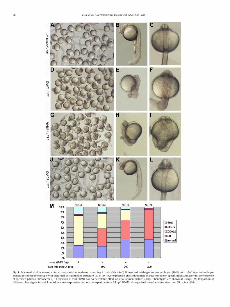
Fig. 1. Maternal Vsx1 is essential for axial–paraxial mesoderm patterning in zebrafish. (A–C) Uninjected wild-type control embryos. (D–F) vsx1 tbMO injected embryosexhibit dorsalized phenotype with disturbed dorsal midline structure. (G–I) vsx1 overexpression elicits inhibition of axial mesoderm specification and aberrant convergenceof specified paraxial mesoderm. (J–L) Injection of vsx1 sbMO has no detectable effect on development before 24 hpf. Phenotypes are shown at 24 hpf. (M) Proportion ofdifferent phenotypes in vsx1 knockdown, overexpression and rescue experiments at 24 hpf. DDMS: disorganized dorsal midline structure. SB: spina bifida.
Y. He et al. / Developmental Biology 386 (2014) 96–11098

Low level of maternal vsx1 mRNA was detected by an RNaseprotection assay in zebrafish (Passini et al., 1998). Our expressionprofile analysis by qRT-PCR showed that the level of vsx1 mRNA
was maintained at the same from one cell stage to late blastulastage, decreased to very low during gastrulation but increasedfrom 6-somite stage (Fig. 4A). To determine whether zygotic vsx1
Fig. 2. Comparison of axial and paraxial mesoderm formation among wild type, maternal Vsx1 suppressed and vsx1 overexpression embryos. (A–J) ntl marked axialmesoderm domain in uninjected wild-type control embryos (A and B), vsx1 tbMO injected embryos (C–F) and vsx1 mRNA injected embryos (G–J). (K–T) myoD markedparaxial mesoderm domain in uninjected wild-type control embryos (K and L), vsx1 tbMO injected embryos (M–P) and vsx1 mRNA injected embryos (Q–T). Note that vsx1overexpression inhibits the convergence of paraxial mesoderm cells but has no impact on paraxial mesoderm cell specification and somite formation. Riboprobes areindicated at the top of each group of figures. All the images of single embryo are dorsal view with animal pole towards the top.
Y. He et al. / Developmental Biology 386 (2014) 96–110 99

mRNA is required for paraxial mesoderm domain patterning, asplice-blocking vsx1 MO (sbMO), which can interfere with thesplicing of newly synthesized zygotic vsx1 mRNA but leave thematernal vsx1 mRNA intact, was used. The embryo receivinginjection of up to 15 ng vsx1 sbMO developed a normal DV axisand no abnormality was observed until 24 hpf (Clark et al., 2008;Fig. 1J–L), indicating that zygotic vsx1 mRNA is not involved inregulating early embryogenesis. Taken together, our resultsdemonstrated that maternal Vsx1, rather than zygotic Vsx1,regulates axial–paraxial patterning by repressing axial mesodermspecification to preserve paraxial mesoderm specification in theventrolateral region.
Maternal vsx1 mRNA directs translation in the ventrolaternal regionat blastula stage
The expression profile (Fig. 4A) indicated that during blastulastage the amount of zygotic vsx1 transcripts was very small (ifsynthesized) in comparison with that of deposited maternal vsx1transcripts. We next analyzed the spatial expression pattern ofmaternal Vsx1 at blastula stage. Whole mount in situ immuno-histochemistry analysis showed that Vsx1 protein was detectablein the nuclei of most blastomeres but not in a few blastomeres onone side at 1k cell stage (Fig. 4B–D and D'). The contrast betweenthe two regions became more striking at dome stage (Fig. 4H–J).
Fig. 3. Maternal Vsx1 is essential for promoting normal paraxial mesoderm specification. (A–F) comparison of msgn1 expression between uninjected wild type control (A–C)and maternal vsx1 tbMO injected embryos (D–F). (G–L) Comparison of tbx24 expression between uninjected wild type control (G–I) and maternal vsx1 tbMO injectedembryos (J–L). Riboprobes are indicated at the bottom of each figure. (B, E, H and K) Dorsal view of embryos with animal pole towards the top. (C, F, I and L) Animal pole viewof the embryos with dorsal towards the right.
Y. He et al. / Developmental Biology 386 (2014) 96–110100

Fig. 4. Expression pattern of vsx1 during early embryogenesis. (A) Expression profile of vsx1 mRNA during early embryogenesis. Results are expressed as mean7SEM. (B–M)Localization of maternal Vsx1 protein in uninjected wild type control and vsx1 tbMO injected embryos at 1k cell (B–G) and dome stages (H–M). (C, F, I and L) Lateral view ofembryos with animal pole towards the top and dorsal towards the right. (D, D', G, J and M) Animal pole view of the embryos with dorsal towards the right. D' is the magnifiedimage of D, in which one can see that Vsx1 protein is localized in the nuclei of the blastomeres.
Y. He et al. / Developmental Biology 386 (2014) 96–110 101

When 4 ng vsx1 tbMO was injected, the Vsx1 protein wasundetectable in the embryos during cleavage (Fig. 4E–G andK–M). These results suggest that maternal vsx1 mRNA directsVsx1 translation in the ventrolaternal region during cleavage,correlating well with that maternal Vsx1 promotes paraxialmesoderm specification in the ventrolateral side.
Vsx1 is essential for repressing flh expression in the ventrolateralmargin
Because axial mesoderm regulatory gene flh can repress para-xial mesoderm regulatory gene spt and paraxial mesoderm speci-fication (Amacher and Kimmel, 1998; Yamamoto et al., 1998), we
examined whether maternal Vsx1 is able to repress flh expressionand preserve spt expression during early developmental stage.Whole mount in situ hybridization analysis revealed that it isindeed the case. From 30% epiboly stage onward, the expression offlh was restricted within the dorsal region in the wild typeembryos (Talbot et al., 1995; Fig. 5B and B', E and E') but expandedto the ventral margin in vsx1 knockdown embryos (Fig. 5A andA', D and D'). In contrast, flh expression was significantly repressedin the dorsal margin of vsx1 mRNA injected embryos (Fig. 5C andC', F and F'). Conversely, the expressions of spt and its downstreamtarget gene paraxial protocadherin (papc) in the margin weredramatically suppressed in the vsx1 knockdown embryos both at30–40% epiboly stage (Fig. 5G and G', L and L') and during
Fig. 5. Vsx1 represses flh to preserve spt and papc expression in the ventolateral margin. The injected reagents are indicated at the top of each column. Riboprobes areindicated at the bottom of each figure. (A–P) Dorsal view of embryos with animal pole towards the top. (A'–P') Animal pole view of the embryos with dorsal towards theright. Arrow heads indicate that the paraxial mesoderm marker is detected in the presumptive axial mesoderm region in vsx1 overexpression embryos.
Y. He et al. / Developmental Biology 386 (2014) 96–110102

gastrulation (Fig. 5I and I', N and N'). Injection of vsx1 mRNAresulted in expansion of spt and papc expression to the dorsalmargin during gastrulation (Fig. 5K and K', P and P'), suggestingthat vsx1 overexpression elicited fate change from axial mesodermto paraxial mesoderm in the anterior dorsal region. It is clear thatmaternal Vsx1 is essential for repressing flh ectopic expression andpreserving spt expression in the ventrolateral margin at lateblastula stage and during gastrula stage.
Since zygotic Wnt8a and Bmp2b signaling pathways arerequired in repressing dorsal genes expression and maintainingventrolateral identity during gastrulation (Ramel and Lekven, 2004;Ramel et al., 2005), we next examined whether maternal Vsx1 isessential for activating the genes of the zygotic Bmp2b and Wnt8asignaling pathways. Whole mount in situ hybridization analysisshowed that injection of vsx1 tbMO has no detectable influence onthe expression patterns of wnt8a, bmp2b and their targets vent andvox at middle gastrula stage (Fig. 6). Therefore, the repression of flhexpression in the ventrolateral margin by maternal Vsx1 is unlikelymediated by zygotic Wnt8a and Bmp2b signaling pathways.
Vsx1 directly represses flh transcription
Vsx1 contains a DNA-binding homeodomain. Previous experi-ments identified that the potential core consensus sequence ofDNA for homeodomain binding is TAATTN (Ades and Sauer, 1994;Rodrigo et al., 2004). Sequence analysis showed that there are 11potential Vsx1 binding sites in the proximal promoter of flhupstream of the transcription start site (Fig. S2). To determinevsx1 directly or indirectly represses flh, we first examined if Vsx1could bind to the potential binding sites at the proximal promoterof flh by chromatin immunoprecipitation (ChIP) assay in normalgastrula embryos. After immunoprecipitaion with the anti-Vsx1antibody, the binding of Vsx1 to all the 11 potential binding siteswas examined by specific PCR with 5 pairs of primers (Fig. 7A).PCR amplification was only detected with the primer pair span-ning the potential binding site 11 (Fig. 7B) in the flh promoterregion 5. Sequence analysis confirmed that the PCR product wasindeed identical to that of the flh promoter region 5. PCR productswere not detected from the controls with preimmune serum or
Fig. 6. Injection of vsx1 tbMO has no obvious influence on the expression patterns of wnt8, bmp2b and their target vent/vox at shield stage. The injected reagents areindicated at the left side of the images. Riboprobes are indicated at the top of images. The images of single embryo are animal pole view with dorsal towards the right.
Y. He et al. / Developmental Biology 386 (2014) 96–110 103

solution without antibody, or from the immunoprecipitation assaywith primers spanning the 3rd exon region of flh (Fig. 7B). Theseresults indicate that Vsx1 specifically binds to flh proximalpromoter at the binding site 11 in normal chromatin environment.
Direct interaction between Vsx1 homeodomain and its poten-tial binding consensus sequences at binding site 11 of flh promoterwas tested by electrophoretic mobility shift assay (EMSA). Afterfusion peptide Vsx1-HD-His (containing the Vsx1 homeodomain)was incubated with the biotin-5′end-labeled probe of the flhpromoter sequence, the binding complexes were detected with amuch slower motility (Fig. 7C). The specificity of peptide–DNAbinding was confirmed with unlabeled probe and mutated com-petitor. Dose-dependent effect was observed in competing experi-ment using unlabeled probe. When 500-fold of excess unlabeledprobe was used prior to incubation with biotin-labeled probe, onlya weak band of binding complex was detected (Fig. 7C). By
contrast, no competitive effect was detected with 500-fold ormore of excess unlabeled mutant competitor in which the TAATTGwas converted into TCCCCG (Fig. 7C). This result indicates thatVsx1 can directly bind to flh promoter at the TAATTG motif nearthe core transcription element.
To determine whether Vsx1 can repress gene expression from theTAATTG motif of the binding site 11, we constructed different GFPreporter gene sensors driven by a 1.9k flh proximal promoter contain-ing all the 11 TAATTN motifs, deleted proximal promoter containingonly the TAATTG motif of the binding site 11, or a 1.9k mutantproximal promoter in which the TAATTG motif at the binding site 11was converted to TCGATG (Fig. 7D). Transcription analysis showed thatall the wild type or mutant flh proximal promoter fragments droveGFP expression successfully and ubiquitously after middle blastulastage (Fig. S3). However, the transcriptional level of GFP sensor drivenby the 1.9k mutant flh proximal promoter was much higher than that
Fig. 7. Vsx1 directly binds to a specific site of flh proximal promoter. (A) The positions of potential Vsx1 binding sites at the proximal promoter of flh. B1–B11 indicates thepotential binding sites of Vsx1. Regions represent the examined regions in ChIP assay. (B) ChIP assay on extracts from Wild type embryos. Input is positive control with thesonicated original genomic DNA fragment. These results show the specific recruitment of Vsx1 by flh promoter region 5. (C) Gel electrophoretogram of EMSA of flh promoterregion 5. Arrow and arrow head indicate the protein-bound probe and the free probe, respectively. 500-fold of excess unlabeled oligonucleotides identical to the probe andunlabeled mutant oligonucleotides were added as competitors when shown. Solution containing no Vsx1-HD polypeptide chain is the positive control. (D) Diagram of GFPreporters driven by a series of truncated or mutant fragments of the flh proximal promoter. (E) Vsx1 binding site 11 mediates Vsx1-dependent repression of flh. Results areexpressed as mean7SEM, and statistical analyses were done by unpaired t test. nnnPo0.001.
Y. He et al. / Developmental Biology 386 (2014) 96–110104

of the GFP sensors driven by wild type flh proximal promoter (Fig. 7E).When 200 pg vsx1 mRNA was co-injected with 40 pg of each the GFPreporter sensors at one-cell stage, the level of gfp expression from thesensors driven by flh proximal promoter containing the binding site 11was significantly decreased but from the sensor driven by the mutantkept high (Fig. 7E and Fig. S3). We noted that the expression level ofGFP in the mutant reporter and vsx1 mRNA coinjected embryos wasdecreased about 30% in comparison to that in mutant reporter aloneinjected embryo. The statistical significance of the change is Po0.05.Since ntl and flh are reciprocally dependent on one another in theirexpression (Talbot et al., 1995; Melby et al., 1997; Halpern et al., 1997),the suppression of mutant reporter by injection of vsx1 mRNA can beexplained by an indirect repression due to the suppression ofendogenous ntl. This result established that the TAATTG motif at thebinding site 11 is the sole Vsx1 binding consensus sequences formediating Vsx1 repressing flh in vivo.
Vsx1 contains two amino acid motifs (FGIDKSR and FAITDLLG,Passini et al., 1998) similar to the repressor domains of Engrailed(Smith and Jaynes, 1996). To define that Vsx1 is a transcriptionalrepressor, we created a transcriptional repressor fusion constructmRNA En-vsx1 by replacing the N-terminal 125 amino acids of Vsx1with the Engrailed repressor domain (amino acids 1–298, Fan andSokol, 1997; Fig. 8A) and a transcriptional activator fusion constructVP16-vsx1 with the VP16 activator domain (amino acids 410–490,Sadowski et al., 1988; Fig. 8A). QRT-PCR analysis showed that gfpexpression mediated by a 1.9k flh promoter is suppressed by En–Vsx1fusion protein, whereas significantly activated by VP16–Vsx1 fusionprotein (Fig. 8B). Injection of 200 pg En–vsx1 mRNA into one-cell-stage embryos produced 58% of embryos with bifurcated axes(N¼93) and abolished the expression of flh in the dorsal margin asefficiently as injection of wild type vsx1 mRNA (Fig. 8F–H). Incontrast, injection of 200 pg VP16–vsx1 mRNA resulted in 43% ofembryos (N¼71) exhibiting a vsx1 morphant-like phenotype andventral expansion of flh expression domain (Fig. 8I–K). These resultsdemonstrated that the function of N-terminal region of Vsx1 issimilar to the Engrailed repressor domain and Vsx1 acts as atranscriptional repressor.
Discussion
To understand how the axial–paraxial mesoderm patterning isinitiated, we investigated why the expression domains of paraxialmesoderm decisive gene spt and axial mesoderm regulatory gene flhinitially overlap but are rapidly restricted within the ventrolateral anddorsal margin, respectively, during blastula stage. We demonstratedthat maternal Vsx1 can directly repress flh transcription to preserve sptexpression and paraxial mesoderm specification in the ventrolateralmargin of blastula embryo. Thus, the original overlapped axial andparaxial mesoderm domains are divided and the initial distinctionbetween axial and paraxial mesoderm domain takes shape. Inhibitingmaternal vsx1 mRNA translation resulted in confusion of axial andparaxial mesoderm markers expression and axial–paraxial mesodermpatterning. These results suggest that direct transcriptional repressionof a decisive axial mesoderm gene by maternal ventralizing factoris essential for initiating axial–paraxial mesoderm patterning invertebrates.
Initial axial–paraxial mesoderm patterning requires complexcooperation between maternal ventral and dorsal determinants
It has been well established that dorsal organizer genes canrepress ventral genes. Thereby, restricting the expression of dorsalorganizer genes within the normal dorsal region is also essentialfor normal paraxial mesoderm specification and axial–paraxialmesoderm patterning. Maternal Runx2bt2 can activate vent, vox
and ved to restrict the expression of dorsal organizer genes (Floreset al., 2008), suggesting that it might be a maternal ventralizingfactor involved in initiation of axial-paraxial mesoderm patterning.To establish a comprehensive understanding of initial axial-paraxial mesoderm patterning, it is worthy to examine whethermaternal Vsx1 cooperates with maternal Runx2bt2 and coopera-tively interacts to maternal dorsal determinants.
Maternal Vsx1 has a role in patterning the convergence and extensiondomains
Previous experiments observed that Spt can cell-autonomouslyregulate lateral mesoderm cell convergence to the dorsal midline(Ho and Kane, 1990; Kimmel et al., 1989) due to that papc is notpromoted in spt mutant embryos (Yamamoto et al., 1998). It hasbeen demonstrated that, during convergence, PAPC is not only animportant signaling molecule of the Wnt/planar cell polaritypathway (Medina et al., 2004; Unterseher et al., 2004; Wanget al., 2008) but also a critical cell adhesion molecule essential forembryonic cell sorting and orientation migration in both Xenopusand zebrafish (Chen and Gumbiner, 2006; Kim et al., 1998).Therefore, papc expression domain depicts the convergence terri-tory of the early gastrula embryo. Since papc is a downstreamtarget of Spt and Flh is the repressor of spt, the patterning of papcexpression and repression domains in the margin of embryo arethe consequences of the patterning of spt and flh expressiondomains, respectively. Maternal Vsx1 is required for the initialdefinition of flh and spt/papc expression domains, implying that ithas a role in patterning convergence and extension domains.
Indeed, ntl marked axial mesoderm domain was expanded inwidth but shortened in length in maternal vsx1 knockdownembryos, myoD marked paraxial mesoderm domain failed inconverging to the normal dorsal position. These phenotypessubstantiate that both inhibition and misexpression of vsx1 canresult in convergence and extension defects.
There are two parallel pathways in the maintenance of paraxialmesoderm identity during gastulation
Inhibition of maternal vsx1 translation also elicited strongectopic expression of flh and concomitant repression of spt andpapc in the ventrolateral margin of middle gastrula embryos(Fig. 5). Since maternal Vsx1 had been depleted in normalembryos at gastrula stage (Fig. 4A), this phenomenon suggeststhat maternal Vsx1 might regulate the maintenance of axial–paraxial mesoderm patterning in an indirect manner. However,maternal Vsx1 has no impact on the expression of wnt8a, bmp2b,vox and vent (Fig. 6), the genes essential for maintaining theventral identity during gastrulation (Ramel et al., 2005). Theseobservations indicate that there are two parallel mechanisms co-regulating the maintenance of paraxial mesoderm identity duringgastrulation. Some of genes involved in paraxial mesoderm speci-fication and differentiation are regulated by maternal Vsx1 andothers are regulated by zygotic Wnt8b and Bmp2b signalingpathways. Therefore, it is impossible to fully convert the paraxialmesoderm into axial mesoderm at the trunk and tail region byinhibiting the function of maternal Vsx1 alone.
Aberrant axial–paraxial mesoderm patterning in vsx1 knockdownand overexpression embryos is due to confused gene expression andconvergent extension defects
Though ectopic expression of axial gene flh resulted in repres-sion of paraxial genes spt and papc, the expression of otherparaxial mesoderm genes in the zygotic Wnt8a and Bmp2bsignaling pathways was maintained in the ventrolateral margin
Y. He et al. / Developmental Biology 386 (2014) 96–110 105

during gastrulation. Reasonably, the suppression of paraxial meso-derm in the vsx1 morphant cannot be simply explained by cell fatechange from paraxial to axial mesoderm, but rather, by confusedexpression of axial and paraxial mesoderm genes and concomitantdefects of convergent extension. It has been observed that theexpression of a dominant negative PAPC can inhibit paraxial myoD
expression due to convergence and extension defects (Yamamotoet al., 1998). Therefore, PAPC absence and concomitant convergentextension defects may contribute to the suppression of myoDexpression during gastrulation in the vsx1 morphants.
Similarly, the formation of expanded width but shortenedlength domain of axial ntl in the vsx1 morphants also can be
Fig. 8. Vsx1 is a transcriptional repressor. (A) Schematic representation of the Vsx1 mutants. (B) Comparison of flh promoter mediated gfp expression under the regulation of afusion protein En–Vsx1 and a fusion protein VP16–Vsx1. Results are expressed as mean7SEM, and statistical analyses were done by unpaired t test. ***Po0.001. (F–H) En–Vsx1fusion protein functions as a wild type Vsx1. (I–K) VP16–Vsx1 fusion protein functions as a Vsx1 antimorph. The injected reagents are indicated at the right side of the images andriboprobes are indicated at the bottom of each figure. (C, F and I) Dorsal view of phenotypes at 8–10 somite stage with head towards the top. (D, G and J) Dorsal view of embryoswith animal pole towards the top. (E, H and K) Animal pole view of the embryos with dorsal towards the right. In situ hybridization of flh is shown at 30–40% epiboly stage.
Y. He et al. / Developmental Biology 386 (2014) 96–110106

explained by the confused expression of axial and paraxialmesoderm genes and concomitant convergent extension defects,rather than the cell fate change from paraxial to axial mesoderm.Previous studies have established that ntl and flh are reciprocallydependent on one another in their expression (Talbot et al., 1995;Melby et al., 1997; Halpern et al., 1997), and Spt is likely a repressorof ntl activity (Amacher and Kimmel, 1998). Moreover, Ntl is apossible inhibitor of cell migration of prechordal mesoderm as froghomolog Brachyury does (Kwan and Kirschner, 2003). It is possiblethat maternal vsx1 knockdown may elicit ntl misexpression, as aconsequence of the overexpression of flh and the loss of sptexpression, in the paraxial mesoderm and prechordal mesodermcells. In this case, the paraxial mesoderm cells simultaneouslyexpress markers of both axial and paraxial mesoderm (the widthof ntl marked axial mesoderm domain appears ventrallyexpanded) and the migration of prechordal mesoderm cells tothe anterior region is inhibited (the length of the axial ntl domainappears shortened). Further experiments are ongoing to test thispossibility and the likely impact on early embryogenesis.
Confused expression of axial and paraxial mesoderm genes andconcomitant convergent extension defects can also explain whyvsx1 overexpression embryos has widely bifurcated paraxialmesoderm domains but no distinguishable dorsal midline struc-tures at the trunk region. In fact, it has been observed that flhmutant axial cells simultaneously express markers of both axialand paraxial mesoderm (Halpern et al., 1995). Therefore, byrepressing flh expression in the axial mesoderm, injection ofvsx1 mRNA is able to disturb notochord formation and paraxialmesoderm convergent extension, but is unable to fully convert theaxial mesoderm fate into paraxial mesoderm fate at the trunk andtail region.
Complex interaction between Vsx1 and binding site recognition
There are 11 potential homeodomain binding sites that containthe consensus sequence TAATTN in the analyzed proximal pro-moter region of flh. Both ChIP and mutant examinations of thebinding site demonstrated that the site near the core transcriptionelement is the sole Vsx1 binding site. This observation suggeststhat, beside the homeodomain and the consensus DNA bindingsequence of homeodomain, other functional domains of Vsx1 andcis-elements of the flh promoter may take part in the complexinteraction between the protein and DNA recognition. Of all the 11potential binding sites at the flh proximal promoter, the uniquestructural characteristic of the actual binding site of Vsx1 is withina GC-rich region (Fig. S2). Therefore, the GC-rich region at theproximal promoter of flh might contain cis-elements for Vsx1 toselect the binding site. It is interesting to investigate whether theevolutionary conserved CVC domain of Vsx1 plays a role in thebinding site recognition at the GC-rich region.
Materials and methods
Animals and obtaining of embryos
Zebrafish were maintained at 28.5 1C in a 14 h/10 h light/darkcycle. Embryos were collected after fertilization and staged bymorphology as described by Kimmel et al. (1995). Embryos weredechorionated with 0.25% trypsin in 1� PBS.
RNA extraction and reverse transcription
Total RNA from zebrafish embryos or adult tissues wasextracted by SV Total RNA Isolation System (Promega) and wastreated with the TURBO DNA-freeTM Kit (Ambion, USA) to remove
DNA contamination. The reaction of reverse transcription (totalvolume of 10 μL) contained approximately 500 ng of total RNA,0.5 mL 100 mM Random 6 mers, 0.5 mL 50 mM oligo dT Primer, 2 mL5� PrimeScriptTM Buffer and 0.5 mL PrimeScriptTM RT Enzyme MixI using PrimeScriptTM RT reagent Kit (TaKaRa, Japan). The firststrand cDNA was synthesized for 15 min at 37 1C, and the RTEnzyme was inactivated at 85 1C for 5 s. The products of reversetranscription were subjected to the next PCR reactions.
Preparation of capped mRNA
According to zebrafish vsx1 encoding sequence (GenBank acces-sion number: BC059574.1), vsx1 ORF was amplified by RT-PCR fromzebrafish retina, using primers 5′-CAGGACGAATTCATGACGGGAAGA-GAAGAAGCT-3′ and 5′-GGGCGCTCGAGTTAACTCTCATTTTCAGAATCG-3′ (the restriction enzyme sites are underlined). vsx1 mis-ORF whichhas 5 synonymous mutation bases downstream of vsx1 translationstart site was generated by RT-PCR using the primers 5′-AGGAC-GAATTCATGACaGGcAGgGAgGAgGCa-3′ (the lowercase indicates themutation bases) and 5′-GGGCGCTCGAGTTAACTCTCATTTTCAGAATCG-3′. Zebrafish flh ORF and spt ORF were generated by RT-PCR fromembryos. Primers used for flh amplification were designed accordingto the sequence of zebrafish flh (GenBank accession number:NM_131055). Primers used for spt amplification were designedaccording to the sequence of zebrafish spt (GenBank accessionnumber: AF077225). To generate the expression plasmids of thesegenes, the ORFs were inserted separately into the EcoR I and Xho Isites of an expression vector pCS107. Capped mRNAs were in vitrosynthesized in the presence of cap analog using the mMESSAGEmMACHINE SP6 Kit (Ambion, USA) according to the instruction ofproducts and purified by Quick Spin Columns (Roche, Switzerland).
Morpholinos
vsx1 Morpholino antisense oligonucleotides (MO) weredesigned and synthesized by Gene-tools (Philomath, OR). Thesequences of vsx1 translation blocking MO (tbMO) is TGTAGCTTCTTCTCTTCCCGTCATG and vsx1 splice-blocking MO (sbMO)is AGCAAAGTGATTCGTACCGGAGTAA as published (Clark et al.,2008).
Generation of GFP-sensors, En–vsx1 and VP16–vsx1 fusion constructs
Wild type and mutant flh proximal promoter driven andmutant GFP reporter sensors were constructed by insertingdifferent flh proximal promoter fragments into the pEGFP-1plasmids between the Sac I and Kpn I sites, and were namedaccording to the length and type of flh proximal fragmentsinserted (Fig. 7D). Fragments of flh proximal promoter wereamplified by PCR with specific primers designed according to thezebrafish flh genomic sequence (GenBank accession number:BX571943.8). The En–vsx1 construct was generated by recombi-nant PCR to fuse the Engrailed repressor domain (amino acids1–298) to the N-terminal of Vsx1 and the VP16–vsx1 construct wasgenerated by recombinant PCR to fuse the VP16 activation domain(amino acids 410–490) to the N-terminal of Vsx1 (Fig. 8A). All therecombinants were reconfirmed by sequencing.
Microinjection
Samples were injected into the zebrafish embryos at the 1 to 2-cell stage. For co-injection, the desired samples were mixedthoroughly prior to injection. Injected embryos were maintainedat 28.5 1C in tap water with antibiotics.
Y. He et al. / Developmental Biology 386 (2014) 96–110 107

Whole mount in situ hybridization
The desired lengths of code sequence of genes were insertedinto pBluescriptIISK plasmids. The constructed plasmids werelinearized and antisense RNA probes were synthesized in vitrousing 50 units of the appropriate RNA polymerase (T7 or T3) in thepresence of DIG mix (Roche, Switzerland). Whole-mount in situhybridization was carried out as described by Thisse and Thisse(2008) with minor modification.
Quantitative RT-PCR
Real-time quantitative RT-PCR was performed in a PCR Light-Cycler 480 System (Roche, Switzerland) using SYBRs Prime-ScriptTM RT-PCR Kit (TaKaRa, Japan) according to therecommendation of the manufacturer. ef1α2 was employed asthe internal standard. The melting curve was analyzed afteramplification to identify the specific product in all PCR reactions.The threshold cycle (Ct) values of 2�ΔΔCT was calculated by qRTsoftware provided for the LightCyclers 480 System (Roche, Swit-zerland). The histogram for fold comparison of different sampleswas generated by inputting the 2�ΔΔCT values of different samplesinto the GraphPad Prism4 program software (Roche, Switzerland).For each sample, the test and control reactions were run intriplicate.
Proteins expression and polyclonal antibody preparation
A polypeptide chain containing Vsx1 homeodomain (residues132–224) and the C-terminal of zebrafish Vsx1 peptide (residue267–340) were expressed with prokaryotic expression vectorpGEX-4T-1. The purity of the two obtained protein was assessedby western blot. We prepared the mouse polyclonal antibodyagainst zebrafish Vsx1 by injecting the purified C-terminal pep-tides into the mouse for 4 times according to the routine protocol.The specificity of the obtained antibody in recognizing Vsx1protein was verified by Western blotting examination (Fig. S4).
Immunohistochemistry
Vsx1 protein was detected by immunohistochemistry. Zebra-fish embryos at dome stage were fixed in 4% paraformaldehyde inPBS overnight at 4 1C, dehydrated through 25%, 50%, 75%, 100%methanol/PBST (PBS/Tween-20, 0.1% ) by turns and stored inmethanol at �20 1C until required (2 h to several months). Atfirst, embryos were rehydrated by successive incubations in 75%,50%, 25% methanol/PBST, washed twice in PBST and twice in 1%DMSO/PBST. Endogenous peroxidases were inactivated by a solu-tion of 80% methanol with 3% H2O2 for 15 min at RT (roomtemperature). The embryos were transferred into 1% DMSO/PBST,blocked in blocking buffer (10% goat serum in 1% DMSO/PBST)overnight at 4 1C, and then immersed in polyclonal antibodyagainst ZF-VSX1 diluted in 1:500 with blocking buffer. The goatanti-IgG (HþL) secondary antibody (cwbiotech, Lot05181013,Beijing, China) was used at a dilution of 1:1000, shaking for 2 hat room temperature. Before detection, the fresh DAB solution wasmade according to the manufacturer's instructions (SIGMA,D4293, USA). DAB reaction was stopped with 1% DMSO/PBST after25 min.
Chromatin immunoprecipitation (ChIP)
ChIP assays were performed using the ChIP-ITTM Express kit(Active motif, California, USA). Wild type embryos were harvestedat 90% epiboly stage and crosslinked in 1% formaldehyde, thenwashed in PBS for three times. Crosslinking was stopped using
glycine stop-fix solution, and after washing in PBS, embryos wereresolved in lysis buffer, then homogenized with Dounce homo-genizer, later transferred to shearing buffer. Extract was thensonicated to produce DNA fragments between 200 and 1500 bp.After sonication, one-tenth of supernatant was removed as inputDNA, the other was incubated overnight at 4 1C with 25 μl ofprotein G magnetic beads and respective antibodies. Beads werewashed with CHIP buffer 1 and buffer 2, resuspended andincubated in Elution Buffer AM2. Cross-linking was reversed byreversed cross-linking buffer and incubating the samples at 95 1Cfor 15 min. Finally samples were digested with proteinase K andstopped with stop solution, thus DNA was then used in PCRimmediately. A pre-denaturation of 3 min at 94 1C was followedby 30 cycles (20 s at 94 1C, 30 s at 60 1C, and 30 s at 72 1C). Primersused in amplifying different regions of flh proximal promotercontaining potential Vsx1 binding sites (Fig. 7A) were designedaccording to the zebrafish flh genomic sequence (GenBank acces-sion number: BX571943.8).
Electrophoretic mobility shift assay (EMSA)
Oligonucleotides were 5′end labeled with biotin. The sequences ofbiotin labeled probe, unlabeled wild type and mutant competitiveprobes are indicated in Fig. 7C. EMSA was performed according tomanufacture's instruction of Lightshift Chemiluminescent EMSA Kit(Pierce, USA). Briefly, 0.8 μL expressed and purified Vsx1 home-odomain peptide was used in the binding reaction. Protein:DNAmixes were resolved on 6% non-denaturing polyacrylamide gels.After electrophoresis, DNA oligonucleotides were transferred ontonylon membrane by electroblotting and UV crosslinked. Biotin-labeled DNAs were visualized with streptavidin-bound HRP andLuminol/Enhancer chemiluminescent substrate (Pierce, USA) andchemiluminescence detected by exposure to Kodak imaging film. Incompeting experiments, different unlabeled probes were incubatedwith the purified Vsx1 homeodomain peptide ahead of the incuba-tion with labeled probe.
Acknowledgments
This work was supported by funds of NSFC 30971654, State KeyBasic Research Project of China (2010CB126301) and the Funda-mental Research Funds for the Central Universities of China. Wewould like to thank Dr. P. Wu and Dr. Qiu for facilities and support.
Appendix. Supporting information
Supplementary data associated with this article can be found inthe online version at http://dx.doi.org/10.1016/j.ydbio.2013.11.022.
References
Ades, S.E., Sauer, R.T., 1994. Differential DNA-binding specificity of the engrailedhomeodomain: the role of the residue 50. Biochemistry 33, 9187–9194.
Amacher, S.L., Kimmel, C.B., 1998. Promoting notochord fate and repressing muscledevelopment in zebrafish axial mesoderm. Development 125, 1397–1406.
Barth, K.A., Kishimoto, Y., Rohr, K.B., Seydler, C., Schulte-Merker, S., Wilson, S.W.,1999. Bmp activity establishes a gradient of positional information throughoutthe entire neural plate. Development 126, 4977–4987.
Bauer, H., Lele, Z., Rauch, G.J., Geisler, R., Hammerschmidt, M., 2001. The type Iserine/threonine kinase receptor Alk8/Lost-a-fin is required for Bmp2b/7 signaltransduction during dorsoventral patterning of the zebrafish embryo. Devel-opment 128, 849–858.
Chen, X., Gumbiner, B.M., 2006. Paraxial protocadherin mediates cell sorting andtissue morphogenesis by regulating C-cadherin adhesion activity. J. Cell Biol.174, 301–313.
Clark, A.M., Yun, S., Veien, E.S., Wu, Y.Y., Chow, R.L., Dorsky, R.I., Levine, E.M., 2008.Negative regulation of Vsx1 by its paralog Chx10/Vsx2 is conserved in thevertebrate retina. Brain Res. 4 (1192), 99–113.
Y. He et al. / Developmental Biology 386 (2014) 96–110108

Dale, L., Wardle, F.C., 1999. A gradient of BMP activity specifies dorsal-ventral fatesin early Xenopus embryos. Semin. Cell Dev. Biol. 10, 319–326.
D'Autilia, S., Decembrini, S., Casarosa, S., He, R.Q., Barsacchi, G., Cremisi, F.,Andreazzoli, M., 2006. Cloning and developmental expression of the Xenopushomeobox gene Xvsx1. Dev. Genes Evol. 216, 829–834.
De Robertis, E.M., Larrain, J., Oelgeschlager, M., Wessely, O., 2000. The establish-ment of Spemann's organizer and patterning of the vertebrate embryo. Nat.Rev. Genet. 1, 171–181.
Dosch, R., Gawantka, V., Delius, H., Blumenstock, C., Niehrs, C., 1997. Bmp-4 acts as amorphogen in dorsoventral mesoderm patterning in Xenopus. Development124, 2325–2334.
Dougan, S.T., Warga, R.M., Kane, D.A., Schier, A.F., Talbot, W.S., 2003. The role of thezebrafish nodal-related genes squint and cyclops in patterning of mesendo-derm. Development 130, 1837–1851.
Fan, M.J., Sokol, S.Y., 1997. A role for Siamois in Spemann organizer formation.Development 124, 2581–2589.
Fior, R., Maxwell, A.A., Ma, T.P., Vezzaro, A., Moens, C.B., Amacher, S.L., Lewis, J.,Saúde, L., 2012. The differentiation and movement of presomitic mesodermprogenitor cells are controlled by Mesogenin 1. Development 139, 4656–4665.
Flores, M.E.C., Lam, E.Y.N., Crosier, K.E., Crosier, P.S., 2008. Osteogenic transcriptionfactor Runx2 is a maternal determinant of dorsoventral patterning in zebrafish.Nat. Cell Biol. 10, 346–352.
Goutel, C., Kishimoto, Y., Schulte-Merker, S., Rosa, F., 2000. The ventralizing activityof Radar, a maternally expressed bone morphogenetic protein, reveals complexbone morphogenetic protein interactions controlling dorso-ventral patterningin zebrafish. Mech. Dev. 99, 15–27.
Graff, J.M., 1997. Embryonic patterning: To BMP or not to BMP, that is the question.Cell 89, 171–174.
Green, J.B., Smith, J.C., 1990. Graded changes in dose of a Xenopus activin Ahomologue elicit stepwise transitions in embryonic cell fate. Nature 347,391–394.
Green, J.B.A., New, H.V., Smith, J.C., 1992. Responses of embryonic Xenopus cells toactivin and FGF are separated by multiple dose thresholds and correspond todistinct axes of the mesoderm. Cell 71, 731–739.
Griffin, K.J.P., Amacher, S.L., Kimmel, C.B., Kimelman, D., 1998. Molecular identifica-tion of spadetail: regulation of zebrafish trunk and tail mesoderm formation byT-box genes. Development 125, 3379–3388.
Gurdon, J.B., Harger, P., Mitchell, A., Lemaire, P., 1994. Activin signaling andresponse to a morphogen gradient. Nature 371, 487–492.
Gurdon, J.B., Bourillot, P.Y., 2001. Morphogen gradient interpretation. Nature 413,797–803.
Halpern, M.E., Thisse, C., Ho, R.K., Thisse, B., Riggleman, B., Trevarrow, B., Weinberg,E.S., Postlethwait, J.H., Kimmel, C.B., 1995. Cell-autonomous shift from axial toparaxial mesodermal development in zebrafish floating head mutants. Devel-opment 121, 4257–4264.
Halpern, M.E., Hatta, K., Amacher, S.L., Talbot, W.S., Yan, Y.-L., Thisse, B., Thisse, C.,Postlethwait, J.H., Kimmel, C.B., 1997. Genetic interactions in zebrafish midlinedevelopment. Dev. Biol. 187, 154–170.
Hammerschmidt, M., Pelegri, F., Mullins, M., Kane, D.A., Brand, M., van Eeden, F.J.M.,Furutani-Seiki, M., Granato, M., Haffter, P., Heisenberg, C.-P., Jiang, Y.-J., Kelsh, R.N., Odenthal, J., Warga, R., Nüsslein-Volhard, C., 1996. Mutations affectingmorphogenesis during gastrulation and tail formation in the zebrafish, Daniorerio. Development 123, 143–151.
Héon, E., Greenberg, A., Kopp, K.K., Rootman, D., Vincent, A.L., Billingsley, G.,Priston, M., Dorva, K.M., Chow, R.L., McInnes, R.R., Heathcote, G., Westall, C.,Sutphin, J.E., Semina, E., Bremner, R., Stone, E.M., 2002. vsx1: A gene forposterior polymorphous dystrophy and keratoconus. Hum. Mol. Genet. 11,1029–1036.
Ho, R.K., Kane, D.A., 1990. Cell-autonomous action of zebrafish spt mutation inspecific mesodermal precursors. Nature 348, 728–730.
Imai, Y., Gates, M.A., Melby, A.E., Kimelman, D., Schier, A.F., 2001. The homeoboxgenes vox and vent are redundant repressors of dorsal fates in zebrafish.Development 128, 2407–2420.
Itoh, K., Sokol, S.Y., 1999. Axis determination by inhibition of Wnt signaling inXenopus. Genes Dev. 13, 2328–2336.
Jones, C.M., Smith, J.C., 1998. Establishment of a BMP-4 morphogen gradient bylong-range inhibition. Dev. Biol. 194, 12–17.
Kwan, K.M., Kirschner, M.W., 2003. Xbra functions as a switch between cellmigration and convergent extension in the Xenopus gastrula. Development130, 1961–1972.
Kim, S.H., Yamamoto, A., Bouwmeester, T., Agius, E., De-Robertis, E.M., 1998. Therole of paraxial protocadherin in selective adhesion and cell movements of themesoderm during Xenopus gastrulation. Development 125, 4681–4690.
Kimmel, C.B., Kane, D.A., Walker, C., Warga, R.M., Rothman, M.B., 1989. A mutationthat changes cell movement and cell fate in the zebrafish embryo. Nature 337,358–362.
Kimmel, C.B., Ballard, W.W., Kimmel, S.R., Ullmann, B., Schilling, T.F., 1995. Stages ofembryonic development of the zebrafish. Dev. Dyn. 203, 253–310.
Kimelman, D., 2006. Mesoderm induction: from caps to chips. Nat. Rev. Genet. 7,360–372.
Kramer, C., Mayr, T., Nowak, M., Schumacher, J., Runke, G., Bauer, H.,Wagner, D.S., Schmid, B., Imai, Y., Talbot, W.S., Mullins, M.C., Hammersch-midt, M., 2002. Maternally supplied Smad5 is required for ventral speci-fication in zebrafish embryos prior to zygotic Bmp signaling. Dev. Biol. 250,263–279.
Kuhl, M., Sheldahl, L.C., Malbon, C.C., Moon, R.T., 2000. Ca21/calmodulin-dependentprotein kinase II is stimulated by Wnt and frizzled homologs and promotesventral cell fates in Xenopus. J. Biol. Chem. 275, 12701–12711.
Lekven, A.C., Thorpe, C.J., Waxman, J.S., Moon, R.T., 2001. Zebrafish wnt8 encodestwoWnt8 proteins on a bicistronic transcript and is required for mesoderm andneuroectoderm patterning. Dev. Cell 1, 1013–1114.
Levine, E.M., Schechter, N., 1993. Homeobox genes are expressed in the retina andbrain of adult goldfish. Proc. Natl. Acad. Sci. USA 90, 2729–2733.
Levine, E.M., Hitchcock, P., Glasgow, E., Schechter, N., 1994. Restricted expression ofa new paired-class homeobox gene in normal and regenerating adult goldfishretina. J. Comp. Neurol. 348, 596–606.
Lu, F.I., Thisse, C., Thisse, B., 2011. Identification and mechanism of regulation of thezebrafish dorsal determinant. Proc. Natl. Acad. Sci. USA 108, 15876–15880.
McDowell, N., Gurdon, J.B., 1999. Activin as a morphogen in Xenopus mesoderminduction. Semin. Cell Dev. Biol. 10, 311–317.
Medina, A., Swain, R.K., Kuerner, K.M., Steinbeisser, H., 2004. Xenopus paraxialprotocadherin has signaling functions and is involved in tissue separation.EMBO J. 23, 3249–3258.
Melby, A.E., Warga, R.M., Kimmel, C.B., 1996. Specification of cell fates at the dorsalmargin of the zebrafish gastrula. Development 122, 2225–2237.
Melby, A.E., Kimelman, D., Kimmel, C.B., 1997. Spatial regulation of floating headexpression in the developing notochord. Dev. Dyn. 209, 156–165.
Melby, A.E., Beach, C., Mullins, M., Kimelman, D., 2000. Patterning the earlyzebrafish by the opposing actions of bozozok and vox/vent. Dev. Biol. 224,275–285.
Mintzer, K.A., Lee, M.A., Runke, G., Trout, J., Whitman, M., Mullins, M.C., 2001. Lost-a-fin encodes a type I BMP receptor, Alk8, acting maternally and zygotically indorsoventral pattern formation. Development 128, 859–869.
Nguyen, V.H., Schmid, B., Trout, J., Connors, S.A., Ekker, M., Mullins, M.C., 1998. Ventraland lateral regions of the zebrafish gastrula, including the neural crest progenitors,are established by a bmp2b/swirl pathway of genes. Dev. Biol. 199, 93–110.
Nikaido, M., Kawakami, A., Sawada, A., Furutani-Seiki, M., Takeda, H., Araki, K.,2002. Tbx24, encoding a T-box protein, is mutated in the zebrafish somite-segmentation mutant fused somites. Nat. Genet. 31, 1995–1999.
Ohtoshi, A., Justice, M.J., Behringer, R.R., 2001. Isolation and characterization of anovel mouse CVC paired-like homeobox gene expressed during embryogenesisand in the retina. Biochem. Biophys. Res. Commun. 286, 133–140.
Ohtoshi, A., Wang, S.W., Maeda, H., Saszik, S.M., Frishman, L.J., Klein, W.H.,Behringer, R.R., 2004. Regulation of retinal cone bipolar cell differentiationand photopic vision by the CVC homeobox gene vsx1. Curr. Biol. 14, 530–536.
Passini, M.A., Kurtzman, A.L., Canger, A.K., Asch, W.S., Wray, G.A., Raymond, P.A.,Schechter, N., 1998. Cloning of zebrafish vsx1: expression of a paired-likehomeobox gene during CAN development. Dev. Genet. 23, 128–141.
Payne, T.L., Postlethwait, J.H., Yelick, P.C., 2001. Functional characterization andgenetic mapping of alk8. Mech. Dev. 100, 275–289.
Ramel, M.C., Lekven, A.C., 2004. Repression of the vertebrate organizer by Wnt8 ismediated by Vent and Vox. Development 131, 3991–4000.
Ramel, M.C., Buckles, G.R., Baker, K.D., Lekven, A.C., 2005. WNT8 and BMP2B co-regulate non-axial mesoderm patterning during zebrafish gastrulation. Dev.Biol. 287, 237–248.
Reim, G., Brand, M., 2006. Maternal control of vertebrate dorsoventral axisformation and epiboly by the POU domain protein Spg/Pou2/Oct4. Develop-ment 133, 2757–2770.
Rodrigo, I., Bovolenta, P., Mankoo, B.S., Imai, K., 2004. Meox homeodomain proteinsare required for bapx1 expression in the sclerotome and activate its transcrip-tion by direct binding to its promoter. Mol. Cell Biol. 24, 2757–2766.
Sadowski, I., Ma, J., Triezenberg, S., Ptashne, M., 1988. GAL4-VP16 is an unusuallypotent transcriptional activator. Nature 335, 563–564.
Saneyoshi, T., Kume, S., Amasaki, Y., Mikoshiba, K., 2002. The Wnt/calcium pathwayactivates NF-AT and promotes ventral cell fate in Xenopus embryos. Nature 417,295–299.
Semina, E.V., Hittner, H.A., Murray, J.C., 2000. Isolation and characterization of anovel human paired-like homeodomain-containing transcription factor gene,vsx1, expressed in ocular tissues. Genomics 63, 289–293.
Shen, M.M., 2007. Nodal signaling: developmental roles and regulation. Develop-ment 134, 1023–1034.
Sidi, S., Goutel, C., Peyrieras, N., Rosa, F.M., 2003. Maternal induction of ventral fateby zebrafish radar. Proc. Natl. Acad. Sci. USA 100, 3315–3320.
Shimizu, T., Yamanaka, Y., Nojima, H., Yabe, T., Hibi, M., Hirano, T., 2002. A novelrepressor-type homeobox gene, ved, is involved in dharma/bozozok-mediateddorsal organizer formation in zebrafish. Mech. Dev. 118, 125–138.
Smith, S., Jaynes, J., 1996. A conserved region of engrailed, shared among all en-,gsc-, Nk1-, Nk2- and msh-class homeoproteins, mediates active transcriptionalrepression in vivo. Development 122, 3141–3150.
Talbot, W.S., Trevarrow, B., Halpern, M.E., Melby, A.E., Farr, G., Postlethwalt, J.H.,Jowett, T., Kimmel, C.B., Kimelman, D., 1995. A homeobox gene essential forzebrafish notochord development. Nature 378, 150–157.
Thisse, C., Thisse, B., Postlethwait, J.H., 1995. Expression of snail2, a second memberof the zebrafish snail family, in cephalic mesendoderm and presumptive neuralcrest of wild-type and spadetail mutant embryos. Dev. Biol. 172, 86–99.
Thisse, C., Thisse, B., 2008. High-resolution in situ hybridization to whole-mountzebrafish embryos. Nat. Protoc. 3, 59–60.
Unterseher, F., Hefele, J.A., Giehl, K., De-Robertis, E.M., Wedlich, D., Schambony, A.,2004. Paraxial protocadherin coordinates cell polarityduring convergent exten-sion via Rho A and JNK. EMBO J. 23, 3259–3269.
Y. He et al. / Developmental Biology 386 (2014) 96–110 109

Valleix, S., Nedelec, B., Rigaudiere, F., Digbiero, P., Pouliquen, Y., Renard, G., LeGargasson, J.F., Delpecb, M., 2006. H244R vsx1 is associated with selective coneon bipolar cell dysfunction and macular degeneration in a PPCD family.Investig. Ophthalmol. Vis. Sci. 47 (1), 48–54.
Wang, Y., Janicki, P., Köster, I., Berger, C.D., Wenzl, C., Großhans, J., Steinbeisser, H.,2008. Xenopus paraxial protocadherin regulates morphogenesis by antagoniz-ing Sprouty. Genes Dev. 22, 878–883.
Yamamoto, A., Amacher, S.L., Kim, S.H., Geissert, D., Kimmel, C.B., De Robertis, E.M., 1998.Zebrafish paraxial protocadherin is a downstream target of spadetail involved inmorphogenesis of gastrula mesoderm. Development 125, 3389–3397.
Yoo, K.W., Kim, C.H., Park, H.C., Kim, S.H., Kim, H.S., Hong, S.K., Han, S., Rhee, M.,Huh, T.L., 2003. Characterization and expression of a presomitic mesoderm-specific mespo gene in zebrafish. Dev. Genes Evol. 213, 203–206.
Y. He et al. / Developmental Biology 386 (2014) 96–110110


