Embed Size (px)
Citation preview

Mature retinal pigment epithelium cells are retained in the cellcycle and proliferate in vivo
Heba Al-Hussaini, Jaimie Hoh Kam, Anthony Vugler, Ma’ayan Semo, Glen Jeffery
(The first two authors contributed equally to this work.)
Institute of Ophthalmology, University College London, United Kingdom
Purpose: To investigate the capacity of mature retinal pigment epithelium (RPE) cells to enter the cell cycle in vivo usinga range of RPE-specific and proliferative specific markers in both pigmented and albino rats.Methods: Whole-mounted retinas of both Dark Agouti and albino rats were immunolabeled with cell cycle markers Ki67or PCNA and double labeled with RPE cell marker RPE65 or CRALBP. The number and distribution of these cells wasmapped. An additional group of Dark Agouti rats were given repeated intraperitoneal injections of Bromodeoxyuridine(BrdU )for 20 days and then sacrificed 30 days later. The retinas were then processed for BrdU detection and Otx, a RPEcell-specific marker. For comparison, human RPE tissue from a postmortem donor was also labeled for Ki67.Results: In both pigmentation phenotypes, a subpopulation of mature RPE cells in the periphery were positive for bothcell cycle markers. These cells were negative for Caspase 3, hence were not apoptotic. Ki67-positive cells were also seenin human RPE. Further, many cells positive for BrdU were identified in similar retinal regions, confirming that RPE cellsnot only enter the cell cycle but also divide, albeit at a slow cell cycle rate. There was a ten fold increase in the numberof RPE cells positive for cell cycle markers in albino (approximately 200 cells) compared to pigmented rats (approximately20 cells).Conclusions: Peripheral RPE cells in rats have the capacity to enter the cell cycle and complete cellular division.
In the mature mammalian retina, cells such as neurons[1-4] and retinal pigment epithelium (RPE) [5,6] are lost withage. While there is no evidence of cell proliferation, an ocularstem cell population has been identified in the ciliary body[7]. Another ocular pigmented tissue that has the latentcapacity to replicate is the RPE. The RPE is an integral partof the retina and plays a critical role in both neural retinaldevelopment and retinal function. The mature RPE is held ina state of senescence by the adjacent neural retina, becausewhen the retina is detached, RPE cells proliferate [8]. In theamphibian, removal of the neural retina results in RPE cellproliferation as in mammals, but these cells thentransdifferentiate to produce a completely new functionalretina. In amphibians, the mature, but not the developing, RPEexpresses the tissue specific marker RPE65. Following retinalremoval RPE cells downregulate RPE65 whiletransdifferentiating, and it is only upregulated when retinalproduction is complete and transdifferentiation ceases [9].Hence, it appears that, like the amphibian RPE, themammalian RPE has the capacity to proliferate but not totransdifferentiate into the diverse cell types found in the retina.
Here we use three independent RPE cell markers, RPE65,Cellular retinaldehyde-binding protein (CRALBP), and Otx,
Correspondence to: Glen Jeffery, Institute of Ophthalmology,University College London, Bath Street, London EC1V 9EL, UnitedKingdom; Phone:+44 207 6086837; Fax: +44 207 6086850; email:[email protected]
making identification of these cells unambiguous. RPE65 is akey element in normal RPE function. It plays an importantrole in the visual cycle and in vitamin A metabolism. RPE65is also associated with retinol binding protein and 11-cis-retinol dehydrogenase [10]. CRALBP binds to 11-cis-retinalin the visual cycle, and its function is associated with normaldark adaptation [11]. Otx2 is a specific marker for RPE cellsan important in their specification [12]. There is an anecdotalevidence for cell cycle events in mature albino RPE [13].Hence, here we examine the RPE in mature rats and humanusing three independent proliferative markers, Ki67,proliferating cell nuclear antigen (PCNA), and BrdU, to assessthe latent capacity of this tissue to proliferate while the retinais in place. Both pigmented and albino rats are used, as duringdevelopment albino retinas are abnormally proliferative dueto the absence of L-dopa, a key cell cycle regulator and anupstream element in melanin synthesis [14,15].
METHODSTissue source: Pairs of eyes were obtained from 2-month-oldDark Agouti (DA) rats (n=14) and albino Wistar rats (n=10)Pairs of eyes were also obtained from 1-year-old DA rats(n=5) and albino Wistar rats (n=5). DA rats were also takenat the following embryonic (E) and postnatal (P) days: E18(n=3), P0 (n=5), P5 (n=4), P10 (n=5), P15 (n=6), P20 (n=5),P25 (n=5), and P45 (n=6). Four additional rats were taken atP90 and P150. Animals were euthanized by carbon dioxideinhalation. When rats were collected at E18, the mother was
Molecular Vision 2008; 14:1784-1791 <http://www.molvis.org/molvis/v14/a210>Received 24 May 2008 | Accepted 9 September 2008 | Published 6 October 2008
© 2008 Molecular Vision
1784

euthanized by carbon dioxide inhalation and the pupsremoved. The eyes from pups were enucleated and fixed in4% paraformaldehyde overnight, and the anterior segment,vitreous, and retina removed leaving the eye cup with the RPEexposed. All procedures were performed under UKGovernment (Home Office) and local animal ethicscommittee approval.
An additional group of 18 P25 DA rats were givenintraperitoneal injections of 50 µg/kg BrdU in 0.007 NaOH/phosphate buffered saline (PBS). This additional group wasdivided into six groups of three animals each. The first groupof 3 was given a single injection and euthanized 3 h later. Thesecond group of animals were injected at 3 h intervals andgiven a total of 4 injections before being euthanized. The thirdgroup was given a single injection and was euthanized 12 hlater. The fourth group were given 4 injections separated by12 h and euthanized 12 h after the last injection. The fifthgroup was given 5 injections separated by 24 h in a similarway and euthanized. The final group was injected once eachday for 20 days and then euthanized one month after the lastdose of BrdU. All animals were euthanized with carbondioxide and the eyecups were fixed in 4% paraformaldehydefor 30 min. These experiments were undertaken to confirmthat cell addition was taking place and cast limited light on thelength of the cell cycle.
Healthy eyes from an 83-year-old postmortem donorwere obtained from the Eye Bank at Moorfields Eye Hospital.Full Local Research Ethic Committee approval andappropriate consent were obtained under The Human TissueAct. Consent is given prior to death or from the relativesfollowing death. The eyes was approximately 36 hpostmortem. The eyecups were fixed in 4% paraformaldehydefor 30 min, and the RPE and the underlying choroid removedas a single tissue sheet. Approximately one-third of this largesheet was trimmed to span the equatorial to peripheral retinaso it could be used for analysis.Tissue staining: The rat eye cups containing the RPE werewashed four times in 0.1 M PBS (pH 7.4), then blocked with5% normal donkey serum (NDS) in 3% Triton X-100 in PBSfor 2 h. Primary antibody incubation with 1:2,000 dilution ofrabbit anti-Ki67 (Novocastra, Newcastle, UK) and 1:500dilution of rabbit antiproliferative cell nuclear antigen(PCNA; Abcam, Cambridge, UK) in 1% NDS in 3% Triton X−100 in PBS was performed overnight at room temperature.Primary analysis was undertaken on tissue stained with Ki67,and PCNA was used in a confirmatory role. In most of thealbino Wistar rats and approximately half of the DA rats, RPEcells were labeled with a second primary monoclonalantibody; we used a 1:500 dilution of either mouse anti-RPE65 (Chemicon, Hampshire, UK) or mouse anti-CRALBP(Affinity BioReagents, Cambridge, UK). Without this secondmonoclonal antibody, albino RPE was impossible to image.Following four washes in PBS the tissue was incubated for 2
2 h in a secondary antibody that consisted of a 1:200 dilutionof TRITC donkey antimouse and FITC donkey antirabbit(Jackson ImmunoResearch laboratories, West Grove, PA) in1% NDS in 0.3% Triton X-100. A 1:2,500 dilution of DAPIin PBS was added to the tissue for one min to label the nucleiof cells in the tissue. The eyecups were then washedextensively in 0.05 M Tris buffer (pH 7.4), mounted flat RPEup with Vecta shield, and examined under fluorescentmicroscopy. The same protocol of labeling was used for theRPE sheet taken from human tissue; however, only Ki67 andnot RPE65 was used. As a negative control, some RPE cupswere processed in the absence of primary antibodies.
Cell cycle proteins can be upregulated in mature cellswhen they initiate caspase-related apoptosis [16,17]. Tocontrol for this, we used caspase staining to determine if itwould colocalize with cells positive for Ki67. For this welabeled with rabbit polyclonal anti-Caspase 3 (Abcam).Retinas were blocked as described in the last section andincubated overnight with a 1:500 dilution of Active Caspase3 and 1:500 dilution of goat polyclonal anti-Ki67 (Santa CruzBiotechnology, Santa Cruz, CA). Following four washes inPBS the tissue was incubated for 2 h in a 1:200 dilution ofTRITC donkey antirabbit and FITC donkey antigoat (JacksonImmunoResearch Laboratories,) in 1% NDS in 0.3% TritonX-100. DAPI was added, and the tissue was washed andflatmounted as described in the previous paragraph.
From the six rats injected with BrdU, one eye wasprocessed for BrdU detection and the other one was doubleprocessed for BrdU detection and Ki67 or for BrdU and Otx.The eyes, which were double labeled, were first incubatedovernight with either a 1:1,000 dilution of Ki67 or Otx (SantaCruz Biotechnology) in 1% NDS in 3% Triton X-100 in PBS.The tissue was then incubated in the secondary antibody andfixed in 4% paraformaldehyde for 10 min.
Antigen retrieval was necessary for detection of BrdU inRPE cells. This was undertaken by placing the tissue in 6 Mhydrochloric acid in 1% Triton X-100 in PBS for 30 min.Before incubation in BrdU the tissue was washed extensivelywith PBS to equilibrate the tissue to a normal pH. The tissuewas then blocked with normal donkey serum for 2 h. Anovernight incubation of 1:5 dilution of BrdU in 1% NDS in3% Triton X-100 in PBS was performed at room temperature.Following four washes in PBS, the tissue was incubated for 2h in a 1:200 dilution of TRITC donkey antimouse (JacksonImmunoResearch Laboratories) in 1% NDS in 0.3% TritonX-100. The tissue was then washed once with PBS andextensively with Tris buffer, mounted flat, RPE up with vectorshield, and examined under fluorescent microscope.Analysis: The number and distribution of Ki67-positive cellswas mapped in the RPE flat mounts. The position of each cellwas marked on a composite map, created using AdobePhotoshop CS version 8. RPE65 is a tissue-specific markernormally expressed in all RPE cells. Hence, only the variation
Molecular Vision 2008; 14:1784-1791 <http://www.molvis.org/molvis/v14/a210> © 2008 Molecular Vision
1785

in RPE65 expression was determined in cells positive forKi67. When cells were positive for both Ki67 and RPE65 theirposition was plotted. Tissue processed for BrdU detection wasmapped in a similar way with the relative location of BrdUand Ki67 plotted on schematic drawings. Human tissue wasexamined to see if Ki67 cells were detectable in the RPE.Because of the relatively large size of the human RPE tissuesheet it was necessary to process it in strips. This resulted ina loss of relative location of the positive cells, andconsequently these were not plotted in relation to one anotheror other major land marks. Here, the primary aim was only todetermine if they were present.
Many rodent RPE cells are binucleated [18]. Todetermine the relative distribution of these cells in relation tocell positive for cell cycle markers, we divided rodent RPEinto three regions of approximately equivalent areas: central,equatorial, and peripheral. In each of these regions, three areaswere analyzed measuring 150×150 μm. These were separatedby approximately 120 degrees in relation to the optic nervehead, like three spokes of a wheel. Binucleated andmononucleated cells were counted within the defined areas.Cells with more than two nuclei were rarely encountered andwere not recorded for the purpose of this study.
One-way ANOVA was used to analyze the statisticalsignificance of Ki67 labeling in each experiment and wasfollowed by post hoc Newman-Keuls multiple comparisontest when appropriate.
RESULTSRPE cells positively identified with RPE65 and CRALBPwere apparent in all rat retinas examined. These cells had aclear hexagonal morphology. In the pigmented animals, all ofthe cells were packed with melanin granules. In bothpigmented phenotypes, the size was consistent with RPE cellsin a single hexagonal matrix in a single plane (Figure 1). Someof these cells were clearly labeled with the proliferativemarker, Ki67 (Figure 1A,D); similar cells were also labeledwith PCNA, the other proliferative marker used (Figure1K,L). However, fewer cells were labeled with PCNAbecause Ki67 labels cells in all phases of the cell cycle exceptG0, while PCNA labels cells in S phase alone. Labeling withRPE65 and CRALBP clearly defined RPE cells in bothpigmented (Figure 1) and albino rats (not shown), andconfirmed that the cells in the cell cycle were truly RPE cells.However, some RPE cells that were Ki67 positive expressedlow levels of RPE65, although the levels of expression werestill greater than in negative controls (Figure 1D-F).
Ki67-labeled cells were only present in equatorial andperipheral regions (Figure 2). None were seen centrally, closeto the optic nerve head. Negative controls in which all stagesof immune processing were undertaken, except theapplication of the primary antibody, failed to show any labelin the RPE in either central or peripheral locations with either
Ki67 or PCNA. With the exception of cell numbers there wereno geographic differences in the patterns of labeling foundusing either Ki67 or PCNA.
Because Ki67 labels a larger proportion of cells in the cellcycle, analysis focused on this proliferative marker. In 2-month-old pigmented rats the number of Ki67-positive cellsfound in the RPE was relatively small, being of the order of20–30 in each retina (Figure 2C,D). Within the regions where
Figure 1. Labeling patterns in retinal pigment epithelium (RPE)sheets in whole mount preparations taken from DA rats. A: RPE cellspositively labeled for Ki67. These appear to be in anaphase. B: Thisis the same region as shown in A, but stained with RPE65, which isan RPE specific marker in this tissue. C: This is the same region asshown in A and B but stained with DAPI to reveal the nuclei of theimaged cells. D-F: These are stained in the same way as A-C,however here the cell positive for Ki67 shown in D has down-regulated RPE65 as shown in E. F is the corresponding DAPI stainedimage. G: This shows a Ki67 positive RPE cell which has also inH been stained with CRALBP, which is a second RPE specificmarker in this tissue. I is the same region stained with DAPI to revealnuclei. J shows a Ki67 positive RPE cells. Taken in black and whitethe melanin granules in the cell can be clearly identified (arrow),which along with the RPE65 and CRALBP confirm that the tissuesheet examined is RPE. K shows an RPE cell positive for a secondcell cell cycle marker PCNA, and L shows the same image stainedwith DAPI. The scale bar represents 10 µm.
Molecular Vision 2008; 14:1784-1791 <http://www.molvis.org/molvis/v14/a210> © 2008 Molecular Vision
1786
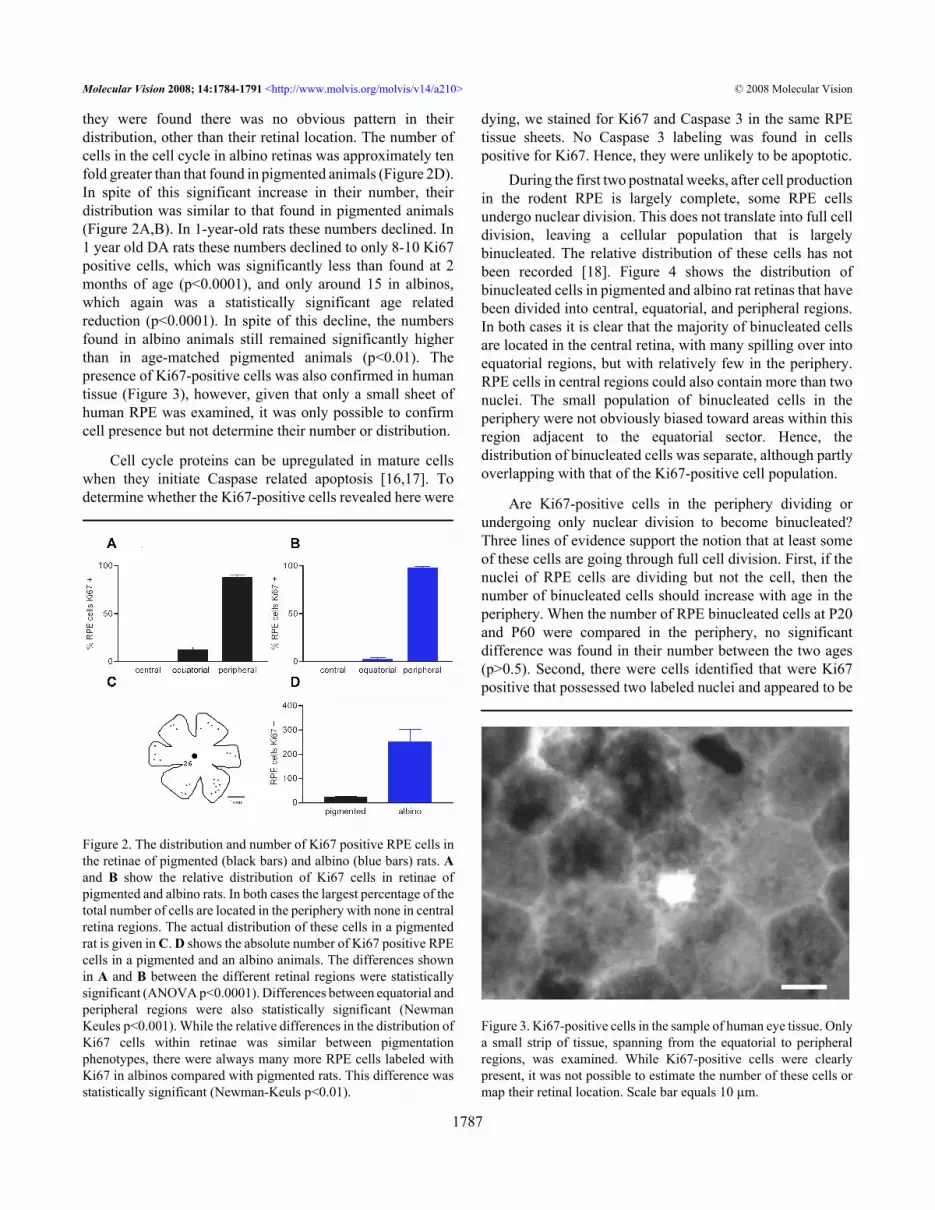
they were found there was no obvious pattern in theirdistribution, other than their retinal location. The number ofcells in the cell cycle in albino retinas was approximately tenfold greater than that found in pigmented animals (Figure 2D).In spite of this significant increase in their number, theirdistribution was similar to that found in pigmented animals(Figure 2A,B). In 1-year-old rats these numbers declined. In1 year old DA rats these numbers declined to only 8-10 Ki67positive cells, which was significantly less than found at 2months of age (p<0.0001), and only around 15 in albinos,which again was a statistically significant age relatedreduction (p<0.0001). In spite of this decline, the numbersfound in albino animals still remained significantly higherthan in age-matched pigmented animals (p<0.01). Thepresence of Ki67-positive cells was also confirmed in humantissue (Figure 3), however, given that only a small sheet ofhuman RPE was examined, it was only possible to confirmcell presence but not determine their number or distribution.
Cell cycle proteins can be upregulated in mature cellswhen they initiate Caspase related apoptosis [16,17]. Todetermine whether the Ki67-positive cells revealed here were
Figure 2. The distribution and number of Ki67 positive RPE cells inthe retinae of pigmented (black bars) and albino (blue bars) rats. Aand B show the relative distribution of Ki67 cells in retinae ofpigmented and albino rats. In both cases the largest percentage of thetotal number of cells are located in the periphery with none in centralretina regions. The actual distribution of these cells in a pigmentedrat is given in C. D shows the absolute number of Ki67 positive RPEcells in a pigmented and an albino animals. The differences shownin A and B between the different retinal regions were statisticallysignificant (ANOVA p<0.0001). Differences between equatorial andperipheral regions were also statistically significant (NewmanKeules p<0.001). While the relative differences in the distribution ofKi67 cells within retinae was similar between pigmentationphenotypes, there were always many more RPE cells labeled withKi67 in albinos compared with pigmented rats. This difference wasstatistically significant (Newman-Keuls p<0.01).
dying, we stained for Ki67 and Caspase 3 in the same RPEtissue sheets. No Caspase 3 labeling was found in cellspositive for Ki67. Hence, they were unlikely to be apoptotic.
During the first two postnatal weeks, after cell productionin the rodent RPE is largely complete, some RPE cellsundergo nuclear division. This does not translate into full celldivision, leaving a cellular population that is largelybinucleated. The relative distribution of these cells has notbeen recorded [18]. Figure 4 shows the distribution ofbinucleated cells in pigmented and albino rat retinas that havebeen divided into central, equatorial, and peripheral regions.In both cases it is clear that the majority of binucleated cellsare located in the central retina, with many spilling over intoequatorial regions, but with relatively few in the periphery.RPE cells in central regions could also contain more than twonuclei. The small population of binucleated cells in theperiphery were not obviously biased toward areas within thisregion adjacent to the equatorial sector. Hence, thedistribution of binucleated cells was separate, although partlyoverlapping with that of the Ki67-positive cell population.
Are Ki67-positive cells in the periphery dividing orundergoing only nuclear division to become binucleated?Three lines of evidence support the notion that at least someof these cells are going through full cell division. First, if thenuclei of RPE cells are dividing but not the cell, then thenumber of binucleated cells should increase with age in theperiphery. When the number of RPE binucleated cells at P20and P60 were compared in the periphery, no significantdifference was found in their number between the two ages(p>0.5). Second, there were cells identified that were Ki67positive that possessed two labeled nuclei and appeared to be
Figure 3. Ki67-positive cells in the sample of human eye tissue. Onlya small strip of tissue, spanning from the equatorial to peripheralregions, was examined. While Ki67-positive cells were clearlypresent, it was not possible to estimate the number of these cells ormap their retinal location. Scale bar equals 10 µm.
Molecular Vision 2008; 14:1784-1791 <http://www.molvis.org/molvis/v14/a210> © 2008 Molecular Vision
1787

passing through full cell division, establishing a cytoplasmicmembrane between their nuclei. These cells also appeared tobe irregular within the geometric configuration of the regularRPE (Figure 5). Ki67 and PCNA are only associated withproliferation as they are simply cell cycle. However, thecritical factor in favor of this resulting in cell division is thatBrdU was detected in peripheral retinal cells. These were a
Figure 4. The percentage of binucleated RPE cells in different retinalregions in pigmented (black bars) and albino (blue bars) rats. In adultrodent retinas, many of the RPE cells were binucleated. Theproportion of these have been determined in both pigmented (A) andalbino (B) retinas. The retinas were divided into three roughly equalgeographic regions: central, equatorial, and peripheral. In bothpigmentation phenotypes the majority of the binucleated cells werelocated toward the central retina, although many were also found inequatorial regions. The distribution of these cells is the reversepattern of that found for Ki67-positive cells shown in Figure 2. Thedifferences in A and B are statistically significant (ANOVA,p<0.001). In both cases, the differences between the percentage ofbinucleated RPE cells between central and equatorial regions werenot statistically significant. However, differences in the percentageof binucleated RPE cells found central and peripheral regions weresignificantly different (Newman-Keuls p<0.01). The differencesbetween equatorial and peripheral regions were also statisticallysignificant (Newman-Keuls p<0.01).
mixed population with some having a single nucleus andothers being binucleated. Cells with a single nucleus werecommonly in adjacent pairs (Figure 6). Multiple injections ofBrdU given at 3 and 12 h intervals failed to significantlyincrease the size of this population compared with animalsgiven a single pulse. However, those given at 24 h intervalsover 5 days did significantly expand the number of positivecells compared with all other groups (ANOVA=0.0001,Dunnett's Multiple Comparison Test=0.001, Figure 7).Hence, it is likely that the cell cycle rate is very slow.
As the retina develops with a center to periphery gradient,such that late cell division occurs in the peripheral retina[19], it is natural to ask whether the relatively peripheralpatterns of Ki67 labeling found in the mature RPE are notsimply a reflection of those patterns present during latedevelopment, or whether they represent a distinct and separateevent. We labeled RPE flat mounts from pigmented animalsat progressive stages from E18 through the postnatal periodand into maturity with Ki67. Unfortunately, it was not possibleto generate retinal whole-mounts of sufficient quality beforeE18 without inducing some damage to the periphery thatresulted in loss of labeled cells. At E18, approximately 50Ki67-positive cells could be identified in the RPE sheet.However, three days later on the day of birth, there was a largeincrease in the number of positive cells found, from around50 to approximately 260, which is statistically significantcompared to the earlier time point (Newman-Keuls, p<0.001;Figure 8). From this point onward the number of positive cellsdeclined gradually until around p20–P25 when their numberreached comparable levels to those found in older animals. Atall of these stages of development, few if any Ki67-positivecells were identified in central regions, rather they wereconfined to equatorial and peripheral retinal regions similarto that shown in Figure 2 for the adult. These patterns of Ki67labeling through postnatal development were similar in
Figure 5. RPE cells were identified that appear to be going throughfull cell division. In the peripheral retinal regions, a small number ofcells could be identified that appeared to be passing through full celldivision. In both pictures, arrows point to two labeled nuclei thatappear to be forming a plasma membrane between them. Both setsof cells appear irregular in the RPE cell matrix. Taken together withthe finding that there was no increase in the number of binucleatedcells in the peripheral retina, these photographs demonstrate that atleast some of the cells in this region were undergoing full celldivision.
Molecular Vision 2008; 14:1784-1791 <http://www.molvis.org/molvis/v14/a210> © 2008 Molecular Vision
1788

albinos, although the absolute number of positive cells foundwas markedly elevated (data not shown). These data areconsistent with the notion that the increase in cell labelingfound in the RPE around the time of birth may represent adistinct and separate event from earlier patterns that startcentrally and end in the periphery.
DISCUSSIONUsing three independent markers for proliferation and threeindependent functional markers of RPE cells, we havedemonstrated that a proportion of mature RPE cells areretained in or have the capacity to enter into the cell cycle anddivide in peripheral and equatorial retinal regions. Thenumber of these cells were significantly elevated whenpigment was absent. In pigmented animals multiple injectionsof BrdU only elevated cell numbers at 24 h intervals over 5days, indicating that the cell cycle rate is probably slow. It isinteresting in the light of this result that in an analysis of 50
Figure 6. BrdU labeling in mature RPE cells. A and B: Shows abinucleated labeled cell with BrdU on red channel and Otx greenchannel. C shows two adjacent mono-nucleated cells that are labeledwith BrdU. Scale bar equals 20 µm. D shows an outline drawing onwhich the location of RPE cells labeled with Ki67 and BrdU aremarked. The diagram shows the distribution of positive BrdU-labeled binucleated (black dots) and mononucleated (black circle)cells. Only a small number of the BrdU-labeled cells were morecentrally located than those labeled for Ki67. The mononucleatedcells were almost always found in pairs of close proximity. The reddots represent the number and distribution of Ki67 positive RPEcells. While these largely overlap with the BrdU labeled populationof RPE cells, they tend to occupy a slightly more peripheral location.The scale bar represents 2.5 mm.
of the most highly expressed retinal genes, 11 showeddifferential expression between central and peripheral humanRPE. Of these, two were cell cycle genes that weredownregulated in the central retina compared to the periphery[20].
Consideration of the relative distribution of binucleatedcells and those in the cell cycle could lead to the conclusionthat there might be a slow progressive wave of nucleardivision across the retina, starting in the center and ending inthe periphery. However, the presence of cells positive forBrdU that contained only one nucleus argues against this.Further, examination of data from younger and older animalsdoes not support this assertion, as at every stage ofdevelopment examined, the distribution of binucleated cellswas largely confined to central and equatorial regions alone,and that of Ki67-positive cells to equatorial and peripheralregions (data not shown).
There are marked differences between pigmentationphenotypes in the number of Ki67 labeled cells found in theirrespective RPE. There are two possible reasons for this. First,during development albino retinas are more proliferative thanpigmented because they lack dopa, an upstream element in thesynthetic pathway of melanin, shown to encourage cell cycleexit [14,21]. It is possible that the absence or reduction of dopa
Figure 7. The number of BrdU positive cells in the RPE followingmultiple pulses at progressive times. BrdU was given as a singlepulse (50µg/kg) or as multiple pulses in 2 month old DA rats. Forcomparison animals were given 4 injections at 3 h intervals andeuthanized 3 h later. Similar experiments were undertaken at 12 hintervals, with single pulse and euthanized 12 h later compared with4 pulses separated by 12 h each and euthanized 12 h after the lastpulse. There was no statistically significant increase in labeled cellsfollowing multiple injections compared with single injections.However, when BrdU was pulsed at 24 h intervals over 5 days, therewas a significant increase in the number of RPE labeled cellscompared with labeling in any of the other groups (ANOVA,p<0.0001. Dunnett’s Multiple Comparison Test p<0.001). Hence theRPE population is proliferating, but with a cell cycle rate ofapproximately 5 days.
Molecular Vision 2008; 14:1784-1791 <http://www.molvis.org/molvis/v14/a210> © 2008 Molecular Vision
1789

during development has a persistent impact upon their retinasextending into maturity. Second, albinos have far fewer rodphotoreceptors than normally pigmented animals [22,23], andhence the RPE phagocytotic workload is probably reduced.As RPE proliferation is enhanced by retinal detachment [8],it is possible that there is a relationship between photoreceptornumbers and their contact with the RPE, and the probabilityof RPE cells entering the cell cycle. However, this remains tobe proven. There are likely to be significant differencesbetween the rats used here other than their pigmentationphenotype, and include variation in genetic background andocular light experience, among other difference. In light ofthis, an analysis of the number and distribution of Ki67-positive cells in mutant animals having photoreceptordystrophies would be of value.
Why has cell division in the mature RPE not beenidentified before and why does it occur? Rodent RPE is rarelyviewed in whole-mount because it is difficult to remove as acoherent tissue without becoming significantly damaged inthe process. For this reason it is commonly viewed in section,where this important, but relatively sparse cell populationwould be hard to identify. Examination of normal adult retinaslabeled with tritiated thymidine to mark dividing cells in catfailed to produce positive label in the RPE [8]. However, Ts’oet al. [13] noted two mitotic nuclei in mature albino retinas,but these could have been dividing nuclei as opposed todividing cells, and were not noted in other retinas used in theirstudy. Another reason these cells may not have been identifiedis simply that no investigation has used cell cycle markers,
Figure 8. The number of Ki67-positive cells found in the RPE flatmounts of pigmented animals sampled from embryonic day 18 (E18)through to postnatal day 150. Relatively few cells were in the cellcycle at E18, which is when the normal patterns of cell division indeveloping tissue end. However there was a marked increase(ANOVA, p<0.0001) in the number of these cells on the day of birth(0), which was statistically significant compared to E18 (Newman-Keuls, p<0.001). From P0 on, cell number gradually declined withage.
such as Ki67, on whole mount mature mammalian RPEbefore. In spite of this, a proliferative zone in the neural retinaand the RPE at the retinal margin has been noted during earlystages in the hatched chick [24,25], and here the level ofproliferation is of sufficient magnitude to be obvious insection, but it is unclear how long this is sustained for.
While we demonstrate that a population of cells in themature RPE divide, we have no data to indicate that thenumber of RPE cells actually increases with age. In fact in arecent study from this laboratory, a striking feature of rat RPEis that it shows little age-related cell loss (unpublished).Hence, if the peripheral RPE is undergoing gradual celladdition, then it is probable that this is a process thatreplenishes the tissue when it is subjected to normal agerelated cell loss. However, as RPE cell loss is likely to be panretinal, the local addition of these cells toward the peripherywould require gradual shifts in the RPE toward the centralretina. Interestingly such a proposal has been made by DelPriore et al. [6]. They found evidence for cell death in thehuman RPE but no evidence for age-related changes in centralcell density. Interestingly, the data on age related changes inthe human RPE is mixed. Some studies find evidence for age-related loss [5], some no change [6], and anothercomprehensive analysis demonstrates an actual increase indensity with age [26]. Whichever study is examined, there isa wide diversity in individual data. It is possible that afundamental mechanism exists to replenish the RPE throughperipheral division to balance cell loss, but that this balanceis easily disrupted leading to significant variation betweenindividuals.
The proliferative capacity of the RPE has an additionaldimension in that it is enhanced if the retina is removed [8],although only in peripheral and not central regions [27]. Someamphibians have the ability to take this a stage further and areable to regenerate a new retina through transdifferentiation ofthe proliferating RPE population [9]. Mammals seem to haveretained an element of this process in their ability toproliferate, but appear to lack the regulatory elementsnecessary to control RPE proliferation appropriately andinduce transdifferentiation. However, if their entry into thecell cycle could be regulated, it would have very significantimplications for disease processes where the RPE is either lostor damaged, such as age-related macular degeneration.
ACKNOWLEDGMENTThis work was sponsored by The Wellcome Trust. Heba Al-Hussaini was awarded a fellowship from Kuwait University.
REFERENCES1. Curcio CA, Sloan KR, Kalina RE, Hendrickson AE. Human
photoreceptor topography. J Comp Neurol 1990;292:497-523. [PMID: 2324310]
2. Cunea A, Jeffery G. The ageing photoreceptor. Vis Neurosci2007; 24:151-5. [PMID: 17640405]
Molecular Vision 2008; 14:1784-1791 <http://www.molvis.org/molvis/v14/a210> © 2008 Molecular Vision
1790

3. Gao H, Hollyfield JG. Aging of the human retina. Differentialloss of neurons and retinal pigment epithelial cells. InvestOphthalmol Vis Sci 1992; 33:1-17. [PMID: 1730530]
4. Balazsi AG, Rootman J, Drance SM, Schulzer M, Douglas GR.The effect of age on the nerve fiber population of the humanoptic nerve. Am J Ophthalmol 1984; 97:760-6. [PMID:6731540]
5. Panda-Jonas S, Jonas JB, Jakobczyk-Zmija M. Retinal pigmentepithelial cell count, distribution, and correlations in normalhuman eyes. Am J Ophthalmol 1996; 121:181-9. [PMID:8623888]
6. Del Priore LV, Kuo YH, Tezel TH. Age-related changes inhuman RPE cell density and apoptosis proportion in situ.Invest Ophthalmol Vis Sci 2002; 43:3312-8. [PMID:12356840]
7. Tropepe V, Hitoshi S, Sirard C, Mak TW, Rossant J, van derKooy D. Direct neural fate specification from embryonic stemcells: a primitive mammalian neural stem cell stage acquiredthrough a default mechanism. Neuron 2001; 30:65-78.[PMID: 11343645]
8. Anderson DH, Stern WH, Fisher SK, Erickson PA, Borgula GA.The onset of pigment epithelial proliferation after retinaldetachment. Invest Ophthalmol Vis Sci 1981; 21:10-6.[PMID: 7251293]
9. Chiba C, Hoshino A, Nakamura K, Susaki K, Yamano Y,Kaneko Y, Kuwata O, Maruo F, Saito T. Visual cycle proteinRPE65 persists in new retinal cells during retinal regenerationof adult newt. J Comp Neurol 2006; 495:391-407. [PMID:16485283]
10. Redmond TM, Yu S, Lee E, Bok D, Hamasaki D, Chen N,Goletz P, Ma JX, Crouch RK, Pfeifer K. Rpe65 is necessaryfor production of 11-cis-vitamin A in the retinal visual cycle.Nat Genet 1998; 20:344-51. [PMID: 9843205]
11. Saari JC, Nawrot M, Kennedy BN, Garwin GG, Hurley JB,Huang J, Possin DE, Crabb JW. Visual cycle impairment incellular retinaldehyde binding protein (CRALBP) knockoutmice results in delayed dark adaptation. Neuron 2001;29:739-48. [PMID: 11301032]
12. Martinez-Morales JR, Rodrigo I, Bovolenta P. Eyedevelopment: a view from the retina pigmented epithelium.Bioessays 2004; 26:766-77. [PMID: 15221858]
13. Ts'o MO, Friedman E. The retinal pigment epithelium. I.Comparative histology. Arch Ophthalmol 1967; 78:641-9.[PMID: 4963693]
14. Ilia M, Jeffery G. Retinal mitosis is regulated by dopa, a melaninprecursor that may influence the time at which cells exit thecell cycle: analysis of patterns of cell production in pigmentedand albino retinae. J Comp Neurol 1999; 405:394-405.[PMID: 10076934]
15. Kralj-Hans I, Tibber M, Jeffery G, Mobbs P. Differential effectof dopamine on mitosis in early postnatal albino andpigmented rat retinae. J Neurobiol 2006; 66:47-55. [PMID:16187306]
16. Cernak I, Stoica B, Byrnes KR, Di Giovanni S, Faden AI. Roleof the cell cycle in the pathobiology of central nervous systemtrauma. Cell Cycle 2005; 4:1286-93. [PMID: 16082214]
17. Yang Y, Herrup K. Cell division in the CNS: protectiveresponse or lethal event in post-mitotic neurons? BiochimBiophys Acta 2007; 1772:457-66. [PMID: 17158035]
18. Stroeva OG, Mitashov VI. Retinal pigment epithelium:proliferation and differentiation during development andregeneration. Int Rev Cytol 1983; 83:221-93. [PMID:6315626]
19. Mann I. The development of the human eye. Grune & Stratton,New York: 1964.
20. Ishibashi K, Tian J, Handa JT. Similarity of mRNA phenotypesof morphologically normal macular and peripheral retinalpigment epithelial cells in older human eyes. InvestOphthalmol Vis Sci 2004; 45:3291-301. [PMID: 15326154]
21. Tibber MS, Whitmore AV, Jeffery G. Cell division andcleavage orientation in the developing retina are regulated byL-DOPA. J Comp Neurol 2006; 496:369-81. [PMID:16566005]
22. Donatien P, Aigner B, Jeffery G. Variations in cell density inthe ganglion cell layer of the retina as a function of ocularpigmentation. Eur J Neurosci 2002; 15:1597-602. [PMID:12059967]
23. Grant S, Patel NN, Philp AR, Grey CN, Lucas RD, Foster RG,Bowmaker JK, Jeffery G. Rod photopigment deficits inalbinos are specific to mammals and arise during retinaldevelopment. Vis Neurosci 2001; 18:245-51. [PMID:11417799]
24. Fischer AJ, Reh TA. Identification of a proliferating marginalzone of retinal progenitors in postnatal chickens. Dev Biol2000; 220:197-210. [PMID: 10753510]
25. Fischer AJ. Neural regeneration in the chick retina. Prog RetinEye Res 2005; 24:161-82. [PMID: 15610972]
26. Harman AM, Fleming PA, Hoskins RV, Moore SR.Development and aging of cell topography in the humanretinal pigment epithelium. Invest Ophthalmol Vis Sci 1997;38:2016-26. [PMID: 9331265]
27. Kiilgaard JF, Prause JU, Prause M, Scherfig E, Nissen MH, laCour M. Subretinal posterior pole injury induces selectiveproliferation of RPE cells in the periphery in in vivo studiesin pigs. Invest Ophthalmol Vis Sci 2007; 48:355-60. [PMID:17197554]
Molecular Vision 2008; 14:1784-1791 <http://www.molvis.org/molvis/v14/a210> © 2008 Molecular Vision
The print version of this article was created on 6 October 2008. This reflects all typographical corrections and errata to thearticle through that date. Details of any changes may be found in the online version of the article.
1791


