Embed Size (px)
Citation preview
Journal of Cellular Biochemistry 99:1572–1581 (2006)
Mechanism of 24,25-Dihydroxyvitamin D3-MediatedInhibition Of Rapid, 1,25-Dihydroxyvitamin D3-InducedResponses: Role of Reactive Oxygen Species
Ilka Nemere,1* Cody Wilson,1 Wendy Jensen,1 Marla Steinbeck,2 Ben Rohe,3 and Mary C. Farach-Carson3
1Department of Nutrition and Food Sciences and the Center for Integrated BioSystems,Utah State University, Logan, Utah2Department of Orthopaedic Surgery, Thomas Jefferson University, Philadelphia, PA3Department of Biological Sciences, University of Delaware, Newark, DE
Abstract In intestine, 24,25(OH)2D3, which is made under conditions of calcium-, phosphate-, and 1,25(OH)2D3
sufficiency, inhibits the stimulatory actions of 1,25(OH)2D3onphosphate and calciumabsorption. In the currentwork,weprovide evidence that 24,25(OH)2D3-mediated signal transduction occurs mechanistically through increased H2O2
productionwhich involves binding of 24,25(OH)2D3 to catalase and resultant decreases in enzyme activity. Physiologicallevels ofH2O2mimicked the action of 24,25(OH)2D3 on inhibiting 1,25(OH)2D3-stimulated phosphate uptake in isolatedenterocytes. Moreover, the molecular basis of such inhibition was suggested by the presence of two thioredoxin domainsin the 1,25D3-MARRS protein/ERp57: Exposure of cells to either 24,25(OH)2D3 or H2O2 gradually reduced1,25(OH)2D3 binding to 1,25D3-MARRS protein, between 10 and 20 min of incubation, but not to VDR. Feedingstudies with diets enriched in the antioxidants vitamins C and E showed that net phosphate absorption in vivo nearlydoubled relative to chicks on control diet. Antioxidant diets also resulted in increased [3H]1,25(OH)2D3 binding to both1,25D3-MARRS and VDR, suggesting benefits to both transcription- and membrane-initiated signaling pathways.Intriguingly, phosphorous content of bones from birds on antioxidant diets was reduced, suggesting increased osteoclastactivity. Becausemature osteoclasts lackVDR,we analyzed a clonal osteoclast cell line byRT-PCR and found it containedthe 1,25D3-MARRS mRNA. The combined data provide mechanistic details for the 1,25(OH)2D3/24,25(OH)2D3
endocrine system, and point to a role for the 1,25D3-MARRS protein as a redox-sensitive mediator of osteoclast activityand potential therapeutic target. J. Cell. Biochem. 99: 1572–1581, 2006. � 2006 Wiley-Liss, Inc.
Key words: phosphate absorption; antioxidants; 24,25-dihydroxyvitamin D; chickens; 1,25D3-MARRS/ERp57/GR58
Vitamin D is metabolized in the liver toproduce 25-hydroxyvitamin D3, and thenfurther hydroxylated in the kidney to either1,25-dihydroxyvitamin D3 [1,25(OH)2D3] when
serum levels of calcium and phosphate are low,or to 24,25-dihydroxyvitamin D3 [24,25(OH)2D3]when mineral levels are normal or high.Initially, 24,25(OH)2D3 was thought to be aninactivation product. However, many labora-tories have contributed observations that indi-cate 24,25(OH)2D3 has unique, physiologicallyrelevant actions [see Farach-Carson andNemere, 2003 for review]. In intestine, 24,25(OH)2D3 inhibits phosphate and calcium trans-port [Nemere, 1996, 1999; Tryfonidou et al.,2002; Zhao and Nemere, 2002], thereby creatingan endocrine feed back loop.
We recently reported that a binding proteinfor 24,25(OH)2D3 has sequence similarity to theenzyme catalase [Larsson et al., 2006], suggest-ing that 24,25(OH)2D3 may modulate the levelsof hydrogen peroxide within the cell. Hydrogenperoxide, in turn, functions as a signalingmolecule [Waypa et al., 2002; Yano and Yano,
� 2006 Wiley-Liss, Inc.
Grant sponsor: USDA NRI/SCREES; Grant number: 2004-35206-14134; Grant sponsor: Utah Agricultural Experi-ment Station; Grant sponsor: NIDCR; Grant number:DE12641.
Cody Wilson’s present address is Kirksville College ofOsteopathic Medicine, Kirksville, MO.
Wendy Jensen’s present address is Department of Naturaland Health Sciences, University of Northern Colorado,Greeley, CO.
*Correspondence to: Ilka Nemere, Prof., Department ofNutrition and Food Sciences, Utah State University,Logan, UT 84322-8700. E-mail: [email protected]
Received 28 February 2006; Accepted 2 May 2006
DOI 10.1002/jcb.21008
2002; Watanabe et al., 2003; Wood et al., 2003].We therefore undertook the current studies todetermine the potential role of reactive oxygenspecies in 24,25(OH)2D3 signaling as it pertainsto phosphate uptake in intestinal cells.
MATERIALS AND METHODS
Animals, Surgical Procedures, and Cell Isolation
All procedures were approved by the Institu-tional Animal Care and Use Committee at UtahState University. White Leghorn cockerels(Privett Hatchery, Portales, NM) were raisedfor 3–7 weeks on a standard vitamin D-supplemented diet (Nutrena Feeds, Murray,UT). Unless otherwise indicated, cells wereisolated from one duodenum per experiment.On the day of use, animals were anesthetizedwith chloropent, the duodenal loop removed toice-cold physiological saline, and chilled for15 min. The pancreas was then excised, the loopeverted, rinsed in fresh saline, and cells isolatedby chelation in citrate media at pH 5.0 [Nemereet al., 2004].
The isolated cells were collected by centrifu-gation at 500g, 5 min (48C), and resuspended in40 ml of Gey’s balanced salt solution (GBSS;Nemere et al., 2004; Sterling and Nemere, 2005)without bicarbonate.
Determination of Catalase Activityand H2O2 Production
For the determination of catalase activity,3.2 ml of cell suspension were incubated for a5 min basal period prior to the addition of thevehicle ethanol (0.02% final concentration),6.5 nM 24,25(OH)2D3, or 130 pM 1,25(OH)2D3.Aliquots (100 ml per time point) were removed totubes on ice during the basal incubation periodand at 1, 3, 5, 7, and 10 min after addition of testsubstances. The tubes were centrifuged at 500g,5 min (48C) and the supernatant carefullyseparated from the cell pellet. Supernatantsand cell homogenates were analyzed for cata-lase activity by the method of Aebi [1984].
For the determination of H2O2 production,isolated intestinal epithelial cells were resus-pended in 30 ml of GBSS, and 5 ml aliquotsplaced in six, 50-ml conical tubes. The suspen-sions were treated with the vehicle ethanolor 6.5 nM 24,25(OH)2D3 and incubated for 2, 5,or 10 min. The tubes were then placed on ice for15 min prior to homogenization and determina-
tion of H2O2 by the procedure of Hill et al.[1988].
Phosphate Uptake
Isolated intestinal cells resuspended in GBSSwere combined with 2 ml/ml H3
32PO4 (Perk-inElmer, Boston, MA) and then further pipettedinto tubes containing 50 mM H2O2 (final con-centration) or an equivalent volume of waterat T¼�10 min. Samples (100 ml each) wereremoved at T¼�5 and T¼�1 min and pipettedinto 900 ml ice-cold GBSS. Hormone (130- or300 pM 1,25(OH)2D3) or vehicle was then addedat T¼ 0 min and additional samples taken atT¼ 5, 10, 15, and 20 min, and pipetted intoice-cold GBSS. After centrifugation (1,000g,5 min, 4 C), the supernatants were decantedand the insides of the tubes swabbed with aKimwipe. Cell pellets were resuspended inwater and analyzed for radioactivity by liquidscintillation spectrophotometry.
Determination of Specific [3H]1,25(OH)2D3
Binding to 1,25D3-MARRSProtein and the VDR
Isolated intestinal epithelial cells were resus-pended in 30–40 ml of GBSS and 5 ml of cellsuspension placed in each of 6–8 tubes. At T¼0 min, cells were treated as controls, or exposedto 6.5 nM 24,25(OH)2D3, and in parallel experi-ments, cells were treated as controls or exposedto 50 mM H2O2. At T¼ 5, 10, 20, and 30 min, thesuspensions were centrifuged, and the pelletshomogenized in 250 mM sucrose, 5 mM histi-dine-imidazole, 2 mM EGTA, pH 7.0 (30 strokesin a Potter-Elvehjem homogenizer).
For the 1,25D3-MARRS protein, approxi-mately 50 mg of cell protein was incubatedovernight on ice in triplicate either with 1 nM[3H]1,25(OH)2D3 (PerkinElmer; adjusted to82 Ci/mmol) alone (total binding) or in triplicatein the presence of a 200-fold molar excess ofunlabeled steroid (non-specific binding). Thebuffer was 10 mM Tris, 1.5 mM EDTA, pH 7.4,deliberately omitting dithiothreitol whichregenerates free thiol moieties. The followingmorning, bound and free radioactivity wasseparated using the perchloric acid procedure[Larsson and Nemere, 2003a,b].
Equivalent incubations were performedfor the VDR, but bound and free radioactivitywere separated by the hydroxylapatite (HAP)method [Larsson and Nemere, 2003a,b].
24,25(OH)2D3 and Reactive Oxygen Species 1573
Protein Determinations
Protein was analyzed with the Bradfordreagent (BioRad, Hercules, CA) with bovine g-globulin (Sigma Chemical Co, St. Louis, MO) asstandard.
Feeding Studies
White Leghorn cockerels were maintained for3 days on chick grower diet (TD.04258, Harlan-Teklad, Madison, WI) with vitamin D reduced to200 IU/kg diet in order to avoid over productionof 24,25(OH)2D3. Chicks were then separatedinto four groups (five chicks per group). Thecontrol group was maintained on the same diet,while the other groups were fed diet eitherenriched with 800 ppm ascorbic acid (þC diet;TD.04259), 100 IU/kg of supplemental vitaminE (2�E; TD.04260), or containing both 800 ppmascorbic acid and 100 IU/kg of supplementalvitamin E (þC, 2�E; TD.04261). Each groupwas maintained for 3.5 weeks, and housedseparately for the last 3 days. During the final3 days, feed consumption was monitored andmanure collected for determination of phos-phorous. At the end of this time, duodenalmucosa was collected from one chick per groupfor determination of specific [3H]1,25(OH)2D3
binding to either the 1,25D3-MARRS protein (inthe post nuclear pellet obtained at 20,000g,10 min) or the VDR (in the nuclear pelletobtained at 1,000g, 20 min) as described above.The remaining four chicks per group were usedto determine phosphate absorption after 1 minin vivo following introduction of luminalH3
32PO4 (2 mCi/lumen). Blood was collectedfollowing decapitation, and serum analyzed forradioactivity. Wings and femurs were collectedfor the determination of tensile strength andphosphorous content of bone ash.
Statistical Analyses
For experiments comparing two groups,Student’s t-test for unpaired observations wasused; all others were analyzed by ANOVA.
Analysis of Osteoclasts for 1,25D3-MARRS
RNA was isolated from clonal HD-11 EM cellsgrown as previously described [Steinbeck et al.,1998; Daumer et al., 2002] and stored in 75%ethanol. Samples were centrifuged at 10,000rpm for 10 min to pellet RNA. The ethanolsupernatant was removed and the pellet was
allowed to dry for 10 min. RNA was resuspendedin 500 ml nuclease-free water and the concen-tration was determined by spectrophotometry.Reverse transcription reaction was carried outusing Omniscript RT kit using the protocolsupplied by Qiagen (Valencia, CA). The PCRreaction was performed using HotStar Taq PCRkit (Qiagen) using 50TACTATGATGTGTAT-GA30 as the forward primer and 50TATGGAG-AAGGCACATCATT30 as the reverse primer toyield a product of 545 base pairs.
RESULTS
Effect of Seco-Steroids on Catalase Activity
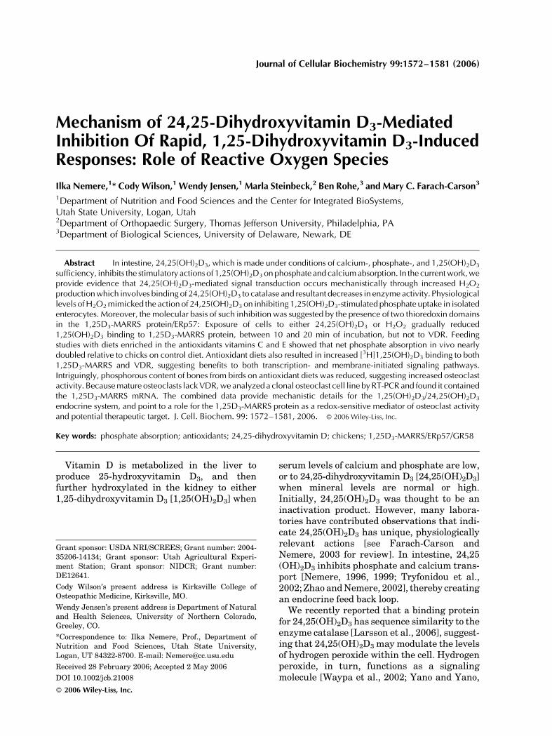
The effect of 6.5 nM 24,25(OH)2D3 on cellularcatalase activity is depicted in Figure 1A. Whilecellular levels of the enzyme in vehicle controls(open circles) remained at approximately250 mm/min/mg protein, preparations treatedwith 6.5 nM 24,25(OH)2D3 (closed circles)exhibited a decrease in specific activity to 64%of controls 1 min after seco-steroid (P< 0.05).At 10 min after seco-steroid, specific catalaseactivity was 72% of controls (P< 0.05). Incontrast, treatment of cells with 130 pM 1,25(OH)2D3 resulted in no significant changes incatalase specific activity relative to controls(Fig. 1B). Supernatant levels of catalase, nor-malized to cellular protein, showed no signifi-cant changes in response to either steroid underthe conditions used (data not shown). Commer-cially available bovine catalase activity wasassessed with 0.0325 mg of protein in the absen-ce or presence of 6.5 nM 24,25(OH)2D3, 300 pM1,25(OH)2D3, or 100 nM 25(OH)2D3. Only 24,25(OH)2D3 reduced catalase levels to 66% ofvehicle controls (0.16% ethanol, final concentra-tion; P< 0.05, for eight determinations).
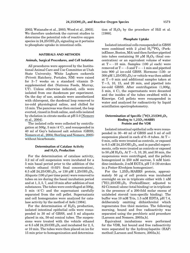
The logical outcome of a decrease in catalasespecific activity would be an increase in H2O2 inresponse to 24,25(OH)2D3. The data presentedin Figure 2 indicate that this indeed was thecase. While cell suspensions treated with vehi-cle control maintained approximately 40 nmH2O2/mg protein, cells treated with 6.5 nM24,25(OH)2D3 exhibited an increase that wassignificant (P< 0.05) at 5 and 10 min of incuba-tion, relative to corresponding controls (Fig. 2).Indeed, the 30% decrease in catalase specificactivity in response to a 10-min exposure to24,25(OH)2D3 was paralleled by a 30% increasein H2O2 elicited by the steroid.
1574 Nemere et al.
Effect of H2O2 on 1,25(OH)2D3-StimulatedPhosphate Uptake
We have previously reported that 24,25(OH)2D3 inhibits 1,25(OH)2D3-stimulated phos-phate uptake both in the perfused duodenalloop, as well as in isolated intestinal cells [Zhaoand Nemere, 2002]. We, therefore, tested theeffect of H2O2 as a potential signal transductionagent for its effect on hormone-stimulatedphosphate uptake in enterocytes. Dependingon cell protein, incubations with physiological
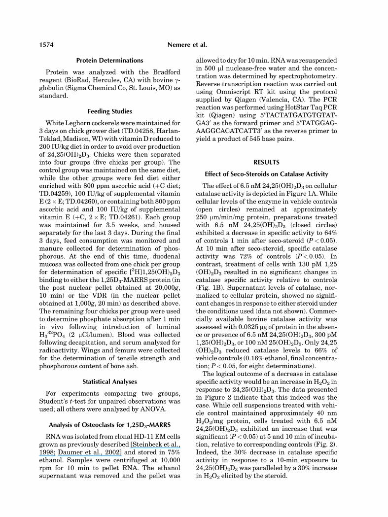
levels of 24,25(OH)2D3 produced 40–100 mMH2O2, comparable to what was reported in othersystems [Dhar-Mascareno et al., 2003]. We,therefore, selected a concentration at the lowerend of this range. Figure 3A,B illustrates theresults of these experiments using two differentconcentrations of 1,25(OH)2D3 (closed circles)relative to cells exposed to 50 mM H2O2 prior toaddition of hormone (closed triangles), andvehicle controls (open circles). While H2O2 hadno effect on phosphate uptake by controls (datanot shown), the freshly prepared reactive oxy-gen species completely abolished the stimula-tory effect of 130 pM 1,25(OH)2D3. After 20 minof incubation, steroid stimulated 32P uptakerose to 153% of equivalent preparations exposedto 50 mM H2O2 (P< 0.02, Fig. 3A). In anotherseries of incubations, in which the H2O2 wasdiluted 1 h prior to use, rather than immediatelyprior to addition, inhibition of 1,25(OH)2D3
stimulated phosphate uptake was transient,occurring for the first 7 min of the time course,with a return to stimulated levels by 10 min ofincubation. Thus, inhibition was reversible.
In additional experiments, 300 pM 1,25(OH)2
D3, equivalent to circulating levels in youngcockerels [Sedrani, 1984] was tested. As shownin Figure 3B 50 mM H2O2 also inhibited thishigher level of steroid hormone. Addition ofhormone to cell suspensions resulted in a signi-ficant increase in 32P levels at 5 min (P< 0.05,
Fig. 1. Rapid inhibition of catalase activity in isolated intestinalcells treated with 24,25(OH)2D3. Chick duodenal cells wereisolated by a chelation procedure, collected by centrifugation at500g, 5 min, and resuspended in 40 ml of Gey’s Balanced SaltSolution (GBSS). Two, 3.2-ml aliquots were placed in separate50-ml conical centrifuge tubes, and 100 ml samples removed tomicrofuge tubes on ice at T¼�5 and 0 min. Immediatelythereafter, control incubations (open circles) received vehicle(ethanol, 0.05% final concentration, and either 6.5 nM24,25(OH)2D3 (A; closed circles) or 130 pM 1,25(OH)2D3
(B; closed circles). Additional samples were removed at theindicated times, centrifuged at 1,000g, 5 min, the supernatantsdecanted, and the inside of the tubes swabbed with a Kimwipewhile inverted. Cell pellets were homogenized in 0.5 ml doubledistilled water and analyzed for decreasing absorbance at240 nm following addition of 40 mM H2O2. Change inabsorbance was related to cell protein. Values representmean� SEM for n¼ 8 independent experiments (A) and n¼5independent experiments (B).
Fig. 2. Production ofH2O2 in cells treatedwith 24,25(OH)2D3.Cells were isolated as described in the legend to Figure 1 andresuspended in 30 ml GBSS. Six, 5-ml aliquots were placed into50ml conical centrifuge tubes and either treatedwith the vehicleethanol (0.05%, final concentration) or 6.5 nM 24,25(OH)2D3.At 2-, 5-, and 10-min, one control and one treated sample wereplaced on ice and chilled for 15 min prior to homogenization.One ml of each whole homogenate was analyzed for H2O2 witha colorimetric procedure. Values represent mean� SEM for n¼5 independent experiments.
24,25(OH)2D3 and Reactive Oxygen Species 1575
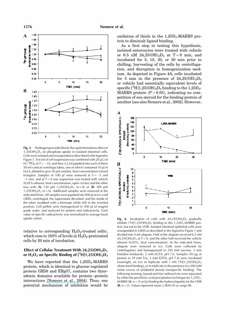
relative to corresponding H2O2-treated cells),which rose to 183% of levels in H2O2-pretreatedcells by 20 min of incubation.
Effect of Cellular Treatment With 24,25(OH)2D3
or H2O2 on Specific Binding of [3H]1,25(OH)2D3
We have reported that the 1,25D3-MARRSprotein, which is identical to glucose regulatedprotein GR58 and ERp57, contains two thior-edoxin domains available for protein–proteininteractions [Nemere et al., 2004]. Thus, onepotential mechanism of inhibition would be
oxidation of thiols in the 1,25D3-MARRS pro-tein to diminish ligand binding.
As a first step in testing this hypothesis,isolated enterocytes were treated with vehicleor 6.5 nM 24,25(OH)2D3 at T¼ 0 min, andincubated for 5, 10, 20, or 30 min prior tochilling, harvesting of the cells by centrifuga-tion, and disruption in homogenization med-ium. As depicted in Figure 4A, cells incubatedfor 5 min in the presence of 24,25(OH)2D3
or vehicle had essentially equivalent levels ofspecific [3H]1,25(OH)2D3 binding to the 1,25D3-MARRS protein (P> 0.05), indicating no com-petition of one steroid for the binding protein ofanother [see also Nemere et al., 2002]. However,
Fig. 3. Hydrogenperoxide blocks the rapid stimulatory effect of1,25(OH)2D3 on phosphate uptake in isolated intestinal cells.Cellswere isolated and resuspendedas described in the legend toFigure 1. Tenml of cell suspensionwas combinedwith 20 mCi ofH3
32PO4 at T¼�10, and then 3.2 ml pipetted into each of three50-ml conical centrifuge tubes, one of which contained 10 ml ofH2O2 diluted to give 50 mM oxidant, final concentration (closedtriangles). Samples of 100 ml were removed at T¼�5 and�1 min, and at T¼ 0 one suspension was treated with vehicle(0.05% ethanol, final concentration, open circles) and the othertwo with (A) 130 pM 1,25(OH)2D3 (n¼6) or (B) 300 pM1,25(OH)2D3 (n¼ 6). Additional samples were removed at theindicated times. All sampleswere pipetted into 900 ml of ice-coldGBSS, centrifuged, the supernatant decanted, and the inside ofthe tubes swabbed with a Kimwipe while still in the invertedposition. Cell pellets were homogenized in 500 ml of reagentgrade water, and analyzed for protein and radioactivity. Eachvalue of specific radioactivity was normalized to average basaluptake values. Fig. 4. Incubation of cells with 24,25(OH)2D3 gradually
inhibits [3H]1,25(OH)2D3 binding to the 1,25D3-MARRS pro-tein, but not to the VDR. Isolated intestinal epithelial cells wereresuspended in GBSS as described in the legend to Figure 1 anddivided into 5-ml aliquots. Half of the aliquots received 6.5 nM24,25(OH)2D3 at T¼ 0, and the other half received the vehicleethanol (0.05%, final concentration). At the indicated times,aliquots were removed to ice. Cells were collected bycentrifugation and homogenized in 250 mM sucrose, 5 mmhistidine-imidazole, 2 mM EGTA, pH 7.0. Samples (50 mg ofprotein in 10 mM Tris, 2 mM EDTA, pH 7.4) were incubated(overnight, on ice) in triplicate with 1 nM [3H]1,25(OH)2D3
alone (total binding), or in triplicate in the presence of a 200-foldmolar excess of unlabeled steroid (nonspecific binding). Thefollowing morning, bound and free radioactivity were separatedby either the perchloric acid precipitation technique for 1,25D3-MARRS (A; n¼3) or by binding the hydroxylapatite for the VDR(B; n¼2). Values represent mean� SEM (A) or range (B).
1576 Nemere et al.
10–20 min of incubation with 24,25(OH)2D3
reduced specific binding of [3H]1,25(OH)2D3 byapproximately 80% (P< 0.05. relative to corre-sponding controls). Equivalent incubations inthe presence of dithiothreitol restored specificbinding, supporting the involvement of thiols.
In several of the cell preparations, specificbinding to the VDR was also determined(Fig. 4B). Specific binding of [3H]1,25(OH)2D3
to the VDR was not affected by incubation ofcells with 24,25(OH)2D3, relative to correspond-ing controls, at the times tested.
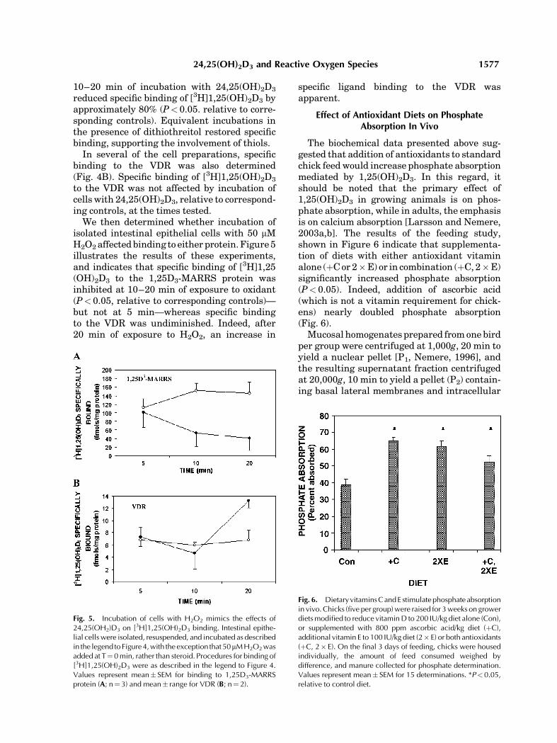
We then determined whether incubation ofisolated intestinal epithelial cells with 50 mMH2O2 affected binding to either protein. Figure 5illustrates the results of these experiments,and indicates that specific binding of [3H]1,25(OH)2D3 to the 1,25D3-MARRS protein wasinhibited at 10–20 min of exposure to oxidant(P< 0.05, relative to corresponding controls)—but not at 5 min—whereas specific bindingto the VDR was undiminished. Indeed, after20 min of exposure to H2O2, an increase in
specific ligand binding to the VDR wasapparent.
Effect of Antioxidant Diets on PhosphateAbsorption In Vivo
The biochemical data presented above sug-gested that addition of antioxidants to standardchick feed would increase phosphate absorptionmediated by 1,25(OH)2D3. In this regard, itshould be noted that the primary effect of1,25(OH)2D3 in growing animals is on phos-phate absorption, while in adults, the emphasisis on calcium absorption [Larsson and Nemere,2003a,b]. The results of the feeding study,shown in Figure 6 indicate that supplementa-tion of diets with either antioxidant vitaminalone (þC or 2�E) or in combination (þC, 2�E)significantly increased phosphate absorption(P< 0.05). Indeed, addition of ascorbic acid(which is not a vitamin requirement for chick-ens) nearly doubled phosphate absorption(Fig. 6).
Mucosal homogenates prepared from one birdper group were centrifuged at 1,000g, 20 min toyield a nuclear pellet [P1, Nemere, 1996], andthe resulting supernatant fraction centrifugedat 20,000g, 10 min to yield a pellet (P2) contain-ing basal lateral membranes and intracellular
Fig. 5. Incubation of cells with H2O2 mimics the effects of24,25(OH2)D3 on [3H]1,25(OH)2D3 binding. Intestinal epithe-lial cells were isolated, resuspended, and incubated as describedin the legend to Figure4,with the exception that 50mMH2O2wasadded at T¼0min, rather than steroid. Procedures for binding of[3H]1,25(OH)2D3 were as described in the legend to Figure 4.Values represent mean� SEM for binding to 1,25D3-MARRSprotein (A; n¼3) and mean� range for VDR (B; n¼2).
Fig. 6. Dietary vitaminsCandE stimulatephosphate absorptionin vivo. Chicks (five per group)were raised for 3weeks on growerdietsmodified to reduce vitaminD to 200 IU/kg diet alone (Con),or supplemented with 800 ppm ascorbic acid/kg diet (þC),additional vitamin E to 100 IU/kg diet (2� E) or both antioxidants(þC, 2� E). On the final 3 days of feeding, chicks were housedindividually, the amount of feed consumed weighed bydifference, and manure collected for phosphate determination.Values represent mean� SEM for 15 determinations. *P<0.05,relative to control diet.
24,25(OH)2D3 and Reactive Oxygen Species 1577
organelles [Nemere, 1996]. Analyses of the P2
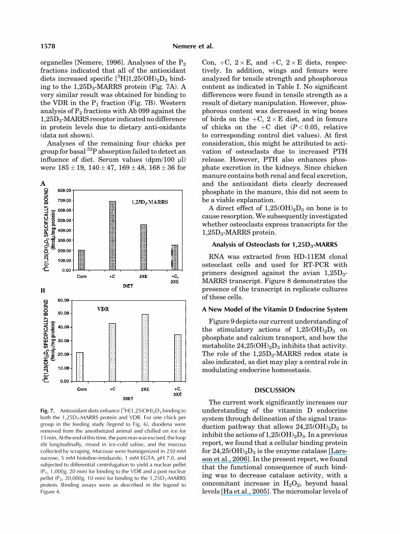
fractions indicated that all of the antioxidantdiets increased specific [3H]1,25(OH)2D3 bind-ing to the 1,25D3-MARRS protein (Fig. 7A). Avery similar result was obtained for binding tothe VDR in the P1 fraction (Fig. 7B). Westernanalysis of P2 fractions with Ab 099 against the1,25D3-MARRS receptor indicated no differencein protein levels due to dietary anti-oxidants(data not shown).
Analyses of the remaining four chicks pergroup for basal 32P absorption failed to detect aninfluence of diet. Serum values (dpm/100 ml)were 185� 19, 140� 47, 169� 48, 168� 36 for
Con, þC, 2�E, and þC, 2�E diets, respec-tively. In addition, wings and femurs wereanalyzed for tensile strength and phosphorouscontent as indicated in Table I. No significantdifferences were found in tensile strength as aresult of dietary manipulation. However, phos-phorous content was decreased in wing bonesof birds on the þC, 2�E diet, and in femursof chicks on the þC diet (P< 0.05, relativeto corresponding control diet values). At firstconsideration, this might be attributed to acti-vation of osteoclasts due to increased PTHrelease. However, PTH also enhances phos-phate excretion in the kidneys. Since chickenmanure contains both renal and fecal excretion,and the antioxidant diets clearly decreasedphosphate in the manure, this did not seem tobe a viable explanation.
A direct effect of 1,25(OH)2D3 on bone is tocause resorption. We subsequently investigatedwhether osteoclasts express transcripts for the1,25D3-MARRS protein.
Analysis of Osteoclasts for 1,25D3-MARRS
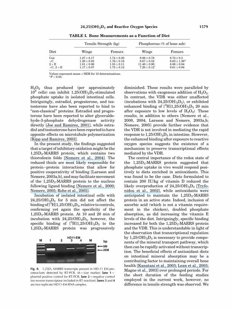
RNA was extracted from HD-11EM clonalosteoclast cells and used for RT-PCR withprimers designed against the avian 1,25D3-MARRS transcript. Figure 8 demonstrates thepresence of the transcript in replicate culturesof these cells.
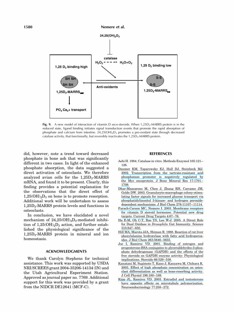
A New Model of the Vitamin D Endocrine System
Figure 9 depicts our current understanding ofthe stimulatory actions of 1,25(OH)2D3 onphosphate and calcium transport, and how themetabolite 24,25(OH)2D3 inhibits that activity.The role of the 1,25D3-MARRS redox state isalso indicated, as diet may play a central role inmodulating endocrine homeostasis.
DISCUSSION
The current work significantly increases ourunderstanding of the vitamin D endocrinesystem through delineation of the signal trans-duction pathway that allows 24,25(OH)2D3 toinhibit the actions of 1,25(OH)2D3. In a previousreport, we found that a cellular binding proteinfor 24,25(OH)2D3 is the enzyme catalase [Lars-son et al., 2006]. In the present report, we foundthat the functional consequence of such bind-ing was to decrease catalase activity, with aconcomitant increase in H2O2, beyond basallevels [Ha et al., 2005]. The micromolar levels of
Fig. 7. Antioxidant diets enhance [3H]1,25(OH)2D3 binding toboth the 1,25D3-MARRS protein and VDR. For one chick pergroup in the feeding study (legend to Fig. 6), duodena wereremoved from the anesthetized animal and chilled on ice for15min.At the endof this time, thepancreaswas excised, the loopslit longitudinally, rinsed in ice-cold saline, and the mucosacollected by scraping. Mucosae were homogenized in 250 mMsucrose, 5 mM histidine-imidazole, 1 mM EGTA, pH 7.0, andsubjected to differential centrifugation to yield a nuclear pellet(P1, 1,000g, 20 min) for binding to the VDR and a post nuclearpellet (P2, 20,000g, 10 min) for binding to the 1,25D3-MARRSprotein. Binding assays were as described in the legend toFigure 4.
1578 Nemere et al.
H2O2 thus produced (per approximately109 cells) can inhibit 1,25(OH)2D3-stimulatedphosphate uptake in isolated intestinal cells.Intriguingly, estradiol, progesterone, and tes-tosterone have also been reported to bind to‘‘non-classical’’ proteins: Estradiol and proges-terone have been reported to alter glyceralde-hyde-3-phosphate dehydrogenase activitydirectly [Joe and Ramirez, 2001], while estra-diol and testosterone have been reported to haveopposite effects on microtubule polymerization[Kipp and Ramirez, 2003].
In the present study, the findings suggestedthat a target of inhibitory oxidation might be the1,25D3-MARRS protein, which contains twothioredoxin folds [Nemere et al., 2004]. Thereduced thiols are most likely responsible forprotein–protein interactions that allow forpositive cooperativity of binding [Larsson andNemere, 2003a,b], and may facilitate movementof the 1,25D3-MARRS protein to the nucleusfollowing ligand binding [Nemere et al., 2000;Nemere, 2005; Rohe et al., 2005].
Incubation of isolated intestinal cells with24,25(OH)2D3 for 5 min did not affect thebinding of [3H]1,25(OH)2D3, relative to controls,confirming yet again the specificity of the1,25D3-MARRS protein. At 10 and 20 min ofincubation with 24,25(OH)2D3, however, thespecific binding of [3H]1,25(OH)2D3 to the1,25D3-MARRS protein was progressively
diminished. These results were paralleled byobservations with exogenous addition of H2O2.In contrast, the VDR was either unaffected(incubations with 24,25(OH)2D3), or exhibitedenhanced binding of [3H]1,25(OH)2D3 20 minafter exposure to low levels of H2O2). Theseresults, in addition to others [Nemere et al.,2000, 2004; Larsson and Nemere, 2003a,b;Nemere, 2005] provide further evidence thatthe VDR is not involved in mediating the rapidresponse to 1,25(OH)2D3 in intestine. However,the enhanced binding after exposure to reactiveoxygen species suggests the existence of amechanism to preserve transcriptional effectsmediated by the VDR.
The central importance of the redox state ofthe 1,25D3-MARRS protein suggested thatphosphate uptake in vivo would respond posi-tively to diets enriched in antioxidants. Thiswas found to be the case. Diets formulated tocontain 200 IU/kg of vitamin D reduced thelikely overproduction of 24,25(OH)2D3 [Tryfo-nidou et al., 2002], while antioxidants wereanticipated to maintain the 1,25D3-MARRSprotein in an active state. Indeed, inclusion ofascorbic acid (which is not a vitamin require-ment in the chicken), doubled phosphateabsorption, as did increasing the vitamin Elevels of the diet. Intriguingly, specific bindingincreased for both the 1,25D3-MARRS proteinand the VDR. This is understandable in light ofthe observation that transcriptional regulationby 1,25(OH)2D3 is necessary to provide compo-nents of the mineral transport pathway, whichthen can be rapidly activated without transcrip-tion. The beneficial effects of antioxidant dietson intestinal mineral absorption may be acontributing factor to maintaining overall bonehealth [Kanatani et al., 2003; Lean et al., 2003;Magne et al., 2003] over prolonged periods. Forthe short duration of the feeding studiesemployed in the current work, however, nodifference in tensile strength was observed. We
TABLE I. Bone Measurements as a Function of Diet
Diet
Tensile Strength (kg) Phosphorous (% of bone ash)
Wings Femurs Wings Femurs
Con 1.27� 0.17 1.74� 0.20 9.98� 0.78 9.72� 0.3þC 1.20� 0.03 1.76� 0.18 8.67� 0.52 6.63� 1.26*2�E 1.01� 0.06 1.55� 0.11 11.40� 0.90 8.88�0.84þC, 2�E 1.17� 0.07 1.75� 0.12 7.20�0.12* 9.61�0.94
Values represent mean�SEM for 10 determinations.*P< 0.05.
Fig. 8. 1,25D3 MARRS transcripts present in HD-11 EM pre-osteoclasts detected by RT-PCR. M¼ size marker; lane 1¼plasmid positive control for RT-PCR; lane 2¼negative control(no reverse transcriptase included in RT reaction); lanes 3 and 4are two replicate HD11-EM RNA samples.
24,25(OH)2D3 and Reactive Oxygen Species 1579
did, however, note a trend toward decreasedphosphate in bone ash that was significantlydifferent in two cases. In light of the enhancedphosphate absorption, the data suggested adirect activation of osteoclasts. We thereforeanalyzed avian cells for the 1,25D3-MARRSmRNA, and found it to be present. Clearly, thisfinding provides a potential explanation forthe observations that the direct effect of1,25(OH)2D3 on bone is to promote resorption.Additional work will be undertaken to assess1,25D3-MARRS protein levels and functions inosteoclasts.
In conclusion, we have elucidated a novelmechanism of 24,25(OH)2D3-mediated inhibi-tion of 1,25(OH)2D3 action, and further estab-lished the physiological significance of the1,25D3-MARRS protein in mineral and ionhomeostasis.
ACKNOWLEDGMENTS
We thank Carolyn Stephens for technicalassistance. This work was supported by USDANRI/SCREES grant 2004-35206-14134 (IN) andthe Utah Agricultural Experiment Station.Approved as journal paper no. 7769. Additionalsupport for this work was provided by a grantfrom the NIDCR DE12641 (MCF-C).
REFERENCES
Aebi H. 1984. Catalase in vitro. Methods Enzymol 105:121–126.
Daumer KM, Taparowsky EJ, Hall DJ, Steinbeck MJ.2002. Transcription from the tartrate-resistant acidphosphatase promoter is negatively regulated bythe Myc oncoprotein. J Bone Mineral Res 17:1701–1709.
Dhar-Mascareno M, Chen J, Zhang RH, Carcamo JM,Golde DW. 2003. Granulocyte-macrophage colony-stimu-lating factor signals for increased glucose transport viaphosphatidylinositol 3-kinase- and hydrogen peroxide-dependent mechanisms. J Biol Chem 278:11107–11114.
Farach-Carson MC, Nemere I. 2003. Membrane receptorsfor vitamin D steroid hormones: Potential new drugtargets. Current Drug Targets 4:67–76.
Ha E-M, Oh C-T, Bae YS, Lee W-J. 2005. A Direct Rolefor Dual Oxidase in Drosophila Gut Immunity. Science310:847–850.
Hill MA, Marota JJA, Shiman R. 1988. Reaction of rat liverphenylalanine hydroxylase with fatty acid hydroperox-ides. J Biol Chem 263:5646–5655.
Joe I, Ramirez VD. 2001. Binding of estrogen andprogesterone-BSA conjugates to glyceraldehydes-3-phos-phate dehydrogenase (GAPDH) and the effects of thefree steroids on GAPDH enzyme activity: Physiologicalimplications. Steroids 66:529–538.
Kanatani M, Sugimoto T, Kano J, Kanzawa M, Chihara K.2003. Effect of high phosphate concentration on osteo-clast differentiation as well as bone-resorbing activity.J Cell Physiol 196:180–189.
Kipp JL, Ramirez VD. 2003. Estradiol and testosteronehave opposite effects on microtubule polymerization.Neuroendocrinology 77:258–272.
Fig. 9. A new model of interaction of vitamin D seco-steroids. When 1,25D3-MARRS protein is in thereduced state, ligand binding initiates signal transduction events that promote the rapid absorption ofphosphate and calcium from intestine. 24,25(OH)2D3 promotes a pro-oxidant state through decreasedcatalase activity, that functionally, but reversibly inactivates the 1,25D3-MARRS protein.
1580 Nemere et al.
Larsson B, Nemere I. 2003a. Effect of growth andmaturation on membrane-initiated actions of 1,25-dihy-droxyvitamin D3. I. Calcium transport, receptor kinetics,and signal transduction in intestine of male chickens.Endocrinology 144:1726–1735.
Larsson B, Nemere I. 2003b. Effect of growth andmaturation on membrane-initiated actions of 1,25-dihy-droxyvitamin D3. II. Calcium transport, receptorkinetics, and signal transduction in intestine of femalechickens. J Cellular Biochem 90:901–913.
Larsson D, Anderson D, Smith N, Nemere I. 2006. 24,25-Dihydroxyvitamin D3 binds to catalase. J CellularBiochem (in press).
Lean JM, Davies JT, Fuller K, Jagger CJ, Kristein B,Partington GA, Urry ZL, Chambers TJ. 2003. A crucialrole for thiol antioxidants in estrogen-deficiency boneloss. J Clinical Invest 112:915–923.
Magne D, Gluteau G, Faucheux C, Palmer G, Vignes-solombeix C, Pilet P, Rouillon T, Caverazsio J, Weis P,Daculsi G, Guicheux J. 2003. Phosphate is a specificsignal for ATDC5 chondrocyte maturation and apoptosis-associated mineralization: Possible implication of apop-tosis in the regulation of endochondral ossification.J Bone Mineral Res 18:1430–1442.
Nemere I. 1996. Apparent non-nuclear regulation ofintestinal phosphate transport: Effects of 1,25-dihydrox-yvitamin D3, 24,25-dihydroxyvitamin D3, and 25-hydro-xyvitamin D3. Endocrinology 137:2254–2261.
Nemere I. 1999. 24,25-dihydroxyvitamin D3 suppresses therapid actions of 1,25-dihydroxyvitamin D3 and parathyr-oid hormone in chick intestine. J Bone Mineral Res 14:1543–1549.
Nemere I. 2005. The 1,25D3-MARRS protein: Contributionto steroid-stimulated uptake in chicks and rats. Steroids70:455–457.
Nemere I, Ray R, McManus W. 2000. Immunochemicalstudies on the putative plasmalemmal receptor for 1,25-dihydroxyvitamin D3: I. chick intestine. Am J Physiol278:E1104–E1114.
Nemere I, Yazzie-Atkinson D, Johns D, Larsson D. 2002.Biochemical characterization and purification of a bind-ing protein for 24,25-dihydroxyvitamin D3 from chickintestine. J Endocrinol 172:211–219.
Nemere I, Farach-Carson MC, Rohe B, Sterling T, NormanAW, Boyan BD, Safford SE. 2004. Ribozyme knockdown
functionally links a 1,25(OH)2D3 membrane bindingprotein (1,25D3-MARRS) and phosphate uptake inintestinal cells. Proc Natl Acad Sci USA 101:7392–7397.
Rohe B, Safford SE, Nemere I, Farach-Carson MC. 2005.Identification and Characterization of 1,25D3-Mem-brane-Associated Rapid Response, Steroid (1,25D3-MARRS) Binding Protein in Rat IEC-6 Cells. Steroids70:458–463.
Sedrani SH. 1984. Changes in serum levels of 1,25-dihydroxyvitamin D3, calcium and phosphorous withage and vitamin D status in chickens. British J Nutr 52:329–334.
Steinbeck M, Kin J-K, Trudeau MJ, Hauschka PV,Karnovsky MJ. 1998. Involvement of hydrogen peroxidein the differentiation of clonal HD-11EM cells intoosteoclast-like cells. J Cell Physiol 176:574–587.
Sterling TM, Nemere I. 2005. 1,25-Dihydroxyvitamin D3
stimulates vesicular transport within 5 s in polarizedintestinal epithelial cells. J Endocrinol 185:81–91.
Tryfonidou MA, Stevenhagen JJ, van den Bemd GJCM,Oosterlaken-Dijksterhuis MA, DeLuca HF, Mol JA, vanden Brom WE, van Leeuwen JPTM, Hazewinkel HAW.2002. Moderate cholecalciferol supplementation depres-ses intestinal calcium absorption in growing dogs. J Nutr132:2644–2650.
Watanabe N, Iwamoto T, Bowen KD, Dickinson DA, TorresM, Forman HJ. 2003. Bio-effectiveness of Tat-catalaseconjugate: A potential tool for identification of H2O2-dependent cellular signal transduction pathways. Bio-chem Biophy Res Commun 303:287–293.
Waypa GB, Marks JD, Mack MM, Boriboun C, MungaiPT, Schumacker PT. 2002. Mitochondrial reactive oxy-gen species trigger calcium increases during hypoxia inpulmonary arterial myocytes. Circulation Res 18:719–726.
Wood Z, Poole LB, Karplus PA. 2003. Peroxiredoxinevolution and the regulation of hydrogen peroxidesignaling. Science 300:650–6533.
Yano S, Yano Y. 2002. Regulation of catalase enzymeactivity by cell signaling molecules. Molec Cell Biochem240:119–130.
Zhao B, Nemere I. 2002. 1,25(OH)2D3-Mediated phosphateuptake in isolated chick intestinal cells: Effect of24,25(OH)2, signal transduction activators, and age.J Cell Biochem 86:497–508.
24,25(OH)2D3 and Reactive Oxygen Species 1581


