Embed Size (px)
Citation preview
Mechanisms of 4-Hydroxy-2-nonenal Induced Pro- and Anti-Apoptotic Signaling
Pankaj Chaudhary, Rajendra Sharma*, Abha Sharma, Rit Vatsyayan, Sushma Yadav,Sharad S. Singhal, Navin Rauniyar, Laszlo Prokai, Sanjay Awasthi, and Yogesh C. Awasthi*Department of Molecular Biology and Immunology, University of North Texas Health ScienceCenter, Fort Worth, Texas 76107
AbstractIn recent years, 4-hydroxy-2-nonenal (4-HNE) has emerged as an important second messenger incell cycle signaling. Here we demonstrate that 4-HNE induces signaling for apoptosis via both, theFas mediated extrinsic and the p53 mediated intrinsic pathways in HepG2 cells. 4-HNE inducesFas-mediated DISC independent apoptosis pathway by activating ASK1, JNK and caspase-3. Inparallel treatment of 4-HNE to HepG2 cells also induces apoptosis by p53 pathway throughactivation of Bax, p21, JNK, and caspase-3. Exposure of HepG2 cells to 4-HNE leads to theactivation of both Fas and Daxx, promotes the export of Daxx from the nucleus to cytoplasm andfacilitates Fas-Daxx binding. Depletion of Daxx by siRNA results in potentiation of apoptosisindicating that Fas-Daxx binding in fact is inhibitory to Fas mediated apoptosis in cells. 4-HNE-induced translocation of Daxx is also accompanied by the activation and nuclear accumulation ofHSF1 and up-regulation of heat shock protein Hsp70. All these effects of 4-HNE in cells can beattenuated by ectopic expression of hGSTA4-4, the isozyme of glutathione S-transferase with highactivity for 4-HNE. Through immunoprecipitation and liquid chromatography–tandem massspectrometry, we have demonstrated covalent binding of 4-HNE to Daxx. We also demonstratethat 4-HNE modification induces phosphorylation of Daxx at Ser668 and Ser671 to facilitate itscytoplasmic export. These results indicate that while 4-HNE exhibits toxicity through severalmechanisms, in parallel it evokes signaling for defense mechanisms to self regulate its toxicity andcan simultaneously affect multiple signaling pathways through its interactions with membranereceptors and transcription factors/ repressors.
Reactive oxygen species (ROS) produced during exposure of cells to UV radiation, heatshock, or xenobiotics and during metabolic processes cause the oxidation of polyunsaturatedfatty acids in membrane lipid bilayers that are one of the early targets of ROS. Previousstudies have indicated that the exposure of cells to even minimal transient stress causessubstantial lipid peroxidation (LPO) (1,2). These studies show that transient exposure ofcells to UV, H2O2, or oxidants leads to a significant (~50%) rise in the levels of 4-hydroxy-2-nonenal (4-HNE) which is a stable end-product of LPO. 4-HNE has been widelyimplicated in the mechanisms of oxidant toxicity and also in cell cycle signaling (3–9) butmost of the associated mechanisms are not completely understood. Liver being the majorsite for metabolism, and biotransformation of xenobiotics is constantly exposed to ROS. Inthe event of insufficient levels of defense mechanisms such as free-radical scavengers or
*To either of whom the correspondence and reprint requests should be addressed: Department of Molecular Biology and Immunology,University of North Texas Health Science Center, Fort Worth, Texas 76107. Tel: 817-735-2366; Fax: 817-735-2118;,[email protected], or [email protected] INFORMATION AVAILABLEAdditional Materials and Methods, and Figures S1–S5 as described in the text. This material is available free of charge via the Internetat http://pubs.acs.org.
NIH Public AccessAuthor ManuscriptBiochemistry. Author manuscript; available in PMC 2011 July 27.
Published in final edited form as:Biochemistry. 2010 July 27; 49(29): 6263–6275. doi:10.1021/bi100517x.
NIH
-PA Author Manuscript
NIH
-PA Author Manuscript
NIH
-PA Author Manuscript
antioxidants, increasing levels of lipid hydroperoxides and peroxides can be produced inliver by self-perpetuating chain reactions resulting in the formation of 4-HNE that can exerttoxicity through interactions with cellular macromolecules, including proteins, lipids, andnucleic acids. For example, chronic alcohol consumption (10), high-iron diet (11), high-fatdiet (12), or exposure to hepatotoxic agents like CCl4 markedly elevate the intracellularconcentration of 4-HNE from its basal constitutive levels and damage hepatocytes and theliver (13,14). 4-HNE is also believed to be involved in the mechanisms of diseases such asatherosclerosis (15,16), diabetes (17), Alzheimer’s disease (18,19), Parkinson's disease (20),cataract (21), and cancer (22,23).
In recent past (1,2,6–9), studies in our laboratories have clearly shown that signaling forapoptosis by many oxidants including superoxide anion generated by xanthine-xanthineoxidase system is mediated through 4-HNE and it could be inhibited by acceleratingdisposal of 4-HNE by forced over expression of GSTA4-4 which is highly specific for 4-HNE but is not involved in the detoxification of ROS such as H2O2, O2
•−. A multitude ofstudies by other investigators (3–5,24), and in our laboratories indicate a global role of 4-HNE in modulation of cellular processes. Studies in our lab have shown that the signalingfor proliferation, transformation, apoptosis, and differentiation is associated with alterationsin the intracellular levels of 4-HNE in a wide variety of cells (9,25–28). These findings posean intriguing question as to how 4-HNE is able to exert such a multitude of effects oncellular processes.
During the present studies we have addressed this question by investigating the role of 4-HNE mediated signaling in the pathways associated with the regulation of programmed celldeath in a liver derived cell line HepG2. Specifically, we have investigated the effect ofincreased 4-HNE concentrations in cells by direct exposure or by oxidant treatment ondifferent pathways leading to apoptosis. Conversely, we have evaluated the role of 4-HNE insignaling of these pathways by lowering its intracellular concentration through thetransfection of cells with hGSTA4. We have also studied the interactions of 4-HNE withsome of the key proteins involved in these pathways. Furthermore, to examine the in vivosignificance of these findings we have also studied some of these effects of 4-HNE in theliver tissues of Gsta4 null mice where 4-HNE levels are consistently maintained at highlevels due to its impaired disposition (29). The results of these studies show that 4-HNEcauses toxicity to HepG2 cells via necrosis and apoptosis induced by more than onepathway. These findings integrate the mechanisms for the multifarious effects of 4-HNE oncellular processes suggesting that 4-HNE through direct interactions with membranereceptors, transcription factors, and transcription repressors regulates trafficking, and thesignaling functions of key proteins to affect various cellular processes.
MATERIALS AND METHODSMaterials
4-Hydroxynonenal was purchased from Cayman Chemical (Ann Arbor, MI). Bradfordreagent, bis-acrylamide, and SDS for SDS-PAGE were obtained from BioRad (Hercules,CA). The apoptosis detection system (CaspACE FITC-VAD-FMK in situ marker) waspurchased from Promega Inc. (Madison, WI). The cytoplasmic and nuclear proteinextraction kit was acquired from Imgenex Co. (San Diego, CA), protein A/G-agarose fromSanta Cruz Biotechnology (Santa Cruz, CA), JNK inhibitor SP6000125 from A–G Scientific(San Deigo, CA), and Western blot stripping buffer from Pierce Co. (Rockford, IL). Allother reagents and chemicals were purchased from Sigma-Aldrich (St. Louis, MO). The cellculture medium RPMI-1640, geneticin (G418), Lipofectamine 2000 transfection reagent andfetal bovine serum were from GIBCO (Invitrogen, Carlsbad, CA).
Chaudhary et al. Page 2
Biochemistry. Author manuscript; available in PMC 2011 July 27.
NIH
-PA Author Manuscript
NIH
-PA Author Manuscript
NIH
-PA Author Manuscript
Cell lines and Culture ConditionsThe HepG2 human hepatocarcinoma cells purchased from the American Type CultureCollection were cultured in RPMI-1640 supplemented with 10% fetal bovine serum, 1% of astock solution containing 10,000 IU/mL penicillin and 10 mg/mL streptomycin in anincubator at 37°C under a humidified atmosphere containing 5% CO2.
Preparation of cell extracts and Western blot analysisCells were collected, washed with cold PBS and then incubated in 100 µL of RIPA lysisbuffer (50 mM Tris-HCl, pH 7.5; 1% NP-40; 150 mM NaCl; 1 mg ml−1 aprotinin; 1 mgml−1 leupeptin; 1 mM Na3VO4; 1 mM NaF) at 4°C for 30 min. Cell debris was removed bycentrifugation at 12,000 g for 10 min at 4°C. Protein concentrations were determined byBradford assay (30) as described in standard protocol. Cell extracts were separated on SDSpolyacrylamide gels (4–20%), and transferred onto nitrocellulose (Bio-Rad). Membraneswere blocked with 5% fat-free milk at room temperature for 60 min, and incubatedovernight at 4°C with the appropriate primary antibody in 5% milk in Tris-buffered saline(TBS) containing 50 mM NaF and 0.05% Tween 20. After three times washing with T-TBS(Tris-buffered saline containing 0.05% Tween 20), the membrane was incubated with theappropriate secondary antibody at room temperature for 2 h. After washing again with T-TBS, the membrane was treated with Super signal ‘West Pico’ chemiluminescent reagent(Pierce, Rockford, IL) as per manufacturer's instructions, and exposed to Hyperfilm ECLfilm (Amersham) at room temperature. Isolation of nuclear and cytoplasmic fractions wasachieved by Imgenex nuclear extraction kit as per the manufacturer’s instructions (Imgenex,San Deigo, CA).
Stable transfection with pTarget and hGSTA4HepG2 cells at a density of 5 × 10⁵ cells per 100 mm Petri dish were plated for thetransfection. Petri dishes having >50% confluent cells were used for the transfection. Thecells were transfected with 24 µg of either empty pTarget-T vector (VT) or the pTargetvector with the open reading frame (ORF) of the hGSTA4 sequence (hGSTA4-Tr), usingLipofectamine 2000 reagent (Invitrogen, Carlsbad, CA) as per the manufacturer’sinstructions.
Transfection of Daxx siRNA in HepG2 cellsSmall interfering RNA (siRNA) transfection experiments against Daxx were performedusing double-stranded RNA synthesized by Dharmacon (ON-TARGET Plus SMARTpool,Dharmacon, Chicago, IL). Briefly, HepG2 cells (2 × 10⁵ cells per well) were plated in a six-well tissue culture plate, in 2 mL normal growth medium supplemented with FBS. Cellswere cultured at 37°C until 60–80% confluency. For each transfection, 100 nM double-stranded non-targeting control siRNA (Dharmacon, used as control), or Daxx-specificsiRNA were transfected into HepG2 cells using DharmaFECT 4 transfection reagent(Dharmacon) according to the manufacturer’s protocol. Cells were harvested at appropriatetime points and the silencing of Daxx was examined by Western blotting.
Immunofluorescence studiesHepG2 cells were grown to 50% confluence on glass cover slips in 12-well plates. The cellswere exposed to 20 µM 4-HNE. Untreated cells remained as controls. Treated and untreatedcells were incubated for 2 h, washed twice with ice-cold PBS (pH 7.4), fixed with 4%paraformaldehyde for 30 min and then permeabilized with 0.1% Triton X-100 for 30 min.The slides were then washed with PBS, incubated with 5% goat serum in PBS for 2 h, andthen incubated with anti-Daxx or anti-HSF1 antibodies (Santa Cruz Biotechnology, SantaCruz, CA) diluted 1:50 in PBS containing 1% goat serum for overnight at 4°C temperature.
Chaudhary et al. Page 3
Biochemistry. Author manuscript; available in PMC 2011 July 27.
NIH
-PA Author Manuscript
NIH
-PA Author Manuscript
NIH
-PA Author Manuscript
After washing with ice cold PBS, the cover slips were incubated with FITC-labeled goatanti-rabbit immunoglobulin G (Southern Biotech, USA) diluted 1:200 in PBS containing 1%goat serum for 2 h at room temperature in dark. The cover slips were then washed threetimes with ice cold PBS and mounted on glass slides with 20 µL of VectaShield mediumcontaining DAPI (1.5 µg/mL) (Vector Laboratories, Inc., USA). The slides were examinedusing LSM 510 Meta confocal system equipped with an inverted microscope (AxioObserver Z1, Carl Zeiss).
In situ caspase-3 assay for ApoptosisCells (2 × 10⁴) were treated with 0–40 µM 4-HNE or with 250 ng/mL Fas agonistic CH11antibodies, that are known to induce apoptosis, for 2 h at 37°C. Apoptotic cells weredetected by staining with in situ marker (10 µM, CaspACE FITC-VAD-FMK, Promega) for30 min in the dark. The slides were fixed with 4% paraformaldehyde for 30 min, rinsed withPBS twice, mounted in a medium containing 1.5 µg/mL DAPI and observed under OlympusAX70 fluorescence microscope.
Chromatin Immunoprecipitation (ChIP) assayTo determine whether after nuclear translocation HSF1 binds to Hsp70 promoter, ChIPassay was performed using the ChIP-IT kit from Active Motif (Carlsbad, CA) following themanufacturer's instructions. Briefly, 5 × 10⁵ cells were grown and treated with 20 µM 4-HNE for 2 h and fixed with 1% formaldehyde. Cells were washed with PBS followed by theaddition of glycine stop solution and washing with PBS. The cells were collected andresuspended in lysis buffer, incubated for 10 min on ice, vortexed for 10s, and centrifugedfor 10 min at 4,000 g at 4°C. The DNA pellet was resuspended in shearing buffer, sonicatedand the sheared DNA sample was centrifuged. The resultant supernatant was incubated withpositive control IgG as well as negative control IgG (provided by Active Motif) and anti-HSF1 IgG (Santa Cruz, CA) overnight at 4°C. Protein G beads were added to the reactionmixtures and incubated for 2 h at 4°C. The beads were pelleted by centrifugation followedby washing, and eluted with 100 µL of ChIP elution buffer. Eluted DNA samples werepurified using the DNA purification mini columns and amplified by PCR using the controlprimers and negative control primers (provided by Active Motif) and hHsp70 primers. PCRproducts were analyzed by running on 1% agarose gels.
Preparation of 4-HNE-Daxx adduct and identification of 4-HNE binding sites by liquidchromatography–tandem mass spectrometry (LC–MS/MS)
4-HNE adducts of purified bacterially expressed human Daxx (1 mg/mL) were prepared byreacting with 2 mM 4-HNE in 0.1 M phosphate buffer, pH 7.4, at 37°C for 2 h. Theenrichment of 4-HNE-modified peptides from the proteolytic digest of Daxx wasaccomplished by using solid-phase hydrazide (SPH) reagent as described previously (31)and analyzed by LC–MS/MS analyses (32,33). LC–MS/MS was performed on a hybridlinear ion trap-FTICR (7-Tesla) mass spectrometer (LTQ-FT, Thermo Finnigan, San Jose,CA) equipped with a nanoelectrospray ionization source and operated with the Xcalibur(version 2.2) and Tune Plus (version 2.2) data acquisition software. MS/MS data generatedby data dependent acquisition via the LTQ-FT were extracted by BioWorks version 3.3 andsearched against a composite IPI human (version 3.47, number of entries is 144164 × 2)protein sequence database containing both forward and randomized sequences using theMascot v 2.2 (Matrix Science, Boston, MA) search algorithm. (34,35) MS/MS spectra of 4-HNE modified peptides were visually inspected to verify peptide fragment ions. The MS-Product module of Protein Prospector (http://prospector.ucsf.edu) was used to calculate them/z values of b- and y-type ions of 4-HNE-modified Daxx tryptic peptides, obtained duringCID-MS/MS, where His residues were replaced with 4-HNE-His Michael adducts (M +156).
Chaudhary et al. Page 4
Biochemistry. Author manuscript; available in PMC 2011 July 27.
NIH
-PA Author Manuscript
NIH
-PA Author Manuscript
NIH
-PA Author Manuscript
Statistical AnalysisThe data are expressed as the mean ± SD for each group. The statistical significance wasdetermined by Student's t test and was set at p < 0.05.
RESULTS4-HNE causes apoptosis and necrosis in HepG2 cells
The cytotoxicity of 4-HNE to HepG2 cells was evaluated by MTT assay and apoptosismeasured by flow cytometry, caspase activation, and PARP cleavage. In MTT assay (seesupporting information), 4-HNE concentrations ranging from 10 to 100 µM graduallydecreased cell viability corresponding to an IC50 value of 53 ± 2.39 µM (n = 8). Based onthese results, 4-HNE concentrations of 5–40 µM were used to examine its effect onapoptotic signaling in HepG2 cells. 4-HNE-induced proteolytic cleavage of caspase-3 andPARP was also monitored. In the effector stage of apoptosis, caspase-3 is activated by itsproteolytic cleavage into 17 and 12 kDa fragments. Like wise, poly (ADP-ribose)polymerase (PARP), which is normally involved in DNA repair, DNA stability, and othercellular events, is cleaved by members of the caspase family during early apoptosis. Resultspresented in Figure 1A showed that 4-HNE caused a dose dependent increase in 17 kDafragment from the procaspase-3 and that of the 89 kDa cleavage product from PARP. 4-HNE-induced apoptosis in HepG2 cells was further analyzed by flow cytometry. Resultspresented in Figure 1B showed that after treatment with different concentrations of 4-HNEranging from 0–100 µM for 2 h, the viability of cells decreased from 86.5% to 28.3% withan increase in the percent of late apoptotic cells from 9.7 to 16.1% in a dose dependentmanner. A significant increase in necrotic cell population i.e. 31.8% and 55.4%, wasobserved in cells treated with 80 and 100 µM of 4-HNE respectively (Figure 1C). Theseresults indicated that initial response to sub-lethal doses of 4-HNE (5–40 µM) treatmentpredominantly caused apoptotic cell death that ultimately proceeded to necrosis of HepG2cells at lethal 4-HNE concentrations (80–100 µM).
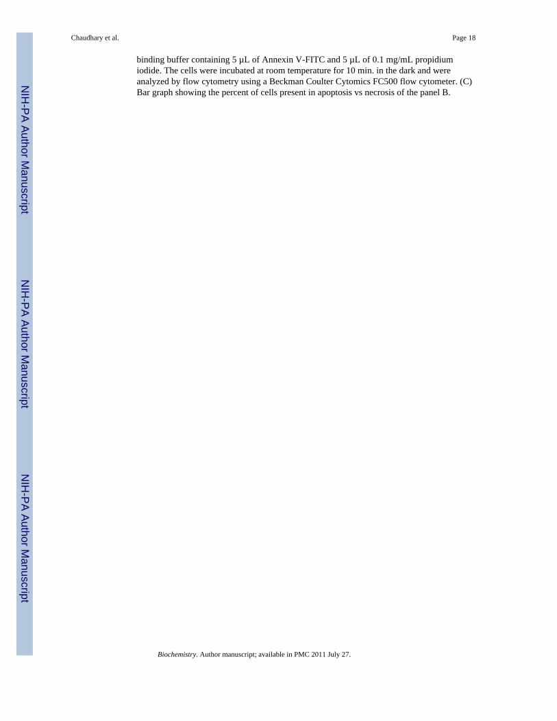
Over-expression of hGSTA4-4 inhibits 4-HNE induced apoptosisHepG2 cells were stably transfected with hGSTA4 and over expression of hGSTA4-4protein was confirmed by the results of Western blot analysis (Figure 2A). GST activitytowards 4-HNE was found to be enhanced in hGSTA4 transfected cells along with theexpected decrease in the constitutive 4-HNE levels (data not presented). 4-HNE-inducedapoptosis in the empty vector and hGSTA4 transfected cells was analyzed by usingCaspACE™ FITC-VAD-FMK in situ marker that binds to the cleaved caspase-3. Results ofthese experiments showed that the hGSTA4 transfected cells acquired significant resistanceto 4-HNE-induced apoptosis as compared to the empty vector transfected HepG2 cells(Figure 2B upper panel). hGSTA4 transfected cells also acquired significant resistance toDoxorubicin (DOX)-induced apoptosis (Figure 2B lower panel). Since DOX-inducedapoptosis has been attributed to generation of ROS, these results suggest the role of 4-HNEin the mechanism of apoptosis caused by oxidants in general (6, 7).
4-HNE and Fas mediated apoptosis4-HNE activates Fas—To elucidate the mechanism of 4-HNE-induced apoptosis inHepG2 cells, we analyzed first the effect of 4-HNE on the expression of Fas. Results ofWestern blot analyses (Figure S1A and B in supporting information) indicated that 4-HNEcaused a time, and dose dependent induction of Fas in HepG2 cells. These results wereconsistent with the previously reported induction of Fas by 4-HNE in HLE B-3, Jurkat, andCRL2571 cells (26, 28) indicating that 4-HNE mediated induction of Fas is not limited to aspecific cell types.
Chaudhary et al. Page 5
Biochemistry. Author manuscript; available in PMC 2011 July 27.
NIH
-PA Author Manuscript
NIH
-PA Author Manuscript
NIH
-PA Author Manuscript
Interaction of 4-HNE with Fas is required for apoptosis—To address the question,whether or not the interaction of 4-HNE was required to induce apoptosis, HepG2 cells werecoated with the antagonistic Fas monoclonal antibodies (B-10) to mask cell surface Fas. TheB-10 antibodies bind to cell surface Fas but unlike the agonistic CH11 antibodies that causeapoptosis, these antibodies do not induce apoptosis (36) and this was also confirmed duringthe present studies by examining their effects on HepG2 cells (data presented later in thissection). 4-HNE induced apoptosis was significantly inhibited in B-10 coated cellsindicating that Fas-4-HNE interaction was required for apoptosis (Figure 3A). It may benoted that 4-HNE induced apoptosis was not completely inhibited in B-10 coated cells.Although, cells coated with antagonistic Fas antibodies (B-10) were resistant to 4-HNE-induced apoptosis, a small population of cells still underwent apoptosis suggesting theinvolvement of other signaling pathways.
4-HNE causes the activation of ASK1-SEK1-JNK-Caspase-3—Previous studieshave shown that Fas-mediated apoptosis during stress conditions is accompanied by theactivation of ASK1, SEK1 and JNK, the kinases involved in the downstream signaling forapoptosis (37–41). It has also been shown that 4-HNE directly interacts with Fas and JNK(28,42). We therefore, examined the effect of 4-HNE on these down stream apoptoticsignaling molecules in HepG2 cells. Results of these studies showed a dose dependentactivation of ASK1, p-ASK1 (Thr845), p-SEK1 (Thr261) and p-JNK (Thr183/Tyr185)(Figure 3B). The observed activation of ASK1, SEK1 and JNK by 4-HNE in present studieswas rapid and sustained suggesting that a sustained activation and phosphorylation of JNKmay be needed for 4-HNE-induced apoptosis. This prediction was supported by the resultsof experiments showing that pretreatment of HepG2 cells with the JNK inhibitor SP600125made these cells resistant to apoptosis by 4-HNE (Figure S2, supporting information). Wehave previously demonstrated that in Jurkat cells, Fas mediated apoptosis by 4-HNE isindependent of FADD. Results presented in Figure S3A and B (see supporting information)show the lack of FasL and FADD induction and also the lack of FADD binding to Fas asindicated by the results of immunoprecipitation experiments with 4-HNE treated cells(Figure S3C, supporting information). These results further support our previousobservations that 4-HNE affects Fas mediated signaling for apoptosis through activation ofASK1 and JNK and is independent of DISC and this phenomenon is not limited to specificcell types.
Effect of 4-HNE on Daxx expression, phosphorylation, and traffickingDeath domain-associated protein Daxx that was originally cloned as a CD95/Apo-1 (Fas)-interacting protein (37) was shown to bind to the cytoplasmic domain of Fas and suggestedto activate signaling for apoptosis during oxidative stress in different types of cells (40,43).Therefore, we studied the effect of 4-HNE on Daxx in HepG2 cells and the results of theseexperiments presented in Figure 4 showed that 4-HNE treatment caused a time andconcentration dependent induction of Daxx. Since its discovery, the sub-cellular localizationof Daxx has been a controversy. Several studies have suggested that Daxx is predominantlya nuclear protein in unstressed cells, which under the conditions of oxidative stress,translocates to cytoplasm and binds to the cytoplasmic domain of Fas (41,44). The effect of4-HNE on Daxx trafficking and sub-cellular localization, was therefore analyzed by Westernblot and immunofluorescence. Furthermore, the interaction of Daxx with Fas was examinedby immunoprecipitation studies. Results of Western blot analysis of 4-HNE treated cells(Figure 5A and B) showed a marked increase of Daxx in the cytoplasmic fraction indicatingthat 4-HNE facilitated the export of Daxx from nucleus to cytoplasm and these results werefurther confirmed by immunofluorescence studies (Figure 5C).
Chaudhary et al. Page 6
Biochemistry. Author manuscript; available in PMC 2011 July 27.
NIH
-PA Author Manuscript
NIH
-PA Author Manuscript
NIH
-PA Author Manuscript
The translocation of Daxx from nucleus to cytoplasm may be mediated through itsphosphorylation on Ser and Thr residues (45,46). Western blot analysis of the extracts of 4-HNE treated HepG2 cells indicated that 4-HNE caused phosphorylation of Daxx at Ser671and Ser668 residues (Figure 4A). Together, these results show that 4-HNE promotes thetranslocation of Daxx from nucleus to cytoplasm through the phosphorylation of theseresidues. These results also suggest, that 4-HNE generated during oxidative stress may bethe actual causative factor for the previously reported (41,44) translocation of Daxx tocytoplasm during oxidative stress.
Interactions of 4-HNE with Fas and DaxxWe have examined the possible interaction of Daxx with 4-HNE and its binding to Fas inorder to get insight into the mechanisms of 4-HNE-induced Fas-mediated apoptosis. Theextracts from 4-HNE treated cells were immunoprecipitated with Daxx antibodies, and theresulting immunoprecipitates were probed separately with anti-Fas or anti-4-HNE 11-Santibodies (Figure 6A). The results of Western blot analysis showed that Daxx binds withFas and also to 4-HNE as indicated by the presence of 4-HNE Daxx-adduct in theimmunoprecipitate. When the immunoprecipitates obtained using Fas antibodies wereprobed with anti-Daxx antibody, the results (Figure 6B) further confirmed the binding ofDaxx with Fas. Together, the results of these experiments indicated that 4-HNE interactswith both Fas and Daxx and it promotes the binding of Fas to Daxx (Figure 6C).
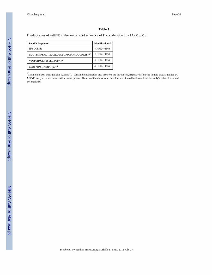
4-HNE covalently binds to DaxxTo better understand binding of 4-HNE to Daxx, we performed an ex vivo modification ofbacterially expressed purified human Daxx (see supporting information) with 4-HNE. Afterdigestion of the modified protein with trypsin and using solid-phase hydrazide enrichment,LC–MS/MS analysis of Daxx identified four tryptic peptides with 4-HNE modification. Allof these adducts showed preferential attachment of 4-HNE to His residues via a Michaeladdition mechanism (+156 Da). A representative CID-MS/MS spectrum of the doubly-charged 4-HNE-carbonylated Daxx tryptic peptide, VDSPSH*GLVTSSLCIPSPAR (whereH* indicates 4-HNE Michael adduct on the His), at m/z 1118.58 is shown in Figure S4A(supporting information). The observed 4-HNE neutral loss peak at m/z 1040.8 (loss of 78m/z from [M+2H]2+ ion) served as a diagnostic for the presence of H* in the peptide. TheMascot ion score was 67.8 (46.9 indicated identity or extensive homology) that surpassedthe defined probability-based threshold for identification for this particular peptide derivedfrom Daxx. Based on our results (Figure S4, supporting information), His residues atposition 300, 404, 691 and 711 in Daxx were modified preferentially (Table 1).
Daxx silencing sensitizes cell to apoptosisThe role of Daxx, in particular its ability to promote or inhibit apoptosis, still remainscontroversial (41,44). Some reports suggest that the death domain of Fas can directlyactivate apoptosis signal-regulating kinase 1 (ASK1) facilitating the translocation of Daxxfrom nucleus to cytoplasm. The interaction of translocated Daxx with the death domain ofFas has been shown to mediate cellular apoptosis via the Fas-Daxx-ASK1-JNK signalingpathway (47). On the other hand, it has been demonstrated that the binding of Daxx to Fas isnot necessarily an apoptotic signal (48–51) and 4-HNE induced Daxx translocation fromnucleus to the cytoplasm and it’s binding to Fas, in fact inhibits apoptosis (28). Therefore,we examined the role of Daxx in 4-HNE induced Fas mediated apoptosis in HepG2 cells.For these studies, we silenced the expression of Daxx by siRNA to evaluate the effect ofDaxx depletion on 4-HNE-Fas mediated apoptosis. In HepG2 cells transfected with DaxxsiRNA, the level of Daxx was only about 11% that of the cells transfected with scrambledsiRNA after 48 hours incubation indicating a successful knock down of Daxx expression bysiRNA (Figure 7A). Upon depletion of Daxx, cells became remarkably more sensitive to 4-
Chaudhary et al. Page 7
Biochemistry. Author manuscript; available in PMC 2011 July 27.
NIH
-PA Author Manuscript
NIH
-PA Author Manuscript
NIH
-PA Author Manuscript
HNE-induced apoptosis as indicated (Figure 7B and C) by the enhanced activation ofcaspase-3 and JNK in Daxx depleted cells as compared to the controls. Moreover, Daxxdepleted cells also became more sensitive to the apoptosis caused by agonistic CH11 Fasantibodies, as indicated by the onset of increased apoptosis in Daxx-deficient cells treatedwith these antibodies (Figure 7C, lower two panels). Together, these results suggested thatthe binding of Daxx to Fas inhibits Fas mediated apoptosis and Fas-Daxx binding was notnecessary for either activation of JNK and caspase-3 or the onset of apoptosis as reportedpreviously (37).
4-HNE causes the activation and translocation of HSF1 to the nucleus and induction ofHsp70
Daxx is known to be involved in transcriptional control of many genes by interactingdirectly with several transcription factors (41,44,52). The results of present studies showedthat 4-HNE induced HSF1 in HepG2 cells in a time dependent manner (Figure 8A). 4-HNEalso induced HSF1 translocation from cytoplasm to nucleus as indicated by the results ofWestern blot analysis of cytoplasmic and nuclear fractions (Figure 8B and C) andimmunofluorescence analysis (Figure 8E). These results are consistent with 4-HNEmediated translocation of HSF1 from cytoplasm to nucleus in colon cancer RKO cellsreported previously (53,54). Activation and translocation of HSF1 is known to be anindication of the transcriptional up-regulation of stress-responsive heat shock proteins likeHsp70. Consistent with this prediction, 4-HNE was found to induce the expression of Hsp70in HepG2 cells (Figure 8A). 4-HNE induced a robust accumulation of HSF1 within thenuclear compartment and a dense patchy staining of HSF1 was observed at a higherconcentration (Figure 8E, see arrows). To demonstrate that this patchy nuclear staining forHSF1 was due to its binding to Hsp70 promoter sequence, we performed ChIP assay in thenuclear extracts of HepG2 cells treated with 4-HNE using HSF1 antibodies forimmunoprecipitation. No signal was seen upon immunoprecipitation with negative controlIgG. The positive control in which the antibodies used were against RNA polymerase IIgave the expected positive signal validating our assay system. Neither the positive nor thenegative controls were affected by 4-HNE treatment. The results presented in Figure 8Dclearly indicated that the binding of HSF1 to the Hsp70 promoter was higher in 4-HNEtreated HepG2 cells as compared to untreated cells, indicating that 4-HNE induced theactivation and nuclear accumulation of HSF1 accompanied with the transcriptional up-regulation of Hsp70.
4-HNE induces p53, phospho-p53, and activates p21, JNK, and BaxTo examine a possible role of 4-HNE in the p53 mediated apoptotic pathway, we determinedthe effect of 4-HNE on the expression of p53 and its phosphorylation in HepG2 cells. Theresults of these studies showed that at 20 µM concentration, 4-HNE caused a time dependentinduction of p53 and phosphorylated p53 at Ser15 (Figure 9). Since activation andphosphorylation of p53 in response to oxidative stress has been associated with apoptosisthrough the induction of its target genes including p21 and Bax, we examined the effect of4-HNE on the expression of its down stream targets. During apoptosis, Bax has been shownto undergo a conformational shift, and inserts into the outer mitochondrial membrane whereit is believed to induce the opening of the mitochondrial membrane resulting in the releaseof cytochrome c and other pro-apoptotic factors from the mitochondria, leading to activationof caspases (55,56). Consistent with one of the mechanisms of p53 mediated apoptosis,treatment of HepG2 cells with 4-HNE resulted in the activation of pro-apoptotic proteinsBax and p21. In parallel, the expression of anti-apoptotic Bcl-xL was found to be downregulated in 4-HNE treated cells. To determine the effect of 4- HNE on caspase-3independent apoptosis in these cells, we analyzed the expression of AIF in 4-HNE treatedcells. No significant change in the expression of AIF in control and 4-HNE treated cells
Chaudhary et al. Page 8
Biochemistry. Author manuscript; available in PMC 2011 July 27.
NIH
-PA Author Manuscript
NIH
-PA Author Manuscript
NIH
-PA Author Manuscript
(data not shown) suggests that 4-HNE does not affect AIF mediated caspase-independentapoptosis of these cells. The activation and phosphorylation of p53 along with p53 clientpro-apoptotic proteins indicate that in addition to Fas mediated apoptosis 4-HNE alsoactivates p53 mediated intrinsic apoptotic pathway. However, our results showing theresistance of Fas (B-10) antibodies coated HepG2 cells to 4-HNE induced apoptosisdemonstrate that, as compared to Fas-mediated apoptosis, the relative contribution of p53-mediated apoptosis was only minimal (Figure 3A). No significant effect on 4-HNE inducedactivation of p53, JNK and Bax in B-10 coated cells (Figure S5, supporting information)suggest that the activation of p53 pathway is not dependent on Fas-4-HNE interaction.Further studies are needed to evaluate the physiological significance and relativecontributions of Fas and p53 mediated pathways in 4-HNE induced apoptosis.
Over-expression of hGSTA4-4 inhibits the activation of Fas and p53 mediated apoptoticpathways
To investigate any possible role of hGSTA4-4 in the regulation of Fas and p53 mediatedapoptosis, we studied the effect of hGSTA4-transfection on the components of thesepathways. Results of these experiments presented in Figure 10A showed that the activationof the component of both these pathways (Fas, Daxx, ASK1, p53, Bax and JNK) wassignificantly suppressed in hGSTA4 transfected cells as compared to that with vector-transfected cells. These results demonstrate an important regulatory role of hGSTA4-4during apoptosis induced by oxidative stress via the Fas and p53 pathways and areconsistent with reported rapid induction of this isozyme in stress conditions that lead to 4-HNE generation (26, 57).
Increased 4-HNE levels in liver of mGsta4 null mice activate Fas and p53 mediatedapoptotic pathways in vivo
The in vitro cell culture studies presented above were extended to in vivo conditions usingthe liver tissues from mGsta4 null mice which have been shown to contain approximately 3-fold higher concentration of 4-HNE in liver (29). To examine whether the effects of 4-HNEin cell cultures were also reflected in vivo, we compared the expression of Fas, Daxx, p53,HSF1, ASK1, p-SEK1, and JNK in the tissues of mGsta4 (−/−) and wild-type (+/+) mice.Consistent with our prediction, results of Western blot analysis presented in Figure 10Bshowed that the expression of these proteins was remarkably up regulated in liver tissues ofmGsta4 (–/–) mice examined in this study. These results demonstrated a role of 4-HNE insignaling, in vivo and are consistent with our earlier suggestion (25,58) that LPO and 4-HNEin particular, play an important role in oxidative stress induced signaling.
DISCUSSION4-HNE, which was initially thought to be merely a toxic end-product of LPO, has emergedas an important second messenger signaling molecule in recent years. A multitude of studies(See reviews: 3–9,24) over the past 10 years attest to its role in the regulation of geneexpression, and modulation of various cellular processes including proliferation,transformation, differentiation, cell cycle regulation, and apoptosis. The mechanismsthrough which 4-HNE exerts such a wide variety of effects are largely unknown. Forexample, it remains to be determined as to how 4-HNE exerts seemingly opposite effectssuch as proliferation and programmed cell death in a concentration dependent manner.Present studies validate the important signaling role of 4-HNE and in addition, providesignificant insight into the possible mechanisms through which 4-HNE exerts itsmultifarious effects in cells.
Chaudhary et al. Page 9
Biochemistry. Author manuscript; available in PMC 2011 July 27.
NIH
-PA Author Manuscript
NIH
-PA Author Manuscript
NIH
-PA Author Manuscript
Here we demonstrate that 4-HNE causes toxicity to HepG2 cells through apoptosis andnecrosis in a dose dependent manner. At sub lethal concentrations of 4-HNE, these cellsundergo apoptosis via two separate pathways. Results of these studies clearly demonstratethat 4-HNE binds to Fas, and affects the downstream signaling mediated by this deathreceptor and that this 4-HNE-induced apoptosis is independent of the classical Fas-mediatedapoptosis through FADD and DISC (59) because 4-HNE neither activates FADD norpromotes the binding of Fas to FADD. However, 4-HNE induced, Fas-mediated apoptosisinvolves the activation of ASK1, JNK and caspase-3 but does not induce FasL or theconstituents of DISC. As indicated by the lack of apoptosis by 4-HNE in cells coated withantagonistic Fas antibodies, our results clearly demonstrate that binding of 4-HNE with Fasis essential for the execution of apoptosis. This is further indicated by abrogation of 4-HNE-induced activation of ASK1, JNK, and caspase-3 in cells when Fas is masked by coatingwith antibodies. Our results showing inhibition of apoptosis and lack of the activation ofASK1 and JNK in cells over expressing hGSTA4-4 pinpoint these effects to 4-HNE becausethis enzyme is highly specific for the conjugation of 4-HNE to GSH. 4-HNE inducedactivation of p53 and down stream targets including p21 and Bax in HepG2 cells alsosuggest the involvement of p53 mediated pathway in the apoptotic mechanisms of thesecells but the contribution of this pathway seems to be minimal. All these effects of 4-HNEcan also be abrogated by GSTA4-4 over expression. These finding are consistent withprevious studies suggesting important role of GST isozymes in the regulation of stress-induced signaling, by attenuating 4-HNE concentrations (8) as well as by inhibiting JNK(60–62).
These signaling effects of 4-HNE observed in in vitro cell lines appear to mirror in the livertissues of mice which have higher basal levels of 4-HNE due to the disruption of Gsta4gene. This is important because it may enhance our understanding of the mechanisminvolved in manifestation of the toxicity of ROS and oxidants, and the role of relevantdefense mechanisms in vivo. Even though Gsta4 (−/−) mice are more sensitive to oxidanttoxicity, they seem to have no apparent toxic manifestations in stress free conditions and infact may have an increased life span (63). Thus the question may arise as to why these micedo not show any apparent toxicity despite activation of Fas and p53 mediated apoptoticpathways. The answer to this question may lie in our results showing that in parallel to itsapoptotic signaling, 4-HNE invokes signaling for the defense against its own toxicity. First,by inhibiting its apoptotic effect by facilitating cytoplasmic export of Daxx and its bindingwith Fas and second, by activating HSF1 initiated stress responsive transcription ofprotective heat shock proteins such as Hsp70. The involvement of 4-HNE in promoting thecytoplasmic translocation of Daxx is further confirmed by our results demonstrating a directbinding of 4-HNE with Daxx, and phosphorylation of Daxx at Ser668 and Ser671 that isreported (45,46) to be required for the translocation of Daxx from nucleus to cytoplasm.Once in cytoplasm Daxx binds to Fas and inhibits its own apoptotic effect that may be animportant defense mechanism to check apoptosis. Our results clearly demonstrate that thebinding of Fas to Daxx does indeed inhibit apoptosis (28,48–51) rather than causing a pro-apoptotic effect as suggested in some earlier studies (37,43,47,64,65). The potentiation ofthe apoptotic effect of the agonistic CH11 anti-Fas antibodies in Daxx depleted cells furthersubstantiates the anti-apoptotic role of Daxx and indicates that its inhibitory role is notlimited to oxidative stress/ 4-HNE mediated apoptosis.
In addition to Daxx mediated inhibition of apoptosis, in parallel 4-HNE induces traffickingof HSF1 from cytoplasm to nucleus to promote transcription of stress responsive genes toreinforce the defense response. In RKO cells 4-HNE mediated translocation of HSF1 hasbeen shown and a role of HSF1 in anti-apoptotic mechanisms has been demonstrated(53,54). Present studies through Western blot and immunofluorescence analyses provideclear evidence for the trafficking of HSF1 from cytoplasm to nucleus in HepG2 cells where
Chaudhary et al. Page 10
Biochemistry. Author manuscript; available in PMC 2011 July 27.
NIH
-PA Author Manuscript
NIH
-PA Author Manuscript
NIH
-PA Author Manuscript
it promotes the expression of Hsp70 indicating that this effect of 4-HNE is general and notlimited to a specific cell type. Since the effect of 4-HNE in the liver of Gsta4 null mice aresimilar to those observed in in vitro cell culture studies, the defense mechanisms evoked dueto the elevated levels of 4-HNE in these mice may also explain the observed increased lifespan of Gsta4 null mice (63) without any apparent toxicity. The effects of 4-HNE on Daxx,HSF1, and p53 reported here may perhaps include other transcription repressors/factors. Ifso 4-HNE should have a profound effect on the expression of client genes. This predictionfinds strong support from the results of several studies by us (26–28,66) and otherinvestigators (5,67–69) showing profound changes in the expression of a multitude of geneswhen 4-HNE concentrations are altered in cells. It may be argued that the steady state 4-HNE concentrations in cells are reported (24) to be much lower than those used in presentstudies. However, it should be pointed out that during oxidative stress the intracellularconcentration could be even higher than used in these studies (70).
Since our results indicated the binding of 4-HNE to Fas, HSF1, p53 and Daxx, proteomicsstudies were carried out with Daxx to demonstrate 4-HNE binding to its specific residues.Through these studies, we have identified several binding sites, although possible functionalconsequences of the binding are yet to be determined. It is possible that observedconcentration dependent effects of 4-HNE may be determined by the differential reactivityof selective target residue(s) of key proteins such as Daxx for 4-HNE binding that mayevoke differential signaling. An interesting novel paradigm seems to emerge from the resultsof present studies. It is possible that through interactions with membrane receptors such asFas, EGF, VEGF, transcription factor such as p53, HSF1, and transcription repressors suchas Daxx, 4-HNE could have a global effect on the expression of genes regulating a variety ofcellular processes. The cellular concentrations of 4-HNE and its relative affinity to theresidues of target proteins may be determinants of these interactions and consequent effects.Our previous studies have shown that suppression of 4-HNE levels in cells of at least twodifferent types leads to profound alteration in the expression of genes in these cells andresults in their transformation (27,66). Thus, it seems likely that 4-HNE, through directinteractions with some key proteins such as those investigated in the present studies, canhave profound effects on gene expression and consequently on various cellular processes.This would also imply that the Alpha class GSTs including GSTA4-4 that regulate LPO and4-HNE levels in cells (25,57,71), play an important role in stress-induced signaling.Ongoing studies in our lab suggest that 4-HNE does indeed affect the signaling pathwaysmediated by VEGF, and VEGFR, in a concentration dependent manner but numerousconclusive studies will have to be conducted to substantiate this implication.
Supplementary MaterialRefer to Web version on PubMed Central for supplementary material.
AcknowledgmentsFunding: This work was supported in part by NIH Grants ES012171, EY04396 (YCA), AG025384 (LP) andCA77495 (SA), and by the Welch Foundation (LP, endowment number BK-0031).
We greatly appreciate the gift of human Daxx cDNA from Dr. Alnawaz Rehemtulla, (Department of RadiationOncology and Radiology, University of Michigan, Ann Arbor, MI 48109-2200, USA). We thank Xiangle Sun(Core Facility at the University of North Texas Health Science Center) for helping with flow Cytometry.
Abbreviations
4-HNE 4-hydroxy-2-nonenal
Chaudhary et al. Page 11
Biochemistry. Author manuscript; available in PMC 2011 July 27.
NIH
-PA Author Manuscript
NIH
-PA Author Manuscript
NIH
-PA Author Manuscript
DOX doxorubicin (Adriamycin)
GST glutathione S-transferase
siRNA small interfering RNA
Daxx death domain-associated protein
ASK1 apoptosis signal-regulating kinase 1
SEK1 stress-activated protein kinase/extracellular-signal regulated kinase kinase
JNK c-Jun NH2-terminal kinase
HSF1 heat shock factor 1
WT wild type
IP immunoprecipitation
ChIP chromatin immunoprecipitation
DAPI 4',6-diamidino-2-phenylindole
PARP Poly (ADP-ribose) polymerase
MTT 3-(4,5-dimethylthiazol-2-yl)-2,5-diphenyl tetrazolium bromide
Hsps Heat shock proteins
AIF apoptosis inducing factor
FADD Fas-associated death domain
DISC death-inducing signaling complex
REFERENCES1. Cheng JZ, Sharma R, Yang Y, Singhal SS, Sharma A, Saini MK, Singh SV, Zimniak P, Awasthi S,
Awasthi YC. Accelerated metabolism and exclusion of 4-hydroxynonenal through induction ofRLIP76 and hGST5.8 is an early adaptive response of cells to heat and oxidative stress. J. Biol.Chem. 2001; 276:41213–41223. [PubMed: 11522795]
2. Yang Y, Sharma A, Sharma R, Patrick B, Singhal SS, Zimniak P, Awasthi S, Awasthi YC. Cellspreconditioned with mild, transient UVA irradiation acquire resistance to oxidative stress and UVA-induced apoptosis: role of 4-hydroxynonenal in UVA-mediated signaling for apoptosis. J. Biol.Chem. 2003; 278:41380–41388. [PubMed: 12888579]
3. Dianzani MU. 4-hydroxynonenal from pathology to physiology. Mol. Aspects Med. 2003; 24:263–272. [PubMed: 12893004]
4. Nakashima I, Liu W, Akhand AA, Takeda K, Kawamoto Y, Kato M, Suzuki H. 4-Hydroxynonenaltriggers multistep signal transduction cascades for suppression of cellular functions. Mol. AspectsMed. 2003; 24:231–238. [PubMed: 12893001]
5. Barrera G, Pizzimenti S, Dianzani MU. 4-Hydroxynonenal and regulation of cell cycle: effects onthe pRb/E2F pathway. Free Radic. Biol. Med. 2004; 37:597–606. [PubMed: 15288118]
6. Awasthi YC, Sharma R, Cheng JZ, Yang Y, Sharma A, Singhal SS, Awasthi S. Role of 4-hydroxynonenal in stress-mediated apoptosis signaling. Mol. Aspects Med. 2003; 24:219–230.[PubMed: 12893000]
7. Awasthi YC, Yang Y, Tiwari NK, Patrick B, Sharma A, Li J, Awasthi S. Regulation of 4-hydroxynonenal-mediated signaling by glutathione S-transferases. Free Radic. Biol. Med. 2004;37:607–619. [PubMed: 15288119]
8. Awasthi YC, Ansari GA, Awasthi S. Regulation of 4-hydroxynonenal mediated signaling byglutathione S-transferases. Methods Enzymol. 2005; 401:379–407. [PubMed: 16399399]
Chaudhary et al. Page 12
Biochemistry. Author manuscript; available in PMC 2011 July 27.
NIH
-PA Author Manuscript
NIH
-PA Author Manuscript
NIH
-PA Author Manuscript
9. Awasthi YC, Sharma R, Sharma A, Yadav S, Singhal SS, Chaudhary P, Awasthi S. Self-regulatoryrole of 4-hydroxynonenal in signaling for stress-induced programmed cell death. Free Radic. Biol.Med. 2008; 45:111–118. [PubMed: 18456001]
10. Sampey BP, Stewart BJ, Petersen DR. Ethanol-induced modulation of hepatocellular extracellularsignal-regulated kinase-1/2 activity via 4-hydroxynonenal. J. Biol. Chem. 2007; 282:1925–1937.[PubMed: 17107949]
11. Tsukamoto H, Horne W, Kamimura S, Niemel O, Parkkila S, Yl-Herttuala S, Brittenham GM.Experimental liver cirrhosis induced by alcohol and iron. J. Clin. Invest. 1995; 96:620–630.[PubMed: 7615836]
12. Nanji AA, Zhao S, Sadrzadeh SMH, Dannenberg AJ, Tahan SR, Waxman DJ. Markedly enhancedcytochrome P450 2E1 induction and lipid peroxidation is associated with severe liver injury in fishoil-ethanol-fed rats. Alcohol Clin. Exp. Res. 1994; 18:1280–1285. [PubMed: 7847620]
13. Dwivedi S, Sharma R, Sharma A, Zimniak P, Ceci JD, Awasthi YC, Boor PJ. The course of CCl4induced hepatotoxicity is altered in mGSTA4-4 null (−/−) mice. Toxicology. 2006; 218:58–66.[PubMed: 16325313]
14. Kamimura S, Gaal K, Britton RS, Bacon BR, Triadafilopoulos G, Tsukamoto H. Increased 4-hydroxynonenal levels in experimental alcoholic liver disease: Association of lipid peroxidationwith liver fibrogenesis. Hepatology. 1992; 16:1014–1021. [PubMed: 1398481]
15. Uchida K, Toyokuni S, Nishikawa K, Kawakishi S, Oda H, Hiai H, Stadtman ER. Michaeladdition-type 4-hydroxy-2-nonenal adducts in modified low-density lipoproteins: markers foratherosclerosis. Biochemistry. 1994; 33:12487–12494. [PubMed: 7918471]
16. Yang Y, Yang Y, Trent MB, He N, Lick SD, Zimniak P, Awasthi YC, Boor PJ. Glutathione-S-transferase A4-4 modulates oxidative stress in endothelium: possible role in humanatherosclerosis. Atherosclerosis. 2004; 173:211–221. [PubMed: 15064094]
17. Awasthi S, Singhal SS, Yadav S, Singhal J, Vatsyayan R, Zajac E, Luchowski R, Borvak J,Gryczynski K, Awasthi YC. A Central Role of RLIP76 in Regulation of Glycemic Control.Diabetes. 2010; 59:714–725. [PubMed: 20007934]
18. Sayre LM, Zelasko DA, Harris PL, Perry G, Salomon RG, Smith MA. 4-Hydroxynonenal-derivedadvanced lipid peroxidation end products are increased in Alzheimer's disease. J. Neurochem.1997; 68:2092–2097. [PubMed: 9109537]
19. Montine KS, Olson SJ, Amarnath V, Whetsell WO, Graham DG Jr, Montine TJ.Immunohistochemical detection of 4-hydroxy-2-nonenal adducts in Alzheimer's disease isassociated with inheritance of APOE4. Am. J. Pathol. 1997; 150:437–443. [PubMed: 9033259]
20. Selley ML. (E)-4-hydroxy-2-nonenal may be involved in the pathogenesis of Parkinson's disease.Free Radic. Biol. Med. 1998; 25:169–174. [PubMed: 9667492]
21. Awasthi S, Srivatava SK, Piper JT, Singhal SS, Chaubey M, Awasthi YC. Curcumin protectsagainst 4-hydroxy-2-trans-nonenal-induced cataract formation in rat lenses. Am. J. Clin. Nutr.1996; 64:761–766. [PubMed: 8901798]
22. Zarkovic N, Tillian MH, Schaur J, Waeg G, Jurin M, Esterbauer H. Inhibition of melanoma B16-F10 growth by lipid peroxidation product 4-hydroxynonenal. Cancer Biother. 1995; 10:153–156.[PubMed: 7663575]
23. Hammer A, Ferro M, Tillian HM, Tatzber F, Zollner H, Schauenstein E, Schaur RJ. Effect ofoxidative stress by iron on 4-hydroxynonenal formation and proliferative activity in hepatomas ofdifferent degrees of differentiation. Free Radic. Biol. Med. 1997; 23:26–33. [PubMed: 9165294]
24. Esterbauer H, Schaur RJ, Zollner H. Chemistry and biochemistry of 4-hydroxynonenal,malonaldehyde and related aldehydes. Free Radic. Biol. Med. 1991; 11:81–128. [PubMed:1937131]
25. Cheng JZ, Singhal SS, Saini M, Singhal J, Piper JT, Van Kuijk FJ, Zimniak P, Awasthi YC,Awasthi S. Effects of mGST A4 transfection on 4-hydroxynonenal-mediated apoptosis anddifferentiation of K562 human erythroleukemia cells. Arch. Biochem. Biophys. 1999; 372:29–36.[PubMed: 10562413]
26. Li J, Sharma R, Patrick B, Sharma A, Jeyabal VP, Reddy PM, Saini MK, Dwivedi S, Dhanani S,Ansari NH, Zimniak P, Awasthi S, Awasthi YC. Regulation of CD95 (Fas) expression and Fas-
Chaudhary et al. Page 13
Biochemistry. Author manuscript; available in PMC 2011 July 27.
NIH
-PA Author Manuscript
NIH
-PA Author Manuscript
NIH
-PA Author Manuscript
mediated apoptotic signaling in HLE B-3 cells by 4-hydroxynonenal. Biochemistry. 2006;45:12253–12264. [PubMed: 17014078]
27. Sharma R, Brown D, Awasthi S, Yang Y, Sharma A, Patrick B, Saini MK, Singh SP, Zimniak P,Singh SV, Awasthi YC. Transfection with 4-hydroxynonenal-metabolizing glutathione S-transferase isozymes leads to phenotypic transformation and immortalization of adherent cells.Eur. J. Biochem. 2004; 271:1690–1701. [PubMed: 15096208]
28. Sharma R, Sharma A, Dwivedi S, Zimniak P, Awasthi S, Awasthi YC. 4-Hydroxynonenal self-limits fas-mediated DISC-independent apoptosis by promoting export of Daxx from the nucleus tothe cytosol and its binding to Fas. Biochemistry. 2008; 47:143–156. [PubMed: 18069800]
29. Engle MR, Singh SP, Czernik PJ, Gaddy D, Montague DC, Ceci JD, Yang Y, Awasthi S, AwasthiYC, Zimniak P. Physiological role of mGSTA4-4, a glutathione S-transferase metabolizing 4-hydroxynonenal: generation and analysis of mGsta4 null mouse. Toxicol. Appl. Pharmacol. 2004;194:296–308. [PubMed: 14761685]
30. Bradford MM. A rapid and sensitive method for the quantitation of microgram quantities of proteinutilizing the principle of protein-dye binding. Anal. Biochem. 1976; 72:248–254. [PubMed:942051]
31. Roe MR, Xie H, Bandhakavi S, Griffin TJ. Proteomic Mapping of 4-Hydroxynonenal ProteinModification Sites by Solid-Phase Hydrazide Chemistry and Mass Spectrometry. Anal. Chem.2007; 79:3747–3756. [PubMed: 17437329]
32. Rauniyar N, Stevens SM Jr, Prokai-Tatrai K, Prokai L. Characterization of 4-hydroxy-2-nonenal-modified peptides by liquid chromatography-tandem mass spectrometry using data-dependentacquisition: neutral loss-driven MS3 versus neutral loss-driven electron capture dissociation. Anal.Chem. 2009; 81:782–789. [PubMed: 19072288]
33. Rauniyar N, Prokai-Tatrai K, Prokai L. Identification of carbonylation sites in apomyoglobin afterexposure to 4-hydroxy-2-nonenal by solid-phase enrichment and liquid chromatography-electrospray ionization tandem mass spectrometry. J. Mass Spectrom. 2010 (In press) DOI10.1002/jms.1725.
34. Stevens SM Jr, Rauniyar N, Prokai L. Rapid characterization of covalent modifications to rat brainmitochondrial proteins after ex vivo exposure to 4-hydroxy-2-nonenal by liquid chromatography-tandem mass spectrometry using data-dependent and neutral loss-driven MS3 acquisition. J. MassSpectrom. 2007; 42:1599–1605. [PubMed: 18085542]
35. Rauniyar N, Prokai L. Detection and identification of 4-hydroxy-2-nonenal Schiff-base adductsalong with products of Michael addition using data-dependent neutral loss-driven MS3acquisition: method evaluation through an in vitro study on cytochrome c oxidase modifications.Proteomics. 2009; 9:5188–5193. [PubMed: 19771555]
36. Fadeel B, Thorpe CJ, Yonehara S, Chiodi F. Anti-Fas IgG1 antibodies recognizing the sameepitope of Fas/APO-1 mediate different biological effects in vitro. Int. Immunol. 1997; 9:201–209.[PubMed: 9040002]
37. Yang X, Khosravi-Far R, Chang HY, Baltimore D. Daxx, a novel Fas-binding protein thatactivates JNK and apoptosis. Cell. 1997; 89:1067–1076. [PubMed: 9215629]
38. Ichijo H, Nishida E, Irie K, ten Dijke P, Saitoh M, Moriguchi T, Takagi M, Matsumoto K,Miyazono K, Gotoh Y. Induction of apoptosis by ASK1, a mammalian MAPKKK that activatesSAPK/JNK and p38 signaling pathways. Science. 1997; 275:90–94. [PubMed: 8974401]
39. Chang HY, Nishitoh H, Yang X, Ichijo H, Baltimore D. Activation of apoptosis signal-regulatingkinase 1 (ASK1) by the adapter protein Daxx. Science. 1998; 281:1860–1863. [PubMed: 9743501]
40. Soh Y, Jeong KS, Lee IJ, Bae MA, Kim YC, Song BJ. Selective activation of the c-Jun N-terminalprotein kinase pathway during 4-hydroxynonenal-induced apoptosis of PC12 cells. Mol.Pharmacol. 2000; 58:535–541. [PubMed: 10953046]
41. Salomoni P, Khelifi AF. Daxx: death or survival protein? Trends Cell Biol. 2006; 16:97–104.[PubMed: 16406523]
42. Parola M, Robino G, Marra F, Pinzani M, Bellomo G, Leonarduzzi G, Chiarugi P, Camandola S,Poli G, Waeg G, Gentilini P, Dianzani MU. HNE interacts directly with JNK isoforms in humanhepatic stellate cells. J. Clin. Invest. 1998; 102:1942–1950. [PubMed: 9835619]
Chaudhary et al. Page 14
Biochemistry. Author manuscript; available in PMC 2011 July 27.
NIH
-PA Author Manuscript
NIH
-PA Author Manuscript
NIH
-PA Author Manuscript
43. Khelifi AF, D'Alcontres MS, Salomoni P. Daxx is required for stress-induced cell death and JNKactivation. Cell Death Differ. 2005; 12:724–733. [PubMed: 15861194]
44. Lindsay CR, Morozov VM, Ishov AM. PML NBs (ND10) and Daxx: from nuclear structure toprotein function. Front Biosci. 2008; 13:7132–7142. [PubMed: 18508722]
45. Song JJ, Lee YJ. Tryptophan 621 and serine 667 residues of Daxx regulate its nuclear exportduring glucose deprivation. J. Biol. Chem. 2004; 279:30573–30578. [PubMed: 15128734]
46. Ecsedy JA, Michaelson JS, Leder P. Homeodomain-interacting protein kinase 1 modulates Daxxlocalization, phosphorylation, and transcriptional activity. Mol. Cell Biol. 2003; 23:950–960.[PubMed: 12529400]
47. Ko YG, Kang YS, Park H, Seol W, Kim J, Kim T, Park HS, Choi EJ, Kim S. Apoptosis signal-regulating kinase 1 controls the proapoptotic function of death-associated protein (Daxx) in thecytoplasm. J. Biol. Chem. 2001; 276:39103–39106. [PubMed: 11495919]
48. Michaelson JS, Bader D, Kuo F, Kozak C, Leder P. Loss of Daxx, a promiscuously interactingprotein, results in extensive apoptosis in early mouse development. Genes Dev. 1999; 13:1918–1923. [PubMed: 10444590]
49. Michaelson JS, Leder P. RNAi reveals anti-apoptotic and transcriptionally repressive activities ofDAXX. J. Cell Sci. 2003; 116:345–352. [PubMed: 12482920]
50. Chen LY, Chen JD. Daxx silencing sensitizes cells to multiple apoptotic pathways. Mol. Cell. Biol.2003; 23:7108–7121. [PubMed: 14517282]
51. Zobalova R, Swettenham E, Chladova J, Dong LF, Neuzil J. Daxx inhibits stress-induced apoptosisin cardiac myocytes. Redox Rep. 2008; 13:263–270. [PubMed: 19017466]
52. Boellmann F, Guettouche T, Guo Y, Fenna M, Mnayer L, Voellmy R. DAXX interacts with heatshock factor 1 during stress activation and enhances its transcriptional activity. Proc. Natl. Acad.Sci. U.S.A. 2004; 101:4100–4105. [PubMed: 15016915]
53. Jacobs AT, Marnett LJ. Heat shock factor 1 attenuates 4-Hydroxynonenal-mediated apoptosis:critical role for heat shock protein 70 induction and stabilization of Bcl-XL. J. Biol. Chem. 2007;282:33412–33420. [PubMed: 17873279]
54. Jacobs AT, Marnett LJ. HSF1-mediated BAG3 expression attenuates apoptosis in 4-hydroxynonenal-treated colon cancer cells via stabilization of anti-apoptotic Bcl-2 proteins. J.Biol. Chem. 2009; 284:9176–9183. [PubMed: 19179333]
55. Vousden KH, Lu X. Live or let die: the cell's response to p53. Nat. Rev. Cancer. 2002; 2:594–604.[PubMed: 12154352]
56. Wang XW. Role of p53 and apoptosis in carcinogenesis. Anticancer Res. 1999; 19:4759–4771.[PubMed: 10697590]
57. Sharma A, Sharma R, Chaudhary P, Vatsyayan R, Pearce V, Jeyabal PV, Zimniak P, Awasthi S,Awasthi YC. 4-Hydroxynonenal induces p53-mediated apoptosis in retinal pigment epithelialcells. Arch. Biochem. Biophys. 2008; 480:85–94. [PubMed: 18930016]
58. Yang Y, Sharma R, Zimniak P, Awasthi YC. Role of alpha class glutathione S-transferases asantioxidant enzymes in rodent tissues. Toxicol. Appl. Pharmacol. 2002; 182:105–115. [PubMed:12140174]
59. Nagata S. Apoptosis by death factor. Cell. 1997; 88:355–365. [PubMed: 9039262]60. Gilot D, Loyer P, Corlu A, Glaise D, Lagadic-Gossmann D, Atfi A, Morel F, Ichijo H, Guguen-
Guillouzo C. Liver protection from apoptosis requires both blockage of initiator caspase activitiesand inhibition of ASK1/JNK pathway via glutathione S-transferase regulation. J. Biol. Chem.2002; 277:49220–49229. [PubMed: 12370186]
61. Adler V, Yin Z, Fuchs SY, Benezra M, Rosario L, Tew KD, Pincus MR, Sardana M, HendersonCJ, Wolf CR, Davis RJ, Ronai Z. Regulation of JNK signaling by GSTp. EMBO J. 1999;18:1321–1334. [PubMed: 10064598]
62. Romero L, Andrews K, Ng L, O’Rourke K, Maslen A, Kirby G. Human GSTA1-1 reduces c-JunN-terminal kinase signalling and apoptosis in Caco-2 cells. Biochem J. 2006; 400:135–141.[PubMed: 16836488]
63. Singh SP, Niemczyk M, Saini D, Sadovov V, Zimniak L, Zimniak P. Disruption of the mGsta4gene increases life span of C57BL mice. J. Gerontol. A Biol. Sci. Med. Sci. 2010; 65:14–23.[PubMed: 19880816]
Chaudhary et al. Page 15
Biochemistry. Author manuscript; available in PMC 2011 July 27.
NIH
-PA Author Manuscript
NIH
-PA Author Manuscript
NIH
-PA Author Manuscript
64. Kim KS, Hwang HA, Chae SK, Ha H, Kwon KS. Upregulation of Daxx mediates apoptosis inresponse to oxidative stress. J. Cell Biochem. 2005; 96:330–338. [PubMed: 16088932]
65. Boehrer S, Nowak D, Hochmuth S, Kim SZ, Trepohl B, Afkir A, Hoelzer D, Mitrou PS,Weidmann E, Chow KU. Daxx overexpression in T-lymphoblastic Jurkat cells enhances caspase-dependent death receptor- and drug-induced apoptosis in distinct ways. Cell Signalling. 2005;17:581–595. [PubMed: 15683733]
66. Patrick B, Li J, Jeyabal PV, Reddy PM, Yang Y, Sharma R, Sinha M, Luxon B, Zimniak P,Awasthi S, Awasthi YC. Depletion of 4-hydroxynonenal in hGSTA4-transfected HLE B-3 cellsresults in profound changes in gene expression. Biochem. Biophys. Res. Commun. 2005;334:425–432. [PubMed: 16005854]
67. Fazio VM, Barrera G, Martinotti S, Farace MG, Giglioni B, Frati L, Manzari V, Dianzani MU. 4-Hydroxynonenal, a product of cellular lipid peroxidation, which modulates c-myc and globin geneexpression in K562 erythroleukemic cells. Cancer Res. 1992; 52:4866–4871. [PubMed: 1516044]
68. Ruef J, Rao GN, Li F, Bode C, Patterson C, Bhatnagar A, Runge MS. Induction of rat aorticsmooth muscle cell growth by the lipid peroxidation product 4-hydroxy-2-nonenal. Circulation.1998; 97:1071–1078. [PubMed: 9531254]
69. Chiarpotto E, Domenicotti C, Parola D, Vitali A, Nitti M, Pronzato MA, Biasi F, Cottalasso D,Marinari UM, Dragonetti A, Cesaro P, Isidoro C, Poli G. Regulation of rat hepatocyte proteinkinase C beta isoenzymes by the lipid peroxidation product 4-hydroxy-2,3-nonenal: a signalingpathway to modulate vesicular transport of glycoproteins. Hepatology. 1999; 29:1565–1572.[PubMed: 10216144]
70. Uchida K. 4-Hydroxy-2-nonenal: a product and mediator of oxidative stress. Prog Lipid Res. 2003;42:318–343. [PubMed: 12689622]
71. Zhao T, Singhal SS, Piper JT, Cheng J, Pandya U, Clark-Wronski J, Awasthi S, Awasthi YC. Therole of human glutathione S-transferases hGSTA1-1 and hGSTA2-2 in protection againstoxidative stress. Arch. Biochem. Biophys. 1999; 367:216–224. [PubMed: 10395737]
Chaudhary et al. Page 16
Biochemistry. Author manuscript; available in PMC 2011 July 27.
NIH
-PA Author Manuscript
NIH
-PA Author Manuscript
NIH
-PA Author Manuscript
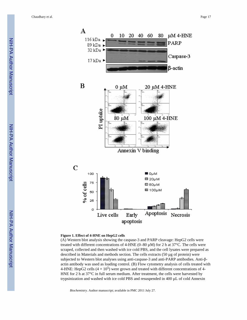
Figure 1. Effect of 4-HNE on HepG2 cells(A) Western blot analysis showing the caspase-3 and PARP cleavage: HepG2 cells weretreated with different concentrations of 4-HNE (0–80 µM) for 2 h at 37°C. The cells werescraped, collected and then washed with ice cold PBS, and the cell lysates were prepared asdescribed in Materials and methods section. The cells extracts (50 µg of protein) weresubjected to Western blot analyses using anti-caspase-3 and anti-PARP antibodies. Anti-β-actin antibody was used as loading control. (B) Flow cytometry analysis of cells treated with4-HNE: HepG2 cells (4 × 10⁵) were grown and treated with different concentrations of 4-HNE for 2 h at 37°C in full serum medium. After treatment, the cells were harvested bytrypsinization and washed with ice cold PBS and resuspended in 400 µL of cold Annexin
Chaudhary et al. Page 17
Biochemistry. Author manuscript; available in PMC 2011 July 27.
NIH
-PA Author Manuscript
NIH
-PA Author Manuscript
NIH
-PA Author Manuscript
binding buffer containing 5 µL of Annexin V-FITC and 5 µL of 0.1 mg/mL propidiumiodide. The cells were incubated at room temperature for 10 min. in the dark and wereanalyzed by flow cytometry using a Beckman Coulter Cytomics FC500 flow cytometer. (C)Bar graph showing the percent of cells present in apoptosis vs necrosis of the panel B.
Chaudhary et al. Page 18
Biochemistry. Author manuscript; available in PMC 2011 July 27.
NIH
-PA Author Manuscript
NIH
-PA Author Manuscript
NIH
-PA Author Manuscript
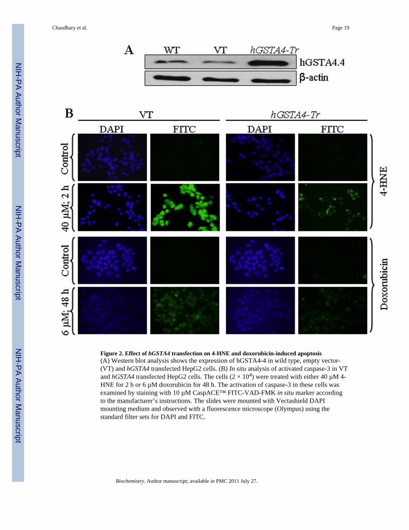
Figure 2. Effect of hGSTA4 transfection on 4-HNE and doxorubicin-induced apoptosis(A) Western blot analysis shows the expression of hGSTA4-4 in wild type, empty vector-(VT) and hGSTA4 transfected HepG2 cells. (B) In situ analysis of activated caspase-3 in VTand hGSTA4 transfected HepG2 cells. The cells (2 × 10⁴) were treated with either 40 µM 4-HNE for 2 h or 6 µM doxorubicin for 48 h. The activation of caspase-3 in these cells wasexamined by staining with 10 µM CaspACE™ FITC-VAD-FMK in situ marker accordingto the manufacturer’s instructions. The slides were mounted with Vectashield DAPImounting medium and observed with a fluorescence microscope (Olympus) using thestandard filter sets for DAPI and FITC.
Chaudhary et al. Page 19
Biochemistry. Author manuscript; available in PMC 2011 July 27.
NIH
-PA Author Manuscript
NIH
-PA Author Manuscript
NIH
-PA Author Manuscript
Figure 3. Effect of 4-HNE on Fas mediated apoptosis in HepG2 cells(A) In situ detection of Fas-mediated apoptosis in HepG2 cells. 2 × 10⁴ cells were grown onglass cover slides and after pretreatment with or without antagonistic anti-Fas (B-10)antibodies for 2 h (250 ng/mL) HepG2 cells were treated with 0, 20 and 40 µM 4-HNE for 2h followed by the addition of in situ caspase marker and fixation. The slides were mountedwith Vectashield DAPI mounting medium and observed under a fluorescence microscope(Olympus). The photographs were taken at 400× magnification. (B) The cells were treatedwith 0, 5, 10, 20 and 40 µM 4-HNE for 2 h at 37°C. Total protein lysates (30 µg) wereanalyzed by western blotting for ASK1, p-ASK1 (Thr845), p-SEK1 (Thr261), and p-JNK(Thr183/Tyr185). β-actin was used as a loading control.
Chaudhary et al. Page 20
Biochemistry. Author manuscript; available in PMC 2011 July 27.
NIH
-PA Author Manuscript
NIH
-PA Author Manuscript
NIH
-PA Author Manuscript
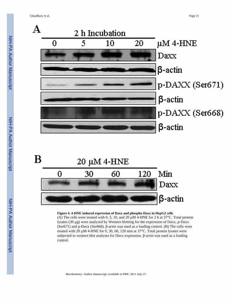
Figure 4. 4-HNE induced expression of Daxx and phospho-Daxx in HepG2 cells(A) The cells were treated with 0, 5, 10, and 20 µM 4-HNE for 2 h at 37°C. Total proteinlysates (30 µg) were analyzed by Western blotting for the expression of Daxx, p-Daxx(Ser671) and p-Daxx (Ser668). β-actin was used as a loading control. (B) The cells weretreated with 20 µM 4-HNE for 0, 30, 60, 120 min at 37°C. Total protein lysates weresubjected to western blot analyses for Daxx expression. β-actin was used as a loadingcontrol.
Chaudhary et al. Page 21
Biochemistry. Author manuscript; available in PMC 2011 July 27.
NIH
-PA Author Manuscript
NIH
-PA Author Manuscript
NIH
-PA Author Manuscript
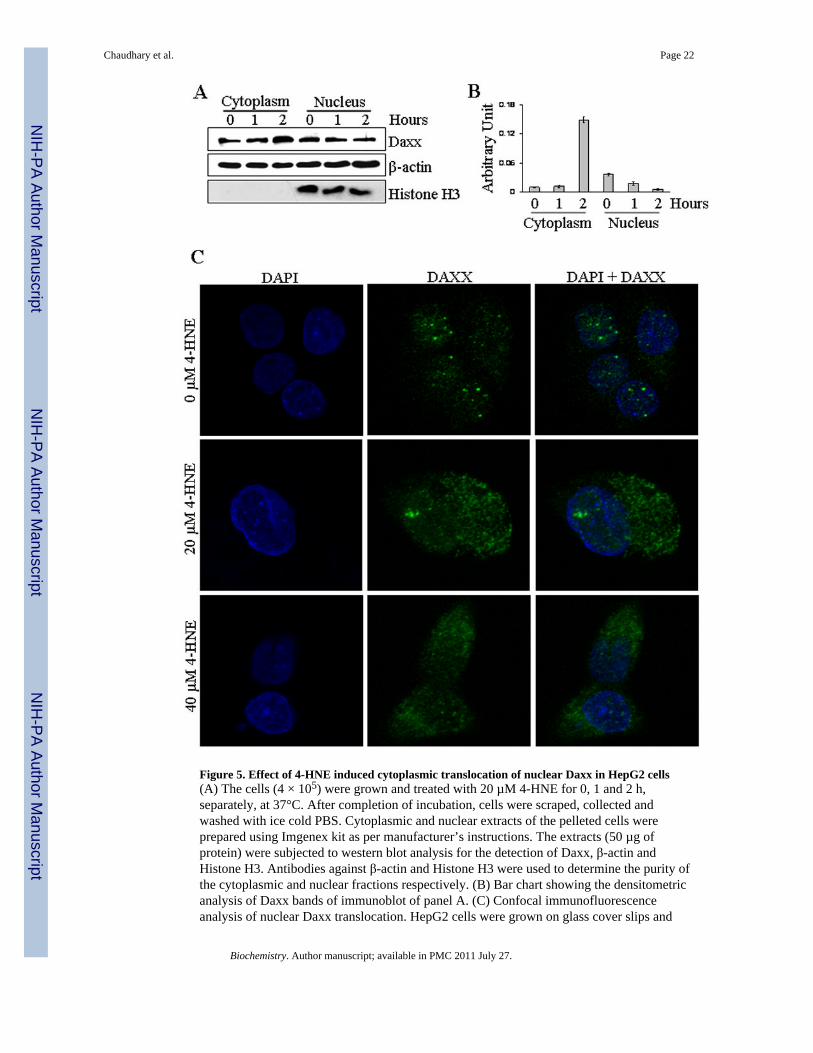
Figure 5. Effect of 4-HNE induced cytoplasmic translocation of nuclear Daxx in HepG2 cells(A) The cells (4 × 10⁵) were grown and treated with 20 µM 4-HNE for 0, 1 and 2 h,separately, at 37°C. After completion of incubation, cells were scraped, collected andwashed with ice cold PBS. Cytoplasmic and nuclear extracts of the pelleted cells wereprepared using Imgenex kit as per manufacturer’s instructions. The extracts (50 µg ofprotein) were subjected to western blot analysis for the detection of Daxx, β-actin andHistone H3. Antibodies against β-actin and Histone H3 were used to determine the purity ofthe cytoplasmic and nuclear fractions respectively. (B) Bar chart showing the densitometricanalysis of Daxx bands of immunoblot of panel A. (C) Confocal immunofluorescenceanalysis of nuclear Daxx translocation. HepG2 cells were grown on glass cover slips and
Chaudhary et al. Page 22
Biochemistry. Author manuscript; available in PMC 2011 July 27.
NIH
-PA Author Manuscript
NIH
-PA Author Manuscript
NIH
-PA Author Manuscript
untreated and treated (20 µM 4-HNE for 2 h) cells were fixed, permeabilized and incubatedwith polyclonal anti-Daxx antibody, followed by fluorescein (FITC) conjugated secondaryantibody. DAPI staining shows the nucleus. Slides were analyzed using Zeiss LSM510META laser scanning fluorescence microscope.
Chaudhary et al. Page 23
Biochemistry. Author manuscript; available in PMC 2011 July 27.
NIH
-PA Author Manuscript
NIH
-PA Author Manuscript
NIH
-PA Author Manuscript
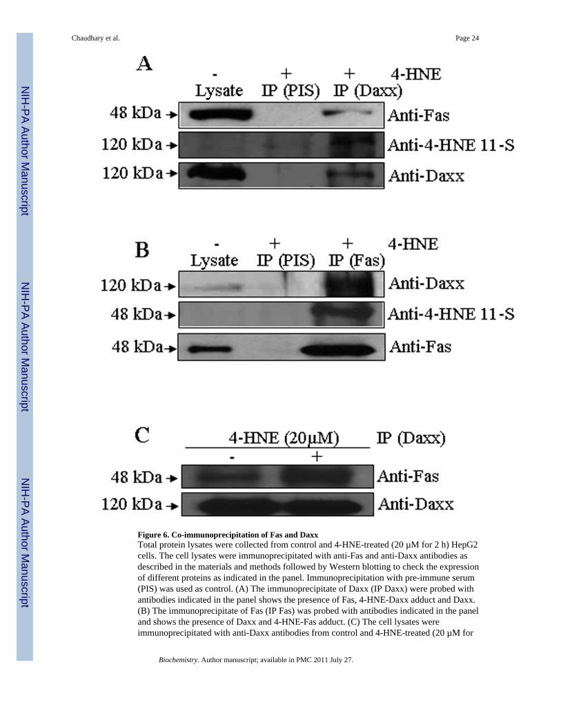
Figure 6. Co-immunoprecipitation of Fas and DaxxTotal protein lysates were collected from control and 4-HNE-treated (20 µM for 2 h) HepG2cells. The cell lysates were immunoprecipitated with anti-Fas and anti-Daxx antibodies asdescribed in the materials and methods followed by Western blotting to check the expressionof different proteins as indicated in the panel. Immunoprecipitation with pre-immune serum(PIS) was used as control. (A) The immunoprecipitate of Daxx (IP Daxx) were probed withantibodies indicated in the panel shows the presence of Fas, 4-HNE-Daxx adduct and Daxx.(B) The immunoprecipitate of Fas (IP Fas) was probed with antibodies indicated in the paneland shows the presence of Daxx and 4-HNE-Fas adduct. (C) The cell lysates wereimmunoprecipitated with anti-Daxx antibodies from control and 4-HNE-treated (20 µM for
Chaudhary et al. Page 24
Biochemistry. Author manuscript; available in PMC 2011 July 27.
NIH
-PA Author Manuscript
NIH
-PA Author Manuscript
NIH
-PA Author Manuscript
2 h) HepG2 cells followed by Western blotting to check the expression of Fas and Daxxproteins as indicated in the panel.
Chaudhary et al. Page 25
Biochemistry. Author manuscript; available in PMC 2011 July 27.
NIH
-PA Author Manuscript
NIH
-PA Author Manuscript
NIH
-PA Author Manuscript
Figure 7. Silencing of Daxx expression in HepG2 cells by siRNA and potentiation of apoptosis by4-HNE and CH11 antibody(A) Silencing of Daxx was performed by the ON-TARGET plus SMART pool Daxx siRNAas per the manufacturer’s instructions (Thermo Scientific Dharmacon) and control cells weretreated with ON-TARGET plus Non-targeting siRNA in a similar way. After transfection,the cells were harvested after 48 h and the expression level of Daxx was examined byWestern blot analysis. (B) Western blot analysis showing the enhanced activation ofcaspase-3 and JNK in Daxx depleted cells after 4-HNE treatment (20 µM HNE for 2 h). (C)Enhanced activation of apoptosis was also performed by In Situ immunofluorescence study.The cells (2 × 10⁵) were grown on glass cover slips in twelve-well plate and transfected with
Chaudhary et al. Page 26
Biochemistry. Author manuscript; available in PMC 2011 July 27.
NIH
-PA Author Manuscript
NIH
-PA Author Manuscript
NIH
-PA Author Manuscript
non targeting siRNA or Daxx siRNA. After 48 h of siRNA transfection, the cells weretreated with either 50 µg/well Fas-agonistic antibodies (CH11) or 20 µM 4-HNE for 2 h at37°C. The activation of caspase-3 in these cells was examined by CaspACE FITC-VAD-FMK in situ marker as per the manufacturer’s instructions and then observed under afluorescence microscope.
Chaudhary et al. Page 27
Biochemistry. Author manuscript; available in PMC 2011 July 27.
NIH
-PA Author Manuscript
NIH
-PA Author Manuscript
NIH
-PA Author Manuscript
Chaudhary et al. Page 28
Biochemistry. Author manuscript; available in PMC 2011 July 27.
NIH
-PA Author Manuscript
NIH
-PA Author Manuscript
NIH
-PA Author Manuscript
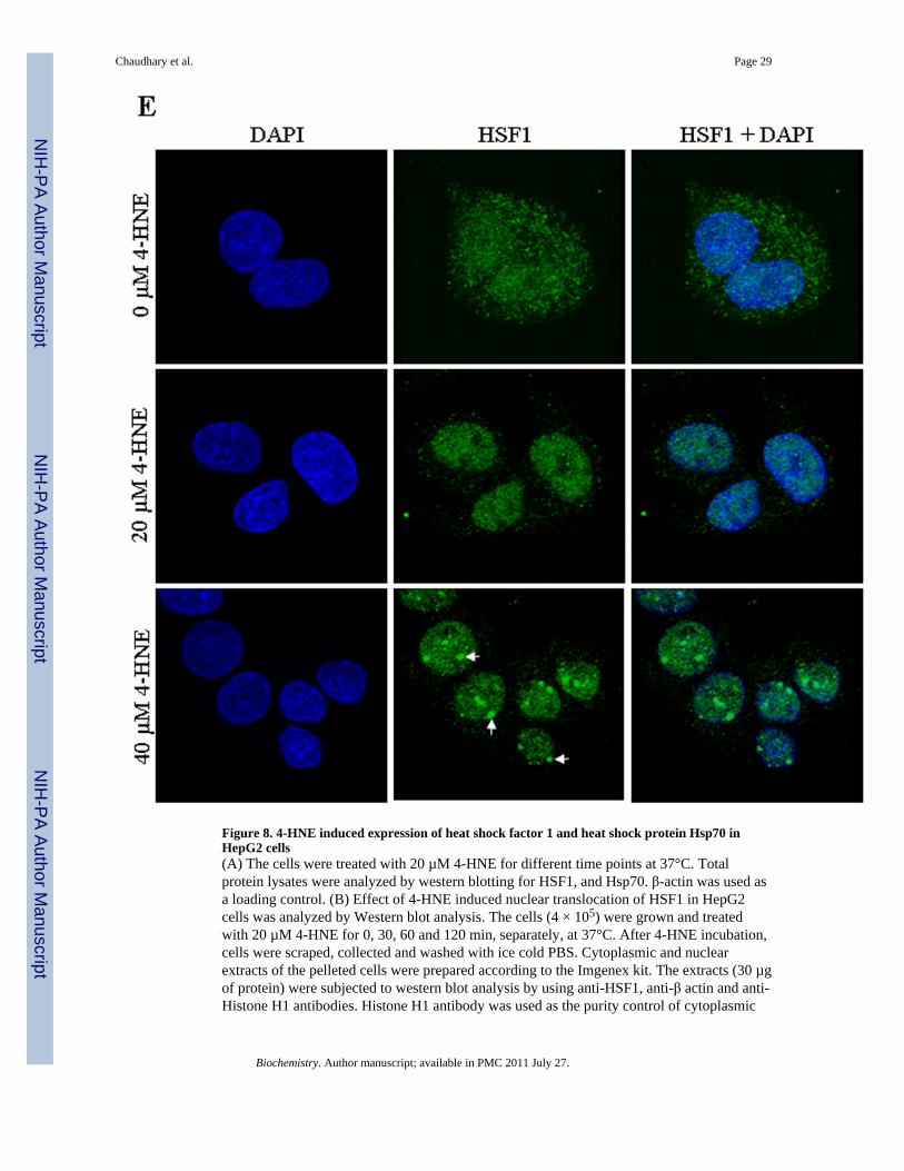
Figure 8. 4-HNE induced expression of heat shock factor 1 and heat shock protein Hsp70 inHepG2 cells(A) The cells were treated with 20 µM 4-HNE for different time points at 37°C. Totalprotein lysates were analyzed by western blotting for HSF1, and Hsp70. β-actin was used asa loading control. (B) Effect of 4-HNE induced nuclear translocation of HSF1 in HepG2cells was analyzed by Western blot analysis. The cells (4 × 10⁵) were grown and treatedwith 20 µM 4-HNE for 0, 30, 60 and 120 min, separately, at 37°C. After 4-HNE incubation,cells were scraped, collected and washed with ice cold PBS. Cytoplasmic and nuclearextracts of the pelleted cells were prepared according to the Imgenex kit. The extracts (30 µgof protein) were subjected to western blot analysis by using anti-HSF1, anti-β actin and anti-Histone H1 antibodies. Histone H1 antibody was used as the purity control of cytoplasmic
Chaudhary et al. Page 29
Biochemistry. Author manuscript; available in PMC 2011 July 27.
NIH
-PA Author Manuscript
NIH
-PA Author Manuscript
NIH
-PA Author Manuscript
and nuclear fractions. (C) Bar chart showing the densitometric analysis of HSF1 bands ofimmunoblot of panel B. (D) Chromatin binding of HSF1 by ChIP assay. HepG2 cells (4 ×10⁵) were grown and treated with 20 µM 4-HNE for 2 h followed by fixing with 1%formaldehyde for 10 min. Cells were scraped, and the chromatin was sheared by theprotocol given under materials and methods. The chromatin was immunoprecipitated usingthe negative control IgG (provided by Active Motif), positive control IgG (provided byActive Motif), and HSF1 IgG (Santa Cruz, CA) followed by binding with protein G beads.The chromatin was eluted from the protein G beads and was amplified by PCR using thecontrol primers as well as negative control primers (provided by Active Motif) and hHsp70primers. PCR products were analyzed by running on 1% agarose gel. (E) Confocalimmunofluorescence analysis of HSF1 translocation. HepG2 cells were grown on glasscover slips and untreated and treated (20 or 40 µM 4-HNE for 2 h) cells were fixed,permeabilized and incubated with polyclonal anti-HSF1 antibody, followed by fluorescein(FITC) conjugated secondary antibody. DAPI staining shows the nucleus. Slides wereanalyzed using Zeiss LSM 510META laser scanning fluorescence microscope.
Chaudhary et al. Page 30
Biochemistry. Author manuscript; available in PMC 2011 July 27.
NIH
-PA Author Manuscript
NIH
-PA Author Manuscript
NIH
-PA Author Manuscript
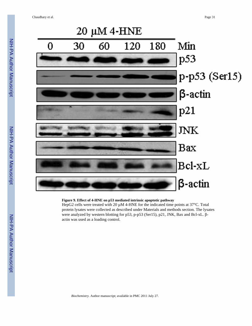
Figure 9. Effect of 4-HNE on p53 mediated intrinsic apoptotic pathwayHepG2 cells were treated with 20 µM 4-HNE for the indicated time points at 37°C. Totalprotein lysates were collected as described under Materials and methods section. The lysateswere analyzed by western blotting for p53, p-p53 (Ser15), p21, JNK, Bax and Bcl-xL. β-actin was used as a loading control.
Chaudhary et al. Page 31
Biochemistry. Author manuscript; available in PMC 2011 July 27.
NIH
-PA Author Manuscript
NIH
-PA Author Manuscript
NIH
-PA Author Manuscript
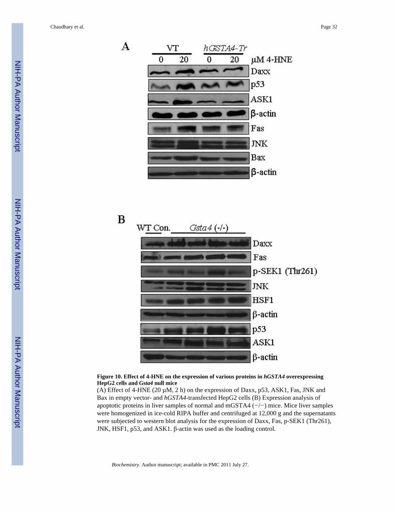
Figure 10. Effect of 4-HNE on the expression of various proteins in hGSTA4 overexpressingHepG2 cells and Gsta4 null mice(A) Effect of 4-HNE (20 µM, 2 h) on the expression of Daxx, p53, ASK1, Fas, JNK andBax in empty vector- and hGSTA4-transfected HepG2 cells (B) Expression analysis ofapoptotic proteins in liver samples of normal and mGSTA4 (−/−) mice. Mice liver sampleswere homogenized in ice-cold RIPA buffer and centrifuged at 12,000 g and the supernatantswere subjected to western blot analysis for the expression of Daxx, Fas, p-SEK1 (Thr261),JNK, HSF1, p53, and ASK1. β-actin was used as the loading control.
Chaudhary et al. Page 32
Biochemistry. Author manuscript; available in PMC 2011 July 27.
NIH
-PA Author Manuscript
NIH
-PA Author Manuscript
NIH
-PA Author Manuscript
NIH
-PA Author Manuscript
NIH
-PA Author Manuscript
NIH
-PA Author Manuscript
Chaudhary et al. Page 33
Table 1
Binding sites of 4-HNE in the amino acid sequence of Daxx identified by LC-MS/MS.
Peptide Sequence Modifications*
H*SLGLPR 4-HNE (+156)
LQGTSSH*SADTPEASLDSGEGPSGMASQGCPSASRa 4-HNE (+156)
VDSPSH*GLVTSSLCIPSPARa 4-HNE (+156)
LSQTPH*SQPPRPGTCKa 4-HNE (+156)
aMethionine (M) oxidation and cysteine (C) carbamidomethylation also occurred and introduced, respectively, during sample preparation for LC-
MS/MS analysis, when these residues were present. These modifications were, therefore, considered irrelevant from the study’s point of view andnot indicated.
Biochemistry. Author manuscript; available in PMC 2011 July 27.


