Embed Size (px)
Citation preview
97 (2006) 249–261www.elsevier.com/locate/ydbio
Developmental Biology 2
Mesodermal and neuronal retinoids regulate the induction and maintenanceof limb innervating spinal motor neurons
Sheng-Jian Ji a,1, BinQuan Zhuang a,1, Crystal Falco a, André Schneider b,2,Karin Schuster-Gossler b, Achim Gossler b, Shanthini Sockanathan a,⁎
a Department of Neuroscience, Johns Hopkins University School of Medicine, 725 N Wolfe Street, Baltimore, MD 21205, USAb Institute for Molecular Biology OE5250, Medizinische Hochschule Hannover, Carl-Neuberg-Str. 1, D-30625 Hannover, Germany
Received for publication 4 March 2006; revised 13 April 2006; accepted 11 May 2006Available online 19 May 2006
Abstract
During embryonic development, the generation, diversification and maintenance of spinal motor neurons depend upon extrinsic signals thatare tightly regulated. Retinoic acid (RA) is necessary for specifying the fates of forelimb-innervating motor neurons of the Lateral MotorColumn (LMC), and the specification of LMC neurons into medial and lateral subtypes. Previous studies implicate motor neurons as therelevant source of RA for specifying lateral LMC fates at forelimb levels. However, at the time of LMC diversification, a significant amountof retinoids in the spinal cord originates from the adjacent paraxial mesoderm. Here we employ mouse genetics to show that RA derivedfrom the paraxial mesoderm is required for lateral LMC induction at forelimb and hindlimb levels, demonstrating that mesodermallysynthesized RA functions as a second source of signals to specify lateral LMC identity. Furthermore, reduced RA levels in postmitotic motorneurons result in a decrease of medial and lateral LMC neurons, and abnormal axonal projections in the limb; invoking additional roles forneuronally synthesized RA in motor neuron maintenance and survival. These findings suggest that during embryogenesis, mesodermal andneuronal retinoids act coordinately to establish and maintain appropriate cohorts of spinal motor neurons that innervate target muscles in thelimb.© 2006 Elsevier Inc. All rights reserved.
Keywords: Retinoids; Motor neuron; Specification; Maintenance
Introduction
The generation and maintenance of different neuronalsubtypes during embryonic development are essential eventsin the establishment of a functional nervous system. Invertebrates, this complex process involves the implementationof cell-intrinsic events by extrinsic signals emanating from localcellular sources (Jessell and Melton, 1992; Liu and Joyner,2001). In many cases, inductive signals are expressed both bycells adjacent to the nervous system, and by cells resident withinthe nervous system itself (Jessell, 2000). However, few studies
⁎ Corresponding author.1 These authors contributed equally to this work.2 Present address: Max-Planck-Institut für Herz- und Lungenforschung
(K. Schuster-Gossler), Parkstr. 1, 61231 Bad Nauheim, Germany.
0012-1606/$ - see front matter © 2006 Elsevier Inc. All rights reserved.doi:10.1016/j.ydbio.2006.05.015
have examined the relative contributions of these differentsources towards specifying neuronal fates, or if there are laterrequirements for these signals in maintaining neuronal numbers.
One model system where the expression of extrinsic factorsknown to be involved in neuronal fate specification isjuxtaposed in adjacent tissues is in developing spinal motorneurons (Jessell, 2000). All motor neurons derive from a distinctventral progenitor domain, set up in part by sonic hedgehogsecreted by the notochord and floorplate (Jessell, 2000;Shirasaki and Pfaff, 2002; Price and Briscoe, 2004). The cellbodies of motor neurons are organized into distinct columnsdefined by their position along the rostral–caudal axis and theirstereotypic axonal projections in the periphery (Landmesser,1978; Hollyday, 1980a,b; Gutman et al., 1993). Motor neuronsof the medial Median Motor Column (MMC) are found at allaxial levels whereas lateral MMC neurons and preganglionic
250 S.-J. Ji et al. / Developmental Biology 297 (2006) 249–261
autonomic motor neurons are located specifically in thoracicregions (Prasad and Hollyday, 1991; Gutman et al., 1993;Cornbrooks et al., 1997). Motor neurons of the Lateral MotorColumn (LMC) are generated exclusively at forelimb andhindlimb levels and form medial (LMCm) and lateral (LMCl)divisional subtypes which project to ventral and dorsal musclesin the limb, respectively (Landmesser, 1978; Hollyday, 1980a,b;Tosney and Landmesser, 1985a,b). Individual motor columnsand divisions can be molecularly defined by the combinatorialexpression of LIM homeodomain protein family members(Tsuchida et al., 1994). These transcription factors functionallycontribute to the settling patterns of motor neuron cell bodies andtheir axonal projection profiles (Pfaff et al., 1996; Sharma et al.,1998, 2000; Kania et al., 2000).
Biochemical studies, combined with analyses of reportermice sensitive to activated retinoid receptors, demonstrate thatretinoids are present within the developing spinal cord at thetime of motor neuron generation and specification; suggesting arequirement for RA signaling during these events (Rossant et al.,1991; McCaffery and Drager, 1994; Solomin et al., 1998).Recently, work in chicks and Vitamin A deficient quailsdescribes critical roles for RA signaling in establishing andgenerating motor neuron progenitors, and in mediating theirsubsequent differentiation into postmitotic motor neurons(Novitch et al., 2003; Diez del Corral et al., 2003; Wilson et al.,2004). Remarkably, retinoids can also influence the fates ofmotor neurons postmitotically. When dominant-negative reti-noid receptors are introduced into postmitotic spinal motorneurons, forelimb level motor neurons do not acquire LMCsubtype identities but instead adopt thoracic motor columnproperties (Sockanathan et al., 2003). This suggests thatretinoid signaling imposes forelimb LMC identities partlythrough the suppression of thoracic fates. Surprisingly,hindlimb LMC formation is not perturbed, suggesting thatlumbar LMC identity may be specified by alternative mecha-nisms (Sockanathan et al., 2003). In addition to influencingcolumnar identity, retinoids are also required for the divisionalspecification of LMC neurons. Experiments in the chick showthat retinoids can induce LMCl neurons and thus contribute tothe diversification of LMC motor neurons into medial andlateral subtypes. Further, retinoid signals are also required forthe maintenance of LMCl properties during the migration ofthese neurons to their final settling position (Sockanathan andJessell, 1998).
What is the source of retinoids responsible for these criticalevents in motor neuron specification and maintenance?Biochemical, gene expression and genetic studies have shownthat retinaldehyde dehydrogenase 2 (RALDH2) is the majorretinoid synthetic enzyme present during embryonic develop-ment (Zhao et al., 1996; Niederreither et al., 1997, 1999;Swindell et al., 1999; Mic et al., 2002). RALDH2 is found in themeninges surrounding the neural tube, and is dynamicallyexpressed within the paraxial mesoderm directly adjacent to thespinal cord. Initially, paraxial mesoderm-specific expression ofRALDH2 is found at all axial levels; however, higher levels ofexpression are subsequently concentrated at forelimb regions(Niederreither et al., 1997; Berggren et al., 1999; Swindell et al.,
1999; Blentic et al., 2003). At later stages of development,RALDH2 is detected within the spinal cord and is localized toLMC motor neurons (Zhao et al., 1996; Sockanathan andJessell, 1998). Studies of Raldh2 null embryos rescued fromearly lethality by maternally administered RA confirm thatmesodermal and neuronal RALDH2 expression are primarilyresponsible for retinoid levels within the spinal cord (Mic et al.,2002; Niederreither et al., 2002). Given that RALDH2expression prior to LMC generation is largely mesodermal,both motor neuron generation and LMC formation are likelydependent on paraxial mesoderm-derived retinoid signaling.In contrast, the specification and maintenance of LMCl neuronsare believed to rely on retinoids generated by early born LMCmneurons. However, at the time of LMC diversification intomedial and lateral subtypes, retinoids in the spinal cord derivefrom both paraxial mesoderm and motor neuron sites ofRALDH2 expression (Niederreither et al., 1997, 2002; Berggrenet al., 1999; Swindell et al., 1999; Mic et al., 2002; Blentic et al.,2003).
Attempts to define the specific contributions of each retinoidsource to LMC subtype specification and LMC maintenancehave been stymied by the early lethality of Raldh2 null embryosprior to motor neuron generation (Niederreither et al., 1999). Arecent study where Raldh2 expression was genetically ablatedin forelimb spinal motor neurons led to a decrease of forelimbLMCl neurons, supporting the idea that motor neuron sources ofRA are partly required for LMCl specification (Vermot et al.,2005, Fig. 8A). The homeobox protein Hoxc8 is thought tomediate this process as Hoxc8 expression is lost in LMCneurons in mice lacking motor neuron-specific expression ofRALDH2, and there are reduced numbers, and aberrant axonalprojections of remaining LMCl cells in Hoxc8 mutants (Vermotet al., 2005). However, one striking observation from thesestudies is that 80% of LMCl neurons are preserved in theabsence of motor neuron sources of RA. Given that significantamounts of RA within the spinal cord derive from RALDH2activity in the adjacent paraxial mesoderm, it is possible that RAsynthesized in the paraxial mesoderm provides an importantsource of retinoids for specifying LMCl identity (Mic et al.,2002; Niederreither et al., 2002). Moreover, the contribution ofmesodermal and neuronally synthesized retinoids to hindlimbLMC development and maintenance of LMC neurons respec-tively remains unknown.
Here we have used genetic approaches to independentlyreduce RALDH2 levels in paraxial mesoderm and in spinalmotor neurons at forelimb and hindlimb levels of the spinal cordto examine the requirements for mesodermal and neuronalretinoids in LMC specification and maintenance. We find thatreduction of RALDH2 in the paraxial mesoderm results in thespecific decrease of both forelimb and hindlimb LMCl neurons,whereas LMCm neurons are unaffected. Thus, RA derived fromthe mesoderm functions as an additional source of signals tospecify LMCl fates (Fig. 8B). In contrast, knockdown ofRALDH2 in postmitotic motor neurons results in reducednumbers of forelimb and hindlimb LMCm and LMCl neuronslater in development, and the atrophy of remaining limb-innervating neurons. This result suggests that continued RA
251S.-J. Ji et al. / Developmental Biology 297 (2006) 249–261
synthesis in motor neurons may be critical for maintenance ofLMC neuronal numbers and properties (Fig. 8C). Given that themajority of remaining forelimb LMC neurons lose Hoxc8expression, we suggest that Hox proteins may partly mediate theretinoid-dependent maintenance of LMC neurons. Takentogether, these genetic studies uncover new roles for meso-dermal and neuronal retinoids in the specification, maintenanceand survival of spinal motor neurons that target the limb.
Materials and methods
Generation of mouse lines
Raldh2lox line: Mouse genomic clones were isolated from a 129/Sv genomiclibrary (Stratagene) and loxP sites introduced into the NsiI andKpnI sites located5′ and 3′ of Exon 4. Introduction of a thymidine kinase promoter neomycin/hygromycin cassette into the KpnI site in the reverse direction resulted in anadditional EcoRI site within the Raldh2 locus, generating an 11 kb EcoRI bandwhen homologously recombined instead of the wild-type 14 kb band. A 2 kbKpnI fragment outside the targeting construct was used as a 3′ probe. ES celltransfections, screening and injection into blastocysts were carried out accordingto standard procedures. Raldh2+/− lines were generated by integrating a pgkneomycin cassette within the ClaI site of Exon 4. Details for msd:Cre mice areavailable by request fromA. Gossler. Specific primers and PCR protocols used togenotype Cre, Raldh2+/− and Raldh2lox alleles are available upon request.Gtrosa26tm1Sor mice were obtained from Jackson Laboratories. RARE-hspLacZmice engineered by Rossant et al. (1991) were obtained from C. Mendelsohnwith the permission of J. Rossant.
Production of RALDH2 antibodies
A C-terminal mouse RALDH2 peptide (SEVKTVTVKIPQKNS) wascoupled to keyhole limpet hemocyanin and injected into guinea pigs (Covance).Sera were affinity purified using a peptide column (ABR).
In situ hybridization and immunohistochemistry
In situ hybridization was performed as described by Schaeren-Wiemers andGerfin-Moser (1993). The mouse Raldh2 in situ probe consisted of a 937 bpfragment from the ATG. Immunohistochemistry on sections and whole mountswas performed as described in Sockanathan and Jessell (1998) and Giger et al.(2000). Images were acquired using Zeiss LSM 510 or LSM 5 PASCALconfocal microscopes. Primary antibodies were used at the following dilutions:K5 (rabbit anti-Isl1/2), 1:3000; guinea pig anti-Isl1/2, 1:20000; rabbit anti-Pea3,1:1000 (Arber et al., 1999); rabbit anti-Lhx3, 1:3000 (Sharma et al., 1998); 4F2(mouse anti-Lim1/2) 1:2, mouse anti-Hoxc8 1:200 (all provided by T.M. Jessell)and 2H3 (anti-neurofilament) 1:50 (Developmental Studies Hybridoma Bank,University of Iowa); guinea pig anti-RALDH2, 1:20000; rabbit anti-RALDH2,1:2000 (provided by P. McCaffery); rabbit anti-LacZ, 1:1000 (Cappel, Durham,NC). Beta-galactosidase staining on tissue sections was carried out usingstandard protocols.
Motor neuron counts
Motor neuron counts were confined to the limb regions corresponding to theentire rostrocaudal extent of the LMC, identified by Isl2+/Lim1+ neurons. Neuronsin approximately 15–18 sections/embryo were counted, depending on develo-pmental stage. Typically 3–6 embryos were analyzed for each experiment.
Western blots
Western blots were performed according to standard protocols and bandswere visualized using ECL Plus™ Western Blotting Detection Reagents (GEHealthcare). Densitometry analysis was carried out using ImageJ (NIH). Rabbitanti-RALDH2 antibodies were used at a 1:5000 dilution.
RA bioassay
The RA reporter cell line F9-RARE-lacZ (Sil-15) was maintained accordingto standard protocols (Wagner et al., 1992). Tissue preparation and reporterassays were carried out as described by Haselbeck et al. (1997).
Results
Generation of Raldh2 conditional knockout mice
To examine the contribution of mesodermal and neuronalretinoids to LMC specification, we used Cre-lox technology(Rajewsky et al., 1996) to specifically ablate RALDH2 in eitherthe paraxial mesoderm or in spinal motor neurons. Fig. S1 showsthe targeting strategy used to introduce lox-P sites flanking exon4 of one Raldh2 allele in the mouse genome (Raldh2lox). Exon 4was chosen as it encodes the nicotinamide adenine dinucleotide(NAD) binding domain essential for RALDH2 activity; removalof this exon results in a non-functional RALDH2 enzyme(Niederreither et al., 1999; Vermot et al., 2003, 2005).Homozygous Raldh2lox/lox mice were crossed to mice thatcontained a single Raldh2 allele inactivated by insertion of theneomycin gene within exon 4 to generate Raldh2lox/− lines(Raldh2/−, Fig. S1). Raldh2lox/− mice were viable, fertile andmorphologically indistinguishable from heterozygote litter-mates, in contrast to Raldh2−/− mice which die at embryonicday post-coitum 9 (E9).
Targeted loss of Raldh2 in the paraxial mesoderm
In order to generate embryos with reduced RALDH2 in theparaxial mesoderm, we utilized transgenic mice that expressedCre recombinase under the control of the “msd” fragment fromthe Delta1 promoter (msd:Cre), which drives heterologous geneexpression in the paraxial mesoderm from E7 (Beckers et al.,2000). We first monitored the profile of Cre recombinaseactivity in msd:Cre mice by breeding them with mouse reporterlines that express betagalactosidase upon Cre-mediated recom-bination (Gtrosa26tm1Sor; Soriano, 1999). Strong betagalactosi-dase staining is detected in forelimb level paraxial mesoderm atE9.5 of msd:Cre+ Gtrosa26tm1Sor embryos prior to spinal motorneuron generation, and is maintained until at least E12.5 (Figs.1A, E, Nornes and Carry, 1978). At these developmental stages,betagalactosidase activity within the spinal cord was localizedexclusivelywithin thevasculature.Thus,msd:Cre+RALDH2lox/−
embryos should lack RALDH2 in the paraxial mesoderm butpreserve LMC-specific expression of the enzyme.
Msd:Cre+ Raldh2lox/− embryos are morphologically normal,and survive to birth and adulthood. Embryos were examined atE10.0 and at E12.5 to determine the extent of RALDH2 proteinloss in the paraxial mesoderm at forelimb levels compared withRaldh2lox/− littermates. Forelimb embryonic regions wereserially sectioned and an antibody specific to RALDH2 wasused to detect the protein by indirect immunofluorescence.Mesodermal RALDH2 expression was significantly diminishedin msd:Cre+ Raldh2lox/− embryos compared with Raldh2lox/−
littermates at E10.0, a developmental time-point when
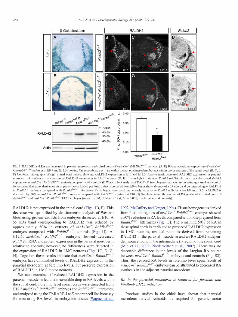
Fig. 1. RALDH2 and RA are decreased in paraxial mesoderm and spinal cords of msd:Cre+ RALDH2lox/− mutants. (A, E) Betagalactosidase expression of msd:Cre+
Gtrosa26tm1Sor embryos at E9.5 and E12.5 showing Cre recombinase activity within the paraxial mesoderm but not within motor neurons of the spinal cord. (B, C, E,F) Confocal micrographs of right spinal cord halves, showing RALDH2 expression at E10 and E12.5. Arrows mark decreased RALDH2 expression in paraxialmesoderm. Arrowheads mark preserved RALDH2 expression in LMC neurons. (D, H) In situ hybridization of Raldh2 mRNA. Arrows mark decreased Raldh2expression in msd:Cre+ RALDH2lox/− mutants compared with controls (I) Western blot analysis of RALDH2 in embryonic extracts. Actin staining is used as a controlfor ensuring that equivalent amounts of protein were loaded per lane. Extracts prepared from E9 embryos show absence of a 55 kDa band corresponding to RALDH2in Raldh2−/− embryos compared with Raldh2lox/lox littermates. E9 embryos were used due to early lethality of Raldh2 nulls between E9 and E9.5. RALDH2 isdecreased by 50% in msd:Cre+ Raldh2lox/− embryos compared with Raldh2lox/− controls at E10. (J) Graph depicting the amount of RA produced in spinal cords ofRaldh2lox/− and msd:Cre+ Raldh2lox/− E12.5 embryos (mean ± SEM, Student's t test, *P = 0.001, n = 8 mutants, 4 controls).
252 S.-J. Ji et al. / Developmental Biology 297 (2006) 249–261
RALDH2 is not expressed in the spinal cord (Figs. 1B, F). Thisdecrease was quantified by densitometric analysis of Westernblots using protein extracts from embryos dissected at E10. A55 kDa band corresponding to RALDH2 was reduced byapproximately 50% in extracts of msd:Cre+ Raldh2lox/−
embryos compared with Raldh2lox/− controls (Fig. 1I). AtE12.5, msd:Cre+ Raldh2lox/− embryos showed decreasedRaldh2mRNA and protein expression in the paraxial mesodermrelative to controls; however, no differences were detected inthe expression of RALDH2 in LMC neurons (Figs. 1C, D, G,H). Together, these results indicate that msd:Cre+ Raldh2lox/−
embryos have diminished levels of RALDH2 expression in theparaxial mesoderm at forelimb levels, but preserve expressionof RALDH2 in LMC motor neurons.
We next examined if reduced RALDH2 expression in theparaxial mesoderm led to a measurable drop in RA levels withinthe spinal cord. Forelimb level spinal cords were dissected fromE12.5msd:Cre+ Raldh2lox/− embryos and Raldh2lox/− littermates,and analyzed using the F9-RARE-LacZ reporter cell line bioassayfor measuring RA levels in embryonic tissues (Wagner et al.,
1992;McCaffery and Drager, 1994). Tissue homogenates derivedfrom forelimb regions of msd:Cre+ Raldh2lox/− embryos showeda 50% reduction in RA levels compared with those prepared fromRaldh2lox/− littermates (Fig. 1J). The remaining 50% of RA inthese spinal cords is attributed to preserved RALDH2 expressionin LMC neurons, residual retinoids derived from remainingRALDH2 in the paraxial mesoderm and an RALDH2-indepen-dent source found in the intermediate (i) region of the spinal cord(Mic et al., 2002; Niederreither et al., 2002). There was nodetectable difference in the levels of the i-region RA sourcebetween msd:Cre+ Raldh2lox/− embryos and controls (Fig. S2).Thus, the reduced RA levels in forelimb level spinal cords ofmsd:Cre+ Raldh2lox/− embryos can be attributed to decreased RAsynthesis in the adjacent paraxial mesoderm.
RA in the paraxial mesoderm is required for forelimb andhindlimb LMCl induction
Previous studies in the chick have shown that paraxialmesoderm-derived retinoids are required for generic motor
253S.-J. Ji et al. / Developmental Biology 297 (2006) 249–261
neuron generation (Diez del Corral et al., 2003; Novitch et al.,2003) and the specification of forelimb LMC neuronal identity(Sockanathan et al., 2003), both of which occur prior to thediversification of medial and lateral LMC neurons. Weexamined if either of these events was compromised in msd:Cre+ Raldh2lox/− embryos. We first quantified the number ofIsl1/2+ motor neurons at forelimb levels inmsd:Cre+ Raldh2lox/−
embryos compared to Raldh2lox/− littermates at E12.5, after thepeak of motor neuron generation (Nornes and Carry, 1978). Wedetected a small but statistically significant reduction ofapproximately 10% in total motor neuron numbers in msd:Cre+
Raldh2lox/− embryos compared with controls (Fig. S3). Further-more, the specification of LMC neuronal identity in msd:Cre+
Raldh2lox/− embryos appeared normal as RALDH2 expression inLMCneurons was not altered and these neurons had not acquiredthoracic motor column fates (data not shown).
If reduced RA levels in the spinal cord affect the generationof all motor neurons, the 10% loss of Isl1/2+ neurons should beevenly distributed among all forelimb spinal motor columns ofmsd:Cre+ Raldh2lox/− embryos. The number of Isl1/2+/Lhx3+
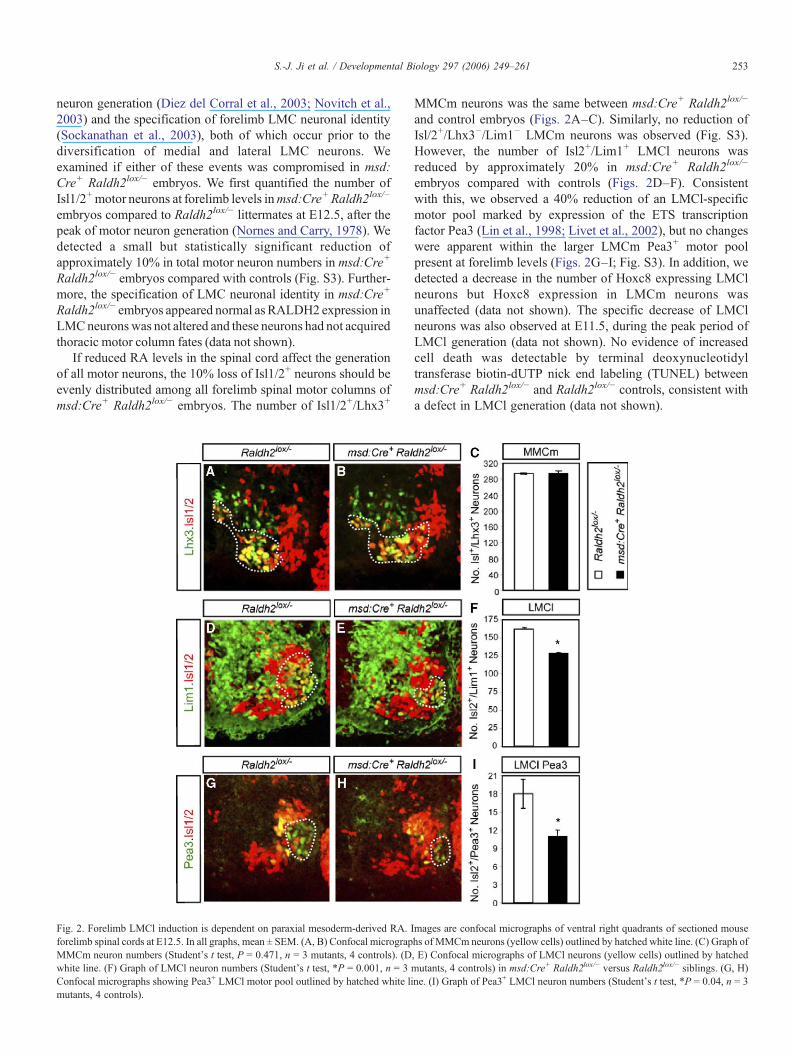
Fig. 2. Forelimb LMCl induction is dependent on paraxial mesoderm-derived RA. Iforelimb spinal cords at E12.5. In all graphs, mean ± SEM. (A, B) Confocal micrograpMMCm neuron numbers (Student's t test, P = 0.471, n = 3 mutants, 4 controls). (Dwhite line. (F) Graph of LMCl neuron numbers (Student's t test, *P = 0.001, n = 3Confocal micrographs showing Pea3+ LMCl motor pool outlined by hatched white limutants, 4 controls).
MMCm neurons was the same between msd:Cre+ Raldh2lox/−
and control embryos (Figs. 2A–C). Similarly, no reduction ofIsl/2+/Lhx3−/Lim1− LMCm neurons was observed (Fig. S3).However, the number of Isl2+/Lim1+ LMCl neurons wasreduced by approximately 20% in msd:Cre+ Raldh2lox/−
embryos compared with controls (Figs. 2D–F). Consistentwith this, we observed a 40% reduction of an LMCl-specificmotor pool marked by expression of the ETS transcriptionfactor Pea3 (Lin et al., 1998; Livet et al., 2002), but no changeswere apparent within the larger LMCm Pea3+ motor poolpresent at forelimb levels (Figs. 2G–I; Fig. S3). In addition, wedetected a decrease in the number of Hoxc8 expressing LMClneurons but Hoxc8 expression in LMCm neurons wasunaffected (data not shown). The specific decrease of LMClneurons was also observed at E11.5, during the peak period ofLMCl generation (data not shown). No evidence of increasedcell death was detectable by terminal deoxynucleotidyltransferase biotin-dUTP nick end labeling (TUNEL) betweenmsd:Cre+ Raldh2lox/− and Raldh2lox/− controls, consistent witha defect in LMCl generation (data not shown).
mages are confocal micrographs of ventral right quadrants of sectioned mousehs of MMCm neurons (yellow cells) outlined by hatched white line. (C) Graph of, E) Confocal micrographs of LMCl neurons (yellow cells) outlined by hatchedmutants, 4 controls) in msd:Cre+ Raldh2lox/− versus Raldh2lox/− siblings. (G, H)ne. (I) Graph of Pea3+ LMCl neuron numbers (Student's t test, *P = 0.04, n = 3
254 S.-J. Ji et al. / Developmental Biology 297 (2006) 249–261
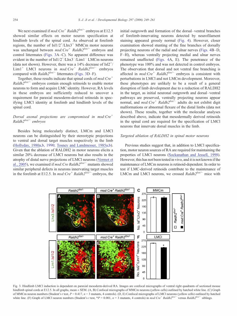
We next examined ifmsd:Cre+ Raldh2lox/− embryos at E12.5showed similar effects on motor neuron specification athindlimb levels of the spinal cord. As observed at forelimbregions, the number of Isl1/2+/Lhx3+ MMCm motor neuronswas unchanged between msd:Cre+ Raldh2lox/− embryos andcontrol littermates (Figs. 3A–C). No apparent difference wasevident in the number of Isl1/2+/Lhx3−/Lim1− LMCm neurons(data not shown). However, there was a 14% decrease of Isl2+/Lim1+ LMCl neurons in msd:Cre+ Raldh2lox/− embryoscompared with Raldh2lox/− littermates (Figs. 3D–F).
Together, these results indicate that spinal cords of msd:Cre+
Raldh2lox/− embryos contain enough retinoids to enable motorneurons to form and acquire LMC identity. However, RA levelsin these embryos are sufficiently reduced to uncover arequirement for paraxial mesoderm-derived retinoids in spec-ifying LMCl identity at forelimb and hindlimb levels of thespinal cord.
Dorsal axonal projections are compromised in msd:Cre+
Raldh2lox/− embryos
Besides being molecularly distinct, LMCm and LMClneurons can be distinguished by their stereotypic projectionsto ventral and dorsal target muscles respectively in the limb(Hollyday, 1980a,b, 1990; Tosney and Landmesser, 1985a,b).Given that the ablation of RALDH2 in motor neurons elicits asimilar 20% decrease of LMCl neurons but also results in theatrophy of distal nerve projections of LMCl neurons (Vermot etal., 2005), we examined if msd:Cre Raldh2lox/− mutants showedsimilar peripheral defects in neurons innervating target musclesin the forelimb at E12.5. In msd:Cre+ Raldh2lox/− embryos, the
Fig. 3. Hindlimb LMCl induction is dependent on paraxial mesoderm-derived RA.hindlimb spinal cords at E12.5. In all graphs, mean ± SEM. (A, B) Confocal micrograof MMCm neuron numbers (Student's t test, P = 0.417, n = 3 mutants, 4 controls). (Dwhite line. (F) Graph of LMCl neuron numbers (Student's t test, *P = 0.001, n = 3
initial outgrowth and formation of the dorsal–ventral branchesof forelimb-innervating neurons detected by neurofilamentstaining appeared grossly normal (Fig. 4). However, closerexamination showed stunting of the fine branches of dorsallyprojecting neurons of the radial and ulnar nerves (Figs. 4B–D,F–H), whereas ventrally projecting medial and ulnar nervesremained unaffected (Figs. 4A, E). The penetrance of thephenotype was 100% and was not detected in control embryos.The observation that dorsal and not ventral fine branching isaffected in msd:Cre+ Raldh2lox/− embryos is consistent withperturbations in LMCl and not LMCm development. Moreover,these phenotypes are unlikely to be a result of a generaldisruption of limb development due to a reduction of RALDH2in the target, as initial neuronal outgrowth and dorsal–ventralpathways are preserved, ventrally projecting neurons appearnormal, and msd:Cre+ Raldh2lox/− adults do not exhibit digitmalformations or abnormal flexure of the distal limbs (data notshown). These results, together with the molecular analysesdescribed above, indicate that mesodermally derived retinoidsin the spinal cord are required for the specification of LMClneurons that innervate dorsal muscles in the limb.
Targeted ablation of RALDH2 in spinal motor neurons
Previous studies suggest that, in addition to LMCl specifica-tion, motor neuron sources of RA are required for maintaining theproperties of LMCl neurons (Sockanathan and Jessell, 1998).However, this has not been tested in vivo, and it is not known if themaintenance of LMCm neurons is retinoid-dependent. In order totest if LMC-derived retinoids contribute to the maintenance ofLMCm and LMCl neurons, we crossed Raldh2lox/− mice with
Images are confocal micrographs of ventral right quadrants of sectioned mousephs of MMCm neurons (yellow cells) outlined by hatched white line. (C) Graph, E) Confocal micrographs of LMCl neurons (yellow cells) outlined by hatchedmutants, 4 controls) in msd:Cre+ Raldh2lox/− versus Raldh2lox/− siblings.
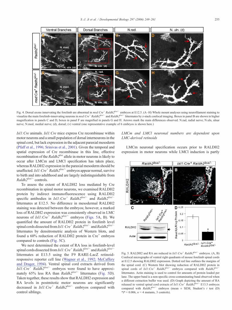
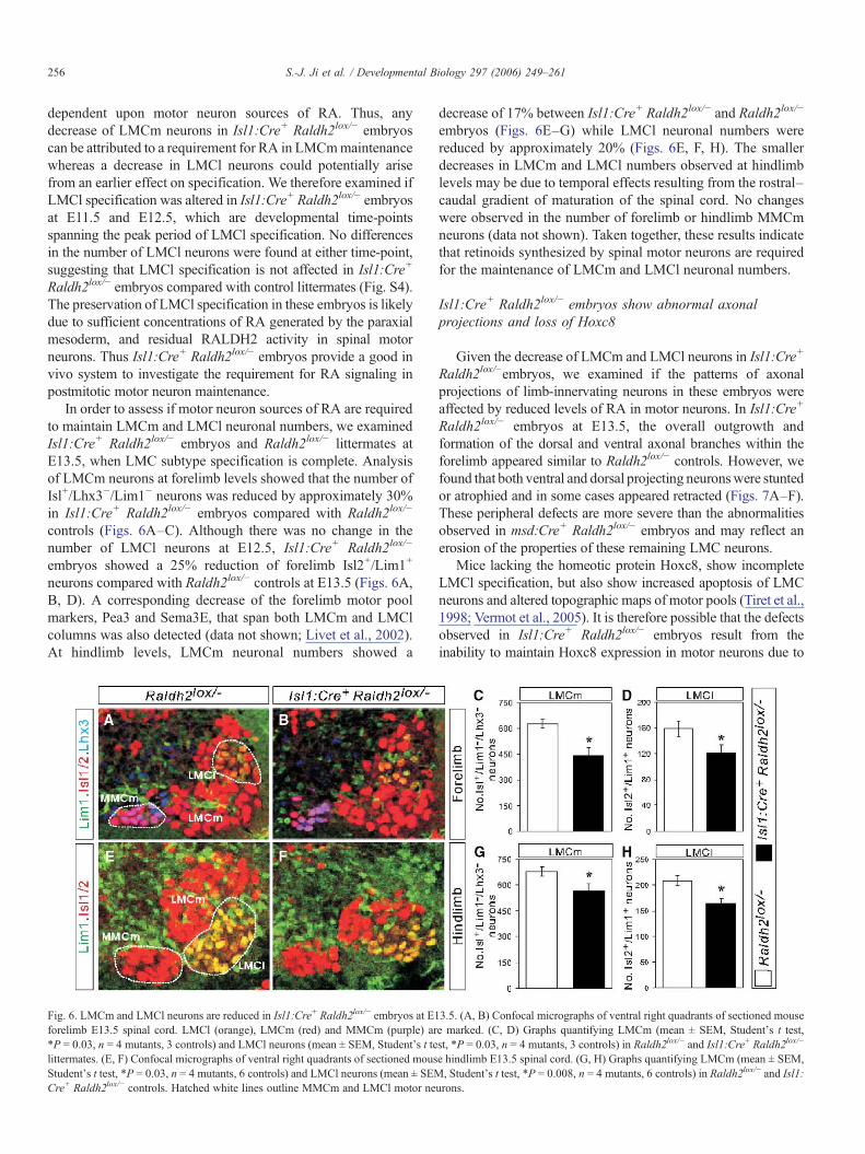
Fig. 5. RALDH2 and RA are reduced in Isl1:Cre+ Raldh2lox/− embryos. (A, B)Confocal micrographs of ventral right quadrants of mouse forelimb spinal cordsat E12.5 showing RALDH2 expression. Dotted red line outlines the margins ofthe spinal cord. (C) Western blot showing reduction of RALDH2 protein inspinal cords of Isl1:Cre+ Raldh2lox/− embryos compared with Raldh2lox/−
littermates. Actin staining is used to control for amounts of protein loaded perlane. The upper band is a non-specific cross-contaminating band observed whena different extraction buffer was used. (D) Graph depicting the amount of RAreleased in ventral spinal cord extracts of Isl1:Cre+ Raldh2lox/− E13.5 embryoscompared with Raldh2lox/− embryos (mean ± SEM, Student's t test (D)*P = 0.006, n = 4 mutants, 3 controls).
Fig. 4. Dorsal axons innervating the forelimb are abnormal in msd:Cre+ Raldh2lox/− embryos at E12.5. (A–H) Whole mount analyses using neurofilament staining tovisualize the main forelimb-innervating neurons inmsd:Cre+ Raldh2lox/− and Raldh2lox/− littermates by z-stack confocal imaging. Boxes in panel B are shown in highermagnification in panels C and D, boxes in panel F are magnified in panels G and H. Arrows mark the main differences observed. N.rad, radial nerve; N.uln, ulnarnerve; N.med, medial nerve; (d), dorsal; (v) ventral (one representative example of 6 embryos is shown here.)
255S.-J. Ji et al. / Developmental Biology 297 (2006) 249–261
Isl1:Cre animals. Isl1:Cre mice express Cre recombinase withinmotor neurons and a small population of dorsal interneurons in thespinal cord, but lack expression in the adjacent paraxialmesoderm(Pfaff et al., 1996; Srinivas et al., 2001). Given the temporal andspatial expression of Cre recombinase in this line, effectiverecombination of the Raldh2lox allele in motor neurons is likely tooccur after LMCm and LMCl specification has taken place,whereas RALDH2 expression in the paraxialmesoderm should beunaffected. Isl1:Cre+ Raldh2lox/− embryos appear normal, surviveto birth and into adulthood and are largely indistinguishable fromRaldh2lox/− controls.
To assess the extent of RALDH2 loss mediated by Crerecombination in spinal motor neurons, we examined RALDH2protein by indirect immunofluorescence using RALDH2specific antibodies in Isl1:Cre+ Raldh2lox/− and Raldh2lox/−
littermates at E12.5. No difference in mesodermal RALDH2staining was detected between the embryos; however, a markedloss of RALDH2 expression was consistently observed in LMCneurons of Isl1:Cre+ Raldh2lox/− embryos (Figs. 5A, B). Wequantified the amount of RALDH2 protein in forelimb levelspinal cords dissected from Isl1:Cre+Raldh2lox/− andRaldh2lox/−
littermates by densitrometric analysis of Western blots, andfound a 60% reduction of RALDH2 protein in Cre+ embryoscompared to controls (Fig. 5C).
We next determined the extent of RA loss in forelimb-levelspinal cords dissected from Isl1:Cre+Raldh2lox/− andRaldh2lox/−
littermates at E13.5 using the F9 RARE-LacZ retinoid-responsive reporter cell line (Wagner et al., 1992; McCafferyand Drager, 1994). Ventral spinal cord extracts derived fromIsl1:Cre+ Raldh2lox/− embryos were found to have approxi-mately 65% less RA than Raldh2lox/− littermates (Fig. 5D).Taken together, these results show that RALDH2 expression andRA levels in postmitotic motor neurons are significantlydecreased in Isl1:Cre+ Raldh2lox/− embryos compared withcontrol siblings.
LMCm and LMCl neuronal numbers are dependent uponLMC-derived retinoids
LMCm neuronal specification occurs prior to RALDH2expression in motor neurons while LMCl induction is partly
256 S.-J. Ji et al. / Developmental Biology 297 (2006) 249–261
dependent upon motor neuron sources of RA. Thus, anydecrease of LMCm neurons in Isl1:Cre+ Raldh2lox/− embryoscan be attributed to a requirement for RA in LMCmmaintenancewhereas a decrease in LMCl neurons could potentially arisefrom an earlier effect on specification. We therefore examined ifLMCl specification was altered in Isl1:Cre+ Raldh2lox/− embryosat E11.5 and E12.5, which are developmental time-pointsspanning the peak period of LMCl specification. No differencesin the number of LMCl neurons were found at either time-point,suggesting that LMCl specification is not affected in Isl1:Cre+
Raldh2lox/− embryos compared with control littermates (Fig. S4).The preservation of LMCl specification in these embryos is likelydue to sufficient concentrations of RA generated by the paraxialmesoderm, and residual RALDH2 activity in spinal motorneurons. Thus Isl1:Cre+ Raldh2lox/− embryos provide a good invivo system to investigate the requirement for RA signaling inpostmitotic motor neuron maintenance.
In order to assess if motor neuron sources of RA are requiredto maintain LMCm and LMCl neuronal numbers, we examinedIsl1:Cre+ Raldh2lox/− embryos and Raldh2lox/− littermates atE13.5, when LMC subtype specification is complete. Analysisof LMCm neurons at forelimb levels showed that the number ofIsl+/Lhx3−/Lim1− neurons was reduced by approximately 30%in Isl1:Cre+ Raldh2lox/− embryos compared with Raldh2lox/−
controls (Figs. 6A–C). Although there was no change in thenumber of LMCl neurons at E12.5, Isl1:Cre+ Raldh2lox/−
embryos showed a 25% reduction of forelimb Isl2+/Lim1+
neurons compared with Raldh2lox/− controls at E13.5 (Figs. 6A,B, D). A corresponding decrease of the forelimb motor poolmarkers, Pea3 and Sema3E, that span both LMCm and LMClcolumns was also detected (data not shown; Livet et al., 2002).At hindlimb levels, LMCm neuronal numbers showed a
Fig. 6. LMCm and LMCl neurons are reduced in Isl1:Cre+ Raldh2lox/− embryos at E1forelimb E13.5 spinal cord. LMCl (orange), LMCm (red) and MMCm (purple) ar*P = 0.03, n = 4 mutants, 3 controls) and LMCl neurons (mean ± SEM, Student's t telittermates. (E, F) Confocal micrographs of ventral right quadrants of sectioned mousStudent's t test, *P = 0.03, n = 4 mutants, 6 controls) and LMCl neurons (mean ± SEMCre+ Raldh2lox/− controls. Hatched white lines outline MMCm and LMCl motor ne
decrease of 17% between Isl1:Cre+ Raldh2lox/− and Raldh2lox/−
embryos (Figs. 6E–G) while LMCl neuronal numbers werereduced by approximately 20% (Figs. 6E, F, H). The smallerdecreases in LMCm and LMCl numbers observed at hindlimblevels may be due to temporal effects resulting from the rostral–caudal gradient of maturation of the spinal cord. No changeswere observed in the number of forelimb or hindlimb MMCmneurons (data not shown). Taken together, these results indicatethat retinoids synthesized by spinal motor neurons are requiredfor the maintenance of LMCm and LMCl neuronal numbers.
Isl1:Cre+ Raldh2lox/− embryos show abnormal axonalprojections and loss of Hoxc8
Given the decrease of LMCm and LMCl neurons in Isl1:Cre+
Raldh2lox/−embryos, we examined if the patterns of axonalprojections of limb-innervating neurons in these embryos wereaffected by reduced levels of RA in motor neurons. In Isl1:Cre+
Raldh2lox/− embryos at E13.5, the overall outgrowth andformation of the dorsal and ventral axonal branches within theforelimb appeared similar to Raldh2lox/− controls. However, wefound that both ventral and dorsal projecting neuronswere stuntedor atrophied and in some cases appeared retracted (Figs. 7A–F).These peripheral defects are more severe than the abnormalitiesobserved in msd:Cre+ Raldh2lox/− embryos and may reflect anerosion of the properties of these remaining LMC neurons.
Mice lacking the homeotic protein Hoxc8, show incompleteLMCl specification, but also show increased apoptosis of LMCneurons and altered topographic maps of motor pools (Tiret et al.,1998; Vermot et al., 2005). It is therefore possible that the defectsobserved in Isl1:Cre+ Raldh2lox/− embryos result from theinability to maintain Hoxc8 expression in motor neurons due to
3.5. (A, B) Confocal micrographs of ventral right quadrants of sectioned mousee marked. (C, D) Graphs quantifying LMCm (mean ± SEM, Student's t test,st, *P = 0.03, n = 4 mutants, 3 controls) in Raldh2lox/− and Isl1:Cre+ Raldh2lox/−
e hindlimb E13.5 spinal cord. (G, H) Graphs quantifying LMCm (mean ± SEM,, Student's t test, *P = 0.008, n = 4 mutants, 6 controls) in Raldh2lox/− and Isl1:
urons.
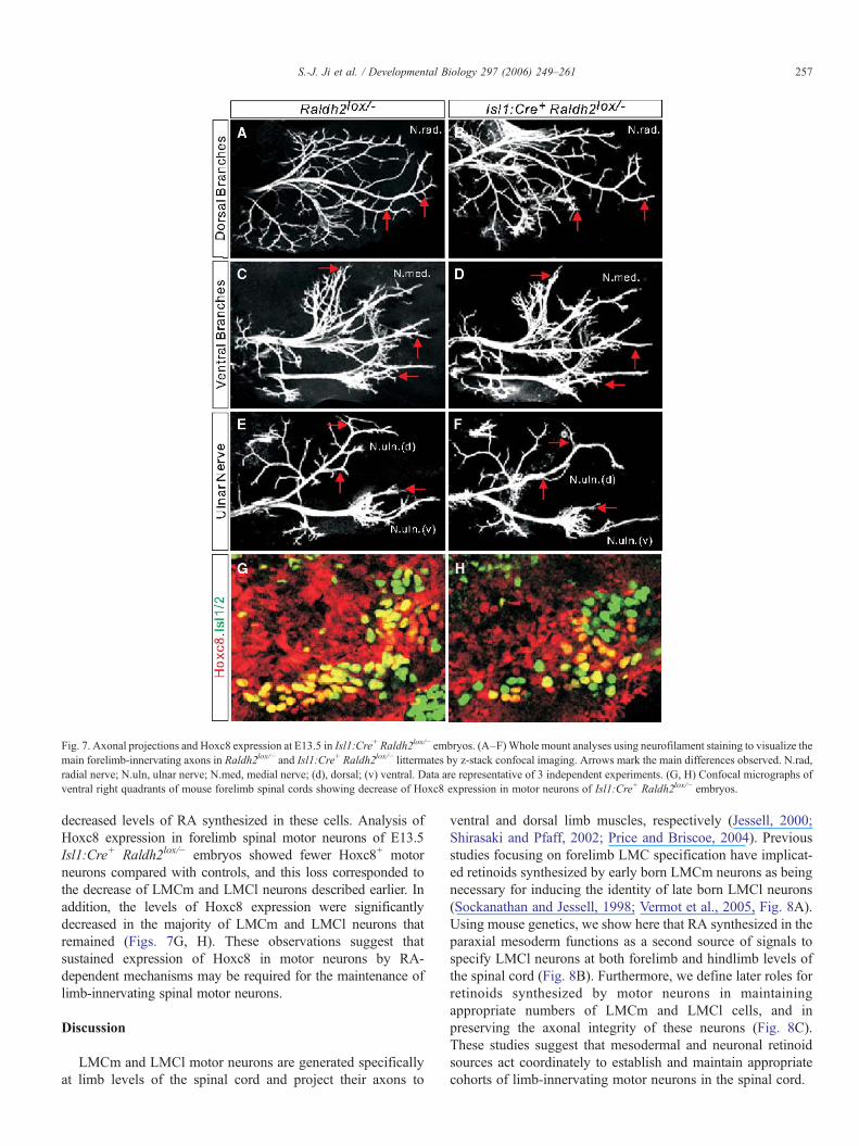
Fig. 7. Axonal projections and Hoxc8 expression at E13.5 in Isl1:Cre+ Raldh2lox/− embryos. (A–F)Whole mount analyses using neurofilament staining to visualize themain forelimb-innervating axons in Raldh2lox/− and Isl1:Cre+ Raldh2lox/− littermates by z-stack confocal imaging. Arrows mark the main differences observed. N.rad,radial nerve; N.uln, ulnar nerve; N.med, medial nerve; (d), dorsal; (v) ventral. Data are representative of 3 independent experiments. (G, H) Confocal micrographs ofventral right quadrants of mouse forelimb spinal cords showing decrease of Hoxc8 expression in motor neurons of Isl1:Cre+ Raldh2lox/− embryos.
257S.-J. Ji et al. / Developmental Biology 297 (2006) 249–261
decreased levels of RA synthesized in these cells. Analysis ofHoxc8 expression in forelimb spinal motor neurons of E13.5Isl1:Cre+ Raldh2lox/− embryos showed fewer Hoxc8+ motorneurons compared with controls, and this loss corresponded tothe decrease of LMCm and LMCl neurons described earlier. Inaddition, the levels of Hoxc8 expression were significantlydecreased in the majority of LMCm and LMCl neurons thatremained (Figs. 7G, H). These observations suggest thatsustained expression of Hoxc8 in motor neurons by RA-dependent mechanisms may be required for the maintenance oflimb-innervating spinal motor neurons.
Discussion
LMCm and LMCl motor neurons are generated specificallyat limb levels of the spinal cord and project their axons to
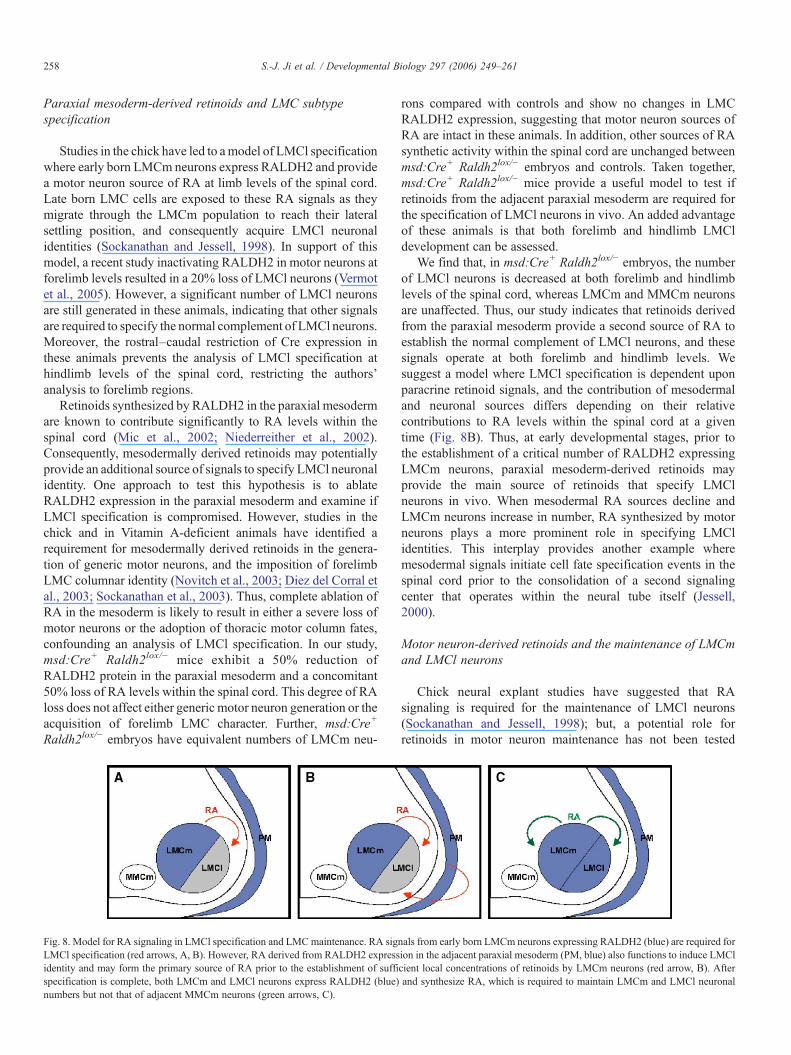
ventral and dorsal limb muscles, respectively (Jessell, 2000;Shirasaki and Pfaff, 2002; Price and Briscoe, 2004). Previousstudies focusing on forelimb LMC specification have implicat-ed retinoids synthesized by early born LMCm neurons as beingnecessary for inducing the identity of late born LMCl neurons(Sockanathan and Jessell, 1998; Vermot et al., 2005, Fig. 8A).Using mouse genetics, we show here that RA synthesized in theparaxial mesoderm functions as a second source of signals tospecify LMCl neurons at both forelimb and hindlimb levels ofthe spinal cord (Fig. 8B). Furthermore, we define later roles forretinoids synthesized by motor neurons in maintainingappropriate numbers of LMCm and LMCl cells, and inpreserving the axonal integrity of these neurons (Fig. 8C).These studies suggest that mesodermal and neuronal retinoidsources act coordinately to establish and maintain appropriatecohorts of limb-innervating motor neurons in the spinal cord.
258 S.-J. Ji et al. / Developmental Biology 297 (2006) 249–261
Paraxial mesoderm-derived retinoids and LMC subtypespecification
Studies in the chick have led to amodel of LMCl specificationwhere early born LMCm neurons express RALDH2 and providea motor neuron source of RA at limb levels of the spinal cord.Late born LMC cells are exposed to these RA signals as theymigrate through the LMCm population to reach their lateralsettling position, and consequently acquire LMCl neuronalidentities (Sockanathan and Jessell, 1998). In support of thismodel, a recent study inactivating RALDH2 in motor neurons atforelimb levels resulted in a 20% loss of LMCl neurons (Vermotet al., 2005). However, a significant number of LMCl neuronsare still generated in these animals, indicating that other signalsare required to specify the normal complement of LMCl neurons.Moreover, the rostral–caudal restriction of Cre expression inthese animals prevents the analysis of LMCl specification athindlimb levels of the spinal cord, restricting the authors'analysis to forelimb regions.
Retinoids synthesized by RALDH2 in the paraxial mesodermare known to contribute significantly to RA levels within thespinal cord (Mic et al., 2002; Niederreither et al., 2002).Consequently, mesodermally derived retinoids may potentiallyprovide an additional source of signals to specify LMCl neuronalidentity. One approach to test this hypothesis is to ablateRALDH2 expression in the paraxial mesoderm and examine ifLMCl specification is compromised. However, studies in thechick and in Vitamin A-deficient animals have identified arequirement for mesodermally derived retinoids in the genera-tion of generic motor neurons, and the imposition of forelimbLMC columnar identity (Novitch et al., 2003; Diez del Corral etal., 2003; Sockanathan et al., 2003). Thus, complete ablation ofRA in the mesoderm is likely to result in either a severe loss ofmotor neurons or the adoption of thoracic motor column fates,confounding an analysis of LMCl specification. In our study,msd:Cre+ Raldh2lox/− mice exhibit a 50% reduction ofRALDH2 protein in the paraxial mesoderm and a concomitant50% loss of RA levels within the spinal cord. This degree of RAloss does not affect either generic motor neuron generation or theacquisition of forelimb LMC character. Further, msd:Cre+
Raldh2lox/− embryos have equivalent numbers of LMCm neu-
Fig. 8. Model for RA signaling in LMCl specification and LMCmaintenance. RA sigLMCl specification (red arrows, A, B). However, RA derived from RALDH2 expressidentity and may form the primary source of RA prior to the establishment of suffispecification is complete, both LMCm and LMCl neurons express RALDH2 (blue)numbers but not that of adjacent MMCm neurons (green arrows, C).
rons compared with controls and show no changes in LMCRALDH2 expression, suggesting that motor neuron sources ofRA are intact in these animals. In addition, other sources of RAsynthetic activity within the spinal cord are unchanged betweenmsd:Cre+ Raldh2lox/− embryos and controls. Taken together,msd:Cre+ Raldh2lox/− mice provide a useful model to test ifretinoids from the adjacent paraxial mesoderm are required forthe specification of LMCl neurons in vivo. An added advantageof these animals is that both forelimb and hindlimb LMCldevelopment can be assessed.
We find that, in msd:Cre+ Raldh2lox/− embryos, the numberof LMCl neurons is decreased at both forelimb and hindlimblevels of the spinal cord, whereas LMCm and MMCm neuronsare unaffected. Thus, our study indicates that retinoids derivedfrom the paraxial mesoderm provide a second source of RA toestablish the normal complement of LMCl neurons, and thesesignals operate at both forelimb and hindlimb levels. Wesuggest a model where LMCl specification is dependent uponparacrine retinoid signals, and the contribution of mesodermaland neuronal sources differs depending on their relativecontributions to RA levels within the spinal cord at a giventime (Fig. 8B). Thus, at early developmental stages, prior tothe establishment of a critical number of RALDH2 expressingLMCm neurons, paraxial mesoderm-derived retinoids mayprovide the main source of retinoids that specify LMClneurons in vivo. When mesodermal RA sources decline andLMCm neurons increase in number, RA synthesized by motorneurons plays a more prominent role in specifying LMClidentities. This interplay provides another example wheremesodermal signals initiate cell fate specification events in thespinal cord prior to the consolidation of a second signalingcenter that operates within the neural tube itself (Jessell,2000).
Motor neuron-derived retinoids and the maintenance of LMCmand LMCl neurons
Chick neural explant studies have suggested that RAsignaling is required for the maintenance of LMCl neurons(Sockanathan and Jessell, 1998); but, a potential role forretinoids in motor neuron maintenance has not been tested
nals from early born LMCm neurons expressing RALDH2 (blue) are required forion in the adjacent paraxial mesoderm (PM, blue) also functions to induce LMClcient local concentrations of retinoids by LMCm neurons (red arrow, B). Afterand synthesize RA, which is required to maintain LMCm and LMCl neuronal
259S.-J. Ji et al. / Developmental Biology 297 (2006) 249–261
genetically in mammalian model systems. By reducingRALDH2 levels in motor neurons once they are postmitotic,we find that RA synthesized by motor neurons is required formaintaining forelimb and hindlimb LMCm and LMCl neuronalnumbers, whereas adjacent MMCm neurons are unaffected.Interestingly, no effect on LMCm numbers is observed in msd:Cre+ Raldh2lox/− embryos although the spinal cords of theseembryos have lower overall RA levels compared with Isl1:Cre+
Raldh2lox/− embryos (50% versus a 35% reduction, S.J. and S.S.unpublished data). This observation suggests that local, short-range RA signals may operate to maintain LMC numbers andraises the possibility that this might occur through cell-autonomous mechanisms.
Our studies suggest that genes downstream of RA signals actspecifically to maintain spinal motor neurons that innervate thelimb. What are the molecules that mediate motor neuronmaintenance? In the case of LMC neurons, we suggest thatHoxc8 may be required to maintain LMC neurons at posteriorforelimb levels of the spinal cord. Interestingly, Hoxc8 also actsearlier to specify LMCl identity (Vermot et al., 2005). This dualrole for Hoxc8 is supported by analyses of Hoxc8 knockoutmice which show defects in motor neuron specification, alteredsomatotopic projection maps and increased cell death (Tiret etal., 1998; Vermot et al., 2005). Hoxc8 expression in motorneurons is restricted to the C7, C8 and T1 regions at forelimblevels of the spinal cord (Tiret et al., 1998; Dasen et al., 2003)suggesting that other proteins must mediate the maintenance ofanterior forelimb and hindlimb LMC neurons. Since theexpression of Hox gene clusters span distinct regions of thespinal cord, it is possible that different Hox proteins mayfunction downstream of RA signaling to maintain LMC neuronsat other axial levels (Carpenter, 2002). Of note, Hox proteinsinvolved in fate specification may not always have a secondfunction in motor neuron maintenance. Two examples areHoxd10 and Hoxa10 which are expressed in lumbar LMCneurons, have roles in hindlimb LMC specification (Lin andCarpenter, 2003; Shah et al., 2004) but are largely down-regulated in spinal motor neurons at later stages of development(Choe et al., 2006).
What are the cellular processes affecting maintenance thatmight be impacted by decreased retinoid signaling in motorneurons? Potential pathways include those mediatingprogrammed cell-death and cell-survival through the action oftarget-derived neurotrophic factors (Buss and Oppenheim,2004; Zweifel et al., 2005). Neurotrophic factors such as NT3and NGF bind to members of the Trk family of receptors tomediate cell survival, partly through the antagonism of celldeath pathways (Zweifel et al., 2005). RA has the capability toregulate and stabilize the expression of TrkA, TrkB and p75receptors in cell lines raising the compelling possibility that RAsignaling may mediate motor neuron survival through similarmechanisms in vivo (Scheibe andWagner, 1992; Lucarelli et al.,1995; Xie et al., 1997). Interestingly, the requirement for RAsignaling in motor neurons may extend to adulthood, as adultrats deprived of Vitamin A undergo motor neuron degeneration(Corcoran et al., 2002). Although in this case, the loss of RA isnot confined to motor neurons, it is possible that continuous RA
synthesis in motor neurons is necessary for the maintenance andsurvival of motor neurons throughout life.
In summary, we have utilized genetic approaches to definenew roles for mesodermal and motor neuron sources of RA inthe specification and maintenance of limb-innervating motorneurons of the spinal cord. This work leads to a revised model ofLMCm and LMCl development, where RAs in the spinal cordderived from adjacent paraxial mesoderm and motor neuronsfunction together to specify the correct complement of LMClneurons. Retinoids synthesized by motor neurons have additio-nal roles later in development to maintain appropriate numbersof LMCm and LMCl neurons. The identification of retinoid-responsive downstream pathways that mediate these criticalevents in motor neuron specification and maintenance will beessential to understand the molecular mechanisms by whichretinoids govern neuronal identity, maintenance and survival.
Acknowledgments
We thank T.M. Jessell for the Isl1:Cre mouse line andantibodies, C. Mendelsohn and J. Rossant for RARE-hspLacZmice, P. McCaffery for antibodies and the RARE-lacZ reportercell line, and T.M. Jessell, A. Kolodkin, F. Rajaii and M. Raofor critical reading of the manuscript and scientific discus-sions. The generation of the Raldh2+/− and Raldh2lox lineswas carried out in the laboratory of T.M. Jessell together withB. Han and M. Mendelson. This work was funded by grantsfrom the German Research Council (DFG) to A.G.; and theWhitehall Foundation, Muscular Dystrophy Association andNINDS to S.S.
Appendix A. Supplementary data
Supplementary data associated with this article can be found,in the online version, at doi:10.1016/j.ydbio.2006.05.015.
References
Arber, S., Han, B., Mendelsohn, M., Smith, M., Jessell, T.M., Sockanathan, S.,1999. Requirement for the homeobox gene Hb9 in the consolidation ofmotor neuron identity. Neuron 23, 659–674.
Beckers, J., Caron, A., Hrabe de Angelis, M., Hans, S., Campos-Ortega, J.A.,Gossler, A., 2000. Distinct regulatory elements direct delta1 expression in thenervous system and paraxial mesoderm of transgenic mice. Mech. Dev. 95,23–34.
Berggren, K., McCaffery, P., Drager, U., Forehand, C.J., 1999. Differentialdistribution of retinoic acid synthesis in the chicken embryo as determinedby immunolocalization of the retinoic acid synthetic enzyme, RALDH2.Dev. Biol. 210, 288–304.
Blentic, A., Gale, E., Maden, M., 2003. Retinoic acid signalling centres in theavian embryo identified by sites of expression of synthesising andcatabolising enzymes. Dev. Dyn. 227, 114–127.
Buss, R.R., Oppenheim, R.W., 2004. Role of programmed cell death in normalneuronal development and function. Anat. Sci. Int. 79, 191–197.
Carpenter, E.M., 2002. Hox genes and spinal cord development. Dev. Neurosci.24, 24–34.
Choe, A., Phun, H.Q., Tieu, D.D., Hu, Y.H., Carpenter, E.M., 2006. Expressionpatterns of Hox10 paralogous genes during lumbar spinal cord development.Gene Expr. Patterns, doi:10.1016/j.modgep.2005.12.004.
Corcoran, J., So, P.L., Maden, M., 2002. Absence of retinoids can induce
260 S.-J. Ji et al. / Developmental Biology 297 (2006) 249–261
motoneuron disease in the adult rat and a retinoid defect is present inmotoneuron disease patients. J. Cell Sci. 115, 4735–4741.
Cornbrooks, E.B., Newton, C.J., Forehand, C.J., 1997. Development ofdifferential preganglionic projections to pre- and paravertebral sympatheticganglia. J. Comp. Neurol. 382, 1–18.
Dasen, J.S., Liu, J.P., Jessell, T.M., 2003. Motor neuron columnar fate imposedby sequential phases of Hox-c activity. Nature 425, 926–933.
Diez del Corral, R., Olivera-Martinez, I., Goriely, A., Gale, E., Maden, M.,Storey, K., 2003. Opposing FGF and retinoid pathways control ventralneural pattern, neuronal differentiation, and segmentation during body axisextension. Neuron 40, 65–79.
Giger,R.J.,Cloutier, J.F., Sahay,A., Prinjha,R.K., Levengood,D.V.,Moore, S.E.,Pickering, S., Simmons, D., Rastan, S., Walsh, F.S., Kolodkin, A.L., Ginty,D.D., Geppert, M., 2000. Neuropilin-2 is required in vivo for selective axonguidance responses to secreted semaphorins. Neuron 25, 29–41.
Gutman, C.R., Ajmera, M.K., Hollyday, M., 1993. Organization of motor poolssupplying axial muscles in the chicken. Brain Res. 609, 129–136.
Haselbeck, R.J., Ang, H.L., Deltour, L., Duester, G., 1997. Retinoic acid andalcohol/retinol dehydrogenase in the mouse adrenal gland: a potentialendocrine source of retinoic acid during development. Endocrinology 138,3035–3041.
Hollyday, M., 1980a. Organization of motor pools in the chick lumbar lateralmotor column. J. Comp. Neurol. 194, 143–170.
Hollyday, M., 1980b. Motoneuron histogenesis and the development of limbinnervation. Curr. Top. Dev. Biol. 15, 181–215.
Hollyday, M., 1990. Specificity of initial axonal projections to embryonic chickwing. J. Comp. Neurol. 302, 589–602.
Jessell, T.M., 2000. Neuronal specification in the spinal cord: inductive signalsand transcriptional codes. Nat. Rev., Genet. 1, 20–29.
Jessell, T.M., Melton, D.A., 1992. Diffusible factors in vertebrate embryonicinduction. Cell 68, 257–270.
Kania, A., Johnson, R.L., Jessell, T.M., 2000. Coordinate roles for LIMhomeobox genes in directing the dorsoventral trajectory of motor axons inthe vertebrate limb. Cell 102, 161–173.
Landmesser, L., 1978. The distribution of motor neurons supplying the chickhind limb muscles. J. Physiol. 284, 371–389.
Lin, A.W., Carpenter, E.M., 2003. Hoxa10 and Hoxd10 coordinately regulatelumbar motor neuron patterning. J. Neurobiol. 56, 328–337.
Lin, J.H., Saito, T., Anderson, D.J., Lance-Jones, C., Jessell, T.M., Arber, S.,1998. Functionally related motor neuron pool and muscle sensory afferentsubtypes defined by coordinate ETS gene expression. Cell 95, 393–407.
Liu, A., Joyner, A.L., 2001. Early anterior/posterior patterning of the midbrainand cerebellum. Annu. Rev. Neurosci. 24, 869–896.
Livet, J., Sigrist, M., Stroebel, S., De Paola, V., Price, S.R., Henderson, C.E.,Jessell, T.M., Arber, S.A., 2002. ETS gene Pea3 controls the central positionand terminal arborization of specific motor neuron pools. Neuron 35,877–892.
Lucarelli, E., Kaplan, D.R., Thiele, C.J., 1995. Selective regulation of TrkA andTrkB receptors by retinoic acid and interferon-gamma in humanneuroblastoma cell lines. J. Biol. Chem. 270, 24725–24731.
McCaffery, P., Drager, U.C., 1994. Hot spots of retinoic acid synthesis in thedeveloping spinal cord. Proc. Natl. Acad. Sci. U. S. A. 91, 7194–7197.
Mic, F.A., Haselbeck, R.J., Cuenca, A.E., Duester, G., 2002. Novel retinoic acidgenerating activities in the neural tube and heart identified by conditionalrescue of Raldh2 null mutant mice. Development 129, 2271–2282.
Niederreither, K., McCaffery, P., Drager, U.C., Chambon, P., Dolle, P., 1997.Restricted expression and retinoic acid induced downregulation of theretinaldehyde dehydrogenase type 2 (RALDH-2) gene during mousedevelopment. Mech. Dev. 62, 67–78.
Niederreither, K., Subbarayan, V., Dolle, P., Chambon, P., 1999. Embryonicretinoic acid synthesis is essential for early mouse post-implantationdevelopment. Nat. Genet. 21, 444–448.
Niederreither, K., Vermot, J., Fraulob, V., Chambon, P., Dolle, P., 2002.Retinaldehyde dehydrogenase 2 (RALDH2)-independent patterns of retinoicacid synthesis in the mouse embryo. Proc. Natl. Acad. Sci. U. S. A. 99,16111–16116.
Nornes, H.O., Carry, M., 1978. Neurogenesis in spinal cord of mouse: anautoradiographic analysis. Brain Res. 159, 1–6.
Novitch, B.G., Wichterle, H., Jessell, T.M., Sockanathan, S., 2003. Arequirement for retinoic acid-mediated transcriptional activation in ventralneural patterning and motor neuron specification. Neuron 40, 81–95.
Pfaff, S.L., Mendelsohn, M., Stewart, C.L., Edlund, T., Jessell, T.M., 1996.Requirement for LIM homeobox gene Isl1 in motor neuron generationreveals a motor neuron-dependent step in interneuron differentiation. Cell84, 309–320.
Prasad, A., Hollyday, M., 1991. Development and migration of aviansympathetic preganglionic neurons. J. Comp. Neurol. 307, 237–258.
Price, S.R., Briscoe, J., 2004. The generation and diversification of spinal motorneurons: signals and responses. Mech. Dev. 121, 1103–1115.
Rajewsky, K., Gu, H., Kuhn, R., Betz, U.A., Muller, W., Roes, J., Schwenk, F.,1996. Conditional gene targeting. J. Clin. Invest. 98, 600–603.
Rossant, J., Zirngibl, R., Cado, D., Shago, M., Giguere, V., 1991. Expression ofa retinoic acid response element-hsplacZ transgene defines specific domainsof transcriptional activity during mouse embryogenesis. Genes Dev. 5,1333–1344.
Schaeren-Wiemers, N., Gerfin-Moser, A., 1993. A single protocol to detecttranscripts of various types and expression levels in neural tissue andcultured cells: in situ hybridization using digoxigenin-labelled cRNAprobes. Histochemistry 100, 431–440.
Scheibe, R.J., Wagner, J.A., 1992. Retinoic acid regulates both expression of thenerve growth factor receptor and sensitivity to nerve growth factor. J. Biol.Chem. 267, 17611–17616.
Shah, V., Drill, E., Lance-Jones, C., 2004. Ectopic expression of Hoxd10 inthoracic spinal segments induces motoneurons with a lumbosacralmolecular profile and axon projections to the limb. Dev. Dyn. 231,43–56.
Sharma, K., Sheng, H.Z., Lettieri, K., Li, H., Karavanov, A., Potter, S.,Westphal, H., Pfaff, S.L., 1998. LIM homeodomain factors Lhx3 and Lhx4assign subtype identities for motor neurons. Cell 95, 817–828.
Sharma, K., Leonard, A.E., Lettieri, K., Pfaff, S.L., 2000. Genetic andepigenetic mechanisms contribute to motor neuron pathfinding. Nature 406,515–519.
Shirasaki, R., Pfaff, S.M., 2002. Transcriptional controls and the control ofneuronal identity. Annu. Rev. Neurosci. 25, 251–281.
Sockanathan, S., Jessell, T.M., 1998. Motor neuron-derived retinoid signalingspecifies the subtype identity of spinal motor neurons. Cell 94, 503–514.
Sockanathan, S., Perlmann, T., Jessell, T.M., 2003. Retinoid receptor signalingin postmitotic motor neurons regulates rostrocaudal positional identity andaxonal projection pattern. Neuron 40, 97–111.
Solomin, L., Johansson,C.B., Zetterstrom,R.H., Bissonette, R.P., Heyman, R.A.,Olson, L., Lendahl, U., Frisen, J., Perlmann, T., 1998. Retinoid X receptorsignaling in the developing spinal cord. Nature 395, 398–402.
Soriano, P., 1999. Generalized lacZ expression with the ROSA26 Cre reporterstrain. Nat. Genet. 21, 70–71.
Srinivas, S., Watanabe, T., Lin, C.S., William, C.M., Tanabe, Y., Jessell, T.M.,Costantini, F., 2001. Cre reporter strains produced by targeted insertion ofEYFP and ECFP into the ROSA26 locus. BMC Dev. Biol. 1, 4.
Swindell, E.C., Thaller, C., Sockanathan, S., Petkovich, M., Jessell, T.M.,Eichele, G., 1999. Complementary domains of retinoic acid production anddegradation in the early chick embryo. Dev. Biol. 216, 282–296.
Tiret, L., Le Mouellic, H., Maury, M., Brulet, P., 1998. Increased apoptosis ofmotoneurons and altered somatotopic maps in the brachial spinal cord ofHoxc-8-deficient mice. Development 125, 279–291.
Tosney, K.W., Landmesser, L.T., 1985a. Development of the major pathways forneurite outgrowth in the chick hindlimb. Dev. Biol. 109, 193–214.
Tosney, K.W., Landmesser, L.T., 1985b. Growth cone morphology andtrajectory in the lumbosacral region of the chick embryo. J. Neurosci. 5,2345–2358.
Tsuchida, T.N., Ensini, M., Morton, S.B., Baldessare, M., Edlund, T., Jessell,T.M., Pfaff, S.L., 1994. Topographic organization of embryonic motorneurons defined by expression of LIM homeobox genes. Cell 79,957–970.
Vermot, J., Niederreither, K., Garnier, J.M., Chambon, P., Dolle, P., 2003.Decreased embryonic retinoic acid synthesis results in a DiGeorge syndromephenotype in newborn mice. Proc. Natl. Acad. Sci. U. S. A. 100,1763–1768.
261S.-J. Ji et al. / Developmental Biology 297 (2006) 249–261
Vermot, J., Schuhbaur, B., Le Mouellic, H., McCaffery, P., Garnier, J.M.,Hentsch, D., Brulet, P., Niederreither, K., Chambon, P., Dolle, P., Le Roux, I.,2005. Retinaldehyde dehydrogenase 2 and Hoxc8 are required in the murinebrachial spinal cord for the specification of Lim1+ motoneurons and thecorrect distribution of Islet1+ motoneurons. Development 132, 1611–1621.
Wagner, M., Han, B., Jessell, T.M., 1992. Regional differences in retinoidrelease from embryonic neural tissue detected by an in vitro reporter assay.Development 116, 55–66.
Wilson, L., Gale, E., Chambers, D., Maden, M., 2004. Retinoic acid and the
control of dorsoventral patterning in the avian spinal cord. Dev. Biol. 269,433–446.
Xie, P., Cheung, W.M., Ip, F.C., Ip, N.Y., Leung, M.F., 1997. Induction of TrkAreceptor by retinoic acid in leukaemia cell lines. NeuroReport 8, 1067–1070.
Zhao,D.,McCaffery, P., Ivins,K.J.,Neve, R.L., Hogan, P., Chin,W.W.,Drager,U.C.,1996. Molecular identification of a major retinoic acid synthesizing enzyme, aretinaldehyde-specific dehydrogenase. Eur. J. Biochem. 240, 15–22.
Zweifel, L.S., Kuruvilla, R., Ginty, D.D., 2005. Functions and mechanisms ofretrograde neurotrophin signalling. Nat. Rev., Neurosci. 6, 615–625.


