Embed Size (px)
Citation preview

Copyright by American Heart Association, Inc. All rights reserved.
1
Metabolic Gene Remodeling and Mitochondrial Dysfunction in Failing Right
Ventricular Hypertrophy due to Pulmonary Arterial Hypertension
Gomez-Arroyo et al: Mitochondrial Dysfunction and Metabolic Remodeling in Right Ventricular Dysfunction
Jose Gomez-Arroyo, MD1; Shiro Mizuno, MD, PhD2; Karol Szczepanek, PhD3;
Benjamin Van Tassell, PharmD1,3; Ramesh Natarajan, PhD1;
Cristobal G. dos Remedios, PhD4; Jennifer I. Drake, PhD1; Laszlo Farkas, MD1;
Donatas Kraskauskas, DVM1; Dayanjan S. Wijesinghe, PhD5,6; Charles E. Chalfant, PhD5,6;
John Bigbee, PhD8; Antonio Abbate, MD, PhD1,3; Edward J. Lesnefsky, MD3 5;
Harm J. Bogaard, MD, PhD7; Norbert F. Voelkel, MD1
1Victoria Johnson Center for Lung Obstructive Disease Research, Virginia Commonwealth
University, Richmond, VA 2Division of Respiratory Diseases, Kanazawa Medical University, Ishikawa, Japan
3VCU Pauley Heart Center, Virginia Commonwealth University, Richmond, VA 4Anatomy and Histology, School of Medical Sciences, Bosch Institute, Sydney, Australia
5McGuire Department of Veterans Affairs Medical Center, Richmond, VA 6Department of Biochemistry and Molecular Biology, Virginia Commonwealth University School of
Medicine, Richmond, VA 7Department of Pulmonary Medicine, VU University Medical Center, Amsterdam, Netherlands
8Department of Anatomy and Neurobiology, Virginia Commonwealth University, Richmond, VA
Correspondence to Norbert F. Voelkel, Director Victoria Johnson Center for Obstructive Lung Disease Research Virginia Commonwealth University 1220 East Broad Street Richmond, VA 23298 Telephone: 804-6283334 Fax: 804-6280325 E-mail: [email protected]
DOI: 10.1161/CIRCHEARTFAILURE.111.966127
Journal Subject Codes: [18] Pulmonary circulation and disease, [130] Animal models of human disease, [107] Biochemistry and metabolism
ersitty,y,y,y,yy,y, IIIIIIIshshshshshshshikikikikikikikawawawawawawawa,a,a,a,a,aa J
Pauley Heart Center, Virginia Commonwealth University, Richmond,
a A
G A
Biochemistry and Molecular Biology, Virginia Commonwealth Univ r
Paaaaaulululululey HHHHHeeeaee rttttt CCCenter, Virginia Commmmoonwealth UUUUUniversity, Richmond,
annnnndd ddd Histologgy,y Schhhhooool l l of MMMMMededdicalal Scccieenceseseseses, BBBosccch InIInstiitiii uuutu e, SSSSSydddnneyy, A
Guiririririreeeee DeDeDeeDepaaartrtr mememementtntt of VeVeVeVV tetetetet ransnnsnsns AAAAAffffffafafaf iriirii sss s MeMeMeMeMedididididicacacacc lllll CeCeCeCC ntntn erereere , RiRiRiRiRichchchchchmooomoondndnd, VAVAVVAVffff
Biochemistry and MMMMololololo ecececececulululuularararara BBBBBioiooollolll gyyyy, ViViViViVirgrgrgrgr inininiaiaiaiaia CCCCComomomommmommm nwealth Univer
MeMeMeMMedididididicicicicinenenenene, , RiRiRiRiRichchchchchmomomomomondndndndd,, VAVAVAVAVA

Copyright by American Heart Association, Inc. All rights reserved.
2
Abstract
Background—Right ventricular dysfunction (RVD) is the most frequent cause of death in
patients with pulmonary arterial hypertension. Whereas abnormal energy substrate utilization
has been implicated in the development of chronic left heart failure, data describing such
metabolic remodeling in RVD remain incomplete. Thus, we sought to characterize metabolic
gene expression changes and mitochondrial dysfunction in functional and dysfunctional RV
hypertrophy.
Methods and Results—Two different rat models of RV hypertrophy were studied. The model
of RVD (SU5416/hypoxia) exhibited a significantly decreased gene expression of PPAR-
gamma coactivator-1 alpha (PGC-1 ), PPAR- and ERR- . The expression of multiple
PCG-1 target genes required for fatty acid oxidation (FAO) was similarly decreased.
Decreased PGC-1 expression was also associated with a net loss of mitochondrial protein
and oxidative capacity. Reduced mitochondrial number was associated with a downregulation
of TFAM and other genes required for mitochondrial biogenesis. Electron microscopy
demonstrated that in RVD tissue, mitochondria had abnormal shape and size. Lastly,
respirometric analysis demonstrated that mitochondria isolated from RVD-tissue had a
significantly reduced ADP-stimulated (state 3) rate for complex I. Conversely, functional RV
hypertrophy in the pulmonary artery banding (PAB) model showed normal expression of
PGC-1 , whereas the expression of FAO genes was either preserved or unregulated.
Moreover, PAB-RV tissue exhibited preserved TFAM expression and mitochondrial
respiration despite elevated RV pressure-overload.
Conclusions—Right ventricular dysfunction, but not functional RV hypertrophy in rats,
demonstrates a gene expression profile compatible with a multilevel impairment of fatty acid
metabolism and significant mitochondrial dysfunction, partially independent of chronic
pressure-overload.
Key Words: pulmonary heart disease, metabolism, pressure, fatty acids, mitochondria
ococococococociaiaiaiaiaiaiateteteteteteted d ddddd wiwiwiwiwiwiwiththththththth aaaaaa dddddddooooooo
s. Elececececececectrtrtrtrtrtrtrononononononon mmmmmmmicicicicicicicrorrorrrr s
a
al is demonstrated that mitochondria isolated from RVD-tiss e
u u
he pulmonary artery banding (PAB) model showed normal expre
h i f FAO i h d l
at tt t t ininininin RVDVDVDVDVD tisisiissue, mitochondria hadaad abnormamamamamal shhhhapa e ana d size. La
alalalalalysysysyysis demonoonstrarrateddd that mmiiitochhondnndririiiria aa a a isisisii ooolatatted frorom mmmm RVVVVVD-DDDD ttisssuuue
uced ADP-stimulatatatattedee (((state 3)3 raaaatete fororororor ccomplex I. Conversely, fu
hehehe pppulululmomomonananaryryry aaartrtrterereryyy bababandndndiniininggg (P((P(P( ABABAB) )) momomodededded lll shshshowowowededed nnnororormamamalll exexexprprpreee
hh ii ff FAO ii hh dd ll

Copyright by American Heart Association, Inc. All rights reserved.
3
Non-standard Abbreviations and Acronyms
ACADM Acyl-Coenzyme A dehydrogenase, C-4 to C-12 straight chain
ACADVL Acyl-Coenzyme A dehydrogenase, very long chain
ACADS Acyl-Coenzyme A dehydrogenase, C-4 to C-8(6) short chain
ACSL1 Acyl-CoA synthetase long-chain family member 1
CD36 Trombospondin Receptor/Fatty acid translocase
COUP-TF1 Chicken Ovalbumin Upstream Promoter Transcription Factor 1
CPT Carnitine palmitoyltransferase
CS Citrate Synthase
ERR Estrogen-related receptor
GLUT Solute carrier family 2 (facilitated glucose transporter)
IDH1 Isocitrate Dehydrogenase 1
PDHb Pyruvate dehydrogenase subunit beta
PDK Pyruvate dehydrogenase kinase
PGC-1 Peroxisome proliferator-activated receptor gamma coactivator-1
POLG2 Polymerase (DNA directed), gamma 2, accessory subunit
POLRMT Polymerase (RNA) mitochondrial (DNA directed)
PPAR Peroxisome proliferator-activated receptor
SP Transcription factor Sp1
TFAM Transcription factor A mitochondrial
TOP1mt Topoisomerase (DNA) I, mitochondrial
anspooooooortrtrtrtrtrtrtererererererer)))))))y ( g p )y ( g p )
IIIsII ocitratetetetete DDDDDehehehehehydydydydydrororororogeeeeenanananan se 11111
PyPyPyPyPyrururuvatetete dddddehehehhhydyddrogegegenaseeeee ssssububububbunnittittt bbbbbeteteteteta
PyPyPyrururuvavavav tetete ddehehydydyy rororogegegegg nananasesese kkininasasaseee

Copyright by American Heart Association, Inc. All rights reserved.
4
Pulmonary Arterial Hypertension (PAH) is a severe and often rapidly progressive group of
diseases which are characterized by a chronically and frequently progressive increase in the
right ventricular (RV) afterload(1). Increased RV afterload is partially compensated by RV
hypertrophy but eventually leads to RV dysfunction (RVD), RV failure and untimely death,
regardless of medical treatment(1). Given the prognostic importance of RVD in PAH, the
cellular-and molecular mechanisms underlying RVD need to be investigated, as they can be
potentially reversible. Human and experimental chronic left heart dysfunction is
characterized by decreased oxidative metabolism(2), abnormal mitochondrial respiration(3)
and impaired mitochondrial biogenesis(4). These changes have in part been explained by a
deregulated expression of critical transcription factors such as the peroxisome proliferator-
activated receptor (PPAR) alpha(5), the estrogen-related receptor (ERR) alpha(6) and the
master regulator of oxidative metabolism, the PPAR-gamma coactivator-1alpha (PGC-
1 )(7).
Akin to left heart failure, it has been postulated that RVD is characterized by abnormal
energy metabolism(8,9). Studies in animal models have demonstrated that RVD exhibits an
increased expression of glycolysis-related genes(10) and increased enzymatic glycolysis
rate(11). However, is largely unknown to what extent this switch in cardiac bioenergetics
(also known as metabolic remodeling(12)) involves changes in fatty acid oxidation (FAO),
whether metabolic remodeling is a response to chronic pressure overload, or to what extent
mitochondrial structure and function are also compromised in RVD. Thus, we sought to 1)
characterize the metabolic gene expression profile associated with RV dysfunction, 2)
determine whether pressure overload is sufficient to explain metabolic gene remodeling, 3)
assess the structure and function of mitochondria in the dysfunctional RV.
iiiin n n n n nn papapapapapapartrtrtrtrtrtrt bbbbbbbeeeeeeeeeeeeeen n n n n nn exexexexexexexplplplplplplplaaa
he perererererereroxoxoxoxoxoxoxisisisisisisisomomomomomomomeeeeeee pprpppp p p
or (PPAR) alpha(5), the estr en-related rec tor (ERR) alpha( )
of oxidative metabolism, the PPAR-gamma coactivator-1alpha (
p p p
orrr (((((PPAR) alaalaa phphhhha(a(a(a(a(5)5)5)5)5),,,,, thththththee eee esesesese trrroggggeen---reelatatatatatededededed rrrrreccepepeptototottorr (EE(E(EERRRRRRRRRR)) ) ) ) alalalllphphphphpha(a(a(a(a(6)
of fff oxoxoxoxoxidididddativivivee memmetatatat boolililisssm, thththththeee PPPPPPPP ARARARARAR-gggggamamamammmamamama coaoaccctivatatatatatooor-11111alalllphphphhhaa (

Copyright by American Heart Association, Inc. All rights reserved.
5
Here, we report that RV dysfunction – as assessed in rat and human RV tissue samples – is
characterized by a downregulation of PGC-1 expression and decreased expression of several
PGC-1 target genes encoding key-enzymes that regulate fatty acid oxidation (FAO), as well
as other genes involved in mitochondrial biogenesis. In addition, we present evidence for
impaired mitochondrial structure and function (respiration). Some of these findings have
been previously reported in the form of abstract(13,14).
Methods
Animal models
SU5416/Hypoxia model (SuHx): An animal model of severe angioproliferative PAH and
RVD was generated in male Sprague-Dawley rats (body weight 200 g, age 6 weeks) with a
20 mg/kg, one-time subcutaneous injection of a VEGF-receptor blocker (SU5416) followed
by 4 weeks of 10% hypoxia as described previously(15,16) and in the online supplement.
Upon return to normoxia, RV function was evaluated by transthoracic echocardiogram. The
tricuspid annular plane systolic excursion (TAPSE) was the reference parameter for RV
function. For hemodynamic measurements, median sternotomy was performed, and right
ventricular systolic pressure was measured with a 4.5 mm Millar conductance catheter
inserted at the RV outflow-tract. As previously described, the RV in the SuHx rat model
responds to pulmonary hypertension with a robust degree of hypertrophy, followed by
dysfunction and failure(17). The RV in this rat model is characterized by fibrosis, capillary
rarefaction and cardiomyocyte apoptosis(15), which are associated with decreased cardiac
output, markedly dilated RV and decreased exercise capacity(18). As previously described,
the SuHx RV dysfunction model reproduces some features of human RV dysfunction, such
as paradoxical septal movement and RV dilatation (Supplemental Video 1). Pulmonary
gigigigigigigiopopopopopopoprororororororolililililililifefefefefefeferararararararatitititititt veveveveveveve PPPPPPP
20000 6666666ed e Sp gue w ey s (body we g 00 g, ge 6 wee
6
0% hypoxia as described previously(15,16) and in the online s p
ormoxia, RV function was evaluated by transthoracic echocard o
ed e Sp gue w ey s (b(( ody we g g, ge wee
mememee subcutatattat nneououououussss inininininjejejejejectctctctctioioioioi nn offff aa VVVEGEGGGGFF-FFF rerereceeppptorrr bblooooockcccc ererererer ((((SUSUSUUU545454545416f
0% hyhhhh poxiiia as ddescribibiibi ed pre iviiiou lslyy(1515151515,11,111666)66 a ddnd iin thhhhhe onliline sup
ormo ixia,a,a,a,a, RRRRRV V VVV fufufuufunccccctitititiononononon wwwwwasasassas eeeeevavavav lululululuatatatatatedededede bbbbby yy yy trtrtrtrtranananananstststssthohohohohorararararacicicicicic cc c c ececececechohohhh ca drdiio

Copyright by American Heart Association, Inc. All rights reserved.
6
Artery Banding (PAB): Surgical ligation of pulmonary artery was achieved through a left
thoracotomy in male Sprague-Dawley rats weighing 180 to 200 g, with a silk suture tied
around an 18-gauge needle alongside the pulmonary artery, as described previously(15) (also
in online supplement). The PAB rats were sacrificed to collect organs 6 weeks after the
surgery, to allow for significant hypertrophy as reported by Bogaard et al (15,19). As a model
of strictly mechanical RV pressure overload, the PAB rat model demonstrates preserved RV
function despite generating significantly high RV afterload and hypertrophy(15,19)
(Supplemental Figure 1 F-H). Thus, the PAB-model was utilized as a model of non-
dysfunctional RV hypertrophy and was studied as above. The Table presents the
echocardiographic and hemodynamic data illustrating the degree of pulmonary hypertension
and RV function in all conditions studied.
Human samples were obtained from patients diagnosed with PAH that underwent cardiac
transplantation.
Oxygen consumption by intact mitochondria was measured with Clark-type oxygen
electrodes. Immunohistochemistry and gene and protein expression studies were performed
with standard procedures, as outlined in the supplemental methods.
Statistical analysis
Differences between groups were assessed with one-way or two-way ANOVA or Kruskall-
Wallis tests. Bonferroni’s and Dunn’s post-hoc tests were used to assess significant
differences between groups. A p-value < 0.05 was accepted as significant. Correlation
analysis was done with Spearman’s test. Results are reported as means±SEM, or fold-change
mean±SEM unless specified otherwise. Four-to-six rats were used per group, unless
otherwise specified. Statistical analysis was done with PASW V.18 (IBM, Armonk, New
York) and GraphPad Prism (La Jolla, CA).
eeeeee ooooooof f f f f f pupupupupupupulmlmlmlmlmlmlmonononononononararararararary yyyyyy hhhhhhh
were obtained from atients di nosed with PAH that underw n
ption by intact mitochondria was measured with Clark-type oxyg
weweweere obtaiiiinenennn d dddd frfrfrfrfromomomomom pppppatatatatatieieieiei ntntntn s dididdd agggnnosesesesesedddd d wiwiwiwwiththth PPPAHAAAHA ttttthahahahah t tttt ununununundedededederwrwrwrwrweneeee
ppptitiononon bby y y y inintatatactctct mmmititococochohondndddririrrr aaa wawawasss mememeasasasssururureded wwwitithh ClClarararkk-tytytyyypepepep oooxyxyxyggg

Copyright by American Heart Association, Inc. All rights reserved.
7
For detailed information regarding echocardiography, hemodynamic measurements, gene
expression, protein expression, human samples, mitochondrial isolation, measurement of
mitochondrial respiration, chromatography electrospray ionization tandem mass spectrometry
and immunohistochemistry, see the online supplement.
Results
RV dysfunction is characterized by a load-independent downregulation of PGC-1 , PPAR-
and ERR- .
We have previously reported that dysfunctional RVs from SuHx-rats, differentially express
multiple gene signalling pathways when compared with non-dysfunctional hypertrophied
RVs(20). Importantly, we reported that RVD is associated with gene expression changes that
suggest an abnormal metabolism, with a particularly strong signal relating to the peroxisome
proliferator-activated receptor (PPAR) signaling pathways. Therefore, as a first step, we
measured the expression of PGC-1 , a direct coactivator and master regulator of the PPAR
family of transcription factors. PGC-1 regulates oxidative metabolism and many aspects of
mitochondrial biology(21).
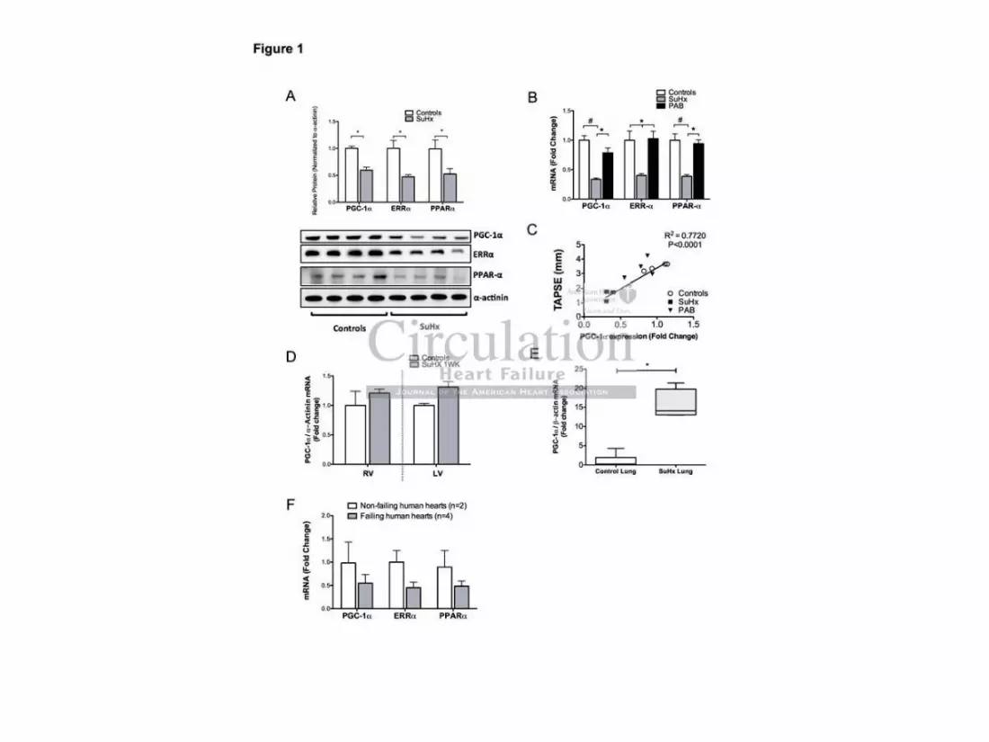
Western blots of protein samples obtained from SuHx RV tissues showed a significantly
decreased amount of PGC-1 protein (Figure 1A). qPCR analysis revealed a significant
downregulation of PGC-1 mRNA levels (Figure 1B) indicating that the change in PGC-1
expression also occurred on the transcription level. Decreased PGC-1 gene expression was
accompanied by a decreased expression of the ERR- and PPAR- genes (Figure 1B). To
evaluate whether pure mechanical RV pressure-overload was sufficient to downregulate
PGC-1 expression, we measured PGC-1 transcript levels in PAB-RV tissues and found
that the expression of PGC-1 , ERR- and PPAR- was not significantly decreased in the
non-failing hypertrophied RVs of PAB rats, despite the high RV pressure and RV
sssssssfufufufufufufuncncncncncncnctititititititionononononononalalalalalalal hhhhhhhypypypypypypypererererererer
iiiiiiiy, we epo ed V s ssoc ed w ge e e p ess o
rmal metabolism, with a particularly strong signal relating to the
v s
p f
yyyy,,, we epppppo ed V s ssoc ed w gegggg e e p ess o
rmamamaal metabobobobollismmmmm, wiwiwiwiwithththtt aaaaa ppaaarticuculaaarlly ststsss rooongnng sssiignannal rererererellatititititingnngngng tttttooooo thththththe
vateddddd receptor ((PPPPPARRRRR))) sign lalllling pap hhthhways. ThThThTher feffore, as a fifirst sr
ppressiion n nn ofofofofof PPPPPGCGCGCGCGC-1-1111 ,, a aa aa dididdd rererererectctctctct cccoaoaoaoaoactctctctctivivivivivatatatttororororor aaaaandndndndd mmmmmasasasasasteteteteter r rr rererereregugugugugulalalll tor fof

Copyright by American Heart Association, Inc. All rights reserved.
8
hypertrophy (Figure 1B). Figures 1C and Supplemental Figure 2A-B illustrate that PGC-1
(R2=0.72, p=0.001), PPAR- (R2=0.72, p=0.001) and ERR- (R2=0.76, p=0.002) transcript
levels strongly correlated with the tricuspid annular plane systolic excursion (TAPSE), a
heart-rate independent variable of RV function.
We have previously described that SU5416 treatment alone does not induce RV dysfunction
and has a limited impact on gene expression(20). However, because decreased expression of
PGC-1 could be a direct effect of the combination of SU5416 and hypoxia rather than a
consequence of RVD, we measured the expression of PGC-1 at one week after the SuHx
protocol had been initiated. At one week no RVD is present and indeed, as shown in
Figure1D, PGC-1 expression is not decreased in the RV or in the LV. In addition, we
measured the expression of PGC-1 in rat lung tissues after four weeks of SuHx, a time-point
where plexiform-like lesions have already formed, and we found a significant increase in
PGC-1 expression (Figure 1E). To further evaluate a potential toxic effect of SU4516 in the
setting of RV pressure overload, we examined the RV gene expression of PAB animals
exposed to SU4516 and found no change in the PGC-1 , ERR- or PPAR- mRNA
transcript levels (Supplemental Figure 2C). Next, we measured the expression of PGC-1 in
LV tissue obtained from animals sacrificed four weeks after initiation of the SuHx protocol.
Although somewhat decreased, the SuHx-LV tissue did not show a significant change in
PGC-1 expression (Supplemental Figure 2D). In the aggregate, the data indicates that the
decreased expression of PGC-1 is unlikely due to a toxic effect of SU5416.
Because nuclear receptors other than PGC-1 have been implicated in phenotypic metabolic
changes which occur during normal cardiac development, we decided to evaluate the
expression of the estrogen related receptor gamma (ERR- ), and of the transcription factors
COUP-TF1 and SP1 (22,23). QPCR analysis showed that the expression of these
transcription factors was not affected in dysfunctional RV tissues (Supplemental Figure 2E).
ttttthehehehehehehe LLLLLLLV.V.V.V.V.V.V. IIIIIIIn n n n n n n adadadadadadaddididididididititittt ooooooo
pression of PGC-1 in rat lung tissues after four weeks of SuH
m-like lesions have already formed, and we found a significant i c
ion (Figure 1E). To further evaluate a potential toxic effect of SU
essure overload, we examined the RV gene expression of PAB a
preeeeessssssssssioioioioion n n nn ofofofoff PPPPPGC-1 in rat lung ttttisisisisissusss es after fouuuuur rrrr weeks of SuHx
m---lill kkekkk lesionsnns havaave alalalalalreadyy ffformmeddd, annnnnd d d dd wwweww ffoooundnnd aaa sssssignninn ffifff caccant innnc
ion (Figure 1E). TTTTo ooo fufufufufurtrtrrtr heheheheher r rr eveveve aluauauatetetetee aaaaa ppotootottenennenentititititialalalalal toxic effect of SU
esessusurere oooooveveveveverlrlrlrlr oaoaoaoaoad,dd,dd wwwwweeee exexexexamamamamaminininininededededed ttttthehehehehe RRRRRVVV VV gegegegegenenenenene eeeexpxpxpxpxprererereressssssssssioioioioionnn nn ofofofofof PPABAB aa

Copyright by American Heart Association, Inc. All rights reserved.
9
Lastly, to examine whether the changed expression of the metabolic gene pattern was also
observed in human myocardium, we examined the gene expression of PGC-1 , PPAR- and
ERR- in RV tissue samples from patients with PAH. Indeed, we found a comparable
expression pattern in the human RV tissue as we had observed in the SuHx RV rat tissue
(Figure 1F).
Dysfunctional RV hypertrophy is characterized by decreased expression of genes involved
in fatty acid and glucose oxidation
Figure 2A illustrates that multiple PGC-1 /PPAR- /ERR- target genes encoding critically
important proteins, required for fatty acid transport into the cardiomyocytes and into the
mitochondria, were downregulated in the SuHx dysfunctional RVs. In addition, the
expression of genes encoding proteins required for beta-oxidation (ACADM, ACADVL and
ACADS) was decreased (Figure 2B). Western blot analysis confirmed the decreased
expression of ACADM and ACADVL in RVD (Figure 2C). Supporting the observation of
normal expression of PGC-1 /PPAR- /ERR- , non-failling hypertrophied (PAB) RVs
demonstrated a normal expression of ACADM and a significantly increased expression of
ACADS and ACADVL. These Acyl-CoA dehydrogenases are required to metabolize
medium, short and long fatty acids, respectively.
In accordance with our previous reports(20), dysfunctional RV hypertrophy in the SuHx rats
was characterized by increased expression of GLUT1 and Hexokinase-1; these two genes
play an important role in glycolysis (Figure 2D). In contrast, we found a downregulation of
genes that encode enzymes involved in aerobic glucose catabolism, such as the genes
encoding the Krebs cycle enzymes citrate synthase and isocitrate dehydrogenase.
Furthermore, the beta subunit of pyruvate dehydrogenase – an important link between
glycolysis and glucose oxidation – showed a 50% decreased mRNA expression, whereas the
dididiiidiiomomomomomomomyoyoyoyoyoyoyocycycycycycycytetetetetetetes s s sss s anananananananddd d ddd
ere downregulated in the SuHx dysfunctional RVs. In addition, t
n C
e a
CADM and ACADVL in RVD (Figure 2C). S rtin the ob e
ere dddddowowowowownrnnnn egegegegegulated in the SuHx dddddysyyyy functional RRRRRVsVVVV . In addition, t
nnnes ssss encodingnng ppprooooteeeininininins rerererereququuireedd fooorr beeeeetatatatata-o-o-o-- xiidddationoon (((((AAACAA ADADADADADMMMM, AAAC
ecreased (Figure 2B)B)B)B)B . Westerern nn bllllototot anananananalylyyysis confirmed the decrea
CCADM ananananand dddd ACACACACACADADADADADVLVLVLVLVL iiiiin nnn RVRVRVRVRVD D D D D (F(F(F(F(Figigigigigururururu e ee ee 2C2C2C2C2C).))) SSSSSupupupupuppopopopoportrtrtrtrtinininining ggg g thththththe obse

Copyright by American Heart Association, Inc. All rights reserved.
10
gene expression of pyruvate dehydrogenase kinase (PDK-4), an enzyme controlling pyruvate
dehydrogenase activity was upregulated. Conversely, non-failing hypertrophied PAB RVs
exhibited a normal expression of glycolysis-related genes, a significantly lower expression of
PDK-4 and normal expression of isocitrate dehydrogenase and citrate synthase (Figure 2D).
RVD is characterized by abnormal mitochondrial ultrastructure, impaired mitochondrial
respiration and abnormal mitochondrial biogenesis.
PGC-1 regulates mitochondrial biogenesis along with oxidative metabolism(21). Therefore,
we sought to explore abnormalities in mitochondrial biology. PGC-1 exerts pleiotropic
effects by direct coactivation of an array of nuclear and non-nuclear receptors employed in
the control of cellular metabolism(21). Among them, TFAM regulates mitochondrial DNA
replication and maintenance, and is required for cellular and mitochondrial viability(24,25).
Associated with the decreased expression of PGC-1 , SuHx-RVD tissue had a decreased
expression of TFAM (Figure 3A). Because, reduced TFAM mRNA levels is associated with
alterations in mitochondrial biogenesis, we measured the expression of Top1mt, POGL2 and
POLRMT, a set of genes that encode enzymes required for the replication of mtDNA and
mitochondrial biogenesis(4,25). All three genes were downregulated in dysfunctional (SuHx)
RV hypertrophy but not in PAB-induced RV hypertrophy as illustrated in Figure 3A. To test
for decreased mtDNA transcription, we measured the expression of two mtDNA-encoded
proteins proteins: NADH-ubiquinone oxidoreductase subunit 4L (ND4L) and cytochrome B
(CytB). These two proteins are subunits of the mitochondrial electron-transport chain
complexes I and III respectively. As illustrated in Figure 3B, the relative protein expression
of ND4L and CytB was significantly decreased in SuHx-RVs.
High-power magnification electron microscopy demonstrated that the mitochondrial
ultrastructure in RVD tissue was highly abnormal. In comparison to controls (Figure 3C),
llllllleaeaeaeaeaeaear rrrrr r rererererererecececececececeptptptptptptptorororororororss s ememememememem
l iiiiiii hhhhhhh dllular metabolism(21). Among them, TFAM regulates mitochond
m
d
F o
llular r r mememeetatt bobobobobolism(21). Among themeememem, TFAM regegegegegulates mitochond
mmmaiiintenance,e,e andndndndnd iiiiisss rrrerr quququuuirirededd ffoor ceccellulululululararararar aaandd mitiiii ococcchohohohohondddddriririririaaal viaiaiabbibbb
the decreased exprprprprresesesesssion of PGPP C-CCC 1 , , ,, , SuS Hx-RVD tissue had a d
FFAM (FiFiFiFiFigugugugugurerererere 33333A)A)A)A)A . .. .. BeBeBeBeBecacacacacausususususe,e,e,e,e, rrrrreeeedududududucececececed dddd TFTFTFTFTFAMAMAMAMAM mmmmmRNRNRNRNRNA A A A A lelelelelevevevevevelslslslsls is asso

Copyright by American Heart Association, Inc. All rights reserved.
11
mitochondria in SuHx RVs were consistently abnormal in shape and size, and clumped
together in clusters (Figure 3D). Although clustering of mitochondria was also present in the
PAB RVs (Figure 3E, arrow), the overall distribution of mitochondria was similar to that of
control RVs. Upon isolation, RVD tissues exhibited a significantly decreased amount of
mitochondria, as evidenced by mitochondrial yield and by citrate synthase activity (Figure
4A-B). Isolated mitochondria were studied by respirometry to evaluate the efficiency of
oxidative phosphorylation. RVF-mitochondria demonstrated a significantly decreased ADP-
stimulated (State 3) respiration rate when utilizing glutamate (Figure 4C) but not when
utilizing succinate as electron donors to complex I and II, respectively (Supplemental Table
2). The complete respirometry results are depicted in Supplemental Table 2.
Since mitochondrial dysfunction could contribute to the generation of reactive oxygen
species (ROS), we measured the levels of 8-isoprostane (8-isoProstaglandin F2 ) in RVD
tissues. 8-Isoprostane has been proposed as a marker of antioxidant deficiency and enhanced
oxidative stress(26). Analysis from LC tandem mass spectrometry of SuHx RV tissue
demonstrated no change in the amount of 8-isoprostane in comparison to controls
(Supplemental Figure 3A) but increased levels in the lungs (Supplemental Figure 3B).
However, whereas the amount of ROS generated might have not been sufficient to cause
significant lipid peroxidation in whole RV tissues, ROS could still induce damage. mtDNA is
particularly susceptible to ROS-induced damage(27) and a common marker of mtDNA
damage is the formation of 7,8-dihydro-8-oxoguanine (8-oxoG), a mutagenic base byproduct
that results from direct exposure of DNA to ROS(28). Figure 5 shows that 8-oxo-G positive
staining in dysfunctional RV tissues, particularly in the endomyocardial area.
ntntntntntntntalalalalalala TTTTTTTababababababablelelelelelele 2222222.....
ion ooooooofffffff rerererererereacacacacacacactitititittitiveveveveveveve oooooxy g
w
stane has been proposed as a marker of antioxidant deficiency an
26). Analysis from LC tandem mass spectrometry of SuHx RV t
y g
wwwe measuredededeed thehehehehe lllllevevevevevelelelelels ofofofofof 888-iiisossos prosoo taaaaannennn (((((8-8-issoPrPrPrPrP osostatatatataglglglgllananananandididididin nn nn FFFFF2 )
stanenenenee hhhhaaas bbbbeeeennn prproooposososedededdd assss aaaa a mamammarkrkkkkeeer oooofff ff ananananantiiiiioxoxoxididdiddannant dededededefifififificiiienencycyc aan
226)6). AnAnAnalala ysysysyy isis ffrororommm LCLCLC tttananandedemmm mamamassssss ssspepepepepectctctrororomememetrtrtry y y yy ofof SSSuHuHuHxxx RVRVRV ttt

Copyright by American Heart Association, Inc. All rights reserved.
12
Discussion
Right ventricular failure is a common consequence of severe chronic pulmonary hypertension
and the most frequent cause of death in patients with PAH(1). Although RV dysfunction
plays an important prognostic role in patients with PAH(29,30), there are relatively few
experimental data shedding light on the mechanisms of chronic RV dysfunction (RVD) and
failure(31,32). Whereas it has been proposed that RVD is associated with metabolic gene
remodeling(8), a comprehensive metabolic gene profile of the failing RV is still lacking.
Here we demonstrate that dysfunctional RV hypertrophy – in rats and patients with PAH –
exhibit a significant reduction in the expression of PGC-1 and its corresponding nuclear
receptors (PPAR- and ERR- ). Interestingly, the change in PGC-1 expression appears to
be largely independent of the RV pressure overload and hypertrophy. Moreover, multiple
PGC-1 target genes encoding proteins required for fatty acid metabolism were significantly
decreased in expression in RVD tissues. Particularly, the expression of genes encoding the
acyl-CoA dehydrogenases which are specific for fatty acid -oxidation, was significantly
decreased in dysfunctional SuHx-RV hypertrophy but not in adaptive PAB-RV hypertrophy.
Conversely, functional PAB-RV hypertrophy was associated with a high expression of
ACADS and ACADVL, the latter being the most important heart acyl-CoA dehydrogenase
for FAO. Altogether the gene and protein expression data suggest that in RVD, FAO is
impaired on multiple levels. Along with the metabolic gene remodeling, we show evidence
for an abnormal mitochondrial ultrastructure and decreased mitochondrial respiration at the
level of complex I of the electron transport chain. Moreover, RVD is characterized by
decreased expression of genes encoding proteins required for mitochondrial biogenesis such
as TFAM, Top1mt, POGL2 and POLRMT. RVD also demonstrated a significantly low
mitochondrial yield in comparison to control RVs. Finally we demonstrate that RVD exhibits
high levels of 7,8-dihydro-8-oxoguanine, consistent with ROS-induced DNA damage.
GGGGGGGC-C-C-C-C-C-C-1111111 eeeeeeexpxpxpxpxpxpxprererererereressssssssssssssioioioioioioionnnnnnn
h MMMMMMMendent of the RV pressure overload and hypertrophy. Moreover,
g
c
r n
endentntnt of f f thhhhhe eeee RV pressure overloadadadadad and hypertrrrrropoooo hy. Moreover,
geeennennn s encodddid nng ppppproteteteteteins ssss rerrerr qqquirreed foffor fafafafafattttttttttyyy acciiid mmmettttabababababolllllisisisisismmm mm wewereere
ression in RVD tisisisisssusususuues. Particiciculllarararlylylyyy, ththtththe e exprpppp ession of genes enc
rrogenaseseseseses ssss whwhwhwhwhicicicicich hhhh ararararare eeee spspspspspecececececififififificicicicic fffffororororor fffffatatatatattyyyy aaaaacicicicicid dddd -o-o-o-o-oxixixixixidadadadadatititititiononononon, ,,,, wawwww s sign

Copyright by American Heart Association, Inc. All rights reserved.
13
We decided to focus on central transcriptional regulators such as PGC-1 and its
corresponding nuclear receptors ERR- and PPAR- , because multiple gene knockout studies
have illustrated that these proteins play an important role during the functional bioenergetic
adaptation of the heart to pressure overload(21,29,30). PGC-1 is preferentially expressed in
tissues with high oxidative capacity and coordinates several biological processes of
mammalian energy metabolism by activating genes involved in cellular uptake and
mitochondrial oxidation of fatty acids(33). Heart tissue obtained from PGC-1 knockout
mice displays a reduced palmitoyl-L-carnitine state 3 respiration, suggesting reduced FAO,
and a reduction in the amount of ATP generated per oxygen consumed (34). Of equal
importance, in the absence of PGC-1 , the expression of mitochondrial genes in the heart is
suppressed, the activities of mitochondrial enzymes are altered and ATP production is
reduced (35). As it has been shown in models of left heart failure(7,35,36), the SuHx model
of severe PAH and RVD is characterized by reduced PGC-1 expression (Figure 1A-B). We
consider this reduced expression as a central component of RV metabolic remodeling.
Whereas downregulation of PGC-1 is a feature of dysfunctional hypertrophy, it remains
unclear what drives the downregulation of PGC-1 expression during RVD. As the SuHx
model is characterized by capillary rarefaction(15), ischemia and hypoxia could potentially
drive the metabolic remodeling. However, PGC-1 expression is not decreased until RVD
occurs. Because PGC-1 expression is an HIF-independent hypoxia-inducible gene(37), it is
unlikely that the downregulated expression of PGC-1 and the associated metabolic
remodeling profile would be entirely explained by hypoxia or by HIF activation. Whereas
decreased PGC-1 mRNA expression has been reported in human left heart failure (38),
recent studies using samples of left ventricles obtained from patients with heart failure have
demonstrated a relatively normal expression of PGC-1 (4). Perhaps these discrepant results
may be explained by different drug treatments of the patients with LV failure.
hhononononononondrdrdrdrdrdrdriaiaiaiaiaiaial l ll l ll gegegegegegegenenenenenenenessssss s ininininininin
and ATATATATATATATPPPPPPP prprprprprprprodododododododucucucucuucu ty p
s u
n e
u
y p
s iiit has beenenenenen shohohohohownwnwnwnwn iiiiin nnnn momomomomodedededd ls oof llleeft heheheheheararararart t fafafaililururururure(e 7,7,7,7,7,3535353535,3,3,3,3,36)6)6)6)6), thththththe eeee Su
nd RVRVRVRVRVDDDDD isss cchahahahh raractcc errizizized bbbbbyyy yy reeeedududdud ceced ddd PGPGGPGPGCCCC-C 11111 eexppxpressssssss ioioioioion (F(F(F(F(Figigigurure
uucececedd exexexprprprprp esesessisiononon aaasss aaa cececentntntrararall cococompmpmppponononenenennnttt ofof RRRV VV mememetatataboboliliccc rereremomomodd

Copyright by American Heart Association, Inc. All rights reserved.
14
Whereas impaired glucose oxidation has been well characterized in the monocrotaline-injury
model of RVD, changes in fatty acid metabolism are less clear(39). In our study, the
downregulation of PGC-1 , ERR- , and PPAR- expression was coupled to a decreased
expression of genes encoding FA transport proteins and FAO, which suggests to us that FA
catabolism in the failing RVs is likely compromised on the levels of regulation, transport and
catabolism. Others have reported that changes in FAO occur in the monocrotaline-injury
model of PH, mainly in CPT-1 expression(40), and few case reports have shown reduced
uptake of radiolabeled fatty acid analogues in the RV of patients with PAH(41). However, it
remains unclear whether the changes in FA metabolism are beneficial or detrimental in the
overall function of the RV. In the left ventricle, multiple studies have shown that the rate
FAO is preserved or increased in physiological/adaptive hypertrophy, and that FAO
decreases during the progression of heart failure(42). Similarly, we demonstrate a
normal/increased expression of ACADM, ACADS and ACADVL in adaptive PAB-RV
hypertrophy. These results are supported by the data of Fang and Archer, who demonstrated
that rats with PAB-RV hypertrophy exhibit higher rates of FAO(43). We postulate that along
with capillary rarefaction, fibrosis and ROS-induced damage, mitochondrial metabolic
remodeling in RVD is pathological. It has been reported that FAO inhibition may have a
therapeutic potential(43) however, it will remain to be tested whether further inhibition of
FAO is beneficial in the model of SuHx RVD.
While it has been reported that RVD is associated with mitochondrial hyperpolarization(44),
a comprehensive analysis of mitochondrial respiration in RVD is still lacking. Here we
demonstrate that RVD is associated with significant mitochondrial dysfunction, reduced
mitochondrial yield and reduced overall oxidative capacity. Surprisingly, although the
expression of PGC-1 and TFAM was unchanged in PAB-RV hypertrophy, mitochondrial
yield and citrate synthase activity were similarly decreased in both SuHx-RVD and PAB-RV
hahahahahahahaveveveveveveve ssssssshohohohohohohownwnwnwnwnwnwn ttttttthahahahahahahattttttt
h ddddddd ttttttthhhhhhh ttttttt FAFFFFd o c e sed p ys o og c / d p ve ype op y, d
g
d expression of ACADM, ACADS and ACADVL in adaptive PA
ese results are supported by the data of Fang and Archer, who de
d o c e sed p ys o og c / d p ve ype op y, d
g thhhehh progreesssiononononn ooooof f f ff hehhhh ararararart tttt fafafaff ilurure(42442). SSSSSimmmmmillararrlly,,, weweeee dddddemmmmmonononono stststststraaaaatetetete
d expression fof AAACAAAAADDMMDM, ACACACACACADADADAA SS andddd d ACACACAACADADADAADVLLVL in ddadddap itive PPAA
eese resululltststststs aaaarerererere ssssupupupupppopopopoportrtrtrtrtededededed bbbbbyyyyy ththtthe e e dadadadadatatatatat ooof f fff FaFaFaFaFangngngngng aaaaandndndndnd AAAAArcrcrcrccheheheeher,r,r,r,r, whho dde

Copyright by American Heart Association, Inc. All rights reserved.
15
hypertrophy. Because PGC-1 regulates mitochondrial biogenesis, our results would suggest
an “uncoupling” between PGC-1 expression and mitochondrial biogenesis in adaptive PAB-
RV hypertrophy. Yet, abnormal mitochondrial biogenesis can occur in the presence of
normal PGC-1 levels(4). We speculate that preserved PGC-1 and TFAM expression could
explain the better preserved mitochondrial respiration in PAB-RV hypertrophy when
compared to SuHx-RVD, as both proteins play a critical role in mitochondrial
function(21,25).
Study Limitations and Future Directions
We do not know whether a decrease of in PGC-1 expression is a consequence or a cause of
RVF. Nonetheless, our results show that decreased PGC-1 gene expression is not explained
by a toxic effect of SU5416, hypoxia or RV pressure overload. Several mechanisms may
participate in the downregulation of PGC-1 expression in RVD and the associated gene
metabolic remodeling. We did not explore whether the metabolic remodeling-dependent gene
and protein expression profile affects enzymatic activity.
Conclusions
Our data illustrate that right ventricular dysfunction is associated with a complex multilevel
disturbance of fatty acid oxidation and mitochondrial respiration that are not entirely
explained by pressure overload or hypertrophy. We propose that RV metabolic remodeling is
a consequence of decreased PGC-1 expression. To what extent metabolic remodeling or
mitochondrial dysfunction is of functional importance for the development of RVD remains
to be investigated.
aaaaaaa ccccccconononononononsesesesesesesequququququququenenenenenenenceceeeeee ooooooo
ss, our results show that decreased PGC-1 gene expression is n
of SU5416, h oxia or RV ressure overload. Several mechan s
e t
deli g. We did not e plore whether the metabolic remodeli g-dep
ss, ououououour r rr rererereresuuuuultltltltlts show that decreaseseseseed dddd PGC-1 gennnnne eeee expression is n
ofofofofof SU541666, hypyypoxxiaiaiaiaia or RVRVV preressssuure ovovovovoverererlooaaad. SSeveveveveral memememm cchanannis
e downregulation ofofofof PPPPPGCGCGCGCGC-111 eeexpppprerereessssssssssioioioioion n inininin RRRRRVDVDVDVDVD and the associat
ddelingg. WeWeWeWeWe dddddididididid nnnnnotottotot eeeeexppxpplolololoorerereere wwwwwhehehehehetttttheheheheherrr rr thththt e eeee mememememetatatatatabobobobobolililililiccccc rererereremomomomomodededededelillll ngg-depp

Copyright by American Heart Association, Inc. All rights reserved.
16
Acknowledgements
We would like to thank Ayser A. Alhussaini and Daniela Farkas for technical support. We
would like to acknowledge Drs. Julio Sandoval and Stefano Toldo for their critical comments
on the project.
Sources of Funding
This work was supported by funds from the Victoria Johnson Center for Obstructive Lung
Disease Research. E.J.L was supported by the Office of Research and Development, Medical
Research Service, the Department of Veterans Affairs, and The Pauley Heart Center, Virginia
Commonwealth University. D.SW received a CDA1 grant and C.E.C a Merit Review 1 and a
Research Career Scientist Award from the Medical Research Service, Department of
Veterans Affairs. C.E.C has support from the National Institutes of Health HL072925,
CA117950, NH1C06-RR17393 (VCU) and NRSA T32GM00895 (D.S.W).
Disclosures
None.
References 1. Sandoval J, Bauerle O, Palomar A, Gómez A, Martínez-Guerra ML, Beltrán M, Guerrero
ML. Survival in primary pulmonary hypertension. Validation of a prognostic equation. Circulation. 1994; 89:1733–1744.
2. Neubauer S. The failing heart--an engine out of fuel. N Engl J Med. 2007; 356:1140–1151. 3. Rosca MG, Vazquez EJ, Kerner J, Parland W, Chandler MP, Stanley W, Sabbah HN, Hoppel
CL. Cardiac mitochondria in heart failure: decrease in respirasomes and oxidative phosphorylation. Cardiovasc Res. 2008; 80:30–39.
4. Karamanlidis G, Nascimben L, Couper GS, Shekar PS, del Monte F, Tian R. Defective DNA replication impairs mitochondrial biogenesis in human failing hearts. Circ Res. 2010; 106:1541–1548.
5. Barger PM, Brandt JM, Leone TC, Weinheimer CJ, Kelly DP. Deactivation of peroxisome proliferator-activated receptor-alpha during cardiac hypertrophic growth. J Clin Invest. 2000; 105:1723–1730.
6. Huss JM, Imahashi K-I, Dufour CR, Weinheimer CJ, Courtois M, Kovacs A, Giguère V, Murphy E, Kelly DP. The nuclear receptor ERRalpha is required for the bioenergetic and functional adaptation to cardiac pressure overload. Cell Metab. 2007; 6:25–37.
CCCCCCC.E.E.E.E.E.E.E.C.C.C.C.CC.C aaaaaaa MMMMMMMerererererererititititttt RRRRRRReeeeeee
rviceeeeeee DDDDDDDepepepepepepepararararararartmtmtmtmtmtmtmeeenee, p
. C.E.C has su ort from the National Institutes of Health HL07
1
, p
. CCC.CC E.C haaas ssss susuuuuppppppppppororororort tt t t frfrfrff omomomomom thehehe NNattioonaaaaal l l ll InInInInInsttstiti utututeesesee of f ff f HeHeHeHeHealalalalalththththth HHHHHL0L0L0L0L07
1C0606060606 RR-RRRRRRR1R 737373737393939333 ((((VCVCVCVCCU)U)U)U)U) anddddd NNNNNRSRSRSRR A AAAA T3T3T3T3T32G2G2G2G2GM0M0M0M0M008008080 9595959595 (D.DDDD S.SSSS W)W)WWW .

Copyright by American Heart Association, Inc. All rights reserved.
17
7. Arany Z, Novikov M, Chin S, Ma Y, Rosenzweig A, Spiegelman BM. Transverse aortic constriction leads to accelerated heart failure in mice lacking PPAR-gamma coactivator 1alpha. Proc Natl Acad Sci USA. 2006; 103:10086-10091.
8. Tuder RM, Davis LA, Graham BB. Targeting Energetic Metabolism: a New Frontier in the Pathogenesis and Treatment of Pulmonary Hypertension. Am J Respir Crit Care Med. 2012; 185:260-6.
9. Rehman J, Archer SL. A proposed mitochondrial-metabolic mechanism for initiation and maintenance of pulmonary arterial hypertension in fawn-hooded rats: the Warburg model of pulmonary arterial hypertension. Adv. Exp. Med. Biol. 2010; 661:171–185.
10. Drake JI, Bogaard HJ, Mizuno S, Clifton B, Xie B, Gao Y, Dumur CI, Fawcett P, Voelkel NF, Natarajan R. Molecular Signature of a Right Heart Failure Program in Chronic Severe Pulmonary Hypertension. Am J Respir Cell Mol Biol. 2011; 45:1239-47.
11. Piao L, Fang Y-H, Cadete VJJ, Wietholt C, Urboniene D, Toth PT, Marsboom G, Zhang HJ, Haber I, Rehman J, Lopaschuk GD, Archer SL. The inhibition of pyruvate dehydrogenase kinase improves impaired cardiac function and electrical remodeling in two models of right ventricular hypertrophy: resuscitating the hibernating right ventricle. J Mol Med. 2010; 88:47–60.
12. van Bilsen M, van Nieuwenhoven FA, van der Vusse GJ. Metabolic remodelling of the failing heart: beneficial or detrimental? Cardiovasc Res. 2009; 81:420–428.
13. Gomez-Arroyo JG, Mizuno S, Drake JI, Hussaini Al AA, Farkas L, Farkas D, Natarajan R, Kraskauskas D, Bogaard HJ, Voelkel NF. Adrenergic Receptor Blocker Improves Metabolic Remodeling In Experimental Right Ventricular Failure Due To Pulmonary Hypertension. AmJ Respir Crit Care Med. 2011; 183(1 MeetingAbstracts):A2520.
14. Gomez-Arroyo J, Szczepanek K, Syed A, Farkas L, Farkas D, Kraskauskas D, Alhussaini A, Lesnefsky EJ, Voelkel N, Bogaard HJ. Metabolic Remodeling In Right Ventricular Failure Is Associated With Abnormal Mitochondrial Biogenesis. Am J Respir Crit Care Med [Internet]. 2012 May 14;185:A:3455. Available from: http://ajrccm.atsjournals.org/cgi/reprint/185/1_MeetingAbstracts/A3455?sid=7a5c6260-c6c7-4d03-a20a-a76749f7fe57
15. Bogaard HJ, Natarajan R, Henderson SC, Long CS, Kraskauskas D, Smithson L, Ockaili R, McCord JM, Voelkel NF. Chronic pulmonary artery pressure elevation is insufficient to explain right heart failure. Circulation. 2009; 120:1951-1960.
16. Sakao S, Taraseviciene-Stewart L, Cool CD, Tada Y, Kasahara Y, Kurosu K, Tanabe N, Takiguchi Y, Tatsumi K, Kuriyama T, Voelkel NF. VEGF-R blockade causes endothelial cell apoptosis, expansion of surviving CD34+ precursor cells and transdifferentiation to smooth muscle-like and neuronal-like cells. The FASEB Journal. 2007; 21:3640–3652.
17. Oka M, Homma N, Taraseviciene-Stewart L, Morris KG, Kraskauskas D, Burns N, Voelkel NF, McMurtry IF. Rho kinase-mediated vasoconstriction is important in severe occlusive pulmonary arterial hypertension in rats. Circ Res. 2007; 100:923–929.
18. Bogaard HJ, Natarajan R, Mizuno S, Abbate A, Chang PJ, Chau VQ, Hoke NN, Kraskauskas D, Kasper M, Salloum FN, Voelkel NF. Adrenergic receptor blockade reverses right heart remodeling and dysfunction in pulmonary hypertensive rats. Am J Respir Crit Care Med. 2010; 182:652–660.
19. Bogaard HJ, Mizuno S, Hussaini Al AA, Toldo S, Abbate A, Kraskauskas D, Kasper M, Natarajan R, Voelkel NF. Suppression of histone deacetylases worsens right ventricular dysfunction after pulmonary artery banding in rats. Am J Respir Crit Care Med. 2011; 183:1402-10.
20. Drake JI, Bogaard HJ, Mizuno S, Clifton B, Xie B, Gao Y, Dumur CI, Fawcett P, Voelkel NF, Natarajan R. Molecular signature of a right heart failure program in chronic severe pulmonary hypertension. Am. J. Respir. Cell Mol. Biol. 2011; 45:1239–1247.
818111111:4:4:4:4:4:4:420202020202020–4–4–4–4–4–4–428282828282828....... s L,, FFFFFFFarararararararkakakakakakakassssss s D,D,DDDDD NNNNNNNaaBloccccccckekekekekekekerrrrrrr ImImImImImImImprprprprprprproovovovovovo eg , g p p
E erJ, Szczepanek K, Syed A, Farkas L, Farkas D, Kraskauskas D, Ao u Me
journals org/cgi/reprint/185/1 MeetingAbstracts/A3455?sid=7a5
g , g p pExxxxxpepepepepe iiirimememememennntalalalalal Right Ventricular FaFaFaFaFaiilure Due ToToTo PPPPPulmonary Hyperrer Med. 2000001111 ; ;;;; 18181818183(3(3(3(3(11111 MeMeMeMeMeeetee ininingAgAgg bsssttraccccctttstt ):):):):):A2A2AA2A 525252520000.0 J, SzSzSzSS czeppppanananaa ekek KKK, Syyyyyedeeee AAAAA, FFFarkkas LL, FaFaFaFaFarkrkrkaaas DDD, KrKKrasasasasskkakkk usssskakakakk s D, AAoelllllkekekekekel ll N,NNNN BBBBBoggogaaaardrdrd HHHHHJJJ. MMMMMetetetettabababababolololollicicic ReRReRR momommom dededededeliiiiingngng IIIIInn RiRiiiRighghghghghtttt t VeVeVV ntntntriririiicucuAbnormal Mitochohohohondndndndndriririririalalalalal BBBBBioioi geeneneneeesisisisisis.ss. AmAmmm JJJJJ RRRRResesesese pir Crit Care Me
5:5:5:A:A:A:3434345555555 . AvAvAvaiailalablbleee frfromomom:::jjjouournrnalalalss ororg/g/g//cgcgi/i/i/i/rereprprininint/t/t//181185/5/5//11 MeMeetetininingAAgAbsbsbsb trtracactsts/A/A/A/A34345555?s?sididid=7=7a5a5

Copyright by American Heart Association, Inc. All rights reserved.
18
21. Finck BN, Kelly DP. PGC-1 coactivators: inducible regulators of energy metabolism in health and disease. J Clin Invest. 2006; 116:615–622.
22. Alaynick WA, Kondo RP, Xie W, He W, Dufour CR, Downes M, Jonker JW, Giles W, Naviaux RK, Giguère V, Evans RM. ERRgamma directs and maintains the transition to oxidative metabolism in the postnatal heart. Cell Metab. 2007; 6:13–24.
23. Sack MN, Disch DL, Rockman HA, Kelly DP. A role for Sp and nuclear receptor transcription factors in a cardiac hypertrophic growth program. Proc Natl Acad Sci USA. 1997; 94:6438–6443.
24. Virbasius JV, Scarpulla RC. Activation of the human mitochondrial transcription factor A gene by nuclear respiratory factors: a potential regulatory link between nuclear and mitochondrial gene expression in organelle biogenesis. Proc Natl Acad Sci USA.1994; 91:1309–1313.
25. Kelly DP, Scarpulla RC. Transcriptional regulatory circuits controlling mitochondrial biogenesis and function. Genes Dev. 2004; 18:357–368.
26. Morrow JD, Frei B, Longmire AW, Gaziano JM, Lynch SM, Shyr Y, Strauss WE, Oates JA, Roberts LJ. Increase in circulating products of lipid peroxidation (F2-isoprostanes) in smokers. Smoking as a cause of oxidative damage. N Engl J Med. 1995; 332:1198–1203.
27. Shigenaga MK, Hagen TM, Ames BN. Oxidative damage and mitochondrial decay in aging. Proc Natl Acad Sci USA. 1994; 91:10771–10778.
28. Bartz RR, Suliman HB, Fu P, Welty-Wolf K, Carraway MS, MacGarvey NC, Withers CM, Sweeney TE, Piantadosi CA. Staphylococcus aureus sepsis and mitochondrial accrual of the 8-oxoguanine DNA glycosylase DNA repair enzyme in mice. Am J Respir Crit Care Med. 2011; 183:226–233.
29. van de Veerdonk MC, Kind T, Marcus JT, Mauritz GJ, Heymans MW, Bogaard HJ, Boonstra A, Marques KM, Westerhof N, Vonk-Noordegraaf A. Progressive right ventricular dysfunction in patients with pulmonary arterial hypertension responding to therapy. J. Am. Coll. Cardiol. 2011; 58:2511–2519.
30. Voelkel NF, Gomez-Arroyo J, Bogaard HJ, Abbate A, Nicolls MR. Pathobiology Of Pulmonary Arterial Hypertension and Right Ventricular Failure. Eur Respir J. 2012; [Epub ahead of print]
31. Voelkel NF, Quaife RA, Leinwand LA, Barst RJ, McGoon MD, Meldrum DR, Dupuis J, Long CS, Rubin LJ, Smart FW, Suzuki YJ, Gladwin M, Denholm EM, Gail DB. National Heart Lung and Blood Institute Working Group on Cellular and Molecular Mechanisms of Right Heart Failure. Right ventricular function and failure: report of a National Heart, Lung, and Blood Institute working group on cellular and molecular mechanisms of right heart failure. Circulation. 2006; 114:1883-91
32. Bogaard HJ, Abe K, Vonk Noordegraaf A, Voelkel NF. The right ventricle under pressure: cellular and molecular mechanisms of right-heart failure in pulmonary hypertension. Chest. 2009; 135:794–804.
33. Puigserver P, Spiegelman BM. Peroxisome proliferator-activated receptor-gamma coactivator 1 alpha (PGC-1 alpha): transcriptional coactivator and metabolic regulator. Endocr Rev. 2003; 24:78–90.
34. Lehman JJ, Boudina S, Banke NH, Sambandam N, Han X, Young DM, Leone TC, Gross RW, Lewandowski ED, Abel ED, Kelly DP. The transcriptional coactivator PGC-1alpha is essential for maximal and efficient cardiac mitochondrial fatty acid oxidation and lipid homeostasis. Am J Physiol Heart Circ Physiol. 2008; 295:H185–96.
35. Arany Z, He H, Lin J, Hoyer K, Handschin C, Toka O, Ahmad F, Matsui T, Chin S, Wu P-H, Rybkin II, Shelton JM, Manieri M, Cinti S, Schoen FJ, Bassel-Duby R, Rosenzweig A, Ingwall JS, Spiegelman BM. Transcriptional coactivator PGC-1 alpha controls the energy state and contractile function of cardiac muscle. Cell Metab. 2005; 1:259–271.
acGaGaaaaaarvrvrvrvrvrvrveyeyeyeyeyeyey NNNNNNNC,C,CCCCC WWWWWWWmitooooooochchchchchchchononononononondrdrdrdrdrdrdriaiaiaiaiaiaialllllll aaacaap y p
N C2k
latients with pulmonary arterial pertension responding to the p0mez-Arroyo J Bogaard HJ Abbate A Nicolls MR Pathobiology
p y pNANAAAA gggggllllycococococosyyyyylllall se DNA repair enzyyyyymme in micecececece. AmAmAmAmAm J Respir Crit C23333333.333 k MMMCMM , Kindndnd TT, MMarcrcrccrcusuuuu JJJJT,T,TT,T MMMaauuuritttzz GJGJGJGJGJ, HeHH ymymymy annnss MWMMWMM , BoBoBoBB ggaaarddd a, WWWWWeseesesesteteterhofofffof NNNNN, VVVoV nknknkkk NN-NNNoordrdrdrdrdegrggg aaaaafff ff A.AAAA PPPPPrororororogrrreesessisiisiiveeve rigigigigighththththt vvenentrtrtriciicii uulatients with pulmooonananan ryryryryry aaaaartrtrtrtr erererereriaiai l hyhypepepepepertrtrtrtrtenensissionononnn rrrrresesesesesponding to therap0101011;1;1; 5558:8:8:2525252 111111 22–2515151999. memezz-ArArroroyoyo JJ BoBogagaarardddd HJHHJ AbAbAbAbbababab tete AA NiNiNiNicocolllllllss MRMR PaPathththhobobobioioiololologygy

Copyright by American Heart Association, Inc. All rights reserved.
19
36. Lehman JJ, Barger PM, Kovacs A, Saffitz JE, Medeiros DM, Kelly DP. Peroxisome proliferator-activated receptor gamma coactivator-1 promotes cardiac mitochondrial biogenesis. J Clin Invest. 2000; 106:847–856.
37. Arany Z, Foo S-Y, Ma Y, Ruas JL, Bommi-Reddy A, Girnun G, Cooper M, Laznik D, Chinsomboon J, Rangwala SM, Baek KH, Rosenzweig A, Spiegelman BM. HIF-independent regulation of VEGF and angiogenesis by the transcriptional coactivator PGC-1alpha. Nature. 2008; 451:1008–1012.
38. Sebastiani M, Giordano C, Nediani C, Travaglini C, Borchi E, Zani M, Feccia M, Mancini M, Petrozza V, Cossarizza A, Gallo P, Taylor RW, d'Amati G. Induction of mitochondrial biogenesis is a maladaptive mechanism in mitochondrial cardiomyopathies. J. Am. Coll. Cardiol. 2007; 50:1362–1369.
39. Piao L, Marsboom G, Archer SL. Mitochondrial metabolic adaptation in right ventricular hypertrophy and failure. J Mol Med. 2010; 88:1011–1020.
40. Buermans HPJ, Redout EM, Schiel AE, Musters RJP, Zuidwijk M, Eijk PP, van Hardeveld C, Kasanmoentalib S, Visser FC, Ylstra B, Simonides WS. Microarray analysis reveals pivotal divergent mRNA expression profiles early in the development of either compensated ventricular hypertrophy or heart failure. Physiol. Genomics. 2005; 21:314–323.
41. Kim Y, Goto H, Kobayashi K, Sawada Y, Miyake Y, Fujiwara G, Chiba H, Okada T, Nishimura T. Detection of impaired fatty acid metabolism in right ventricular hypertrophy: assessment by I-123 beta-methyl iodophenyl pentadecanoic acid (BMIPP) myocardial single-photon emission computed tomography. Ann Nucl Med. 1997; 11:207–212.
42. Abel ED, Doenst T. Mitochondrial adaptations to physiological vs. pathological cardiac hypertrophy. Cardiovasc Res. 2011; 90:234–242.
43. Fang Y-H, Piao L, Hong Z, Toth PT, Marsboom G, Bache-Wiig P, Rehman J, Archer SL. Therapeutic inhibition of fatty acid oxidation in right ventricular hypertrophy: exploiting Randle's cycle. 2012; 90:31-43.
44. Nagendran J, Gurtu V, Fu DZ, Dyck JRB, Haromy A, Ross DB, Rebeyka IM, Michelakis ED. A dynamic and chamber-specific mitochondrial remodeling in right ventricular hypertrophy can be therapeutically targeted. J Thorac Cardiovasc Surg. 2008; 136:168-78.
gggghththththththt vvvvvvvenenenenenenentrtrtrtrtrtrriciciciciciciculululululululararararararar hhhhhhhyyyyyyyd (BMBMMMBMBMMIIIIIIIPPPPPPPPPPPPPP))))))) mymymymymymymyocococococococaa1:207070707070707 222222212121212121212p g p y ;
t T. Mitochondrial adaptations to physiological vs. pathologicalrL, Hong Z, Toth PT, Marsboom G, Bache-Wiig P, Rehman J, rb x
2u Mand chamber-specific mitochondrial remodeling in right ventricu
p g p y ;t T.T.T.T.T. MMMMMitititititocococoo hohohohohondrial adaptations tooooo pphysiologigigigigical llll vvsvvv . pathologicalrdrdrdrr iiioii vasc RRRRReseseee . 20202020201111111111;;;;; 9090909090:2:2:2:2:234343433 –2–2222442. LLLLL,,,,, HoHHHH ng ZZZZZ, TTothtth PT,T,T,T,T, Maraaaa sbsbbooomm G,GG BBBBBaaacaca hhhe-W-WWiiggg P,,,,, RRRRRehhhhhmammmamann J,, AAArbititiiiiononoonon ooofff ff fafafattttyyy acacidii oxixixidddadd tiiiiiononononn iiiin ririiir ghghghht ttt vveveventntntntnt iiriiicucuculalalal rr hyhyhhh pepepepep rtrtrtrtrtroophphhhhy:y:y eexx
2012; 90:31-43. urururtututu VVV, FuFuFuF DDDZ,Z,Z,, DDDycycycyy kk JRJRJRB,B,B,, HHHarararomomomyyy yy A,A,A, RRRososossss DBDBDB, ,,,, ReReRebebeykykyyy aaa IMIMIM, MMananddd chchchamambebebeb rr-spspececifififificicic mmitititii ocochohohoh ndndnddriririialalall rrememodododdelelelininini gg ininini rrigigigi hththth vvenentrtricicicuuu

Copyright by American Heart Association, Inc. All rights reserved.
20
Table. Rat characteristics, echocardiographic, hemodynamic measurements in control, SU5416/hypoxia (SuHx) and pulmonary artery banded
(PAB) animals
BW RV RV/LV+S
RVW/BW RVSP TAPSE HR
Lung Vascular Remodeling
(g) ( g) (%) (%) (mmHg) (mm) (bpm)
Controls 340±08 204±23 25±01 0.6±0.03 24±3.12 3.46±0.12 315±25
No
SuHx 296±19 543±63† 65±02†
1.83±0.10* 92±2.5†
1.60±0.16* 246±28
Yes
PAB 395±6.5‡ 500±52† 56±03† 1.26±0.05 76±2.5†
3.25±0.27‡
257±23‡
No
4
4±3.12 3.46±6±6±±6 0.0.0.0.0.1212121212 333331555±2±2±2±2±255555NoNoNoNoNo

Copyright by American Heart Association, Inc. All rights reserved.
21
Four to six rats were included per group. BW = body weight, RV = right ventricular weight; LV = left
ventricular weight, S = interventricular septum weight; RVSP=Right ventricular systolic pressure;
TAPSE= Tricuspid Annular Plannar Systolic Excursion measured by echocardiogram; HR=Heart
rate at the time of catheterization. Data is shown in mean SEM. *p<0.05 vs. Controls; † p<0.0001 vs.
controls; ‡ p<0.05 vs. SuHx; § p<0.05 vs. PAB

Copyright by American Heart Association, Inc. All rights reserved.
22
Figure Legends
Figure 1. A Western blots on SuHx RV whole tissue lysates show a downregulation of PGC-1 ,
ERR- and PPAR- on the protein level. B) Quantitative RT-PCR mRNA expression analysis from
right ventricles of control, SuHx, and PAB animals. Compared to controls and PAB, SuHx-RV tissue
exhibits decreased PGC-1 transcript levels along with a decreased expression of the nuclear
receptors ERR- and PPAR- . C) PGC-1 correlation with right ventricular function (TAPSE). D)
qPCR analysis shows no change in PGC-1 expression in RV and LV of SuHx rats at one week after
SuHx had been initiated. E) mRNA expression of PGC-1 in SuHx lung tissue. F) PGC-1 , ERR-
and PPAR- mRNA transcript levels in human RV samples. Data are shown in fold changes SEM
over controls; **p<0.001; # p<0.0001.
Figure 2. A) mRNA expression of genes encoding key rate-limiting enzymes involved in fatty acid
transport into the cell (CD36), transport into the mitochondria (ACSL1, CPT1 , CPT1 , CPT2) were
decreased. B) Genes encoding a family of acyl-CoA dehydrogenases specific for fatty acid -
oxidation (ACADS, ACADM, ACADVL) also were downregulated in dysfunctional SuHx right
ventricles, but significantly increased in PAB RV tissue. C) Western blotting on cytosolic protein
extracts obtained from RV tissue lysate, shows decreased protein expression of ACADM and
ACADVL in SuHx-RVD. D) Genes encoding key rate-limiting enzymes for aerobic glucose
oxidation (Kreb’s cycle) were downregulated, while genes encoding enzymes necessary for glycolysis
were upregulated. Data are shown in fold changes SEM over controls; *p<0.01; # p<0.0001.
Figure 3. A) mRNA transcript levels of genes required for mitochondrial biogenesis and mtDNA
replication and transcription. B) Western blotting of RV tissue demonstrates a significantly decreased
protein expression of mt-DNA encoded proteins in SuHx-RV dysfunction. C-D) Electron microscopy
demonstrates abnormal ultrastructure of mitochondrial in RVD. Data are shown in fold changes SEM
over controls; *p<0.01; # p<0.0001.
Figure 4. A) Amount of mitochondrial protein per 100 mg of RV tissue. SuHx and PAB RV tissues
demonstrate significantly decreased mitochondrial yield when compared to control RV tissue. B)
g enzzzzzzzymymymymymymymeseseseseseses iiiiiiinvnvnvnvnvnvnvolololololololvevvvv
Genes encoding a fami of ac -CoA deh rogenases specific f r
D a
cccccelelelelellll l (CD363636363 ),),),),), traraaraansnsnsnsnspopopopoportrtrtrtrt iiiintntntntnto o o o ththhtht e mimmm tooochhonnnnndrdrdrdrdriaiaiaiaia (((((666 ACACACA SLSLSLSLSL1,1 CCCCCPTPTPTPTPT11111(((( , ,,,, CPCPCPCPCPT1TTTT
Gennnnneseseseses eeeencncncncncodododododininininng gg aa aa famimimimimilylylylyy of ffff acacacacacylylylylyl-C-C-CCCoAoAoAoAoA dddddehehehehehydydydydydrororororogegegegegenananananaseeeeesssss spspspspspececcccififififficiciici ffforororoo
DS, ACADM, ACADDDDDVLVLVLVLVL) ) ) alalala sososososo wwwwwerrrree e dodododoownwnwnw rererereeguguguggulalalalalateteteteted in dysyy functiona

Copyright by American Heart Association, Inc. All rights reserved.
23
Whole tissue citrate synthase activity assay demonstrates that SuHx has a reduced oxidative capacity
when compared to control RV. C) State 3 respiration with complex I substrate (glutamate) is
significantly decreased in SuHx RVD when compared to control or PAB RV tissues. The lines in the
box-and-wiskers plots illustrate the median while the + sign illustrates the mean. *p<0.01; #
p<0.0001.
Figure 5. Immunofluoresence shows increased7,8-dihydro-8-oxoguanine (green) in RVD tissue when
compared to SuHx-RVD.
Copyright by American Heart Association, Inc. All rights reserved.

Copyright by American Heart Association, Inc. All rights reserved.

Copyright by American Heart Association, Inc. All rights reserved.

Copyright by American Heart Association, Inc. All rights reserved.

Copyright by American Heart Association, Inc. All rights reserved.


